Bacterial Wilt
378
The Australian Centre for International Agricultural Research (ACIAR) was established in June 1982 by an Act of the Australian Parliament. Tts mandate is to help identify agricultural problems in developing countries and to commission collahorative research between Australian and developing country researchers in fields where Australia has special research competence. Where trade names are used this constitutes neither endorsement of, nor discrimination against, any product by the Centre. ACIAR PROCEEDINGS This series of publications includes the full proceedings of research workshops or symposia organised or supported by ACIAR. Numbers in this series are distributed internationally to selected individuals and scientific institutions. Recent numbers in the series are listed inside the back cover. © Australian Centre for InternalionalAgricultural Research, GPO BOl( 1571. CanberraAcr2601 Hartman, G.L. and Hayward, A.C., ed., 1993. Bacterial wilt. Proceedings of an international conference held at Kaohsiung, Taiwan, October 1992. ACIAR Proceedings No. 45, 381 p. ISBN I 86320 0878 Typesetting and layout: Arawang Information Bureau Ply Ltd, Canberra, Australia. Printed by: Watson Ferguson and Company, Brisbane, Australia. Cover. Pseudomonas solanaceanun growing oncullufC (inset) and infecting a floret of Perilla crispa. Photo taken by w.F. Hong and relates to the paper on p373.
-
Upload
khangminh22 -
Category
Documents
-
view
2 -
download
0
Transcript of Bacterial Wilt

The Australian Centre for International Agricultural Research (ACIAR) was established in June 1982 by an Act of the Australian Parliament. Tts mandate is to help identify agricultural problems in developing countries and to commission collahorative research between Australian and developing country researchers in fields where Australia has special research competence.
Where trade names are used this constitutes neither endorsement of, nor discrimination against, any product by the Centre.
ACIAR PROCEEDINGS
This series of publications includes the full proceedings of research workshops or symposia organised or supported by ACIAR. Numbers in this series are distributed internationally to selected individuals and scientific institutions. Recent numbers in the series are listed inside the back cover.
© Australian Centre for InternalionalAgricultural Research, GPO BOl( 1571. CanberraAcr2601
Hartman, G.L. and Hayward, A.C., ed., 1993. Bacterial wilt. Proceedings of an international conference held at Kaohsiung, Taiwan, 2~31 October 1992. ACIAR Proceedings No. 45, 381 p.
ISBN I 86320 0878
Typesetting and layout: Arawang Information Bureau Ply Ltd, Canberra, Australia. Printed by: Watson Ferguson and Company, Brisbane, Australia. Cover. Pseudomonas solanaceanun growing oncullufC (inset) and infecting a floret of Perilla crispa. Photo taken by w.F. Hong and relates to the paper on p373.

Organisers:
Bacterial Wilt
Proceedings of an international conference held at Kaohsiung, Taiwan, 28-31 October 1992
Editors: G.L. Hartman and A.C. Hayward
Asian Vegetable Research and Development Center (AVRDC) Australian Centre for International Agricultural Research (ACIAR) International Crops Research Institute for the Semi-Arid Tropics (lCRISAT) International Potato Center (CIP) Rothamsted Experimental Station

Contents
Preface
Phenotype, Genotype and Phylogeny: Identification and Diagnostic Methods
Bacterial Wilt: Past, Present, and Future L. Sequeira
Whole Genome Analysis of Pseudomonas B. W. Holloway, A.R. St G. Bowen and M.D. Escuadra
Diversity of Pseudomonas solanacearum and Related Bacteria in Southeast Asia S. J. Eden-Green
Identification and Characterisation of Pseudomonas solanacearum Using Metabolic Profiles R. Black and A. Sweetmore
9
11
12
22
28
32
The use of the Biolog Identification System for the Rapid Identification of Plant 45 Pathogenic Pseudo monads Xiang LiandA.C. Hayward
Classification and Identification of Pseudomonas solanacearum and Other Pseudomonads 49 by Fatty Acid Profiling D.E.Stead
Serological Detection of Pseudomonas solanacearum by ELISA 54 A. Robinson
Serological and Molecular Approaches to Identification of Pseudomonas solanacearum 62 Strains from Heliconia
A.M. Alvare::, 1. Berestecky, J.l. Stiles. S.A. Ferreira andA.A. Benedict
Diversity of Pseudomonas solanacearum in Peru and Brazil 70 E.R. French, P. Aley, E. Torres and U. Nydegger
Pathogenicity of the New Phenotypes of Pseudomonas solanacearum from Peru 78 J.E. Marin and H.M. El-Nashaar
Genomic Fingerprinting and PCRAnalysis: Rapid, Sensitive and Inexpensive Means 85 of Differentiating Strains of Pseudomonas solanacearum M. Gillings and P. Fahy
Phylogeny of Biovars of Pseudomonas solanacearum Based on Sequencing of 16S rRNA 93 Xiang Li, M. Dorsch. T. Del Dot, L.l. Sly, E. Stackebrandt and A. C. Hayward
Strain Differentiation of Pseudomonas solanacearum by Molecular Genetic Methods 96 D. Cook and L. Sequeira
Development of Molecular Diagnostic Techniques for Detection of Pseudomonas 97 solanacearum and Identification of Subgroups within this Species S. Seal, L. Jackson and M. Daniels
3

Study of Latent Infection of Potato Tubers by Pseudomonas solanacearum in Burundi 106 L. G. Skoglund. S. Seal, l. G. Elphinstone and D. E. Berrios
Host Resistance 111
Some Characteristics Involved in Bacterial Wilt (Pseudomonas solanacearum) 112 Resistance in Tomato V. Grimault, l. Schmit and P. Prior
Comparison of Inoculation Techniques for Screening Tomato Genotypes for Bacterial 120 Wilt Resistance G. C. Somodi. l.B. lanes and J. W. Scoft
Breeding for Resistance to Bacterial Wilt of Tomato in Queensland, Australia J.A. Bames and L. Vawdrey
Testing Tomato Genotypes and Breeding for Resistance to Bacterial Wilt in Florida l. W. Scoff, G.c. Somod; and 1.B. lanes
124
126
Characterisation of Pseudomonas solanacearum and Evaluation of Tomatoes in Nepal 132 1:B. Adhikori, J.B. Manandhar and G.L. Hartman
Resistance of Tomato Rootstock Cultivars to Pseudomonas solanacearum Evaluated by 138 Infection Rate under Different Testing Conditions K. Nalwho and S. Talwya
Resistance to Bacterial Wilt in Tomato: Gene Dosage Effects 142 N. Anand. A. T. Sadashiva, S.K. Tikoo, Ramkishun and K. Madhavi Reddy
Inheritance of Resistance to Bacterial Wilt in Tomato 149 S. Monma and Y. Salwta
Virulence Studies of Pseudomonas solanacearum and Inheritance of Resistance in Lycopersicon esculentum
A.M. Mahir, K.S. Diong andA.lsmail
Developing Bacterial Wilt Resistant F I Hybrids for Processing in Tomato (Lycopersicon esculentum)
H. V. Sathyanarayana and N. Anand
Studies on Genetic Resistance to Bacterial Wilt and Root-knot Nematode in Tomato S. Ninnaladevi and S.K. Tikoo
DNA Genetic Marker Mapping of Genes for Bacterial Wilt Resistance in Tomato S.R. Aarons, D. Danesh and N.D. Young
Mapping of Bacterial Wilt Resistance Genes in Tomato Variety Hawaii 7996 P. Thoquet, S. Stephens and N. Grimsley
154
158
163
170
176
Development of an In Vivo Complementation System for Identification of Plant Genes 177 using Yeast Artificial Chromosomes (YACS) A.B. Bonnema, R. Peytavi, R.A.J. Van Dae/en, P. Zabel and N. Grimsley
Breeding for Resistance to Bacterial Wilt in Tomato, Eggplant and Pepper K. V. Peter, T.R. Gopalakrishnan, S. Rajan and P.G. Sadhan Kumar
Screening of Eggplant Accessions for Resistance to Bacterial Wilt Hanudin and M.A. Hanafiah Gaos
4
183
191

Inoculation Procedures and the Evaluation of Peppers for Resistance to Pseudomonas solanacearum K.D.A. Perera, G.L Hartman and 1.M. Poulos
Current U.S. Breeding Efforts for Improving Bacterial Wilt Resistance in Flue-Cured Tobacco Y.A. Sisson and E. A. Wemsman
193
199
Screening Eucalyptus Selections for Resistance to Bacterial Wilt Caused by 206 Pseudomonas solanacearum
1. C. Dianese and M. C. G. Drisllg
Management of Bacterial Wilt of Groundnut Using Genetic Resistance and 211 Cultural Practices Y.K. Mehan, S.N. Nigam and D. McDonald
Research on Bacterial Wilt of Groundnut in Vietnam 219 N.x. Hong and Y.K. Mehan
Control of Peanut Bacterial Wilt Through Crop Rotation 221 M.Machmud
Bacterial Wilt of Groundnuts in Malaysia 225 S. Hamidah and K. 1': Lum
Breeding for Resistance to Bacterial Wilt of Groundnuts in Uganda 228 C.M. Busolo-Bulafu
Molecular Basis ofVimlence and Pathogenicity 231
Studies of the Hrp Pathogenicity Genes from Pseudomonas solanacearum GMII 000 232 M. Arlat, F. van Gijsegem, S. Genin, C. L Gough, C. Zischek, P.A. Barberis and C. Boucher
Analysis of Polygalacturonase as a Component of Bacterial Wilt Disease 239 C. Allen, L Simon, M. Atkinson and L Sequeira
One Specific DNA Piece in Pseudomonas solanacearum Affecting Arachis hypogaea
J. F eng, M. Zhang, X. Bai, R Han, T. Liu, M. Fan, J. Tang and Q. Ma
Regulation of Virulence in Pseudomonas solanacearum T.P. Denny, S.M. Brumbley, RF. Camey, S.l. CloughalldM.A. Schell
H rp Mutants of Pseudomonas solanacearum for the Biological Control of Tomato Bacterial Wilt P. Frey, P. Prior, D. Trigalet-Demery and A. Trigalet
Antibiotic-Induced Virulence and Changes in Colony Morphology of Pseudomonas solanacearum
A. Y. Gadewar, G. S. Shekhawat and S. K. Chakrabarti
Disease Management: Biological and Cultural Methods
Management of Bacterial Wilt of Tobacco E. Akiew, P.R. Trevorrow and P.E. Tonello
Integrated Control of Bacterial Wilt of Potato in the Warm Tropics of Peru 1. G. Elphinstone and P. Aley
5
245
252
257
261
269
270
276

Integrated Control of Bacterial Wilt in Seed Production by the Burundi National Potato Program
D. Berrios M. andA. Rubirigi
Disease Management Strategies for the Control of Bacterial Wilt Disease of Potato in Mauritius
S. Saumtally, LJ.C. Autrey, P. Ferre and A. Dookun
Contribution to Integrated Control Against Bacterial Wilt in Different Pedoclimatic Situations: Guadeloupe Experience
P. Prior, M. Beramis, M. Cia iron, H. Quiquampoix, M. Robert and J. Schmit
Colonisation of Roots and Control of Bacterial Wilt of Tomato by Fluorescent Pseudomonads
S.T. Hsu, C.CChen, H.Y. LiuandK.C. Tzeng
Exopolysaccharides of Pseudomonas solanacearum: Relation to Virulence
D. Trigalet-Demery, H. Mon/rozier. G. Orgambide, V. Patry, O. Adam, L Navarro, V. Cotelle and A. Trigalet
Lysogeny and Lysotypes of MaJaysian Strains of Pseudomonas solanacearum HiryatiAbdullah
A Biocontrol Agent for Pseudomonas solanacearum
G. C. Wall and J.L. Sanchez
Potential of BioJogical and Chemical Control of Bacterial Wilt
G. LHartman, w.F. Hong, Hanudin andA.C. Hayward
284
289
294
305
312
316
320
322
Possibilities of Biological Management of Potato Bacterial Wilt with Strains of 327 Bacillus sp., B. subtilis, Pseudomonasfluorescens and Actinomycetes
G.S. Shekhawat, S.K. Chakrabarti, V. Kishore, V. Sunaina and Ashok V. Gadewar
New and Current Reports 331
Effect of Seedling Preparation, Soil Amendment and Varietal Inheritance on the Incidence 332 of Bacterial Wilt of Tomato
P. Sirithorn, S. Ruaysoongnern and K. Hanmungtharn
Bacterial Wilt in Malaysia: Hosts, Disease Incidence and Geographical Distribution 334
H.Abdulwh
Status of Bacterial Wilt of Potato in Kenya 338 S.Ajanga
Advance of Bacterial Wilt in Bananas in Mexico 341
LZ. FucikovskyandM.O. Santos
Bacterial Wilt Due to Pseudomonas solanacearum in Reunion: General Situation and 343 Current Research
J.c. Girard, J.F. Nicole, J.J. Chiron, A.M. Gaubiac, O. Huvier, B. Oudard and H. Suzor
Interaction of Pseudomonas solanacearum and Phytophthora capsici on Peppers
G.L. Hartman, Y.H. Huang and w.F. Hong
Bacterial Wilt of Tomato in Andaman and Nicobar Islands C.R. RameshandA.K. Bandyopadhyay
6
348
352
Bacterial Wilt Potential of Soils of Andaman and Nicobar Islands 355 C.R. Ramesh and A.K. Bandyopadhyay
Prevalence of Bacterial Wilt of Solanaceous Vegetables in the Mid-Hill Subhumid Zone 358 of Himachal Pradesh, India
A.K. Sood and B.M. Singh
An Approach to Management of Bacterial Wilt of Potato Through Crop Rotation and 362 Farmers' Participation P.M. Pradhanang, R.R. Pandey, S.R. Ghimire, B.K. Dhital andA. Subedi
Occurrence of Pseudomonas solanacearum in Tomato Seeds Imported into Nepal 371 D.D.Shakya
Bacterial Wilt of Perilla crispa: New Host and New Transmission Method 373 w.F. Hong, S.T. HsuandK.C. Tzeng
Plenary Session Reports 377
Plenary Session 378 A.C. Hayward
Host Resistance 380 E.R. French
7

Preface
THE first international conference on bacterial wilt was held in Raleigh, North Carolina, USA, in July 1976. Since then there have been several other meetings which were either regionally based, like the one held at PCARRD, Los Banos, the Philippines in October 1985 (ACIAR Proceedings 13, 1986) or devoted to particular crops such as peanut (ACIAR Proceedings 31, 1990) or potato (International Potato Center-CIP ] 988. Bacterial diseases of the potato. Report of the planning conference on bacterial diseases of the potato, 1987). The second truly international meeting on bacterial wilt of all crops was held in Kaohsiung, Taiwan, from 28-30 October 1992.
There are several compelling reasons why the subject of bacterial wilt should be addressed at international meetings dedicated to the topic. The disease is primarily of importance in the tropics and subtropics, that is, of the developing rather than the developed world, and the literature on the subject is widely scattered and of uncertain status, partly reflecting the wide range of hosts and localities where the disease is endemic, and the lack of coordination of effort. The amount of research effort directed at bacterial wilt in North America and Europe is necessarily somewhat limited, and tends to fluctuate and lack continuity. Plant pathologists and plant breeders working in the tropics recognise the slow progress in reducing the serious depredations caused by bacterial wilt on a range of major food crops and are frustrated in their attempts to control the disease by lack of know ledge about the basic biology of the pathogen. One consequence of predicted global warming is that significantly different climatic regions, some them more favourable to bacterial wilt, will be created from those to which major crop and pasture plants are currently adapted. The expected impact of these changes gives urgency to greater research effort on bacterial wilt. All of these are reasons why interested workers should come together to discuss advances, establish priorities for future research, and maintain formal and informal networks, in the general interest of a more coordinated research effort and a more efficient use of resources. It is greatly to be hoped that these proceedings will not only encourage this processs but also the holding of future international meetings at more regular intervals of, say, five years.
9
GLHartman A. C. Hayward
Editors
Phenotype, Genotype and Phylogeny: Identification and Diagnostic Methods

Bacterial Wilt: Past, Present, and Future
L. Sequeira *
IT is certainly a pleasure and an honour to have an opportunity to open this International Symposium on Bacterial Wilt. I am grateful to the Organising Committee, and to Drs Hartman and Hayward, in particular, for the invitation to speak about my favourite subject: bacterial wilt. Having spent almost 40 years of my professional life in a sometimes futile attempt to unlock some of the secrets of Pseudomonas solanacearum, there is an almost irresistible urge to recount some of the pleasures as well as some of the frustrations of life with this organism. My purpose today, therefore, is to provide you with a bit of philosophy about our field: where we have been, where we are now, and what we may look forward to in the future.
I trust that you will not mind if! personalise much of this account. I could have given you yet another listing of the accomplishments that a long and distinguished group of investigators has made since I came into the field of bacterial wilt. We have been blessed, however, by a long series of excellent reviews in recent years, including those of Ivan Buddenhagen (1986), Chris Hayward (1991), Chris Boucher et al. (1992), and Tim Denny et al. (1991) that give an excellent account of the literature on the biology, epidemiology, host-parasite interactions, and genetics of P. solanacearum. It would not serve any useful purpose to re-review this information. It would be impossible to evaluate the whole field in a short talk, in any case. Thus, the purpose of
*Department of Plant Pathology, University of Wisconsin, Madison, Wisconsin, 53706, USA
12
this talk is to present a highly personalised account of four aspects of bacterial wilt that I have selected because they have been the focus of my research interests for so many years: the evolution, survival, dissemination, and host-parasite interactions of P. solanacearum. I will attempt to use the ultimate reductionist approach, i.e. a) what does the information encoded in its DNA tell us about the evolutionary past of P. solanacearum, b) what genes are important in its present interactions with the soil and with the plant host, and c) to what extent can we predict new genomic rearrangements that may increase its host range and perhaps cause havoc in new and unsuspected hosts? But first, a bit of personal history.
Brief Encounters of the Third World
For me, life with P. solanacearum has been, like most marriages, interesting but characterised by periods of complete elation as well as by periods of utter frustration. Interest and frustration have a common origin; P. solanacearum chooses to do things differently from other bacterial pathogens. Thus, it is frequently dangerous to apply to P. solanacearum many of the findings from other bacterial-plant associations. It is a difficult and treacherous organism, astonishing in its unpredictability. I am no longer surprised by what this bacterium is able to do or by the fact that it sometimes reveals its secrets to us but that, for the most part, it remains aloof, secretive, and impervious to any attempts at investigation. Yes, we do know a great deal more about bacterial wilt today than we did when Arthur Kelman (1953) published the first compre-

hensive review in. No, we have not answered many of the key questions regarding the basic biology of the organism. In this regard, it is instructive that Ivan Buddenhagen (1986) chose to close his excellent review at the last regional meeting on bacterial wilt in 1985 in Los Banos by merely quoting the same series of unanswered questions that he and Arthur Kelman had listed in their review of 1964 (Buddenhagen and Kelman 1964).
Slow progress is, of course, due not solely to the inherent difficulties of the system, but to the relatively low number of investigators that have chosen to pursue basic research with P. solanacearum. Yet, there has been a quantum leap in the number of laboratories dealing with this bacterium today as compared with 1951, the first time I encountered my future scientific consort and nemesis. As a fledgling graduate student at Harvard, I was pursuing field data for my Ph.D. thesis at I1CA in Turrialba, Costa Rica on a totally unrelated problem on coffee. There, I met Eddie Echandi who was doing research on bacterial wilt of potatoes and was suffering from a common malady that afflicts most researchers who deal with P. solanacearum: his cultures would lose virulence very rapidly. I attempted to assist Eddie, but to little avail. We were a few years too early in this work, for it was not until 1954 that Arthur Kelman established the means to retain virulence in cultures of this organism, which he described in his now classic paper on the relationship of colony appearance to virulence (Kelman 1954). It is sobering to realise, however, that after 40 years we still do not know the nature of the mechanisms involved in spontaneous loss of virulence.
My next encounter with P. solanacearum proved to be more permanent. In the summer of 1953, having just returned to Costa Rica from a year's postdoetoral work in Brazil, I found myself unemployed and thoroughly demoralised until, several months later, I met Harry Stover. Harry was head plant pathologist for United Fruit Company in La Lima, Honduras, and he was searching for someone who could look at the microbiological changes assoeiated with flood fallowing, then the only means to control Fusarium wilt of bananas. I accepted the position. Harry inquired whether, while I waited for the visa that would allow me to work in Honduras, I would be willing to visit the plantations at Coto Sur, the newest division of United Fruit in Costa Rica and where, reportedly, a wilt disease unlike Fusarium wilt was causing serious problems. I agreed, but did not realise then that this was the beginning of a new encounter with P. solanacearum and of an 8-year sojourn in what was then a jungle outpost in one of the most remote and inaccessible corners on the southwest coast of Costa Rica.
13
At the time, the Coto Valley was being planted to bananas, which required draining a large flood plain and felling thousands of acres of rain forest. Bananas were planted in the midst of this rotting morass of foliage, trunks, roots, and stumps, but as the plants reached maturity, a premature fruit rot appeared which was often preceded by wilting of the youngest leaves. It did not take me long to establish that this new disease was Moko disease, which is caused by P. solanacearum and had been described previously from the Guyanas and Trinidad. That determination sealed my fate, for it was soon thereafter that company officials decided to establish a laboratory at Coto which would serve as a centre for research on Moko disease.
I have retold this story of my initiation into bacterial wilt research (Sequeira 1988) only to make an important point: the initial evidence supported the concept that P. solanacearum strains, capable of attacking bananas, were present in virgin woodland and presumably had evolved on native species. It did not take long for Ivan Buddenhagen and myself to establish that the pathogen was endemic in several species of the native musaceous genus Heliconia, a common component of the forest understory (Buddenhagen and Ke1man 1964; Sequeira and Averre 1961). The important conclusion, however, was that only relatively few strains could cause Moko disease of bananas; most of the strains from Heliconia were pathogenic only on Heliconia. Planting several thousand acres of bananas, Musa hybrids which are not native to the American tropics, provided an effective screen. Only those strains capable of attacking bananas became the predominant feature of the soil environment, while the vast majority of the Heliconia strains declined as their hosts disappeared. This was an unusual opportunity to observe evolution in action. A wonderful example of the pernicious influence of humans who, by disturbing the natural balance in the forest, caused epidemics of vast proportions the effects of which are being felt even today.
Months of tortuous treks through the jungle provided ample evidence that the native Heliconia population carried a wide variety of strains of the bacterium, but they were present in discrete pockets throughout the Coto landscape (Sequeira and Averre 1961). Surprisingly, try as we might, we were never able to isolate Moko disease-causing strains from Heliconia, although the scattered, wilting banana plants provided ample evidence of their existence. One could increase the virulence of a particular Heliconia strain to bananas by serial passage, but only within certain limits that did not include the full symptoms of Moko disease. Evidently, the Moko-causing strains were rare in the rain forest but became a serious problem on bananas only because they could be readily

transmitted from plant to plant by mechanical means during the pruning and harvesting operations (Sequeira 1958). How these same strains are transmitted among the heliconias in the natural forest is, of course, unknown.
These exciting and interesting findings about the distribution of such a distinct group of strains of P. solanacearum in the wild stimulated a number of important questions regarding the evolution of the pathogen. Did it evolve in the Coto Valley from some primeval soil bacterium? Or, did P. solanacearum vary so quickly that the Heliconia strains could adapt readily to a new host, bananas? If so, why only the Heliconia strains? Other strains that attacked solanaceous hosts coexisted with the Heliconia strains in the soils of the Coto Valley, yet none of the former attacked the triploid cultivars of banana. I preferred to think that P. solanacearum originated only once, early in evolutionary history and that, from that centre of origin, it spread throughout the warmer regions of the world. I favoured the notion that geographical isolation played an important role in the coevolution of the bacterium with specific hosts, such as heliconias in the Caribbean region. These thoughts remained with me for more than 30 years until the new techniques of molecular genetics allowed my colleagues, Douglas Cook and Elizabeth Barlow, to begin an analysis of the evolutionary history of P. solanacearum.
A Peek into the Life and Times of P. solanacearum
There is increasing evidence that P. solanacearum is an ancient species that is only remotely related to other species of Pseudomonas. The fact that, taxonomically, P. solanacearum as a group stands pretty much by itself within the pseudomonads has been known for many years. Whether one uses numerical analysis of phenotypic traits (Col well and Liston 1961), DNADNA or RNA-DNA hybridisation (De Vos et a1. 1985; Pal1eroni et a1. 1973), the conclusion is the same: P. solana-cearum strains form a distinct group that bears little relationship to other plant-pathogenic pseudomonads and, indeed, to the xanthomonads. That conclusion has now been confirmed on the basis of sequence analysis of the 16S fragment of ribosomal RNA (Stackebrandt et a1. 1988). It seems likely that P. solanacearum is a member of the beta subdivision of the Proteobacteria, where it finds itself in rather odd company. This is the group that contains the purple photosynthetic bacteria and their non-photosynthetic relatives. The closest relative to P. solanacearum is P. cepacia, which is in a phylogenetic branch that contains a preponderance of non-photosynthetic species, represented by genera such as Spirillum.
14
Alcaligenes. Aquaspirillum, etc.(Stackebrandt et al. 1988). P. solanacearum shares with other nonpigmented pseudomonads in this group (P. caryophylli, P. cepacia, and P. marginata) certain metabolic properties, including the ability to produce 2-hydroxyputrescine in culture (Busse and Auling 1988). The 16S RNA sequence data confirmed the phylogenetic relationships that DeVos et al. (1985) had established back in 1983 on the basis of DNA-rRNA hybridisation. The general conclusion is that P. solanacearum as a group is highly homogeneous, phylogenetically distinct, and only distantly related to other homology groups within the genus Pseudomonas.
If one accepts at face value the universal phylogenetic tree constructed by Woe se (1987) from rRNA sequence comparisons, it is clear that all of the bacteria in the group of Eubacteria that includes P. solanacearum are relatively ancient species that share some unique metabolic properties derived from an ancestor that may have been a purple, photosynthetic bacterium. I bring out these points only to emphasise the fact that P. solanacearum occupies a rather unique place in bacterial evolutionary history and that we should not be surprised, therefore, by the fact that it exhibits some rather unusual genetic characteristics. For example, in addition to its peculiar ribosomal RNA structure (Stackebrandt et a1. 1988), one can point to the preponderance of unusual transcriptional start codons in its DNA (Huang and Schell 1990a) and the two-step process, involving a lipoprotein intermediate, that it employs to export extracellular enzymes such as endoglucanase (Huang and Schelll990b).
It is interesting that within the rRNA homology group n, P. solanacearum bears little relationship to the other plant pathogens within this group. The closest are P. pickettii and P. syzygii, the first an occasional pathogen of humans (Ralston et al. 1973) and the second, the agent of a wilt disease of clove in Sumatra (Roberts et al. 1990).
Its worldwide distribution, its association with native plants growing in virgin soils, and the large number of strains that exist in distinct geographical regions, provide ample evidence that P. solanacearum has been present in tropical soils for eons. As Buddenhagen and Kelman (1964) have suggested, it is possible that early in geological history P. solanacearum was a pathogen of the ancestors of modern plants. Modern methods of genetic analysis have made it possible to obtain a picture of the possible evolution of P. solanacearum; the results support the notion that it originated from a common ancestor, possibly at a single location.
The use of restriction fragment polymorphism (RFLP) analysis, which involved southern hybridisation of DNAs of over 200 strains of P. solanacearum

with seven different probes encoding infonnation essential for virulence and induction of the hypersensitive response (HR), has provided evidence for the existence of about 35 groups that share common DNA fragments (Cook et al. 1989). The 200 strains involved in this study comprised all known races (Buddenhagen and Kelman 1964) and biovars (Hayward 1964). Similarity coefficients among RFLP groups were determined and then used for cluster analysis, which could then be depicted in the fonn of a dendrogram. It was immediately apparent that all strains could be divided into two main groups that had only 13.5% similarity between them (Cook et at. 1989). Division I contains all members of race 1, biovars 3, 4, and 5; Division II contains all members of race 1, biovar I, and races 2 and 3. Within each division, the coefficients of similarity are very high: 78% for Division I and 62% for Division lI.
The major divisions also corresponded, with a few exceptions, to the geographic distribution of the strains: 90% of the strains in Division I were from Asia and Australia, whereas 98% of those in Division n were from the Americas. The inevitable conclusion is that, early in evolution, P. solanacearum became divided into two groups which then evolved in geographic isolation giving rise to strains that are typical of the Old World and of the New World. We excluded from this analysis all strains of race 3 from potato that had been collected in Africa, Asia and Australia, because it was evident from the RFLP patterns that all belonged to the same RFLP groups, 26 and 27, which originated from the Andean regions in South America. Since, statistically, there is no possibility that these same patterns could have evolved independently, we concluded that humans had been responsible for disseminating race 3 on infected potato tubers from their centre of origin in Latin America to the rest of the world (Cook et al. 1991).
The RFLP data provide several additional examples of the role of geographic isolation in the evolution of P. solanacearum. Strains from ginger and mulberry, for example, fonn distinct groups that are restricted to certain parts of Asia. The strains from Heliconia are in groups that are restricted to the American tropics and, within these, those that cause Moko disease of bananas are not distinguishable except for the insect-transmitted strains associated with relatively recent epidemics on bananas and plantains. Each of these epidemics in Central and South America appears to be associated with a specific RFLP group of strains (groups 24, 25, and 28).
In contrast with the case of race 3, which consists of a very compact group of strains originating in the Andean region, there is evidence that strains capable of attacking bananas evolved independently in the Asian
15
and American continents. An old disease of bananas, called blood disease, has now been shown to be caused by P. solanacearum (Eden-Green and Sastraatmadja 1990). The RFLP data confinn this diagnosis; all isolates that we examined contained DNAs that hybridised with our seven probes. The RFLP patterns we obtained, however, are so different from all other strains that it is evident that this group bears little relationship to the American race 2 strains. It is likely that these strains occupy a separate branch within the Division I strains and represent a clear case of convergent evolution.
The RFLP analysis has also helped us to resolve several taxonomic problems that would have been difficult to approach by other means. For example, it is well known that there is correspondence between race 3 and biovar 2 (Buddenhagen and Kelman 1964). Yet, not all biovar 2 strains belong in race 3. This was not immediately evident following the isolation by Martin and collaborators in Peru (Martin et al. 1981) of biovar 2 strains from the Amazon Basin capable of attacking potatoes. The RFLP data show quite clearly that they belong to groups that are distinguishable from the highland biovar 2 strains (Cook et al. 1991). Is it only a coincidence that these strains share certain biochemical features? Are the lowland strains the progenitors of strains that acquired the capacity to attack potatoes at high elevations and to multiply and survive at relatively low temperatures? We can only speculate as to the answers to these questions.
The general conclusion from the RFLP analysis is that P. solanacearum is an ancient but rather homogeneous taxon. This long evolution, in combination with geographic isolation, has resulted in a large number of rather divergent strains, but the species does not appear to be as highly variable as once thought. The presence of clonal populations within very extensive geographical areas (e.g. the SFR strains that attack bananas in Latin America) indicates a great deal of stability in the species. This is an inevitable conclusion in spite of the continued appearance of strains that have an expanded host range (e.g. the relatively recent reports of new strains capable of attacking Eucalyptus in Brazil or Casuarina in China).
A Soil-Borne Pathogen that is Poorly Adapted for Survival in the Soil
A persistent problem in attempts to control bacterial wilt by crop rotation has been the unpredictability of the survival of P. solanacearum in the soil. The early experience of Dutch researchers in Indonesia pointed to the need for long periods (up to 8 years) of rotation with non-susceptible crops that had to be employed to allow planting of tobacco in certain soils, and then only for a

single season (Kelman 1953). The extensive experiments of Smith (1944) in North Carolina led to the notion that P. solanacearum could persist for several years in fall owed soil, even in the absence of vegetation. In contrast, race 2 disappeared rapidly from soils in Costa Rica once weed hosts were eliminated, thus allowing replanting with bananas after six months (Sequeira 1962). Almost every attempt to measure survival in the laboratory has led to the conclusion that populations drop below the detection level within a few months, even under optimum moisture and temperature conditions (Graham et a!. 1979).
These incongruities may be related to: a) inherent differences in ability of different strains to survive in the soil; b) differences in the composition of competing microfloras; and c) differences in the methods used for assaying the bacterium. I hold the rather iconoclastic view that P. solanacearum does not survive in the soil for prolonged periods because it is not a strong competitor. We all know that this organism is difficult to isolate from rotting tissues; it lives in the highly protective environment of the xylem vessels, but confronted with competition in the outside, it does poorly. Therefore. how does the bacterium survive in the soil for prolonged periods? The answer may be found in the report of Granada and Sequeira (1983) on survival of this organism in soils and on or in plant roots. This paper has not received wide recognition, perhaps because it was based on greenhouse and growth room experiments. Nevertheless. we concluded that P. solanacearum does not survive in the soil itself but on or in plant roots. The bacterium appears to survive by continually infecting the roots of susceptible or carrier plants or by colonising the rhizospheres of non-host plants.
Survival of P. solanacearum in the rhizosphere has been documented, mainly as a result of the work of Quimio and Chan (1979) who reported gradual declines of the populations of the bacterium in the rhizospheres of rice and maize, and increases in the rhizosphere of Portulaca oleracea. Perhaps even more significant was the finding by Granada and Sequeira (1983) that the bacterium invades the roots of presumed non-hosts, such as bean and maize. Long-term survival was associated with localised or systemic infection of plants that did not express symptoms of bacterial wilt. The existence of such symplomless carriers has been well documented in the literature. For example, in India, Ageratum conyzoides and Ranunculus scleratus showed no symptoms even though the bacterium was readily isolated from surface-sterilised roots (Sunaina et al. 1989). Thus, it is evident that several important crop plants may act as carriers of the bacterium. This may explain the failure of crop rotations. For example, in Costa Rica, Jackson and Gonzalez (1981) reported
16
that rotations with maize, cowpea, sweet potato, or wilt-resistant tomatoes, failed to reduce the incidence of disease on potatoes planted one year later.
The most logical explanation for long-term survival of P. solanacearum is its association with plant roots. This is merely a hypothesis, which must be tested in the field. Survival is an issue that is fundamental to our understanding of the biology of P. solanacearum as well as to the design of rational means of control. It is evident, for example, that rotations with presumed nonhosts often fail because the crop acts as a carrier and can, in fact, lead to multiplication of the bacterium and maintenance of a high inoculum in the soil. In general, however, few plants of non-host crops, such as maize, become infected; thus, infection remains localised, leading to slow decreases in bacterial populations with time.
A related problem concerns the possibility that P. solanacearum may exist as a leaf epiphyte under some conditions. Although there is limited evidence. it seems likely that, under conditions of high relative humidity, epiphytic colonisation of Capsicum can occur and, in fact. can lead to lesion development on the leaves. Survival was detected for more than 15 days when the pathogen was sprayed on the upper surface of Capsicum leaves and the plants were maintained at 95% relative humidity (M off ell et al. 1981). As Hayward (1991) has pointed out, the epiphytic phase in the life cycle of the bacterium may be an important source of inoculum for the renewal of soil populations. Future investigations should focus on field studies designed to determine whether or not epiphytie growth plays a significant role in the epidemiology of the bacterial wilt organism.
The Process ofInfection: a Continuing Enigma
It is surprising, indeed, that at this late date we should know so very little about the processes of penetration of roots, establishment, and colonisation of the vascular elements of a susceptible host by P. solanacearum. It is even more surprising that so very little effort has been made to unravel this important part of the life cycle of the bacterium since Arthur Ke1man and I reported on our attempts to determine how infection occurred in tomatoes (Kelman and Sequeira 1965), At that time. we concluded that, for plants grown in sand and subjected to sub irrigation with nutrient solution, a population of al least 50000 bacteria per mL was necessary for infection to occur in the absence of wounding. When the roots were wounded, only a few bacteria pcr mL were needed to cause infection, a fact that is consistent with the frequent observation that mechanical damage to roots in the field, whether by

cultivation practices or by nematode infection, results in rapid increases in the incidence and severity of bacterial wilt. A more important observation, made by placing inoculum on agar along the length of the roots, was the fact that penetration occurred along the points of emergence of secondary roots. Later in the disease cycle, bacteria are extruded in exceedingly high numbers from the same location on the roots.
These data are consistent with the frequent observation of successive involvement of plants along the row in the field. As the population of the bacterium in the soil increases, the possibility of infection of healthy roots via the lateral root emergence points increases. We concluded at the time that 'the concept that individual plants are each infected separately from a residual population of the wilt bacterium in the soil must be modified'. It is surprising that no one, to my knowledge, has challenged that statement.
Conclusive proof that the bacteria enter and egress directly from the points of secondary root emergen.:e still awaits confirmation by careful histologic studies. It is ludicrous that at this stage of high tech science we still do not know for certain how P. solanacearum infects the host via the roots, and indeed, whether it infects different hosts in the same manner.
It is interesting to consider the possible reasons for the very high cell number of bacteria that are required for penetration of unwounded roots. One possibility is that the bacteria must digest their way through the mucilaginous coating at the edge of the emerging root. Penetration at these points may require the massing of cells in order to digest enzymatically this mucilaginous sheath. These mucilages are pectinaceous in nature and, therefore, it is not surprising that en do- and exopectinases should be important factors in virulence of P. solanacearum. The importance of these enzymes in penetration of the host is rarely tested, however, for most of the virulence assays are based on stem inoculation, which bypasses the root infection process. It is also evident that pectinases must be important at other points in the life cycle of the bacterium, as I shall point out later.
We are fortunate in that we know a considerable amount about the genes that control pectinase synthesis and that we are beginning to understand their regulation. At the University of Georgia, Mark Schell, Tim Denny and collaborators have cloned and characterised the gene pehA that is involved in the synthesis of the polygalacturonase produced in ordinary culture media (Schell et al. 1988). In our laboratories, Caitilyn Alien has shown that total PG activity per cell increases in the presence of the plant (Alien et al. 1991, 1992) but that this induction involves mostly two additional PGs, PehB and PehC. We will learn a great deal more about these genes and their regulation at this
17
conference. Suffice it to say that pectin oligomers are good inducers of PG and we can assume that they are present at the point of emergence of secondary roots, where microbial degradation of mucilage must occur very rapidly. Thus, we can envision that the ability to produce increasing amounts of pectinase and the ability to utilise the products of mucilage breakdown may lead to an environment that is conducive to rapid multiplication and, thus, to rapid colonisation of the space between the emerging root epidermis and the root cortex.
How does the bacterium reach the vascular system? Unfortunately, there is no cytological evidence of this critical stage in penetration, but it is assumed that the bacterium has to digest its way through the primary wall of the weakened cortical cells as well as of the tracheary elements, where it is exposed between the spiral thickenings. This is probably the reason why mutants that lack endoglucanase (cellulase), obtained by site-specific mutagenesis of the egl gene, are substantially reduced in virulence to tomato seedlings (Schell et al. 1988). Furthermore, a double mutant for endoglucanase and PG was shown to be less virulent than an endoglucanase single mutant (Denny et al. 1990). Although P. solanacearum is not a tissue-macerating bacterium, it must degrade plant cell walls at various stages in its life cycle, one being the penetration of the vascular connections of secondary roots. Since the primary cell wall is composed mostly of pectin, cellulose, and hemicelluloses, it is not surprising that the ability to degrade these compounds should be intimately tied to the virulence of the bacterium.
Life in a Black Hole
Several years ago, George Pegg referred to the multiplication of vascular invaders of plants as 'life in a black hole'. This is an appropriate term for the location where P. solanacearum has chosen to complete its life cycle, a habit that leads to so much grief for the plant host as well as for the growers. The xylem vessel is a rather foreboding environment that has only one advantage for the bacterium: an isolated location where it can avoid competition, multiply freely, and roam at will with the aid of the transpiration stream.
The bacterium, however, must overcome at least three problems: a) it must not cause a hypersensitive response (HR) that would lead to rapid death of the parenchyma cells that surround the primary xylem; b) it must prevent attachment to cell walls, particularly when multiplying in intercellular spaces, i.e. during the process of penetration; and c) it must obtain sufficient nutrients in the lumen of the xylem, an environment that contains only water and traces of sugars and amino acids, encased by an impervious substance, lignin.

There is little question that P. so/anacearum, over a period of long evolution with the host, has developed highly coordinated systems to resolve these problems. We know some of the details, as I will indicate, but this is precisely the area that holds the most promise for future investigation.
First, the matter of HR induction. We now know that this process is mediated by sets of hrp genes that control both HR induction in incompatible hosts and pathogenicity in compatible hosts. There are at least two sets of hrp genes and we know a good deal about the genetic organisation of one cluster as a result ofthe extensive work in Chris Boucher's laboratory (Arlat et al. 1992; Boucher et a!. 1992). The recent discovery that this hrp cluster codes for proteins that are important in export of extracellular proteins in many different Gram-negative bacteria is a key finding that will allow a detailed examination of the regulation of pathogenicity. Another important finding from Steve Beer's laboratory is that P. solanacearum strain K60 contains a homologue of the gene that codes for harpin, a peptide responsible for induction of the HR and first isolated from Ern!inia amylovora (Beer et aJ. 1992). Thus, harpin may be the elusive HR inducer that so many of us have searched for in the past. I believe that Beer's as well as Boucher's laboratories have confirmed the production of a harpin-Iike HR inducer from strains K60 and GM I 00 I, respectively.
To place this information in the proper context, it is important to remember that all of the HR work has been done by infiltrating the intercellular spaces of tobacco leaves, a rather unnatural environment for P. solanacearum. We must demonstrate that the HR is induced at the point of penetration of the cortical tissue in the roots-that is where the action is. In addition, we know that harpin production is induced in minimal medium. But is it also induced in the plant? Work with other bacterial-plants systems would suggest that hrp genes are induced in planta, but, if so, why is the HR repressed in the compatible combination? Thcse are all fundamental questions that are now amenable to investigation by modem methods.
Assuming that the bacterium is able to inhibit or delay a host response, it must now face the problem of attachment to plant cell walls. Back in 1976, we reported that strains that lack extracellular polysaccharide (EPS) are rapidly attached and enveloped at the surface of mesophyll cells in tobacco leaves (Sequeira et al. 1977). The importance of that observation was not readily apparent until we determined that attachment to cell walls of suspension cultured tobacco cells is mediated by the interaction of the acidic lipopolysaccharide (LPS) of the bacterium and a lectin-like. highly basic hydroxyproline-rich glycoprotein that is present at the surface of the host cell (Duvick and Sequeira
18
1984a,b). Significantly, that interaction is efficiently inhibited by bacterial EPS. Thus, EPS appears to provide an effective coating for potential binding sites that, if exposed, would immobilise the bacteria at the plant cell. This seems to be one reason why mutants, spontaneous or induced, that lack EPS are affected in virulence, although to a different degree depending on the site of the mutation (Kao and Sequeira 1992). EPS, of course, is also important in vascular plugging and wilt induction.
The importance of EPS as a virulence factor, first stated by Kelman (1954), justified the increasing efforts at various laboratories. including our own, to determine the structure of the main components of EPS (Orgambide et al. 1991) and to clone the genes involved in its biosynthesis (Cook and Sequeira 1991; Kao and Sequeira 1992) as well as in the regulation of its production (Denny et al. 1991). The structure of a component of the acidic fraction of EPS has been elucidated and found to be a repeating unit of two unusual amino sugars, bacillosamine (2, 4-diamino-2, 4, 6 trideoxy-glucose) and galactosaminuronic acid (Orgambide et al. 1991). Some of the biosynthetic genes are located in one cluster called OPS, which we have determined to contain at least seven complementation units whose function affects both EPS and LPS synthesis. One ops complementation unit, opsG, is involved in the synthesis or assembly of the rhamnose residue in LPS and EPS (Kao and Sequeira 1991, 1992). We predict that opsC and opsD are involved in the synthesis of N-acetyl glucosamine and xylose, but this remains to be determined.
Another important cluster of EPS genes in P. solanacearum is the EPSI and EPSIl group of genes which map to adjacent locations in the bacterial chromosome and in a position distinct from that of the ops cluster (Denny et al. 1988). Mutations in EPSI, but not in EPSII, result in decreased virulence, probably because EPSII mutants produce EPS in planta.
There is evidence that EPS production in P. solanacearum is subject to both negative and positive regulation. We have described a potential negative regulator, epsR, and Tim Denny's group has described a potential candidate for a positive regulator of EPS expression, the product of the plicA gene, which can complement production in some but not all spontaneous EPS mutants of P. solanacearum (Brumbley and Denny 1990). Thus, we are in a very good position to characterise the EPS biosynthetic gcnes and their regulation. This information will be extremely useful in deciphering the physiological interactions of the bacterium with its hosts.
As a final step in the colonisation of the xylem tissues, the bacterium must alter the nutritional status of the xylem fluids. There is little question that the

bacterium accomplishes this through the production of growth regulators. We know that the bacterium is an efficient producer of 3-indolyl-acetic acid (IAA) and early investigations in our laboratory defined the pathways that it utilises to synthesise IAA from its precursor, tryptophan (phelps and Sequeira 1968). We also know that it produces substantial amounts of the cytokinin. lrans-zeatin (Akiyoshi et al. 1987). and of ethylene (Bonn et al. 1975). The picture that emerges from all this work is as follows: IAA and cytokinin stimulate the xylem parenchyma to redifferentiate and divide; this creates a new zone of meristematic activity that reroutes nutrients into the xylem parenchyma and, ultimately, into the infected xylem. Ethylene, at the same time, causes a strong shift in host tissues toward increased synthesis of aromatic compounds. including tryptophan and IAA, and of phenolic compounds that inhibit IAA oxidase, thus allowing accumulation of IAA in the infection court (Sequeira 1973). These changes are evidenced, at the histological level, by hyperplasia and hypertrophy of the xylem parenchyma, which results in crushing of young tracheids in many instances. At the biochemical level, these changes are associated with a rapid increase in sugars and amino acids at the infection court (Pegg and Sequeira 1968).
The coordinated phenomena that I have described are based on very old observations. It is time now to determine whether these biochemical interpretations are correct. The most logical approach is via the use of mutagenesis, cloning, and functional analysis of the genes involved in synthesis of growth regulators. The cytokinin gene of P. solanacearum, trans-zeatin synthase (tzs), has been cloned and sequenced (Akiyoshi et al. 1987). It appears to be nearly identical to the cytokinin gene in Agrobacterium lume/aciens. Since IAA genes from other bacteria have been cloned, it should be possible, with current techniques, to generate defined auxin and cytokinin mutants, and to determine how important these substances are in virulence.
The final stages in the life cycle of the bacterium involve its lateral movement into other xylem elements, and, eventually, its release from that walled prison. One can envision that lateral movement in most plants occurs via the bordered pit pairs, which contain pit membranes that separate adjoining adjacent xylem elements. This membrane is essentially a remnant of the primary cell wall and, thus, it is susceptible to digestion by the pectinases and cellulases produced by the pathogen. Similarly, the bacterium can escape into the adjoining parenchyma through simple pits and there it can cause tissue maceration through the concerted action of pectinases, cellulases, and proteases. The Iysigenous cavities that result are typical of the last
19
stages of wilt; they are filled with bacteria, which are released into the environment upon collapse of the tissues. Perhaps more important to the life cycle of the bacterium, however, is the constant release of bacteria into the soil through the same port that allowed penetration in the first place: the points of emergence oflateral roots (Kelman and Sequeira 1965).
Epilogue
I have attempted to use the information we have today on the genetic makeup of P. solanacearum to draw possible pictures of its evolution through time and space, of its survival, and of its colonisation of the host. One cannot fail to be impressed by the fact that, through its long coevolution with higher plants. this bacterium has managed to develop a strong capacity to adapt to different environments. This tremendous plasticity is exhibited often in the frequent reports of new hosts and of new epidemics that continue to baffle us by their intensity and unpredictability. The ability of this bacterium to attack plants from the hot, steamy depths of the Amazon Valley to the frozen high altitudes of the Andes, never ceases to amaze me. The stability of different strains throughout large geographical areas provides an interesting contrast with its instability in culture. Or is it 'instability'? Are we merely dealing with different forms of the bacterium as it adapts to unusual environments? If so, how does it manage to switch from one stage to another? How does it acquire the ability to invade new hosts? These and many other questions will provide the excitement that we all feel as modem tools of molecular biology and bacterial genetics begin to be applied and the bacterium, slowly but surely, starts to yield some of its tightly-held secrets.
References
Akiyoshi, D.E., Regier, D.A. and Gordon, M.P. 1987. Cytokinin production by Agrobacterium and Pseudomonas spp. 10urnal of Bacteriology, 169,4242-4248.
Alien, C., Huang, Y. llnd Sequeira, L 1991. Cloning of genes affecting polygalacturonase production in Pseudomonas solanacearum. Molecular Plant-Microbe Interactions, 4, 147-154.
Alien, C, Sirnon, L. and Sequeira, L. 1992. Genetic and structural characterization of pehR. a positive regulator of polygalacturonase production in Pseudomonas solonacearum. In: Proceedings of the 6th. International Symposium on Molecular Plant-Microbe Interactions, 11-16 July, 1992, Seattle, Washington. USA, 209. Abstract.
Arlat. M., Gough, CL, Zischek. c.. Barberis. P.A., TrigaJet, A. and Boucher, C.A. 1992. Transcriptional organisation and expression of the large, Hrp gene cluster of Pseudomollas solallacearum. Molecular Plant-Microbe Interactions. 5,187-193.

Beer, S.Y., Wei, Z.-M., Laby, R.J., He, S.-Y., Bauer, D.W., Coli mer, A. and Zumoff, C. 1992. Are harpins universal elicitors of the hypersensitive response of phytopathogenic bacteria? In: Proceedings of the 6th. International Symposium on Molecular Plant-Microbe Interactions, 11-16 July,l992, Seattle, Washington, USA, 148. Abstract.
Bonn, W.G., Sequeira, L. and Upper, C.D. 1975. Technique for the detennination of the rate of ethylene production by Pseudomonas solanacearum. Plant Physiology, 56, 688-691.
Boucher, C.A., Gough, C.L. and Arlat, M. 1992. Molecular genetics of pathogenicity detenninants of Pseudomonas solanacearum with special emphasis on hrp genes. Annual Review of Phytopathology, 30,443-461.
Brumbley, S.M. and Denny, T.P. 1990. Cloning of wild-type Pseudomonas solanacearum phcA, a gene that when mutated alters expression of multiple traits that contribute to virulence. Journal of Bacteriology, 172,5677-5685.
Buddenhagen, LW. 1986. Bacterial wilt revisited. In: Persley, G.1., ed., Bacterial wilt disease in Asia and the South Pacific. ACIAR Proceedings No. 13, 126-139.
Buddenhagen, LW. and Kelman, A. 1964. Biological and physiological aspects of bacterial wilt caused by Pseudomonas solanacearum. Annual Review of Phytopathology, 2, 203-230.
Busse, J. and Auling, G. 1988. Polyamine pattern as a chemotaxonomic marker within the Proteobacteria. Systematic and Applied Microbiology, 11, 1-8.
Colwell, R.R. and Liston, J. 1961. Taxonomic relationships among the pseudomonads. Journal of Bacteriology, 82, 1-14.
Cook, D. and Sequeira, L. 1991. Genetic and biochemical characterization of a Pseudomonas solanacearum gene cluster required for extracellular polysaccharide production and for virulence. Journal of Bacteriology, 173, 1654-1662.
Cook, D., Barlow, E. and Sequeira L. 1989. Genetic diversity of Pseudomonas solanacearum: detection of restriction fragment length polymorphism with DNA probes that specify virulence and the hypersensitive response. Molecular Plant-Microbe Interactions, 2, 113-121.
-1991. DNA probes as tools for the study of host-pathogen evolution: The example of Pseudomonas solanacearum. In: Hennecke, H. and Venna, D.P.S., ed., Advances in Molecular Genetics of Plant-Microbe Interactions. The Netherlands, Kluwer Academic Publications, I, 103-108.
De Yos, P., Goor, M., Gillis, M. and De Ley, J. 1985. Ribosomal ribonucleic acid-dstron similarities of phytopathogenic Pseudomonas species. International Journal of Systematic Bacteriology, 35,169-184.
Denny, T.P., Brumbley, S.M, Carney, B.F. and Clough, SJ. 1991. Regulation of virulence in Pseudomonas solanacearum. In: Proceedings UM-USDA-Monsanto 4th International Symposium Biotechnology and Plant Protection, 21-23 October, 1991.
Denny, T.P., Carney, B.F. and Schell, M.A. 1990. Inactivation of multiple virulence genes reduces the ability of Pseudomonas solanacearum to cause wilt symptoms. Molecular Plant-Microbe Interactions, 3, 293-300.
Denny, T.P., Makini, F.W. and Brumbley, S.M. 1988. Characterization of Pseudomonas solanaceaTllm Tn5 mutants deficient in extracellular polysaccharide. Molecular Plant-Microbe Interactions, 1,215-223.
20
Duvick, J .P. and Sequeira, L. 1984a. Interaction of Pseudomonas solanacearum lipopolysaccharide and extracellular polysaccharide with agglutinin from potato tubers. Applied Environmental Microbiology, 48, 192-198.
-1984b. Interaction of Pseudomonas solanacearum with suspension-cultured tobacco cells and tobacco leaf cell walls in vitro. Applied Environmental Microbiology, 48, 199-205.
Eden-Green, S.J. and Sastraatmadja, H. 1990. Blood disease of banana present in Java. FAO Plant Protection Bulletin, 38, 49-50.
Graham, 1., Jones, D.A. and L1oyd, A.B. 1979. Survival of potato strain (race 3) of Pseudomonas solanaceaTllm in the deeper soil layers. Australian Journal of Agricultural Research, 30, 489-96.
Granada, G.A. and Sequeira, L. 1983. Survival of Pseudomonas solanacearum in soil, rhizosphere, and plant roots. Canadian Journal of Microbiology, 29, 433-440.
Hayward, A.C. 1964. Characteristics of Pseudomonas solanaceaTllm. Journal of Applied Bacteriology, 27, 265-277.
-1991. Biology and epidemiology of bacterial wilt caused by Pseudomonas solanaceaTllm. Annual Review of Phytopathology, 29, 65-87.
Huang, J. and Schell, M.A. 199Oa. DNA sequence analysis of pglA and mechanism of export of its polygalacturonase product from Pseudomonas solanacearum. Journal of Bacteriology, 172,3879-33887.
-1990b. Evidence that extracellular export of the endoglucanase encoded by egl of Pseudomonas solanacearum occurs by a two-step process involving a lipoprotein derivative. Journal of Biological Chemistry, 262, 11628-11632.
Jackson, M.T. and Gonzalez, L.C. 1981. Persistence of Pseudomonas solanacearum (race I) in a naturally infested soil in Costa Rica. Phytopathology, 71, 690-693.
Kao, C. and Sequeira, L. 1991. A gene cluster required for the coordinated biosynthesis of both Iipopolysaccharides and exopolysaccharide also affects virulence of Pseudomonas solallacearum. Journal of Bacteriology, 173,7841-7848.
-1992. The function and regulation of genes required for extracellular polysaccharide synthesis and virulence in Pseudomonas solanacearum. In: Kado, C. and Crosa, J., ed., Molecular Mechanisms of Bacterial Virulence, Kluwer, MA. In press.
Kelman, A. 1953. The bacterial wilt caused by Pseudomonas solanaceaTllm. A literature review and bibliography. North Carolina Agricultural Experiment Station Technical Bulletin 99,194p.
-1954. The relationship of pathogenicity in Pseudomonas solanacearum to colony appearance on a tetrazolium medium. Phytopathology, 44, 693~95.
Kelman, A. and Sequeira, L. 1965. Root-to-root spread of Pseudomonas solanacearum. Phytopathology, 55, 304-309.
Martin, C., French, E.R. and Nydegger, U. 1981. Bacterial wilt of potatoes in the Amazon Basin. Plant Disease, 65, 246-248.
Moffett, M.L., Wood, B.A. and Hayward,A.C. 1981. Seed and soil sources of inoculum for the colonization of the foliage of solanaceous host by Pseudomonas solanaceaTllm. Annals of Applied Biology, 98, 403-411.

Orgambide, G., Montrozier, H., Servin, P., Roussel, J., Trigalet-Demery, D. and Trigalet. A. 1991. High heterogeneity of the exopolysaccharides of Pseudomonas solanacearum strain GMll 000 and the complete structure of the major polysaccharide. Journal of Biological Chemistry, 266, 8312-832 L
PaIleroni, NJ., Kunisawa, R.. Contopoulou, R. and Doudoroff, M. 1973. Nucleic acid homologies in the genus Pseudomonas. International Journal of Systematic Bacteriology, 23, 333-339.
Pegg. G.F. and Sequeira, L. 1968. Stimulation of aromatic biosynthesis in tobacco plants infected by Pseudomonas solanacearum. Phytopathology, 58. 476--483.
Phelps, R.H. and Sequeira, L. 1968. Auxin biosynthesis in a host-parasite complex. In: Wightman, E and Setterfield. G., ed., Biochemistry and Physiology of Plant Growth Substances. Ottawa, Runge. 197-212.
Quimio, AJ and Chan, H.H. 1979 Survival of Pseudomonas solanacearum E.E Smith in the rhizosphere of some weed and economic plant species. Philippine Phytopathology, IS, 108-12L
Ralston, E" PaIleroni, N.J. and Doudoroff, M. 1973. Pseudomonas pickettii. a new species of clinical origin related to Pseudomonas solanacearum. International Journal of Systematic Bacteriology, 23, 15-19.
Roberts, S.1., Eden-Green, 5.1., Jones. P. and Ambler, D.1. 1990. Pseudomonas syzygii, sp.nov., the cause of Sumatra disease of cloves. Systematic and Applied Microbiology, 13, 34-43.
ScheIl, M.A., Roberts, D.P. and Denny, ToP. 1988. Analysis of Pseudomonas solanacearum polygalacturonase encoded by pg/A and its involvement in phytopathogen icily. Journal of Bacteriology, 170,4501-4508.
21
Sequeira, L. 1958. Bacterial wilt of bananas: dissemination of the pathogen and control of the disease. Phytopathology, 48, 64-69.
-1962. Control of bacterial wilt of banana by crop rotation and fallowing. Tropical Agriculture, 39, 211-217.
-1973. Hormone metabolism in diseased plants. Annual Review Plant Physiology, 24, 353-380.
-1988. On becoming a plant pathologist: the changing scene. Annual Review Phytopathology, 26,1-13.
Sequeira, L. and Averre, C. 1961. Distribution and pathogenicity of strains of Pseudomonas solanacearum from virgin soils in Costa Rica. Plant Disease Reporter, 45, 435-440.
Sequeira, L., Gaard, G. and De Zoeten, G.A. 1977. Interaction of bacteria and host cell wall: its relation to mechanisms of induced resistance. Physiological Plant Pathology, 10, 43-50.
Smith, T.E. 1944. Control of bacterial wilt (Bacterium solanaceamm) of tobacco as influenced by crop rotation and chemical treatment of the soil. Circular United States Department of Agriculture, 692. 16 p.
Stackebrandt, E .. Murray, R.G.E. and Tlilper, RG. 1988. Proteobacreria c1assis nov., a name for the phylogenetic taxon including the 'purple bacteria and their relatives'. International Journal of Systematic Bacteriology, 38, 321-325.
Sunaina, V., Kishore, V. and Shekhawat, G.S. 1989. Latent survival of Pseudomonas solanacearum in potato tubers and weeds. Pflanzenkrankheiten und Pflanzenschutz, 96, 361-364.
Woese, C.R. 1987. Bacterial evolution. Microbiological Reviews, 51, 221-271.

Whole Genome Analysis of Pseudomonas
B.W. Holloway, A.R. St G. Bowen and M.D. Escuadra*
Abstract
Genetic analysis of bacteria has been traditionally carried out by in vivo gene transfer techniques. principally conjugation. transduction and transformation. The advent of recombinant DNA procedures has enabled a totally new approach to be used. A combination of pulsed field gel electrophoresis (PFGE). restriction endonuclease site mapping. cosmid libraries, insertion sequence location, restriction fragment length polymorphisms, genetic probes from cloned genes and the use of computer banks of nucleotide and amino acid sequences now provide a means of whole genome mapping of any bacterium. A combined physical and genetic map has now been constructed for Pseudomonas aeruginosa using Spel, PFGE and a wide host range vector-based cosmid library. The genome size is 5.9 kb, there are 38 Spe/ fragments and over 100 genes have been located. This map has enabled a detailed genetic analysis of the OriC (chromosomal replication site) to be made. Similar maps of P. putida and P. so/allocearum are being made but these organisms bave proved to present a variety of technical difticuities not encountered in P. aeruginosa.In P. putida there is a large number of small Spe/ fragments, making construction of the physical map difticult. In P. solanacearum there is abundant repetitive DNA, a complication which also makes construction of a coherent physical map more difficult. However, the knowledge so far obtained for P. solanacearum has helped in understanding the relationship of genes which contribute towards diseaseproducing characteristics and in understanding the role of acridine orange in causing genome deletions.
GENETIC analysis of microorganisms has enabled a highly sophisticated understanding of the nature of the bacterial cell and the use of such detaHed genetic data is of increasing value for applied aspects of biology, especially plant-bacterial interactions and biotechnology, The history of classical microbial genetics which resulted in the discovery of gene exchange mechanisms including conjugation, transduction and transformation involved the intensive study of a few selected organisms of which Escherichia coli K 12 was the most prominent. The need for genetic analysis of newly isolated strains of a variety of bacteria, plus the advantages of whole genome analysis, has meant that these classical techniques no longer play such an important role in bacterial genetic analysis. They have been replaced by a range of physical genetic procedures which are readily applicable to almost all bacterial genera and enable mapping of chromosomal and plasmid components by a variety of criteria including restriction sites, markers affecting phenotypic criteria and DNA sequences other than those of structural genes.
*Department of Genetics and Developmental Biology. Monash University, Clayton. Victoria 3168, Australia.
22
These techniques have been used for various species of Pseudomonas notably P. aeruginosa. and are now being extended to other species including P. purida and P. solanacearum. These techniques will play a seminal role in identifying the genetic components of P. solanacearum that determine bacterial wilt disease in plants, and the spccificity of such disease by particular bacterial strains for selected hosts.
Physical Genetic Mapping
The development of techniques for the physical analysis of genetic material has been extensively documented (Sambrook et al. 1989). The techniques that are of particular use for bacteria have been discussed in two recent review articles (Krawiec and Riley 1990; Smith and Condemine 1990). The following procedures can be used for the whole genomic analysis of most bacteria.
Construction of a Genomic Library Using a Cosmid Vector
It is important to use a wide host range cosmid vector such as pLA2917 which can replicate in most Gramnegative bacteria (Alien and Hanson 1985). Compre-

hensive eosmid banks using this vector have been constructed in P. aeruginosa (Ratnaningsih et al. 1990), P. putida (A. F. Morgan, pers. comm.) and various methylotrophs (Lyon et al. 1988; Lee et al. 1991) and P. solanacearum (A. Bowen, M. Escuadra and B. Holloway, unpublished data). These banks have been constructed using the Escherichia coli K12 strain S17-1 (Simon et at 1983) as the host. The advantage of this strain is that it has genetic material of an IncP-l plasmid integrated into the bacterial chromosome. This enables direct transfer of pLA2917 and cloned DNA to other bacterial recipients by two parent matings. This facility is of particular value for complementation tests to identify genes on cloned chromosomal fragments. It has been found that 25 kb is a suitable size for such cloned inserts and comprehensive bacterial genome banks can be constructed provided at least 750 individual clones are selected. In practice the number of such clones isolated is usually a thousand or more.
Preparation of a Whole Genome Restriction Map
By a combination of Pulsed Field Gel Electrophoresis (PFGE) and the use of restriction enzymes with rare cutting sites, it has been possible to prepare restriction maps of whole bacterial genomes. One of the first bacteria examined in this way was Escherichia coli KI2 using the enzymes NotI and SfiI, resulting in an ordered sequence of the fragments produced by each enzyme (Smith et al. 1987). Similar maps have now been prepared for many bacteria, and a number have been listed by Krawiec and Riley (1990). Ideally, the number of restriction fragments should be in the range 20 to 45 to provide accurate separation of fragments in PFGE.
An important feature resulting from PFGE analysis is that it provides an accurate measurement of the genome size. Measurements have shown that, in bacteria, this ranges from 565 kb for the human pathogen Mycoplasma genitalium to 9454 kb for Myxococcus xanthus. Various Pseudomonas strains have been measured in this way for genome size (Ratnaningsih et al. 1990; Romling and Ttimmler 1991; Grothues and Tiimmler 1991) and most fall into the 5000-6000 kb range.
Construction of a Physical Genetic Map
Genes can be located on the restriction enzyme fragments by probing techniques. The first step is the screening of a cosmid bank to enable the location of genes by complementation of known mutants. If a particular cosmid clone can restore the phenotype of a mutant to wild type, then the cloned fragment can be assumed in most cases to carry the wild type allele of a
23
homologous gene. Mutants of the homologous organism are not necessarily required for such complementation. For example. genes on individual cosmids of genome libraries of Methylophilus viscogenes and Methylophilus methylotrophus were identified by complementation of mutants of P. aeruginosa PAO. Essential technical features of this procedure are: firstly, that P. aeruginosa can express heterologous genes very efficiently, and secondly, by growth at 43·C, P. aeruginosa becomes restriction deficient and will accept heterologous DNA at high efficiency (Holloway \965). Using this complementation approach it was possible to construct a preliminary map of each of these methylotrophs, a feature of this map being that the relative gene order was constructed using the wild type alleles of the methylotrophs, in contrast to the use of mutant alleles for the construction of a genetic map (Lyon et al. 1988). Thus, genes can be identified on individual cosmids and by using such characterised cosmid clones as probes against the restriction enzyme fragments, it is possible to identify the location of particular genes in terms of individual fragments.
Probing can also be carried out using either cosmids, cloned genes or oligonucleotides obtained using data derived from DNA or protein sequence banks. A feature of linkage data obtained in this way is that genes can be located which could not be mapped by traditional gene exchange procedures. For example, Smith et al. (1991) have precisely located the origin of chromosome replication (oriC) in P. aeruginosa and have demonstrated the presence of a second ori locus with as yet an unknown function about to kb away from oriC. It would be impossible to obtain such precise data using gene exchange procedures.
A physical and genetic map of P. aeruginosa is now available which identifies the relative location of SpeI and DpnI fragments and the location on fragments of some 137 markers (HolIoway et al.. in press). While some of this information has been derived more easily because there is an extensive genetic map of P. aeruginosa (Holloway and Zhang 1989; Holloway and Carey 1992) prepared by traditional mapping procedures, there is no requirement that data be obtained by these methods and the techniques used to construct such a physical and genetic map could be used for an organism which lacks any gene exchange procedures.
Construction of a Physical Genetic Map for P. solanacearum
There are no adequate techniques for gene exehange in P. solanacearum, so the physical genetic techniques described above are ideally suited for genomic

mapping of this bacterium. 1\vo strains have been examined, PSOl and PS 1000, both isolated in Australia, both biovar 3, race 1 (Hayward, pers. comm.), PSOl isolated from potato and PS I 000 from eggplant. Both can cause wilt of tomatoes using the stem inoculation procedure. A preliminary SpeI map has been constructed using the techniques developed in P. aeruginosa (Ratnaningsih et al. 1990). Thirty fragments have been identified as shown in Table l. PSOl and PS 1 000 both show the same SpeI fragment pattern as measured by the number of fragments and their sizes.
Table 1. Sizes of SpeI fragments of P. solanacearum strains psa I and PS 1 000 obtained using the CHEF mode of PFGE.
SpelbandNo.· Size (kb)b Spelband No." Size (kb)b ---""""- . __ ._._----_ .... _-
A 467 P 173 B 448 Q 162 C 431 R 162 D 431 S 140 E 410 T 118 F 347 U 84 G 269 V 66 H 243 W 60 I 229 X 52 J 208 Y 29 K 200 Z 21 L 200 AA 18 M 190 AB 14 N 183 AC 11 a 173 AD 8
Genome Size 554~
"The fragments have been designated by letters of tbe English alphabet based on the proposed nomenclature by Tilmmler et al. (1992). Bands with identical size are inferred doublet bands based nn appearance and intensity of fluorescence of the ethidium bromide stained gel.
"The sizes of Spel fragments A-H were determined using a single pulse time of 35 seconds; Spel baods 1-T were determined using a single pulse time of 12.5 seconds; Spel bands U·X were sized using a single pulse time of eitber 6 or 3 secoods; Spel bands Y-AD were measured using a pulse time of 2 seconds.
To detennine the linkage relationships of these fragments a library of linking clones was constructed, each clone containing a SpeI site. This method was used successfully by Ratnaningsih et at. (1990) in the construction of a physical map of P. aeruginosa. In all, 82 such linking clones were isolated in PS 1 000 but a comprehensive range of SpeI sites has not been obtained to date. A total of 16 SpeI junctions involving 22 SpeI fragments was identified. They could be arranged into six unlinked groups as shown in Figure l. Romling and Tiimmler (1991) have achieved the same result using two-dimensional PFGE.
24
AD-H-AA-U
M-G-J-V
W-A-Q/R-F
e - AB S
K/L - R
O-p
(BC/D] - Y
z
Fig. 1. Linkage arrangements of Spel fragments in P. solanacearum PS I()()(). The letters refer to the fragments identified in Table I. Fragments B, C and D have not yet been distinguished with respect to which are linked to the Ilanking fragments U and Y.
One of the difficulties encountered in establishing linkage of the SpeI fragments by cloning techniques is the presence of repetitive DNA sequences in the genome of P. solanllcearum PS 1 000. Up to 60% of the SpeI linking clones gave hybridisation signals for a number of SpeI fragments, in one case eight such signals were obtained for one clone, this result arising from uncharacterised repetitive DNA. Clearly, a variety of strategies will have to be used to establish a comprehensive physical map of the SpeI fragments of this organism.
Individual genes have been located on individual SpeI fragments by probing with members of a cosmid library. A library of PS 1000 was constructed using the broad host range cosmid vector pLA2917 (Alien and Hanson 1985), E. coli S17-1 (Simon et al. 1983) being the host for this library. The PSOl library consisted of 1000 individual cosmid clones while the PSlOOO library had 2500 members. In each case the average insert size was 25-27 kb. Characterised mutant strains of P. aeruginosa PAO were used as recipients to identify genes on individual cosmid clones by complementation. In all, 48 different auxotrophic loci of P. aeruginosa were complemented by one or more cosmid clones carrying fragments of the P. solanacearum chromosome. Such complementation demonstrated linkage between certain markers, such linkage being identified by the fact that more than one marker could be complemented by a single cosmid clone. The extent of such linkages is shown in Table 2.
It is important to note that some of these linkage relationships differ from those found in P. aeruginosa and P. putida. For example, the close linkage of trpA, trpB, trpF and /eu-JO is different, as is the clustering of the histidine genes examined.
By probing with those cosmid clones on which genes had been identified by complementation it was

Table 2. Cornplernentation linkage patterns in P. solanacearum PSO}OOO established by cosrnid complementation of P. aeruginosa PAO auxotrophic mutants. The prefix C indicates that linkage has been obtained by complementation (Lyon et al. 1988). Marker abbreviations are those used by Holloway and Zhang (1989).
- CtrpB CtrpFl - C-leu-lO CargB - Cmet-28 Cpur-70 - Cpur-75 ChisV CargA - Cnad-9003 ChisI - ChisIIA ChislIB - ChislII Cthr-48 - Cthr-9001 Caro-} - Cphe-2 Cmet-9011
possible to map some genes to particular SpeI fragments. The results are shown in Table 3. The extent of this physical genetic map of P. solanacearum will need to be extended by further probing with cosmids carrying identified genes or with cloned material.
Genomic Location of Genes of P. solanacearum Affecting Virulence
The cos mid banks so constructed in P. solanacearum have also been used for the identification and location of genes involved with extracellular polysaccharide production (EPS). It has been shown that at least one of the genes concerned with EPS synthesis is closely linked to argA and nad-9003. Three EPS regions were characterised by complementation of Tn5-induced Eps· mutants, restriction enzyme mapping and Southern hybridisation. A fourth EPS region was identified but not further characterise.d. By PFGE and Southern hybridisation using cosmids carrying eps markers as probes, it was possible to locate these markers on SpeI fragments A, W, Nand F (see Table 1).
Message et aI, (1978) first demonstrated that treatment of P. solanacearum with acridine orange resulted in mutants which had lost the ability to infect plants, as well as incurring other phenotypic changes including auxotrophy, pigment production and loss of the hypersensitive response on tobacco plants. It was subsequently shown by Boucher et al. (1988) that mutants of P. solanacearum resistant to acridine orange had incurred deletions in megaplasmid DNA. It was also shown that survivors of acridine orange treatment deleted hrp genes in all P. solanacearum races tested.
In the present study. mutants resistant to acridine orange were isolated and the results of Message et aL (1978) confirmed. In all. four classes of acridine orange resistant mutants were identified. Examples of classes 1 and 2 were examined by PFGE and it was demonstrated that there was a changed pattern of SpeI fragments. The data are shown in Table 4.
25
'Thble 3. Localisation of genetic markers on SpeI fragments of P. solanacearum by Southern hybridisation using cosmid clones as probes.
A/W' B,CID!> F E G H I ] KlLb M N
EPSIIl region EPSII region
Markers
EPSIV, rDNA c, pgl region phe-2, aro-J ilvBIC mer-28, proA, argB, argC thr-48, rhi-J leu-J. leu-2, leu-lO, trpA, trpB, trpF pyrF,pgl. rDNAc
rDNAc
argA, cys-J. cys-50, ilvD. nad-9003, EPSJ region hisl, hisllA, hisllB, hislll, trpC, trpD, rDNAc
pyrD argG his V, pur-70,pur-75
• A and W are adjoining Spel fragments and the cosmid carrying the EPSIII region also includes a Spel site
!>Doublet bands of similar size cProbing for ribosomal genes was carried out using the plasmid pP Al 0 which carries rDNA genes of P. aeruginosa (Housiaux el al. 1988).
Abbreviations: (1eg. arginine; am. aromatic amino acids; cys. cysteine; EPS. extracellular polysaccharide production; his, histidine; itv. isoleucine + valine; leu. leucine; meT. methionine; nod. nicotinamide; pgl, polygalacturonase; phe. phenylalanine; pro, proline; pur. adenine; pye. pyrimidine; rDNA. ribosomal DNA; rhi. thiamine; thr, threonine; rrp, tryptophan.
The disappearance of the characterised SpeI fragments listed in Table 4 was accompanied by the appearance of fragments of other sizes. but in all cases there was a loss of total genomic size. On the present data it is likely that SpeI fragments A, F and AC comprise part of a megaplasmid (Boucher et al. 1986). Given the linkage data in Table 3 and the association of SpeI fragments listed in Figure 1. it may be possible with further data to identify genes and other fragments associated with one or more megaplasmids. The potential of this approach to genomic analysis is seen by these data. Further work should clearly distinguish the SpeI sites of the megaplasmid components of the P. solanacearum genome and the location of chromosomal and extrachromosomal genes that contribute to the virulence phenotype of P. solanacearum. A similar type of analysis has previously been carried out with Rhodobacter sphaeroides to distinguish chromosomal and plasmid genomic components (Suwanto and Kaplan 1989), and Sobral et al. (1991) showed that the two megaplasmids and the chromosome of Rhizobium meliloli 1021 could be separated using Transverse Alternating Field Electrophoresis.

Thble 4. Phenotypic and physical genomic changes in acridine orange resistant mutants of P. solanacearum PSOl. The SpeI fragments referred to are those described in Table I.
Class
2
3
4
Phenotype
Lacks extracellular polysaccharide production, polygalacturonase activity, auxotrophic for methionine and producing a dark brown pigment
Normal extracellular polysaccharide production, but lacks polygalacturonase activity; auxotrophic for methionine and producing a dark brown pigment Lacks extracellular polysaccharide production, normal polygalacturonase activity; prototrophic with normal pigmentation Lacks extracellular polysaccharide production, normal polygalacturonase activity; auxotrophic for serine or glycine with normal pigment production
"One oflhe members of Ihis doublet is no longer present.
Discussion
The techniques for whole genome analysis of bacteria open up experimental approaches for a range of problems involving the disease-producing potential of P. so[anacearum. It is still unclear as to the genetic differences between isolates identified as biovars (Hayward 1964) and races (Buddenhagen et al. 1962). Recently, genomic differences between race 3 (biovar 2) and other isolates were demonstrated using a probe obtained by subtractive hybridisation (Cook and Sequeira 1991). 11 would be highly productive to determine if these differences were confined to particular regions of the P. solanacearum genome. Cook and Sequeira (1991) suggest horizontal gene transfer as the cause for part of the genomic differences found in race 3, putting forward the highly interesting proposal that the differences found in race 3 specific DNA are due to a part of a prophage specific for that race. Differences of this type in which specific components of the Pseudomonas genome have been acquired by accretion have also been discussed by Holloway and Morgan (1986), and evidence of chromosomal acquisition of specific fragments of plasmids has been demonstrated for P. aeruginosa by Sinc\air and Holloway (1991). In addition to the general procedures of subtractive hybridization to detect genomic differences between field isolates of P. solanacearum, ampli-
26
Mutant number
PS022
PS023
PS025
PS026
PS024
PS021
PS020
Spel fragment changes
A, ,F,W,AC Total loss 358 kb
A, F,S,AC Total loss 428 kb
A,F,AC Total loss 519 kb
A,F,AC Total loss 5 19 kb
A,F,AC Total loss 428 kb
Not examined
Not examined
fication of genomic DNA using arbitrary primers (Caetano-Anol1es et al. 1991) can be used to detect such differences. The sequences so identified by these and similar methods can then be localised on a physical and genetic map. By a combination of such techniques the differences between races and biovars could be precisely identified in terms of DNA sequences and hence lead to an understanding of the gene products which determine the host range of P. solanacearum.
One of the surprising results to come from PFGE analysis of Pseudomonas species is the variation of genome size in different isolates of the same species. For example, 17 isolates of P. aeruginosa varied in size between 4.4 and 5.4 megabases (Bautsch et a!. 1988), all less than the well characterised PAO strain (5.9 Mb). For P. purida DSM5029 1 , a genome size of 4.4 Mb was obtained (Grothues and Tiimmler 1991), much less than the 5.9 Mb reported for the P. pufida strain PPN (Hollow ay et al. 1991). The most likely explanation of these differences is that there has been acquisition of genetic material by plasmids or bacteriophages, or gross chromosomal re-arrangements such as deletions, additions or transpositions of chromosomal material have occurred, followed by errors in chromosomal DNA replication. The importance of such genomic changes needs to be considered more carefully in terms of the host specificity of P. solanacearum strains.

Acknowledgments Work in the authors' laboratory is supported by the Australian Research Council and the Australian Centre for International Agricultural Research. M.D. Escuadra was supported by an AIDAB postgraduate scholarship.
References AlIen, L. and Hanson, R.S. 1985. Construction of broad bost
range cosmid cloning vectors: identification of genes necessary for growth of Methylobacterium organophilum on methanol. Journal of Bacteriology, 161, 955-962.
Bautsch, W., Grothues, D. and Tiimmler, B. 1988. Genome fingerprinting of Pseudomonas aeruginosa by twodimensional field inversion gel electrophoresis. FEMS Microbiological Letters, 52, 255-258.
Boucher, C.A., Barberis, P.A. and Arlat, M. 1988. Acridine orange selects for deletion of hrp genes in all races of Pseudomonas solanacearum. Molecular Plant-Microbe Interactions, 1,282-288.
Boucher, C.A., Martinet, A., Barberis, P.A., AlIoing, G. and Zischek, C. 1986. Virulence genes are carried by a megaplasmid of the plant pathogen Pseudomonas solanacearum Molecular and General Genetics, 205, 270-275.
Buddenhagen, I., Sequeira, L. and Kelman, A. 1962. Designation of races in Pseudomonas solanacearum. Phytopathology, 52,726.
Caetano-Anolles, G., Bassam, BJ., and Gresshof, P.M. 1991. DNA amplification fingerprinting using very short arbitrary oligonucleotide primers. Biorrechnology, 9, 553-557.
Cook, D., and Sequeira, L. 1991. The use of subtractive hybridi7.ation to obtain a DNA probe specific for Pseudomonas solanacearum race 3. Molecular and General Genetics, 227, 40 1--410.
Grothues, D., and Tiimmler. B. 1991. New approaches in genome analysis by pulsed. field gel electrophoresis: application to the analysis of Pseudomonas species. Molecular Microbiology, 5, 2763-2776.
Hayward, A.C. 1964. Characteristics of Pseudomonas solanacearum. Journal of Applied Bacteriology, 27, 265-277.
Holloway, B.W. 1965. Variations in restriction and modification of bacteriophage following increase of growth temperature of Pseudomonas aeruginosa. Virology, 25, 634-642.
Holloway, B.W., Bowen, A., Dhannsthiti, S., Krishnapillai, V., Morgan, A., Ratnaningsih. E. and Sinclair, M. 1991. Genetic tools for the manipUlation of metabolic pathways. In: Heslot, J., Davies, J., Florenl, J., Bobichan, L., Durand, G. and Penasse ed., Proceedings of the 6th International Symposium on Genetics and Industrial Microorganisms. Paris, Societe Francaise de Microbiology, 1,227-238.
Holloway, B.W. and Carey, E. 1992. Pseudomonas aeruginosa PAO. In: O'Brien, S. ed., Genetic Maps 6th ed. Cold Spring Harbor: Cold Spring Harbor Laboratory Press (in press).
Holloway, B.W. and Morgan, A.F. 1986. Genome organization in Pseudomonas. Annual Review of Microbiology, 40, 79-105.
HolIoway, B.W. and Zhang. C. 1989. Pseudomonas aeruginosa PAO. In: O'Brien, S. ed., Genetic Maps 5th ed. Cold Spring Harbor: Cold Spring Harbor Laboratory Press, 2.71-2.78.
27
Housiaux, PJ., Hill. D.F. and Peterson, G.B. 1988. Nucleotide sequence of a gene for 5s ribosomal RNA from Pseudomollas aeruginosa. Nucleic Acids Research. 16. 2722.
Krawiec. S. and Riley, M. 1990. Organization of the bacterial chromosome. Microbiological Reviews, 54, 502-539.
Lee, K.E., Stone, S., Goodwin, P.M. and Holloway, B.W. 1991. Characterization of transposon insertion mutants of Methylobacterium extorquens AMI (Methylobacterium strain AMI) which are defective in methanol oxidaiton. Journal of General Microbiology, 137, 895-904.
Lyon, B.R., Kearney, P.P., Sinclair, M.I. and Holloway, B. W. 1988. Comparative complementation mapping of Methylophilus spP. using cosmid clone libraries and prime plasmids. Journal of General Microbiology, 134, 123-132.
Message, B., Boistard. P., Pitrat, M., Schmil, J. and Boucher, C. A. 1978. A new class of fluidal avirulent mutants of Pseudomonas solanacearum unable to induce a hypersensitive reaction. In: Proceedings of the Fourth International Conference Plant Pathogenic Bacteria, Beacouze, France. INRA, 823-833.
Ratnaningsih, E., Dharmsthiti, S .• Krishnapillai, V., Morgan, A., Sinclair, M. and Ho)]oway, B. W. 1990. A combined physical and genetic map of Pseudomonas aeruginosa PAO. Journal of General Microbiology, 136. 2351-2357.
Romling, U. and Tiimmler, B. 1991. The impact of twodimensional pulsed-field gel electrophoresis techniques for the consistent and complete mapping of bacterial genomes: refined physical map of Pseudomonas aeruginosa PAO. Nucleic Acids Research. 19,3199-3206.
Sambrook, J., Fritsch, E.F. and Maniatis, T. 1989. Molecular Cloning, a Laboratory Manual, 2nd ed., Cold Spring Harbor, Cold Spring Harbor Laboratory Press.
Simon, R., Priefer, V. and Phler, A. 1983. A broad host range mobilisation system for ill vivo genetic engineering: transposon mutagenesis in gram negative bacteria. Bioi Technology, 1.784-791.
Sinclair. M.I. and Holloway, B.W. 1991. Chromosomal insertion of TOL transposons in Pseudomo/Uls aerugitlosa PAO. Journal of General Microbiology, 137, II 11-1I 20.
Smith, C.L. and Condemine, G. 1990. New approaches for physical mapping of small genomes. Journal of Bacteriology, 172, 1167-1172.
Smith, c.L., Econome, J.G., Schutt. A., Klco. S. and Cantor, C. R. 1987. A physical map of the E. coli K 12 genome. Science (Washington),236,1448-1453.
Smith, D.W., Yee, T.W., Baird, C. and Krishnapillai, V. 1991. Pseudomonas replication origins: paradigm for bacterial origins? Molecular Microbiology, 5, 2581-2587.
Sobral, B.W.S., Honeycutt. RJ., Atherley, A.G. and McClelland, M. 1991. Electrophoretic separation of the three Rhizobium meliloti replicons. Journal of Bacteriology. 173,5173-5180.
Suwanto, A. and Kaplan, S. 1989. Physical and genetic mapping of the Rhodobacter sphaeroides 2.4.1 genome: presence of two unique circular chromosomes. Journal of Bacteriology, 171,5850-5859.
Tilmmler, R., Romling, U .• Ratnaningsih, E .• Morgan, A.F., Krishnapillai, V. and Holloway, B.W. 1992. A common system of nomenclature for the physical map of the chromosome of Pseudomonas aeruginosa PAO. In: Galli, E., Silver, S. and Witholt, B. ed., Pseudomonas, Molecular Biology and Biotechnology. Washington: American Society for Microbiology (in press).
Diversity of Pseudomonas solanacearum and Related Bacteria in Southeast Asia
s. J. Eden-Green*
Abstract
Several studies of phenetic and genomic variation in Pseudomonas solanacearum have shown that biovars 3, 4 and 5 belong to a major subspecific grouping predominant in Southeast Asia. There are, however, lesser-known strains that show a closer relationship to biovars I and 2 of non-Southeast Asian origin. Sumatra disease of clove (P. syzygii) and blood disease of bananas, caused by bacteria closely related to P. solanacearum, are endemic to Indonesia. In the Philippines, bugtok disease causes a widespread nonlethal infection of the floral raceme in cooking bananas. Characteristics ofbugtok isolates are very similar to certain straiT's of race 2 of P. solall(Jcearum from Central America, but those of the blood disease bacterium and P. syzygii are unique.
DISTINCTIVE features of isolates of Pseudomonas solanacearum from southern Asia were recognised many years ago and isolates were formerly given the subspecific status 'var asiaticum' (Kelman 1953). Although this designation is no longer recognised, it was based on metabolic properties of biovar 3 and accords with the predominance of strains of this and related biovars 4 and 5 in Asia (Hayward 1991). This report describes some lesser-known P. solanacearumrelated pathogens from Southeast Asia and discusses their significance.
Sumatra Disease of Cloves
The early history and determination of bacterial etiology of Sumatra disease of cloves have been described by Wall er and Sitepu (1972) and Hunt et al. (1987). The pathogen was thought at first to be related to fastidious xylem-limited bacteria of the genus Xylella, but was subsequently characterised as a new species, P. syzygii, closely related to P. solanacearum (Roberts et al. 1990). The bacterium affects only clove and a few other species of Myrtaceae, and is transmitted by xylem-feeding insects of the genus Hindola (Eden-Green et al. 1992). The disease is known only in
*Natural Resources Institute, Chatham, Kent ME4 4TB, UK.
28
Sumatra and Java and is thought to have evolved in indigenous forest host(s) (Lomer et al. 1992). P. solanacearum has also been isolated occasionally from clove trees, but is not specifically associated with Sumatra disease (Eden-Green and Adhi 1987).
Blood Disease
Blood disease was described about 80 years ago (Gaumann 1921. 1923), when it caused the abandonment of newly-established dessert banana plantations in the Indonesian island of Sulawesi (formerly Celebes). The disease was apparently endemic to the island and the causal agent was described as 'P. celebensis·. There do not appear to have been further reports of the disease until it was identified in West Java in 1987 (Eden-Green and Sastraatmadja 1990). Symptoms are similar to those of moko and include leaf yellowing. flaccidity and collapse, internal reddish-brown discolouration of fruits and vascular tissues and exudation of bacterial ooze from the latter. Infection is systemic and may spread throughout the mat. Inflorescence symptoms typical of insect-borne infection are often seen in varieties with dehiscent bracts and this mode of transmission probably accounts for the rapid spread of blood disease in Java. The disease now poses a significant threat to neighbouring islands.

Bugtok Disease
Bugtok or tapurok disease in the Philippines was first reported by Roperos (1965) and is widespread in cooking bananas such as saba and cardaba (Zehr and Davide 1969; Soguilon 1990). In contrast to both blood disease and moko disease-the latter was reportedly introduced to southern Mindanao in 1969 (Buddenhagen 1986)--bugtok is incompletely systemic and symptoms are typically confined to the floral raceme. Affected fruits arc discoloured red or brown but vascular discolouration does not usually extend far into thc fruit stem. The disease is not thought to be spread in planting material and the normal route of infection is via the inflorescence. Bugtok disease causes very high losses of cooking bananas in some areas but, although the causal bacterium is pathogenic to dwarf Cavendish bananas by experimental inoculation. the disease is not recognised as such in dessert banana plantations.
Bacteriological Characteristics
Preliminary characteristics of the causal bacteria showed that bugtok isolates were very similar to P. solanacearum and differed from blood disease bacterium (BDB) and P. syzygii in several respects. All isolates of BDB produced small. non-fluidal colonies on TZe and other commonly used media. but special media formulations had to be developed for P. syzygii. All these organisms could be grown on a simple mineral salts medium and a more extensive comparsion of isolates was based on their ability to utilise a range of single carbon energy sources. A numerical analysis of results of more than 40 phenotypic properties (EdenGreen, unpublished data) showed that P. syzygii and blood-disease isolates formed discrete phenons, whereas bugtok strains eould not be distinguished from moko isolates of P. solanacearum (Fig. I).
Pathogenicity tests with isolates from banana. using tissue-cultured banana plantlets and tomato, eggplant and tobacco seedlings. showed that bugtok and moko isolates were pathogenic to both banana and solanaceous hosts, whereas BDB isolates produced symptoms only in banana.
Discussion
Bacteriological characteristics clearly distinguished strains of P. syzygii and BDB from biovars of P. solanacearum. and results of nucleic acid studies (Cook et al. 1991; Seal et al. 1992) support the view that these organisms are closely related to P. solanacearum but have probably evolved independently in Indonesia. Further characterisation may justify reinstatement of species status for the BDB.
29
In contrast, characteristics of bugtok isolates were typical of P. solanacearum. The close genomic similarities to certain moko strains from Central America suggest either that moko was introduced to the Philippines earlier than previously supposed or that, despite evidence to the contrary (Buddenhagen 1960; Sequeira and Averre 1961), Central American strains could have Originated in the Philippines. Further studies are required to resolve this question.
There are considerable differences in the symptoms, distribution and epidemiology of bugtok and moko diseases in the Philippines and it might be thought surprising to suggest that both diseases are caused by the same organism. However, the ecology of the two diseases is quite distinct. Moko is a problem in large monocultures of Dwarf Cavendish bananas where intensive cultivation practices encourage mechanical transmission and persistence in soil, but insect-borne transmission via inflorescences is unlikely. In contrast, smallholder cultivation is largely of non-dessert varieties which are highly vulnerable to insect transmission and are often grown under shifting cultivation. It thus seems plausible to suppose that the same strain of P. solanacearum may be responsible for the two disease syndromes under different ecological conditions.
References
Buddenhagen, I.W. 1960. Strains of Pseudomonas solanacearum in indigenous hosts in banana plantations in Costa Rica, and their relationship to bacterial wilt of bananas. Phytopathology, SO, 660-664.
- 1986. Bacterial wilt revisited. In: Persley, G.J. ed •• Bacterial wilt disea~e in Asia and the South Pacific. AClAR Proceedings No. 13, 126-143.
Cook, D., Barlow, E., and Sequeira. L. 1991. DNA probes as tools for the study of host-pathogen evolution: the example of Pseudomonas solallacearum. In: Hennecke. H. and Venna, D.P., ed., Advances in molecular genetics of plant microbe interactions, Vo1. 1. Dordrecht, The Netherlands, Kluwer, 103-108.
Eden-Green, 5.1., and Adhi. E. 1987. Sumatra disease of cloves and Pseudomonas solanacearum. ACIAR Bacterial Wilt Newsletter. 1, 2-3
Eden-Green, SJ.. Balfas, R.. and Sutardjo, T. 1992. Characteristics of the transmission of Sumatra disease of cloves by tube-building cercopoids, Hindola spp. Plant Pathology, 41, 702-712.
Eden-Green, 5.1., and Sastraatmadja, H. 1990. Blood disease in Indonesia. FAO Plant Protection Bulletin, 38, 49-50.
Gaumann, E. 1921. Onderzoekeningen over de bloedziekte der bananen op Celebes 1 & n. Mededeelingen van het Instituut voor Plantenziekten. 50. 55 p.
-1923. Onderzoekeningen over de bloedziekte der bananen op Celebes 1 & 11. Mededeelingen van het Instituut voor Plantenziekten 59, 47 p.

<H Cl
50
60
70
80
90
100 TTTTTT' I , I • , I I , •• I , " • I I ,. I , I I I • , I , I I I I I1 I TTlTTrTTrr. ,. I If, I I • , , • '" , " • I I " • II1 ri, , , I i , i i • i i i I i j , I1 r I i' i
Bugtok Ps2 '---____ ---11 I I L-..l
Ps 1 BDB Ps3&4 P syz
Fig. I. Group average linkage cluster analysis of similarity coefficients of P. solanacearum (Ps) biovars 1,2,3 and 4, P. syzgii (P syz), bugtok, and blood disease (BDB) isolates.

Hayward, A.C. 1991. Biology and epidemiology of bacterial wilt caused by Pseudomonas solanacearum. Annual Review of Phytopathology, 29, 65-87.
Hunt, P., Bennett, C.P.A., Syamsu, H., and Nurwenda, B. 1987. Sumatra disease of cloves induced by a xylem-limited bacterium following mechanical inoculation. Plant Pathology,36,154-163.
Kelman, A. 1953. The bacterial wilt caused by Pseudomo/Uls solanacearum. A literature review and bibliography. North Carolina Agricultural Experiment Station Technical Bulletin, 99, 194 p.
Lomer, C.l., Eden-Green, S.1., Stride, A.B., and Boa, E.R. J 992. Evidence for a forest origin of Sumalra disease of cloves. Tropical Science, 32,95-98.
Roberts. S.l., Eden-Green, S.1 .• Jones, P., and Ambler, DJ. 1990. Pseudomonas syzygii sp. nov., the cause of Sumatra disease of cloves. Systematic and Applied Microbiology 13, 34-43.
Roperos, N.I. 1965. Note on the occurrence of a new disease on cooking banana in the Philippines. Coffee and Cocoa lournal. Philippines 8,135-136.
31
Seal. S.B., lackson, L., and Daniels, M.I. 1992. The use of tRNA consensus primers to indicate subgroups of Pseudomonas solanacearum by polymerase chain reaction amplification. Applied and Environmental Microbiology, 58,3759--3761.
Sequeira, L., and Averre, C. 1961. Distribution and pathogenicity of strains of Pseudomo/Uls sola/Ulcearum from virgin soils in Costa Rica. Plant Disease Reporter, 45, 435-440.
Soguilon, C.E. 1990. Survey, etiology and control of the 'bugtok' disease of cooking bananas. MSc Thesis, University of the Philippines at Los Baiios, 65 p.
Wailer, J.M., and Sitepu, D. J 972. Sumatra disease of cloves in Indonesia. Pest Articles and News Summaries (PANS), 21, 141-147.
Zehr, E.I., and Davide, R.G. 1969. An investigation of the cause of the 'Tapurok' disease of cooking banana in Ncgros Oriental. Philippine Phytopathology, 5,1-5.
Identification and Characterisation of Pseudomonas solanacearum Using Metabolic Profiles
R. Black and A. Sweetmore*
Abstract
The studies reported here were part of a wider program of developing appropriate diagnostic systems for plant clinics in less developed countries. Metabolic profiles of 64 isolates of Pseudomonas solanacearum from different hosts and geographical areas, five isolates of P. syzygii (Sumatra disease of cloves) and four isolates of blood disease bacterium (BDB) were produced using the 95-substrate Biolog system of microplates. All but five of the isolates of P. solallacearum were correctly identified when matched against the system's MicroLog Gram Negative (GN) database. P. syzygii and BDB were not included in the database, but the isolates of the former appeared close to P. solanacearum, while those of the latter had a characteristic profile. Metabolic profiles for each isolate were not completely consistent within and between experiments but sufficiently consistent results were obtained for identification purposes. On this basis, it was possible to identify some substrates used by all or most of the isolates tested, and others that had differential value. The reactions to the biovar substrates did not completely distinguish the isolates of P. solanacearum to biovar but several overall profiles could be used to differentiate biovars and other subspecific groups. Alternative software to that supplied by Biolog may be used to identify bacteria from the microtitre plate results, leading to the possibility of a cost-effective customised database of tropical and sub-tropical plant bacteria.
As part of a wider program of providing support to diagnostic and advisory services (plant clinics) in lessdeveloped countries, commercially available systems for identification and characterisation of plant pathogenic bacteria have been evaluated. In this work, special attention was given to Pseudomonas solanacearum and related bacteria. Apart from the global importance of P. solanacearum, there are several types of bacterial wilt specific to Southeast Asia: bugtok and blood disease of Musa (Eden-Green and Sastraatmadja 1990) (the pathogen of the latter designated as 'Pseudomonas celebense') and Sumatra disease of clove (P. syzygii; Roberts et al. 1990).
Serological and molecular diagnostics have great potential for the identification and characterisation of bacterial wilt pathogens. However, these organisms will still need to be isolated, identified and characterised for studies of pathogenicity and screening for host resistance. The Biolog system, based on a microtitre plate with 95 different substrates, is marketed as a complete identification system for plant pathogenic and other bacteria (Bochner and Savagrau 1977; Bochner
*Natural Resources Institute, Chatham Maritime, Kent ME4 4TB, United Kingdom
32
1989). A tetrazolium dye is used to indicate positive substrate reactions which may be read by eye or with a plate reader. Each taxon reportedly has unique metabolic profiles of substrate reactions which enable identification against a database of standard profiles. In this study, it was assumed that there would be sufficient redundancy among the substrates to permit characterisation of isolates of P. solanacearum below the species level. The API 50 series of strips (API Systems S.A.) is designed primarily for metabolic studies of bacteria rather than identification. Use of substrates is indicated by either a colour reaction (acid production) or turbidity from growth. The API 50CH set of strips with 49 carbohydrates was considered suitable for study of P. solanacearum and related bacteria. All six of the substrates necessary for determining the biovar of P. solanacearum (Hayward 1964) are contained in the API 50CH kits. The Biolog GN plate contains all but dulcitol.
The Biolog plates are used with computer software to record the substrate reactions and compare with profiles in the database. In addition to the software supplied by the manufacturers, the Bacterial Identifier program (Blackwell Scientific Publications Ltd) was evaluated for performing these functions.

Materials and Methods
Cultures or bacteria
Details of isolates of P. solanacearum and related bacteria are given in Table I. Isolates were obtained from Rothamsted Experimental Station, Herts, U.K. and are referred to by the numbering in the RES collection. The cultures were stored in distilled water, with backup in 20% glycerol at _80° C. For the Biolog system, log-phase cultures were prepared by overnight incubation at room temperature on a shaking incubator in Oxoid nutrient broth (or for P. syzygii, a medium based on 7.5 g/L Difco casamino acids). For API 50CH plates, 24-48 hour cultures on nutrient agar were used to prepare a suspension of a suitable inoculation medium.
Biolog system
Inoculation of GN plates. To prepare inoculum, exogenous nutrients were washed off by centrifuging approximately 20 mL of culture three times in 25 mL disposable sterile universal bottles at 4000 r.p.m. and 15°C for 30 min, resuspending with 20 mL sterile distilled water each time. The final resuspension was checked for turbidity in the target range 00590 0.4-0.6, diluting if necessary. Each well was inoculated with 150 J.IL of suspension using an eight-channel pipette and the plates were incubated at 28° C overnight. For blood disease bacterium (BOB), plates were incubated for up to five days at 28°C.
Reading of plates. After initial tests to compare accuracy of reading plates by eye and with a microtitre plate reader, all readings were done by the latter method (Anthos 2001, 590 nm filter). The plate reader was controlled and absorbance readings interpreted by ELISA LITE plate-reading software (Meddata Inc.) into negative, positive and borderline substrate use (based on intensity of reaction). For plates with low control (AI) well and low background, a data reduction template based on recommendations from Biolog was used with threshold values for borderline and positive of (A 1 x lA) and (A I x 2.1), respectively. With a high background, absolute thresholds were used: borderline 1.3, positive 1.6. Occasionally, no sensible result would be obtained with the template appropriate to the condition of the plate, whereas the other template would yield a meaningful result.
Identification of bacteria. Substrate reactions were fed manually into the MicroLog 1 program (Release 3.01), coupled with the MicroLog Gram-negative (GN) database (Version 3.00) and the resultant identification noted. (The program takes account of borderline results.) A matrix of the 96 wells was also set up with the Bacterial Identifier program (Blackwell Scientific Publications) to compare well reactions against a
33
database of profiles of a representative range of plant pathogenic bacteria and saprophytes.
Analysis of metabolic profiles. Cluster analysis of substrate reactions was done with the NTSYS-pc program. version 1.60 (Applied Biostatistics Inc.).
API 50 CH system To use API 50 strips a bacterial suspension is made up in an inoculation medium which is then pi petted into the substrate wells. Substrate use is detected by eye, either by a colour change signifying acid production, or by the presence of turbidity signifying growth. Several different inoculation media were tried without obtaining any growth or colour change in the strips with several isolates of P. solallacearum, including: distilled water; Ayers, Rupp and 10hnson medium; and API 50 CHE medium. Finally, an inoculation medium based on API CHE without the phenol red indicator was found to be suitable. API 50CH strips were incubated at 28°C for up to 5 days.
Results and Discussion
Identificatiou with Biolog system
Preliminary work with P. solanacearum and other bacteria investigated the reproducibility of results. Using replicates and repeats on separate occasions, it was found that variation could occur in several substrates, as reported previously (Hartung and Civerolo 1991), but there was sufficient consistency for purposes of identification. Some differences in substrates reactions occurred between distilled water and Ringer's as inoculation media but neither gave consistently superior identification. In the interests of simplicity and making the system as accessible as possible, distilled water was adopted as standard without replication. With the release of the new GN database, the manufacturers now recommend trypticase soy broth for inoculum preparation, but because it is readily available, nutrient broth was used in this work wherever possible.
The results obtained with the Biolog system are given in Table I. An isolate is considered identified to a given species in the database when the arbitrary identification threshold (similarity) of 0.500 is reached. All but 5 of the 64 isolates of P. solanacearum (92%) were correctly identitled to species. The system was able to distinguish two sub-isolates of R156, one fluidal and one non-fluidal, both isolated from Mokoaffected Musa. Of the four other isolates which were not identified or incompletely identified, one (RS98) used an unusually wide range of metabolites, while the others were unusually restricted in metabolic capabilities. BOB is not included in the GN database; isolates were consistently misidentified as related to

Table 1. Isolates of Pseudomonas solanacearum, P. syzygii and blood disease bacterium and results of identification with Biolog system.
Res Code Source code" Host, Status Country Similaritl Biovar
RooI S444 Clove, Sumatra disease Indonesia O.44lc Roo2 S442 Clove, Sumatra disease Indonesia 0,457d
ROO4 S627 Hindolafulva, Sumatra dis. Indonesia 0.520e Roo8 S504 Clove, Sumatra disease Indonesia O.309f
R027 S915 Potato Indonesia 0.553 4 R038 UW25 Tomato USA 0.839 1 R040 NCPPB790 Physalis angulata Costa Rica 0.809 3 R099 T255 Clove, Sumatra disease Indonesia 0.3768
RI52 IMIB305 Heliconia Colombia 0.865 RI56a Moko (Fluidal), Bluggoe Grenada 0.696 R156b Moko (Non-fluidal) Grenada R207 Moko SFR, Bluggoe Belize 0.634 R221 Clove Indonesia 0.860 1 R229 T389 Blood disease, Banana Indonesia O.472d 1 R230 T334 Blood disease, Banana Indonesia h I R234 T391 Blood disease, Banana Indonesia 0.489i I R273 Moko, Banana Trinidad 0.638 1 R276 UW130 Tomato Peru 0.697 3 R278 UWI47 Tobac.::o Australia 0.949 3 R279 UWI51 Ginger Australia 0.860 4 R280 UWI52 Potato Australia 0.867 3 R281 UWI54 Tobacco Colombia 0.636 1 R282 UWI60 Moko SFR, Plantain Peru 0.665 1 R283 WI67 Moko, Banana Costa Rica 0.947 1 R284 UW255 Pepper Costa Rica 0.665 3 R285 UW256 Potato Costa Rica 0.867 1 R286 UW275 Melampodium peifoliatum Costa Rica 0.828 1 R287 UW278 Tobacco Mexico 0.772 1 R288 UW373 Mulberry China 0.493d 5 R289 UW378 Olive China 0.804 4 R290 UW380 Olive China 0.783 3 R291 UW360 Mulberry China 0.725 3/4 R292 UW361 Mulberry China 0.853 5 R293 UW369 Groundnut China 0.660 4 R294 UW359 Ginger China 0.936 4 R295 UWI19 Potato Costa Rica 0.844 2? R296 UW90 Tobacco Brazil 0.762 1 R297 UW74 Potato Sri Lanka 0.785 4? R298 UW73 Potato Sri Lanka 0.470c 2 R300 UW27 Tobacco USA 0.959 4 R301 UW26 Tomato USA 0.839 I R303 UWI9 Potato Colombia 0.715 2 R304 UW8 Eupatorium odoratum Costa Rica 0.959 3 R309 CIP335 Potato Australia 0.761 2 R335 CIP204 Potato Peru 0.827 2 R357 CIP68 Potato Mexico 0.870 2 R361 CIP61 Potato Peru 0.861 N2 R367a CIP32 Moko SFR, Plantain Colombia 0.738 I R369 CIP28 Moko, Plantain Colombia 0.862 I R372 CIP8 Moko, Plantain Costa Rica 0.804 I R374 CIP6 Heliconia Costa Rica 0.867 I R423 S12 Groundnut Malaysia 0.947 3 R483 P13 Bugtok, Banana Philippines 0.736 I R543 UWI47 Tobacco Australia 0.789 3 R522 PI4 Moko, Banana Philippines 0.222d I R568 CIP226 Potato Brazil 0.879 N2 R570 CIPI9 Moko, Plantain Costa Rica 0.633 I R576 CIP238 Potato Peru 0.712 2 R578 CIP332 Potato Peru 0.368d N2 R583 CIPI72 Potato Peru 0.71.7 N2 R589 CIP126 Moko, Plantain Grenada 0.834 I R598 Moko, Banana Grenada 0.703 I R604 Blood disease, Banana Indonesia 0,491 i I R625 Potato Philippines 0.738 2 R632 Potato Philippines 0.874 2
34

Table 1. (cont'd) Isolates of Pseudomcmas solanacearum, P. SYC{ygii and blood disease bacterium and results of identification with Biolog system.
Res Code Source code" Host, Status Country Similarity6 Biovar
R633 Moko, Banana Philippines 0.675 1 R634 Moko, Banana Philippines 0.625 1 R639 Bugtok, Banana Philippines 0.698 1 R650 Moko, Banana '1 0.770 1 R653 Potato Indonesia 0.589 2 R703 Potato Brazil 0.667 2 R710 Potato Kenya 0.761 2 R714 Potato Buruodi 0.833 2
"CIP28~UW176; CIP~UW6; and CIPI9=UW 155. bAll are species identification similarity 10 P. solanocearum unless footnoled. "Genus identification: Pseudomonas (closest to P. solanacearum). dNumber identification: closest to P. sola"acearum. eSpecies identification: De/eya aesta. fGenus identification: De/eya aesla. gNumber identification: closest to De/eya (Usta. bSpecies identification: AcillelObaCler calcoaceticus. lNumber identification: A.ca/COlJCelicus.
Acinetobacter calcoaceticus. Neither is P. syzygii in the database, but generally these isolates were identified as a P. solanacearum-Iike species. It is possible that some metabolic activities were repressed when the bacteria were grown in casamino acids medium.
Overall metabolic profiles Metabolic profiles of taxa were produced by recording the percentage of isolates showing full positive well reactions. The substrates in the Biolog ON plate and the profiles for P. solanacearum, BDB and P. syzygii are given in Figure 1. In the ON database, there are two profiles for P. solanacearum: P. solanacearum A: and 'P. solanacearum B'. All the isolates tested here related to P. solanacearum A. The profile of P. solanacearum B contains fewer positive substrates and is quite unrepresentative of the isolates tested in this work.
BDB had a characteristic profile resembling part of the P. solanacearum profile, with no additional substrates used. It is interesting to note that P. syzygii, which is recognised as a separate species (Roberts et al. 1990), appears closer metaboIically to P. solanacearum than BDB which is not yet recognised as a separate species.
Substrates used by P. solanacearum could be conveniently divided into those used by most or all of the isolates (>84%), substrates used by less than 20% of the isolates and those used by approximately 25-70% of isolates ('variable substrates ').
Characterisation of isolates Biovars. The correspondence of metabolic profiles to the accepted biovars of P. solanacearum (Hayward 1964) is given in Appendix 1. Biovars are as assigned in
35
the Rothamsted collection. Also included are trehalose and m-inositol, which are among substrates used by the N2 biovar (Hayward et al. 1990), and arabitol, which was used differentially. The results are summarised in Table 2 as profiles, excluding cellobiose and lactose to which no isolates gave a full positive reaction. The Biolog plates register only use of the substrates and not acid production as in the true biovar tests. In any case, the standard biovar test includes a carbon source (peptone) without which P. solanacearum cannot use the disaccharides. Clearly, substrate reactions for maltose, cellobiose and lactose in the Biolog plate give no indication of biovar. Any positive reaction in the wells is probably due to cometabolism of other carbon sources such as residual intracellular reserves or dead bacterial cells. However, biovars 3 and 4 were distinguishable from the other biovars by their use of mannitol, sorbitol and also arabitol. A single isolate of those given as biovar 1 (R285) was positive for both mannitol and sorbitol. As this isolate was from potato, it is suggested that it may in fact belong to biovar 3. Trehalose and m-inositol were used by all N2 isolates but only by a minority ofbiovar 2 isolates.
A selection of isolates from different biovars was processed on API 50CH strips. Biovar substrates were among the very few which were used over a 5-day period. Several of the biovar I isolates were negative for all substmtes, so there was no indication that any growth had occurred. Interpretation of the biovar of the other isolates tested was inconsistent.
Patterns of use of other substrates. The use of the other 19 'variable' substrates was analysed for further differential patterns. The profiles of the eight most significant are summarised in Table 3. Individual isolates of P. solanacearum on different hosts may not
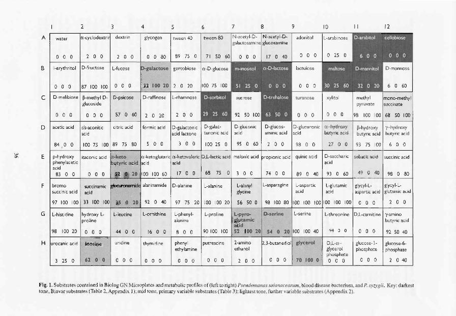
, .... .0,
2 3 4 5 6 7 8 9 10 11 12
AI water rx-cyclodextr'i dextrin glycogen tween 40 I twccn 80 I N-.cety l-D- I N .acety l.D- I adonito l
o 0 0 2 0 0 2 o 0 0 0 RO 89 75 0
B I i-erythritol D-fl'uctose I L-fucose iliC~el gentobiose I n-D -glucose
.t?'</~w~r:l 0 0 0 87 100 100 I 0 0 0
CID-melibiose l p-methyl D- D-psicose glucoside
60 I 2
I pyruvate
0 0 0 I 0 0 0 57 0 0 20 2 0 0 0 0 0 0 0 0 98 100 100 168 50 100
D I acetic acid I eis-aconitic citric acid I fo rm ic acid D-galactonic D-gluconic D-glucos- D-glucu mnic a - hydroxy Il -hydroxy '(-hydroxy acid acid lactone acid aminic acid acid butyric add butyric acid butyric acid
84 0 0 100 75 100 89 75 80 5 0 0 3 0 0 9S 0 60 2 0 0 98 0 0 27 0 0 93 75 100 6 0 0
E p-hydroxy itaconic acid malonic acid ropionlc aCid quinic ac id D-sacchar ic sebacic acid Isucci nlc aCid phenylacetic acid acid acid
83 0 0 0 0 0 100 60 17 0 0 68 75 0 3 0 0 74 0 0 89 0 40 93 Q 60 I 49 0 40 I 98 0 80
F I bromo alaninamide D-alanine L-alanine L-aJany! L-aspal'agine L-aspartlc L-glutamic glycyl-L- glycyl-L-succinic acid acid glycine acid acid aspartic acid glutamic acid
97 100 100 33 100 92 0 40 97 75 20 100 100 20 56 50 0 98 100 80 100 100 100 100 100 100 0 o 0 2 0 0
G L-histidine hydroxy L- r L-ornith ine L-phenyl- L.proline L-sel'ine L-threonine D,L-carn itine y-amino alanine buty ric acid
8 0 0 90100 100 94 0 0 0 0 0 92 50 40
phenyl putrescine 2-amlno D,L·a- glucose-l - glucose-6-ethylamine ethanol glycerol
phosphate phosphate phosphate
3 25 0 F~~H'A.Q Wl o 0 0 I 0 0 0 0 0 0 0 o 0 2 0 0 0 0 0 0 0 0 0 0 0 2 o 40
Fig. I. Substrates contained in Biolog GN Microplates and me tabolic profiles of (l eft to ri ght) Pseudomonas sO/{//loct!antm. blood disease bacteri um. and P. syzygii , Key: darkest tone, Biovar substrates (Table 2. Appendix I) : mid tune, primary variahle substr.ltes (Table 3 ): lightest tonc, further variable substrates (Appendix 2).
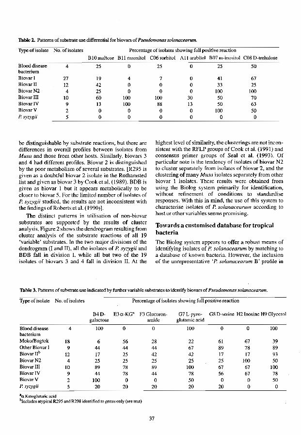
'Thble 2. Patterns of substrate use differential for biovars of Pseudomonas so/anacearum.
Type of isolate No. of isolates Percentage of isolates showing full positive reaction
BIOmaltose B II mannitol
4 25 0 bacterium Biovar I 27 19 4 Biovar 11 12 42 0 BiovarN2 4 25 0 BiovarlII 10 60 100 BiovarIV 9 13 100 BiovarV 2 0 0 P. syzygii 5 0 0
be distinguishable by substrate reactions, but there are differences in overall profiles between isolates from Musa and those from other hosts. Similarly, biovars 3 and 4 had different profiles. Biovar 2 is distinguished by the poor metabolism of several substrates. fR295 is given as a doubtful biovar 2 isolate in the Rothamsted list and given as biovar 3 by Cook et a!. (1989). BDB is given as biovar 1 but it appears metabolically to be closer to biovar 5. For the limited number of isolates of P. syzygii studied, the results are not inconsistent with the findings of Roberts eta!. (1990)].
The distinct patterns in utilisation of non-biovar substrates are supported by the results of cluster analysis. Figure 2 shows the dendrogram resulting from cluster analysis of the substrate reactions of all 19 'variable' substrates. In the two major divisions of the dendrogram (I and II), all the isolates of P. syzygii and BDB fall in division I, while all but two of the 19 isolates of biovars 3 and 4 fall in division 11. At the
C06 sorbitol A II arabitol B07 m-inositol C08 D-Irehalose
25 0 25 50
7 0 41 67 0 0 33 25 0 0 100 100
100 30 50 70 88 13 50 63
0 0 100 50 0 0 0 0
highest level of similarity, the clusterings are not inconsistent with the RFLP groups of Cook et a!. (1991) and eonsensus primer groups of Seal et al. (1993). Of particular note is the tendency of isolates of biovar N2 to cluster separately from isolates of biovar 2, and the clustering of many Musa isolates separately from other biovar I isolates. These results were obtained from using the Biolog system primarily for identification, without refinement of conditions to standardise responses. With this in mind, the use of this system to characterise isolates of P. solanacearum according to host or other variables seems promising.
Towards a customised database for tropical bacteria
The Biolog system appears to offer a robust means of identifying isolates of P. solanacearum by matching to a database of known bacteria. However, the inclusion of the unrepresentative 'P. solanacearum B' profile in
'Thble 3. Patterns of substrate use indicated by further variable substrates to identify biovars of Pseudomonas soianacearum. =----:-c---::----::-=----::c----~ ................... --~ .... . Type of isolate No. of isolates Percentage of isolates showing full positive reaction
B4D- E3a-KG' F3 Glucuron- G7L-pyro- G8 D-serine H2 Inosine H9 Glycerol galactose amide glutamic acid
Blood disease 4 100 0 0 100 0 0 100 bacterium MokolBugtok 18 6 56 28 22 61 67 39 Other Biovar I 9 44 44 44 67 89 78 89 Biovar lIb 12 17 25 42 42 17 17 93 BiovarN2 4 25 25 25 25 25 100 50 BiovarllI 10 89 78 89 100 67 67 100 BiovarlV 9 44 78 44 78 56 67 78 BiovarV 2 100 0 0 50 0 0 50 P. syzygii 5 20 20 20 20 20 0 0
aa Ketoglutaric acid b1nc!udes atypical R295 and R298 identified to genus only (see text)
37

0.4
I
n
0.6
I
'-
,---
-1
-
0.8
I
I I
~
1.0
I
~
M5J -1
c-
I
I
1.2
I Ill! BD! -, 1/30 BD! --.J 1/88 815 1/34 801 R6001 100 1113 illS! 1001 ',mm! Km I¥l Rill III iDOl I,SYlm!
:::: ::: ] 1134 illS! 1518 IlK! i5!! IIIS~ iDll III iDOl I,S!1lll! 1110 IIIS~ ill!! I,svml! 1115 illS! R70l III 1110 III 1I11 111 1153 III 1l0! III
- I, IIll 1111.1 1/01 illS! am 1111.1 Ill! 1111.1 am III 15M illS! 1/1/ illS! 1113 illS! 1/86 m 1/96 III 15!! 1115! 1/81 III 1138 III 1l0I m 1125 III RIM III R/IO III al9l 8IZ il!l III 1;61 8m RIll III Illl III IlOoIIll Will RI!! III 1/16 III ROll III R.!!I III I!.Il III R1IIIIl I/!I 114 IlIIIIII Rm III 1114 III RI" III RIll III 1111 III 1lI3 III 1135 m ail! lIISi il$! lIISi IlIl ml m"lIIl.I Ilil lIISi am III RI!l III
Fig. 2. Cluster analysis of substrate use by Pseudomonas spp. Dendrogram produced by UPGMA from SM coefficients.
38

the database distorts the identification process (see below) and BOB and P. syzygii are missing from the database. Common saprophytes and other non-pathogenic bacteria commonly associated with plants are not well represented in the Biolog databases. In some versions of the Biolog software, user-defined profiles may be used and customised databases constructed. However, such versions are at present only available in a package combined with plate-reading software; such a package was not purchased by this laboratory because general plate-reading software was already available and the cost was several times that of the nonexpandable MicroLog 1 program.
An alternative was to use the Bacterial Identifier software package, by Ward (1992). A matrix of the substrates in the Biolog plate was constructed instead of an array of conventional tests for which the program was originally designed. A working database was made by adding profiles of a number of other bacteria as well as P. solanacearum, P. syzygii and BOB. Identifications are made in a similar way to the Biolog software by entering well reactions (borderline results are entered as missing values). The major advantage of Bacterial Identifier is that it provides an alterable or expandable database for approximately the same cost as the non-expandable MicroLog 1 system. Bacterial Identifier also gives more information in cases where there is no positive identification and has the added advantage that individual sets of results can be stored. When a model pattern for P. solanacearum based on the substrates used by at least 84% of the isolates was entered in the Biolog identification program, there was no identification, but a similarity of 0.457 to P. so/anacearum B. With Bacterial Identifier and a database containing the' A' and 'B' profiles, this model isolate was identified satisfactorily as the' A' isolate.
A single comprehensive profile for P. so/anacearum may offer the best way of identifying this taxon. Discrimination of biovars or other sub-specific groups could be accomplished by analysis of metabolic profiles. However, the best way of proceeding needs to be evaluated by testing a series of 'unknown' isolates against differently arranged profiles. On completion of the wider project, a database of BioIog profiles and the matrix of substrates will be sent to the publishers of Bacterial Identifier with a view to future distribution.
Conclusions
Approximately 92% of aIJ the isolates of P. solanacearum tested were identified correctly with the Biolog system. Some interpretation of biovars is possible, with these and other sub-divisions becoming apparent on analysis of substrate patterns. With refinement of conditions of preparation and incubation, the Biolog system could become a powerful tool for investigating
39
the metabolism of P. solanacearum and related species. There is a need for representative profiles of these bacteria to be established in a database of profiles of tropical and sub-tropical bacteria from plants (including saprophytes). Alternative software to Biolog's own can be used to identify bacteria on the basis of results from the Biolog plates. This, combined with the Biolog plate system, could be a cost-effective means of identifying P. solanacearum in otherwise poorly equipped and supplied laboratories.
Acknowledgments
This work was funded under the Program Development Commission of the Overseas Development Administration (Project 10037). The authors acknowledge the supply of a review copy of Bacterial Identifier on extended loan from Blackwell Scientific Publishers and are grateful to Or Keilh Shawe for assistance with cluster analysis and Ms Amanda Burstow for technical assistance.
References
Bochner, B.R. 1989. Product review: sleuthing out bacterial identities. Nature (London), 339.157-158.
Bochner, B.R. and Savagrau, M.A. 1911. Generalized indicator plate for genetic, metabolic and taxonomic studies with microorganisms. Applied and Environmental Microbiology 33, 434-444.
Cook, D., Barlow, E. and Sequeira L. 1989. Genetic diversity of Pseudonwnas solanacearum: detection of restriction fragment length polymorphism with DNA probes that specify virulence and the hypersensitive response. Molecular Plant-Microbe Interactions, 2, 113-121.
Cook, D., Barlow, E. and Sequeira, L. 1991. DNA probes as tools for the study of host-pathogen evolution: The example of Pseudomonas solallacearum. In: Hennecke, H. and Venna, D.P.S. cd., Advances in Molecular Genetics of PlantMicrobe Interactions. The Netherlands, Kluwer Academic Publications, I, 103-108.
Eden-Green SJ. and Sastraatmadja, H. 1990. Blood disease of banana present in Java. FAO Plant Protection Bulletin, 38, 49-50.
Hartung, 1.S. and Civerolo, E.L. 1991. Variation among strains of Xanthomonas campestris causing citrus bacterial spot. Plant Disease, 75, 622-626.
Hayward, A.C. 1964. Characteristics of Pseudomonas solanacearum. Journal of Applied Bacteriology, 21, 265-271.
Hayward, A.C., El-Nashaar, H.M., Nydegger, U. and De Lindo, L. 1990. Variation in nitrate metabolism in biovars of Pseudomonas solallacearum. Journal of AppJied Bacteriology, 69, 269-280.
Roberts, S.1., Eden-Green, S.J., Jones, P. and Ambler, DJ. 1990. Pseudomonas syzygii, sp. nov., the cause of Sumatra disease of cloves. Systematic and Applied Microbiology. 13, 34-43.

Seal, S.E., Jackson, A.L. and Daniels, M.J. 1993. The use of tRNA consensus primers to indicate sub-groups of Pseudomonas solanacearum by polymerase chain reaction amplification. Applied and Environmental Microbiology. In press.
40
Ward, A. 1992. Software Reviews: Bacterial Identifier for the IBM PC and compatible microcomputer. Binary, 4, 42-43.

Appendix 1. Use of substrates differential for biovars of P. solanacearum. as indicated by substrate reactions on Biolog plate. (O=negative, I =borderline, 2= positive.)
Code Country Status
R229 Indonesia BOB R230 Indonesia BOB R234 Indonesia BOB R604 Indonesia BOB R483 Philippines Bugtok R639 Philippines Bugtok R650? Moko R369 Colombia Moko R589 Colombia Moko R372 Costa Rica Moko R570 Costa Rica Moko RJ56a Grenada Moko R598 Grenada Moko R375 Peru Moko R633 Philippines Moko R634 Philippines Moko R273 Trinidad Moko R522 Philippines Moko R283 Costa Rica Moko B R207 Belize Moko SFR R367 R282 R221 RI52 R286 R285 R281 R287 R296 R038 R301 R309
R703
R714
R576 R303 R295 R653 R710 R335 R632 R625 R298 R578 R568 R361 R583 R284 R304 R423 R374
Colombia Moko SFR Peru Moko SFR Indonesia Clove Colombia Heliconia Colombia Me/ampodium Costa Rica Potato Colombia Tobacco Mexico Brazil USA USA Australia
Brazil
Burundi
Chile
Tobacco Tobacco Tomato Tomato Potato
Potato
Potato
Potato Colombia Potato Costa Rica Potato Indonesia Potato Kenya Potato Peru Potato Philippines Potato Philippines Potato Sri Lanka Potato Peru Eggplant Brazil Potato Peru Potato Peru Potato Costa Rica Capsicum Costa Rica Eupatorium Malaysia Groundnut Costa Rica Heliconia
Given Correct AI2 BOg BIO BB C06 All B07 COS biovar ID Cellobiose Lactose Maltose Mannitol Sorbitol Arabitol m-inositol O-trehalose
1 2 2
2 2 2 2 2 2 2 2 2 2
N2 N2 N2 N2 3 3 3 3
NA NA NA NA Yes Yes Yes Yes Yes Yes Yes Yes Yes Yes Yes Yes Yes No Yes Yes Yes Yes Yes Yes Yes Yes Yes Yes Yes Yes Yes Yes
Yes
Yes
Yes Yes Yes Yes Yes Yes Yes Yes No No Yes Yes Yes Yes Yes Yes Yes
o o o o o o o o o o o o o o o o o o o o o o o o o o o o o o o o o o o o o o o o o o o o o o o o o o o
41
o o o o o o o o o o o o o o o o o o o o o o o o 1 o o o o o o o o 1
o o o o o o o o 1 o o o o o o o o
o 2 o o
1 2 1 2
1 2
o o o
1 2
1 2
o 1
2 2
1 2 2 2 1 2
1 1 2
o o o o o o o o o o o o o o o o o o o o o o o o 1 2 o o o o o o o 1 o o 1 o o o 1 o o o o o o 2 2 2 2
o o 2 o o o o o o o o 1 o o o o o o o o o o o o o 2 2 o o o o o o
o o 1 o o o o
o o o o 2 2 2 2
o o o o o o o o o o o o o o o o o o o o o o o o o o o o o o o o o o o o o o o o o o o o o o o 2 o o
o o o 2 2 2 2 o o 1 2 o o I 2 2 o o 2 o o 2 o
2 2
1 2 o o 1
2
1
o
1 1 o 2 I 2 2 2 2 2 2 2 o 2 o
o 2 2 o o 2 2 2 2 2
2 2 o 2 2 2 o 2 2 2 2 2 2 2 o
1 o
2 o o 1
2 2 2 o o o o o 1 2 2 2 2 o 2 2 2

Appendix 1. (cont' d) Use of substrates differential for biovars of P. solanacearum, as indicated by substrate reactions on Biolog plate. (O=negative, l=borderline, 2= positive.)
Code Country Status Given Correct Al2 B08 BIO Bll C06 All B07 C08 biovar ID Cellobiose Lactose Maltose Mannitol Sorbitol Arabitol m-inositol D-trehalose
R040 Costa Rica Physalis 3 Yes 0 2 2 2 2 0 2 R280 Australia Potato 3 Yes 0 0 2 2 2 1 2 R543 Australia Tobacco 3 Yes 0 0 2 2 2 2 2 R278 Australia Tobacco 3 Yes 0 0 2 2 0 2 0 R276 Peru Tomato 3 Yes 2 2 2 2 2 R291 China Mulberry 3\4 Yes 1 1 2 2 2 0 2 R279 Australia Ginger 4 Yes 0 0 2 2 1 2 0 R294 China Ginger 4 Yes 0 0 2 1 0 1 0 R293 China Groundnut 4 Yes 0 0 2 2 0 0 2 R290 China Olive 4 Yes 0 0 2 2 2 2 R289 China Olive 4 Yes 0 0 2 2 2 2 2 2 R027 Indonesia Potato 4 Yes 0 0 2 2 0 0 R297 Sri Lanka Potato 4 Yes 0 0 1 2 2 2 2 R300 USA Tobacco 4 Yes 0 0 1 2 2 1 2 R288 China Mulberry 5 No 0 0 0 0 0 0 2 2 R292 China Mulberry 5 Yes 0 1 I 0 0 2 ROOl Indonesia P. syzigii NA NA 0 0 0 1 1 0 0 ROO2 Indonesia P. syzigii NA NA 1 0 2 2 0 1 ROO4 Indonesia P. syzigii NA NA 0 0 1 0 0 0 0 0 ROO8 Indonesia P. syzigii NA NA 0 1 2 1 1 0 1 1 R099 Indonesia P. syzigii NA NA 0 0 0 0 0 0 0
42

Appendix 2. Use of further variable substrates by Pseudomonas solanacearum and related species, as indicated by well reactions on Biolog plales. (O=negative, l=bOl'derline, 2=positive.) ------- ..... -~.~---- --~.-~.~.--... -~.~----~.
COOe Country Status Given Correct A06 A08 B04 C03 Cl2 DlO E03 E05 E06 Ell F02 F03 FO? G03 G04 G07 G08 H02 H09 biovar ID
R229 R230 R234 R604 R483 R639 R650 R369 R589 R372 R570 R156a R598 R375 R633 R634 R273 R522 R283 R207 R367 R282 R221 R152 R286 R285 R2% R281 R287 R038 R301 R309 R703 R714 R576 R303 R295 R653 R710 R335 R632 R625 R298 R578 R568 R361 R583 R284 R304 R423 R374 R040
Indonesia BDB Indonesia BDB Indonesia BDB Indonesia BDB Philippines Bugtok Philippines Bugtok ? Moko Colombia Moko Colombia Moko Costa Rica Moko Costa Rica Moko Grenada Moko Grenada Moko Peru Moko Philippines Moko Philippines Moko Trinidad Moko Philippines Moko Costa Rica Moko B Belize Moko SFR Colombia Moko SFR Peru Moko SFR Indonesia Clove Colombia Heliconia Colombia Melampodium Costa Rica Potato Brazil Tobacco Colombia Tobacco Mexico Tobacco USA Tomato USA Tomato A ustralia Potato Brazil Potato Burundi Polato Chile Polato Colombia Potato Costa Rica Potato Indonesia Potato Kenya Potato Peru Potato Philippines Potato Philippines Potato Sri Lanka Potato Peru Eggplant Brazil Polalo Peru Potato Peru Potato Costa Rica Capsicum Costa Rica Eupatorium Malaysia Groundnut Costa Rica Heliconia Costa Rica Physalis
2 2 2 2 2 2 2 2 2 2 2 2
N2 N2 N2 N2 3 3 3 3 3
NA NA NA NA Yes Yes Yes Yes Yes Yes Yes Yes Yes Yes Yes Yes Yes No Yes Yes Yes Yes Yes Yes Yes Yes Yes Yes Yes Yes Yes Yes Yes Yes Yes Yes Yes Yes Yes Yes Yes Yes No No Yes Yes Yes Yes Yes Yes Yes Yes
o 0 2 0 0 0 0 0 020 0 0 0 2 0 0 2 o 2 0 0 0 0 0 2 0 2 0 0 0 0 2 002
202 2 0 002 0 2 0 2 0 0 2 002 202 2 0 0 0 2 1 2 0 2 0 0 2 002 1 002 122 0 2 221 022 1 2 2 020 2 1 I 1 222 0 2 2 0 200 I 2 0 201 1 0 1 0 2 2 0 2 0 0 2 2 2 202 2 2 2 2 2 0 1 221 2 0 022 1 2 221 1 222 2 1 2 2 2 1 021 1 220 2 2 2 1 021 I 200 100 1 000 1 200 0 0 2 0 2000010211220121
100 1 222 2 022 2 2 2 2 2 0 0 2 2 0 0 0 1 1 I 002 2 0 000 1 100 1 0 0 1 102 0 2 200 200 000 1 000 101 2 0 11022 202 220 220 222 o 0 0 0 2 0 101 0 0 1 0 0 0 0 0 2 1 2121210221002102 2 2 000 2 2 0 2 1 000 0 0 2 I 0 o 0 001 202 I 0 2 I 0 221 000 2 2 2 222 2 2 2 2 I 2 202101021011102120 2 1 0 2 0 0 102 2 1 202 1 2 2 2 2 2 2 022 1 2 1 222 1 220 2 2 2 2 2 0 2. I 2 1 I 022 1 100 0 2 002 2 002 2 1 2 2 2 2 202 2 2 2 222 20020 I 0 1 2 0 100 1 022 002 202 202 0 I 222
20021 22222 22 2212 20022 222 2 2 221 2 222 0001201010 11 10 01 100 1 2 0 0 0 0 0 I 0 0 0 0 0 2 211210101 21122112 200 2 2 0 0 0 2 2 2 2 222 2 0 2 20122010221122002 10222 1 202 2 I 2 1 0 2 2 2 2
001000000 2 0 0 0 000 2 200 I 2 0 001 0 1 000 0 0 0 2 200 2 2 0 0 0 2 1 222 1 2 002 2 002 2 000 1 0 I 0 0 002 2122222212221202 2 2 2 2 122 2 2 222 2 222 222 2 0 200 000 0 1 000 0 2 0 2 122 2 2 2 1 222 221 2 2 2 2 220200 1 10022 20020001 00020 202 222 2 2 2 2 2 2 2 2 222 J 2 1 0 2 2 2 202 1 2 1 2 0 2 2 2 2 2122210222002102 20222 I 2 0 2 2 2 2 2 2 2 2 2 2 221 2 122 2 2 222 2 2 2 2
43

Appendix 2 .(cont'd) Use of further variable substrates by Pseudomonas solanacearum and related species, as indicated by well reactions on Biolog plates. (O=negative, l=borderIine. 2=positive.)
Code Country Status Given Correct A06 A08 BM C03 C12 D10 E03 005 E06 Ell F02 F03 F07 003 G04 007 G08 H02 H09 biovar ID
R280 Australia Potato 3 Yes 2 0 2 2 2 2 2 1 2 2 2 2 2 2 2 2 2 2 R543 Australia Tobacco 3 Yes 2 0 2 0 2 2 2 2 2 2 2 2 2 2 2 2 1 2 2 R278 Australia Tobacco 3 Yes 2 0 2 2 2 0 0 2 2 2 1 2 0 2 2 R276 Peru Tomato 3 Yes 2 2 2 2 2 2 1 2 2 2 2 2 2 2 2 2 2 R291 China Mulberry 3\4 Yes 2 2 0 2 2 2 2 2 2 2 1 2 2 2 2 R279 Australia Ginger 4 Yes 0 0 0 2 2 2 2 0 2 2 2 0 2 2 2 2 R294 China Ginger 4 Yes 1 0 0 2 2 1 0 2 I 0 2 2 2 R293 China Groundnut 4 Yes 0 2 0 I 0 1 2 I 2 2 0 2 2 0 0 I 0 0 I R290 China Olive 4 Yes 2 0 2 2 2 2 2 2 2 2 2 2 2 2 2 2 I 2 R289 China Olive 4 Yes 2 2 2 2 2 2 2 2 2 2 2 2 2 2 2 2 2 R027 Indonesia Potato 4 Yes 2 0 I 1 2 0 0 0 2 1 1 0 0 0 0 1 0 1 0 R297 Sri Lanka Potato 4 Yes 0 2 2 2 2 2 1 2 2 2 2 I 0 2 2 2 2 R300 USA Tobacco 4 Yes 0 2 2 2 2 2 1 2 2 0 0 2 0 0 2 2 2 2 R288 China Mulberry 5 No 0 0 2 0 2 0 0 0 1 0 0 0 0 0 2 0 0 1 R292 China Mulberry 5 Yes 2 0 2 0 0 0 0 2 2 1 1 1 0 2 ROOl Indonesia P. syzigii NA NA 0 2 I 2 2 0 0 0 2 I 2 0 0 0 0 1 1 I 0 ROO2 Indonesia P. syzigii NA NA 1 2 2 2 2 0 0 0 2 2 2 0 0 0 0 2 0 1 ROO4 Indonesia P. syzigii NA NA 2 0 0 0 2 0 0 0 0 2 0 0 0 0 0 0 0 0 0 ROO8 Indonesia P. syzigii NA NA 2 0 1 2 0 0 0 0 2 2 I 1 0 0 1 0 0 R099 Indonesia P. NA NA 2 0 0 2 2 0 2 0 0 2 0 0 0 0 0 2 0 0
44

The use of the Biolog Identification System for the Rapid Identification of Plant Pathogenic Pseudomonads
Xiang Li and A.C. Hayward*
Abstract
The Biolog Identification System (Biolog. Inc., Hayward. California. USA) with microclimate 2 software was successfully applied within 24 hours to identify type and reference strains of Pseudomonas solanacearum (n=15) including biovars 1.2,3 and 4, P. andropogonis (n=17), P. caryophylli (n=l), P. cepacia (n=5), P. gladioli pv.gladioli (n=2), P. pseudomallei (n=2), P. pickeltii (n=l). P. rubrisulbabicans (n=l) and other pseudomonads and xanthomonads. Data analysis using the Taxan program (University of Maryland, MD. USA) showed that P. solanacearum isolates separated into two divisions equivalent to thuse obtained on restriction fragment-length polymorphism (RFLP) by other scientists. Biovar I including the type strain (K60) and biovar 2 comprised one division and shared 81.6% similarity; the other division containing biovars 3 and 4 shared 84.6% similarity. The two divisions were joined at 72.2% similarity. P. pickettii showed 66.6% similarity with P. solanacearum, whereas the remaining rRNA group 1I species joined with P. pickettii and P. solanacearum at a 55.2% level of similarity. We conclude that the Biolog Identification System is a useful tool for the rapid identification of nonfluorescent plant pathogenic pseudomonads including P. solanacearum.
Mf>.lIATIJRISED identification systems such as the API System have been widely and successfully used for the identification of clinically important bacteria, but their use in plant bacteriology has hitherto been somewhat limited. Whereas the API 20E serics is dcsigned for the diagnosis of human diseases caused by members of Enterobacteriaceae, API 20NE is uscd for bacteria of environmental origins. When the authors tested four strains of Pseudomonas andropogonis (E.F. Smith) Slapp from various hosts using API 20NE the results were identical, although identification was not achieved (unpublished data) which suggests limited application at present for plant pathogenic bacteria.
Biolog Identification System (BI System)-Biolog, Ine. Hayward, California. USA-serves for the identification of 569 taxa of Gram-negative and 223 Grampositive bacteria. The identification is based on the respiratory activity developing on a panel of 95 different carbon sources in a microtitre plate within a 4 hour and 16-24 hour incubation period, leading to a tetrazolium-based colour change. Organisms are identified according to the reaction pattern using a computer driven by MicroLogTM 2 software. There are
*Department of Microbiology, University of Queensland, Queensland 4072, Australia
45
no reports on the use of this system in identification of plant pathogenic pseudomonads. We report here on the application of BI System rapid identification of plant pathogenic pseudomonads, including P. solanacearum E.E Smith
Materials and Methods
Bacteria tested
Strains and isolates of P. andropogonis and P. solanacearum tested are listed in Tables 1 and 2. Other bacterial species listed in Table 3 were obtained from the Australian Collection of Microorganisms, University of Queensland (ACM) except for P. caryophylli (Burkholder) Starr and Burkholder 2313Q (NCPPB2151) which was obtained from Barbara Wood of the Queensland Department of Primary Industries (QOPI) and for P. cepacia (Burkholder) which was supplied by Paula Cathcart. All cultures except strains of P. solanacearum were previously identified using conventional bacteriological tests and SOS-PAGE of whole cell proteins. Strains and isolates of P. andropogonis were also identificd using immunofluorescence assay with monoclonal antibodies (Li ct al. 1993).
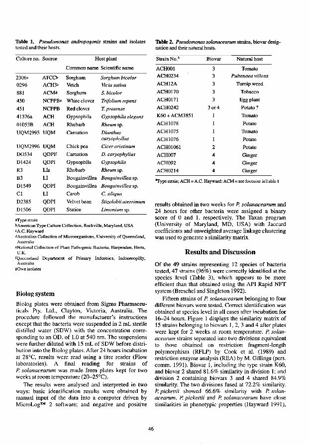
Table 1. Pseudomonas andropogonis strains and isolates tested and their hosts.
Culture no. Source Host plant
Common name Scientific name
2306a ATCCb Sorghum Sorghum biealor
0296 ACH3c Vetch Vicia sativa
881 ACMd Sorghum S. bicolor
450 NCPPBe White clover Trifolium repens
451 NCPPB Red clover T. pratense
41376a ACH Gypsophila Gypsophila elegans
01053B ACH Rhubarb Rheumsp.
UQM2995 UQM Carnation Dianthus caryophyllus
UQM2996 UQM Chickpea Cicer arietinum
00534 QOPlf Carnation D. caryophyllus
01424 QOPI Gypsophila Gypsophila
R3 LIg Rhubarb Rheum sp.
B3 LI Bougainvillea Bougainvillea sp.
01549 QOPI Bougainvillea Bougainvillea sp.
Cl LI Carob C. siliqua
02385 QOPI Velvet bean Stizolobii aterrimum
Dl506 QOPI Statice Limonium sp.
aType strain bAmerican Type Culture Collection, Rockville, Maryland, USA cA.C. Hayward dAustralian Collection of Microorganisms, University of Queensland, Australia
eNational Collection of Plant Pathogenic Bacteria, Harpenden, Herts, U.K.
fQueensland Department of Primary Industries, Indooroopilly, Australia
gOwn isolates
Biolog system
Biolog plates were obtained from Sigma Pharmaceuticals Pty. Ltd., Clayton, Victoria, Australia. The procedure followed the manufacturer's instructions except that the bacteria were suspended in 2 mL sterile distilled water (SDW) with the concentration corresponding to an OD. of 1.0 at 540 nm. The suspensions were further diluted with 15 mL of SDW before distribution into the Biolog plates. After 24 hours incubation at 28°C, results were read using a titre reader (Flow laboratories). A final reading for strains of P. solanacearum was made from plates kept for two weeks at room temperature (20-25°C).
The results were analysed and interpreted in two ways: basic identification results were obtained by manual input of the data into a computer driven by MicroLogTM 2 software; and negative and positive
46
Table 2. Pseudomonas solanacearum strains, biovar designation and their natural hosts.
Strain No.a
ACHOOl
ACH0234
ACH12A
ACH0170
ACH0171
ACH0242
K60+ACM3851
ACH1078
ACH1075
ACH1076
ACH01061
ACHOO7
ACH092
ACH0214
Biovar
3 3
3
3
3
30r4
2
4
4
4
Natural host
Tomato
Pultenaea villosa
Tumipweed
Tobacco
Eggplant
Potato?
Tomato
Potato
Tomato
Potato
Potato
Ginger
Ginger
Ginger
"Type strain; ACH = A.C. Hayward; ACM = see footnote inTable 1
results obtained in two weeks for P. solanacearum and 24 hours for other bacteria were assigned a binary score of 0 and 1, respectively. The Taxan program (University of Maryland, MD, USA) with Jaccard coefficients and unweighted average linkage clustering was used to generate a similarity matrix
Results and Discussion
Of the 49 strains representing 12 species of bacteria tested, 47 strains (96%) were correctly identified at the species level (Table 3), which appears to be more efficient than that obtained using the API Rapid NFf system (Breschel and Singleton 1992).
Fifteen strains of P. solanacearum belonging to four different biovars were tested. Correct identification was obtained at species level in all cases after incubation for 16-24 hours. Figure 1 displays the similarity matrix of 15 strains belonging to biovars 1,2,3 and 4 after plates were kept for 2 weeks at room temperature. P. solanacearum strains separated into two divisions equivalent to those obtained on restriction fragment-length polymorphism (RFLP) by Cook et a1. (1989) and restriction enzyme analysis (REA) by M. Gillings (pers. comm. 1991). Biovar 1, including the type strain K60, and biovar 2 shared 81.6% similarity in division 1; and division 2 containing biovars 3 and 4 shared 84.9% similarity. The two divisions fused at 72.2% similarity. P. pickettii showed 66.6% similarity with P. solanacearum. P. pickettii and P. solanacearum have close similarities in phenotypic properties (Hayward 1991),

Table 3. The accuracy of bacterial identification using Biolog Identification System with MicroLog'" 2 software.
Species
(No. of strains)
Biolog identification
(Species level)
Correct identification
Pseudomo/Uls andropogonis (17)T P. caryophylli (l)T
P. cepocia (5)T
P. gladioli pv. gladioli (2)T
P. pseudomallei (2)
P. pickettii (1)
P. solanacearum (l5)h
P. rubrilineans (l)
P. andropogonis
P. caryophylli
P. cepacia
P. g. pv. gladioli
P. pseudomallei
P. pickettii
P. sola/Ulcearum
P. rubrilineans
2
15
P. rubrisubalbicans (1)
P. syringae pv. syringae (2)
Xanthomo/Uls campestris
pv. campestris (1)
P. rubrisubalbicans
P. syringae
X. c. pv. campestris
E. coli (I)T
'Some of these results were repeated in expired plates b-rype strain included.
E. coli
as well as in whole cell fatty acid analysis (Janse 1991). Both species differed from the other rRNA group II pseudomonads, such as P. caryophyUi and P. gladioli, in lacing two ornithine amide Jipids (Galbraith and Wilkinson 1991), and form a separate subgroup within
Similarity (%)
"C.-,O _~_!LO _~_!LO_~_7Lf _~_~~o _,--_lL'_,,----,I"
4{1.70
Fig. 1. Dendrogram generated from Biolog Identification System using Taxan with laccard coefficients and unweighted average linkage clustering. A: Strains of Pesudomonas cepacia and P. gladioli pv. gladioli. B: P. caryophyulli. C: P. pseudomallei. D: P. pickettii. E: Biovars 3 & 4 of P. solanacearum. F: Biovars I & 2 of P. solanacearum. G: Strain of P. rubrisubalbicans. H: strains of P. andropogonis. I: P. rubrilineans. J: X. campestris pv. campestris.
47
the rRNA group 11 pseudomonads on the basis of rRNA-DNA hybridisation (Palleroni et al. 1973: Palleroni 1984). The other rRN A group II pseudomonads tested, including P. caryophylli, P. cepacia, P. gladioli Severini and P. pseudomallei (Whitmore) Haynes, formed another group at a similarity level of 67.4%-see Figure 2 in this study. Because of their close phenotypic relationship, these strains could not be further subdivided.
Seventeen strains of P. andropogonis isolated originally from 12 different host plants were identified as P. andropogonis (n = 14) and P. woods;i (n 3). These two species were always listed as the top two species in the list of 10 most closely related species given on analysis of the data generated for each of the 17 strains of P. andropogonis. It is widely accepted that P. woodsii (Smith) Stevens is a later synonym of P. alldropogonis (Hayward 1983). The results of identification by the BI System were in agreement with other results such as conventional bacteriological tests, whole cell protein profiles (data not shown), and immunofluorescence assay with monoclonal antibodies (Li et al. 1993). and showed that these strains of P. andropogonis were almost identical. There were no major differences in the profiles obtained for P. andropogonis strains in expired and unexpired Biolog plates (data not shown).
Strain ACM 2552 received as P. rubrilileans (Lee et al.) Stapp was identified as P. avenae Manns in this study, which supports the proposal that P. rubrilileans and P. aveltae are synonymous (Ram undo and ClaHin 1990).

av.
3 0171
3 0234
3 012A
? 0242
• 092
3 001
30170A
• 0214
4 007
I 3851
2 0158
201061
101076
101078
101075
lOO
94.12
92.16
96.08
93.75
Similarity (%) 90
88.0
84.91
80
72.22
Fig. 2. Dendrogram of strains of Pesudomonas solanacearum in Biolog Identification System using Taxan program with Jaccard coefficients and unweighted average linkage clustering.
ACM 978 is an authentic culture of P. rubrisubalbicans (Christopher and Edgerton) Krasil, Nikov, a species which is not yet included in the MicroLogTM 2 software data base. The strain shared 69.8% similarity with P. andropogonis. Both P. rubrisubalbicans and P. andropogonis were recently included in the rRNA group 11 pseudomonads (De Vos et al. 1985). Both species formed a separate subgroup in the similarity matrix (Fig. 1).
Although the identifications of P. syringae pv. syringae and Xanthomonas campestris pv. campestris were successful at species level, we failed to identify correctly at pathovar level. Both species contain a large number of pathovars (Palleroni 1984), which cannot be differentiated using standard bacteriological tests, especially X. campestris (Moffett and Croft 1983). To identify these pathovars using BI System, more representative isolates need to be investigated.
It is concluded that the Biolog Identification System is a useful tool for the rapid identification of plant pathogenic pseudomonads including P. solanacearum, with 47 strains representing 12 species of bacteria being identified to species level with an accuracy of 96%.
Acknowledgments The expert advice and assistance of Derek Warren, Manager of SPL Diagnostics in use of the Biolog
48
Identification System and the financial support of the University of Queensland to the first author by the award of a University of Queensland Postgraduate Research Scholarship are gratefully acknowledged. We thank Miss Barbara Wood (QDPI) and Miss Paula Cathcart (UQM) for supplying some cultures.
References Breschel, T.S. and Singleton, F.L. 1992. Use of the API rapid
NFf system for identifying nonfermentative and fermentative marine bacteria. Applied and Environmental Microbiology, 58, 21-16 .
Cook, D., Barlow, E. and Sequeira, L. 1989. Genetic diversity of Pseudomonas solanacearum: detection of restriction fragment length polymorphisms with DNA probes that specify virulence and the hypersensitive response. Molecular Plant-Microbe Interactions, 2, 113-121.
De Vos, P., Goor, M., Gillis, M. and De Ley,J. 1985. Ribosomal ribonucleic acid-dstron similarities of phytopathogenic Pseudomonas species. International Journal of Systematic Bacteriology, 35,169-184.
Galbraith, L. and Wilkinson, S.G. 1991. Polar Jipids and fatty acids of Pseudomonas caryophy/li, Pseudomonas gladioli and Pseudomonas pickettii. Journal of General Microbiology, 137,197-202.
Hayward, A. C. 1983. The non-fluorescent pseudomonads. In: Fahy, P. C. and Persley, G. J. eds, Plant Bacterial DiseasesA diagnostic guide. Sydney, Australia: Academic Press, 108-140.
-1991. Biology and epidemiology of bacterial wilt caused by Pseudomonas solanacearum. Annual Review of Phytopathology, 29, 65-87.
Janse, J. D. 1991. Infra- and Intraspecific classification of Pseudomonas solanacearum strains, using whole cell fatty acid analysis. Systematic and Applied Microbiology, 14,335-345.
Li, X., Wong, W.C. and Hayward, A.C. 1993. Production and use of monoclonal antibodies to Pseudomonas andropogonis. Journal of Phytopathology. In press.
Moffett, M.L. and Croft, B.J. 1983. Xanthomonas. In: Fahy, P. C. and Persley, G. J. ed., Plant Bacterial Diseases-a diagnostic guide. Sydney, Australia, Academic Press, 189-228.
Palleroni, N. J. 1984. Genus Pseudomonas Migula 1894, In: Krieg, N. R. and Holt, J. G. eds, Bergey's Manual of Systematic Bacteriology Vol: 1. Baltimore, The Williams and Wilkins Co., 141-199.
Palleroni, NJ., Kunisawa, R., Contopoulou, R. and Doudoroff, M. 1973. Nucleic acid homologies in the genus Pseudomonas. International Journal of Systematic Bacteriology, 23, 333-339.
Ramundo, B. A. and Claflin, L. E. 1990. Demonstration of synonymy between the plant pathogens Pseudomonas avenae and Pseudomonas rubrilineans. Journal of General Microbiology, 136,2029-2033.

Classification and Identification of Pseudomonas solanacearum and Other Pseudomonads
by Fatty Acid Profiling
D. E. Stead*
Abstract
Classification of Pseudomonas solanacearum and other pseudomonads by fatty acid profiling paralleled that based on nucleic acid homology. Classification of taxa in rRNA homology groups 1,2 and 3 was made on the basis of the types of hydroxy fatty acid found. Fatty acid group 1 members (the fluorescent pseudomonads) all had 10:0 30H and 12:0 30H. Group 2 members (P. solanacearum and its allies) all had 14:0 30H. 16:1 20H, 16:0 20H and 18:1 20H. Group 3 members (now largely reclassified in Comamonas and Acidovorax) all had 10:0 30H. P. solanacearum and its allies were further subdivided according to the additional presence of 16:0 30H, present in P. cepacia and its allies but absent in P. solanoceorum, P. syzygii and the banana blood disease bacterium. Differences in profiles of taxa within group 2 were based largely on differences in the amounts of fatty acid present rather than di rr erences in the types of acid found. but these were sufficient for accurate differentiation at species level except for some P. gladioli strains which were identified as P. cepacia. P. solanacearum, P. syzygii and the banana blood disease bacterium and had very similar fatty acid profiles and seem to form a discrete group. Differentiation of races and biovars of P. solanacearum was not sufficiently accurate under the cultural conditions selected. The use of slightly older cultures on other media may allow accurate race determination. However, fatty acid profiling showed that several strains of P. solanacearum tested had atypical profiles although they undoubtedly belonged to this sub-group. Fatty acid prot11ing offers a rapid, accurate method for diagnosis of disease caused by P. so/anacearum.
THE genus Pseudomonas comprises several nucleic acid homology groups worthy of genus status and recently species have been transferred to other genera including Comnmonas, Acidovorax, Sphingomonas and Xanthomonas (Young et al. 1992). Pseudomonas solanacearum and its allies are not closely related to the fluorescent pseudomonads (Palleroni 1984).
Fatty acid profiling is now regarded as a valuable taxonomic tool and several workers have shown the value of the hydroxy fatty acids in classification of Gram-negative bacteria and especially Pseudomonas (Oyaizu and Komagata 1983; Stead 1992a).
Fatty acid profiling is also being increasingly used as a rapid method of strain determination, and at least one commercial system is now available and gaining credence as a rapid, accurate method for identification
*National Collection of Plant Pathogenic Bacteria, Central Science Laboratory, MAFF. Hatching Green. Harpenden. Hertfordshire AL5 2BD, United Kingdom
49
of bacteria including the plant pathogens (Stead el al. 1992).
Fatty acid profiling comprises culture of the strain under standard conditions, harvesting, saponification of cellular Iipids, methylation. extraction and purification of resulting fatty acid methyl esters (FAMEs), gas chromatographic separation, identification and quantification of the FAMEs and finally computerised comparison of this profile with a FAME library containing a wide range of profiles of strains grown under the same conditions.
This study aimed to assess the value of fatty acid profiles for classification and identification of P. solanacearum and its allies in rRNA homology group 2 (Palleroni 1984).
Materials and Methods
All strains were from the National Collection of Plant Pathogenic Bacteria (NCPPB) and private collections, or were freshly isolated from imported diseased potato

tubers. They were maintained either freeze-dried or on beads at -BO°C (Protect System). Fatty acid profiles were prepared by the method of Stead (1992b) from 24 hour trypticase soy agar plates (28°C) for all strains except for Pseudomonas symploci (48 hours on potato dextrose agar), P. syzygii (6 days on casein salts agar) and the banana blood disease bacterium (48 hours on trypticase soy agar). Identification by comparison with profile libraries was made using the Microbial Identification System (MIDI), Delaware, USA. Profile libraries and dendrogram analyses were prepared using Library Generation Software (MIDI).
Results
All profiles selected for further analysis had more than 50000 units of peak area to avoid diluting out minor FAMEs. All also had more than 85% and usually more than 95% of the total peak area comprised of named peaks. The major FAMEs present in each taxon in rRNA group 2 (> 0.3% of the total peak area) are presented in Figure 1. The results in Table I indicate the presence of hydroxy FAMEs in fatty acid profile groups 1-3 and Xanthomonas. Table 2 lists the main groups and taxa occurring in each of groups 1-3.
FAME profile libraries were prepared for all selected taxa and a dendrogram based on Euclidean distance prepared (Fig. 2). This showed several distinct groups. The xanthomonads were clearly differentiated. P. solanacearum grouped closely with P. syzygii and the banana blood disease bacterium. The two Acidovorax species grouped together, as did all the fluorescent pseudo monads and P. corrugata. This
Table 1. Grouping of Pseudomonas species according to hydroxy fatty acids.
10:0 12:0 14:0 16:1 16:016:018:1 \3:01SO
30H 30H 30H 20H 20H 30H 20H 30H
l.rRNAI + + (+)a (+) (Fluorescent pseudomonads) 2a. rRNA2 (+) + + + + + (P. cepacia and allies) 2b.rRNA2 + + + + P. solanacearum and allies 3.rRNA3 + Comamonas and Acidovorax spp. (P. avenae) 4. Xanlhomonas spp. (+) + (P. maltophilia)
a( +) occurs in some strains.
50
Table 2. Species included in fatty acid profile groups 1-4.
Group 1 P. fluorescens (12), P. syringae pv. syringae (19), P. syringae pv. phaseolicola (32), P. corrugata (12), P. alcaligenes (4), P. pseudoa/caligenes (2)
Group 2a P. cepacia (13). P. andropogonis (12), P. caryophylli (7), P. gladioli pv. gladioli (14), P. gladioli pv. agaricicola (5), P. gladioli pv. alliicola (47), P. glumae (14), P. plantarii (4), P. symploci (6)
Group 2b P. solanacearum (49), P. syzygii (4), Banana blood disease bacterium (6)
Group 3 Acidovorax avenae pv. avenae (28), A. avenae pv. cattleyae (3), A. avenae pv. citrulli (6), A. konjaci (3), Comamonas acidovorans (4), C. testosteroni (5)
Group 4 Xanthomonas campestris pv. campestris (36), Xanthomonas campestris pv. citri (10)
latter group was quite closely associated with the two Comamonas spp. All members of fatty acid profile group 2 clustered together although the Euclidean distances between the various sub-clusters were quite large. P. alcaligenes and P. pseudoalcaligenes clustered together but most closely to P. caryoplJylli.
Validation of the library was obtained by comparing all profiles considered for library preparation against the final library (Fig. 3). In most cases all strains of a given taxon were correctly identified, although several strains of P. gladioli pv. alliicola and P. gladioli pv gladioli were wrongly identified as P. cepacia. A single strain out of 130 P. solanacearum strains was wrongly identified as P. syzygii. This was strain NCPPB 1621, a potato strain from Mauritius which did not conform with biovars 1-4. The similarity index with P. syzygii was 0.51, considered to be too low for accurate identification.
For all other taxa, accuracy of identification was 100%. A further 50 P. solanacearum strains from the NCPPB or freshly isolated from imported potatoes (all race 3 isolates) were profiled and compared with this library. All were accurately identified.
Dendrograms and two-dimensional principal component plots of individual profiles of P. solanacearum strains representing biovars 1, 2, 3 and 4 and races I, 2, 3, showed no diffcrentiation at biovar level (results not shown). Although races 2 and 3 showed great homology and perhaps could be differentiated, race 1 strains showed much greater variation. This heterogeneity did not allow accurate differentiation between race 1 and the races 2 and 3. This was further reflected by the relative inaccuracies obtained during validation of a separate library containing entries from each of the races (results not shown).

:::
l.o) __
~~~~~~:~~~3:: U'l(7'NCD ___ ..... _~U'!(pmNl
----~~------ ........ -------,. ~~
_!'\)_~W ___ !DN ...... ........ " .. \C-'<.nCDWN ...... <D<'o.O_
~~--.-...... ....." ..... ~"....,.,...... ............... N .................... _I7>. __ ............ wo ....... o.o ...... ""mCH.nmmJ)o ......................................................... --
N N~ NV ....... CDl:Io_ ............ 't·~,I:Io.~W ..... " .... . wcn_C'It)'1o ..... IC;.jlo...oa: .. ,c ..... .............. ...... ............
W,......U'1 ....... C'l.-.,......-..~ .. <"r, __ __
~~e~~~~e~~~~
N _N_ _OO_U"'. __ <:,.:I<:.l_ ......
_~~~~O:...~~~~NO N ___ N,-.. .................... WNW ___ _
~~~ee~~~~b;e~
,......... ......N N.b_~U'I __ Q)U'ltJ·Vl4;iJ.
U1:~:~:::Z~~:: a, ;..".~:...;..,;..,::..::n~~~~~ .oJ ....... .....,..., ........... "-''-'_ .... ,...''"'',.,..
_ N
NN_C.o.
O_COW .. - -.,. ..... t"n_ ..........
t..nQl,.....C:::HXI
~ .. CD ....
0.0'10 -......... ...... ..c ..... ./:lI1'\,)
"' ~
p, Indropogorrfs
P. caryop,'1yl11
P. Cepacfa
P. gladioli pv. gladioli
CI.l
= r:::r OQ ""I
P. g}adf.1f pv. agulclcold g
P. glil01o]f pv. an Heal,
P. glumde
P. planUrH
P.symploc1
P. saJanacearum
P. syzygff
6anana blood bacterium
'"0 N ~
CI.l = r:::r OQ ""I 0 = '"0 N r:::r
Fig. 1. Fatty acid profiles for Pseudomonas Group 2. Values give mean percentages in profile with standard deviation in brackets.
51
Xanlhomonas-campestris-campestris Xanth()monas~campestris-citri _J ........ -----... ~- ----,
Pseudomonas-syzygii Pseudomonas-solanacearum
Banana blood disease bacterium Acidovorax-konjaci
Acidovorax-avenae-citrulli Acidovorax-avenae-cattleyae
Acidovorax-avenae-avenae Pseudomonas-j,yringae-syringae
Pseudomonas-syringae-phasco!ieola Pseudomonu'i-fluorescens
Pseudomonas-corrugata Comamonas-testosteroni
C omamonas-acidoyorans Pselid()mona:~-gladioli-alliicola
Pseudomunas-plantarii Pseudomona!~~gladioli-agaricicola
Pseudomonuj-gladioli.gladioli Pseutiomonas-cepacia ..... ,.-.
Pseudomonas-andropogonis Pseudomonas-!)ymploci '-.0
Pseudomonas-glumae Pseudomonas-caryophy/li -----, Pseudomonas-a/ca/igenes '-__ I
Psewiomonas-pseudualcaligenes
Euclidian distance
Fig. 2. Classification of Pseudomonas spp., Acidovarox spp., Comamonas spp .• and X{Ul,Iwmonas spp. according 10 fally acid profiles .
Discussion
Classification of the plant pathogenic and some other pseudomonads based on hydroxy fatty acid profiles paralleled that by nucleic acid homology (Palleroni 1984; Stead 1992a) and this technique must now be considered a very reliable and simple way of determining genus within the pseudomonads. Most species in each group were also readily differentiated. Profiles were generally very similar for all taxa except for those in group 2a (P. cepacia and its allies). All members of this group possess a diversity of profiles that, in the author's experience are almost unique. There was great variation in the amounts of several FAMEs found in the cell membrane (16: I cis 9, 17:0 cyclo. 18: 1 cis 11) and also in the presence of some hydroxy FAMEs presumably part of the lipopolysaccharide moiety (10:0 30H, 12:0 30H). These low molecular weight hydroxy FAMEs are not normally present in these bacteria. So far, this qualitative and quantitative variation appears to be associated with colony morphology and pigmentation and will be discussed elsewhere. Nevertheless, the variation accounts for the large Euclidean distances between clusters of taxa within Group 2a. This type of variation is not found in members of Group 2b (P. solanacearum and its allies).

P. andropogonis
P. caryophyl/i
P. cepacia
P. gladioloi
P. glumae
P. plantarii
P. solanacearum
P. symploci
P. syzygii
Banana blood bacterium
No. of GC runs
... '" ';:: " 0()
" is--§ <!
'" Q.;
::: .2 :§ .S! %- <..J " 0 '" ] e-'" <..J <..J 0()
Q.; Q.; Q.;
Bacterial strains .. e '" Je ....
'" :E: " .c; " <..J
~ ~ '" ..!:: .~ ",.g <!
<!
~ ~ ..S! ..S! N' § ., " Q §u "" '" Q.; Q.; Q.; Q.; Q.; ~15
Fig. 3. Validation of 250 fatty acid profiles against a self-generated library. Values give accuracy of identification in per cent.
These fatty acid profile differences may well support a case for creation of two genera within rRNA homology group 2, one for P. solanacearum and its allies and another for P. cepacia and its allies. This has occurred in rRNA group 3 (fatty acid profile group 3) now referred to as the Comamonadaceae which contains Comamonas and Acidovorax, again both of which cluster differently in Figure 2. Dendrograms such as this must be cautiously interpreted as relatively small changes in the major cell membrane fatty acids may mask taxonomically important hydroxy acids present in relatively small amounts. This explains the relative similarities between P. caryophylli and the non-fluorescent members of rRNA group 1 (P. alcaligenes and P. pseudo-alcaligenes) and between the Comamonas spp. and the fluorescent Pseudomonas spp. in which the major difference is the absence in Comamonas of 12:0 30H. Even in Pseudomonas rRNA 1 members, this FAME rarely comprises more than 5% of profiles under these cultural conditions. Nevertheless, classification of profiles based on the pattern recognition systems used in strain identification (Fig. 2) does not compromise that obtained from simple presence or absence of key hydroxy fatty acids (Table 2).
52
Library validation, in general showing 100% accuracy of identification, indicated the excellent potential for the use of fatty acid profiles for rapid identification at species level. Again, such validations must be treated with caution since they tend to be incestuous, in that the very profiles used in library generation are re-compared with that library. Failure to obtain a high accuracy of identification at this level would almost certainly mean decreased accuracy of identification of other strains. However, such failures usually imply either wrong classification of the taxa or, at the least, incorrect identification of some of the strains currently included in that taxon.
P. solanacearum, P. syzygii and the banana blood disease bacterium clustered closely together and were quite discrete from any of the other pseudomonads both in their hydroxy FAME and overall profiles (Table I; Figs. 1 and 2). This was the case even though all three taxa were cultured under differing conditions although all were probably at a similar physiological age. It is thus difficult to be certain to what extent the differences between these taxa are due to their taxonomic differences or the cultural differences. Nevertheless, all but one of 180 P. solanacearum strains either in the validation or identification exercises were correctly identified.

Results not presented here showed that there was much greater variation between profiles of race 1 strains of P. solanacearum than those of races 2 and 3. In general. validation showed that race 3. biovar 2 strains were identified with a higher similarity index than race 1 and 2 strains. This probably reflects this homogeneity within race 3 and the fact that approximately half the 49 strains finally selected for the P. solanacearum library entry were race 3 isolates. Apart from the homogeneity in biovar 2 (equivalent to race 3) strains. no other relationship was noticed for biovars 1,3 and 4. It is interesting to note that the one P. solanacearum strain incorrectly identified in the validation exercise did not belong to any of the four P. solanacearum biovars (Stead 1988). Its profile was definitely of group 2b type. In a similar study, Janse (1991) was able to demonstrate that, based on 48-hour cultures on trypticase soy agar at 28°C, four major clusters were found. corresponding to races 1.2 and 3. The fourth cluster contained several deviating race I, biovar I strains. Another study, still in progress (Stead, unpublished data). has further demonstrated that slightly older cultures (48 hour) may give more stable profiles with better differential qualities. Also, certain types of culture medium give similar profile types, but some media produced profiles that not only showed great difference in the relative amounts but also in the types of FAME present. It is hoped that improved selection of cultural conditions will further improve the taxonomic value of fatty acid profiling both in classification and identification of P. solanacearum strains. This value has already been demonstrated at species level and fatty acid profiling has proven to be an excellent rapid method for identification of P. solanacearum and many other pseudomonad species.
Results can be obtained in 24--48 hours from obtaining a pure culture. Costed over a five-year period, based on U.K. prices and including consideration of equipment, consumable and staff costs per identification, fatty acid profiling proved much more cost effective than disease diagnosis or strain identification by conventional means such as those of L.elliolt and Stead (1987).
Acknowledgments This work was funded by the Ministry of Agriculture, Fisheries and Food, Plant Health Division (projects PH14000 and SCI8000). I wish to thank Mrs J. Sell wood, Mrs J. Wilson and Miss 1. Hennessy for technical support and Mr J. Morrison and Mrs 1. Tough and her staff for assistance with graphic design and word processing.
53
References
Janse, 1.0. 1991. Infra- and intraspecific classification of Pseudomonas solanacearum strains, using whole cell fatty acid ananlysis. Systematic and Applied Microbiology, 14, 335-345.
Lelliott, R.A.and Stead, D.E. 1987. Methods for the diagnosis of bacterial disease of plants. Oxford, Blackwell Scientific Publications, 216 p.
Oyaizu, H. and Komagata. K. 1983. Grouping of Pseudomonas species on the basis of cellular fatty acid composition and the quinone system with special reference to the existence of 3-hydroxy fatty acids. Journal of General Microbiology,29,17-30.
PaIleroni, N.l. 1984. Genus Pseudomonas Migula 1894, In: Krieg, N.R., and Holt, J.G. eds., Bergey's Manual of Systematic Bacteriology VoL l. Baltimore, The WilIiams andWilkinsCo .. 141-199.
Stead, D.E. 1988. Catalogue of cultures in the National Collection of Plant Pathogenic Bacteria, 2nd ed. Harpenden, Central Science Laboratory Publications, 144 p.
-1992a. Grouping of plant pathogenic and some other Pseudomonas spp. using cellular fatty acid profiles. International Journal of Systematic Bacteriology, 42. 281-295.
-I 992b. Techniques for detecting and identifying plant pathogenic bacteria. In: Duncan, J. and Torrance, L.ed .• Techniques for rapid diagnosis of plant pathogens. Oxford. Blackwell Scientific Publications, 76-11l.
Stead, D.E., Sellwood, J.E., Wilson, 1. and Viney, I. 1992. Evaluation of a commercial microbial identification system based on fatty acid profiles for rapid, accurate identification of plant pathogenic bacteria. Journal of Applied Bacteriology, 72, 315-321.
Young, J.M., Takikawa, Y., Gardan, L. and Stead, D.E. 1992. Changing concepts in the taxonomy of plant pathogenic bacteria. Annual Review of Phytopathology, 30, 67-105.

Serological Detection of Pseudomonas solanacearum byELISA
A. Robinson*
Abstract
Several assays ro; the detection of Pseudomonas solallacearum are currently being developed in laboratories worldwide. Many of these techniques, although highly efficient at detecting the bacterium, require sophisticated equipment and are difficult to perform under field conditions, (e.g. the use of DNA specific probes). The Enzyme Linked Immunosorbent Assay (ELISA) does not require sophisticated equipment, is relatively inexpensive, requires the minimum of training and may be readily adapted for use in developing countries in the tropics, where the disease is prevalent. An ELISA has therefore been developed which may be used to detect the bacterium in both plant and soil samples. This ELISA has been optimised with respect to sensitivity, and is able to routinely detect bacterial levels as low as 1 x 10⁴ cfu/mL. The assay at present uses polyclonal antibodies but recent advances in the production of P. solallacearum specific monoclonals mean that they will be used in the future. Such monoclonals may be produced (indefinitely) as standardised reagents, which will further improve the reliability of the assay.
BACfERIAL wilt caused by Pseudomonas solanacearum is a disease widely distributed in tropical, subtropical and wann temperate regions of the world. It is one of the most destructive bacterial diseases of plants and affects a wide range of economically important crops.
Research into a suitable assay for diagnosing the presence of P. solanacearum in plant and soil samples has been under way for a number of years. The original technique involving isolation and purification of the bacterium from the infected sample, followed by biochemical analysis (Hayward 1964), is tedious and time consuming. New techniques currently being reported, e.g. the use of non-radioactive DNA probes (Seal et aL 1992), although far more rapid, are still not suitable for use under field conditions. Immunoenzymatic techniques however, are ideal and have been used by plant virologists for the rapid identification of viruses for years. Such techniques have also been used for the diagnosis of plant pathogenic bacteria since 1978 (Cambra and L6pez 1978; Vruggink 1978).
The serology of P. solanacearum has been studied by many workers, e.g. MoTIon et aL (1966) and Schaad et al. (1978), but only recently has this been applied to
*Plant Pathology Department, AFRC-IACR, Rothamsted Experimental Station, Harpenden, Herts., AL5 2JQ, V.K.
54
immunocnzymatic techniques. For example, an Enzyme Linked lmmunosorbent Assay (ELISA) for the detection of P. solanacearum in potatoes was developed (Bellstedt and Van Der Merwe, unpublished data). This ELISA, as with many in the past, used polyclonal antibodies. The quantity, quality and affinity of such antibodies for a particular antigen often vary from animal to animal and, quite often, even from different bleeds of the samc animaL This leads to variability in serum quality which, in turn, may lead to an unreliable assay. Polyclonals also tend to lack specificity, reacting not only with the bacterium to which they were made, but also with closely related strains.
The introduction of hybridoma technology by Kohler and Milstein (1975) provided a revolutionary advance in the production of antibodies, eliminating many of the problems associated with polyclonals. Hybridoma cells result from the fusion of Blymphocytcs (antibody producing cells) with myeloma cells (malignant cells which do not secrete antibody and are immortal in cell culture), and are therefore able to produee antibodies and be cultured indefinitely ill vitro. Each hybridoma cell secretes identical antibodies to single anti genic detenninants and may therefore be selected for secretion of the desired antibody. Such antibodies are known as monoclonal. These monoclonals have several advantages over the

conventional polyc1onals in that: (a) an unlimited quantity of antibody is available; (b) there is no variability between different batches; and (c) specific antibodies may be selected for.
This paper discusses the development of an ELISA using polyc1onals and the subsequent development of monoclonals for use in the assay.
Materials and Methods
Production of polyclonal antibodies
Polyclonal antibodies were raised in Dutch-Lop cross rabbits. Initial immunisations of 5 x 10⁸ glutaraldehyde-fixed whole bacterial cells (Allan and Kelman 1977) in Freunds' complete adjuvant (Difco Labs) were followed with an identical boost in incomplete adjuvant 4 weeks later. All injections were given intramuscularly. Rabbits were bled via the ear vein after 2 weeks and at 2-3 weekly intervals thereafter. Collected blood was allowed to clot at room temperature, separated by centrifugation (1500g for 15 minutes) and the serum collected. The amount of antibody present in the sera (the titre) was determined byELISA.
Production of mono clonal antibodies Immunisation Protocols. Balb-c mice were immunised intraperitoneally using one of the following protocols. An injection of 1 x 10⁸ glutaraldehyde-fixed p. solanacearum cells in sterile saline was followed four weeks later by a test bleed (via the tail vein). The titres were measured by ELISA and, if sufficient, a final boost (as above) was given. In general, however, a second immunisation was required before the final boost to improve the titre (protocol a). In a second method, injection with glycoprotein antigens of P. solanacearum prepared from 1 x 10⁹ bacterial cells, as described by Digat and Cambra (1976), was followed with a second identical injection after 5 weeks and a final boost 6 weeks later (protocol b). In the third method, two groups of mice (A and B) were injected with a mixture of 1 x 10⁸ glutaraldehyde-fixed P. syzygii and the blood desease bacterium (BDB). Two days later group A were immunised with 20 mglkg cyclophosphamide (which kills proliferating Band T lymphocytes) in sterile saline. This entire procedure was repeated a further three times, at two-weekly intervals. Test bleeds were taken during the intervening weeks and titres compared, with group B mice acting as controls. After a three-week rest, the mice were immunised with a mixture of 5x 10⁸ glutaraldehydefixed whole cells of P. solanacearum strains, followed by a final identical boost a week later (protocol e).
The Fusion. Three days after the final boost in each of the above immunisation protocols, the mice were
55
euthanised and their spleens removed. The spleen cells were then fused with the myeloma cell line NSO, by spinning together in the presence of 50% polyethylene glycol, (Mo1.Wt. 1500, Boehringer Mannheim) and 10% dimethyl sulfoxide (99.5% pure, Sigma), after the method of Kennet et a1. (1978). Growth of the fused cells (hybridomas) was aided by supplementing the routine cell culture media, Dulbecco's modification of Eagle's medium (Flow Labs) containing 20% Foetal Calf Serum (Imperial Labs), with peritoneal macrophages from young mice.
Enzyme linked immunosorbent assay
There are many different ways of configuring ELISAs and a wide array of formats. The basic ELISA protocol used in this study was as described below. Any alterations to the protocol are detailed in the appropriate sections.
Incubations throughout were at either 3TC for one hour or 4'C overnight. After each stage, excess reagent was removed from the wells by 'flicking out'. The plates were then washed in three changes of PBSTween (phosphate buffered saline plus 0.05% v/v Tween 20), for 3 minutes each. After washing, the plates were gently 'bang dried' on tissue paper.
The routine protocol was as follows: 96 well microtitre plates were coated with a suspension of whole bacterial cells (lOO JlL per well) diluted to I x 108 cfuJ mL in 0.05 M carbonate coating buffer, pH 9.6. After incubation and washing, 100 JlL of either a monoclonal or polyclonal antibody diluted in blocking buffer (0.05% vlv Tween 20, 2% w/v polyvinyl pyrrolidone (PVP, Mol. Wt. 44000) and 0.5% w/v NIDO milk powder (Nestle) in phosphate buffered saline) was added and incubated. After washing, a second antibody, horseradish peroxidase conjugated rabbit anti-mouse or goat anti-rabbit (Sigma) at a 1:2000 or 1:5000 dilution, respectively, in blocking buffer, was added and incubated. Finally, 100 JlL ofTMB substrate (1 mg! mL 3,3',5,5'-tetramethyl-benzidine (Sigma), 0.1% v/v hydrogen peroxide and 10% v/v sodium acetate, pH 5.8 in distilled water) were added to each well and incubated at room temperature until sufficient colour developed. The reaction was stopped by adding 3 M sulphuric acid (25 JlL per well). Results were assessed visually, and then quantified by reading the absorbance at 450 nm (including a turbidity correction at 650 nm), using a microtitre plate reader (ICN Flow).
Optimisation of the ELISA
Plate type. Different brands of microtitre plate differ in binding capacity and also in consistency of binding within and between plates. Several commercially available microtitre plates were therefore tested for their binding abilities using a half-log dilution series of

whole bacterial cells (1 x 10⁸_1 X 101 cfulmL) to determine the lowest level of bacteria detected by a polyclonal antibody (R283 diluted to 1:5000).
Coating buffer. Phosphate-buffered saline (PBS). 0.1 M citrate buffer. pH 5.0 and pH 3.0, and 0.05M carbonate buffer, pH 9.6, were all compared for their ability to bind bacteria to the microtitre plate.
Blocking buffer. Buffers containing 1 % w/v Bovine Serum Albumin (BSA). O.S% w/v BSA, 1 % w/v full cream dried milk powder (DMP) or O.S% w/v DMP with or without the presence of Tween 20 (0.05% w/v) and, or PVP (2% w/v), were compared for their ability to prevent or reduce non-specific binding throughout theELISA.
Washings. Washings with tap water, PBS and PBSTween were compared using the method described previously.
Incubations. To determine the optimum incubation times of first and second antibodies, at 37"C, all combinations of 2 hours, 1 hour and O.S hour were compared. Optimum incubation for coating of bacteria was then determined using the optimal first and second antibody incubation periods.
EL/SA design. Double antibody sandwich ELISAs (DAS) were compared with the indirect binding ELISA previously described. In DAS, microtitre plates were pre-coated with monoclonal antibody diluted 1:100 or with polyclonal antibody diluted 1:2000 in coating buffer, to 'capture' the antigen (the bacteria). The routine ELISA protocol was then followed.
Diagnostic Assay
Plant samples. One-centimetre sections were cut from the stem bases at or close to the intersection between stem and tap root, or for tubers a plug of tissue containing vascular tissue from the stolon end, of artificially inoculated and naturally infected plants. The plant tissue was macerated mechanically and resuspended at I mUg in 0.05 M carbonate buffer, pH 9.6, containing 0.2% sodium sulphite (an antioxidant). The sample was allowed to settlc for 30 minutes and the supematant used to coat wells of a microtitre plate (100 j.lL per well) in the routinc ELISA.
Soil samples. One gram samples of artificially inoculated soil were suspended in 2 mL coating buffer and shaken vigorously for I minute. After allowing to settle for 2 minutes, 100 j.lL of the supematant was removed and used in the routine ELISA. During testing, two coating buffers were used: (i) O.OSM carbonate buffcr, pH 9.6, and (H) an amended coating buffer containing 8.3% polyvinyl pyrrolidone and 4% sodium cholate in 0.05 M carbonate buffer, pH 9.6, (S.M.D. Forde, pers. comm.).
Results
Polyclonal Antibodies
To date, three polyc1onal antibodies to P. solanacearum (R283, R303, R608) and onc to the blood discase bacterium (BDB, R230) have been raised at Rothamsted Experimental Station (Eden-Green and Robinson. unpublished). All were found by ELISA to have very high titres (Table I). The polyc1onal to BDB is as good at detecting P. solanacearum as those antibodies actually raised to P. solanacearum. All antibodies show cross-reactions to P. syzygii, the BDB P. pickettii and P. cepacia. They also lack specificity with respect to the biovarslraces of P. solanacearum, detecting all isolates at 1 x 10⁸ cfulmL (Table 2). If the bacterial concentrations are lowered to < 1 x 10⁵ cful mL. not all isolates are detected.
Monoclonal antibodies
Monoclonal antibodies produced by immunisation protocol (a) proved to be more specific than the polyclonals, detecting only certain P. solanacearum isolates at 1 x 10⁸ cfulmL. However, cross-reactions with P. syzygii, the BDB and P. pickettii still occurred (Table 3).
Immunisation protocol (b) proved to be toxic to the mice, killing 20% within a day of the injection. Titres from the surviving mice pre-fusion were fairly low, i.e. 1;1600 compared with >1:12800 for protocol (a), and less antibody secreting hybridomas were obtained postfusion. The few monoclonals resulting from this fusion
Table 1. TItres of polyclonal antibodies to Pseudomonas solanacearum, as determined by EUSA.
R283,bv.1 R303,bv.2 R608,bv.3 R230,BDB
R283, by. 1 1:256000 >1:520000 1:8000 1:32000 R295, by. 2 I: 128000 >1:520000 1:520000 1:128000 R608. by. 3 1:128000 1:256000 1:256000 1:16000 R297,bv.4 1:520000 >1:520000 1:256000 1:256000 R230, BDB 1:520000 1:520000 1:64000 1:128000
56

Table Z. Detection of P. solanacearum and other bacteria from the Rothamsted culture collection, by polyclonal antibodies.
Race R283 R230
R232 R281 R638 2,moko R651 2,buglok RI28 2 3 R710 2 3 R568 2(N) ? R361 2(N) ? R143 3 R799 3 R811 3 41 R812 3 4? R277 4 4? R300 4 I R471 4 1 R288 5 5 R292 5 5 Al P. cepacia A2 P. cepacia
ROOI P. syzygii
ROO2 P. syzygii
ROll B. subtiUs
R036 X. campestris
E. coli Rlll A. tumefaciens R137 BDB R226 BDB R228 BDB R230 P. picketti
R707
showed no greater specificity than those obtained with protocol (a).
During immunisation protocol (c) a comparison of the titres of cyclophosphamide treated and control mice showed that the cyclophosphamide had succeeded in removing cross-reactions. The fusion was only performed recently but, of the few monoclonals screened so far. no cross-reactions have been observed. Some interesting results regarding specificity to the biovarslraces of P. solanacearum have also been obtained (Table 4).
Optimisation of the ELISA Plate type. Table 5 shows the results obtained when different microtitre plates were tested. Both Griener (medium binding) and Nunc poJysorp plates gave good results and were therefore used for all subsequent assays. The lowest levels of detection obtained were high due to the polyclonal antibody being diluted 1:5000.
+ + + + + + + + + + + + + + + + +
+1-
+ +
+ +
+
57
+ + + + + + + + + + + + + + + + + + + + + + + + + + + + + + + + + + + + + + + + + + + + + + + + + + +
+1- +/- +
+ + + + + +
+ + + + + +
+ + +/- +1-
Coating buffer. The use of different buffers to coat the bacteria onto the plate made very little difference to the end results. Carbonate buffer O.05M, pH 9.6, was chosen for further work, however, as slightly higher absorbance readings were obtained with no increase in background levels.
Blocking buffer. Dried milk powder (0.5%) was found to decrease background levels to a greater extent than Bovine Serum Albumin (BSA). When the dried milk powder was then used with Tween 20 and polyvinyl pyrrolidone, non-specific binding was even further reduced with no detrimental effect on the sensitivity of the ELISA.
Washings. Washings done with PBS-Tween were found to be the most effective, giving lowest background readings with no loss in sensitivity.
lncubations. One-hour incubations were found to be sufficient for all stages, with 30 minutes sufficient for the second antibody.

Table 3. Detection of P. solanacearum and other bacteria from the Rothamsted Culture Collection, by monoclonal antibodies produced by immunisation protocol (a).
Race
R232 R296 R702 + R281 + R045 I 11 R638 I 2,moko R651 1 2, bugtok +/-R128 2 3 R710 2 3 R568 2(N) ? R573 2(N) ? + R361 2(N) ? RI43 3 R304 3 R799 3 R811 3 4? + R812 3 4? +/-R277 4 4? R289 4 1 R300 4 R471 4 R293 4 1 R288 5 5 R292 5 5 ROOt P. syzygii Roo2 P. syzygii
R036 X. campeslris
R044 P. cepacia
R226 BDB BDB
R228 P.picketli R707
EUSA design. Figure 1 shows the configuration of the three ELISAs which were compared. Both DAS ELISAs were found to be less sensitive than the indirect ELISA, and so were not used further.
Diagnostic Assay
Plant samples. Using the routine ELISA, P. solanacearum was detected in artificially inoculated and naturally infected plants, even when wilting was not apparent.
Soil samples. No bacteria were detected by the ELISA when the routine O.05M carbonate coating buffer, pH 9.6, was used to coat the soil samples to the plates. If, however, the bacteria were extracted from the soil particles and coated directly onto the plates using a buffer containing 8.3% polyvinyl pyrrolidone and 4%
+
+ + +
+ +
58
278
+/-+ + + + + +
+/-
+ + + + +
+ +
+/- + + + +
+/- + + +/-
+ + + +
+/- + + + + + + + + + + + +
+ + + + + + + +
sodium cholate in O.05M carbonate buffer, pH 9.6, (MacDonald 1986), P. solanacearum could be detected in the soil down to levels as low as 1 x 10⁴ cful mL. Bacterial levels as low as 1 x 10² cfu/mL were detected in some assays.
Discussion
The development of an ELISA to detect P. solanacearum has been described in this paper. This assay does not require sophisticated equipment, is relatively inexpensive and requires minimum of training, making it ideal for use in developing countries in the tropics, where the disease is prevalent. The ELISA is rapid and may be completed in as little as 3.5 hours. It has also been shown to be ve~ sensitive, routinely detecting bacterial levels of 1 x 10 cfulmL in both plant and soil samples.

Table 4. Detection of P. solanacearum and other bacteria from the Rothamsted Culture Collection, by monoclonal antibodies produced by immunisation protocol (c).
Race 35a.l 35b
R232 R296 R702 + + R281 + + R045 11 R638 2,moko R651 2, bugtok Rl28 2 3 R710 2 3 R568 2(N) ? + + R573 2(N) ? + + R361 2(N) ? + + R143 3 R304 3 R799 3 + + R81l 3 4? + + R812 3 41 + + R277 4 41 +1- +/-R289 4 1 R300 4 + + R471 4 1 + + R293 4 1 + + R288 5 5 R292 5 5 ROOl P. syzygii ROO2 P. syzygii
R036 X. campestris
R044 P. cepacia
R226 BDB BDB
R228 P. pickettii R707
Double antibody sandwich
Coat with monoclonal l.J Add antigen ~ Add test sample c:1J
Double antibody sandwich
Coat with polyclonal ~J Add antigen • ~ I
Indirect Coat with antigen lx
Fig. 1. Double antibody sandwich ELlSA vs indirect ELISA.
Add test sample I ! .. ~.
Add test sample. • I L::J
59
38b 47
+ + + +
+ + + + + +
+ + + + + +
+ + + + + +
Add enzyme conjugate 11: Lowest detection 2.5 x 10⁶ cfu/mL
II~'I Add enzyme conjugate LlJ Lowest detection 6.25 x 10⁵ cfulmL
'l' Add enzyme conjugate • y . ~
Lowest detection I x 104 cfulmL

Table S. Evaluation of several commercially available microtitre plates.
Plate type Background levels Lowest detection Other factors Rating level (cfulmL)
Ix Moderate Griener. medium Low 1 x 10⁷ Good Nunc, maxisorp High 5x 10⁷ Moderate Nunc, polysorp Low 5x 10⁷ Good Bioreba Medium 1 x 10⁷ Inconsistent Poor Costar Very high 0 Poor Falcon, flexible High 1 x 10⁸ Flexible plate Poor Techne 1 x 10⁸ Flexible
The detailed ELISA currently uses polyclonal antibodies that have been produced relatively easily. The fact that these polyclonals cross-react with several closely related bacteria is probably of little practical significance when testing infected plant tissue, as high populations of P. solanacearum are likely to occur in the absence of the cross-reacting bacteria. However, in Indonesia where both P. syzygii and P. solanacearum may be found in cloves and both the BOB and P. solanacearum in bananas, the test would be of little use. When testing soil samples, however, crossreactions may be a problem, as P. pickettii or P. cepacia may be present in high numbers.
Because of this cross-reactivity and the fact that polyclonals cannot be produced in unlimited quantities, a program to produce monoclonal antibodies to P. solanacearum was begun. It was also postulated that, by producing monoclonal antibodies, some specificity between the biovars/races of P. solanacearum would be obtained. Preliminary attempts at producing monoclonals by immunising mice with glutaraldehydefixed whole cells removed only those cross-reactions with P. cepacia. The monoclonals were far more selective in their detection of P. solanacearum, however, detecting only certain isolates. The majority of the isolates that these monoclonals detected seemed to show no specificity with regard to biovars/races.
Digat and Cam bra (1976) surveyed work on the serology of P. solanacearum and stated that the glycoprotein antigens of P. solanacearum give rise to antisera that are fairly specific for differentiating races and strains. Glycoprotein antigens were therefore prepared and used to immunise mice. The antigen was found to be toxic and not highly immunogenic, giving far lower titres than those obtained from immunising with whole cells, The resulting monoclonals were also disappointing, giving rise to a similar set of monoclonals as previously obtained.
The cytotoxic drug, cyclophosphamide, has been used by several workers (e.g. Matthew and Sandrock
60
1987) to 'manipulate' the immune system of mice to produce monocIonals of a desired specificity. Cyclophosphamide was used here to increase the tolerance of mice to a set of antigens (namely P. syzygii and the BDB). Immunisation with a similar set of closely related antigens (P. solanacearum) followed, in an attempt to produce antibodies which would distinguish between the two groups. Positive results were seen immediately, with test bleeds detecting none of the cross-reacting strains. Monoclonals produced by this method showed no cross-reactions and detected only certain isolates of P. solanacearum. Varying results were found within the biovars/races, except for (a) Bugtok and Moko, which were not detected, (b) biovars 2 and 2(N) which could be distinguished from each other, and (c) race 5 which could not be detected at all. Further analysis of these monoclonals is being performed to determine if this differential activity is consistent with any other grouping system, e.g. the RFLP groups described by Cook et al. (1990).
A panel of the monoclonals produced by immunisation schedules (a) and (b), which differentially detect certain isolates of P. solanacearum, has been used in the ELISA. The use of such a panel has allowed P. solanacearum to be identified at the biovar/race level. Results obtained were satisfactory but some isolates of P. solanacearum tested were not detected and closely related bacteria were sometimes detected. The use of cyclophosphamide may overcome this problem, as it may be used to give rise to monoc1onals of different specificity (e,g. if a mouse were first 'tolerised' to P. solanacearum biovars I, 2, 3 and 4, followed by immunisation with biovar 5, biovar 5 specific monoclonals might result). Such an assay would bc far superior to that developed using polyc1onals, as it would not only be detecting P. solanacearum to the biovar/race level, but it would also be more reliable, as the monoc1onals could be produced, indefinitely, as standardised reagents.

Acknowledgments
I thank Dr Simon Eden-Green for his help in producing polyc\onal antibodies, Stephen Forde for producing all bacterial stocks, and Dr John Elphinstone and Phil Jones for their help and critical reading of the manuscript This work was commissioned and funded by the Natural Resources Institute, Chatham, U.K., (project XOO71), and was carried out under MAFF licence PHFI 307 AI7 4/22.
References
Allan, E. and Kelman, A. 1977. ImmunoHuorescent stain procedure for detection and identification of Erwinia carotovora var. atroseptica. Phytopathology, 67, 1305-1316.
Cambra M. and L6pez M.M.. 1978. Titration of Agrobacterium radiobacrer var tumefaciens antibodies by using enzyme labelled anti-rabbit-globulins (ELISA indirect method). In: Proceedings of the fourth international conference on plant pathogenic bacteria. Angers, France, 327-331.
Cook, D., Barlow, E. and Sequeira, L. 1990. DNA probes as tools for the study of host-pathogen evolution: The example of Pseudomonas solonocearum. In: Hennecke, H. and Verma, D.P.S., ed., Advances in Molecular Genetics of Plant-Microbe Interactions. Kluwer Academic Publications: The Netherlands. Voll, 103-108.
Digat, B, and Cambra. M. 1976. Specificity of antigens in Pseudomonas so/alltlcearum E. F. Smith and application of serology for studying bacterial wilt. In: Sequeira, Land Kelman, A., ed., Proceedings of the First International Planning Conference and Workshop on the Ecology and Control of Bacterial Wilt caused by PseudomofUls so/anGcearum. Raleigh, North Carolina, July 18--24, 1976,38.
61
Hayward, A.C. 1964. Characteristics of Pseudomonas solanacearum. Journal of Applied Bacteriology, 27, 265-277.
Kennet R.H., Davis, K.A., Tung, A.S., and Kilman, N.R. 1978. Hybrid plasmocyta. Production: Fusion with adult spleen cells, spleen fragments, neonatal spleen cells and human spleen cells. Current Topics in Immunology, 81, 77-91.
Kohler, G. and Milstein, C., 1975. Continuous cultures of fused cells secreting antibody of predefined specificity. Nature (London), 256, 495-97.
Macdonald R.M., 1986. Sampling soil microfloras: dispersion of soil by ion exchange and extraction of specific microorganisms from suspension by elutriation. Soil Biology and Biochemistry, 18,399--406.
Matthew, W.D. and Sandrock, A.W. Jr., 1987. Cyclophosphamide treatment used to manipulate the immune response for the production of monoclonal antibodies. Journal ofImmunological Methods, lOO, 73-82.
Morton D.J., Dukes P.D. and Jenkins, S.F. 1966. Serological relationships of races J, 2 and 3 of Pseudomonas solanacearum. Plant Disease Reporter, 50, 275-277.
Schaad, N.W., Takatsu, A. and Dianese, 1. C. 1978. Serological identification of strains of Pseudomonas solanacearum in Brazil. In: Proceedings of the fourth international conference on plant pathogenic bacteria. Angers, France, 295-300.
Seal, S., Elphinstone J.G., Skoglund L., Berrios D., 1992. Comparison of new techniques for the detection of latent infection in seed potatoes during their multiplication in Burund. ACIAR Bacterial Wilt Newsletter, 8, 2-3.
Vruggink H., 1978. Enzyme-linked immunosorbent assay (EllS A) in the serodiagnosis of plant pathogenic bacteria. In: Proceedings of the fourth international conference on plant pathogenic bacteria. Angers, France, 307-310.

Serological and Molecular Approaches to Identification of Pseudomonas solanacearum Strains from Heliconia
A.M. Alvarez*, J. Beresteckyt, J.I. Stiles*, S.A. Ferreira*, and A.A. Benedict*
Abstract
Strains of Pseudomonas solanacearum isolated from Heliconia grown on three islands of Hawaii were characterised by: pathogenicity to banana, Heliconia, and tomato; biochemical tests; monoc\onal antibodies (mAbs); and restriction fragment length polymorphisms (RFLP). One species-specific mAb separated P. solanacearum from other pseudomonads as well as other genera of bacteria and was useful for the initial identification of P. solanacearum cultured from crude extracts of plant tissues. Strains were grouped based on serological reactivity patterns but no single mAb was uniquely associated with race 2 strains from banana or Heliconia. Using RFLP analysis, race I strains from Heliconia showed distinct banding patterns that differentiated them from race 2 strains from Heliconia and further separated them from a race 2 strain isolated from banana in Costa Rica. Two strains recovered from banana bugtok disease in the Philippines were distinct from the Costa Rican race 2 banana strain as well as the Heliconia strains from Hawaii.
BACTERIAL wilt, a widespread disease of tropical crops, occurs in Hawaii on tomato, pepper, ginger, and Heliconia. In August 1989, the bacterial wilt pathogen, Pseudomonas solanacearum (Smith 1896) Smith 1914 was isolated in Cairns, Queensland from diseased Heliconia rhizomes 3 months after release from a 3-month quarantine of plants imported from Oahu, Hawaii. One strain, identified as biovar I, caused wilt of tissue-cultured bananas, but a second strain from Helicona, identified as biovar 3 or 4 caused only local necrosis and chlorosis but no wilt (LE Muirhead, Department of Primary Industries, Queensland, pers. comm.). In subsequent identifications, two of six strains isolated from 55 Heliconia plants caused severe wilt on Musa accuminara Colla. Williams (AAA), M. accuminara Sucrier (AA) and Helicona spp., but not on Lycopersicon esculenrum Mill. F10radade and Solanum melongena L. Blackbell and were thus considered to be race 2 (Akiew and Hyde 1992). Further tests confirmed that the strains were in RFLP group 28, which contains the insect-transmitted SFR
*Departments of Plant Pathology, Plant Molecular Physiology, and Microbiology, University of Hawaii, Honolulu, Hawaii 96822;
tDepartment of Science, Kapiolani Community College, 4303 Diamond Head Road, Honolulu, HI 96816.
62
strain of race 2 (moko disease) from Venezuela (Akiew and Hyde 1992; Cook et al. 1989). Other strains from Heliconia were in race 1 and not pathogenic to Musa spp. (Akiew and Hyde 1992). Heliconia strains pathogenic to triploid banana arc present in Hawaii (Ferreira et al. 1991), and bacterial wilt of banana (bugtok disease) has been reported from the Philippines (Buddenhagen 1986). Moko disease caused by P. solanacearum race 2 occurs in cultivated banana only in Latin America, but if introduced to Asia and the Pacific, this insect-transmitted disease could spread to cultivated and wild banana species. To assess the potential risk of introducing moko disease on symptomless Heliconia cuttings, further procedures were developed for detection and identification of race 1 and race 2 strains of P. solanacearum strains from Heliconia using monoc\onal antibodies (mAbs), biochemical tests, and genetic probes.
Materials and Methods
Bacterial strains One hundred and two strains of P. solanacearum representing races I, 2, and 3 from Central America, Asia, and Hawaii were collected and restreaked onto tetrazolium chloride (TZC) medium (Kelman 1954) to check for purity. A survey of 20 Hawaiian farms for

bacterial wilt of Heliconia included isolations from 842 rhizomes from 417 clones introduced to Kauai from Ecuador. Venezuela. and St Lucia. Twelve strains were isolated from Heliconia rhizomes on Oahu, Maui, and Hawaii; 11 were from H. psittacorum and 1 from H. rostrata. An additional strain was isolated from ornamental banana (Musa sp.)
Pathogenicity tests
Initial pathogenicity tests were performed by wounding the rhizome of Heliconia or pseudostem of banana with a sharp metal rod (I-mm diameter) and injecting ca. 1 x 10⁸ colony forming units (cfu) into the wounded tissue. Plants were incubated in the greenhouse. and symptoms of wilt, chlorosis, or necrosis were read daily for 21 days. Pathogenicity tests wcre repeated on tissue-cultured banana cv. Williams. Plants were inoculated 2 months after deflasking by injecting approxiametly I mL of inoculum into the pseudostem; plants were incubated at 26 to 2S"C under a 12 hour day-night cycle using Gra-Lux illumination (60 ~m2/s, as measured with Li C or solar monitor using a quantum flux sensor) and checked for symptoms of bacterial wilt over 21 days. Tomato cv. Healani and pepper (Capsicum annum cv. Keystone) were inoculated ca. 21 days after germination by exposing roots to inoculum, transplanting into a potting mix consisting of fine peat and silica sand. Symptoms of wilt were observed 4-8 days after inoculation.
Biochemical tests
Fourteen strains from Heliconia, banana, tomato and potato were characterised by enzymatic reactions and carbon source utilisation using API® rapid NFT strips. Oxidation of 95 substrates was tested on microplates using the Biolog MicroStation™ System, Version 3.00 (Biolog. Inc. Hayward, CA). Oxidation of mannitol, sorbitol, lactose, maltose and cellobiose was recorded separately to give a tentative indication of biovar (Hayward 1964).
Antibody production
Monoclonal antibodies were produced for banana and Heliconia strains using methods previously described (Alvarez et al. 1985). Mice were immunised with a mixture of bacterial cells of race 1 (strains A3380, A3464, and A3467) or individually with strain A33S0, A3294 (race 1), A3381 (race 2, banana), A390S (race 2, Heliconia). After seven separate fusions, supematants from hybridoma clones were tested with 75 strains in races 1,2, and 3 and with 73 strains from 5 pathovars of P. syringae, 10 other Pseudomonas sp., and 24 strains of 4 other genera (Alvarez et al. 1985; Benedict et al. 1989).
63
Restriction fragment length polymorphism analysis.
A collection of 14 strains composed of representatives of each race was analysed by RFLP analysis using cosmids containing fragments from a race 2 isolate as probes.
DNA was isolated from P. solanacearum race 2 strain 3908 isolated from Heliconia. Bacteria were grown in a litre of low salt Luria broth, washed with PBS, suspended in 50 mL of 50 mM glucose. 10 mM EDTA, 25 mM tris (pH S.O) and lysed by the addition of 1 mglmL lysozyme. After 1 hour at O'C, 10 mL of 50 mM tris, 20 mM EDTA (pH 7.5), 200 flg/mL proteinase K and 1.8 mL of Sarkosyl was added. After I hour at 50'C the lysate was centrifuged at 49500 g for 1 hour. CsCl was added to the supematant (S.6 g110 mL). After the CsCl was dissolved, ethidium bromide (1 mg/ 10 mL) was added and the extract was centrifuged in a Beckman type 70.1 rotor for 16 hours at 55000 rpm followed by 1 hour at 40000 rpm. The DNA was recovered and dialysed exhaustively against 10 mM tris, 0.5 mM EDTA (pH 7.5).
The DNA was partially digested with Sau3A and size fractionated on NaC) gradients. Gradient fractions containing DNA from 35 to 45 kb in size were pooled and Iigated into SuperCos 1 (Stratagene, La Jolla, CA), packaged and propagated on E. coli strain NM554 using procedures recommended by the manufacturer. Individual colonies were isolated and cosmid DNA was prepared from mini-Iysates as described by the manufacturer (Stratagene). Cosmid DNA was labeled by 'nick translation' using the Bethesda Research Laboratories Nick Translation Kit and a protocol supplied by the manufacturer.
For RFLPanalysis, DNA was extracted from 100 mL cultures grown for 48 hours at room temperature. Cells were collected by centrifugation, washed in 100 mL PBS and suspended in 15 mL of 50 mM glucose, 10 mM EDTA, 20 mM tris (pH S.O) and 100 mg lysozyme. After 30-60 minutes at O'C, 0.2 mL 2-mercaptoethanol, 50 mg proteinase K and 1 mLof35% Sarkosyl were added and the extract was incubated for 1 hour at 65'C. The extract was made 0.7 M in NaCl and 8.1 mL of 1 % CTAB in 0.7 M NaCI were added. After 10 minutes at 65'C an equal volume of chloroform and isoamyl alcohol was added. The aqueous phase was recovered after centrifugation and re-extracted with phenol, chloroform and isoamyl alcohol. The aqueous phase was recovered and the nucleic acids precipitated by the addition of 0.6 volume of isopropanol. The nucleic acids were dissolved in 10 mM tris, 0.5 mM EDTA and the RNA was digested with RNAse A (200 JlglmL) at 3TC for 1 hour. The digest was extracted once with phenol and

chloroform and once with chloroform. The DNA was recovered by precipitation with 2.5 volumes of ethanol.
Genomic DNAs were digested with either BamHI or EcoRI using conditions recommended by the manufacturer (Promega). Digests (500 ng each) were separated by electrophoresis on 0.8% agarose gels. DNA was transferred to nylon membranes by vacuum blotting and crosslinked by UV light. Hybridisation to 'nick translated' cosmids was done at 65'C in 6 x SSPE containing 2 x Denhardt's solution, 1 % SDS and 50 j..tg/ mL denatured salmon sperm DNA for approximately 18 hours. Filters were washed four times in 2 x SSC containing 1 % SDS at 65"C for at least 30 minutes each wash. RFLP fragments were visualised by autoradiography for 24-48 hours at -80'C.
RFLPDataAnalysis Each strain was scored for the presence or absence of a specific restriction fragment. Fragment data were converted to similarity coefficients and subjected to cluster analysis. Coefficients of similarity (F) were calculated using the formula: F = 2nx/(nx + ny), where nx and ny are the total number of restriction fragments observed in strains X and Y, respectively, and nxy is the number of fragments shared by X and Y (Nei and Li
1979; Hartung and Civerolo 1989). Cluster analysis was performed by the average taxonomic distance method of the NTSYS-pe program (Exter Publishing, Ltd, Setauket, NY).
Results
Pathogenicity tests Of 13 strains recovered from Heliconia and Musa spp. from islands of Oahu, Maui, and Hawaii following a survey of 842 plants, 7 strains caused wilt of both banana and Heliconia, but four of these also caused wilt of tomato (Table 1). Yellowing and leaf curl were visible on banana within 2 weeks and later developed into wilt. Veins darkened at the point of inoculation and proceeded upwards into the pseudostem. Plants died within 21-28 days after inoculation. Controls (injections with sterile saline or suspensions of P. cicchori, E. herbicola, and Xanthomonas campestris) developed no symptoms. Five Heliconia strains caused wilt on Heliconia and tomato, and one strain affected only Heliconia. Tomato and Heliconia plants wilted within 4 and 21 days, respectively. None of the strains recovered from Heliconia grown on Kauai was pathogenic.
Table 1. Pathogenicity tests of Pseudomonas solanacearum on tomato, banana, and Heliconia.
Lab. No. Aquisition No. Host Ability to cause wilt Race Origin Source"
Tomato Banana Heliconia
+ + A3904 PS5 Heliconia + + 1 Hawaii A3906 KVI.1 Heliconia + + 2 Hawaii 1 A3907 KVl.2 Heliconia + + + 2b Hawaii 1 A3908 KV2.2 Heliconia + + 2 Hawaii 1 A3909 KV3.1 Heliconia + + + 2b Hawaii 1 A3910 909-87 Heliconia + + 1 Hawaii 1 A3911 CIOJ7-IA Heliconia + + + 2b Hawaii I A3912 909-128 Musasp. + + + 2b Hawaii 1 A3913 KV5.1 Heliconia + 2e Hawaii 1 A3914 CI0161A Heliconia + + 2 Hawaii 1 A3915 PS7 Heliconia + + 1 Hawaii 1 A3916 PS8 Heliconia + + 1 Hawaii 1 A3381 167 Banana + NT 2 Costa Rica 2 A4125 1694 Banana + NT 2 Philippines 3 A4128 1690 Banana + NT 2 Philippines 3 A34S0 30 Tomato + NT 1 Trinidad 2 A34S9 130 Tomato + NT 1 Peru 2 A344S 23 Potato + NT 3 Israel 2 A3447 19 Potato NT 3 Colombia 2 A34SS 73 Potato NT 3 Sri Lanka 3
"Cultures were received from (I) S. Ferreira, University of Hawaii; (2) D. Cook. University of Wisconsin; and (3) M. Natural. University of the Philippines. Los Banos. ~entatively identified as race 2, although race 2 should not give positive reactions on tomato. <This reaction is typical of D strains from Heliconia reported by Buddenhagen, 1965.
64

Biochemical tests Of 14 strains tested with the NFf analytical tabs, all but two (A3912 and A3455) had the general characteristics of P. solanacearum. Strains varied with respect to growth on seven carbon sources, but were consistent in being positive for nitrate reduction and glucose utilisation, and negative for tryptophanase, arginine dihydrolase, urease, esculin hydrolysis, gelatinase. and hydrolysis of p-nitro-phenyl-beta-galactopyranoside (PNPG). Strains A3912 and A3455 were negative for nitrate reduction but positive for PNPG.
Based on differential oxidation of 95 carbon sources in the Biolog system, P. solanacearum was the closest match for 8 of the 14 strains tested. Strains from Heliconia and tomato failed to oxidise carbon sources that characterise biovars of P. solanacearum, and thus were similar to biovar 1; however, strains from potato classified as biovar 2 (Cook et a!. 1989; Hayward 1964; Hayward 1991) failed to oxidise maltose, lactose and cellobiose in these assays.
Monoclonal antibodies Antibody-producing hybridoma clones of selected specificities are shown in Table 2. '!Wo distinct types of mAbs were selected: those that reacted with nearly all P. solanacearum strains and those that subgrouped the P. solanacearum strains. MAbs Ps 1 a and Ps I b, produced by hybridomas selected from separate fusions following immunisation with strains from peanut or banana, showed identical specificities. They reacted with 73 of 75 strains tested but did not react with two avirulent strains that formed butyrous, dark red colonies on TZC medium. MAb Ps I a was useful for
preliminary identification of P. solanacearum in diseased plant samples. When tested against 9 banana strains from the Philippines, it reacted with all but two strains, which later were found to be nonpathogenic on banana and failed to give a hypersensitivity response (HR) on tobacco. Other mAbs were useful for subgrouping P. solanacearum, but reactivity patterns of race 2 strains overlapped with those of tomato and potato strains (Table 3).
RFLP analysis
Blots of restriction digests of DNA from 14 strains probed with cPs3908-1 and cPs3908-2 are shown in Figures 1 and 2. Strains from Heliconia race I showed similar patterns by restriction analysis and were distinct from race 2 strains. DNAs from the two strains, A3912 and A3455, which were atypical in their inability to reduce nitrate (Hayward et al. 1990), and failed to hybridise or hybridised weakly with these probes. A dendrogram based on the combined RFLP data from cPs3908-1 and cPs3908-2 shows the relationships between the strains isolated from heliconia, banana, tomato, and potato (Fig. 3.)
Discussion
Based on pathogenicity to banana and Heliconia only three of the P. solanacearum strains isolated from Heliconia grown on the islands of Oahu. Maui, and Hawaii were clearly race 2 (Buddenhagen and Kelman 1964). Intermediate forms pathogenic on banana, Heliconia, and tomato were tentatively classified as race 2, and may represent the transitional forms
Table 2. Reactions of monoclonal antibodies with Pseudomonas solanacearum strains, other pseudomonads and other bacterial genera.
Mab.No. Clone No. Immunising strain Reactivity with bacterial strains
P. solanacearum Other pseudomads Other genera
PsI 197.30.1.1 A3380, RI, Peanut 73n5 strains' none
Psla 196.24.1.1 A3381, R2, Banana 73n 5 strainsb none Ps2 196.213.1.1 A3381, R2, Banana 20n5 strains none Ps3 196.25.1.1 A3381, R2, Banana 16nS strains none Ps4 200.31.6.3 A3380, RI, Peanut 63nS strains none Ps5 204.114.1.2 A33S0c, RI. Peanut 21nS strains P. putida Ps6 229.65.3 A3294, RI, Tomato 6n5 strains none Ps7 229.68.2.1 A3294, RI, Tomato 5SnS strains none PsS 229.90.3 A3294, RI, Tomato 7nS strains none
229-41.2 Tomato 3ns strains several
"The mAb produced by Ihis clone reacted with all P. solallacearum strains tested except two avirulent mutant strains from tomato. ~ MAb produced by this clone was identical to 19730.1.1 C Antigen was a mixture of three strains. A3380. A3464 (RI. Mulberry). and A3467 (RI. Olive).
65
none
none none none none none none none none
several

66
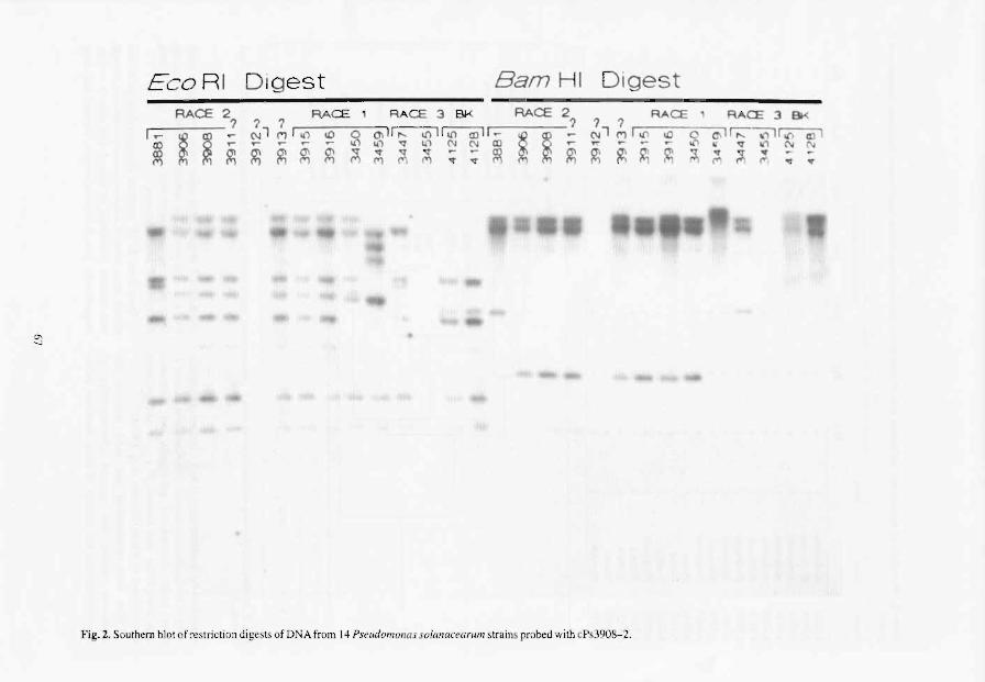
C'\
'"'
E C G RI DIgest Bam H I D igest RACE 2
r------? ? Ir- ~ ~ .... (\11 Cl) .... .....
~ ~ ~
RACE ' RACE 3 BK RACE 2 ? ? MI\{) 10 0 0;11" 1O'~ 1 -.............. 0.(\10 '<1 u1 NN Cl)
~ ~ ~ ~ M M ~ ; ; ~ ~ S ? 'I R .... CE' RACE J 8K
N 1 M r '" oD Co CI,r;:::-~~ .,... .... ~- r' "1 ,, ~W1~ N
~~ ~~~ ;J,;:'~~.,,,
Fig. 2. Southern blot of re striction di ges t ~ of DNA from 14 Pseudomonas so /all (lCellrllm strains probed with cPs3908-2.

Tabl .. 3. Reactivity of monoclonal antibodies with Pseudomonas solanacearum strains from tomato, heliconia, banana, and potato.
Strain Host Race Monoclonal antibody
PsI Ps2 Ps3 Ps4 Ps6 Ps8 Ps9
A3903 Heliconia 1 + + + + + A3904 Heliconia I + + + + + A391S Heliconia 1 + + + + + A3916 Heliconia 1 + + + + + A3906 Heliconia 2 + + + + + + A3910 Heliconia 1 + + + + A3908 Heliconia 2 + + + + A3911 Heliconia 2 + + + + A3912 Musasp. 2 + + + + A3914 Heliconia 2 + + + A344S Potato 3 + + + A3447 Potato 3 + + + A34SS Potato 3 + + + A3907 Heliconia 2 + + + + + A3381 Banana 2 + + + + + + A412S Banana 2 + + A34S0 Tomato 1 + +
1 +
Strain Race Host Origin
3381 2 Banana Costa Rica
I 3447 3 Potato Colombia 4125 2 Banana Philippines 4128 2 Banana Philippines
3906 2 lIeliconia Hawaii 3908 2 Heliconia Hawaii
n 3911 lIeliconia Hawaii 3913 lIeliconia Hawaii 3916 Heliconia Hawaii
- l 3915 Heliconia Hawaii
3950 I Tomato Trinidad 3459 1 Tomato Peru 3912 2 Musasp. Hawaii 3455 3 POlato Sri Lanka
o 0.25
Fig. 3. Dendrogram showing relationships between P. solanacearum strains based on RFLP data using cosmids Ps3908~ I and Ps3908-2.
described by Buddenhagen (l965). Strains pathogenic only to Heficonia and tomato, classified here as race 1, have been encountered previously on Heliconia spp. from Oahu and Hawaii, but have not yet been encountered on Kauai (Ferreira et al. 1991).
With both biochemical and mAb analyses the Heliconia strains had general characteristics of P. solanacearum but neither of these methods delin-
68
eated race 2 strains from other races. However, mAb Ps-la is very useful for species identification directly from plant samples and was negative for avirulent mutants of P. soianacearum.
Data from RFLP analysis distinguished Heliconia strains from other P. solanacearum strains and formed two clusters consisting of race 1 and race 2 strains, respectively. The Heliconia strains showed less than

50% homology of RFLP fragments with the Costa Rican race 2 strai n reported in RFLP group 24 (Cook et al. 1989). The bugtok strains isolated from banana in the Philippines appear to be distinct both from the Hawaiian Heliconia strains and the race 2 strain from Costa Rica. On blots from two additional probes, cPs3908-3 and cPs3908-4, however, banding patterns between the Costa Rican strain and the two Philippine strains were nearly identical (data not shown).
Although only tentative conclusions can be made from these preliminary results, the RFLP groupings show geographical and biological significance. Pending further analysis, we will focus on developing DNA probes specific for the Heliconia strains that differentiate them from other race 2 strains of P. solanacearum.
Acknowledgments
We gratefully acknowledge the technical assistance of Carla Mizumoto and Karen Pitz.
References
Akiew, E. and Hyde, K.D. 1992. First detection of Pseudomonas solanacearum race 2, strain SFR on Heliconia in Australia. Plant Disease. In press.
Alvarez. A.M., Benedict. A.A. and Mizumoto, C.Y. 1985. Identification of xanthomonads and grouping of strains of Xanthomonas campeslris pv. campestris with monoclonal antibodies. Phytopathology, 75, 722-728.
Benedict, A.A., Alvarez, A.M., Berestecky, 1., Imanaka, W., Mizumoto, C.Y., Pollard, L.W., Mew, T.W. and Gonzalez, c.F. 1989. Pathovar-specific monoclonal antibodies for Xanthomonas campestris pv. oryzae and for Xanlhomonas campestris pv. oryzicola. Phytopathology, 79, 322-328.
69
Buddenhagen. I.W. 1965. The relation of plant pathogenic bacteria to the soil. In: Baker, K. F., and Snyder. W. C., cd., Ecology of soil-borne plant pathogens. Berkeley, University of California Press, 269-284.
-1986. Bacterial wilt revisited. In: Persley, G.J. ed., Bacterial Wilt Disease in Asia and the South Pacific. ACIAR Proceedings No. 13, 126-139.
Buddenhagen, I.W. and Kelman, A. 1964. Biological and physiological aspects of bacterial wilt caused by Pseudomonas solanacearum. Annual Review of Phytopathology, 2,203-230.
Cook, D., Barlow. E. and Sequeira L. 1989. Genetic diversity of Pseudomonas solanacearum: detection of restriction fragment lenght polymorphism with DNA probes that specify virulence and the hypersensitive response. Molecular Plant-Microbe Interactions, 2, 113-121.
Ferreira, S., Pitz, K. and Alvarez, A. 1991. Heliconia wilt in Hawaii. Phytopathology. 81, 1159. Abstract.
Hartung, 1.S. and Civerolo, E.L. 1989. Restriction fragment length polymorphisms distinguish Xanlhomonas campesIris strains isolated from Florida citrus nurseries from X. c. pv. citrii. Phytopathology, 79, 793-799.
Hayward, A.C. 1964. Characteristics of Pseudomonas solanacearum. Journal of Applied Bacteriology. 27. 265-277.
-1991. Biology and epidemiology of bacterial wilt caused by Pseudomonas solanacearum. Annual Review of Phytopathology. 29, 65-87.
Hayward, A.c., EI-Nashaar, H.M .. Nydegger, U. and De Lindo, L. 1990. Variation in nitrate metabolism in biovars of Pseudomonas solanacearum. Journal of Applied Bacteriology, 69, 269-280.
Kelman, A. 1954. The relationship of pathogenicity in P. solanacearum to colony appearance on a tetrazoIium medium. Phytopathology, 44, 693-{595.
Nei, M. and Li, W.-H. 1979. Mathematical model for studying genetic variation in terms of restriction endonucleases. Proceedings of the National Academy of Science, USA, 76. 5269-5273.

Diversity of Pseudomonas solanacearum in Peru and Brazil
E.R. French*, P. Aley*, E. Torrest and U. Nydegger*
Abstract
Isolates of P. solanacearum in the International Potato Center (CIP) collection have been classified into biovars. Those from Peru are: biovar IIrace I from the Amazon lowlands and Northern highlands; biovar IIrace 2-strain A and biovar 3/race 1 from the Amazon lowlands; biovar 2-Alrace 3 from the Andean highlands and cool coastal desert; and biovar 2-T/tropicallowland variant found in San Ramon, lunin Department (1150 m elevation) and Yurimaguas, Loreto Deptartment (170 m). Strains of biovar IIrace 1 and biovar 2-T have been found together in the same fields and even in the same plant at Yurimaguas. These differed consistently in size on Kelman' s medium (average diameters 1.0-1.5 mm and 1.8-2.5 mm, respectively) and they have distinctive fonnazan pigment patterns. Brazilian isolates are: biovar lIrace 1 and biovarllrace 2 in the wanner ecoregions; biovar 2-Alrace 3 in the south; biovar 2-T in central Brazil; and biovar 3/race 1 in central and northern Brazil. Biovar 2-T, that is metabolically more active than biovar 2-A, was pathogenic on potato, tomato and eggplant in field tests in San Ramon, but not on pepper and tobacco; whereas biovar 2-A was only pathogenic on potato at Umari, Huanuco Department (2,400 m). Biovar 2-T was less virulent on clones developed for resistance to bacterial wilt, 7 of 32 being highly resistant at tropical San Ramon, whereas only four were resistant to biovar 2-A in the highlands at Umari. However. only one of the clones was commonly resistant at bolh locations, indicating a marked differential host-pathogen interaction. Biovar 2-T was not present among the numerous biovar 2 isolates from other potato-producing countries in other continents.
BACTERIAL wilt caused by Pseudomonas solanacearum E.p. Smith was not noticed in Peru until 1966 when Moko disease of plantains and bananas, and wilt of tomato (Lycopersicon esculentum L.) were detected along the Amazon river (French and Sequeir& 1970), and a year later wilt of potato (Solanum tuberosum L) was found in the highlands (Herrera and French 1970). Attempts to find native hosts by observing wilt symptoms of highland vegetation, native jungle species or among jungle crops and weeds were unproductive, though some transplanted wild jungle plants were shown to maintain or increase the inoculum potential (French et al. 1981).
The Amazon basin, which is located primarily in Brazil and Peru, was recognised as an area of greater diversity for P. solanacearum (French 1979). A biovar 2 strain that was not the low-temperature potato strain
*International Potato Center (CIP), Apartado 5969. Lima 100, Peru
tUniversidad Nacional de Huanuco, Huanuco, Peru
70
synonymous to race 3, was found in the lowlands of Peru in valleys of tributaries of the Amazon river. Its host range seemed greater, since eggplant (Solanum melongena L.) wilted under field conditions where it was found. Since this strain is indigenous in tropical rain forest, it was postulated that race 3 may have diverged from this tropical strain when potatoes were planted in these lowlands, and healthy looking tubers from wilted plants were carried with latent infection to the highlands and used as sccd. Over considerable time, a new strain adapted to the potato, which is usually grown in cool climates, may have evolved (French 1986; Martin et a1. 1981; Torres 1983). The readiness with whieh P. solanacearum is disseminated as latent infection in tubers has been shown (Ciampi et a1. 1980; French and Nydegger 1990).
At The International Potato Center (CIP) we estab· Iished collaboration with Drs A.C. Hayward and L. Sequeira to further define these and other strain differences. A collection of 327 isolates of P. solanacearum was assembled and detailed physiological and RFLP

studies conducted. These led to findings which included confinnation of the existence of two phenotypes of biovar 2. The tropical lowland phenotype was designated biovar 2-T (CIP 1991; Hayward et at 1990a, 1 990b, 1992).
The presence of more than one bacterial strain in a given plant was recognised because of different colony size and fonnazan pigmentation patterns in an agar streak, when both a biovar 1 and a biovar 2 were present in a plant from the highland site of Huambos, Cajamarca. Similarly biovars 1 and 2 were isolated from one plant from the Yurimaguas jungle site (French and Nydegger 1987).
Numerous reports confinn that biovar 2/race 3, which has the greatest distribution in association with the potato, primarily in the cool tropical highlands and cooler latitudes, has a lower optimum temperature for growth and causes infection at lower temperatures than other strains (French 1986). Studies of the optimum temperature for growth of the Peruvian lowland strains of biovar I and 2 and isolates of biovars I and 2 from cool temperature locations showed no significant differences between them (Nydegger and French 1984). An explanation for the ability of race 3 to cause wilt at cool temperatures, other than a lower optimum temperature for growth, may be its greater aggressiveness or pathogenicity. Gutarra (1980) found that five strains of biovar 2 were on average more pathogenic than two strains of biovar 1, whilst Martin et al. (1982) tested six Peruvian strains, the two biovar 2 being more pathogenic than the four biovar L Ciampi et a!. (1981) determined that thc variety Molinera (resistant to wilt) was not affected by a race I (biovar 1) strain, whereas a race 3 (biovar 2) strain caused 20% wilt.
To better understand the diversity of P. solanacearum in the Brazilian and Peruvian tropical belt of South America, and especially the variation within biovar 2/race 3 that (along with biovar I), causes bacterial wilt of potato from the cold highlands to the hot tropical lowlands, infonnation in the CIP P. solanacearum collection listing (gathered in part by the authors) was used. Research was conducted on host susceptibility, latency in potato tubers, and the optimum temperature for growth of strains of the bacterium.
Materials and Methods
The accession list of the CIP P. solanacearum isolate collection was amplified to show biovar 2 isolates classified as biovar 2-T, and the remaining biovar 2 strains tested were designated biovar 2-A (for Andean), The frequency of presence in the collection of biovars collected within Peru and Brazil was plotted on a map, adding infonnation about the host except when
71
it was potato. The names of Departments of Peru and States of Brazil, which included data, were added.
To detennine if five solanaceous hosts commonly grown in other parts of Peru were susceptible to the strain of P. solanacearum found when potatoes were introduced to jungle soils, susceptibility tests were conducted in a field heavily infested with biovar 2-T during November and December, the time of heavy summer rains. True seed of hot pepper (Capsicum baccatum var. Escabeche), tomato (var. Marglobe). tobacco (Nicotiana tabacum L. var. Sansum), eggplant (var. Black Beauty) and potato (var. Atzimba) were germinated in trays containing peat and sand (2:1). These were then transplanted when 2-3 cm tall to Jiffy 7 peat blocks and 25 days later planted at La Chincana, San Ramon District (11 '03'S, 75'19'W at an altitude of 1150 m above sea level) in a completely randomised block design (CRBD) with four replications. Two-row plots 6 m long containing 20 plants of a given plant species were planted with between row spacings of 90 cm and within-row spacings of 50 cm. Wilt symptoms were recorded three times at intervals of IS days. Samples of wilting plants were taken from each plot, to determine the possible variations in strains. The same species of plants were planted at the highland site of Umari, Huanuco (2400 m above sea level) but in continuous rows of each species in sequence, in a field that had been maintained as a wi It nursery.
To detennine the resistance performance of several clones bred for resistance to P. so[anacearum with genes derived from Solanum phureja (Torres 1983), a selection of 32 was planted at both La Chincana, Junin Department (1150 m above sea level) where biovar 2-T was consistently isolated, and at Umari, Huanuco Department (9'55'5, 760 14'W, 2400 m above sea level) where biovar 2-A was the only strain present. The pedigrees of the 11 clones that grew well in both environments and were of greater interest, and of the susceptible and resistant checks, are shown in Table I. The tests were planted twice during the year-end rainy seasons with a CRBD design with three replications of five plants in one row, planted with a between-row spacing of 100 cm and a within-row spacing of 40 cm.
To observe colony characteristics, two isolates of each biovar strain werc streaked on Kelman's medium, incubated at 30'C for 48 hours, and colonies viewed in inverted plates with a stereoscopic dissecting microscope with oblique reflected light. Twenty wellseparated colonies on five plates per isolate were measured and formazan pigmentation characteristics were recorded.
The optimum temperature for growth was determined in liquid culture (Kelman's medium without agar or TZC) in an oscillating temperature gradient incubator with a temperature range of 25-42"C with

Table 1. Pedigrees of potato clones bred for resistance to Pseudomonas solanacearum that are shown in Table 2.
Code for clones
BR 63.15 and BR 63.76
MS 35.91 MS 82.60 MS 84.5 MB5.24
Pedigree"
Atzimba(Mexico)xA-l [(Phu 1386 x Phu 1339) X Katahdin (U.S.)] CA-Purace (Colombia) x A-I CP30xP.95 Atzimbax P.7 MS 10.1 [(Anita(Mexico)xP.I)]x P.I
MB 6.1 and MB 6.42 MS 10.1 x P.7 MB 34.22 and MB 34.99 ICA-Nevada (Colombia) x P.7 Cruza 148 Monserrate (Colombia) x PI
(Mexico) Checks: Ticahuasi
Molinera
Ticanel (Costa Rica) x Chata blanca (Peru) Peruvian
'P.!. P.7 and P.95 = Phu 1386.26 x Katahdin (Phu = Solanum phureja. Central Colombian Collection); and Mexico varieties containing S. demissum resistance to late blight
intervals of 0.5"C between each pair of tubes. Tests were repeated four times with two isolates simultaneously. Initial concentration was 1 x 10⁷ cfu/mL. Readings were made by optical density (OD) at 600 nm in a spectrophotometer, at hourly intervals for 8 hours, until the temperature at which growth was fastest at an absorbance of OD 0.5 or more was reached. The eight isolates tested were: two biovar2-A; two biovar 1 from highcr, cooler elevations; two biovar 1 from a low, hot elevation; and two biovar-2-T from the same low elevation (Table 3).
To determine the potential of isolates of the different strains to produce wilt disease and latent infection in potato tubers, plants of Ticahuasi variety were grown in 15 cm diameter clay pots containing about 1 kg disinfested soil mix (2 soil: 1 sand: I moss). Inoculation was done by pouring into the soil mix 50 mL of a suspension of 1 x 10⁸ cfulmL for a given isolate. Isolates used were: biovar 1,015 and 077; biovar 2-A, 017 and 102; and biovar 2-T, 107 and 142 (Table 3). Inoculation treatments were 42 and 70 days aftcr planting and they were harvested 54 and 40 days later, respectively. Two greenhouse temperaturc treatments were 17126.5 and 26/30SC night/day. Percent wilt was determined 10 days before harvest. Tubers with symptoms at harvest were discarded and the remaining healthy appearing tubers were stored at 27-32'C for 42 days, at which time symptoms were recorded either as externally visible or by exudation of the vascular system of halved tubers. Symptomless tubers were assayed by aseptically removing two portions of tissue
72
Table 2. Percent wilted plants and percent tuber infection (average of two seasons) of potato clones field tested at La Chincana. Junin Department (1150 m above sea level) and Umari, Hminuco Deptartment (2400 m above sea level).
(biovar 2-T) (biovar 2-A)
Plant Tuber" Plant Tuber"
BR63.15 23 0 Oa 7 BR 63.76 5a 5 Oa 2
MS 35.9 56c 10 90d 52 MS 82.60 Oa 10 38b 11 MS 84.5 Oa 12 47bc 37 MB5.24 Oa 75 68c 37 MB6.1 Oa 0 40b 32 MB 6.42 31b 33 Oa 8 MB 34.22 Oa 0 40b 28 MB34.99 Oa 5 38b 70 Cruza 148 Oa 0 73c 3 Checks: Ticahuasi 73 c 67 lood lOO (susceptible) Molinera 14a 36 18a 9 (resistant)
15.5 19.5 42.5 30.5
aNo tuber symptoms at harvest. Reflected latency 30 days later. bSum of symptoms at harvest plus latency 30 days later. cNumbers followed by the same letter are not statistically different at P 0.05 as determined by Dunean' s multiple range test. The test was performed independently for each site.
per tuber of about 0.5 x 0.5 x 2.0 cm, that included the vascular system, and each was placed in a test tube containing 5 mL of sterilc water for 15 minutes. Streaking on Kelman's medium followed and incubation for 48 hours at 30'C.
Results
There were 98 isolates from Peru in the collection, most of them contributed by CIP scientists, and 36 from Brazil contributcd by colleagues there. The geographical origin of these is shown in Figurc 1. In the highlands (above 1500 m above sea level) of the Peruvian Departments of Amazonas, Ancash, Cajamarca. Huanuco, La Libertad, Junin and Piura, as well as the cool coastal Lima, there were eight biovar 1 and 52 biovar 2-A strains, all isolated from potato. In the lowlands (below 1500 m above sea level) of the Peruvian Departments of Loreto and Junin, 32 isolates of biovar I included 26 isolated from potato, 2 from tomato and 4 from plantain; 5 biovar 2-A isolatcs were from potato; 16 biovar 2-T included 13 from potato, 2 from eggplant and I from tomato; a single biovar 3 was from tomato (another lowland tomato isolate was a biovar 1 from a lowland jungle site in Hm"inuco, as shown in Fig. I).
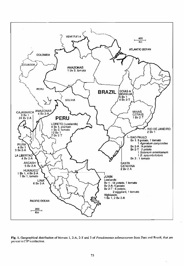
COLOMBIA
(
CAJAMARCA AMAZONAS 2 Bv 1 4 Bv 2-A
24 Bv 2-A
LALlBERTAD 4 By 2-A
AN CASH 5 By 2-A
HUANUCO 1 Bv 1,4 Bv 2-A
1 By 1, tomato
LIMA 6 By 2-A
PACIFIC OCEAN
~ Km
JUNIN Lowlands:
400 t--------l
Km
ATLANTIC OCEAN
SAOPAULO By l' 5 potato, 1 tomato
1 Ageratum conyzoides By 2-A : 9 potato Bv 2-T: 2 potato
1 Solanum americanum 1 S. sysymbrifolium
Bv 3 : 1 tomato
SANTA CATARINA 2 By 2-A
By 1 : 14 potato, 1 tomato By 2-A: 5 potato By 2-T : 6 potato,
2 eggplant, 1 tomato Highlands: 1 Bv 1, 2 By 2-A
Fig. 1. Geographical distribution of biovars 1, 2-A, 2-T and 3 of Pseudomonas solanacearum from Peru and Brazil, that are present in CIP's collection,
73

All isolates from Brazil were from below 1500 m above sea level, and al1 were from the southeast except for a tomato isolate of biovar 3 in Amazonas State. A similar isolate was from Sao Paulo State. Another 34 isolates in the southeastern states of Goias (and Federal District of Brasilia), Minas Gerais, Rio de Janeiro, Sao Paulo and Santa Catarina were: 14 biovar 1 (12 potato, I tomato and I Ageratum conyzoides L.); 11 biovar 2-A from potato; and 9 biovar 2-T (7 potato, 1 Solanum americanum Mill. and 1 S. sysymbrifolium Lam.). Biovars 1 and 2 have, in addition, been reported in Rio Grande do SuI and biovar 2 in Espiritu Santo, and several weed hosts reported without specifying biovar or race (Lopes and Reifschneider 1983; Robbs et al. 1974).
At the La Chincana lowland site, symptoms appeared first in potato and eggplant, with 2.5% wilt incidence on average for each at 15 days. First symptoms on tomato were recorded at 30 days. There were no symptoms in tobacco or pepper at 45 days. The average per plot at 45 days was: potato, 74%; eggplant, 39%; and tomato, 16%. P. so/anacearum was isolated repeatedly from each plot with a susceptible host. All isolates were characteristic of biovar 2-T in colony size and formazan pigmentation; representative samples were accessed to the collection and were classified by physiological tests as biovar 2-T. At the Umari highland site only potatoes developed wilt, and all isolations were biovar 2-A. These records on biovar strains appear in the Huanuco and Junin listings in Figure I.
Percentage wilt and tuber infection (Table 2) data for the potato clones bred for resistance to wilt and tested at two sites are averages for two seasons. Only BR 63.76 and its sister clone Molinera (BR 63.65 resistant check) were resistant at both locations. MS 35.9 and the susceptible check were susceptible at both locations. Clones BR 63.15 and MB 6.42 were moderately resistant at La Chincana and resistant at Umari. Clones MS 84.5, MB 5.24, and Cruza 148 were resistant at La Chincana and susceptible at Umari. The remaining four were resistant at La Chincana and
moderately resistant at Umari. Tuber symptoms at harvest were present in Umari and absent at La Chincana. Symptoms after storage of 10 symptomless tubers per plot for 30 days at 28±3"C occurred for both sites.
In the study of colony characteristics it was found that biovar 1 isolates had diameters ranging from 1.0-1.5 mm; biovar 2-A from 1.8-2.3 mm; and biovar 2-T from 2.4-2.5 mm. Formazan pigmentation was highly characteristic for biovar 2-T in which dark helicoidal red streaks were superimposed on a diffuse reddish central pigmentation. Differences between biovar 1 and biovar 2-A colonies were apparent but not so easily defined. In one instance, during the process of isolation of the causal agent from a wilted plant collected at Yurimaguas, two colony types were easily distinguished in the streaks on agar plates resulting in isolates 177 (biovar 2-T) and 178 (biovar 1). Similar situations allowed the isolation of pairs ofbiovar I and biovar 2-A isolates (202-203 at Huambos, Cajamarca; also 300-301, 302-303, 304-305, 306-307 at San Ramon, Junin). Biovar 2-T was also isolated, singly, at this last site, and readily diagnosed as such.
Results of studies of optimum temperature for growth of isolates are shown in Table 3 with their respective temperature optima and hours taken to reach 0.0. 0.5 at that optimum temperature. Results were consistent between repetitions. The isolates from the three high elevation sites took longer. and had the lowest optimum temperature for growth, but these optima were similar to some of those for lowland strains.
Percent wilt and latent infection resulting from the inoculation of Ticahuasi potato plants in two temperature regimes is shown in Table 4. Biovar 2-A isolates were more pathogenic than the others at both temperature regimes, whilst biovar I and biovar 2-T were similar at the higher temperature and biovar 1 more pathogenic than biovar 2-T at the cooler temperature. Latent infection differed with some strains according to the date of inoculation and temperature. On average,
Table 3. Optimum temperatures for growth of isolates of different biovars (biovar) of Pseudomonas solanacearum isolated from potato in Peru, and number of hours taken to reach optical density (OD) 0.5.
2-A
2-T
017 102 042 015 141 077 107 142
Cajamarca Chocon
Huancayo Tea Gardens Yurimaguas Yurimaguas Yurimaguas
(m above sea level)
74
2700 3500 3350 1600 170 170 170 170
temperatureoC
34.5 34.S 34.S 36.4 35.7 37.7 34.6
to
000.5
8 8 8 7 7 7 6 6

Table 4. Percent wilt and percent latent infection of tubers for plants grown in greenhouses at either 17.0126.5 or 26.0I30SC night/day regimes, after inoculation by soil infestation with isolates of biovar 1. biovar 2-A and biovar 2-T (average for two isolates of each).
Biovar 1 Biovar2-A Biovar2-T
Temperatures Dr" Wilt Latencyb Wilt Latency Wilt Latency
42 45.0 4.0 82.0 31.0 32.0 18.9 70 67.5 4.7 66.0 16.3 50.0 0
Average 56.2 4.3 74.0 23.6 41.0 9.5 26-30SC 42 62.5 6.2 100 0 80.0 6.5
70 78.0 18.4 92.0 27.3 68.0 44.0 Average 70.2 12.3 96.0 13.7 74.0 25.2 General average 65.2 8.3 85.0 18.6 57.5 17.4
"Days inoculated. 42 and 70. Plants were harvested 54 and 40 days later, respectively. "Tubers with symptoms were discarded at harvest. others were stored at 27-32'C for 42 days after which symptoms were recorded as latent.
biovar I resulted in less latent infection, whereas it was greater and similar for biovars 2-A and 2-T. At the lower temperature, however, biovar 2-A produced more than double the latently infected tubers than biovar2-T.
Discussion
Only Peruvian isolates originated above 1500 m elevation in the highlands (since none of the Brazilian isolates were from regions with highlands), where biovar 2-Nrace 3 predominated and was isolated only from potato. A low frequency of biovar I/race 1 isolates came from the lower reaches of these Andean Highlands. having been isolated from potato and tomato. whereas there were numerous isolates ofbiovar 1 from potato and a few from tomato for the lowlands. A few isolates of biovar 2-A of recent isolation were from one field in the lowlands at San Ramon.
In Brazil, biovar 2-A also originated only from potato, but rather than coming from high elevations it was from more southerly higher latitudes. Biovar 1, on the other hand, originated in lower latitudes. from potato, tomato and A. conyzoides. Biovar 1 isolates outnumbered those ofbiovar 2-A.
Biovar 2-T isolates were from the lowlands and primarily from potato in lands recently placed in cultivation in both Peru and Brazil, but also from eggplant and tomato in Peru and two solanaceous weeds in Brazil.
Apart from the biovar l/race 1 isolates mentioned above. four biovar 1 isolates of race 2-Amazon strain from Peru were representatives of the epidemic reported as moving west up the Amazon river basin from Brazil and Colombia into Peru (French and Sequeira 1970). In addition to biovars 1, 2-A, and 2-T, isolates of biovar 3/race 1 isolated from tomato were from the Amazon basin in both Brazil and Peru and also Suo Paulo State in southern BraziL
75
The distribution of strains is consistent with the proposition that biovar 2-Nrace 3 is a strain highly specialised on potato, and that it has spread primarily in latently infected tubers. The only case of infection in the lowlands of Peru resulted after potatoes had been grown for many years utilising highland seed, which could have been at times infected latently with P. solanacearum, Its presence in southern Brazil on potatoes only, as is also the case in numerous potatogrowing countries at much coldcr latitudes, is further circumstantial evidence that this is a strain that has spread worldwide from one common origin, probably the Andean highlands, and likely through Europe from where potatoes were redistributed to most potatogrowing countries worldwide (French 1979).
Nineteen isolates of biovar 2-T were from the tropical rain-forest lowlands of eastern Peru and in or close to the 'cerrado' brushland of eastern Brazil, whilst the remaining 111 biovar 2 isolates from loeations worldwide. were biovar 2-A. Should future surveys demonstrate that these are the margins of a vast region that may extend even further north and south, in which biovar 2-T is indigenous, this would suggest that the variability of biovar 2 is greatest in South America, and that both biovar 2-T and 2-A originated in this region, with the more specialised 2-A evolving from 2-T (French 1979; Hayward et al. 1992; Martin et al. 1981).
The testing of five solanaceous crops in land infested with only biovar 2-T at La Chincana, as demonstrated with the isolations made. further supports the above conclusions since under field conditions eggplant, tomato and potato developed wilt. Biovar 2-A generally causes wilt of potato only and rarely tomato (French 1986). It would be interesting to test these crops in land infested with biovar 2-T in the 'cerrado' of Brazil. where another strain named 'eampo-bi' causes 100% wilt when virgin land is planted to potatoes, and none if planted a second time;

but if beans are planted first, they do not wilt, whereas a subsequent crop of potatoes does (Lopes and Reifschneider 1983),
The testing, in the La Chincana lowland site infested with biovar 2-T and the Vmari highland site with biovar 2-A of potato clones bred for resistance to bacterial wilt, resulted in less wilt and fewer clones being susceptible to biovar 2-T in spite of higher temperatures favourable for disease. This differential pathogen-host interaction between the two sites needs confirmation under identical environmental conditions. Biovar 2-A resulted in more than double the average wilt incidence under less favourable temperature conditions than that caused by biovar 2-T (however, the inoeulum potential was slightly higher as suggested by the percentage wilt in the resistant check, 18 vs. 14%). In the greenhouse inoculations, biovar 2-A isolates of cool climate origin were more pathogenic than both biovar 2-T and biovar I isolates that originated in warmer locations. These results lend further support to the proposition that it is due to greater aggressiveness, especially expressed at lower temperatures, that biovar 2-Alrace 3 strains are pathogenic under cool climate conditions atypical for P. so/anacearum. (Ciampi et al. 1981; French 1979; Gutarra 1980; Martin et al. 1982).
The studies of optimum temperature for growth of isolates from different agroecologies, on the other hand, did not show clear differences between the three strains. It should not be expected, a priori, that strains indigenous in the same ecological niche (biovar 1 and biovar 2-T from Yurimaguas) would differ, but even the optimum temperatures for growth of biovars from .the high Andes and low Amazon Basin differed very little in this study.
Latent tuber infection resulted in the field test with biovar 2-T. but no symptoms at harvest; biovar 2-A produced symptoms at harvest in spite of a cooler climate, and also latent infection. Average tuber infection was greater with biovar 2-A. Both factors again suggest, a greater aggressiveness of biovar 2-A. In greenhouse tests, biovar 2-A was also more pathogenic at both temperature regimes than biovar 2-T (and biovar 1) but latent infection resulted from both. and was even greater for biovar 2-T at the higher-temperature regime more akin to tropical climates where it occurs. Biovar 2-T was less aggressive, resulting in little or no external symptoms. Thus, tubers harvested under the tropical conditions where this strain occurs could be carried to higher elevations where seed production is preferred. Such a happening centuries or millenia ago may have been the beginning of a process of evolution of the more specialised biovar 2-A. Latent infection by biovar I was lower, which may indicate it had less probability of evolving into a highland strain
76
(however, isolate 042 originated at 3350 m above sea level, an altitude similar to the highest elevations recorded for biovar2-A).
Colony phenotype (size, formazan pigmentation) was shown to be a useful tool to distinguish mixtures of isolates during the isolation from plants with dual infections. Thus, distinctions were recorded as having been made between biovar 1 and biovar 2-A, and biovar 1 and biovar 2-T. For one field at San Ramon the distinction was made between biovars 1, 2-A and 2-T (the first two in dual infections, the last singly). Colony differences are thus useful in the separation of different strains for given locations, but these differences may not hold true over a greater geographical range.
It can be concluded that the Amazon basin and adjacent lands (in Brazil, Peru and possibly neighbouring countries) are a region of greater diversity for P. so[anacearum, it being the only region in the world where the lowland variant biovar 2-T occurs. Further, there is evidence pointing to biovar 2-Alrace 3 having evolved from biovar 2-T. If so, the worldwide distribution of biovar 2-Alrace 3 could be explained only as having resulted from the transport of latently infected tubers from the Andean region of the New World to Europe, and subsequently in the traditional seed trade routes (or even by exchange between agricultural researchers) to the large number of countries on all inhabited continents that now have it, including southern BraziL
References
Clampi, L., Sequeira, L, and French, E. R. 1980. Latent infection of potato tubers by Pseudomonas solanacearum. American Potato Journal, 57, 377-386.
Ciampi-Panno, L, Sequeira. L. and French, E. R. 1981. Pseudomonas solanaceanlln distribution in potato plants; establishment of latent infections. In: Lozano. J. C, ed., Proceedings of the Fifth International Conference of Plant Pathogenic Bacteria. Cali-Colombia. Centro Internacional de Agricultura Tropical (CIAT), 148-161.
CIP (International Potato Center). 1991. Annual Report. International Potata Center, Apartado 5969, Lima 100, Peru.
French, E.R. 1979. Classification, distribution and origin of Pseudomonas solanacearum. In: Developments in control of potato bacterial diseases. International Potato Cent er, Apartado 5969, Lima 100, Peru, 28-35.
-1986. Interaction between strains of Pseudonwnas so/anacearum, its hosts and the environment. In: Persley, 0.1., ed., Bacterial Wilt Disease in Asia and the South Pacific. ACIAR Proceedings No. 13,99-104.
French, ER, Martin, C. and Nydegger, U. 1981. Tropical rainforest vegetation'> influence on survival of Pseudomonas solanacearum. In: Lozano, J. C., ed., Proceeding of the Sth International Conference on Plant Pathogenic Bacteria, Cali, Colombia. Centro International de Agricultura Tropical (CJAT), 195-202.

French, ER and Nydegger, U. 1987. Incidence ofbiovars I and 11 of Pseudomonas solanacearum in two ecological zones of Peru. Proceedings f the 6th International Conference on Plant Pathogenic Bacteria, June 2-7, 1985. Beltsville-MD, 769. Abstract.
-1990. A two decade epidemic of PseudomofUls solanacearum on potato in Peru. In: Klement, Z., ed., Proceeding of the 7th International Conference on Plant Pathogenic Bacteria, Budapest, Hungary, 317-322.
French, E.R. and Sequeira, L 1970. Strains of Pseudomonas solanacearum from Central and South America: a comparative study. Phytopathology, 60, 506--512.
Gutarra, L 1980. Variacion de la bacteria PseudomofUls solanocearum de papa en el Peru. (Variation of the bacterium P. solanacearum of potato in Peru) Tesis Ing. Agr. Univ. Nac. Centro, Huancayo-Peru. 68 p.
Hayward, A.C., EI-Nashaar, H.M., Nydegger, U. and De Lindo. 199Oa. The use of microtiter plates in the phenotypic characterization of phytopathogenic pseudomonads. In: Klement, Z., ed., Proceeding of the 7th International Conference on Plant Pathogenic Bacteria, Budapest, Hungary, 593-598
-I 990b. Variation in nitrate metabolism in biovars of Pseudomonas solanacearum. Iournal of Applied Bacteriology, 69, 269-280.
Hayward, A.C., Sequeira, L, French, E.R., EI-Nashaar, H.M. and Nydegger. U. 1992. Tropical variant of biovar 2 of Pseudomonas solanacearum. Phytopathology, 82, 608. Abstract.
77
Herrera, I.A. and French, E.R. 1970. Strains de Pseudomonas solanacearum aislados de papa en el Peru. (Strains of P. solanacearum isolated from potato in Peru). Investigaciones Agropecuarias del Peru, 1,47-52.
Lopes, C.A. and Reifschneider, EI.B. 1983. Bacterial diseases of potatoes in Brazil: present and future trends in research. In: Hooker, W., ed., Research for the Potato in the Year 2000. International Potato Center (CIP), 121-122.
Martin, c., French, E.R. and Nydegger, U. 1981. Bacterial wilt of potatoes in the Amazon Basin. Plant Disease, 65, 246-248.
-1982. Strains of Pseudomonas solanacearum affecting Solanaceae in the Americas. Plant Disease, 66, 458-460.
Nydegger. U. and French, E.R. 1984. Temperaturas optimas para Pseudomonas solanacearum aislado de papa en distintos climas. (Optimum temperatures for P. soianacearum isolated from potato in different climates). Fitopatologia, 19,54. Abstract.
Robbs, C.E, Kimura, 0., Ribeiro, R.L.D. and Deslandes, J.A. 1974. Some wild host plants of Pseudomonas solanaceamm E. E Smith found in southeastern Brazil. Fitopatologia, 9, 70. Abstract.
Torres, E.M. 1983. Respuesta de hibridos de papa a Pseudomonas solanacearum. (Response of potato hybrids to P. solanacearum). Tesis MS Universidad Nacional Agraria La Molina, Lima, Peru, 105 p.

Pathogenicity of the New Phenotypes of Pseudomonas solanacearum frolll Peru
J.E. Marin* and H.M. EI-Nasbaart
Abstract
Pathogenicity of bacterial strains of phenotype A (063,162 and 299) and phenotype B (050, 204 and431) representing biovar 2 of Pseudomonas solanacearum was determined using Solanum tuberosum cv. Ticahuasi, Lycopersicon esculentum cv. Marglobe, Nicotiana tabacum cv. Samsun, S. melongena cv. Black Beauty, S. nigrum, Cyphomandra betacea and Datura stramonium. Isolates from biovar I (003 and 303) and biovar 3 (072) were used as tester standards. Ten 40-50 day old seedlings were inoculated with P. solanacearum using a Hamilton microsyringe delivering 10 ~L per site/plant of a suspension containing Ix108 cfulmL into the stem. For potato, cuttings of similar age were used. Plants were evaluated after 5 days of incubation at 26--30°C and every 5 days thereafter for 20 days. Except those isolates of phenotype B which did not cause wilt of tobacco, all strains induced some host response. The reaction of N. tabacum and S. nigrum to inoculation with strains ofbiovars 1,2 and 3 was differential. This demonstrated that phenotype A has a much wider host range than phenotype B. It is evident that phenotype A behaves similarly to isolates of race I, due to its ability to induce a hypersensitive reaction and to cause wilt in tobacco. Phenotype B is, therefore, less related to phenotype A. Added evidence for the great diversity of P. solanacearum and the existence of a variant(s) of biovar I, which is closely related to phenotype B, are reported.
BACfERIAL wilt caused by Pseudomonas solanacearum E.F. Smith affects many crops in tropical and subtropical regions of the world. The pathogen has a wide host range and survives in soil for extended periods (Buddenhagen and Kelman 1964; Hayward 1991; Kelman 1953). In Peru, potato and banana are the two crops most often affected by the disease (French and Sequeira 1968, 1970; French and Nydegger 1990).
Strains of P. solanacearum vary greatly in biochemical activities, host range and ability to cause disease. Various classification schemes have been proposed. Buddenhagen et al. (1962), classified a large number of isolates into three races: race 1 contains strains that are pathogenic on tobacco, many other solanaceous hosts and other plant species; race 2 strains are primarily pathogenic on plants from the family Musaceae; race 3 strains affect potato and tomato and a limited number of other hosts. Hayward (1964)
* University of Ca jam arc a, Cajamarca, Peru. tThe International Potato Center (CIP), Apartado 5969, Lima, Peru.
78
grouped isolates of P. solanacearum into four biovars (biovars 1-4) according to their ability to oxidise disaccharides or utilise hexose alcohols. In a study which included isolates from Morus alba from China, He et al. (1983), described a group of isolates that did not fit any of the previously reported races and biovars. They proposed that these isolates belonged to biovar 5 and possibly race 4. Isolates from race 3 correspond to biovar 2, but the reverse is not always the case (Buddenhagen 1986).
In Peru, biovars 1-3 are present (French and Nydegger 1990; Martin et al. 1981). Studies of the variabilities of strains from these biovars are lacking. Recently, Hayward et al. (1990a) determined that there are two phenotypes within biovar 2 that differ in their metabolic activities. Phenotype A which includes isolates that are metabolically more active, was thought not to behave as race 3 and most probably to have a wider host range. Isolates representing phenotype B are less metabolically active, but possibly correspond to race 3. The purpose of this study was to confirm the biochemical characteristics of the Peruvian

isolates of biovar 2; determine the pathogenicity of isolates from phenotypes A and B; and, investigate the raceibiovar relationship of the Peruvian strains of biovar2.
Material and Methods
Strain selection and maintenance
Thirty four Pseudomonas solanacearum isolates representing biovar 2 were used for biochemical characterisation. Six strains equally representing phenotypes A and B of biovar 2 were evaluated for study of host range and symptomatology. In addition, strains CIP-003 and CIP-303 (biovar 1) and strain CIP-On (biovar 3) were used as references. Details concerning the origins of all strains used are shown in Table 1.
The International Potato Center (CIP) maintains one of the largest collections of P. solanacearum in the world. Strains are kept in screw-capped tubes containing bacterial suspension in sterilised distilled water (Kelman and Person 1961). Bacterial strains were replated on Kelman medium with 2,3,5- Triphenyltetrazolium Chloride (TZC) (Kelman 1954) for initial verification. Typical colonies after 48 hours of incubation at 28-30°C were transferred onto Kelman medium without TZC. Typical P. solanacearum cultures were then transferred into new tubes containing sterilised distilled water (SOW). New tubes were used as the source of bacterial isolates throughout the experiments.
Inoculum preparation
Inocula for pathogenicity tests and biochemical characterisation were prepared from 48 hour cultures on Kelman medium without TZC grown at 28-30°C. Inocula for pathogenicity tests were prepared by suspending bacteria in SOW and adjusting to 1 x 10⁸
cfu/mL using a spectrophotometer (Bausch and Lomb spectronic 20), transmission at A600 = 0.1. Inoculum was used immediately after preparation.
Biochemical characterisations
Characterisation of all 34 isolates was carried out using tests for oxidase, catalase and hydrolysis of Tween-80 according to Lelliott and Stead (1987), solubility in 3% KOH (Suslow et al. 1982), nitrate reduction (Hayward et al. I 990b ) and production of dark brown pigments on Kelman medium without TZC, supplemented with 0.01 % I-tyrosine (Hayward 1964).
Determination of biovars and phenotypes was carried out by applying the methods of Hayward (1964) and Hayward et al. (1990a). Pectate gel degradation was tested as described by Hildebrand (1971).
79
Table 1. Strains of Pseudomonas solanacearum used in this investigation.
Code-CIP Host
B iovar 2IPhenotype A 010 Potato
061 Potato 063- Potato 107 Potato 108 Potato 142 Potato 162- Soil 167 Potato 172 Potato 175 Potato 177 Potato 184 Potato 299- Soil
312 Eggplants
Biovar 2IPhenotype B 014 Potato 016 Potato 050-079 161 185 197 198 199 201 204-208 246 253 262 270 272 273 384 431-Biovar 1 003-303-
Biovar3
Potato Potato Soil Potato Potato Potato Potato Potato Potato Potato Potato Potato Potato Potato Potato Potato Potato Potato
Potato Potato
On" Tomato
Location
La Chine ana, San Ram6n, Junfn Yurimaguas, Loreto Yurimaguas, Loreto Yurimaguas, Loreto Yurimaguas, Loreto Yurimaguas, Loreto San Ram6n, Junfn La Chineana, Junfn La Chineana, Junfn Yurimaguas, Loreto Yurimaguas, Loreto La Chineana, Junfn San Miguel, San Ram6n, Junfn La Chincana, San Ram6n, Junfn
Chota, Cajamarea Cajamarea, Cajamarea Umari, Hmlnueo Otuzco, La Libertad Umari, Huanuco La Molina, Lima Cajabamba, Cajamarea Cauday, Cajamarea Chota, Cajamarea Cutervo, Cajamarea Huambos, Cajamarea Virt1, La Libertad Chota, Cajamarea Alfiaeo, Cutervo, Cajamarea Huayao, Huaneayo, Junfn Sihuas, Huaraz, Aneash Chachapoyas, Amazonas San Feo. Yeso, Amazonas Namora, Cajamarea Cafiete, Lima
Morrop6n, Piura San Miguel, San Ram6n, Junfn
Lupuna, Loreto
"Strains used for pathogenicity.
Pathogenicity tests
Year
1979
1976 1976 1977 1977 1978 1980 1980 1979 1979 1979 1981 1987
1989
1974 1974 1976 1981 1980 1981 1982 1982 1982 1982 1982 1982 1984 1984 1984 1984 1987 1987 1991 1991
1974 1988
1966
Host plants. Plants of Solanum tuberosum L. cv. Ticahuasi, Lycopersicon esculentum Mill. cv. Marglobe, Nicotiana tabacum L. cv. Samsun, Solanum melongena L. cv. Black Beauty, were used, together with Solanum nigrum L., Cyphomandra betacea L. (tree tomato) and Datura stramonium L.

Potato plants were obtained by sowing pathogen free tubers in 6-inch pots containing CIP's sterilised soil mix consisting of organic matter:sand:soil (2: I: I). Twenty-five days later, apical cuttings were obtained, allowed to root in sterilised distilled water and were transplanted to 4-inch pots (I-plant per pot) containing the same soil mix. Plants were incubated at ambient screenhouse until inoculation. Two days before inoculation, plants were transferred into the greenhouse where the temperature was 26-32°C and the relative humidity 78-95%.
Plants other than potato were grown from botanical seeds in trays containing sterilised soil mix consisting of organic matter:soil (2: I). Vigorous seedlings were treated in the same fashion as the rooted cuuings of potato plants.
Inoculation. Stems were injected (Winstead and Kelman 1952) using glass microsyringe (Hamilton Co., Reno, Nevada, USA). Ten microlitres of bacterial suspension from each of the nine bacterial strains (Table I) were delivered into the axil of the second or third leaf from the apical meristem.
Symptomatology and severity. Five genera with 7 species were used for symptomatology and disease severity tests. Each of the 9 strains was injected into 10 plants from each host. Plants were evaluated at 5-day intervals for 20 days after inoculation. Disease severity was recorded on a 1-5 scale: 1= no visible symptoms; 2= 1-25% of the plant is wilting; 3= 26-50% wilt; 4::: 51-75% wilt; and 5= more than 75% wilt. Vimlence of each strain was rated according to the mean performance of 10 plants from each host 20 days after inoculation: avimlence 1.0; low vimlence 1.1-25; medium vimlencc::: 2.6-4.0; and high virulence::: 4.1-5.0 (Kelman and Person 1961).
Results
Biochemical characterisations
All 34 strains presented morphological characteristics that were lypical of P. solanacearum on Kelman medium with TZC. Strains reacted positively for oxidase. catalase, solubility in 3% KOH tests and for reduction of nitrate to nitrite. These results agreed with the reported pattern of this species (Palleroni 1984).
Oxidation of the disaccharides and utilisation of the hexose alcohol, permitted verilication of each strain to its respective biovar. Additional tests allowed the conlirmation of the phenotypes A and B of Hayward et al. (I 9I)Oa). Results of these lesls arc shown in Table 2.
Strain CIP-003 (biovar I) was less mctabolically aclive than strain CIP-303 of thc same biovar. The former strain reacled similarly to phenotype B (biovar 2), whereas the latter strain had a metabolic pattern which was similar to phenotype A (biovar 2). Strain
80
CIP-072 (biovar 3) gave positive reaction in all tests realised in this study. In addition, this strain was the most active among all evaluated strains.
Pathogenicity tests
Symptomatology and severity. In general, wilt symptoms varied with time, host plant and strain of P. solanacearum (Figs IA-D and 2A-C). The symptoms that developed on potato and tomato plants initiated on the leaf just below the injection site about 4-5-days after inoculation (DAI). Plants from both hosts wilted rapidly in response to inoculation by any of the isolates. Data for five strains are shown (Figs lA-B). Eggplant (Fig. 1 C) required 8-10 days for symptom development upon inoculation with any of the strains. In tobacco (Fig. ID) symptoms were developed after 4 and 8 days from inoculation by isolate CIP-303 (biovar I) and CIP-On (biovar 3), respectively. Similarly, symptoms were developed 5 and 9 DAI with (CIP-063, CIP- I 62) and CIP-299 (biovar 2 phenotype A). On the other hand, no symptoms were developed 20 DAI by biovar 2 phenotype B strains. Inoculation with CIP-003 (biovar I) induced wilt symptoms 10 DAI. Symptoms on Datura stramonium (Fig. 2A) were developed at 5 DAI with BV-JI phenotype A (CIP-063, CIP-162 and CIP-299) and CIP-303 (biovar I). Strain clP-on (biovar 3) required 8 DAI for development of symptoms. Symptoms were observed at 10 and 13 DAI with strains biovar 2 of phenotype Band CIP-003, respectively. All strains except CIP-063 caused wilt symptoms on Cyphomam/ra belacea (Fig. 2B) plants at 8-9 DAI. Strain C1P-063 induced symptoms at 5-DAI. Solanum nigrum plants (Fig. 2C) which were inoculated with strains ofbiovar 2 phenotype B or CIP-003 (biovar I) developed symptoms within 6-10 DAI. For tbe plants that were inoculated with strains of biovar 2 phenotype A, 13-15 days were necessary before symptoms development. Strains CIP-303 (biovar I) and ClP-on (biovar 3) did not induce any symptoms during the observation period of this experiment.
Vimlence of each of the 9 strains used for pathogenicity tests is presented in Table 3. All strains were highly virulent on tomato, potato, eggplant, tree tomato and D. stramonium. Strains of biovar 2 phenotype A and CIP-303 (biovar I) were highly virulent on tobacco plants, but virulence of CIP-072 (biovar 3) was intermediate. Virulence of CIP-003 (biovar I) was very low. No virulence was recorded when tobacco was challenged by any of the strains from biovar 2 phenotype B. The latter phenotype B strains and strain CIP-003 (biovar 1) were extremely virulent on S. nigrum. Vimlence of biovar 2 phenotype A strains was very low on the same host. CIP-303 (biovar I) and ClP-on (biovar 3) did not register any

Table 2. Biochemical properties ofthe Peruvian strains of Pseudomonas solanacearum
010 + 061 + 063b + 107 + 108 + 162b + 142 + 167 + 172 + 175 + 177 + 184 + 299b + 312 + Biovar 21Phenotype B 014 + 016 + 050b + 079 + 161 + 185 197 198 199 201 204b
208 246 253 262 270 272 273 384 431 b
Biovar I 003b
303b
Biovar3 072b
+ + + + + + + + + + + + + + +
+
+ + + + + + + + + + + + + +
+ + + + + + + + + + + + + + + + + + + +
+
man
+
sor
+
Ire
+ + + + + + + + + + + + + +
+
+
+ + + + + + + + + + + + + +
+
tar
+ + + + + + + + + + + + + +
+
+
pec
+++ +++ +++ +++ +++ +++ +++ +++ +++ +++ +++ +++ +++ +++
++ ++ ++ ++ ++ ++ ++ ++ ++ ++ ++ ++ ++ ++ ++ ++ ++ ++ ++ ++
++ +++
+++
tyr
+ + + + + + + + + + + + + + + + + + + +
+
+
"Key: lac= lactose. mal: maltose. man: mannitol. sor= sorbitol. tre: trehalose. rib: ribose, tar= tartrate, pec= peclolylic activities. tyr= tyrosine activities.
bStmins used for pathogenicity.
virulence response. P. solanacearum was re-isolated from all hosts on which no virulence response was recorded. The recovery of the bacterium from asymptomatic plants was possible at 20 DAt but the pathogen was confined to tissues near the inoculation site.
Discussion
Biochemical characterisation methods of Hayward (1964) and of Hayward et al. (l990a) successfully distingushed biovars and phenotypes of P.
81
solanacearum strains used in this investigation. In addition, among the Peruvian strains a new variant was identified within biovar 1. CIP-003 from potato differed from the San Ramon strain CIP-303 from potato in biochemical activities, colony morphology on Kelman with TZC and in pathogenicity tests, although both strains belonged to biovar I. Differences in metabolic activities between phenotypes A and B of Hayward et al. (1990a) were complemented by the pathogenic differences reported in this study. Tobacco and S. nigrum were the two hosts which represented

Strain 5
Strain
003 BY~I 003 BV·I
~4 050 8Y~IIB 072 BY· III
iD ~~ 0638V·11 A > iD ;.- 072 BV-III 303 BV·I if)
3 303 BV-I 431 BV·IIB <l> (/)
ro <l> <J) , ~ 02 ,( , ,
"'."' ... ;. , __ -t A -. B
0 10 15 20 25 0 5 10 15 20 25 Days After Inoculation Days After Inoculation
5 5 Stram Strain
l! 003 BY~I 003 BV~I
c4 063 B~~II A
I,~ c4 072 BV-III ,.
;:; .~ iD > > <l> iD 299 BV-II A if) if)
3 303 BV~! 3 303 BV-I iD ill if)
Ol ro iD (\)
~w. t7//' ,IQ
02 j' 02
C D
0 10 15 20 25 0 10 15 20 25 Days After Inoculation Days After Inoculation
Fig. 1. A-D. Relationship between disease severity and time of inoculation of (A) Lycopersicon esculentum, (B) Solanum tuberosum, (C) Solanum melongena, and (D) Nicotiana tabacum with strains of Pseudomonas solanacearum from Peru. BV=Biovar, II-A and I1-B represent phenotypes A and B of biovar 2. Vertical lines represent the LSD (P > 0.05) value at respective time of evaluation.
Strain Strain
003 BV·j , , l(
;0 , 003 BV·I
c4 050 BV-IIB c4 063 BV·II A .'" .'" QJ iD > > QJ 162BV~IA :JI. iD 303 BV-I if) if)
3 303 BV-I , 3 431 BV-II B (j) ,
iD
'" '" ro " ro ,. I (j) , / . iD /,'
<IJ . / <f)
02 02 ,'" X
A I _ --i" C 0 5 10 15 20 25 0 5 10 1 20 25
Days After Inoculation Days After Inoculation 5
Strain
003 BV·I Fig. 2. A-C.Relationship between disease severity and time of ,. inoculation of (A) Datura stramonium, (B) Cyphomandra :E'4 072 BV-III , , belacea, and (C) Solanum nigrum with strains of Pseudomonas
iD > soianacearum from Peru. BV=Biovar, II-A and II-B represent ID 204 8V-11 B if) phenotypes A and B of biovar 2. Vertical lines represent the LSD
3 303 BV-I (P>O.05) value at respective time of evaluation. (j) <f)
ro iD ~!Il . , 02 ,
",x/ . ~~ ~
-~ B
0 10 15 20 25
Days After Inoculation
82

Table 3. Relative pathogenicity of Pseudomonas solanacearum strains representing biovars 1, 2, and 3 of Peru.
Strains Relative pathogenicity and host"
Tom Pot Egp Biovar 2IPhenotype A 063 H H H 162 H H M 299 H H H
Biovar 2IPhenotype B 050 H H H 204 H H M 431 H H H
Reference
Tob
H M H
a a a
Dat
H H H
M M H
Cyp
H H M
H H H
Sni
L L L
H H H
003 H H H L M M H 303 H H H H H H a 002 H H H M H H a -------------------------------------"Relative pathogenicity (=severity) measured as the mean value of 10 plants per host. Mean calculation after 20 days from inoculation by applying a virulence scale of I- 5: avirulence (a) '= 1.0. low virulence (L) = 1.1-2.5. medium virulence (M) '= 2.6-4.0 and high virulence (H) = 4.1-5.0. (Kelman and Person, 1961). Tom= Tomato. Pot= Potato, Egp: Eggplants. Tob: Tobacco, Dat= Datura stramonium. Cyp= Cyphoman· dro betoceo. Sni= Solanum nigrum.
differential response among the 9 strains evaluated for pathogenicity. S. nigrum might be the model host to study the genetics of P. solanacearum-host interactions.
All strains from biovar 2 phenotype A that were isolated from the lowland (relatively high temperature) of the Peruvian jungle, gave the same reactions as CIP-072 (biovar 3) and CIP-303 (biovar 1) strains when cultured on Kelman medium with TZe, evaluated for patterns of biochemical properties and in pathogenicity tests. indicating that strains of biovar 2 phenotype A are closely related 10 those of biovars 1 and 3. Strains that belonged to the latter two biovars correspond to race I , whereas strains of biovar 2 are referred to as race 3 of P. solanacearum (Hayward 1991). On the other hand, strains of biovar 2 phenotype B reacted similarly to the strain CIP-003 of biovar 1 in all biochemical properties except for utilisation of the three disaccharides which differentiate between the two biovars. These strains were collected from the high altitudes of the Andes, which represent geographical zones of a relatively lower temperature and are certainly agroecologically different from the above mentioned zones of the lowlands.
Phenotypes A and B differed in their metabolic activities (Hayward et al. 1990a), host range (present sludy), and their abilities to induce a hypersensitive response (HR) in tobacco leaves (Mann 1992; Marin and EI-Nashaar, unpublished data). Tobacco host
83
response was the property used by Lozano and Sequeira (1970) to differentiate betVl;een races (1-3) of P. solanacearum. Strains of biovar 2 phenotype A induced necrosis within 48-72 hours after infiltration of tobacco leaves with the bacterial suspension. This was the typical host response of race 1 strains. The same phenotype caused wilt in tobacco at least 5-9 days after inoculation, which was an added characteristic for race 1 strains. Meanwhile, biovar 2 phenotype B induced chlorosis of the infiltrated area of the leaves. This differed from the response of phenotype A and confirmed the close relationship between race 3 strains and those of phenotype B. Although strain CIP-003 behaved (in pathogenicity and biochemical properties) as a variant of biovar 1, such reactions classify it closely to phenotype B of biovar 2.
We realise that additional data about host reaction to a large group of isolates are needed. At the time this investigation was done, the Peruvian strains employed were the only ones available. Recently. additional strains were collected from Piura. These strains were verified as biovar 1 and are being used in comparative host range studies.
References
Buddenhagen, LW. 1986. Bacterial wilt revisited. In: Persley, GJ. ed., Bacterial Wilt Disease in Asia and the South Pacific. ACIAR Proceedings No. ]3, 126-139.
Buddenhagen, LW. and Kelman, A. 1964. Biological and physiological aspects of bacterial wilt caused by Pseudomonas solanacearum. Annual Review of Phytopathology. 2.203-230.
Buddenhagen, I,W" Sequeira, Land Kelman, A. 1962. Designation of races of Pseudomonas solanacearum. Phytopathology. 52.726. Abstract.
French, E.R. 1988. Strategies for bacterial wilt control in potato. In: Bacterial Diseases of the Potato. Report of the planning conference on bacterial diseases of the potato, 1987. CIP 1988,133-141.
French, E.R. and Nydegger, U. 1990. A two decade epidemic of Pseudomonas solanacearum on potato in Peru. In: Klement. Z., ed., Proceeding of the 7th International Conference on Plant Pathogenic Bacteria, Budapest, Hungary, 317-322.
French, E.R. and Sequeira. L 1968. Marchitez bacterial 0
moko del pllitano en el Peru. Fitopatologia, 3, 27-38. -1970. Strains of Pseudomonas solanflcearum from Central
and South America: a comparative study. Phytopathology, 60,506-512.
Hayward, A.C. 1964. Characteristics of Pseudomonas solanacearum. Journal of Applied Bacteriology, 27, 265-277.
-1991. Biology and epidemiology of bacterial wilt caused by Pseudomonas solanacearum. Annual Review of Phytopathology, 29, 65-87.
Hayward. A.C., E1-Nashaar, RM., Nydegger, U. and De Lindo, L 199Oa. The use of microtiter plates in the phenotypic characterization of phytopathogenic pseudomonads.

In: Klement, Z, ed., Proceeding of the 7th Inter-national Conference on Plant Pathogenic Bacteria, Buda-pest, Hungary, 593-598
-1990b. Variation in nitrate metabolism in biovars of Pseudomonas solanacearum. Journal of Applied Bacteriology, 69, 269-280.
He, L.Y., Sequeira, L. and Kelman, A. 1983. Characteristics of strains of Pseudomonas soianacearum from China. Plant Disease,67,1357-1361.
Hildebrand, D.C. 1971. Pectate and pectin gels for differentiation of Pseudomonas sp. and other bacterial plant pathogens. Phytopathology, 61,1430-1436.
Kelman, A. 1953. The bacterial wilt caused by Pseudomonas soiallacearum. A literature review and bibliography. North Carolina Agricultural Experimental Station Technical Bulletin 99, 194 p.
-1954. The relationship of pathogenicity in Pseudomonas so/allacearum to colony appearance on a telrazolium medium. Phytopathology, 44, 693--{)95.
Kelman, A. and Person, L.H. 1961. Strains of Pseudomonas soianacearnm differing in pathogenicity to tobacco and peanut. Phytopathology, 51, 158-161.
84
Lemott, R.A. and Stead, D.E. 1987. Methods for the diagnosis of bacterial diseases of plants. Oxford, Blackwell Scientific Publications, 216 p.
Lozano, 1.C. and Sequeira, L. 1970. Differentiation of races of Pseudomonas soianacearnm by a leaf infiltration method. Phytopathology, 60, 833-838.
Marin, J.E. 1992. Variaci6n Patogenica de Pseudomonas so/allacearum (E. F. Smith) Smith, en el Peru. Universidad NacionalAgraria La Molina, (M.sc. Thesis). 160 p.
Martin, C., French, ER and Nydegger, U. 1981. Bacterial wilt of potatoes in the Amazon Basin. Plant Disease, 65, 246-248.
Palleroni, N.J. 1984. Genus Pseudomonas Migula 1894, In: Krieg. N.R., and Holt, J.G., ed., Bergey's Manual of Systematic Bacteriology, Vol: 1. Baltimore, The Williams and Wilkins Co., 141-199.
Suslow, T. V., Schroth, M.N. and Isaka, M. 1982. Application of a rapid method for gram stain differentiation of plant pathogenic and saprophytic bacteria without staining. Phytopathology, 72, 917-918.
Winstead, N.N. and Kelman, A. 1952. Inoculation techniques for evaluating resistance to Pseudomonas soianacearum. Phytopathology, 42, 628--{)34.

Genomic Fingerprinting and peR Analysis: Rapid, Sensitive and Inexpensive Means of Differentiating
Strains of Pseudomonas solanacearum
M. Gillings and P. Fahy*
Abstract
There is a wealth of evidence from physiological, biochemical and ecological studies showing that Pseudomonas solanacearum is a heterogeneous species, The complexity of the species must be taken into account by workers in plant health, quarantine, epidemiology and resistance breeding, since the different strains of P, solanacearum may have quite different properties and geographic distributions. There is a need for rapid methods of both detecting the bacterium and identifying isolates to infrasubspecific levels. These methods must be easy to perfonn, suitable for use in developing countries and readily reproducible without recourse to reagents not widely available within the scientific community. A number of DNAbased methods offer the possibility of identifying individual isolates to the level of clonal lines, and yet also fuHil the criteria listed above. Two such methods are analysis of total genomic restriction patterns, and restriction analysis of an amplified fragment of the endopolygalacturonase gene (peh A). Both methods readily differentiate isolates, subdividing P. solanacearum into groups based on genetic relatedness. They have allowed rapid identification of unknown isolates, and provided infonnation on the evolution and epidemiology of individual strains. Total genomic DNA restriction analyses of isolates confonning to biovars I, 2, N2 and 3 have been used to establish the current distribution of individual strains, to reconstruct their possible origins and subsequent dissemination. In some cases, individual P. solanacearum strains were shown to have restricted natural geographic distributions and/or plant hosts. The established classification schemes of biovars and races were confinned by this analysis, with the exception of race 1, which cut across biovars and genetic groups.
PSEUDOMONAS solanacearum is a complex and heterogeneous species group. Its wide host range (Kelman 1953; Buddenhagen et at 1962), the variability in its biochemical properties (Hayward 1964; Harris 1972), serological reactions (Coleno et al, 1976; Schaad t't al. 1978) membrane proteins (Dristig and Dianese 1990), and phage susceptibility (Okabe and GolO 1963) all show that the species is composed of a number of quite distinct strains, Such diversity poses a number of problems, particularly in the production of resistant cultivars, where strain diversity may explain the failure of many attempts to breed universally resistant lines (Thurston 1976; French and de Undo 1982). Although the species occurs worldwide, the distribution of individual strains is not uniform, and consequently resistant cultivars produced in one geographic area need not be resistant to bacterial strains in another. The circumscribed distribution of some virulent strains
*Biological and Chemical Research Institute, NSW Agriculture, PMB 10, P.D. Rydalmere, NSW 2116, Australia
85
poses problems for quarantine authorities who want to identify and exclude these from their own country.
There has always been considerable interest in schemes for subdividing the species, since the properties of unknown isolates might be predictable if groupings based on ecological or physiological characters accurately reflected the overall biological properties of strains. Two schemes have gained wide acceptance for subdividing P. solanacearum. These are the race and biovar classifications. The race system groups isolates according to host range and colony morphology. Race 1 strains are pathogenic on solanaceous hosts, diploid bananas and other plants such as peanuts, olives and ginger; race 2 strains are restricted to triploid banana and Heliconia spp.; race 3 strains affect potato and occasionally tomato, but are not virulent on other crops (Buddenhagen et at. 1962). The biovar scheme divides the species into four groups based on the ability of strains to metabolise specific disaccharides and hexose alcohols (Hayward 1964).

The biovar and race schemes attempt to establish meaningful categories on the basis of phenotypic characters, but there is little apparent correlation between the two, except that race 3 may be equivalent to biovar 2 (Buddenhagen and Kelman 1964; Buddenhagen 1986). This lack of concordance is perhaps not suprising, since the race system is based on ecological categories, while the biovar system is based on classical phenotypic characters.
DNA-based Methods for Differentiating Bacterial Strains
Advances in DNA technology mean that it is now possible to identify bacterial strains, to define taxonomic schemes that accurately reflect phylogenetic relationships, and to conduct sensitive epidemiological investigations, solely on the basis of DNA manipulations. The more useful DNA techniques utilise type II restriction endonucleases. These enzymes recognise and cleave specific DNA sequences, resulting in a complex mixture of fragments when genomic DNA is digested. This process is highly repeatable; digestion of the same DNA with the same enzyme will always result in exactly the same fragments. These DNA fragments can be separated according to size using electrophoresis such that each of the thousands of individual fragments migrates to a different position on the gel. All (or a large proportion) of these fragments can be detected using direct staining techniques (genomic fingerprinting-Hartung and Civerolo 1987), or the fragments produced from specific genes can be detected using hybridisation with labelled DNA probes (RFLP analysis = Restriction Fragment Length Polymorphism-Lazo et al. 1987; Cook et al. 1989).
Genomic fingerprinting and RFLP analysis have already been used to identify strains and investigate populations of plant pathogenic bacteria, including Erwinia (Cother et al. 1992), Xanthomonas (Hartung and Civerolo 1987; Law et al. 1987; Gabriel et al. 1988; Broadbent et al. 1992), and Agrobacterium (Gillings and Ophel 1992). In each case, bacterial populations were shown to consist of a number of distinct clonal lines that often had restricted host ranges or geographical distributions.
DNA Studies on Pseudomomas solanacearum
The solid framework of a DNA-based phylogeny for P. solanacearum has already been laid down by Cook et aL (1989). These authors used hybridisation with a panel of DNA probes that specified virulence or the hypersensitive response, to define 28 RFLP groups. This analysis could be used to predict the race and
biovar of unknown isolates. Examination of the relationships between RFLP groups showed P. solanacearum was composed of two genetically distinct Divisions, found in Australasia and the Americas. The number of RFLP groups has since been expanded to 33 (Cook et a1 1991), and it is certain that this number will continue to grow as more isolates are examined.
Our laboratory uses a genomic fingerprinting technique to identify P. solanacearum clones. By analysing P. solanacearum isolates that have also been examined by Cook et al. (1989, 1991), we have been able to accumulate a reference library of known RFLP groups such that our work can be directly related to the work at the University of Wisconsin, Madison. Essentially, the correspondence between Cook et al. (1989, 1991) RFLP groups and our genomic fingerprint types is exact. Hence, their RFLP terminology is used in the remainder of this paper. To avoid confusion, subspecHic categories referred to in the remaining text will be used in the following senses. In dcscending hierarchical order: the species P. solanacearum is composed of two divisions (sensu Cook), each of which can be subdivided into biovars (divison 1 = biovars 3-5; division 2 = biovars 1,2 and N2). Biovars can, in turn, be divided into RFLP groups (sensu Cook). In some instances, RFLP groups can be further subdivided into distinct clonal lines using genomic fingerprinting (Fig. 1). Such clones have been designated by Ictters appended to the numerical RFLP groups. We use the term isolate to refer to individual cultures and strain to refer to a group of isolates with the same phenotypic properties. Although there may be exceptions to this general scheme, it is a useful working arrangement.
SPECIES
SUBSPECIES
BroVARS
RFLP GROUPS
CLONES
Pseudomonas solanacearum
Division I
C'asiaticumH)
Division 2
(HamericanuID")
2
FF~!-=--I-'~=-:-~ 26
28 27
RACES 2 3
86
Fig. 1. Generalised scheme for infrasubspecific relationships within Pseudomonas solanacearnm. The data used to construct this scheme has been derived from many sources, including Buddenhagen et al. 1962; Hayward 1964; Cook et al. 1989,1991; Hayward et al. 1992; and our own research.

Genomic fingerprinting
When total genomic DNA is digested with restriction enzymes, the average size of the fragments generated depends on the type of restriction enzyme employed and, to a lesser extent, on the GC content of the target DNA. An enzyme recognising a 6 base pair (bp) sequence produces an average fragment size of 4096 bp, whilst those recognising 4 bp cleavage sites produce fragments with an average fragment size of 256 bp. Since most organisms have genomes with large amounts of DNA in comparison to these average fragment sizes, it will be appreciated that any particular restriction digest may contain many thousands of unique restriction fragments. In practice, this usually means that separation of restriction digests using electrophoresis produces a smear of fragment sizes with little or no recognisable pattern, and few individually recognisable fragments.
Bacteria, on the other hand have fairly small genomes, usually between 4 and 6 million base pairs in length. The genome of P. solanacearum, for instance, consists of approximately 5 million base pairs. This means it is possible to resolve the relatively fewer fragments produced by digestion of bacterial DNA, especially when optimum combinations of enzymes, separation media and staining techniques are used. Although it is possible to perform genomic fingerprinting analysis using 6 bp enzymes, agarose electrophoresis, and ethidium bromide staining, we find that the best resolution and sensitivity to variation is obtained when bacterial DNA is digested with enzymes that recognise 4 bp sequences; the digests are resolved on polyacrylamide gels, and individual bands visualised using silver staining (Cother et al. 1992; Broadbent et al. 1992). Using this technique, the majority of the fragments can be electrophoresed from the bottom of the gel, allowing just the high molecular weight fragments of the digest to be analysed. It is the analysis of these high molecular weight fragments that gives this particular method its sensitivity. The larger a restriction fragment, the more likely it is to participate in events that might alter the restriction pattern, such as mutation, deletion, and inversion.
Hence, the larger restriction fragments in a digest are a more sensitive measure of existing and newly arising variation. It is this factor that gives our particular method its sensitivity and its ability to discriminate between even closely related strains of P. su!anacearum,
The uses of genomic fingerprinting
Various important questions about the evolution and population structure of P. solanacearum remain essentially unanswered (Buddenhagen and Kelman 1964; Buddenhagen 1986). What are the origins and natural
87
hosts of particular P. solanacearum strains? What are the current distributions and means of dissemination of these strains? How closely do the biovar and race schemes reflect the evolution of the species? We believe that genomic fingerprinting studies can give preliminary answers to all these questions, and will use some of our recent research to illustrate this belief.
Case study 1: bacterial wilt of potato, race 3lbiovar 2 strains. Bacterial wilt caused by biovar 2 or race 3 strains of P. solanacearum is a significant disease of potatoes in many regions. It has often been suggested that the race 3 strains originated in the high Andes, perhaps coevolving with the wild potato (Buddenhagen 1986). Other authors have provided evidence suggesting race 3 may be endemic elsewhere (Seneviralne 1969). There is probably a correspondence between biovar 2 strains isolated in the Andean highlands and race 3 strains, but biovar 2 strains collected in the Amazon basin (Martin et al. 1981) have different phenotypic properties both in terms of pathogenicity on various Solanum species (Torres et al. 1985) and in metabolic activity. This second phenotype has been designated biovar N2 (Hayward et al. 1990) to draw attention to these differences.
Questions posed about the biogeography and evolution of race 3/biovar 2 strains might include the following: Did race 3 coevolve with the potato in the high Andes? What is the relationship between biovars 2 and N2, and do these groups both correspond to race 3? Are the occurrences of race 3 strains on potatoes in Africa, Asia and Australia indigenous or exotic organisms? We attempted to answer some of these questions by examination of a collection of biovar 2 isolates using total genomic fingerprinting.
Biovar 2 isolates all belonged to RFLP 26 or 27, and simple inspection of the restriction patterns showed that these two RFLP groups were closely related. Hence the biovar 2 (,highland') strains of the potato pathogen form a distinct group. Biovar N2 (,lowland') isolates generated restriction patterns corresponding to RFLP groups 29, 30, or 32. These patterns exhibited little similarity to RFLPs 26 and 27 or to each other, demonstrating that biovar N2 is a genetically heterogeneous group of strains, despite their apparently homogeneous phenotype (Hayward et al. 1990, 1992).
Of the 54 biovar 2 and N2 isolates examined, 34 belonged to a single cIonalline, RFLP 26A. This clone was found in many potato-growing areas of the world, including Australia, Brazil, Egypt, Greece, India, Israel, Kenya, Nepal, Portugal and Sri Lanka. Very minor variations on this restriction pattern were found in Australia (RFLP 26B), Southeast Asia (RFLP 26C), Sweden (RFLP 26D) and Peru (RFLP 26E). All other biovar 2 or N2 RFLP groups were found only in South America. Since RFLP 27 is genetically almost

identical to RFLP 26. and the fonner group is restricted to South America. the conclusion must be made that RFLP 26 also originated on this continent. since the chance of independent evolution of such similar genomic fingerprints is negligible.
The dissemination of RFLP 26 to other continents presumably occurred due to movement of infected. but symptom less, potato seed. since latent infections can occur (Ciampi and Sequeira 1980; Ciampi et al. 1980, 1981). The reason that RFLP 26 has been widely disseminated while RFLP 27 apparently has not (even though it is similar genetically and phenotypically) may lie in the natural distribution of the clones within South America. RFLP 26 is found east of the Andes watershed, while RFLP 27 is found to the west. It may be that the original dissemination ofRFLP 26 occurred during the Spanish conquest. as suggested previously (Buddenhagen 1986), since explorers entered and left South America via the east (Burton 1966), where RFLP 27 does not occur.
This case study confinns the conclusions made by previous workers on the origin and dispersal of biovar 2. Since most biovar 2 isolates were found on potato or tomato the correspondence of race 3 and biovar 2 seems confirmed. However. biovar N2 is probably not equivalent to race 3 since it was found on other Solanum species (such as nightshades), and it does not occur in the region where potatoes originated.
Case study 2: identification of race 2 strains in symptom less Heliconia. Bacterial wilt of plantain and triploid bananas (Moko disease), is cllused by race 2 strains of P. solanacearum. These strains confonn to biovar I, but conversely the majority ofbiovar 1 strains do not belong to race 2. In the 1960s research directed towards identifying the source of inoculum and the means of disease spread showed that the bacterium could be spread by insects and that the natural host of race 2 strains was probably wild Heliconia species (Buddenhagen 1960; Sequeira and Averre 1961). It was suggested that the separate outbreaks were caused by separate strains of race 2 with different geographic distributions (Sequeira and Averre 1961; Buddenhagen and EIsasser 1962). How true were these conclusions and can DNA analysis confinn them?
Because of the possibility that Heliconia could act as carriers of bacterial strains capable of causing Moko disease, Australian quarantine authorities have been cautious about the introduction of Heliconia and, since 1987, have required that phytosanitary certificates accompany imported Heliconia if post-entry quarantine lime is to be minimised. In 1989, it was reported that P. solanacearum had been isolated from Heliconia on Oahu, Hawaii, the major source of Heliconia being brought into Australia.
88
As a result of this discovery, Heliconia imports were suspended and all Heliconia imported over the previous 2 years were recalled for bacterial testing. Steve Akiew and colleagues at the Queensland Department of Primary Industries found five plants amongst a batch of 367 rhizomes imported from Oahu during April 1989 that tested positive for P. solanacearum (Hyde et al. 1992). One of the plants had already been released from quarantine, while the remaining four were still in post-entry quarantine glasshouses. Only three of the positive plants exhibited any symptoms. Biochemical testing of the isolates showed that they confonned to biovar 1 (Hyde et aI. 1992), which does not occur in Australia. To detennine if the strains were capable of causing Moko disease (race 2). we compared the genomic fingerprints of the unknowns to a reference panel ofbiovar 1 strains known to belong to race 1 or race 2 (Gillings 1992). Race 1 strains generated diverse fingerprints. The race 2 strains examined produced only 3 genomic fingerprint types, corresponding to RFLP groups 24, 25 and 28 of Cook et al. (1989). The isolates from the imported Heliconia were a clone belonging to RFLP group 28 and had a very high similarity coefficient to isolates collected from Moko diseased banana or plantain in Venezuela and Trinidad during the 1960s. This identification was confinned by work perfonned at the same time at the University of Wisconsin. Because the genome of the unknown was so similar to RFLP 28, it was likely that they had similar phenotypic properties. In was not until much later that we showed the isolates were capable of causing Moko disease when inoculated into bananas. This potential outbreak shows how DNA-based methods can generate rapid and specific information about the identity of unknown strains, and predict their properties.
How do these results tally with those of previous workers. and what do they tell us about the evolution and epidemiology of P. solanacearum strains capable of causing Moko? Firstly, race 2 seems to be a natural grouping of strains, distinct from the other members of biovar l. Secondly, the distribution of each individual RFLP type is restricted to particular areas within Latin America. These distribution data alone suggest a Latin American origin for race 2 strains. The genomic fingerprinting data clearly show that the epiphytotics during the 1960s were caused by three quite distinct RFLP groups.
The isolation of race 2 from symptomless Heliconia agrees with reports (Sequeira and Averre 1961) that these plants are probably the natural hosts of race 2 strains. The susceptibility of banana (which originated in Asia) to bacterial RFLP groups indigenous to Latin America highlights an important plant health issue. As crops are moved around the globe, it seems likely that

more examples of first contact diseases will be recorded, especially when virgin land is cleared for production of exotic crops, and especially when those crops are grown under different environmental conditions from those in their country of origin. Indeed, the recently reported wilt diseases of Australian native plants such as Eucalyptus in Brazil (Oianese et al. 1990) and Casuarina in China (He 1986) would seem to be good examples of this phenomenon. The identification of the natural hosts of these RFLP groups is of prime importance to Australian quarantine authorities, who would like to prevent these strains entering Australia. The dissemination of P. solanacearum strains via distribution of symptomlessly infected planting material should concern all countries currently importing known, or suspected, hosts of the bacterium.
Case study 3: RFLP group 10 in Australia. During the extensive screening of Heliconia following the introduction of RFLP 28 into Australia, a number of plants with wilt symptoms were found to be infected with biovar 3 strains of P. solanacearum, rather than biovar I (Diatloff et al. 1992). We analysed these isolates using genomic fingerprinting and compared them with other biovar 3 isolates from our reference collection and from collections made along the northern and eastern coasts of Australia (GilIings 1992). A total of 42 isolates was examined, all of which belonged to RFLP group ID (Cook et al. 1989), but could be subdivided on the basis of genomic fingerprinting into a number of clonal lines. RFLP lOA was found on a wide range of hosts including Strelitzia, eggplant, black sapote, tomato. potato, tobacco, and Heliconia. RFLP tOC was found mainly on Heliconia, but was also found on Alexandra palm, an Australian native. RFLP 100 was found only on Heliconia. The wide distribution, genetic diversity, and the number of naturally infected hosts of RFLP 10 in Australia all suggest that the group is indigenous. Hence the wilt diseases of the hosts listed, with the exception of the Alexandra palm. could all be regarded as first contact diseases. where an exotic host is infected by a native bacterium. The endemic status of RFLP 10 agrees with the Australasian distribution of biovars 3 and 4 (Division l)(Cooketal. 1989).
Polymerase Chain Reaction
The Polymerase Chain Reaction (PCR) is a powerful tool for selectively amplifying a particular DNA sequence (Saiki et al. 1985; Mullis et a1. 1986). The method utilises two short synthetic DNA molecules. called primers, that are homologous to either end of the gene of interest. When purified genomic DNA is melted into two single strands and cooled in the presence of primers, these can bind (anneal) to the homologous
89
regions of the gene under normal base-pairing rules. Annealed primers act as initiation sites for DNA replication. The newly replicated ON A can then be remelted by heating, and upon cooling. the excess primers present in the reaction mix can anneal to the same homologous regions, thereby initiating the reaction again. Ordinarily, DNA polymerases would denature and become inactivated during the heating step. but it is possible to use one of a number of DNA polymerases from thermophilic organisms that are stable at the temperatur::s required to melt DNA. Utilising a heattolerant enzyme, it is possible to perform multiple rounds of melting, annealing and polymerisation, thereby selectively replicating the region of DNA between the primers. With mUltiple rounds of replication, a geometric increase in the number of newly synthesised DNA fragments is obtained. The quantities of DNA produced mean that it is possible to detect and analyse specific genes using conventional agarose electrophoresis.
peR and P. solanacearum
PCR has a number of advantages for the analysis of bacteria. Because of its potential sensitivity. it can be used as a detection method, and then the amplified product further analysed to identify the strain involved. It is also a very rapid technique. potentially requiring less than one day to detect and analyse specimens. peR is not dependent on the use of purified DNA, in fact the best pre~aration method involves simply boiling 5 !Jl. of aID cfufmL cell suspension. This has obvious advantages for analysis of P. solanacearum, where cultures can be maintained and distributed as distilled water suspensions.
To detect P. solanacearum using peR we have designed primers based on the published DNA sequence of the gene for endopolygalacturonase (peh A-Huang and Schell 1990). We believe that it is important to target a gene with a known contribution to the aspect of the species being investigated (in this case pathogenicity). and one that could reasonably be expected to be present in all strains of interest. Our peR assay amplifies peh A from all races and biovars of P. solanacearum tested. but does not amplify DNA from the related species P. pickettii, P. gladioli, P. cepacia or P. pseudomallei. Successful amplifications have been performed from purified DNA, from pure culture suspensions, and from crude preparations made by suspending infected plant tissuc in water.
The PCR assay produces a DNA fragment of 504 bp which has been confirmed as being part of the polygalacturonase gene by DNA sequence analysis. By digesting the fragment with the rcstriction enzyme Hae Ill. we have defined 6 restriction fragment length polymorphism (RFLP) groups that reflect the known

genetic makeup of P. solanacearum. Three of these groups correspond to the 3 RFLP groups of race 2 isolates (RFLP 24, 25 and 28). The fourth group contains all biovar 3 or 4 isolates examined, while the fifth group contains all biovar 2, N2 and the majority of biovar IIrace 1 isolates. The final group contains the type strain (K60) and other isolates from the southern USA that also belong to RFLP group 1. The PCR work therefore confirms the general relationships first proposed by Cook et al. (1989). A major advantage of this form of analysis is the time taken to presumptive strain identification, usually less than 2 days, without the need for culturing. PCR assays of this type will undoubtedly become an important diagnostic tool for epidemiological, quarantine and resistance-breeding studies.
The Future-Some Predictions and Speculations
Examination of P. solanacearum strains at the DNA level has clarified the relationship between strains and the validity of current subspecific classification schemes. Thc major genotypic groups (divisions) are absolutely correlated with the major phenotypic groups (biovars), in that division I contains biovars 3,4 and 5, while division II contains biovars L 2 and N2 (Cook et al. 1989. 1991; also see Fig. 1). Biovar 2 is composed of two RFLP groups, 26 and 27, and seems to correspond exactly with race 3. The genotypic differences between RFLPs 26 and 27 are correlated with differences in phenotype and geographic distribution. A single clonal line of this group, RFLP 26A. is responsible for the majority of records of biovar 2 outside South America.
Strains of the banana and plantain pathogens (race 2) belong to one of three known RFLP groups, RFLPs 24, 25 or 28. Race 2 would therefore seem to be a well defined sub-group of biovar I strains within Division 11. The same cannot be said of other biovar I strains, which are heterogeneous at the DNA level. Likewise, the race 1 classification cuts across division and biovar classifications, and while useful as a general indication of the host range of individual strains, does not reflect any evolutionary orphylogenetic relationship.
The picture that is emerging is that P. solanacearum is a complex species composed of a large number of genetic groups each of which can be subdivided into a number of clonal lines (see Figure I). In many cases, the genetic groups (RFLP groups) have restricted hosts ranges, restricted geographic distributions, or both (Cook et al. 1989, 1991; Gillings 1992). As more isolates are closely examined, it may well be that the apparent complexity of the species will resolve into a number of well circumscribed genetic groups with
90
predictable and uniform properties, Whether these groups should be formally recognised is a matter for discussion. Already it would seem possible for two pathovars to be raised: pv. solanacearum (or tuberosum?) (for race 3, biovar 2, RFLPs 26 and 27), and pv. musacearum (for biovar 1, race 2, RFLPs 24, 25 and 28). Alternately, it might be possible to erect subspecies that recognise the two major divisions of the species as subspecies. However, this scheme would obscure the two groups for which the most information is known-the race 3 and race 2 groups mentioned. Whatever is decided will require consultation amongst the many workers with an interest in bacterial wilt.
It does seem likely that DNA methods will continue to supply us with a clearer picture of the biogeography and evolution of P. solanacearum. These studies also allow some predictions to be made about thc future of bacterial wilt research. There will be a continual increase in the number of plant hosts recorded as more 'first contact' diseases are described. In most cases, these strains will be new. or undescribed RFLP groups. As more workers examine pristine environments and search for the natural plant hosts of particular strains, a yet more diverse picture of the species will emerge. Studies of the natural hosts and niches of P. solanacearum strains will be accelerated by using the sensitive polymerase chain reaction (Saiki et a1. 1985; Mullis et al. 1986) to detect low numbers of bacterial cells in the environment. PCR also offers the opportunity to re-examine archival material held as herbarium specimens. where no extant culture is available. Studies such as these may allow us to reconstruct the evolutionary history of this complex species, and be in a better condition to make predictions about its behaviour and control.
Acknowledgments
Thanks to Chris Hayward, Department of Microbiology, University of Queensland, Australia; John Young, Department of Scientific and Industrial Research, Auckland, New Zealand; Steve Akiew and Lex Diatloff, Queensland Department of Primary Industries, Indooroopilly, Australia; and Jose Dianese, Universidade de Brasilia, for supplying cultures and helpful advice. Thanks are also due to Luis Sequeira, University of Wisconsin, Madison and Douglas Cook, Texas A & M University. for access to their unpublished data.
References
Broadbent. P .. Fahy, P.c., GilIings, M.R., Bradley, J.K. and Bames, D. 1992. Asiatic citrus canker detected in a pummelo orchard in Northern Australia. Plant Disease. 76, 824-829.

Buddenhagen. lW. 1960. Strains of Pseudomonas so/anacearum in indigenous hosts in banana plantations of Costa Rica and their relationship to bacterial wilt of bananas. Phytopathology. 50. 660-664.
-1986. Bacterial wilt revisited. In: Persley, GJ. ed .. Bacterial Wilt Disease in Asia and the South Pacific. ACIAR Proceedings No. 13,126-143.
Buddenhagen, lW. and Elsasser, T.A. 1962. An insect-spread bacterial wilt epiphytotic of B luggoe banana. Nature (London),I94,164-165.
Buddenhagen, I. and Kelman, A. 1964. Biological and physiological aspects of bacterial wilt caused by Pseudomnnas solanacearum. Annual Review of Phytopathology, 2, 203-230.
Buddenhagen, I., Sequeira, L. and Kelman. A. 1962. Designation of races of Pseudom01U1s solanacearum. Phytopathology. 52,726. Abstract.
Burton. W.G. 1966. The Potato. A survey of its history and of factors influencing its yield. nutritive value. quality and storage. 2nd ed. Wageningen. Holland. H. Veenman & ZonenN.V ..
Ciampi, L. and Sequeira. L. 1980. Influence of temperature on race 3 strains of Pseudomonas solanacearum. American Potato Journal, 57, 307-317.
Ciampi, L., Sequeira, L. and French. E.R 1980. Latent infection of potato tubers by Pseudorrumas solanacearum. American Potato Journal. 57, 377-386.
Ciampi-Panno, L., Sequeira. L. and French. E. R. 1981. Pseudomonas solanacearum distribution in potato plants; establishment of latent infections. In: Lozano, J. e. ed., In:.Proceedings of the Fifth International Conference of Plant Pathogenic Bacteria. Cali-Colombia. Centro Internacional de Agricultura Tropical (ClAn. 148-161.
Coleno. A., Hingand, L., and Rat. B. 1976. Some aspects of serology of Pseudomonas solanacearum E.E Smith. In: Sequeira, L. and Kelman, A., ed., Proceedings of the First International Planning Conference and Workshop on the Ecology and Control of Bacterial Wilt caused by Pseudomonas solanacearum, Raleigh. North Carolina. 18-24 July .1976.
Cook. D .• Barlow, E. and Sequeira, L. 1989. Genetic diversity of Pseudomonas solanacearum: Detection of restriction fragment length polymorphisms with DNA probes that specify virulence and the hypersensitive response. Molecular Plant-Microbe Interactions, 2. 113-121.
-1991. DNA probes as tools for the study of host-pathogen evolution: The example of Pseudomonas solanacearum. In: Hennecke. H. and Verma, D.P.S. ed.. Advances in Molecular Genetics of Plant-Microbe Interactions. The Netherlands, KJuwer Academic Publications, I, 103-108.
Cother. E.1 .• BradJey, J.K., Gillings. M.R. and Fahy, P.C. 1992. Characterization of Erwinia chrysanthemi biovars in alpine water sources by biochemical properties, GLC fatty acid analysis and genomic fingerprinting. Journal of Applied Bacteriology, 73. 99-107.
Dianese, J.C.. Dtistig, M.e.G. and Cruz. A.P. 1990. Susceptibility to wilt associated with Pseudomonas soiallacearum among six species of Eucalyptus growing in equatorial Brazil. Australasian Plant Pathology. 19,71-76.
Diatloff. A .• Akiew, E., Wood. B. and Hyde. K.D. 1992. Characteristics of Pseudomonas solarUlcearum isolates from Heliconia. Australasian Plant Pathology. In press.
91
Dristig, M.C.G. and Dianese, lC. 1990 Characterisation of Pseudomonas solanacearum biovars based on membrane protein patterns. Phytopathology, 80, 641-646.
French, E.R. and de Undo, L. 1982 Resistance to Pseudomonas solanacearum in potato: Specificity and temperature sensitivity. Phytopathology, 72.1408-1412.
Gabriel, D.W., Hunter, J.E., Kingsley, M.T., Miller. J.W. and Law. G.R. 1988. Clonal population structure of Xanthomonas campestris and genetic diversity amongst citrus canker strains. Molecular Plant-Microbe Interactions, 1.59-65.
Gillings M.R 1992 Bacterial wilt diseases in heliconia, ginger and banana: DNA based methods for detection and identification. Proceedings of the 7th International Heliconia Society Conference. Cairns Australia, 3--8 April 1992. In press.
Gillings, M.R. and Ophel. K. 1992 Comparison of Agrobacterium vitis strains from grapevine source areas in Australia. Australasian Plant Pathology. In press.
Harris. D.C. 1972. Intraspecific variation in Pseudomonas sola/lQcearum. In: Proceedings of the Third International Conference on Plant Pathogenic Bacteria. Wageningen, The Netherlands, 289-292.
Hartung, J .S. and Civerolo, E.L. 1987. Genomic fingerprints of Xallthomo/las campestris pv. citri from Asia, South America and Florida. Phytopathology, 77, 282-285.
Hayward. A.e. 1964. Characteristics of Pseudomonas solanacearum. Journal of Applied Bacteriology, 27. 265-277.
Hayward. A.C., El-Nashaar. H.M .• Nydegger, U. and De Undo, L. 1990. Variation in nitrate metabolism in biovars of Pseudomonas solanacearum. Journal of Applied Bacteriology, 69. 269-280.
Hayward, A.e.. Sequeira, L.. French. E.R., EI-Nashaar, H.M. and Nydegger, U. 1992. Tropical variant of biovar 2 of Pseudomonas solanacearum. Phytopathology, 82, 608. (Abstract.)
He, L.Y. 1986, Bacterial Wilt in the People's Republic of China. In: Persley. G.J. ed., Bacterial Wilt Disease in Asia and the South Pacific. ACIAR Proceedings No. 13,40--48.
Huang, J. and Schell. M.A. 1990. DNA sequence analysis of pgl A and mechanism of export of its polygalacturonase product from Pseudomo/las solanacearum. Journal of Bacteriology. 172,3879-3887.
Hyde. K.D., McCulloch. B., Akiew, E. Peterson, RA, and Diatloff, A. 1992. Strategies used to eradicate bacterial wilt of Heliconia (race 2) in Cairns, Australia, following introduction of the disease from Hawaii. Australasian Plant Pathology, 21. 29-31.
Kelman, A. 1953. lbe bacterial wilt caused by Pseudomonas solanacearum. A literature review and bibliography. North Carolina Agricultural Experiment Station Technical Bulletin 99,194p.
Law, G.R. Roffey, Rand Gabriel D.W. 1987. Pathovars of Xallthomo/las campestris are distinguishable by restriction fragment length polymorphism. International 10urnal of Systematic Bacteriology. 37. 214-221.
Martin, e., French, E.R. and Nydegger, U. 1981. Bacterial wilt of potatoes in the Amazon basin. Plant Disease, 65, 246-248. .
Mullis, K. Faloona. E, Scharf, S .• Saiki, R, Horn. G. and Erlich, H. 1986. Specific enzymatic amplification of DNA in

vitro: The polymerase chain reaction. Cold Spring Harbor Symposia on Quantitative Biology, 51, 263-273.
Okabe, N. and Goto, M. 1963. Bacteriophages of plant pathogens. Annual Review of Phytopathology, I, 397--418.
Saiki, R.K., Scharf, S., Faloona, E, Mullis, K.B., Horn, G.T., Erlich, H.A. and Arnheim, N. 1985. Enzymatic amplification ofB-globin genomic sequences and restriction site analysis for diagnosis of sickle cell anemia. Science (Washington),230, 1350-1354.
Schaad, N.W., Takatsu,A. and Dianese, J. C. 1978. Serological identification of strains of Pseudomonas solanacearum in Brazil. In: Proceedings of the Fourth International Conference on Plant Pathogenic Bacteria, Angers, France, 295-300.
Seneviratne, A. 1969. On the occurrence of Pseudomonas solanacearum in the hill country of Ceylon. Journal of Horticultural Science 44, 393--402.
92
Sequeira, L. and Averre, C.W. 1%1. Distribution and pathogenicity of strains of Pseudomonas solanacearum from virgin soils in Costa Rica. Plant Disease Reporter, 45, 435--440.
Thurston, H. D. 1976. Resistance to bacterial wilt (Pseudomonas solanacearum). In: Sequeira, L, and Kelman, A., ed., Proceedings of the First International Planning Conference and Workshop on the Ecology and Control of Bacterial Wilt caused by Pseudomonas solanacearum, Raleigh, North Carolina, 18-24 July, 1976, 58-{,)2.
Torres, E.M., French, E.R. and Martin, C. 1985. Differential reaction of potato clones to Pseudomonas solanacearum at two sites in Peru. Fitopatlogfa, 20, I---{).

Phylogeny of Biovars of Pseudomonas solanacearum Based on Sequencing of 16S rRNA
Xiang Li, M. Dorsch, T. Del Dot, L.I. Sly, E. Stackebrandt and A.C. Hayward*
Abstract
Sequence comparison of ribosomal RNAs (rRNA) or rDNAs extracted from organisms representing the whole range of taxonomic ranks allows delineation of evolutionary relationships. Using the polymerase chain reaction and dideoxy chain termination techniques, the 16S rDNA of four different biovars of P. solanacearum and of some other rRNA of group 11 pseudomonads were amplified and directly sequenced. The sequence data were used as the basis for discerning evolutionary relationships. Strains of P. solanacearum representing biovars I, 2, 3, and 4 showed 0.7% sequence dissimilarity. Biovars 3 and 4 grouped in one cluster with 0.1 % sequence divergence, and biovar 1 and 2 had 0.3% divergence in another cluster, which was clearly shown in the phylogenetic dendrogram generated from sequence dissimilarity values. The clusters formed were similar to those obtained by restriction fragment-length polymorphism by other scientists and probably reflect evolutionary divergence and geographic distribution of the species. The sequence results supply further evidence for the separation ofbiovars 1 and 2 from biovars 3, 4 and 5, the former being primarily of American and the latter of Asian origin. P. solanacearum and P. pickettii were phylogenetically closely-related species and had 2.3% sequence dissimilarity; both species had 9.4% and 9.6% sequence dissimilarity from P. cepacia representing another genetic cluster of the rRNA group II pseudomonads in the beta subclass of the Proteobacteria.
PSEUDOMONAS solanacearum causes bacterial wilt on several hundreds of species representing more than 44 plant families. Based on the differences in host range, colony form and metabolic properties, P. solanacearum strains have been subdivided into 5 biovars and 3 races (Buddenhagen and Kelman 1964; Hayward 1991).
To better understand the phylogenetic relationship within the species, we analysed the 16S rRNA sequence data of 4 strains of P. solanacearum representing biovars 1,2,3 and 4 comparing them with those for some other rRNA group II pseudomonads. The results for P. solanacearum are reported in full here and those for the other species representing this group will be reported elsewhere (Li and Hayward, these proceedings),
Materials and Methods Four strains of P. solanacearum representing biovars 1 (Type strain, K60=PDDCC 2712. PDDCC: Plant Diseases Division, DSIR, Auckland, New Zealand), 2
·Centre for Bacterial Diversity and Identification, Department of Microbiology, University of Queensland, Queensland 4072, Australia.
93
(ACH 0158, ACH: A.C. Hayward), 3 (ACH 0171) and 4 (ACH 092) were identified using both conventional bacteriological tests and the Biolog Identification System (Li and Hayward 1993). Bacterial genomic DNAs were extracted using Marmur's procedures (Marmur 1961) with minor modification (Sly et al. 1986).
Polymerase chain reaction (PCR) was carried out as described by Weisburg et al. (1991), and PCR products were purified using the Prep-A-Gene Kit (Biorad).
Dideoxy chain termination methods followed the Sequenase Version 2.0 Kit (United States Biochemicals) with minor modifications (Dorsch and Stackebrandt 1992). Sequencing products were separated and autoradiographed using standard techniques.
Results and Discussion
The determined sequence data, which covered about 95% of the 16S rRNA molecules of the 4 strains of P. solanacearum, was deposited in EMBLlGenBank. After removing the stretches of alignment uncertainties and positions of undetermined nucIeotides, a distance matrix (Table I) was computed between

range base positions 50-1372 (E. coli numbering system-Woese et a1. 1983) from the percent similarity using the correction of Jukes and Cantor (1969). A phylogenetic dendrogram (Fig. 1) was constructed with average linkage clustering from the distance matrix by the algorithm of De Soete (1983). The sequence data of P. picketlii (Li et al. 1993) were used as an outgroup while those of P. cepacia, contained in the National Scientific Foundation (NSF) Ribosomal Database Project, were used as the root.
The four biovars of P. solanacearum were subdivided into two clusters (Fig. 1) on the basis of the sequence distance matrix (Table 1). The locations and identities of the residues of sequence differences among biovars are shown in Table 2. The major differences between the two clusters were in positions 458-474 (E. coli numbering system). which might have the diagnostic potential for developing biovar-specific probes.
The four strains of P. solanacearum showed less than 0.7% sequence dissimilarity among them. Strains of biovars 3 and 4 had almost identical sequences (Table 2) in cluster I, and biovars 1 and 2 had 0.3% divergence (Table I) in cluster H. The results are
equivalent to those obtained by restriction fragmentlength polymorphism (RFLP) (Cook et al. 1989) and confirmed the conclusion that biovars 1.2. and 3, 4. 5 reflected phylogenetic or evolutionary relationships within the species, as well as the geographic distribution of the bacteria. The sequence results are also consistent with numerical taxonomic analyses using various procedures (Engelbrecht and Hattingh 1989; Li and Hayward 1993). Nutritionally, biovars 1 and 2 are less versatile than biovars 3 and 4 (Hayward 1991).
Pseudomonas solanacearum and P. pickettii were shown to be closely related species and had 2.3% sequence divergence. Both species have similar phenotypic properties (Hayward 1991). fatty acid fingerprints (Janse 1991), and DNA-RNA and DNADNA homology (De Vos and De Ley 1983; Palleroni 1984), which differed from the other rRNA group H pseudomonads. P. solanacearum and P. pickettii had 9.4% and 9.6% sequence divergence with P. cepacia, a mcmber of another subgroup of the rRNA group II pseudomonads. The relationship between these two species and other members of the rRNA group H pseudomonads and the beta subclass of Proteobacteria are discussed elsewhere (Li et al. 1993).
Table 1. Sequence distance matrix of P. solallacearum biovars and some other related bacteria"
Name No. Biovars K60 0158 0171 092
P. s%llacearum 1 0.3 0.6 0.7 ACHOIS8 2 0.4 0.5 ACH017 1 3 0.1 ACH092 4
P. pickellUb (PPI) P. cepaciab (PCE)
• Adapted from Li et al. 1993. b-rype strain.
Table 2. The 16S rRNA sequence differentiation among P. solallacearum biovars·.
K60d (Bv.l)
167 G 264 A 458-460 VVC 474 A 849 G 1424 V 1428 G 1451 V
• Adapted from Li et al. 1993. bE. coli numbering system (Woese et al. 1983) "Standard nucleotide abbreviation. <!.rype strain.
NucleotidesC
0158 (Bv. 2) 0)71 (Bv. 3)
A A A A
VVC ACV A V A G V C G A C C
94
PP] PCE
2.3 9.4 2.1 9.5 1.9 9.4 2.0 9.5
9,6
092 (Bv.4)
A G
ACU V G C A C

KM (r) Bv, 1
'--__ ACH 0158 8v, 2
ACH 0 l7l 8v, 3
ACH 092 Bv, 4
P. pickettii
Fig. 1. Phylogenetic dendrogram showing the relationships of Pseudomonas solanacearum strains representing four biovars (B vs.) on the basis of average linkage clustering of sequence dissimilarity values. Bar=O.1 % sequence differences. P. cepacia is used as root. T=Type strain.
Conclusion
This study provides phylogenetic evidence in support of the conclusion (Cook et al. 1989) that P. solanacearum comprises two clusters. Cluster I containing biovars 1 and 2 is primarily distributed in Americas. Cluster Il containing biovars 3, 4 and most likely 5 is of Asian origin. Both groups may be potentially regarded as subspecies.
Acknowledgments
This work was supported by a grant from the National Health and Medical Research Council, Australia. The financial support of the University of Queensland to Xiang Li by the award of a University of Queensland Postgraduate Research Scholarship is gratefully acknowledged
References
Buddenhagen, I.W. and Kelman, A. 1964. Biological and physiological aspects of bacterial wilt caused by P solanacearum. Annual Review of Phytopathology, 2, 203-230.
Cock, D., Barlow, E. and Sequeira, L. 1989. Genetic diversity of Pseudomonas solanacearum: detection of restriction fragment length polymorphisms with DNA probes tbat specify virulence and the hypersensitive response. Molecular Plant -Microbe Interactions, 2, 113-121.
9S
De Soete, G. 1983. A least square algorithm for fitting additive trees to proximity data. Psychometrika, 48, 621'-{;26.
De Vos, P. and De Ley, J. 1983. Intra- and Intergeneric similarities of Pseudomonas and Xanthomonas ribosomal ribonucleic acid cistrons. International Journal of Systematic Bacteriology, 33, 487-509.
Dorsch, M. and Stackebrandt, E. 1992. Some modifications in the procedure of direct sequencing of PCR amplified 16S rDNA. Journal of Microbiological Methods, in press.
Engelbrecht, M. C. and Hattingh, M. J. 1989. Numerical analysis of phenotypic features of Pseudomonas solanacearum isolated from tobacco and other hosts in South Africa. Plant Disease. 73,893-898.
Hayward, A.C. 1991. Biology and epidemiology of bacterial wilt caused by Pseudomonas solanacearum. Annual Review of Phytopathology, 29, 65-87.
Janse, I.D. 1991. Infra- and Intraspecific classification of Pseudomonas solallacearum strains, using whole cell fatty acid analysis. Systematic and Applied Microbiology, 14, 335-345.
Jukes, T.H. and Cantor, C.R. 1969. Evolution of protein molecules. In: Munro, H. N. ed., Mammalian Protein Metabolism. New York, Academic Press, Inc., 21-132.
Li, X" Dorsch, M., Del Dot, T" Sly, L.I., Stackebrandt, E. and Hayward, A.C. 1993. Phylogenetic studies of the rRNA group II pseudomonads based on 168 rRNA gene sequences. Journal of Applied Bacteriology, in press.
Marmur, J. J961. A procedure for the isolation of deoxyribonucleic acid from microorganisms. Journal of Molecular Biology, 3, 208-218.
Palleroni. NJ. 1984. Genus I. Pseudomonas Migula, In: Krieg, N.R. and Holt, J.G" ed., Bergey's Manual of Systematic Bacteriology, Vol. 1. Baltimore, The Williams aand Wilkins Co,,141-199.
Sly, L.l., Blackall, L.L., Kraat, P.C., Tao, T.S. and Sangkhobol, V. 1986, The use of second derivative plots for the determination of mol% guanine plus cytosine of DNA by the thermal denaturation method. Journal of Microbiology Methods, 5,139-156.
Weisburg, W.G., Barns, S.M., Pelletier, D.A. and Lane, DJ. 1991. 16S ribosomal DNA amplification for phylogenetic study, Journal of Bacleriology, 173,697-703.
Woese, C.R., Gutell, R., Gupta, R. and Noller, H.F. 1983. Detailed analysis of the higher-order structure of 16S-like ribosomal ribonucleic acids. Microbiological Reviews, 47, 621-669.

Strain Differentiation of Pseudomonas solanacearum by Molecular Genetic Methods
D. Cook* and L. Sequeirat
Abstract
Traditionally, strains of Pseudomonas solanacearum have been organised into three races based on host range, and five biovars based on the oxidation of certain sugars and sugar alcohols. Although this type of analysis has proven useful to catalogue diversity within the species, it has provided little basis for understanding the origin and significance of this diversity. We previously used restriction fragment length polymorphism (RFLP) analysis of 62 strains of P. solaflacearum as the basis for a new classification scheme. In this study we extend the RFLP analysis to include 102 additional strains. We demonstrate that although most of the strains we analysed are members of previously defined RFLP groups, thus confirming the utility of this technique, some of the strains are associated with newly defined RFLP groups. RFLP groups are most strongly correlated with geographical origin of the strain, and to a lesser extend with the host of origin. From analysis of the data we are able to tentatively identify two general classes of RFLP mutations: those which are recurrent, and therefore indicate either parallel or convergent evolution; and those which are non-recurrent, probably rare events that indicate divergence between populations.
*Department of Plant Pathology and Microbiology, Texas A & M University, College Station, TX 77843 USA.
tDepartment of Plant Pathology, University of Wisconsin, Madison, WI 53706 USA.
96

Development of Molecular Diagnostic Techniques for Detection of Pseudomonas solanacearum and Identification of Subgroups within this Species
S. Seal*, L. Jackson t and M. Daniels t
Abstract
The aim of our research is to develop nucleic-acid-based diagnostic methods for detecting the presence of Pseudomonas solanacearum, and for rapidly differentiating subgroups within this pathogen. A speciesspecific DNA probe, selected by subtraction hybridisation, was shown in field trials in Burundi to be a useful tool for screening potato stems and tubers for bacterial wilt infection. However, the detection limit of the probe is 105-106 cells and so this test would not be suitable for detecting latent infection. Sequencing the species-specific DNA allowed the construction of oligonucleotide primers that can be used for detecting the presence of 100 or fewer P. solanacearum cells by polymerase chain reaction (PCR) amplification. The relationship of P. solanacearum to the blood disease bacterium (BDB), the Bugtok bacterium and the Sumatra Clove disease bacterium (Pseudomonas syzygii) was studied by RFLP analysis and partial 16S rRNA sequencing. The latter has allowed the construction of oligonucleotide primers specific for P. solanacearum, P. syzygii and the BDB, which can detect as few as one cell after 50 rounds of PCR amplification. Fingerprints produced by PCR amplification with tRNA consensus primers allow the species to be divided into three main subgroups. These groups correspond well with previously reported divisions of RFLP groups.
BACI'ERIAL wilt on potato and banana is known to have been spread within and between countries in latently infected planting material (e.g. Akiew et al. 1990; Ciampi et al. 1980; Lehman-Danziger 1987; Nyangeri et al. 1984; Olsson 1976). Other crops such as tomato are also capable of carrying latent infections of P. solanacearum (Prior et al. 1990). Thus, there is a clear need for sensitive quarantine measures against P. solanacearum, not only for tropical and subtropical countries, but also for temperate zone countries where race 3 could be a threat to farmers growing potato (Olsson 1976). The effectiveness of quarantine procedures relies on the use of simple yet sensitive detection techniques. Traditionally, identification methods for P. solanacearum have consisted of a series of biochemical tests on purified single colonies, which
*Natural Resources Institute (NRI), Central Avenue, Chatham Maritime, Kent ME4 4TB, United Kingdom
tThe Sainsbury Laboratory, John Innes Centre, Colney Lane, Norwich NR4 7UH, United Kingdom
97
can take two weeks. More recently, methods such as metabolic profiling (Bochner and Savageau 1977) or computer-assisted fatty acid profiling (Stead 1988), although speeding up the process, still require purification of a single P. solanacearum colony which may be obscured by the more rapid growth of other plantassociated microorganisms.
Antibodies, DNA probes and specific primers for polymerase chain reaction (PCR) amplification are tools enabling specific detection of organisms without the need for purification or cultivation. In many instances this is advantageous as selective media have been shown to reduce the efficiency with which microorganisms are recovered from the environment (Roszak and Col well 1987). Moreover, some bacteria can enter a non-culturable but apparently viable state (Pickup 1991) and not be detected by traditional isolation procedures.
Work at The Sainsbury Laboratory has concentrated on the development of nucleic-acid-based techniques for detection and identification of

P. solanacearum. Complementary work at Rothamsted Experimental Station (Harpenden, U .K.) is aimed at the production of specific monocIonal antibodies for P. solanacearum.
DNA probes represent an attractive approach to the detection and identification of bacteria and such probes have been developed and applied successfully in the detection and identification of a number of human pathogens and plant-pathogenic bacteria. However, the development of a DNA probe specific for P. solanacearum is complicated by a number of factors. Thus, the species P. solanacearum shows a high degree of genetic diversity (Cook et al. 1989; Seal et aI., unpublished data) as well as a high level of DNA homology to Pseudomonas pickettii (Ralston et al. 1973) and the clove disease bacterium Pseudomonas syzygii (Bennett et al. 1985; Roberts et al. 1990). P. solanacearum. P. syzygii and P. pickettii are found within the same DNA-DNA homology group of the Pseudomonas rRNA homology group II (Palleroni 1984; Roberts et al. 1990). In addition, a closely related pseudomonad 'Pseudomonas celebense' (Gliumann 1923; Eden-Green and Sastraatmadja 1990), which causes blood disease of Musaceae in Indonesia, reacts with antisera raised to P. solanacearum and some P. solanacearum DNA probes (Cook et a1. 1991; Seal et aI., unpublished data). In this paper we describe the isolation and field testing ofa species-specific probe for P. solanacearum.
The taxonomic relationship of P. solanacearum to the blood disease bacterium (BDB) and to P. sYl.ygii have not been investigated in detail. Welsh and McClelland (1991) designed consensus !RNA primers and a radioactive PCR method for measuring the degree of DNA relatedness at the species and genus level. The organisation of tRNA genes is thought to be reasonably stable over time, and therefore polymorphisms should be fairly consistent within a species and similar between closely related species. We therefore tested the use of their primers in resolving the taxonomic relationship of P. solanacearum to P. sYl.ygii and the BDB. A modified non-radioactive technique using two of their primers supported a close relationship between these three species and, unexpectedly, also proved to be a useful quick test for grouping strains of P. solanacearum.
We also studied the relationship between strains of P. solanacearum, BDB, P. sYl.ygii, and P. pickeftii by partial sequencing of the DNA encoding 16S rRNA, a technique useful for taxonomical characterisation of species (Woese 1987). The sequence information allowed the development of a highly sensitive PeR test specific for the group P. solanacearum. P. syzygii and BDB.
98
Materials and Methods
Bacterial strains, culture media and growth conditions P. solanacearum was routinely cultured at 28°C in CPG broth (Hendrick and Sequeira, 1984) or on CPG plates containing 15 g!L agar and 0.1 g/L 2,3,5,-triphenyltetrazolium chloride. Escherichia coli strains were grown at 37°C in LB medium (Miller 1972). All other bacterial species used were grown in NYG medium (Daniels et a!. 1984). Whole P. solanacearum cells to be used for providing template for PeR amplifications were grown in MM minimal medium (Boucher et a1. 1985) to ensure adequate lysis of the cells. Lysis was performed by boiling 100 !1L of an overnight MM culture (or a loopful of bacteria from a colony resuspended in 100!1L sterile distilled water) for 5 minutes. After cooling to room temperature, 1-5 !1L of the boiled culture was used per reaction without further treatment.
DNA manipulations Bacterial DNA was isolated by the method of Boucher et a!. (1987). The procedures for agarose gel electrophoresis, Southern blotting, preparation of competent eells, ligation and transformation were as described by Maniatis et al. (1982). Restriction enzyme digestions were carried out according to the conditions defined by the supplier. Hybridisations were performed at 65-68°C with probe DNA labelled with digoxigenin-lldUTP, which was detected by chemiluminescence, according to manufacturer's instructions (Boehringer Mannheim). Plant squashes or blots were made on Boehringer positively charged nylon membranes, which were subsequently placed on Whatman 3 MM paper soaked in 1.5 M NaCl, 0.5 M NaOH for 7 minutes, followed by 5 minutes neutralisation on 1.5 M NaCI, 0.5 M Tris-CI pH 7.2, 1 mM EDTA. The DNA from the plant squashes was fixed to the membrane either by UV crosslinking or by boiling the membrane for 1 minute in distilled water.
Isolation of a Pseudomonas solanacearumspecific DNA probe by subtraction hybridisation. To avoid laborious screening of randomly cloned DNA fragments for their specificity for P. solanacearum, a simple subtraction hybridisation method was utilised. Sequences present in one strain (the 'target' strain) not present in another strain (the 'driver' strain) were enriched by removal of common DNA. Xanthomonas campestris pv. vesicatoria was used as the driver strain and 250 IJg DNA was sheared by ultrasonication (MSE, model Soniprep 150) to a size range of 1-3 kb, and then mixed with I j.1g MboI-digested P. solanacearum UW25 DNA. The mixture was

denatured at 100"C for 5 minutes and then allowed to reassociate for 18 hours at 86°C in 2.4 M phosphate buffer pH 6.8. The reassociated DNA mixture was dialysed extensively against lO mM Tris-CI, 1 mM EDTA pH 8.0 (TE), precipitated with ethanol and redissolved in 250 iJL sterile distilled water. Ligations of subtracted mixture with phosphatase-treated BamHIdigested pBR322 DNA were carried out overnight at 12°C. Aliquots of the ligation mixture were transformed into competent E. coli ED8767 cells and transformants selected on LB plates supplemented with ampicillin (Amp). Transformants containing recombinant plasmids were tetracycline sensitive. Preparation of the insert DNA from the clones was carried out by PCR amplification using oligonucleotide primers corresponding to sequences flanking the BamHI site of pBR322. Sequencing of the clone's inserts was performed using a Sequenase kit (United States Biochemical, Cleveland, Ohio, U.S.A.).
peR amplifications.
peR reaction volumes (25-100 iJL) contained Ix PCR buffer (10 mM Tris-CI pH 8.3 at 25°C, 50 mM KC), 1.5 mM MgCI2, O.OOI%(w/v) gelatin (Sigma G-25(0), 0.05%(v/v) Nonidet P40 (Sigma N-3516), 0.05%(v/v) Tween 20 (Sigma P-1379», 0.2 mM of each dNTP, 1.25U AmpliTaq polymerase (Perkin-Elmer Cetus)150 iJL reaction volume, I j.l.M of primers and template DNA. Each reaction was overlaid with two drops of light mineral oil (Sigma M-3516), heated at 96°C for 2 minutes to ensure complete denaturation of the template DNA, and then cycled through a temperature profile. For amplification of the P. solanacearumspecific product, primers PS96-H and PS96-1 were used. After 2 minutes denaturation at 96°C, 50 iJL reaction volumes were cycled 35-50 times through phases of denaturation (94°C for lO sec), annealing (64 "c for 20 sec) and extension (74°C for 20 sec), with a final extension for lO minutes.
For rapid subgroup identification, the primers T3A and T5A (Welsh and McClelland 1991) were used. Reaction volumes were typically 50 iJL and template DNA was either 20-100 ng of purified DNA orfrom 1-5 ~ of boiled culture. Reactions were heated at 96°C (2 minutes) and then cycled 35 times through a denaturation phase (94°C, lO sec), an annealing phase (48°C, 15 sec) and an extension phase (72°C, 1 minute). Finally the reactions were kept a further 10 minutes at 72°C.
PCR products were resolved by running 15 iJL on 2,0% agarose (Sigma, A-6013) gels equilibrated in Ix TBE buffer (8.9 mM Tris, 0.25 mM Na2EDTA, 0.89 mM boric acid pH8.3), and staining with ethidium bromide. Band sizes were estimated by comparison with a Ilcilobase (kb) size ladder (Gibco-BRL, Cat. No. 520-5615SA).
99
Partial sequencing of DNA encoding 16S rRNA A 292 base pair (bp) fragment of the DNAs encoding 16S rRNA of 40 P. solanacearum, 4 BDB, 3 P. syzygii and 2 P. pickettii strains was amplified by peR using conserved primers for bacterial 16S rDNA. The PCR product was purified using Qiagen 'Tip-5' columns (Diagen GmbH, Dusseldorf, Germany) according to the manufacturer's protocol. Purified products were sequenced in both directions using a T7 sequencing kit (Pharmacia LKB). Sequences were aligned with each other and with respective sequences from related bacteria stored in the EMBL Data Library.
Results
Species-specific probe Of the 121 clones containing P. solanacearum subtracted DNA obtained, 44 had inserts that were large enough (>100 bp) to be labelled adequately with digoxigenin-l1-dUTP. The insert PS2096 from one plasmid hybridised to 82 out of 85 tested P. solanacearum strains in Southern and cell blots. Three strains (UWl19, UW373 and GMIl336) did not hybridise to PS2096 under stringent conditions; neither did 27 strains from other bacterial species.
To determine the sensitivity of this probc, dilutions of exponentially-growing cultures were dotted onto nylon membranes and probed with PS2096 labelled either with 32p_dCTP or with digoxigenin-ll-dUTP. PS2096 hybridised to a greater extent to some strains than others, with the limit of detection between 4 x 10⁵
and 4 x 10⁶ cfulmL. No significant difference was observed between 32p_ and digoxigenin-Iabelling when the latter was detected by chcmiluminescence. Although this sensitivity allows PS2096 to be used for rapid identification of P. solanacearum and diagnosis of moderately infected plant material, the sensitivity is not considered adequate for detection of low-level latent infections or for quarantine purposes.
PCR amplification of DNA using specific primers has been shown to permit sensitive detection of target organisms. PS2096 was sequenced and determined to consist of 172 bp of P. solanacearum DNA. The sequence enabled synthetic oligonucleotide primers, PS96-H and PS96-I, to be constructed. Purified DNAor lysed cells of P. solanacearum strains (with the exception of GMI1336, UW119 and UW373), and extracts of wilt-infected potato plants or tomato seedlings inoculated with P. solanacearum, gave a characteristic 148 bp product on agarose gels after PCR amplification with primers PS96-H plus PS96-L PCR applied to purified DNA or cells from other species or to extracts of uninoculated tomato seedlings failed to generate products of any size.

To determine the limit of detection of the PeR as ay with primers PS96-H plus PS96-1, dilutions of exponentially growing cultures of five strains were used as template for the PeR reaction. After electrophoresis of aliquots of the PCR reactions (50 rounds), the 148 bp product could be seen from amples containing between 5 and 116 cells, depending on the train used.
Consensus-tRNA primer PCR test
Three clear ubgroup, corresponding to certain race and biovar combinations. werc detected within P. solallacearum using thc primer combination T3A
piu T5A (see Fig. I). All P. soiallacearum strains producerl a low band (-100 bp). and fingerprints produced within this species could be categorised into three main types. Type I strain possessed only the low band, type 2 strain alway had an additional band just below 200 bp. Type 3 strains gave an additional band at approximately 450 bp (and also usually the 200 bp band of type 2 strain) . The e three types of pattern correlated well with divisions made in previous studies (Cook et a!. 1989; Engclbrecht and Hattingh 1989; Hayward 1964; Palleroni and Doudoroff 1971). Type 3 strains represented biovar 3. 4 and 5 strains and hence corre ponded to the Division 1 made by Cook et al .
tRNA consensus primers T3A + TSA
tf) ~ 0 r-.. ::l 0 r-..
~ ~ 0 ~ ~ 00 ~ ~ 00 ~ ~
00 r-.. ~ 0'\ 0'\ ~ ~ 0 ~ ::>
lI')
f1 ~ tf) -... . .... N
L:) ~ ~ 'S<l 'S<l ~ lI') ..:.: ~ ~ 0 r-.. 0 0 0 0 - ..:.: 00 ~ ~ co co co ~ ~ '3 N
aDo ~ ~ 0 0 Cl Cl Cl ~ ~ :E ~ lI')
:E ~ ~ ~ ~ c:::: co co co ~ ::>
0'\ ~ 0 lI') N N 00 ~ 0
~ lI')
00 00 tf) ~ N
~ -... ~ 0 ~ :E Cl tf) tf)
Q.. ;J c:::: ::> () (J) ()
~ ~
~ .... c:::: ...
00 0 N
~ ..:.: "" 0 ...
~ c:::: L:)
'0 !:: ~ 0 v
'" ~ ~ 00
~ '" ~
-450 bp •
-200 bp • -100 bp •
Fig. 1. Fingerprints of P. so/anacell flllll , I~ s 'zy ·ii and blood di case bacteriu m (BDB ) strains produced by PeR amplification with tRNA consensus primers T3A plus TSA . Fingerprint bands were resolved by 2% agarose gel electrophoresis and sta ining the gel with ethidiurn bromide . Approximate band sizes are marked on the ri ght in base pairs (bp).
l OO

(1989, 1991). Their Division 11 is divided into two subdivisions sharing 30-50% similarity to each other and type 1 and type 2 strains accurately represent these two subdivisions. Hence, type 2 strains are all tested race I biovar 1 strains and a subgroup of race 2 strains, whereas type 1 strains represent the other subgroup of race 2, all of race 3 and the lowland biovar 2 strains which have not been designated a race to date. Other combinations of the three tRNA primers designed by Welsh and McClelland (1991) also gave variable PCR patterns, but these could not be correlated clearly to race, biovar or origin of species.
Of 112 P. solanacearum strains tested only three strains, R27, R142 and UW378, showed unexpected patterns for their racelbiovar and origin. These three strains were from Asia, and two of the three are atypical in other respects; R27 is the only P. solanacearum strain studied that does not readily oxidise glucose, and R142 is unusual in being a biovar 2 strain isolated from cloves in Sumatra, Indonesia. Biovar 2 strains are typically isolates from potatoes grown at higher elevations in the tropics. Lowland biovar 2 strains have been found in Brazil, Peru and Nigeria (Cook and Sequeira 1991) but here they were isolated from solanaceous hosts or soil. Moreover, the pattern shown by R142 is type 2 rather than the type 3 shown by all other biovar 2 isolates.
Partial sequencing of DNA encoding 16S rRNA The sequences of the 40 P. solanacearum strains fell into a tight cluster with only 2 variable sites along the 292 bp. Each site had the possibility of2 bases resulting in a total of 4 groups within P. solanacearum. One of these groups had only 2 members and a sequence identical over the 292 bp to that of the BDB strains. P. syzygii strains lacked 2 of the 292 bases and hence showed a combination not shown by any of P. solanacearum or BOB strains. P. pickeftii strains showed 11-13 different bases over the 292 bp. compared to 17 different bases between P. solanacearum and Alcaligenes eutrophus.
Oligonucleotides specific for the group P. solanacearum, the BOB and P. syzygii, were constructed by sequence comparison with the EMBL Data Library (in particular 16S rRNA sequences of P. picketrii, P. cepacia. and A. eutrophus). The sensitivity of the PCR test was determined as outlined above for primers PS96-H and PS96-I, and found to be able to dctect P. solanacearum and BDB down to the single cell stage. The sensitivity of the test applied to P. syzygii was not determined.
Field trial of probe and peR primers. PS2096 was tested on membranes containing squashes of potato stems and tubers collected at lleld stations in
101
Burundi. PS2096 hybridised to 5 out of 8 samples from wilted plants, but not to the three healthy plant controls as shown in Figure 2. The localisation of P. solanacearum cells in the vascular bundles can be observed where stem squashes on the membrane were performed carefully (rows 4,5.6 and 10). PCR applied to extracts from healthy field-grown potatoes failed to generate products of any size (Seal et al. 1992). The PCR technique could detect lower numbers of P. solanacearum cells in field samples than the probe PS2096; all samples from wilted potato plants. including the three that did not hybridise detectably to probe PS2096 (Fig. 2, row 7, 8 and 9), did produce the specific band with primers PS96-H plus PS96-I after PCR amplification.
Samples from ginger rhizomes. pepper, potato. tomato and eggplant planting material were prepared in Nepal and Indonesia by Or John Elphinstone, and posted for testing at The Sainsbury Laboratory. The PCR test detected more positives than the probe, and results indicated the wide applicability of the molecular techniques.
Discussion
Three nucleic-acid-based techniques for detection of P. solanacearum, BDB, P. syzygii, and subgroup identification of P. solanacearum have been developed, as follows.
DNA probe A 172 bp P. solanacearum-specific probe PS2096 was isolated by screening 44 subtraction hybridisation library clones against 85 P. solanacearum strains and 27 strains from other bacterial species. The 85 P. solanacearum strains originated from 49 different host-country combinations and were considered to be a representative sample of the species. Only three nonpathogenic P. solanacearum strains, GMIl336, UW119 and UW373, did not hybridise to PS2096 under stringent conditions. These three strains have previously been reported not to hybridise to certain clones containing genes required for pathogenicity and production of a hypersensitive response on an incompatible plant [hrp genes (Boucher et al. 1987»). Strains UW1l9 and UW373 did not hybridise to three hrp clones. pT13, pKD1602 and pK1506, used for RFLP analysis (Cook et at. 1989). In that study, another strain, UW90, also did not hybridise to pT13 or pKD 1602, but did hybridise with pK1506. The non-pathogenic strain UW90 did, however, hybridise to PS2096, and thus PS2096 is not a part of either pTl3 or pKDI602. It remains possible that it is part of the pK1506. No significant related ne ss was found between PS2096 and sequences present in the EMBL-GenBank Data Library. or with the hrp clone pVir2 (M. Arlat. S.

Field Test at MWQkora Potato Multiplication Station
E..C..B
1 Healthy stem
2 Healthy neighbour tuber
3 Healthy stem
4 Wilted stem +
5 Wil ted stem +
6 Wilted stem +
7 Seed tuber +
Wilted stem + 8
9 Soft roVBR tuber +
1 0 Wilted stem +
1 1 Wilted stem +
Fig. 2. Fidd test at Mwokora (Burundi) Potato Multiplication Sl.'lt ion: comparison of probe PS2096 and PeR primers for uetection of P. SOlll l1occarum infect i()n in potato. Stem anu tuner sqm hes were made on positively charged nylon membrane. probed with digoxigenin- l l -dUTP labe lled PS2096 and detected by a chemiluminescent detection method (left side). Water extract. of the same sample were tested hy PeR amplification with the P. solll l1m:eanll1l-specific primers. Presence or the $)lceifi product on aga rose gels is denoted by a '+' (right side). BR; brown r t symptoms.
Gcnin , C. Gough, INRA, Toulouse, per . comm.) . However, man naturally occurring aviru lent .·trains arry large de l lions of hundreds of kj10bases
in hIding the hrp aenes (Boucher eL al. 1987), 0 il is possible that PS2096 i' derived from a region close to the Irrp g nes.
The DNA probe PS2096 offers a rapid and precise identi fication method. with minimu m dete lion level o f _1 05_106 bacteria .T he DNA probe was sequenced, allowing conSiTUcti on of specific o ligonuc leotide primers for mo re sen it ive detecti n through the application o f PCR technology. Between 5 and 116 cf'u of
102
P ofallocearlllll could be detected u ing primer PS96-H and PS96-1 and 50 rounds of amplificat ion . The sensitivity cou ld be improved further by hybridisalion of the peR product w ith an internal part of the amplified fragmenl or by a second round of amplifical ion. Ei ther of these method should bring the en ilivity down 10 the single cell tage, but run the increa. ed risk of contamination occurring.
To dale, few diu ,nostic tests for P J'oianocearum have included strains of P pickellii as control , as Ihis opportuni :tic pathogen was not c nside d 10 be present in or on planting materia l. P pickcllii is usually

recorded from hospital-acquired human infections arising from contaminated water supplies. However, one isolate E1625, was supplied to our laboratory as originating from a wilt-infected potato field in Ethiopia. As P. pickettii colonies have been found on isolations from diseased potatoes in Ethiopia and the pathogen is considered to be commonly found in soils, it is clearly important that probes and other diagnostic tests should discriminate P. solanacearnm from this species.
tRNA consensus primer test
This PeR test is proposed as a quick technique for subgroup determination, requiring only 5 hours after the isolation of a pure culture of the bacterium. It is not a definitive test for identifying the species P. solanacearum, which can be achieved with the probe PS2096 and PeR primers. Nevertheless, most bacterial species produced a tRNA-PeR fingerprint that was clearly distinguishable from the fingerprints produced by P. solanacearum strains. For example, strains R215, E I lOb and E 1625 were sent to our laboratory as putative P. solanacearum isolates, but subsequently identified not to be by their PCR patterns. This was confirmed by their nutritional characteristics and lack of homology with the P. solanacearnm specific DNA probe PS2096.
Strain R142's fingerprint is very similar to those of the BDB-strains, including the fainter bands present in the pattern (Fig. 1). However, biochemical properties of strain R 142 are more typical of P. solanacearum than BDB-strains, whieh show a different profile of carbon source utilization (Eden-Green and Sastraatmadja 1990). The PeR patterns support close relationship between the BDB strains, P. syzygii and P. solanacearum, previously suggested by DNA-DNA hybridisation (Cook et al. 1991; Roberts et al. 1990, Seal et aI., unpublished data) and serological properties (Hayward 1991; Roberts et al. 1990). P. syzygii has been determined to be a separate species (Roberts et al. 1990) and as BDB strains show the same pattern as P. syzygii and as type 2 strains of P. solanacearum, it cannot be determined if the BDB strains should be classified as a new species or a subgroup of P. solanacearum.
Bugtok disease is a serious disease affecting cooking bananas throughout the Philippines. By carbon source utilisation, RFLP analysis and partial 16S rRNA sequencing (Seal et aI., unpublished results) the causal organism has been unequivocally shown to be a P. solanacearum strain. The PeR fingerprint and our RFLP data suggest that bugtok and Moko banana strains of the Philippines have a common origin with a subgroup of Central American banana strains. This raises an interesting question. Bugtok disease was present in the Philippines (Roperos 1965) a few years
before the reported introduction of Moko in banana rhizomes to the Philippines (Buddenhagen 1986), suggesting that either an earlier introduction occurred, or that the Philippine strains were previously imported into Central America. The former seems more plausible as these strains fall into the same group as race 3 biovar 2 strains considered indigenous to South America (Buddenhagen 1986; Cook et al. 1989, 1991).
Biovar 2 (race 3 and unclassified) strains from solanaeeous hosts show the same PCR pattern, despite coming from countries scattered around the world. This supports beliefs (Buddenhagen 1986; Cook et al. 1989, 1991) that race 3 spread extensively via infected potato tubers throughout the world, and also suggests that the lowland biovar 2 strains have a common origin with the highland race 3 strains.
The banana strain R204a was obtained from the International Mycologica\ Institute (CMI #B8605) and was deposited as an isolate from wilt-infected banana in India. The similarity of the fingerprint of R204a, including the fainter bands, with the banana strain R581 from Honduras suggests that these two strains may have a common origin. However, the status of baeterial wilt of banana in India has not been confirmed (Buddenhagen 1986) and further isolates from India need to be examined.
This quick test to determine subgroup may be of value in quarantine laboratories, P. solanacearum being a quarantine organism for many countries. The introduction of race 3 of P. solanacearum on potatoes eould be a threat to potato growers in temperate countries, as was illustrated in Sweden (Olsson 1976). A fast test to discriminate this race and the lowland biovar 2s from race 1 biovar 1, 3 and 4 strains, which should not pose a threat in temperate climates, would be useful. Fingerprint types produced by T3A plus T5A amplification support the division of P. solanacearnm into three subspecies.
The tRNA-PCR test ideally uses samples from purified cultures. However, preliminary tests on crude ooze from diseased potato stems and tubers suggest that the test may be applicable also to direct testing of oozing field samples. Ooze contains high numbers of P. solanacearum cells and the fingerprint from these cells should be readable through the faint pattern from trace amounts of other DNAs in the sample.
Partial sequencing of DNA encoding 16SrRNA
103
The principle of using rRNA sequences to characterise microorganisms has by now gained wide acceptance (Woese 1987). A close relationship between P. solanacearnm, P. syzygii, and P. pickettii has been established on the basis of DNA-rRNA hybridisation (Johnson and Palleroni 1989; Ralston et al. 1973;

Roberts et at. 1990). Partial 16S rRNA sequence data confinns these studies and shows BDB strains to also be a member of this cluster. P. solanacearum has previously suggested to be a heterogeneous species at the DNA level (Palleroni and Doudoroff 1971; Roberts et al. 1990). and variation in the 16S rRNA sequence data thus supports previous findings.
It is clear from our research that P. celebense (the 'blood disease bacterium') is extremely closely related to P. solanacearum. However, uncertainty remains as to whether it should be classified as a further subgroup of P. solanacearum or a separate species. DNA from P. celebense strains hybridised to some P. so/anacearum DNA probes (Cook et al. 1991; Cook and Sequeira 1991; Seal et al.. unpublished data), but not to PS2096 and many others (Seal et aI., unpublished data). The BDB strains do not produce any PCR products with primers PS96-H and PS96-I, even when annealing temperatures were reduced from 64°C to 50°C, suggesting that they do not possess a sequence similar to PS2096. Although the tRNA fingerprint data do not allow clear differentiation of the BDB from P. solanacearum, this test also does not discriminate between these species and P. syzygii (which has been determined to be a closely related, but distinct species). P. celebense strains do possess several distinct nutritional characteristics (Eden-Green and Sastraatmadja ) 990; Seal et al.,unpublished data) and are unable to wilt tomato seedlings. A more detailed analysis, including complete 16S rRNA sequence detennination, is required before the taxonomical position of BDB in relation to P. solanacearum can be accurately assessed.
Many questions regarding the epidemiology of bacterial wilt have remained unanswered to date, partly due to lack of a simple, rapid and sensitive identification procedure. Seed has been suggested to be a vehicle for the spread of bacterial wilt, but no conclusive data have been reported (Hayward 1991). Although many weeds have been shown to be symptomless carriers of P. solanacearum (Hayward 1991), there are probably also many unknown hosts that maintain high inoculum levels between successive crops. The application of the probe and peR tests developed should allow these aspects and many other aspects of the epidemiology to be reinvestigated.
Acknowledgments
We would like to thank all who kindly donated strains to our collection, and those who helped with field trials of the nucleic-add-based tests. This work was commissioned by the Natural Resources Institute, Chatham, U.K., (project XOO82) and was carried out under MAFF licence Nos. PHF 1I85A199(81), PHF 1185BI 10(111) and PHF I 185A162( 11), issued under the Plant Health (Great Britain) Order, 1987. The Sainsbury
Laboratory is supported by the Gatsby Charitable Foundation.
References
Akiew, E., Hyde, K., Diatloff, L. and Peterson, R. 1990. Bacterial wilt of Heliconia plants from Oahu, Hawaii. ACIAR Bacterial Wilt Newsletter, 6, 5.
Bennett, C.P.A., Hunt, P. and Asman, A. 1985. Association of a xylem-limited bacterium with Sumatra disease of cloves in Indonesia. Plant Pathology, 34.487-494.
Bochner, B.R. and Savageau, M.A. 1977. Generalized indicator plates for genetic, metabolic and taxonomic studies with microorganisms. Applied Environmental Microbiology, 33, 434-444.
Boucher, C.A., Barberis, P.A., Trigalet, A.P. and Demery, D.A. 1985. Transposon mutagenesis of Pseudomonas solanacearum: Isolation of Tn 5-induced avirulent mutants. Journal of General Microbiology, 131,2449-2457.
Boucher, C.A., Van Gijsegem, F., Barberis, P.A., Arlat, M. and Zischek, C. 1987. Pseudomonas solanacearum genes controlling both pathogenicity on tomato and hypersensitivity on tobacco are clustered. 10urnal of Bacteriology, 169,5626-5632.
Buddenhagen, I.W 1986. Bacterial wilt revisited. In: Persley, G.I., ed., Bacterial Wilt Disease in Asia and the South Pacific. ACIAR Proceedings No. 13, 126-139.
Ciampi, L., Sequeira, L., and French, E. R. 1980. Latent infection of potato tubers by Pseudomonas solanacearum. American Potato Journal, 57, 377-386.
Cook, D., Barlow, E. and Sequeira L. 1989. Genetic diversity of Pseudomonas solanacearum: detection of restriction fragment length polymorphism with DNA probes that specify vim lence and the hypersensitive response. Molecular Plant-Microbe Interactions, 2,113-121.
-1991. DNA probes as tools for the study of host-pathogen evolution: The example of Pseudonwnas so[anacearum. In: Hennecke, H. and Verma, D.P.S., ed., Advances in Molecular Genetics of Plant-Microbe Interactions. The Netherlands, Kluwer Academic Publications, 1, 103-108.
Cook, D. and Sequeira, L. 1991. The use of subtractive hybridization to obtain a DNA probe specific for Pseudomonas soianaceamm race 3. Molecular and General Genetics, 227, 401-410.
Daniels, M.I .• Barber, C.E, Turner, P.c., Cleary, WG. and Sawczyc, M. 1984. Isolation of mutants of Xanthomonas campestris pv. campestris showing altered pathogenicity. Journal of General Microbiology, 130.2447-2455.
Eden-Green, SJ. and Sastraatmadja, H. 1990. Blood disease of banana present in Java. FAO Plant Protection Bulletin 38, 49-50.
Engelbrecht, M.C. and Hattingh, MJ. 1989. Numerical analysis of phenotypic features of Pseudomonas solanacearum isolated from tobacco and other hosts in South Africa. Plant Disease. 73, 893-S98.
Gliumann, E. 1923. Onderzoekeningen over de bloedziekte der bananen op Celebes n. Mededeelingen van het Instituut voor Plantenzieklen, 59,1-47.
Hayward, A.C. 1964. Characteristics of Pseudomonas so/anacearum. 10umal of Applied Bacteriology, 27, 265-277.
104

-1991. Biology and epidemiology of bacterial wilt caused by Pseudomonas solanacearum. Annual Review of Phytopathology, 29, 65-87.
Hendrick, C.A., and Sequeira, L. 1984. Lipopolysaccharidedefective mutants of the wilt pathogen Pseudomonas solanacearum. Applied and Environmental Microbiology,48,389-395.
Johnson, J.L. and Palleroni, N.J. 1989. Deoxyribonucleic acid similarities among Pseudomonas species. International Journal of Bacteriology, 39, 230-235.
Lehman-Danziger, H. 1987. The distribution of Moko disease in Central and South America and its control on plantains and bananas. In: Seminar Proceedings, Improving Citrus and Banana Production in the Caribbean through phytosanitation. SI. Lucia, West-Indies, 2-5 September, 1986. Wageningen,CTAfCARDI, 130-155.
Maniatis, T., Fritsch, E.F. and Sambrook, J. 1982. Molecular Cloning: a Laboratory Manual. Cold Spring Harbor, New York, Cold Spring Harbor Laboratory, 545 p.
Miller, J.H. 1972. Experiments in Molecular Genetics. Cold Spring Harbor, New York, Cold Spring Harbor Laboratory Press.
Nyangeri, 1.B., Gathuru, E.M. and Mukunya, D.M. 1984. Effect of latent infection on the spread of bacterial wilt of potatoes in Kenya. Tropical Pest Management, 30,163-65.
0)550n, K. 1976. Eltperience of brown rot caused by Pseudomonas solanacearum (Smith) Smith in Sweden. Bulletin, OEPP, 6,199-207.
Palleroni, N.J. 1984. Genus Pseudomonas Migula 1894, In: Krieg, N.R., and Holt, J.G., ed., Bergey'5 Manual of Systematic Bacteriology, Vol. I. The William5 and Wilkins Co., Baltimore, 141-199.
Palleroni, N.J. and Doudoroff, M. 1971. Phenotypic characterization and deoxyribonucleic acid homologies of
105
Pseudomonas solanacearum. Journal of Bacteriology, 107, 690-696.
Pickup, R.W. 1991. Development of molecular methods for the detection of specific bacteria in the environment. Journal of General Microbiology, 137, 1009-1019.
Prior, P., Beramis, M., Chillet, M. and Schmit, J. 1990. Preliminary studies for tomato bacterial wilt (Pseudomonas solanacearum E.F. Smith) resistance mechanisms. Symbiosis, 9, 393-400.
Ralslon, E., Palleroni, N.J., and Doudoroff, M. 1973. Pseudomonas pickettii, a new species of clinical origin related to Pseudomonas solanacearum. International Journal ofSystemalic Bacteriology, 23, 15-19.
Roberts, S.1., Eden-Green, S.1., Jones, P. and Ambler, DJ. 1990. Pseudomonas syzygii, sp.nov., the cause of Sumatra disease of cloves. Systematic and Applied Microbiology, 13, 34-43.
Roperos, NJ. 1965. Note on the occurrence of a new disease on cooking banana in the Philippines. Coffee and Cocoa Journal, Philippines, 8,135-136.
Roszak, D.B., and R.R. Colwell. 1987. Survival strategies of bacteria in the natural environment. Microbiological Reviews, 51, 365-379.
Seal, S., Elphinstone, l., Skoglund L. and Berrios, D .. 1992. Detection of Pseudomonas solanacearum latent infections in seed potatoes during their multiplication in Burundi. ACIAR Bacterial Wilt Newsletter, 8, 2-3.
Stead, D.E. 1988. Identification of bacteria by computerassisted fatty acid profiling. Acta Horticulturae, 225, 39-46.
Welsh, J. and McClelland, M. 1991. Genomic fingerprints produced by PCR with consensus !RNA gene primers. Nucleic Acids Research, 19,861-866.
Woese, C.R. 1987. Bacterial evolution. Microbiological Reviews, 51, 221-271.

Study of Latent Infection of Potato Tubers by Pseudomonas solanacearum in Burundi
L. G. Skoglund*, S. Sealt, J. G. Elphinstone§ and D. E. Berrios**
Abstract
Pseudomonas solanacearum, race 3, is a major constraint to production of potato (Solanum tuberosum) in Burundi. A major effort in the control strategy has been to minimise latent infection during field multiplication of seed tuber stocks originating from basic materials multiplied in vitro. A collaborative study was undertaken to determine infection frequency in seed stocks by use of NCM-ELISA, a non-radioactive DNA probe, and polymerase chain reaction (PCR) amplification with P. solanacearum-specific primers. Comparisons of the techniques were made and sampling techniques were evaluated. A preliminary study showed that all three techniques would detect the pathogen under field conditions (up to 80% positive) and that the best method for sampling was maceration of tuber tissue. In a more detailed study, detection was possible only with PCR in which 3.3% of samples were positive. This indicates that latent infection is highly variable by cultivar, season and year.
BACfERIAL wilt (Pseudomonas solanacearum E.F. Smith) is a major constraint to production of potato (Solanum tuberosum L.) in Burundi. The predominant race is race 3. The National Potato Program, with assistance from the International Potato Center (CIP), initiated a program to produce high-quality seed free of latent infection. Through a system of rigorous positive and negative selection, crop rotation, sanitation, and downhill flow of seed stocks from high altitudes, it was determined that the amount of wilt was reduced from more than 60% to less than 1 % (Autrique 1984, 1987; Rueda-Sarmiento 1991).
There is a need for rapid and sensitive methods to detect P. solanacearum in tubers for research and quarantine purposes in Burundi and other developing countries. Polyclonal antibodies have been produced at CIP (Peru), Rothamsted Experimental Station (U.K.),
*Intemational Potato Center, P.O. Box 25171, Nairobi, Kenya.
tThe Sainsbury Laboratory, Colney Lane, Norwich, NR4 7UH, U.K.
§Rothamsted Experimental Station, Harpenden, Herts, AL5 21Q, U.K.
**Intemational Potato Center, P.O. Box 75 Bujumbura, Burundi.
and other locations. Nakashima and Nydegger (1986) found that latex and ELISA tests with polyclonal antibodies were more sensitive and less timeconsuming for detection from sap and tubers than culturing on specific medium. Janse (1988) reported the use of an indirect immunofluorescence antibody stain using polyclonal antibodies to be quick, specific and sensitive. This was combined with pathogenicity testing, a time consuming method. Recently, other techniques have become available for field testing.
Materials and Methods
DNA probes and nylon membranes were spotted with 10 ~ of extract for NCM-ELISA and DNA probe. The cut surface of the tuber was flooded with 500 ~ of sterile, distilled water using a micropipetter. Five microlitres of liquid were placed in a tube and boiled for 5 minutes for PCR. Nitrocellulose membranes were spotted with 10 ~ of liquid for NCM-ELISA.
Stem sections about 20 mm long were cut and placed into tubes containing 500 ~ of sterile, distilled water and allowed to ooze bacteria, or placed in individual plastic bags with 500 ~ extraction buffer (TBS) with 0.2% Na
2S0
3 and macerated. Ten micro-
106

litres of the bacterial suspensions from the tubes and plastic bags were spotted onto nitrocellulose membrane for detection by NCM-ELISA. Ten microlitres of the bacterial suspensions from the plastic bags were spotted onto nylon membrane for the DNA probe. Five micro]itres from each tube were placed in another tube and boiled for 5 minutes for detection by PCR. When stems were visibly oozing bacteria cells, the cut ends were blotted onto nylon membrane for detection by DNA probe.
Detection was by: l. NCM-ELISA technique using a polycJonal antibody to a race 3 isolate of P. solanacearum (CIP 308); 2. a non-radioactive, P. solanacearum-specific DNA probe; and 3. polymerase chain reaction (PCR) amplification with P. solanacearumspecific primers.
Detailed study
A sample of 30 tubers of Ndinamagara and Uganda 11 were selected at random from the store at M wokora Seed Farm 6 weeks after harvest. Tubers of Uganda 11 from Munanira Seed Farm were collected also. Samples were prepared as follows for detection by DNA probe, PCR amplification, and NCM-ELISA.
Tubers were surface disinfested by washing in 2.5% NaOCI. Several rnillimeters of the stolon and heel ends were sliced off each tuber. The tuber tissue was excised from the slices, divided into halves, and placed in separate plastic bags.
One half was macerated with 250 IJL sterile, distilled water, and immediately spotted onto nylon membrane (20 IJL) for detection by the probe. The remaining liquid from the macerate was placed in a screw-top eppendorf tube and boiled for 5 minutes for detection with PCR. The other half was macerated with 250 IJL extraction buffer (TBS) with 0.2% Na2S03 and allowed to stand. Twenty microlitres of liquid from the macerate were spotted onto nitrocellulose membrane for NCM-ELISA.
Samples for PCR were kept frozen until shipped to England. Samples on nylon membranes were fixed by UV light treatment after the strands were separated with NaOH. The tests were then carried out according to standard methods. Duplicate nitrocellulose membranes were sent to England and Kenya for processing by standard NCM-ELISA procedures.
After removal of stolon and heel ends, tubers were allowed to sprout and then planted in pots. These were grown in the screenhouse at Gisozi. Plants were observed for symptoms of wilt, and progeny tubers were eut and examined for brown rot at harvest.
Samples of 100 tubers were removed at harvest from Mwokora and Munanira. These were placed in a rustic, diffused light store in Bujumbura at ambient temperature (ca. 25°C). At intervals, tubers were observed for rot.
Results
Preliminary stndy
Positive results were obtained by all detection methods and all sampling techniques for plants and tubers collected from the field at Mwokora (Table 1). Maceration of tissue was most effective for detection of the bacteria by NCM-ELISA and DNA probe in terms of number of positive samples and intensity ofreaction.
Only the DNA probe detected bacteria in a tuber from an apparently healthy neighbour of a wilted plant. The tuber may have been latently infected and only the probe was sufficiently sensitive for detection. Further comparisons of detection methods cannot be made due to lack of conformity among samples.
In the case of apparently healthy tubers from diffused light stores, NCM-ELISA and PCR detected bacteria in basic seed tubers from Mwokora (Table 2). Data from the DNA probe clearly indicated that cross contamination had occurred.
More samples of the bacterial wilt-susceptible cultivar Uganda 11 tested positive for latent infection than the more tolerant Sangema (4 of 5 samples and 2 of 5 samples, respectively). This would indieate a latent infection rate of between 8 and 80% for Uganda 11 and between 4 and 40% for Sangema. Examination of tubers during sampling indicated that 6 of 50 (12%) Sangema tubers had slight browning of vascular bundles at the stolon end, whereas 13 of 50 (26%) Uganda 11 tubers had similar discoloration.
The pathogen was not detected in pre-basic seed from Munanira or from pre-basic seed or in vitrogenerated mini tubers from Gisozi.
Detailed study
No latent infection of tubers from the stores at Munanira and Mwokora was detected by NCM-ELlSA or by DNA probe. The pathogen was detected from stolon ends by peR when 50 cycles were used in one sample of Ndinamagara and one sample of Uganda 11 from Mwokora, each of which cultivar had a latent infection rate of 3.3%. The tuber of Ndinamagara which tested positive failed to germinate. Wilting and rotting of progeny tubers were observed in the plant from the tuber of Uganda 11 which tested positive. All other tubers of Uganda 11 and Ndinamagara gave rise to healthy plants and progeny tubers (Table 3).
Due to lack of positive results, we could not assess the movement of bacteria between stolon and heel ends of the tuber. In the diffused light store in Bujumbura, rot was not observed in any of the tubers of Ndinamagara. Nine percent of tubers of Uganda II from Mwokora rotted.
107

Table 1. Comparison ofNCM-ELISA, DNA probe and PCR for the detection of P. sollllUlcearum in healthy and infected potato stem and tuber samples from Mwokora Seed Farm, Burundi.
Sample Cultivar Detection"
ELISA Probe PCR ELISA Probe
Stem Healthy Muruta _b
Healthy Muruta HealthyC P3 Wilted Muruta +++ +++ +++ + +++ Wilted Uganda 11 n.t.d +++ + 1" Wilted Uganda 11 ++ +++ + + + Wilted Ndinamagara +++ +++ ++ + ++ Wilted Muziranzara ++ ? Wilted P3 n.t. n.t. ++ + +++ Wilted Sangema ++ ++ +++ + ++
Tuber HealthyC P3 +++ n.t. BR progeny Muruta ++ ++ +++ + ++ BR progeny Uganda 11 ? ++ + ? BR progeny Sangema +++ +++ +++ + ++ BR seed Muziranzara +++ ? ++ BR seed Sangema n.t. n.t. +++ n.t. ++ BR seed Ndinamagara ++ +++ ++ ?
Control 308 ++ n.t. n.t. ++ n.t.
aSampling methods employed: 1) Stem andlor tuber tissue macerated (in 500 IlL TBS with 0.2% Na2S03) for: spotting (10 IlL) onto nitrocellulose membrane for ELISA. and spotting (10 IlL) onto nylon membrane for probe, 2) ooze from stems collected in water (500 IlL) for: spotting (10 IlL) onto nitrocellulose membrane for ELISA, and 5 IlL of boiled sample tested by PCR, 3) tuber surface flooded with water (500 IlL) and liquid used for PCR (5 IlL) and ELISA (10 IlL). and 4) stem ends blotted onto nylon membrane for probe.
bRelative intensity of reactions indicating detection
CNeighbouring wilted plant
dNot tested.
e Ambiguous reaction
Discussion
The preliminary study indicated that NCM-ELISA, non-radioactive DNA probe, and P. solanacearumspecific primers were able to detect the pathogen found in Burundi from tubers (latently infected as well as with brown rot) and stems. Because of bulking of tubers into samples of 10, levels of latent infection could not be determined precisely, but were greater than 4% and possibly as high as 80%. Due to variations in sampling techniques, direct comparisons of efficacy and sensitivity of detection methods could not be made. From our experience and the results of this study (Table I), we concluded that maceration of stolon-end tissue could be used for further studies. We also believed it was important to test both stolon and heel ends as it has been observed that the location of the bacteria change during storage (EI-Nashaar, pers. comm.)
The tuber-tissue-maceration technique was an effective sampling method, from a technical viewpoint. If large numbers of tubers are to be sampled, a more efficient cutting system and implement must be devised. We experienced cross contamination of DNA
108
probe samples in the preliminary study due to the great variety of methods being employed. Sanitation practices between samples to prevent cross contamination are based on the detection technique used and the objectives of the test. For example, alcohol and/or flame sterilisation may be adequate for ELISA, which detects whole cells, but not possibly for PCR, which detects DNA. Further testing of flame sterilisation and other techniques (i.e. acid dip, rinse, alcohol flame) is needed.
A major problem encountered was starch in the sample. This can be partially overcome by allowing the sample to settle and carefully pipetting off the sample for spotting. There are methods for removing starch from membranes. These and simple cleaning procedures for PCR preps need more study.
Levels of infection at Munanira and Mwokora Seed Farms in 1992 were below the limits of detection by NCM-ELISA (104_105), DNA probe (105_106), and PCR at 35 cycles (50--100 cells, based on pure cultures). Only when the amplification by PCR was increased to 50 cycles (sensitivity of 1-10 cells, based on pure cultures) was the bacterium detectable in two

Table 2. Comparison of NCM-ELISA, PCR and DNA probe for detection of P. solanacearum in samples consisting of 10 appar-ently healthy stored seed tubers at various locations in Burundi.
Cultivar Sample Location Seed quality ELISA (I)" PCR(3)" Probe (ll"
Uganda 11 1 Gisozi In vitro + Sangema 1 Gisozi In vitro ?b Ndinamagara 1 Gisozi Pre-basic +++ Uganda 11 1 Gisozi Pre-basic + Uganda 11 2 ++ Uganda 11 3 +++ Uganda 11 4 + Uganda 11 5 ++ Sangema 1 Munanira Pre-basic + Sangema 2 + Sangema 3 + Sangema 4 ? Sangema 5 ++ Uganda 11 1 Munanira Pre-basic + Uganda 11 2 +++ Uganda 11 3 +++ Uganda 11 4 ++ Uganda 11 5 ? Sangema 1 Mwokora Basic ++ Sangema 2 Sangema 3 ? Sangema 4 ++ Sangema Se +++ +++ + Uganda 11 1 Mwokora Basic ++ + Uganda 11 2 +++ +++ + Uganda 11 3 ? + Uganda 11 4 n.t.d +++ ? Uganda 11 5 ++ +++ ? 308 (control) +++ n.t. n.t.
aSee Table I for sampling methods employed. b = Ambiguous reaction cVisible brown rot symptoms present. dNot tested.
Table 3. Comparison of NCM-ELISA, DNA probe and PCR for detection of P. solanacearum in 30 tubers of Uganda 11 and Ndinamagara from Munanira and Mwokora Seed Fanns, Burundi, and observation for symptoms of disease in plants and progeny tubers.
Site Cultivar Sample Disease detection
ELISA Probe PCR Wilt" Rotb
Munanira Uganda 11 All 0 Mwokora Uganda 11 1 + + 2
Ndinamagara 1 + ?< ?<
aTubers planted in pots at Gisozi after removal of stolon and heel ends and plants observed for symptoms of wilt. bNumber of progeny tubers from pot test with symptoms of rot. cUnknown due to non-emergence.
samples from Mwokora. This indicates that 1. peR is the most sensitive method for detection; and 2. our sampling technique was able to sample for very low levelS of infection.
Detection was correlated to disease in the case of Uganda 11, as the tuber found positive by peR also produced a wilted plant and rotten progeny tubers. The
tuber of Ndinamagara failed to germinate; this might be attributable to infection. However, Ndinamagara is tolerant to bacterial wilt and seldom exhibits clear symptoms of the disease.
Latent infection, measured by peR was 3.3%, more than the 1 % previously believed. However, 9% of tubers of Uganda 11 rotted when incubated in
109

Bujumbura at ambient temperatures. None ofthe tubers of Ndinamagara rotted under this treatment. It is often difficult to determine the cause of such rot by visual examination and over-estimation may result. We found it interesting that field infection, based on wilted plants rogued out, was less than I % at both farms.
From these two studies we conclude that latent infection is still present in the potato seed from Mwokora Seed Farm and is probably higher than 1 %. There is a high variability of latent infection by cultivar, season and year. Further work is under way to verify these conclusions. It is very important to determine practical levels of detection to decide if the sensitivity of PCR at 50 cycles is of relevance.
More field experience is needed to determine the best method for detection of latent infection in potato tubers on both a scientific and practical basis. For national programs in developing countries such as Burundi to utilise such upstream techniques as DNA probes and PCR amplification, they will need to collaborate with other national and international organisations and networks until national capabilities are sufficiently strengthened.
Acknowledgments
The authors wish to thank the Director General of ISABU and the Head of the National Potato Program for Burundi, Mr Ch. Muvira, for their support of this research. We also thank Mr Alphonse Rubirigi and the staff of the National Potato Program for their assistance with the sample collection.
References
Autrique, A. 1984. Essais de controle du fletrlssement bacterien de la pomme de terre au Burundi. In: Haverkort, A. ed., Research on bacterial wilt caused by PseudomolUls solalUlcearum in Central Africa. PRAPAC, 22-32.
-1987. Recherches sur le controle du fletrissement bacterien de la pomme de terre (PseudomolUls solanacearum E.F. Smith) au BurundL In: Seminaire sur la production des semences de pommes de terre en lien avec la bacteriose. PRAPAC, 119-134.
Janse, I.D. 1988. A detection method for Pseudomonas soialUlcearum in symptomless potato tubers and some data on its sensitivity and spedfidty. Bulletin OEPPIEPPO Bulletin, 18,343-351.
Nakashima, J. and Nydegger, U. 1986. Detecion de Pseudomonas solanacearum mediante las tecnicas serologicas de latexy ELISA. Fitopotologfa, 21, 46-51.
Rueda-Sarmiento, J.L. 1991. Seed potato improvement under bacterial wilt (Pseudomonas solanacearum E.F. Smith) pressure: an integrated approach. Ph.D. dissertation.
110

Host Resistance

Some Characteristics Involved in Bacterial Wilt (Pseudomonas solanacearum) Resistance in Tomato
v. Grimault*, J. Schmitt and P. Prior*
Abstract
Attempts to control the tropical and subtropical widespread occurrence of race 1 of Pseudomonas solanacearum which causes bacterial wilt and severely affects vegetable crops in the lowlands throughout the world. have been hampered by the large variability of the strains and by interactions between host plants and environment. Although widely used, the physiological bases of genetic resistance are still poorly understood. To elucidate some of the mechanisms that could be involved in host-resistance, three hypotheses were examined: (i) absence of bacterial penetration; (ii) restricted colonisation of vascular tissues; and (iH) plant resistance without limitation of colonisation. P. solanacearum was enumerated by plate-counting at three levels (taproot, collar, and mid-stem) of resistant and susceptible tomatoes, which confirmed latent infections in symptomless plants genetically resistant to bacterial wilt, and proved that resistance does not result from absence of root penetration. The resistant plants tolerate the pathogen, i.e., the densities of bacterial cells detected at collar and middle stem did not differ significantly in symptomless resistant cultivars. The bacterial wilt resistance of 10 cultivars from the United States, Taiwan and the French West Indies was negatively correlated with the detection frequency of the pathogen in the middle stem samples. Whether colonisation could result from constitutive or from induced defence mechanisms is being investigated.
NUMEROUS reports indicate that soilborne bacterial wilt (bacterial wilt) caused by Pseudomonas solanacearum E. F. Smith is the most ubiquitous and damaging bacterial disease in tropical and subtropical countries (Buddenhagen 1986; French 1988; Kelman 1985; Middleton and Hayward 1990; Persley 1986). This is particularly true of race I, which affects the development of a broad range of subsistence and cash crops (tomato, eggplant, potato, groundnuts, ginger), that are considered major crops for peri-urban diversification and low-income fanning systems.
The bacterium is particularly well adapted to soil and rhizosphere survival (Granada and Sequeira 1983; Schmit et al. 1990) and enter the roots from artificial or natural openings (Kelman and Sequeira 1965; Schmit 1978). Colonisation of vascular tissues results in internal and external symptoms evolving into a total wiltofthe plant (Kelman 1953).
*Institut National de la Recherche Agronomique: Station de Pathologie Vegetale, de Phytoecologie et de Malherbologie, BP 1232,97185, Pointe-a-Pitre Cedex, Guadeloupe, France.
tStation de Pathologie Vegetale, 78026, Versailles Cedex, France.
, As with other xylem~borne bacterial diseases, it is
very difficult and costly to control bacterial wilt with chemicals. Therefore, reduction of the disease using fanning practices necds further development and adaptation. Nevertheless, some vegetables have been grown in heavily infested soils for almost 40 years using locally appropriate resistant materials (Messiaen 1981). Breeding for resistance remains the best control strategy, even if this property may fluctuate from breeding to cropping areas, due to extreme variability and adaptation of the pathogen (Persley 1986). In the French West Indies, selection for bacterial wilt resistance on tomato and eggplant has been consistent (Anais 1986; Ano et al. 1991; Daly 1986; Denoyes 1988), and with a particular consideration of strain variation (Prior and Steva 1990; Prior et aL 1990a). At least five genes contribute to resistance in tomato (Messiaen 1981; Singh 1961), but the underlying biological mechanism has not been described. This may account for the difficulty in evaluating the effect of environmental factors on the field performance of resistant cultivars, and why bacterial wilt behaves as a persistent disease.
112

At present, most research is focused on compatible interactions between P. solanacearum and susceptible hosts; with noticeable recent advances in understanding of genetic detenninants of pathogenicity (Alien et al. 1991; ArJat and Boucher 1988; Boucheret al. 1987; Cook and Sequeira 1991; Denny and Back 1991; Huang et a!. 1990). It is likely that modern nucleic-add-based approaches will soon allow for the development of biological control using avirulent recombinant mutants (Trigalet and Trigalet-Demery 1990). Whatever the strategy, control of the disease can be achieved only through a better understanding of the complex interactions between P. solanacearum, the host genotype and the environment.
Independent reports indicated that root-knot nematodes were a major bacterial wilt predisposing factor causing breakdown in bacterial wilt field resistance (Haider et al. 1987; Napiere and Quimio 1980; Pitcher 1963; Samuel and Mathew 1983; Sellam et al. 1982). Synergism between nematodes and P. solanacearum on tomato was mostly associated with additional wounds caused by penetration of larvae into roots, which facilitate ingress of bacterial cells. Cadet et al. (1989) provided further evidence that Meloidogyne arenaria larvae hastened bacterial wilt development in a resistant cultivar, and showed that this was not caused solely by additional wounds. Larvae injuries could explain earlier wilt symptoms compared to plants without nematodes, but could not account for the variation in disease progress curves. These results suggest that plant resistance was overcome when both pathogens were developing in host tissues. These authors have considered nematode stress, i.e. development of galls on the roots (Sitaramaiah and Sinha 1985), more as a wilt triggering factor than a predisposing factor. This suggests P. solan-acearum should be already present in bacterial wilt resistant cultivars as a latent pathogen; and that the breakdown in resistance could result from synergism between bacterial wilt and physiological stress due to gall fonnation, rather than from a mere increase in available entry points.
From this observation, research programs in the French West Indies have focused on the interaction between P. solanacearum, host plant, and environment, with special attention to the mechanisms that are involved in tomato resistance. Two hypotheses were examined: absence of bacterial penetration in roots; and tolerance of vascular tissues to bacterial colonisation. This paper presents recent progress of the research undertaken in the French West Indies on this particular aspect of tomato resistance.
Latent Infection
As for other bacterial diseases (Aldwinckle and Preczewski 1976; Hayward 1974). observations on the ability of P. solanacearum to persist in plants without
symptoms has been reported in some weeds (Hayward 1991), tomato lines and resistant tobacco (Digat 1966; Win stead and Kelman 1952), and peanut (Middleton and Hayward 1990). Although latent infections were well-investigated in potato tubers (Ciampi et al 1980) and plants (Ciampi and Sequeira 1980), there have been no experimental data to explain the early and late behaviour of P. solanacearum occurring in resistant tomato cultivars.
Latent infection by P. solanacearum was assessed, using an ELlSA diagnostic test on different sources of resistance in tomato (Lycopersicon esculentllm var. cerasiforme): CRA 66 from the French West Indies; line Hawaii 7996 from the United States; and CRA 66 progeny (Caraibo; Carmido, which is Caraioo with nematode resistance Mi gene). The results of this experiment (Prior et al. 1990b) provided evidence that latent stem infection was frequent. and occurred from 50 to 70% in Hawaii 7996 and from 80 to 100% in Caraibo and Cannido. The wilt resistance source CRA 66 was the exception, i.e. no plants wilted and ELISA detection was negative after root inoculation with P. solanacearum. This led us to consider two major components of resistance in tomato: 1. resistance may be attributed to the roots acting like a physical barrier to bacterial entrance (CRA 66); and/or 2. tolerance to P. solanacearum in conducting vessels (Hawaii 7996, Caraibo and Cannido). The bacteria recovered from the stem of resistant cultivars maintained their pathogenicity on susceptible tomato.
Further studies based on ELISA diagnosis and population counts in symptomless resistant tomatoes (Grimault and Prior 1990; Grimault and Prior 1992a), confinned previous results in resistant varieties, including the resistant genitor CRA 66. Variability of bacterial diagnosis in CRA 66 was also suspected to be associated with environmental variables. Therefore, P. solanacearllm was capable of invading cultivars tested regardless of their susceptibility or resistance to bacterial wilt (Fig. 1). This demonstrated that the mechanism of resistance did not arise from resistance of the root to penetration by bacteria, and indicated that the resistance in tomato was related to the tolerance of condudng vessels to P. so[anacearum.
Tomato cultivars with different levels of resistance to bacterial wilt were comparcd to investigate the resistance mechanism(s}. Colonisation of vascular tissues by a race I biovar I isolate was studied from taproot to mid-stem by two measurements: the baeterial density in conducting vessels; and the kinetics of spatial spread. In addition, these measures were assessed in cleft-grafting experiments using susceptible and resistant tomato genotypes.
113

,.....100
/"'-"'-~ ~ (a) c: 80 0 .~ ... '2 60
~;z:>:-=:' 0
"8 40 ~ ·c ~ 20 g -----..0 .-----:- ~ III
0 ... 'G>- -
0 5 10 15 20 25
Days after inoculation
,.....100 ~ (b) c: 80 0 .~
60 '2 0 '0 u 40 ~ ·c ~ 20 g III
0
0 5 10 15 20 25
Days after inoculation
Fig. 1. Development of colonisation frequencies in tomato cultigens after inoculation with Pseudomonas solanacearum. Data from collar and middle stem were expressed as (number of (a) middle stem or (b) infected collar/number of corresponding infected taproots) x 100. Data resulted from positive Spiral isolations of 20 plants randomly sampled (Grimault and Prior 1992a). F10radel (S) -.A.-; Cara'lbo (R) -e-; CRA 66 (R) ___ ; and Hawaii 7996 (R) -0-.
Bacterial Density in Conducting Vessels
Plant production and inoculation
Seeds of tomato cultivars that differed in resistance to bacterial wilt were planted into steam disinfested organic soil. Ten-day-old seedlings were transplanted into individual pots placed in an insect-proof greenhouse. P. solanacearum strain GMI8217, a fluidal, spontaneous mutant resistant to 200 j.lgImL streptomycin and 50 ).Ig/mL rifampicin, was selected from the previously described wild-type strain GTl (encoded CFBP 3256) (Prior and Steva 1990; Prior et al. I 990b ). Virulent, fluidal wild-type colonies were selected after 48 hours growth at 30°C on Kelman's tetrazolium chloride medium (TZC) (Kelman 1954) with antibiotics (selective medium) and suspended in sterile distilled water (SOW). These colonies were restreaked on the same medium without TZC. After 48 hours growth on selective medium free of TZC, cells were harvested by flooding the plates with SOW, and
114
inoculum consisting of a suspension of 2 x 10⁷ cells! mL adjusted spectophotometrically (107 cells/mL corresponds to an OD of 0.01 at 650 nm). Two millilitres of inoculum were poured around the base of plants and a scalpel was inserted 4--5 cm into the soil to cut the roots along one side. Symptom development was recorded every other day from 3 days to 34 days after inoculation.
Bacterial counts. Taproots were crushed and other cuttings were placed in 4 mL SOW and stored at 10°C for 15 hours to allow deposition of bacteria. Extracts were streaked on selective medium using a Spiral plater and colonies were counted after incubation of plates for 48 hours at 30°C. The limit of sensitivity for Spiral counts was 102 cfu/mL. Populations of P. solanacearum counted in tomato extracts were expressed as bacteria in cfu/g of fresh matter (cfu/g FM) and were log-transformed. A variance test was used to compare data analysis.
Whatever the susceptibility or resistance performance of cultivars, P. solanacearum was isolated 5 days after inoculation from all the plant stems and roots tested. There was no difference in successful infection rate, i. e., rate of positive counts in the taproot (Fig. 2).
Bacterial density increased from 106 to 108 cfu/g FM in susceptible plants from day 5 to 25 in all sites tested. Differences between resistant and susceptible cultivars occurred only at day 25, when popUlation counts were stable in resistant taproot and collar samples (106 cfu/g FM on average) and decreased in mid-stem sites. These results were obtained from three resistant cultivars only, which could make questionable the validity of observations. Consequently, another experiment was undertaken (Grimault and Prior 1992b) with 10 selected resistant cultivars originating from three different geographical areas (Table I).
Table 1. Origin and principal characteristics of selected tomato cultivars (data from Grimault and Prior 1992b).
Cultivar Origin Country Growtha Reaction6
Floradel Petoseed United States u S Caraibo INRA Antilles d R PT4165 AVRDC Taiwan d MR Caracoli INRA Antilles u MR CRA66 INRA Antilles u R FMTT3 AVRDC Taiwan d R CRA90-30 INRA Antilles d R Hawaii 7996 Univ. United States d R CLN657 AVRDC Taiwan u R Calinago INRA Antilles d R
aDetenninate (d) or indetenninate (u) growth. i>.nJe cultivar was indicated as susceptible (S), moderately resistant (MR) or resistant (R) to bacterial wilt.

8 (a)
6
::E ~
~4 ..2 u Oll
£2
0
0
8 (b)
6
::E ~
~4 ..2 u Oll
£2
0
0
I I I
5 to 15 20 25 Days after inoculation
5 to 15 20 25 Days after inoculation
a b ~O
a O~O 0- b _____ ab.
!~·-I~·~b .. 1Ir----... ~~ a-b--.-I
~"'-"'-b--b b
5 to 15 20 25 Days after inoculation
Fig. 2 Development of P. solanacearum populations in bacterial wilt resistant (R) and susceptible (S) tomato cultivars. Bacterial density was monitored in three sites (a, collar; b, middle stem; c, taproot) of 20 plants randomly sampled. Average bacterial density in colonised plants was expressed as average logarithm of cfu per gram of fresh matter (FM). Values without or with the same superscript were not statistically different at P = 0.05 level (Grimault and Prior 1 992a). Floradel (S) -0-; Caraioo (R) -e-; CRA 66 (R) ____ ; and Hawaii 7996 (R) -.... -.
For these cultivars, 100 plants with 3-4 fully expanded leaves were infected and planted the next day in the field. Statistical design consisted of five randomised complete blocks, each of them containing 6 m planting rows with 20 plants of all cultivars . Ten wilted plants per cultivar were sampled and a 2 cm length fragment was cut off at midstem. Remaining symptomless plants were collected 34 days after inoculation, with a maximum of 35 plants per cultivar, and fragments were sampled at taproot, collar and midstem. All samples were tested for presence and density of P. solanacearum. Frequencies of wilted plants ranged from completely susceptible Floradel to highly resistant Hawaii 7996 (Fig. 3). All the cultivars were colonised by P. solanacearum at collar sites (from 75 to 100% positive counts). All wilted plants reached similar bacterial densities (3.8 x 10⁹ ± 0.4 x 10⁹ cfulg FM on average) at mid-stem and all symptomless plants were more heavily invaded at collar (7.6 x 10⁶ ± 0.6 x 10⁶ cfu/g FM) than at mid-stem (1.3 x 10⁴ ± 0.6 x 10⁴ cfulg FM), regardless of the resistance level or the origin of the cultivar. This confirmed that latent infections are a general phenomenon in bacterial wilt resistant tomatoes. In addition, the limited variations in P. solanacearum counts in wilted plants, regardless of the genotype, provided evidence that the level of field resistance did not depend on a threshold of bacterial density in the stem.
Spatial Distribution
In all experiments, bacterial colonisation increased from taproot to upper sites of the stem (Grimault and
100 H.7996
90 CRA66
~ 80 ' ... >~'''';'' . Caraioo
'" ---,
."'=: .•••.••• ~~- ...... ~. CRA90-30 C 70 '" Calinago
~60 " '" " PT4165 E 50 B 8' 40
'" Caracoli Vl 30
20 \ ,
10 ." CLN657 ."--.......... _-.. FMTT3
0 Floradel
0 to 20 30 40 Days after inoculation
Fig. 3. Field resistance of the selected cultivars after inoculation with P. solanacearum (data from Grimault and Prior I 992b)
115

Prior 1992a,b; Prior et al. 1990a,b). As already noted, there was no difference in successful infections between resistant and susceptible cultivars. Differences occurred in that susceptible plants were simultaneously invaded at taproot and collar sites, but resistant ones were not. It is worthwhile noting the high, consistent, colonisation rate at mid-stem of susceptible plants (almost 100% detection) compared with the low colonisation rate in resistant cultivars (from 0 to 57% detection): the better the resistance, the lower the stemcolonisation. Because it was difficult to compare colonisation of cultivars with different levels of field resistance, i.e. symptomless plants were not of equal size, a measure (% of balanced colonisation) was elaborated. This measure consisted of a balance of the percentage of observed colonisation with the percentage of wilted and symptom less plants according to the formula: % wilted plants (100% colonisation) + [(% symptomless plants) x (% colonisation)].
Results from the 10 selected cultivars indicated a strong positive correlation between succesful infections at mid-stem and bacterial wilt frequencies (Fig. 4). This result provides the first evidence that resistant cultivars tolerate consistently high P. solanacearum concentrations, while restricting the spatial colonisation in the stem.
100
• ~ 80 • <= .g ill 60 ·s 0 '0 u "=' 40 • 8 1ij "iI t:Q 20
• 0
0 20 40 60 80 100 Wilted plants (%)
Fig. 4. Correlation between bacterial spread and wilt frequencies within selected cultivars. To compare all the cultivars, the bacterial colonisation at mid-stern was expressed with consideration to bacterial wilt frequencies according to the formula: %wilted plants (100% colonisation) + [(% symptomless plants x % colonisation)]. A strong positive correlation (y = -0.012 x2 + 2.14 x + 1.2; ~=O.97) occurred between bacterial colonisation and bacterial wilt frequencies (data from Grimault and Prior, 1992b).
Cleft-grafting
Although time-consuming and an out-of-date method, grafting a susceptible genotype with local adaptation onto a wilt resistant eggplant (Solanum melongena L, S. torvum Sw., and S. a:thiopicum L.) or tomato rootstock, provides a useful alternative to avoid bacterial wilt in situations of high infestation and when resistant tomato cultivars are not available. Attention to latent infection and tolerance to P. solanacearum, which have been recently observed in tomato, provides a new approach in understanding the mechanism of resistance through grafting.
Two tomato cultivars FloradeI (Flo) and Caraibo (Car), were selected for susceptibility and resistance to bacterial wilt, respectively. Grafting was performed with 4 week-old root-stocks and 3 week-old scions. Root-stocks were topped above the second leaf, and thc foliar surface of scions was reduced by cutting leaves by half. Grafts were bound using a parafilm sheet and plants were protected in a shadehouse (8-10 days) until shoot was well established. Scion-rootstock grafting experimentation consisted of 110 Flol Car, 40 Car/Flo, 20 FlolFIo and 20 Car-Car. All homogeneous plants (80% size) were inoculated 10 days after grafting. Alcohol-disinfected stem cuttings (2 cm) were sampled 3 weeks after inoculation at collar, below and above graft scar and 10 cm above graft scar.
Invasiveness of P. solanacearum was assessed in plants resulting from different scion-root-stock combinations. Severity of bacterial wilt in plants with susceptible root-stocks was 50%, whereas no plant wilted when using resistant root-stock. Bacterial density and spread in grafted plants are presented in Table 2. There were no significant differences in detection frequencies of P. solanacearum at collar sites of resistant and susceptible root-stocks. This was a confirmation that resistance in tomato did not result from unsuccessful infection. Resistant and susceptible scions were invaded at similar rates and densities when grafted on the same root-stock. Colonisation rates above graft-scars decreased from 80-100% to 20-40% and bacterial densities from nearly 3 to 2 log units when scions grafted onto susceptible root-stocks were compared with the same scions grafted onto resistant root-stocks (Table 2). These results provided further evidence that bacterial wilt resistance in tomato is associated with the ability of the plant to restrict bacterial invasiveness.
Prospects for Mechanisms of Plant Resistance
The restriction of pathogen spread in conducting vessels has been reported as a wilt resistance mechanism in various vascular plant diseases
116

Thble 2. Comparison of P. solanacearum distribution inside plants that had different combinations of cleft-grafts between susceptible (F1oradel) and resistant (Caraioo) tomato genotypes (data from Grimault and Prior I 992b).
Log cfulg FM" % colonisation
Graftb >GS lOcm>GS Collar <OS >OS lOcm>GS
F101Car 3.53 a 3.56 a 95.5 68.5 39.3 20.2 Car/Car 4.09 ab 2.61 a 100 64.3 35.7 28.6 F101F1o 6.88c 6.08b 100 100 93.8 87.5 CarlFlo 5.78c 4.92c 100 100
'Spiral counts were expressed as log cfu per gram fresh matter (FM). Data with different superscripts are significantly different according to variance analysis
bScion-root-stock; Flo = Floradel and Car = Carai'bo cSpiral counts below <. above>. and IOcm above graft-scar (as)
(Beckman 1987; Beckman et al. 1972; Elgersma et al. 1972). Different hypotheses have accounted for resistance, including differences in vascular anatomy andlor the number of xylem vessels free of pathogens, and variations in tyloses and accumulation of amorphous material (Beckman 1987). Investigations of colonisation of P. solanacearum in susceptible hosts, have included studies on: bacterial penetration (Schmit 1978); mode of colonisation and mechanical plugging of water-conducting vessels by bacterial cells (Petrolini et a!. 1986); production and nature (paired or impaired) of complex exopolysaccharides in planta (Orgambidet et al. 1991); and induction of tyloses (Lin and Stevenson 1979; WaIlis and Truter 1978).
In regard to the high density of bacterial cells counted in the stems of symptomless resistant plants, further researeh is needed to determine if the restriction of bacterial spread in stem tissues results from a constitutive or from an induced defence mechanism. The current investigations on the incompatible interactions between virulent strains and resistant host are likely to contribute to the answer to that question.
Also, the past and recent attempts to induce resistance in susceptible hosts (Prior and Beramis 1990; Sequeira et a!. 1977), and to protect susceptible hosts with spontaneous (McLaughlin and Sequeira 1988; McLaughlin et al. 1990) or recombinant avirulent mutants (Trigalet and Demery 1986; Trigalet and Trigalet-Demery 1990) highlight some aspects of resistance mechanisms. Such reexamination may explain, to some extent, why attempts to select stable resistance in tomato were unsuccessful (Hayward 1991), compared with peanuts and tobacco (Thurston 1976).
Breeding for More Stable Resistance
Buddenhagen (1986) stated, 'if resistance is insufficient in a crop rapidly breedable (such as tomatoes,
117
chillies, etc.), one should ask why?' Polygenic resistance of tomato has been reported to be overcome (Messiaen 1981; Thurston 1976; Singh 1961), and was generally thought to be caused by strain variations andl or to be a consequence of the interactions between host genotype, pathogen, and environment (Hayward 1991). The ability of P. solanacearum to establish symptomless infections in Hawaii 7996, which was reported to express a complete, dominant resistance simply inherited (Messiaen et al. 1991), supports this latter view. The positive detection of bacterial wilt in nine tomato cultivars with different levels of resistance (Grimault and Prior 1992b) indicates that symptomless colonisation in resistant tomatoes is of general occurrence. This was recently confirmed (I.C. Girard, CIRAD-IRAT, pers. comm.) in another area, Reunion Island, on several tomato cultivars. The perfonnance of the different resistant cultivars tested in Guadeloupe corresponded to the expected level of resistance, except for FMTT 3 and CLN 657 from Taiwan (AVRDC). Resistance was not expressed in those cultivars but it was in PT 4165 which is considered to have the same origin. This result suggests that the level of resistance of a cultivar may depend not only on pathogenicity of local strains, but also on the influence of the environment.
The general criterion used in breeding for bacterial wilt resistance in tomatocs has been extent of will. We propose to introduce in breeding programs a complementary measure based on the extent of colonisation by P. solanacearum in the mid-stem. Brceding in the French West Indies will apply this strategy to evaluate field resistance in tomato lines and progenies.
We postulate that stable resistance will be selected in the future through multilocational collaborative evaluations of bacterial colonisation. This could help to determine if variations in this measure of resistance could occur within a cultivar, in relation to strain aggressiveness andlor environmental variables.

Acknowledgments
Thanks go to G. Anais and J. C. Girard for providing tomato cultivars. We are grateful to J. Fournet and J. G. Elphinstone for valuable discussions and advice during the writing of this paper, and to A. Trigalet for providing P. solanacearum strain 8217.
References
Aldwinckle, H.S. and Preczewski, J.L. 1976. Reaction of terminal shoot of apple cultivars to invasion by Erwinia amylovora. Phytopathology, 66,1439-1444.
Alien, C., Huang, Y. and Sequeira, L. 1991. Cloning of genes affecting polygalacturonase production in Pseudomonas solanacearum. Molecular Plant-Microbe Interactions, 4, 147-154.
Anais, G. 1986. Utilisation de la resistance varietale dans la lutte contre le fletrissement bacterien de la tomate dO a Pseudomonas solanacearum E. F. Smith. In: Ministere de I' Agriculture, ed., Bulletin Technique d'Information, Paris, 409-411,449-453.
Ano, G., Hebert, Y., Prior, Ph. and Messiaen, C.M. 1991. A new source of resistance to bacterial wilt in eggplants obtained from a cross: Solanum rethiopicum L x Solanum melongena L. Agronomie, 11,555-560.
Arlat, M. and Boucher, C. 1988. Identification of a cluster of dsp genes controlling agressivity in Pseudomonas solanacearum. 4th International Symposium in Molecular and Genetic Plant-Microbe Interactions, Acapulco, Mexico, 15-20 May, 1988. In press.
Beckman, C. H. 1987. The nature of wilt diseases of plants. St Paul, Minnesota, APS press, 175 p.
Beckman, c.H., Elgersma, D.M. and MacHardy, W. E. 1972.The localisation of fusarial infections in the vascular tissue of single-<iominant-gene resistant tomatoes. Phytopathology, 62, 1256--1260.
Boucher, C.A., Van Gijsegem, F., Barberis, P.A., Arlat, M. and Zischek, C. 1987. Pseudomonas solanacearum genes controlling both pathogenicity on tomato and hypersensitivity on tobacco are clustered. Journal of Bacteriology, 169,5626--5632.
Buddenhagen,I.W. 1986. Bacterial wilt revisited. In: Persley, G.J. ed., Bacterial Wilt Disease in Asia and the South Pacific. ACIAR Proceedings No. 13, 126--139.
Cadet, P., Prior, P. and Steva, H. 1989. Influence de Meloidogyne arenaria sur la sensibilite de deux cultivars de tomate 11 Pseudomonas solanacearum (E.F. Smith) dans les Antilles Fran<;aises. L' Agronomie Tropicale, 44,263-268.
Ciampi, L., and Sequeira, L. 1980. Multiplication of Pseudomonas solanacearum in resistant potato plants and the establishment of latent infections. American Potato Journal, 57, 319-329.
Ciampi, L., Sequeira, L., and French, E. R. 1980. Latent infection of potato tubers by Pseudomonas solanacearum. American Potato Journal, 57, 377-386.
Cook, D. and Sequeira, L. 1991. Genetic and biochemical characterization of a Pseudomonas solanacearum gene cluster required for extracellular polysaccharide production and for virulence. Journal of Bacteriology, 173, 1654-1662.
Daly, P. 1986. L'implantation de r Aubergine aux Antilles ou la necessaire adaptation du materiel vegetal 11 des problemes sanitaires. In: Ministere de I' Agriculture, ed., Bulletin Technique d' Information, Paris, 409-411, 453-457.
Denny, T.P. and Baek, S.R. 1991. Genetic evidence that extracellular polysaccharide is a virulence factor of Pseudomonas solanacearum. Molecular Plant-Microbe Interactions, 4, 198-206.
Denoyes, B. 1988. Recherche de geniteurs de tomate resistants a Pseudomonas solanacearum E. F. Smith. L' Agronomie Tropicale, 43,133-137.
Digat, B. 1966. Retablissement chez la tomate infectee par le Pseudomonas solanacearum E.F. Smith. Compte Rendu de I' Academie d' Agriculture Fran<;aise, 13, 1005-1009.
Elgersma, D.M., MacHardy, W.E. and Beckman, C.H., 1972. Growth and distribution of Fusarium oxysporum f. sp. lycopersici in near-isogenic lines of tomato resistant or susceptible to wilt. Phytopathology, 62,1232-1237.
French, E.R. 1988. Strategies for bacterial wilt control in potato. In: Bacterial Diseases of the Potato. Report of the planning conference on bacterial diseases of the potato, 1987. CIP 1988,133-141.
Granada G.A. and Sequeira L. 1983. Survival of Pseudomonas solanacearum in soil, rhizosphere, and plant roots. Canadian Journal of Microbiology, 29, 433-440.
Grimault, V. and Prior, P. 1990. Approche des mecanismes de resistance au fletrissement bacterien (Pseudomonas solanacearum E.F.Smith) chez la tomate. In: Societe Fran<;aise de Phytopathologie, 2eme Congres de la SFP, Montpellier, 28-30 November 1990.
-1992a. Tomato bacterial wilt resistance associated with tolerance of vascular tissues to Pseudomonas solanacearum. Plant Pathology, in press.
-1992b. Evidence that restricted bacterial spread in xylem is involved in the bacterial wilt resistance of tomato. In: INRA and ORSTOM (ed.) Les colloques de I'INRA: Proceedings of the VIIIth International Conference on Plant Pathogenic Bacteria, Versailles, June 9-12 1992, in press.
Haider, M.G., Nath, R.P., Thakur, S.c. and Ojha, K.L. 1987. Interaction of Meloidogyne incognita and Pseudomonas solanacearum on tomato plants. Indian Journal of Nematology, 17,174-176.
Hayward, A.C. 1974. Latent infections by bacteria. Annual Review of Phytopathology, 12,87-97.
-1991. Biology and epidemiology of bacterial wilt caused by Pseudomonas solanacearum. Annual Review of Phytopathology, 29, 65-87.
Huang, Y., Xu, P. and Sequeira, L. 1990. A second cluster of genes that specify pathogenicity and host response in Pseudomonas solanacearum. Molecular Plant-Microbe Interactions, 3, 48-53.
Kelman, A. 1953. The bacterial wilt caused by Pseudomonas solanacearum. A literature review and bibliography. North Carolina Agricultural Experimental Station Technical Bulletin, 99, 194 p.
-1954. The relationship of pathogenicity in Pseudomonas solanacearum to colony appearance on a tetrazolium medium. Phytopathology, 44, 693~95.
-1985. Plant pathology at the crossroads. Annual Review of Phytopathology,23,1-11.
Kelman, A. and Sequeira, L. 1965. Root-to-root spread of Pseudomonas solanacearum. Phytopathology, 55, 304-309.
118

Lin, C.V. and Stevenson, W R. 1979. The multiplication and interaction of virulent strain and avirulent mutant of Pseudomonas solanacearum in tomato plants. Plant Protection Bulletin, Taiwan, 21. 351-357.
McLaughlin, R.J. and Sequeira, L. 1988. Evaluation of an avirulent strain of PseudomOlUls solanacearum for biological control of bactelial wilt of potato. American Potato Journal, 65, 255-268.
McLaughlin, R.l, Sequeira, L. and Weingartner, D,P, 1990. Biocontrol of bacterial wilt of potato with an avirulent strain of Pseudomonas solanacearum : interactions with root-knot nematndes, American Potato Journal, 67, 93-107.
Messiaen, C.M. 1981. Les varietes resistantes, Methode de lutte contre les maladies des plantes. Institut National de la Recherche Agronomique, 374 p.
Messiaen, C.M., Blancard. D .• Rouxel, F. and Lafon, R. 1991. Les maladies des plantes maraicheres. 3e edition. Paris, Institut National de la Recherche Agronomique, 552 p.
Middleton, K.J. and Hayward, A.C. 1990. Bacterial wilt of groundnut. Proceedings of an ACIARlICRISAT collaborative research planning meeting held at Genting Highlands, Malaysia on 18-19 March 1990. ACIAR Proceedings 31. 58 p,
Napiere, C. M. and Quimio, A. J. 1980. Influence of root-knot nematode on bacterial wilt severity on tomato. Annals of Tropical Research, 2, 29-39.
Orgambidet, G., Montrozier, H., Servin, P., Roussel, J., Trigalet-Demery, D. and Trigalet, A. 1991. High heterogeneity of the exopolysaccharides of Pseudomonas solanacearum strain GMI 1000 and the complete structure of the major polysaccharide. Journal of Biological Chemistry, 266, 8312-8321.
Persley, G.J. 1986. Bacterial wilt disease in Asia and the South Pacific: proceedings of an international workshop held at PCARRD, Los Banos, Philippines, 8-10 October 1985. ACIAR Proceedings No. 13, 145 p.
Petrolini, B., Quaroni, S. and Saracchi, M. 1986. Scanning electron microscopy investigations on the relationships between bacteria and plant tissues. H, Investigations on the initial processes of Pseudomonas solanacearum pathogenesis. Rivistadi Patologia-Vegetale, lV, 22.100-115.
Pitcher. R. S. 1963. Role of root-knot nematodes in spread of brown rot in potatoes. Indian Phytopathology, 23,158-159.
Prior, P. and Beramis, M. 1990. Induction de la resistance au fletrissement bacterien du a Pseudomonas solanacearum E.F. Smith chez un cultivar de tomate repute sensible. Agronomie, 10,391-401.
P.rior. P. and Steva, H. 1990. Characteristics of strains of Pseudomonas solonacearum from the French West Indies. Plant Disease, 74,13-17.
Prior, P., Steva, H. and Cadet. P. 1990a. Aggressiveness of strains of Pseudomonas solanacearum from the French West Indies on tomato. Plant Disease, 74, 962-965.
Prior, P., Beramis, M" Chi1Iet, M. and Schmit, 1. 1990b. Preliminary studies for tomato bacterial wilt (Pseudomonas solanacearum E.F. Smith) resistance mechanisms. Symbiosis, 9, 393-400.
Samuel, M. and Mathew, J, 1983. Role and association of rootknot nematode Meloidogyne incognita in the induction of bacterial wilt of ginger incited by Pseudomonas solanacearum. Indian Phytopathology, 36, 398-399.
Schmit, J. 1978. Microscopic study of early stages of infection by Pseudomonas solanacearum E.F.S. on in vitro grown tomato seedlings. In: Proceedings of the Fourth International Conference on Plant Pathogenic Bacteria, Angers, France, 841-856.
Schmit, J., Prior, P., Quiquampoix, H. and Robert, M. 1990. Studies on survival and localization of Pseudomonas solanacearum in clays extracted from vertisols. In: Klement, Z. ed., Plant Pathogenic Bacteria, Proceedings of the 7th International Conference on Plant Pathogenic Bacteria, June 11-16, 1989. Akademiai Kiado, Budapest, 1001-1009.
Sellam, MA, Rushdi, M.H. and El Gendi, D.M. 1982. Interrelationship of Meloidogyne incognita Chitwood and Pseudomonas solanacearum on tomato. Egyptian Journal of Phytopathology, 12,35-42.
Sequeira, L., Gaard, G. and De Zocten, G.A. 1977. Interaction of bacteria and host cell wall: its relation to mechanisms of induced resistance. Physiological Plant Pathology, 10, 43-50.
Singh, K. 1961. Inheritance of North Carolina type of bacterial wilt resistance in tomato Lycopersicon esculentum L. M.S. Thesis. University of Hawaii, HonolUlu.
Sitaramaiah, K. and Sinha, S.K. 1985. Histological aspects of Pseudomonas solanacearum and root-knot nematode wilt complex in brinjal. Indian Journal of Nematology, 14, 175-178.
Thurston, H. D. 1976. Resistance to bacterial wilt (Pseudomonas so[anacearum). In: Sequeira, L, and Kelman, A., ed., Proceedings of the First International Planning Conference and Workshop on the Ecology and Control of Bacterial Wilt caused by Pseudomonas solanacearum. Raleigh, North Carolina, 18-24 July, 1976, 58--62.
Trigalet, A. and Demery, D. 1986. Invasiveness in tomato plants of Tn5-induced avirulent mutants of Pseudomonas solanacearum. Physiological and Molecular Plant Pathology, 28,423-430.
Trigalet, A. and Trigalet-Demery, D. 1990. Use of avirulent mutants of Pseudomonas solanacearum for the biological control of bacterial wilt of tomato plants. Physiological and Molecular Plant Pathology, 36, 27-38.
Wallis, F. M. and Trutcr, S. J. 1978. Histopathology of tomato plants infected with Pseudomonas solanacearwn with emphasis on ultrastructure, Physiological Plant Pathology, 13,307-317.
Winslead, N,N. and Kelman, A. 1952. Inoculation techniques for evaluating resistance to Pseudomonas soianacearum. Phytopathology, 42, 628--{}34.
119

Comparison of Inoculation Techniques for Screening Tomato Genotypes for Bacterial Wilt Resistance
G.C. Somodi, J.B. Jones and J.W. Scott*
Abstract
Several screening techniques were compared for differentiating tomato genotypes which are susceptible or resistant to bacterial wilt. The procedure which appeared most effective involved pouring inoculum over plant roots in Todd planter flats and transplanting 3 days later to the field. Wounding the roots with a knife during the ino<'uiation procedure did not influence disease development. This procedure was extremely effective except during the colder months when resistant plants could not be effectively discerned from susceptible genotypes. Dipping roots in inoculum or planting diseased susceptible plants adjacent to or alternately with noninoculated test plants resulted in significantly fewer susceptible tomato plants developing bacterial wilt symptoms than treatments in which inoculum was poured over the root system prior to transplanting. In Horida, where naturally infested soil may not be available for screening tomato genotypes, pouring 5 mL of a suspension containing 107 cfu/mL of P. solanacearum over the roots oftest plants approximately 3 days prior to transplanting, provides an adequate method for screening.
BACTERIAL wilt of tomato (Lycopersicon esculentum Mill.), incited by Pseudomonas solanacearum E. F. Smith, is a devastating disease in tropical and subtropical regions of the world. The disease has limited both commercial and home tomato production. Resistance to bacterial wilt in tomato genotypes has been reported from several sources (Acosta et al. 1964; Atabug and San luanl981; Peterson et al. 1983; Sonoda and Augustine 1978; Winstead and Kelman 1952); however, incorporation of resistance into material with good horticultural characteristics has been very difficult (Thurston 1976).
Selection of resistant material depends on the bacterial strain variation (Prior et al. 1990), genetic variability of the plant and reproducibility of the inoculation technique. Various techniques have been used for screening genotypes with high levels of resistance to bacterial wilt. Winstead and Kelman (1952) distinguished bacterial wilt resistant genotypes from susceptible genotypes by pouring the inoculum around the base of the plant and then inserting a knife to cut the
*University of Florida, Gulf Coast Research & Education Center, 5007 60th Street, E. Bradenton, FL 34203. USA.
roots. They compared this with inoculating the stems and determined the stem injection to be too severe. However, stem inoculation has been used successfully for differential determinations of bacterial wilt susceptibility (Krausz and Thurston 1975; Mew and Ho 1977). Other techniques which have been used to screen for resistance include: screening in naturally infested soil (Peterson et al. 1983; Sonoda and Augustine 1978); dipping roots in a bacterial suspension (Win stead and Kelman 1952); petiole inoculation (Atabug and San Juan 1981); soil inoculations and dipping seedling roots into inoculum (A costa et al. 1964 ); and inoculating susceptible plants and placing them adjacent to test plants (Narayanankutty and Peter 1986). Correctly identifying material with moderatc levels of resistance or breeding for resistance, from genetic material with polygenic resistance, requires that a technique be selected that is not too severe.
This paper compares a number of techniques for differentiating between bacterial wilt resistant and susceptible genotypes. Reproducibility of these techniques for identifying resistant material and some of the problems associated with screening for resistance were also studied.
120

Materials and Methods
Bacterial strains and inoculum production All strains used in this study were isolated from tomatoes in Florida except for one supplied by S. M. McCarter (Department of Plant Pathology, University of Georgia, Athens, Georgia, USA). Strains were grown on a tetrazolium medium (Kelman 1954). Virulent colonies were selected and streaked for inoculum onto nutrient-yeast-dextrose agar and incubated for 48 hours at 28°C. Bacterial growth was gently removed from the agar surface and suspended in 0.01 M MgS04·7H20 to a concentration of 3 x 10⁸ cfU/mL by adjusting the suspension to 0.15 O.D. at 590 nm with a spectrophotometer. The suspension was diluted to the desired concentration in 0.01 M MgS04·7H20.
Comparison of inoculum concentration and inoculation techniques for bacterial wilt development in the field Seeds of the tomato genotypes Hawaii 7997 (resistant), Waiter (susceptible), and MHl (susceptible) were sown in Black Beauty spent coal (Reed Minerals Div., Highland, IN) in wooden flats and covered with a layer of vermiculite. Ten days after sowing, seedlings were transplanted individually to planter flats (Todd Model 51). Transplants were grown for 30 days and then inoculated by one of the following methods: (a) 5 mLof a bacterial suspension consisting of 107 cfU/mL were applied to the surface of each cell; ~b) 5 mL of a bacterial suspension consisting of 10 cfU/mL were applied to the surface of each cell; (c) same as treatment I except that the roots of each plant were damaged by inserting the blade of a knife prior to inoculation to the bottom of each cell; 4. same as treatment 2 except that the roots of each plant were damaged by inserting the blade of a knife prior to inoculation to the bottom of each cell; 5. MHl plants were inoculated as in treatment 1 and used as a source of inoculum for Hawaii 7997 and WaIter plants in the field by planting inoculated plants adjacent to healthy plants in the same hole; or 6. MH I plants were inoculated as in treatment 2 and used as a source of inoculum for Hawaii 7997 and Waiter plants in the field by planting inoculated plants adjacent to healthy plants in the same hole.
Transplants were set in the field in plastic-mulched raised beds fumigated with methyl bromide:chloropicrin (67%:33%). Fertilizer and pesticides were applied according to standard recommendations. Water was applied by seepage irrigation. The experiment was set up as a randomised complete block design consisting of four blocks. Each plot consisted of 10 plants spaced 45 cm apart. Two months after transplanting to the field, the percentage of healthy plants was determined.
Seedling inoculation
Seedlings were sown in spent coal as previously described. Roots of 10-day-old seedlings were dipped in one of three concentrations of P. solanacearum inoculum (l08, 107, or 106 cfU/mL). Inoculated seedlings were transplanted to Todd planter flats and placed in Percival growth chambers at one of four temperatures (20, 25, 30, or 35°C). Symptom development was determined 11 days after inoculation.
Effect ofinoculum concentration on differential reaction to bacterial wilt
Four-week-old Hawaii 7997 and WaIter plants grown in Todd planter flats were inoculated by pouring 5 mL of inoculum (l08, 107, or 106 cfu/mL) of P. solanacearum around the base of the plant. The plants were transplanted to 10 cm pots and grown at 30°C. Twentyfour days after inoculation, plants were rated for bacterial wilt symptoms.
Results and Discussion
In the spring 1990 field study, Hawaii 7997 had significantly fewer plants showing bacterial wilt symptoms than the susceptible cultivar, Waiter (Table 1). This was evident throughout the experiment. Inoculum concentration did not have any effect on bacterial wilt except early in the experiment with the higher concentration causing significantly more visible disease development than the lower concentration. There was no increased disease development when plant roots were wounded prior to inoculation compared with plants inoculated without wounding. A likely explanation for
121
Table 1. The effect of concentration of Pseudomonas solanacearum and inoculation technique on percentage survival of Hawaii 7997 (resistant) and WaIter (susceptible) plants in the field.
Survival (%)
March 22 April 26 June 6
CUltigen Hawaii 7997 91.9a" 91.2a 93.1a WaIter 18.8b 1.3b O.Ob Bacterial concentrationb
107 64.4a 47.4a 47.4a 108 46.3b 45.0a 45.6a Inoculation technique Wounding 55.0a 46.2a 46.2a No wounding 55.6a 46.3a 46.2
"Numbers in the same column followed by the same letter are not significantly different at P = 0.05.
bPor bacterial concentration and inoculation technique the values represent the average of the two genotypes.

this may be that root wounding occurs when the plants are removed from the flats at the time of transplanting. In comparing the three inoculation methods in which the plants were inoculated directly by placing the inoculum on the roots (wounded or nonwounded) or by placing inoculated susceptible plants adjacent to healthy resistant and susceptible plants, pouring the inoculum over wounded or unwounded roots was more effective in differentiating the susceptible plants from the resistant ones (Table 2). No interaction existed between inoculum concentration and genotype. The first two techniques eliminated all susceptible plants, whereas the adjacent technique eliminated only approximately 75 percent of the susceptible plants. When inoculum was poured over the roots of plants in the field, disease development was very low on both susceptible and resistant genotypes (data not shown).
Table 2. Effect of inoculation method using Pseudomonas solanacearum on survival oftomato plants.
Inoculation technique
Roots-wounded· Roots-not woundedc
Adjacentd
Survival (%)
Waiter O.Oab
O.Oa 23.8b
Hawaii 7997 92.4a 93.8a 97.5a
"Tomato plants were inoculated by wounding the root system and pouring the inoculum directly over the roots.
"Numbers in the same column followed by the same letter are not significantly different at P = 0.05.
cTomato plants were inoculated by adding inoculum directly to the roots.
dTomato plants were inoculated by placing diseased susceptible plants adjacent to test plants.
Growth room studies for screening for resistance to bacterial wilt produced variable results (Table 3). In the first experiment the two higher concentrations differentiated between the resistant and susceptible genotypes. In the second experiment none of the concentrations was useful for differentiating between the two genotypes. In screening tests, results similar to the second experiment do not commonly occur and it may be the result of the plants being grown under relatively cool conditions before inoculation. In that particular experiment the average low temperature during plant growth prior to inoculation was 3.3°C lower than normal. In an experiment (data not shown) conducted in February 1990, most Hawaii 7997 plants were affected at 108 cfU/mL. Again, the temperature was quite low during the growing of the plants prior to inoculation.
In experiments where seedlings were uprooted and roots were inoculated by dipping in bacterial suspensions of various concentrations, variable results were
122
Table 3. The effect of inoculum concentration of Pseudomonas solanacearum on bacterial wilt of a resistant (Hawaii 7997) and a susceptible (MH1) genotype when the inoculum was poured over the root surface without wounding the roots.
Genotype
Hawaii 7997 MHl
Experiment I
106 107 108
0· 13 0 o 75 88
Experiment 2
106 107 108
38 63 100 25 100 100
·Value represents percentage of plants out of eight which were wilted or dead.
associated with the two experiments (Table 4). There was no clear indication that resistant plants could be distinguished from susceptible ones. Plant age may be responsible for the lack of resistance in Hawaii 7997. Winstead and Kelman (1952) observed that younger plants are more susceptible to the bacterial wilt organism. Plants in our study were considerably younger than those Winstead and Kelman tested and in turn may be less resistant to bacterial wilt than what they observed.
Although seedling inoculation procedures can work effectively if conditions remain constant, screening in naturally affected soil appears to offer more promise for advancing breeding material. Peterson et al. (1983) and Sonoda and Augustine (1978) were successful in selecting for resistant genotypes. Furthermore, Peterson et al. (1983) developed the tomato cultivar, Scorpio, a moderately large sized fruit which has excellent resistance to biovar 3 strains in Australia.
The complexity of the resistance, coupled with what appears to be a close linkage with fruit size, necessitates that careful selection be used. Hawaii 7997, which apparently derives its resistance from L. pimpinellifolium PI 127805A, appears to have a number of genes associated with resistance (Acosta et al. 1964). Acosta et al. (1964) noted that fruit size has continued to be an elusive goal in selecting for bacterial wilt resistance. Waiter (1967) also referred to the difficulties of incorporating bacterial wilt resistance into plant material with large fruit. Screening plant material in non-field conditions may result in loss of material with intermediate levels and in some cases high levels of resistance. This may be especially true in Florida where temperature fluctuations between summer and winter can be extremely high and appear to result in erratic growth room screening. Evaluations in naturally infested soils appear to offer advantages that greenhouse or growth room conditions do not. However, where naturally infested soil is not available, progress has been made by pouring inoculum over the roots of transplants in flats and transplanting to the field several days later.

Table 4. The effect of inoculum concentration and incubation temperature on bacterial wilt of a resistant (Hawaii 7997) and a susceptible (MH1) genotype when roots were inoculated by dipping in a suspension of Pseudomonas solanacearum.
Genotype
20°C 25°C
106 107 108 106 107
Experiment 1 Hawaii 7997 0' 0 0 0 0 MHl 0 10 0 0 0 Experiment 2 Hawaii 7997 10 10 MHI 10 10
'Value represents the percent of plants out of 10 tested which wilted.
References
Acosta, I.C., Gilbert, I.e. and Quinon, V.L. 1964. Heritability of bacterial wilt resistance in tomato. Proceedings of the American Society of Horticultural Science, 84,455-462.
Atabug, R. G. and San Juan, M.O. 1981. Screening of tomato accessions for bacterial wilt resistance. Philippine Phytopathology, 17, 63-&i.
Kelman, A. 1954. The relationship of pathogenicity in Pseudomonas solanacearum to colony appearance on tetrazolium medium. Phytopathology, 44, 693-695.
Krausz, J. P. and Thurston, H.D. 1975. Breakdown of resistance to Pseudomonas solanacearum in tomato. Phytopathology, 65,1272-1274.
Mew, T.W., and Ho, W.e. 1977. Effect of soil temperature on resistance of tomato cultivars to bacterial wilt. Phytopathology 67, 909-911.
Narayanankutty, e. and Peter, K. V. 1986. Spot-planting technique to confirm host reaction to bacterial wilt in tomato. Agricultural Research Journal Kerala, 24, 216-218.
Peterson, R.A., Inch, A.J., Herrington, M. E., and Saranah, J. 1983. Scorpio. A tomato resistant to bacterial wilt biovar 3. Australasian Plant Pathology, 12, 8-10.
Wilt(%)
30°C 35°C
108 106 107 108 106 107 108
10 0 20 70 10 80 80 10 20 90 70 0 80 80
80 0 40 50 10 40 40 40 20 40 40 10 10 100
Prior, P., Steva, H., and Cadet, P. 1990. Aggressiveness of strains of Pseudomonas solanacearum from the French West Indies (Martinique and Guadeloupe) on tomato. Plant Disease, 74, 962-965.
Schaad, N.W. 1980. Laboratory guide for identification of plant pathogenic bacteria. American Phytopathological Society: St. Paul, Minn.
Sonoda, R.M., and Augustine, J. 1978. Reaction of bacterial wilt-resistant tomato lines to Pseudomonas solanacearum in Florida. Plant Disease Reporter, 62, 464-466.
Thurston, H. D. 1976. Resistance to bacterial wilt (Pseudomonas solanacearum). In: Sequeira, L. and Kelman, A. eds, Proceedings of the First International Planning Conference and Workshop on the Ecology and Control of Bacterial Wilt caused by Pseudomonas solanacearum. Raleigh, North Carolina, July 18-24, 1976, 58-62.
Walter,1.M. 1967. Hereditary resistance to disease in tomato. Annual Review of Phytopathology, 5, 131-162.
Winstead, N.N. and Kelman, A. 1952. Inoculation techniques for evaluating resistance to Pseudomonas solanacearum. Phytopathology, 42, 628-634.
123

Breeding for Resistance to Bacterial Wilt of Tomato in Queensland, Australia
J.A. Barnes and L. Vawdrey*
Abstract
Past and present breeding programs for bacterial wilt resistance in tomatoes are discussed. Results have been limited by the lack of high-temperature resistance. Selections in each succeeding generation have been made in the field. The field sites used have been uniform and severely infested with the bacterial wilt organism. During the past year the resistance of field selections has been confirmed under controlled temperatures using a glasshouse seedling screening technique. In the present program Rodade was used as the source of bacterial wilt resistance. Seventh generation progeny are currently being assessed. The seedling screening method has confirmed that this resistance will breakdown above 32°C. However, released cultivars would have potential for at least 50% ofBundaberg's production season.
BACfERIAL wilt (Pseudomonas solanacearum) is the most important bacterial disease of tomatoes in Queensland, Australia and has been a limiting factor in tomato production in the wet coastal areas of the State. Resistant cultivars are necessary for the control of bacterial wilt.
The North Carolina wilt-resistant cultivars, Venus and Saturn, did not withstand the strains of the pathogen present in Queensland. The Philippine line VC9-1 was highly resistant to local strains of P. solanacearum and was crossed with Floradel. The bacterial wilt resistant cultivar Scorpio was released at the FIO generation (Petersen et al. 1983). Scorpio was grown commercially in wilt-infested soil in southeast Queensland for a number of years. Losses were minimal except under conditions of very high temperatures. Mew and Ho (1977) showed the resistance in the parent VC9-1 was unstable at soil temperatures above 32°C. Scorpio plants had a high mortality rate at soil temperatures above 32°C.
Two further cultivars were released from this program. Redlands Summertaste is the hybrid between Scorpio and Flora-Dade (Herrington and Saranah 1985). Redlander is an inbred derived from Redlands Summertaste (Herrington and Brown 1988).
*Queensland Department of Primary Industries, Bundaberg Research Station, MSI08, Ashfield Road, Bundaberg 4670. Queensland, Australia
These cultivars were satifactory for districts that were only short distances from markets. However, during the past 10 years the Bundaberg district has emerged as the major tomato production area of Queensland. Tomatoes are supplied to all capital cities of Australia. Bacterial wilt also became a problem during this time. Existing cultivars were not suitable for commercial production at Bundaberg.
Rodade, a cultivar bred in South Africa with resistance to South African strains of P. solanacearum (Bosch et al. 1985), showed good tolerance to the strains present at Bundaberg. Rodade has small, good quality fruit (lOO g) and was used as a parent in a new breeding program begun in 1989 with the aim of transferring the resistance and fruit quality of Rodade into a larger fruited cultivar (150--170 g). Seventh generation progeny are currently being field-evaluated.
Resistance to bacterial wilt was assessed by planting five-to six-week-old container grown seedlings into a uniform and severely infected site on a grower's property. In each planting the entire site was interplanted with susceptible indicator plants (cv. FloraDade). The site was mapped for disease incidence in 1990 and showed 96% indicator plant death. A suspension of P. solanacearum was pumped through the drip irrigation to further infect the site. Indicator plant survival has been almost zero in the later plantings.
124
The development of a seedling screening technique has allowed for the field selections to be tested under controlled glasshouse conditions. The method uses heated seedling propagation trays filled with sterilised potting mix. Five-week-old seedlings are root dipped in a known concentration of the bacterium, planted into the trays and rated for disease incidence over a twoweek period. The method allows the selections to be screened for disease incidence at different temperatures.
Results from the initial screening indicate that the bacterial wilt resistance of cultivars resulting from this new breeding program will not be sufficient at soil temperatures above 32°C. A source with resistance to high temperatures will be necessary for incorporation into the program. However, cultivars containing the resistance from Rodade will be satisfactory for at least four months or 50% of the production season at Bundaberg.
References
Bosch, S.E., Louw, A.I. and Aucamp, E. 1985. 'Rodade', bacterial wilt resistant tomato. HortScience, 20, 458--459.
Herrington, M.E. and Brown, P.J. 1988. 'Redlander' tomato. HortScience, 23, 225-226.
Herrington, M.E. and Saranah, S. 1985. 'Redlands Summertasts' tomato. HortScience, 20, 958-959.
Mew, T.W. and Ho, w.e. 1977. Effect of soil temperature on resistance of tomato cultivars to bacterial wilt. Phytopathology, 67, 909-911.
Peterson, R.A., Inch, A.I., Herrington, M. E. and Saranah, I. 1983. Scorpio. A tomato resistant to bacterial wilt biovar m. Australasian Plant Pathology, 12,8-10.
125

Testing Tomato Genotypes and Breeding for Resistance to Bacterial Wilt in Florida
J.W. Scott, G.C. Somodi and J.B. Jones*
Abstract
Hawaii 7997 tomato plants had greater resistance to bacterial wilt than CRA 66 and PI 126408 in only one of six seasons. All three of these genotypes had reasonably good resistance, generally greater than that of Hawaii 7998. Other genotypes with good resistance (> 80% healthy plants) in 1992 testing were Hawaii 7996, GA 1565, GA 1405, and GA 219 (derived from PI 126408). Bacterial wilt resistant varieties with intermediate (40-80% healthy) resistance were Caribe, Calingo, and Caricoli. Rodade was susceptible (12.5% healthy). Accessions GA 1095,84 BWR, and IHR 663 had intermediate resistance. Breeding lines derived from Hawaii 7997, GA 1565, and GA 219 had re si stances comparable to their respective resistant parents in a spring experiment, but only the breeding line derived from GA 219 was statistically as resistant as its parent in a summer experiment. Hybrids with Hawaii 7997, Hawaii 7998, GA 1565, and GA 219 as parents tended to be more resistant than the susceptible control line in two seasons of testing. Collectively, hybrids were skewed toward resistance, and genetic control based on hybrid results favours incomplete dominant gene action for these four resistant sources.
BACTERIAL wilt of tomato (Lycopersicon esculentum Mill.), incited by Pseudomonas solanacearum E.F. Smith. is a devastating disease in many humid tropical regions (Yang 1979). Developing commercially acceptable, resistant varieties has been a goal of many breeding programs (Denoyes and Aviers 1989; Open a et al. 1989; Tikoo et al. 1989). Although several resistant varieties have been developed, the resistance often breaks down in other growing regions. This is likely due to different races or biovars of the pathogen and/or inadequacy of the resistance under environmental conditions favourable to the pathogen.
In Florida. Sonoda and Augustine (1978) found Hawaii 7997, CRA 66, and PI 126408 to be among the best resistant sources that they tested. Since that time we have tested some additional genotypes.
Genetic information regarding control of resistance in several tomato genotypes has been reported. Tikoo et al. (1983) found resistance from CRA 66 to be primarily recessive. Scou et al. (1988), found resistance from Hawaii 7998 was largely controlled by a single dominant gene but other genes were also considered important. Acosta et al. (1964) found
*University of Florida, IFAS, Gulf Coast Research and Education Center, 5007 60th Street East, Bradenton, H, 34203, USA
resistance from PI 270805A, the progenitor of Hawaii 7996,7997, and 7998, to be partially dominant early in the season. The hybrid resistance broke down more than PI 270805A late in the season. The objectives of this paper are to update the information available on resistant sources to bacterial wilt that are most effective under conditions at Bradenton, Florida, and to give some genetic information based on hybrid performance for several resistant sources. Optimal breeding approaches are precluded by the lack of a coordinated worldwide effort to test resistant sources, as was suggested some time ago (Thurston 1976). This concept will be discussed anew.
Materials and Method
Field studies over seven seasons from 1989 to 1992, compared bacterial wilt incidencc for three resistant genotypes (Hawaii 7997. eRA 66, and PI 126408) identified in Florida by Sonoda et al. (1979); Hawaii 7998, a bacterial wilt tolerant accession with bacterial spot (Xanthomonas campestris pv. vesicatoria) resistance (Scott et al. 1988); and susceptible controls Fla. MHl or Horizon. Seed for each genotype was sown in sterilised wood flats containing an inert processed product of spent coal, and lightly covered with coarse vermiculite. When seedlings germinated
126

and cotyledons were expanded, they were transplanted into Todd (R) planter flats with 3.8 x 3.8 x 6.3 cm cells containing vermiculite and Canadian peat (1:1, v:v) amended with superphosphate. dolomite and hydrated lime (6.7,3.3 and 1.7 kglm3, respectively). Thirty days later, plants were inoculated with 5 mL of 107 cfulmL of PseudomoTUlS solanacearum dispensed at the base of each plant as described by Somodi et al. in a paper elsewhere in these proceedings comparing inoculation technique. Inoculum was prepared by selecting virulent colonies of local strains of the bacterium grown on tetrazoJium chloride medium (Kelman 1954) and transferring to nutrient-yeast-dextrose agar for 48 hours at 28°C. The bacteria were washed from the plates, suspended in 0.01 M MgS04 and adjusted to 107 cfu/ mL. Three days after inoculation, plants were transplanted into raised beds of EauGallie fine sand which were 20 cm high and 84 cm across. The beds were 152 cm apart and were covered with plastic mulch. At least two weeks before planting the beds were fumigated with methylbromide:chloropicrin (67:33%). Standard fertiliser and pesticide practices were uscd. Irrigation was seepage from ditches between every six beds. Plants were staked and tied. A completely randomised design was used with two replications of 10 plants in summer and fall, 1990; three replications of 10 plants in summer 1989. fall 1991, and summer 1992A; four replications of 10 plants in fall 1989; and five replications of 5 plants in spring 1990. There were 46 cm between plants in each row. The spring planting was transplanted in late February 1990, the three summer experiments were transplanted in early July and the three fall experiments were transplanted in late August. Plants were rated weekly for bacterial wilt symptoms, which ranged from wilted leaves to death. Approximately 3.5 months after field setting. the percentage of healthy plants was calculated and transformed to the square root of the arcsine for analysis of variance.
In a second summer 1992 experiment. 23 genotypes were compared for bacterial wilt resistance. These consisted of: 11 genetic resistant accessions, 5 varieties with reported resistance; 4 resistant Florida breeding lines chosen because of their high resistance levels during earlier testing; 1 resistant x susceptible hybrid; and 2 susceptible controls. Seedlings were grown and inoculated as previously described. Field transplanting (6 July 1992) was in a randomised block design with 4 blocks of 10 plants per plot. Plants were rated weekly for disease symptoms. Data are presented for readings made on 8 September 1992. 57 days after transplanting (OAT). All other growing systems and data analysis were described earlier.
In spring 1992, an experiment compared four of the more resistant breeding lines to Hawaii 7997. Hawaii
7998, eRA 66, PI 126408. GA 219. and GA 1565. 'Florida MH 1 ' was used as a susceptible control. These 10 genotypes were grown in a randornised block design with 3 blocks of 8 plants per plot. Plants were transplanted to the field on 28 February 1992 and disease incidence data were taken on 10 June 1992 (103 OAT). Growing conditions. inoculation procedure, and data analysis were as described earlier.
In 1990. spring and summer experiments compared bacterial wilt resistant sources and resistant x susceptible hybrids derived from several of these sources. The inbreds tested were: Hawaii 7997; Hawaii 7998; GA 1565; GA 219; F1a 7421 (breeding line derived from Hawaii 7997); CRA 66; GA 140S; GA 1095; and 84 BWR. The first four inbreds above were each crossed with a susceptible inbred and the resultant hybrids were tested. F1a 7421 was crossed with four susceptible inbreds and these four hybrids were tested. Horizon was the susceptible control. In the spring. experiment plants were field set on 27 February 1990, and rated weekly for bacterial wilt. Data are presented for the last day: 7 June 1990 (100 DAT). In the summer experiment, plants were transplanted to the field on 24 July 1990. Data were recorded weekly for bacterial wilt symptoms and the data presented was taken on 13 September 1990 (SI OAT). Both experiments were randomised block designs with three blocks and seven plants per plot. Greenhouse growing, inoculation, lleld growing, and data analysis were all as described previously.
Temperatures in the spring experiments typically ranged from 21-23°/12-24°C day/night in February and March, respectively, gradually increasing to about 33/22°C day/night in June. Temperatures of the summer experiments ranged from 33-3so/22-24°C day/night from July through September. Temperatures of the fall experiments started at 33-3so/22-24°C dayl night in August, gradually decreasing to 24-27°112-ISoC day/night in early December. Most precipitation occurs in the summer months (June through September) while other months tend to be dry.
Result., and Discussion
Hawaii 7997, Hawaii 7998, CRA 66 and PI 126408 were tested over 6 or 7 seasons (Table I). Hawaii 7997 was most consistent in resistance. ranging from 61-100% healthy. Except forfall 1989, Hawaii 7997 did not have significantly greater resistance than CRA 66 (66-97% healthy) or PI 126408 (4S-97% healthy). Hawaii 7998 ranged from 30-80% healthy plants and tended to be less resistant than Hawaii 7997, CRA 66, and PI 126408. Susceptible controls were always more susceptible than the resistant lines and exhibited 0-15% healthy plants over the 6 seasons they were tested.
127

Table 1. Bacterial wilt incidence for tomato genotypes grown over several seasons in the field.
Summer Fall Spring Summer Fall Fall Summer 1989 1989 1990 1990 1990 1991 1992A
Hawaii 7997 61.1a" 89.4a l00.0a 9O.0a 80.0a 65.5a l00.0a Hawaii 7998 47.9a 30.Oc 80.0 a 35.0ab 49,Ob 73.3b CRA66 69.4a 65.9b 88.0a (40,0)b 85.0a 77.4a 96.7a PI 126408 72.3a 47.0bc 9O.0a 45,0 ab 73.6a 96.7a FloridaMHl O.Ob O.Ob O.Ob 3.3c 13.3c Horizon 15.0b
aMeans in columns followed by the same letter are not significantly different by Dunean', mUltiple range test, 5% level. Data were transformed to the arcsine of the square root for analysis.
bData from one replication only. not analysed statistically.
In the second summer 1992 experiment, there were no significant differences among the four resistant accessions listed above (Table 2). Also, among the most resistant genotypes were Hawaii 7996, GA 1565. GA 1405, and GA 219. Other resistant accessions tended to be more intermediate in resistance, although differences were not significant with at least some ofthe most resistant group mentioned above. Neither did several of the 'intermediate group' have significantly less disease than the susceptible controls Sunny or
Florida 7060, which had 26.7 and 30% healthy plants, respectively. Caribe had the highest percentage of healthy plants (70%) of the varieties tested, but it was not significantly different from Calinago, Caracoli, and Rotam-2. Rodade had the lowest percentage of healthy plants and was statistically similar only to the two susceptible controls. F1a 7421 and F1a 7342, both derived from Hawaii 7997, and F1a 220-1, derived from GA 1565, had a lower percentage of healthy plants than did their respective parents, whereas F1a 228-2 was not
Table 2. Bacterial wilt incidence for 24 tomato genotypes 57 days after inoculation in the field, summer 1992.
Genotype Seed source Source of resistance Healthy (%)
Rodade S, Africa N. Carolina BW2 12,5 j" Sunny Florida, USA 26.7 i-j Fla7060 Florida, USA 30,0 h-j Rotam-2 S.Africa 40.0g-i Caracoli Guadeloupe CRA66 50,0 f-i IHR663 India VC 8-1-2-1 50,Oe-i Fla7421 Florida, USA Hawaii 7997 52.5 d-i Fla 220-1 Florida, USA GA 1565 52.5 d-i 84BWR India 52.5d-i GA 1095 Georgia, USA PI 196298 55.0 d-i Calinago Guadeloupe CRA66 55.0 d-i Fla 228-2 Florida, USA GA219 57.5 d-i Fla 7060 x PIa 7421 Florida, USA Hawaii 7997 6O.0c-i Fla 7342 Florida, USA Hawaii 7997 67.5c-h Caribe Guade10upe CRA66 70.0b-g CRA66 Florida, USA 80.0a-f GA219 Georgia, USA PI 126408 82.5 a-e PI 126408 Florida, USA PI 126408 82.5 a-e Hawaii 7998 Florida, USA PI127805A 85.0a-e GA1405 Georgia, USA PI 251323 85.0a-d GA 1565 Georgia, USA PI 263722 85.0a-c Hawaii 7996 Florida, USA PI127805A 95.0 ab Hawaii 7997 Florida, USA PI 1 27805A 97.5 a
"Means in columns followed by the same letter are not significantly different by DUllCan's mUltiple range test, 5% level. Data were transformed to the arcsine of the square root for analysis.
128

significantly different from its resistant parent, GA 219. In a spring 1992 experiment, the four breeding lines mentioned above were statistically similar to each other and all the resistant parents (Table 3).
Table 3. Incidence of bacterial wilt for tomato genotypes in the field, spring 1992.
Genotype Source of resistance Healthy(%)
Hawaii 7997 l00.0a" Fla220 GA 1565 95.8a GA219 93.3a PI 126408 93.3a Fla 7421 Hawaii 7997 87.5a GA 1565 86.7a Fla228 GA219 86.3a Hawaii 7998 78.3a Fla 7342 Hawaii 7997 69.6a CRA66 63.3a FlaMHl lQ.Ob
"Means in columns followed by the same letter are not significantly different by Duncan's multiple range test, 5% level. Data were transformed to the arcsine of the square root for analysis.
In general, hybrids with Hawaii 7997, Hawaii 7998, GA 1565, GA 219. and Fla 7421 had greater resistance than the susceptible control, Horizon (Table 4), In summer 1990, the WaIter x Hawaii 7997 hybrid was statistically similar in resistance to Hawaii 7997 and significantly greater than Horizon. Although Hawaii 7997 was not tested in the spring 1990, the Hawaii 7997 X Waiter hybrid was more resistant than Horizon. Results with Hawaii 7998 and its hybrid were similar 10 those for Hawaii 7997, except that the resistance of Hawaii 7998 was lower than expected in summer 1990. The results support the contention that bacterial wilt resistance from Hawaii 7998 is controlled by a high degree of dominance for genetic control of resistance, which agrees with our previous finding (Scott et al. 1988). Earlier work by Acosta et al. (1964), who studied inheritance of Hawaii 7998's progenitor PI 127805A, indicated resistance was incompletely dominant, with FI resistance becoming less effective than homozygous resistance late in the season. A high degree of dominance for resistance was also seen in hybrids between GA 1565, GA 219 and susceptible genotypes (Table 4). All four hybrids derived from Fla 7421 had significantly greater resistance than Horizon in the spring experiment. In the summer experiment, two hybrids were not significantly different from Fla 7421 and had greater resistance than Horizon, while the other two hybrids were not significantly different from Horizon. Thus, if using any of these resistant sources, a hybrid breeding approach using one resistant parent may be of some value. This might be the best way to obtain adequate horticultural quality in
the short term. Homozygous resistance would be better in the long term.
The breeding lines tested (Tables 2-3) were chosen because they were among the most resistant from a large pool of breeding lines previously inoculated. However, it has been difficult to obtain breeding lines as resistant as the source from which they were derived. The reason for this is not clear. Since most plants are killed by our inoculation technique before flowering, it cannot be related to a fruit load effect. However, the recurrent parents utilised are generally large fruited and determinate. Associations between bacterial wilt resistance and small fruit size and indeterminate plant habit have been reported for PI 127805A, the progenitor of the Hawaiian lines in this study (Acosta et a!. 1964). Even though breeding advances for fruit size in determinate plant habits have been made, some resistant gene tie factors may still be lacking due to linkage, Hawaii 7996 is determinate and had very good resistance in summer 1992. Thus, a linkage between indeterminate plant habit and resistance would be broken with this line. This does not preclude such a linkage is still in Hawaii 7997 or could be in GA 1565 or GA 219. Critical testing of possible linkages has not been done.
Fla 7421 is a determinate, heat-tolerant breeding line with medium to large fruit. It has had a better level of resistance than most breeding lines over several summers of testing (data not shown). Even though the
Table 4. Bacterial will incidence for tomato inbreds and hybrids over two seasons.
Summer 1990 Spring 1990
Hawaii 7997 95.2ab GA 1405 95.2ab 90.5abc CRA66 95.2ab Fla 7421 85.7abc Waiter x H 7998 81.0abc 86.7abcd GA 1565 80.9abc loo.0a 7060 x GA 1565 80.labcd 74.3bcd GAI096 75.4abcd l00.0a Waiter x H 7997 71.4bcd 84. I abcd 7197x7421 71.4bcd 54.8d 7060xGA219 66.6bcde 76.2abcd 7060x7421 66.6bcde 61.9cd 84BWR 6L9cde 9O.5abc Hawaii 7998 6O.Ocdef 7178x7421 47.6def 95.2ab 7182x7421 23.8ef 52.4d Horizon 23.8f 4.8e
"Means in columns followed by the same letter are not significantly different by Duncan's multiple range test, 5% level. Data were transformed to the arcsine of the square root for analysis.
129

resistance was good in spring 1992 (Table 3) and summer 1990 (Table 4) it was a disappointment in summer 1992 (Table 2). Fla 7421 may have a useful level of resistance, but it is not as resistant as Hawaii 7997. Thus, it may be missing a resistance factor linked to plant habit or fruit size. Furthermore, the breakdown of Fla 7421 and some other resistant cUltigens in some replicates in 1992 (Table 2) and the less than total resistance of our best sources (Table 1) underscores the limitations of resistance when environment favours the pathogen.
Our results support earlier work that Hawaii 7997, CRA 66. and PI 126408 are good resistance sources (Sonoda and Augustine 1978). GA 1565 also had good resistance (Tables 2-4). GA 1565 was derived from PI 263722 which did well in previous testing (Sonoda and Augustine 1978). GA 219. which was derived from PI 126408, had been rigorously screened for two generations with seed saved from 2% and 55% of plants which survived (Jaworski et al. 1987). Yet in our testing GA 219 was remarkably similar to PI 126408 (Tables 2-3). This illustrates a major breeding problem. The difference between healthy and diseased plants is often due to non-genetic causes. Breeders save seed from healthy plants but too often superior resistance is not selected. The presence of healthy escapes in susceptible lines exacerbates the problem, as do difficulties in obtaining reliable results from inoculation (Somodi et aI., these proceedings). In Florida, several resistant sources have been used but none has proven superior in developing breeding lines with a greater resistance level than the others. One possibility would be to combine resistances to obtain an enhanced level of resistance. However, it is difficult to know which ones to combine. If genetic mechanisms are different, then different genes can be assumed. But it is not known if they would enhance each other. If genetic control is similar, one cannot rule out that mechanisms may be different, and that they might enhance each other. Selecting for combined resistance also would be difficult due to screening limitations already discussed. Perhaps molecular linkage work will be useful in this regard, but that work is fragmented at present.
If this disease is to be overcome genetically, it will likely take a more coordinated worldwide approach. First, worldwide testing of all available resistance sources is needed. From this, the most broadly adapted sources could be identified, as would sources which would complement due to race differentials. If adequate molecular linkage markers were then found for these lines, crosses between them could be made with selection for the markers. Selected lines could then be tested on a worldwide basis. Although such an approach would take time and would not be easy. it
would be more likely to solve the problem which needs to be identified on a global basis. The first step in the scientific method is to define the problem, and the interaction between resistant hosts, pathogen races and environment has not been defined on a global basis. In the meantime, we have our hands full trying to develop a variety for Florida.
Acknowledgment
This research was supported by the U.S. Department of Agriculture under CSRS Special Grant No. 89-34135-4572 managed by the Caribbean Basin Advisory Group (CBAG).
References
Acosta, J.C., Gilbert, J.C. and Quinon, V.L. 1964. Heritability of bacterial wilt resistance in tomato. Proceedings of the American Society of Horticultural Science, 84,455-462.
Denoyes, B. and Aviers, G. 1989. Evaluation of tomato materials for use in the processing tomato breeding program in Martinique. In: Green, S.K. ed., Tomato and Pepper Production in the Tropics.Shanhua, Tainan, Taiwan, AVRDC. 63-69.
Jaworski, C., Phatak, S.C., Ghate, S.R., Gitaitis, R.D. and Widorlechner, M. P. 1987. GA 1565-2-4 BWT, GA219-1-2 BWT, GA 1095-1-4 BWT, and GA 1405-1-2 BWTbacterial wilt-tolerant tomato. HortScience. 22. 324-325.
Kelman, A. 1954. The relationship of pathogenicity in Pseudomonas solanacearnm to colony appearance on a tetrazolium medium. Phytopathology, 44, 693-695.
Opena, R.T., Green, S.K., Talekar, N.s. and Chen, J.T. 1989. Genetic improvement of tomato adaptability to the tropics: progress and future prospects, In: Green, S.K. ed .• Tomato and Pepper Production in the Tropics. Shanhua, Tainan, Taiwan. AVRDC, 7(}...85,
Scoll, lW., Somodi. G,C. and Jones. J. B. 1988. Bacterial spot resistance is not associated with bacterial wilt resistance in tomato. Proceedings of the Florida State Horticultural Society, 101,390-392.
Sonoda, R.M. and Augustine, J.J. 1978. Reaction of bacterial wilt-resistant tomato lines to Pseudomonas soianaceanlm in Florida. Plant Disease Reporter, 62, 464-466.
Sonoda, R. M., Augustine, J. and Volin, R. B. 1979. Bacterial wilt of tomato in Florida: history, status, and sources of resistance. Proceedings of the Florida State Horticultural Society, 92,100-102.
Thurston. H. D. 1976, Resistance to bacterial wilt (Pseudomonas solanacearnm). In: Sequeira, L. and Kelman, A., ed., Proceedings of the First International Planning Conference and Workshop on the Ecology and Control of Bacterial Wilt caused by Pseudomonas solanacearnm. Raleigh. North Carolina. 18-24 July. 1976, 58-62.
Tikoo. S.K., Anand, N. and Ramkrishna. 1983. Presence of two independent genetic systems for resistance to bacterial wilt (Pseudomonas solanaceamm) in tomato. In: Proceedings of the 15th International Genetics Congress, 12-23 December, New Delhi, India. Abstract.
130

1ikoo, S.K., Anand, N., Kishun, R. and Reddy, P. P. 1989. Breeding for combined resistance to bacterial wilt and root knot nematodes in tomato. In: Green, S.K. ed., Tomato and Pepper Production in the Tropics. Shanhua, Tainan, Taiwan, AVRDC,99-105.
131
Yang, C.F. 1979. Bacterial and fungal diseases of tomato. In: Proceedings of the First International Symposium on Tropical Tomato, Shanhua, Taiwan, 111-123.

Characterisation of Pseudomonas solanacearum and Evaluation of Tomatoes in Nepal
T.B. Adhikari*, J.B. Manandhart and G.L. Hartmant
Abstract
Twenty-five isolates of Pseudomonas solanacearum from eggplant, marigold, pepper, potato, tobacco and tomato plants grown in two distinct agroclimatic zones, one in lowland SUbtropics (lOO to 500 m above sea level) and the other in the high hills (900 to 1800 m above sea level) were characterised in terms of biochemical and physiological tests, and to pathogenicity on eggplant, peanut, pepper, potato, sesame, tobacco and tomato. Based on the biovar classification scheme, 8 isolates from high hills were placed in biovar 2 and the rest from lowland subtropics in biovar 3. On the basis of pathogenicity tests, 8 strains were race 3 and 17 strains were race I. The nature of their occurrences suggests that biological and environmental factors play an important part in determining the distribution ofbiotypes. Race I and biovar 3 are first reports from Nepal. Tomato line CU131 and variety Rampur Local were resistant to P. solanacearum.
BACfERIAL wilt caused by Pseudomonas solanacearum E.F. Smith is one of the most important and widespread bacterial diseases of crop plants in Nepal. The disease was first reported in early 1960s in the Kathmandu valley 1300 m above sea level (Shrestha 1977). The pathogen also affects eggplant, pepper, tobacco and tomato in the lowland subtropics (altitude 100-500 m) where the temperatures are hot in the summer and cool in the winter. Economic losses due to bacterial wilt may be greater for tomato than other crops, although most surveys have been done on potato, with infested areas in Dolakha, Kaski, Kathmandu, Makwanpur, Palpa and Sindhupalchok districts being examincd (Shrestha 1977). Recently, bacterial wilt has also been observed at Ghandruk, Sabet and Ulleri locations in the high hills altitude 1800 m) and the disease incidence in potato fields was estimated to be around 70% (Pradhanang and Dhita11991, unpublished data).
*Department of Plant Pathology, Institute of Agriculture and Animal Sciences (IAAS), Rampur, Chitwan, Nepal.
tAsian Vegetable Research and Development Center (AVRDC), P.O. Box 42, Shanhua, Tainan 741, Taiwan, R.O.C.
P. solanacearum has been separated into races based on host range (Buddenhagen et al. 1962). Race 1 affects tobacco, tomato, many solanaceous plants, some weeds. and certain diploid bananas. Race 2 causes wilt of triploid banana. Heliconia spp. or both. Race 3 affects potatoes and tomatoes but is not highly virulent on other solanaceous crops. Hayward (1964) differentiated strains of the pathogen into four biovars depending upon their abilities to oxidise three disaccharides and three hexose a1cohols. Biovar 1 oxidises none of the carbohydrates; biovar 2 oxidises only disaccharides; biovar 3 oxidises both disaccharides and hexose a1cohols; and biovar 4 oxidises the hexose alcohols only.
P. solanacearum isolates from potato ficlds in Nepal were grouped into race 3 and biovar 2 (Shrestha 1977). Similar studies at Lum1e Agricultural Center with strains isolated from potato revealed characteristics of race 3 and biovar 2 (Pradhanang and Dhital 1991, unpublished data). However. little is known about strains from the lowland subtropics of Nepal. In this region. tomatoes are grown throughout the year. This paper reports additional information on the identification and distribution of races and biovars of P. solanacearum from different crop plants in the central region of Ncpal.
132

Materials and Methods
Isolation of the pathogen Diseased specimens of eggplant (Solanum melongena L.), marigold (Tagetes sp.), pepper (Capsicum annuum L.), potato (Solanum tuberosum L.), tobacco (Nicotiana tabacum L.) and tomato (Lycopersicon esculentum Mill.) were collected from farmers' fields and kitchen gardens in lowlands and high hills during May 1989 to September 1991 as detailed in Figure 1 and Table 1. Stem sections of 15-20 mm long were cut with razor blades and placed in test tubes containing sterile water. After 5-10 minutes, when bacterial ooze was observed streaming out of the xylem, a loopful of the suspension was streaked on tetrazolium chloride (TZC) medium (Kelman 1954) and plates were incubated at 30°C for 48 hours. Three to five colonies of P. solanacearum were selected and maintained in sterile distilled water in screw-cap test tubes.
Biochemical tests Unless otherwise specified, the isolates were identified using standard laboratory techniques (Hayward 1964; Sands et aI. 1980; Winstead and Kelman 1952). Biochemical tests, Gram stain and microscopic examinations were used in addition to the observation on fluorescence on King's B medium (King et al. 1954). In
Table 1. Twenty-five isolates of Pseudomonas solanacearum collected from different host plants from different locations of Nepal.
Isolates
Epl toEp3
MgI Ppl andPp2 Ptl to Ptg Thl andTh2 Tml toTm9
Crop
Eggplant
Marigold Pepper Potato Tobacco Tomato
Locality Elevation
Chanauli, Gunjanagar and Raijung Rampur L Hetauda and Rampur L Hile and Lumle Hb Gunjnagar and Juneligaon L Chanauli, Janakpur and L
"Lowland subtropics (altitude 100-500 m). bHigh hills (altitude 900-1800 m).
addition. studies were made on the posItIve and negative reactions to acetone production, action on litmus milk, catalase and oxidase activities, effect of growth on I, 3, and 5% sodium chloride, gelatin liquefaction, glucose utilisation, hydrogen sulfide production, hydrolysis of Tween 20, indole production, levan production, nitrate reduction, oxidation or fermentation tests, starch hydrolysis, and growth at various temperatures.
Susceptibility to antibiotics was tested with Bacto antibiotic sensitivity disks as recommended by the manufacturer (Difco Laboratories, Detroit, MI48232).
Fig. 1. Map of Nepal showing districts (shaded areas) from where 25 isolates of Pseudomonas solanacearum were collected from different host plants to test their biochemical, physiological and pathogenic characteristics.
133

Biovar detennination Basal media containing 1.Og NH4H2P04, 0.2 g; KCl, 0.2 g MgS04.7H20, 1.0 g peptone (Duco), 0.3 mL 1 % bromothymol blue, and 18.0 g agar (Difco) per L were prepared to include one of three disaccharides (cellobiose, lactose and maltose) or three hexose alcohols (dulcitol, mannitol and sorbitol). The pH was adjusted to 7.1 with 40% NaOH (w/v) before addition of the agar. Five mL of 10% (w/v) solution of the previously autoclaved sterile carbohydrates were added to 45 mL of molten basal medium, which was then dispcnsed in sterile test tubes to a depth of about 4 cm as described by Hayward (1964). A loopful of bacterial cells of all 25 isolates was inoculated individually to each of six media. Cultures were incubated at 30°C for 28 days and monitored daily for colour change in the media.
Pathogenicity tests Pathogenicity of all 25 isolates was tested on eggplant cv Norkee, peanut (Arachis hypogaea L. cv Janak), pepper cv California Wonder, potato cv Kufri Jyoti, sesame (Sesamum indicum L. cv Rampur Local), tobacco cv Belachapi and tomato cv Pusa Ruby. Seedlings of eggplant, pepper, tobacco, and tomato were transplanted in 30-cm diameter plastic buckets 3 weeks after sowing. Seeds of peanut and sesame, and potato tubers were planted direetly in the plastic buckets. All plants were grown in the greenhouse at 28 ± 8°C.
Inoculum was adjusted to 108 cfulmL and 10 mL of inoculum per plant was poured around the base of the plants. An alcohol-flamed knife was inserted 4-5 cm into the soil to cut the roots along one side, as described by Winstead and Kelman (1952). Ten plants of each of seven test hosts (mentioned above) were inoculated with each of the 25 isolates. Inoculated plants were placed on the greenhouse benches at 28 ±8°C. Severity of wilting was rated using the disease scale of He et al. (1983), in which 1 = no symptom, 2 = inoculated leaf wilted, 3 = two or three leaves wilted, 4 = four or more leaves wilted and 5 = plant dead.
Evaluation of tomato cultivars Seeds of 15 tomato lines/cultivars obtained from the Asian Vegetable Research and Development Center (AVRDC), Shanhua, Tainan, Taiwan, the National Vegetable Development Division, Khumaltar, Lalitpur, Nepal, and the Horticulture Research Station, Bangalore, India were evaluated for resistance to P. solanacearum in the greenhouse. Two-week-old seedlings were transplanted in clay pots. The test plants were inoculated 3 weeks after transplanting. Isolate Tm9 was used as inoculum source for the experiment. Virulent colonies were selected on TZC medium and inoculum density was adjusted to 108 cfulmL. Inoculated plants were incubated in the greenhouse at
28±8°C. The incubation period (JP = number of days required to develop visible symptoms) and latent period (LT 10 = number of days required to produce 10% wilted plants) of each cultivar were regularly recorded after inoculation. The percentage of wilted plants was estimated each week for 5 weeks. From these readings, average percentage wilt (APW) was calculated, and a disease scale was developed to differentiate levels of resistance and susceptibility among tomato cultivars. The experiment, which was done twice, was set out in completely randomised block design with four replications. Means of two experiments were averaged to calculate lP, LT 10 and APW.
Results
Biochemical tests All strains were rod-shaped, motile and Gramnegative. On TZC agar medium, all virulent isolates produced fluidal colonies with pink or light-red centres in 48 hours. None of the isolates produced fluorescent pigm,fnt on King's B medium. All isolates grew rapidly at 30 C. Isolates Ep 1, Pp2, Tb 1 and Tm9 showed slight growth at 40°C. Isolates Pt6, Pt7 and Pt8 showed slight growth at lOoC. All isolates were negative for acetone production, gelatin liquefaction, indole production, 3% and 5% concentrations of NaCI and starch hydrolysis. All isolates were aerobic, and positive for alkaline reaction on litmus milk, hydrolysis of Tween 20, production of levan, nitrate and hydrogen sulfide, presence of catalase and oxidase activities, and growth in 1% NaCI.
All isolates tested were resistant to penicillin and tetracycline. However, isolates varied in their reaction to chloramphenicol, ciprofloxacin, gentamycin and norfloxacin (Table 2).
Biovar determination Isolates differed in their ability to utilise three disaccharides and three hexose alcohols. Except for isolate PtlPt8, which was negative to hexose a1cohols, all isolates were positive to all carbohydrates except for isolate PtI-Pt8 which was negative to hexose alcohols. According to Hayward's classification, eight of the 25 isolates were biovar 2 and the rest biovar 3 (Table 3).
Pathogenicity test All isolates caused rapid wilting of tomato plants. Eggplant, pepper and tobacco plants showed marked differences in their reactions to specific isolates. Peanut plants were weakly susceptible to Epl-Ep3. Pp2, ThI, Tb2. Tm2-Tm6, and Tm9 isolates, while sesame plants were mildly susceptible to Tmt and Tm2 isolates. Potato plants were susceptible to most isolates. Isolates were differentiated into two races on
134

Thble 1. Susceptibility of 19 isolates of Pseudomo1lOS solanacearum to antibiotics.
Antibiotic Concentration Isolates C!.1g1mL)
Epl, Ep2, PPl,Pt3, Pt5- PP2, Thl, Tm2, Pt2, Pt4 Ep3 Pt7, Tml, Tm3, Tm8, Tm5,Tm6
Tm9
Chloramphenicol 30 R S S Ciprofloxacin 5 R S S R
Gentamycin 10 R-WS R S R
Norfloxacin 10 R-WS S WS R
Penicillin 10 R R R R
Tetracycline R R R R
aR = resistant, zone ofinhibition absent; S = susceptible, wide l.one ofinhibition (> 12 mm); and WS = weakly susceptible, zone ofinhibition indefiniteorsmall «I I mm).
Table 3. Biovar determination of25 isolates of PseudoltW1IOS solanacearum using six carbohydrates.
Carbohydrate Isolates of PseudoltWnas solanacearum
Epl-Ep3, MgI,PPI-pp3, PtI-Pt8 ThI-Th2, Tml-Tm9
Cellobiose +' + Dulcitol + Lactose + + Maltose + + Mannitol + Sorbitol +
a+ and -denote positive and negative responses.
the basis of host reaction. Isolates from host plants other than potato were members of race 1, whereas isolates from potato plants were race 3 (Table 4).
Evaluation of tomato cultivars The lP, LTlO, APW and reaction of tomato lines! cultivars were significantly lower in CL1131 and Rampur Local compared with all other tesllines (Table 5). Of the 15 cultivars tested, four cultivars (CLN475 series) were susceptible, six (AC282, CLN475BCIF2-274-0-15-0, CLN5915 series) were moderately suscep-
tible, three (ACll1, AC142 and BWRl) were moderately resistant and two (CL1131 and Rampur Local) were resistant.
Discussion
The isolates did not vary in their biochemical tests except for the tolerance to chloramphenicol, ciprofloxacin, gentamycin and norfloxacin. Tolerance to penicillin (1O~) and tetracycline (30 tlg) by all isolates is probably inherent to the strains, since farmers are not known to apply antibiotics to their crops.
Isolates varied in their physiological characteristics. The races and biovars identified in this study were similar to those identified in other countries (Buddenhagenet al. 1962; Hayward 1964; He et a1. 1983; Seneviratne 1969). The isolates from lowland regions in Nepal tested in our study were biovar 3 and race 1. In the absence of any evidence of the introduction of biovar 3 and race 1 from outside Nepal to the virgin soils of the lowland subtropics, it is suggested that P. solanacearum is endemic to these soils and is a normal component of the soil micro flora, a situation similiar to that reported from Sri Lanka (Seneviratne 1969). The planting of susceptible tomatoes such as cv Pusa Ruby and its monoculture practice all year round in lowland
Table 4. Pathogenicity of 25 isolates PseudoltWnas solanacearnm from Nepal on different host plants.
Eggplant Peanut Pepper Potato Sesame Tobacco Tomato
Epl toEp3 L-H L M-H L-H O-L O-L H 1 Mgl H 0 M L 0 L H 1 Ppl andpp2 L-H O-L L-H M 0 O-L M-H 1 Ptl to PtS L-M 0 L-M H 0 0 M-H 3 Tbl andTh2 M-H L-M H L-H 0 M-H H I Tml toTm9 L-H O-M L-H L-H O-M O-L H 1
aBased on mean disease indices of 10 plants in 30 days afterinoculation, whereO= none, L = low (1.1-2.5), M = medium (2.6-4). and H = high (4.1-5).
135

Table S. Percent plant wilting, incubation period, number of days required for 10% plants to show wilting and reaction of tomato lineslcultivars to Pseudomonas solanacearum grown in the greenbouse.
CLN475BCl F2-285"()-12"() S
CLN475BCIF2-265-4-19 AVRDC 71 3 5 S
CLN475BCI F2-274-0-15-4 AVRDC 67 4 11 S
CLN475BCl F2-265-1 2-1-20 AVRDC 64 3 5 S
CLN475BCIF2-274"()-15-0 AVRDC 56 4 7 MS
CLN5915-93D4-1-0-L-2 AVRDC 50 3 8 MS
CLN59 I 5-206D4-2-5..() AVRDC 46 5 9 MS
AC282 Nepal 45 3 7 MS
CLN475BCIF2-285-0-20-0 AVRDC 44 5 9 MS
CLN5915-93D4-1-0 AVRDC 43 5 12 MS
ACI42 Nepal 40 5 10 MR
AC111 Nepal 34 7 16 MR
BWR1 India 33 8 18 MR
Rampur Local Nepal 19 9 23 R
CLI131 AVRDC 18 9 22 R
• Average percent wilt. blncubation period (number of days required forplanls 10 show symptoms) C Latent period (number of days required for 10% of plants to show wilting) dS = susceptible. MS = moderately susceptible. MR '" moderately resistant and R = resistant.
subtropics has no doubt contributed to ·the build up of inoculum and the importance of bacterial wilt. Tomato lines CLll3! and Rampur Local, which were identified as resistant in greenhouse tests, need to be rigorously field tested along with further tomato lineslcultivars at different locations.
Isolates belonging to biovar 2 were all from potato plants isolated from the high hills, confirming earlier reports (Pradhanang and Dhital 1991, unpublished data; Shrestha 1977; Shrestha 1988). High hills of the Andes are the source for both wild potatoes and biovar 2 (race 3) of P. solanacearum.1t appears that this strain has now been spread throughout the world by latentlyinfected seed potatoes (Hayward 1991).
Acknowledgment
This research was supported by the International Foundation for Science (IFS), Sweden under the Grant No. C/964. Thanks are due to the Horticultural Research Center, Bangalore, India, and National Vegetable Development Program, Khumaltar, Lalitpur, Nepal for providing tomato seeds.
References
Buddenhagen, I.W., Sequeira, L. and Kelman, A. 1962. Designation of races of Pseudomonas solanacearum. Phytopathology 52, 726. Abstract.
Hayward, A.C. 1964. Characteristics of Pseudomonas so/anacearum. Journal of Applied Bacteriology, 27, 265-277.
-1991. Biology and epidemiology of bacterial wilt caused by Pseudomonas so/anacearum. Annual Review of Phytopathology, 29, 65--87.
He, L.Y., Sequeira, L. and Kelman, A. 1983. Characteristics of strains of Pseudomonas solanacearum from China. Plant Disease, 67,1357-1361.
Kelman, A. 1954. The relationship of pathogenicity in Pseudomonas solanacearum to colony appearance on a tetrazolium medium. Phytopathology, 44, 693-{j95.
King, E.O., Ward, M.K. and Raney, D.E. 1954. Two simple media for the demonstration of pyocyanin and Huorescin. Journal Laboratory Clinical Medicine, 44, 301-307.
Sands, D.e., Schroth, M.N. and Hildebrand, D.e. 1980. Pseudomonas. In: Schaad, N.W .• ed., Laboratory guide for identification of plant pathogenic bacteria. Amercan Phytopathological Society, SI. Paul, Minnesota, 36-44.
Senevirante, A. 1969. On the occurrence of Pseudomonas solanacearum in the hill country of Cey1on. Journal of Horticultural Science, 44. 393-402.
136

Shrestha, S.K. 1977. Preliminary study on brown rot of potatoes in Nepal. Nepalese Journal of Agriculture, 12, 11-2!.
-1988. Occurrence and spread of bacterial wilt disease of potatoes with its effect on seed potato production in Nepal. In: Proceedings of National Conference on Science and Technology, 24-29 April, 1988, Kathmandu, Nepal, 213-219.
137
Winstead, N.N. and Kelman, A. 1952. Inoculation techniques for evaluating resistance to Pseudomonas solanacearum. Phytopathology, 42, 628-634.
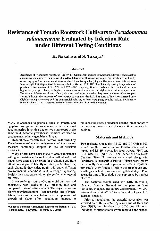
Resistance of Tomato Rootstock Cultivars to Pseudomonas solanacearum Evaluated by Infection Rate
under Different Testing Conditions
K. Nakaho and S. Takaya*
Abstract
Resistance of two tomato rootstocks (LS-89, BF-Okitsu 101) and one commercial cultivar (Ponderosa) to Pseudomonas solanacearum was evaluated by determining the infection ratio of the infection as well as by observing symptoms under conditions in which three factors, leaf stage at the time of inoculation (from four to eight leaf stage), inoculum concentration (from 105 to 108 cfu/mL) and growing temperature of plants after inoculation (30°C-25°C and 25°C-20°C, day-night) were combined. Disease incidence was higher on younger plants, at higher inoculum concentrations and at higher incubation temperatures. Resistance of the rootstocks was clearly demonstrated especially when they were incubated at low temperatures, although the response of two rootstocks was not identical. The ratio of infection differed only slightly among rootstocks and the commercial cultivar, as there were many healthy looking but latently infected plants of the rootstocks under mild conditions for disease development.
MANY solanaceous vegetables, such as tomato and eggplant, are grown in succession or after a short rotation period involving one or two other crops in the same field, because greenhouse facilities are used to produce most other vegetables in Japan.
Under these circumstances, bacterial wilt caused by Pseudomonas solanacearum is severe and the countermeasure commonly adopted is use of resistant rootstocks.
Many efforts have been made to obtain rootstocks with good resistance. In such studies, wilted and dead plants were used as a criterion for evaluation and little attention was paid to latently infected plants. However, such plants, if they exist, might wilt under different environmental conditions and although appearing healthy they may cause wilt on the grafted commercial cultivars.
In our study, resistance of commercial cultivars and rootstocks was evaluated by infection rate and compared to visual ratings of wilt. The objective was to clarify how three factors-leaf age atthe time of inoculation, inoculum concentration and temperature of growth of plants after inoculation-interact to
*Chugoku National Agricultural Experiment Station, 6-12-1, Nishifukastu, Fukuyama, Hiroshima 721, Japan.
inffuence the disease incidence and the infection rate of two resistant rootstocks and a susceptible commercial cultivar.
Materials and Methods
Two resistant rootstocks, LS-89 and BF-Okitsu 101, which are the most common tomato rootstocks in Japan, and LS-89, a selection from Hawaii 7998 and BF-Okitsu 101 (NC-1953-64N, received from North Carolina State University) were used along with Ponderosa, a susceptible cultivar. Plants were grown individually from seed in peat moss pellet (Jiffy-9, 50 mm single, Jiffy Products Ltd) in the greenhouse until seedlings reached from four- to eight-leaf stage. Plant age at the time of inoculation was expressed by number ofleaves.
The bacterial isolate used was 8107 (biovar 4) obtained from a diseased tomato plant at Nara Prefecture in Japan. The culture was stored in 10%( w/v) skimmed milk at -20°C to reduce development of avirulent mutants.
Prior to inoculation, the bacterial suspension was streaked on to the selective agar medium of Hara and Ono (1983) and incubated at 30°C for 48 hours. Individual virulent colonies were removed and suspen-
138

sions in distilled water were adjusted to a concentration from 108 to 105cfulmL by serial dilution in conjunction with a Bausch and Lomb Spectronic 20 colorimeter.
Inoculations were made by dipping trimmed roots in the bacterial suspension in plastic cases. Plants were kept in growth chambers set at 2S-30°C (day) and 20-2SoC (night) and illuminated by ca. 30000 lux light intensity with 12-hour photoperiod.
Final observations of wilted plants and disease ratings were made 14 days after inoculation following the system of Winstead and Kelman (1952). Symptomless plants were examined for latent infection as follows: a piece of stem 2 cm in length was cut from basal portion and dipped in a test-tube containing 3 mL distilled water. After incubating at SOC for 12 hours, the test-tube was vortexed and 20 mL of the suspension was plated onto Hara and Ono's selective agar medium and cultured at 30°C for 48 hours. Virulent colonies were distinguished from other bacterial colonies on the agar medium by their appearances. Number of plants used in a treatment was from eight to ten. Experiments were repeated two or three times.
- - r-
I-
50
.....
50
o
r-
r--
.....
Results
For LS-89 under high temperatures almost all plants wilted or died when they were inoculated at the fourleaf stage (Fig. 1). Fewer plants wilted as the leaf stage advanced and the inoculum was diluted. The number of latently infected plants inoculated at the eight-leaf stage was nearly twice that of wilted plants. Under lowtemperature conditions, there were only a few wilted plants, but latently infected plants were recognised regardless of leaf stage at time of inoculation, especially when the plants were inoculated at the fourleaf stage, as more than SO% of them were latently infected.
For BF-Okitsu 101 under high temperatures almost all plants wilted (Fig. 2). Under low temperatures, plants wilted when inoculated at the five- and sevenleaf stage. The number of wilted and latently infected plants decreased as inoculum was diluted. At the eightleaf stage, plants did not wilt, and 10 to 40% were latently infected.
r-
[iI" - r--
... .. ........ fti~ -
"'r---- aL '-- -- ..... ....... -'--
10 4-leaf stage 6-leaf stage 8-leaf stage
Inoculum concentration (cfU/rnL)
Fig. 1. Percentage of wilted and infected plants and disease index of LS-89 inoculated with Pseudomonas solanacearum at four concentrations, at three different leaf stages and grown at A) high temperature condition (30°CI25°C day/night), and B) low temperature condition (25°C/20°C) .• Infected plants (%); D Wilted plants (%); and. disease index.
139

100 A - re: -.- r-~i',
!-
I-
50 ., !-
r--r-~i'. r-
~ 1---0 -- ~ -- -- '-- '-- '-- a:~
lOO B
50
o 107
5-leaf stage 7 -leaf stage 8-leaf stage
Inoculum concentration (cfU/mL)
Fig. 2. Percentage of wilted and infected plants and disease index of BF-Oldtsu 101 inoculated with PseudomolUls solanacearum at four concentrations at three different leaf stage and grown in A) high temperature (30°C-25°C day-night), and B) low temperature (25°C-20°C) .• Infected plants (%); 0 Wilted plants (%): and. disease index.
When comparing wilt percentage of LS-89 and BFOkitsu 101, the effect of two different growth temperatures differed significantly, as more plants of LS-89 wilted than of BF-Okitsu 101.
For Ponderosa under high temperatures all plants wilted except at the lowest inoculum concentration regardless of leaf stage at the time of inoculation (Fig. 3). Under low temperatures, the final results were essentially the same as those at high temperatures, although disease indexes were slightly lowered.
Virulent colonies were detected on Hara and Ono's selective agar medium from plants of all three cultivars which did not have brown discolored vascular bundles.
Discussion
There are few reports in which the resistance of tomato cultivars tested for resistance to bacterial wilt was assessed on the basis of infection rate.
Hitherto, results obtained by evaluation of ratio of diseased plants and disease index in our experiments,
were compared to those already reported (Krausz and Thurston 1975; Mew and Ho 1976, 1977; Winstead and Kelman 1952), and no great differences were apparent.
The disease incidence increased when plant age at time of inoculation was younger, and inoculum concentration and incubation temperature were higher. Resistance of rootstock cultivars was clearly demonstrated especially when they were maintained after inoculation at low temperature conditions, though the response of two rootstock cultivars differed.
However, when infection rate was used to evaluate the resistance, differences between rootstock and commercial cultivars were less apparent, although the trend in the data was the same as that obtained by the former method. There were many healthy looking, but latently-infected plants in rootstock cultivars under mild conditions for disease expression.
These observations lead us to suggest that resistance of the two rootstock cultivars is quantitative in nature and variable under different conditions.
140
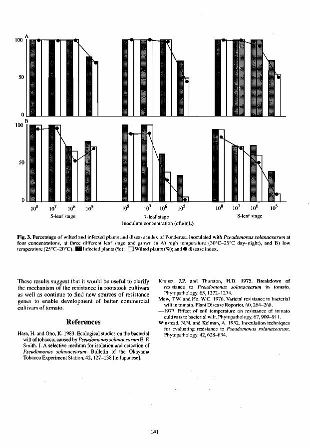
50
0 B
100 .f- ~ -.,...... \
-f\ .. ~
r ~ \ .. " itV -50 ,
- 1\ ~ 1\
~ 0 -- - 1-1...- -- .... -- .... -- ., -'- '-'- -'-
108 107 106 lOS 108 107 106 105 108 107 106 105
5-leaf stage 7 -leaf stage 8-leaf stage
Inoculum concentration (cfu/mL)
Fig. 3. Percentage of wilted and infected plants and disease index of Ponderosa inoculated with Pseudomonas solanacearum at four concentrations, at three different leaf stage and grown in A) high temperature (30°C-25°C day-night), and B) low temperature (25°C-20°C) .• Infected plants (% );. DWilted plants (%); and. disease index.
These results suggest that it would be useful to clarify the mechanism of the resistance in rootstock cultivars as well as continue to find new sources of resistance genes to enable development of better commercial cultivars of tomato.
References
Hara, H. and Ono, K. 1983. Ecological studies on the bacterial will of tobacco, caused by Pseudomonas sola1U1Cearum E. F. Smith. I. A selective medium for isolation and detection of Pseudomonas solanacearum. Bulletin of the Okayama Tobacco Experiment Station, 42,127-138 [in Japanese].
141
Krausz, J.P. and Thurston, H.D. 1975. Breakdown of resistance to Pseudomonas solanacearum in tomato. Phylopathology,65,1272-1274.
Mew, T.W. and Ho, W.C. 1976. Varietal resistance to bacterial wilt in tomato. Plant Disease Reporter, 60, 264-268.
-1977. Effect of soil temperature on resistance of tomato cultivars to bacterial wilt. Phytopathology, 67, 909-911.
Winstead. N.N. and Kelman, A. 1952. Inoculation techniques for evaluating resistance to Pseudomonas solanacearum. Phytopathology, 42, 628--{)34.

Resistance to Bacterial Wilt in Tonlato: Gene Dosage Effects
N. Anand*, A.T. Sadashiva*, S.K. Tikoot, Ramkishun§, and K. Madhavi Reddy*
Abstract
Sixteen breeding lines and exotic introductions of tomato were identified as having resistance to Pseudomonas solanacearum. Two experiments were designed to elicit information on (i) behaviour ofFl s for disease resistance using lines differing in levels of resistance, (ii) genetic makeup of resistant lines, and (iii) possibilities of exploiting heterosis for yield with these lines. In the first experiment, several parents differing in survival rates were used in a diallel cross. F 1 s showed dominance for resistance in two parents and recessive in one. Fl from a cross of two moderately resistant parents had higher levels of resistance, indicating additive gene action. High level of resistance was reflected by high survival rates, reduced browning of vascular tissue and absence of ooze. In the second experiment, seven resistant (R) lines, including two from the previous set, were crossed with a susceptible (S) line and among each other. Twenty F 1 s among R x R parents and nine F 1 s of R x Sand F2s of R x Rand R x S were studied. Although all the R parents had 100% survival, Fl s ofR x S had variable survival percentages, indicating differences in levels of dominance among the resistant parents. Vascular browning, indicative of bacterial invasion, was used as a selection criterion for resistance. Fls of R x R crosses with parents differing in genetic systems for resistance had higher levels of resistance than R x S crosses, suggesting complementation of resistance genes. Segregation ratios of R x R clearly indicated the nonallelic nature of resistance genes in four of the parents. Significant heterosis for yield in both experiments indicated greater genetic diversity for yield and its components. This was also evident among the parents. In the development of durable bacterial wilt resistant F 1 hybrids, it is essential that both parents carry resistance genes at different loci, so as to utilise the gene dosage effects for enhanced resistance in an appropriate genetic background.
BACTERIAL wilt caused by Pseudomonas solanacearum E.F. Smith is a devastating soilborne pathogen in the tropics. In India it causes serious economic losses in the states of Kerala, Karnataka, Orissa, Maharashtra, Bihar, West Bengal and Himachal Pradesh. In a bid to combat this disease, breeding efforts have been under way at our Institute for over two decades. Several resistant sources have been identified and two varieties, Arka Adha and Arka Alok, have been released for commercial cultivation.
*Division of Vegetable Crops, Indian Institute of Horticultural Research, Hessaraghatta Lake PO, Bangalore -560 089.
tPHI Biogene Limited, 61/A, 14th Cross, R.T. Nagar, Bangalore-560032.
§Central Institute of Horticulture for Northern Plains, B-217, indirannagar, Lucknow-226 016, India.
F, hybrid tomatoes are rapidly gaining popularity among growers in India. It is therefore essential to develop bacterial wilt resistant F, hybrids that are durable over space and time. The nature of a resistance will determine its durability. Several types of gene action have been attributed to bacterial wilt resistance, including: multiple recessives (Tikoo et al. 1983); single dominant gene (Tikoo et al. 1983); partial dominance in the seedling stage and recessive genes in mature plants (Acosta et al. 1964); semi-dominant genes (Anais 1986); partial dominance (Digat and Derieux 1968); polygenic, additive (Ferrer 1984), monogenic partial dominance (Raj an and Peter 1986); additive gene action (Villareal and Lal 1978) and multiple recessive genes acting additive1y (Anon. 1975). Conflicting conclusions about the genetic control of resistance could be attributed to strong genotype x environment interactions involving polygenic
142

systems in both host and pathogen (Amold and Brown 1968). Studies on development of resistant FI hybrids with different gene actions for resistance have not been reported.
The objectives of our study were to use parents with varying levels of resistance to bacterial wilt as well as to study the levels of resistance in Fls when parents with different inheritance for resistance to this disease were employed so as to arrive at a strategy to develop durable resistant F I hybrids.
Materials and Methods
Sixteen breeding lines and exotic introductions of tomato were identified as having resistance to Pseudomonas solanacearum. Two experiments were designed to elicit information on (i) behaviour of Fls for disease resistance using lines differing in levels of resistance, (ii) genetic makeup of resistant lines, and (Hi) possibilities of exploiting heterosis for yield with these lines.
18ble 1. Characteristics of tomato lines with different levels of survival to bacterial wilt used in a diallel set.
Line Source Reaction to bacteriaCwilt" Remark6
Saturn Or J. Augustine, Florida, USA MR-Upward movement of pathogen as shown sp+, gs large oblate fruits (IHRS2l) by vascular browning is restricted but high
downward movement occurs (57.1 %) BWR5 Pureline selection from CL- R browning at site ofinoculation only (100%) sp, Ig, large fruit
114-t-t-OAVRDC, Taiwan BWR13 Line derived from CRA 66 MR-Plants show slight browning and ooze Semi-determinate Ig, medium
Sel-A xSel-24 (41.9%) sized fruits BWR4 Selection from VC 13-21 P HR-No browning or ooze (1 00%) sp. 19. medium sized fruits
AVRDC, Taiwan PusaRuby Commercial cultivar derived S-Browning and heavy ooze (0%) Semi-determinate Ig, medium
frnm Siou" x Meerutj sized fruits BWR9 Selection from VC8-1-2-1, R- No browning (100%) Semi-determinate 19, medium
AVRDC, Taiwan sized fruits CRA66 Or Buddenhagen, Hawaii, R - Some plants show browning but none sp+, small fruits
USA succumbed
"Key: HR=highly resistant (100% survival), R=resistant (80-100% survival), MR=moderately resistant (40-80% survival), and S=susceptible (40% surviVal).
bKey:sp+ = indelenninate, gs=green shoulder in unripe fruits, sp=<lcterminate. and Ig=light green unripe fruits.
Table 2. Characteristics of tomato lines used to study the inheritance of resistance to bacterial wilt.
Line Source Reaction to bacterial wilt Remarks·
BWRI Selection from 663-12-3 Resistant, 100% survival Semi determinate medium sized (VC 8-1-2-1, AVRDC) oblate fruits, Ig
BWR5 Selection from IHR 719 originally Resistant, 100% survival sp, Ig, large fruits Cl 114-t+9AVRDC
1661 T-89-L3984AVRDC Resistant, 100% survival Semi determinate large, oblate fruits,lg
15S8 Derivative through pedigree Resistant, 100% survival Semi determinate large, oblate selection fruits,lg
1836 'CARAIBO' (France) Resistant, 100% survival sp, oblate medium sized fruits, gs 1643 Ponderosa Red-L-295 Resistant, 100% survival Semi-determinate medium sized
fruits, gs IS81 Redlander (Australia) Resistant, 100% survival Semi-determinate medium sized,
firm fruits, gs Sonali Developed as BWR line at Akola, Resistant, 100% survival sp, oval. small fruits
Maharashtra 709 'Florida 1011' Susceptible sp, firm, medium sized fruits 1001 PetoEarly Susceptible sp, processing variety 1334 EC 1193, (N~PGR, New Delhi) Susceptible sp, ovalfruits, 70 g thick fleshed
"Key: sp = determinate, gs = green shoulder in unripe fruits and 19 = light green unripe fuils.
143

In the first experiment, several parents differing in survival rates were used in a diallel cross (Table 1). In the second experiment, seven resistant (R) lines, including two from the previous set, were crossed with a susceptible (S) line and among each other (Table 2).
To study the inheritance of resistance to bacterial wilt the following F2 combinations were also studied among resistant crosses (R x R): BWR 5 x 1661, BWR 5 x 15 SB, BWR 5 x 1881,1881 x 1661, 1661 x 1643, 1643 x Sonali, and BWR 1 x BWR 5; and the following F 2 crosses among R x susceptible (S) combinations: 16bl x 977, 1881 x 1817, 15 SB x 1816, and 1643 x 1817.
The experiments were carried out between 1986 and 1992. The prevailing maximum temperature during crop growth periods ranged from 29 to 36.6°C and the minimum temperature from 14.1 to 22.8°C. In parents and FIs, 15 plants were replicated three times. For F2s, 150 seedlings were planted out at a spacing of 90 x 30 cm in wilt-sick soil and fertilized with 120 kg Nlha and 60 kg K/ha. Four harvests were made and yield per plant computed by dividing each plot yield by the number of transplants. Carbofuran was applied at 3 g/m2
to control nematodes. VIrulent colonies of P. solanacearum were identified on peptone casein hydrolysate agar containing 0.005% 2.4,5-triphenyl tetrazolium chloride (Kelman 1954), multiplied on nutrient agar medium and incubated at 30± 1°C. After 48 hours of growth on nutrient agar medium, the pathogen was suspended in water and inoculated to leafaxils using two drops per 20-day-old transplant.
The survivors were counted on the l00th day. The spread of the pathogen was assessed by recording at final harvest, the degree of browning of split stems at the root-shoot junction. Three plants per entry from each replicate were rated on the following scale: 0 = no browning; 1 = light brown colour, spread restricted to 2 cm from the point of inoculation; 3 dark brown colour, widespread browning of vascular tissue. Bacterial ooze was observed by dipping a piece of the lower cut stem from just above the root zone in a tube of clear water. The scoring for ooze was as follows: 0 = no ooze; 1 = thin strand of ooze not continuous, flow stops after 3 minutes; 2 = continuous thin white, flow not restricted; 3 = heavy ooze, turning the water turbid within 2 minutes.
Resu1ts and Discussion
Survival percentages of the parents in the diallele set ranged from 0 to 100% (Table 3). The Fls ofIHR 719, BWR 4 and BWR 9 crossed to Pusa Ruby showed complete resistance, indicating control by dominant gene(s) in these parents. BWR 4 and BWR 9 had no vascular browning or ooze, indicating that these sources of resistance reduced multiplication of the
pathogen in the plant system. Prior et at (1990) used ELISA at various stem levels in four cultivars of tomato and reported that three of them had field tolerance because their vascular systems were invaded, while in the fourth variety the pathogen was not detected. Association between vascular browning and ELISA tests may help elicit information on presence and multiplication of bacteria.
The moderately resistant lines, IHR 821 and BWR 13, had scores of less than 3 for browning and less than 2.2 for ooze, while the susceptible line Pusa Ruby was rated 3 in both tests. Moderately resistant lines IHR 821 and BWR 13 when crossed with resistant parents, produced highly resistant Fls. FIs of IHR 821 and BWR 13 crossed with CRA 66 Sel A, [known to possess recessive polygenes for resistance (Tikoo et al. 1983)] had higher levels of resistance than IHR 821 or BWR 13. Moderate resistance in IHR 821 and BWR 13, controlled by many genes acting additively and in combination with those of CRA 66 Sel A, resulted in resistant F 1 s. Additive gene action for resistance to bacterial wilt has also been reported by Ferrer (1974), Graham and Yap (1976), ViIlareal and Lal (1978), and at AVRDC (Anon. 1990; Hartman 1992). Further proof of additive gene action conferring resistance arises from the FI ofIHR 821 x BWR 13 which had a higher survival percentage than either of the parents, and had significantly lower browning and ooze scores (Table 3).
In studies on resistance, none of the plants in the seven resistant parents wilted, while the susceptible lines had less than 10% survival (Table 4). Fls ofBWR 1, BWR 5, 1661, 15 SB and 1836, crossed with susceptible lines had 100% survival, indicative of dominant gene action, while F I s of 1881 and Sonali had 80% and 50% survival, respectively, and had low ooze scores, indicating incomplete dominance for resistance to bacterial wilt. These results suggest differences in inheritance of resistance among the selected resistant lines and confirms the dominance in BWR 1 reported by Tikoo et at (1983).
Fls of crosses among bacterial wilt resistant lines having complete dominance had very high levels of resistance. Fls of crosses between resistant parents (BRW1. BWR5, 15 SB and 1661) and a susceptible parent also had 100% survival (Table 4). However. the mean scores of browning were significantly less in R x R set of FIs compared with the set of R x S Fls indicating that multiplication of the pathogen was inhibited in the vascular system of the heterozygote from two resistant parents (Tables 4 and 5).
Fls of crosses between resistant lines having dominant gene action (BWR 1, BWR 5, 15SB) and incomplete dominance (1881 and Sonali) exhibited high levels of resistance and low scores for browning. F j of Sonali with all other resistant lines including
144

'Thble 3. Perfonnance of parents and Ft sin diallel set after inoculation with Pseudomonas soianacearutll.
Survival (%)
IHR 821 S7.1 JHP821 xBWRS 100.0
xBWR 13 74.6 xBWR4 100.0 x Pusa Ruby 47.1 xBWR9 100.0 xCRA66SelA 100.0
BWRS 100.0 BWR5 xBRWI3 100.0
xBWR4 100.0 x Pusa ruby 100.0 xBWR9 100.0 x CRA66 SelA 100.0
BWR13 41.9 BWR13 xBWR1 100.0
x Pusa ruby 61.1 xBWR9 100.0 xCRA 66 SeIA 100.0
BWR4 100.0 BWR4 x Pusa Ruby 100.0
xBWR9 100.0 xCRA66SelA 100.0
PusaRuby 0.0 Pusa Ruby x BWR 9 100.0
xCRA66SelA 8.3
BWR9 100.0 BWR9 xCRA66SelA 100.0 CRA 66 Sel.A 100.0
FValue IS.74** C.D.5% IS.8 C.S.I% 22.4
1881, again had high levels of resistance confinning complementation of genes for resistance. Expression of resistance in the heterozygotes in our study had reduced mUltiplication, shown by low browning scores and lack of ooze. These parameters may be useful for selection of resistance in segregation populations.
Even the resistant lines BWR 4, 1661 and 1881 showed slight browning (Table 4). Linear advance of the pathogen in the vascular tissue may be related to cell multiplication rate, resulting in different levels of resistance. Resistance also could result from the ability of the host to produce toxin in areas where the pathogen is active, so as to cheek its spread. Lines having vascular browning should be avoided when moreresistant lines are available, as they may display reduced levels of resistance at higher temperatures. Resistance to bacterial wilt was reported to be temperature sensitive and strain specific (Krausz and
Browning score Ooze score Yield/plant (kg)
2.0 2.1 0.36 1.0 0.7 1.79 1.0 0.0 1.27 0.0 0.0 1.19 2.3 1.7 0.87 I.S 0.7 1.42 1.2 0.3 0.97
0.4 0.0 0.98 0.0 0.0 1.13 0.1 0.0 0.65 1.0 0.3 1.73 0.3 0.0 0.97 0.6 0.1 1.02
2.8 1.0 1.42 1.7 1.3 1.56 1.3 1.3 0.74 0.0 0.0 1.S7 0.6 0.0 1.06
0.0 0.0 1.13 2.0 1.7 1.70 2.0 1.3 1.S8 0.7 0.3 1.63
3.0 3.0 0.25 0.3 0.3 1.60 2.8 2.5 O.SO
0.0 0.0 1.34 0.0 0.0 1.10 0.0 0.0 0.58
4.84** 3.78** 6.10** 0.4 O.S 0.6 0.7 1.1
Thurston 1975). Whether this is primarily a function of virulence factors expressed at high temperatures (Tung et al. 1990b), or the lack of expression of resistance genes, is not known. Terminologies related to resistance or susceptibility may become clearer when techniques such as DNA probes, monoclonal antibodies, and ELISA are available to test bacterial populations in the host plant.
Variances of general and specific combining ability obtained through diallel analysis (Table 6) indicated that both additive and nonadditive gene actions were important. The additive genetic variance and the dominance variance were important with dominance variance the more so. Importance of nonadditive gene action was indicated by resistance in FJs of R x S crosses. Nonadditive gene action has also been implied for bacterial wilt resistance at AVRDC (Opeiia et al. 1990). Resistant parents differing in level of resistance
145

Table 4. Perfonnance of resistant and susceptible parents, and their crosses in Pseudomonas solanacearum infested soil.
Survival Browning Ooze score Yield/plant (%) score (kg)
FIs BWRI x 709 100 0.3 0.0 2.27 BWRSx 1001 100 0.6 0.0 3.50 1661 x 1334 100 0.8 0.2 2.62 ISSBx 1334 100 0.2 0.0 2.68 1836 x 709 96 0.7 0.0 2.30 1643 x 709 72 1.0 0.3 1.99 Sonalix709 68 1.1 0.9 2.41 Susceptible 709 0 3 3 0 1001 6 3 3 0.14 1334 7 3 3 0.10 Resistant BWRI 100 0.1 0 1.86 BWRS lOO 0.2 0 1.97 1661 lOO 0.2 0 1.79 ISSB 100 0.2 0 2.07 1836 100 0.4 0 I.S8 1643 100 0.4 0 1.62 1881 100 0.4 0 1.92 Sonali 100 0.4 0 1.01 FValue 12.86 8.32** S.31** 10.35** C.D.S% 14.3 0.2 0.6 0.44 C.D.I% 22.0 0.4 1.1 0.78
Table S. Perfonnance of resistant x resistant Fls in wilt-infested soil.
Survival Browning Ooze Yield/ (%) score score plant (kg)
BWRI xBWRS 100 0 0 1.93 BWRSx 1661 lOO 0.2 0 3.20 BWRSx 15 SB lOO 0 0 2.61 BWR 1 x 1661 lOO 0 0 2.8S 1836x 1661 lOO 0.2 0 3.00 BWR I x 1836 100 0 0 1.59 1661 x IS SB 100 0 0 2.S0 1836xBWR5 100 0.1 0 2.50
II 1836 x 1881 100 0 0 2.86 1661 x 1881 lOO 0.2 0 1.83 IS SB x 1881 lOO 0 0 2.43 15SB x 1643 lOO 0 0 2.01 BWR 5 x 1881 100 0 0 1.47 BWRSx 1643 100 0.2 0 1.88 BWR I x 1881 100 0.1 0 2.S1 1643xBWR 1 100 0.1 0 2.36
III 1643 x 1881 lOO 0.2 0 3.10 Sonali x 1643 100 0.3 0 2.32 1881 xSonali 100 0.1 0 1.96 FYalue 6.2** 12.10** C.D. S% 0.1 0.49 C.D.l% 0.2 0.82
Table 6. General and specific combining ability (GCA and SCA) obtained through diallel analysis based on plant survival of tomatoes inoculated with Pseudomonas solanacearum.
Survival % GCA SCA
F. value Variance Additive Dominance GCA:SCA genetic variance
ratio variance
510:24 578 63 S8-46 255-12
and genetic systems can additively enhance levels of resistance (Opefia et a!. 1990). Interaetions among resistant genes can be complementary when two or more nonallelic resistance genes confer resistance (RusseIl1978).
F2s among BWR 1, BWR 5 and 15 SB (a derivation ofBWR 1) had 100% survival (Table 7). These parents have similar genetic systems for resistance, although backcross data are required to confirm allelism. F2s of crosses among 1661, 1881, 1643 and Sonali, and between each of these and BWR 1 and BWR 5 had 70-85% survival. This indicates that the former set of resistant lines differ from each other in the resistance factors they carry and also differ from BWR 1 and BWR 5. Only F2s involving parents carrying resistance gene(s) at identical loci are expected to show 100% survival.
Exclusive cultivation of a single genotype can exert strong selection pressure on the pathogen. Velupillai and Stall (1984) indicated that a shift in the population of P. solanacearum towards more aggressive strains was possible. Nonallelic heterozygotes differing in resistance genes would help combat more strains of the pathogen. In the absence of markers, better procedures are needed to identify different resistance genes in segregating populations, to enable pooling of these genes. This could be a form of genetic diversification whereby the pathogen would have to cope with a variety of resistance genes.
Heterosis for yield is a function of genetic diversity among the parents. The parents involved in both the experiments were morphologically distinct. BWR 13 was the highest yielder among those tested in the diallel set. It was a moderately resistant line with levels equal to BWR 5, BWR 4 and BWR 9 (all resistant lines), and was significantly higher yielding than IHR 821, Pusa Ruby and eRA 66 Sel A. Eight of the Fls in the first experiment showed heterotic yields over the best parent (Table 3). The F I of IHR 821 x BWR 5 had the highest yield (1.79 kg/plant) followed by the FI of BWR5 xBWR 9 (1.73 kg/plant) andBWR4x BWR 9 (1.58 kg/plant). Heterotic yields in R x R crosses were as high as in R x S or R x MR crosses. Genetic diversity for yield and its components was obviously high among
146

Table 7. Survival of F2s inoculated with Pseudomonas solanacearum.
Crosses
Resistant x Resistant BWR5x 1661 BWR5x15SB BWR5x1881 1881 x 1661 1661 x 1643 1643 x Sonali BWRI xBWR5 Resistant x Susceptible 1661 x977 1881 x 1817 15SBx1816 1643 x 1817
Survival %
85 100 77 72 85 77
100
62 25 71 41
the parents used. Widening the genetic base for both resistance and adaptation to the environment (Tung et at 199Ia,b) may be very important in breeding for bacterial wilt resistance.
In experiment 2, heterosis of a high order was manifested in Fls (Table 5). The over all heterosis in Fls was 26.8% over the mean yield of resistant parents. FI hybrids in this experiment were grouped as follows: A = when both parents were resistant and resistance was governed by dominant genes; B = when one of the parents had resistance governed by dominant genes and the other by incompletely dominant genes (above 80% and 50% survival); and C = when both the parents had resistance controlled by incompletely dominant genes. The mean yield ofF,s in Group A was 2.48 kg/plant. In Group B, Fls among parents having incomplete dominance (80% survival) yielded 2.59 kg/plant, while Fls between parents with incomplete dominance (50% survival) yielded 2.17 kg/plant. In Group C, the mean yield ofF,s was 2.46 kg/plant. Heterosis for yield in Fls of R x R could be independent of the nature of resistance in the two parents. This could be tested by growing these in soil free from bacterial wilt.
Wide genetic diversity exists among the lines, for yield as well as for bacterial disease resistance. This provides an opportunity for widening the genetic base for resistance, transferring resistance genes into appropriate genetic background thereby enlarging the scope of exploiting F, hybrids forresistance and yield. F I s for R x S parents were as high yielding as R x R crosses when grown under bacterial wilt infested soil and if artificially inoculated. Constructing genetically diverse resistant parents would consume time and effort, and it may be worth while exploiting R x S crosses pending the development of a wider array of resistant lines with a broader genetic base, different genetic systems for resistance, and high in general combining ability.
The development of potential parents with bacterial wilt resistance genes will require identification of resistance genes at different loci, as well as the full complement of minor genes, modifiers, and multiple genes or polygenes. Tracing isozymic linkages if available or RFLi' mapping can help in this effort. Use of serological techniques, such as raising antisera against several different isolates, identification of the most virulent pathotype, and application of ELISA to detect pathogen population in different field-resistant lines, could form the basis for a more refined classification of resistance. Screening using these methods may be more reliable and rapid.
Single plant selections with complete absence of browning have been made in and F3 generations in wilt-infested soil from R x R crosses, after inoculation with bacterial suspension. Screening under heavy pathogen load, aided by the use of polyclonal antisera, will help to develop stable resistant lines.
References
Acosta, I.C., Gilbert, 1.C. and Quinon, Y.L. 1964. Heritability of bacterial wilt resistance in tomato. Proceedings of the American Society of Horticultural Science, 84, 455-462.
Anais, G. 1986. Utilisation de la resistance varietale dans la lutte contre le llelrissement bacterien de la tomate dO a Pseudomonas solanacearum E. F. Smith. In: Ministere de l' Agriculture,ed., Bulletin Technique d'Infonnation, Paris, 409-411,449-453.
Anon. 1975. The Asian Vegetable Research and Development Center Tomato Report for 1975,25-28.
Anon. 1990. 1987 Progress Report. Shanhua, Tainan, Asian Vegetable Research and Development Center, 234-238.
Amold, M.H. and Brown, S.J. 1968. Variation in the host parasite relationship of a crop disease. 10urnal of Agricultural Science. 71,19-36.
Digat, B. and Derieux, 1968. A study of the varietal resistance of tomato to bacterial wilt H. The practical value of Ft hybrids and their contribution to the genetic basis of resistance. Proceedings of 6th Annual Meeting Caribbean Food Crops Society, 85-100.
Ferrer, Z.A. 1974. Resistance to Pseudomonas solanacearum in Lycopersicon esculentum. Ph.D. Thesis, University of Florida, USA.
-1984. The nature of resistance in a tomato tolerant to Pseudomonas solanacearum. Phytopathology, 74, 1014. Abstract.
Graham, K.M. and Yap, T.C. 1976. Studies on bacterial wilt I. Inheritance of resistance to Pseudomonas solanacearum in tomato. Malaysian Agricultural Research. 5, 1-8.
Hartman, GL 1992. Bacterial wilt of solanaceous vegetables, AVRDC report. In: SAVERNET Proposed Work Plan, 1992-1994. ADB Project TA No. 5461.Shanhua, Tainan, Taiwan, AVRDC. In press.
147
Kelman, A. 1954. The relationship of pathogenicity in Pseudomonas solanacearum to colony appearance on a tetrazolium medium. Phytopathology, 44, 693-695.

Krausz. I.P. and Thurston, H.D. 1975. Breakdown of resistance to Pseudomonas solanacearum in tomato. Phytopathology, 65.1272-1274.
Opeila, R.T., Hartman, G.L., Chen, J.T. and Yang, C.H. 1990. Breeding for bacterial wilt resistance in tropical tomato. In: 3rd International Conference on Plant Protection in the Tropics 1990. Malaysian Plant Protection Society, Vo!. V, 44--50.
Prior, P., Beramis, M., Chillel, M. and Schmit, J. 1990. Preliminary studies for tomato bacterial wilt (Pseudomonas solanacearwn E.F. Smith) resistance mechanisms. Symbiosis (Rebovot), 9, 393-400.
Rajan, S. and Peter, K. V. 1986. Incomplete dominance of bacterial wilt resistance. Report Tomato Genetics Cooperative, 36, 24.
RusseJl, G.E. 1978. Plant breeding for pest and disease resistance. In: Chapter 2. General principles and methods of breeding for resistance. Butterworth London, Boston: 25.
TIkoo, S.K .• Anand, N. and Ramkrishna. 1983. Presence of two independent genetic systems for resistance to bacterial wilt (Pseudomonas solanacearum) in tomato. In: Proceedings of the 15th International Genetic Congress, 12-23 December 1983, New Delhi, India.
Tung, P.X. and Roseo, E.T.J., Vandel7..aag, P. and Schmiedeche, P. 19903. Resistance to Pseudomonas solanacearum in the potato I. Effects of sources of resistance and adaptation. Euphytica, 45, 203-210.
-199Ob. Resistance to Pseudomonas solanacearum in the potato n. Aspects ofhos! pathogen environment interaction. Euphytica, 45. 211-215.
Vellupillai. M. and Stall. R.E. 1984. Variation among strains of PseutWmonas solanacearum from Florida. Proceedings of the State Horticultural Society, 97, 209-213.
Villareal, R.L. and Lal, S. H. 1978. Reaction of three tomato cultivars--their FI's and three way crosses to two isolates of bacterial wilt. HortScience, 13,366.
148

Inheritance of Resistance to Bacterial Wilt in Tomato
S. Monma and Y. Sakata*
Abstract
Bacterial wilt resistant tomato lines D-9 and Hawaii 7998 were crossed with susceptible TPL-S. Resistance of the cross between D-9 and TPL-5 was evaluated in 1989, and TPL-5 and Hawaii 7998 in 1990. Parents, F I s, F2s and backcross plants were transplanted into a field heavily infested with Pseudomonas solanacearum and inoculated with the pathogen by pouring a bacterial suspension into the soil. Bacterial wilt resistance was evaluated by recording the date of plant death and by a resistance index (RI) from I (susceptible) to 13 (highest resistance) for each plant. The Rls of the resistant parents, D-9 and Hawaii 7998, were 12.8 and 13.0, and those of the susceptible parent, TPL-5 were 1.0 and 1.1, respectively. Mean RIs of the FI generation of the two crosses were 4.5 and 6.2, which were lower than each midparent value of 6.9 and 7.1, respectively. This suggests that the bacterial wilt resistance was partially recessive as there was incomplete dominance towards susceptibility. No correlation existed between RI and fruit weight in the F2 generations of the two crosses (r = -D.074, r = -D.019). This indicated that it was possible to select plants possessing both high resistance to bacterial wilt and large fruit.
BACfERIAL wilt caused by Pseudomonas solanacearum E.F. Smith is one of the major problems of tomato in southeastern Japan, from Kantoo area of the main island to Kyushu Island. The disease causes heavy losses in tomato production, as application of chemicals. soil fumigation and crop rotation are practically ineffective. The use of resistant varieties is the most effective means for control of bacterial wilt. There are no fresh market varieties possessing both high resistance and high fruit quality available. whereas several highly resistant rootstock varieties have been developed. Growers have been grafting fresh market varieties onto resistant rootstocks to avoid bacterial wilt infection in infested areas. Grafting. however. is a time and labor consuming practice and development of highly resistant fresh market varieties is in high demand.
Knowledge on the inheritance of resistance is important in breeding for resistance. A few experiments on the inheritance of resistance have been reported (Acosta et al. 1964; Ferrer 1974; Lum 1982) but the mode of the inheritance is still uncertain. This paper reports on the inheritance of the resistance in tomato to bacterial wilt.
*National Research Institute of Vegetables, Ornamental Plants and Tea, Department of Vegetable Breeding, Laboratory of Solanaceous Vegetable Breeding, Ano, Mie 514-23, Japan
Materials and Methods
Two resistant parents, 0-9 introduced from Malaysia and Hawaii 7998 from Hawaii, and one susceptible parent. Tomato Parental line 5 (TPL-5), were used. 0-9 (PI) and TPL-5 (P2), and TPL-5 (P3) and Hawaii 7998 (P 4) were crossed to obtain F I seed. The F I S were backcrossed to each parent and self-pollinated to obtain F2 seed. Resistance of the cross between 0-9 and TPL-5 was evaluated in 1989, and for the cross between TPL-5 and Hawaii 7998 in 1990. Cross of D-9 and TPL-5. Seeds of PI' P2• FI• BCP,. BCP2 and F2 were sown on 7 March 1989 in a greenhouse. On 1 April. seedlings were transplanted to 9-cm plastic pots containing sterilised field soil. On 27 April, the seedlings were transplanted to a bacterial wilt infested field. A randomised complete block design was used with three blocks. Seven plants for the PI' P2 and FI generations. 14 plants for the BCPI and BCP2 generations, and 48 plants for the F2 generation were planted per block. Plants were staked and pruned.
P. solanacearum was isolated from a diseased tomato plant. A pure culture was multiplied at 30°C for 48 hours in Wakimoto medium (Wakimoto 1962). Bacterial concentration was adjusted to 2 x 10⁸ viable cells/mL. On 17 July, 8I-day-old transplants were inoculated by pouring 50 mL of bacterial suspension into the soil at the base of each plant.
149

For the evaluation of resistance, the date of plant death was recorded from 20 June to 26 August. Using this date, a resistance index (RI) from 1 to 13 was assigned to each plant (Fig. 1). The resistance categories described by the RI were as follows: 1-2 = susceptible; 3-5 = weak resistance; 6-8 = moderately resistant; 9-11 = high resistance; and 12-13 = highest resistance. On 7 and 12 July. fruits of each plant were harvested and weighed, and mean fruit weights were calculated for the evaluation of fruit size.
Cross ofTPL-5xHawaii 7998. Seeds ofP\> P2, FI, BCP3• BCP4 and F2 were sown on 5 March 1990 in a greenhouse. On 30 March. seedlings were transplanted to 9-cm plastic pots containing sterilised field soil. On 1 May, the seedlings were transplanted to a bacterial wilt infested field. Seven plants for the PI' P2 and FI generations, 14 plants for the BCP1 and BCPz generations, and 49 plants for the F2 generation were planted per block. On 28 June. plants were inoculated and the dates of death were recorded from 8 June to ] 0 August. On 5 July. fruits of each plant were harvested and weighed.
Results
Cross of D-9 x TPL-5 in 1989. A difference in frequency distributions of the RI between the resistant parent, 0-9 (PI) and the susceptible parent, TPL-5 (P2) was evident and the RI frequency distribution of the F I was skewed towards the susceptible parent (Fig. 2). Almost all categories of resistant plants were observed in the BCP!. On the other hand, there were no resistant plants in the BCP2. The F2 segregated in a discontinuous pattern and was skewed towards the susceptible parent. The plants with highest resistance comprised 6% of the F2s.
The mean RIs of 0-9 and TPL-5 were 12.8 and 1.0, respectively, and the mean RI of F I was 4.5, which was significantly lower than the mid-parent value of 6.9 (Table I). The mean RIs of BCP) and BCP2 were close to those of the mid-parent and susceptible parent, respectively. The mean RI of the F2 was close to that of theF1•
mculation
7 I 17 18-23
2
6 21
7
E:3 p, D P • • F, 20
10
BCP,
o 00
F.
30
20
10
O~~~~~~~LU~~~--~~~ 1 2 3 4 5 6 7 8 9 10 11 12 13
Resistance index
Fig. 2. Frequency distributions for bacterial wilt resistance index of resistant parent (PI :D-9l. susceptible parent (P2:TPL-5), Ft. BCPl , BCP2 and F2 generations. Higher numbers indicate higher resistance.
Fruits of four plants could not be harvested because of bacterial wilt. These four plants were excluded from the correlation analysis between resistance and fruit weight. No correlation was observed between RI and mean fruit weight among the F2s (r = -0.074, Fig. 3).
Cross of TPL-5 x Hawaii 7998. A difference in frequency distributions of the RI between the susceptible parent, TPL-5 (P3), and the resistant parent,
30 I 331 36 1 37 - Days after inocloDOOn
14-18 . 17-19 20-22 23- - Date of 1989 8-10 11-13
8 9 10 I 11 12 13 - Resistance index
Fig. 1. Assignment of resistance index by the date of plant death in 1989.
150

Table 1. Bacterial wilt resistance index and fruit weight of parents, F), BCP), BCP2, and F2 generations in '0-9' x 'TPL-S'
Parent and Resistance Mean fruit Number of generation index weight (g) plants
PI: D-9 12.8 69.6 19
P2:TPL-S 1.0 14S.0 21
F) 4.S 14S.0 21
BCP) 7.3 101.6 38
BCP2 1.9 127.0 32
F2 4.4 118.7 144
Mid-parent value 6.9 107.3
LSOS% 1.0 22.1 LS01% 1.4 31.S
2SO 0
r:-O.074
c 0 0
200 11 8 0 0
8 0 8 0 (J) § § 0
(J) 0 rso 0 0
0 0 .o.P' 0 § ~ r 8 § 0
~ 0 g 9 0
@> 0
'S I 8 ~ 0 8
~ ~ 0
§ ./::100 0 0 ~ c 9 0 0 0 0 ." r 11 0
:I 8 ~ f
0 oP' 0 0 0 Cl!> 0 0
0 0 0 SO
0 0
0
o~~~~~~~~~~~~~~~
234 5 678 9 ~ " n 0 Resistance index
Fig. 3. Relationship between resistance index and mean fruit weight in F2 generation of '0-9' x 'TPL-S'. Higher numbers indicate higher resistance.
Hawaii 7998 (P4) was evident (Fig. 4). The frequency distribution of the RI for the F j was between, but slightly less than, parental means. The range for this F j
was wider than that of the F j from D-9 x TPL-5. All categories of resistant plants were observed in BCP3' Almost all categories of resistant plants were also observed in BCP 4' in which plants with a RI of 13 showed the highest frequency. The F2s segregated in a continuous pattern and the distribution skewed slightly towards the susceptible parent. The plants with highest resistance were observed to comprise 6% of the F2s.
ISI
The mean RIs ofTPL-5 and Hawaii 7998 were 1.1 and 13.0, respectively, and the mean RI of their F\ was 6.2, which was slightly lower than the mid-parent value of7.1 (Table 2). The mean Rls of BCP3 and the F2 were the same as that of the F j . The mean RI of BCP 4 was higher than the midparent value.
Fruit of one F2 plant could not be harvested because the plant died of bacterial wilt before fruit maturity. This plant was excluded from the correlation analysis between resistance and fruit weight. No correlation was observed between RI and mean fruit weight among the F2s (r = -0.019, Fig. 5).
Discussion
Differences in methodologies for evaluating host resistance may affect the final interpretation of the inheritance of bacterial wilt resistance. The evaluation of resistance should be quantitative because resistance to bacterial wilt was reported to be controlled by several genes (Ferrer 1974; Lum 1982). Seedling inoculation methods such as clipping or root dipping have been applied for selection of
DP3 e<lP • • F,
20
10
30
20
Resistance index
Fig. 4. Frequency distributions for bacterial wilt resistance index of susceptible parent (P3:TPL-S), resistant parent (P4:Hawaii 7998), F), BCP3, BCP4 and F2 generations. Higher numbers indicate higher resistance.

Table 2. Bacterial wilt resistance index and fruit weight of parents. F J, BCP3, BCP4, and F2 generations in 'TPL-5' X 'Hawaii 7998'.
Parent and generation
P3: TPL-5
P4: Hawaii 7998
F,
BCP)
BCP4
Fz Mid-parent value
LSD 5%
LSD1%
250
200
o 0 o o a
50 (11) § o !
Resistance index
1.1
13.0
6.2
6.3
9.8
6.3
7.1
2.1
2.9
o
o o
o
o
o
Mean fruit Number of weight (g)
147.0
34.0
61.3
65.6
58.2
66.0
90.5
18.7
26.6
1':-0.019
o 0
000
o (11)
o
o
plants
21
21
21
42
41
156
o
o~~~~~~~~~~~~=-~ 1 2 3 4 5 6 7 8 9 ~ n ~ a Resistance index
Fig. 5, Relationship between resistance index and mean fruit weight in Fz generation of TPL-5 x Hawaii 7998. Higher numbers indicate higher resistance.
resistant seedlings, but the reaction of the inoculated seedling is usually death or survival. These results from artificial inoculation are qualitative. Although the varieties that survived the natural infection were either resistant, moderately resistant, or susceptible when inoculated (Mew and Ho 1976), it was pointed out that segregating populations were best screened under field conditions at the flowering stage (Mew and Ho 1976). The method used in this study was one that combined natural infection from infested fields with artificial inoculation to ensure disease. The resistance of each plant was shown by the resistance index (RI) according to the date of plant death. Differences in resistance classes among each parental line and generation were
clear, indicating that the method could be used for the seleetion of resistant plants and for the evaluation of plant resistance in segregating populations.
It is important to understand whether or not bacterial wilt resistance in tomato is a dominant character because in Japan almost all fresh market varieties are hybrids. Acosta et al. (1964) demonstrated that resistance was partially dominant until 7 weeks after transplanting, but recessive in more mature plants. The RI of the Fl in the cross of 0-9 x TPL-5 was lower than the mid-parent value and the RI of the F) from the cross of TPL-5 x Hawaii 7998 was close to the mid-parent value. This suggested that the bacterial wilt resistance was partially recessive depending on the degree of resistance of the resistant parent. These result were similar to those reported by Acosta et al. (1964), where resistance was evaluated in mature plants.
It has been reported that several genes control bacterial wilt resistance (Ferrer 1974; Lum 1982) and that small fruit size was associated with resistance (Bosch et al. 1982; Ferrer 1974;) leading to the problem of developing a resistant variety with good commercial quality. The frequency distributions of the RIs of BCP3 and the F2 from TPL-5 x Hawaii 7998 showed a continuous pattern. This indicated that the resistance was quantitative, although the number of genes controlling resistance may be small because the frequency distributions of the RIs of BC Pt and F2 from 0-9 x TPL-5 segregated in discontinuous patterns.
No correlations were observed between RI and fruit weight in the F2 of the crosses. This indicated that selection of resistant materials for large fruit size was not a problem in resistance breeding, and that it was possible to select plants with both high resistance and large fruit weight in the segregating populations. This result contrasts with the results of Ferrer (1974) and Bosch et al. (1982).
Differences in frequency distributions of the crosses were evident, and the resistance of Hawaii 7998 was higher than the resistance of 0-9. To explain the difference in resistance of the parents, it was assumed that the number of genes controlling resistance in Hawaii 7998 was greater than in D-9.
References
Acosta, l.C., Gilbert, 1.C. and Quinon. Y.L. 1964. Heritability of bacterial wilt resistance in tomato. Proceedings of the American Society of Horticultural Science, 84,455-462.
Bosch, S.E., Boelema, B.H. and Young, B.W. 1982. Bacterial wilt disease in tomatoes. Farming in South Africa leaflet series: Tomatoes, Peppers and Egg-fruit H6. 3 p.
Ferrer, Z.A. 1974. Resistance to Pseudomonas solanacearum in Lycopersicon esculentum. Ph.D. Thesis, University of Florida, USA.
152

Lum, K.Y. 1982. Host resistance and biological antagonism in the control of bacterial wilt of tomatoes. Ph. D. Thesis, University of Wisconsin, USA.
Mew, T.W. and Ho, W.e. 1976. Varietal resistance to bacterial wilt in tomato. Plant Disease Reporter, 60, 264-268.
Wakimoto, S. 1962. Studies on the multiplication of OPt phage (Xanthomonas oryzae bacteriophage) LOne-step growth experiment under various conditions. Science Bulletin of the Faculty of Agriculture, Kyushu University, 15,151-160.
153

Virulence Studies of Pseudomonas solanacearum and Inheritance of Resistance in Lycopersicon esculentum
A.M. Mahir, K.S. Diong and A. Ismail*
Abstract
Eleven isolates of Pseudomonas solanacearum from different Lycopersicon esculentum plants grown in experimental plots at the Universiti Kebangsaan Malaysia and one ginger (Zingiber officinale) isolate from Universiti Pertanian Malaysia were tested for virulence using a stem-puncture inoculation method. The UPM ginger isolate was most virulent and was used to determine resistance and its inheritance in seven L esculentum varieties. Local varieties (FP-5, Intan and MT-J) had relatively higher levels of resistance. Most of the imported varieties with improved agronomic characters were susceptible. Data from the FI hybrids and parents indicated that numerous modes of inheritance of resistance to P. solanacearum, including dominance, recessiveness, and additive gene action. were present. However, most resistances were conferred by a single dominant gene.
PSEUDOMONAS solanacearum E.F. Smith is one of the major diseases of Lycopersicon esculentum (tomato) in Malaysia, especially in the lowland areas. Use of resistant varieties from various breeding programs (Graham et al. 1977; Tan. unpublished data) and the Asian Vegetable Research and Development Centre (AVRDC) (Apandi and Mahir 1987; Tee et at 1979) has not been effective in solving the problem.
Lack of understanding of the host, pathogen and environment might be one of the reasons for the limited success in controlling the disease. Understanding the host and pathogen is a prerequisite for devising proper methods of control. Disease resistances in most hosts interact with the pathogen and environment (Nelson 1977).
Breeding programs have been hampered partly because of differences in virulence and behaviour of isolates ofthe pathogen from different hosts (Abdullah 1986). Unfortunately, attempts to classify isolates have not always involved genetic interaction with the host plants, and few inheritance studies have used a wide range of isolates. Modes of inheritance of resistance have been shown to be: recessive and polygenic (Singh 1961; Anon. 1975); partial dominant at seedling stage and recessive at adult stage (Acosta et a!. 1964);
"'Department of Genetics, Faculty of Life Science, Universiti Kebangsaan Malaysia, 43600 Bangi, Selangor, Malaysia.
additive (Graham and Yap 1976); oligogenic (vertical resistance); polygenic (horizontal resistance); intermediate (Mew and Ho 1976); and complementary dominant (Screelathakumary and Peter 1984),
This preliminary study was conducted to investigate the relative virulence of isolates from different hosts, and the differences in virulence to various L esculentum varieties; and to evaluate the modes of inheritance of resistance in L esculentum germplasm.
Materials and Methods
Variability of isolates
Eleven isolates of P. solanacearum were obtained from several diseased L esculentum varieties from experimental plots of Universiti Kebangsaan Malaysia (UKM, National University of Malaysia). One Zingiber officinale isolate was from Universiti Pertanian Malaysia (UPM, University of Agriculture). Thirty 2-week-old seedlings of variety Sida KU [previously reported as susceptible (Rasheeda 1992, unpublished data»), were used to inoculate plants with each isolate in three replications. Three punctures were made in the stem at the axil of the third leaf with a sterile pin, and a sterile micropipette containing 20 J1L of inoculum (630 nm = 1.5 OD.) was immediately placed in the wounds.
154

The seedlings were rated 2 weeks after inoculation using the following scale: 0 = no symptom, 1 = one leaf partially wilted, 2 = two to three leaves wilted, 3 = all leaves except two to three tenninalleaves wilted, 4 = all leaves wilted, and 5 = dead plant. A disease index (DI) for each isolate was computed using the following fonnula: DJ =110(0) +n)O) + n2(2) + n3(31 +n4(4) + ns(5) x lOOn x 5, where n = total number of plants, no = number of plants with disease score of 0, nl = number of plants with disease score of 1, n2 = number of plants with disease score of 2, n3 = number of plants with disease score of 3, ll.J = number of plants with disease score of 4, and ns = number of plants with disease score of 5 (Winstead and Kelman 1952).
DitTerentation of virulence The Z officinale isolate (UPM) was the most virulent among isolates in a previous experiment and was used to inoculate 30 plants in three replications of seven L esculentum varieties from three different geographical areas (The Netherlands, Thailand and Malaysia). Inoculation procedures and computation of disease index for each variety were as previously described. In addition, 30 plants of each variety were planted in three different experimental areas into infested field soil. The disease index of each variety was computed as previously.
Inheritance of resistance The same seven varieties already mentioned, and their F) populations, were used to detennine the inheritance pattern. Inoculation procedures and computation of disease index were as previously described.
Results and Discussion
Variability of isolates The relative virulences of the 12 isolates of P. solanacearum on susceptible Sida KU are shown in Table l.
Disease index of the Z officinale isolate was significantly higher than other isolates from L esculentum; it killed most of the inoculated plants. All the L esculentum isolates had lower than 20% disease index values. Among the L esculentum isolates, the degree of virulence ofisolates 1,3, 10, 11 and 4 did not differ significantly. Isolates 10, 11,4,8,5,7,2 and 9 did not differ significantly while isolate 6 was the least virulent. Isolates from different plant species differed in their virulence; while those from the same species but different gcographical areas had similar disease index values.
With regard to the host, variety Sida KU was highly susceptible to the isolate from Z officinale, but not to L esculentum isolates. If a disease index of 50% is considered the cut-off point, then this variety was resistant to all isolates from L esculentum. Sida KU could be considered to possess a gene for resistance that corresponds to a gene conditioning non-virulence in L esculentum isolates and another gene for susceptibility that corresponds to a gene for virulence in an isolate from Z officinale.
Differentation of virulence
Variety FP-5 had the lowest disease index (49.7%) when inoculated with the highly virulent isolate from Z officinale (Table 2). The other six varieties had over 50% disease index values. Most of the imported varieties were susceptible. Both varieties from Thailand had significantly high disease index valucs. Local varieties fared well against the isolate.
The isolate from Z officinale at UPM had specific differences in its virulence to various L esculentum varieties, indicating that Sida THIP#2 may have a gene for susceptibility similar to that of Sida KU. Amateur had another specific gene for moderate susceptibility corresponding to a gene for moderate virulence in the pathogen. Both varieties Abunda and MT-I had a
Table 1. Disease index values of Sida KU inoculated with various Pseudomonas solanacearum isolates.
Ginger (UPM, Malaysia) 97.6a" Tomato 1 SR526 (lVT, The Netherlands) 19.6b Tomato 3 Intan (UKM. Malaysia) lS.9bc Tomato 10 Nemato (IVT, The Netherlands) I6.9bcd Tomato 11 Buffalo (lVT, The Netherlands) I6.4bcd Tomato 4 Intan (UKM, Malaysia) 16.2bcd Tomato S AV-4 (AVRDC, Taiwan) IS.Sed Tomato S Intan (UKM, Malaysia) IS.Sed Tomato 7 Local (UKM. Malaysia) 14.9d Tomato 2 GSR254 (lVT, The Netherlands) 14.7d Tomato 9 FP-5 (UM, 14.2d Tomato 6 Intan 1O.7e
'Values with same letter denote non-significant difference (P < 0.05).
155

Table 2. Disease index values oflocal and imported Lycopersicoll esculentum varieties inoculated with UPM's ginger isolate under infested field conditions.
Varieties Origin UPM's isolate(%) Infested field (%)
Thailand SidaKU Thailand 9O.54a Amateur The Netherlands 74.42b Abunda The Netherlands 63.00c MT-J Malaysia 6O.21c Intan Malaysia 55.14d FP-5 49.74d
aValues with same letter denote non· significant difference (P< 0.05).
similar gene for low susceptibility, and both varieties Intan and FP-5 had similar genes for resistance.
Disease index values of all varieties differed under field conditions to those inoculated with the isolate from Z. officinale. All local varieties were stable in local habitat (Mak et a1. 1979; Ho 1988). and had disease index values of less than 50%. Surprisingly, Sida THIP#2 also may have a low disease index which indicated that it had polygenic and horizontal resistance to various local P. solanacearum isolates. Amateur had the highest disease index (90.3%) and appears susceptible to all local isolates. Sida KU showed similar susceptibility to Abunda.
Inheritance of resistance
Numerous modes of inheritance of P. solanacearum resistance were observed in this experiment (Table 3).
Table 3. Disease index values of local, imported Lycopersicon esculentllm parents and their hybrids inoculated with a ginger isolate.
Intan Sida THIP#2 MT-I SidaKU Sida THIP#2
Intan Sida TIlIP#2 MT-l
55.1 91.5 69.S"
60.2
Sida KU Sida TIlIP#2
69.1' 90.5 81.7b
91.5
FP-5 Amateur Abunda MT-I
FP-5 49.7 Amateur Abunda MT-I
'Dominant resistance. bPure sllsceptible.
74.4
cRecessive resistance/additive. d Additivelheterosis.
70.9" 63.0 53.7d
60.2
4S.00c 71.33b 9O.33a 70.00b 4O.33c 24.67d 37.3300
Resistance was controlled mostly by a single dominant gene. This inheritance differed from those recorded in earlier reports (A costa et al. 1964; Screelathakumary and Peter 1984). Disease index values of hybrids Intan x Sida THIP#2, Sida THIP#2 x MT-l, MT-1 x Sida KU, and FP-5 x Amateur were less than those of their respective midparents. The PI data suggest that resistance was largely dominant in different local L esculentum varieties. Abunda, an imported variety, showed little resistance. This was recessive and additive when crossed to Amateur, and additive and heterotic when crossed with local variety MT-!. The disease index of the FI (Abunda x MT-l), suggests that heterosis for resistance may be expressed, with genes for resistance coming together from both parents. As expected, hybrids of both susceptible varieties from Thailand had a high disease index indicating similar genes for susceptibility.
References
Abdullah, H. 1986. Strains of P. soianacearum, from legume, differing in pathogenicity. In: Second International Conference of Plant Protection in the Tropics, 350-351.
Acosta, J.e., Gilbert. I.C .• and Quinon. V.L. 1964. Heritability of bacterial wilt resistance in tomato. Proceeedings of the American Society of Horticultural Science. 84. 55.
Apandi, MJ. and Mahir, A.M. 1987. Evaluation of disease resistant lowland tomatoes. Malaysia Applied Biology. 16, 327-328.
Anon. 1975. The Asian Vegetable Research and Development Center. Tomato Report for 1975, 25-28.
Graham, K.M. and Yap, T.C. 1976. Studies on bacterial wilt l. Inheritance of resistance to Pseudomonas soianaceaTllm in tomato. Malaysian Agricultural Research. 5, 1-8.
Graham, K.M., Tan, H., Chong, K.Y .. Yap. T.C. and Vythilingam. S. 1977. Breeding tomatoes for the lowland of Malaysia. Malaysia Applied Biology, 1, 1-34.
Ho, B.L. 1988. Perfonnance of local tomato varities against bacterial wilt disease. Teknology Sayur-Sayuran (Vegetable Technology), 4. 47-52.
Mak, C. Rusiah, S. and Liew, T.K. 1979. Variation and covariation of some quantitative traits in tomatoes resistant
156

to bacterial wilt Pseudomonas solanacearum. Malaysia Applied Biology, 88, 103-111.
Mew, T.W. and Ho, W.C. 1976. Varietal resistance to bacterial will in tomato. Plant Disease Reporter, 60, 264-268.
Nelson, R.R. 1977. Breeding Plants For Disease Resistance.University Park and London. The Pennsylvania State University Press. 401 p.
Sceelathakumary, L.A. and Peter, K.V. 1984. Inheritance of combined will resistance in tomato. Tomato Genetics Cooperative Report, 34,16.
157
Singh, K. 1961. Inheritance of North Carolina type of bacterial wilt resistance in tomato Lycopersicon esculentum L. M.S. Thesis. University of Hawaii, Honolulu.
Tee, T.S., ViIlareal, R.L. and Rejab, M. 1979. Single seed descent-a new approach to the inprovement of tomato in the tropics. In: First International Symposium on Tropical Tomato. Shanhua, Taiwan, AVRDC, 225-232.
Wlnstead. N.N. and Kelman, A. 1952. Inoculation techniques for evaluating resistance to Pseudomonas solanacearum. Phytopathology, 42, 628-634.

Developing Bacterial Wilt Resistant F 1 Hybrids for Processing in Tomato (Lycopersicon esculentum)
H.V. Sathyanarayana and N. Anand*
Abstract
Tomato cultivation is severely affected by bacterial wilt caused by the soilborne pathogen Pseudomonas solanacearwn. Attempts were made to exploit the dominant source of resistance combined with good processing qualities and yield. Line x tester analysis was perfonned using two lines with combined resistance to both bacterial wilt and nematodes, and three lines with resistance to bacterial wilt alone, as female parents in crosses with six testers having good horticultural qualities but susceptibility to bacterial wilt. All 30 F I hybrids exhibited a high degree of resistance (93-100% survival) to bacterial wilt. Seven of these hybrids were suitable for processing based on fruit size, finnness and colour. These hybrids had the following characteristics: high total soluble solids (4.7-5.8°B), lycopene (8.4-13.0 mg/lOO g), acidity (0.3l--{}.46), pH (3.75-4.15) and a thick pericarp. The best FI was BWRI5 x !HR 1614 which hada high lycopene (13.0 mg/I 00 g) content and an estimated yield of76 tlha. Eleven hybrids were classified as dual purpose types, both for processing and fresh market. Highest yielding FI among them was BWR5 x IHR 1032-1, which had a yield of 62 tlha and an average fruit weight of 70 g.
TOMATO cultivation in the tropics is hampered by bacterial wilt, an important soilborne disease caused by Pseudomonas solanacearum E.F. Smith. The disease is widespread in India over the states of Kamataka, Kerala, Maharastra, Orissa and West Bengal, causing yield losses up to 90.6 percent (Ramkishun 1987).
In the absence of effective chemical control measures, growing resistant varieties appears to be at least a partial solution. Several sources of resistance to bacterial wilt have been reported in India like LE79 (Ramachandran et al. 1980), CRA66-Sel A (Rao et al. 1975), and IHR 663-12-3 (Tikoo et al. 1983). The following are the varieties released for commercial cultivation in India: Arka Abha and Arka Alok from the Indian Institute of Horticultural Research, Bangalore; Sonali from Akola, Maharashtra; and Shakti from Kerala Agricultural University, Kerala. The feasibility of exploiting the dominant source of genetic resistance to produce F 1 hybrids was explored by Tikoo et al. (1989).
The tomato processing industry is making rapid strides in India. With the increasing popularity of Ft
*Division of Vegetable Crops, Indian Institute of Horticultural Research, Hessaraghatla Lake, PO Bangalore 560 089. India.
hybrids and the absence of wilt resistant hybrids in India, an attempt has been made to develop processing andlor fresh market hybrids resistant to bacterial wilt.
Materials and Methods
A line x tester analysis was carried out using five lines and six testers. Two lines (SI-6 and SI-ll) were resistant to both nematodes and bacterial wilt, while three others (BWR5, BWR14-1 and BWRI5 SB) were resistant to bacterial wilt alone. The lines were used as female parents. Six testers with good horticultural qualities but susceptible to bacterial wilt were used as male parents (Tables 1 and 2).
The parents and hybrids having combined resistance to both nematodes and bacterial wilt were raised in seed pans with infested soil containing 100-120 larvae per 100 g of soil. The three wilt-resistant parents and their crosses were raised in nursery beds. Thirty-day-old healthy seedlings of 11 parents and 30 hybrids were transplanted in infested soil (108 cfu/g soil) in 3.5 m rows with a spacing of 90 cm between rows and 30 cm between plants in a randomised block design with three replications accommodating 11 plants per entry per replication. Twenty-five days after transplanting, a bacterial suspension of 0.5 OD (107
cfu/g soil) was inoculated into the third and fourth leaf
158

Table 1. Parents used and their characteristics.
S I.no. Lines Source Remarksa
1. S 1-6 I1HR advaneedline R, small fruit, soft, derived from cross BWR-l x SD, BWR, N IHR 915 Resistant
2. SI-11 -do- Oval,Mediumsize, BWR, N Resistant, S.D
3. BWR-5 Selection form Sqr to oblong 19, MF, IHR-719 IHR-719 BWR (CL 114-5-1-0) (AVRDC, Taiwan)
4. BWR 14-1 F8 progeny of R, Medium size, Ig, BWR1 x Arka BWR Saurabh
5. BWR 15 SB -do- R. Medium F, Ig, BWR
Testers 6. IHR674 UC82B D, F, Oval. BWR
7.
8. 9.
10.
11.
IHR857
IHR858 IHR932
Dr. Dickinson University of California, Davis, California UCx99m,l M.A. Stevens Hort. F8 California USA UCx99m-2 -doFloradade Dr. 1. W. Seot! University of Florida, USA
IHR 1032-1 EC 130041 NBPGR.New Delhi
IHR 1614 E6203
R,Mediumsize&D.
-doR,gs,MF,SD
Medium size F, SqR, D.
Oval, P, Medium A.L. Castle, size,
Hill, SD. USA
'R = Round. SqR = Square round. gs = Green shoulder, Ig = Light green shoulder, 0 = Determinate, MF Medium firm, BWR = Bacterial wilt resistant, SO = Semi determinate, F = Firm, and N = Nematode.
axils by piercing them with a needle to ensure the presence of sufficient inoculum.
The same set of parents and hybrids was also evaluated in noninfested soil to assess the genetic yield potential of hybrids. The fertilizer dose followed for both the trials was 60 kg N, 75 kg PP5 and 60 kg K20 per ha as basal dose and the same dose after 30 days, followed by 60 kg N 60 days after transplanting.
The percentage survival of each parent and crosses was recorded at 60, 90 and 120 days after transplanting. Quality parameters such as total soluble
solids (TSS) were recorded using a refractometer. Lycopene content was estimated using the method developed by Adsule and Ambadan (1979) and pH recorded using a meter. Acidity was estimated following the methods of AOAC (1975). Statistical analyses were carried out using the methods of line x tester analysis (Kempthrone 1957).
Results and Discussion
Plants from the resistant parental lines had 100% survival. Up to 90% mortality was recorded in test material. All the Fls were highly resistant to bacterial wilt (93.2-100% survival). TheFIs with SI-6 and Sl-ll as one of the parents showed a high degree of resistance to nematodes. The delayed and low incidence of wilt (7%) coincided with high temperatures. The influence of temperature on the expression of resistant gene( s) has been reported (Monma and Sakata 1990; Sonada 1978).
The hybrids involved in this study were semideterminate, and the parents included were determinate and semi determinate types. The susceptible parents were selected based on processing qualities such as good pericarp thickness, high Iycopene, low pH and acidity. Among 30 FIs evaluated, seven hybrids were identified as suitable for processing having high TSS (above 5°B), high Iycopene (above 8 mg/lOO g), good pericarp thickness (above 0.75 cm) with low acidity (below 0.45) and pH (less than 4.5) (Table 3).
In India varieties that could suit the processing industry as well as fresh market would serve a dual purpose. Eleven FIs were suitable for processing as well as the fresh market, based on the quality parameters and larger fruit size (above 60 g) (Table 4).
Pericarp thickness, which imparts firmness to fruits, helps to maintain longer shelf life. Pericarp thickness among parents ranged from 0.49-0.83 cm with the best parent, IHR 1614 having 0.83 cm. In hybrids the range was 0.46-0.83 cm. Since heterosis was not manifested for this characteristic, appropriate choice of parents is essential. Only eight Fls exceeded 0.75 cm pericarp thickness.
Total soluble solids is a very important parameter for processing. High TSS above 5°B is preferable. Variability for TSS among parents ranged from 3.4 to 4.9°B. The range in FI hybrids was 4.0-5.50 8, Restriction in TSS can also be imposed by determinate or semideterminate plant growth. It may be difficult to build up high solids in determinate types. Ten hybrids recorded TSS above 5°B. The hybrids BWR 15 x 1032-1 recorded the highest TSS of 5.53°B with an average fruit weight of 69.2 g and an estimated yield of 62 tlha.
High Iycopene content is essential for both fresh market and processing fruit. Lycopene content among parents ranged from 3,0-11.1 mg/lOO g. It was highest
159
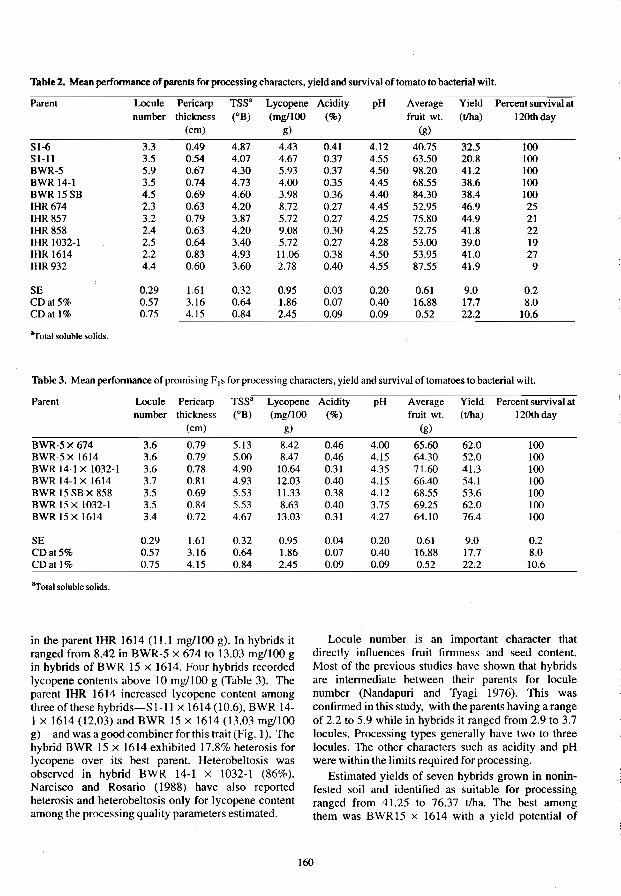
Table 2. Mean perfonnance of parents for processing characters, yield and survival of tomato to bacterial wilt.
Parent Locule Pericarp TSS' Lycopene Acidity pH Average Yield Percent survival at number thickness (OB) (mg/lOO (%) fruit wt. (t/ha) I 20th day
(cm) g) (g)
SI-6 3.3 0.49 4.87 4.43 0.41 4.12 40.75 32.5 100 SI-1I 3.5 0.54 4.07 4.67 0.37 4.55 63.50 20.8 lOO BWR-5 5.9 0.67 4.30 5.93 0.37 4.50 98.20 41.2 lOO BWR 14-1 3.5 0.74 4.73 4.00 0.35 4.45 68.55 38.6 lOO BWR 15SB 4.5 0.69 4.60 3.98 0.36 4.40 84.30 38.4 100 IHR674 2.3 0.63 4.20 8.72 0.27 4.45 52.95 46.9 25 IHR 857 3.2 0.79 3.87 5.72 0.27 4.25 75.80 44.9 21 IHR 858 2.4 0.63 4.20 9.08 0.30 4.25 52.75 41.8 22 IHR 1032-1 2.5 0.64 3.40 5.72 0.27 4.28 53.00 39.0 19 IHR 1614 2.2 0.83 4.93 11.06 0.38 4.50 53.95 41.0 27 IHR 932 4.4 0.60 3.60 2.78 0.40 4.55 87.55 41.9 9
SE 0.29 1.61 0.32 0.95 0.03 0.20 0.61 9.0 0.2 CD at 5% 0.57 3.16 0.64 1.86 0.07 0.40 16.88 17.7 8.0 CD at 1% 0.75 4.15 0.84 2.45 0.09 0.09 0.52 22.2 10.6
"Total soluble solids.
Table 3. Mean performance of promising F I S for processing characters, yield and survival of tomatoes to bacterial wilt.
Parent Locule Pericarp TSS' Lycopene number thickness (OB) (mg/100
(cm) g)
BWR-5x674 3.6 0.79 5.13 8042 BWR·5x 1614 3.6 0.79 5.00 8.47 BWR 14-1 X 1032-1 3.6 0.78 4.90 10.64 BWR 14-1 X 1614 3.7 0.81 4.93 12.03 BWR 15 SB X 858 3.5 0.69 5.53 11.33 BWR 15 x 1032-1 3.5 0.84 5.53 8.63 BWR 15x 1614 304 0.72 4.67 13.03
SE 0.29 1.61 0.32 0.95 CDat5% 0.57 3.16 0.64 1.86 CDat)% 0.75 4.15 0.84 2.45
"Total soluble solids.
in the parent IHR 1614 (11.1 mg/loo g). In hybrids it ranged from 8.42 in BWR-5 x 674 to 13.03 mg/loo g in hybrids of BWR 15 x 1614. Four hybrids recorded lycopene contents above 10 mg/1OO g (Table 3). The parent IHR 1614 increased Iycopene content among three of these hybrids-S 1-11 x 1614 (10.6), B WR 14-I x 1614 (12.03) and BWR 15 x 1614 (13.03 mg/loo g)-and was a good combiner for this trait (Fig. I). The hybrid BWR 15 x 1614 exhibited 17.8% heterosis for Iycopene over its best parent. Heterobeltosis was observed in hybrid BWR 14-1 x 1032-1 (86%). Narcisco and Rosario (1988) have also reported heterosis and heterobeltosis only for Jycopene content among the processing quality parameters estimated.
Acidity pH Average Yield Percent survival at (%) fruit wt. (t/ha) 120thday
(g)
0.46 4.00 65.60 62.0 100 0.46 4.15 64.30 52.0 100 0.31 4.35 71.60 41.3 100 0040 4.15 66040 54.1 lOO 0.38 4.12 68.55 53.6 100 0.40 3.75 69.25 62.0 100 0.31 4.27 64.10 76.4 lOO
0.04 0.20 0.61 9.0 0.2 0.07 0.40 16.88 17.7 8.0 0.09 0.09 0.52 22.2 10.6
Locule number is an important character that directly influences fruit firmness and seed content. Most of the previous studies have shown that hybrids are intermediate between their parents for locule number (Nandapuri and Tyagi ] 976). This was confirmed in this study, with the parents having a range of 2.2 to 5.9 while in hybrids it ranged from 2.9 to 3.7 locules. Processing types generally have two to three locules. The other characters such as acidity and pH were within the limits required for processing.
Estimated yields of seven hybrids grown in noninfested soil and identified as suitable for processing ranged from 41.25 to 76.37 t/ha. The best among them was BWR15 x 1614 with a yield potential of
160

76.37 tlha. It al 0 was rcsistant to bacterial wilt and had an average fru it weight of 64.1 g. In addition. this hybrid had the highest hctero is (92.8% over mid parent). 85.9% hcterobcllosis and 63.1 % 0 er the best parent. IHR 674. The other two promising F1s BWR 15 x 1032- 1 and BWR-S x 674 recorded estimated yields of62 t/ha (fable 3).
13 0
12
11 F
10
9 E
8 C B
7
6 E A C 0 F
5 A B
4
I 3
2
0
TSS Lycopene
Eleven hybrids were considered to be useful both for processing and frcsh market (dual purpose). Sev n of thesc hybrids recorded higher average fruit weights (above 60 g). The best F I among them was BWR-S X
1032-1 with an estimated yield of 63.87 tlha and an average fruit weight of 70.2 g. followed by hybrids BWR-S x 857 and BWR 15 x674 (Tablc4).
A B C D F
Acidity pH
Fig. 1. Performance of parents and best F,s. A=slIsccp,ihle parent ,; B=resistant parents; C=overall mean of hyhrids: D. E. F=bcst hyhrids.
Tahle 4. Mean perfomlance of promising dual purpose hyhrids for qua lity parameters. yield and surv ival.
Parent Lncule Pericarp TSS' Lycopene Acidity pH Avel'llge Yield Percent survival at number thickness (OB) (mg/IOO (% ) fruit wl. (t/ha) 120th day
(cm) g) (g)
S-16 x 674 3 0 0.49 4.10 8.47 0.46 3.67 40.45 45.2 97
S-16x 857 3.2 0.46 4.13 7.04 0.27 3.95 51.70 39.5 lOO S-16 x 858 3.1 0.57 4.00 9.78 0.30 4.25 57.50 40.6 93 S-16 x 1614 3.2 0.53 4.77 9.51 0.46 3.97 64.40 46.1 93 S-I 11 x 858 3.5 0.50 4 .10 8.71 0.41 4.02 60.25 41.5 97 S· I I I x 1614 3.3 0.58 4.75 10.60 0.37 4 .02 56.90 49.6 9:1 BWR-5 xl'57 3.5 0.75 4.87 7.59 0.36 HO 88.95 62.6 100 BWR-5 x 1032-1 3. t 0.79 S.OO 6.06 0.38 4.32 70.20 63.9 100 BRW 14-Jx 857 3.6 0.71 4.73 5.43 0.25 4.17 69.20 44.5 100 BRW 14·lx858 3. 1 0.75 5.07 9.02 0.49 3.95 69.90 4:l.0 100 BWR 15x674 2.9 0.73 4.13 7.57 0.31 4.50 6:1.70 57.7 lOO
SE 0.29 1.61 0.32 0.95 0.03 0.20 0.61 9.0 0.2 CDat5% 0.57 3.16 0.64 1.86 0.07 0.40 16.8R 17.7 8.0 CDatl% 0.75 4.15 0. 4 2.45 D.O' .09 052 22.2 10.6
"Tolal soluhle solids
16!
This study helped to establish the possibility of developing Ft hybrids resistant to bactcrial wilt and suitable for processing. In order to improve the processing qualities of hybrids further, it is essential to enhance the specific attributes in potential bacterial wilt resistant parental lines.
References
Association of Official Analytical Chemists 1975. Official Methods of the Association of Official Analytical Chemists. Washington, D. C.
Adsule, P.G. and Ambadan. 1979. Simplified extraction procedure in the rapid spectrophotometric method for Iycopene estimation in tomato. Journal Food Science and Technology, 16,216.
Kempthrone, U. 1957. An introduction to genetic statistics. New York, John Wiley and Sons.
Monma, S. and Sakata, Y. 1990. Varietal differences of bacterial wilt resistance in tomato. Tomato Genetics Cooperative Report, 40, 23-24.
Nandapuri, KS. and Tyagi. 0.0. 1976. Inheritance of some characters in tomato. Journal of Agriculture Research, Punjab Agricultural University, 13,80--84.
Naracisco, J. O. and Rosario, T. L. 1988. Bacterial wilt resistance and processing quality of tomato F I hybrids. Philippine Agriculturist, 71,387-396.
162
Ramachandran, C., Gopalakrishnan, P.K and Peter, KV. 1980. Evolving high yielding tomato varieties with resistance to bacterial wilt. Trichur, Kerala, Annual Report of Kerala Agricultural University.
Ramkishun 1987. Loss in yield of tomato due to bacterial wilt caused by Pseudomonas solanacearum. Indian Phytopathology, 40, 152-155.
Ran, M.V.B., Sohi, H.S. and Tikoo, S.K. 1975. Reaction of wild resistant tomato varieties and lines to Pseudomonas solanacearum in India. Plant Disease Reporter, 59, 734-736.
Sonada, R.M. 1978. Behaviour of tomato lines selected for resistance to southern bacterial wilt, in a field infested with the pathogen. Research Report, Agricultural Research Center, Fort Pierce, 5 p.
TIkoo, S.K., Anand, N., Kishun, R. and Reddy, P. P. 1989. Breeding for combined resistance to bacterial wilt and root knot nematodes in tomato. In: Green, S.K, ed., Tomato production in the tropics. Shanhua, Tainan, Taiwan, Asian Vegetable Research and Development Centre,99-1 05.
TIkoo, S.K., Anand, N. and Ramkrishna. 1983. Presence of two independent genetic systems for resistance to bacterial wilt (Pseudomonas solanaceamm) in tomato. In: Proceedings of the 15th International Genetic Congress, 12-23 December, New Delhi, India.
Studies on Genetic Resistance to Bacterial Wilt and Root-knot Nematode in Tomato
s. Nirmaladevi and S.K. Tikoo*
Abstract
Bacterial wilt caused by Pseudomonas solanacearum and root-knot nematode caused by Meloidogyne incognita are major factors limiting tomato production in the areas where it is traditionally grown. Development of resistant varieties is economical and an environmentally safe means of control. This study: (i) examined the inheritance of combined resistance to bacterial wilt and root-knot nematode; (ii) evaluated the interaction of these soilborne pathogens on the resistant genes present in the host genotype; and (iii) identified a broad spectrum of resistant sources among known lines/varieties. The results showed that the Fl of parents with dominant sources of resistance to wilt and root-knot nematode were resistant to both the diseases in the field. The interaction between pathogens has modified the genetic ratios of the crosses. A significant correlation between ooze and gall indices indicated that the nematodes predisposed the plants to bacterial wilt. The lines selected for resistance to bacterial wilt at Hessaraghata have shown differential response to isolates of P. solanacearum from Maharashtra, Kerala and Chethalli. The best F 1 hybrids for fresh market were BWR 1 x Rossol, 83 BWR 12-2 x IHR 998, 83 BWR 120 x Patriot and MITA 668 x 83 BWR 120.
BACTERIAL wilt caused by Pseudomonas solanacearum E.F. Smith is an important disease of solanaceous vegetables in most of the States of India (Rao 1976). Losses in tomato yields due to the disease have been reported to be as high as 90% (Kishun 1985). Genetic resistance to wilt was located in some lines of tomato introduced from Hawaii and Taiwan. Root-knot nematode, Meloidogyne incognita, is also recognised as a major pest of tomato in tropical countries and is reported to cause yield losses up to 61 % (Naganathan 1984). Use of resistant plants enables the grower to control root-knot without increasing the cost of production.
While genetic resistance to bacterial wilt has been known to breakdown in certain cases associated with root-knot nematode infection, the genetics of interaction of P. solanacearum and root-knot nematode on bacterial wilt resistant varieties has not been studied. In addition, Rao et al. (1975) reported that, of the 23 varieties and lines (from USA and Philippines) previously considered resistant to P. solanacearum, only eRA 66 Sel A was resistant at Hessaraghata both under artificial inoculation and field tests. This indicated the possibility of differential pathogenicity to different
*Indian Institute of Horticulture Research, Hessaraghata Lake, PO Bangalore 560 089, India.
geographic isolates. Therefore, the present study was undertaken to evaluate the inheritance of combined resistance to bacterial wilt and root-knot nematode, to study the interaction of these soilborne pathogens on the resistant genes present in the host and to identify a source of broad-spectrum resistance to bacterial wilt.
Materials and Methods
Selection of parents and hybridisation
Six lines resistant to bacterial wilt and 12 cultivarsnines resistant to root-knot nematode were used (Table 1). Each of the six bacterial wilt resistant (BWR) lines were crossed with all the nematode resistant (NR) lines to obtain F 1 s to be studied for yield and reaction to the diseases.
Evaluation of parents and F 1 S in infested field soil
The parents and F 1 S were planted in the bacterial wilt infested plot (108 cfulg of soil) in a randomised block design. At the time of planting, 100 g of nematodeinfested soil containing about 1000 M. incognita larvae were put in the planting holes. The plants were rated for survival, ooze and gall indices, and yield. Three Fl hybrids resistant to both pathogens, two Fls resistant to
163

Table I. Bacterial wilt and nematode (NR) resistant (BWR) tomato lines used in hybridisation studies
SI no.
2
3
4
5 6
BWRlines
BWRl
CRA66SelA
83BWR 12-2
BWR5
83 BWR90 MITA668
S1.no.
1 2 3 4 5 6 7 8 9
10 11 12
NRlines
Rossol Ronita
IHR998 IHR999 Patriot
VFN-8 Pelican LA 655 LA 656
83BWR 120 84BWR 30 84 BWR 32
nematodes and susceptible to P. solanacearum, and one FI resistant to P. solanacearum and susceptible to nematodes were selected and selfed to obtain the F2 population, then backcrossed with their respective male and female parents to obtain backcross populations (BI and B2 ) for further studies.
Evaluation or six generations for different characters The parents, Fls, F2s, Bls and B2s were planted in fieldinfested soil and rated for survival, ooze and gall indices and yield. Based on reactions 10 bacterial wilt and root-knot nematode, plants were grouped into four categories, viz.: resistant to nematode and bacteria (NRBR); resistant to nematode and susceptible to bacteria (NRBS); susceptible to nematode and resistant to bacteria (NSBR); and susceptible to both (NSBS). The data were statistically analysed as suggested by Panse and Sukhatme (1985).
Results and Discussion
Performance of parental and Fllines Survival of plants. The BWR lines had significantly higher survival rates than the nematode-resistant lines in the infested field (Table 2). The survival of BWR 1 and CRA 66 Sel A was less than previously reported (Tikoo et al. 1989), probably because the nematodes had predisposed these lines to wilt. Such predisposition has been reported earlier by SelIam et al. (1980). Among the nematode-resistant lines. MITA 668, 84 BWR 30 and 84 BWR 32 had higher survival rates because MITA 668 was originally introduced as a bacterial wilt resistant line and the other two were derivativesofMITA668.
The F] hybrids of 83 BWR 12-2, BWR 1 and BWR 5 were resistant to wilt, indicating the dominant source of resistance in these lines (Table 2). There were
susceptible, moderately resistant and resistant lines among the Fls of 83 BWR 90, probably because this line was a derivative of CRA 66 Sel A, reported to be a recessive source of resistance to wilt. The dominant source of resistance to wilt in 83 BWR 120 was indicated by the resistance of its F I s to wilt.
Ooze index. The ooze index in BWR lines was more than 1.0, indicating that the bacteria were able to multiply in these lines without killing the plants (Table 2). The ooze indices of NR lines were significantly higher than those of BWR lines in a manner directly proportional to their susceptibility to wilt (Table 2). The ooze indices of the Ft hybrids of 83 BWR 12-2, BWR 1 and BWR 5 were more than 1.0 but they survived in the field indicating that the F] s are also capable of withstanding bacterial multiplication, The ooze indices of the F I s of eRA 66 Sel A also indicated susceptibility to wilt, but these plants were susceptible only after 1 ()() days, probably because of delayed expression of disease.
Gall index. In the field infested with both pathogens, the NR lines remained resistant to nematodes as shown by the gall index 1 (Table 2). On the contrary. the BWR lines were highly susceptible to nematodes. In general, the F] hybrids of BWR lines with NR lines were resistant to nematodes (Table 2). However, the FI hybrid of BWR lines with MITA 668 was susceptible to nematodes. MITA 668 has been reported (Tikoo and Anand 1986) to have a recessive source of resistance to nematodes. The Ft hybrids of BWR lines with 84 BWR 30 and 84 BWR 32 were only moderately resistant to nematodes, indicating that resistance is not completely dominant in this line, probably because they were derivatives of MITA 668.
Genetics of combined resistance to bacterial wilt and root-knot nematode
The inheritance pattern of combined resistance studied in six crosses did not fit the expected 9:3:3: I ratio in the F2s (Table 3). F2 populations of BWR 1 crosses had higher than expected numbers of plants in NSBS class and there was perfect fit for a 9:3:4 ratio because of the shift in numbers from NSBR class to NSBS class due to susceptibility to nematodes which increased wilt. This indicated that simultaneous interaction of the two pathogens has modified the host response. The X2 value was highly significant in the BI population whereas it was not significant in the B25. These results confirmed the modification of genetic ratios as a result of lack of independent assortment of the two genes or, as reported by Sidhu (1984), parasitic epistasis.
The F2, BI and B2 population of eRA 66 Sel A hxbrids with Rossol and Patriot had significantly higher X2 values, indicating that the pattern of segregation did not fit any Mendelian ratio. This could be due to the
164

Table:!. Percentage survival, ooze and gall index of parental and F I tomato lines in wilt-infested field soil.
Sl.no. Lineslhybrids Survival (%) Ooze index Gall index
Days after transplanting
20 40 60 80 100
83BWR 12-2 94.2 94.2 94.2 94.2 94.2 2.3 2.8 2 BWRI 98.8 96.3 91.3 91.3 91.3 1.3 3.7 3 83BWR90 100.0 100.0 98.8 97.6 96.3 1.5 3.7 4 83 BWR 120 95.2 90.9 85.4 85.4 85.4 2.0 3.5 5 CRA66SeIA 97.6 88.4 88.4 88.4 88.4 1.7 2.8 6 BWR5 98.8 98.8 85.8 85.8 85.8 1.2 4.0 7 Rossol 80.6 72.2 48.7 30.5 2.6 2.9 1.0 8 Ronita 95.8 81.9 47.4 47.4 10.8 2.4 1.0 9 !HR 998 98.5 62.0 37.5 24.9 3.0 2.9 1.0 10 !HR 999 94.9 75.0 24.2 2.6 0.0 3.0 1.0 11 Patriot 90.5 48.1 20.4 0.0 0.0 4.0 1.0 12 VFN8 76.2 56.5 8.4 3.7 0.0 4.0 1.0 13 Pelican 51.9 44.3 1.6 1.6 0.0 3.7 1.0 14 LA 656 91.5 73.0 28.7 9.8 9.8 3.0 1.0 15 LA655 25.0 0.0 0.0 0.0 0.0 3.4 1.0 16 84 BWR 30 98.7 91.6 82.5 82.5 57.3 2.0 1.0 17 84 BWR 32 100.0 88.7 79.3 79.3 79.3 1.3 1.0 18 MITA668 98.7 75.7 39.4 39.4 39.4 2.7 1.1 19 83 BWR 12-2 x Rossol 100.0 98.8 98.8 98.8 91.2 2.0 1.0 20 83 BWR 12-2 x Ronita 93.3 93.3 93.3 93.3 93.3 1.9 1.2 21 83BWR12-2x !HR 998 98.0 98.0 98.0 98.0 98.0 2.7 1.1 22 83 BWR 12-2 x !HR 999 84.7 75.8 65.5 65.5 29.5 2.6 1.3 23 83 BWR 12-2 x Patriot 95.7 93.3 93.3 93.3 93.3 2.5 1.3 24 83 BWR 12-2 x VFN 8 100.0 92.6 92.6 92.6 92.6 2.7 1.0 25 83 BWR 12-2 x Pelican 10.0 98.3 98.3 98.3 98.3 3.2 2.2 26 83 BWR 12-2 x LA656 98.8 98.8 98.8 98.8 95.2 1.7 1.1 27 83 BWR 12-2 x 84 BWR 32 100.0 98.8 93.0 93.0 93.0 1.7 1.7 28 BWR I x Rossol 98.7 94.9 85.1 85.1 85.1 2.2 1.0 29 BWR I x Ronita 100.0 98.5 94.9 94.9 94.9 1.7 1.1 30 BWR I x !HR 998 90.0 83.6 71.4 71.4 71.4 2.1 1.0 31 BWR I x !HR 999 90.2 72.5 72.5 72.5 72.5 2.4 1.2 32 BWR I x Patriot 97.0 97.0 92.2 92.2 92.2 2.6 1.9 33 BWR I xVFN8 100.0 92.8 87.6 87.6 87.0 1.5 1.2 34 BWR I x Pelican 95.0 92.5 92.5 92.5 92.5 2.3 2.1 35 BWR I x 84 BWR 30 94.9 82.1 67.9 67.9 67.9 2.6 2.5 36 BWR I x84 BWR 32 98.7 89.2 86.2 86.2 86.2 1.5 1.5 37 BWR I xLA656 100.0 98.7 98.7 98.7 98.7 2.4 1.5 38 BWR I xLA655 97.6 93.3 77.8 77.8 77.8 1.9 1.0 39 83 BWR 90 x Rossol 98.8 85.1 74.1 74.1 74.1 2.1 1.7 40 83 BWR 90 x Ronita 97.7 81.7 61.1 61.1 61.1 1.9 1.3 41 83 BWR 90 x!HR 998 89.7 76.6 61.2 56.7 56.7 3.0 1.3 42 83 BWR 90 x lliP999 100.0 98.5 93.3 93.3 93.3 2.8 1.2 43 83 BWR 90 xPatriot 96.5 91.5 91.5 91.5 91.5 2.9 1.7 44 83 BWR 90 x Pelican 100.0 98.0 98.0 98.0 98.0 1.6 1.4 45 83 BWR 90 x 84 BWR 30 98.5 98.5 98.5 93.5 93.5 1.7 1.6 46 83 BWR 90 x 84 BWR 32 98.7 93.7 81.3 81.3 81.3 2.5 1.6 47 83 BWR 90 x LA 656 89.2 85.3 85.3 85.3 85.3 2.3 1.6 48 83 BWR 90 xLA655 96.3 44.6 27.9 27.9 27.9 2.8 1.3 49 83 BWR 120 x Ronita 93.3 61.5 56.0 39.3 39.3 2.9 1.3 50 83 BWR 120 x !HR 998 92.1 66.4 50.1 34.0 34.0 3.2 1.9 51 83 BWR 120 x Patriot 94.9 76.3 64.2 64.2 64.2 2.0 1.0 52 83 BWR 120 xVFN 8 97.0 80.7 69.1 69.1 69.1 2.0 1.4 53 83 BWR 120 x Pelican 93.7 76.6 42.1 42.1 42.1 2.4 1.5 54 83 BWR 120 x LA655 100.0 92.5 92.5 48.5 48.5 1.3 1.0 55 CRA 66 Sel A x Rossel 90.9 70.3 27.3 12.9 0.0 1.1 1.2 56 CRA 66 Sel A x Ronita 98.8 94.5 81.1 52.3 18.0 2.1 1.2 57 CRA 66 Se! A x IHR 998 97.6 71.6 30.0 30.0 9.4 2.2 1.0 58 CRA 66 Se! A x IHR 999 98.8 80.6 26.1 18.9 11.3 2.7 1.1 59 CRA 66 Sel A x Patriot 95.8 73.0 60.8 57.5 50.0 1.8 1.0 60 CRA66SelA xVFN 8 83.6 59.2 55.5 41.8 41.8 2.5 1.2 61 CRA 66 Se! A x 84 BWR 30 95.2 86.4 51.5 51.5 2.3 1.7 2.6 62 CRA 66 Se! A x LA 656 97.3 84.3 33.5 33.5 2.6 2.5 1.3 63 CRA 66 Se! A x LA 655 88.4 64.5 57.3 50.6 23.0 1.7 2.3
165
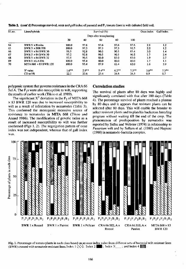
Table 2. (cont'd) Percentage survival, ooze and gall index of parental and FI tomato lines in wilt-infested field soil.
Sl.no. Lineslhybrids Survival (%) Ooze index Gall index
Days after transplanting
20 40 60 80 100
64 BWR 5 x Ronita 100.0 97.6 97.6 97.6 97.6 2.1 1.2 65 BWR 5 x IHR 998 100.0 97.3 97.3 97.3 92.5 2.2 1.2 66 BWRS x84 BWR 30 98.S 92.8 9O.S 9O.S 83.4 3.0 1.4 67 BWRS x84 BWR 30 95.2 92.8 90.5 9O.S 9O.S 1.7 2.4 68 BWR5 x84BWR32 97.S 9O.S 83.0 83.0 83.0 1.9 2.7 69 BWRS xLA656 100.0 9S.4 80.0 80.0 80.0 1.7 1.1 70 MITA 668 x 83 BWR 120 100.0 95.4 87.9 62.4 62.0 1.6 3.9
'Fvalue 1.6** 2.9** S.4*· 6.2*' 7.S'· 3.8" 7.3""
polygenic system that governs resistance in the eRA 66 Sel A. The Fjs were also susceptible to wilt, supporting the results of earlier work (Tikoo et al. 1989).
The significant X2 deviation in the F2 of MITA 668 x 83 BWR 120 was due to increased susceptibility to wilt as a result of infestation by nematodes (Table 3). This confirmed the monogenic recessive source of resistance to nematodes in MITA 668 (Tikoo and Anand 1986). The modification of genetic ratios as a result of increased susceptibility to wilt was further confirmed (Figs 1, 2). The segregation pattern of ooze index was not independent, whereas that of gall index was.
100
'" '" 75 '" c; ..c u
'" ., .5 tl § 50 c.. .... 0 ., Ol}
~ ., ~ 25 '" I)..
o
Correlation studies The survival of plants after 80 days was highly and significantly correlated with that after 100 days (Table 4). The percentage survival of plants reached a plateau by 80 days and it appears that resistant plants can be selected after 80 days. This will enable the breeder to select resistant plants and to plan his backcross breeding program without waiting till the end of the crop. The phenomenon of predisposition by nematodes was reported by Sidhu and Webster (1974) in relationship to Fusarium wilt and by Sellam et al. (1980) and Napiere (1980) in nematode-bacteria complex.
BWR 1 x Rossol BWR 1 x Patriot BWR 1 x Pelican CRA66SELAx CRA66SELAx MITA668x83 Rossol Patriot BWR 120
J<'ig. 1. Percentage of tomato plants in each class based on an ooze index value from (BWR) crossed with nematode resistant lines.Index 1 ~; Index 2 _; Index 3
166
sets of bacterial wilt resistant lines , and Index 4 •.

Table 3. Segregation ratios of segregating populations of different crosses used to determine the inheritance of resistance to Pseudomonas solanacearum
Generation NRBR NRBS NSBR NSBS Total Xl Probability
PI BWRl IS 15 Pz Rossol 15 15 Ft BWR 1 xRossol 13 2 15 F2 BWR 1 x Rossol FI bulk 99 43 31 24 197 15.57 BI FI xBWR I 52 37 14 103 4.09 0.05-0.02 B2 FI x Rossol 53 49 I 4 107 0.38 0.70-0.50
PI BWRI 15 15 Pz Patriot 15 15 FI BWR 1 xPatriot 12 3 15 F2 BWR 1 xPatriot FI bulk 108 43 39 22 212 7.12 0.10-0.05 BI FI xBWR I 52 34 19 105 4.58 0.05--0.02 B2 FI x Patriot 50 56 2 J08 0.37 0.70-0.05
PI BWRI 15 15 P2 Pelican 15 15 FI BWR 1 x Pelican 13 2 15 F2 BWR I x Pelican FI bulk 112 38 34 22 206 7.15 0.10-0.05 BI FI xBWRI 52 21 25 98 16.18 B2 FI xPelican 40 45 I 4 90 0.55 0.50-0.30
PI CRA66 Sel A 6 9 15 P2 Rossol 15 15 FI CRA 66 Sel A x Rossol 2 11 2 15 Fz CRA 66 Sel A x Rossol 8 139 9 51 207 506.54
FI bulk BI FI xCRA66SelA 8 39 9 43 99 67.93 B2 FI xRossol 15 83 4 102 45.49
PI CRA66SelA 6 9 15 P2 Patriot 15 15 FI CRA 66 Sel A x Patriot 15 15 Fz CRA 66 Sel A x Patriot 10 126 10 57 203 473.66
FI bulk BI FI x CRA66 SeI A 7 48 9 42 106 76.45 B2 FI xPatriot 13 90 2 105 56.50
PI MITA668 15 15 Pz 83 BWR 120 7 8 15 FI MITA668 x 83BWR 120 9 6 15 Fz F2 MITA 668 x 83 BWR 40 14 104 49 207 4.13 0.25--0.10
120F) bulk xMITA668 46 5 20 35 106 36.10 x83BWR 120 2 0 75 22 99 5.81
167

tOOl
~ 75 ;:;
1 ,5 II § 50 Q.
'0 ~ ~ ~ 25
o PIP2FIF1BIB2 PIP2FIF2BIBz PIP1FIF2BIBz PIP2FIF2BIBz PIP1FIF2BIBz P,P2FIF2BIBz
BWR I x Rossol BWR 1 x Patriot BWR Ix Pelican CRA66SELAx CRA66SELAx MITA668x83 Rossol Patriot BWR 120
Fig. 2. Percentage of tomato plants in each class based on a gall index value from different sets of bacterial wilt resistant lines (BWR) crossed with nematode resistant lines.Index 1 ss::J; Index 2 _; Index 3 D; Index 4 l1li; and Index 5 ~,
Negative correlation between survival of plants after 1 00 days and gall and ooze indices indicates that both root-knot nematode and bacterial infection reduce the survival rates of plants (Table 4), This phenomenon has been reported earlier by Goth et al. (1983) and Routarary et al. (1986).
Response ofBWR lines to four isolates of P. solanacearum
The reactions of four BWR Iines-BWR 1, CRA 66 Sel A, 83 BWR 90 and 83 BWR 120-to four isolates of
Table 4. Estimates of correlation coefficients,
Character 20th day 40th day 60th day 80th day
P. solanacearum from Hessaraghata and Chethalli (Karnataka), Dapoli (Maharashtra) and Thrissur (Kerala) were studied. Significant differences in response were observed between the four isolates (Table 5). The Maharashtra and Kerala isolates proved to be more pathogenic.
Fruit yield
The number of fruits per plant, yield per plant and average fruit weight of certain BWR lines and their Fls differed significantly between seasons and lines. The Ft
looth day Ooze index Gall index Fruit per plant
Yield per plant (kg)
--~~-----------------------------------------------------Survival of plants 20th day 0,04 -0.09 -0.12 -0.16 -0,11 0040 0,04 O,ll 40th day 1 0.93** 0.87** 0.78** 0.52 0.38 0.62 0.69* 60th day 1 0.92** 0,86** -0,56 0.36 0.63 0,71* 80th day 1 0.93** -0.56 0.35 0.57 0.63 lOOthday 1 -0.52 -0.35 0.57 0.63 Ooze index I 0.31 -0.49 -0.55 Gall index 1 0.30 0,23
1 0,74* 1
• Significantat5% level. .* Significantall% level
168

Table 5. The response ofBWR lines to isolates of P. solanacearum (1 % survival) at seedling stage.
83 BWR 120 47.25 71.75 CRA66SelA 74.20 82.50 BWR1 15.25 92.35 83 BWR90 62.80 40.50 PusaRuby 0 0 Mean 34.00 52.20
Fvalue Isolates 3.41 * Lines 30.06** Interaction
*Significanl a15% level *·Significanl al 1 % level
hybrids BWR 1 X Rossol, 83 BWR 12-2 X IHR 998,83 BWR 120 X Patriot and MITA 668 X 83 BWR 120 were judged best for fresh fruit for market and were also resistant to bacterial wilt.
The present study indicates that combined resistance to bacterial wilt and root-knot nematode is digenically inherited and that the resistance in bacterial wilt resistant genotypes changes following root-knot nematode infestation. Selection of plants in the segregating population and backcross breeding will help to evolve tomato varieties resistant to bacterial wilt and root-knot nematodes.
Reference
Goth, R.W., Peter, K.V., Sayre, R.M. and Webb, R. E. 1983. Effects of root-knot nematode on bacterial wilt of tomato. Phytopathology, 73, 966. Abstract.
Kishun, R. 1985. Effect of bacterial wilt on yield of tomato. Indian Phytopathology. 38,606. Abstract.
Naganathan, T.G. 1984. Studies on yield loss in vegetable crops due 10 Meloidogyne incognita. South Indian Horticulture, 32, 115-116.
Napiere, C. M. 1980. Varying inoculum levels of bacterianematodes and the severity of tomato bacterial wilt. Annals of Tropical Research, 2, 129-134.
Panse, V.G. and Sukhatme, P.V. 1985. Statistical methods for agricultural workers. New Delhi, Indian Council of Agricultural Research, 353 p.
65,45 59.50 61.15 86.55 79.35 80.75 51.70 20.05 45.45 85.35 22.00 53.25
0 0 0 53.80 30.65
CDat5% 11.08 12.39
Rao, M.V.B. 1976. Bacterial wilt of tomato and eggplant in India, In: Sequeira, L. and Kelman, A., ed., Proceedings of the First International Planning Conference and Workshop on the Ecology and Control of Bacterial Wilt caused by Pseudomonas solanaceamm. Raleigh, North Carolina, 18-24 July, 1976,92-94.
Rao, M. V.B., Sohi, H.S. and Tikoo, S.K. 1975. Reaction of wild resistant tomato varieties and lines to Pseudomonas solanacearum in India. Plant Disease Reporter, 59, 734--736.
Routaray, B.N., Sahoo, H. and Das, S.N. 1986. Response of some nematode resistant cultivars to mixed inoculations of Meloidogyne incognita and Pseudomonas solanacearum. Indian Journal of Nematology. 16,56-59.
Sellam, MA, Rushdi, M.H. and EI-Genm, D.M. 1980. Interrelationship of Meloidogyne incognita Chitwood and Pseudomonos solanacearum on tomato. Egyptian Journal of Phytopathology, 12,35-42.
Sidhu, G. 1984. Parasitic epistasis. Phytophatho!ogy, 74. 382-384.
Sidhu, G. and Webster, I.M. 1974. Genetics of resistance in the tomato to root-knot nematode wilt fungus complex. Joumal of Hercdity, 65,153-156.
TIkoo, S.K. and Anand. N. 1986. A recessive gene for resistance to root-knot nematode in tomato. In: First All India Conference of Cytology and Genetics, 149. Abstract.
Tikoo, S.K., Anand, N., Kishun. R. and Reddy, P. P. 1989.
169
Breeding for combined resistance to bacterial wilt and root knot nematodes in tomato. In: Green, S.K. ed., Tomato and pepper production in the tropics, Shanhua. Tainan, Taiwan, Asian Vegetable Research and Development Corporation, 99-105.
DNA Genetic Marker Mapping of Genes for Bacterial Wilt Resistance in Tomato
S.R. Aarons, D. Danesh and N.D.Young*
Abstract
To locate genes associated with resistance to bacterial wilt (BW; Pseudomonas solanacearum) in tomato, an F2 population segregating for resistance was analysed using DNA genetic markers. The population was derived from a cross between a resistant tomato genotype, L285, and a susceptible cultivar, CLN286. Because this cross is intraspecific in nature and therefore low in DNA sequence polymorphism, 100 clones from the established tomato molecular map were surveyed with 30 restriction enzymes to find a total of 46 restriction fragment length polymorphisms (RFLPs). Additionally, seven random amplified polymorphic DNA markers were added to the map. At 10 days after inoculation, two genomic regions were found to be associated with B W resistance. One of the putative resistance loci, located on a linkage group consisting of RFLPs known to reside on chromosome 6 of tomato, was significantly (p = 0.00(4) correlated with BW resistance. At 14 days after inoculation, three genomic regions were correlated with BW resistance. These included the region on chromosome 6, plus two others on linkage groups seven and 10. The region on linkage group 7 was unusual in that the allele from the susceptible parent (CLN286) was the one associated with higher levels of resistance in the F2• Together, the three putative resistance loci explained 49% of the total variation in BW resistance in the F2• Five cases of significant gene x gene interactions were also observed.
BACfERIAL wilt (BW) is a major disease of many important crops, including several in the Solanaceae. The causal agent, Pseudomonas soianacearum, invades the vascular system of the host and grows in planta, restricting transpiration of water. Wilting occurs, often accompanied by poor plant growth and even death. Although biological control of P. solanacearum has been recommended (Trigalet and Trigalet-Demery 1990), the best method of control known is the use of BW-resistant lines (Opena et aI. 1990). In the past, resistance to BW in tomato has generally not been stably maintained [Opena and Hartman, Asian Vegetable Research and Development Center (AVRDC), Taiwan, pers. comm.J. However, a landrace of Lycopersicon esculentum known as line L285 has moderately durable resistance (Opena and Hartman, AVRDC, pers. comm.). The resistance is thought to be polygenic, as indicated by the pattern of the resistance among F2 progeny, although the number of genes involved and their contribution are unknown.
*Department of Plant Pathology, University of Minnesota, St Paul, MN 55108, USA.
Restriction fragment length polymorphisms (RFLPs) and, more recently, random amplification of polymorphic DNA (RAPDs), have been used to link DNA genetic markers to disease resistance in tomato (Behare et a!. 1991; Messeguer et a!. 1991; Martin et al. 1991; Sarfatti et a!. 1989; Young et al. 1988). Much of this success is due to the availability of a well-saturated RFLP map for tomato (Tanksley et al. 1992), one of the best among higher plants. Using DNA markers, breeding for complex characters such as BW resistance should be much more straightforward. In particular, retammg resistance loci while simultaneously selecting against unwanted characters from a donor parent should be feasible.
To locate the genes that underlie BW resistance in L285, we have initiated a mapping effort based on RFLP and RAPD markers. In this preliminary report, we describe experiments in which 53 DNA markers spread throughout the tomato genome were used to locate regions associated with BW resistance. The results indicate that at least four genomic regions contain putative loci for BW resistance, including one region that plays a major role in the response.
170

Materials and Methods
Plant material Bacterial wilt resistant L285, a cerasiforme-type of tomato, was crossed with a cultivated but susceptible line, CLN286BClFr 25-l4-7 (henceforth cal1ed CLN286), at AVRDC and increased to produce the F2 progeny. The parents and 71 F2 progeny were grown in a soil:vermiculite:peat (1: 1: 1) mix in the greenhouse at the University of Minnesota in St Paul, Minnesota. Leaves were collected from each plant for DNA extraction as described below. Three to four cuttings of each plant were then made for inoculation with P. solanacearum and disease scoring. Cuttings of lateral branches were rooted and transplanted into the soil mix and grown for 7 days prior to inoculation.
Disease scoring P. solanacearum strain UW364, a Race I, Biovar 4 isolated from tomato in China (Cook et al. 1989), was used in inoculation tests. Cells were grown on casamino acid, peptone, and glucose (CPG) medium for 48 hours, washed into sterile distilled water, and diluted to give an optical density (A600) of 0.100 (108 cel1s per mL). Cuttings of the parents and progeny were inoculated by placing 15 uL of the inoculum in the third leaf axil from the top and passing a needle through the drop into the stem of the plant. Control plants were inoculated at the same time with 15 uL of sterile distilled water. Virulence of the pathogen was determined by hypersensitivity tests on Nicotiana tabacum. Plants were maintained in the greenhouse at 28-30°C and preliminary disease symptoms scored 7 days after inoculation (DAI), and then again on 10 and 14 DAI. On day 14 all plants that were not wilted or dead were re-inoculated to prevent escapes, and plants were scored again 7 days after the second inoculation. Symptoms were indexed on a scale of 1 (no symptoms) to 4 (severe wilting and death).
RFLP analysis DNA extracts from the parents were surveyed to identify polymorphic clones. Tomato DNA was extracted using the protocol of Dellaporta et al. (1983) with minor modifications. The extracted DNA was digested with 1 of 30 restriction enzymes, electrophoresed on 1 % agarose gels, and Southern blotted onto Hybond N+ (Amersham, Inc.) membranes (Southern 1975). One hundred tomato genomic clones kindly provided by Dr S. Tanksley (Department of Plant Breeding, Corne11 University, Ithaca, NY) were amplified by polymerase chain reaction before random hexamer labelling (Feinberg and Vogelstein 1983) with a-32P-dCTP. Labelled clones were first hybridised to filters containing restriction enzyme digested parental
171
DNA in order to identify band length polymorphisms. Each polymorphic clone was then hybridised to filters containing parental and F2 DNA digested with the appropriate restriction enzyme for that clone.
RAPD analysis Randomly amplified DNA polymorphisms between L285 and CLN286 and among the F2 progeny were identified using DNA extracted as described above. For each reaction, 2 ng of DNA was amplified with 15 ng of a lO-mer primer (Operon) and 1.75U Taq polymerase (Promega) as described by Williams et al. (1990). A Coy Tempcycler (model 50) was used for 45 cycles of 1 minute at 94°C, 1 minute at 35°C and 2 minutes at 72°C. Amplified fragments were electrophoresed in 1.4% agarose.
Linkage analysis Mapmaker software (Lander et al. 1987) was used to construct a linkage map for the L285 x CLN286 cross, based on segregation data in the F2 for each polymorphic marker. Linkage was inferred with a minimum LOD (log of the odds ratio) score of 2.5. DNA markers associated with BW resistance were then identified using the statistical software, Statview-11 (Abacus Concepts). In the analysis, both simple regression and single factor analysis of variance (ANOVA) were utilised with a significance level of p < 0.01. Two-factor ANOVA was also performed between putative BW resistance loci and the remaining DNA markers on the genetic map to uncover possible gene x gene interactions. In this analysis, a significance level of 0.0 I was also used.
Results and Discussion
Genetic map ofL285 x CLN286 cross
Because ofthe intraspecific nature of the cross between the parents, DNA polymorphism was low. Initially, 10 enzymes (BarnHI, BstNI, Dral, EcoRl, EcoRV, HaeIII, HindIII, Rsal, Taql, Xbal ) were used to survey the clones, of which only 30% were polymorphic. A second set of 10 enzymes (Asel, Bcll, BsaBI, Dpnl, HinfI, Hhal, Mspl, Ndel, NsiI, Scal) was then used to prepare filters, which were probed together with filters from the first 10 enzymes. The rate of polymorphism almost doubled to 53%. The remaining nonpolymorphic clones were then hybridised to filters made with a third set of enzymes (AIUI, Alul, Banl, BglII, BstEII, BstXI, Kpnl, Vspl, Sspl, XmnI) and an additional eight polymorphic clones were identified. In a report by Miller and Tanksley (1990), the percent polymorphism between cultivated tomato and a cerasiforme genotype was low (41 % with 5 enzymes); our results indicated that doubling the number of restriction

enzymes surveyed led to a twofold increase in polymorphisms. Nevertheless, 10 additional restriction enzymes increased the level of polymorphism only slightly.
Linkage analysis of the polymorphic clones produced the map shown in Figure 1. Additional markers were added using RAPDs. The linkage groups generally agreed with the chromosomes of the published tomato map (Tanksley et al. 1992). However, several clones mapped to positions different from those previously reported, presumably because we mapped different segregating bands for those clones. This is particularly striking for the linkage group tenatively assigned as chromosome 7. Because of this and other discrepancies, the chromosome assignments in this report should be considered tentative. Nonetheless, any statistical associations between DNA genetic markers and BW resistance would be unaffected by discrepancies in the assignment of linkage groups.
Some linkage groups were also poorly populated, with groups 3, 4, 5, and 8 having only a single marker. Still other linkage groups (1, 2, 6, and 11) consisted of two linkage blocks unlinked to one another, but
2 3 4 5 6
t t t TGI18
inferred to be on the same chromosome based on the map of Tanksley et a1. (1992). For all of these reasons, the saturation of DNA markers throughout the tomato genome was far from complete in this study and it is possible that some resistance loci have been missed.
Disease scoring
Disease indices of the resistant and susceptible parents were 1.0 and 4.0, respectively (data not shown), while the majority of the Fz feH between these values (Fig. 2). Many F2 progeny showed a score of two, with the distribution slightly skewed toward resistance. The continuous nature of the disease severity in the F2 strongly indicated that multiple genes were involved in the response. Disease index of the F2 individuals changed only slightly between 10 and 14 days, although sufficiently to uncover different putative resistance loci (see next section).
Map locations of putative BW resistance loci
Statistical analysis of BW disease index and DNA marker scores indicated that two genomic regions were associated with resistance at 10 DAI, one residing on
7 8 9 10 11 12
ItNlli ~ IQnQ t ~ TG16b 1 I r" I r,:m
T =1 t TGI83
I I rAI5
rA4
t I TGIlb
rAI7
~
W<Ml Significant at p<OJll 10 d.a.i.
~~~"\l'lI
Significant at p<O.OI 14 d.a.i.
Fig. 1. Linkage map of tomato, consisting of 46 RFLP and 7 RAPD markers. Marker positions are based on segregation in the L285 x CLN286 F2 population, as estimated by the Mapmaker (Lander et al. 1987) program with a minimum LOD of 2.5. Linkage groups were tentatively assigned to 'chromosomes' based on RFLPs common to the map ofTanksley et al. (1992). Those markers that map to a locus different than reported by Tanksley et al. (1992) are noted by the letter 'b' at the end of the marker name. Genomic regions showing significant association with BW resistance at 10 and 14 days after inoculation are highlighted with a bar beside each of the regions. DNA markers showing correlation are noted, and the locus with the highest association is underlined. DNA marker TG 16b, which was found to be significant only in terms of gene x gene interaction, is also noted.
172

50
40 >,
g 30 '" ;:;l
[ u.. 20
10
0 2 3 4
BW Disease Index
Fig. 2. Frequency distribution of bacterial wilt index scores 14 days after inoculation. Three or four cuttings from each of 71 F2 plants were inoculated as described in the material and methods section and scored for BW disease 14 days later. Each group of cuttings was scored on a scale from 1 (no disease) to 4 (severe wilting and/or death).
linkage group 6 and another on linkage group 8 (Table I, Fig. 1). The region on linkage group 6 was particularly significant, with five contiguous markers found to be significant and one (TG180) significant at the 0.0004 level (based on regression). Approximately 17% of the variation in BW reaction could be explained by this marker. Individuals homozygous for the TG 180 allele from L285 were 0.63 disease index units lower than homozygotes for the CLN286 allele.
In contrast, the genomic region on linkage group 8 (TG 181) was only barely significant (p=O.OO89) and had a much smaller impact on BW disease response. The effect of this putative resistance locus was also unusual in that the allele from CLN286 was associated with resistance, even though CLN286 is, itself, more
susceptible to BW disease. In this way, the putative resistance locus near TG181 resembled the one on linkage group 7 described below.
By 14 DAI, three genomic regions were found to have significant associations with BW disease, one each on linkage groups 6, 7, and 10 (Table I, Fig. 1). Results at 21 DAI, 7 days after surviving plants had been reinoculated with P. solanacearum, gave essentially the same results as observed at 14 DAI and are not shown. At 14 DAI, the putative locus previously observed on linkage group 8 was no longer considered significant, having a p-value of 0.0132. By contrast, the association between BW response and the putative resistance locus near TGl80 on linkage group 6 had grown much stronger, with several contiguous markers now showing correlation at the p=O.OOOI level. The proportion of variation explained by marker TG 180 increased to 32% and the difference in disease index between individuals homozygous for the L285 allele compared to those homozygous for CLN286 had also increased to 0.98 units.
On linkage group 10, a single locus (TG230) was found to bc associated with BW response. This marker just missed being counted as significant at 10 DAI, having a significance level of 0.0197 at that time. By 14 DAI, the significance level was p=0.OO33 and approximately 12% of the variation in disease could be explained by this locus. Homozygotes for the L285 allele at TG230 were more than 0.6 disease units lower than homozygotes for CLN286.
The effect of the putative resistance locus on linkage group 7 at 14 DAI resembled the one near TG181 on linkage group 8 at 10 DAI and differed fundamentally from the putative resistance loci near TG 180 and TG230. In the case of the putative resistance locus on linkagc group 7, the allele from CLN286 was the one associated with resistance. Five contiguous DNA
Table 1. Marker loci showing significant relationship to bacterial wilt resistance from rating taken at 10 or 14 days after inoculation (DAI).
Marker Linkage group DAI R-squared Probability Probability Type of effect" Phenotypic (regression (ANOVA) effectb
TG180b 6 10 0.169 0.0004 0.0002 Dom(285) (-)0.63
TGI80b 6 14 0.318 0.0001 0.0001 Dom(285) (-)0.98
TG51b 7 14 0.184 0.0002 0.0002 -" (+)0.66
TGI81 8 10 0.114 0.0089 _d Rec (286) (+)0.55
TG285b 10 14 0.124 0.0033 0.0019 Dom(285) (-)0.60
"Type of gene action. either additive (Add). recessive (Ree). or dominant (Dom). Numbers in paretbeses indicate the parent (L285. "285 and CLN286. "286) carrying the allele that confers higher levels of resistance to BW.
bDifference in BW disease index between L285 homozygotes compared to CLN286 homozygotes. "Type of gene action could not be inferred. dNot significant.
173

markers were found to be correlated to BW response and two of the markers (TG51 and rA04) were significant at p=O.OOO2. Individuals that were homozygous for the CLN286 allele at TG51 were approximately 0.66 disease units lower than homozygotes for the L285 allele.
Interactions among putative BW resistance loci
In a genetically complex type of disease resistance, such as observed in the cross between L285 and CLN286, primary gene effects may be affected by interactions among resistance loci. To uncover putative gene x gene interactions associated with BW resistance, two factor ANOVA was performed between primary loci (identified by regression and/or single factor ANOVA) and the remaining loci on the molecular map. This analysis identified five significant interactions at 14 DAI, three involving the putative resistance locus on linkage group 6 and two involving the putative locus on linkage group 7 (Table 2).
Several interesting trends could be observed from the analysis of gene x gene interactions. First, the putative resistance locus on linkage group 6 interacted with two of the other genomic regions that had previously been found to be individually significant by single factor ANOVA (TG183 on linkage group 7 and TG230 on linkage group 10). Likewise, the putative resistance locus on linkage group 7 interacted with a marker on linkage group 6 (TG35b) that was also found to be significant by itself. Moreover, the putative resistance loci on both linkage groups 6 and 7 interacted with the same locus on linkage group 9 (TGI6b), a marker that was not individually correlated to BW resistance.
Table 2. Loci showing significant gene x gene interactions.
Primary Linkage Interacting Linkage Probability locus group locus group (ANOVA)
TGSlb 7 TG35 6 0.0068 TGSlb 7 TGl6b 9 0.0076 TG1l8 6 TGl83 7 0.0052 TGll8 6 TGl6b 9 0.0008 TG1l8 6 TG230 10 0.0022
Applications of DNA markers linked to BW resistance loci
The primary goal of this study was to map putative resistance loci for BW resistance. However, besides mapping putative resistance loci, DNA marker
analysis also enables the selection of F2 plants suitable for use in breeding and genetic analysis. F2 plants that have retained putative resistance loci from L285, and at the same time carry a minimum of L285 DNA elsewhere in the genome, can now be identified with DNA markers. In the process, individuals with crossovers near the putative resistance loci can also be pinpointed. This should be especially significant in the case of breeding BW resistance from L285, because resistance has previously been associated with small fruit size (Opena and Hartman, AVRDC, pers. comm.).
At the same time, the contribution of different resistance loci can be studied individually, as it is now possible to select F2 plants that can be used to develop lines that carry only one or a few of the putative resistance loci. Considering the very different types of gene action observed for the different resistance loci uncovered in this study (such as the resistance locus on linkage group 6 versus the locus on linkage group 7), this type of genetic dissection should be especially informative.
Finally, using DNA markers near resistance loci to select lines with tightly linked crossover may be useful for purposes other than minimising linkage drag. In particular, crossover located near genes of interest can be used as a basis for fine-structure mapping near those genes. Considering the fact that the putative resistance locus on linkage group 6 controls a very large fraction of the variation in BW response (greater than 30%), fine structure mapping around this locus may provide a basis for physical mapping and eventual gene cloning based on chromosome walking (Young 1990). Although this type of research is still far in the future, the fact that tomato has desirable features such as a comprehensive yeast artificial chromosome library (S. Tanksley, pers. comm.) and high efficiency plant transformation (McCormick et al. 1986) may make it possible to clone some of the resistance loci uncovered in this study. If so, the major BW resistance locus of L285 may then be one of the first quantitative resistance genes to be cloned at the molecular level.
Acknowledgments
We would like to thank Dr Steven Tanksley for providing tomato genomic clones and Drs Luis Sequeira and Elizabeth Barlow for providing strains of Psuedomonas solanacearum. We would also like to thank Drs Romeo Opena and Glen Hartman for kindly providing the F2 population used in this study. This research was supported by the US-AIDIUSDAlCSRS Research Grants Program grant AIDID AN -4136-G-00-1018-00. This paper is published as contribution 20240 of the series of the Minnesota Agricultural Experiment Station.
174

References
Behare, J., Latterot, H., Sarfatti, M. and Zarnir, D. 1991. Restriction fragment length polymorphism mapping of the Stemphylium resistance gene in tomato. Molecular PlantMicrobe Interactions, 4, 489-492.
Cook, D., Barlow, E. and Sequeira L. 1989. Genetic diversity of Pseudomo1UlS solanacearum: detection of restriction fragment lenght polymorphism with DNA probes that specify virulence and the hypersensitive response. Molecular Plant-Microbe Interactions, 2, 113-121.
DeJlaporta, S.L., Wood, J. and Hicks, J.8. 1983. A plant DNA mini preparation: version 11. Plant Molecular Biology Reporter, 1,19-21.
Feinberg, A.P. and Vogelstein, B. 1983. A technique for radiolabeling DNA restriction fragments to high specific activity. Analytical Biochemistry, 132,6-13.
Ganal, M.W., Young, N.D. and Tanksley, S.D. 1989. Pulsed field gel electrophoresis and physical mapping oflarge DNA fragments in the Tm-2a region of chromosome 9 in tomato. Molecular and General Genetics, 215, 395-400.
Lander, E.S., Green, P., Abrahamson, J., Barlow, A., Daly, M.J., Lineoln, S.E. and Newburg, L. 1987. MAPMAKER; an interactive computer program for constructing genetic linkage maps of experimental and natural populations. Genome, 1,174.
Martin, G.B., Williams, J.G.K. and Tanksley, S.D. 1991. Rapid identification of markers linked to a Pseudomonas resistance gene in tomato using random primers and nearisogenic lines. Proceedings of the National Academy of Sciences, U.S.A. 88, 2336-2340.
McCormick, S., Niedermey, J., Fry, l., Bamason, A., Horsch, R. and Fraley, R 1986. Leaf disc transformation of cultivated tomato (L esculentum) using Agrobacterium tumefaciens. Plant Cell Reports, 5, 81-84.
Messeguer, R., Ganal, M., de Vicente, M.C., Young, N.D., Bolkan, H. and Tanksley, S.D. 1991. High resolution RH..P map around the rool knot nematode resistance gene (Mi) in tomato. Theoretical and Applied Genetics, 82, 529-536.
Miller, J. C. and Tanksley, S. D. 1990. RH..P analysis of phylogenetic relationships and genetic variation in the genus Lycopersicon. Theoretical and Applied Genetics, 80, 437-448.
Opeiia, R.T., Hartman, G.L., Chen, J.T. and Yang, CH. 1990. Breeding for bacterial wilt resistance in tropical tomato. In: 3rd International Conference on Plant Protection in the Tropics. 1990 Malaysian Plant Protection Society, Vol. V, 44-50.
Sarfatti, M., Kalan, J., Fluhr, R. and Zarnir, D. 1989. An RFLP marker in tomato linked to the Fusarium oxyspornm resistance gene 12. Theoretical and Applied Genetics, 78, 755-759.
Southern, E. 1975. Detection of specific sequenees among DNA fragments separated by gel electrophoresis. 10urnal of Molecular Biology, 98, 503-517.
Tanksley, S.D., Ganal, M.W., Prince, J.P', deVincente, M.C., Bonierbale, M.W., Broun, P., Fullon, T.M., Giovanonni, J.J., Grandillo, S., Martin, G.B., Messeguer, R, Miller, J.C., Miller, L., Paterson, A.H., Pineda, 0., Roder, M., Wing, RA., Wu, W. and Young, N.D. 1992. High density molecular linkage maps of the tomato and potato genomes: biological inferences and practical applications. Genetics. In press.
Trigalet, A. and Trigalet-Demery, D. 1990. Use of avirulent mutants of Pseudomonas solanacearum for the biological control of bacterial wilt of tomato plants. Physiological and Molecular Plant Pathology, 36, 27-38.
WilIiams, J., Kubelik, A., Livak. K., Rafalsld, T. and Tingey, S. 1990. DNA polymorphisma amplified by arbitrary primers are useful as genetic markers. Nucleic Acids Research. 18, 6531-6535.
Young. N.D. 1990 Potential applications of map-based cloning to plant pathology. Physiological and Molecular Plant Pathology, 37, 81-94.
175
Young, N.D., Zamir, D., Ganal, M.W. and Tanksley, S.D. 1988. Use of isogenic lines and simultaneous probing to identify DNA markers tightly linked to the Tm-2a gene in tomato. Genetics, 120.579-585.

Mapping of Bacterial Wilt Resistance Genes in Tonlato Variety Hawaii 7996
P. Thoquet, s. Stephens and N. Grimsley*
Abstract
In order to map the resistance genes, a cross between two Lycopersicon esculentum varieties, F10radel (susceptible) and Hawaii 7996 (resistant), was used. We plan to compare segregation of resistance in laboratory tests with that in a field trial (in collaboration with INRA, Guadeloupe). These varieties show little polymorphism, making RFLP analysis difficult. However, using RAPD markers it should be possible to find polymorphisms relatively quickly. Since resistance in Hawaii lines was originally introgressed from L pimpinellijolium, it is possible that many polymorph isms will be linked to the genes of interest.
TOMATO was chosen to study naturally occurring mechanisms of resistance to bacterial wilt. Most tomato varieties are susceptible but there are several lines which have been selected for resistance. The genetic basis for the resistance is not known, but there is evidence that several genes are involved. There is also evidence that one major effect may map to chromosome 6, since the character is difficult to combine with nematode resistance (Gilbert and McGuire 1956) or the marker sp (Acosta et al. 1964). A fruit-size effect may also be linked (McGuire 1960). Using crosses between resistant varieties and susceptible varieties or wild species of tomato, we will map the resistance gene(s) using several mapped RFLP markers (S. Tanksley, pers. comm.) and RAPD markers that are being developed in our laboratory.
Bulked segregant analysis (Michelmore et al. 1992) will be used to identify RAPD markers close to resistance genes. In this analysis, DNA from susceptible F2 plants will be bulked, as will DBA from resistant F2 plants. RAPDs which are only found in one of these two groups will be linked to the gene(s) for sensitivity or resistance, respectively. Other RAPD markers that are unlined to these genes will be found in both groups. Polymorphisms in which a RAPD band is linked to resistance in Hawaii 7996 or susceptible F10radel can be transformed into RFLP markers by cloning the band and placing on the map obtained from the crosses Hawaii 7996 x L. pennellii.
*Laboratoire de Biologie Moleculaire des Relations PlantesMicroorganismes, BP 27 Auzeville, 31326 CastanetTolosan Cedex, France.
We will attempt to map RAPD polymorphisms using existing RFL probes on the F10radel x Hawaii 7996 F2•
However, in case there is insufficient polymorphism an F2 population from the cross Hawaii 7996 x L pennelii LA 716 is also available to place the new RAPD markers on the existing tomato genetic map using a set of 24 RFLP markers received from S. Tanksley. This population will also be used for the mapping of various cDNA clones from genes known to be expressed in the course of infection of tobacco by P. solanacearum (Marco et al. 1990). L pennellii was reproducibly sensitive to bacterial wilt, so this popUlation is unsuitable for direct mapping of the resistance genes.
References
Acosta, I.C., Gilbert, I.C. and Quinon, V.L. 1964. Heritability of bacterial wilt resistance in tomato. Proceedings of the American Society of Horticultural Science, 84,455-462.
Gilbert, I.e. and McGuire, D.e. 1956. Inheritance of resistance to rootknot caused by Mi in commercial type tomatoes. Proceedings of the American Society of Horticulture, 68, 437-442.
McGuire, D.e. 1960. Bacterial wilt resistant tomatoes. Hawaii Agriculture Experiment Station Biennial Report, 74.
Marco, YJ., Ragueh, E, Godiard, L. and Froissard, D. 1990. Transcriptional activation of 2-cIasses of genes during the hypersensitive reaction of tobacco leaves infiltrated with an incompatible isolate of the phytopathogenic bacterium Psendomonas solanacearum .. Plant Molecular Biology, 15, 145-154.
Michelmore, R.w., Paran, I. and Kesseli, R.v. 1992.
176
Identification of markers linked to disease-resistance genes by bulked segregant analysis: A rapid method to detect markers in specific genomic regions by using segregating populations. Proceedings of the National Academy of Sciences ofthe United States of America, 88, 9828-9832.

Development of an In Vivo Complementation System for Identification of Plant Genes using Yeast
Artificial Chromosomes (Y A CS)
A.B. Bonnema *, R. Peytavi*, R.A.J. Van Daelen t, P. ZabeJt and N. GrimsJey*
Abstract
Map-based cloning of genes demands the use of vectors containing large inserts of foreign DNA, such as yeast artificial chromosomes (Y ACs). Once the region of the genome containing the locus of interest (for example, a disease resistance gene) has been cloned, final proof of the presence of the gene requires in vivo complementation tests. However, there are at present no techniques to allow the introduction of Y ACs into plants. We are developing a Y AC-plant transformation system. The plant selectable marker genes for hygromycin resistance and kanamycin resistance were introduced into existing Y AC vectors. A partial tomato Y AC library was constructed in these newly constructed Y AC vectors. Techniques to transfer these YACs to plants, by yeast spheroplast-protoplast fusion or via direct DNA transfer to plant protoplasts are being developed.
ONE long-tenn goal in plant biotechnology is to clone the individual genes responsible for controlling agronomically important characters, since very specific characters could then be introduced into plants in a controlled way within a relatively short space of time, by transformation and regeneration of plants in culture. Map-based cloning and cDNA cloning techniques will permit the isolation of genes of interest, but equally important is the development of plant transformation technologies.
The recent development of yeast artificial chromosomes (YACs) allows cloning of fragments of DNA 100-500 kilobases (kb) in size, a factor of 10 larger than fragments cloned in conventional vectors (Burke et al. 1987; Schlessinger 1990), reducing the number of clones necessary for construction of a representative gene library, facilitating 'chromosome walking' and permitting cloning of large eukaryotic genes such as Drosophila bithorax (320 kb, Karch et al. 1985) and
*Laooratoire de Biologie Moleculaire des Relations PlantesMicroorganismes, BP 27 Auzeville, 31326 CastanetTolosan Cedex, France.
tDepartment of Molecular Biology, Agricultural University, Dreijenlaan 3, 6703HA Wageningen, The Netherlands.
factor VIII (190 kb, Gitschier et a!. 1984). Genes of similar size are most likely present in plant cells. VAC vectors exist with replicons and selectable markers for E. coli or yeast, and thus can be propagated in either of these organisms as plasmids or artificial chromosomes, respectivcly.
Genomic libraries in YACs have been constructed for a variety of organisms. These libraries can be used to screen for genes of interest with known genetic map position using tightly linked molecular markers as probes. Once the chromosomal region spanning the gene of interest has been cloned, final identification of the gene depends on successful in vivo complementation tests of plants deficient for the gene function. This remains an enormous task when only the genetic map position of the gene is known, since only relatively small DNA fragments can be transferred to plants using conventional gene-transfer techniques. Functional testing currently requires subeloning fragments of an existing VAC clone, and this has the following disadvantages: (1) a series of closely linked VAC clones would be difficult to test, as too much work in physical mapping, subeloning, and plant transformation would be required; (2) large genes or gene complexes would be lost; and (3) prokaryotic vectors may not tolerate certain kinds of DNA structures found in eukaryotes.
177

The development of a YAC-plant cell complementation system would greatly facilitate this task. Such a system would also permit in vivo complementation tests of large plant genes, such as those with many exons or distant regulatory sequences.
Recently, YACs containing long stretches of cloned DNA have been reintroduced into mammalian cells. Techniques used to transfer the VAC DNA into mammalian cells are both fusion of the mammalian cells with the yeast spheroplasts (Gnirke and Huxley 1991; Huxley et al. 1991; Pachnis et al. 1990). and direct transfer of the naked DNA to mammalian cells (EUceiri et at. 1991; Femandez-Luna et al. 1991; Gnirke and Huldey 1991; Strauss and Jaenisch 1992). Suitable selectable markers for transformation of mammalian cells were introduced into existing human VAC clones by homologous recombination in yeast (Pachnis et a!. 1990). Auxotrophic mammalian cells have been complemented in vivo with YACs carrying the corresponding cloned genes (Eliceiri et al. 1991; Gnirke and Huxley 1991; Huxley et at. 1991). YACs were stably integrated in mammalian genomes, the integrated DNA was not rearranged, and cloned genes were expressed, indicating that YACs can conserve the structure of genomic DNA (Pachnis et al. 1990).
Currently available VAC vectors do not contain plant-selectable markers, rendering them useless for plant transformation. The development ofYAC vectors containing plant-selectable markers, and the development of techniques to transform plants with these YACs containing large stretches of cloned DNA, will be essential tools for future plant biotechnology. Introduction of a VAC carrying plant DNA into plant cells has not been described, although protoplasts can take up and integrate naked DNA into their nuclear genomes (Negrutiu et al. 1987) and can be fused with protoplasts of other species to generate somatic hybrids (Bonnema eta\. 1991).
For the development of the VAC-plant transformation system, we work with a heterologous system to be able to follow the fate of the transforming DNA. A partial tomato VAC library has been constructed in VAC vectors containing plant selectable markers, and this library has been screened for the presence of a wellcharacterised tomato specific HindIII repeat. The tomato genome contains about 75000 copies of this tomato specific HindIlI repeat, which is dispersed over the genome and present on all chromosomes (Zabel et al. 1985). The sequence of this repeat is known. Techniques to transfer these YACs to plants are being developed, using a haploid Nicotiana plumbaginifolia and Nicotiana tabacum 'SR-l' as acceptor species. Their genomes contain no sequences hybridising to the tomato specific HindIII repeat, which facilitates the analysis of the fate of the tomato VAC in the trans-
formed plant. Protoplast isolation and regeneration techniques are well-developed for these species. They are therefore good model systems to use before attempting complementation in tomato.
Materials and Methods
Growth of bacterial strains and in vitro manipulation of DNA were done according to standard protocols (Sambrook et al. 1989), except where otherwise stated. Yeast was handled according to standard protocols (Guthrie and Pink 1991).
Yeast strains The following three yeast strains were used: (i) AB1380: haploid, MAT, U, ura3, trpl, ade2-1, canl-100, lys2-1, his5; (H) YPH252: haploid, MAT. ura3-52, lys2-801 amber, ade2101 ochre, trpl-Dl, his3-D200, leu2DI; and (iii) YPH274 (Mat !Mata; ura3-52; lys2-80l/amber; ade2-101Iochre; trp- 1; his3- 200; leu21)
Construction ofYAC vectors pYACABI and pYACAB2. The XhoI site of pYACRC (Burke et a1. 1987). bordering the left TEL sequence, was changed to an EcoRI site by linker insertion. Plasmid pHP23b (1. Paszkowsky, pers. comm.) containing the NPTII gene under the control of the CaMV 35S promotor and terminator, was modified by removing the SalI and BamHI restriction sites (by digestion, filling-in [Klenow], and blunt-end ligation). The 1.7 kb EcoRI fragment containing the NPTII gene, was ligated into the newly created EcoRI site of pYACRC, so creating pYACABl (NPTII gene transcribed towards the centromere) and pYACAB2 (NPTII gene transcribed towards the TEL sequence) (Fig. I).
pJABl and pJAB2. The plasmid pGL2 contains the hygromycin gene (HPH), under control of the CaMV 35S promotor and terminator constructed in plasmid pDH51 (Gritz and Davies 1983). Plasmid pGZ5 is derived from plasmid pGL2 by inserting XlloI sites, so that the hygromycin (HPH) gene can be recovered as a 1.5 kb XhoI fragment. The BamHI and SalJ restriction sites were removed from the HPH gene, and the 1.5 kb XhoI fragment containing the HPH gene, was Iigated into the SalI sites of pUC7. The HPH gene was then excised as a 1.5 kb BamHI fragment, and Iigated into the unique BamHI site of polylinker 2 of the vector pJS97 (Shero et al. 1991), to create pJAB 1 (HPH gene transcribed towards the TEL sequences) and pJAB2 (HPH gene transcribed away from the TEL sequences).
pJAB4. The 1.7 kb fragment of modified (as above) pHP23b containing the NPTII gene was Klenow filled in, and Iigated into the SmaI site of vector pVl25 (a pUC series derivative containing an altered polylinker)
178

Fig. l. Vectors used for construction of plan t genomic librarie· in yea.~t artiticiul chromosomes (YA s). Biological functions are abhreviated: Amp = Ampicillin resistance (E. coli); on = origin of replication (E. coli); URA. HIS. TRP = auxotrophic se lectahle marker (yeast); CEN .ARS = centromere/replication (yeast); SUP = suppressor (colour marker for c loning) (yeast): TEL = telomere (yeast); HPH = hygromycin resistance (plants); NPTrr = kanamyci n resistance (plants).
(T. Hohn. pcrs. comm.). The NPTn gene was then exci sed as a 1.7 kb BamHl fragment. and ligated into the un ique BamHl site of polylinkcr 2 of the vector plS98 (Shero et al. 199 1), to create pJAB4 (NPTII gene transcribed towards the T EL sequence).
Library construction LigatiQn of high molecular weight DNA i lllo YAC vectors. High molecu lar weight DNA of L esclIienfllm cv. 83M was isolated as described by van Daelen el l. (1989). Agarose plugs ontaining about 4 jJg of total tomato DNA were digested ovemight with the ram cutting restriction enzymes Mlul or Notl (van Daclen et al. 1989).
Forty micrograms of the vectors pYACABl or pYACAB2 were digested to completion with th ~
enzymes BamHI and Mlllr . Samples were phenoV chloroform treated. ethanol prec ipitated. and dissolved in water. The vector arms were phosphatascd (using calf intestine phosphatase as de cribed by suppli r • and ligated overnight with M/liT digested genomic DNA. Agarose was removed by an agarase treatment, and ligation mix was stored at -20°C (100 ).lLIIigation reaction).
Vectors pJAB I and pJAB4 were mixed in a rat io I: I. and 40 ~ of this mix digested to completion with the restriction enzyme Clal and Notl. Pho phata e treat ment of vector arms and ligation with NotI digested tomato DNA. wa as described above.
Trallsformation of yeast spheroplasts. Spheroplast preparation and PEG induced tran formation with the
tomato D A, cloned into VAC vectors. wa' as previou ']y described (Guthrie and Fi nk 1991). Yeast strain AB 1380 was transformed with MIIII digested tomato DNA cloned into pYACA B I or pYACAB2. and the yeast strains YPH252 (~'1at ; ura3-52; lys2-80I/amber; ade2-1 Ol/ochre; trp- I · his3- 200; leu2 I) and YPH274 were tran -fonned with Notl digested tomato DNAcloned into the vector combi nation pJAB 1 and pJAB4. Transformed yeast spheroplasls were plated on selective medium lacking uracil. Individual colonies were tran . [erred t microtitre plates containing selective medium, using toothpicks . Two replicas of the microtitre pl ates were stored at - 70°C as glycerol stocks. and colonies of the original micro tilre plates were dot-blotted onto Genescreen+ membranes.
Screening of the VAC lihraries fo r the preSl'IIce of the tomato specific Hindll! repeat G2. Fillers were preh bridised at 65°C overnight in 10% de ·tran sulphate, I M nel, 1% SDS. I mgl lO mL sal m n ·pcml DNA. Ovem ight hybrid izat ion with 32P-labeled probe WIIS in the same s lu tion a l 65°C, and there were two wa hes of 30 minutes at 65°C in O.5X SS " 1.591 S DS.
Inhibition of yeast with iodoacctamide Yeast 'pheroplast were prepared ace rding standard protacols (Guthrie and Fi nk 199 1). After tw washe by centrifugat ion (3458, 5 minute. ), the spheroplaslS were treated with IOmM i loacetamide in lot of 10 mL at 5° for 15 minules wi th gentle agitat ion. After centrifugation (345g for 5 minute) the :pher -plaslS were resuspcnded in 10 mL prolopla t medium
179

(Bonnema et at 1991), and washed twice in this solution in the same way (345g for 5 minutes) before use for a fusion.
Plant material Nicotiana tabacum 'SR-l' was maintained in sterile culture at 25°C with 16-hour day, 8-hour night, in Murashige and Skoog medium with 20% sucrose. Nieotiana plumbaginifolium, haploid, was cultivated in the same way.
Isolation of plant protoplasts Isolation and culture of protoplasts was done as previously described (Bonnema et al. 1991)
Results
Vectors Several new VAC vectors suitable for plant transformation were produced (Fig. 1). We are currently testing the use of these vectors in complementation experiments (see below) using either VAC or total naked DNA isolated from the appropriate yeast strains, or by attempting to fuse yeast spheroplasts and isolated plant protoplasts.
Tomato gene library Yeast transformation efficiencies of about 3 x Ht colonieslJ.1g transforming DNA were obtained. Table 1 lists the number of clones obtained (only a small part of the ligation mixes was used). To estimate the average insert size of the YACs in both libraries. 30 random clones of the NotI bank in YPH252, and 30 random clones of the MluI bank in AB 1380, were selected. Chromosomal DNA was isolated from 1 mL cultures (YPD) as described by van Daelen et al. (1989), chromosomes were separated on a pulsed-field electrophoresis gel, transferred to Genescreen+ membranes, and hybridised with pBR322. PBR322 sequences hybridize to the VAC vector arms. The average size of the YACs in the NolI bank was 250 kb; average size of YACs in the MluI bank was around 100 kb. Dot-blot filters were screened with the 32P-labeled HindlII insert G2 of plasmid pTHG2 (tomato HindlII repetitive sequence G2, cloned into HindlII site ofpBR322), and 38 of 2700 colonies (1.5%) showed stronger hybridisation than background hybridisation (Table 1). The percentage of colonies, hybridising with G2 (1.5%) is within the expected range of 0.5%-4.5%, assuming that the 450bp repeat occurs in 1O{)()(}"'75000 copies per haploid genome.
Yeast chromosomal DNA of clones, putatively containing the HindlII repeat squence G2, was isolated from I mL cultures (YPD). Chromosomes were separated by FlGE, transferred to Genescreen+
Table 1. Number of yeast clones maintained on selective medium. Clones hybridising to the tomato repetitive HindIII sequence are given in parentheses.
Y AC vector/digest Yeast strain AB 1380 YPH252 YPH274
pYACAB2/MluI PYACAB1IJluI !,!f\~l + pJAB41N0tI
900(21) 700 (13)
800 (4) 300(0)
membranes, and probed with the 32P-labeJed HindlII insert G2 of plasmid pTHG2. Only 6 of 21 putative positive clones hybridised with the repeated DNA sequence G2, at variable intensities. The varying intensities in hybridisation signals can be caused by varying amounts of the repeated sequence per VAC, or by an instability of the VAC under non-selective conditions (chromosomal DNA was isolated from cultures grown under non-selective conditions).
We are currently further characterising the initial positive yeast clones. Using Southern hybridisation, PCR, and selective growth media, we will verify whether YACs, containing HindlII repetitive sequences can be maintained stably in yeast.
Fusion of yeast spheroplasts with plant protoplasts In order to develop a successful fusion technique, it is first necessary to develop ways to inhibit division of yea."t spheroplasts in plant tissue (protoplast) culture, otherwise yeast overgrows the plant cells within 2-3 days of the fusion experiment. A combination of two techniques was used to accomplish this.
Iodocetamide, a growth inhibitor, was used to kill the yeast cultures prior to fusion. 5mM iodoacetamide was sufficient to kill 90% of the yeast spheroplasts (Fig. 2). We noted that the quality of the yeast spheroplast preparation was important for an effective treatment; too much cell-degrading enzyme, or a too long digestion time, led to agglutination of spheroplasts. We found that addition of 5 mg/mL amphotericin to the plant culture medium did not affect protoplast regeneration, but inhibited the growth of yeast. In combination with the iodoacetamide pretreatment, addition of 5 mg! mL amphotericin after protoplast-spheroplast fusion, was sufficient to inhibit further yeast growth.
Initial fusion experiments have been conducted using either PEG-mediated or electroporation-mediated protoplast-spheroplast fusion and we are currently trying to select transgenic calli with kanamycin.
Discussion
The success of this kind of approach in mammalian cell systems indicates that perseverance with the plant
180

l~r----------------------------'
'" 10001 ., ~ u
'0 .8 - .....___YPD ~ 500 1
:z.
6 8 10
Concentration of iodoacetamide (mM)
Fig. 2. The inhibition of yeast growth by iodoacetamide. YPD = yeast medium; TM2G = plant culture medium.
systems will also lead to success, since mammalian and plant cells have similar genetic complexities. Integration of YACs is likely to occur in plants, as it does in animal cells, rather than replicative maintenance. since yeast centromeres are very different to those of plant or mammals. Recombination of incoming transforming DNA, such as plasmid DNA with selectable markers, has been studied fairly extensively in somatic cells of these two systems, which show many similarities. The kanamycin resistance selection for transformed plants has .already been tested in different kinds of plants.
We have developed a way of eliminating all of the yeast cells following the fusion treatment, an important achievement for further continuation of the project. Iodoacetamide uscd before fusion as described docs not affect DNA transfer and has no effect on the regeneration of plant protoplasts. Amphotericin treatment is necessary after fusion to kill the few remaining yeast cells. Although we do not yet have transformed plants, we have found that following these treatments, protoplasts could be regenerated into plants. This is an important observation, since components of fungal cells are known to provoke defensive responses in plants that can lead to plant cell death.
A long-term objective of the lab is to clone tomato genes encoding resistance to the pathogen Pseudomonas solanacearum on a VAC, and to perform functional assays of the resistance genes in vivo. Once molecular markers closely linked to a resistance gene have been identified, a complete VAC library from a tomato line carrying the resistance genes will be screened for VAC clones containing these markers.
181
VAC libraries will be screened using Southern hybridisation and crude lysate PeR (K wiatkowski et al. 1990). These YACs will be used as starting points for chromosome walking towards the resistance genes. Extremities of the inserts of genomic DNA in YACs will be used for chromosomal walking. We propose to test YACs of interest by using the techniques of spheroplast-protoplast fusion that we are developing here. While the approach is of general importance, it is currently difficult to assess the chances of success using tomato variety Hawaii7996, since the genetic basis for resistance of this line is not yet known; we depend upon the identification of a major effect in order to try out the system.
Acknowledgments
We would like to thank Peter Hieter, Majiek Pietrzak, Jurek Pasttowsky and Thomas Hohn for valuable gifts of plasmid DNAs.
References Bonnema, A.B., Melzer, 1.M. and O'ConneIl, M.A. 1991.
Tomato hybrids with mitochondrial DNA from Lycopersicon pennellii. Theoretical and Applied Genetics, 81,339-348.
Burke, T., Carle, G.F. and Olson, M.V. 1987. Cloning oflarger segments of exogenous DNA into yeast by means of artificial chromosome vectors. Science (Washington), 236, 806-812.
Eliceiri, B., Labella, T., Hagino, Y., Srivastava, A., Schlessinger, D., Pilia, G., Palmieri, G. and Dur5O, M. 1991. Stable integration and expression in mouse cells of yeast artificial chromosomes harboring human genes. Proceedings of the National Academy of Sciences of the United States of America, 88, 2179-2183.
Femandez-Luna, J.L., Matthews, RJ., Brownstein, B.H., Schreiber, R.D. and Thomas, M.L. 1991. Characterization and expression of the human leukocyte - common antigen (CD 45) gene contained in yeast artificial chromosomes. Genomics, 10,756-764.
Gitschier, 1., Wood, W.I., Goralka, T.M., Wion, K.L., Cben, E.Y., Eaton, D.H .• Vehar, G.A., Capon, DJ. and Lawn. R.M. 1984. Characterization of the human factor VIII gene. Nature (London), 312, 326-337.
Gnirke, A. and Huxley, C 1991. Transfer of the human HPRT and GART genes from yeast to mammalian cells by microinjection of YAC DNA. Somatic Cell and Molecular Genetics 17,573-580.
Grill., L., and Davies, 1. 1983. Plasmid-encoded hygromycin B resistance: the sequence of hygromycin B phospbotransferase gene and its expression in Escherichia coli and Saccharomyces cerevisiae Gene, 25, 179-188.
Guthrie, C. and Fink, G., 199 J . ed., Guide to yeast genetics and molecular biology. San Diego, Academic Press.
Huxley, C, Hagino, Y., SchJessinger, D. and OI5On, M.V. 1991. The human hprt gene on a yeast artificial chromosome is functional when transferred to mouse cells by cell fusion. Genomics, 9, 742-750.

Karch, F., Weiffenbach, B., Peifer, M., Bender, W., Duncan, I., Celniker, S., Crosby, M. and Lewis, E.B. 1985. The abdominal region of the bithorax complex. Cell, 43, 81-96.
Kwiatkowski, TJ., Zoghbi, H.Y., Ledbetter, S.A., Ellison, K.A. and Chinault, A.C. 1990. Rapid identification of yeast artificial chromosome clones by matrix pooling and crude lysate PCR. Nucleic Acids Research, 18, 7191-7192.
Negrutiu, I., Shillito, R., Potrykus, I., Biasini, G. and Sala, F. 1987. Hybrid genes in the analysis of transformation conditions: Setting up a simple method for direct gene transfer in plant protoplasts. Plant Molecular Biology, 8, 363-373.
Pachnis, v., Pevny, L., Rothstein, B. and Costantini, F. 1990. Transfer of a yeast artificial chromosome carrying human DNA from Saccharomyces cerevisiae into mammalian cells. Proceedings of the National Academy of Sciences of the United States of America, 87, S 109-S1 13.
Sambrook, J., Fritsch, E.F., Maniatis, T. and Irwin, N. 1989. Molecular cloning: a laboratory manual. Cold Spring Harbor, Cold Spring Harbor Laboratory.
182
Schlessinger, D. 1990. Yeast artificial chromosomes-tools for mapping and analysis of complex genomes. Trends in Genetics, 6, 248.
Shero, J.H., McCormick, M.K., Antonarakis, S.E. and Hieter, P. 1991. Yeast artificial chromosome vectors for efficient clone manipulation and mapping. Genomics, 10, SOS-S08.
Strauss, W.M. and Jaenisch, R. 1992. Molecular complementation of a collagen mutation in mammalian cells using yeast artificial chromosomes. EMBO Journal, 11, 417-422
van Daelen, R.A.J., Jonkers, J.J. and Zabel, P. 1989. Preparation of megabase-sized tomato DNA and separation of large restriction fragments by Field Inversion Gel Electrophoresis (FIGE). Plant Molecular Biology, 12,341-3S2.
Zabel, P., Meyer, D., Stolpe van de, 0., Zaal van der B., Ramanna., M.S., Koornneef, M., Krens, F. and Hille, J. 1985. In: Vloten-Doting et al. ed., Molecular form and function of the genome, 609-D2S.

Breeding for Resistance to Bacterial Wilt in Tomato, Eggplant and Pepper
K.V. Peter, T.R. Gopalakrishnan, S. Rajan and P.G. Sadhan Kumar*
Abstract
A total of 165 tomato lines was evaluated for bacterial wilt resistance under field conditions. Line CL32-d-0-1-19 GS was resistant This was confirmed in trials at other locations and under laboratory conditions where it was resistant to three different isolates. In 1984--85, an additional line, Louisiana Pink, was found to be resistant. Of the four methods of selection, single seed descent was most efficient in raising the level of resistance in selected lines. Resistant lines contained more tomatine. Line CL32-d-O-I-19 GS was released as Sakthi. For eggplant, 34 breeding lines and four other species of Solanum were evaluated. Annamalai, SM 6, SM 48, SM 58, SM 71, SM 72 and SM 74 were resistant. Ofthe four selection methods, single seed descent was most effective in raising the level of resistance. The genetic groups SM 6-1, SM 6-4 and SM 6-9 were immune in all the four cycles of selection. Lines SM 121, SM 126, SM 133, SM 135 and SM 140 were resistant in further evaluation trials. The resistant line SM 6-7 was released as Surya. The four chilli varieties, Pant Col, KAU cluster, White Kandari and Chuna, were screened against nine isolates. KAU cluster was resistant to five isolates and was released as Manjeri.
PSEUDOMONAS solanacearum E.F. Smith causes wilt, and limits cultivation of tomato, eggplant and pepper in warm humid tropics. Breeding for disease resistance has been a major avenue for control of the disease.
The occurrence of races and biovars of P. solanacearum with different host reactions has made breeding for resistance a very challenging proposition. Although most breeding strategies to control wilt have not involved using mixtures of resistance;; in the field, Pande (1976) reported that mixtures of bengal gram with resistant components showed a reduction in wilt. Parlevliet (1979) extended the multiline concept to variety or line mixtures where the component genotypes differ for the race-specific resistance genes.
This paper reports on breeding trials with tomato, eggplant and pepper in the context of selection, inheritance and multiline use. The trials led to the release of varieties Sakthi in tomato, Surya in eggplant and Manjeri in hot pepper, which are now cultivated in wiltprone soils of India.
*Kerala Agricultural University, P 0 Vellanikkara - 680 654 (Kerala) India.
Materials and Methods
Tomato
Evaluation of five lines for resistance to nine isolates. Five tomato lines-LE 79, Pusa Ruby, Rutgers, Venus and Saturn-were evaluated in vitro against nine virulent isolates of P. solanacearum.
Evaluation of 15 lines. Fifteen lines reported to be resistant were field-screened in infested soil for two seasons.
Enhancement of fruit size and wilt resistance. Attempts were made to enhance fruit size along with resistance of LE-79 (small fruited-type with green shoulders). Four methods of selection-mass, pure lines, single seed descent from elite selections (SS DES) and bulk-were tested.
Inheritance of resistance. To study the mode of inheritance, line CL32-d-O-1-19 GS was crossed with Pusa Ruby (a susceptible variety). The parents, Fls, F2s, BCls and BC2s were grown in a wilt-infested soil.
Control by using mixtures. Attempts to control bacterial wilt by planting multiple sources of resistance were made. Six tomato lines possessing different gene
183

systems were used to develop 15 one-way FI hybrids and 15 50:50 physical mixtures. The physical mixtures were developed by alternate planting in the main field. The six parental lines, 15 one-way FI hybrids and 15 50:50 physical mixtures were grown in three replications. Parental lines, FI hybrids and mixtures were grown separately in each block. There were two rows of 10 plants for each entry and replication. Spacing was 70 x 60 cm. Pusa Ruby, the variety known to be susceptible,was spot planted to confirm presence of virulent inoculum in each and every planting spot. Plants were observed for incidence of bacterial wilt.
Crossing of two resistant sourr:es. Two sources of reported resistance to bacterial wilt were ineorporated in F I hybrids and the inheritance of resistance studied. The first source was a North Carolina type of resistance, expressed by 10 derivatives of Louisiana Pink; the second was derived from Lycopersicon pimpinellifolium (PI 127805 A). FI hybrids were developed through hand emasculation and pollination using L pimpinellifolium as the male line and the 10 Carolina lines as female. The Fls thus obtained were selfed to generate F2s. The parental lines, Ft and F2s were grown in a diseased field and data were collected on numbers of plants wilted. Bacterial wilt in each of the segregating and non segregating generations was confirmed by ooze test.
Eggplant
The highly-segregating eggplant line SM 6 reported to be resistant to wilt (Anon. 1980) was evaluated against seven virulent isolates. Plants were raised and grouped into distinct classes based on fruit colour, fruit shape and absence or presence of prickles, keeping resistance intact. The 36 genotypes, which consisted of 11 classes in SM 6, two classes in SM 71 and five resistant genotypes from earlier trials were progressed through pure line selection and single plant selection.
Four selection methods-mass, single plant, pure line and single seed descent-were employed continuously for four cycles to improve the level of resi stance.
Eleven genetic groups or types based on fruit colour, shape and presence or absence of prickles within the eggplant line SM 6 formed the basic material for selection and evaluation of bacterial wilt resistance under field conditions. Each of the 11 genetic groups was improved through four methods of selection through four successive cycles.
Heterosis breeding was conducted to upgrade three selected lines. The Fls involved using these lines are SM 6-6 x Arka Kusumkar, SM 6-6 x SM 132, SM 6-2 x Pusa Purple Cluster, SM 6-2 x Plant Samrat, SM 6-7 x Arka Navneet and SM 6-7 x Plant Rituraj. The nine parental lines and six F 1 hybrids were further evaluated for resistance to bacterial wilt.
Hot pepper Six hot pepper lines, four from India and two from the U.S.A. were evaluated for resistance to nine isolates. The four lines from India were C. annuum cvs. Pant C-l and KAU cluster, and C. frutescens cvs. White Khandari and Chuna. The lines from the USA were Cubanelle and Early Calo-Wonder. TetrazoIium medium (Kelman 1954) was used to select virulent isolates. Disease indices were determined as symptoms appeared and scored as resistant «25%), moderately resistant (25-50%) or susceptible (>50%). The experiment was repeated six times with 20 plants per reatment.
Two cultivars, KAU cluster and Pant C-l, and 10 FI hybrids of pepper were further evaluated in a highly wilt -sick soil for reaction to bacterial wilt. Inoculum potential of each planting spot was verified by wilting of known susceptible plant (Hungarian Wax). Ooze tests were done to confirm bacterial wilt.
Results and Discussion
Tomato Evaluation of five lines for resistance to nine isolates. LE 79 was resistant to isolates K 60, 126408-1 and Tifton 80-1, while Venus was resistant to only 126408-1 (Rajan and Peter 1984). Pusa Ruby, Rutgers and Venus were susceptible to all the isolates.
Evaluation of 15 lines. The lines LE 124, LE 217, LE 79, LE 79LFG, LE 79DG,and LE 79SPF were resistant (Table 1).
Enhancement of fruit size and wilt resistance. Of the four methods of selection, SSDES was superior in breeding for enhancing levels of resistance to bacterial wilt: level of resistance was increased from 77.9 to 90.1 %. When using SSDES, one or two of the largest seeds from each fruit borne on the most elite plant were taken and bulked. The selected progenies were advanced for three generations and raised in three different seasons in wilt-infested plots. The lowest susceptibility to bacterial wilt (9.8%) was recorded under SSDES in the third generation (Table 2).
Inheritance of resistance. For the inheritance of bacterial wilt resistance, a monogenic and incompletely-dominant gene action was recorded (Table 3).
Control by using mixtures. The lines LE 214 and LE 217 were resistant, with wilt reactions of 12 and 13%, respectively (Table 4). All F\ hybrids except IIHR Bwr 93 x IIHR Bwr 34A and IIHR Bwr 34A x LE 206 were resistant. Among physical mixtures, six mixtures LE 214 + LE 217, LE 214 + IIHR Bwr 93, LE 214 + IIHR Bwr34A, LE 217 + IIHR Bwr93, LE 217 + IIHR Bwr 34A and IIHR Bwr 93 + IIHR Bwr 34A were resistant with disease reactions of 17%, 18%, 10%, 15% and 13%, respectively. The above mixtures had higher
184

Thble 1. Percentage wilt of tomato lines evaluated in infested soil in two seasons·.
Lines Wilt (%)b
Season 1 Season 2
LE 206 (CL-9-0-0-1-30-4) 23.4 (MR) 42.9 (MS)
LE 208 (CL 143-0-10-3-1-2) 31.8 (MR) 50.0 (MS)
LE 209 (CL 11-4--0-71-4-2) 73.0 (S) 76.6 (S)
LE 210 (CL 1131-00-38-40) 43.3 (MS) 54.9 (MS)
LE 211 (CL 1351-1-6) 23.9 (MR) 43.1 (MS)
LE 212 (CL 1351-1-9) 26.0 (MR) 52.3 (MS)
LE 213 (CL 1219-0-6-2) 32.9 (MR) 58.6 (MS)
LE 214 (CL 948-0-20-2) 24.4 (MR) 11.6 (R)
LE 217 (Louisiana Pink) 18.0 (R) Il.l (R)
LE 79 (32d-0-1-19 GS) 38.9 (MR) 28.6(R)
LE 79 LFG(CL32d-0-1-1-1-1-19 GS) 15.6 (R) 11.9 (R)
LE 79 DG(CL32d-I-1-1-1-19-GS) 28.0 (MR) 12.7 (R)
LE 79 LFF (CL32d-0-1-1-1-1-19 GS) 44.7 (MS) 42.3 (MS)
LE 79 SPF (CL 32d-0-l-l-l-I-19 GS) 14.0 (R)
IIHR Bwr34A 40.7 (MS)
LE 5 (Pusa Ruby) loo.O(S) 100.0 (S)
·Season I = January-March 1984; S2 = September-February 1984-85. ~ = Resistant - less than 20% plants wilted; MR =Moderately resistant-20-40% plants wilted;
MS = Moderately susceptible-40-60% plants wilted; S Susceptible-more than 60% plants wilted.
Table 2. Evaluation for resistance to bacterial wilt in progenies developed through three methods of selection
Method of selection Generation Total plants Plants wilted
Juvenile stage Adult plant Total %" stage
Mass selection 750 27 66 93 12.4 al
11 750 15 100 115 15.3 a2
III 750 20 62 82 10.9 a3c1
Pure line selection 750 38 79 117 15.6 al bl
11 750 3 103 106 14.1 a2
III 750 31 60 91 12.3 a3 b3
Single seed descent I 750 46 93 139 18.5 bl from elite sections
11 750 16 11 126 16.8 a2
III 750 14 60 74 9.8 b3c3
Bulk I 100 6 20 26 26.0
11 100 12 14 26 26.0
III 100 2 14 16 16.0
Base population 2377 171 286 457 22.1
"Common letters a. b, c show no significant difference; and numbers I, 2, 3 refer to I, II and III generations, respectively.
185

Table 3. Inheritance of resistance of a resistant parent (LE 79) and a susceptible parent (Pusa Ruby) to bacterial wilt.
Generations Number of plants Expected ratio assuming X2a P
Total Resistant Susceptible partial penetration
PI (LE 79) 50 36 14
P2 (Pusa Ruby) 50 0 50
FI 50 6 44
F2 200 41(48)b 159(152) 0.94:3.04 1.32 0.3-0.20
BC I (F1xLE79) 200 71(84) 129(116) 0.84:1.16 3.47 0.1-0.05
200 0.06:0.94 3.19 0.1-0.05
:'x2 =3.84: (P =0.05). b Figures in parentheses are expected numbers assuming partial penetrnnce of resistance
Table 4. Evaluation of six tomato lines, their FI hybrids and 50:50 physical mixtures for resistance to bacterial wilt.
Entries Wilt reaction Entries Wilt reaction"
Lines
LE79LEF(1) 56.67 (MS) 4x5 35.29(MR)
LE214(2) 11.67 (R) 4x6 13.33 (R)
LE217(3) 13.33 (R) 5x6 30.00 (MR)
Physical mixtures
IIHR Bwr93 (4) 21.67 (MR) 1+2 43.33 (MS)
IIHR Bwr 34A (5) 20.69(MR) 1+3 31.67 (MR)
LE 206 (6) 43.JO(MS) 1+4 41.66 (MS)
Flhybrids lx2 8.69 (R) 1+5 40.00 (MS)
Ix3 5.17(R) 1+6 50.00 (MS)
Ix4 10.35 (R) 2+3 16.67 (R)
lx5 5.17(R) 2+4 18.33 (R)
Ix6 17.24 (R) 2+5 10.00 (R)
2x3 6.90 (R) 2+6 25.00 (MR)
2x4 15.00 (R) 3+4 15.00 (R)
2x5 5.00(R) 3+5 15.00 (R)
2x6 13.33 (R) 3+6 28.33 (MR)
3x4 6.90(R) 4+5 13.33 (R)
3x5 8.33 (R) 4+6 30.00 (MR)
3x6 6.67 (R) 5+6 25.00 (MR)
aR = Resistant (20% plants wilted) MR = Moderately resistant, 20-40% wilted MS = Moderately susceptible. 40-60% wilted, and S = Susceptible, 60% plants wilted.
levels of resistance than the expected values based on parental performance. The physical mixture with highest resistance, LE 214 + IIHR Bwr 93 (600.4 gI plant), had 24 fruits/plant, took 67 days to fruit set and 95 days to fruit harvest. The mixture I1HR Bwr 93 + IIHR Bwr 34A showed only 13% susceptibility, while the respective Ft hybrid showed 35% susceptibility.
IIHR Bwr A + LE 206 also showed an increased resistance to bacterial wilt when compared with its respective F I hybrid.
These observations confirm the utility of physical mixtures to check the spread of disease through the mechanism of obstruction as in a multiline. Shorter and Frey (1979) listed the advantages of mixtures over monocultures as: 1. more stable resistance to diseases; 2. greater stability of performance across diverse environments; and 3. higher yield through more efficient use of environmental resources.
Crossing of two resistant sources. A complementary type of gene action involving two separate gene systems determined resistance (Table 5). The gene system operating in L esculentum was notated as r2 r2 and that of L pimpinellifolium as r) r).
Eggplant The disease reaction varied from immunity to
moderate susceptibility in progenies developed through pure line selection. SM 6-7 was moderately resistant (Table 6).
Goth et al. (1983) inoculated seedlings of SM 6 with seven virulent isolates of P. solanacearum belonging to race I and race 3. SM 6 was resistant to TFP 13 (race 1), 126408-1 (race 1) and W 82 (race 3). It was tolerant to isolate A 21, but susceptible to isolates K 60 (race I), TFP 12 (race 1) and Tifton 80-1 (race 1).
Of the four selection methods, SSDES was the most effective, raising the level of resistance to 92.8% during the fourth cycle of selection (Table 7) (Asha Sankar et al. 1987).
Among the 11 lines, SM 6-2, SM 6-6 and SM-6-7 were promising (Table 8), but were comparatively late to bear and had poor performance.
Among parental lines, the lowest wilt was observed in SM 132. SM 6-2, SM 6-6, SM 6-7 and Pusa Purple Cluster were also resistant to bacterial wilt. Among hybrids, the lowest percentage of wilt incidence was observed in SM 6-6 x SM 132 (5.26%). SM 6-2 x Pusa Purple Cluster was also resistant to bacterial wilt (fable 9).
186

Table 5. Genetics of combined wilt resistance assuming partial and
Probability
Susceptible Resistant Susceptible Resistant
CL 9-0-0-1-30-4 5 5 20 0 lxll 17 13 20.7 9.3 2.13 0.25--0.10 (l6.88)b (13.13) (0.002) (0.9)
2 CL 123-2-4-1 7 3 2xll 20 0 2xlJ 21 15 28.5 7.5 9.47 0.005
(20.25) (15.75) (0.064) (0.25-0.5)
3 CLl43-O-10-3-1-2 4 6 3xll 20 0 3xll 19 21 29.8 10.2 15.34 0.005
(22.50) (17.5) (1.244) (0.25-0.5)
4 CL 1104-0-0-71-4-2 7 3 4xll 20 0 4xll 17 12 20.6 8.4 2.14 0.25-0.10
(16.31) (12.68) (0.066) (0.9)
5 CL 1131-00-38-40 6 4 5xll 20 0 5xll 27 17 35.6 8.4 10.87 0.005
- (24.75) (19.25) (0.468) (0.25-0.5) QC)
6 CL 1351-1-6 5 5 6xll 20 0 6xll 21 17 28.7 9.3 8.43 0.005 '" (21.38) (16.63) (0.015) (0.75-0.9)
7 CL 1351-1-9 6 4 7xll 20 0 7xll 18 16 25.6 8.4 9.12 0.005
(19.13) (14.88) (0.151) (0.5-0.75)
8 CL 1219-0-6-2 7 3 8xll 20 0 8xll 17 10 19.5 7.5 1.15 0.25-0.5
(15.19) (11.81) (0.493) (0.25-0.5)
9 CL 948-0-20-2 6 4 9xll 20 0 9xll 18 12 21.24 8.76 1.68 0.1--0.25
(16.88) (13.13) (0.172) (0.5-0.75)
10 Louisiana Pink 6 4 1 Ox 11 20 0 10xl1 16 20 25.8 10.2 13.13 0.005
(20.25) (15.75) (2.039) (0,] 0--0.25)

Thble 6. Screening of selected progenies of egg plant for resislance 10 bacterial wilt.
Progenies Total plants Single plant selection
SM1 SM6-I SM6-2 SM6-3 SM6-4 SM6-5 SM6·6 SM6·7 SM6·8 SM6·10 SM 6·11 SM48 SM56 SM7J-I SM7J-2 SM72 SM74
50 50 50 50 50 50 50 50 50 50 50 50 50 50 50 50 50
No.ofplants No.o(plants Total (%) wilted at the wilted at the
young stage adult stage
6
5 7
9 12 20 15 4
6 18
20 4
o o 6 7
12
5 3 4 5 3
12 3 5 2
3
2 16 14 17
12
11
15
22 16 22 28
30 64
36 18
16 42 44 40 28
34 36 36 54
3 3 3 3 3 4 3 3 3 3 3 3
3 3 3 3 4
Pureline selection No.ofplants No.ofplants Total(%) wilted .tthe wilted at the young stage adult stage
o o 8 6 7
15 9
4
2 10 15
7 2 o 6 4
4
o 3
12 4
14 12 9 5 2 6 8
10 14
12 8 12
8 o 22 36 22 58 42
26 14 24
70 30 24 28
36 24
26
Score"
2 1
3
3 3
4
3 3 3 3 4
3 3 3 3 3
3
a l-immune (0% plants wilted). 2-highly resistant (1-\0% plants wilted). 3- moderately resistant (11-50% plants wilted). 4- moderately susceptible (51-70% plants wilted). 5 -highly susceptible (71-100% plants wilted).
Table 7. Comparison of four selection methods to increase level of resistance to bacterial wilt in egg plant.
Mass
Single plant
Pure lines
seed descent
4.54
4.24
5.15
3.94
1.82
3.33 26.21
21.83
7.59 10.42
7.22
Thble 8. Evaluation of progenies derived through 4 months of selection in the third and fourth cycle for wilt resistance in egg plant.
Progeny
SM6·J
SM6·2
SM6·3
SM6-4
SM6-5 SM6·6
CM 6-7
No. of Total no. of Percentage of plants wilted cycles plants Mass Single plant Pure line Single seed Mass Single plant Pure line Single seed
C3 C4 C3 C4 C3 C4 C3 C4 C3 C3 C4 C3
60 60 60 60 60 60 60 60 60 60 60 60
selection descent selection descent
40.00
21.67
6.67 6.00
31.67
20.00
0.0 6.67
85.00
33.33
13.33
8.33
18.33
15.00
8.33
5.00
33.33
16.67 10.67 3.33
78.33
20.00
5.00
10.00
8.33
16.67
8.33
11.67
0.0 8.33
65.00
10.00 6.67
6.67
188
20.00
8.33
JO.oo 10.00
8.33
36.67
6.67 55.00
18.33
8.83
10.00
4
3 2
2 3 3
2 5 4 3 2
3 3 2
2 3 3 2 2 5 3 2 2
2 3
2 3 1
2 4 2 2 2
3
2 2 2 2 4 2 4 3 2 2
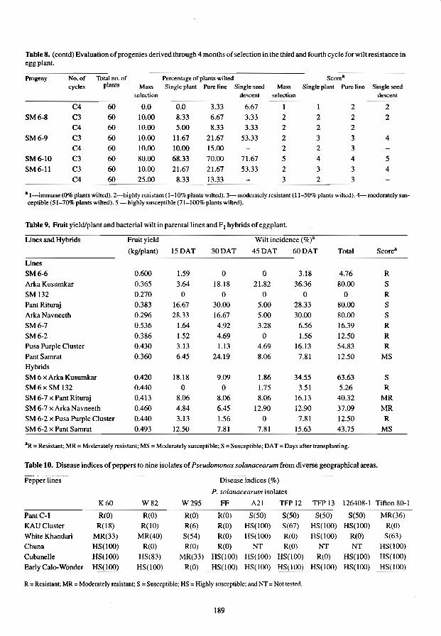
Table 8. (contd) Evaluation of progenies derived through 4 months of selection in the third and fourth cycle for wilt resistance in eggplant.
Progeny No. of Total no. of Percentage of plants wilted cycles plants Mass Single plant Pure line Single seed Mass Single plant Pure line Single seed
selection descent selection descent
60 0.0 0.0 3.33 6.67 1 2 2
SM6-8 C3 60 10.00 8.33 6.67 3.33 2 2 2 2 C4 60 10.00 5.00 8.33 3.33 2 2 2
SM6-9 C3 60 10.00 11.67 21.67 53.33 2 3 3 4 C4 60 10.00 10.00 15.00 2 2 3
SM6-lO C3 60 80.00 68.33 70.00 71.67 5 4 4 5
SM6·11 C3 60 10.00 21.67 21.67 53.33 2 3 3 4 C4 60 25.00 8.33 13.33 3 2 3
a I-immune (0% plants wilted). 2-highly resistant (1-10% plants wilted). 3-moderately resistant (11-50% plants wilted). 4- moderately sus· ceptible (51-70% plants wilted). 5 - highly susceptible (71-100% plants wilted).
Table 9. Fruit yield/plant and bacterial wilt in parental lines and FI hybrids of eggplant.
Lines and Hybrids Fruit yield Wilt incidence (%)'
(kg/plant) 15DAT 30DAT 45DAT 60DAT Total Score"
Lines
SM6·6 0.600 1.59 0 0 3.1S 4.76 R
Arka Kusumkar 0.365 3.64 18.18 21.82 36.36 80.00 S
SM 132 0.270 0 0 0 0 0 R
Pant Rituraj 0.383 16.67 30.00 5.00 28.33 80.00 S Arka Navneeth 0.2% 28.33 16.67 5.00 30.00 80.00 S
SM6-7 0.536 1.64 4.92 3.28 6.56 16.39 R SM6-2 0.386 1.52 4.69 0 1.56 12.50 R Pusa Purple Cluster 0.430 3.13 1.13 4.69 16.13 54.83 R PantSamrat 0.360 6.45 24.19 8.06 7.81 12.50 MS Hybrids
SM 6 x Arka Kusumkar 0.420 18.18 9.09 1.86 34.55 63.63 S SM6xSM 132 0.440 0 0 1.75 3.51 5.26 R SM 6-7 x Pant Rituraj 0.413 8.06 8.06 8.06 16.13 40.32 MR SM 6-7 x Arka Navneeth 0.460 4.84 6.45 12.90 12.90 37.09 MR SM 6-2 x Pusa Purple Cluster 0.440 3.13 1.56 0 7.81 12.50 R SM 6·2 x Pant Samrat 0.493 12.50 7.81 7.81 15.63 43.75 MS
OR = Resistant; MR = Moderately resistant; MS = Moderately susceptible; S = Susceptihle; OAT = Days after transplanting.
Table 10. Disease indices of peppers to nine isolates of Pseudomonas solanacearum from diverse geographical areas.
Pepper lines Disease indices (%)
P. solanacearum isolates
K60 W82 W295 FF A21 TFP12 TFP13 126408-1 Tifton 80-1
PantC-l R(O) R(O) R(O) R(O) S(50) S(50) S(50) S(50) MR(36)
KAUCluster R(lS) R(lO) R(6) R(O) HS(lOO) S(67) HS(100) HS(lOO) R(O)
White Khandari MR(33) MR(40) S(54) R(O) HS(lOO) R(O) HS(lOO) R(O) S(63)
Chuna HS(lOO) R(O) R(O) R(O) NT R(O) NT NT HS(100)
Cubanelle HS(lOO) HS(83) MR(33) HS(lOO) HS(lOO) HS(100) R(O) HS(lOO) HS(lOO)
Early Cal(). Wonder HS(I00) !:IS(lOO) R(O) H~(lOO) HS(IOO) HS(lOO) ...
HS(IOO) HS(lOO) H~(100)
R = Resistant; MR = Moderately resistant; S = Susceptible; HS = Highly susceptible; and NT = Not tested.
189

Hot pepper
No pepper lines tested were resistant to all nine isolates and only isolate A 21 was pathogenic to all the pepper lines (Table 10). The most resistant line was Pant C-l, a line derived as an advanced generation selection from a cross between NP46-A and Khandari. It was susceptible to only Florida isolates A 21. TFP 12, TFP 13 and 126408-1. KAU cluster was resistant to five isolates, and White Khandari had differential resistance (Peter et aJ. 1984).
References
Asha Sankar, M., Jessykutty, P.e. and Peter, K.V. 1987. Efficiency of four selection melhods to improve level of bacterial wilt resistance in eggplant. Indian 10umal of Agricullure Science. 57, 138-141.
Anon. 1980. Kerala Agriculture University Research Report 1980-81. Directorate of Research. K.A.U., Vellanikkara. India. 146 p.
Geth, R.w.. Peter, K.V. and Webb. R.E. 1983. Bacterial wilt, Pseudomonas solanacearum resistance in pepper and eggplnat lines. Phytopathology, 73. 808. Abstract.
Kelman, A. 1954. The relationship of pathogenicity in Pseudomonas soianacearum to colony appearance on a tetrazolium medium. Phytopathology, 44, 693-695.
Pande, K. 1976. Intergenotypic competition studies in bengal gram (Cicer arietinum L.). Ph.D. Thesis, G.B. Pant University of Agriculture and Technology, Pantnager, Nainital, India.
Parlevliet, J.E. 1979. The multiline approach in cereals to rusts -aspects, problems and possibilities. Indian Journal of Genetics 39, 22-29.
Peter, K.V., Goth, R.W. and Webb, R.E. 1984. Indian hot peppers as new sources of resistance to bacterial wilt, Phytophthora root rot, and root-knot nematode. HortScience, 19.277-278.
Rajan, S. and Peter, K. V. 1984. Incomplete dominance of bacterial wilt resistance. Tomato Genetics Cooperative Report, 34,14.
Shorter, R. and Frey, K.l. 1979. Relative yields of mixtures and monocultures of oat genotypes. Crop Science, 19,548-549.
190
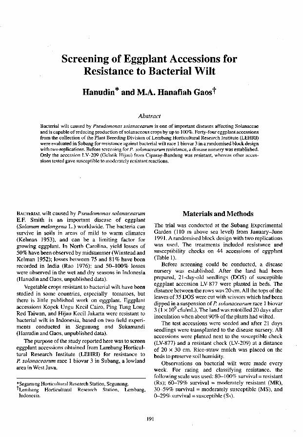
Screening of Eggplant Accessions for Resistance to Bacterial Wilt
Hanudin * and M.A. Hanafiah Gaos t
Abstract
Bacterial wilt caused by Pseudomonas solanacearum is one of important diseases affecting Solanaceae and is capable of reducing production of solanaceous crops by up to 100%. Forty-four eggplant accessions from the collection of the Plant Breeding Division of Lembang Horticultural Research Institute (LEHRI) were evaluated in Subang for resistance against bacterial wilt race 1 biovar 3 in a randomised block design with two replications. Before screening for P. solanacearum resistance, a disease nursery was established. Only the accession LV-209 (Gelatik Hijau) from Ciparay-Bandung was resistant, whereas other accessions tested gave susceptible to moderately resistant reactions.
BACTERJAL wilt caused by Pseudomonas solanacearum E.F. Smith is an important disease of eggplant (Solanum meiongena L.) worldwide. The bacteria can survive in soils in areas of mild to warm climate~ (Kelman 1953), and can be a limiting factor for growing eggplant. In North Carolina, yield losses of 50% have been observed by midsummer (Winstead and Kelman 1952); losses between 75 and 81% have been recorded in India (Rao 1976); and 50-100% losses were observed in the wet and dry seasons in Indonesia (Hanudin and Gaos, unpublished data).
Vegetable crops resistant to bacterial wilt have been studied in some countries, especially tomatoes, but there is little published work on eggplant. Eggplant accessions Kopek Ungu Kecil Cairo, Ping Tung Long Red Taiwan, and Hijau Kecil Jakarta were resistant to bacterial wilt in Indonesia, based on two field experiments conducted in Segunung and Sukamandi (Hanudin and Gaos, unpublished data).
The purpose of the study reported here was to screen eggplant accessions obtained from Lambang Horticultural Research Institute (LEHRI) for resistance to P. solanacearum race 1 biovar 3 in Subang, a lowland area in West Java.
*Segunung Horticultural Research Station, Segunung. tLembang Horticultural Research Station, Lembang, Indonesia.
191
Materials and Methods
The trial was conducted at the Subang Experimental Garden (110 m above sea level) from January-June 1991. A randomised block design with two replications was used. The treatments included resistance and susceptibility checks on 44 accessions of eggplant (Table 1).
Before screening could be conducted, a disease nursery was established. After the land had been prepared, 21-day-old seedlings (DOS) of susceptible eggplant accession LV-877 were planted in beds. The distance between the rows was 20 cm. All the tops of the leaves of 35 DOS were cut with scissors which had been dipped in a suspension of P. solanacearum race 1 biovar 3 (1 x 10⁹ cfulmL). The land was rototilled 20 days after inoculation when about 90% of the plants had wilted.
The test accessions were seeded and after 21 days seedlings were transplanted to the disease nursery. All accessions were planted next to the susceptible check (LV-877) and a resistant check (LV-209) at a distance of 20 x 30 cm. Rice-straw mulch was placed on the beds to preserve soil humidity.
Observations on bacterial wilt were made every week. For rating and classifying resistance, the following scale was used: 80-100% survival =: resistant (Rs); 60-79% survival =: moderately resistant (MR), 30-59% survival = moderately susceptible (MS). and 0-29% survival =: susceptible (Ss).

Table 1. Survival percentage, reaction, fruit type and colour of eggplant accessions screened for resistance in the bacterial wilt nursery, Subang, Indonesia 1991.
Acc.code Survival % Reactions' Fruit typeb Colour"
LV-36
LV-53
LV-60
LV-71
LV-83
LV-96
LV-107
LV-1I7
LV-141
LV-I 50
LV-155
LV-I 72
LV-209
LV-266
LV-312
LV-348
LV-349
LV-398
LV-401
LV-498
LV-500
LV-502
LV-536
LV-611
LV-63 I
LV-674
LV-700
LV-715
LV-73 I
LV-738
LV-740
LV-746
LV-2803
LV-2806
LV-2809
LV-28 10
LV-28 11
LV-2824
LV-2837
25.6
5.0
56.2
0.0
0.0
0.0
10.0
38.8
32.5
0.0
31.1
52.5
85.0
62.5
2.6
8.2
78.9
21.1
56.7
20.7
35.3
10.4
35.0
5.0
40.8
43.4
27.8
5.5
42.1
2.5
69.4
41.0
35.0
76.2
30.9
60.0
51.6
8.3
0.0
LV-2838 2.9
LV-2839 8.3
LV-2840 2.5
LV-209 (Rck) 89.8
LV-877 (Sck) 0.8
b Ss
b Ss
a MS b Ss
b Ss
b Ss
b Ss
a MS a MS b Ss
a MS a MS a Rs
a MR bd Ss
b Ss
a MR b Ss
a MS b Ss
a MS b Ss
a MS b Ss
a MS a MS b Ss
b Ss
a MS b Ss
a MR a MS a MS a MR a MS a MR a MS b Ss
b Ss
b Ss
b Ss
b Ss
a* Rs
b Ss
4
4
4
3
4
4
4
4
2
2
5
3
2
4
4
4
2
4
5
5 2
2
5
4
4
5
5 4
4
4
2
4
4
5 5
4
5
6
6
4
4
4
2
6
7
I
I
7
I
7
7
7
7
I
9 7
7
I
7
7
I
7
I
7
7
I
9
I
8
380-100% survival = resistant (Rs); 60--79% survival = moderately resistant (MR); 80--1cx>% sUNival = moderately susceptible (MS); and 00-29% survival =
bsusceptible (Ss). I = broader than long; 2 = as long as broad; 3 = slightly longer than broad; 4 = twice as long as broad or three times as long as broad; 5 = several times as long as broad; and 6::: round and big.
cl::: green: 2 ::: milk white; 3 = deep yellow; 4 = fire red; 5 = scarlet red; 6 = lilac ,F.rey; 7 ~ purple; 8 ~ purple black; .nd 9 ~ black.
Mean values followed by the same letter are not significantly different according to ScolI-Knott Test at 5% level.
192
Results and Discussion
Variance analysis of the percentage of wilted eggplant accessions was highly significant. Under the test conditions, only the accession LV-209 showed a high level of resistance against bacterial wilt race 1 biovar 3. Five accessions-LV-266, LV-349, LV-740, LV-2806, and LV-2810--were moderately resistant, whereas all other accessions were moderately susceptible or susceptible (Table 1).
In North Carolina, eggplant Kopek from Java and Matale from Sri Lanka were resistant to P. solanacearum (Winstead and Kelman 1952). While the genetics of resistance in eggplant have not been intensely studied, it has been reported that expression of resistance under high temperatures may be lacking, as was reported by Krausz and Thurston (1975) for tomato. In the experiments reported here, air temperatures ranged from 23.5-33,4°C and it is possible that some of the accessions reported resistant elsewhere were not resistant at these high temperatures.
Differences in resistance may also be due to the occurrence of different races or pathotypes. Buddenhagen and Kelman (1964) reported that three races can occur. In addition to the differences in races that may cause some pathogenic variation from location to location, the breakdown of resistance may also be due to a synergistic effect of parasitic nematodes. In these trials we found a number of different genera of nematodes, including Criconemoides, Helicotylenchus, Meloidogyne, Rotylenchus, and Xiphinema (Hanudin, unpublished data).
Acknowledgments The authors are grateful to Dr G.J.H. Grubben and 395 ATA's Project Staff for supplying funds for the trial. Ir. G. van Vreden, M.sc.; Ir. Agus Muharam, M.S.; and Ir. Budi Marwoto, M.S. are thanked for their guidance.
References Buddenhagen, I.W. and Kelman, A. 1964. Biological and
physiological aspects of bacterial wilt caused by Pseudomonas solanacearum. Annual Review of Phytopathology, 2, 203-230.
Kelman, A. 1953. The bacterial wilt caused by Pseudomonas solanacearum. A literature review and bibliography. North Carolina Agricultural Experiment Station Technical Bulletin 99,194p.
Krausz, l.P. and Thurston, H.D. 1975. Breakdown of resistance to Pseudomonas solanacearum in tomato. Phytopathology, 65,1272-1274.
Rao. M. V. 1976. Bacterial wilt of tomato and eggplant in India. In: Planning conference and workshop on the ecology and control of Pseudomonas solanacearum, Raleigh, North Carolina State University, 92-94.
Winstead, N.N. and Kelman, A. 1952. Inoculation techniques for evaluating resistance to Pseudomonas solanacearum. Phytopathology, 42, 628-{i34.

Inoculation Procedures and the Evaluation of Peppers for Resistance to Pseudomonas solanacearum
K.D.A. Perera, G.L. Hartman and J.M. Poulos*
Abstract
Experiments were conducted to develop an effective inoculation procedure to evaluate host plant resistance in peppers to Pseudomonas solanacearum, the causal agent of bacterial wilt. Three inoculation techniques-tooth-pick stabbing, soil drenching with root severing and soil drenching without root severing-were tested with two strains of P. solanacearum on 20- and 35-day-old seedlings. Tooth-pick stabbing and soil drenching with root severing yielded similar percentages of wilt in susceptible and resistant lines, indicating that they were equally effective screening procedures. The root-severing technique, however, was relatively easy to apply and more clearly resembled natural inoculation than did tooth-pick stabbing. The final percentage wilt was not significantly affected by the age at which the plants were inoculated but the rate of wilting was higher for younger seedlings. Although there was no significant difference in the percentage wilt caused by the two strains, the pepper strain PS71 was slightly more aggressive than the tomato strain PS4. Of 81 varieties screened for resistance, several showed complete absence of wilting with the root-severing technique.
BACTERIAL wilt caused by Pseudomonas solanacearum is one of the most important and widespread bacterial disease of crops in tropical environments. The pathogen has a wide host range representing 44 families (He et a!. 1983). Recent investigations have shown that it attacks several woody perennials, including cashew (Anacardium occidentale) (Shiomi et a!. 1989), custard apple (Annona spp.) (Mayers and Hutton 1987) and Alexandra palm (Archontophoenix alexandrae) (Akiew and Hams 1990). Among the solanaceous crops, potato, tobacco and tomato are greatly affected by the disease, while under certain tropical conditions pepper, especially sweet pepper, is also severely affected, There is little information, however, about the extent of damage caused to pepper by bacterial wilt.
Resistant varieties offer an effective means of controlling bacterial wilt. There has been some success with resistant varieties of tomato, tobacco and peanut. In potato, resistance genes from wild species have been introduced into cultivated species (Schmiediche 1986), The resistance, however, was found to be temperature sensitive and strain specific (French and De Undo
*Asian Vegetable Research and Development Center, Shanhua, Tainan, Taiwan, 741.
193
1982). Opefia et a!. (1990) concluded that both additive and non-additive gene action regulate the genetic system for wilt resistance in tomato. Tung et a!. (1990) found that wide genetic background and good adaptation were the key factors that favoured resistance to bacterial wilt in potato,
The importance of effective screening methods for the evaluation of germplasm for resistance to bacterial wilt has been emphasised. Winstead and Kelman (1952) found that puncturing the stem and adding a drop of bacterial suspension, or pouring a suspension over wounded roots, were equally effective for causing disease on susceptible tomato, tobacco and peanut plants. They concluded that injury to roots was the best technique to differentiate between resistant and susceptible plants. In tomato and many other hosts, young plants were more susceptible than older plants. Mew and Ho (1976) concluded that resistance to P. solanacearum in tomato was not necessarily comparable between seedlings and adult plants. Segregating populations were best screened under field conditions at the flowering stage. Field epidemics of bacterial wilt of pepper have been difficult to create (Yoon et a!. 1988). Development of a standardised technique for the evaluation of pepper germplasm is therefore necessary. A series of experiments was conducted to determine the

most effective inoculation procedure that can be applied to screen large germplasm collections under screenhouse conditions.
Materials and Methods
Comparisons of inoculation methods
Two strains of Pseudomonas solanacearum were tested using three inoculation techniques on susceptible sweet pepper plants of the commercial cultivar Giant Bell (F I ) (Evergrow Seed Co.) that were 20- and 35-days old. A split-plot design with four replications was used. with main plots as two strains and subplots as a factorial of different ways to inoculate plants and different plant ages. Seedlings were raised in 9-cmdiameter pots and inoculated individually with bacterial strains PS4 and PS7l. The inoculation techniques were: (1) tooth-pick stabbing; (2) soil drenching with root severing; and (3) soil drenching without root severing. For tooth-pick stabbing. sterile tooth-picks were rubbed in bacterial colonies and inserted into the stem at the axil of the second or third expanded leaf below the stem apex. Soil drenching with root severing was made after injuring the roots by passing a knife into the soil 1.5 cm away from the seedling stems and pouring a bacterial suspension over the severed roots. Soil drenching without root severing was done by pouring a bacterial suspension over the soil surface. For soil drenching, 107 ceJl/mL bacterial suspension was used at a rate of 30 mL per seedling. Plants in the control treatments for each method were treated with water. Diseased plants were counted daily for the first 2 weeks after inoculation and at 2-day intervals thereafter. Plants with aJl the leaves wilted were counted as wilted.
Evaluation of host resistance
Five plants of twelve resistant and four susceptible lines were inoculated with strain PS71 25 days after emergence. After inoculation, plants were kept in a heated glasshouse (24-34°C). Two inoculation procedures-tooth-pick stabbing and soil drenching with root severing-were conducted as two experiments using a randomised complete block design with three replications each. For soil drenching, seeds were sown in soil in 50 x 34 cm plastic flats. Six lines were planted in three rows in each flat. The space between rows was 10 cm with 5 cm between plants. At the time of inoculation, plants were 19-29 cm high and had 7-10 expanded leaves. The soil surface between the lines was cut with a sharp knife and 1.2 L of bacterial suspension (107 celVmL) was added to each flat. Diseased plants were recorded at 2-day intervals for the first week after inoculation and at 5-day intervals thereafter up to 28 days after inoculation (DAI). For
tooth-pick stabbing. plants were raised in 9-cmdiameter pots and inoculated as previously described. At the time of inoculation, plants were 11-15 cm high and had 7-9 expanded leaves. Data on pereentage wilt were recorded at 28 DAI.
Seventy-six accessions of pepper tested from entries in the International Hot Pepper Trial network (INTHOPE) were inoculated in another two experiments, one having 35 accessions, the other 41 accessions, using soil drenching with root severing. The inoculation procedure was as described previously. The experiment with 35 accessions was first conducted from 26 November 1991-30 January 1992 (winter). with the plants kept in a heated glasshouse after inoculation. The same experiment was repeated from 4 June 1992-31 August 1992 (summer); the other 41 accessions were tested in the 1992 summer.
Results
Comparisons of inoculation methods
Wilt symptoms were first observed 3 DAI on plants inoculated with infested tooth-picks. Plants that were inoculated by soil drenching with root severing and soil drenching without root severing wilted 8 and 12 DAI, respectively. Plants that were inoculated with infested tooth-picks developed wilt rapidly in young plants, with over 75% of plants wilted 15 DAI (Fig. I a). At 39 DAI, however. there was no significant difference in percentage wilt due to tooth-pick stabbing (85%) and soil drenching with root severing (77%). In older plants, disease progress was rapid in soil drenching with the root-severing method by 14 DAI and by 39 DAI 85% of the plants had wilted (Fig. Ib). There was no significant diffcrence in final percentage wilt due to soil drenching with root severing (85%) vs tooth-pick stabbing (73%). Soil drenching without root severing caused 40% of young seedlings to wilt and 28% of older seedlings. The overall wilt percentage by soil drenching alone was significantly less than that of tooth-pick stabbing and soil drenching with root severing (Table I).
The two strains, PS71 and PS4, did not cause any significant difference in the final disease rating. Pepper strain PS71 was more aggressive, causing 77% wilt, 22% more than that of the tomato strain PS4 at 39 DAI (Fig. 2).
Evaluation of host resistance Most of the susceptible varieties had over 90% wilt within 10 DAI with both methods of inoculation. Disease development was rapid when plants were inoculated by infested tooth-picks and symptoms occurred within 15 DAI for all sensitive cultivars tested. Using the root-severing technique, maximum
194

100 (8)
80
20
Ol----' ______ "'-__ IIL. __ '--~=_....L_....L_....L....L _ _l.._ ......... ..I'_______' _ ___.L _ ___'___ '-----''-----'-_-'-_--'-1
3 5 7 9 11 13 15 17 19 21 23 25 27 29 31 33 35 37 39
Days after inoculation
20l
ol~~--Alu=-",.--:d~~~~~~=----,--- ......... '-1 --1---"---'-----'--'-
3 5 7 9 11 13 15 17 19 21 23 25 27 29 31 33 35 37 39
Days after inoculation
Fig. I. Bacterial wilt on (a) 20-day-old peppers and (b) 35.-day-old peppers using three inoculation techniques. -O-tooth-pick stabbing; -. -soil drenching plus wounding; and --.A.- soil drenching with no wounding.
3 5 7 9 11 13 15 17 19 21 23 25 27 29 31 33 35 37 39
Days after inoculation
Fig. 2. Two strains, PS 71--D--; and PS4 -e-, of Pseudomonas solanacearum showing mean bacterial wilt (%) combined over 20- and 35-day-old inoculated plants and over three inoculation techniques.
195

Table 1. Comparison of bacterial wilt incidence on a susceptible pepper cultivar using three inoculation techniques.
Inoculation technique Wilt (%)"
Tooth-pick stabbing Soil drenching with root severing Soil drenching without root severing LSD (p: 0.05)b
79.2 80.8 35.0 32.1
8Mean of four replications and two isolates of P. solanacearum at 39 days after inoculation.
bFisher's prOlccled LSD.
Thble 2. Percentage bacterial wilt and area under the disease progress curve (AUDPC) for sixteen pepper varieties using two inoculation methods.
Variety Root-severing Tooth-pick stabbing
Wilt(%) AUDPC Wilt(%)
Giant Bell (FI )
YoloWonder California Wonder Cubanelle Doux d'Espagne Hungarian Wax Sweet Banana Antibois All Season Pant Cl Sinagtala Hong San Ho (FJ)
CAB MC5 MC4 CA8
LSD (P = 0.05)a
aFisher's protected LSD.
100 2112.5 100 95 1682.5 100 95 2155.0 100 95 1810.0 100 95 1992.5 100 95 2042.5 90 90 1020.0 90 65 1697.7 83 65 517.5 45 50 847.5 25
5 7.5 30 5 7.5 25 o 0.0 5 5 22.5 0 o 0.0 0 o 0.0 0
25 598.4 19
wilt occurred at more than 25 D AI. Comparisons of the area under the disease progress curve (AUDPC) calculated for the root-severing technique indicated a significant difference among the varieties (Table 2). Toothpick stabbing caused from 0-100% wilt with significant differences among varieties. The most susceptible varieties had 90-100% wilt with both inoculation methods. The high AUDPC values obtained from the root-severing method were related to highly susceptible cultivars as determined by final wilt. Some varieties that had the same final percentage wilt differed in AUDPC values. There were differences in the rate at which these varieties succumbed to wilt, some wilting more rapidly than othcrs. The varieties that had 0-5% wilt from root-severing had 0-30% wilt using the tooth-pick method. The varieties that had 50-
65% wilt using the root-severing technique, had 25-83% wilt with the tooth-pick method. Among the varieties tested, the plants which survived using the tooth-pick method showed some lesion development above or below the wound point along the stem. In some instances, the top of the plant died but later recovered by axillary bud growth. These were scored as resistant plants. MC 4 and CA 8, two varieties that had no wilt with either method of inoculation showed localised stem necrosis at the pricking point.
The 76 accessions of pepper screened using the soil drenching with root-severing technique showed different degrees of resistance and susceptibility among the cultivars (Tables 3 and 4). Some of the accessions showing high and moderate levels of susceptibility during the winter displayed a high level of resistance in summer (Table 4). Factors such as soil moisture variability and mite infestation during the summer screening may have interfered with host-pathogen compatibility.
Discussion
Tooth-pick stabbing and soil drenching with root severing were equally effective procedures to evaluate bacterial wilt resistance in peppers. The tooth-pick method showed rapid development of disease symptoms. as the direct introduction of the pathogen into the vascular tissues enabled faster movement and multiplication of the pathogen. Most of the plants inoculated with the tooth-pick method showed slight leaf chlorosis and stem distortion above the point of insertion of the tooth-pick. often retarding the terminal bud growth. One disadvantage of the method was the extra care necessary to avoid damage to the seedling when the tooth-pick was inserted into the stem, especially in younger plants. The root-severing method allowed for a more passive introduction of the bacteria into the plants. Similar final wilt incidence resulting from tooth-pick stabbing and root-severing techniques indicated that root severing of the secondary roots provided an ample opportunity for bacterial infection. Under field conditions, with transplanting or when inter-cultivation practices are carried out using farm implements, pepper plants are often subjected to rootsevering. As natural infection occurs mainly through these wounded roots, the soil drenching with root severing technique may mimic the field situation. When large numbers of accessions are evaluated, this is an easier and less labour-intensive technique. In addition, for further selection for other traits, soil drenching with root severing has an advantage because plant growth is not distorted. as sometimes occurs with the tooth-pick stabbing method. These advantages indicate that the root-severing methodology may be
196

Table 3. Bacterial wilt (%) for 35 accessions of pepper by the soil drenching with root-severing technique.
lAC Ubatuba Cambuci BrazillIAC Extra Long Selection IndiaIPAU Yangiao Taiwan! AVRDC Hungarian Wax 632 USNCelanese Seeds Ludhiana Long Selection IndiaIPAU Atarodo Niger Long Chili (Ft) Taiwan! Known You Long Fruit Thailand! AVRDC California Wonder USA PunjabLaI IndiaIPAU Cheongryong Korea/KBNU Hot Long (Ft) Korea/Seoul Seeds Twist Green (Ft) Korea/Seoul Seeds Sinagtala Philippines Lv. 1092 IndonesiaILEHRI Chain Fair (F I) Taiwan!Known You KA2 Sri Lanka/RARC-KA KAll Sri Lanka/RARC-KA Huaruar ThaiJandlKKU Var. PL2289 Nigeria/lAR Var. P. Sakaraho Nigeria/lAR Szechwan 10 Taiwan!AVRDC KKUCluster Thailand!KKU lawahar218 IndiaIPAU LV 1583 IndonesiaILEHRI LV 2319 (Tit Super) IndonesiaILEHRI MI2 Sri Lanka/RARC-MI MC5 MalaysiaIMARDI Huay Sithon Thailand!KKU Hong San Ho (Ft) Korea/Seoul Seeds Keriting IndonesiaILEHRI LV 2323 IndonesiaILEHRI Cipanas IndonesiaILEHRI Hot Beauty (FI ) Taiwan!Known You MC4 MalaysiaIMARDI
aMean of three replications rated 35 days after inoculation. "Fisher's protected LSD
more suitable for evaluation of bacterial wilt resistance in pepper.
Young pepper plants succumbed to wilt more rapidly than older plants. This phenomenon has been observed in tomato and many other crops, and it has been suggested that resistance in young plants cannot be compared to that found in older plants (Mew and Ho 1976). Nevertheless in the tests reported here, the final level of susceptibility in the pepper accessions did not change significantly due to the age at which the plants were inoculated.
The 81 accessions of peppers screened using the soil drenching and tooth-pick method showed different degrees of resistance and susceptibility among
Winter 1991-92 Summer 1992
100.0 86.7 93.3 66.7 93.3 60.0 86.7 80.0 60.0 80.0 80.0 80.0 40.0 80.0 93.3 73.3 80.0 73.3 33.3 73.3 66.7 13.3 62.2 60.0 0.0 60.0 60.0 53.3 33.3 46.7 80.0 46.7 6.7 40.0 0.0 33.3 0.0 33.3 0.0 33.3 0.0 26.7 6.7 26.7 0.0 26.7 0.0 26.7 24.4 13.3 20.0 26.7 16.7 13.3 6.7 6.7 0.0 6.7 0.0 6.7 0.0 0.0
34.80 32.90
cultivars. Some of the accessions showed complete absence of wilting, indicating that a high level of resistance was available among the germplasm tested. In the repeated experiment, the resistance level was consistent for most of the cultivars, with some exceptions. Observed differences in resistance levels in the same cultivars tested in two seasons may be due to genotypic differences within the cultivar or physiological imbalances resulting from mite infestation or soil moisture variability. In these experiments, accessions MC 4, CA 8, CAB, LV 2323 (Jatilaba), and Cipanas were the best sources of resistance to bacterial wilt of pepper.
197

Table 4. Bacterial wilt (%) in 41 accessions of pepper by root-severing technique.
Variety Origin!donor
TAM Mild Chile-2 USAffAES TAM Mild Jalapeno-I USAffAES Pangalengan-2 Indonesia Hidalgo USAffAES Tarn Veracruz]alapeno USAffAES California Wonder USA Da Chang Niujiao ]iao TaiwanlFu Ning Co. C01511 Italy Salmon Senegal/ AVRDC Gwangju Korea/KBNU KA-Il Sri Lanka/RARC-KA SlamChili TaiwanIKnown You PantC-1 IndialIARC PI 215743 Peru PBC389 AVRDC C. baccatum pend. 3-4 France/INRA COI647 Bulgaria Num Thailand!Kasetsart
University Var.PI-2190 NigerialIAR PBCI99 Thailand! AVRDC Unknown 41 Thailand KA-2 Sri LankaRARC-KA C0l621 Hungary Var.P.Sakaraho NigerialIAR Var.PL-2289 NigerialIAR KunJa Korea/HES-RDA Szechwan 8 Taiwan! AVRDC MI2 Srilanka/RARC-MI Var.PL-38475 NigerialIAR Var.U-Kashingurgu NigerialIAR Var.U-Kimba NigerialIAR Var.UL-2190 NigerialIAR RI·26(l7) Malaysia/MARDI Cili Langkap Malaysia/MARDI PBC384 Malaysia! AVRDC PBC385 Malaysia! AVRDC MC4 Malaysia/MARDI King Gum Go Chu Korea CheongYang Korea!ORDI LV 1583 IndonesiaILEHRI Orias Kossarvu Hungary
• Mean of three replications rated 35 days after inoculation. "Fisher's protected LSD.
Wilt (%)a
100.0 100.0 100.0
93.3 93.3 86.7 86.7 86.7 80.0 80.0 73.3 60.0 60.0 46.7 46.7 40.0 40.0 20.0
20.0 15.0 13.3 13.3 13.3 6.7 6.7 6.7 0.0 0.0 0.0 0.0 0.0 0.0 0.0 0.0 0.0 0.0 0.0 0.0 0.0 0.0 0.0
198
References
Akiew, E.B.and Hams, F. 1990. Archontophoenix alexandrae, a new host of P. solanacearum in Australia. Plant Disease, 74,615.
French, E.R. and De Lindo, L. 1982. Resistance to Pseudomonas solanacearum in potato: strain specificity and temperature sensitivity. Phytopathology, 72, 1408-1412.
He, L.Y., Sequeira. L. and Kelman, A. 1983. Characteristics of strains of Pseudomonas solanacearum from China. Plant Disease, 67,1357-1361.
Mayers, P.E. and Hutton, D.G. 1987. Bacterial wilt, a new disease of custard apple: symptoms and etiology. Annals of Applied Biology, 111,135-141.
Mew, T.W. and Ho, T. W. 1976. Varietal resistance to bacterial wilt in tomato. Plant Disease Reporter, 60, 264-268.
Opeiia, R.T., Hartman, G.L., Chen, J.T. and Yang, CH. 1990. Breeding for bacterial wilt resistance in tropical tomato. In: 3rd International Conference on Plant Protection in the Tropics. 1990 Malaysian Plant Protection Society, Vol. V, 44-50.
Schmiediche, P. 1986. Breeding potatoes for resistance to bacterial wilt caused by Pseudomonas solanacearum. In: Persley, G,J. ed., Bacterial wilt disease in Asia and the South Pacific. ACIAR Proceedings No. 13, lOS-Ill.
Shiomi, T., Mulya, K. and Oniki, M. 1989. Bacterial wilt of cashew (Anacardium occidentale) caused by Pseudomonas solanacearum in Indonesia. Indonesia Crop Research Journal, 2, 29-35.
Tung, P.X., Roseo, E.TJr., Vandenaag, P. and Schmiedeche, P. 1990. Resistance to Pseudomonas solanacearum in the potato. I. Effects of sources of resistance and adaptation. Euphytica, 45, 203-210.
Winstead, N.N., and Kelman, A. (1952). Inoculation techniques for evaluating resistance to Pseudomonas solanacearum. Phytopathology, 42, 628-634.
Yoon, J.Y., Green, S.K., Tschanz, A.T., Tsou, S.CS, Chang, L. C. 1988. Pepper improvement for the tropics: problems and the AVRDC approach. In: Green, S.K., ed., Tomato and pepper production in the tropics, Taiwan, Asian Vegetable Research and Development Center, 86-98.

Current U.S. Breeding Efforts for Improving Bacterial Wilt Resistance in Flue-Cured Tobacco
V.A. Sisson* and E. A. Wemsmant
Abstract
Through pedigree selection over the past 50 years tobacco breeders have been successful in increasing the level of resistance to bacterial wilt (Pseudomonas solanacearum) in tlue-cured varieties. Despite this success, bacterial wilt continues to cause significant economic losses to tobacco each year. Current breeding efforts seek to: 1. increase the level of wilt resistance; 2. improve the yield and quality characteristics for highly resistant varieties; 3. locate and utilise new sources of resistance to bacterial wilt; and 4. develop new breeding and evaluation methods for bacterial wilt resistance. In an effort to improve the levels of wilt resistance available in the breeding program, crosses have been made with TI 448A. the original source of resistance. and Xanthi. TT 79A. 79X. DSPA, and Enshu. These efforts have achieved only moderate success. High levels of resistance have been recovered but additional backcrossing is needed to recover tlue-cured characteristics. Haploid breeding methods are being used as a means of obtaining simultaneous disease resistance evaluation and line stabilisation. A simple in vitro procedure that pennits rapid cloning of haploid plants has been developed recently. Utilisation of this breeding technology leads to improved rates of genetic gain and shortens the time for cultivar development.
BACTERIAL wilt (Pseudomonas solanacearum E.F. Smith), commonly referred to in the United States as Granville wilt. is a major disease of flue-cured tobacco (Nicotiana tabacum L.). This disease has been reported in all of the flue-cured tobacco producing areas in the U.S. Initially, it was found in the Middle Belt area located in the rolling hills of the piedmont region of North Carolina. It has since spread into the sandy coastal plain areas, where it is the most important chronic disease problem in the Eastern and Border production belts. Even tobacco growers in the adjacent states of South Carolina and Virginia are now plagued by serious bacterial wilt infestations.
Over the last 10 years disease loss estimates for bacterial wilt on tobacco in North Carolina have averaged 1.4% of the crop, compared with an average total disease loss of 6.9%. Normally, bacterial wilt losses rank a close second behind those from black shank, caused by Phytophthora parasitica var. nicotianae (Breda de Haan) Tucker. These two
*USDA-ARS, Crops Research Lab, PO Box 1168, Oxford. North Carolina 27565. USA.
tCrop Science Department. Box 7620. North Carolina State University, Raleigh. North Carolina 27695. USA.
diseases account for nearly 50% of all flue-cured tobacco disease losses. In many areas bacterial wilt is the most important disease problem.
Wilt symptoms on tobacco are not unlike those of many other crops. Unilateral wilting of the leaves is characteristic. Initially only one Of two leaves wilt. with only half of the leaf wilting. Affected leaves become light-green to yellow. Tan or brown discoloration develops in the vascular tissue just beneath the bark. Infected roots become blackened and decay. Eventually all of the leaves wilt and die, but remain attached to the stalk.
Control measures such as crop rotation, early stalk and root destruction, and soil treatment with nematicides or mUltipurpose fumigants have been effective in reducing losses caused by this disease. but no one measure has provided adequate control. Corn, soybean, small grains, fescue, and lespedeza have been found to be acceptable rotation crops. Most U.S. f1uecured tobacco farmers follow a 2-year rotation, which can provide a significant disease reduction; however, longer rotations are recommended, especially if disease infestations are high. Multipurpose fumigants are available, but have shown limited effectiveness when used as the only means of control. While all of these
199
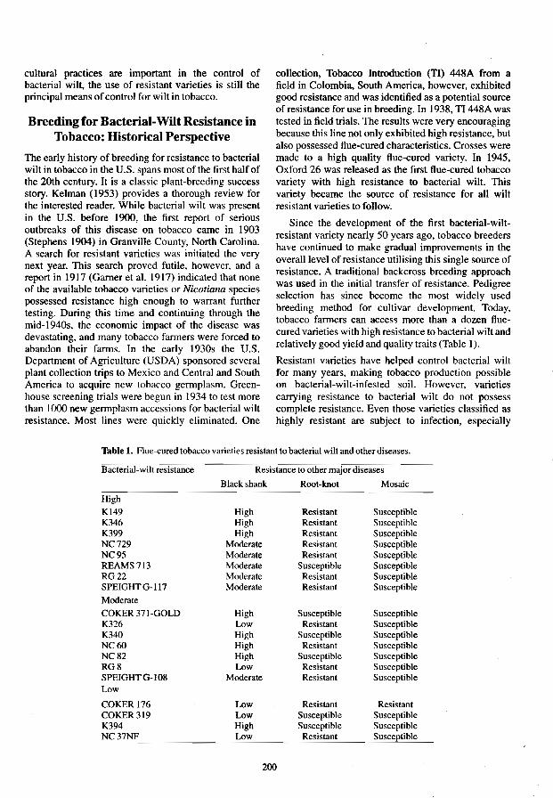
cultural practices are important in the control of bacterial wilt, the use of resistant varieties is still the principal means of control for wilt in tobacco.
Breeding for Bacterial-Wilt Resistance in Tobacco: Historical Perspective
The early history of breeding for resistance to bacterial wilt in tobacco in the U.S. spans most of the first half of the 20th century. It is a classic plant-breeding success story. Kelman (1953) provides a thorough review for the interested reader. While bacterial wilt was present in the U.S. before 1900, the first report of serious outbreaks of this disease on tobacco came in 1903 (Stephens 1904) in Granville County, North Carolina. A search for resistant varieties was initiated the very next year. This search proved futile, however, and a report in 1917 (Garner et at 1917) indicated that none of the available tobacco varieties or Nicotiana species possessed resistance high enough to warrant further testing. During this time and continuing through the mid-I 940s, the economic impact of the disease was devastating, and many tobacco farmers were forced to abandon their farms. In the early 1930s the U.S. Department of Agriculture (USDA) sponsored several plant collection trips to Mexico and Central and South America to acquire new tobacco germplasm. Greenhouse screening trials were begun in 1934 to test more than 1000 new germplasm accessions for bacterial wilt resistance. Most lines were quickly eliminated. One
col1ection, Tobacco Introduction (TI) 448A from a field in Colombia, South America, however, exhibited good resistance and was identified as a potential source of resistance for use in breeding. In 1938, TI 448A was tested in field trials. The results were very encouraging because this line not only exhibited high resistance, but also possessed Hue-cured characteristics. Crosses were made to a high quality Hue-cured variety. In 1945, Oxford 26 was released as the first flue-cured tobacco variety with high resistance to bacterial wilt. This variety became the source of resistance for all wilt resistant varieties to follow.
Since the development of the first bacterial-wiltresistant variety nearly 50 years ago, tobacco breeders have continued to make gradual improvements in the overall level of resistance utilising this single source of resistance. A traditional backcross breeding approach was used in the initial transfer of resistance. Pedigree selection has since become the most widely used breeding method for cultivar development. Today, tobacco farmers ean access more than a dozen ftuecured varieties with high resistance to bacterial wilt and relatively good yield and quality traits (Table 1).
Resistant varieties have helped control bacterial wilt for many years, making tobacco production possible on bacterial-wilt-infested soil. However, varieties carrying resistance to bacterial wilt do not possess complete resistance. Even those varieties classified as highly resistant are subject to infection, especially
Table 1. Flue-cured tobacco varieties resistant to bacterial wilt and other diseases .
Bacterial-wilt resistance Resistance to otherrnajor diseases ... _--
Black shank Root-knot Mosaic
High
K149 High Resistant Susceptible K346 High Resistant Susceptible K399 High Resistant Susceptible NC729 Moderate Resistant Susceptible NC95 Moderate Resistant Susceptible REAMS 713 Moderate Susceptible Susceptible RG22 Moderate Resistant Susceptible SPEIGHT G-l17 Moderate Resistant Susceptible
Moderate COKER371-GOLD High Susceptible Susceptible K326 Low Resistant Susceptible K340 High Susceptible Susceptible NC60 High Resistant Susceptible NC82 High Susceptible Susceptible RG8 Low Resistant Susceptible SPEIGHTG-108 Moderate Resistant Susceptible Low
COKER 176 Low Resistant Resistant COKER 319 Low Susceptible Susceptible K394 High Susceptible Susceptible NC Low Resistant
200

under conditions where soil infestation levels are high and ameliorating cultural practices are not carefully followed.
Despite the fact that bacterial wilt is widespread throughout the flue-cured tobacco production area, less than 13% of the acreage in 1991 was planted to varieties with high resistance. Farmers have been reluctant to grow these varieties. Less resistant varieties are typically grown because of their higher yield and better agronomic and quality features. V.S. flue-cured tobacco farmers are still looking for a highly-resistant variety with superior agronomic performance.
Current Breeding Efforts
The VSDA's Crops Research Laboratory (formerly the Tobacco Research Laboratory) has a long and rich heritage in breeding for bacterial-wilt resistance. Located in Granville county, North Carolina, site of the first severe disease problems, pathologists and breeders at this laboratory were directly involved in the development of the first wilt-resistant variety. This focus on bacterial-wilt resistance has continued through the years and today remains a major part of a cooperative VSDA-State breeding program.
Our objectives with regard to breeding for bacterialwilt resistance have changed over the years. Most new cuItivars now possess some resistance to bacterial wilt. Current breeding efforts have the following objectives: ( I) to improve the yield and quality characteristics of highly-resistant varieties; (2) to increase the level of resistance in flue-cured varieties; (3) to locate and utilise new sources of resistance; and (4) to develop new and more efficient breeding and evaluation methods. In the remainder of this paper, current research and development addressing each of these objectives will be discussed.
Breeding for improved yield and quality
Significant improvements in the yield and quality of flue-cured tobacco have occurred over the years. Bowman et. al (1984) used a regression technique to compare the performance of all new cultivars entered into the North Carolina Official Variety Test over the period 1954-1981. He estimated the average annual yield increase for new cultivars was 49.5 kg/ha (approximately 2% per year), of which 68% was attributed to improved technology and 32% (or 16 kg! ha) to genetic improvement. Genetic gains in leaf quality were also evident, but of a lower magnitude. This study did not differentiate among cultivars with different combinations or levels of disease resistance, but did show a definite favorable trend among all cultivars. Since many cultivars with improved wilt resistance were released during this time, it can be
201
assumed that the yield and quality of these cultivars also improved.
Current efforts 10 improve yield and quality still follow traditional breeding approaches. This is necessitated, to some extent, by the rigid quality standards established for each of the various classes and types of tobacco (i.e., flue-cured, air-cured, cigar, oriental, etc.). The amount of unique germplasm anyone class can tolerate without adversely affecting leaf quality is quite limited. Therefore, where new cultivars are the principal objective, their source populations are usually restricted to segregating generations from single- or three-way crosses among released cultivars of a common class. While this practice restricts the germplasm pool, good cultivars have been developed. The flue-cured tobacco cultivar NC 729 (Sisson 1992). which has some of the highest bacterial-wilt resistance among currently available cultivars, was recently released from our laboratory. It produces above average yield, and ranks among the best varieties for quality. Several other new cultivars with similar performance have also been developed recently by commercial breeders.
Increased resistance to bacterial wilt
As already noted, even those cultivars classified as highly resistant are subject to infection in heavily infested soils. Efforts to enhance current levels of resistanee are under way. These efforts are limited, in part, by the fact that current resistance in flue-cured tobacco is derived from a single source, TI 448A. Resistance from this source has been reported to be recessive and controlled by multiple genes (Smith and Clayton 1948). Effective use had been made of pedigree selection to improve resistance. Segregating populations are normally planted in a P. solanacearuminfested disease nursery. Seeds from disease-free F2 plants are saved and planted as F3 progeny rows in a wilt nursery the following year. Visual selection for resistance is practiced among lines, and plants within lines. The best F3 plants are self-pollinated and tested the next year. Each succeeding generation is traceable to a single-plant selection in the previous generation. Bulking or combining of seed from multiple plants is generally done after the F5 generation, at which time plants are considered genetically stable. This pedigree selection process has been successful in accumulating resistance genes and improving bacterial-wilt resistance over the years. Although the increments of increase are typically small, continued progress has been made in improving the level of wilt resistance. This method permits simultaneous selection for resistance to other diseases as well.
At this point we would like to briefly mention the procedures used for detecting resistance to P. solanacearum. Our evaluation of tobacco for

bacterial-wilt resistance is based mainly on test plantings of large populations in naturally-infested fields. Because the disease is so wide spread in the fluecured tobacco production area, fields with naturally high levels of inoculum are relatively easy to locate, and often farmers are willing to allow the use of such land since it has little value for tobacco production. We have found it necessary, however, to alternate yearly plantings between test plots and a highly susceptible variety to prevent reductions in the natural inoculum levels in the soil over time. In our bacterial wilt nursery at the Oxford Tobacco Research Station, we have also found it beneficial to artificially inoculate the field each year (Moore et a!. 1963). This not only helps to maintain a stable inoculum level over time, but more importantly provides for a uniform development of wilt in successive years. Several litres of inoculum are prepared just before the transplant operation for addition to the transplant water at the time of transplanting. 1be root rone of each transplant receives approximately 100 mL of water containing between 1 x Ht' to 1 x 10⁸ cells/mL. For many years a strain of P. solanacearnm designated K60 was used as the inoculum source. We have recently switched to a more virulent strain designated H20. While this procedure has been successful in inducing satisfactory levels of disease development, seasonal conditions still have a significant impact on overall wilt severity. Disease counts are taken every 3-4 weeks depending on disease development and a disease index is calculated from disease counts taken over the entire season.
Various greenhouse inoculation procedures (Winstead and Kelman 1952) have been used in evaluating resistance of tobacco to bacterial wilt; however, most breeders have experienced difficulty in utilising these tests. In most instances, these procedures are limited by the number of plants that can be tested, the labour involved, and the greater variation in results from test to test. In addition, disease severity, even in the most resistant lines or varieties, exceeds that observed under natural conditions in the field. Furthermore, field plantings allow evaluation of characteristics other than resistance. We are not using greenhouse evaluations at this time.
Locating and utilising new sources of wilt resistance
The development of improved wilt resistance in tobacco has obviously been hampered by the absence of sources of resistance. Resistance from TI 448A has been used exclusively in U.S. cultivars. Other possible sources of resistance have been identified, but to date have proven too difficult to work with or simply have not been used. Low resistance was reported in the old U.S. flue-cured cultivars Davis Special, Pinkney
202
Arthur, and 400 (Clayton and Smith 1942). These cultivars show evidence of resistance under mild wilt infestation, but under severe conditions appear as completely susceptible. They have no value for crossing with susceptible genotypes, as the resistance proves inadequate and impossible to recover in full (Clayton and Smith 1942).
Several lines or strains have been described as having mild or moderate resistance. These include: TI 79A and Sumatra C from Indonesia; Xanthi, a cultivar grown in Turkey; and the Japanese domestic cultivars, Awa, Hatano, Kokubu, and Odaruma. Early work (Clayton and Smith 1942) with the non-Japanese lines showed that progenies from crosses between these lines and flue-cured varieties typically have small leaves and are generally poor plants. It was at this same time that work with TI 448A was showing promise and these lines were abandoned as functional sources of wilt resistance.
Other than TI 448A, very few lines have been reported to have high bacterial-wilt resistance. The breeding line designated 79X is a stable line developed from a cross between TI 79A and Xanthi (Clayton and Smith 1942). Interestingly, crossing these two moderately resistant lines resulted in strains with higher resistance than either of the parental lines. When 79X was used as a source of resistance in the development of flue-cured cultivars, the resistant segregants tended to have small leaves and poor quality (Clayton and Smith 1942). Japanese workers have reported (Matsuda and Ohashi 1973) that the domestic cultivars Enshu and Hatanodaruma have high wilt resistance, but in our disease nurseries in the U.S. we have not observed the same high level of resistance which they report for these cultivars. Nevertheless, they do appear to possess resistance.
Thus, while we have relied on a single source of bacterial-wilt resistance in breeding U.S. flue-cured tobacco cultivars, there are a number of other possibilities. Breeding work and genetic studies have provided valuable information on the inheritance of wilt resistance in tobacco (summarised in Table 2). Results suggest there are different genetic systems ranging
Table 2. Mode of inheritance of bacterial wilt resistance from different sources.
Source
TI448A 79X Awa Enshu DSPA SumatraC Xanthi
Inheritance Reference
Recessive, polygenic Smith and Clayton 1948 Recessive, polygenic Smith and Clayton 1948 Rps Matsuda and Ohashi 1973 Rps, polygenic Matsuda and Ohashi 1973 polygenic Matsuda 1977 Rps, polygenic Matsuda 1977 Rxa Matsuda 1977

from single dominant genes to recessive poJygenes control1ing wilt resistance. Accumulating and combining the genetic factors from these different resistant sources may therefore make it possible to enhance the level of resistance.
In 1980, a long-term research project with this goal was initiated by Or G. R. Gwynn, tobacco breeder at the Crops Research Laboratory at that time. Although he retired in 1988, this important work has been continued. The following is a brief review of its development and current status.
The general breeding scheme selected is one of phenotypic recurrent selection and backcrossing. To yield initial single-cross populations, the resistancc sources, TI 448A. DSPA, TI 79A, 79X, Xanthi, and Enshu, were crossed using a partial diallel mating design which included the flue-cured variety Speight 0-15. These FI hybrids were naturally self-pollinated. The next year, F2 populations were grown in a bacterial wilt nursery. Disease-free plants of single-cross populations were intermated. These double-cross hybrids were naturally self-pollinated in the greenhouse during the winter. During the third year, the double-cross populations were grown in a bacterial wilt disease nursery. Disease-free plants were allowed to naturally self-pollinate. In year 4, the F2' 3 lines were again grown in a disease nursery and one or more of the following procedures was performed on selected lines: (I) plant-to-plant crosses were made for a third generation of intermating; (2) superior F3 lines from second generation intermating were advanced by self-pollination; and (3) superior F3 lines from second generation intermating were backcrossed to a high quality flue-cured cultivar. Since 1985 there has been continued evaluation, selection and advancement within the various generations of interrnating and backcross populations. In addition, a second backcross to a good yielding, high quality flue-cured variety has been made.
After working with these non-flue-cured and somewhat exotic sources of resistance, it is understandable why they were abandoned by early workers. Progress has been difficult and slow. Although it has been possible to identify and maintain high levels of resistance among the breeding lines selected, resistance appears to be no greater than that currently available in some existant highly resistant flue-cured cultivars. In addition, plant type of most resistant selections is uniformly poor, even after two backcrosses to high quality flue-cured parents. While the progress has been somewhat disappointing, it also likely reflects the complexity of this effort and reaffirms the need for additional wilt-resistant gerrnplasm for tobacco.
As mentioned previously, disease evaluations are carried out exclusively in field nurseries. Environ-
mental conditions have a major impact on bacterial development and disease expression. It is likely we will need to utilise more discriminating methods of disease evaluation before we can fully take advantage of these additional sources of wilt resistance.
Development and application of new breeding methods
The last area we wish to discuss is a new breeding strategy for developing wilt-resistant germplasm, which takes advantage of some unique features of the tobacco plant. Recent research has demonstrated the feasibility and efficacy of the use of haploids in tobacco breeding. Haploids are sporophytic plants which have the gametophytic chromosome number of the species. For tobacco, which is an allotetraploid, n = 2x=24. Plant breeders have long recognised the potential of haploids as tools for the simplification and acceleration of breeding programs. Although they have been observed in most plant species, only a few species can produce sufficient numbers of haploids for practical breeding purposes. Because of the relative ease with which haploids can be produced and the large haploid populations that can be generated, tobacco has been used as a model system for haploid research. Tobacco haploids can be produced by in vitro anther culture (anther-derived haploids) or by interspecific hybridisation using tobacco as the female and pollen of the wild species N. africarul Merxm. and Butler (ovulederived haploids). Completely homozygous diploid (or doubled haploid, OH) lines are produced by doubling the chromosome complement of haploid individuals. Chromosome doubling is accomplished by either treating small anther-derived haploid plantlets with an aqueous colchicine solution (Burk et al. 1972), or haploid plants may be chromosome-doubled by in vitro tissue culture of the leaf midvein (Kasperbaur and Collins 1972). Research has shown (reviewed by Wernsman 1991) that tobacco doubled haploids of maternal origin are competitive with conventionally inbred genotypes, while anther-derived doubled haploids typically have reduced performance.
Tobacco breeding in the haploid state
Many of the procedures for producing and using haploids in tobacco breeding are now well established (Fig. 1). In the initial steps FI plants from desired single-crosses are pollinated with the pollen from N. africana. Capsules form normally and produce large quantities of seed. When germinated, those seeds which are true interspecific hybrids (99+%) will die in the cotyledonary stage due to a seedling lethaIity factor associated with N. africana. The survivors are either aneuploids which contain one or more N. africana chromosomes, or maternal- or ovule-derived haploids.
203

These plantlets can usually be distinguished by various morphological differences.
Haploid seedlings are transplanted to pots and moved to the greenhouse. Once these plants have attained sufficient size, leaf mid-veins are aseptically cultured on a shoot-fonning medium to restore the diploid chromosome compliment. Sections of a leaf midvein are surface sterilised, first in 70% ethanol, followed by 10% sodium hypochlorite; rinsed in sterile distilled water; and placed on a shoot-fonning tissue culture medium (Kasperbaur and Collins 1972). Small shoots fonn in 3-4 weeks and are excised and transferred to a rooting medium. Shoots nonnally root in 10-20 days. Rooted plantlets are potted in soil and allowed to produce seed. Completely homozygous lines are thus recovered in about 3 years from the time of the initial cross. This can be contrasted to the 5-6 years required to reach genetic stability by conventional methods.
A number of procedures has been developed to enhance the efficacy of this breeding strategy. Single detached leaves of haploid plants can be inoculated with TMV. PVY, and wildfire (Rufty et al. 1987). Disease reactions can be observed on the detached leaf. This procedure makes it possible to test for multiple disease resistance in a non-destructive evaluation of the haploid plant. In this way, only those haploids expressing desired combinations of resistance would be doubled for further testing and evaluation.
Because the procedure using N. africana produces haploids in the fonn of seed, production of haploid plants can be synchronised with the growing season. Seeds can be sown in plantbeds in the spring where haploids are then identified. These haploids are transplanted directly to a disease nursery for testing. Resistant selections must be rescued for doubling the chromosome number before the haploid plant succumbs to the disease. Problems with endophytic fungi and bacteria in field-grown plants are a major obstacle, and make leaf mid-vein culture from field grown plants difficult.
A more recent procedure involves vegetatively cloning haploid plants. One clone is retained in a secure environment as a future explant source for doubling. and remaining clones can be subjected to destructive testing.
Haploids are produced in the nonnal manner from crosses with N. africana and grown to the 5-leaf stage. The apical meristem and surrounding leaves are cut from the plant. surface sterilised. and aseptically cultured in small glass vials containing a tissue-rooting medium. Roots typically fonn in 10-12 days at 26°C,
At the time of decapitation all but two leaves are removed. In 10 days, leaf axillary buds produce shoots or suckers. When these shoots reach 2.5-3.5 cm, they
204
_ ....
Evaluate and select haploid plants (field or greenhouse)
Rescue and double i
""""-.. haploids
Collect seed
Evaluate in replicated yield and
quality trials
In vitro culture of apical
meristem from haploids
Retain rooted menstemsin
secure environment
Remove from cold storage
Grow selected haploid
genotypes in greenhouse
Double chromosome number by in
vitro leaf midvein culture
in replicated yield trial
In vitro culture ofleaf axillary
shoots (vegetative
clones)
Subject haploid clones to replicated
disease tests
Identify superior haploid
genotypes
Fig. 1. General scheme for tobacco disease breeding in haploid plant popu)ations. Alternative methods allow direct screening of haploid plants or screening of haploid clones.

are cut from the plant, surface sterilised, and cultured on a rooting medium similar to the apical meristem. Rooting again occurs in 10-12 days. Since tobacco contains multiple axillary buds per leaf, secondary shoots will develop in leafaxils and can be rooted as welL
This procedure permits a number of clones or copies (up to six or eight) of each haploid plant to be produced. These multiple copies permit replicated test of individual genotypes, thereby increasing the precision of testing.
Clones can be stored safely for up to 90 days in a low temperature incubator maintained at 4°CI18-hour photoperiod. Plant growth is greatly retarded in this environment As clones are needed for testing they are removed from low temperature storage and transferred to a normal 26T incubator for 24 hours, the medium washed from the roots, and the clone transplanted to soil. In three weeks time the plant will attain sufficient size to be transplanted to the field or greenhouse. Upon completion of the disease-screening test, healthy explants for chromosome doubling of selected genotypes can be obtained from individuals maintained in the secured environment.
Haploid breeding combined with vegetative cloning provides an extremely powerful procedure for screening for bacterial wilt. It allows the use of field nurseries which we feel is a key to evaluating bacterial wilt resistance. Selected genotypes are instantly true breeding. These true breeding lines can be subjected to replicated yield and quality trials and are ready for immediate release to the growers if found superior.
This approach has been used successfully in the screening and development of black shank resistant breeding lines. We have also successfully tested haploid plants in the field for bacterial wilt resistance, but this is the first year we have actually begun to use the vegetative cloning aspects of this procedure in developing wilt resistant cultivars. Based on our experience with black shank resistance, however, we are confident that this approach will be successful in improving the efficiency of our breeding effort for bacterial-wilt resistance and result in the development of new and improved wilt-resistant flue-cured tobacco cultivars.
References
Bowman, D.T., Wemsman, E.A., Corbin T.C. and Tart, A. G. 1984. Contribution of genetics and production technology to long-tenn gains in flue-cured tobacco. Tobacco Science, 28, 30-35.
205
BurIc, L.G., Gwynn, G.R and Chaplin, J.F. 1972. Dipolidized haploids from aseptically cultured anthers of Nicotiana tabacum: A colchicine method applicable to plant breeding. Journal of Heredity, 63, 355-360.
Clayton, E.E. and Smith, T.E. 1942. Resistance of tobacco to bacterial wilt (Bacterium solanacearum). Journal of Agricultural Research, 65,547-554.
Gamer, W.w., Wolf, EA. and Moss, E.C. 1917. The control of tobacco wilt in the flue-cured district. Bulletin United States Bureau of Plant Industry, 562. 20 p.
Kasperbaur, MJ. and Collins, G.B. 1972. Reconstitution of diploids from leaf tissue of anther-derived haploids in tobacco. Crop Science, 12,98--101.
Kelman, A. 1953. The bacterial wilt caused by Pseudomonas solanacearum. A literature review and bibliography. North Carolina Agricultural Experiment Station Technical Bulletin 99.194p.
Matsuda, T. 1977. Fundamental studies on the breeding of bacterial wilt resistant varieties in tobacco. Bulletin Utsunomiya Tobacco Experiment Station No. 15. 95 p. English summary.
Matsuda, T. and Ohashi, Y. 1973. Inheritance of resistance to bacterial wilt disease in tobacco. Japanese Journal of Breeding, 23, 175-180. English summary.
Moore, E.L., Kelman, A., Powell, N.T. and Bunn,RH. 1963. Inoculation procedures for detecting resistance to tobacco to Pseudomonas solanacearum in the field. Tobacco Science,7,17-20.
Rufiy, R.C., Wemsman, EA and Gooding, G.VJr. 1987. Use of detached leaves to evaluate tobacco haploids and doubled haploids for resistance to tobacco mosaic virus, Meloidogyne incognita, and Pseudomonas syringae pv. tabaci. Phytopathology, 77, 60-62.
Sisson, V.A. 1992. Registration on 'NC 729' tobacco. Crop Science, 32, 1066.
Smith, T.E. and Clayton, E.E. 1948. Inheritance of resistance to bacterial Will in tobacco. Journal of Agricultural Research, 76,27-32.
Smith, T.E., Clayton, E.E. and Moss, E.G. 1945. Flue-cured tobacco resistant to bacterial (Granville) wilt. United States Department of Agriculture Circular 727.7 p.
Stephens, EL. 1904. The history of the tobacco wilt in Granville County, North Carolina. United States Department of Agriculture, Bulletin, 142, 166-168.
Wernsman, E.A. 1991. Varied roles for the haploid sporophyte in plant improvement. In: Stalker. RT. and Murphy. J.P. ed5., Plant Breeding in the 19905: Proceedings of the Symposium on Plant Breeding in the 1990s. CAB International: Wallingford, UK. 461-485.
Winstead, N.N. and Kelman. A. 1952. Inoculation techniques for evaluating resistance to Pseudomonas solanacearum. Phytopathology, 42, 628-634.

Screening Eucalyptus Selections for Resistance to Bacterial Wilt Caused by Pseudomonas solanacearum
J.C. Dianese and M.C.G. Dristig*
Abstract
Methods of screening Eucalyptus spp. for resistance to Pseudomonas solanacearum were tested by using differences in inoculum concentrations, inoculum placement on the host, physiological ages of seedlings, and bacterial isolates. Screening for tolerance to bacterial wilt among 42 genotypes belonging to 20 Eucalyptus spp. was conducted in a growth chamber at 28 to 30°C with a 12-hour photoperiod. Seedlings with 8-10 leaves were inoculated with isolate UnB 577 of P. solanacearum by placing a 1 0 ~L drop of 106
cfu per mL in the axil of the second pair of true leaves previously wounded with a needle. The plants were rated for disease 10 and 18 days later. Tolerance was present in one selection of each of the following species: Eucalyptus camalduiensis, E. citriodora, E. c/oeziana. E. deanei, E. pellita, E. resinifera, and E. tereticomis. Two selections each of E. grandis and E. torelliana were also wilt tolerant.
PREVIOUS screenings for resistance to bacterial wilt in Eucalyptus spp. were conducted in Brazil by Cruz and Dianese (1986) and Dianese et at. (1990) with Pseudomonas solanacearum biovar 1 (Dianese and Takatsu 1985). In China, Wu and Liang (1988) selected genotypes tolerant to biovar 3 isolates. Selections tolerant to biovar 1 were detected in E. deglupta, E. pellita and E. tereticomis in Brazil (Dianese et. al. 1990), while tolerance to biovar 3 was reported from China in E. sa ligna, E. citriodora and E. exerta (Wu and Liang 1988). The objective of this study was to develop techniques for mass screening of Eucalyptus genotypes for resistance to bacterial wilt.
Materials and Methods
Experiments were conducted in growth chambers using 10 replications of one plant each. The virulence of nine isolates of P. solanacearum was tested by inoculating 6- to 8-leaf seedlings of a susceptible selection of E. grandis in wounds in the axil of the second pair of true leaves. A virulent isolate (UnB 577) and a weaklyvirulent isolate (UnB-573) were then selected for the experiments conducted to develop the method of inoculation.
*Departamento de Fitopaloiogia, Universidade de Brasilia. 70910-900 Brasilia. DF. Brazil.
Seedlings were transplanted to 200 mL plastic cups at the 2-6-leaf stage. When at the 11}-12-leaf stage, plants were inoculated separately with isolates UnB 577 and UnB 573 using five different methods: (i) soil treated with a 10 J.1L suspension of 106 cfu; (H) same as treatment 1. but roots wounded using a small scalpel; (Hi) leaf axil wounding with a needle before placing a J 0 J.1L suspension; (iv) laying 10 mL of suspension around the crown of the seedling just after transplanting to cups; and (v) root wounding and dipping in 106 cfu suspension before transplanting.
The most favourable physiological age for inoculation was tested by comparing seedlings with 2-4, 6-8, and 11}-12 true leaves. Inoculated plants were incubated for 18 days in growth chambers at 30°C and a 12-hour photoperiod. Data from the above experiments were used to select the inoculation procedure to screen for resistant genotypes and also to test the virulence of isolates on a selection of E. grandis from Passagem, State of Minas Gerais. Brazil.
Disease scores were reeorded at 10 and 18 days after inoculation using the following disease scale: 1= plants without symptoms; 2 = wilted plants; and 3 = dead plants (Fig. I). The following wilt index (WI) was calculated for each genotype:
WI = [(1 x f l ) + (2 x f2) + (3 x f3)]/(fl + f2 + f3);
where 1. 2, and 3 are the disease rating scores and fl • f2' and f3 are the respective frequencies of each type.
206
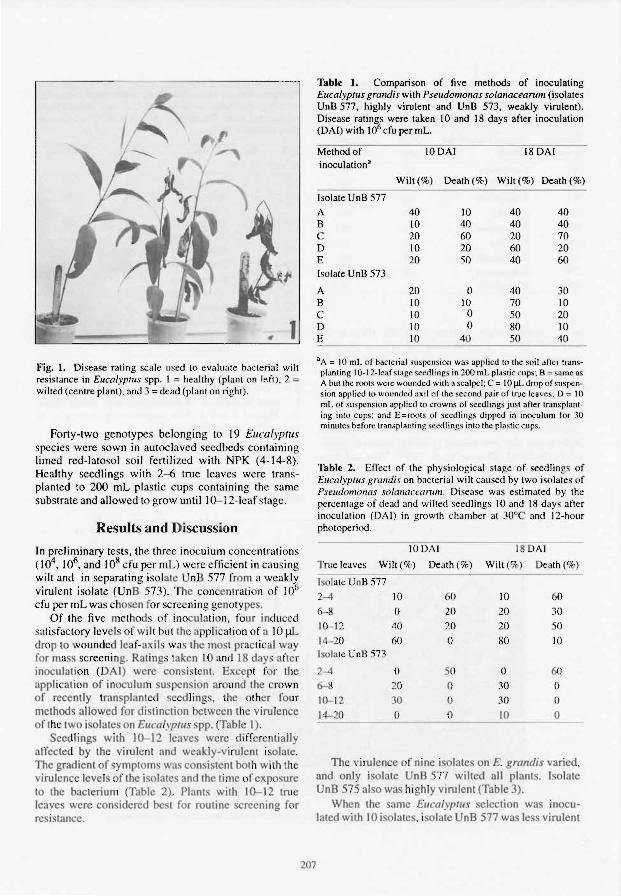
Fig. I. Disease rating scale used to evalu:ltc bacterial wilt resistance in EucalYflllls spp. 1 = healthy (plant on left). 2 = wilted (centre plant). and 3 = de:ld (plant on right).
Forty-two gcnotypes bclonging to 19 Eucalyptus species were sown in autoclaved seedbeds containing Iimcd rcd-Iatosol soil fertilized with NPK (4- 14-8). Healthy seedlings with 2-6 true leaves were transplanted to 200 mL plastic cups containing the same substrate and allowed to grow until 10-1 2-lcaf stage.
Results and Discussion
In prcliminary tests. the three inoculum concentrations (104
• 106• and 108 cfu per mL) were efficient in causing
wilt and in separating isolate UnB 577 from a weakly virulent isolate (UnB 573). The con entrat ion of lO6 cfu per mL was chosen for screening g notypes.
Of the five methods of inoculation, four induced salisfactory levels of wilt hut the applicalion of a 10 pL drop to wounded leaf-axil wa s Ihe most practic al wa for mass screening. Rat ings taken 10 and 18 days a fter inocu lation (DAI) were consi. tenl. Except for the appl ication of inoculum suspension ar und the rown of recently tran plant I seedli ngs, the ther four methods allowed for dist in lion between the virulence of the two isolates on EllcalyptllS spp. (Table I).
Seedlings with 10--12 leaves were differentially affected by the vi ru lent and weakly-viru lent isolate. The gradient of symptoms was consistent both with the virulence leve ls of the isolates and the lime of expo ure to the bacterium (Table 2) . Plant with 10--12 tru ' leaves were con idered be t for routine screening for resi tance.
207
Table l. Comparison of five methods of inoculating EucalYPlIIs grandis with Pseudomonas solanacearum (isolates UnB 5;77, highly virulent and UnB 573, weakly virulent). Disease ratings were taken 10 and 18 days after inoculation (DAI) with 106 cfu per mL.
Method of inoculation"
Isolate UnB 577
A B C 0 E Isolate UnB 573
A B C D E
IODAI
Wilt(%) Death (%)
40 10 10 40 20 60 10 20 20 SO
20 0 10 10 10 0 \0 0 10 40
18DAI
Wilt(%) Death(%)
40 40 40 40 20 70 60 20 40 60
40 30 70 10 50 20 80 10 50 40
"A = 10 mL of bacterial suspension was applied to the soil after transplanting IO-12-lcaf stagc seedlings in 200 IllL plastic cups: B = same as: A but the root< were wounded with" scalpel; C = 10 J,lL drop of suspens ion :lpplicd to wounded axil o f the second pair of true Icavcs: [) = 10 mL of SlIspension applied to crowns o f scculings just after IJan splanling into cup.;; and E=roQ1S of seedlings dipped in inoculum for 30 minutes hefo re transplanting seedlings into Ihe plastic cups.
Table 2. Effect of the physiological stage of seedlings of Eucalyptus gralldis on bacterial wilt caused hy two isolates of Pscudom()nas solanacellrum. Discase was estimated hy the percentage of dead and wilted seedlings 10 and 18 days after inoculation (DAI) in growth charnhcr at 30°C and 12-hour photoperiod.
10UAI 18 DAI
Tnlt: leaves Wilt("h) Death (% )
Isolate Un B 577
2-4 10 ,() 10 00
6-X 0 20 20 30
10--12 40 20 20 50
14-20 60 0 80 10 Isolate UnB 573
2-4 0 50 () 60 6-8 2-D 0 .10 ()
to-- I2 30 0 30 0
14--20 0 0 10 0
The vi ru lence 0 nine isolates on E. grandis varied, and only isolate UnB 577 wilted all plants. Isolate UnB 575 al 0 wa highly virulent (Table 3).
When the same Eucalypflls seleclion wa inocu lated with 10 isolates. isolate UnB 577 was le. virulent

Table J. Virulence raling of nine i. ol3les of Pseudomonas solanacearum on a ~eleclion of Eucalyptus grandis from Passagem. Stale of Minas Gerais inoculated in growth chamber at 30·C and! 2-hour photoperiod during) 5 days.
Bacterial Original hosts of the Will Death
isolates Isolates (%) (%)
539 E./Irophyl/a 10 0
540 E. grandis 10 10 573 E. urophyl/a 20 0
574 E. pellita 20 30
575 E. urophylla 20 70
576 E. pellita 20 0 577 E. urophyl/a 40 60 579 E. urophylla 10 0 603 E. urophylla 10 0
to E. grandis than isolates UnB 588 (biovar 1), UnB 210 (biovar 2), and UnB 139 (biovar 3) from tomato; UnB 68 (biovar 2) from potato; UnB 75 (biovar 3) from Solanum gilo; and UnB 468 (biovar 3) from bell pepper (Table 4).
All the isolate te led were virulent to E. gralldis, and only UnB 577 was used to screen for resistance. The bacterium infected and multiplied in the xylem of wilting and also non-wilting (tolerant) plants as previously observed (Dianese et al. 1990). This indicates that tolerance to bacterial wilt was present in 12 of the genotypes tested which were arbitrarily defined as those with a WI (wilt index) Ics than J.5 at 10 OAf and
Table 4. Response of a selection of ErICalyp/us grandis from Passagem. Stale of Minas Gerai 10 growth-chamber inoculalions with P. solanaceanlln biovars I, 2. or 3 isolaled from solanaceous hosts. Seedlings were kept at 30·C and 12-hour photoperiod for 18 day .
UnB Biovar Hosts Wih(%) Dealh(%) isolates
577 E. llrophyl/a 40 50 115 So/a/II/m 20 45
//lberomm
588 Lycopersicon 10 90 esculentrun
661 L MlIsasp. 50 5
210 2 L esclllelllmn [5 70
68 2 S. wberoslll1l 5 90 139 3 L. l!scII/entrun 5 90
75 3 So/alII/in gilo 5 90
534 3 S. melollge/IQ 10 to 468 3 Capsicum 25 70
a/mllllm
a WI less than 1.8 at J 8 DAI. The tolerant genotypes belonged to eight of 18 Eucalyptus pp. tested (Table 5). Figures 2 and 3 show, re pectively, responses of susceptible and tolerant species.
A mass-screening technique to locate sources of re istance to bacterial wilt among a broad spectrum of Eucalyptus pedes and provenances is important to avoid major 10 es in tropical and sllbtropical areas infe ted with P. sofanQcearum.
Fig.2. ElIcalyptlls lIrophy/la wilting and dyi ng after inoculation with PselldOlllonas so/aliI/a urum.
208

Fig. 3. Survival of Ellcaiyllls tore/lialla (resistant) al"ttr inoculation with PselldomOllas so/allacearrmr.
Table S. Response of EllcaiYPlls spp. to growth-chamber inoculations with isolate UnB 577 of Pseudomollas so/anaccan/m. expressed in wilt index taken 10 (WI 10 DAI) and 18 days (WI 18 DAI) after inoculation.
Euca/yptus species Origin' WIIODAI WI18DAl Tolerant selections6 Seed supplier"
coma/du/ensis Ibitira 1.4 1.5 + ( I)
citriodora SI. B;irbara 1.9 2.3 (I)
citriodora Ibitira 1.2 2.1 (I)
citriodora Alagoinhas 2.5 2.8 (I)
citriodora lIatinga 2.0 2.3 (I)
citriodora Ouri,angas 1.1 1.9 (I)
citriodora 11762 1.5 1.6 + (2)
c/oeziona Carbon ita 1.4 1.8 + (I)
c1oeziano 1035-CB 2.1 2.7 (3)
c10eziana Anhembi 2.0 2.6 (2)
c/oeziOlro 9785 2.6 2.7 (I )
c/oeziallo Tilbllry 2.6 3.0 (I)
deml1rei L1 -CB 1.3 1.6 + (3
deglrlpra M. DOllrado 1.7 2.5 (5)
gralldis Ibitira 1.4 1.8 + (I)
grmrdis M. Gua,u 1.6 2.2 (I)
grandis 9783-CB 1.7 2.4 (3)
grandi! Passagem 1.7 2.3 (I)
gralldis Orchard 1.4 1.6 + (4)
gralldis CPAC 1.0 1.3 + (4)
macu/ata Zimbabwe 1.2 2.6 (I)
microcorys L1-CB 2.3 2.6 (3)
pellita Faz. Eng. 2.1 2.4 (2)
pel/ita Carbonita 2.2 2.5 (I )
209

1Bble S. (contd.) Response of Eucalypus spp. to growth-chamber inoculations with isolate UnB 577 of Pseudomonas solanacearum. expressed in wilt index taken 10 (WI 10 DAI) and 18 days (WI 18 DAl) after inoculation.
Eucalyptus species Origin"
pellita l20B-CB
pellita Dionfsio
pellila Q.GeraI
pilularis Carbonita
pilularis Australia
punctata CASB
pyrocarpa Carbonita
resinifera Carbonita
resinifera Zimbabwe
robusta M.Gua~6
saligna ltatinga
tereticomis CASB
lereticomis 11953
torelliana Faz. Eng.
torelliana T. Freitas
urophylla Dionfsio
urophylla Faz. Eng.
B-12oo
"Numbers are CSIRO Australia designations. b + indicates the tolerant genotypes.
WIIODAI WI 18DAI Tolerant selections6 Seed supplier"
1.9 2.2 (1)
1.6 1.9 (3)
1.3 1.7 + (1)
25 2.7 (1)
2.0 2.1 (1)
2.2 2.4 (1)
2.6 2.8 (1)
2.1 25 (3)
1.2 1.4 + (l)
2.4 2.7 (1)
2.3 2.5 (1)
2.3 2.4 (1)
1.1 1.1 + (1)
1.3 1.5 + (1)
1.4 1.4 + (l)
2.0 2.1 (1)
1.6 2.0 (1)
1.8 2.3
c(l) Companhia Agricola e Florestal Sta. Barbar", B. Horizonte. MG; (2) Companhia Cimetal. Belo Horizonte, MG; (3) Cimento Pirineus, Pe. Bemardo. Goias; (4) CPAClEMBRAPA, Planaltina. DF; (5) Companbia Florestal Monte Dourado, Jar!, Para, Brazil.
References
Cruz, A.P. and Dianese, ],c. 1986. Tolerancia a murcha bacteriana em eucaHpto. Fitopatoiogia Brasileira, 11. 396.
Dianese. J.C .• Dristig, M.C. and Cruz. A.P. 1990. Susceptibility to wilt associated wilh Pseudomonas solanacearwn among six species of Eucalyptus growing in equatorial Braz.il. Australasian Plant Pathology, 19,71-76.
Dianese, I.C. and Takatsu, A. 1985. Pseudomonas solanacearum biovar 1 isolada de eucaUpto em Monte Dourado, Estado do Para. Fitopatologia Brasileira, 10,362.
Wu, G.P. and Liang, Z.c. 1988. Selection of species and provenances of Eucalyptus for resistance to bacterial wilt. Journal of South China Agricultural University, 9, 41-45.
210

Management of Bacterial Wilt of Groundnut Using Genetic Resistance and Cultural Practices
V.K. Mehan, S.N. Nigam, and D. McDonald*
Abstract
The use of bacterial wilt-resistant groundnut cultivars is an effective and practical way of reducing yield losses from the disease. Many sources of resistance have been reported from the People's Republic of China, Indonesia, and Uganda where bacterial wilt is a serious problem. Much progress has been made in the development of several wilt-resistant cultivars in China and Indonesia. Several of these cultivars are now widely grown in various parts ofIndonesia (e.g. Gajah, Pelanduk, Tupai) and south and central China (e.g. El Hua 5, Lu Hua 3, Yue You 92. Guiyou 28, Zhong Hua 2), and have had much impact in reducing yield losses in wilt-affected areas. The wilt-resistance in Indonesian cultivars Schwarz 21 and Gajah continues to be useful even after several decades of their cultivation. However, recent studies in China have highlighted the existence of strainslpathotypes in the wilt pathogen, Pseudomonas solanacearum. This necessitates more critical genetic evaluation of wilt-resistance to elucidate host-pathogen interactions. Future research into stability and durability of wilt-resistance, as well as incorporation of resistance to other yield reducing diseases, are indicated. Crop rotation with feasible non-host crops (e.g. rice and maize) is also a useful means of reducing the disease levels in humid tropical areas. An integrated approach to disease management, involving wilt-resistant cultivars, crop rotation, intercropping, and crop sanitation, is emphasised.
BACTERIAL wilt (BW) of groundnut (Arachis hypogaea L.) occurs in several countries of southern Asia, Southeast Asia, Africa, and North America, but it is economically important in only the People's Republic of China, Indonesia and Uganda (Hayward 1990; He 1990; Machmud 1986; Mehan et al. 1986; SimbwaBunnya 1972). The disease has recently been reported as severe in some groundnut-producing areas, of Vietnam (Mehan et a!. 1991), In the People's Republic of China, the disease is most severe in central and southern groundnut-producing areas where more than 0.2 million hectares are infested with the wilt pathogen, Pseudomonas solanacearum E.F. Smith, and yield losses of 10-30% are common (Liao et a1. 1990). However, within any region there are variations in disease incidence and severity attributable to soil type, pathogen populations, cropping patterns, climatic factors and groundnut cultivars. The persistence of the pathogen in soil, and its wide host range, often limit the effectiveness of cultural and chemical control
*Intemational Crops Research Institute for the Semi-Arid Tropics, Patancheru P.O., Andhra Pradesh 502324, India.
211
practices. Although rotation with non-host crops provides effective disease management. control of BW of groundnut has been achieved mainly with resistant cultivars. In this review paper we provide information on sources of resistance to the disease and examine how these sources are being utilised in the breeding of resistant cultivars. Resistant cultivars and cultural control measures are considered for use in integrated disease management systems.
Host Plant Resistance
Sources of resistance
Attempts to select wilt-resistant groundnut cultivars were first made in Indonesia in the 1920s. Most of the cultivars selected as BW-resistant, such as Hybrid No. 3, Katjan Tocban, and Pure Line 21, later showed high mortality of plants under severe disease pressure (Palm 1926). Subsequently, from an extensive wilt resistance breeding program in Java, a highly resistant cultivar, Schwarz 21, was developed by selection from a wiltresistant groundnut line of unknown origin (Schwarz and Hartley 1950). Resistance in this cultivar was

confirmed in several field and greenhouse screening tests (Jenkins et a!. 1966; Machmud and Lasimin 1987; Machmud and Middleton 1988; Yeh 1990). From extensive screening of several hundred germplasm and breeding lines in wilt-sick plots in Indonesia, several lines with varying levels of resistance to BW wcre identified (Machmud and Middleton 1988, 1990; Sharma and Soekarno 1992).
Various Iines/cultivars resistant to P. solanacearum have also been reported from Mauritius, South Africa, Uganda, the United States, the Philippines and Sri Lanka (Orian 1949; Sellschop 1947; Jenkins et al. 1966; Simbwa-Bunnya 1972; Natural et al. 1988; Jayasena and Rajapaksa 1990). However, some of these were susceptible in China (Sun et al. 1981).
Many sources of resistance have been reported from the People's Republic of China where a selection and breeding program for wilt resistance was begun in the late , 950s. Of over 500 germpJasm lines screened in Guangdong Province in the early 1960s, 30 wiltresistant lines were identified (Zhou and Liu 1962; OCRI 1976). From extensive screening of over 4000 germplasm accessions and breeding lines in the 1970s and 1980s, many additional sources of resistance were identified (OCR I 1977; XU et a!. 1980; Wang et a!. 1983; Tan and Liao 1990; Duan and Li 1987; Duan et a!. 1991). These included Xiekangqing, Taishan Sanlirou, Lukangking I, Yue io 589, Huangchuan Zhili, Tianjing Dou, Taishan Zhengzhu, and many lines of 'Chinese Dragon' type. Some of these (Taishan Zhengzhu, Yue io 589) were later shown to be susceptible (Sun et a!. 1981). In the next decade, several other cultivars (Xiekongchung. Sueitian, Teishan Sanliyue, and Huongzhuanzhili) were reported resistant (Sun et a!. 1981).
Recently. several wilt-resistant cultivars with high yield and quality were released in China (Yeh 1990; Liao et al. 1990). These cultivars are Lu Hua 3. El Hua 5, Zhong Hua 2, Yue You 92, Yue You 256. Guiyou 28. and Jinyou 3121. The cultivars El Hua 5. Zhong Hua 2 and Yue You 92 have shown stable wilt resistance in multilocation tests carried out in Hubei Province. China (Liao et al. 1990). These wilt-resistant lines! cultivars have approximately 90% plant survival compared with below 10% survival in susceptible cultivars in infested fields.
Differential disease reactions of some linesl cultivars have been noted in different locations. For instance, the lines PI 414332 and Ne Ac 17130. found resistant in China (Yeh 1990). were susceptible in Indonesia (Machmud and Middleton 1988, 1990). Some cultivars (Gajah, Kidang. Macan, Banteng) reported resistant in Indonesia were moderately resistant in China (Yeh 1990). Likely causes of variations in wilt reactions of these lines include inoculum
pressure. pathogen virulence, environmental factors and genetic background of the lines. Many of the reported sources of resistance have only field resistance to BW: they show substantial wilt incidence in glasshouse screening tests. Only a few lines have been found resistant to BW in both field and glasshouse tests (Machmud and Middleton 1990).
Field screening under high disease pressure is useful for identifying sources of resistance. In most cases, glasshouse tests and disease ratings in the field do not correlate well. In general. certain inoculation techniques (e.g. hypodermic syringe inoculation) used in the glasshouse give high levels of wilt in most genotypes (Machmud and Middleton 1990). It is noteworthy that several accessions of Schwarz 21 (lCG 1609, ICG 5313, and ICG 8666) glasshouse tested in Bogor, Indonesia, showed susceptibility (over 60% wilt incidence) with this technique, while this cultivar has shown consistent resistance in many field and glasshouse tests. These genotypic variations in reactions to BW (in field and glasshouse tests) can be attributed to pathogen virulence, inoculum pressure, and inoculation techniques. Stem-inoculation techniques often give higher disease levels than rootinoculation techniques. It is important to employ inoculation techniques (in glasshouse screening tests) that simulate natural field conditions, especially in terms of infection route. Some studies have shown higher levels of wilt-resistance in selected progenies of Gajah and several other wilt-resistant lines than in unselected bulk of Gajah and other lines (Sharma and Soekarno 1992). This emphasises the need for maintaining a high level of resistance in cuItivars by proper selection and maintenance of breeder seed through plant progeny evaluation, particularly under high disease pressure.
Lines!cultivars resistant to BW are listed in Table 1. A majority of resistance sources belong to Arachis hypogaea subsp. Jastigiata. However, there are some sources, such as Tianjin Dou, that belong to subsp. hypogaea.
High levels of resistance to BW have also been reported in several wild Arachis species (Yeh 1990). These species areA. duranensis,A. spegazzini, A. correntina, A. stenosperma, A. cardenasii, A. chacoense. A. villosa, A. appressipila, A. pusilla and A. glabrata. These species also have high levels of resistance or immunity to rust, late leaf spot and early leaf spot pathogens. Some of the wilt-resistant lines/cultivars possess resistance to two or more major groundnut diseases. For instance, some Peruvian accessions (PI 393641, PI393531, NC Ac 17127, NC Ac 17129) have resistance to rust and late leaf spot (Subrahmanyam et at 1980; Mehan et al. 1986; Yeh 1990). Some interspecific hybrid derivatives such as CS 7 and CS 30 possess resistance to bacterial wilt and rust and late leaf spot
212

Table 1. Groundnut Iineslcultivars resistant to bacterial wilt.
Lineslcultivars Other identity" Country of origin Reference
Schwarz21 Indonesia Schwarz and Hartley (1950)
Winslead and Kelman (1952)
PI 267771 Maljan Surinam lenkins et al. (1966)
PI 341884 Israel lenkins et al. (1966),
Simbwa.Bunnya (1972)
PI341885 Israel Simbwa·Bunnya (1972)
PI 341886 Israel
Rusa,Anoa Indonesia Anon. (1983.1984),
Tupai, Gaja Indonesia Machmud (1986),
Kidang. Banteng. Pelanduk Indonesia Wakman (1988)
ICGS{E)·61 India Machmud and Lasimin (1987)
F4924/1014 Indonesia
F472611014 Indonesia
Holland St. Runner USA Machmud and Middleton (1990)
Bulundi ICG7502 Uganda
ICGV87206 India
ICGV86606 India
EGPN 11 GH 32INC Ac 17090·4B·1O India
ICGV·87160·BWR·lB India Sharma and Soekamo (1992)
ICGV·88275·BWR·B India
ICGV·88278·BWR·B India
Ah3 x NCAc 17090·BWR-2 India
Altika· B WR-2B
Taishan Sanlirou China Zhou and Liu (1962)
TIanjing Dou China Xu et al. (1980)
Xiekangqing China Wang et al. (1983)
Huangchuan Zhigan China Xiekongchung China Sun et al. (1981)
Teishan Sanliyue China
Sueitian China
YueYou22 PI 476842 China Hammons and Porter (1982)c
YueYou589 PI 476834 China
Vue You 320-14 PI 476824 China
HaiHua PI 476825 China
Lok·wou PI 445925 China Mixon et al. (1983)
Sui-man-tai-zong PI 445926 China
Dingzixili ICG 11506 China Guang Rou Zheng (1984)c
linake ICG 11508 China
Dunduzai China
Liamzhou China
Bairizai ICG 11505 China
Bayuehao China
Yuebeizhong ICG 11516 China
Shuikouyazai ICG 11512 China
Qujiangdazhiaou lCG 11650 China
Yangjiangpudizhan China
Yue You 92 China Liaoet al.
213

Thble 1. (cont' d.) Groundnut lines/cultivars resistant to bacterial wilt.
Lineslcultivars
YueYou256
NCAc 17124
NCAc 17127
NCAc 17129
NCAc 17130
PI393531
PI 393641
PI 393528·B
PI 414332
ICG 1073
ICG5346
7343,8632,8647
CS7,CS30
LuHua3
El Hua5
JinYou3121
Guiyou28
ZhongHua2
IPB PN 82-68·16
IPB PN 82-68-174
!PB PN 82·70-67
IPB PN 82-70-27
IPB PN 87-71-27
ICG 31
aICG: ICRISAT Groundnut Number. b_:Notknown. cO. McDonald (peTS. comm).
Other identitya
WCG 168(1CG 1702)
WCG 173(ICG 1703)
ICG 1704
WCG 182(1CG 1705)
Tingo Mafia (lCG 7893)
ICG7894
Resistente Largo (ICG 7900)
Rasteiro
Rasteiro
India
(Yeh 1990). Schwarz 21 has resistance to BW, verticillium wilt and pythium pod rot (Frank and Krikun 1969: Jenkins et a1. 1966).
Breeding for resistance to bacterial wilt Breeding for wilt-resistance was first initiated in Indonesia, where bacterial wilt was the most important groundnut disease. A highly resistant cultivar, Schwarz 21, was selected from a local population of groundnut in the early 19205. This cultivar was derived from wiltresistant populations obtained from the Cheribon region of Java, where the disease was especially severe. Schwarz 21 and its derivatives are grown predominantly in Java, and have made groundnut cultivation possible in areas where other cultivars previously suffered heavy yield losses. In the 1950s, crosses between Schwarz 21 and other introductions from Japan, Israel and the USA led to the development of several wilt-resistant cultivars, viz. Gajah, Kidang, Banteng, and Macan. These four wilt-resistant cuItivars were released in Indonesia in 1952. Several
Country of origin
China
Peru
Peru Peru
Peru
Peru
Peru
Peru
Honduras
USA
Brazil
Indonesia
India
China
China
China
China China
Philippines
Philippines
Philippines
Philippines
Philippines
Reference
Yeh (1990)
Liao et a!. (1990)
GAAS (1987), Liao et al. (1990)
Wang et al. (1990), Liao et a!. (1990)
Natural et a!. (1988)
other crosses between Gajah and Kidang and introductions from Honduras and the USA led to the development of wilt-resistant cultivars Rusa, Anoa, Pelanduk, Tupai and Tapir. and these were released in 1982-83 (Anon. 1983, 1984). Resistance in these cultivars has been verified in several field and greenhouse tests in Indonesia (Machmud 1986; Wakman 1988), Recently, hybridisation of the improved BW-resistant Indonesian cultivars with other accessions has been achieved, with particular emphasis on incorporation of resistance to rust and leaf spots. Emphasis on maintenance of wilt resistance has been a high priority objective throughout the history of crop improvement in Indonesia. Genetic resistance to bacterial wilt is essential, and selections are made in wilt-sick plots. It is noteworthy that resistance can be enhanced by selection under severe bacterial wilt pressure in wilt-sick plots (Sharma and Soekarno 1992).
In the People's Republic of China, breeding efforts in the 1960s led to the development of two moderately
214
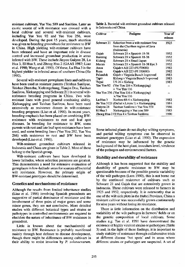
resistant cultivars, Yue You 589 and Sueitien. Later an exotic source of wilt resistance was crossed with a local cultivar and several wilt-resistant cultivars, including Yue You 92 and Yue You 256, were developed. During the past 15 years, much attention was given to breeding groundnuts for resistance to BW in China. High yielding wilt-resistant cultivars have been released and have an important role in disease control and increased groundnut production in areas infested with BW. These ine1ude Jinyou Guiyou 28, Lu Hua 3, El Hua 5, and Zhong Hua 2 (GAAS 1987; Liao et al. 1990; Wang et al. 1990). Of these, Lu Hua 3 is the highest yielder in infested areas of southern China (He 1990).
Several wilt-resistant germplasm lines and cultivars have been used as resistant parents (Teishan Sanliyue, Teishan Zhenzhu, Xiekongchung, Tianjin Dou, Taishan Sanlirou, Xiekangqing and Schwarz 21) in several wiltresistance breeding programs in China. Two wiltresistant lines with good general combining ability, Xiekangqing and Taishan Sanlirou, have been used extensively as resistance donors in wilt-resistance breeding programs (Liao et al. 1990). In recent years, breeding emphasis has been placed on combining BWresistance with resistances to rust and leaf spot diseases. In breeding for resistance to BW, some parents with rust and late leaf spot resistance have been used, and some breeding lines (Yue You 202, Yue You 266) with resistance to rust and BW have been developed (Liao et a1. 1990).
Wilt-resistant groundnut cultivars released in Indonesia and China are given in Table 2. Most of these belong to the Spanish group.
Wilt-resistant cultivars have been developed in lower latitudes, where seleetion pressures are greatest. This demonstrates a need for extensive evaluation of germplasm in low-latitude areas for sources of bacterial wilt resistance. However, the primary origin of BW-resistant genotypes should be determined.
Genetics and mechanisms of resistance
Although the results from limited inheritance studies (Liao et al. 1986) involving Spanish groundnuts are suggestive of partial dominance of resistance and the involvement of three pairs of major genes and some minor genes, they are not conclusive. More detailed studies with different botanical types and strains or pathotypes in controlled environments are required to elucidate the nature of inheritance of BW resistance in groundnut.
Little is known about the mechanism(s) of resistance to BW. Resistance is probably manifested mainly through host defence to disease development, though there might be differences among cultivars in their ability to resist invasion by P. solanacearum.
Thble 2. Bacterial wilt-resislant groundnut cultivars released in Indonesia and China.
Cultivar Pedigree Year of release
Schwarz 21 Selection from a wilt-resiSlant line 1925 from the Cheribon region of Java (Indonesia)
Gajah Schwarz21 x Spanish 18-38 1952 Banteng Schwarz21 x Spanish 18-38 1952 Kidang Schwarz 21 x Small Japan 1952 Macan Schwarz 21 x Spanish 18-38 Eyc 3 1952 Anoa Gajah x AH 223 (PI350680) 1982 Rusa Gajah x AH 223 (PI 350680) 1982 Pelanduk Gajah x Virginia Bunch Improved 1983 Tapir Kidang x Virginia Bunch ImproVed 1983 Tupai US 26 x Kidang 1983 YueYou92 (YueYou 116 x Xiekangqing)
x YueYou 116 Yue You 256 (Yue You 116 x Xiekangqing)
x Yue You 116 Lu Hua 3 Xuzhou 68-4 x Xiekangqing 1982 JinYou3121 (Daliaix Linyu 1) x Kiekangqing 1984 Guiyou 28 Taishan Sanlirou x Yue You 551 1986 El Hua 5 Xiekangqing x Yue you 589 1985 Zbong Hua 2 El Hua 4 x Taishan Sanlirou 1990
Some infected plants do not display wilting symptoms, and partial Wilting symptoms can be observed in resistant genotypes (Liao et al. 1990). The expression of resistance may be influenced by the genetic background of the host plant, inoculum level, virulence of the pathogen and environmental factors.
Stability and durability of resistance
Although it has been suggested that the stability and durability of genetic resistance to BW may be questionable because of the possible genetic variability of the wilt pathogen (Lum 1990), this is not borne out by the continued resistance of cultivars such as Schwarz 21 and Gajah that are extensively grown in Indonesia. These cultivars were released to farmers in 1925 and 1952, respectively. It is noteworthy that in one of the wilt sick plots in the Hubei Province, China a resistant cultivar was successfully grown continuously for nine years without losing its resistance.
There is little information on the distribution and variability of the wilt pathogen in farmers' fields or on the genetic composition of local cultivars. Some studies e.g. Tan et al. 1991 have demonstrated the existence of highly virulent strains or pathotypes (Table 3) and, in the light of these findings, it is important to study stability of resistance through collaborative trials at different disease 'hot spots' and in areas where different strdins or pathotypes are suspected. A set of
215

Table 3. Reactions of groundnut isolates of Pseudonwnas solanaceanun on some diagnostic groundnut cultivars".
Cultivars Pathotypes of P. solanacearumb
1 2 3 4 5 6 7
Xiekangqing R R R M M M S Taishan Sanlirou R R M R M M S Huangchuan Zhigan R M M M M M S Lukangqing R M M M M M S Fuhuasheng S S S S S M S Ehual S
aData from Tan et al. (1991); R = Highly resistant « I 0% wilt incidence); M = Moderately resistant (10-50% wilt incidence); and S = Susceptible (> 50% will incidence)
"Tt.irty-six isolates from various parts of China were divided into seven pathotypes based on their virulence to six cultivars with different levels of BW -resistance.
differentials should be included in these trials. Some description of the strains of the pathogen to which these Iineslcultivars are subjected is obviously important.
Durability of wilt-resistance should be ensured by adopting appropriate breeding strategies and by bringing in new resistance genes from diverse sources and wild species.
Impact of wilt-resistant cultivars Releases of wilt-resistant cultivars in Indonesia have significantly reduced yield losses in various parts of the country where disease was a serious problem. Two early bred resistant cultivars, Schwarz 21 and Gajah, are widely grown in Java where the disease previously' caused heavy yield losses (25-45%) in susceptible cultivars. Some wilt-resistant cultivars (Pelanduk, Tupai) developed in the 1980s are now becoming popular with farmers. The disease in farmers' fields is now sporadic because of the cultivation of improved Indonesian cultivars resistant to BW. Wilt-resistant, high-yielding cultivars play an important role in disease control and increased groundnut production in several provinces of China where BW is a production constraint. El Hua 5 is grown in most of the bacterialwilt-affected areas, covering over 10000 ha, in central China (Liao et a!. 1990). Zhong Hua 2, an early maturing wilt-resistant cultivar with wide adaptation, is becoming popular with farmers in southern China. Yue You 92, Lu Hua 3, Jinyou 3121 and Guiyou 28 are grown extensively in Guangdong, Guangxi, Fujian and Shandong provinces. Some recently developed cultivars have multiple resistances to BW and rust and are awaiting release in the near future. They are expected to further increase groundnut production in areas where both BW and foliar fungal diseases are serious.
Cultural Measures
Rotation of groundnuts with non-host crops such as rice, sugarcane, maize and wheat is an effective means of controlling BW of groundnut (Sun et a!. 1981; He 1990). It is possible to minimise and even achieve near complete control of BW following rotation of groundnuts with paddy rice for 3-5 years (Wang et a1. 1983; He 1990). Several groundnut-rice rotation systems are widely adopted in irrigated areas of Guangdong, Fujian, Hubei and Shandong provinces of China where the disease is a production constraint. In Guangdong Province of China, rotation of groundnut with sugarcane for 2-3 years has been found effective in reducing BW incidence from 60% to below 10% (OCRI 1977). In the Hubei Province, rotation of groundnuts with wheat is helpful in reducing wilt incidence in uplands. In drylands, rotation of groundnuts with maize, sorghum and sugarcane for longer periods (4-5 years) is useful in containing the disease. In such areas intercropping of groundnuts and maize can also be used as a means of reducing wilt incidence. Intercropping experiments in the Philippines showed that maize was the most effective of several intercrops in reducing BW disease incidence in potatoes (Kloos et a!. 1987). Little is known as to how these cropping systems affect the perpetuation and survival of the B W pathogen.
Flooding of groundnut fields for 15-30 days before planting effectively reduces BW incidence (OCRI 1977; Li eta!. 1981).
Some soil amendments such as S-H mixture, urea and mineral ash can be useful in reducing BW incidence (Chang and Hsu 1988; Sun and Huang 1985). The SoH mixture is very rich in calcium and silicon, and calcium is likely to enhance host resistance to wilt. Other components of S-H mixture may stimulate soil microbial activity against the wilt pathogen. Further
216

research is required to elucidate mechanisms of BW control following soil amendments.
Crop sanitation (e.g. burning of crop residues and removal of solanaceous weeds, and cleaning of tools and machinery after operations in infested fields) help reduce disease levels.
Concluding Remarks
The cultivation of wilt-resistant groundnut cultivars is highly useful in effective management of bacterial wilt. Over 60 germplasm lines have been identified as resistant to BW, and 20 wilt-resistant cultivars have been developed, mainly in China and Indonesia. It is imperative to broaden the genetic base of wiltresistance by using different parents with high levels of stable resistance, including highly resistant wild Arachis species. International 'bacterial wilt nurseries' should be established to test stability of resistance and pathogen variability. Since rust and leaf spot diseases are severe in many areas where BW is a production constraint, priority should be given to incorporating resistances to bacterial wilt, rust and leaf spots into high-yielding cultivars adapted to specific environments. Further genetic evaluation of BW-resistance, involving several strains or pathotypes, is required to devise strategies for effective use of sources of resistance in breeding programs. Priority should be given to testing of selected resistant lines, including differentials, for their reaction to several strains or pathotypes of the pathogen from different regions of the world. This work should be possible in a country where groundnut is not grown and where P. solanacearum is not a problem. It is necessary to identify the most virulent strains so that they can be used for screening in breeding programs. Research efforts are needed to elucidate the mechanisms and components of wilt-resistance. Some cultural practices such as rotation with rice, maize or sugarcane and crop sanitation are effective in containing the disease. In irrigated areas or relatively high rainfall areas of southern Asia and Southeast Asia, crop rotation with paddy rice is feasible and is effective in reducing disease levels. More information is needed on the effects of different cropping systems and rotations on survival of P. solanacearum, and on the mechanisms involved in decreasing or increasing populations of the pathogen. Although sanitation practices are helpful in reducing disease levels, the impact of crop sanitation practices. particularly management of crop residues and weeds, on the BW pathogen and the disease needs to be demonstrated. The soil solarisation approach to wilt control should be investigated; this may prove effective in humid tropical countries. Emphasis should be placed on an integrated approach to bacterial wilt control, involving wilt-resistant cultivars, rotation with
non-host erops (rice, sugarcane or maize), and crop sanitation.
References
Anon. 1~3. Varietas kacang tanah Rusa dan Anoa. Warta Penelitian dan Pengembangan Pertanian. Departemen Pertanian Republik Indonesia, 5,11.
-1984. Varietas unggul baru kacang tanah dan kacang hijau. Warta Penelitian dan Pengembangan Pertanian. Departemen Pertanian Republik Indonesia, 6, 8.
Chang, M.L. and Hsu, S. T. 1988. Suppression of bacterial wilt of tomato by soil amendments. Plant Protection Bulletin, Taiwan, 30, 349-359.
Duan, L.X. and Li, W.R. 1987. Resources of resistance to bacterial wilt in gennplasm lines in Arachis hypogaea var. hirsuta. In: Proceedings of 1st Chinese Oil Crops Conference, 1-4 April 1987, Wuhan, China.
Duan, L.x., Tan, Y.J., Jiang, H.F. and Sun, D.R. 1991. Progress in evaluating groundnut gennplasm for resistance to bacterial wilt in China. ACIAR Bacterial Wilt Newsletter, 7. 3.
Frank, Z.R., and Krikun, J. 1969. Evaluation of peanut (Arachis hypogaea) varieties for venicillium wilt resistance. Plant Disease Reporter, 53, 744-746.
GAAS (Guangxi Autonomous Regional Academy of Agricultural Sciences, China) 1987. A new high-yielding and brown wilt (Pseudomonas solanacearum) resistant variety of peanut: 'Guiyou 28'. Guangxi Agricultural Science. 6, 8-10.
Hayward, A.C. 1990. Diagnosis, distribution and status of groundnut bacterial wilt. In: Middleton, K.1. and Hayward, A.C., ed .• Bacterial Wilt of Groundnut. ACIAR Proceedings No.31,12-17.
He, L.Y. 1990. Control of bacterial wilt of groundnut in China with emphasis on cultural and biological methods. In: Middleton, K.1. and Hayward, A.C., ed., Bacterial wilt of groundnut. ACIAR Proceedings No. 31,22-25.
Jayasena, K.W. and Rajapaksa, R.H.S. 1990. Present status of groundnut bacterial wilt research in Sri Lanka. In: Middleton, KJ. and Hayward. A.C., ed., Bacterial wilt of groundnut. ACIAR Proceedings No. 31, 52-53.
Jenkins, S.F., Hammons, R.O. and Dukes, P.D. 1966. Disease reaction and symptom expression of seventeen peanut cultivars to bacterial wilt. Plant Disease Reporter. 50, 520-523.
Kloos, I.P., Tulog, B. and Tumapon, A. 1987. Effects of intercropping potato on bacterial wilt. Philippines Agriculture, 70, 83-90.
Li, WR., Chen, C.R. and Xu, Z. Y. 1981. Studies on bacterial wilt of peanut. 11. Investigation of disease epidemiology in east Hubei. Chinese Oil Crops, 1.43-47.
Liao, B.S., Li, W.R. and Sun, D.R. 1986. A study on inheritance of resistance to Pseudomonas soltmacearum E.F. Smith in Arachis hypogaea L. Oil Crops of China, 3, 1-8.
Liao, B.S., Wang, Y.Y., Xi a, X.M., Tang, G.y', Tan, YJ. and Sun, D.R. 1990. Genetic and breeding aspects of resistance to bacterial wilt in groundnut. In: Middleton, K.J. and Hayward, A.C. ed., Bacterial wilt of groundnut. ACIAR Proceedings No. 31,39-43.
217

Lum, K.Y. 1990. Host plant resistance to Pseudomonas solanacearom. In: Middleton, K.1. and Hayward, A.C. ed., Bacterial wilt of groundnut. ACIAR Proceedings No. 31, 18-21.
Machmud, M. 1986. Bacterial wilt in Indonesia. In: Persley, G. J., ed., Bacterial wilt disease in Asia and the South Pacific. ACIAR Proceedings No. 13,30-34. .
Machmud, M. and Lasimin. 1987. Incidence of bacterial wilt on ICRISAT and Indonesian peanut lines. ACIAR Bacterial Wilt Newsletter, 2, 5-7.
Machmud, M. and Middleton, KJ. 1988. Research progress on peanut diseases in Indonesia. n. Bacterial wilt. Presented at the Review Meeting of ACIAR's Food Legume and Oil Seeds Program, Malang, Indonesia, 18-23 ApriI19!!8.
-1990. Progress of research on bacterial wilt of peanut in Indonesia-1989-1990. Presented at the Annual Review of the Collaborative Research between ACIAR and CRIFC on Peanut Improvement in Indonesia, Malang, Indonesia, 26-28 November 1990.
Mehan, V.K., McDonald, D. and Subrahmanyam, P. 1986. Bacterial wilt of groundnut: control with emphasis on host plant resistance. In: Persley, G.J., ed., Bacterial wilt disease in Asia and the South Pacific. ACIAR Proceedings No. 13, 112-119.
Mehan. V.K., Hong. X.X. and Ly. N.T. 1991. On the identification of pathogens causing the so-called groundnut wilt disease in north Vietnam. In: Improved cultivation technologies for groundnut and other food legumes in Vietnam. Hanoi. Vietnam, Ministry of Agriculture and Food Industry. Vietnam. Agricultural Publishing House, 105-110.
Natural, M.P .• Valencia, L.D. and Pua, A.R. 1988. Peanut-a natural host of Pseudomonas solanacearum in the Philippines. Bacterial Wilt Newsletter. 3, 3.
OCRI (Oil Crops Research Institute, Chinese Academy of Agricultural Science) 1976. An evaluation of peanut sources of resistance to bacterial wilt. Science and Technology of Oil Crops, 3, 13-19.
-1977. A review of bacterial wilt of peanut. Science and Technology of Oil Crops, I. 29-35.
Orian, G. 1949. Division of Plant Pathology, Department of Agriculture, Mauritius, Annual Report 1948.69-74.
Palm, B.T. 1926. Verslag van het Deli Proefstation over 1 Januari 1925-31 December 1925. Deli Proefstation Te Medan, Medendelingen, Series 2, 42, 35 p.
Schwarz, M.B. and Hartley, C. 1950. De selectie van Schwarz 21, een tegen slijmziekte resistente Jijn van Arachis hypogaea. Landbouw (Bogor), 22, 223-244.
Sellschop, J. 1947. Groundnuts. Farming in South Africa, 22, 705-712.
Sharma, D. and Soekamo, B. 1992. Identification of additional groundout sources resistant to bacterial wilt under field conditions in East Java, Indonesia. International Arachis Newsletter, 11,23-25.
Simbwa-Bunnya. M. 1972. Resistance of groundnut varieties to bacterial wilt (Pseudomonas solanacearom) in Uganda. East African Agricultural and Forestry Journal, 37,341-343.
Sun, D.R., Chen, C.R. and Wang, Y.Y. 1981. Resistance evaluation of bacterial wilt (Pseudomonas solanacearum E.F. Smith) of peanut (Arachis hypogaea L.) in the People's Republic of China. Proceedings of American Peanut Research and Education Society, 13.21-28.
Sun. S.K. and Huang. J.W. 1985. Formulated soil amendment for controlling Fusarium wilt and other soil borne diseases. Plant Disease, 69, 917-920.
Subrahmanyam, P., Mehan, V.K .• Nevill, DJ. and McDonald. D. 1980. Research on fungal diseases of groundnut at International Crops Research Institute for the Semi-Arid Tropics. In: Proceedings of the International workshop on groundnuts, 13-17 October 1980. Patancheru, A.P., India, 193-198.
Tan, YJ., Duan, LX, Liao, 8.S., Li, W.R. and Xu, Z. Y. 1991. Research on groundnut bacterial wilt in OCRI, CAAS. Paper presented at the International Workshop on Groundout, 26-30 November 1991, ICRISAT, Patancheru 502324, A.P., India.
Tan YJ. and Liao BS. 1990. General aspects of groundnut bacterial wilt in China. In: Middleton, K.J. and Hayward, A.C., ed., Bacterial wilt of groundnut. ACIAR Proceedings No. 31,44-47.
Wakman, W. 1988. Evaluasi ketahanan varietas kacang tanah terhadap penyakit layu bakteri (Pseudomonas solanacearom). Laporan Hasil Penelitian Penyakit Tanaman 1987/1988. Balittan Maros, 99-106.
Wang, 1.S., Hou, X.Y. and Hu, BJ. 1983. Studies on the control of the bacterial wilt of peanut. Acta Phytophylactica Sinica, 10. 79-84.
Wang, Y.Y., Thang, G.Y., Xia, X.M., Liao, B.S. and Li, J. 1990. Selection of a new groundnut cultivar, Zhong Hua 2 with high resistance to Pseudomonas solanacearom, high yield potential and superior quality. Oil Crops of China, 3, 5-9.
Winstead, N.N. and Kelman. A. 1952. Inoculation techniques for evaluating resistance to Pseudomonas solanacearum. Phytopathology, 42, 628-34.
Xu, Z.Y., Li, W.R., Chen. C.R., Wang, J.S. and Hou, X. Y. 1980. Studies on bacterial wilt of peanut. I: virulence of strains of Pseudomonas solanacearom. Chinese Oil Crops, 2, 29-34.
Yeh. W.L. 1990. A review of bacterial wilt of groundout in Guangdong Province, People's Republic of China. In: Middleton, KJ. and Hayward, A.C., ed., Bacterial wilt of groundout. ACIAR Proceedings No. 31,48-51.
Zbou. L.G. and Liu, C.Z. 1962. A study on bacterial wilt.
218
Bulletin of Guangdong Academy of Agricultural Sciences, 1962, 59--65.

Research on Bacterial Wilt of Groundnut in Vietnam
N.X. Hong* and V.K. Mehant
Abstract
Recent systematic surveys of plant diseases in Vietnam indicate that bacterial wilt of groundnut is an important disease in the northern part of the country. The disease is particularly serious in several important groundnut-growing areas of Nghe Tinh Province. All Pseudomonas solanacearum isolates from wilt-affected groundnut plants from these areas were highly virulent to groundnut at 30-32°C. Most of these isolates were pathogenic to potato, tomato, and tobacco. Preliminary results indicated that the bacterial isolates belonged to race 1. Studies are in progress to determine the biovars of the wilt pathogen collected from various disease-affected areas. Identification of wilt-resistant groundnut germplasm and breeding lines is one of the major research thrusts at the National Institute of Agricultural Sciences. Some local cultivars which appear to be resistant to the disease in other countries were evaluated in bacterial-wilt hot spots in Vietnam and their reactions to the disease are discussed.
GROUNDNUT is the most important food legume crop in Vietnam where over 200000 ha are planted annually. The major groundnut-production areas are Nghe Tinh, Thanh Hoa, and Ha Bac provinces in northern Vietnam, and Tay Ninh, Song Be, Long An, and Dong Nai Provinces in the south. Average pod yield is about 970 kg/ha.
Diseases are major constraints of groundnut production in Vietnam, with bacterial wilt caused by Pseudomonas solanacearum one of the important diseases responsible for groundnut yield losses.
Systematic disease surveys conducted during 1990-1992 indicated that bacterial wilt of groundnut was present in all groundnut-growing provinces of northern Vietnam, but disease incidence varied greatly from place to place. The disease is particularly serious in several important groundnut-growing areas of Nghe Tinh and Thanh Hoa Provinces where plant mortality can be 15-35%. The disease is becoming increasingly important (20-30% plant mortality) in some areas of Long An and Tay Ninh Provinces of southern Vietnam.
* National Institute of Agricultural Sciences (INSA), Hanoi, Vietnam.
tIntemational Crops Research Institute for the Semi-Arid Tropics (ICRISAT). Patancheru 502 324, A.P., India.
Research on bacterial wilt of groundnut is now being conducted mainly by the National Institute of Agricultural Sciences (INS A), Hanoi. This report summarises some of the research at INSA and outlines plans for future research.
Pathogen Isolation and Virulence
All Pseudomonas solanacearum isolates from wiltaffected groundnut plants collected from several disease hot spots in northern Vietnam were highly pathogenic to groundnut at 30-32°C, All isolates tested grew well on sucrose peptone agar (SPA) at 30-35°C. Most of these isolates were virulent to potato, tomato, eggplant and groundnut, but less virulent to tobacco. Extensive research is in progress to determine the biovars of the wilt pathogen collected from various disease-affected areas of northern Vietnam.
Host Resistance
In all groundnut-growing areas of northern Vietnam, the disease progress peaks at podding (65-70 days after sowing). Identification of wilt-resistant groundnut germplasm and breeding lines is one of the major research thrusts at INSA. Nineteen germplasm and breeding lines reported as resistant to bacterial wilt
219

in other countries were evaluated for resistance to Pseudomonas solanacearum in glasshouse tests using a root-inoculation technique. Two-week-old plants were inoculated with inoculum of a 48-hour culture of the bacterium grown on SPA medium. Seven of the 19 lines were highly resistant to isolates of the wilt pathogen tested (Table I). These included four accessions of the cultivar Schwarz 21, a germplasm line (U4-47-7), and a breeding line (ICGV 87206). Two other breeding lines, ICGV 88271 and ICGV 88274, were moderately resistant to bacterial wilt. These breeding lines possess resistance to rust and late leaf-spot diseases. The cultivars Gajah, Matjan, and Kidang showed considerable wilt incidence. There were no marked differences between the isolates for wilt incidence in different cultivarsllines tested.
Table 2. Percentage of wilted plants of 20 groundnut germplasm and breeding lines to root inoculation with two isolates of Pseudomonas solanacearum.
Cultivarlline
ICGIICGV No."
ICG 1703
ICG 1704
ICG 1705 ICG7893 ICG7894
ICG5272
ICG5273 ICG5276 ICG 7343
ICG7968
ICG 1609
ICG8666
ICG 11210
ICG 3263
ICGV 87165 ICGV87206
ICGV88252
ICGV 88271 ICGV88274
Identity
NCAc 17127
NCAc 17129
NCAc 17130 PI 393531
PI 393641
Gajah
Matjan Kidang Schwarz21
Schwarz 21
Schwarz 21
Schwarz 21
Kidang
U 4-47-7
Son Nghe Anb
Wilt intensity (%) P. solanacearum isolates
Ha Bac isolate N.An isolate
12.5 22.5 67.0 55.5 28.5 44.5 50.0 28.5
50.0 50.0
37.5 30.8
50.0 27.3
55.5 57.1 10.0 11.0 11.0 7.8
17.0 8.3
16.6 11.0
40.0 33.3 11.0 10.0
27.0 33.3
12.5 10.0
40.0 33.3
22.0 25.0
28.5 22.0
60.0 62.5
"ICG =ICRISAT Groundnut Accession Number and ICGV = ICRISAT Groundnut Variety Number.
b Local cultivar.
Some local cultivars such as Li and Tram Xuyen appear to be resistant to bacterial wilt under field conditions. Some breeding lines introduced from ICRISAT (e.g., ICGV 87157, ICGV 87206, ICGV 87165) have also been observed to show low incidence of bacterial
wilt in hot spots. Thirty germplasm and breeding lines reported resistant to the disease in other countries are now being evaluated for field resistance to bacterial wilt in disease hot spots.
Future Plans
Intensive surveys will be carried out to assess the distribution and economic importance of bacterial wilt, particularly in southern Vietnam. Future research into stable genetic resistance to bacterial wilt, and incorporation of resistance to other yield-reducing diseases (e.g., leaf spots and rust) will be given high priority.
220

Control of Peanut Bacterial Wilt Through Crop Rotation
M.Machmud*
Abstract
A three-year crop rotation trial was done at Muara Experimental Farm, Bogor, in an effort to control bacterial wilt of peanut caused by PseudolnOlUls solanacearum. Two crops were grown annually: wet season and dry season crops. The rotation treatments consisted of time durations and crop combinations. The durations of rotations were one, two and three years. The crop combinations in each year were: peanut-peanut; peanut-maize; lowland rice-lowland rice, soybean-soybean, maize-soybean, and sweet potato-sweet potato. To eliminate variation due to planting season and crop combination, two trials were done: one was started in the WS 1987-88 and another in the DS 1988. Gajah, a resistant peanut cultivar (cv) was used in the rotation. Susceptible peanut cv. Kelinci and tomato cv. Gondol Hijau were used as indicator plants to assess the wilt intensity in each plot after the rotation. Generally the wilt intensity in plots treated with the different rotations was reduced. Three-year rotations could reduce wilt intensity from 64-12% on peanut cv. Kelinci and from 80-22% on tomato cv. Gondol Hijau. The longer the rotation period, the lower was the wilt intensity. Rotation of peanut with low land or irrigated rice is the most effective in controlling the disease, followed by maize-soybean, maize-peanut, soybean-soybean, sweet potato-sweet potato, and peanut-peanut. A two-year rotation with a non-host crop such as rice or maize would be sufficient to control the disease. However, it needed a longer rotation period when resistant hosts such as sweet potato, soybean, or peanut cv. Gajah were used. This is probably because the resistant hosts could still harbour the pathogen. Crop sanitation from weed hosts, infected plant debris and avoidance of contaminated irrigation water from upstream also need to be attended to during crop rotation.
PEANUT is the second most important food legume in Indonesia. Its annual harvest area is approximately 550000 ha with production of 500000 tlha. About 60% of peanut crops in Indonesia are grown under upland conditions, while the rest are irrigated or lowland crops. Peanuts are grown either as a monoculture or intercropped with other food crops such as maize and cassava.
Bacterial wilt caused by Pseudomonas solanacearum is an important disease of peanut in Indonesia. Since it was first reported by van Breda de Haan in 1905, the disease has been shown to be wide spread in the country. Despite various efforts to control it by Dutch and Indonesian scientists, such as through the development of resistant cultivars and the adoption of different cultural practices, the disease remains a major problem in peanut production (Machmud 1986).
*Bogor Research Institute for Food Crops (BORIF), n., Tentara Pelajar 3a, Bogor 16111, Indonesia.
The effectiveness of crop rotation to control bacterial wilt is not clear-cut (Kelman 1953). In some countries, such as Brazil (Robbs 1960), India (Sohi et al. 1981), Indonesia (Machmud 1986), and China; Wang and Hou 1982), bacterial wilt incidence in the field has been considerably reduced by rotation with irrigated rice or by flooding, but not in Sri Lanka (Seniveratne 1976). More recent reports suggest greater effectiveness of crop rotation in controlling the disease (He 1990; Shekhawat et al. 1988). Most trials reported, however, were done on crops other than peanut.
Except from China (He 1990) and Indonesia (Machmud 1986), there have been very few reports on the control of bacterial wilt of peanut, particularly through crop rotation. This is probably because bacterial wilt of peanut has so far been important only in these countries.
221
This paper reports results of a three-year crop rotation trial using resistant and non-host crops to control bacterial wilt of peanut.

Materials and Methods
The trial on peanut rotation with other food crops was conducted at Muara Experimental Farm, Bogor, in an area known to be heavily infested with P. solanacearum race I. Duration periods of the rotation were I, 2 and 3 years, but the whole experiment took 4 years to complete, from the wet season (WS) of 1987/88 to the dry season (DS) of 1992. Peanut cv. Kelinci and tomato cv. Gondol Hijau, both susceptible to bacterial wilt, were used as standard cheeks or indicator plants to determine the wilt incidence in the field before and after the rotation. The food crops used in the rotation were peanut cv. Gajah (resistant to bacterial wilt), soy bean cv. Orba, maize cv. Arjuna, lowland rice cv. Cisadane. and sweet potato cv. Borobudur. In each rotation period. two crops were grown, one following another. One crop was grown in the wet season (OctoberFebruary). the other in the dry season (April-August). The crop combination patterns were: peanut-peanut; peanut-maize; rice-rice; soybean-soybean; maizesoy bean. and sweet potato-sweet potato. To eliminate variation due to effect of planting season and crop combination, two trials with exactly the same treatments were arranged. One trial was initiated in the WS 1987-88, the other in the WS 1989-90. The two trials were close to each other.
Before laying out the trial, peanut cv. Kelinci and tomato cv. Gondol Hijau were grown in the experimental site to check the intensity of the bacterial wilt and provide a more uniform pathogen distribution. After assessment of the wilt intensity. the plants were ploughed under to spread inoculum of the pathogen uniformly and 2.5 m x 5 m plots were made. Drainage canals and ditches were made between plots to prevent flood or irrigation water running from one plot to another. During the rotation, peanut and soy bean plants were grown in a 40 cm x 15 cm spacing, one plant per hill. Maize. sweet potato, and rice plants were grown in 50 cm x 100 cm, 50 cm x 50 cm, and 20 cm x 20 cm spacings, respectively. The rice plants were grown under irrigated or lowland condition. Crop treatments, such as fertiliser application, weeding. and insect control were done accordingly. After harvesting the first crop, the land was cleared and prepared for the second crop. After the second crop. 100 seeds of peanut cv. Kelinci and } 00 seedlings of tomato cv. Gondo} Hijau were grown alternately in each of the plots with one-year rotation treatment. Bacterial wilt incidence in these plots was assessed from the wilt intensities on both indicator plants four weeks after planting. For the two- or three- year rotation treatments, after harvest of the second crop, the plots were subsequently grown with the second-year rotation crops. The indicator plants were grown again after every rotation period.
The experimental design of each trial involved a split plot with two replications. The main plots were the periods of rotations and the sub plots the crop combinations.
Results and Discussion
The field which was used for the rotation trial had been planted continuously for 2-3 years with peanuts as a monoculture or intercropped with other food crops. The bacterial wilt intensities on susceptible peanut cultivars. such as Kelinci and Landak. were always high (60-70%). When the field was grown with the susceptible indicator hosts, peanut cv. Ke1inci and tomato cv. Gondol Hijau. before the rotation trial, the wilt intensities were 64% and 80%. respectively. During the trial, the wilt intensities on the indicator hosts grown in plots with no rotation were consistently high, ranging from 60-66% on cv. Kelinci and 80-83% on cv. Gondol Hijau. The data also indicated that, under the Muara Experimental Farm's conditions, the season the crop rotation was started did not affect wilt intensity in the field (Table I). This is probably because agroclimatic conditions in this area did not vary significantly, particularly with regard to seasonal rainfall and soil relative humidity, and were conducive to development of the disease.
After the rotations with the different crops and crop combinations, including that with resistant peanut cv. Gajah, bacterial wilt incidence in each plot was reduced. The wilt reduction varied with the crop combinations used in the rotation as well as with the duration of the rotation (Table 1). However, the wilt intensities in all plots treated with the different rotation combinations were continuously reduced when the rotation period was lengthened from 1-3 years. After 1 year of rotation, the wilt intensities on peanut cv. Kelinci were reduced from an average of 60% to a range from 58-33%. The reduction was highest when the rotation was with lowland rice, followed by rotation with maizesoybean, peanut-maize. soybean-soybean, peanutpeanut. and sweet potato-sweet potato. In the second year, the wilt intensities ranged from 21 % on plots with lowland rice combination, to 47% on plots with peanut cv. Gajah or sweet potato combination. After the third year of rotation, the level of wilt intensities on plots with lowland rice combination was still the lowest (Table 1). The ranking of effectiveness of the crop combinations used in the rotation in reducing the bacterial wilt intensities were, in order, lowland rice. followed by maize, soy bean. resistant peanut, and swect potato.
The variabilities of effectiveness in reducing the bacterial wilt intensities were probably due to inability of the bacterium to multiply and infect the plants. Granada and Sequeira (1983), however, stated that a
222

Table 1. Bacterial wilt intensities on susceptible peanut cv. Kelinci and tomato cv. Gondol Hijau after one- to three- year rotations with resistant or non-host crops: Muara Experimental Farm, Bogor, 1987-1991.
Bacterial wilt intensity (%)a
Peanut Tomato
Rotation combination 2 3 2 3
Rotation started in the wet season 1987-88 Peanut-peanut 56 47 38 70 56 48 Peanut-maize 48 40 28 68 48 42 Maize-soybean 44 31 23 56 42 30 Soybean-soybean 50 36 26 62 46 37 Rice-rice 33 21 12 45 33 22 Sweet potato-sweet potato 58 47 42 71 60 54
Average 48 37 28 62 48 39 Check plotsb 66 62 65 83 80 82
Rotation started in the dry season 1988 Peanut-peanut 58 46 40 68 58 52 Peanut-maize 50 42 30 62 50 40
Maize-soybean 46 32 25 58 44 34 Soybean-soybean 51 38 27 60 50 38 Rice-rice 36 24 14 42 34 24
Sweet potato-sweet potato 54 44 38 70 56 50 Average 49 38 29 60 49 40
Check 64 60 62 81 82 80
• Assessments of bacterial wilt were made before and after the rotation. I, 2 and 3 periods of rotation (in year). bCheck plots were plots which are continously grown with susceptible peanut cv. Kelinci.
particular crop may be a non-host to P. solanacearum, but susceptible in the presence of interacting nematodes. Such a phenomenon may overide any effect on pathogen suppression. This might have happened when sweet potato, soy bean or peanut cv. Gajah was used in the rotation. In Vietnam, however, soy bean has been reported susceptible to bacterial wilt (Tung 1986). He (1990) also reported from China that the swect potato strains of the bacterium were able to infect groundnut and those from groundnuts could infect sweet potato.
Symptomless infection could occur on resistant hosts or nonhost plants such as weeds. This has been shown on alligator weed, Portulaca Qieracea (Hayward and Moffett 1978). It is therefore important to carry out crop sanitation during rotations.
The effectiveness of peanut rotation with lowland rice confirmed previous reports from Indonesia (Mach mud 1986) and China (He 1990; Wang and Hou 1982). He (1990) reported that flooding of groundnut fields for 15 days before planting was effective in reducing bacterial wilt incidence. Other monocots that have been used successfully in controlling bacterial wilt by crop rotation are wheat and barley (He 1990; Shekawat et al. 1988), sugarcane and sorghum (He
1990; Wang and Hou 1982). Apart from the fact that they are non-hosts to the bacterium, monocot crops are generalIy also highly competitive against weeds that may act as alternate hosts for P. solanacearum or nematodes. Non-host dicots that have been reported to reduce bacterial wilt intensity are cowpea, cabbage, okra and radish (Sohi et at. 1981), particularly on tomato and eggplant.
The optimum timeJor rotation may also be affected by several interacting factors, such as strains of the pathogen present, environmental conditions and soil types. Under Muara Experimental Farm conditions, at least a two-year rotation with lowland rice is necessary to significantly reduce the effect of bacterial wilt. In China, two- to five- year periods of rotation are generally needed for an adequate reduction of baeterial wilt caused by race I (He 1990).
Conclusion
Rotation of a susceptible peanut cv. Kelinci with resistant peanut cv. Gajah, maize, lowland rice, soybean, or sweet potato reduced wilt intensities in the field. Peanut rotation with irrigated lowland rice is the most effective way of controlling bacterial wilt,
223

followed by rotation with maize, soybean, resistant peanut, and sweet potato. A longer rotation period of from one to three years, resulted in a lower wilt intensity. Two-year rotation of peanut with lowland rice is generally sufficient to reduce the disease considerably.
Acknowledgment
This work has been done as a part of the AARDACIAR collaborative research program between the Agency for Agricultural Research and Development (AARD), Indonesia, and the Australian Centre for International Agricultural Research (ACIAR) through ACIAR Project No. 8834 (Strengthening of peanut production in Indonesia). The author wishes to acknowledge the valuable suggestions and encouragement of Mr Keith Middleton, Queensland Department of Primary Industries, Australia.
References
Granada G.A. and Sequeira L. 1983. Survival of Pseudomonas solanacearum in soil, rhizosphere, and plant roots. Canadian Journal of Microbiology, 29, 433-440.
Hayward, A.C. and Moffett, M.L. 1978. The role of weed species in the survival of Pseudomonas solanacearum in tomato cropping land. Phytopathology, 68, 2-6.
He, L. Y. 1990. Control of bacterial wilt of groundnut in China with emphasis on cultural and biological methods. In: Middleton, KJ. and Hayward, A.C. ed., Bacterial wilt of groundnut. ACIAR Proceedings No. 31, 22-25.
Kelman, A. 1953. The bacterial wilt caused by Pseudomonas solanacearum. A literature review and bibliography. North Carolina Agricultural Experiment Station Technical Bulletin 99. 194p.
Macbmud, M. 1986. Bacterial wilt in Indonesia. In: Persley, G.J., ed., Bacterial wilt disease in Asia and the South Pacific. ACIAR Proceedings No. 13,32-34.
Robbs, C.F. 1960. Inlluencia da rotacao de cultura na incidencis da murcha bacteriana de tomateiro (P. solanacearum). In: Bacterioses Fitopatogenicas no Brasil. Serie Divulgacao de Pesquisas No. 2. University Rural Itaguai, RJ.Brazil,l13-120.
Seneviratne, S.N. de S. 1976. In: Sequeira, L., and Kelman, A., ed., Proceedings of the First International Planning Conference and Workshop on the Ecology and Control of Bacterial Wilt caused by Pseudomonas solanacearum. Raleigh, North Carolina, July 18-24, 1976.
Shekhawal, G.S., Oadewar, A.V., Baltal,Y.K and Verma, R.K 1988. Cultural practices for managing bacterial wilt of potatoes. In: Report of Planning Conference on Bacterial Diseases of the Potato 1987. International Potato Center (Crp), Lima, Peru, 65-84.
Sohi, H.S., Rao, M.V.B., Rawal, RD. and Kishun, R. 1981. Effect of crop rotation on bacterial wilt of tomato and eggplant. Indian Journal of Agricultural Sciences, 51, 572-573.
Tung, P.X. 1986. Bacterial wilt in Vietnam. In: Persley, O.J., ed., Bacterial wilt disease in Asia and the South Pacific. ACIAR Proceedings No. 13.68-70.
Wang, J. and Hou, X. 1982. A study on control of bacterial wilt of peanut. Agriculture Science and Technology in Lingyi, I, 1-11.
224

Bacterial Wilt of Groundnuts in Malaysia
s. Hamidah* and K.Y. Lumt
Abstract
Local production of groundnuts in Peninsular Malaysia has declined from some 20000 t annually in the 19808 to 5000 t currently, primarily as a result of a reduction in planted areas. The two major production areas are in the States of Kelantan and Terengganu on the east coast of Peninsular Malaysia, where the incidence of bacterial wilt on the crop has remained high. Information on bacterial wilt of groundnuts is being gathered through collection and identification of isolates from plants. Results from a resistance screening program involving ICRISA T accessions and Indonesian and lncal varieties have shown that selected lines do not perform consistently when subjected to field trials under local situations. These observations, together with laboratory characterisation of some bacterial wilt isolates, suggest the existence of different local pathogenic strains.
BACfERIAL wilt of groundnuts caused by Pseudomonas solanacearum is the only important bacterial disease of groundnuts (Mehan et a1. 1986). The potential threat of the disease is very real, especially in the warm humid regions of the world where groundnuts are grown. In Malaysia, the majority of groundnut farms are smallholdings in Ke1antan (1219 ha), Terengganu (242 ha), Perak (252 ha), and Kedah and Perlis (82 ha). The total area sown to groundnuts declined from 5197 ha in 1980 to 1318 ha in 1986. Total production similarly decreased, from 19437 t to less than 5000 t within the same period. However, the net demand for the commodity is on the increase. In 1986, imports of groundnuts totalled 44871 t with a value of MR 29.7 million, mainly in the form of shelled nuts, with smaller amounts as groundnut oil and oil cake residues for livestock feed. Major exporters to Malaysia are Vietnam. United States, China. Thailand, Taiwan and Hongkong. However, Malaysia also exports groundnuts in the form of canned ~'1d uncanned nuts to a value of MR 5.8 million.
The decline in production is attributed to the high cost of production, particularly for labour, unfavourable
*Division of Horticulture, Malaysian Agricultural Research and Development Institute (MARDI). PO Box 12301.50774 Kuala Lumpur, Malaysia.
tFundamental Research Division, MARDI, PO Box 12301, 50774 Kuala Lumpur, Malaysia.
yield levels and high incidences of bacterial wilt disease in the two major production areas in Kelantan and Terengganu. The pathogen affects a whole range of crops in the country, but information on the pathogen attacking groundnuts is scanty. Schwarz (1926), in rotation trials with groundnuts, tobacco, potato, tomato and eggplant, attributed differing wilt disease incidences to strain differences in the pathogen. Various other reports (McClean 1930; Dowson 1949; Kelman and Person 1961; He et al. 1983) suggest the existence of pathogenic strains. Pathotypes associated with particular geographical locations have also been suggested (Buddenhagen and Kelman 1964; SimbwaBunnya 1972). Tan and Liao (1990) reported evidence that strains of groundnut bacterial wilt differ in their pathogenicity to the same host cultivar in different parts of China.
Materials and Methods
Screening for bacterial wilt resistance
In collaboration with ICRISAT, groundnut lines were screened for resistance to bacterial wilt at a number of localities. Typically, 20 seeds of each groundnut line were planted at 10 cm x 50 cm spacing, together with the susceptible checks. Gajah, Mat jam and MKTl. Recordings for disease incidence as a result of natural infection were taken 4 weeks after planting. Trials were generally replicated thrice and carried out in plots
225

situated in Kelantan and Kedah. In another trial at Serdang, plants were artificially inoculated using the multi-pinprick method with isolates collected from groundnut-growing areas of Kelantan and Terengganu. Percentage of plants wilted was recorded one week after artificial incoulation.
Characterisation of isolates
Isolates of P. solanacearum were identified using GN plates of the BIOLOG microbial identification system (Biolog Inc., Hayward, California, USA.). Four typical groundnut isolates were compared with an isolate from tomato. Isolates GNI-4 were from groundnut and obtained from Pasir Mas, Kelantan, except for GN-4 which came from Serdang, Selangor. The T-I isolate from tomato originated from Serdang, Selangor.
These isolates were also inoculated onto groundnut cultivar MBT 91-2 and disease progression compared. Each isolate was inoculated onto 20 plants, each plant receiving a dose of 50 f.lL of the appropriate bacterial suspension set at a fixed concentration (0.15 O.D. at 590 nm) and delivered with a calibrated micropipette.
Results and Discussion
Screening for bacterial wilt resistance
Screening results of the ICRISAT lines and local checks differed in their response to infection (Table 1).
100
80
~ "0 60 '" (1)
"0
~ 0. '+-< 40 0
0 Z
20
0----...-"-"·
While reliance on natural inoculum meant lower infection percentages, there were differences in the perfonnances of the check cultivars at the two different localities of Kelantan and Kedah. Artificial inoculation caused higher infection percentages, and in some cases were in direct contrast to natural infection results. This reinforces the need for detailed evaluation of host materials over time and locations to determine more precisely the perfonnance of host material.
Characterisation of isolates
The tomato isolate was more virulent on groundnut cultivarlline MBT 91-2 (= ICGU 86188) than the four groundnut isolates (Fig. 1). Tomato isolate caused plants to wilt more rapidly although at 12 days after inoculation more than 88 % of the plants inoculated with either T-I, GN-I or GN-2 had similar levels of wilt. The isolates GN-3 and GN-4 were less virulent than the others.
Using the BIOLOG characterisation, groundnut isolates were classified as 'P.solanacearum A' and the tomato isolate as 'P.solanacearum B'. Comparison of the ninety-five tests which fonned the BIOLOG GN plate system indicated a number of key differences in the pattern of utilisation of substrates between the isolates (Table 2). It was also noted that although there were variations between the four groundnut isolates themselves, the tomato isolate differed from the four in the utilisation pattern for a number of substrates.
5 6 7 9 10 12 13 14 15 16 17 19
Days after inoculation
Fig. 1. Percentage of groundnut plants dead following inoculation with isolates of Pseudomonas solanacearum from groundnut and tomato: •. T-I;O, ON-I; '"", On-2;., On-3; and 0, ON-4.
226

Table 1. Percentage wilt of groundnut lines under natural infection in the field and when artificially inoculated with P. soianacearum.
Cultivarlline Location
Kelantana Kedaha Serdangb
25 0 20
Kelinchi 40 0 75
Macanq 65 5 30
Mat jam 100 30 65
MKT) 80 0 100
ICGU 86309 20 100
ICGU 86188 25 90
ICGU 86199 0 50
ICGU 86243 0 100
ICGU 86330 10 100
ICGU 86302 0 90
IeGU 86708 5 70
ICGU 86745 0 70
rCGU 86635 0 20
ICGU 86680 5 90
ICGU 86743 20 90
ICGU 86303 0 70
ICGU 86699 5 90
ICGU 87281 0 60
ICGU 86691 35 65
ICGU 86315 5 50
ICGU 87237 0 100
ICGU 86707 0 20
ICGU 86310 0 100
• Natural infection. b Artificial inoculation
Acknowledgments The authors would like to thank the Director-General, MARDI for pennission to present the paper. Thanks are also due to Siti Rohani Abu Bakar and Napsiah Abdul Rahim for technical assistance.
References Buddenhagen, lW. and Ke1man, A. 1964. Biological and
physiological aspects of bacterial wilt caused by Pseudomonas so/anacearum. Annual Review of Phytopathology, 2, 203-230.
Dowson, WJ. 1949. Manual of bacterial plant diseases, Adam and Charles Black, 183 p.
He, L. Y., Sequeira, L. and Kelman, A. 1983. Characteristics of strains of Pseudorrumas sola1U1cearum from China. Plant Disease, 67, 1357-136\.
Kelman, A. and Person, L.H. 1961. Strains of Pseudomonas solanacearum differing in pathogenicity to tobacco and peanut. Phytopathology, 51, 158-161.
McClean, A.P.D. 1930. The bacterial wilt disease of peanuts. South African Deptartment of Agriculture Science Bulletin, 87,14.
227
Table 2. Differences in the utilisation of substrates under the BIOLOG GN Plate system for five isolates of P. solanacearum.
Dextrin N-acetyl-Dglucosamine D-galactose M-inositol
Maltose
D-mannose. Psicose
D-sorbitol
Fonnicacid
8-hydroxy-butyric acid
((-hydroxy· butyric acid
T-I
+
Propionic acid +1-
Bromo-succinic acid +1-Succinamic acid
L-alanyl-glycine
D-serine L-threonine
((-amino-butyric acid
Urocanic acid
Glycerol
Mono-methyl succinate
Acetic acid
D-gluconic acid
D,L-Iactic acid
Sebacic acid
Glucuronamide
Alaninamide
L-Jeucine
L-pyro-glutamic acid
Inosine
GN-l
+ +/-
+/-
+
+ +
+!-+
+ + + + + + + + +
GN-2 GN-3
+ +1-
+ +1-
+ +
+ +1-+1-
+ +
+!-+ +
+1-
+
+ + +
+1- + + + +
+!- + + + + +
+1- + +1- + + + + + + + + +
GN-4
+
+!+
+1-
+
+
+ +1-+1-+
+1-+
+1-+ + + + + + + +
Mehan, v.K., McDonald, D. and Subrahmanyam, P. 1986. Bacterial wilt of groundnut: control with emphasis on host plant resistance. In: Persley, G.J. ed., Bacterial wilt disease in Asia and the South Pacific. ACIAR Proceedings No. 13, 112-119.
Schwarz, M.B. 1926. De invloed van de voorvrucht op het optreden van slijmziekte (Bacterium solanacearum) in Arachis hypogaea en eenige andere gewassen. Institut Voor Pfankenziekten (Dutch East Indies), Mededelingen 71, 37p. (English summary).
Simbwa-Bunnya, M. 1972. Resistance of groundnut varieties to bacterial wilt (Pseudomonas sola1U1cearum) in Uganda. East African Agriculture and Forestry Journal, 37,341-343,
Tan Yujun and Liao Boshou 1990. General aspects of groundnut bacterial wilt in China. In: Middleton, KJ. and Hayward, A.c., ed., Bacterial wilt of groundnut. ACIAR Proceedings No. 31,44-47.

Breeding for Resistance to Bacterial Wilt of Groundnuts in Uganda
C.M. Busolo-Bulafu*
Abstract
While groundnut (Arachis hypogaea) is the second most widely grown grain legume in Uganda, various factors have lowered productivity of groundnut crops by interfering with plant growth and development. One of the diseases gaining in importance on groundnuts in Uganda is bacterial wilt caused by Pseudomonas solanacearum. Research efforts have emphasised breeding for resistance to the disease and preliminary results have identified a number of promising genotypes including RMP-12, AT4 74/3/5/4, AT5 138121313. AT5 13916/4/1 and A T4 7413135/3.
BACfERIAL wilt disease caused by Pseudomonas solanacearum E.F. Smith has been observed in some areas of central (Lake Crescent) and northwestern Uganda. The disease has undoubtedly been present on groundnuts in the country for many years. It was first diagnosed on groundnuts in Uganda at Bukalasa in 1938 by Hansford and reported to have caused 10% loss of the crop. Simbwa-Bunnya (1972) reported up to 60% losses in 1963. Bacterial wilt is known to attack several other crops in Uganda, including tomatoes, tobacco, eggplants, and potatoes.
Groundnut cultivars differ in their reaction to the disease. The Red Valencia type cultivars are highly susceptible. These are the varieties preferred by the great majority of the people in Uganda, especially in the Central Region. In general, the spreading types appear to be less susceptible. The disease is particularly associated with partially waterlogged soils and the causal bacterium seems to be spread on the surface of water.
It has been suggested that the disease may be seed borne in groundnuts and, for this reason, it has been departmental policy to discourage the distribution of any seed for planting from areas where the disease is known to occur in Uganda. Nevertheless, because of free movement of groundnut seed by traders to various markets in the country, the disease could be of wider distribution than is presently known.
*Groundnut Breeder. Namulonge Research Station, PO Box 7084, Kampala, Uganda.
Control of bacterial wilt has been quite difficult due to the fact that it is soilborne, thus making chemical control ineffective. Other control methods, such as crop rotation, biological control and soil amendments, have been tried with limited success in some countries. Breeding for disease resistance is likely to be the most effective means of control, at least in Uganda.
Several sources of resistance are available and these include RMP-12, AT4 74/3/5/4, AT5 261/5/2/8, AT4 74/315/3 and AT4 107/21413. Some hybridisation work was carried out in 1973 to incorporate resistance into the popular but susceptible varieties. Screening work on some gerrnplasm, including those mentioned above was reactivated at Namulonge Research Station recently. This screening is currently being carried out in the field, but glasshouse screening using artificial inoculation is also being considered.
Materials and Methods
The varieties were evaluated at Namulonge Research Station and Bukalasa Variety Trial Centre (VTC) under field conditions during the first rainy season of 1991. Variety B I was included as the susceptible check and Roxo as the commercial variety. The experimental design was the randomised complete block design (ReBD) with five replications. Each plot consisted of six rows, 5 m long and 60 cm apart. Observations were made on the wilted plants commencing 2-3 weeks after emergence and their wilt intensities counted. Yield data were also collected.
228

'Thble 1. Yields (kglha) and wilt count (plantlplot) at two locations in Uganda, 1991.
Code letter Variety Yield of dry pods Wilt count
BukaJasa Namulonge Bukalasa Namulonge
A X3 IS80b 17 20
B BI (Control) 1750e 1830b 38 46
C AT1261/5/2/S 2400bc 21S0a 3 2 D Roxo 2350c 2440a 46 41
E AT4 107/214/3 1740e 1860b 6 5
F AT 74/3/5/4 2440bc I830b 7 7
G AT513S/2/3/3 1980d 19S0b 20 17 H AT5 139/61411 2570a lS30b 16 13 I AT474/3/513 2720a 2030a 4 6 ] RMP-12 2030d 1960b 3 4
CV% IS.5 19.9
"Column means followed by the same letter(s) do not differ significantly at the 5% level of probability (P < 0.05) according to Duncan's Multiple Range Test.
Results and Discussion
Significantly higher yields were obtained from seven lines (A, C, F, G, H, I and J) over the control (B I), while three lines (A, H and I) significantly out yielded the commercial variety, Roxo, at Bukalasa (Table I). At Namulonge only two lines (C and I) had significantly higher yields than B 1. Ratings for bacterial wilt indicated that lines C, I and J were least susceptible, though all the selections were quite resistant. However, the lines have to be tested using standard glasshouse procedures in order to ascertain the resistance levels.
229
Reference
Simbwa-Bunnya, M. 1972. Resistance of groundnut varieties to bacterial wilt (Pseudomonas solanacearum) in Uganda. East African Agricultural and Forestry Journal, 37, 341-343.
Molecular Basis of Virulence and Pathogenicity

Studies of the Hrp Pathogenicity Genes from Pseudomonas solanacearum GMIIOOO
M. Arlat, F. van Gijsegem, S. Genin, C. L. Gougb, C. Zischek, P.A. Barberis and C. Boucber*
Abstract
Hrp genes are essential for the ahility of the Pseudomonas solanacearum strain GMI 1 000 to he pathogenic on tomato and to induce a hypersensitive response (HR) on tohacco. These genes are known to he clustered in a region of about 23 kh of the genome. DNA sequencing of 20 kh of this cluster led to the identification of 19 genes, six of which share homology with essential pathogenicity genes from the animal and human pathogens Yersinia pestis, Y. enterocolitica and Shigella jIexneri. Based on these homologies we predicted that an hrp-dependent HR-inducing factor should he produced in the supematant of P. solanacearum cell cultures. Preliminary experimental data suggest that such a factor does exist as a heaHesistant protein released into the culture medium via an hrp gene encoded secretion machinery. We also report on the DNA homologies existing hetween hrp genes from P. soianacearum, P. syrillgae, Xanthomonas campestris and Erwinia amylovora.
BASED on the economic losses caused worldwide, Pseudomonas solanacearum E.F. Smith is considered one of the most important plant pathogenic bacteria (Hayward 1991). This results not only from the broad geographical distribution of this bacterium which includes tropical and subtropical areas of the globe but also from an unusually wide host range that includes a large number of major agricultural crops. The economic importance of the disease also reflects the limited means of protection currently feasible, these being mostly restricted to certain fanning practices and the use of resistant varieties when available.
The precise identification of the biochemical determinants of pathogenicity for this organism, and definition of their respective roles in disease development, is clearly needed in order to understand the mechanisms underlying the pathogenicity processes. This approach, aimed at a further understanding of the key factors determining plant bacteria interactions, should allow new methods of disease control to be developed by interfering with the nonnal course of the infection process.
*Laboratoire de Biologie Moleculaire des Interactions Plantes-Microorganismes, CNRS-JNRA, Chemin de Borde Rouge, BP 27,31326 Castanet Tolosan cedex, France.
Genetics and molecular biology offer powerful tools for deciphering elementary steps occuring during plant-bacterial interactions. Using this approach, and starting from a wild-type fully pathogenic strain, it is possible to create mutants affected in pathogenicity or in the ability 10 induce a hypersensitive response which differ from the wild-type parent by the inactivation of a singlc gene. This approach allows an unambiguous identification of genes playing an important role in disease development. Once identified, these genes can be cloned (physically isolated from the rest of the genome) and then studied to detennine the biochemical functions they encode and the mechanisms governing regulation of their expression.
In the last decade this approach has been used to identify important pathogenicity genes of P. solanacearum and to re-examine the possible role of certain bacterial compounds, including extracellular hydrolytic enzymes and exopolysaccharides, in disease development. Most of this work has been reviewed recently (Boucher et al. 1992; Denny and Schell 1992).
In our laboratory we have identified a cluster of genes, called hrp, which is required both for disease development on compatible hosts and for the elicitation of the hypersensitive response (HR) on non-host plants (Boucher et al. 1988). These genes have been found in
232

all pathogenic strains studied (Boucher et al. 1988; Cook et a!. 1989). Further characterisation of these genes has been achieved in our laboratory using strain GMIlOOO. This strain induces a typical HR following infiltration into tobacco leaf parenchyma and is fully pathogenic on tomato. Most of its hrp deficient derivatives have lost the HR-inducing activity and have become nonpathogenic on tomato irrespective of the inoculation procedure used and the developmental stage of the inoculated plants. However, mutations occurring in the far right-hand-end of the cluster led to 'leaky' mutants which only induced a weak andlor delayed HR and displayed a weak aggressivity toward tomato. Hrp mutants that were non-pathogenie on tomato did, however, retain the ability to penetrate tomato plants and to colonise the stem. Nevertheless, in these infected plants the bacterial population remained at least two orders of magnitude lower than in plants infected with the wild-type parental strain (Trigalet and Demery 1986).
The hrp genes in strain GMIlOOO are organised in at least six transcription units covering about 23 kilobases (kb) of the genome (Arlat et al. 1992). This region is carried on the megaplasmid of greater than one megabase which has been shown to be present in this strain (Boucher et al. 1986). Previous studies have also established that the majority of hrp genes are not expressed during culture of the bacterium in a peptonecontaining medium but that expression occurs following inoculation on plants or during growth in minimal medium (Arlat et aI. 1992).
Although the hrp region is now physically welldefined, the identification of individual genes within this region and the characterisation of the biochemical and physiological functions encoded by these genes remains to be done. Recent data obtained in our laboratory and reported in this paper contribute significantly to the achievement of this goal.
Identification ofIndividual Hrp Genes within the Cluster
We have recently proposed that the P. solanacearum hrp gene cluster is organised within a minimum of six transcription units (Arlat et at 1992). DNA sequencing of 20 kb spanning the four transcription units located at the left-hand-end of the cluster had led to the identification of 19 open reading frames (ORFs) which have a high coding probability as deduced from codon usage. The relative position of these ORFs within the cluster, together with the proposed nomenclature for the corresponding genes, is presented in Figure 1. This organisation is in agreement with the previously proposed transcriptional organisation of this region (Arlat et al. 1992). Analysis of the amino acid (aa) sequences of the
RI RI RI RI
I
.. ... .. .. 2 4
A BeD E F H I JK N 0 P Q Tu V WX
2kb
Fig. 1. Genetic organisation of the hrp gene cluster: top line is an EcoRl (RI) restriction map of the left end of the cluster. Solid arrows show the individual transcription units. Open arrows at the bottom of the figure represent the individual open reading frames for the putative hrp proteins. When a protein is predicted to have transmembrane domain(s) the corresponding arrow is shaded.
predicted proteins has revealed that 7 of them contain putative transmembrane alpha helix domain(s) and could therefore be located in the membrane (Fig. 1).
HrpB Codes for a Positive Regulator
The hrpB gene has a coding capacity for a 477 aa peptide. This putative peptide shows significant similarity to several procaryotic transcriptional activators (Genin et al. 1992) including the AraC protein of Escherichia coli (Migula) CasteUani and Chalmers (Wall ace et at 1980). the XylS protein of P. putida (Trevisan) Migula (Mermod et al. 1987) and the VirF protein of Yersinia enterocolitica (Schleisfstein and Coleman) Fredericksen (Comelis et al. 1989). The hrpB gene product therefore belongs to a different family of bacterial regulators to that described for the HrpS protein of P. syringae pv. phaseolicola (Burkholder) Young et at. (Grimm and Panopoulos 1989). Genetic evidence demonstrates that during growth in minimal medium, the hrpB gene product actually acts as a positive regulator for the expression of transcription units 2, 3 and 4 of the hrp gene cluster (Fig. 2) (Genin et al. 1992).
In addition, the introduction of hrpB into P. solanacearum using the mu]ticopy plasmid vector pLAFR3 also resulted in an increased level of transcription unit 5 when bacteria were grown under inducing conditions.
233
The hrpB gene also controls the expression of additional genes located to the left of the hrp gene cluster, for which no involvement in plant-bacteria interactions has yet been identified (Genin et al. 1992). Finally, we have established that expression of hrpB is partly self-activated, although dependent on additional regulatory faclor(s).

1450 1485 1454 1459 1462 1475 1447
~Ii!I - .. •
lA lB
1450 1485 1454 1459 1462 1475
MM MM hrf£J
hrp
1492 1399
I I --
1447 1469 1492
Rich medium 3 Iillll!l
4 -Rich medium hrpB
3 kb
1425 1423
I t • .. -
1399 1425 1423
Fig. 2. Regulation of hrp gene expression and flanking regions by hrpB: The top of the figure represents the EcoRI restriction map of the hrp gene cluster and of the left flanking region together with the localisation of the lacZ reporter gene fusions which have been generated using transposon Tn5-B20 (Arlat et al. 1992). The triangle above the map shows the location of the insertion of the omega interposon generating a hrpB mutation. Histograms represent the relative level of expression of each gene fusion under inducing conditions in the wild-type (I) versus a hrpB mutant background (2) and in rich medium in the wild-type (3) versus a hrpB mutant background (4) respectively.
Homologies between Hrp Genes and Genes Governing the Production ofExtracelJular
Pathogenicity Factors in Animal and Human Pathogens
Comparison of the aa sequence of the putative hrp proteins with protein sequences present in various data banks revealed that, in addition to hrp8, at least five other hrp proteins showed clear homology with proteins of the human and animal pathogens Yersinia pestis (Lehmann and Neumann) Van Loghem, Y. enterocolitica and/or Shigella flexneri Castellani and Chalmers (Gough et al. 1992).
• HrpA codes for a putative protein of 568 aa which is 34% identical over its entire length to the YscC protein of Y. enterocolitica (Michiels et al. 1991).
• HrpE codes for a putative 439 aa protein which is 44% identical to the putative Spa47 protein of Shigella flexneri (Venkatesan et al. 1992).
• Hrp/ codes for a putative lipoprotein of 269 aa which is 35% identical over its entire length to
the YscJ lipoprotein of Y. enterocolitica (Michiels et al. 1991).
• HrpO codes for a putative protein of 690 aa which is 43% identical to the LcrD protein from Y. pestis (Piano et al. 1991) and 34% identical over 690 aa to VirH protein of S. flexneri (Sasakawa, unpublished, Gene Bank accession 010999).
• HrpQ codes for a putative protein of 355 aa which is 26% identical over 69 aa to the sequenced part of ORF spa33 of S. flexneri (Venkatesan et al. 1992).
• HrpTcodes for a putative protein of218 aa which is 40% identical to the putative protein encoded by the spa24 gene of S.flexneri (Venkatesan et al. 1992).
Interestingly, Yersinia and Shigella mutants defective for these proteins are nonpathogenic and have lost the ability to release certain extracellular proteins (called Yops for Yersinia or Ipas for Shigella) which act as primary determinants of pathogenicity on host cells (Goguen et al. 1984; Michiels et al. 1991; Halle 1991).
234

It is assumed that, in these organisms, the proteins homologous to hrp proteins are constituents of a secretory machinery specifically involved in the transmembrane transport of Yops and Ipas. Based on these homologies and the fact that most hrp mutants were totally non-pathogenic. we postulated that hrp genes might be involved in the secretion of essential pathogenicity protein(s) which would be directly active on plant cells. The following data tend to support this hypothesis.
P. solanacearum Produces an HR-like Inducing Factor in Culture Supernatants
When the wild-type strain GMIlOOO was grown in conditions that allow hrp gene expression (minimal medium with glutamate as sole carbon source) (Arlat et al. 1992). it was possible to detect an HR-like inducing activity in the non-dialysable fraction (MW> 10000) of a 20-fold concentrate of the supematant. This activity occurred following infiltration into tobacco leaf parenchyma and subsequent incubation for 24-48 hours at 2ST. It resulted in the development of a necrotic lesion restricted to the infiltrated tissues which was highly reminiscent of the reaction induced by live bacteria both with respect to the kinetics of the response and the aspect of the necrotic lesions. Such activity was not found in the supernatant of any of the hrp mutants which mapped in transcription units 1 to 4, thus indicating that this activity was hrp-dependent and not the resu It of a nonspecific toxic effect of the supernatant on plant tissues. This activity was still present in the supematant of mutants mapping in transcription units 5 and 6 which had previously been shown to retain a partial HR-inducing activity.
The activity present in the supernatant of the wildtype strain GMIlOOO is assumed to be due to the presence of a heat stable protein(s), since it was resistant to heating for 8 minutes at lOOT but could be removed by treatment with proteinase K.
Possible Involvement of Hrp Genes in Controlling Secretion of the HR~like Eliciting Factor
Although no HR-like inducing activity could be detected in the supernatant of any of the hrp mutants that mapped in transcription units I to 4, an activity was found in filter-sterilised. sonicated cell Iysates prepared from most of the hrp mutants, tested as well as from the wild-type strain. However, the two hrpB mutants tested which failed to produce a functional hrp positive regulator, had no intracellular activity, indicating again that this activity does not result from a nonspecific toxic effect of the cell lysate, but rather that
is hrp gene specific. Similar to the extracellular activity, the intracellular activity was found to be heat-resistant and sensitive to proteinase K indicating that both intraand extra-cellular activities could be due to the same protein(s) present both within and outside the bacterial cells. If this is true, our data relate in a similar way to Yersinia or Shigella counterparts, as several hrp genes are involved in the transport through the bacterial cell envelopes of extracellular protein(s) which. in the case of P. solanacearum, have a HR-like inducing activity.
The requirement of a functional hrpB gene for the production of the intracellular activity suggests that the structural gene(s) coding for the active compound(s) is part of the hrp gene regulon. However, since all the other mutants which map within the hrp gene cluster had retained an intracellular activity. it is most probable that this gene(s) map(s) outside of the hrp gene cluster.
Hrp Genes from P. solanacearum Share Homology with Hrp Genes from Other
Gram-Negative Plant-Pathogenic Bacteria
We previously established that hrp genes from strain GMI 1 000 have a functional counterpart in all of the 90 virulent strains of P. solanacearum tested, irrespective of their race. biotype, or geographical origin (Boucher et al. 1988; Cook et al. 1989). Similarly, the P. solanacearum hrp gene cluster has been shown to hybridise with genomic DNA from all of eight pathovars of Xanthomonas campestris (Pammel) Dowson tested (Boucher et al. 1987). It has further been demonstrated that in X. campestris these homologous sequences correspond to functional hrp genes required both for pathogenicity and for HR induction (Arlat et al. 1991). Based on functional characteristics, the presence of hrp genes has been established in various pathovars of P. syringae and in Erwinia amylovora (Burrill) Winslow which was reviewed by Willis et aI. (1991). However, it remains to be determined whether certain of these genes were homologous to P. solanacearum hrp genes. Because we have shown that several hrp genes are conserved in animal and human pathogens, this prompted us to investigate whether certain of these conserved genes may also be common to other plant pathogenic bacteria. To this aim, DNA probes consisting of internal fragments of hrpA, hrp/ and hrpO were hybridised against Southern blots of genomic DNA of X. campestris pv. campestris. P. syringae pv. pho.seolicola and E. amylovora, and against the corresponding cloned hrp gene clusters (Gough et a!. 1992). The experimental results established that the conservation of all three llrp genes in X. campestris and the conservation of hrpO in both P. syringae and E. amylovora had occurred.
235

Discussion and Conclusions
We have shown that in P. solanacearum the hrp genes are involved in the production and transport through the bacterial cell envelope of a compound that mimics the ability oflive bacteria to induce an HR following infiltration into tobacco leaves. Although the role of hrp genes in controlling a secretion process remains to be clearly established, the discovery that a plant pathogenic bacterium produces an extracellular signal that is sufficient to induce an HR-like reaction sheds new light on our understanding of plant-pathogen interactions. Preliminary characterisation of this active compound has been achieved. Since it was nondiaIysable and sensitive to proteinase K it was assumed to be a protein. The question remains as to whether the activity observed on tobacco was due to a single faetor, or whether several factors with a similar activity are involved. Purification and biochemical characterisation of the active compounds(s) is under way and should lead to the identification and cloning of the corresponding structural gene(s).
The next step will be to understand the mode of aetion of such eompounds on plant cells. A preliminary goal will be to determine whether only one or several HR-like inducing factors are produced by the bacterium and whether these compounds are individually active on a limited or a large number of plant species. Of particular importance in this respect, will be the knowledge of whether there is a differential effect of these factors on host versus non-host plants.
Having purified and characterised the HR-like inducing factor(s), it will become possible to study their mode of action and to identify the corresponding targets present in the plant.
We have previously reported the conservation of hrp genes between P. solanacearum and X. campestris (Boucher et al. 1988; Arlat et a!. 1991). Here we show that, in addition to the conservation between these two organisms, at least one of these genes is conserved in P. syringae and E. amylovora. Recent reports based on DNA sequence analysis of hrp genes from X. campestris pv. vesicatoria and P. solanacearum established the existence of hrp proteins homologous to the hrpA, hrpE. hrpl and hrpO proteins of P. solanacearum (Fenselau et a1. 1992; Gough et a1. 1992).
Huang et a!. (1992) have also reported on the characterisation of two hrp genes from P. syringae pv. syringae. These genes are homologous to hrpA and hrpO (data not shown). Moreover it has recently been reported (Wei et a!. 1992) that one of the hrp genes of E. amylovora codes for a heat stable protein that induces an HR following infiltration into tobacco leaf parenchyma. The corresponding structural gene has
been sequenced and shown to encode a glycine-rich, 44 kb protein. To date, no gene encoding a homologous protein has been found within the sequenced part of the hrp gene cluster of P. solanacearum. This observation together with (i) the existence of common hrp genes, (ii) the similar size of the hrp gene clusters in these different organisms and (Hi) the similar changes in certain biological traits related to bacteria-plant interaetion and associated with mutations in the hrp genes, indicates that a common core of essential pathogenicity genes is shared among representatives of most Gram-negative plant pathogenic bacteria. This is an indication that, independent of the host-range and of the type of symptoms they can induce (i.e., wilt, necrosis, maceration or chlorosis), most Gramnegative plant pathogenic bacteria have developed a common strategy of plant infection. It is intriguing that this strategy is also shared by certain animal and human pathogens belonging to the genus Yersinia and Shigella. This raises the question as to how the corresponding genes have evolved in order to be present in such taxonomical1y distant organisms.
In addition to these common hrp-encoded pathogenicity functions, it is clear that each individual pathogen must have a set of specific genes controlling functions such as the production of toxins, hydrolytic enzymes or plant growth hormones which are probably directly responsible for the type of symptoms each organism induces on its own host plant(s). Nevertheless, phenotypic expression of these specifie functions appears to be strictly dependent on the presence of a fuctional core of hrp genes.
The identification of a set of common pathogenicity genes among the vast majority of plant pathogenic bacteria and the characterisation of an extracellular HR-like inducing signal released by the bacteria open new perspectives in our understanding of the interactions of pathogens with plants. Moreover, since hrp gcnes are conserved between the individual species under study, it is clear that new information obtained on one model will be readily transposable to the others. Due to this cumulative effect, rapid progress should be made in understanding the basic mechanisms of pathogenicity towards plants. Therefore, one can predict that, in the near future, such knowledge should lead to the conception of new strategies for plant protection.
Acknowledgments
We thank Anne Stanford for reviewing this manuscript. This work was supported by grants from the European Economic Community (BIOT-CT90-0168) and the North Atlantic Treaty Organization ([30]880310/88).
236

References
Arlat, M., Gough, CL., Barber, CE., Boucher C. and Daniels, MJ. 1991. Xanthomonas campestris contains a cluster of Hrp genes related to the larger Hrp cluster of Pseudomonas solanacearum. Molecular Plant-Microbe Interactions, 4, 593-601.
Arlat, M., Gough, CL., Zischek, C., Barberis, P.A., Trigalet, A. and Boucher, CA. 1992. Transcriptional organisation and expression of the large Hrp gene cluster of Pseudomonas solanacearum. Molecular Plant-Microbe Interactions, 5, 187-193.
Boucher, CA, Barberis, P.A. and ArIat, M. 1988. Acridine orange selects for deletion of Hrp genes in all races of Pseudomonas solanacearum. Molecular Plant-Microbe Interactions, 1,282-288.
Boucher, CA., Martinel A., Barberis, P., Alloing, G. and Zischek C 1986. Virulence genes are carried by a megaplasmid of the plant pathogen Pseudomonas solanacearum. Molecular and General Genetics, 205, 270-275.
Boucher, CA., Gough. CL. and Arlat, M. 1992. Molecular genetics of pathogenicity determinants of Pseudomonas solanacearum with special emphasis on Hrp genes. Annual Review ofPhythopathology, 30,443-461.
Boucher, C.A., Van Gijsegem, E, Barberis. P.A., ArIat. M. and Zischek, C. 1987. Pseudomonas solanacearum genes controlling both pathogenicity and hypersensitivity on tobacco are clustered. Journal of Bacteriology. 169. 5626-5632.
Cook. D., Barlow, E. and Sequeira, L. 1989. Genetic diversity of Pseudomonas solanacearum: detection of restriction fragment length polymorphism with DNA probes that specify virulence and the hypersensitive response. Molecular Plant-Microbe Interactions, 2.113-121.
Cornelis, G., Sluiters, C, Lambert de Rouvroit, C. and Michiels, T. 1989. Homology between VirF, the transcriptional activator of the Yersinia virulence regulon, and AraC, the Escherichia coli arabinose operon regulator. Journal of Bacteriology, 171, 254-262.
Denny. T.P. and Schell, M.A. 1992. Virulence and pathogenicity of Pseudomonas solanacearum: genetic and biochemical perspectives. Fourth International Symposium on Biotechnology and Plant Protection. 21-23 October 1991, University of Maryland, College Park, USA. In press.
Fenselau. S., Balbo, I. and Bonas, U. 1992 Determinants of pathogenicity in Xanthomonas campestris pv. vesicatoria are related to proteins in secretion in bacterial pathogens of animals. Molecular Plant-Microbe Interactions. In press.
Genin, S .• Gough, CL., Zischek, C and Boucher C.A. 1992. Evidence that the HrpB encodes a positive regulator of Hrp genes from Pseudomonas solanacearum. Molecular Microbiology. In press.
Goguen, J.D., Yother, 1. and Straley S.C. 1984. Genetic analysis of the low calcium response in Yersinia pestis
237
Mudl(Aplac) insertion mutants. Journal of Bacteriology, 160,842-848.
Gough, CL., Genin, S., Zischek, C. and Boucher, CA. 1992. Hrp genes of Pseudomonas solanacearum are homologous to pathogenicity determinants of animal pathogenic bacteria and are conserved among plant pathogenic bacteria. Molecular Plant Microbe Interactions. In press.
Grimm and Panopoulos, N.J. 1989. The predicted product of a pathogenicity locus of Pseudomonas syringae pv. phaseolicola is homologous to a highly conserved domaln of several procaryotic regulatory proteins. Journal of Bacteriology, 171,5031-5038.
Halle, T.L. 1991. Genetic basis of virulence in Shigella species. Microbiological Reviews, 55, 206-224.
Hayward, A.C 1991. Biology and epidemiology of bacterial wilt caused by Pseruk>monas solanacearum. Annual Review of Phytopathology. 29, 65-87.
Huang, H.C, He, Y.S., Bauer, D.W. and Collmer, A. 1992. Characterization of two Pseudomonas syringae pv. syringae 61 Hrp genes encoding envelope proteins. Book of abstracts Sixth International Symposium on Molecular Plant Microbe Interactions, Seattle, USA, July 11-16, 176. Abstract.
Mermod. N., Ramos, J .L., Bairoch, A. and limmis, K.N. 1987. The xylS gene positive regulator of TOL plasmid pWWO: identification, sequence analysis and over production leading to constitutive expression of meta cleavage operon. Molecular and General Genetics, 207, 349-354.
Michiels, T., Vanooteghem. 1.-C, Lambert de Rouvroit, C., China. B., Gustin, A., Boudry, P., and Comelis, G.R. 1991. Analysis of vi rC, an operon involved in the secretion ofYop proteins by Yersinia enterocolitica. Journal of Bacteriology, 173,4994-5009 .
PIano, G.V., Barve, S.S., and Straley, S.C. 1991. LerD, a membrane-bound regulator of the Yersinia pestis lowcalcium response. JournalofBactenology, 173.7293--7303.
Trigalet, A. and Demery, D. 1986. Invasiveness in tomato plants of Tn5-induced avisulent mutants of Pseudomonas solanacearum. Physiological and Molecular Plant Pathology, 28,423-430.
Venkatesan, M.M., Buysse, J .M. and Oaks, E. V. 1992. Surface presentation of Shigella ftexneri invasion plasmid antigens required the products of the spa locus. Journal of Bacteriology, 174, 1990-2001.
Wallace, R.G .• Lee, N. and Fowler, A.V. 1980. The arae gene of Escherichia coli: transcriptional and translational startpoints and complete nucleotide sequence. Gene, 12, 179-190.
Wei, Z.M., Laby, RJ., Zumoff, CH. ,aver, D.W., He, S.Y .• CoUmer, A. and Beer, S.V. 1992. Harpin. elicitor of the hypersensitive response prnduced by the plant pathogen Envinia amylovora. Science, 257, 85-88.
Willis, DK. Rich, J.J. and Hrabak, E.M. 1991. Hrp genes of phytopathogenic bacteria. Molecular Plant-Microbe Interactions, 4, 132-138.

Analysis of Polygalacturonase as a Component of Bacterial Wilt Disease
C. Alien, L. Simon, M. Atkinson and L. Sequeira*
Abstract
Pseudomonns solanaceanun produces at least three extracellular polygalacturonases (PG). An endo-PG of pI 9.2 (PehA) is produced abundantly when P. solanacearum grows in rich culture media, but has been shown to play only a minor role in disease development. Two exo-PGs of pI 7.2 (PehB) and 4.9 (PehC) were barely detectable when the bacterium was grown in rich culture medium, but were produced at much higher levels when the bacterium was grown in planta. Two mutant strains unable to produce PehB or PehC, respectively, were substantially reduced in virulence relative to the wild type strain. These results suggest that exo-PG, which is induced in planta, is a significant virulence component for P. solanacearum. Expression of the PG structural genes was controlled at the transcriptional level by a positive trans-acting regulatory locus called pehR. While pehR was required for pehA expression above basal levels, pehB mutants still produced exo-PG at about 50% of wild type levels. pehR mutants exhibited reduced virulence similar to that of the exo-PG mutant strains. In addition, pehR mutants exhibited reduced phosphorylation of an 85 kb membrane tyrosine phosphoprotein. DNA sequence analysis of the pehR region indicated that it has homology to several prokaryotic environmentally-responsive twocomponent regulators.
MANY papers about bacterial wilt mention that the physiological basis of wilt is poorly understood. Recently, several research groups have isolated various genes for factors believed to contribute to pathogenesis by Pseudomonas solanacearum. Using the powerful and highly specific tools of mutagenesis, cloning, and sequencing, we have identified many genes whose products or actions are required for normal disease development. Some of these products, such as extracellular polysaccharide (EPS), may be indispensable for disease; however, EPS alone is probably not sufficient to explain wilting.
A generalised model that explains how growth of P. solanacearum in plant xylem vessels causes wilt has not been produced, although groups of non-pathogenic mutants affected in phenotypes ranging from the highly specific (such as altered production of cellulase and endo-polygalacturonase) to thc less defined (such as induction of the hypersensitive response) have been
*Departmenl of Plant Pathology, University of WisconsinMadison, Madison. Wisconsin, 53706, USA.
produced. To construct such a model, the idea of a single factor responsible for bacterial wilt may instead be envisioned as a complex disease aetiology with many significant components.
P. solanacearum produces several extracellular plant-ceIl-waIl-degrading enzymes. These include a cellulase (specifically, endoglucanase) that contributes to pathogenicity (Roberts et al. 1988) and several polygalacturonases (henceforward abbrieviated PG), PO degrades the pectic compounds present in the plant cell wall and middle lamella by a hydrolytic mechanism. There are two types of PG: endo-PGs, which cleave the polygalacturonate polymer internally at random, generating rapid loss of viscosity; and exoPOs, which remove one or a few galacturonate residues at a time from the end of the polymer, generating an increase in reducing sugar ends. The action of the purified enzyme results in tissue maceration, formation of pectin gels that may occlude xylem vessels, and release of oligosaccharides that are believed to signal plant defence responses (Ryan 1987). Our research has focused on the role of PG in wilt and the regulation of PG gene expression.
238

Recently. we have found that the extracellular endoPG encoded by pehA. which predominates when cells multiply in rich culture media, makes only a minor contribution to virulence (Allen et al. 1991). Similar results were obtained by Schell et a!. (1988). A positive trans-acting regulatory locus, pehR, is required for normal PG production. pehR appears to act at the transcriptional level (Alien et at. 1991). Since PG production is induced IOD-fold when P. solanacearum grows in tobacco leaf tissue, pehR may be involved in response to a plant signal (Alien et al. 1991). In the present study, we examined the contribution of exo-PG to bacterial wilt and further characterised the pehR locus using mutagenesis with a transposon-reporter gene construct.
Materials and Methods
Bacterial strains and plasmids We used P. solanacearum wild type strain K60 (race I, biovar I) (Kelman 1954), which is pathogenic on tobacco, tomato. and eggplant, and elicits a hypersensitive response on cucumber. P. solanacearum was cultured at 28°C on Casamino-acid peptone glucose (CPG) plates containing 0.05% 2,3,5,-triphenyl tetrazolium chloride (TZC medium) (Kelman 1954). For broth culture, bacteria were grown in either CPG broth or Boucher's minimal medium (BMM) (Boucher et al. 1985) supplemented with 0.1 % citrate and 0.1 % galacturonate as carbon sources. Plasmids and mutant strains are given in Table L
Table 1. Bacterial strains and plasmids used in this study
DNA manipulations General molecular genetic procedures were carried out as described by Ausubel et at (1989). A pehA mutant was generated by ligating the blunted-ended streptomycin resistance cassette from pUC8-Q (Prentki and Krisch 1984) into the blunted Clal site of pPG3, which interrupts the pehA structural gene (Allen et a!. 1991). The resulting plasmid was electroporated into K60 and marker exchange mutants were selected and confirmed by Southern blot analysis as described by AlIen et a1. (1991). The resulting streptomycin-resistant PehAdeficient mutant strain was called K60-06.
Mutagenesis and screening Transposon Tn5 mutagenesis of strain K60-06 was conducted as described by Xu et al. (1988), using plasmid pSUP2021 as a suicide vector. Kanamycinand streptomycin-resistant exconjugants were screened for altered pectolytic enzyme production on pectinase-detection plates (Reverchon et a!. 1985). Saturation mutagenesis with transposon Tn3-gus was conducted as described by Bonas et a1. (1989).
Enzyme assays PG activity was measured in culture supernatants after strains were grown for 96 hours in BMM supplemented with 0.1 % galacturonate and 0.1 % cilrate. Cleared supernatants were concentrated 20-fold in Centricon-30 microconcentrators (Amieon). Endo-PG activity was determined viscometrically as described
Pseudomonas solanacearom Relevant characteristics· Source
K60 Race 1, biovar 1 wildtype Kelman 1954
K60-06 pehA::!lSmr This study
K7-06 pehA::!l pehB::Tn5 Smf Kmr This study
K14-06 pehA::o.pehC::Tn5 Smf Kmr This study
K7 pehR::Tn3-gus Kmf ApT This study
K12 pehR::Tn3-gus Kmf Apr This study
K18 pehR::Tn3-gus Kmf ApT This study
K35 pehR::Tn3-gus Kmf Apf This study
K71 pehR::Tn3-gus Kmf ApT This study
K82 pehR::Tn3-gus Kmf ApT This study
KII6 pehR::Tn3-gus Kmf ApT This study
KI76 pehR::Tn3-gus Kmr ApT This study
KI90 pehR::Tn3-gus Kmr Apf This study
Plasmids
pLAFR3 Tef Staskawicz et at. 1986
pKH19 pehR+,Tcf AlIen et al. 1991
pPG3 pehA+,Apf Alien et al. 1991
pK7-pKI90 pehR::Tn3-gus Kmf ApfTcf !1Ji~~tllclY ......................................... _--
'Abbreviations: Ap. ampicillin; Km. kanamycin: Srn, streptomycin; Tc, tetracycline.
239

by Keen et at. (1984). Exo-PG activity was measured as reducing sugar ends generated using the method of Nelson (1944). Pectolytic activity was observed in isoelectric focusing gels by the thin-layer substrate overlay method of Reid and Collmer (1985). ~glucuronidase activity produced by the 'gus' (uidA) reporter gene was measured fluorimetricalIy from bacterial sonicates as described by lefferson (1987). To measure expression of pehR in planta, we infused a 108
cfulmL suspension of washed bacteria into tobacco leaves and then sampled ~-glucuronidase activity from five homogenised and sonicated 1 cm2 leaf disks removed at each time point. Leaf disks were removed in parallel, homogenised, and dilution plated to determine the cfulcm2 tobacco leaf.
Pathogenicity assay Three-week-old eggplants (cv. Black Beauty) were injected in the axil of the second true leaf with 50 flL of a 107 cfu suspension of bacteria in water. They were rated daily using a 0-4 disease index, where 0 = no wilting, 1= 1-25% wilted, 2 = 26-50% wilted, 3 = 51-75% wilted, and 4 = 76-100% wilted or dead. For each assay, we used sixteen plants per treatment; assays were repeated at least three times.
In planta growth Growth of P. solanacearum mutants in plant tissue was determined after infiltrating tobacco leaves with bacterial suspensions, as described by Sequeira and Hill (1974).
Results and Discussion
P. solanacearum produces at least three extracellular polygalacturonases When concentrated K60 culture supematants are separated on isoelectric focusing gels, substrate overlays reveal three bands ofpectolytic activity at pIs 9.2,7.2, and 4.9 (data not shown). The pI of 9.2 corresponds to the 56 kd endo-PG encoded by peM (AlIen et al. 1991; Schell et al. 1988); the other two PGs were named PehB (pI =7.2) and PehC (pI =4.9).
PehB and PehC are exo-polygalacturonases Concentrated culture supernatants from strain K60-06, which carries an inactivating insertion in the peM locus, did not contain the pI 9.2 peM endo-PG but still contained the pI 7.2 and 4.9 PehB and PehC PG activities as determined by the isoelectric focusing gel overlay technique. These supernatants were assayed for endo- and exo-PG activity. No endo-PG activity was detected, but the strain produced wild type levels of exo-PG activity (Table 2). This result suggests strongly that PehB and PehC are exo-PGs.
Thble 2. Effect of pehR and pehA mutations on polygalacturonase production.
Strain Endo-PG Exo-PG activity" activity"
K60 (wild type) 100 100 K60-06 (pehA) ndb 100 K176,12,l16,35,19O 10 50 K176, 12, 116,35,190 with pKH19 25 100 K7,71 5 50 K7,71 with pKH19 40 100 K18,82 10 50 K18, 82 with pKH19 20 100
"Endo· and exo-PG activity detennined as decsribed in the text and given as % wild-type.
bnd, none detected.
Obtaining exo-polygalacturonase mutants
Production of PehA by the wild type strain masks exoPG production on pectinase detection plates, so we used a peM mutant strain to screen for exo-PG mutants. Double mutants affected in production of PehA and either PehB or PehC were generated by subjecting strain K60-06 (peM -) to transposon TnS mutagenesis and screening for reduced PG production on pectinase plates. Two mutants, K7-06 and KI4-06 (affected in production of PehB and PehC, respectively), were identified. Cloned Tn5-containing fragments from each mutant were marker-exchanged into wild type strain K60 to generate single exo-PG mutants.
Polygalacturonase mutant strains grow normally in plant tissue
Bacterial strains were infused into tobacco leaves, which were sampled and dilution-plated over time to generate in planta growth curves. Growth of mutants K60-06 (peM), K7-06 (peM, pehB), KI4-06 (peM, pehC), and K71 (pehR) in tobacco leaves was indistinguishable from wild type strain K60. An hrp mutant strain, KDI506, which was included as a control, did not grow in tobacco leaves. Although leaf intercellular space is not the normal route of infection for P. solanacearum, this result does indicate that these mutants do not have the kind of fundamental inability to multiply in plant tissue that characterises hrp mutants. We cannot conclude, however, that PG is not required for growth in planta since all these mutants still produce some PG activity. All strains tested (incuding the hrp mutant) exhibited wild type growth in CPGbroth.
240

Exo-polygaJacturonase mutants are severely affected in virulence
To determine the effects of these mutations on virulence, eggplants were inoculated with each of the single mutants (pehA, pehB, and pehC) and with two double mutants (pehAlpehB and pehAlpehC). The results suggest that PehB and PehC make a more significant contribution to virulence than PehA (Fig. 1). Double mutants lacking PehA and either PehB or PehC were less virulent than the exo-PG mutants alone. This was an unexpected result since purified endo-PG can macerate plant tissue more effectively than exo-PG and is usually considered to be more important for virulence in other plant pathogens. Although generalised tissue disintegration is common in the last stages of bacterial wilt, it is not characteristic of the early and middle periods of disease development. These data suggest that exo-PG may play a more subtle role in the disease. Are the pectic oligomers released by enzyme activity inducing expression of other bacterial virulence genes? Do these oligomers induce expression of plant genes that contribute to symptom development? Are exo-PGs important in early stages of disease development when pectin degradation may help bacteria enter plant roots or move across xylem vessel pit membranes to invade new vascular tissue? To test these hypotheses, we must construct a mutant strain unable to produce any exo-PG activity, as well as a completely PG-negative strain. Further, such strains
-0-- K60 (wildlype)
4 - pehA - pehC -- pehAlpBhC - pehB - pehAlpehB
Or-~-*~~.-~-'~~-r-'--~~
o 4 6 8 IQ
Days post-inoculation
Fig. 1. Virulence on 21-day-old eggplants of various P. solanacearum mutants affected in polygalacturonase production. Fifty microlitres of a bacterial suspension was injected into the axil of the second true leaf; plants were rated daily on a disease index from 0-4. For each virulence assay, 16 plants were inoculated with each strain; each point is the mean of 16 plants. The experiment was replicated three times; a representative experiment is shown.
could be used to determine conclusively whether PG production contributes to virulence or is absolutely required for pathogenicity in P. solanacearum.
Saturation mutagenesis ofthepehR region To better charcterise the positive regulator of PG production, we used transposon Tn3-gus, which carries a promoterless p-glucuronidase reporter gene, to saturate the 4.5 kb EcoRI fragment containing the previously-identified pehR region (Allen et al. 1991). 1Wenty-four separate transposon insertions were selected for further study (Fig. 2). Curiously, although we analysed more than 200 mutants, we did not obtain any carrying the transposon insertion in the opposite orientation from the direction of transcription. We know of no explanation for this phenomenon. Each of the 24 insertions was marker-exchanged into the wild type strain to generate a population of pehR:reporter gene chromosomal mutants.
pehR mutants are substantially affected in virulence
241
Eggplants inoculated with pehR mutant strain K71 developed fewer symptoms at a slower rate than plants inoculated with either wild type strain K60 or the PehA endo-PG mutant K60-06 (Fig. 3). Similar results were obtained with four other pehR mutants. Many plants exhibited a low level of wilting which they eventually outgrew; a few showed no symptoms whatsocver. No such 'escapes', or symptomless plants, were observed when plants were inoculated with the wild type strain. The reduction in virulence of pehR mutants is similar to that observed for the pehAlpehB double mutant discussed above.
The pehR locus affects production of exo-PG and endo-PG differently
All pehR mutants tested produced very little endo-PG activity, ranging from 5-10% of wildtype levels, depending on the region of the transposon insertion. However, all mutants tested still produced about 50% of wild type levels of exo-PG activity (Table 2). This reduction in exo-PG activity could result from lower expression of both pehB and pehC, or could be caused by the complete absence of expression of one isozyme of exo-PG. Preliminary data in the form of isoelectric focusing gels from pehR mutant culture supematants suggested that both isozymes are present at lower levels (data not shown). Reporter gene insertions in each exoPG gene are necessary to confirm this result. There was a small but reproducible difference in the level of endoPG produced by the mutants related to the region of transposon insertion (Table 2). Insertions K21 through K47, in the central region of the DNA fragment, produced about half as much endo-PG as those in the

K82 K161 KG18 ----I
K51 K53
K47 K71 K17 K13
K45 K42 K75
K7 K21
K63
Eeo RI Xho I Eeo RV
Kpn I Cia I
KB5 Sail! Ace I
K183 K35 K92 K116 K30
~~-sall/Aeel
K29
K190K12
K176 K182
Aeel
Eeo RI
Fig. 2. Physical map of the 4.5 kb EcoRI fragment from pKH19. Triangles represent insertions of transposon Tn5; Hags represent insertions of transposon Tn3-gus. The direction of transcription (as determined by reporter gene expression) is indicated by the arrow. No Tn3-gus insertions were obtained in the opposite orientation; all insertions affect polygalacturonase activity levels.
outer regions. 'Complementation' studies with transmerodiploids and DNA sequencing results supported the division of the locus into these three apparent units (c. Alien and L. Sequeira, unpublished data).
The pehR locus probably extends beyond the 4.5 kb Eco RI fragment of pKH19
No strains carrying insertions in the 4.5 kb EcoRI fragment produced normal Icvels of PG; even inser-
4 --0-- wlldtype - pehR -- pehA
>< " "" ,::; 1;l
" 2 &;
cs
0 0 2 4 6 10
Days post~inoculation
Fig. 3. Virulence on 21-day-old eggplants of P. solanacearum mutants affected in pehR or pehA (pI 9.2 endo-polygalacturonase). (See legend to Fig. I.)
tions at the extreme ends of the fragment (K82 and K182) reduced PG production. Furthermore, pKH19, present in trami, was unable to restore wild type levels of endo-PG expression to any pehR mutant, although it did increase endo-PG expression and restore wild type exo-PG production (Table 2). These results suggest that the region of DNA required for normal PG production is larger than the 4.5 kb fragment previously described.
ThepehR locus is not autoregulated
Several regulatory loci in plant-associated bacteria [see, e.g. Schlaman et aI., (1992) and Winans (1992)] have been shown to be autoregulated: that is, the protein product of the regulatory gene can repress its own transcription either directly or indirectly. To determine if pehR regulates itself, we transformed three different pehR::TnJ-gus chromosomal mutants with plasmid pKH19, containing the 4.5 kb EcoRI fragment, and the vector alone, pLAFR3. If the pehR locus were autoregulated, we would expect to see a decrease in expression of pehR-gus fusions in the presence of multiple copies of pKH19. However, flglucuronidase expression in these strains was essentially unaffected by the presence of these plasmids (Table 3). This result is consistent with the hypothesis that genes for several virulence components are controlled by a global regulatory cascade (M.Schell, pers. comm.). Nevertheless, this conclusion must be preliminary since we suspect pKHI9 does not contain the complete pehR locus (see earlier), so it is still possible that the region responsible for autoregulation is not present on this plasmid.
242

Table 3. Effect of multiple copies of pKH 19 on expression of I'chR.
Strain (plasmid)
K35 K35 (pKHI9)91 K35 (pLAFR3) K71 K71 (pKHI9) K71 (pLAFR3)
K7
K1(pKHI9)
K7(pLAFR3)
g /lS activity' (% parent mutant)
100
101
100
89 101 100 93
110
"Resuhs given here arc the means of three experiments
Expression of the pehR locus is increased in minimal medium and in plant tissue
Although l3-gl ucUIonida e aC!J vlty was barely detectable when pehR::TnJ-gus mutant strain were grown in rich culture medium. activity level increased about five-fold when bacteria grew in minimal medium supplemented with ci trate and galacturonic acid and ten-fold when they grew in tobacco leaves (Fig. 4).
3~~.-----------------------------. _____ In planla
______ rlChmedlum
------T-- rmnlmBr medJum
o~ .. ~~~~~--~·--,-~--~ o 20 40 80
Time (ilOIlJ"SI
Fig. 4. Expression of pe/rR::TnJ-gus fusions in bacteria grown under different conditions. Bacteria grown in ePG (rich medium) or Boucher's Minimal Medium supplemented with 0.1 % each citrate and galacluronale (minimal medium) were either quantified by dilution plating or sonicated. Bacteria grown in tobacco leaf tissue were freed from leaf disks by homogenisation and then dilution plated or sonicated. The sonicates were assayed for ~-g l ucuronidase activity lluorimetricalIy as described in the text. p-glucuronidase OClivi ty was normalised to activity produced per cfu.
2
-85 kd
Fig. 5. Reduced in vitro phosphorylation of an 85 kd membrane tyrosine phosphoprotein in a pehR mutant stra in. Purified membrane prote in~ (see Atkinson et al .• 1992) were incubated with ['j'-32PJATP and proteins were resolved by electrophoresis on a 9% SOS-polyacrylamide gel and autoradiographed. Lane I, wild type strain K60; Lane 2. pehR mutant . train K18.
These increases in pehR expression paralleled the apparent induc tion of PG activity described previously. Total PG activity increa ed about ten-fold when bacteria grew in minimal rather than rich medium ; a I DO-fold increase was observed when bacteria grcw in tobacco leaves (Alien et al . 1991). We hypothesise that two phenomena contribute to this resu lt: catabol ite repression of both PG production and pehR expression in the presence of rich media, and induction of these activities in response to a signal present in plant lissuc. Preliminary results suggc ted that the rich medium component re ponsiblc for ca tabol ite repression is glu tamine Lewosz e t al. 1990). The putative plant signal remains unknown. It is s triking that expression of the PG structural genes and their regulatory element appear to be linked : this provides further sUPJX>rt for the existence of an ' upstTeam' regulator controlli ng expression of pehR, whi h in tu rn activates ex pression of pe/rA, pehB. and pehC.
243

Phosphorylation of a tyrosine phosphoprotein is reduced inpehR mutants In prokaryotes, reversible protein phosphorylation often plays an important role in transduction of signals within the cell and across the cell wall (as, for example, in response to an environmental condition) (Winans 1992). We previously described the existence of an unusual prokaryotic tyrosine kinase activity in P. solanacearum (Atkinson et a1. 1992). We have observed that phosphorylation of the kinase's target, an 85 kd membrane tyrosine phosphoprotein, was reduced in membranes from pehR mutant strains (Fig. 5). Since phosphorylation in the mutant membranes was not entirely absent but ranged from 0% to 40% of wild type, we do not believe that pehR mutations interrupt the tyrosine phosphoprotein structural gene. Rather, pehR may regulate expression of this protein or its kinase in addition to the PG structural genes. Isolation and mutagenesis of the phosphoprotein structural gene will allow us to determine what roIe, if any, it plays in bacterial wilt pathogenesis.
Acknowledgments
We would like to thank Jim Bina, Doug Cook, and Jcrzy Lewosz for technical help and useful discussions.
References
Alien, C., Huang, Y. and Sequeira, L. 1991. Cloning of genes affecting polygalacturonase production in Pseudomonas solanacearum. Molecular Plant-Microbe Interactions, 4, 147-154.
Ausubel, F.M., Brent, R., Kingston, R.E., Moore, D.D., Seidman, J.G., Smith, J.A., and Struhl, K. 1989. Current Protocols in Molecular Biology. New York, John Wiley and Sons. Inc.
Atkinson, M., Alien, C. and Sequeira, L. 1992. Tyrosine phosphorylation of a membrane protein from Pseudomonas solanacearum. Journal of Bacteriology, 174,4356--4360.
Bonas, U., Stall, R. and Staksawicz, B. 1989. Genetic and structural characterization of the avirulence gene avrBs3 from Xanthomonas campestris pv. vesicatoria. Molecular and General Genetics, 218,127-136.
Boucher, C.A., Barberis, P.A., Trigalet, A.P. and Demery, D.A. 1985. Transposon mutagenesis of Pseudomonas solanacearum: Isolation of Tn5-induced avirulent mutants. Journal of General Microbiology, 131, 2449-2457.
Jefferson, RA. 1987. Assaying chimeric genes in plants: the GUS gene fusion system. Plant Molecular Biology Reporter, 5,387-405.
Keen, N., Dahlbeck, D., Staskawicz, B. and Belser, W. 1984. Molecular cloning of pectate lyase genes from Erwinia chrysanthemi and their expression in Escherichia coli. Journal of Bacteriology, 159,825--831.
Kelman, A. 1954. The relationship of pathogenicity of Pseudomonas solanacearum to colony appearance on a tetrazolium medium. Phytopathology, 44, 693--695.
Lewosz, 1., Alien, C. and Sequeira, L. 1990. Factors influencing the production of pectinolytic enzymes by Pseudomonas solanacearum. Phytopathology, 80, 1054.
Nelson, N. 1944. A photometric adaptation of the Somogyi method for the determination of glucose. Journal of Biological Chemistry, 153,375-380.
Prentki, P. and Krisch, H.M. 1984. In vitro insertional mutagenesis with a selectable DNA fragment. Gene, 29, 303-313.
Reid, J.L., and Collmer, A. 1985. Activity stain for rapid characterization of pectic enzymes in isoelectric focusing and sodium dodecyl sulphate-polyacrylamide gels. Applied and Environmental Microbiology, 50, 615--622.
Reverchon, S., Hugouvieux-Cotte-Pattat, N. and RobertBaudouy, J. 1985. Cloning of genes encoding pectolytic enzymes from a genomic library of the phytopathogenic bacterium Erwinia chrysanthemi. Gene, 35, 121-130.
Roberts, D.P., Denny, T.P. and Schell, M. A. 1988. Cloning of the egl gene of Pseudomonas solanacearum and analysis of its role in phytopathogenicity. Journal of Bacteriology, 170, 1445-1451.
Ryan, C.A. 1987. Oligosaccharide signalling in plants. Annual Review of Cell Biology, 3,295-317.
Schell, M.A., Roberts, D.P. and Denny, T.P. 1988. Analysis of Pseudomonas solanacearum polygalacturonase encoded by pgLA and its involvement in phytopathogenicity. Journal of Bacteriology, 170,4501-4508.
Schlaman,H.R., Okker, R. and Lugtenberg, B.J. 1992. Regulation of nodulation gene expression by NodD in Rhizobia. Journal of Bacteriology, 174,5177-5182.
Sequeira, L. and Hill, L.M. 1974. Induced resistance in tobacco leaves: the growth of Pseudomonas solanacearum in protected tissues. Physiological Plant Pathology, 4, 447-455.
Staskawicz, B., Dahlbeek, D., Keen, N. and Napoli, C. 1986. Molecular characterization of the cloned avirulence genes from Race 0 and Race 1 of Pseudomonas syringae pv. glycinea. Journal of Bacteriology, 169,5789-5794.
Winans, S.C. 1992. Two-way chemical signalling in Agrobacterium-plant interactions. Microbiological Reviews. 56, 12-31.
Xu, P., Leong, S.A., and Sequeira, L. 1988. Molecular cloning of genes that specify virulence in Pseudomonas solanacearum. Journal of Bacteriology, 170,617-622.
244

One Specific DNA Piece in Pseudomonas solanacearum Affecting Arachis hypogaea
J. Feng, M. Zhang, x. Bai, B. Han, T. Liu, M. Fan, J. Tang, and Q. Ma*
Abstract
We previously reported that a 12.8 kb DNA fragment (in plasmid pGX1252) of Pseudomonas solanacearum strain TI005 (isolated from Arachis hypogaea) could extend the host range of a nonpathogenic strain TI003 to A. hypogaea. The genomic DNAs of 21 P. solanacearum strains, representing strains isolated from nine hosts in different places, were digested with EcoRI, or BamHIIEcoRI and probed with radioactively labeled pGX 1252. It turned out that all these P. solanacearwn strains carried DNA sequences homologous to this 12.8 kb DNA hut showing different RFLP patterns, with which the 21 strains were classified into seven groups. It seemed that all 16 pathogenic strains to A. hypogaea had the 1.9 kh BamHIIEcoRI fragment on the right end part of pGX1252, hut the five nonpathogenic (to A. hypogaea) strains had homologous fragments of different sizes. The 1.9 kh DNA alone could not extend the host range of TI003. More left-flanking sequences were needed for this function. The major homologous fragment of pGXl252 in T2003 was cloned in pGX1415. pGX1415 and pGX1252 were different not only in restriction sites but also in the ability to render T2003 pathogenic to A. hypogaea. No homologous sequences of pGXI252 were found in Rhizobium sp. vigna (Arachis), Pseudomonas moculicola, Pseudomonas cororuIjaciens, Pseudomonas jluorescens, Xanthomonas campestris pv. campestris, Xanthomonas oryzae pv. oryzae, Corynebacterium sepedonicum, Erwinia carotovora subsp. carotovora in our conditions.
MUCH work has shown that dozens of bacterial genes are involved in the interaction between bacteria and their host plants. Among these genes, some are essential to confer what has been called the 'basic compatibility' of the bacterium with its plant partner, some determine the host range of a bacterium and yet others may modulate certain stages in interaction between the bacterium and its host plant (Daniels et al. 1988; Ellingboe 1981; Keen and Staskawicz 1988; Long 1989). Pseudomonas solanacearum E.F. Smith can cause wilt in many economically important crops such as Solanum tuberosum L., Nicotiana tabarum L., Lycopersicon esculentum Mill, Arachis hypogaea L., Capsicum annuum L. and Musa /uInG Lour. throughout the world (Buddenhagen and Kelman 1964). The very extensive host range of P. solanacearum includes several hundred
*Lahoratory of Molecular Genetics, Guangxi Agricultural College, Nanning, Guangxi, 530005, People's Repuhlic of China.
species representing 44 families of plants, and many newly recognised hosts (Hayward 1991). For some P. solanacearum strains the host range is narrow, but with others it is wide.
Molecular analysis of P. solanacearum has demonstrated that factors acting in both negative and positive ways may function in determining its host range (Carney and Denny 1990, 1991; Ma et al. 1988). Carney and Denny (1990) identified an avrA gene from P. solanacearum strain AWl, nonpathogenic to N. tabacum, which restricted the host range of a N. tabacum-pathogenic strain NC252 to exclude N. tabacum. Recently they showed that a 4.2 kb EcoRU BamHI fragment of AWl, where avrA was located, rendered another N. tabacum-pathogenic strain K601 nonpathogenic to N. tabacum (Carney and Denny 1991). However, unlike avr genes acting at the race-cultivar level of other phytopathogenic bacteria, the derivative mutant strain of AWl, in which the avirulence gene had been inactivated by the insertion of Tn5, did not become pathogenic on N. tabacum, suggesting that
245

AWl lacks positive-acting host range genes that are required for pathogenicity on N. tabacum (Camey and Denny 1990, 1991).
We previously described a 12.8 kb EcoRI DNA fragment, carried in plasmid pGX1252, from a P. solanacearum strain 1'2005, which could extend the host range of strain 1'2003 from S. tuberosum (nonpathogenic to A. hypogaea) to include A. hypogaea (Ma et al. 1988). Therefore, it was of interest to know if DNA sequences homologous to this 12.8 kb DNA were present in other P. solanacearum strains isolated from different host plants in different places, in Rhizobium sp. vigna (Arachis) which specifically nodulate A. hypogaea and plants of its cross-inoculation group, and also in other common phytopathogenic bacteria.
Here we present data to show that this 12.8 kb DNA sequence is present and shows RFLP pattern in all the 21 P. solanacearum strains tested, in Pseudomonas syzygii and blood disease bacterium of banana, but not in other common phytopathogenic bacteria. It seems that all the 16 A. hypogaea-pathogenic strains share the 1.9 kb BamHIlEcoRI fragment on the right end part of pGX1252.
Restriction Map ofpGX1252 and Its Two Deletion Mutants
A restriction map of pGX1252 and its two subclones. pGXI401 and pGXI402 is shown in Figure 1. There are no Hindlll and HpaI sites along the 12.8 kb
Table 1. List of strains of Pseudomonas solanacearum used in this study and grouping of these strains according to their hybridising patterns in different digestions with probe pGX1252'
Strains Host' Location" Disease RFLP groups according to digestion withC Source of reference on peanutb EcoRI BamHI EcoRIlBamHI
1'2003 S. tuberosum Yunnan A a He et al. 1983
Ma et al. 1988
P042 S. tuberosum Beijing A a Li-yuanHe
P044 S. tuberosum 'rJ A a Li-yuanHe
P046 S. tuberosum 'rJ A a Li-yuanHe
1'2005 A. hypogaea Huanjiang. Guangxi + 2 B b Heetal. 1983
Ma et a!. 1988
PPs A.hypogaea Shandong + 2 B b Jian-hua Zhang
Pol S. tuberosum Taian, Shandong + 2 B b He eta!. 1983
TbPs N. tabacum Shandong + 2 B b lian-hua Zhang
MPs M.alba Shandong + 2 B b Jian·hua Zhang
T305 S. aromaticum Central Java + 2 B b SJ. Eden-Green
S710 S. aromaticum West Java + 2 C c S.J. Eden-Green
S925 S. aromaticum West Java + 2 D d S.J. Eden-Green
Pel C. annum Liuzhou, Guangxi + 3 E e He eta!. 1983
Tm2 L esculentum Liuzhou, Guangxi + 3 E e He et al. 1983
TPs L esculentum Anhui + 3 E e He-yuan Cheng
Bnl B. nivea Ttantai, Zhejiang + 3 E e Li-yuanHe
ZI Z officinale Zaozhuang,Shandong + 3 E e He etal. 1983
GPsl Z officinale Shandong + 3 E e Jian-hua Zhang
GPs2 Z officinale Anhui + 3 E e He-yuan Cheng
S825 S. aromaticum WeslJava + 3 E e SJ. Eden-Green
S729 S. aromaticum West Java + 4 F f S.l. Eden-Green
B9043 S. aromaticum West Sumatra 5 G g S.J. Eden-Green
aHOSI plant and geographical origin of each strain arc given according 10 previously published infonnation or according to personal infonnation from the source. ~e pathogenicity of all these strains on A. hypogaea is given according to the plant tests carried out in our laboratOf}' using the stem-injection method: - means nonpathogenic to A. hypogaea and + stands for pathogenic toA. hypogaea.
eStrains Ihat show an identical hybridising pattern with a particular digestion share an independent common RFLP group number. Size of hybridising bands (in kb) in all the digestions is: I: 9.4, 3.8, 0.6.2; 12.8.3; 13.7.4; 16.5.5: 16.0,3.4. A; 13.5,10.2,5.6. B: 1.7.8.0.7.6. C: 9.7, 11.9. D: 11.6,8.0, 7.6. E: 1.7.9.9.7.6. F: 1.7. 11.6, 7.6.G: 11.2,7.3. a: 7.0, 3.5, 2.2. b: 1.7.8.0,1.9. c: 9.7,1.9. d: 1.9,8.0,1.9. e: 1.7,9.9.1.9. f: 1.7, 11.6. 1.9. g: 9.5,6.6. 1.8. The fragments read from left to right in B, C, D. E, F, b, c, d. e and fare homologous to the 1.7 kb. 8.0 kb and 1.9 kb DNA fragments of pGX1252 (BamHI! EcoRI digestion). respectively.
d?ambiguous or unknown.
246

sequences. The presence of pGX1252 in twenty-one P. solanacearum strains (Table 1) representing strains isolated in different places from S. tuberosum, C. annuum, L. esculentum, Boehmeria nivea Gaud., Zingiber officinale Rose., A. hypogaea, N. tabacum, Morus alba L. and Syzygium aromaticum Merr. et Perry, were investigated for homology with pGXI252. The pathogenicity of all these strains on A. hypogaea was retested and confirmed by a stem-injection method previously described (Ma et al. 1988) (Table 1). Extraction of genomic DNA, digestion with restriction enzyme, gel electrophoresis, Southern transfer, and separation of DNA fragments from low-melting-point agarose gel were conducted according to the standard procedures (Hopwood et al. 1985; Ma et al. 1988; Maniatis et al. 1982;). Radioactively-Iabeled DNA was prepared by nick-translation (Rigby et al. 1977). Hybridisation was carried out in 3 x saline sodium citrate (SSC), 5 x Denhardt's solution, and 100 !JgImL of denatured salmon sperm DNA at 68°C for 16-20 hours. The filters were washed in a solution of 0.2 x SSC, 0.2% SDS at 68°C for 1 hour then exposed to Fuji X-ray film at -70°C in the presence of intensifying screens.
The EcoRI, BamHI or BamHUEcoRI digested genomic blots of 21 P. solanacearum strains were hybridised with the 12.8 kb DNA of pOX1252. It turned out that variable homologous sequences of this 12.8 kb DNA were present in all the strains tested (Figs. 2-4 and Table I). Digestion with BamHI or BamHU EcoRI was more effective than with EcoRI to produce RFLP. In one sort of digestion, strains sharing identical hybridising band(s) were given an independent common RFLP group number. Thus, these strains might be classified into seven (BamHU
EcoRI digestion) clearly different RFLP groups (Table I). Strains in groups b, c, d, e, andfwere pathogenic to A. hypogaea and had a 1.9 kb homologous DNA fragment of pOX1252 in the genomes, but the nonpathogenic (to A. hypogaea) strains in group a and g did not. This 1.9 kb DNA fragment was confirmed to correspond to the 1.9 kb BamHUEcoRI fragment on the right end part of pOX 1252 (Fig. 1) by probing BamHU EcoRI digested genomic DNAs of A. hypogaea-pathogenic strains with the plasmid of pOX 1402 (Fig. I, Fig. Slane B-N). S710and S925 belonging to group c andd, respectively, did not give the 1.7 kb hybridising band corresponding to the left 1.7 kb BamHI fragment of pOX1252 as strains in group b, e and f did in BamHI or BamHUEcoRI digested hybridisations. Strains from each group of b, c, d, e andf carried DNA fragments of different size homologous to the internal 8.0 kb BamHI fragmentofpGX1252.
These findings might indicate that the DNA sequences on the right end part of pOX 1252 were more conserved than those at the left part of pGXI252 and therefore that the 1.9 kb BamHUEcoRI DNA fragment which was specific to A. hypogaea-pathogenic strains was more important in determining the host range of these strains to include A. hypogaea. However, when pOXl402 (Fig. 1) was introduced into a nonpathogenic derivative strain ofTIOO3, the resultant transconjugant could not wilt A. hypogaea seedlings. Nevertheless, the deletion mutant pGXl401 (Fig. 1) retained the ability of pGX1252 to extend the host range of TI003 to include A. hypogaea. This demonstrated that the 1.9 kb DNA alone was not enough to render the transconjugant of T2003 pathogenic to A. hypogaea and more left-flanking sequences were needed to do so.
r-;;;.;....;;.-J........:..:....:...--I------:-;;'-7-7:...,:. l~ ____ ..l.___!!.:...!.._-, Bg 1 I I '1:;;;---:--;;-....,-------:::=-12=::: . ..;::8--___ ..-::--=;-_--::---" ReoRl pGX1262 ~~~~~------_r8.~0------~~~~~~B~I
5.3 6.9 Ssil -..... -.. ---- ...............
-lkh
1 I
----........ I - .... .,J RBBS
11 11 I I I PIP
1
P
K I
I P
B B R 1
1 I 11 I Bg'PP
I I I I
RBPP R 11 11
pGX1401
pGI14 02
Fig. 1. Restriction map of the 12.8 kb DNA of strain 1'2005 cloned in the EcoRI sites of pLAFRI and its deletion mutants of pGXI401 and pGXI402 (also in pLAFRI). The numbers are fragment sizes in kilobases. Symbols are: E, EcoRI; B. BamHI; Bg, BglII; S. SstI; P, PstI; K, KpnI; X, XhoI.
247
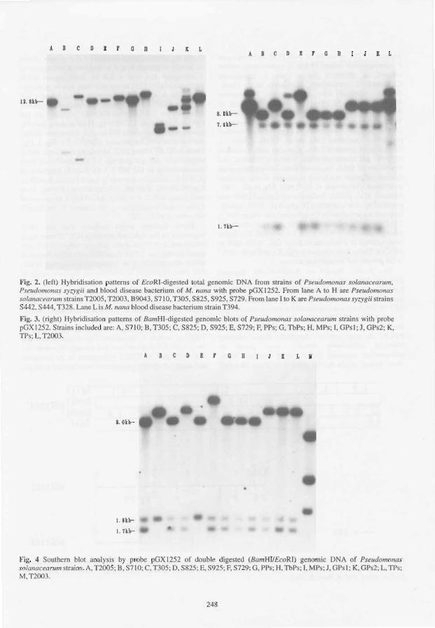
ABCD ! rOR I L
u.n~
8.01lt
l.&k~
I . n~
A B C D I o R I L
Fig. 2. (left) Hybridisalion atlcms of EcoRI-digestcd IOlal genomic DNA from 'trains of Pseudomonas solonaceantm, Pseudomonas sy ygii and blood disease bacterium of M. nallo with probe pGXl252. From lane A to H are PSlmdomonos l'o/anocean/m strains 1'2005, 1'2003.89043, S710. TJ05. S825, S925, S729. From lane IlO K arc Pseudomonas syzygii strains S442, S444. T328. Lane Lis M. milia blood disease bacterium strain TJ94.
Fig. 3. (right) Hybridisation pallems of BamHI-dige ted genomic blot of Pseudomonas solonnceanlln strains with probe pGX 1252. Strains included are: A, 5710; B. T305; C, S825; D. 5925; E, S729; F. PPs; G, Thps; H, MPs; I, GPs I; J. GPs2; K, TPs: L. TI003.
8. 0kb-
t . lkb
I. lkb-
C 11 r Q D I L 11
Fig. 4 Southern blot analysi by pr be pGXI252 of double digested (BamHUEcoRI) genomic DNA I'll' Puudomolla. solallaceaml1lslrains. A. TIOO5; 8.S710; C. TJ05; 0.S825; E.S92S; F. S729 ; G. PPs; H.TbP ; 1. MP ;J, GPsl; K,GPs2; L, TPs; M.T2003.
248
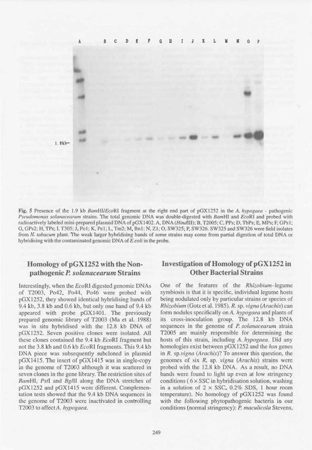
A BCDE 'GB llLlI lI OP
1.9lb-
Fig. 5 Presence of the 1.9 kb BamHlIEcoRI fragment at the right end part of pGXl252 in the A. hypogaea - pathogenic Pseudomonas solol/aceamm strains. The total genomic DNA was double-digested with BamHI and EcoRI and probed with radioactively labeIed mini-prepared pIa mid DNA ofpGX1402. A. DNA (HimlIIl); B. TI005; C, PPs; D, TbP ; E. MPs; F. GPs I; G. GPs2; H. TPs; I. T305; J. Pol; K. PeI; L. Tm2; M. Bnl: N, ZI; 0, SW325; P. SW326. SW325 and SW326 were field isola.tes from N. labacum plant. The weak: larger hybridi ing band~ of some trains may come from partial digestion of total DNA or hybrid ising with the conlaminated genomic DNA of E.coli in the probe.
Homology ofpGXUS2 with the Nonpathogenic P. soulIlacearum Strains
Intere tingly, when the EcoRI dige ted genomic DNA of 1'2003, P042, P044, P046 were probed with pOX 1252, they howed identical hybridi ing bands of 9.4 kb, 3.8 kb and 0.6 kb, but only onc band of 9.4 kb appeared with probe pGXI401. The previously prepared genomic library of 1'2003 (Ma et a!. 1988) was in situ hybridised with the 12.8 kb DNA of pGX 1252. Seven positive clones were isolated. All the e clones contained the 9.4 kb EcoRI fragment but not the 3.8 kb and 0.6 kb EcoRI fragments. This 9.4 kb DNA piece wa ub equenlly 'ubcloned in plasmid pGX1415. The in ert of pGX1415 wa in. ingle-copy in the genome of 1'2003 although it was scallered in seven clones in the gene library. Th restriction sites of Ball/HI, Ps,l and BglII along the DNA lretches of pGX 1252 and pGX 1415 were differen!. ComplemenI ~lion tests showed lhat the 9.4 kb DNA sequences in the genome of 1'2003 were inactivated in controlling 1'200 to affect A. hypogaea.
Investigation of Homology ofpGX12S2 in Other Bacterial Strains
One of the features of the Rhizobium-legume symbiosis i that it is specific, individual legume hosts being nodulated only by particular trains or pecie of Rhizobium (Gotz et a!. 1985). R. sp. vigna (Arachis) an fonn nodules specifically on A. hypogaea and plants of its cross-inoculation group. The 12.8 kb DNA sequences in the genome of P. solanacearum strain 1'2005 are mainly respon ible for detennining the host of this strain, including A. Jrypogaea. Did any homologies ex.ist between pOXI252 and the IIsn genes in R. p.vigna (Arachis)? To an wer thi que tion, the genome of six R. sp. vigna (Arachis) ltain were probed with the 12.8 Icb DNA. As a re ull, no DNA bands were found to light up even at low stringency condition (6 x SSC in hybridi ation solution, wa hing in a solution of 2 x SSC, 0.2% SOS, I hour room temperature). No homology of pGXI252 was found with the following phytopathogenic bacteria in ur conditions (nomlal stringency): P. macll/icola Stevens,
249

P. corona/aciens Stevens, P. fluorescens Migula, Xanthomonas campestris pv. campestris Dowson, Xanthomonas oryzae pv. oryzae, Erwinia carotovora subsp. carotovora Bergey et aI., Corynebacterium sepedonicum. Skaptason et Budmolder. In addition, with pGX1252 as probe, homoiogies were found with three strains of P. syzygii (8442, 8444, T328, provided by Eden-Green), and one strain of blood disease bacterium of M. nana (T394, also provided by EdenGreen) (Fig. 2, lane I-L).
Discussion
We previously reported that some genes determining the hosts of P. solanacearum strain T2005 to include A. hypogaea were carried in a 12.8 kb DNA fragment. A further 20 P. solanacearum strains isolated from different hosts in different places were collected and investigated for homology of pGX1252. Many of the strains, Le. Pol, Pet, Tm2, Bnl, Zl, 8710, T305, 8925, S729 and S825 were primarily considered by the providers to be nonpathogenic or only weakly pathogenic to A. hypogaea (He et al. 1983; S.J. Eden-Green, pers. comm.). However, the pathogenicity tests carried out in our laboratory using the stem-injection method described by Ma et al. (1988) revealed that these 10 strains could cause typical wilting disease on A. hypogaea seedlings. This was confirmed by the hybridisation experiments using pGX1252 as probe. The hybridising patterns of the A. hypogaea-pathogenic strains (according to pathogenicity tests in our laboratory) were clearly distinct from those of nonpathogenic strains. It seemed that all the pathogenic strains had the 1.9 kb BamHllEcoRI fragment located on the right end part of pGX1252 but the nonpathogenic strains had not. This 1.9 kb DNA fragment alone could not extend the host range of T2003 to include A. hypogaea. More left-flanking sequences were needed for this function. Results of the homologies of the 0.7 kb BamHI fragment adjacent to the left of the 1.9 kb BamHUEcoRI fragment and the 0.4 kb BamHI fragment on the left border of pGX 1252 (Fig. 1 ) varied and thus were not considered because even for the original strain T2005 these two small bands could sometimes be detected and sometimes not.
pGXI415 of T2003, homologous to pGXI401 or pGXl252 of T2005. did not have the funetion of pGXl252 or pGXI401 in determining the host range of P. solanacearum strain T2005 to include A. hypogaea. It might be that the DNA sequences of pGXI415 in T2003 were derived from those of pGXi252 in T2005 because of evolution or mutation. The data presented here indicates that DNA sequences on the left part of pGX1252 in the 16 pathogenic strains were more changeable. It is possible that the bases on the right part of or whole pGXI252 will
change further, so that some pathogenic strains will lose the ability to wiltA. hypogaea.
T2003 has at least some essential genes encoding 'basic compatibility' with A. hypogaea because T2003 could affect A. hypogaea by simply obtaining T2005 DNA sequences of pGXI2S2. Anyone P. solanacearum strain can affect a certain number of plants. For each host plant, is any specific factor(s) employed by the bacterium to control its specific interaction with that host plant?
Homologous sequences of pGX1252 were also present in the genomes of P. syzygii, a plant pathogen causing Sumatra disease of S. aromaticum and a pseudomonad that causes blood disease on M. nana. These were in agreement with the previous findings that P. syzygii. the blood disease bacterium of banana and P. solanacearum shared high DNA similarities (Cook et al. 1989; Hayward 1991; Roberts et al. 1990).
The hybridisation data we present here are preliminary because the number of strains employed was rather limited.
Two P. solanacearum strains isolated from N. tabacum during the outbreak of N. tabacum wilt in Yulin of Guangxi in 1990 were probed with pGX1402 and gave the 1.9 kb BamHUEcoRI hybridising band (Fig. 5. lane 0 and P ). In subsequent pathogenicity tests, they wilted A. hypogaea seedlings. We are now sequencing the 1.9 kb BamHUEcoRI fragment. Based on the sequence information, suitable primers may be designed to specifically determine with PCR technique if certain P. solanacearum strains have the potential to affectA. hypogaea. PCR may also be used to detect the viability of P. solanacearum in field collected seed and thus be useful for quarantine activities. The expression and regulation of this gene(s) will also be studied.
Acknowledgments
We thank Li-yuan He, SJ. Eden-Green. lian-hua Zhang, and He-yuan Cheng for providing us with P. solanacearum and other bacterial strains. This work was supported by Rockefeller Foundation Fellowship RF84009 No.7 to Qing-sheng Ma and by grants from the Chinese Academy of Science and Guangxi Science and Technology Committee, China.
References
Buddenhagen, LW. and Kelman, A. 1964. Biological and physiological aspects of bacterial wilt caused by Pseudomonas solanacearum. Annual Review of Phytopathology. 2, 203-230.
earney, B.E and Denny, T.P. 1990. A cloned avirulence gene from Pseudomonas solanacearum determines incompatibility on Nicotiana tabacum at the host species level. Journal of Bacteriology. 172,4836--4843.
250

-1991. Cloning of an avirulence gene from Pseudomonas solanacearum strain AWl and its involvement in host range. Phytopathology, 80, 1156. Abstract.
Cook, D., Barlow, E. and Sequeira L. 1989. Genetic diversity of Pseudomonas solanacearum: detection of restriction fragment length polymorphism with DNA probes that specify virulence and the hypersensitive response. Molecular Plant-Microbe Interactions, 2, 113-121.
Daniels, MJ., Dow, I.M. and Osbourn, A. E. 1988. Molecular genetics of pathogenicity in phytopathogenic bacteria. Annual Review of Phytopathology, 26, 285-312.
ElIingboe, A.H. 1981. Changing concepts in host-pathogen genetics. Annual Review of Phytopathology, 19, 125-43.
Gotz, R, Evans, 1.1., Downie, I.A. and lohnston, A.W.B. 1985. Identification of the host-range DNA which allows Rhizobium leguminosarum strain TOM to nodulate cv. Afghanistan peas. Molecular and General Genetics, 201, 296-300.
Hayward, A.C. 1991. Biology and epidemiology of bacterial wilt caused by Pseudomonas solanacearum. Annual Review of Phytopathology, 29, 65-87.
He, L.v., Sequeira, L. and Kelman, A. 1983. Characteristics of strains of Pseudomonas solanacearum from China. Plant Disease,67,1357-1361.
251
Hopwood, D.A., Bibb, M.I., ChateT, K.F., Kieser, T., Bruton, C,J., Kieser, H.M., Lydiate, DJ., Smith. c.P. and Ward, J. M. 1985. Genetic manipulation of Streptomyces; a laboratory manual. Norwich, England, lohn Innes Foundation.
Keen, N.T. and Staskawicz, B. 1988. Host range detenninants in plant pathogens and symbionts. Annual Review of Microbiology, 42, 421-440.
Long, S.R. 1989. Rhizobium genetics. Annual Review of Genetics, 23, 483-506.
Ma, Q.-S., Chang, M.-F., Tang, J.-L., Feng, I.-X., Fan, M.-I., Han, B. and Liu, T. 1988. Identification of DNA sequences involved in host specificity in the pathogenesis of Pseudomonas solanacearum strain n005. Molecular PlantMicrobe Interactions, I, 169-174.
Maniatis, T., Fritsch, E.F. and Sambrook, I. 1982. Molecular cloning: a laboratory manual. Cold Spring Harbor, New York, Cold Spring Harbor Laboratory, 545 p.
Rigby, P.W.J., Dieckmann, M., Rhodes, C. and Berg, P. 1977. Labeling deoxyribonucleic acid to high specific activity in vitro by nick translation with DNA polymerase I. lournal of Molecular Biology, 113,237-251.
Roberts, S.1., Eden-Green, S.1., lones, P. and Ambler, D.J. 1990. Pseudomonas syzygii, sp. nov., the cause of Sumatra disease of cloves. Systematic and Applied Microbiology, 13, 34-43.

Regulation of virulence in Pseudomonas solanacearum
T.P. Denny, S.M. Brumbley, B.F. Carney, S.J. Clougb, and M.A. Scbell*
Abstract
A complex regulatory network controls production of some extracellular macromolecules that Pseudomonas solanacearum strain AWl needs for full virulence on tomato. Expression of genes for extracellular polysaccharide (EPS) and other virulence factors is coordinately controlled by phcA, since inactivation of this gene results in a pleiotropic change called phenotype conversion (PC). PhcA is likely to be a trans-acting DNA-binding protein because it has strong similarity at the amino acid level to the LysR family of transcriptional activators. At least three different insertions were detected within phcA after spontaneous PC, suggesting that random inactivation of this gene is responsible for this phenomenon. Another locus, phcB, is necessary for production by wild-type P. solanacearum strains of a volatile inducer compound that appears to be required for full activity of PhcA. Cell density, and thus levels ofthe endogenous inducer, appear to mediate expression of a Phc.4-regulated eps::lacZ fusion. Expression of eps genes also depends on three additional loci (xpsR, vsrA. and vsrB), two of which encode membraneassociated proteins and thus may serve as environmental sensors. These findings suggest that current concepts regarding the physiology, ecology, and possible strategies for controlling P. solanacearum should be re-evaluated.
IN the last ten years there has been substantial progress in elucidating the genetic and biochemical bases of pathogenesis of P. solanacearum. For example, the genes encoding several of the putative virulence factors identified in the 1950s were cloned and subsequently inactivated to test their contribution to the typical wilt symptoms. It is now clear that, in planta, the extracellular polysaccharide (EPS) is the primary virulence factor (Denny and Baek 1991; Kao and Sequeira 1992). whereas the extracellular endoglucanase (EG) and en do-polygalacturonase (endo-PG) enzymes. which may act on plant cell walls, are relatively minor virulence factors (Denny et aL 1990). In contrast to these factors that enhance virulence but are dispensable, the hrp genes are essential for pathogenesis on compatible hosts and the hypersensitive response on incompatible hosts (Boucher et al. 1992). Unlike virulence mutants, strains with one or more hrp genes inactivated grow very poorly in planta (Macol 1989;
·Departments of Plant Pathology and Microbiology. 2105 Plant Science Building, University of Georgia, Athens GA 30602,USA.
Trigalet and Demery 1986) and cause no disease symptoms.
Despite these and other significant advances, our understanding of the pathogenic processes of P. solanacearum is still incomplete. Not all of the putative virulence factors have been examined (Denny and Schell 1992) and even those mentioned above have not been tested for their role in other aspects of pathogenesis or saprophytic survival. Even less is known about regulation of genes encoding pathogenesisspecific molecules. This paper will discuss some of our recent research on phenotype conversion in P. solanacearum and the associated regulatory net work that controls virulence.
Phenotype Conversion
Nearly four decades ago Kelman (1954) reported that when P. solanacearum spontaneously changes from a mucoid to a nonmucoid colony morphology there is a concomitant loss of its capacity to wilt plants. These non-mucoid strains are not hrp mutants, because they stilI grow in planta (Denny and Baek 1991) and cause
252

disease symptoms such as stunting, stem necrosis, and proliferation of adventitious roots (nodules) (Denny and Baek 1991; Denny et a1. 1988; Husain and Kelman 1958). Since additional traits are usually affected simultaneously (Brumbley and Denny 1990; Buddenhagen and Kelman 1964), we now call this phenomenon phenotype conversion (PC) and refer to the resulting mutants as PC types.
PC types are easily recognised on standard agar media supplemented with tetrazolium chloride because they are round, butyrous, and dark red, which is in stark contrast to the irregularly-round, mucoid, pink or white parent colonies. This change in colony type is due to loss of one or more components of the EPS (Husain and Kelman 1958; Orgambide et a!. 1991). True PC-type strains also have a coordinate 100-fold reduction in EG activity and are markedly more motile (Brumbley and Denny 1990; Kelman and Hruschka 1973). The decreases in production of EPS and EG are due to reduced transcription of genes that encode these virulence factors and not to mutation or deletion of these genes (Denny and Baek 1991; Huang et a!. 1989; Schell et a!. 1987). In addition, endo-PG activity increases lO-fold and extracellular pectin methylesterase activity decreases about 15-fold (Schell et a!. 1993). Polyacrylamide gel electrophoresis (PAGE) of culture supernatants has shown that additional extracellular proteins are altered by PC (Schell 1987; Schell et al. 1993), but their genes and biochemical functions are unknown.
Large numbers of PC types routinely appear when P. solanacearum is cultured in broth containing glucose and an organic nitrogen source for 5-7 days without shaking, whereas identical cultures that are shaken do not give rise to PC types (Kelman and Hruschka 1973). In contrast to these results, we recently found an inorganic liquid medium that promotes appearance of PC types in shaken as well as unshaken cultures of strain AWL This liquid minimal medium (MM) consists of (per litre); 1.75 g K2HP04,
0.75 g KH2P04, 0.14 g Na citrate-2H20, 26 mg
MgS04-7H20, and 1.25 g (NH4hS04; 0.5% (w/v) glucose is added after autoclaving. The Mg2+ and sodium citrate in MM synergistically enhance growth of wild type strain AWL However,AWI-PC, a PC type derivative of AWl, grows even better in MM than does the wild type, and this difference is enhanced by 4MM, which has four times the standard concentrations of all components except glucose (Table 1). A similar effect was observed when MM was supplemented with either NaCI, KCI, or NH4CI at a final concentration of 0.1 M (approximately equal to the concentration of 4MM). It was presumably this differential growth rate that resulted in the appearance of 4-16% PC types when cultures of AWl in 4MM liquid medium were shaken for 4 days (Table 2). By serially transferring cells to fresh medium each day the number of PC types recovered increased markedly (Table 2), probably because the larger number of generations permitted by this schedule 'amplified' the growth advantage of the PC type cells. PC types also arise during prolonged culture on agar plates and in wilted plants (Buddenhagen and Kelman 1964; Brumbley and Denny, unpublished data), which suggests that there may be natural conditions that favour the appearance or multiplication of PC types.
Table 2. Percentage of P. solanacearum PC types after four days in MM liquid medium with agitation"
Culture conditionsb MM concentration
4 1 114
No transfer 10±6 0.3 0 Three transfers 90±2 50±20 7±O.5
"MM media were as described in Table I. Cultures were started with approximately I x 106 wild type cells/mL. After incubation. serial dilutions were made to determine the percent of PC type colonies; values are the mean for two experiments ± range.
bCultures were incubated for 4 days at 30'C and 250 rpm. One set of cultures was transferred to fresh medium at 24 hour intervals; each subculture was started with approx. I x 106 cells/mL.
Table 1. Growth of P. solanacearum in various MM and BG liquid media with agitation"
Strainb Incubation time
24 hours 48 hours
4MM MM 114MM IIIOBG 4MM MM 114MM IIIOBG
AWl 0.03 0.48 0.32 0.68 0.90 1.55 0.94 0.85 AWl-PC 1.18 1.3 0.68 0.48 >2.0 1.48 0.62 0.74
"MM medium contained 4-, 1- or 1/4-times the standard salt concentration (see text). l/lOBG contained 0.1 % each of peptone. casamino acids, and yeast extract. All media were supplemented with 0.5% glucose after autoclaving. Cultures were started with approx. I x 106 cells/mL, and were incubated at 30°C, 250 rpm. Values are turbidity at 600 nm and are representative of two or more experiments.
bAWl is wild type; AWl-PC is a spontaneous PC type derived from AWl..
253

The Role of PhcA in Phenotype Conversion
Our genetic study of PC began with the isolation of a Tn5-insertion mutant that mimicked a spontaneous PC type (Denny et al. 1988). The Tn5-tagging of a region apparently involved in PC allowed us to isolate a wildtype gene from strain AWl, designated phcA (for phenotype conversion), that restores PC types of AWl to wild type (Brumbley and Denny 1990). Complementation studies and site-directed mutagenesis of 12 other strains of P. solanacearum suggested that PC generally results from the loss of phcA gene expression or the function of the phcA gene product. DNA sequencing of phcA and analysis ofthe deduced amino acid sequence showed that ph cA is similar to members of the LysR family of transcriptional activators and has a putative helix-tum-helix DNA-binding motif (Brumbley 1992; Brumbley and Denny 1991; Henikoff et al. 1988). The greatest similarity was with NahR, from the NAH7 plasmid in Pseudomonas putida. These results suggest that phcA is likely to be a trans-acting, DNA-binding protein that simultaneously regulates transcription of multiple virulence factors. Preliminary data have shown that phcA does bind in vitro to DNA upstream of the xpsR regulatory gene (see next section; Carney and Denny, unpublished data).
An analogous allele, designated phcA 1, was isolated from the spontaneous PC-type strain AWl-PC by using phcA as a hybridisation probe (Brumbley et aI., unpublished data). Sequence analysis of phcAI revealed a two-base insertion near the 3' end, and the resulting frameshift changes the next 24 amino acids and eliminates the last 25 amino acids due to a premature termination of transcription. These changes are apparently sufficient to inactivate phcA. Southern blot analysis of genomic DNA from 10 independently isolated PC types of AWl revealed that only two of the strains had detectable insertions (0.2 and 1.0 kb) in phcA, which suggests that spontaneous PC can be the result of more than one type of mutation in phcA. Thus, it now seems likely that spontaneous PC is often the result of various, random mutational events withinphcA.
Regulation of Virulence by a Complex Network
Although the investigation of phcA gave us our first indication that P. solanacearum can regulate virulence, we have subsequently discovered four additional genes that affect virulence gene expression and hence disease development. One of these loci is phcB, which when inactivated in strain AWI-83 gives a phenotype similar to a spontaneous PC type except for several major differences (Brumbley and Denny 1990). First, phcB is physi-
cally distinct from phcA, which remains functional in AWI-83. Second, unlike a true PC type, AWI-83 is reversibly 'induced' to wild type, EPS+ phenotype when grown in the same Petri dish adjacent to, but not in contact with, a wild-type strain of P. so/anacearum. We now know that this extracellular complementation is due to production by wild type strains of a moderately volatile, water soluble 'endogenous inducer' (El) that AWI-83 does not make (Clough and Denny 1990). The El has not been chemically identified, but several purified compounds, (e.g., methanol and fatty acid methyl esters, but not ethylene), can substitute for the El (Clough 1991; Clough and Denny 1991). Production of the El seems to be another trait regulated by phcA, because PC-type mutants very weakly induceAWI-83.
Based on this and other observations, we believe that phcB is involved in synthesis or modification of an El that is required along with phcA to activate transcription of virulence genes to wild type levels. Thus, P. solanacearum may have an extracellular cell-cell signalling system that regulates virulence that is analogous to the to the cell-density dependent bioluminescence in marine Vibrio species, which depends in part on the concentration of nonvolatile 'autoinducers' (Meighen 1991). Preliminary results with P. solanacearum support this hypothesis, because expression of a phcA-regulated eps::lacZ fusion in a wild-type background was 50-fold less in cells from low density log-phase cultures compared with cells from fully grown log-phase cultures. This type of behaviour means that at low cell density P. solanacearum may have a different phenotype than that which is normally seen at high cell density.
A simpler experiment that may demonstrate intercellular signalling required only spreading serial dilutions of wild type P. solanacearum on medium (without tetrazolium chloride) to give either 1000, 100, or 10 colonies per plate, and then observing carefully during incubation. As the colonies matured, they appeared first as transparent spots visible only by reflected light, but later became opaque and white; we associate this change in colony morphology of the wild type with EPS production, because colonies of a PC type remain transparent even when mature. Surprisingly, opaque white colonies developed sooner, and had IO-fold more cells per colony, on the plate seeded with 1000 cells than at the lower two densities. Comparable plates that had 10 ~ of methanol (i.e., an exogenous inducer) added to the Petri dish lids had opaque white colonies develop sooner at the lower densities than on those plates without methanol. One explanation of these results is that independent colonies act cooperatively to mature more rapidly at high colony density, and that this cooperation may be due to El since the addition of exogenous inducer compensated for low colony density.
254

We have found three more nonallelic loci (vsrA, vsrB, and xpsR) that, when inactivated, give mutants that are deficient in EPS production, probably due to a 25-fold reduction in transcription of eps genes observed for lacZ reporter fusions (Huang et al. unpublished data). Not surprisingly, each of these mutants was markedly reduced in ability to wilt steminoculated tomato plants. PAGE analysis of extracellular proteins showed that inactivation of vsrA, vsrB, or xpsR affects production of some of the extracellular proteins that are regulated by phcA. Based on these and other data, we propose that these loci encode intermediate virulence regulators working below the global level of phcA. In other words, phcA appears to be a master switch that probably controls expression of virulence indirectly via these intermediate regulators. In support of this model, we found that phcA appears to regulate expression of xpsR which, in turn, affects expression of eps genes. Other experiments suggest that xpsR protein requires vsrA to fully activate expression of eps. Various analyses show that vsrA is a membrane-associated protein which is likely to be an autokinase sensor of the two-component type (Stock et al. 1989). vsrB is also membraneassociated, and appears to negatively regulate production of the endo-polygalacturonase. Much of our evidence suggests that vsrA and vsrB respond to environmental signals (e.g., organic nitrogen levels). Therefore, like other pathogenic bacteria (Mekalanos 1992), P. solanacearum has a complex regulatory network to control virulence.
Conclusions
It is now clear that virulence of P. solanacearum is a trait regulated by a sophisticated, multicomponent network that is sensitive to the environment. Rather than being part of this response system, spontaneous PC types are likely to be just one of the many mutations that occur in a microbial population, and may serve no purpose in natural settings. Instead, it now appears as though P. solanacearum can use the phcAIEI system to reversibly switch between a low virulence PC-like phenotype and the highly virulent wild type, partly in response to cell density. At the same time, virulence can be modulated by other environmental signals via vsrA and vsrB. These findings suggest that we should reevaluate our current concepts of the ecology and physiology of P. solanacearum (Hayward 1991). In addition, the finding that virulence of P. solanacearum is responsive to its environment, opens a door to a new line of research that may result in novel approaches to controlling bacterial wilt disease.
255
References
Boucher, c.A., Gough, C.L. and Arlat, M. 1992. Molecular genetics of pathogenicity detenninants of Pseudomonas solanacearum with special emphasis on hrp genes. Annual Review of Phytopathology, 30,443--461.
Brumbley, S. M. 1992. Cloning and characterization of phcA, a regulatory gene of Pseudomonas solanacearum. Ph.D. Dissertation, University of Georgia, Athens.
Brumbley, S.M. and Denny, T.P. 1990. Cloning of wild-type Pseudomonas solanacearum phcA, a gene that when mutated alters expression of multiple traits that contribute to virulence. Journal of Bacteriology, 172,5677-5685.
-1991. Cloning and characterization of phcA, a regulatory gene of Pseudomonas solanacearum. Phytopathology, 81, 1145. Abstract.
Buddenhagen, I.w. and Kelman, A. 1964. Biological and physiological aspects of bacterial wilt caused by PseudomofUls solanacearum. Annual Review of Phytopathology, 2, 203-230.
Clough, SJ. 1991. Regulation of virulence in Pseudomonas solanacearum by an endogenous volatile compound. M.S. Thesis, University of Georgia, Athens.
Clough, SJ. and Denny, T.P. 1990. Volatile compounds induce extracellular polysaccharide production by a Pseudomonas solanacearum mutant. Phytopathology, 80, 1038. Abstract.
-1991. Regulation of virulence in Pseudomonas solanacearum by an endogenous volatile compound. Phytopathology, 81, 1144. Abstract.
Denny, T.P. and Baek, S.R. 1991. Genetic evidence that extracellular polysaccharide is a virulence factor of Pseudomonas solanacearum. Molecular Plant-Microbe Interactions, 4, 198-206.
Denny, T.P. and Schell, M.A. 1992. Virulence and pathogenicity of Pseudomonas solanacearum : Genetic and biochemical perspectives. In: Bills, D. and Kung, S.D., ed., Biotechnology and plant protection: bacterial pathogenesis and disease resistance (The Proceedings of the Fourth International Symposium on Biotechnology and Plant Protection, 1991). Singapore, Singapore Press, in press.
Denny, T.P., Makini, EW. and Brumbley, S.M. 1988. Characterization of Pseudomonas solanacearum Tn5 mutants deficient in extracellular polysaccharide. Molecular Plant-Microbe Interactions, 1,215-223.
Denny, T.P., Carney, B.E and Schell, M.A. 1990. Inactivation of multiple virulence genes reduces the ability of Pseudomonas solanacearum to cause wilt symptoms. Molecular Plant-Microbe Interactions, 3, 293-300.
Hayward, A.C. 1991. Biology and epidemiology of bacterial wilt caused by Pseudomonas solanacearum. Annual Review of Phytopathology, 29, 65-87.
Henikoff, S., Haughn, G.w., Calvo, lM. and Wallace, J.c. 1988. A large family of bacterial activator proteins. Proceedings of the National Academy of Sciences of the United States of America, 85, 6602-6606.
Huang, J., Sukordhaman, M. and Schell, M. A. 1989. Excretion of the egl gene product of Pseudomonas solanacearum. Journal of Bacteriology, 171,3767-3774.
Husain, A. and Kelman, A. 1958. Relation of slime production to mechanisms of wilting and pathogenicity of Pseudomonas solanacearum. Phytopathology, 48,155-165.

Kao, C.C. and Sequeira, L. 1992. Extracellular polysaccharide is required for wild-type virulence of Pseudomonas sotanacearum. Journal of Bacteriology, 174, 1068-1071.
Kelman, A. 1954. The relationship of pathogenicity in Pseudomonas solanacearum to colony appearance on a tetrazolium medium. Phytopathology, 44, 693-695.
Kelman, A. and Hruschka, J. 1973. The role of motility and aerotaxis in the selective increase of avirulent bacteria in still broth cultures of Pseudomonas solanacearum. Journal of General Microbiology, 76,177-188.
Macol, L.A. 1989. A gene from Pseudomonas solana.cearum required for pathogenicity and the hypersensitive response. M.S. Thesis, University of Georgia, Athens, GA.
Meighen, E. A. 1991. Molecular biology of bacterial bioluminescence. Microbiological Reviews, 55, 123-142.
Mekalanos, J.1. 1992. Environmental signals controlling expression of virulence determinants in bacteria. Journal of Bacteriology. 174,1-7.
Orgambide, G., Montrozier, H., Servin, P., Roussel, J., Trigalet-Demery, D. and Trigalet, A. 1991. High heterogeneity of the exopolysaccharides of Pseudomonas solanacearum strain GMT I 000 and the complete structure of the major polysaccharide. Journal of Biological Chemistry, 266,8312-8321.
Schell, M. A. 1987. Purification and characterization of an endoglucanase from Pseudomonas solanacearum. Applied and Environmental Microbiology. 53, 2237-2241.
Schell, M.A., Roberts, D. P. and Denny, T. P. 1987. Analysis of the spontaneous mutation to avirulence by Pseudomonas solanacearum. In: Verma, D.P.S. and Brisson, N.. ed., Molecular genetics of plant-microbe interactions (proceedings of the Third International Symposium). Dordrecht, The Netherlands, Martinus Nijhoff Publishers, 61-66.
Schell, M. A, Denny, T. P. and Huang, J. 1993. Extracellular virulence factors of Pseudomonas solanacearum: role in disease and regulation. In: Molecular biology of bacterial virulence (Proceedings of the Fallen Leaf Lake Conference, 1992). Kluwer Academic Publishers, in press.
Stock, 1. B., Ninfa, A. J. and Stock, A. M. 1989. Protein phosphorylation and regulation of adaptive responses in bacteria. Microbiological Reviews, 53, 450-490.
Trigalet, A. and Demery, D. 1986. Invasiveness in tomato plants of TnS-induced avirulent mutants of Pseudomonas solanacearum. Physiological and Molecular Plant Pathology, 28,423-430.
256

Hrp Mutants of Pseudomonas solanacearum for the Biological Control of Tomato Bacterial Wilt
P. Frey*, P. Prior*, D. Trigalet-Demeryt, and A. Trigalet t
Abstract
During the last decade there has been increased interest in the biological control of bacterial wilt caused by Pseudomonas solanacearum E.F. Smith. Recent advances in knowledge of the genetic determinants of P. solanacearum pathogenicity suggested that a biological control method could be developed using avirulent mutants of P. solanacearum.
Three virulent strains (GA2, GA4, and GT4) collected on Guadeloupe (French West Indies) were transformed by insertion of the Q-Km interposon within the hrp gene cluster. The resulting avirulent mutants (GMI 8171, GMI 8172, and GMI 8173, respectively) were assessed in a contained environment on Guadeloupe. Upon root inoculation, these mutants colonised only the lower part of susceptible tomato plants (cv. Floradel), but did not reach the fruits. Moreover, the presence of an avirulent strain in the plant did not affect fruit production. Inoculation of Floradel with GMI 8171, GMI 8172, or GMI 8173 resulted in some protective effect when the plants were subsequently inoculated with the virulent strain GMI 8217. The avirulent mutants were detected in collar tissue at much lower densities (ca. 105 cfu/g dry weight) than the virulent strain (ca. 1010 cfulg dry weight). Thus protection did not result from exclusion of the virulent strain. Furthermore, there was evidence that strain GMI 8217 produced in vitro and in planta a bacteriocin that inhibited growth of numerous strains of P. solanacearum, including the three avirulent mutants. The results suggest that protection of tomato against bacterial wilt could have been the result of induced plant resistance.
BACTERIAL wilt caused by Pseudomonas solanacearum is a serious disease of several agriculturally important plants in tropical, subtropical and warm temperate regions of the world (Hayward 1991). There have been several attempts to control bacterial wilt using antagonistic bacteria (Kempe and Sequeira 1983; Anuratha and Gnanamanickam 1990) or spontaneous avirulent mutants of P. solanacearum (Mc Laughlin et a1. 1990; Tanaka et a1. 1990), but these met with limited success.
We began to investigate the use of geneticaIlydefined, avirulent mutants of P. solanacearum for
*INRA, Centre de Recherches des Antilles et de la Guyane, Station de Pathologie Vegetale, de Phytoecologie et de Malherbologie, BP 1232, 97185 Pointe-a-Pitre Cede x, France.
tINRA-CNRS, Centre de Recherches de Toulouse, Laboratoire de Biologie Moleculaire des Relations PlantesMicroorganismes, BP 27, 31326 Castanet Tolosan Cedex, France.
biological control. For this purpose we have employed Tn5-induced hrp mutants (Boucher et a1. 1985) that retain the ability to colonise tomato plants without causing disease (Trigalet and Demery 1986). Preinoculation of tomato plants with these avirulent mutants has been correlated with decreased disease severity upon challenge inoculation with a wild type, virulent strain. Furthermore, Tn5-induced mutants were able to exclude the virulent strain from the susceptible host (Trigalet and Trigalet-Demery 1990).
Recently, we have constructed a new series of avirulent mutants by inserting the Q-Km interposon adjacent to the site of one of the original Tn5 insertions (Marie 1989). Three such mutants, each derived from a different virulent strain of P. solanacearum isolated on Guadeloupe, were examined for the ability to control bacterial wilt and for their effect on fruit yield. The experiment was conducted in a contained environment on the island of Guadeloupe (French West Indies), where bacterial wilt is endemic.
257

Materials and Methods
Construction of avirulent mutants. We have previously detennined that, of the original Tn5-induced hrp mutants of P. solanacearum (Boucher et al. 1985), strain GMI 1353 showed greatest potential as a biocontrol agent (Trigalet and Trigalet -Demery 1990). Therefore, using GMI 1353 as the prototype strain, we constructed a new series of hrp mutants by insertion of the Q-Km interposon (Fellay et al. 1987) at an EcoRV site near to the TnS insertion site in GMI 1353 (Marie 1989). Like GMI 1353, these Q-Km mutants were avirulent on tomato plants and still able to multiply within the host plant. We used the Q-Km interposon in order to avoid transposition-associated reversion which is potentially a problem with Tn5-induced mutations. The characteristics of the bacterial strains are given in Table 1.
Thble 1. Characteristics of the bacterial strains.
Code number Characteristics Reference or source
Virulent strains GA2 Wild type (eggplant, Prior and Steva (1990)
Guadeloupe, 1986)
GA4 Wild type (eggplant. Guadeloupe, 1983)
GT4 Wild type (tomato, Guadeloupe, 1984)
GTl Wild type (tomato, Guadeloupe, 1985)
GMI8217 Spontaneous SmR RifR derivative of GTl
Avirulent strains GMJ 8171 Q-Km-induced
derivative ofGA2 GM) 8172 Q-Km-induced
derivative ofGA4 GMJ 8173 Q-Km-induced
derivative ofGT4
Prior and Steva (1990)
Prior and Steva (1990)
Prior and Steva (1990)
A. Trigalet
Marie (1989)
Marie (1989)
Marie (1989)
Inoculum. Bacteria were grown at 300 e on peptone medium (bacto-peptone 109. glucose 5 g. yeast extract I g. casamino-acids 1 g, distilled water 1 L; solid medium supplemented with Bacto-agar 15 g, triphenyltetrazolium chloride 50 mg. and the following antibiotics: kanamycin (Km) 50 mg. streptomycin (Srn) 200 mg. rifampicin (Rif) 50 mg). Inoculum was prepared from 24-hour broth cultures by adjusting the bacterial density with distilled water.
Plant material and root inoculation. The experiments were conducted in a contained environment on Guadeloupe during two separate seasons (cool seasonNovember-April, 20"-27"e night/day; warm season-
May-October, 23°-2~C). Susceptible tomato plants (cv. F1oradel) were grown in 7x7 cm pots containing steam-disinfected greenhouse soil. For root inoculation with the avirulent mutants, 3--4-week old plant'> were soaked in a bacterial suspension (2 X 10⁹ cfu/mL) for 5 minutes. Four weeks later. the plants were challenge inoculated with the virulent strain GMI 8217 (3 mL per plant of an inoculum of about 107 cfu/mL was poured onto the wounded roots).
Bacterial isolation tests. To monitor bacterial populations in planta, 2 cm stem segments were surface-sterilised and placed in 5 mL sterile distilled water at lOoe for 12 hours. Bacteria which streamed from the xylem vessels were counted following dilution plating on selective medium. Bacterial densities obtained with this method were not significantly different from those obtained after crushing the stem segments.
Symptoms and yield notations. Bacterial wilt symptoms were assessed according to the disease index (01) scale of Winstead and Kelman (1952). Yield parameters (number of flowers per plant. number of fruits per plant, mean weight per fruit, yield per plant) were recorded during cool and wann seasons on plants inoculated with avirulent mutants GMI 8171 and GMI 8172, respectively, and on non-inoculated plants.
Bacteriocin production. Direct antagonism between avirulent and virulent strains was studied. Ten microlitres of a bacterial suspension or filtrate (pore size 0.22 J..lffi) of a producer strain was spotted on a bacterial lawn of an indicator strain. After incubation at 300 e for 24 hours, the diameter of the inhibition zone was measured.
Results
Colonisation of tomato plants by avirulent strain GMI 8172. Three months after root inoculation with strain GMI 8172, corresponding to the end of the fruit production period, bacteria were isolated at six points within the plant (taproot, collar, mid-stem, 1st, 2nd and 3rd peduncle). Avirulent bacteria were detected in taproot (8.51 log cfulg dry weight) and collar tissues (5.66 log cfu/g dry weight) only. They were never detected in the mid-stem tissue. or in peduncle tissue. This contrasts with the virulent bacterium, which spreads systemically throughout the diseased plant.
Effect on fruit production. Experiments perfonned during cool and wann seasons showed that the yield parameters of tomato plants inoculated with avirulent mutants GMI 8171 or 8172 were not significantly different from those of non-inoculated plants.
258
Colonisation of tomato plants after inoculation with avirulent strains GMl8171, 8172 or 8173 and challenge inoculation with virulent strain GMl8217. The three avirulent mutants colonised the collar tissue at much lower densities than the virulent strain (Table

2). Nevertheless, preinoculation with the avirulent strains GMI 8171 or GMI 8172 was correlated with a significant reduction in colonisation by the virulent strain GMI 8217. This was not the case with avirulent strain GMI 8173, consistent with the lower protection rate obtained.
Bacteriocin production. We showed that the virulent strain GM) 8217 produces, in vitro, a bacteriocin active against the three avirulent mutants and against numerous wild type strains of P. solanacearum collected in Guadeloupe. This bacteriocin is also produced in planta in tomato stems infected with strain GMI8217.
Conclusions and Perspectives
Tomato plants that were inoculated with Q-Kminduced avirulent mutants exhibited reduced disease severity upon subsequent challenge inoculation with virulent P. solanacearum. Importantly, inoculation with the avirulent mutants did not have a detrimental effect on fruit yield. Challenge inoculation experiments with the virulent strain GMI 8217 led to unexpected results because the three avirulent mutants were unable to exclude the virulent strain from colonising the tomato stem. In collar tissue from coinoculated plants, the virulent bacteria were consistently 5 or 6 orders of magnitude more abundant than the avirulent mutants. However, in the case of the avirulent mutants GMI 8171 and GMI 8172, protection was correlated with a significant reduction in multiplication of the virulent strain GMI 8217. The relative overabundance of virulent bacteria in double-inoculated tissue makes direct antagonism and competition for available resources unlikely mechanisms for control of disease; thus, we consider that protection could result from the induction of host resistance mechanisms.
We have determined that the virulent strain GMI 8217 produces a bacteriocin that is active against numerous strains of P. solanacearum, inc1uding the three avirulent mutants used in these experiments. This result may explain the low protection rates we obtained. As avirulent bacteriocin-producing strains of P. solanacearum are potential biocontrol agents against bacterial wilt (Chen and Echandi 1984; Hara and Ono 1991), it may be informative to test in future experiments avirulent hrp mutants that have bacteriocin-resistant and/or bacteriocin-producing phenotypes.
For several reasons (including the influence of the contained environment, distribution and concentration of applied inoculum, abiotic factors) these experiments did not reproduce the conditions under which bacterial wilt occurs in nature. Therefore, to further examine the efficacy of these hrp mutants as biological control agents, additional tests must be conducted under field conditions where infection by the pathogen occurs naturally.
Acknowledgments
Special thanks to T. Finan and C. Gough for reviewing the manuscript. This investigation was supported in part by Societe Calliope (Nogueres, France) contract No. 9762B).
References
Anuratha, C.S., and Gnanarnanickarn, S.S. 1990. Biological control of bacterial wilt caused by Pseudomonas solanacearum in India with antagonistic bacteria. Plant and Soil, 124, 109-116.
Boucher, C.A., Barberis. P.A., Trigalet. A.P., and Demery. D.A. 1985. Transposon mutagenesis of Pseudomonas solanacearum: isolation of Tn5- induced avirulent mutants. Journal of General Microbiology. 131.2449-2457.
Table 2. Bacterial densities at the eollar level and control rate of each combination.
Avirulent/virulent combination A virulent strain Virulent strain Control strain
Colonised plant log cfufgDWb Colonised plant logcfufgDW DIe (%)a (%)
817118217 75.5 4.64 a a 0.87
control 8217 100 1O.83c 1.89
8172/8217 83.0 5.42 a 98.1 10.17 ab 0.96
control 8217 100 1O.89c 2.46
8173/8217 82.7 5.15a 98.2 1O.S5abe 2.05
control 8217 100 10.74 be 3.02 , .. _------.------,,-_ .. -----<._--
'~ean of 56 plants for each combination. In each column, values followed by the same let1er are not significantly different (p = 0.05). percentage of plants colonised;
Ii DW = dry weight; cm = disease index: d% P = percentage of protection.
259
%pd
54.1
61.1
31.9

Chen, W.Y. and Echandi, E. 1984. Effects of avirulent bacteriocin-producing strains of Pseudomonas solanacearwn on the control of bacterial wilt of tobacco. Plant Pathology, 33, 245-253.
Fellay, R., Frey, J. and Krisch. H. 1987. Interposon mutagenesis of soil and water bacteria : a family of DNA fragments designed for in vitro insertional mutagenesis of Gram-negative bacteria. Gene, 52. 147-\54.
Hayward. A.C. 1991. Biology and epidemiology of bacterial wilt caused by Pseudomonas solanacearum. Annual Review of Phytopathology, 29, 65-87.
Hara. H. and Ono. K. 1991. Effect of weakly-virulent bacteriocin-producing strain of Pseudomonas solanacearum on the protection of tobacco plant from bacterial wilt. Annals of the Phytopathological Society of Japan, 57. 24-31.
Kempe. J. and Sequeira, L. 1983. Biological control of bacterial wilt of potatoes: attempts to induce resistance by treating tubers with bacteria. Plant Disease. 67,499-503.
McLaughlin. R.J .• Sequeira. L., and Weingartner, D.P. 1990. Biocontrol of bacterial wilt of potato with an avirulent strain of Pseudomonas solanacearum: interactions with root-knot nematodes. American Potato Journal, 67, 93-107.
MaTie, C. 1989. Construction par genie genetique de mutants de Pseudomonas solanacearum pour la lutte biologique. Memoire d'ingenieur, CUST, University Blaise Pascal, Clermont H, France. 50 pp.
Prior, P. and Steva, H. 1990. Characteristics of strains of Pseudomonas solanacearum from the French West Indies. Plant Disease, 74,13-17.
Tanaka, H., Negishi, H. and Maeda, H. 1990. Control of tobacco bacterial wilt by an avirulent strain of Pseudomonas solanacearum M4S and its bacteriophage. Annals of the Phytopathological Society of Japan, 56, 243-246.
Trigalet, A. and Demery, D. 1986. Invasiveness in tomato plants of Tn5-induced avirulent mutants of Pseudomonas solanacearum. Physiological and Molecular Plant Pathology, 28, 423-430.
Trigalet, A. and Trigalet-Demery, D. 1990. Use of avirulent mutants of Pseudomonas solanacearum for the biological control of bacterial wilt of tomato plants. Physiological and Molecular Plant Pathology, 36, 27-38.
Winstead, N.N. and Kelman, A. 1952. Inoculation techniques for evaluating resistance to Pseudomonas solanacearum. Phytopathology, 42, 628-634.
260

Antibiotic-Induced Virulence and Changes in Colony Morphology of Pseudomonas solanacearum
A.V. Gadewar, G. s. Shekhawat and S. K. Chakrabarti*
Abstract
Afluidal variants of Pseudomonas solanacearum in the present studies gave Iluidal revertants spontaneously. Both of the antibiotics terramycin and tetracycline increased the number of Iluidal revertants by 5-to 16-fold. The conversion of alluidal cells to tluidal cells was triggered by antibiotics. The cells thus converted behaved in the same manner in their growth and pathogenicity as the normallluidal cells. After plating suspension of avirulent colonies on tetracycline-amended TZC agar plates, all the colonies appeared alluidal up to 48-72 hours of incubation but at 96 hours the lluidal and afluidal colonies could be distinguished. Terramycin reduced total number of colonies by 19.5 and 53% at concentrations of 5 and 10 l1g1mL of the medium, respectively. Tetracycline at concentrations of 3 and 4 l1g1mL of the medium reduced the total number of colonies by 46.6 and 71 %, respectively.
ANTIBIOTICS are widely used to prevent and control bacterial disease in animals and humans. Limited kinds of antibiotics, mostly streptomycin and oxytetracycline, in combinations or separately are also used to control bacterial plant diseases. These or other inhibitory antibiotics are not known to induce or increase the virulence of the target plant pathogenic bacteria. Often, however, a normally effective antibiotic fails to control these diseases. Such failures are mainly attributed to the prevalence of resistant strains, bad weather, reduced efficiency of the antibiotic, heavy inoculum load etc., though resistant strains have not been prevalent (Loper and Henkel 1991)
Pseudomonas solanacearum causes rapid wilt of foliage in many economically important plants in the tropics and subtropics, including potato, tomato, eggplant, banana, and eucalyptus. Wild types of the pathogen producing extracellular polysaccharide (EPS+) undergo irreversible conversion to produce weakly virulent or avirulent variants spontaneously in vivo and in vitro. The fate of these avirulent forms in nature is not known. However, Buddenhagen (1986) and Brumbley and Denny (1990) postulated that these avirulent forms may be converting to virulent forms
*Division of Plant Pathology, Central Potato Research Institute, Shimla 171 001. India.
261
under some kind of stimulus. Recently we found spontaneous reversions of avirulent forms to the virulent forms in two isolates (Shekhawat et al. 1992). The results contained in the present paper show that the rate of reversion is enhanced in presence of the antibiotics terramycin and tetracycline.
Materials and Methods
Isolate of P. solanacearum
The isolate GSC 26A in which spontaneous reversion of afluidal forms to fluidal forms has been reported was used. Afluidal colonies selected in earlier studies were further purified for 150 consecutive generations in succession in the following way on tetrazolium (TZC) agar medium (Kelman 1954) incubated at 30T. On each occasion a well isolated, 72-hour-old afluidal colony was picked into 10 mL of sterilised water, allowed to stand for I hour, vortexed for 10 minutes and subsequently lO-fold serial dilutions were made in 9 mL of sterile water. All the dilutions were vortexed for lO minutes. One tenth of a millilitre of lO-5 dilutions was spread on individual TZC agar plates. A fluidal clone obtained in the 150th generation and its subsequent descendants were used in the present studies.
TZC agar medium containing glucose as carbon source was used throughout. Plates were dried in

inverted position at room temperature before use. F1uidal wild types and aftuidal weakly virulent forms of P. solanacearum were identified on this medium and further tested for pathogenicity.
Antibiotics
Pharmaceutical preparations (meant for human use) of chloromycetin (chloramphenicol, Parke Davis). terramycin (oxytetracycline hydrochloride. Pfizer Ltd), and ambistryn-S (streptomycin sulphate. Sarabhai Chemicals). pure vancomycin (vancomycin hydrochloride. Sigma), and tetracycline (tetracycline hydrochloride. Hindusthan Dehydrated Media. India) were used. Cooled TZC medium was amended with these antibiotics separately to give desired concentrations.
Preliminary studies on effect of antibiotics on reversion pbenomenon
Initially, the effect of antibiotics on reversion was studied on TZC agar medium containing chloromycetin. 50 JlgfmL; vancomycin. 30 JlgfmL; ambistryn-S. 30 JlgfmL and terramycin, 10 JJglmL. The results of these studies were confirmed by repeating the trial. Five repeated trials consisted of terramycin 1, 2, 5, 10, 15, 20, 25 and 30 JJglmL of TZC agar and tetracycline I to 5 JJglmL ofTZC agar. Each time one colony formed one trial.
In every trial, one 72-hour-old afluidal colony was picked from TZe agar plate and mixed in 10 mL of sterilised water. Ten-fold serial dilutions were made as above. From 10.5 dilution, 100 mL of suspension was spread on TZC and antibiotic amended plates, and incubated at 30°C, Two plates for each concentration of an antibiotic were used. Observations on colony types were recorded. Further extensive studies were carried out using terramycin and tetracycline as given below.
Enumeration offtuldal and aftuidal clones obtained from single aftuidal colonies on TZC, terramycin- and tetracycline-containing plates
In a bacterial suspension the bottom layer may tend to have more cells and also the top layer may contain more avirulent cells of P. solanacearum due to aerotaxis (Kelman and Hruschka 1973). To nullify the effect of such variations, the dilution (10-5) was divided into six parts starting from top to bottom. From each part, 0.1 mL suspension was spread to each of TZe and terramycin (5 and 10 JJglmL) -amended plates. This way six plates of each treatment received 0.1 mL suspension individually. The same procedures were repeated on the TZC plates containing tetracyc1ine (3 and 4 /lglmL). These experiments were repeated by
taking nine afluidal clones using terramycin and seven times taking tetracycline. Aftuidal clones obtained on TZC in preceding trials were used in subsequent trials.
Reversions in afluidal clones obtained from antibiotic plates Ten aftuidal colonies were picked from each TZC plate containing each antibiotic concentration in sterile water separately. and reversion percentages were recorded on two TZe plates using dilution plate techniques as described above.
Studies witb aftuidal clones obtained from plants One-month-old tomato seedlings were inoculated with afluidal clones obtained from TZe plates. Ten 72-hourold colonies were used in this experiment. One plant received suspension obtained from one colony. Plants werc inoculated by stem-stab methods and incubated in the glasshouse for 15 days at 30"C. On the 15th day, middle portions (l cm) of these plants were suspended in sterilised water in glass test tubes separately and the suspensions were plated on TZC plates. Five afluidal colonies were picked from each plate, purified and reversion percentage was determined by dilution plate technique as described above.
Antagonism between aftuidal and fluidal cells Seventy one-hour-old fluidal and afluidal clones obtained from single aHuidal clones were picked separately in 10 mL of water. Suspensions obtained from Huidal and aHuidal colonies were streaked on the same plate, crossing each other at an angle of 90'. Observations for inhibition were recorded from 24 hours onwards. The experiment was repeated four times with different clones and dilutions of bacterial suspensions.
Pathogenicity tests with ftuidal and aftuidal forms from TZC and ftuidal forms from terramycin and tetracycline plates Pathogenicity tests were carried out on 4-week-old (after transplanting) healthy tomato seedlings (cv. Pusa Ruby. highly susceptible to P. solanacearum). Inoculum was prepared by taking 10 fluidal and 20 aHuidal colonies from 72 hours growth on TZC and 18 ftuidal colonies from 92 hours growth on terramycin (10 JJglmL)-containing TZC plates. Fluidal colonies obtained from terramycin plate were from a single plate so that spontaneous and terramycin-induced convertants are included in the test. Individual colonies were suspended in I mL of sterilised water separately. After vortexing, each suspension was divided into two parts. From one part, total viable counts of fluidal and afluidal forms were made on TZC plates. The other half
262

of the suspension was used to inoculate two tomato seedlings using the stem-puncture method (Winstead and Kelman 1952). The plants, along with those inoculated with water, were incubated at 30°C in a glasshouse. Severity of the disease and wilt indices were calculated as described by Winstead and Kelman (1952).
In similar manner, 10 fluidal colonies were obtained from tetracycline (4IJg!mL)-containing nc plate and their pathogenicity was compared with 10 aftuidal and 10 fluidal colonies from TZC plates.
Antibiotic dependent ftuidity and growth of ftuidal clones Eighteen and 10 fluidal colonies from terramycin (10 IJg!mL) and tetracycline (4 IJg!mL)-containing TZC agar plates. respectively, were used for pathogenicity trials. It was not known whether the fluidity in colonies was due only to the presence of the antibiotic in medium, and the cells required antibiotic to produce EPS, or whether it was an inherited property of bacterial cells. Estimates of the l1uidal and afluidal cells were made in colonies from antibiotic containing and plain TZC plates for comparison. Twenty-eight ftuidal colonies were used for pathogenicity test from antibiotic plates and 20 fluidal colonies from nc plates. They were plated for 10 subsequent generations on nc plates to assess stability of the fluidity and the total viable cells.
Statistical analysis Experiments on individual colonies were arranged in randomised design. Data on fluidal. afluidal, total and percent ftuidal clones from each colony replicated six times was statistically analysed adopting randomised block layout. The intra-colony errors were tested for homogeneity using Barlett's test of homogeneity and data were pooled over the colonies. Treatment. colony and treatment interactions were tested against the pooled intra-colony error mean square. while the colonies were tested against the pooled intra replicate mean squares.
Results
Purifications of the mother done Diluted suspensions obtained in 150 successive progeny afluidal clones gave fluidal revertants every time in the range 1.2-5.5%. This indicated that ability to revert is not lost even after 150 generations.
Effect of antibiotics on reversion phenomenon In the initial trials diluted suspension (10-5) obtained from two aftuidal colonies at two different times gave total colony forming units (cfu) in the range of 82-115
in each plate of TZC and the number of ftuidal colonies ranged from 2-6. At the same time. equal parts of the same suspensions when spread on the nc plates containing chloromycetin, ambistryn-s and vancomycin gave 2-7 ftuidal colonies. However. the number of the ftuidal colonies ranged from 15-48 in the plates containing terramycin (10 IJg!mLofTZCmedium).
Trials with lower concentrations of terramycin (1-4 IJg!mL) and tetracycline (l and 2 mglmL) did not increase the frequencies of fluidal revertants significantly. while the higher concentrations of terramycin (15-30 IJg!mL) and tetracycline (5IJg!mL) reduced the total colonies to the extent of 90%. Terramycin (5 and 10 IJg!mL) and tetracycline (3 and 4 IJg!mL) gave consistent increase in fluidal colonies and the reduction in total population was also relatively low (Tables 1 and 2) and therefore these concentrations were used in subsequent trials.
Reversions in the aftuidal clones obtained from antibiotic plates Afluidal colonies. obtained from TZC agar plates containing each concentration of chloromycetin. ambistryn-s. vancomycin. terramycin and tetracycline showed revertant fluidal clones in the range of 1.0-6.9%. This also indicated that reversion character is not affected by antibiotics.
Reversions in afluidal clones obtained from plants Fifty afluidal forms obtained from 10 plants showed revertant fluidal forms in the range 1.0-4.9%. This indicated that the phenomenon of reversion was not affected by passage through the host.
Enumerations offtuidal and aftuidal clones obtained from single aftuidal colonies on TZC, terramycin- and tetracyclinecontaining plates On nc plates mean number of 3.3 out of 135.3 cells formed fluidal colonies (Table 1 and Fig. 1). However. in the plates amended with 5 and 10 IJg!mL terramycin the mean numbers offluidal colonies were 16 and 52.7. respectively. Both concentrations of antibiotic reduced the total number of the colonies significantly. by 19.5% and 53.1 % in 5 and 10 J.lg/mL terramycin plates. respectively. Distinct afluidal and fluidal colonies were formed between 48 and 72 hours in nc and terramycin (5 IJg!mL)-containing nc plates. However, on 10 IJg!mL terramycin the colonies appeared between 72 and 94 hours.
263
On nc plates. a mean number of 3.8 of 113.6 colonies was l1uidal (Table 2). However, in nc plates amended with 3 and 4 J.lg/mL tetracycline, mean numbers offluidal colonies were 28.9 and 25.36. respec-

Table 1. Mean number of colonies on each TZC and terramycin-amended medium from 10 afluidal colonies of P. solanacearum and their conversion to Huidal form.
Treatment
Control (no antibiotic) Terramycin (5 mg/mL)
Terramycin (10 mg/mL) SE ±(means)4
CO ( I%)
'(F1uidaI X 1(0)rrotal. 2(e - f org) x ( IOOle). 3(b or c-a»( (I00ld).
Fluidal
3.3a
16.0b
52.7c 1.5
3.9
Number of colonies
Afluidal Total
132.Od 135.3e
92.9 109.0f
10.9 63.6g
2.6 2.9 6.7 7.7
F1uidal' ReductionZ Conversion'!
(%) (%) (%)
2.5 0.0 0.0
16.0 19.5h 9.6m 85.6 53. lk 37.3n
0.9 2.2 1.1
2.4 5.8 3.0
4Intr3-COlony errors were found to be homogenous by Bartlett's test for homogeneity ofvaria.nces. Approximate standard error in reduction (%) was calculated by variance (h-le) = (IOOIeI2 variance (g-f). and for conversion (%) ofafluidalto fluidal by variance (m-n) = (IOO1d)2 variance (b-c).
Table 2 Variation relative 10 total sum of squares of the different components for fluidal, aHuidal, total and percent tluidal colonies.
Source Of Fluidal Afluidal Total Fluidala
(%)
Replications (intra-colonies) 50 3.42 3.00 7.56 0.49
Coloniesb 9 4.83 9.51 26.93 2.59
Treatll1entsb 2 71.35 78.96 50.88 93.46
Colony )( Ir"atmentsb 18 14.24 5.17 6.36 2.50 Error (intra colony) 100 6.16 3.36 8.21 0.96
Total' 109643 580408 3 10800 255634
·(Fluid.1 x I OO)ltotal. bSignificant at I % probability level. eTotal slim of squares are in original units. The treatment component of variation in the ANOV A explained the major portion of total variability in e.ch of the attributes. The colonies differed significantlY from e.ch other in the overall level of all.ributcs. The treatments interacted with the colonies in their responses but the magnitude of interaction was small.
.'ig. 1. Equal amount of suspension (0.1 mL of 10.5 dilution) obtained from single aHuidal colony (72 hours old) of I~ SOllllJl1Cearum was spread on e;\ch plate of TZC and TZC cont,\ining terramycin. Top single plate TZC containing 10 Ilg/ mL terramycin. Lower left TZC plate (control). Lower right TZC plate containing 5 llg/mL terramycin. Terramycin containing plates show decreased numbers of total colonies but significant increase in numbers of fluidal colonies as compared to fluidal colonies in control plate.
264
tively. Reduction in the total number of the colonies was 46.6% and 71 % in 3 and 4 )lg/mL telracycline, respectively. Fluidal and afluidal colonies in tetracycline plates looked alike between 72 and 96 hours. They were red, round and compact with a thin white margin but after 24 hours further incubation colonies grew and fluidity was visible in the periphery of some colonies (Figs 2-4). They were slightly raised and change in colony colour from deep red to pink was visible. Such colonies consisted almost entirely of fluidal cells (90-100%).
Antagonism between afluidal and ftuidal cells Repeated trials with ftuidal and afluidal clones obtained from single mother clones revealed no antagonism between them. Moreover, it was sometimes observed in dilution plating that fluidal and afluidal colonies grew side by side without antagonism.
Pathogenicity tests offtuidal and aftuidal colonies Fluidal colonies from TZC, terramycin (10 )lgImL) and tetracycline (4 )lg/mL)-amended plates were found to

• '0 0\ .
• 0
• 0
• • o •
• • .. • 0 0 . . ..
•
• • •
• • • • f
•• • •
Fig. 2. Equal amount of suspension from a single afluidal co){)ny was spread on TZC and tetracycline-amended plates. Appearance of afluidal and few reverlants on TZC plate (left). Fluidal reverlants on tetracycline plate (right) were red pigmented. compact. more or less round and with broad white margin.
• •
•
• •
•• • • Fig. 3. Suspension from a single alluidal colony was spread on tetracycline-amended plates. Variations in fluidal reverlant colonies. Two red colonies without white broad margin in plate at left were afluidal.
cause wilt in tomato plants (Tables 3 and 4). However. afluid:ll forms from TZC plates caused only slight wilt. This was expected because such afluidal colonies contained few fluidal cells (1.63-3.81%). Initially, plants inoculated with afluidal colonies showed slight wilt but some of thcm recovered completely. This indicated that fluidal forms in presence of a high number of afluidal forms in the same inoculum were unable to cause complete wilt.
Antibiotic dependent fluidity and growth of the fluidal clones
Fluidal colonies from TZC, terramycin and tetracycline plates contained almost equal numbers of fluidal cells
265
Fig. 4. Suspension from a single afluidal colony was spread on tetracycline-amended plate. Close up of fluidal reverlants and afluidal nonreverlanls.
(107-\08) (Tables 3 and 4) during the next 10 generations. Thus, the virulence and fluidity induced by the antibiotic was stable .
Discussion
F1uidal virulent fonns of P. solanacearum are known to change into afluidal avirulent fonns spontaneously. The atluidal forms are believed to be stable and are not known to revert to wild type. The bacterial cells from these colonies are expected to give only atluidal progeny clones unless the colony has originated from mixture of fluidal and afluidal cells . However, in the present studies. alluidal forms obtained from TZC, antibiotic-amended plates and plants gave mixed atluidal and fluidal clones. Chances of mixture were ruled out because a dilution plate technique was used to purify a single mother clone and the purification was repeated over 200 generations before the start of the experiment. Distinct wellisolated colonies were selected after 72 hours of incubation to allow growth of all the cells and late appearing clones if any could be detected on TZC plates. Serial dilution accompanied by vortexing for 200 generations is expected to separate bacterial cells from each other and the single colonies arc expected to arise from individual cells. Moreover. if the cells existed in groups of two or more due to the presence of polysaccharides. each living cell would have grown on TZC medium which supports good growth of fluidal and afluidal cells. There was no inhibition between fluidal and afluidal forms in the present studies as also reported by earlier workers (Averre and Kelman 1964). In such a situation twin cells (fluidal and afluidal) present at the same spot in the medium would have given twin coalesced colonies, easily identifiable

Thble 3. Mean number of colonies on each TZC and tetracycline-amended medium from 7 afluidal colonies of P. solanDCearum and their conversion to fluidal form.
Treatment Number of colonies Fluidal l Reduction2 Conversion3
Fluidal Afluidal Total (%) (%) (%)
Control (no antibiotic) 3.8a 109.8d 113.6e 3.4 0 0 Tetracycline (3 mg/mL) 28.9b 31.8 6O.7f 51.5 46.6h 22.9m Tetracycline (4 mg/mL) 25.3c 7.7 33.0g 82.7 71.0k 19.6n SE±(means)4 1.6 1.8 1.7 2.0 1.5 1.5 CD 4.2
I (F1uidalx IOO),ITotal. 2(e_f or g) x (lOO/e). 3(b orc-il) x (lOO/d). 4lntra-colony errors were found to be homogenous by Bartletf s test for homogeneity of variances. Approximate standard error in reduction (%) was calculated by variance (b-k) = (l OOIe)2 variance (g-O. and for conversion (%) of afluidal to f1uidal by variance (m-n) = (l OOId)2 variance (b-c).
Thble 4. Variation relative to total sum of squares of the different components for fluidal, aHuidal, total and percent Huidal colonies.
Source Df Fluidal Afluidal Total Fluidal (%)"
Replications (intra-colonies) 6.21 1.44 2.51 2.26
Coloniesb 6 19.70 15.57 28.64 8.38 Treatmentsb 2 48.21 69.82 53.61 79.01
Colony x treatmentsb 12 14.44 11.88 13.51 6.82 Error (intra colony) 70 11.44 1.29 1.73 3.53 Total< 31961 262867
"(F1uidal x IOO)/total. bSignificant at I % probability level. <Total sum of squares are in original units. TOO treatment component of variation in the ANOVA explained the majorponion of total variability in each of the attributes. The colonies differed significantly from each other in the overaJllevel of attributes. The treatments interacted with the colonies in tbeir responses bllt the magnitude of interaction was small.
by monitoring growth since the initiation, and also later by observing colonies with the naked eye or under the microscope. In the present studies colonies were monitored for initiation and growth (approximately 36 hours onwards) and no mixed colonies were used.
A maximum of 97.5% afluidal cells and a minimum of 2.5% fluidal cells were deteeted on TZC in a suspension obtained from single afluidal colony (Table 1). There is a possibility that all the 97.5% afiuidal colonies originated from afluidal and fluidal twins but growth of afluidal cells was rapid and hindered growth of fluidal cells and therefore colony type was afluidal. This was considered by conducting pathogenicity tests using susceptible tomato plants. Suspensions were obtained from single afiuidal colonies separately. One part of the suspension was used to estimate viable counts and another part was used to inoculate the plants. Suspensions from afluidal colonies consisted of 1.81-3.81% and 1.63-3.42% fluidal cells in two separate trials (Tables 3 and 4). No plant showed complete wilt. Had there been more fluidal cells, or
fluidal and afluidal cells in the form of twins, wilting of tomato plants was certain because all the cells have equal opportunity of growth in the plant system. Inoculum consisting of a 50:50 proportion of afluidal and f1uidal cells is known to cause wilting in the plants (Averre and Kelman 1964). Plant inoculation tests also indicated that the suspension contained only a few fluidal cells. The possibility of the occurrence of twin cells (fluidal and afluidal) was therefore remote.
The foregoing discussion established that the afiuidal cells of the present isolates were unstable and gave rise to f1uidal cells while growing. When suspensions obtained from afluidal colonies were spread on terramycin and tetracycline plates, there was a 5· to 16-fold increase in number of fluidal colonies. This increase in number of fluidal colonies could be due to: (l) dependence of fluidal clones present in the inoculum on the antibiotics for growth and (2) increase in frequency of spontaneous reversion in presence of antibiotic.
266

Table 5. Disease index (DI) in tomato plants inoculated with Huidal (FL), aHuidal (AFL) and terramycin-induced Huidal colonies 7,14, and 21 days afterinoculation (DAI).
Colony type No. of coloniesl DIfDAI and source plants inoculated 7 14 21
20/40 2.0 5.0 5.5
FL from TZCb 10120 5.0 20.0 72.0
Terramycin 18/36 14.4 44.4 67.2 (lOl1gfmL)-inducedFLc
• Afluidal colonies from 1ZC consisted of total cfn in the range of 1.1-4.1)( 108 and f1uidalcelIs 1.81-3.81%.
"Auidal colonies from 1ZC consisted of total cfu in the range of3.3 X
107107.9)( 108 and afluidal cells 0-6.3%. cFluidal colonies from te~ycin plate consisted of lotal cfu in the rangeof4.1 x 107106.8 x 10 and afluidal cells 0-1 1.1%.
Table 6. Disease index (DJ) in tomato plants inoculated with Huidal (FL), aHuidal (AFL) and terramycin-induced fluidal colonies 7.14, and 21 days after inoculation (DAI).
Colony type No. of colonies! DIfDAI and source plants inoculated 7 14 21
10120 2.0 4.0 6.0
FLfromTZCb 10120 19.0 59.0 93.0
Tetracycline 10120 35.0 71.0 96.0 (4mgfmL)-inducedFLc
aAfluidal colonies from TZe consisted oflotal cfn in the range of 1.1-3.8)( 108 and f1uidal cells 1.6-3.42%.
"Auidal colonies from tetrac~cline plates consisted of total efu in the rangeof4.5 x 107 to 3.5)( 10 and fluidal cells 0-9.1%
"Fluidal colonies from 1ZC consisted of total cfu in the range of 9.8 X 10⁷ 103.2x 10⁸ and f1uidal cells 0-7.7%.
There are reports that some mutant cells of Escherichia coli and other bacteria are dependent on streptomycin for growth (Paine and Finland 1948; Newcombe and Hawirko ] 949). They occurr at very low frequencies. Similarly, carbenicillin is used for the selection of rare natural mucoid revertant from the nonmucoid cultures of P. aeruginosa (Govan et a!. 1979).
In the present studies, increase in the number of Huidal revertants was consistent in the terramycin and tetracycline plates in all the colonies so far studied. The experiments were repeated over a hundred times with different single colonies (data not shown). Therefore it is not a rare phenomenon.
Dependence of Huidal cells from Huidal colonies on antibiotic(s) for growth was studied by spreading almost all the colonies from the terramycin (10 f1g!mL) and tetracycline (4 f1g!mL) single plates. Total number
and types of cells were similar to those present in fluidal colonies from TZC plates. Further, their descendant clones grew very well and maintained fluidity in a subsequent 10 generations studied on the TZC plates. There are reports that E.coli cells accumulate oxytetracycline and cells become yellow. Liquid cultures become turbid without the growth of the cells (Arima and Izaki 1963). In our study, individual resistant fluidal and aHuidal cells multiplied in the presence of terrarnycin (oxytetracycline) and fluidity was maintained in subsequent generations in the absence of the antibiotic. Fluidity was therefore not due to the antibiotic aeeumulation but rather to inherent capacity of the cells to produce fluidity in the colonies. Fluidal cells from fluidal colonies grown on antibiotic were at par with fluidal normal cells not exposed to antibiotic in respect of their capacity to cause wilt of tomato plants. It can be concluded that the increased number of colonies on antibiotic plates compared to TZC plates was not dependent on the antibiotics for growth and fluidity. Afluidal cells present in the inoculum reverted to the Huidal cells under the influence of the antibiotic.
Genetic instability in microorganisms is known to be due to legitimate and illegitimate rearrangement and recombinations (Auerbach 1976; Anderson and Roth 1977; Nevers and Saedler 1977). Tetracycline is known to interfere with aminoacyl t-RNA binding or peptide bond formation (Benveniste and Davies 1973). Exposure of bacteria to subinhibitory concentrations of antibiotic(s) is known to cause amplification in resistance factors present in the plasmids (Franklin and Rownd 1973; Clewell et a!. 1975) and when the determinant is transposon-bome, the activity of the transposition increases (Tomich and Clewell 1980). Which mechanism is operating in the reversion phenomenon is not known.
Acknowledgments
The authors thank Dr J.S.Grcwal, Director, CPRI, Shimla; Dr S.M.Paul Khurana, Head, Division of Plant Pathology; and Dr S.K. Bhaltacharya, Principal Scientist for their interest in the studies and help provided. We are indebted to Dr H.C.Sharma, Sr. Scientist, Statistics, and to Mr M.L.Tandon for doing the statistical analyses.
References
Arima, K. and Izaki, K. 1963. Accumulation of oxytetracycline relevant to its bactericidal action in the cells of Escherichia coli. Nature (London), 200, 192-193.
Averre, C.W. and Kelman, A. 1964. Severity of bacterial wilt as influenced by ratio of virulent and avirulent cells of Pseudomonas soialUlcearum. Phytopathology, 54, 779-783.
267
Auerbach, C. 1976. Mutation research. London, Chapman and Hall,433-449.

Anderson, R.P. and Roth, J.R. 1977. Tandem genetic duplications in phage and bacteria. Annual Review of Microbiology, 31,473-505.
Benveniste. R. and Davies, 1. 1973. Mechanism of antibiotic resistance in bacteria. Annual Review of Biochemistry. 42. 471-506.
Buddenbagen, I. W. 1986. Bacterial wilt revisited. In: Persley, G.J., ed., Bacterial wilt disease in Asia and the South Pacific. ACIAR Proceedings No. 13. 126-139.
Brumbley, S.M. and Denny, T.P. 1990. Cloning of wild-type Pseudomonas solanacearum phcA. a gene that when mutated alters expression of multiple traits that contribute to virulence. Journal of Bacteriology, 172,5677-5685.
Clewell, D.B., Vagi, Y. and Bauer, B. 1975. Plasmid determined resistance in Streptococcus faecalis: evidence for gene amplification during growth in presence of tetracycline. Proceedings of the National Academy of Sciences of the United States of America. 72, 1720-1724.
Franklin, T.J. and Rownd, R. 1973. R factor mediated resistance to tetracycline in Proteus mirabilis. Journal of Bacteriology, 115. 235-242.
Govan, J.R.W., Fife, JAM. and MacMillan, C. 1979. Mucoid Pseudomonas aeruginosa and cystic fibrosis: resistance of the mucoid forms to carbenicillin, flucloxacillin, and tobramycin and the isolation of mucoid variants in vitro. Journal of Antimicrobial Chemotherapy, 4. 233-240.
Kelman, A. 1954. The relationship of pathogenicity in Pseudomonas solanacearum to colony appearance on a tetrazolium medium. Phytopathology. 44, 693-695.
Kelman. A. and Hruschka. 1. 1973. The role of motility and aerotaxis in the selective increase of avirulent bacteria in still broth cultures of Pseudomonas solanacearum. Journal of General Microbiology, 76,177-188.
Loper. J.E. and Heokel, M.D. 1991. Evaluation of streptomycin, oxytetracycline and copper resistance of Erwinia amylovora isolated from peach orchards of Washington state. Plant Disease, 75, 287-290.
Newcombe, H.B. and Hawriko, R. 1949. Spontaneous mutation to streptomycin resistance and dependence in Escherichia coli. Jouroal of Bacterioiogy. 57, 565-572.
Nevers. P. and Saedler, 1977. Transposable genetic elements as agent of genetic instability and chromosomal rearrangements. Nature (London), 268,109-115.
Paine,T.P. and Finland, M. 1948. Streptomycin sensitive. dependent and resistant bacteria. Science (Washington), 107,143-144.
Shekhawat, O.S., Gadewar, A.V. and Chakrabarti, S.K.1992. Spontaneous phenotypic reversions from afluidal to fluidal state in strains of Pseudomonas so/anocearum. ACIAR Bacterial Wilt Newsletter, 8, 5-6.
Tomich, P.K. and Clewell. D.B. 1980. Properties of inducible transposon Tn 917 in Streptococcus faecalis. Journal of Bacteriology,141,1366-1374.
Winstead, N.N. and Kelman, A. 1952. Inoculation techniques for evaluating resistance to Pseudomonas solanacearum. Phytopathology. 42, 628-634.
268

Disease Management: Biological and Cultural Methods

Management of Bacterial Wilt of Tobacco
E. Akiew, P.R. Trevorrow* and P.E. Tonellot
Abstract
Bacterial wilt caused by Psew:WmolUlS solanacearum poses a major threat to flue-cured tobacco crops in Queensland. Breakdown of field resistance has been observed and yield losses were estimated in the range 10-90%. Certain P. solanacearum strains from hosts other than tobacco were virulent on resistant tobacco cultivars and breeding lines. A rapid-screening technique was developed and used to assess tobacco breeding lines for resistance to several local strains of P. soialUlcearum prior to agronomic evaluation. The results of ecological studies and resistance developed in the local breeding program have been used to devise control measures. In conjunction with resistance, most farmers adopted a 2-3 year rotation with signal grass (Brachiaria decumbens), destruction of stubble, and the control of nematode and weeds to reduce disease incidence. Early planting of resistant cultivars is practiced and this significantly reduced losses where bacterial wilt had previously occurred. Alternative approaches (non-chemical) for the management of tobacco bacterial wilt are being investigated.
BAcrERIAL wilt of tobacco (Nicotiana tabacum L.) occurs in tropical countries and in some tobaccogrowing areas with a maximum temperature of 30-35"C during the cropping season. Moderate to severe crop losses have occasionally been reported (Akiew and Trevorrow 1990; Chiang et al. 1990; Engelbrecht and Prinsloo 1985; Melton et al. 1988). The disease is serious when tobacco is grown each year on the same land.
The symptoms of bacterial wilt of tobacco appear on young infected plants as one or two leaves drooping during hot days, often only half of a leaf becoming flaccid. The wilting ofleaves on one side is a characteristic symptom. These symptoms develop slowly when day temperatures do not exceed 30°C. Affected leaves become light green, gradually turn yellow and show necrotic areas between the veins and at the leaf margins. Rapid development of the disease occurs with temperatures above 30°C and causes severe wilt without change in colour of the wilted leaves. Dry, hot weather causes irregular scalding of the affected leaves.
The stem near the base shows brown to black streaks in the woody vascular tissue. One or two of the roots
*Planl Protection Division, Queensland Department of Primary Industries, Mareeba. Queensland 4880, Australia.
tCrop Science Division, Queensland Department of Primary Industries, Mareeba, Queensland 4880, Australia.
show advanced state of decay, becoming dark brown to black. Severely diseased plants have most if not all of the roots completely decayed. The diagnostic sign of the disease is similar to that on other hosts.
Unlike tomato and other solanaceous crops, tobacco has been successfully grown in areas with bacterial wilt. Plant resistance, in conjunction with improved cultural practices. has been used to manage the disease. Effective management programs for tobacco-based farming systems are generally derived from local research. Some of the reeommended management programs are reviewed in this paper.
Production of Healthy Seedlings
The production of uniform and healthy tobacco seedlings provides the basis for a successful tobacco crop. Many diseases of tobacco in the field may originate from poorly managed and disease-infested seedbeds. Some disease outbreaks have been associated with seedlings carrying latent infection of soilborne pathogens like P. solanacearum.
The seedbed site must be well drained, sheltered from prevailing winds and free of shading from trees or buildings. It should also be fenced and far from other solanaceous crops. A wide area around the seedbeds should be ploughed and kept free of weeds. All equipment used in the seedbeds, including sand and
270

cracked gravel, should be sterilised before being introduced into the fumigated seedbed. Plant beds should run across the slope to minimise erosion, and raised 10-15 cm above a pathway to facilitate drainage. A rotational system, incorporating forage sorghum (Sorghum spp.) or other non-host plants, enabling tobacco seedlings to be grown in the same site only once every three to four years, is recommended (Tonello and Gilbert 1990).
Seedbed disinfeetion before sowing is mandatory for successful produetion of seedlings. A liquid mixture of methyl bromide and chloropicrin will control soilborne pathogens and weed seeds. The fumigant is injected 13-18 cm into the soil with a tractor-mounted chisel-type applicator and a plastic cover is placed directly on the treated bed and the edges sealed into the soil immediately after application. The recommended rate per bed area depends on the soil type; heavier and more compact soils require higher dosages. The rate in Australia is 3.5 kg/36 m2, which is slightly higher than that recommended by Lucas (1975). Fumigation begins about midday when high temperature speeds up the release of the gas and its entry into the soil. After 24-36 hours, the gastight cover is removed and the soil aerated before sowing. Should rain fall during this period, the soil is aerated by chipping the top IO cm.
Burning wood or other materials on the beds heats the soil and kills weed seeds and soilborne pathogens. This method is applicable and useful in maryy developing countries where chemical fumigants are either expensive or unavailable. Sometimes, formaldehyde solution is used as a drench to control diseases caused by bacteria and fungi.
Plant Resistance
The most successful control of bacterial wilt of tobacco in countries such as Australia has been through the development and the use of resistant varieties of tobacco. Progress has been made in the selection of tobacco lines with high resistance to the disease (Gill ham and Harrigan 1977; Hansen 1990; Kelman 1953; Lucas 1975; Nakamura et a!. 1974). Recently, somaclonal variation has been used to develop somaclones, of which some show higher resistance to bacterial wilt than their parents (Daub and lenns 1989). There is scope for the use of genetic engineering to develop tobacco varieties with high levels of resistance to this disease.
Wilt-resistant cultivars with good agronomic qualities are widely grown in areas where bacterial wilt is a major problem. These are grown in rotation with crops such as maize or soybean to achieve long-term control of the disease. Resistance alone is not recommended because of the potential for a change in the pathogen towards a more virulent popUlation, as has
271
been observed in many tobacco-growing areas in the tropics.
In north Queensland, Australia, bacterial wilt was detected on the resistant tobacco cultivar ZZI00 after two successive years of cultivation in which moderate to severe losses have subsequently been recorded in infested fields. Moreover, virulent strains of the bacterium have been isolated from wilt-resistant tobacco breeding lines undergoing field tests. The strains are more virulent than previous isolates from tobaeco and other hosts, and are highly pathogenic to a resistant tobacco cultivar that has been released recently to growers.
The importance of using a standard and improved screening method to evaluate resistant breeding lines was emphasised in previous workshops (persley 1986; Sequeira and Kelman 1976). Segregating populations are best screened under field conditions for several years, and in different areas with a history of bacterial wilt. We have developed and used a rapid and reliable method for screening tobacco seedlings for resistance to different strains of P. solanacearum prior to field testing (Akiew and Trevorrow 1990). The technique has been adapted for tomato and other solanaceous crops.
In areas where bacterial wilt is a problem, long-term benefits with the use of resistant cultivars could be achieved by adopting improved cultural management practices. Disease management and resistance are not separate approaches to control.
Cultural Practices
Rotations and fallow Control of bacterial wilt by crop rotation has been extensively reported (Akiew and Trevorrow 1990; Clayton et al. 1944; Gaines and Todd 1953; Garner et a!. 1917; Kincaid 1960; Lucas 1975; Melton and Powe1l1991; Powe1l199O; Smith 1941, 1944). These reports indicate that non-host crops in rotation with tobacco significantly reduce the incidence and severity of bacterial wilt. One-year rotation to maize (Zea mays L.), fescue (Festuca sp.) or soybean (Glycine max (L.) Merr.) reduces the incidence of bacterial wilt of fluecured tobacco. Rotations using maize, cotton (Gossypium L.), soybeans, cowpeas (Vigna sinensis Savi.), velvet beans (Stizolobium deeringianum Bart.) or redtop grass (Agrostis alba L.) are recommended for the control of the disease in North Carolina. These crops are grown on wilt-infested land for 2-3 years before a tobacco crop is grown, but are not recommended for rotation in fields infested with both rootknot nematode and bacterial wilt. In Australia, forage sorghum (Sorghum spp. ) such as cv. Jumbo and Sugarsweet or a signal grass (Brachiara decumbens Stapf.)

fallow is used for rotation in many tobacco fields. However, bacterial wilt could develop on susceptible and moderately-resistant tobacco varieties when grown in summer following a host-free period of less than 2 years. The disease rarely affects winter crops despite the presence of inoculum in the soil. A 7-year rotation with signal grass significantly reduced the incidence of bacterial wilt. However, wilt incidence increased to approximately 20% when two successive crops of tobacco were grown in the same field. A cropping sequence of one tobacco crop after 2-3 years of grass fallow seems to be effective in reducing the incidence of the disease on moderately resistant varieties of tobacco. One tobacco crop after every two to three crops of sorghum is also beneficial in disease control.
The level of disease control with the use of crop rotation and fallow methods is variable, and appears to be dependent on several factors including the ability of local strains to maintain a low but infective population in the absence of a host. The bacterium is apparently a poor survivor in its free state, but could survive for at least 33 weeks in the presence of bean, maize, sorghum, peas or soybean plants (Akiew 1986; Granada and Sequeira 1983). Weeds promote the survival of the bacterium (Jackson and Gonzales 1981), whereas maize, rice and soybean reduce the population of the bacterium in the soil (Quimio and Chan 1979). Our findings on the survival of the bacterium in plots under forage sorghum and fallow for 2 years agree with these reports. The monthly population levels of the bacterium in the soil declined at a faster rate with either sorghum or grass fallow than with bare or weed fallow (Fig. 1). We have consistently recovered at least H~ colony forming units (CFU) per gram of soil, mostly from spots where stubbles were either retained or removed, and from the rhizosphere of the weed host, cobblers peg (Bidens pilosa L.). The bacterium was not equally distributed in the plot, but congregated in certain places (,hot spots') where the conditions are probably suitable for long-term survival. The numbers often dropped to almost undetectable levels during the dry winter months and increased to approximately 106 CFU per gram of soil during the wet season, particularly in the rhizosphere of weed hosts.
Non-host crops in rotation with tobacco have been used to manage the disease, and host-free periods of 2-3 years seem to be adequate. These are proving particularly successful in combination with resistance. Local research is needed to develop a cropping system that is suited to a particular area for the control of bacterial wilt.
Nematode and weed control
The influence of root-knot nematode (Meloidogyne spp.) on the development and severity of bacterial wilt
272
6
Time (months)
Fig. 1. Population density trends of rifampicin-resistant strains of Pseudomonas solanacearum (race I, biovar III) under signal grass (D), rhodes grass (ehloris gayana Kunth) (e), bare fallow (_), weed fallow (0) and forage sorghum (A). The plots were inoculated and planted to tobacco. At the end of harvest, stubble was retained, sorghum and grass planted, and the bacterial numbers were recorded each month over two years. Sorghum was planted each spring and maintained through summer.
of tobacco has indicated that root-knot nematode infestation of tobacco roots enhances the development of bacterial wilt (Akiew et a!. 1991; Johnson and Powell 1969; Lucas et al. 1955). Lucas et al. (1955) inoculated wilt-resistant plants with both root-knot nematode and P. solarwcearum. Within 4 weeks all the plants were dead, whereas similar plants inoculated with either bacterium or nematode alone were still Hving and suffered much less damage. Johnson and Powell (1969) also showed that wilt-susceptible plants exposed to nematodes, then to P. solanacearum 3-4 weeks later developed more severe wilt symptoms earlier than when exposed to both pathogens simultaneously. Their findings are similar to the results in our study on the interaction of P. solarwcearum and Meloidogyne spp. on wilt severity of a moderately resistant tobacco cultivar. Plants inoculated with both pathogens wilted earlier and to a greater extent than those inoculated with only the wilt pathogen. Disease development was more rapid and severilj higher on plants inoculated with the bacterium at 10 than at 104
CFU per gram of soil. High levels of both pathogens significantly increased the severity of wilted plants.
Control of nematodes on wilt-infested land is essential to gain maximum benefits from the use of tobacco varieties with resistance to P. solarwcearum. Effective control methods include nematode- and wiltresistant varieties, chemical soil treatment, soil fumigation and rotation with crops that are resistant to both pathogens, such as signal grass and forage sorghum.

There are conflicting reports as to the influence of weeds in the control of bacterial wilt. The failure to eliminate weed hosts in crop rotation has been considered as one of the factors which accounts for the high incidence of bacterial wilt in a subsequent tobacco crop. However, Smith and Godfrey (1939) found no correlation between the number of susceptible weeds in one season and the severity of wilt in subsequent crops, and the weeds in a maize rotation had little effect on wilt incidence in the following year. Others suggest that weeds promote the survival of the bacterium in the soil, the carry over of inoculum to tobacco crops, and that certain weed hosts reduce the effectiveness of rotation crops (Jackson and Gonzales 19S1; Melton and Powell 1991; Quimio and Chan 1979). We found a significant increase in the numbers of the bacterium in the weed-faIlowed soil during the summer season when rainfall is frequent and the weed host, cobblers peg, is predominant (Fig. 1). This trend is similar to that in plots planted with tobacco each year. Severe (100% incidence) bacterial wilt developed on susceptible tobacco grown in summer after 18 months of weedfallow.
The wide host range of P. solanacearum is known and many weeds could be infected by the bacterium with or without symptoms of wilt (Berg 1971; Bradbury 1986; Hayward 1986; Kelman 1953). The weeds identified in north Queensland as hosts for P. solanacearum include Asclepias curassarica L. (blood-flower), Ageratum Iwustonianum Mill. (blue billy-goat weed), Bidens pilosa (cobblers peg). Calopogonium mucunoides Desv. (calopo). Cassia mimosoides L. (five leaf cassia). Citrullus larwtus Matsum. and Nakai (paddy melon). Corchorus acutangulus L. (native jute). Eclipta alba L. (white eclipta), Erigeron hirta L. (asthma plant). Lablab purpureus L. (Iablab), Physalis spp. (wild gooseberry). Stachytarph ita jamaicensis L. (snake weed) and Tagetes minuta L (stinking roger). Several of these weeds are symptomless, whereas others like cobblers peg show slight wilt which is evident during hot days. In India. Ageratum conyzoides and Ranunculus sceleratus show no symptoms of wilt but the bacterium is readily isolated from the roots (Sunaina et a!. 1989).
Many weeds serve as alternative hosts for P. solanacearum. and the seasonal growth of such weeds in tobacco fields favours the maintenance of a low but infective population of the pathogen in the soil. Thus, weed fallow is not a suitable alternative to non-host crop rotation particularly on wilt-infested land. In north Queensland. herbicides are used when establishing rotation crops. ensuring a pure sward and no alternative host for the pathogen to survive on.
Other cultural practices
Stubble destruction. At the end of harvest, stalk and stubble destruction and farm hygiene are highly recommended to prevent the carry over of inoculum to subsequent tobacco crops. Studies reveal that plant debris provides temporary sheltered sites for the bacterium (Persley 1986). Graham et al. (1979) showed that plant debris could remain infected with P. solanacearum 33 weeks after a potato crop. As previously indicated in Figure 1, stubble retained in bare-fallowed soil maintained a consistently large number of the bacterium even during the dry season. With no stubble left in the soil. the levels dropped from approximately 106 to 103 CFU/g of soil over a period of 8 months. Severe bacterial wilt was on plants grown after IS months of bare-fallow and with stubble retained in the soil.
Planting dates. In north Queensland, because of cool weather conditions, bacterial wilt rarely occurs on tobacco crops planted in autumn and early winter. The average maximum, and minimum temperatures during these seasons range from 24-27°C, and 11-15°C, respectively. The disease will develop on crops planted in spring particularly where bacterial wilt had previously occurred and crop rotation was not practiced. The average temperatures in spring and summer range from 17-1SoC minimum and 29-32°C maximum. High temperature (30--35°C) favours the development of bacterial wilt. and the pathogen is rarely found in areas where mean temperature in typical midwinter falls below 10°C (Lucas 1975).
Biological Control
There seems to be a shift to the idea that biological control can have an important role in the management of bacterial wilt. So far, the method is commercially successful only for Agrobacterium control. For bacterial wilt of tobacco. strains of bacteria have been tested in the glasshouse and naturally infested fields with partial success (Akiew et al. 1991; Chen and Echandi 1984; Chen et al. 1981; Hara and Ono 1991; Lee et al. 19S6; Tanaka et a!. 1990). There is scope for utilising biological control as a complementary strategy to improved cultural practices and breeding for resistant cultivars.
Conclusion
Unlike other solanaceous crops, tobacco has been successfully grown in areas where bacterial wilt is one of the production constraints. This is due mainly to the success in breeding of tobacco varieties with high resistance to the pathogen. and the extensive studies on the control of the disease by cultural methods. The
273

recommended non-host crops and suggested host-free periods for the management of bacterial wilt of tobacco vary considerably, but a reduction of disease incidence by crop rotation is often achieved. So far, the best management program for bacterial wilt of tobacco is a combination of resistant cultivar, rotation with nonhost crops, nematode and weed control, and improved fann management practices.
Acknowledgment
We thank the Australian Tobacco Research and Development Council and the Australian Centre for International Agricultural Research for providing travel funds.
References
Akiew. E. 1986. InHuence of soil moisture and temperature on the persistence of PseudomoTUls solanacearum. In: Persley, G. J., ed .• Bacterial wilt disease in Asia and the South Pacific. ACIAR Proceedings No. 13,77-79
Akiew, E. and Trevorrow. P.R. 1990. Current research on bacterial wilt of tobacco in North Queensland. In: Fourth Australian Tobacco Research Conference, Mareeba, Queensland. Tobacco Research and Development Council. 162-169.
Akiew. E., Trevorrow, P.R. and Waite, C.A. 1991. Tobacco bacterial wilt investigations in North Queensland. In: Conference of the Australian Plant Pathology Society, 8th, Sydney, University of Sydney, 7.
Berg, L.A. 1971. Weed hosts of the SFR strain of Pseudomonas solanacearum, causal organism of bacterial wilt of bananas. Phytopathology,6I,1314-1315.
Bradbury, J.E 1986. Guide to plant pathogenic bacteria. Ferry Lane, Kew, Surrey, England, CAB Intemational Mycological Institute, 331 p.
Chen, W.Y. and Echandi, E. 1984. Effects of avirulent bacteriocin-producing strains of PseudomoTUls soiaTUlcearum on the control of bacterial wilt. Plant Pathology, 33, 245-253.
Cben, W. Y., Echandi, E. and Spurr, H. W.Jr. 1981. Protection of tobacco plants from bacterial wilt with avirulent bacteriocin -producing strains of Pseudomonas solaTUlcearum. In: Proceedings of the Fifth International Conference of Plant Pathogenic Bacteria. CaU, Columbia.
Chiang. T.K., Suo D.T .• Cben. WY., Cben, Y.Y. and Huang, M.C. 1990. Survey of tobacco disease in Taiwan. Bulletin of the Tobacco Research Institute, Taiwan Tobacco and Wine Monopoly Bureau, 32, 39-52.
Clayton, E.E., Gaines, J.G., Smith, T.E., Shaw, KJ and Graham, T. W 1944. Control of Hue-<:ured tobacco root disease by crop rotation. Farm Bulletin United States Department of Agriculture, 1952. 12 p.
Daub, M.E. and lenns, A.E. 1989. Field and greenhouse analysis of variation for disease resistance in tobacco somacJones. Phytopathology, 79, 600-605.
Engelbrecht, M.C. and Prinsloo, G.c. 1985. Pseudomonas solanacearum on tobacco in South Africa. Phytophylactica, 17,171-172.
Gaines, T.G. and Todd, RA. 1953. Crop rotations and tobacco. In: Yearbook United States Department of Agriculture, 553-561.
Garner, W.W, Wolf, EA. and Moss, E.C. 1917. The control of tobacco wilt in the flue-cured district. Bulletin United States Bureau of Plant Industry, 562. 20 p.
Gillham, EE.M. and Harrigan, E.K.S. 1977. Disease resistant Hue-cured tobacco breeding lines for North Queensland. Resistance to bacterial wilt, PseudomoTUls soiaTUlcearum and black shank, Phytophthora nicotiaTUle var. nicotiaTUle. Australian 10urnal of Experimental Animal Husbandry, 17, 659-663.
Graham, 1., Jones, D.A. and L1oyd, A.B. 1979. Survival of Pseudomonas solanacearum race 3 in plant debris and in latently infected potato tubers. Phytopathology, 69 : 11 00-1103.
Granada G.A. and Sequeira L. 1983. Survival of Pseudomonas solanncearum in soil, rhizosphere, and plant roots. Canadian Journal of Microbiology, 29, 433-440.
Hansen, V.l. 1990. Australian tobacco breeding work: 1960-1990. In: Fourth Australian Tobacco Research Conference, Mareeba, Queensland. Tobacco Research and Development Council,186-192.
Hara, H. and Ono, K. 1991. Effect of weakly-virulent bacteriocin-producing strain of PseudomoTUls solanacearum as the protection of tobacco plant from bacterial wilt. Annals of the Phytopathological Society of Japan, 57, 24-31.
Hayward, A.C. 1986. Bacterial wilt caused by Pseudomonas solanacearum in Asia and Australia: an overview. In: Persley, G.J., 00., Bacterial wilt disease in Asia and the South Pacific. ACIAR Proceedings No. 13, 15-24.
Jackson, M.T. and GonzaJez, L.C. 1981. Persistence of Pseudomonas solanacearum (race 1) in a naturally infested soil in Costa Rica. Phytopathology. 71, 690-693.
Johnson, H.A. and Powell, V.T. 1969. Influence of root nematodes on bacterial wilt development in Hue-cured tobacco. Phytopathology, 59, 486-491.
Kelman, A. 1953. The bacterial wilt caused by Pseudomonas solanacearum. A literature review and bibliography. North Carolina Agricultural Experimental Station Technical Bulletin, 99. 194 p.
Kincaid, R.R. 1960. Crop rotation and fallow in relation to tobacco disease control. Botanical Review, 26, 261-276.
Lee, Y.K., Kim, J.H. and Park. W.M. 1986. Inhibition effect of avirulent Pseudomonas solanacearwn on the multiplication of virulent isolate in tobacco plant. Korean Journal of Plant Pathology, 2, 114-120.
Lucas, G.B. 1975. Diseases of tobacco, 3rd 00. North Carolina, Biological Consulting Associates, 621 p.
Lucas, G.B., Sasser, J.N. and Kelman, A. 1955. The relationship of root-knot nematode to Granville wilt resistance in tobacco. Phytopathology, 45, 537-540.
Melton, T.A., Porter, D. and Wood, B.K. 1988. Extensionresearch flue-cured tobacco disease report 1988. North Carolina Agricultural Extension Service, AG-191, 114 p.
Melton, T.A. and Powell. N.T. 1991. Effects of two-year crop rotations and cultivar resistance on bacterial wilt in Huecured tobacco. Plant Disease, 75, 695-698.
Nakamura, A., Matuda, T. and Ohashi, Y. 1974. Inheritance of resistance to bacterial wilt in tobacco. Iwata Tobacco Experiment Station, 83-88.
274

Persley, G.J. 1986. Bacterial wilt disease in Asia and the South Pacific: proceedings of an international workshop held at PCARRD, Los Ballos, Philippines, 8·10 October 1985. ACIAR Proceedings No. 13, 145 p.
Powell, N.T. 1990. Approaches for management of tobacco diseases. In: Shew, H.D. and Lucas, G.B., ed., Compendium of tobacco diseases, The American Phytopathological Society Press, 55-58.
Quimio, AJ. and Chan, H.H. 1979 Survival of Pseudomo1UJS solanacearum E.F. Smith in the rhizosphere of some weed and economic plant species. Philippine Phytopathology, 15, 108-121.
Sequeira, L, and Kelman, A. ed., 1976. Proceedings of the First International Planning Conference and Workshop on the Ecology and Control of Bacterial Wilt caused by Pseudomonas soianacearum. Raleigh, North Carolina, 18-24 July, 1976.
Smith, T.E. 1941. Critical tesls with crop rotation for control of Granville wilt of tobacco. Phytopathology, 31, 20-21. Abstract.
-Control of bacterial wilt (Bacterium solanacearum) of tobacco as influenced by crop rotation and chemical
treatment of the soil. United States Department of Agriculture Circular, 692, 16 p.
Smith, T.E. and Godfrey, R.K. 1939. Field survey of the relation of susceptible weed to Granville wilt control. Phytopathology, 29, 22. Abstract.
Sunaina, V., Kishore, V. and Shebhawat, G.S. 1989. Latent survival of Pseudomonas solanacearum in potato tubers and weeds. PIlanzenkrankheiten und PIlanzenschutz, 96, 361-64.
Tanaka, H., Negishi, H. and Maeda, H. 1990. Control of tobacco bacterial wilt by an avirulent strain of Pseudomo1UJS solanacearum M4S and its bacteriophage. Annals of the Phytopathological Society of Japan, 56, 243-246.
Tonello, P.E.A. and Gilbert EJ. 1990. Tobacco growing in Queensland. Queensland Department ofPrlmary Industries Regional Series RQM 90002, 29 p.
275

Integrated Control of Bacterial Wilt of Potato in the Warm Tropics of Peru
J. G. Elphinstone* and P. Aleyt
Abstract
Strategies to control bacterial wilt caused by Pseudomonas solanacearum (race 1, biovar 1), suitable for use by smallholder potato farmers, were studied in the warm tropics of Peru. The rate of disease development was lower following a 6-month maize rotation; a 6-month fallow with a single herbicide application (metribuzin at 0.5 kg a.i. per ha); and amending soil with CaO(5 tperha) and urea (200 kg N per ha) than disease development following a 6-month weed-infested fallow. Disease development also depended on the potato cultivar grown. However, due to high inoculum potential at the test sites, disease incidence at 90 days after planting reached excessive levels regardless of the control method used. Nevertheless, the combined application of control measures as an integrated package more effectively reduced disease incidence and increased yield. The effect of crop rotation and weed control was associated with a decline in soilborne populations of P. solanacearum and root-knot nematode (Meloidogyne spp.). The decline of P. solanacearum in the maize rhizosphere was related to an increased population of P. cepacia which was antagonistic to P. solanacearum in vitro. Under screenhouse conditions, soil amended with 0.5% (w/w) CaO andlor 0.1 % urea prevented the development of bacterial wilt following inoculation with suspensions of the bacterium containing 108 cfu per mL. In hydroponic tomato seedlings, cultured under axenic conditions, a similar effect was observed when the level of either NH4N03 or KN03 in the medium was increased from 0-16.8 or 33.6 mM. Further research is planned to study the effect of nitrogen and calcium availability on pathogenicity of the bacteria and host resistance.
ONE of the major constraints IimitiI]g potato production in the tropics is the impact of bacterial wilt caused by Pseudomonas solanacearum. Strategies for control of the disease on potato have been reviewed recently (Eden-Green and Elphinstone 1992; French 1988; Hayward 1991). Ideally, the problem could be avoided by ensuring that soils and planting materials are certified free of the pathogen. In practice, routine techniques used to detect the bacterium in soil and plant materials have lacked the level of sensitivity required for reliable certification procedures. Furthermore, in many potato-growing areas of the tropics, much of the available land is already infested. This is particularly true in smallholdings where land scarcity restricts crop rotation and several susceptible food crops may be
*Plant Pathology Department, Rothamsted Experimental Station, AFRC Institute of Arable Crops Research, Harpenden, Hertfordshire., AL5 2JQ, U.K.
tIntemational Potato Center (CIP), Apartado 5969, Lima, lOO-Peru.
produced on a regular basis (e.g. potato, tomato, pepper, eggplant, groundnut and banana). In such areas bacterial wilt can be controlled only by the combination of measures to produce healthy planting materials with measures to suppress soilborne pathogen populations.
The production of pathogen-free potato seed tubers depends on a reliable source of pathogen-free elite seed material, the identification of pathogen-free land for mUltiplication and the establishment of strict quarantine and certification procedures to prevent infestation of pathogen-free soils. At present, growers rely mostly on the presence or absence of symptoms to indicate whether soils and harvested tubers are pathogen-free. However, since latently-infected tubers can be produced on symptomless plants, and the tubers themselves appear healthy, there is no guarantee that a symptomless crop will yield pathogen-free progeny, particularly under cool highland conditions where symptom expression is often suppressed. The movement of infected seed-potato tubers from infested to noninfested areas continues to abet the large-scale
276

interregionaJ and international spread of P. solanacearum throughout the tropics and subtropics.
The benefit of expensive pathogen-free seed is negated if the land available for its multiplication is already infested as a result of previously infected crops or indigenous weed hosts. Under such situations, the suppression of soilborne P. solanacearum is best achieved by the simultaneous application of a broad spectrum of complementary plant protection measures (French 1988). Integrated management systems incorporating the use of resistant cultivars and appropriate cropping patterns and cultural practices can alter the suitability of the microenvironment to the pathogen and hence influence disease incidence. The efficiency of a particular system is location-specific since it must be adapted to suit local climate, soil type. pathogen strain, farming system and the socioeconomic situation.
This paper describes efforts to design an integrated control system appropriate for use by smallholder table-potato farmers in land infested with race 1 (biovar 1) of P. solanacearum in the mid-elevation tropics of Peru. Various control measures were compared, individually and in combination, to determine their effect on the suppression of soilborne populations of the pathogen and the incidence of bacterial wilt.
Materials and Methods
Field experiments were conducted at the mid-elevation tropical field station of the International Potato Cen~er in San Ramon. Peru (800 m above sea level, 11 S latitude). Two fields with similar soil type (sandy loam) were used during the rainy seasons (September-April) in two different years (1988-89 and 1989-90). Both fields were infested with race 1 (biovar 1) of P. solanacearum. having each previously been cropped with bacterial wilt-susceptible potato cultivars in which more than 80% bacterial wilt incidence was observed. Following harvest of the infected potato crops, the infested land was divided into 3 m2 plots, each surrounded by a drainage ditch (30 cm wide and deep), in a randomised block design with five replications. To ensure uniformity of inoculum distribution in the soil, the blocks were located in areas of the field where the previous potato crop had been uniformly affected.
Three short-rotation treatments were compared to determine (a) their effect on pathogen survival and (b) the incidence of bacterial wilt when 40 susceptible (4-week-old) tomato indicator seedlings (cv. Marglobe) were transplanted into each plot after treatment. The rotation treatments were (1) 6 months crop rotation with maize cv. Chato Grande, (2) 6 months unweeded faIJow and (3) 6 months vegetation-free fallow with metribuzin (Sencor, Bayer) herbicide application at a rate of 0.5 kg active ingredient per ha, at the beginning of the rotation period.
Composite samples containing 1 kg of soil from each of five sites per plot were removed from a depth of 20 cm at regular intervals during the fotation period. Rhizosphere soil, sampled from the maize and weedinfested plots was compared with non-rhizosphere soil from the herbicide-treated plots. Bacterial populations were estimated by dilution plating of the suspension in sterile distilled water of 109 subsamples of the soil on modified Granada's SMI selective medium (Granada and Sequeira 1983) containing 5 ppm crystal violet and 50 ppm polymixin-B sulphate as the selective ingredients. Populations in plant roots were determined by dilution plating of the homogenate of surface-sterilised roots in sterile distilled water on the same medium.
The effect of soil amendments on the development of bacterial wilt was studied under screenhouse conditions (average minimum and maximum air temperatures of 22° and 35°C, respectively) by mixing steamsterilised compost (soil:sand:moss = 2: I: I) with either urea (0.1 % by weight), powdered calcium oxide (0.5% by weight) Of both. True (botanical) potato seed (TPS), from a cross between two wilt susceptible cultivars (AtzimbaxR-128) was then sown in seed trays containing either amended or unamended compost. Three-week-old seedlings were inoculated by drenching the compost with a bacterial suspension containing I x 108 cfulmL of the biovar 1 strain of P. solanacearum from San Ramon. The incidence of bacterial wilt was then monitored at regular intervals in three trays per treatment, each tray containing 50 seedlings. "
Under field conditions. soil was amended in 3 m2
plots with a mixture of CaO and urea, at rates of 5 tlha and 200 kg Nihil. respectively, by incorporation to a depth of 30 cm. Populations of P. solanacearum were then monitored over a 6-month period during which time the plots were manually weeded. One month following a second amendment at the same rates, 4-week-old tomato seedlings (cv. Marglobe) were transplanted into each plot (40 plants/plot) and the incidence of bacterial wilt was recorded over time and compared to that in nonamended plots which had undergone the same weeded-fallow treatment.
The effect of nitrogen fertilisation on the interaction between susceptible tomato seedlings (cv. Moneymaker) and biovar I strain of P. solanacearum strain (K -60) was studied using a modification of the in vitro pathogenicity test of Boucher et at. (1985). Two-weekold tomato seedlings, grown axenicaIly on vermiculite containing nutrient solutions with varying concentrations of either KN03 or NH4N03 (at 0, 16.8 or 33.6 mM), were inoculated in culture tubes with 1 mL of a suspension containing I x 108 cfu of the pathogen. Disease incidence (at 30°C and 13000 lux) was then recorded over a 7 -week period.
277

The combined effect of maize rotation, weed control and soil amendment on the level of bacterial wilt in a subsequent potato crop was determined during the 1989-90 season. Immediately after harvest of a highlyinfected potato crop, an area 1800 m2 was prepared and divided into two halves. In one half a 6-month maize crop was grown whereas the other half was left fallow. Soil was amended with CaO and urea (as before) before planting of maize and again immediately after harvest. whereas the soil in the fallow area was not amended. Wecd control in the maize crop was by one application of 2,4-D after planting of the maize and one application of metribuzin after harvest. Weed control was not applied in the fallow area. Soilborne populations of P. solanacearum were monitored during the rotation period as described above. Following harvest of the maize, pathogen-free seed-potato tubers were planted in plots (60 plants in 12 m2Jplot) in randomised blocks with five replications. Disease incidence in susceptible cultivars (LT-l, LT-2 and LT-5) and a resistant cultivar (BR-69.84) was recorded at intervals during the season.
Results
In both seasons, the population of P. solanacearum in maize rhizosphere soil samples declined to below detectable levels more rapidly than in soil taken from the herbicide-treated fallow or the fallow containing natural weed populations (Fig. 1). In the 1989-90 season, soil from weed rhizospheres remained highly infested throughout the rotation period and this was associated with the presence of high populations of the pathogen in roots of the weed Eleusine indica (mean log 10 cfu/g root 3.581±O.618). Colonisation of weed roots was not detected during the 1988-89 season when the pathogen population eventually declined below detectable levels even in the weed-infested fallow. E. indica did not colonise fallow plots in this season. When the bacterial content of maize roots was examined, P. solanacearum was not detected in either year but high populations of another bacterium were always recorded on the selective medium (mean 10glO cfu/g root = 6.999±O.256). This bacterium was later characterised as Pseudomonas cepacia according to the cellular fatty acid profiJe (determined by D.E. Stead, Ministry of Agriculture, Fisheries and Food, Central Science Laboratory, Hatching Green, Harpenden, AL5 2BD, U.K.) and was antagonistic to P. solanacearum on both King's B medium and potato dextrose agar. Populations of P. cepacia in weed rhizospheres or in herbicide-treated soil were significantly lower than those in soil from around maize roots (Table 1) suggesting a possible association between the multiplication of the antagonist and the decline of P. solanacearum populations in the maize rhizosphere.
I LSD (p=O.OS) = 1.48
Time after planting maize (months)
I LSD (p=(l.05) 1.86
Time after planting maize (months)
Fig. 1. Effect of short rotation treatments on soilborne populations of Pseudomonas solaru:lcearum in (a) the 1988-89 and (b) the 1989-90 growing seasons. Maize -0-; fallow + herbicide -x-; and fallow + weeds -0-.
Soilborne populations of P. solanacearum were clearly affected by the rotation treatment but the effect on bacterial wilt incidence in the following susceptible tomato crop was less obvious (Fig. 2). Although pathogen populations fell below detectable levels after maize, the residual inoculum density was sufficient to induce disease in both years. The rate of disease development was lower in both years following maize or herbicide-treated fallow than following weed-infested fallow, reflecting probable differences in inoculum potential in the soil after the rotation treatments.
In the 1988-89 season, the incidence of bacterial wilt after maize eventually reached similar levels to the weed-infested eontrol, whereas it remained significantly lower in the 1989-90 season. Following the herbicide-treated fallow, however, the level of disease remained lower than the control in both seasons. These differences were attributed to damage of the tomato indicator plants by the root-knot nematode during the 1988-89 season (Table 2) which did not occur during the following season. Only trace levels of nematode damage were detected following the herbicide-treated fallow, indicating that the Meloidogyne spp. did not survive the rotation period in the absence of host plants.
Soil amendment with either calcium oxide or urea significantly controlled bacterial wilt following the
278

Table 1. Populations of Pseudomonas cepacia (log 10 cfulg dry soil) in the rhisosphere of maize or common weed species or in herbicide treated soil.
Rotation/treatment
Maize
Fallow + weeds
Fallow + herbicide
LSD
3
6.487ab
S.400bc
1988
1.883
Time after planting maize (months)
5
6. 177abc
4.298c
1.600d
3
5.58Oab
3.227cd
4.002bc
1989
1.672
5
5.978a
3.974bc
1.876d
"Within years. numbers followed by the same letter are not significantly different.
(a) ~ 100
~ 80
~ !I)
60 u c ~
40 u .5
I LSD (p=O.05)=J3.6
I 20
is 0
2 3 4 5 6 7
Weeks after transplanting tomato
(b) - 100 ~ 1)11 80 '-'
J 60
.~ 40
~ 20
0 6 7 8 9 10 11 12 13
Weeks after transplanting tomato
Fig. 2. Effect of short rotation treatments on the incidence of bacterial wilt (% plants affected) following transplanting of susceptible tomato seedlings (cv. Marglobe) to treated plots in (a) the 1988-89 and (b) the 1989-90 growing seasons. Maize -0-; fallow + herbicide -x-; and fallow + weeds -0-.
inoculation of potato seedlings under screenhouse conditions (Table 3). Less drastic, but significant, effects were also observed under field conditions in both seasons (Fig. 3). In 1988, I month after incorporation of the mixture of CaO and urea, the populations of P. solanacearum in the soil of amended and nonamended field plots did not differ significantly (loglO cfulg dry soil=3.839±O.839 and 3.687±O.841, respectively). Similarly, soilborne populations of the pathogen did not vary significantly between amended
Table 2. Incidence of root- knot nematode (Meloidogyne spp.) damage on tomato indicator plants in the 1988-89 cropping season following rotations with maize, weed-free fallow or weed-infested fallow.
~~ ... ---Rotation/treatment Plants with root Mean number of
galls (%) galls per plant
Maize l7.0b
Fallow + herbicide 3.6c 3.Oc Fallow + weeds 98.la 25.5a
LSD 41.6 6.0
"Numbers followed by the same letter are not significantly different.
Table 3. Effect of soil amendment with urea andlor CaO (quick lime) on the percentage of potato seedlings (Atzimba x R-128) with bacterial wilt under screenhouse conditions following inoculation by soil drench with a suspension of P. solanacearum (biovar I) containing 1 X 10⁸ cful mL.
Soil amendment VVeeksafterso~ng
3 6 9 12
None l.3ea 26.7c 75.3b 96.7a
Urea (I.Og per kg) O.Oe 0.7e 2.7de 2.7de
CaO (S.Og per kg) O.7e 3.3de 6.7de 1O.7d
Urea + CaO O.Oe o,Oe O.Oe O.Oe
LSD 9.0
"Numhers followed by the same letter are not significantly different.
and nonamended soil when monitored the following year (Fig. 4). Further experimentation, under sterile in vitro conditions, showed that the incidence and severity of bacterial wilt was reduced in hydroponic ally-grown tomato seedlings when the levels of KN03 or NH4N03 in the nutrient solution were increased prior to inoculation with P. solanacearum (Fig 5).
The additive effeets of maize rotation, herbicide treatment and soil amendment on soilborne populations of P. solanacearum were not greater than the effects of maize rotation alone (Figs. 1 and 6), However, the
279

(a)
~ lOO, ... '
~ 80 L
~ g 60~ ~ 40 ~ I LSD (p=O.OS) = 15.0
.5 !
'" 20~, ~ is 0 ~~_"-.I __
2 3 4 5 7 8 10 Weeks after transplanting tomato
I LSD (p=O.05) =9.3
12
6 7 8 9 10 11 12 13 Weeks after transplanting tomato
Fig. 3. Effect of soil amendment with urea (200 kg Nlha) and CaD (5 tlha) on bacterial wilt incidence in a subsequent susceptible tomato crop (cv. Marglobe) in (a) the 1988-89 and (b) the 1989-90 growing seasons. No amendment = • ; amended urea (urea + CaD) =-
] 6
l 5
i: § <:: =: 3 ..':! I LSD
(p=(),OSj=21
Si Cl.; 2 ,z
<.)
'11: Oil
.3 0
0 2 3 4 Time after soil amendment (months)
Fig. 4. Effect of soil amendment with urea (200 kg Nlha) and CaD (5 tlha) on soilborne populations of P. solanacearum in a weeded-fallow. No amendment = -x-; and amendment =-0-
2 3 4 5 6 7 Weeks after inoculation (R38)
Fig. S. Effect of increased concentrations of KN03 or NH4N03, during hydroponic tomato (cv. Moneymaker) culture under axenic conditions. on the development of bacterial wilt following inoculation with a suspension containing 1 X 10⁸ cfulmL of Pseudomonas solanacearum (biovar I). Disease severity: l=healthy; 2=partial wilt; 3=dead. For basal nutrient medium see Boucher et al. (1985).Nutrient amendment -0-=0000; ..Q..= KN03 (16.8 mM); -e-=NH4N03 (16.8 mM); -X- = KN03 (33.6 mM); and -.4.- =NH4N03 (33.6mM).
combined effects of the control measures on the reduction of bacterial wilt in the subsequent potato crop were highly significant (Fig. 7). The degree of control was also influenced by the cultivar of potato grown. All cultivars showed tuber infection at harvest although this was reduced by the integrated control measures (Table 4). The yield of healthy tubers was higher in the susceptible (but heat-tolerant) cultivars than in the resistant (but non-adapted) cultivar, and yield was significantly higher in plots where the integrated control measures were applied.
O~_~ ____ ~ ______ L-____ ~ __ ~ __
o 5 10 15 20
Weeks after treatments applied
Fig. 6. Effect of integrated control measures (maize rotation, herbicide application and soil amendment) on the soilborne populations of P. solanacearum under field conditions. Integrated control (+) -X-; integrated control (-) -0-.
280

100
I un ~05~ D.: ..... " ................. ' .' .. ' ......... H,::::,': :::: ::::::::::: ;;::::;:~
,,;0;0;"#
"
, ... ".,"'"
45 60 Days after planting potato
75
Fig. 7. Effect of integrated control measures (maize rotation, herbicide application and soil amendment) on the incidence of bacterial wilt in susceptible (LT-I, LT-2 and LT-5) and resistant (BR-69.84) potato cultivad?otato cultivars: -X- LT-2 (+ control) "'X" LT-2 (-control), --0- LT-5 (+ control), "0" LT-5 (- control), _ LT-I (+ control), .-.-- LT-I (- control),-aBR-69,84 (+ control) and--Q· BR-69.84 (- control).
Thble 4. Effect of no control (~ontrol) and of integrated control measures (maize rotation, herbicide application and soil amendment) on the yield and level of tuber infection of susceptible (LT-I, LT-2 and LT-5) and resistant (BR-69.84) potato cultivars.
Cultivar
LT-2
LT-.
LT-5
BR-69.84
Yield (t/ha)
+ control -control
% tubers infected (by number)
+ control -control ~ .. --- ...... ---.. ~
2.3c3 O.ld 48.0b 87.4a
5.4b 1.9cd 31.2bcd 49.lb
ll.la 3.1c 21.9cd 29.9bcd D.8cd O.ld S.2d 43.lbc
1.9 24.0
"Numbers followed by tbe same letter are not significantly different.
Discussion
The rate of decline of populations of P. solanacearum in field soil following a highly infected potato crop was clearly affected by the rotation treatment and cultural practices employed afterwards. However, the method used to detect baeterial populations in soil was insufficiently sensitive to reliably study the relationship between residual pathogen populations in the soil after rotation treatment and the incidence of bacterial wilt in the subsequent susceptible crop. It was obvious that, even where the bacterium could not be detected, the residual pathogen population after one rotation period was sufficient to initiate disease. The sensitivity of detection was probably limited both by the sampling procedure used and by the suppression of growth of P. solanacearum by high populations of saprophytic bacteria which also grew on the selective medium.
281
Furthermore, factors other than the initial inoculum density of P. solanacearum, i.e. density and distribution of root-knot nematodes and antagonistic bacteria (e.g. P. cepacia), as well as the levels of urea and CaO used, influenced disease development. Use of the selective medium in the development of diseaseforecasting procedures was therefore considered inappropriate.
The role of P. cepacia and other interacting soilborne organisms in the suppression of P. solanacearum during crop rotation demands further study. P. cepacia was commonly isolated from maize roots in high population!! in both seasons, both from the experimental sites and from farmers'fields in the surrounding area. Furthermore, cultures from maize roots on the selective medium were usually devoid of other saprophytes which were commonly isolated in cultures from soil or roots of other plants. The presence of this or similar bacteria in maize roots may help to explain the control of bacterial wilt in potato intercropped with maize (Autrique and Potts 1987).
The intef'dCtion between P. solanacearum and Meloidogyne spp. has been well documented (Jatala et aL 1988). In general, root damage by the nematode facilitates infection by the bacterium and aids disease development. As a result the inoculum potential of P. solanacearum required to induce disease is much lower when the nematode is present. Since both organisms often occur together in tropical soils, it follows that any cropping system designed to combat bacterial wilt must also be effective against root-knot nematode.
Similar results on the effects of single rotations on potato bacterial wilt caused by race 1 were obtained in Costa Rica by Jackson and Gonzalez (1981); herbicide treatments provided some control while single rotations

with non-host crops did not affect disease development. Nevertheless, sufficient evidence has been accumulated here to show the value of single rotation crops in an integrated control system. The choice of crop (and cultivar) will be location specific, depending on resistance to infection of the crop itself by local strains, as well as its effect on populations of nematodes, antagonistic bacteria and other interacting organisms. Longterm crop rotation for smallholder farmers is not a practical solution since land is not available. The alternative is therefore to simultaneously apply complementary crop protection measures to suppress pathogen populations as much as possible in the time allowed. Liming, urea application and weed control (whether chemical or manual) are common practices which can be readily adapted to most farming systems.
In Taiwan (Chang and Hsu 1988; Hsu and Chang 1989), control of bacterial wilt by soil amendments containing urea and CaO, as well as other organic and inorganic components, has been associated with indirect effects on the activity of other soilborne microorganisms which influence the survival of P. solanacearum in the soil. In the French West Indies (Prior and Beramis 1990), it was suggested that high applications of urea increased host resistance to bacterial wilt. The results presented here confirm that the soil amendments can reduce bacterial wilt incidence. Control through increased nitrogen fertilisation occurred in the absence of microorganisms other than the pathogen. Furthermore, inoculum potential was not significantly affected by soil amendment under field conditions. It would therefore seem that mechanisms other than the influence of antagonists on P. solanacearum populations were involved in this case. Further research is planned to determine the effects of amendments on host resistance and pathogenicity, aggressiveness or growth rate of the pathogen.
Although integrated control of bacterial wilt was demonstrated, disease incidence was generally high by the end of the growing period and yields were correspondingly low, even in treated plots. This was partly a result of the deliberate choice of high and uniform inoculum potential at the experimental sites and the highly conducive climatic conditions for bacterial wilt development. Also, rapid and extensive spread of infection from diseased to healthy indicator plants within the small experimental plots meant that final disease incidence was not a reliable indication of differences in inoculum potential which resulted from the application of control measures. Better levels of control could be expected in farmers' fields, with natural levels of inoculum, larger experimental areas and more suitable growing conditions for the potato. Furthermore, since crop rotation and the removal of
weeds and groundkeepers are usually more effective at controlling the host-specific race 3 of P. solanacearum than the non-specific race 1 (French 1988), higher levels of control may be anticipated in race-3-infested areas than were observed with race 1.
The choice of potato cultivar is clearly an important factor in the success of an integrated control strategy. Although the cultivar BR-69.84, having resistance genes from Solanum phureja, was least affected by the disease, it was not adapted and yielded poorly. Genes for both adaptation to the environment and resistance to the disease are required for the adequate expression of resistance in potato (Tung et al. 1990). Conversely, the other cultivars, although well adapted, lacked sufficient resistance to escape the disease, although some responded better to the integrated control measures than others. All cultivars produced infected tubers which would be unsuitable for use as seed the following year. It should therefore be stressed that sustainable potato production under such conditions would rely on the constant availability of pathogen-free planting materials from elsewhere.
Only when potato cultivars are developed with high levels of resistance to both bacterial wilt and tuber latent infection, as well as resistance to other important diseases, good adaptation to the environment and acceptable agronomic and cooking quality, will their potential in strategies for the control of bacterial wilt be fully realised. Currently available cultivars with tolerance to the pathogen (Schmiediche 1986) could be utilised in -combination with complementary control measures that maintain inoculum pressure below a critical threshold and/or minimise stress on the plant. However, most of these cultivars permit pathogen mUltiplication and movement within the plant and tubers while remaining asymptomatic, and therefore can maintain inoculum density as well as act as symptomless carriers of the pathogen (Granada 1988). It may therefore be more advisable to select susceptible but adapted cultivars (such as LT-5 in this case) which respond well under local conditions to the integrated control strategy, yield well and which show symptoms when infected (and can therefore be identified and eradicated).
Acknowledgments
Research conducted in Peru was supported entirely by the International Potato Center (CIP). The assistance of Ir. R. Delgado de la Flor is gratefully acknowledged. J. G. Elphinstone is currently supported by the British Overseas Development Administration through CIPRothamsted Experimental Station holdback project R4654H.
282

References
Autrique, A. and Potts, M.J. 1987. The influence of mixed cropping on the control of potato bacterial wilt (Pseudomonas solanacea11lm). Annals of Applied Biology. 111,125-133.
Boucher, C.A., Barberis, P.A., Trigalet, A.P. and Demery, D.A. 1985. Transposon mutagenesis of Pseudomonas solanacearum: Isolation of Tn 5-induced avirulent mutants. Journal of General Microbiology, 131.2449-2457.
Chang, M.L. and Hsu, S.T. 1988. Suppression of bacterial wilt of tomato by soil amendments. Plant Protection Bulletin. Taiwan, 30, 349-359.
Eden-Green, S.J. and Elphinstone, J.G. 1992. Bacterial wilt caused by Pseudomonas solanacearum. Natural Resources Institute Bulletin, in press.
French, E.R. 1988. Strategies for bacterial wilt control in potato. In: Bacterial diseases of the potato. Report of the planning conference on bacterial diseases of the potato, 1987. Lima, Peru, International Potato Center, 133-141.
Granada, G.A. 1988. Latent infections induced by Pseudomonas solanacearum in potato and symptomless plants. In: Report of the planning conference on bacterial diseases of the potato 1987. Lima, Peru, International PotatoCenter, 93-107.
Granada, G.A. and Sequeira, L. 1983. Survival of Pseudomonas solanacearum in soil, rhizosphere and plant roots. Canadian Journal of Microbiology, 29, 433-440.
Hayward, A.C. 1991. Biology and epidemiology of bacterial wilt caused by Pseudomonas solanacearum. Annual Review of Phytopathology, 29, 65-87.
Hsu, S-T. and Chang, M-L. 1989. Effect of soil amendments on survival of Pseudomonas solanacearum. Plant Protection Bulletin,Taiwan, 31, 21-33.
Jackson, M.T. and Gonzalez, L.C. 1981. Persistenmce of Pseudomonas solanacearum (race 1) in a naturally infested soil in Costa Rica. Phytopathology, 71, 690-693.
Jatala, P., Martin, C. and Mendoza, H. A. 1988. Role of nematodes in disease expression by Pseudomonas solanacearum and strategies for screening and breeding for combined resistance. In: Report of the planning conference on bacterial diseases of the potato 1987. Lima, Peru, International Potato Center, 35-38.
Prior, P. and Beramis, M. 1990. Induction de la resistance au lJetrissement bacterien du a Pseudomonas solanacearum E. F. Smith chez un cultivar de tomate reute sensible. Agronomie, 10.391-401.
Schmiediche, P. 1986. Breeding potatoes for resistance to bacterial wilt caused by Pseudomonas solanacearum. In: Pers!ey, G.1., ed., Bacterial wilt disease in Asia and the South Pacific. ACIAR Proceedings No. 13, 105-111.
Tung, P.X., Roseo, E.TJ., Vanderzaag, P. and Schmiedeche, P. 1990. Resistance to Pseudomonas solanacearum in the potato. 1. Effects of sources of resistance and adaptation. Euphytica, 45, 203-210.
283

Integrated Control of Bacterial Wilt in Seed Production by the Burundi National Potato Program
D. Berrios M. and A. Rubirigi*
Abstract
One of the major biotic constraints to potato production in Burundi is bacterial wilt (race 3) which may reduce yields by 30-50%. Latent infection was a serious problem making it almost impossible to produce disease-free seed. From seeds produced above 2250 m above sea level, diseased plants were observed only when they were grown at lower elevations. Because of this, and the associated poor cultural practices, significant yield reductions at lower elevations occurred. To overcome this problem, the Burundi National Potato Program (BNPP) adopted a program to produce high-quality basic seed of improved varieties and to develop appropriate methods for seed production in rural areas. The program has developed and used different technologies, particularly for improved seed production, which has resulted in a reduction of bacterial wilt infection from 60-{).7%. The system referred to as flush-out involves continuous replacement of planting material. The integrated system for production of clean seed in BNPP consists of a strictly one-way flow starting with a disease-free source of plantlets. This integrated system has been tested and adapted by other national programs in Central Africa, especially for use in seed farms for improved seed production.
BURUNDI is a small (27 834 km2) Central African country struggling to achieve economic development. Its population is 5.5 million and growing, so with increasing food requirements, Burundi faces a mounting challenge to raise productivity on the massive number of small fanus that constitute the backbone of the largely agrarian economy.
Greater productivity and more intensive use of available farmland are essential to meet the mushrooming demand for basic staples, as well as to improve rural income and thereby stimulate the growth of non-agricultural activities.
Agricultural planners are aware of the potential of the potato crop to contribute to economic development. Potato cropping was introduced to Burundi at the end of 19th century and was considered an 'exotic' crop for many decades until Institut des Sciences Agronomiques du Burundi (ISABU), with the technical support of the International Potato Center (CIP), established the ISABU National Potato Program in 1979 with the objective of promoting potato production.
*Institut des Sciences Agronomiques du Burindi, B.P. 75 Bujumbura, Burundi, Africa.
Prior to CIP's involvement, all potatoes grown in Burundi were of European origin. They were highly susceptible to late blight and bacterial wilt, the two major constraints to potato production in the country (Autrique and Perreaux 1989), Other main constraints were the Jack of appropriate agronomic technologies and the absence of sufficient quantities of pathogenfree, high-quality seed (Devaux and Bicamumpaka 1984).
Bacterial wilt caused by Pseudomonas solanacearum race 3 is the most common pathogen found in Burundi (Autrique and Potts 1987), particularly in the moderately cool highlands. Race 3 dissemination oceurs through latent infection and the persistence of the pathogen in the soil. This disease is considered one of the limiting factors to potato production in the tropics. Most commercial varieties are very susceptible, especially when grown at field temperatures above 28°C.
The primary objectives of research in the potato program were to explore the possibilities of controlling bacterial wilt by producing high-quality basic seeds of improved varieties, developing appropriate methods for seed production in rural areas and improving
284

cultural practices and testing technologies to increase potato production.
Potato Program ofISABU
The potato program of ISABU has three experimental stations for seed potato production. Gisozi (headquarters) is located at 2050 m above sea level, has a good in vitro laboratory for maintenance and multiplication of disease-free material, and four screenhouse facilities for producing tubers of in vitro material. Munanira is 2150 m above sea level and is used for prebasic seed production (1.5 ha/season). Mwokora is 2250 m above sea level and is used for basic seed production (8-10 ha/season).
The two rainy seasons occur from SeptemberJanuary and from February-June permitting potato growing in the hills. There is a third short dry season from June-August when potatoes are planted in the swamps (marais).
Production of Clean Seed
In vitro plant1ets from CIP, Lima are multiplied in the laboratory in Gisozi. After 2 or 3 in vitro multiplications, plantlets are transplanted to pots, bags or beds in the screenhouse. After 3-4 weeks the rapid multiplication program starts. The first cuttings are taken when the plants have 5-<> simple leaves. Each mother plant can be harvested 2-8 times according to the variety, growing conditions, etc. Each harvest is greater than the first. Cuttings are taken at 15-20 day intervals for a period of about 100-120 days. Other techniques such as sprout cuttings, stem cuttings, top shoot cuttings and in vitro plantlets transplanted directly in the beds are also carried out. Sinee varieties react differently to propagation, the aim is to find the best and the most economical technique.
Tubers from in vitro plants multiplied at Gisozi are used to plant 1.5 ha/season in Munanira. The next step is to plant 8-10 ha/season at the Mwokora basic seed farm, using material from Munanira and smaller amounts of material from positive selection. Seven clones have been released and named as varieties for seed production in the BNPP (Table 1). Potato seed produced at Mwokora is supplied to the National Seed Service (NSS), and Regional Development Projects (RDP) are responsible for further multiplication and seed distribution.
The RDPs follow the methods developed by the program in setting-up their own seed farms (Fig. 1). Coupled with good cultural practices, roguing of the crop, rotation with forage crops, improved seed storage and careful distribution, the new 'healthy material' has significantly reduced bacterial wilt and increased seed quality.
The improved seed stock has reduced the occurrence of bacterial wilt in ISABU seed farms from 64.1 % in 1986 to less than 1 % in 1991, while the basic seed production has increased from 105.2 t in 1985 to 377.5 t in 1991 (Table 2). Also, the incidence of bacterial wilt
Table 1. Potato varieties released in Burundi.
Variety Released Number Origin" UseD
Sangema 1982 800949 CI-Rwa H,V
Muziranzara 1985 378711.5 TF-CIP RV Kinigi 1985 378699.2 Cl-Rwa H,V
Ndinamagara 1985 720118 CI-CIP H,V
Muruta 1985 380506.10 TF-CIP V
Uganda 11 1986 720097 Cl-CIP H
1990 374080.5 CI-CIP
'Cl = clone. TF = tuber family. "It = hill crop, V = valley crop. CBeginning diffusion.
Cleaning varieties
Micropropagation in labs .... __ 111\ Invigoration of plants I""' in green hou se
Production of mini-tubers
Soil free of diseases
Soil with good crop rotation techniques
Fig. 1. Potato seed production scheme of the ISABU National Potato Program (NSS=National Seed Service, RDP=Regional Development Projects.)
285

Table 2. Burundi potato seed production and bacterial wilt (BW) percentage in ISABU seed fanns (1985-1991).
Year Munaniraa Mwokorali
Production BW(%) Production BW(%) (t)3 (t)3
1985 105.2 1986 48.2 1.2 191.2 64.1 1987 50.2 3.1 150.1 65.8 1988 47.6 2.9 160.2 14.5 1989 49.9 1.2 266.9 0.8 1990 68.8 0.7 295.6 0.9 1991 72.5 0.5 377.5 0.7
"2150 m above sea level, 1-1.5 ha /season with in vitro material since 1989 (pre-basic-seed)
"2200 m above sea level. 8-10 ha/season planting. material coming from Munanira and S+(basic seeds)
"Total yieldlyear(total production for seed distribution).
has remained at levels below 3% in the three main Burundi Regional Development Projects for potato seed multiplication (Kajondi. Cvha and Bututsi). As bacterial wilt has been controlled so production has increased (Fig. 2).
Pre-basic and basic seed production and percentage of bacterial wilt from 1988-1991 were compared (Table 3). Negative selection (S-), positive selection (S+) and in vitro material, all affected the amount of 'healthy seed' and contributed to reduced infection levels.
The variety Ndinamagara (Cruza-148) grows well under laboratory conditions (in vitro) and all the basic seed produced in Mwokora comes from second generation in vitro, but Uganda 11 variety has some problems of growth in solid Murashige and Skoog medium under Burundi laboratory conditions, and its multiplication is less than Ndinamagara. The behaviour and the rate of multiplication of other varieties is good.
1986 1987 1988 1989 1990 1991
Production vs bacterial wilt at Mwokora
400
300
200~
100
o
Fig. 2. Potato production and the incidence of bacterial wilt at Mwokora, Burundi from 1988-1991.
Seed source trials confirmed that using good quality seeds results in less wilt. Trials using two seed sources from Munanira 2nd or 3rd in vitro generation and Mwokora 4th or 5th generation, were compared to determine the best seed source at ISABU Mahwa station. located in the Bututsi Region which is warm and of medium elevation. Four potato varietiesNdinamagara, Sangema. Uganda 11 and Lupita-were also tested. The results showed that the Munanira source was the best. On the four varieties tested, the percentages of wilted plants (WP) were, respectively, from Munanira station 0, 2.5, 1.5, and 1.0 and, from Mwokora station, 0, 10.5,87.5 and 4.5. Similar rcsults occurred when the assessment was for total percentage of wilting plants (TBW). It is important to note that there was a high percentage ofWP in Uganda 11 from Mwokora after several multiplications and high yield losses also occurred. This was probably caused by latent infection; it did not occur in the other varieties (Fig. 3).
On-farm research has been an integral part of the development process. The program has adopted a philosophy approximating to the 'farmer back to farmer' model; that is, continuous involvement of the farmer and the use of on-farm research at all stages of development of a technique, variety, etc. Studies were conducted to assess the impact of improved technologies on incidence of bacterial wilt in farmers' fields. In general the trend was for an increase in yield per plant and a decline in bacterial wilt as the seasons progressed (Table 4).
The differences in yield and bacterial wilt were gradually noted by the farmers and during the first 1990 season they decided to implement only the improved agronomic practices.
100
80
O'--~_--'_
Ndinamagara Sangema Uganda II Lupita Seed sources
Fig. 3. Incidence of bacterial wilt on four potato varieties multiplied at two locations: Munanira.; and Mwokora •.
286

18ble 3. Pre-basic seed production per variety with positve (S+) and negative (S-) selection. total seed production (tonneslha) from 1988-1991; percentage of bacterial wilt and average production in each seed origin at Munanira and Mwokora. ISABU Seed Farm.
1988 1989 S+ S- Total S+ S-
MURaRira Ndinamagara 4.0 8.0 12.0 12.5 9.8
Uganda 11 6.7 7.1 13.8 7.7 9.8
Sangema
Muruta 3.2 4.1 7.3 1.4 3.2
Kinigi 0.6 1.8
Muziranzara 1.4 2.0 3.4 0.3 1.1
Lupita 374080.5
Others clones 1.7 Total seed 16 21 35 23 26
BW(%) 2.9 1.2
tlha 13.7 17.3
Mwokora
Ndinamagara 5.8 19.7 25.5 16.9 149.0
Uganda 11 3.1 11.7 14.8 4.6 25.8
Sangema 0.3 1.8 2.1
Muruta 1.0 3.2 4.2 4.9 34.8
Kinigi 11.5 60.2 71.7 3.0 18.0
Muziranzara 13.6 10.9 24.5 1.9 8.1
37408.5(Lupita) 17.4 17.4
Others Total seed 51 108 156 31 236
BW(%) 14.5 0.8
Table 4. Effect of improved technology on the control of bacterial wilt and the effect on yield at Mahwa.
Year/season No. of farmers
88/second 4
89/first 6
89/second 9
9O/first 11 Mean
"Improved technology. bFarmers traditional technology . <Nodala.
Yield (kg/plot)
S+a Fsb
0.405 0.259
0.360 0.330
0.382 0.251
0.656 -<
0.451 0.280
Conclusions
Bacterial wilt (%)
S+ Fs
2.38 7.13
0.49 1.88
0.34 1.13
0.36
0.89 3.40
The full potential of potato as a food crop in Burundi has yet to be realised. Its short vegetative cycle, high production of calories per hectare per day, and its
1990 1991
Total S+ S- Total S+ In vitro Total
22.3 18.3 9.1 27.4 4.8 23.8 28.6
17.5 17.0 9.0 26.0 7.6 22.5 30.1
2.8 2.8 3.6 2.9 6.5
4.6 2.0 0.9 2.9 1.1 1.5 2.6
2.4 1.7 0.7 2.4
1.4 1.7 0.5 2.2
1.0 1.0 4.7 4.7
1.7 4.1 4.1 49 50 20 69 17 55 72
0.7 0.5
20.0 24.0
165.9 53.2 111.2 164.4 201.7 201.7
30.4 14.8 56.9 61.7 57.9 69.5 127.4
7.5 7.5 13.8 13.8
39.7 6.1 8.3 8.4 13.2 13.2
21.0 0.4 8.5 8.9 7.6 7.6
10.0 0.3 2.5 2.8
25.9 25.9 1.6 1.6
12.2 12.2 247 108 187 296 70 307 378
287
0.9 0.7
widely appreciated taste and favourable price, all indicate that potatoes are a crop that growers would like to produce more extensively. A national program of potato research and development is now in place and produces basic planting material. Improvements at the level of the rural development project would help increase the supply of improved quality seed available to growers in many cases.
Race 3 of Pseudomonas solanacearum is the cause of bacterial wilt of potato in Burundi (Autrique and Polts 1987; Autrique and Perreaux (989). and Solanum tuberosum is the major host plant. The main sources of infection are contaminated seeds and infested soil, and rotations can reduce bacterial wilt inoeulum in the soil. A series of trials has been conducted since 1987 that include potato in rotation with natural fallow, herbicide-treated soil, buckwheat, maize, wheat, Setaria sp., peas and beans. Results have shown that cereals such as wheat or Setaria sp., can reduce wilt incidence when planted separately and after potato. The dry season can reduce the wilt inoeulum in soil on well prepared and clean land.

Acknowledgments
Appreciation is expressed to Dr Marco Soto P., PRAPACE Coordinator. Kigali. Rwanda for his valuable assistance in reviewing this manuscript.
References
Autrique, A. and Perreaux, D. 1989. Maladies et ravageurs des cultures de la region des grands lacs D'Afrique Centrale. AGCDNo24-ISABU.188p.
288
Autrique, A. and Potts, M.J. 1987. The influence of mixed cropping on the control of potato bacterial wilt (Pseudo11UJnas solanacearum). Annals of Applied Biology, 111,125-133.
Devaux, A. and Bicamumpaka, M. 1984. Les methodes de controle Iimitant, le development de la bacteriose (Pseudomonas solanacearum) au Rwanda. European Association for Potato Research 9th Triennical Conference Abstracts, 27-28.

Disease Management Strategies for the Control of Bacterial Wilt Disease of Potato in Mauritius
S. Saumtally, L.J.C. Autrey, P. Ferre and A. Dookun*
Abstract
Bacterial wilt caused by Pseudomonas solanacearum is the most serious disease of potato in Mauritius and is a major constraint to production. Race 1, biovar 3 is the only race of the pathogen identified and is present in most cultivated soils of the island. An integrated approach was developed to manage the disease and has proved very successful in reducing wilt. Before release for commercial plantations, varieties are screened to the pathogen in infested soil and by an infectivity titration method. Planting in the cool season, long crop rotation in sugarcane interrows, clean seed and cool storage of seeds are standard practices used to control the disease. The population of the bacterium in tubers decreases markedly after storage for six months at 2--4°C. Soil solarisation and soil amendments are being investigated as control measures to reduce levels of soilborne inoculum. Diagnosis of the bacterium in seed and soil by immunofluorescence, ELISA, NCM-ELISA, use of selective media and bioassay methods is also being investigated. More advanced diagnostic techniques using monoclonal antibodies, DNA probes and PCR have also been acquired. Clones adapted to lowland tropical conditions and resistant to bacterial wilt are being developed in a joint Mauritius Sugar Industry Research Institute-International Potato Center project.
IN Mauritius. 77% of potato production is planted in sugarcane interrows and the association of these two crops has proved to be very successful as neither of their yields is reduced (Govinden 1990). In the 1970s a seed-potato program for local production was launched and the scheme has evolved successfully in the light of experience acquired over the years through production under lowland tropical conditions (Autrey et a1. 1991). Thus. seed production has increased from 864 tonnes in 1979 to 1768 tonnes in 1991. the latter representing 75% of local requirements. Bacterial wilt caused by Pseudomonas solanacearum is one of the diseases that the scheme has to account for. The pathogen occurs in most of the cultivated soils of Mauritius and characterisation of isolates has shown that race 1. biovar 3 is present (Lallmohamed 1976). Since race 1 is soilborne and has a wide host range
*Mauritius Sugar Industry Research Institute, Rectuit, Mauritius.
(Bradbury 1986). its control is difficult and the disease is one of the main constraints to both seed and market potato production. As no single control measure is adequate (French 1988), an integrated approach has been adopted to reduce the impact of the disease. Details of this strategy, its effectiveness and likely future developments are reported in this paper.
Evaluation of Clones to Bacterial Wilt
Field
Potato vanetJes from Europe and the International Potato Center (CIP) were evaluated in the field under irrigated condition from March to June in the northern subhumid region of the island (altitude 80 m; 20-29°C). The trials were established in a low humic latosol soil type. in plots known to be infested by the bacterium. Before planting, a susceptible tomato variety was sown to build up the inoculum. Nevertheless, due to uneven distribution of inoculum in the
289

soil, clones sometimes escaped infection and required further testing or confirmation of their rating the fol1owing year. Table 1 shows the reaction of 12 varieties that have been exposed to bacterial wilt at least twice. It must be emphasised that not all suscep-tible clones are rejected: some are grown commercially to satisfy consumer preference. However, the acqui-sition of a resistant or tolerant clone may allow production to be sustained in regions such as the eastern sector of the island recognised to be conducive to bacterial wilt infection and where potatoes cannot be grown at present.
Table 1. Reaction ofl2 potato clones to bacterial wilt".
Tolerant Intermediate Susceptible
BR 63-76 BR 69-50 Nicola
Caxamarca Earliest of all Remedy
Exodus Greta SaheJ
Lola Up-to -date
"Ratings based on alleasl two testings
Glasshouse
Clones at the final stage of selection are further tested by an infectivity titration method. This involves the preparation of bacterial concentrations of P. solanacearum by serial dilutions from 1 x 10⁹ cells/mL to I x 101 cells/mL. A given volume of suspension is used to inoculate a known weight of soil. The final numbers of bacteria per gram of soil are calculated. A range of concentrations from 4 x 10² to 4 x 10⁷ cells/g of soil was found adequate for evaluating clones. The inoculum is applied to the soil by knapsack sprayer and the soil thoroughly mixed before filling plastic pots. Twenty tuber units per clone are tested for each concentration, uninoculated soil being included as a control.
Typical results obtained with varieties Up-to-date (susceptible), Spunta (intermediate) and BR 63-76 (resistant) are given in Table 2. A concentration of 4 x 10² cells/g of soil was sufficient to induce wilt in variety Up-to-date, whereas 4x10⁵ cells/g of soil was needed for Spunta. A much higher threshold (4 x 10⁷
cells/g) was required for the CIP clone BR 63-76. These results agree with field observations on the disease resistance of these three varieties.
The threshold level for wilt induction will vary with environmental conditions, particularly temperature and humidity.
Table 2. Bacterial wilt percentage induced by different inoculum dosages in three varieties of potatoes.
No. of bacterial Up-to-date Spunta BR 63·76 (% wilt)
4Xl02 5 0 0 4x 10³ 20 0 0 4x 10⁴ 35 0 0 4x 10⁵ 45 15 0 4x 10⁶ 55 50 0 4x 10⁷ 65 55 5 Uninoculated 0 0 0
Cool Season Planting
The planting period in Mauritius is divided into first and second seasons and starts in mid-April. It is recommended that harvest be completed by mid-November, before the summer rains, During the period of cultivation, mean temperatures vary from 20.S-24.7°C in the north, 20.3-23.6°C in the south, 19.7-23.9°C in the east, 21.3-2S.9°C in the west, and 17.3-21.7°C in the central region.
The relatively cool weather and light precipitation do not favour wilt development, although the disease is still present. In certain years, heavy rainfall extending into the first season in May, or a combination of high rainfall and heat at the end of the second season in December, causes an upsurge in the incidence of bacterial wilt, resulting in financial losses due to poor yields and rejection of seed fields. This leads to a need to import both seed and market potatoes. Nevertheless, planting during the cool season greatly mitigates the effects ofthe disease.
Determination of Wilt Potential of Soil
Soil from fields to be planted with potatoes was bioasssayed to determine its wilt potential (Felix and Ricaud 1978). Soil was collected at a depth of 10-30 cm, thoroughly mixed and placed in pots of diameter 27 cm and height 33 cm. These were left to stand in 82 x 62 x 8 cm high trays completely filled with water so that the water content of the soil ranged from field capacity at the top to saturation at the bottom. A minimum of 50 samples/ha is recommended. Tomato seeds of the highly susceptible variety Marglobe are sown in the pots. Sterilised and naturally infested soils are included as controls.
In heavily infested soil, wilt incidence is high and occurs early (within 15 days) whereas in soils with lower inoculum levels, wilting is not only reduced, but also the appearance of the disease is delayed (up to 70 days). While the bioassay method gives a good
290

indication of the wilt potential of a field, it suffers from the following disadvantages: (i) it is lengthy and cumber-some, requiring a sheltered area and large numbers of pots and trays; (ii) only a few samples per field are taken as they are bulky, with the consequence that the bacterium may escape detection; and (Hi) the appearance of symptoms is delayed in moderately infected soil, resulting in field management problems if alternative fields have to be looked for. Other methods are currently being investigated.
Crop Rotation and Weed Control
Seed and market potato production is mainly in the interrows of sugarcane at replanting or in rotational lands between two cane cycles. As each cycle lasts 7-10 years, potato is planted in the same field only after that period. Reports on the persistence of P. solanacearum in soil vary and depend on several interacting factors such as strain, environmental conditions and soil type, but in general,a 2-5-year rotation with non-host species is effective (He 1990). In Mauritius, though the bacterium could survive in host wecds, the extent of survival is apparently low because of good herbicide-based weed control practices by growers. The severity of the disease has been shown to be reduced in potato when weeds are eliminated by herbicide treatments (Jackson and Gonzalez 1981). Lining interrows of sugarcane is also effective in reducing weeds. The long crop rotation period, combined with weed control, therefore contribute to the decline of bacterial wilt inoculum in the soil.
Soil Solarisation and Soil Amendments
Experimental trials are currently under way to determine the potential of soil solarisation and soil amendments for reducing the population of P. solanacearum in the soil.
Soil solarisation using clear plastic sheeting to cover moist soil for 6 weeks yielded only slight control. Un solarised plots had 61 % wilt compared with 54% in solarised plots. However, during the time the experiment was conducted, the differences in temperature between covered and uncovered plots were only 6.9, 6.0 and 5.4°C at 10,20 and 30 cm depths respectively. Higher temperature differences attainable in the hottest summer months (December-February) may prove more effective. Modifications using a combination of transparent and dark plastic to achieve higher temperatures are currently being investigated.
Cool Storage of Seed
Harvested seed potatoes are stored at 2-4°C for at least 6 months at the Agricultural Marketing Board, the
291
central storage facility. Storage under these conditions prolongs the dormancy period and maintains the physiological status of seeds, A study was conducted to investigate the effect of cool storage on the viability of P. solanacearum. Infected tubers were kept over a period of 1 year, samples taken at monthly intervals being incubated at 28°C for 3 weeks to induce proliferation of the bacterium (Gonzalez 1977) before plating on Kelman's tetrazolium chloride medium TZC (Kelman 1954). PLating material was collected by taking tissue cores at the stolon end after surface sterilisation with 70% ethanol and removing the epidermis (Janse 1988). The tissues from 10 tubers were finely sectioned, and allowed to diffuse in 1-2 mL sterile distilled water for I hour. Loopsful of the diffusate were then plated. The number of cells decreased rapidly after the first 4 months of storage to become undetectable after 6 months. Since this experiment was performed with heavily infected tubers, it seems that cool storage could play a role in the elimination of P solanacearum in latently infected tubers. Similar observations have been reported for Erwinia carolovora subsp. carotovora (Dookun and Autrey 1991).
Clean Seed
Seed potato is produced by experienced growers using basic or elite seed material from Europe and multiplied for only two generations before being sold to planters as certified seed (Autrey et al. 1991). Inspections for diseases are carried out during active growth. Fields with wilt infection are rejected or, in case of patchy infection, the infected parts are discarded. At harvest, 40 tuber units/tonne are sampled at random, dormancy broken and tubers planted in the glasshouse and under field conditions at two locations. If the performancetesting reveals the presence of diseases that are not within the set standards, the seed consignment is rejected.
This certification system has proved to be very efficient and has contributed greatly in providing clean planting material to growers. The status of bacterial wilt in these tests has been negligible.
The seed potato program is supported by tissue culture techniques. Micropropagation of promising clones and commercial varieties, coupled with indexing for diseases at all multiplication stages, are being successfully applied to the production of elite seed locally. A scheme has been established with the aim of producing 300 tonnes of two commercial varieties by 1996. Air-conditioned glasshouse and screenhouse facilities are available for bulking under ideal conditions. The seed certification seheme and in vitro micropropagation technique are important aspects in maintaining the healthy status of potato seed in Mauritius.

Development of Clones with Resistance to Bacterial Wilt
A collaborative project between the Mauritius Sugar Industry Research Institute (MSIRI) and CIP was initiated in 1989 to develop clones for cultivation under lowland tropical conditions. Progeny from 110 direct crosses were screened and 152 clones have been selected. Thirty-three of them were included in this year's bacterial wilt resistance trial but the results were inconclusive. They will be evaluated again in 1993, along with the remaining clones.
Detection
Since P. solanacearum is very often present as latent infection in potato (Granada 1988) detection of the bacterium in seed is of great importance. Detection in seed and quantification of the bacterium in soil deserve equal attention since plantation of clean seed in infested soil defeats the purpose. Both specific and sensitive techniques are required for reliable detection of P. solanacearum. The techniques must, however, be easy to perfonn so as to enable handling of a large number of samples. The following methods were investigated: indirect immunofluorescence, ELISA, NCMELISA, and plating on semi-selective media.
Detection in soil
Five gram quantities of soil, sterilised and non-sterilised, were artificially infested with the bacterial wilt organism so that a range of concentrations from 1 x 101
to 1 x 10⁸ cells/g of soil was achieved. After 3 weeks incubation at 28°C, the bacterium was extracted by shaking the soil with 5 mL sterile distilled water and allowing it to stand for 2 hours. The diffusate was then used for the following tests: 10 !J.L spotted on an immunofluorescence slide; 5 !J.L spotted on nitrocellulose membrane; 50 !J.L diluted in 50 !J.L of coating buffer for indirect ELISA; and 10 r.JL spread plated on semi-selective media. Three media were compared: TZC; Granada and Sequeira medium (Granada and Sequeira 1983) as modified by Tsai et al. (1985) and CIP (TZC supplemented with 50 ppm crystal violet and 100 ppm polymyxin B sulphate). The bacterium could not be detected by any of the serological methods from non-sterile soil. In contrast, P. solanacearum could be isolated on all three media when present initially at 106 ceJJs/g of soil. The two tetrazolium media supplemented with antibiotics were by far superior to TZC as the number of contaminants was reduced by 10 times in both of them. Although the bacterium could be isolated on TZe, the recovery rate on the modified medium of Tsai et al. (1985) was increased twice while on the CIP-modified medium the
number of P. solanacearum colonies was four times greater than on the r.lC medium. This test has also proven conclusive with naturally infested soil.
With sterilised soil, it was observed that the bacterium could be detected by all four methods even at the lowest dilution. This could be explained by the proliferation of P. solanacearum in the sterilised soil, being free of competition and antagonists. It is proposed to use the isolation method for the detection of the bacterium in soil. The use of an extraction buffer is being evaluated for extraction of P. solanacearum from soil (Forde and Robinson 1992).
Detection in seed
Detection of latent infection of P. solanacearum in potato seed was investigated using the same techniques as for detection in soil. Tissues were treated as described previously using the stolon end of the tuber. After maceration of tissues and diffusion in sterile distilled water, the diffusates were treated as for detection in soil. All four of the techniques detected latent infection of P. solanacearum in tubers.
Conclusion
In Mauritius, a wide range of measures has been adopted for the control of bacterial wilt. Resistant or tolerant varieties are recommended. although consumer acceptance of the product is a major determinant. Clean seed is used and multiplication over a few generations ensures that good quality planting material is available. Long rotations coupled with the use of herbicides and roguing of infected fields favour the decline of the population of P. solanacearum in the soil. Moreover, growers are encouraged to use a hioassay method to determine the wilt potential of the soil before planting a field with potato. Cool-season planting is also unfavourable for wilt development and storage of seed at 2-4°C helps to eliminate latent infection. These strategies have proved to be effective in reducing the impact of the disease. However, further research is required to ensure better control of P. solanacearum. A significant step forward would be the selection of a resistant variety through the MSIRI-CIP collaborative potato-breeding project. Future research in Mauritius will focus on the feasibility of soil solarisation and soil amendments, as well as the application of antagonistic agents for the control of bacterial wilt in the soil. More sensitive detection methods are also being investigated. Technologies for monoclonal antibody production, polymerase chain reaction (PCR) and DNA probes are already available and are being applied against other plant pathogens. Primers for PCR and DNA probes for the detection of P. solanacearum have already been developed by other
292

laboratories and could be tested under field conditions if made available.
Acknowledgment
The authors are thankful to Dr C. Ricaud, Director, MSIRI for his valuable suggestions in the preparation of the manuscript and permission to publish.
References
Autrey, L.J.C., Pent, P., Dookun, A. and Saumtally, S. 1991. Seed potato production in the lowland tropics: the Mauritian experience. In: Govinden, N., Julien, M.H.R., Hunt, G.L.T. and Autrey, L.J.C., ed., Proceedings of workshop on production, post-harvest technology and utilization of potato in the warm tropics, 23-27 July 1990, Reduit, Mauritius, 29-38.
Bradbury. J.F. 1986. Guide to plant pathogenic bacteria. Ferry Lane, Kew, Surrey, England, CAB International Mycological Institute, 331 p.
Dookun, A. and Autrey, L.J.C. 1991. Influence of cold storage on survival of and varietal susceptibility to soft rot Erwinias. In: Govinden. N., Julien, M. H. R., Hunt, G. L. T. and Autrey, L.J.C., ed., Proceedings of workshop on technology and utilization of potato in the warm tropics. 23-27 July 1990, Reduit, Mauritius, 114-120.
Felix, S. and Ricaud, C. 1978. A practical method for the assessment of the bacterial wilt potential of soils in Mauritius. In: Pathogen and pests of the potato in the tropics. Los Banos, Philippines, Clp, 139-159.
Forde, M.D. and Robinson, A. 1992. Rapid diagnosis of bacterial wilt. International Conference on Plant Pathogenic Bacteria, 9-12 June 1992, Versailles, France. Abstract. In press.
French, E.R. 1988. Strategies for bacterial wilt control in potato. In: Bacterial diseases of the potato. Report of the planning conference on bacterial diseases of the potato, 1987. CIP, 133-141.
Gonzalez, L.C. 1977. Determinacion de nivel de infection para Pseudomonas solanacearum en tuberculos de papa. Fitopatologfa, 12, 10 1-104.
Govinden, N. 1990. Intercropping of sugar cane with potato in Mauritius: a successful cropping system. Field Crops Research, 25, 99-1I0.
Granada, G.A. 1988. Latent infections induced by Pseudomonas solanacearum in potato and symptomless plants. In: Report of the planning conference on bacterial diseases of the potato 1987. Lima, Peru, International Potato Center, 93-107.
Granada, G.A. and Sequeira, L. 1983. A new selective medium for Pseudomonas solanacearum. Plant Disease, 67, 1084-1088.
He, L. Y. 1990. Control of bacterial wilt of groundnut in China with emphasis on cultural and biological methods. In: Middleton, K.J. and Hayward. A.C., ed., Bacterial wilt of groundnut. ACIAR Proceedings No. 3 1,22-25.
Jackson, M.T. and Gonzalez, L.C. 1981. Persistence of Pseudomonas solanacearum (race I) in a naturally infested soil in Costa Rica. Phytopathology, 71,690--693.
Janse. J.D. 1988. A detection method for Pseudomonas solanacearumin symptomJess potato tubers and some data on its sensitivity and specificity. OEPP/EPPO Bulletin, 18, 343-351.
Kelman, A. 1954. The relationship of pathogenicity in Pseudomonas solanacearum to colony appearance on a telrazolium medium. Phytopathology, 44, 693-695.
Lallmohamed, G.M. 1976. Strains of Pseudomonas solanacearum and current research on bacterial wilt in Mauritius.ln: Sequeira, L, and Kelman, A., ed., Proceedings of the first international planning conference and workshop on the ecology and control of bacterial wilt caused by Pseudomonas solanacearum. RaJeigh, North Carolina, 18-24 July, 1976, 104.
Tsai, I.W., Hsu, S.T. and Chen. L. C. 1985. Bacteriocinproducing strains of Pseudomonas solanacearum and their effect on development of bacterial wilt of tomato. Plant Protection Bulletin, Taiwan, 27,267-278.
293

Contribution to Integrated Control Against Bacterial Wilt in Different Pedoclimatic Situations:
Guadeloupe Experience
P. Prior*, M. Beramis*, M. Clairon**, H. Quiquampoixt, M. Robertt and J. Schmit§
Abstract
The severity of bacterial wilt (BW) has been reported to vary with environmental conditions, including the soil environment. In Guadeloupe, the control of BW in two contrasting soil types was attempted by management of water in irrigated vertisols and supply of organic amendments in oxisol soils. In the latter, large nitrogen inputs (ca. 750 nitrogen units ha/crop) greatly reduced the severity of bacterial wilt throughout several successive tomato crops. Among several inexpensive sources of nitrogen assayed, the organic forms, particularly sewage sludges, consistently proved more efficient than mineral ones. No correlation was observed between the reduction ofBW and the fluctuations of several nitrogen-related members of the microflora or potentially antagonistic soil populations. The positive effect of nitrogen amendments presumably results from changes in host metabolism. In contrast to oxisol soils, most Guadeloupe vertisols have natural suppressiveness, mainly of physicochemical origin. Possible alterations of this property have been investigated with special attention to inoculum level and role of water on soil microporosity. Suppressiveness is markedly altered in conditions of high inoculum densities (above 5 X 10⁷ bacteria/g dry soil) and excess of water. Minimal guidelines for water management of these irrigated vertisols have been defined to maintain their effective natural suppressive properties. These results, although some have local specificity, enhanced control measures and contributed to integrated control strategies against BW.
PSEUDOMONAS solanacearum E.F. Smith, the causal agent of bacterial wilt, severely affects solanaceous crops throughout the tropical belt. It has long been recorded as a soilborne, vascular pathogen, that demonstrates a wide variability in its host range, geographic spread and telluric survival (Buddenhagen and Kelman 1964; Hayward 1991; Kelman 1953). The occurrence of wilt seems to vary according to climatic conditions, cropping practices, and telluric environ-ment. The pathogen survives in soils and in the rhizosphere of
* Institut National de la Recherche Agronomique, Station de Pathologie Vegetale, de Phytoecologie et de Malherbologie.
**Institut National de la Recherche Agronomique, Station Agrop6doclimatique, BP 1232, 97185 Pointe-a-Pitre Cedex.
t Institut National de la Recherche Agronomique, Station de Sciences du Sol.
§ Institut National de la Recherche Agronomique, Station de Pathologie Vegetale, 78026 Versailles Cede x France.
numerous solanaceous as well as non-solanaceous plants (Granada and Sequeira 1983; Quimio and Chan 1979). In Guadeloupe (French West Indies), the susceptible crops are primarily tomato and eggplant. Strains were reported to be race 1 (Digat 1968; Prior and Steva 1990), which have been shown to survive longer in soils than others races (Moffett et al. 1983; Ramos 1976). Two main pedoclimatic situations in Guadeloupe are due to differences in the bedrock and the annual rainfall (Figs 1-2). One is prevalent in BasseTerre, the humid mountainous island with oxisol soils where bacterial wilt is very severe. The other is widespread in Grande-Terre, the lower island, which has a drier climate with vertisol soils described as suppressive to bacterial wilt (Bereau and Messiaen 1975).
On a worldwide basis, the diversity of pedologic evolution. the variability of the pathogen and the diversity of crops and cropping practices throughout the tropics have prevented the application of a single strategy for control of bacterial wilt. However,
294

BASSE-TERRE GRANDE-TERRE
Fig. 1. Schematic distribution of soil types in Ouadeloupe.
breeding for ho t resi lance has been predominantly used and has provided substantial, although in ome cases only lemporary, success.
This paper de.als with two experimental approaches Ihat have been carried out either to investigate the suppressiveness of verti ols and outline their management or to reduce the incidence of bacterial wilt in the conducive oxi 015" Both approaches are valuable component of an integrated control strategy.
Aspects of Guadeloupe vertisols The suppressiveness of Guadeloupe verti 01 has long been observed. Although the pH of the oxisols differs from Ihal of the verti 01 . this factor cannot entirely account for the suppre siveness (Berniae and Beramis
Table I. Characteristics of clays from soils in Guade loupe
Soil: Oxisols
•• MaIn BrH3 0' lOmato crops
Scattered production Of tomato
Anr>.J8l ralnfDll
2500mrn
Fig. 2. Location of tomato crops and distribution of rainfall in Ouadeloupe.
1973). Early investigation showed that suppressiveness wa due to physicochemical eharacteri tics of the main constituent clay (Bcreau and Messiaen 1975; Messiaen et al. 1972; Ral 1978b).
Guadeloupe vertisols are characterised by a very high clay content (Table I), almost exclusively composed of montrnorillonite, a smectile clay (Col metDaage 1969; De Crecy 1969). A basic property of this clay family is the ability to well with water uptake. Macro copically, this causes the waler clogging of vertisols in case of heavy rainfalls and the constriction of the clay matrix associated with shrinkage cracks when vertisols dry out. At the micrometric scale. swelling results from the plasticity of the typical tactoid network formed by smectite clays (Tessler 1984).
Venisols
Main clays Locality
Hal10ys ite and kaolinitc 8asse-Terre
Mont morillonite Ora nde-Terre (St-Fran~oi s) 80-90 Clay-content (%)
Cation exchange capacity (meq/J OO g) Total area ( m 2/g) A lkal i saturation pH" Organic maner contentb (%)
(Domaine Duclos) 80--90 5-10 10 Nonsaturated 5-5.5 2.7 (Horizon A) 2.7 (Horizon B)
80--120 750 Saturated 7- ll 3-4 .5 (Horizon A) 1- 2 (Horizon Bo 80 cm)
' pH was recorded on samples dispersed in dis tilled water ~t c{)flcentrations of 1 and 10 II1g or dry matter/niL.
bMcasured on 10 mg of dried clays.
295

Studies on purified montmorillonite inoculated with P. solanacearum indicated that the water potential affected the bacterial population (Schmit and Robert 1984).
Limitation of bacterial wilt in oxisols In early studies the role of nitrogen fertilisation in relation to plant disease received attention (Dommergues and Mangenot 1970; Huber and Watson 1974). The numerous limitations to chemotherapeutic control of soilborne pathogen (Enfinger et al. 1979) have enhanced interest in a control based on agronomic practices (Hoitink and Fahy 1986). A positive effect of high nitrogen fertilisation on bacterial wilt was previously reported in Guadeloupe (Clairon 1984) Starting from these previous reports, further investigations on reduction of bacterial wilt in oxisols of Basse-Terre were carried out (Prior and Beramis 1990).
Materials and Methods
Guadcloupe vertisols Suspensions of virulent strains grown on Kelman's medium without TZC (Kelman 1954), were collected, washed and mixed into 2 g of clay extracted from vertisols by sedimentation (according to Stokes' law) to obtain a fluid paste. The final bacterial count was 1010 cells/g (dry clay), a number needed to increase the probability of viewing cells through the electron microscope, overcoming the problems of interference of the large surface area of these clays and the small area analysed by the electron beam (Schmit et al. 1990). The clay-bacteria samples were accurately equilibrated to a water potential of pF 2 (-10 KPa) with a pneumatic device previously described (Tessier and Berrier 1979).
Wilt incidence in vertisols as a function of inoculum Seven bacterial suspensions, 200 mL each, were prepared by diluting a crude inoculum obtained from a 48-hour growth on Kelman's medium. The suspensions were adjusted to obtain final concentrations between 5.0 x 10⁵ and 109 bacteria/g dry soil, after addition to oxic and vertic topsoils. For each concentration, bacterial sllspensions were sprayed onto 12 kg of humid soil (corresponding to 8 kg of dry matter) and stirred in a concrete-mixer for 20 minutes with alternate directions every 5 minutes. A volume of slerile water similar 10 that of the suspensions was used as a control. Each mixture was equally distributed into six pots and immediately planted with five young plants per pot.
Inoculated soils were placed in a climatic chamber at 28°±3°C under 1] 0 J.1EIm2/s, 12-hour photoperiod,
with 75-100% relative humidity. A foUar ferti1izer was applied weekly. Wilted plants were recorded 21 days after planting. Isolations were made from wilted plants.
Limitation ofbaclerial wilt in oxisols
To monitor bacterial wilt in the experiments, a strain GT4-RN2 carrying a combined resistance to two antibiotics (nalidixic acid and rifampicin) was selected from a culture of a fluidal wild-type strain GT4 (race 1, biovar 3) of known virulence and aggressivity (Prior and Steva 1990; Prior et al. 1989). The pathogenicity and morphological stability of the double-resistant strain was checked.
Amended or non-amended (control) soil samples were inoculated with the resistant derivative from GT4-RN2. Final concentration in substrates was approximately 5 x 10⁶ cfulg dry soil which is close to the level of the resident bacterial flora (2.1 x 10⁷ cfu/g dry soil).
Viable bacterial counts were routinely done on Kelman's medium (Kelman 1954) in modified form (Prior and Beramis 1990), from 40 g soil samples (wet weight).
Threc forms of nitrogen supply were chosen, urea, soybean flour and sewage sludges, the last being used either in a raw form, as obtained from the purification station, or to evaluate the role of the flora brought with these sludges-sterilised three times before use. Amendments were based on 700 kg of nitrogenlha for urea and 1400 kg/ha for the other amendments, ~suming that only half of the organic nitrogen was directly metabolised. Oxisols from Basse-Terre whose main characteristics are summarised in Table 1 were sampled, timed (in the proportion of 1 t CaC0:Yha) and divided in 10 equal lots before amendment and inoculation. Each lot was mixed with the corresponding amendments, inoculated in a concrete mixer and distributed in pots. Twelve days later, three tomatoes of highly susceptible cultivar Floradel with 3-4 developed leaves were planted in each pot. The plants were grown for 7 wecks under waterproof sheds. The unwilted plants were removed at the end of each crop, and the corresponding substrates separately rehomogenised and redistributed for the next crop. Three successive crops were raised.
The main microbial components of the flora (bacteria, actinomycetes and fungi) and the nitrogenrelated populations (ammonifiers, nitrosifiers, nitrifiers, denitrifiers and proteolytics) were monitored every 10 days for each treatment. Twenty gram (wet weight) subsamples per pot were pooled, homogenised and 100 g were used for analysis aecording to the suspension-dilution and the most probable number methods (Pochon and Tardieux 1962).
296

Fig. 3. Cells of Pselldomonas so/allaceamm observed in the porosity of native montmorillonite extracted from a Guadelope vertisol. Water potential of the sample:pF2 (- IOkPa). (a) General view of bacterial cells (b) embedded in the clay matrix. (H) Individual cristallites of montmorillonite associate to form tactoids (T) delimiting pores (P) where bacteria are included. Tactoids are deformable and ahle to wrap around cells (c) or to come in contact at several points.
297

Results and Discussion
Guadeloupe vertisols These studies enabled localisation of cells in pores and illustrated the embedding effect of tactoids close to bacteria (Fig. 3) with varying water contents (Schmit et al. 1990). The embedding properties of this deformable structure are effective even at moderately high water content like those found in the volume explored by roots.
If we assume that water in soil pores follows Laplace's law, we can delimit two domains where bacteria may live and multiply (Fig. 4). It can be postulated that cells are restricted in their passive movements when pore size decreases and/or when metabolites involved in the early stages of infection become trapped due to the high sorptive capacity of smectite clays, which subsequently reduces root infection. If availability of pore space is reduced when vertisols dry out, an excess of water could, in contrast, cause the pores to swell which would give the bacteria space to move and multiply. It may be postulated that breakdown in suppressiveness exists in over-hydrated field conditions. We therefore investigated a possible alteration of the natural property of vertisols with special attention to inoculum level and water management.
Wilt incidence in vertisols as a function of inoculum Disease was not observed in vertisols with up to approximately 107 bacteriaig dry matter (Fig. 5),
6
o --0.01 --0.10 -1.00
Water potential (MPa)
Fig. 4. Schematic representation of the theoretical volume of a vertisol at different water potentials. To the right of the arrow, the theoretical volume of pores (according to Laplace law) becomes smaller than the usual size of bacterial cells. Left of the arrow lies the domain where pore size in calcic smectite is bigger than usual volume of bacteria.
100
80
~ ~ 60 a 0.. "0 40 ~
~ 20
5.5 6.0 6.5 7.0 7.5 8.0 8.5 9.0 Log (number of bacteria/g dry soil)
Fig. 5. Relationship between wilt incidence and inoculum level in an oxisol (0) and a vertisol (e).
whereas above this threshold, suppressiveness was reduced and the disease increased rapidly, reaching 90% wilt at about 108 bacteriaig dry vertisol. In the oxisols, used as a control, mortalit5' reached 70% at an inoculum level as low as 5.0 x 10 bacteriaig dry soil. Moreover, 100% mortality was obtained for an inoculum concentration of 107 bacteriaig, a level which corresponded to an absence of symptoms on plants grown in vertisols.
The previous investigations on Guadeloupe vertisols were all carried out (Berniac and Beramis 1973) with inocula markedly lower than 107 bacteriaig dry soil, the limit shown in Figure 5. Suppressiveness was thus obtained below this threshold in various cropping conditions (greenhouses, fields), which is consistent with the results reported here.
It was noticed early that most soils suppressive to fungi were heavy soils with clay contents probably much lower than that of the vertisols described here (Toussoun 1975). In fact, their very high clay content makes these vertisols behave as pure clays. Thorough investigations on the role of clays (Stotzky 1972, 1980) have related their indirect effect to enhanced multiplication of bacterial antagonists.
If microbial antagonism occurs naturally in these vertisols in situ, they may play only a minor part in suppressiveness since the suppressiveness was maintained after sterilisation (Messiaen et al. 1972). Earlier and more recent studies (Bereau and Messiaen 1975; Messiaen et al. 1972; Prior 1990; Rat 1978b; Schmit and Robert 1984; Schmit et al. 1990;) support the view of a suppressiveness based primarily on a direct effect of the specific hydraulic and mechanical properties of smectite on bacteria, under pedoclimatic conditions including several dry periods each year.
298

On the basis of evidence of an inoculum limit and swelling properties of vertisols we postulated that suppressiveness could break down in over-irrigated conditions.
Since the beginning of an irrigation program that now covers most of the arable lands in Grande-Terre, field surveys and individual observations in irrigated vertisols have confirmed this (Bertrand et al. 1990). So far, bacterial wilt has been found occasionally in vertisols associated with an excess of water at the bottom of slopes and under overhead irrigation (Cabidoche and Ney 1986). When water management has followed agronomic recommendations, no wilt has been observed and expected yields have been obtained (Cabidoche and Ney 1986; Daly 1986.)
Unlike vertisols, oxisols in hill and lowland areas of Basse-Terre are conducive to P. solanacearum. Early observations (Rat 1978a) suggested that organic amendments could limit the incidence of the bacterial wilt in these soils which are widely distributed throughout the tropics, although with some variations.
Limitation of bacterial wilt in oxisols Bacterial wilt was observed in the first crop, regardless of the amendment supplied (Table 2). However,
Table 2. Effect of four organic amendments on the evolution of bacterial wilt in three successive susceptible tomato crops.
Treatment
1st crop (days 12-60)
Control 14
Urea 22
Soybean 23 Dried sludges 41
Sterilised 30 dried
Source: Prior and Beramis 1990.
% wilted crops
2nd crop 3rd crop (days 62-109) (days 112-155)
24 43 15 26
o o o
o o o
although not immediately obtained, the positive effect of amendments was fully effective in the following crops where no wilt was observed. Disease incidence increased regularly in control plots. Urea showed only a limited effect on the disease.
The delayed positive effect of amendments could be related to variations in microbial resident populations following massive nitrogen supplies. Therefore, several telluric populations were jointly monitored: (i) populations of P. solanacearum; (ii) those involved in the nitrogen cycle; and (Hi) those potentially antagonistic to bacterial wilt.
Table 3 presents parameters of the regression curves relative to counts of telluric populations of P. solanacearum. The slopes of the curves obtained with the different amendments do not significantly differ from the slope of the control curve of a nonamended soil, with the exception of the soja treatment where the slope is higher. Overall, the low values of slopes (-{) x 10-3 to -I x 10-2) indicate that P. solanacearum adapts well to different forms of nitrogen supply.
Populations of bacteria, fungi and actinomycetes (Figures 6-8) did not show dramatic variation with treatments. Fungi and actinomycetes were maintained within a range limit of 2 log units up to 150 days. Populations of bacteria tended to settle around a level of 109 bacteriaJg dry soil, although their initial variations in the first half of the recording period were larger (2.5-3 log units).
Similarly, development of microflora related to the nitrogen cycle in treated plots did not differ significantly from the untreated soil, except for the nitrifying bacteria (Nitrosomonas) oxidising ammonium to nitrite, in which populations were 10-100 times higher than in the control. A more detailed analysis (Fig. 9) shows that treatment is not discriminant but that it is in subsequent crops. The whole variability of analyses (57.8% and 42.2% for axes 1 and 2 respectively) is presented. It shows that flora that oxidise ammonium
Table 3. Effect of four organic amendments on the survival of P. solanacearum.
Treatment Control
n 10
Slope -6x 10⁻³
Slope SO 5 x 10⁻⁴
-{l.97
~ 0.95
Source: Prior and Beramis 1990 aDried sludges; bDried sterilized sludges.
Urea OS·
10 10 -1 X 10.2 -8x 10⁻³
1.8 X 10.3 6 X 10⁻⁴
-{l.88 -{l.98
0.78 0.96
DSS6 Soybean Total
10 10 10 -1 X 10.2 -1.6 X 10⁻² -1 X 10.2
6x 10⁻⁴ 1.2 X 10⁻³ 8x 10⁻⁴
-{l.94 -{l.98 -{l.B5
0.90 0.95 0.72
Bartleutest-all treatmenlsx~; 19.55;4df;X~ :9.48 P '" 0.05 Slopes comparison (covariance analysis. Fischer's test) - all treatments F! '" 6.14 > Ftslh ; 4.29 (slopes are significantly different, P = 0.005) Bartlell test - without Soja treatment X!t" 19.14; 3df; X~ '" 7.81 Slopes comparison - without Soja treatment Fi, = 1.82 < F351h = 5.09 (not significant)
299

i " 2 5 eo j OL_ ______ ~ ________ ~ ________ ~~ __________ L_ ______ ~ ____ ! ______ L_ ____ . __ ~ ________ ~
o 20 40 60 80 Days
100 120 140
Fig. 6. Effect of nitrogen-rich amendments on resident bacterial flora during three successive tomato crops (1, 2, 3). Control-O-; Urea -e-; Soja -0-; Dried sludges -.-; and sterilised sludges-<>
~ 10
1-~ 8-
~ .~ 6-g
Crop 1 Crop 2 Crop 3
, ,
~~ on , , '0 4- , , , , , ,
i " 2_
, , , ,
5
160
~ OL-________ L_ ________ -L ________ ~ __________ L_ ________ L_ __ _L ____ _L ________ _L ________ ~ o 20 40 60 80
Days
100 120 140 160
Fig. 7. Effect of nitrogen-rich amendments on resident fungi flora during three successive tomato crops (l, 2, 3). Control-O-; Urea -e-; Soja -0-; Dried sludges -.-; and sterilised sludges -C>-.
10
~ ~ 8 u » § 6
'€2 '" 0 '0 ~ 4 ]"'" § 2_ 5 I
I
Crop 1 Crop 2 Crop 3
o I'--______ ~ ________ ~ ______ '______' __________ '__ ______ ____"__'_: ________ '--______ ---' ________ ---'
o 20 40 60 80
Days
100 120 140
Fig. 8. Effect of nitrogen-rich amendments on resident actinomycetes during three successive tomato crops (1,2, 3). Control-O-; Urea -e-; Soja -0-; Dried sludges -.-; and sterilised sludges -<>-.
300
160

NITRIFIERS
(N02- - N0 3-)
[::::====-
" \
NITRIF1ERS
(NH3 - N0 2-)
/
" ~33
" /
,\*24 \ \
\ *31
\ ..;...32 THIRD CROP
\ ).{. ". ,.. \ ... .,.. ....
\
DENITRIAERS
AXIS 2 ~
----,;;;"\ ~ -- \ - - 21 \ PROTEOLYTIC FLORA
\ \
\ 023
\
\ \
022 I
/ /
/
SECOND CROP
/ /
/ / ,
" " /
\ on .23/
\ / 0 21
, \
'---"
1
'." I I I \ *13 I I \ I
\
\ 0" \
\
\ \
\
FIRST CROP
*" 0'2 \
/ /
\ 0 '2 \
\
*'2/" /
/
,;
I I ,
0"/ , /
,/ "/
Fig. 9. Relative effects of some factors on nitrogen-related microflora represented schematically on a factorial map (after Prior and Beramis 1990). Control-O-; Urea -*-; Soja -*-; Dried sludges -0-; and sterilised sludges -0-. Each individual analysis is positioned in comparison with all others for every treatment and every crop. First figure stands for the crop rank. Second figure represents the sampling date. Arrows show trends of variables that discriminate between the three sets. These sets are separated according to the strength of the correlation of the variables (nitrogen-related microflora) with each axis. Axis 1 is correlated with flora oxidising nitrite \0 nitrate, denitrifiers and proteolytic flora. Axis 2 is negatively correlated with flora oxidising ammonium to nitrite.
301

to nitrite are favoured in the first and the second crops whereas flora oxidising nitrite to nitrate are favoured by each new crop. in contrast with denitrifiers and proteolytic microbes. It was also demonstrated that the effect of the organic amendments used here could not result from a variation of the mean soil humidity (Prior and Beramis 1990).
The absence of variation in the populations of microbes demonstrates that the observed effect does not result from a modification of inoculum of P. solanacearum or from a decrease of its survival ability due to either a direct nutritional effect or to microbial antagonism.
The pathogenicity of P. solanacearum isolates was maintained, which indicates that the reduction in wilting was limited to the period where the amendment was supplied while plant and bacterium interact under this nutrient regime. This suggests a nondurable biological effect.
Examination of the vaseular bundles in plants treated with amendments showed that they were void of the bacterium, but limited necrosis of xylem tissues and malformation of the cambium in the adjacent petiole evolved slowly and resulted in an hyperplastic crack of the petiole. This suggests that high nitrogen fertiliser triggers a resistance mechanism or modifies the interaction between P. solanacearum and the plants.
We have not carried out experiments to support the first hypothesis (induction of a defence mechanism) and the effect of high nitrogen supplies on the physiology of solanaceous plants has not been investigated thoroughly despite some attempts in the early 1980s. Regarding the second hypothesis, modification of interactions between bacterium and plant concerns mostly the hypersensitive reaction (HR). Careful investigation of the HR in the parenchyma (Sequeira et al. 1977) drew attention to production of a bacterial elicitor that induces cell compartmentation when bacteria are recognised by the host plant. However, this recognition took place only with rough strains that have weak or no virulence. In our studies, although we have no definite information on the physiology and morphological state of bacteria in planta, cells encountered in the vessels of plants that do not wilt were assumed to be of the fluidal type. Only a few histological studies have been made on bacterial wilt despite its worldwide incidence (Wallis and Truter 1978; Schmit 1978; Petrolini et al. 1986). Whatever the organs involved, all these studies concerned wild type strains infecting young plants of susceptible tomato cultivars, grown with classical fertilization either in vitro or in controlled conditions. Therefore, further investigations are required in conditions where defence mechanisms are involved.
In Guadeloupe, amendments based on sewage sludges are favoured because of their positive effect on
available nitrogen, phosphorus and subsequently yield. When used at high concentrations (ca. 100 tlha), these sludges acidified soil and subsequently raised AIexchangeable concentration (Mench and Clairon 1991). Repeated use would require monitoring of pH and previous liming to keep the properties at their optimal level.
Tropical countries have to deal, at the same time, with food requirements and care of the environment. If the restoration of an acceptable level of production through amendments cannot be applied because of the lack of readily available organic matter in numerous tropical areas, it does constitute an encouraging approach wherever local sources of organic matter are available. These local sources of organic matter should be evaluated with respect to bacterial wilt control. and environmental and profitability factors.
Conclusions and Prospects
Most reviews (Buddenhagen and Kelman 1964; Hayward 1991; Kelman 1953) have noted that there are contrasting and even contradictory reports on persistence of P. solanacearum in soils and assessment of the suppressiveness of a given soil. Although suppressive soils have been mentioned in various ge(,lgraphic areas, there is at present, no general mechanism that seems able to account for observations and experiments. Despite the Jack of a comprehensive theory, soil suppressiveness has probably been exploited locally but not to a large extent, according to the available literature. Comparison of two conducive and two suppressive soils with high sand content suggested an indirect role of soil type and soil moisture that resulted in variations of microbial populations antagonistic to P. solanacearum (Nesmith and Jenkins 1983, 1985). This proposed mechanism is similar to that put forward in the case of soils suppressive to Fusarium wilt of banana (Stotzky 1972). Undoubtedly, microbial antagonism interacts with survival of P. solanacearum and its efficiency depends on soil conditions. The very high clay content of Guadeloupe vertisols, higher than most of those investigated previously, probably accounts for the fact that microbial antagonism is not primarily involved in the suppressiveness of these soils.
Survival ability of P. solanacearum was compared in soils with a significant clay content and in clay (Moffett et al. 1983). However, the range of water potential investigated corresponded to a highly hydrated state, between -3 and -150 Pa. which prevents further comparison with water potential investigated in vertisols.
In a study of 126 soil samples from Taiwan (Ho et al. 1988), approximately 25% of soils were considered suppressive. A positive correlation was established
302

between pH and reeovery of P. solanacearum, whereas in our study this factor was not clearly related to suppressiveness. Unfortunately, the physicochemical characteristics of the soil samples are not available for comparison.
Breeding for resistance and cropping of resistant cultivars (Buddenhagen 1986; Messiaen 1981), despite variations in stability of resistance (Denoyes 1988; Krausz and Thurston 1975; Mew and Ho 1977), remains a major need in the tropics. This emphasises the interest in maintaining the performance of resistant plants or enhancing it through cultural practices. Improved criteria for screening plant resistance are also obviously required. The attempts to elucidate the mechanism of resistance in existing cultivars should provide a basis for this. In a recent review, Hayward (1991) stated that latent infection in potato is 'a most insidious aspect of disease biology of great concern to plant breeders'. Other recent reports on tomato (Grimault et al. these proceedings; Prior et al. 1990) indicated that latent infections are probably a general feature of plant-bacterial wilt interactions, and they propose reexamination of the current criteria used in breeding.
Because of this greater need to maintain performance of resistant plants in various adverse environments, integrated control strategies that have proven successful so far are likely to remain so in the near future.
The attempts reported here (organic amendments in oxisols, water management in vertisols) reflect the local specificities that were put together to develop an integrated control. The amendment of conducive soils allows the cultivation of susceptible or moderately resistant cultivars which was impossible before. These cultivars can be attractive to farmers for agronomic qualities or economic reasons (price, diversification, consumer demand). The management of areas with suppressive vertisols also permits growth of cultivars of higher susceptibility than previously was the case. In both situations, resistant cultivars available at present or still to come, will benefit favourable situations where risks of breakdown of resistance will be limited. It could be necessary in the future to apply the same approach to other systems of control, such as biological control
It is likely that strategies of biological control through interspecific or intraspecific competition will in future, need local adaptations. This prospect will be a new challenge for the integrated control strategy.
Acknowledgment
The technical assistance of Marie-Therese Rousseau is gratefully acknowledged.
References
Bereau, M. and Messiaen, CM. 1975. Receptivit6 comparee des sols 1i l'infestation par Pseudomonas solanacearnm. Annalesde Phytopathologie, 7.191-193.
Berniac, M. and Beramis, M. 1973. Etude des relations entre nature physico-<:himique des sols de Guadeloupe et installation de Pseudomonas solanacearnm. Nouvelles Agronomiques et maraicberes de I'INRA aux Antilles. 6, 54-62.
Bertrand, P., MutzJ.N. and Hostachy, B. 1990. La tomateel ses contraintes parasitaires aux Antilles Fran<,;aises. Phytoma, 44,55-61.
Buddenhagen,I.W. 1986. Bacterial wilt revisited. In: Persley, G.1., ed., Bacterial Wilt Disease in Asia and the South Pacific. ACIAR Proceedings No. 13. 126-139.
Buddenhagen, I.w. and Kelman, A. 1964. Biological and physiological aspects of bacterial wilt caused by Pseudomonas solanacearnm. Annual Review of Phytopathology. 2, 203-230.
Cabidoche, Y.M. and Ney, B. 1986. Travail du sol et irrigation en vertisols. Bulletin Agronomique Antilles-Guyane, 4, 63-69.
Clairon M., 1984. Possibilites de reduction de l'incideoce du fietrissement bacterien (Pseudomonas solanacearum) chez l'aubergine (Solanum melongena) par modification de la fertilisation azotee. Bulletin Agronomique Antilles-Guyane, 3,4-5.
Colmet-Daage, F. 1969. Ape~u sur les sols des Antilles. In: Proceedings of the 7th Annual Meeting of the Caribbean Food Crops Society, 242-250.
Daly, P. 1986. Irrigation des cultures maraicheres. Bulletin Agronomique Antilles-Guyane, 4, 71-76.
De Crecy, 1. 1969. Les vertisols sur calcaire aux Antilles. Problemes d'utilisation agricole. In: Proceedings of the 7th Annual Meeting of the Caribbean Food Crops Society, 251-265.
Denoyes, B. 1988. Recherche de geniteurs de tomate resislants 1i Pseudomonas solanacearnm E.F. Smith. L'Agronomie Tropicale,43,133-137.
Digat, B. 1968. Why and how to distinguish the Pseudomonas solanacearum strains, causal agent of the bacterial wilt of the solanaceous and musaceous crops in the Caribbean zone. In: Proceedings of the 6th Annual Meeting of the Caribbean Food Crops Society, StAugustine, Trinidad, 86-91.
Dommergues, Y. and Mangenot, F. 1970. Ecologie Microbienne du Sol. Paris, Masson, 796 p.
Enfinger, lM .• Mc Carter, S.M. and Jaworski, CA. 1979. Evaluation of chemicals for inhibition and application rnethods for control of bacterial wilt of tomato transplants. Phytopathology, 59. 637-640.
Granada, G.A. and Sequeira, L. 1983. Survival of Pseudomonas solanacearnm in soil. rhizosphere. and plant roots. Canadian Journal of Microbiology, 29,433-440.
Hayward, A.C. 1991. Biology and epidemiology of bacterial wilt caused by Pseudomonas solanacearum. Annual Review of Phytopathology, 29, 65-87.
Ho, w.c., Chem, LL. and Ko, W.H. 1988. Pseudomonas solanacearnm-suppressive soils in Taiwan. Soil Biology and Biochemistry, 20,489-492.
Hoitink, HA and Fahy, P.C. 1986. Basis for the control of soilborne plant pathogens with composts. Annual Review of Phytopathology, 24, 93-114.
303

Huber, D.M. and Watson, R.D. 1974. Nitrogen fonn and plant disease. Annual Review of Phytopathology, 12, 139-165.
Kelman, A. 1953. TIle bacterial wilt caused by Pseudomonas solanaceannn: a literature review and bibliography. North Carolina Agricultural Experiment Station, Technical Bulletin No. 99,194 p.
-1954. The relationship of pathogenicity in Pseudomonas solanaceannn to colony appearance on a tetrazolium medium. Phytopathology, 44, 693-695.
Krausz, J.P. and Thurston, H.D. 1975. Breakdown of resistance to Pseudomonas solanacearum in tomato. Phytopathology, 65,1272-1274.
Mench, M. and Clairon, M. 1991. Observations preliminaires sur I'evolution de quelques proprietes d'un sol ferrallitique de Guadeloupe (French West Indies) apres un apport de boues urbaines. Agronomie, 11,283-291.
Messiaen. C.M. 1981. Les varietes resistantes. Methodes de lutte contre les maladies et enemis des plantes. Paris, INRA, 374p.
Messiaen. CM., Beramis, M. and Berniac. M. 1972. Comparison of virulence of Pseudomonas solanaceannn in different types of soil. In: Proceedings of the 10th Annual Meeting of the Caribbean Fond Crops Society, Puerto-Rico, 11-14.
Mew, T.W. and Ho, W.C 1977. Effect of soil temperature on resistance of tomato cultivars to bacterial wilt. Phytopathology, 67, 909-911.
Moffett, M.L., Giles. J.E. and Wond, BA 1983. Survival of Pseudomonas solanacearum biovars 2 and 3 in soil: effect of moisture and soil type. Soil Biology and Biochemistry, IS, 587-59J.
Nesmith. W.C. and Jenkins, S.P. Jr. 1983. Survival of P. solanacearum in selected North Carolina soils. Phytopathology,73,1300-1304.
-1985. Influence of antagonists and controlled matric potential on the survival of P. solanaceannn in four North Carolina soils. Phytopathology, 75,1182-1187.
Petrolini, B., Quaroni, S. and Saracchi, M. 1986. Scanning electron microscopy investigations on the relationships between bacteria and plant tissues. n Investigations on the initial processes of Pseudomonas solanacearum pathogenesis. Rivista di Patologia Vegetale, S IV, 22, 100-115.
Pochon, J. and Tardieux" P. 1962. Techniques d'analyse en Microbiologie du sol. Saint Mande, Editions de la Tourelle, 108p.
Prior, P. 1990. Contribution 11 la lutte integree contre le fletrissement bacterien des Solanacees dfi a Pseudomonas solanacearum E.P. Smith. These Universite de Paris Sud Orsay, 221 p.
Prior, P. and Beramis, M. 1990. Induction de la resistance au f1etrissement bacterien dfi 11 Pseudomonas solanacearum E.ES. chez un cultivar de tomate repute sensible. Agronomie, 10,391-401.
Prior, P. and Steva. H. 1990. Characteristics of strains of Pseudomonas solanacearum from the French West Indies. Plant Disease, 74,13-17.
Prior, P., Beramis, M., Chillet, M. and Schmit, J. 1990. Preliminary studies for tomato bacterial wilt (Pseudomonas solanacearum E.F. Smith) resistance mechanisms. Symbiosis, 9, 393-400.
Prior P., Cadet P. and Steva, H. 1989. Variabilite du pouvoir pathogene de Pseudomonas solanacearum aux Antilles
F~aises (Martinique et Guade10upe). Acta Oecologica, OecologicaApplicata, 10. 135-142.
Quimio, AJ. and Chan, H.H. 1979. Survival of Pseudomonas solanacearum E.F.Smith in the rhizosphere of some weeds and economic plant species. Philippine Phytopathology, IS, 108-l2l.
Raroos, A.H. 1976. Comparison of survival of two Pseudo1fwnas solanacearum strains in soil columns under constant perfusion and in field devoid of host cover. In: Sequeira, L. and Kelman, A. ed., Planning conference and workshop on the ecology and control of bacterial wilt caused by Pseudomonas solanacearum. Raleigh, North Carolina State University, 123-131.
Rat, B. 1978a. Some aspects of resistant soils to Pseudomonas solanacearum. In: INRA, ed., Proceedings of the 4th International Conference on Plant Pathogenic Bacteria, Angers, 884-885. Abstract.
-1978b. Mise en evidence de sols resistants au f1etrissernent bacterien des Solanacees. Annales de Phytopathologie, 11, 118. Abstract.
Schmit, J. 1978. Microscopic study of early stages of infection by Pseudomonas solanacearum E.F.S. on s: vitro grown tomato seedlings. In: Proceedings of the 4 International Conference on Plant Pathogenic Bacteria, Angers, 841-856.
Schmit, l. and Robert, M. 1984. Action des argiles sur la survie d'une bacterie phytopathogene Pseudomonas solanacearum E.P'S. Comptes Rendus del Academie des Sciences, Paris, 299, II, n° 11,733-738.
Schmit, l., Prior, P., Quiquampoix, H., and Robert M. 1990. Studies on survival and localization of Pseudomonas solanacearum in clays extracted from vertisols. In: KlemenJi Z., ed., Plant Pathogenic Bacteria, Proceedings of the 7 International Conference on Plant Pathogenic Bacteria, 1l-16June,1989. Budapest, Akademiai Kiado, 1001-1009.
Sequeira, L., Gaard, G. and De Zoeten, G.A. 1977. Interaction of bacteria and host cell wall: its relation to mechanisms of induced resistance. Physiological Plant Pathology, 10, 43-50.
Stotzky, G. 1972. Activity, ecology, and population dynamics of micro-organisms in soil. Critical Reviews in Microbiology, 2, 59-137.
-1980. Surface interactions between clay minerals and microbes, viruses and soluble organics, and the probable importance of these interactions to the ecology of microbes in soil. In: Berkeley, R.CW. et aI., ed., Microbial adhesion to surfaces. Chichester, Hellis HorwondLtd, 231-247.
Tessier, D. and Berrier, 1. 1979. Utilisation de la microscopie eJectronique a balayage dans I'etude des sols. Observation de sols humides soumis a differents pE Science du Sol, I, 67-82.
Tessler. D. 1984. Etude experimentale de I'organiation des materiaux areglleux. Hydreattion, gonflement, et structuration au cours de la dessication et de la rehumectation. These Univerite de Paris VII. Paris, INRA, 361 p.
Toussoun, T.A. 1975. Fusarium-suppressive soils. In: Bruehl, G.W., ed., Biology and control of soil-borne plant pathogens. SI. Paul, Minnesota, American Phytopathological Society, 145-151.
Wallis, EM. and Truter, SJ. 1978. Histopathology of tomato plants infected with Pseudomonas solanacearum with emphasis on ultrastructure. Physiological Plant Pathology, 13,307-317.
304

Colonisation of Roots and Control of Bacterial Wilt of Tomato by Fluorescent Pseudomonads
S.T. Hsu, C.C. Chen, H.Y. Liu and K.C. Tzeng*
Abstract
Strains of fluorescent pseudomonads (FP) isolated from tomato roots varied considerably in their rhizosphere colonisation ability. Th", population densities of 64 strains tested in the rhizosphere of tomato at 21 days after planting of the bacteria-treated seeds ranged from nondetectable to 3.3 x 10⁵ cfulg wet root, with most strains in the range of 103_104 cfulg wet root. The rhizosphere population of FP decreased gradually with plant growth and colonised only portions of root adjacent to the inoculum source. Amending soil with certain substrates enhanced colonisation of FP on tomato roots. Both rhizosphere population and colonisation frequency on root tips of tomato increased significantly by amending soil with a formulated mixture of ammonium sulfate, bone powder, castor pomace, crab meal, glycerol, siliceous slag and valine. Strains of FP also varied in their ability to control bacterial wilt of tomato caused by Pseudomonas solanacearum in the greenhouse test. Among 230 strains tested, only a few significantly reduced (ca 50%) wilt when used to treat roots of tomato seedlings before transplanting to the pathogeninfested soil. In vitro antibiosis of the effective FP strains against P. solanacearum was due mainly to their production of fluorescent siderophores. However, mutants deficient in fluorescent siderophore production from one of the FP strains (D-4) obtained by Tn5 mutagenesis did not lose their efficacy of disease control. When the soil was amended with the formulated mixture, the colonisation of strain D-4 on root systems was enhanced with an increase in the efficacy of disease suppression. Four strains of FP effective in the greenhouse test did not significantly reduce the disease under field conditions.
BACTERIAL wilt caused by Pseudomonas solanacearum E.F. Smith is one of the most important diseases of tomato in Taiwan. The disease is particularly destructive during summer or warm wet seasons, and has been a limiting factor in tomato production. Strains of P. solanacearum isolated from tomato and other host plants in Taiwan were race 1. biovar 3 or biovar 4 , and all were virulent to tomato (Hsu 1991). Since commercial cultivars of tomato were susceptible to the disease, breeding for disease resistance has been the major strategy for bacterial wilt control. The Asian Vegetable Research and Development Center and other agricultural agencies have made substantial progress in breeding tomato for resistance to the disease in Taiwan (Lin and Cben 1982; Opena and Tschanz 1987). However, highly resistant cultivars have not yet been developed.
*Department of Plant Pathology, National Chung Hsing University, Taichung, Taiwan, Republic of China.
Amendment of soil with organic matter has also been studied as a means of disease control. A significant level of bacterial wilt control on tomato was achieved by amending soil with a formulated SoH mixture in greenhouse tests (Chang and Hsu 1988; Hsu and Chang 1989). The use of soil amendment to control the disease in the field has not yet reached practical application.
The objective of the research reported here was to study the possibility of control of bacterial wilt of tomato by biological means. Fluorescent pseudomonads (FP) were selected as the possible biocontrol agents, because during the last 10 years, they have been shown to be effective for controlling various soilborne pathogens and also for promoting plant growth (Suslow 1982; Weller 1988). The use of FP has been reported to be a promising measure for bacterial wilt control on several crops (Anuratha and Gnanamanickam 1990; Aspiras and Cruz 1986; Ciampi.Panno et al. 1989).
305

Materials and Methods
Bacterial strains Strains of FP were isolated on King's B medium from roots of tomato plants collected from various localities in Taiwan. Strain PS95 of P. solanacearum isolated from an infected tomato plant was used in this study. Strains were at room temperature in screw-capped test tubes containing sterile distilled water.
Rhizosphere colonisation tests Sixty-four strains of FP were selected for resistance to rifampicin (100 /lg/mL) and nalidixic acid (250 !lgI mL), and were tested for their ability to colonise the rhizosphere of tomato (cv. Known You 301) by the plastic bag assay method (Chen et aI. 1992). The population dynamics of FP in the tomato rhizosphere was determined by greenhouse pot test. Bacteriatreated seeds were sown one seed per pot in pots containing a field soil, . At weekly intervals after planting, the entire root system was excised from each plant and loose soil shaken away. The root system was washed in O. I M solution of magnesium sulfate, and the washings plated on King's B medium containing rifampicin (lOO !lgImL), nalidixic acid (250 !lgImL) and cycloheximide (50 ug/mL). Distribution of strains of FP on roots after introduction as seed treatments was determined on 14- and 28-day-old seedlings. Only the tap root with side branches removed was assayed. The tap root was cut into 2 cm sections. After washing each section in the magnesium sulfate solution, the bacterial population in the washings for each section was determined as above.
Effect of amending soil with substrates on bacterial colonisation Nineteen organic and inorganic substrates were tested for their effect on root colonisation by FP. Tomato seeds coated with FP were planted in pots containing a field soil amended with a test substrate at a concentration of 0.5 or 0.01 %. The rhizosphere population of FP was determined 40 days after planting as described earlier. The colonisation of root tips by FP was assayed by placing 2-cm root tips on King's B medium containing the antibiotics and examining them under UV light (366 nm) 48 hours after incubation at 30°C. Six to ten root tips randomly selected from each of 6-8 plants were used for each test.
Screening strains for bacterial wilt control Strains of FP were screened for their ability to control bacterial wilt of tomato in the greenhouse by the rootdip method. Roots of tomato (cv. Known You 301) seedlings were dipped in the suspension of FP (about 108 cfu/mL) for 30 minutes before transplanting to the
soil infested with P. solanacearum PS95 (about 107
cfu/g dry soil). Ten seedlings were used for each treatment. The treated plants were placed in the greenhouse with temperatures ranged from 25-34°C. The disease severity was recorded 28 days after transplanting and converted to the disease index of Win stead and Kelman (1952).
Field control experiments
The experiments were conducted in the fields of the Taiwan Seed Improvement and Propagation Station, Shihshieh, Taichung. Four strains of FP that were effective in the greenhouse test were used in the field trials. In the first field test, each of the strains was used as a seed treatment. Thirty-day-old tomato (cv, Known You 301) seedlings derived from seeds treated with the FP were transplanted into field plots naturally infested with P. solanacearum, with two 20-plant rows per plot (7.6 x 1.5 m) and three plots per treatment. The plots were arranged in a randomised complete block design. In the second field test, each of the strains was applied as a root dip. Roots of 30-day-old seedlings treated with the FP (about 108 cfU/mL) for 1 hour were transplanted into field plots, with three plots per treatment. The percentage of wilted plants was recorded periodically after planting.
Results and Discussion
Rhizosphere colonisation ability of strains of FP
Colonisation of the root system by FP is thought to be an important factor in the ability of tomato plants to suppress soilborne diseases (Weller 1988). Sixty-four strains of FP initially isolated from tomato roots were therefore screened for ability to colonise the rhizosphere of tomato. The rhizosphere population varied greatly among strains of FP at 21 days after planting of seeds treated with the bacteria (Table 1). This indicates that strains of FP isolated from tomato roots differed considerably in their ability to colonise the rhizosphere. The initial bacterial inoculum level on seeds may affect the subsequent rhizosphere population density, but reports on this relationship have been variable (Ben nett and Lynch 1981; Bull et al. 1991; Iswandi et aI. 1987; luhnke et al. 1989; Loper et al. 1985; Weller 1984). In the present study, the number of FP cells coated on tomato seeds among strains varied from 5.4-8.6 log cfu/seed, but the resulting rhizosphere population densities of strains from seeds coated with higher inoculum levels were not consistently greater than those of other strains from seed coated with lower inoculum levels. However, when a given strain was tested, the rhizosphere population was directly related to the initial seed inoculum levels (Chen et aI. 1992).
306

Table 1. Colonisation capacity of 64 strains of fluorescent pseudomonads in the rhizosphere of tomato following seed treatment with the bacteria.
No. of strains
4 36 24
Rhizosphere population (cfu/g wet root)8
1.5 X X
1.1 X HP-6.7 X l(f 0.0
8 Detennined on 21-day-old seedlings. Five replicated plants were used for each treatment.
The population dynamics in the rhizosphere were determined for four strains that differed in colonisation ability as revealed from the result of the initial colonisation screening test. The rhizosphere population of these strains declined gradually with plant growth (Fig. t); the populations of strains FPtO and FP3 that were better colonisers decreased more slowly than those of strains FP20 and FP34 that were less efficient colonisers. The population decline in the tomato rhizosphere observed in this study was similar to that reported for certain strains of FP tested in other plants (Kloepper et al. 1980b; Loper et a1. 1985; Weller 1983). Although the population of some strains of FP applied on seeds could be detected in the rhizosphere at different stages of tomato growth, they colonised only portions of root near the point of attachment with the
6
5
Days after planting
Fig. 1. Population changes of four strains of fluorescent pseudomonads in the rhizosphere of tomato following seed treatment with the bacteria in the greenhouse pot test. Each value represents the mean of five replications. FP)"_-; FPIO-
"'-; FP20-0-; and FP34-<>-.
stem (Table 2). When the bacteria-treated tomato seeds were planted in a soil with or without autoclaving, strains of FP were detected on all portions of root in the autoclaved soil. whereas they were detected on only the portion of root near the inoculum source in the nonautoclaved soil (Chen et a1. 1992). This suggests that the failure of strains of FP to migrate along the root may have been due to their inability to effectively compete with indigenous soil microorganisms. Soil micro flora have been reported to have a significant effect on bacterial root colonisation (Chao et a1. 1986; Davies and Whitbread 1989; Scher et al. 1984).
Effect of soil amendment on root colonisation
Since high population densities of strains of FP could not be maintained in the rhizosphere of the growing tomato plants, studies were carried out to detennine if amending soil with certain substrates could increase rhizosphere colonisation. A number of substrates including ammonium sulfate, arginine, bagasse, bone powder, brewers grains, brown sugar, calcium superphosphate, castor pomace. crab meal, earcom chops. glycerol, glycine, mannitol, marine algae. oyster shell, rice husk, siliceous slag, soybean meal and valine were tested for their effects on the rhizosphere population of a strain of FP, the root-tip colonisation by the strain and the growth of tomato plant. From the evaluation of these amendments, seven substrates were selected as candidates for formulation of a soil amendment and different concentrations of these ingredients were tested for their effect on rhizosphere colonisation. A mixture (designated CH-I) consisting of ammonium sulfate 2%. bone powder 10%, castor pomace 30%, crab meal 20%. glycerol 0.8%, siliceous slag 30% and valine 1.2% was fonnulated. When the mixture was applied to soil at the rate of I %, the population of strains of FP in the rhizosphere 30 days after planting of the bacteria-treated seeds increased significantly (Table 3). Percentage of root tips colonised by a strain of FP was also increased remarkably by amending soil with the mixture (Table 4). The CH-l mixture not only enhanced the rhizosphere colonisation by FP, but also reduced the population of P. solanacearom in soil (Table 5). Thus, it is possible to fonnulate a soil amendment which not only enhances the colonisation of FP on roots, but also suppresses the pathogen in soil.
Control efficacy of FP
The intent of the initial colonisation screening test was to select superior rhizosphere-colonising strains for use in evaluating their ability to control bacterial wilt of tomato. However, the strains testcd were not considered to be efficient rhizosphere colonisers, because they were unable to migrate along roots and high population sizes were not maintained in the
307

Table 2. Rhizosphere popu\ations (cfu/cm at root)a offour strains of fluorescent pseudomonads on segments of tomato tap roots \4 and 28 days after planting of the bacteria-treated seeds.
Root segment (cm)b
0--2 2-4 4-6 6--8
8-10 >10
14
_c
FP3
28 14
1.3x10 3
~~~:e;~~!%~a~~r;;~~ ~!,r;';':g~:~~;,ent with stem (0 cm).
C -not detected.
FP 10
Table 3. Effect of amending soil with 1 % CH-I' mixture on colonisation of strains of fluorescent pseudomonads in the tomato rhizosphere.
Strain Rhizosphere colonisation (log cfu/g wet soil)b
0% 1%
FP3 4.42 a 4.92b FP 10 4.49 a 5.36b FP20 4.89a 5.55b FP34 3.41 a 4.09b FP72 2.9\ a 4.10b
'CH-I composition was ammonium sulphate 2%. bone powder 10%. castor pomace 30%. crab meal 20%, glycerol 0.8%, siliceous slag 30% and valine 1.2%.
bValues are the mean of five replicated plants for each treatment, and means between the two concentration treatments for a test strain followed by a different letter are significantly different according to Duncan's multiple range test (p=O.05).
Table 4. Effect of amending soil with different concentrations of CH-I' mixture on colonisation of tomato root tips by fluorescent pseudomonad FPI O.
Concentration (%)
o 0.10 0.25 0.50 0.75 1.00
Root tips colonisedb
(%)
10.0 13.3 21.7 50.0 56.7 80.0
'CH-I composition was ammonium sulphate 2%, bone powder 10%, castor pomace 30%, crab meal 20%, glycerol 0.8%, siliceous slag 30% and valine 1.2%.
l>n,irty-day-old seedlings raised from FPIO-treated seeds were transplanted into the amended soil. Assays were made 30 days after transplanting. Values are the mean of 10 root tips from each of six plants for each treatment.
FP20 FP34
28 14 28 14 28
9.3Xl02 8.0xl0 2 3.0XloJ
Table 5. Effect of amending soil with different concentrations of CH -1 • mixture on survival of P. solanacearum PS95 after 0,7, and 14 days.
Cone. (%) P. solanacearum occurrence log cfu/g dry soilb
0 7 14
0 7.3 a 6.0a 5.4a 0.10 7.5 a 6.0a 4.8b 0.25 7.3 a 5.6c 4.1 c 0.50 7.3 a 5.5d 3.4d 0.75 7.3 a 4.5 e 2.8e 1.00 7.4 a 5.9b 2.7e
"cH-I composition was ammonium sulphate 2%, bone powder 10%, castor pomace 30%, crab meal 20%, glycerol 0.8%, siliceous slag 30% and valine 1.2%.
by alues are the mean of three replications and were assayed on modified SM-I medium (Tsai et al. 1985). Means within the same column followed by the same letter are not significantly different according to Duncan's multiple range test (p = 0.05).
growing plant. In fact, when the 64 strains used in the rhizosphere colonisation study were tested in the greenhouse, none was effective in controlling the disease. Therefore, further strains were isolated. In total, 230 strains of FP were screened by the root-dip method, but only 10 were found to reduce the disease severity significantly. In the repeat experiments, four strains, D-4, T-9-1, G-14 and 59, were more consistent in efficacy, reducing the disease index by, on average, 50-70 % (Table 6).
Production of yellow-green fluorescent pigments that function as siderophores (ferric-specific chelators that have a high affinity for iron) by FP has been shown to be involved in the control of various soilborne diseases (Leong 1986; Well er 1988). Siderophores are believed to sequester the iron in the root environment, making it unavailable to pathogens and ultimately
308

Table 6. Efficacy of selected strains of fluorescent pseudomonads for control of bacterial wilt of tomato in the greenhouse pot tests.
Strain" Disease indexb
T-9-1 D-4 0-14 59 None
--------------------~1~3~.3~c--------
17.5c 33.3 be 20.8 be 83.3 a
"Roots of tomato seedlings were dipped in the suspension of a test strain before transplanting into the soil infested with P. solaMcearum PS95 (107 cfulg dry soil).
"n.e disease severity was recorded 28 days after transplanting. Values are the mean of three experiments with 10 plants per treatment in each experiment. Means followed by the same letter are nOl significantly different from each other according to Duncan's multiple range lest (p = 0.05).
inhibiting their growth (Kloepper et al. 1980a). The role for the fluorescent siderophore in the control of bacterial wilt of tomato by the four strains selected in this study was therefore evaluated.
In vitro, growth of P. solanacearum was inhibited by strains D-4, T-9-1, G-14 and 59 on King's B medium but not on Potato Dextrose Agar (PDA). When FeCI3 was added to King's B medium at 1 and 10 J.lM, the strains produced fluorescent pigments and also inhibited the growth of P. solanacearum, but when added at 100 J.lM or more, the strains no longer produced the pigment and also lost their inhibition activity on P. solanacearum. Hence, the in vitro antibiosis of the strains against P. solanacearum was due mainly to their production of fluorescent siderophores.
Further studies were carried out to isolate from the strains mutants deficient in fluorescent siderophore production. Five nonfluorescent mutants were obtained from strain D-4 by Tn5 site-directed mutagenesis (Simon et al. 1983). Southern hybridisation analysis of Eco RI-digested total DNA from these nonfluorescent mutants probed with Dig-dUTP labelled Tn5 vector, pSUP2021, revealed that they resulted from the single insertion of Tn5 into different regions of the strain D-4 genome. The nonfluores~ent mutants lost their inhibitory effect agamst P. solanacearum on King's B medium, except for one mutant which showed very weak: inhibitory activity. When tomato seedlings were root-treated with each of the mutants and planted in the infested soil, it was found that disease severity was reduced and did not differ significantly from that in seedlings treated with the parent strain D-4 (Table 7), indicating that loss of fluorescent siderophore production did not render strain D-4 ineffective in disease control under greenhouse conditions. The results suggest that production of
309
Table 7. Control efficacy of fluorescent pseudomonad D-4 and its nonfluorescent mutants (D-4 b-f) for bacterial wilt of tomato.
Strain" Disease indexb
14 days 28 days
D-4 40b 40b D-4b 40b 50b D-4c 30b 40b D-4d 40b 60b D-4e 50ab 60b D-4f 22b 40b None 90a
"Roots of tomato seedlings were dipped in tbe suspension of a test strain (108cfulmL) before transplanting to the soil infested with P. solanacearum PS95 (107 cfulg dry soil).
"Numbers are tbe mean of IQ plants for each treatment. Means within tbe same column followed by tbe same letter are not significantly different from each OIher according to the Dunean'. multiple range test (p = 0.05).
the siderophore by the strain was not related to its ability to suppress bacterial wilt of tomato. The mechanism by which the strains control bacterial wilt in tomato remains unknown, but may involve mechanisms such as substrate competition and niche exclusion on the root system (Weller 1988).
Although certain strains of FP were consistently efficaceous in greenhouse tests, they failed to protect tomato from bacterial wilt in field trials (Table 8). The reasons for this are unknown, but there is no doubt that the natural field environment is much more complex than the greenhouse environment. One of the major causes of the disappointing field results may be the inability of the bacteria to multiply rapidly enough to keep pace with the continuously developing root systems of tomato throughout the season. Our prelim-
Table 8. Control of bacterial wilt of tomato by seed or root treatment with strains of fluorescent pseudomonads in the field trials.
Seed treatment" Root treatmentb
T-9-1 17.9 a 36.7 a D-4 21.3 a 34.6 a G-14 20.6 a 63.0 a 59 31.7 a 47.5 a None 35.0a 48.0a
"The experiment was conducted during October-December 1990. and tbe disease was recorded 12 weeks after transplanting into naturally infested soil.
"n.e experiment was conducted during April-May 1991. and tbe disease was recorded 6 weeks after transplanting into naturally infested soil.
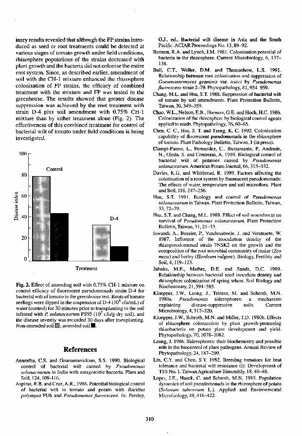
inary results revealed that although the FP strains introduced as seed or root treatments could be detected at various stages of tomato growth under field conditions, rhizosphere populations of the strains decreased with plant growth and the bacteria did not colonise the entire root system. Since, as described earlier, amendment of soil with the CH-l mixture enhanced the rhizosphere colonisation of FP strains, the efficacy of combined treatment with the mixture and FP was tested in the greenhouse. The results showed that greater disease suppression was achieved by the root treatment with strain D-4 plus soil amendment with 0.75% CH-l mixture than by either treatment alone (Fig. 2). The effectiveness of this combined treatment for control of bacterial wilt of tomato under field conditions is being investigated.
100
Control
Treatment
Fig. 2. Effect of amending soil with 0.75% CH-l mixture on control efficacy of fluorescent pseudomonads strain D-4 for bacterial wilt of tomato in the greenhouse test. Roots of tomato seelings were dipped in the suspension of D-4 (l08 cfulmL) of water (control) for 30 minutes prior to transplanting to the soil infested with P. solanacearum PS95 (107 cfulg dry soil), and the disease severity was recorded 30 days after transplanting. Non-amended soilml; amended soil •.
References Anuratha, C.S. and Gnanamanickam, S.S. 1990. Biological
control of bacterial wilt caused by Pseudomonas solanacearum in India with antagonistic bacteria. Plant and Soil, 124, 109-116.
Aspiras, R.B. and Cruz, A.R., 1986. Potential biological control of bacterial wilt in tomato and potato with Bacillus polymyxa FU6 and Pseudomonas ftuorescens. In: Persley,
GJ., ed., Bacterial wilt disease in Asia and the South Pacific. ACIAR Proceedings No. 13,89-92.
Bennett, R.A. and Lynch, J .M. 1981. Colonization potential of bacteria in the rhizosphere. Current Microbiology, 6, 137-138.
Bull, C.T., Weller, D.M. and Thomashow, L.S. 1991. Relationship between root colonization and suppression of Gaeumannomyces graminis var. tritid by Pseudomonas ftuorescens strain 2-79. Phytopathology, 81, 954-959.
Chang, M.L. and Hsu, S.T. 1988. Suppression of bacterial wilt of tomato by soil amendments. Plant Protection Bulletin, Thiwan, 30, 349-359.
Chao, W.L., Nelson, E.B., Harman, G.E. and Hoch, H.C. 1986. Colonization of the rhizosphere by biological control agents applied to seeds. Phytopathology, 76,60-65.
Chen, C. C., Hsu, S. T. and Tzeng, K. C. 1992. Colonization capability of fluorescent pseudomonads in the rhizosphere of tomato. Plant Pathology Bulletin, Taiwan, 1 (in press).
Ciampi-Panno, L., Fernandez, c., Bustamante, P., Andrade, N., Ojeda, S. and Contreras, A. 1989. Biological control of bacterial wilt of potatoes caused by Pseudomonas solanacearum. American Potato Journal, 66, 315-332.
Davies, KG. and Whitbread, R. 1989. Factors affecting the colonisation of a root system by fluorescent pseudomonads: The effects of water, temperature and soil microflora. Plant and Soil, 116,247-256.
Hsu, S.T. 1991. Ecology and control of Pseudomonas solanacearum in Taiwan. Plant Protection Bulletin, Taiwan, 33,72-79.
Hsu. S.T. and Chang, M.L. 1989. Effect of soil amendment on survival of Pseudomonas solanacearum. Plant Protection Bulletin, Taiwan, 31, 21-33.
Iswandi, A., Bossier, P., Vandenabeele, 1. and Verstraete, W. 1987. Influence of the inoculation density of the rhizopseudomonad strain 7NSK2 on the growth and the composition of the root microbial community of maize (Zea mays) and barley (Hordeum vulgare). Biology, Fertility and Soil,4,119-123.
Juhnke, M.E., Mathre, D.E. and Sands, D.e. 1989. Relationship between bacterial seed inoculum density and rhizosphere colonization of spring wheat. Soil Biology and Biochemistry, 21, 591-595.
Kloepper, lW., Leong, J., Teintze, M. and Schroth, M.N. 19803. Pseudomonas siderophores: a mechanism explaining disease-suppressive soils. Current Microbiology, 4,317-320.
Kloepper, l.W., Schroth, M.N. and Miller, T.D. 1980b. Effects of rhizosphere colonization by plant growth-promoting rhizobacteria on potato plant development and yield. Phytopathology, 70, 1078-1082.
Leong, J. 1986. Siderophores: their biochemistry and possible role in the biocontrol of plant pathogens. Annual Review of Phytopathology,24,187-209.
Lin, C.Y. and Chen. S.Y. 1982. Breeding tomatoes for heat tolerance and bacterial wilt resistance (I): Development of TSS No. 1. Taiwan Agriculture Bimonthly, 18,40-46.
Loper, 1.E., Haack, C. and Schroth, M.N. 1985. Population dynamics of soil pseudomonads in the rhizosphere of potato (Solanum tuberosum L.). Applied and Environmental Microbiology, 49. 416-422.
310

Opena, R.T. and Tschanz, A.T. 1987. Bacterial wilt resistance program on tomato at AVROC. ACIAR Bacterial Wilt Newsletter, 2, 1-2.
Scher, EM., Ziegle, 1.8. and Kloepper, J. W. 1984. A method for assessing the root-colonizing capacity of bacteria on maize. Canadian Journal of Microbiology, 30, 151-157.
Simon, R., Priefer. U. and Puhler, A. 1983. A broad host range mobilization system for in vivo genetic engineering: Transposon mutagenesis in Gram negative bacteria. Biotechnology, 1,784-791.
Suslow, T.V. 1982. Role of root-colonizing bacteria in plant growth. In: Mount, M.S. and Lacy, G.H. ed., Phytopathogenic prokaryotes voJ. 1. New York, Academic Press, 187-223.
311
Weller, D.M. 1983. Colonization of wheat roots by a Huorescent pseudomonad suppressive to take-all. Phytopathology,73,1548-1553.
-1984. Distribution of a take-all suppressive strain of Pseudonwnas ftuorescens on seminal roots of winter wheat. Applied and Environmental Microbiology, 48,897-899.
-1988. Biological control of soilborne plant pathogens in the rhizosphere with bacteria. Annual Review of Phytopathology, 26, 379-407.
Winstead, N.N. and Kelman, A. 1952. Inoculation techniques for evaluating resistance to Pseudonwnas solanacearum. Phytopathology, 42. 628-634.

Exopolysaccharides of Pseudomonas solanacearum: Relation to Virulence
D. Trigalet-Demery·, H. Montroziert, G. Orgambide§, v. Patryt, o. Adamt, L. Navarrot, v. Cotellet and A. Trigalet*
Abstract
We have discovered that the exopolysaccharide fraction of Pseudomonas solanacearum is not a homogeneous product as previously believed, but instead is composed of four distinct polymers. We have established the structure of the major polysaccharide, which we have named X I' This is an acidic heteropolymer, with an apparent molecular weight greater than 600kD, containing, in equimolar ratios, Nacetyl-galactosamine and two unusual amino sugars, 2-N-acetyl-2-deoxy-L-galacturonic acid and 2-Nacetyl-4-(3-hydroxybutanoyl)-2,4,6-trideoxy-D-glucose. We have also examined the XI fraction of several transposon-induced avirulent strains carrying mutations in the ops, eps and hrp gene clusters. We conclude that there is a direct correlation between the degree of virulence and the quantity of X I produced in vitro and in planta. In addition, preliminary information about the other polysaccharidic polymers found in supematants ofliquid cultures of P. solanacearum is provided.
PSEUDOMONAS solanacearum E. F. Smith, the causal agent of bacterial wilt disease, a destructive bacterial plant pathogen of economic importance. Among the bacterial compounds that could be involved in virulence, the exopolysaccharide (EPS) abundantly produced in infected xylem vessels of the susceptible host is one of the most likely candidates (Husain and Kelman 1958; Wallis and Truter 1978).
This EPS has been considered by most authors to be primarily composed of galactosamine (Dudman 1959; WhatJey et a1. 1980) and, more specifically, it has been pointed out that the galactosamine residues are acetylated (Akiyama et al. 1984; Drigues et al. 1985). A correlation between EPS and virulence was proposed as a result of comparisons between wild type strains that produce abundant slime (EPS) and have mucoid, fluidal appearance on agar media, and pleiotropic,
*Laboratoire de Biologie Mo1eculaire des Relations PlantesMicroorganismes, INRA-CNRS, BP 27, 31326 CastanetTolosan Cedex, France.
tLaboratoire de Pharmacologie et Toxicologie Fondamentale, CNRS, 205 route de Narbonne, 31077 Toulouse Cedex, France.
§Department of Microbiology, Giltner Hall, Michigan State University, 48.824 East Lansing, MI. U.S.A.
spontaneous avirulent mutants which, on the same media, exhibit a rough colony morphology (Kelman 1954), and were incorrectly described as EPS-. However, conflicting data have been published concerning the biochemical composition of the EPS of these various strains (Trigalet-Demery 1989), and in no case was any structure proposed.
Random mutagenesis of different virulent strains of P. solanacearum allowed the isolation of avirulent mutants. An approach to demonstrate the correlation between EPS and virulence has recently been attempted by using these insertion-induced avirulent mutants, which are impaired in both virulence and production of N-acetyl-galactosamine (Denny et al. 1988; XU et al. 1988). These studies again led to conflicting results, also probably because of a lack of precise chemical definition of the EPS.
We have demonstrated the high degree of heterogeneity of the EPS of P. solanacearum wild type GMIlOOO and have established the structure of the major acidic heteropolymer, which we have named XI (Orgambide et al. 1991). Several clusters of genes involved in the biosynthesis of EPS have been identified and recent genetic evidence suggests that EPS is in fact a virulence factor of P. solanacearum. These EPS biosynthesis gene clusters include the epsI
312

and epsII gene clusters (Denny and Baek 1991) [eps = exopoJysaccharide] and the ops gene cluster (Cook and Sequeira 1991) [ops = outer poly saccharides].
Mutants that are affected in both pathogenicity and the devclopment of a hypersensitive response have been available for some time (Boucher et al. 1985). The mutations have been mapped to an hrp gene cluster (Boucher et al. 1987) [hrp = hypersensitive response and pathogenicity]. Recent advances concerning the transcriptional organisation (ArIat et al. 1992) and potential function(s) of this gene cluster (Genin et al. 1992) have been presented. The majority of these hrp mutants grown on agar media have a colony morphology which is comparable to that of the parental wild type.
Here we report the results of comparative biochemical studies on transposon-induced avirulent mutants located within the ops, eps and hrp gene clusters. Our aim was to relate production of XI to virulence. Preliminary results on the biochemical characterisation of the three other polysaccharidic fractions (X2, X3, and X4) isolated from the supernatant of liquid bacterial cultures are also presented.
Experimental Procedures
The bacterial strains used in this study are listed below. Their origin and relevant characteristics are described in the papers cited: GMIlOOO (wild type) and its derivatives mutated within the hrp gene cluster-GMI1332, GMJl333, GMI1352, GMIl353, GM113S5 (Boucher et al. 1987, 1988); GMIl425, GMI1446. GMIl492, GMI1S00, GMIlSOl (Arlat et al. 1992); Delta 47 and Delta 199 (Boucher, unpublished data). GMI8172 (Frey et al. 1992); -K60 (wild type) and its derivatives mutated within the ops gene c1uster-KDSOO-197, KDSOO-4S, KDSOO-71, KDSOO-127, KDSOO-41 and KDSOO-13 (Cook and Sequeira 1991); and KD700 and KD710 which are mutated in the eps gene cluster (Cheng et aI., 1992).
Experimental procedures have already been described (Orgambide et al. 1991), but are brieHy summarised. The bacteria were grown in liquid media for 30 hours at 30·C on a rotary shaker in rich BG medium and in minimal MM medium supplemented with glucose (Boucher et al. 1987). The liquid culture was centrifuged and the supernatant precipitated overnight at 4°C with six volumes of cold ethanol. The precipitates were dialysed over a XMSO membrane (Amicon, Co.) and lyophilised. The crude extracts were purified by successive washing with chloroform and chromatographed over a cation exchange AG SOWXI2 (H+) resin and lyophilised; this is referred to as total, purified EPS.
EPSs were also isolated from root-infected tomato plants. As soon as the first symptoms of wilting
appeared, surface-sterilised stem pieces were soaked for 20 minutes in sterile water and EPS was purified from the soaking liquid by ethanol precipitation.
The heterogeneity of the material was revealed by gel filtration of the total EPS over Sephacryl S-500 (Pharmacia) under dissociati ve conditions and by anion exchange DEAE-Trisacryl M (IBF, Biotechnics) chromatography under dissociative conditions with a NaCl gradient as eluent. All fractions were methanolysed with anhydrous methanolic chloride and analysed by gas-liquid chromatography. All IH-and 13C-NMR spectra were recorded on a Brucker AM 300 WB instrument equipped with an Aspect 3000 calculator.
Results and Discussion
As a result of our biochemical analysis of the EPS of P. solanacearum we can summarise our present knowledge as follows. Four polysaccharidic fractions, which we have named XI' X2, X3, X4, can be isolated from the bacterial supernatants. XI> the major acidic fraction (Orgambide et al. 1991) was composed of the repetition of a trisaccharidic unit involving N-acetylgalactosamine and two unusual amino-sugars, the 2-acetamido-2-deoxy-L-galacturonic acid and the 2-acetamido-2,4,6-trideoxy-4-(3-hydroxybutyramido)D-glucose, in equimolar ratio: ~3)D-GaINAcp
(al~3) 4-0Ac-LGaINacAp(al~3)DBac2NAc4-
N[30HBut]p (~1~; X2, mannan-rich poly-mers; X3• a rhamnose-rich polymer; and X4, a family of glucans.
XI' the major, acidic polysaccharidic fraction, is produced in rich and minimal liquid media by the virulent strains GMIlOOO and K60, as well as by all of the avirulent hrp mutants so far tested. The quantity of X I produced by all these hrp mutants in liquid media is comparable to that of the parental virulent strain GMIlOOO, with the exception ofthe deletion Delta 199 (18 kb out of a total of 23 kb of the whole hrp gene cluster) which produces half as much as the wild type. The structure of X 1 is always strictly identical to that already established (Orgambide et al. 1991) whatever the strain or medium so far examined. Since these mutations ( transposon insertions and large deletions) together cover most of the hrp gene cluster and do not apparently affect the structure or reduce significantly the amount of XI produced, we conclude that this gene cluster is not involved in the biosynthesis of X I'
K60 and most of its transposon-induced derivatives produce the Xl polymer in rich liquid medium, although there are differences in the levels (Table I). There is a direct correlation between the amount of X I produced by the mutants KDSOO·71. KDSOO·4S, KDSOO-41, KDSOO-13 and KDSOO-127 and their aggressiveness; the more XI' the more pronounced are wilting symptoms. In addition, mutants KDSOO-197
313

and KD700, which do not produce any X I, are strictly avirulent. Mutant KD71 0 however, which produces no XI, does induce some wilting in rich medium. The locations of the mutants KD700 and KD710 are not precisely known, but they are located on either side of the esp/ gene cluster as defined in the paper by Denny and Beak (1991). It may be possible that the production of EPS by mutant KD710 is plant-inducible and that this mutant is located in the epslI region (Denny and Beak 1991).
Table 1. Relation of virulence to the amount of X 1 produced in rich liquid medium.
Strain designation Virulence" Relative content of X 16
K60 (wild type) 100 100 KDsOO-127 (opse) 90 61
KDsOO-13 (opsB) 80 58 KDsOO-41 (opsD) 45 27
KDsOO-45 (opsB) 29 16 KDsOO-71 (opse) 4 Traces
KDsOO-197 (opsA) 0 n.dc
KD700 (eps [) 0 n.d
KD710 (eps 1I?) 35 n.d
'Virulence was estimated as the percentage of tomato plants wilted, 3 weeks after root inoculation (Trigalet and Demery 1986).
bRelative content of X 1 is expressed as dry weight of the polymer obtained from the same aliquot of total purified EPS from virulent and related avirulent mutants.
cn.d. = not detected
The XI polymer is produced in planta by strain GMIlooo; for example, 100 inoculated plants yielded about 60 mg of total EPS. In preliminary experiments with strains KD700 (epsl) and GMIl353 (hrp) EPS production in planta was not detected under similar inoculation and incubation conditions, probably because KD700 does not invade the susceptible tomato plants used and because the invasiveness of strain GM11353 is rather poor compared with that of the wild type strain GMIlooo (Trigalet and Trigalet-Demery 1990).
The mannan polymer(s), the X2 fraction, is readily isolated from total EPS, obtained from rich medium, by chromatography over DEAE-Trisacryl. A polymer that accounts for about 10% of the total EPS after a 30 hour culture period, has been identified as a al~2, al~3, and al~6 linked mannose residue polymer, which is attributed to a previously identified mannan produced by yeast (Montrozier, unpublished data). This mannan can be readily isolated from the bacto yeast extract (Difco) used in rich medium and is therefore a
contaminating polymer. However, small amounts of mannose have been identified by gas-liquid chromatography in the total EPSs from GMIlooo and related hrp mutants when cultivated in minimal liquid medium, Le in the absence ofbacto yeast extract. At the present time, however, it is difficult to decide whether P. solanacearum does produce a mannan in minimum medium, since the putative polymer is present in minute amounts and is not yet structurally characterised.
The rharnnan polymer, X3, is a minor fraction that accounts for less than 5% by weight of total EPS. The X3 fraction is isolated by gel permeation as a polymer whose apparent molecular weight is between 5000 and 15000 (there may be some association of small repeating units). Rhamnose is a labile sugar, sensitive to usual hydrolysis conditions, and supposing that rhamnose is part of Xl' as a lateral branch for instance, it could readily be separated from the major polymer under our hydrolysis conditions. However, the latter remarks are inconsistent with the data obtained from the biochemical analysis of mutant KD5OO-45, which is Xli and X3* (i = impaired, and * = identical to wild type) and mutant KD5OO-13, which iSXl* and X3-(-=notdetected).
It should be noted that rhamnose is a principal sugar component of the lipopolysaccharide (LPS) of P. solanacearum, and that it accounts for about 45% of total carbohydrate of the LPS of GMllooo and is located in the outer core and in the O-antigen (Drigues et al. 1985). Liquid culture media may probably contain some degraded LPS as the result of bacteriolysis and one can predict that incomplete O-antigen is present in the supematant. In fact, recent data (Montrozier, unpublished data) indicate that a tetrasaccharidic unit composed of three rhamnose and one glucosamine can be isolated from the total, purified EPS of GMIlooo. Similar tetrasaccharides have already been reported from other P. solanacearum strains (Akiyama et al. 1984; Kocharova et al. 1992).
A family of glucans, fraction X4, has been isolated by cation exchange and permeation gel chromatography over a range of Biogel media. These glucans are produced in rich and minimal media and their structure is under study. Some avirulent mutants, namely KD5OO-71, KD700 and KD71O, appear helpful in this respect as their EPS is essentially made of glucans.
Work is in progress to determine the structure of the different polymers under study and to establish their potential role(s) in virulence.
Acknowledgments
We thank Clare Gough for reviewing the manuscript.
314

References
Akiyama, Y., Eda, S. and Kato, K. 1984. Structural studies of the O-antigen of the lipopolysaccharide from an avirulent strain M4S of Pseudomonas solanacearum. Carbohydrate Research, 133,289-296.
Arlat, M., Gough, C., Zischek, C., Barberis, P., Trigalet. A. and Boucher, C. 1992. Transcriptional organization and expression of the large hrp gene cluster of Pseudomonas solanacearum. Molecular Plant-Microbe Interactions, 5, 187-193.
Boucher, C., Barberis, P. and Arlat, M. 1988. Acridine orange selects for deletion of hrp genes in all races of Pseudomonas soianacearum. Molecular Plant-Microbe Interactions,l. 282-288.
Boucher, c., Barberis, P .• Trigalet. A. and Demery, D. 1985. Transposon mutagenesis of Pseudomonas solanacearum: isolation of Tn5-induced avirulent mutants. Journal of General Microbiology, 131. 2449-2457.
Boucher. C., Van Gijsegem. F., Barberis, P., Arlat, M. and Zischek, C. 1987. Pseudomonas solanacearum genes controlling both pathogenicity on tomato and hypersensitivity on tobacco are clustered. Journal of General Microbiology, 169,5626-5632.
Cheng K.C., Barlow, E. and Sequeira, L. 1992. Extracellular polysaccharide is required for wild type virulence of Pseudomonas soianacearum. Journal of Bacteriology, 174, 1068-1071.
Cook, D. and Sequeira, L. 1991. Genetic and biochemical characterization of a Pseudomonas soianacearum gene cluster required for extracellular polysaccharide production and virulence, Journal of Bacteriology, 173, 1654-1662.
Denny, T. and Baek, S. 1991. Genetic evidence that extracellular polysaccharide is a virulence factor of Pseudomonas soianacearum. Molecular Plant-Microbe Interactions. 4, 198-206.
Denny, T., Makini, F. and Brumbley, S. 1988. Characterization of Pseudomonas solanacearum Tn5 mutants deficient in extracellular polysaccharide. Molecular Plant-Microbe Interactions 1, 215-223.
Drigues, P., Demery-Lafforgue, D., TrigaJet, A., Dupin, P., Samain, D. and Asselineau, J. 1985. Comparative studies of lipopolysaccharide and exopolysaccharide from a virulent strain of Pseudomonas solanacearum and from three avirulent mutants. Journal of BacterioJogy, 162,504-509.
Dudman, W.F. 1959. Comparison of slime from tomato and banana strains of Pseudomonas solanacearum. Nature, London, 184,1969-1970.
Frey, P., Prior, P., Trigalet-Demery, D., Marie, c., Kotoujansky, A. and Trigalet, A. 1992. Advances in biological control of tomato bacterial wilt using genetically
engineered avirulent mutants of Pseudomonas solanacearum. In: Proceedings of the 8th International Conference of Plant Pathology on Plant Pathogenic Bacteria, Versailles, France, 9-12 June, 1992.
Genin,S., Gough, c., Arlat, M., Zischek. C., Van Gijsegem, E, Barberis, P. and Boucher, C. 1992. Involvement of Pseudomonas solanacearum hrp genes in the secretion of a bacterial compound which induces a hypersensitive-like response on tobacco. In: Proceedings of the 6th International Symposium on Molecular Plant-Microbe Interactions, Seattle, Washington, U.S.A, ll-16July 1992.
Husain, A. and Kelman, A. 1958. Relation of slime production to mechanism of wilting and pathogenicity of Pseudomonas solanacearum. Phytopathology, 48, 155-164.
Kelman, A. 1954. The relationship of pathogenicity in Pseudomonas so/anacearum to colony appearance on a tetrazolium medium. Phytopathology, 44, 693-<i95.
Kocharova, N., Knirel, Y., Shahkov, A., Kotchetkov, N. and Varbanets, L. 1992. The structure of 0 specific polysaccharide from Pseudomonas solanacearum ICMP 4157. Carbohydrate Research, 228, 315-320.
Orgambide, G., Montrozier, H., Servin, P., Roussel, l., Trigalet-Demery, D. and Trigalet, A. 1991. High heterogeneity of the exopolysaccharides of Pseudomonas solanacearum strain GMI 1 000 and the complete structure of the major polysaccharide. Journal of Biological Chemistry, 266,8312-8321.
Trigalet, A. and Demery, D. 1986. Invasiveness in tomato plants of Tn5-induced avirulent mutants of Pseudomonas solanacearum. Physiological Molecular Plant-Microbe Interactions, 28, 423-430.
Trigalet, A. and Trigalet-Demery, D. 1990. Use of avirulent mutants of Pseudomonas solanacearum for the biological control of bacterial wilt of tomato plants. Physiological and Molecular Plant Pathology, 36, 27-38.
Trigalet-Demery, D. 1989. Exopolysaccharides produced by Pseudomonas solanacearum. In: Tjarnos, E. and Beckman, C, ed., Vascular Wilt Diseases of Plants. NATO ASI Series, H28, 219-228.
Wallis, F. and Truter, J. 1978. Histopathology of tomato plants infected with Pseudomonas soianacearum with emphasis on ultrastructure. Physiological Plant Pathology, 13, 307-317.
Whatley, M., Hunter, N., CantreIl, M., Hendrick, L., Keegstra, K. and Sequeira, L 1980. Lipopolysaccharide composition of the wilt pathogen Pseudomonas solanacearum. Plant Physiology, 65, 557-559.
Xu, P., Leong, S. and Sequeira, L. 1988. Molecular cloning of genes that specify virulence in Pseudomonas solanacearum. Journal of Bacteriology, 170, 617-622.
315

Lysogeny and Lysotypes of Malaysian Strains of Pseudomonas solanacearum
Hiryati Abdullah*
Abstract
Thirty-two isolates of Pseudomonas solanacearum were screened for phages. Forty-three percent of the isolates tested were lysogenic. Only one isolate was lysogenic without induction, while the others were lysogenic upon induction with various concentrations of mitomycin C. The reactions of 68 P. solanacearum isolates to the phages showed that no bacterial isolate was lysed by all the phages and none of the isolates was completely resislant to all the phages. Based on the lytic response of five selected phages, 13 Iysotypes were distinguished. All isolates could be typed. Phages may also be useful in studies related to epidemiology and genetics.
PSEUDOMONAS solanacearum is subject to infection by bacteriophages (phages). The interaction of phages with phytopathogenic bacteria, including their potential for disease control, has been the ·subject of several review articles (Civerolo 1972; Okabe and Goto 1963; Videvar 1976). Phages have been used in epidemiological studies (Bald win and Goodman 1963; Kauffman and Reddy 1975; Tagami 1959; Wakimoto 1954; Wakimoto 1960;); in classification and identification (Billing 1963, 1970; Stolp and Starr 1964; Wakimoto, 1960); in distinguishing fine differences in host specificity and differences between distinct pathogens on a single host (Freigoun and Crosse 1975; Garret et a!. 1966; Persley and Crosse 1978); and in genetical studies (Boucher et a!. 1977; Faelen and Toussaint 1976).
Typing phages against a bacterial species enables grouping of Iysosytes within the species, based on sensitivity to the phages tested. Okabe and Goto (1963) indicated that some lysotypes are specialised in their pathogenicity or biochemical characters or both, while others are not specific. Lysotypes specific for pathogenicity or other characters are useful for identifying specialised types in nature, while the nonspecific Iysotypes are a useful label for studying various aspects of the pathogen such as the mode of dispersal, infection source and longevity in nature.
*Department of Plant Protection, Faculty of Agriculture, Universiti Pertanian Malaysia, 43400 UPM, Serdang, Selangor Darul Ehsan, Malaysia.
Paulin and Nassan (1978) carried out phage-typing of 61 strains of E. chrysanthemi from four different hosts and isolated 32 temperate phages. They showed a degree of correlation between phage sensitivity and geographic origin of the strains. Using 12 virulent and temperate phages, Okabe and Goto (1953) divided strains of P. solanacearum into 40 lysotypes.
This study sought to isolate temperate bacteriophages of P. solanacearum and to evaluate their use in typing strains of the pathogen.
Materials and Methods
Isolation of temperate phage
Thirty-two isolates of P. solanacearum were screened for temperate phages. Twenty millilitre aliquots of Casamino-asid peptone glucose (CPG) broth (Cuppels et al. 1978) were inoculated with 0.5 mL of a suspension of a 24-48 hour culture of P. solanacearum in sterile distilled water with a concentration of 8 x 10⁸
cfu/mL. Flasks were incubated for 16 hours at 30°C in an incubator-shaker set at 100 rpm. Initially no induction was done, but in the second and subsequent experiments isolates that did not have phages were subjected to 1, 1.5, 2 or 4 JlgImL of mitomycin C (Sigma Chemical Co., S1. Louis, MI). Mitomycin C was dissolved in distilled water and added to the broth culture to give the required concentration. The culture was incubated for another 4 hours in the dark under the same conditions as previously. The contents of the
316

flask were then transferred to a sterile screw-capped test tube and chloroform added to a final concentration of 5%. Using a vortex mixer, the contents of the tube were shaken vigorously for 60 seconds. Tubes were incubated for a minimum of 4 hours at 20°C and stored at 4°C. Twenty microlitres of the broth were spotted on lawns of potential indicator strains. The indicator lawn (0.5 mL of the indicator strain in aqueous suspension containing approximately 108 cfulmL) was streaked onto a thin layer of buffered soft water agar overlayer (3 mL). which had been poured over yeast extract glucose agar (YA) medium. Plates were incubated at 30°C for 16-24 hours. Those that produced a clear area at the inoculation points were serially diluted in buffered saline solution. Twenty microlitre aliquots of a series of its ten-fold dilution were spotted on indicator lawns. Plaques were purified through three successive single isolations of plaques and stocks prepared. Filter-sterilised stocks were then kept under 1 % chloroform at 4°C.
Typing of bacteriophages
Determination of the routine test dilution (RID) of all the typing phages was carried out using standard procedure described by Adams (1959). Stock suspen-
sions of each phage isolated were diluted to the RID before each typing. The reactions of 68 isolates of P. solanacearum to bacteriophages were determined by the standard technique. A 0.5 mL aliquot of an aqueous suspension (1 x 10⁸ cfulmL) of the P. solanacearum isolate to be tested was added to 3 mL of molten buffered soft water agar maintained at 48°C. The two components were mixed and poured over YA plates to form a complete lawn of the test bacterium on the surface. Plates were allowed to dry and 15 J.l.L aliquots of the typing phages, which were diluted to their RID and 10 x RTD levels, were spotted on the bacterial lawn. Plates were incubated at 30"C for 24 hours.
Results and Discussion
Presence of plaques on the spotted area as a result of dilution indicates the presence of phages, while broth containing bacteriocins produced increasingly hazy inhibition zones with increased dilution. as indicated by Billing (1969). Temperate phages were readily isolated from lysogenic strains of P. solanacearum. Of the 35 isolates tested for lysogeny, 15 (43%) were lysogenic (Table 1). However, only one isolate (200 19)
Table 1. Production of temperate bacteriophages from P. solanacearum isolates.
Bacterial isolates Host plant Concentration of mitomycin C (J.lglmL)
0 1.0 1.5 2.0 4.0
20003 Ginger -' 20011 Ginger ;b + ND ND 20019 Ginger NDr ND ND ND 20022 Ginger 20027 Ginger + ND ND 21028 Tomato 21034 Tomato 21030 Tomato 21009 Tomato 21017 Tomato 21018 Tomato 22029 Brinjal 23019 Chilli 24024 Tobacco 24008 Tobacco + NO ND NO 26023 Groundnut 26003 Groundnut + ND ND ND 26033 Groundnut 27013 Winged bean 28014 French bean 28019 French bean + ND NO ND 29007 Croton + ND NO ND 29113 Cat's whisker + NO NO NO 29012 Croton + NO NO 29128 Zinnia 29003 Euphorbia + ND NO NO 29050 Croton + NO ND 29034 Dahlia 29025 Croton + 29075 Leonurus + 29042 + ND ND 29102
+ ND NO
aNo phage produced; h-remperate bacteriophage produced; "Not determined.
317

from ginger. produced phage without induction. The rest of the lysogenic strains produced phages following induction with various concentrations of mitomycin C. Fourteen phages were isolates and given tentative designations (Table 2). A phage is considered different when it originated from a different host or when it produced different plaques (of different opacity and/or diameter).
Thble 2. Temperate bacteriophage designations and origin.
Phage designation Phage origin B 29007 C 29003 D 20011 E 29050 G 24008 J 28019 K 20019 M 29064 0 29025 P 29075 R 26003 T 20027 Y 29012
29042
All phages produced plaques which were 1 mm or less in diameter. They were of variable opacity, but all had irregular borders. Plaque development of all phages on P. solanacearum isolates tested was slow, so that only a few plaques were visible by 16-24 hours, although lawn development was excellent. However, by 40-48 hours, plaques were seen more readily. The results of the reactions of phages to P. solanacearum isolates were more consistent when using phage stocks diluted to RTD x 10 rather than RID. Among the P. solanacearum isolates, variable results were obtained. No bacterial isolate was lysed by all the phages and none of the isolates was completely resistant to the phages tested. By eliminating some of the phages with similar host ranges and those which could lyse only few strains, a simplified typing scheme using five phages was obtained. Thirteen Iysotypes could be distinguished among the P. solanacearum isolates (Table 3). All isolates could be typed. These could not be correlated to host-plant origin or locality of isolates. However, phages could be useful in both epidemiological and genetical studies. Okabe and Goto (1961) described 13 pathotypes in Japan. They also indicated that there was not always a correlation between biochemical properties and phage sensitivity when compared with host range and virulence for different hosts.
Thble 3. Lysotypes in Pseudomonas solanacearum.
Lysotypes Bacteriophage Number ofisolates
B G K P Y
+ + + + + 32 II + + + + 12
III + + 7 IV + + + 3 V + + + + 4
VI + + + 2 VII + 2
VIII + + + IX + + X +
XI + + + XII +
XIII + + + +
Acknowledgments
The author thanks Universiti Pertanian Malaysia (UPM) for the research grant and the Commonwealth Mycological Institute, Kew, England, for confirmation of the P. solanacearum isolates.
References
Adams, M.H. 1959. Bacteriophages. John WHey & Sons, Inc. New York. 592 p.
Baldwin, C.H. and Goodman, R.N. 1963. Prevalence of Ery"';nia amylovora in apple buds as detected by phage typing. Phytopathology, 53, 1299-1303.
Billing, E. 1963. The value of phage sensitivity tests for the identification of phytopathogenic Pseudonwnas spp. Journal of Applied Bacteriology. 26, 193-210.
-1969. Isolation, growth and preservation of bacteriophages. In; Norris J.R. and Ribbons, D.W., ed., Methods in Microbiology VoL 3b. New Yprk and London; Academic Press, 315-329.
-1970. Further studies on the phage sensitivity and the detennination of phytopathogenic Pseudonwnas spp. Journal of Applied Bacteriology. 33,478-491.
Boucher, CB., Bergeron, M.B., De Bertalmio and Denarie, J. 1977. Introduction of bacteriophage Mu into Pseudomonas solanacearum and Rhizobium melitoti using the R factor RP4. Journal of General Microbiology, 98, 253-263.
Civerolo, E.L. 1972. Interaction between bacteria and bacteriophages on plant surfaces and in plant tissues, In; Proceedings of the International Conference on Plant Pathogenic Bacteria, Wageningen, The Netherlands, 25-37.
Cuppels, D., Hanson, R.S. and Kelman, A. 1978. Isolation and characterization of a bacteriocin produced by Pseudomonas solanacearum. Journal of General Microbiology. 109,295-303.
318

Faelen, M. and Toussaint, A. 1976. Bacteriophage Mu-I: a tool to transpose and localize bacterial genes. Journal of MolecularBiology, 104,525-539.
Freigoun, S.O. and Crosse, J. 1975. Host relations and distribution of a physiological and pathological variant of Pseudomonas morsprunorum. Annals of Applied Biology, 81,317-330.
Garret, C.M.E., Paragopoulos, C.G., and Crosse, J.E. 1966. Comparison of plant pathogenic pseudomonads from fruit trees. Journal of Applied Bacteriology, 29, 342-356.
Kauffman, H.E. and Reddy, A.P.K. 1975. Seed transmission studies of Xanthomonas oryzae in rice. Phytopathology 65, 663~.
Okabe, N. and Goto, M. 1953. Studies on Pseudomonas solanacearum I. Classification of the strains by bacteriophages and virulence of the strains. Report Faculty Agriculture, Shizuoka University, 3, 52-80.
-1961. Studies on Pseudomonas solanacearum XI. Pathotypes in Japan. Report Faculty Agriculture, Shizuoka University, 11,25-42.
-1963 Bacteriophages of plant pathogens. Annual Review of Phytopathology, 1,397-418.
Paulin, J.P. and Nassan, N.A. 1978. Lysogenic strains and phage-typing in Erwinia chrysanthemi, In: Proceedings of
the Fourth International Conference on Plant Pathogenic Bacteria. Angers, France, 539-545.
Persley, G.J. and Crosse, J.E. 1978. A bacteriophage specific to race 2 of the cherry strain of Pseudomonas morsprunorum. Annals of Applied Biology, 89,219-222.
Stolp, H., and Starr, M.P. 1964. Bacteriophage reactions and speciation of phytopathogenic xanthomonads. Phytopathologischz Zeitschrift, 51, 442-478.
Tagami, Y. 1959. Relation between quantity of bacterial leaf blight organism and bacteriophages and occurrence of the disease. Journal of Plant Protection, 13, 389-394. (In Japanese).
Vidaver, A. 1976. Prospects for control of phytopathogenic bacteria by bacteriophages and bactericions. Annual Reveiw of Phytopathology, 14,451-465.
Wakimoto, S. 1954. The determination of the presence of Xanthomonas oryzae by the phage technique. Scientific Bulletin Faculty Agricriculture, Kyushu University 14,495-498.
-1960. Classification of strains of Xanthomonas oryzae on the basis of their susceptibility against bacteriophages. Annals of the Phytopathological Society of Japan, 25, 193-198.
319

A Biocontrol Agent for Pseudomonas solanacearum
G.C. Wall and J.L. Sanchez*
Abstract
Bacterial wilt is a serious disease of solanaceous crops throughout Guam. Fields often become chronically infested with its causal agent, making it unprofitable for farmers to produce these crops. At present, resistant cultivars are either inadequate or unavailable, and grafting methods are labour-intensive. For these reasons, an attempt was made to find a bacteriophage capable of attacking the local strain of Pseudomonas solanacearnm. Soil samples were obtained from fields with a history of bacterial wilt, and from adjacent fields free of the problem. Bacteriophage activity against the local strain of P. soianacearnm was found in both soil types, but higher counts were found in uninfested soils.
BACTERIAL wilt is a serious plant disease problem affecting many valuable solanaceous crops, including tomatoes, bell peppers, eggplant, tobacco and potatoes (Persley 1986). The first three of these crops are extremely important cash crops in tropical regions such as Guam. The disease is caused by a soilborne bacterium, Pseudomonas solanacearum. Infection takes place through wounds or cracks in the roots, rendering the vascular tissue incapable of transporting water and nutrients to the aerial parts, which eventually wilt and die.
Once established in a field, the pathogen is very difficult to control. There are no chemical means of control. Resistant cultivars only exist in only certain of the plant species affected. Breeding efforts in the past have failed to solve the problem, and cultural methods such as grafting to resistant rootstock and rotating fields to sugarcane or rice are labour-intensive or otherwise inappropriate.
Many bacteria, plant pathogenic and otherwise, have very specific natural enemies known as bacteriophages, or phages. Bacteriophages are submicroscopic viruses capable of penetrating their host cells and multiplying within them, using the cells' components and biochemical pathways to their own advantage. Indeed, phages are so specific that they can be used to identify their bacterial hosts in certain cases (Cuppels 1984). For obvious reasons, host specificity is an ideal attribute for biocontrol agents.
*CALSI AES, University of Guam, Mangilao, GU 96923, Guam.
The study reported here sought bacteriophages capable of attacking the bacterial wilt pathogen, for possible use as biocontrol agents.
Materials and Methods
Farmers' fields with a history of bacterial wilt problems were identified through the Cooperative Extension Service. Other fields apparently free from the pathogen were also chosen, Soil samples were collected from the top 20 cm of soil. These were taken at several points in a particular field and later combined to form a composite 150 g sample. Samples were collected as follows: 1. potted soil originally taken from the Santos farm, Mangilao; 2. Watson farm, Radio Barrigada; 3. the same farm as 2, different field; 4. Taitano farm, Yigo; and 5. Department of Agriculture, Mangilao. All samples except 1 and 2 had previous wilt history.
Isolates of P. solanacearum were obtained from wilted tomato plants collected from the Taitano farm in Yigo. Isolates were grown on King's Medium B. Gramnegative isolates not producing fluorescent pigments were selected (Schaad 1988). These were further screened by inoculating tomato seedlings. Only two isolates capable of reproducing wilt symptoms on the seedlings were used for the rest of this procedure.
A method first described by Crosse and Hingorani in 1958 (Johnson and Curl 1972) and used for isolating bacteriophage from P. morsprunorum was followed with slight modifications. Each composite soil sample was first enriched by placing it in an Erlenmeyer flask and adding 150 mL of a 48-hour culture of
320

P. solanacearum in nutrient broth plus 2% glycerine. Cultures were grown in a waterbath-shaker at 32°C. The free liquid was then decanted from the enrichment and passed through filter paper. This filtrate was then passed through a 0.4 J.U11 filter (Millipore) to eliminate larger diameter particles including bacteria.
A serial dilution was prepared by taking I mL of the final filtrate and mixing it with 9 mL of a 24-hour nutrient broth culture of P. solanacearum. After thorough mixing, I mL of this suspension was transferred to another 9 mL of P. solanacearum culture, and repeated until a 106 dilution was prepared. All dilutions were replicated twice. Aliquots of 1 mL of each dilution were then transferred to test tubes containing 9 mL of melted agar medium at 42°C. The agar medium consisted of 1 % agar, 0.5% peptone, 0.3% yeast extract, and 2% glycerine. The pH was adjusted to 7.2 with phosphate buffer. The agar mixture was shaken and poured into sterile culture dishes. Plaques were observed over a period of 48 hours.
Results Cleared zones in a radial pattern amid the more opaque bacterial growth revealed bacteriophage activity. Selecting the appropriate range for an accurate count of plaques per plate was possible because of the serial dilutions prepared. The highest numbers of bacteriophages were found in soil samples from the potted soil originally collected from the Santos farm in Mangilao, and a sample from the Watson farm in Radio Barrigada (Table 1; samples 1 and 2). Numbers of bacteriopha~es per gram of soil sample were 1 x 10⁸
and 2.4 x 10 respectively.
Discussion High bacteriophage counts were found in soils with no bacterial wilt problem. The lower counts came from samples taken from infested fields. Our data strongly suggest that bacteriophages play an important role in the popUlation dynamics of P. solanacearum in the soil, and that their use as a biocontrol agent may hold promise. The fact that these are locally occurring phages has at least two immediate advantages. One relates to quarantine: since no importations are required
Table 1. Counts ofbacteriophages (phage nos/g soil) in Guam soil samples from bacterial wilt-free and Wilt-prone fields.
Sample no. Phage count Wilt prone
I lXlo'l No 2 2.4 X 101 No 3 10 Yes 4 400 Yes 5 20 Yes
no permits would be needed to begin research. The other relates to specificity: these are bacteriophages that attack the local strain of P. solanacearum.
It is interesting that sample 1 originally came from an infested field, but has been kept in pots for over 3 years. While the soil has been used to produce diseased plants it seems to be no longer infested, as susceptible pepper seedlings fail to wilt when transplanted to it.
It is also interesting to note that part of the technique used in isolating phages includes the enrichment of these by the addition of host culture to the soil samples. Future research might perhaps answer the question of whether increasing the host population in field soil would eventually lead to phage population levels capable of controlling the wilt problem, as appears to have occurred in the potted soil of sample 1.
Phages can have negative as well as positive impacts. For example, commercial bacterial fermentation processes for the production of various drugs, foods, drinks, and pesticides are occasionally plagued with phage epidemics (C. Gonzalez, pers. comm.). Commercial companies have had to research this problem and one of the solutions employed is to isolate plaques and bacteria from areas that failed to plaque. Eventually, resistant strains of bacteria are thus developed, and the phage epidemics are controlled with these resistant strains. Returning to the context of soilborne diseases, this suggests that field populations of P. solanacearum might eventually undergo a natural selection process and evolve as a resistant strain if bacteriophages apply severe selective pressure. While biocontrol of P. solanacearum with bacteriophage may thus be possible, the success may in turn be limited by natural selection. More research is needed to clarify these issues.
Acknowledgment We would like to thank the United States Department of Agriculture for providing funds for a summer apprenticeship for J.L. Sanchez.
321
References
Cuppels, D.A. 1984. The use of pathovar-indicative bacteriophages for rapidly detecting Pseudomonas syringae pv. tomato in tomato leaf and fruit lesions. Phytopathology, 74,891-894.
Johnson, L.E and Curl, E.A. 1972. Methods for research on the ecology of soil-borne plant pathogens. Minneapolis, Minnesota, Burgess, 247 p.
Persley, G.J. 1986. Bacterial wilt disease in Asia and the South Pacific: proceedings of an international workshop held at PCARRD, Los Banos, Philippines, 8-10 October 1985. ACIAR Proceedings No. 13. 145 p.
Schaad, N.W. 1988. Laboratory guide for identification of plant pathogenic bacteria. SI. Paul, Minnesota, American Phytopathology Society Press, 164 p.

Potential of Biological and Chemical Control of Bacterial Wilt
G. L. Hartman*, W.F. Hong*, Hanudint and A.C. Hayward§
Abstract
Strains of biological control agents Pseudomonas cepacia, P. fluorescens, and P. gladioli inhibited growth of P. solanacearum on culture medium. A P. cepacia cell suspension applied to soil 7 days before inoculation with P. solanacearum reduced the occurrence of wilt by 65% compared with the control. A combination of P. fluorescens and an experimental bactericide (Terlai) was tested for control of bacterial wilt in pot culture studies. Terlai effectively controlled bacterial wilt, especially when applied before and after inoculations with P. so[anacearum. Plants grown in green-manure-amended soil in pot culture had significantly less wilt than occurred in nonamended soil. Survival of P. solanacearum in soil under controlled conditions was reduced when soil was amended with KN03, NaN03, NaCl, and KC! at a rate of 1:99 salt to soil, but not when amended with liquid pig effluent at 1,5 and 10%. NaNO) reduced the population to 65% that of the control.
BACTERIAL wilt caused by Pseudomonas solanacearum is one of the major constraints to production of solanaceous crops, especially tomatoes. The disease has been reported to cause 29% loss of fresh fruit production in hybrid tomatoes in Taiwan (Hartman et al. 1991), and its importance and occurrence has been well documented (Hayward 1991; Kelman 1953).
Research on microbial antagonists, such as P. fiuorescens has shown some promise in control of bacterial wilt (Hsu et aI., these proceedings). In addition, various Actinomycetes, and bacteria including Bacillus mesentericus, B. megaterium, B. subtilis. B. mycoides, and Erwinia. have been reported to be active biological control agents (Kelman 1953). Aspiras and Cruz (1986) treated infested soil with B. polymyxa and P.jluorescens, which increased the survival of tomato from 0% without the antagonist to 60 and 90%, respectively. P. cepacia, another potential
* Asian Vegetable Research and Development Center, PO Box 42, Shanhua. Tainan, Taiwan, 741.
tSegunung Horticultural Research Station, PO Box 8 Sindanglaya-Cianjur 43253, Indonesia.
§ Department of Microbiology, The University of Queensland, Qld., 4072, Australia.
biological control agent, was shown to occur in high numbers in association with maize grown as a rotation crop with potatoes to reduce the incidence of wilt (E1phinstone and Aley, these proceedings). It also has been shown to produce ketoglutonic acid which is also inhibitory to P. solanacearum (Akiyama et aJ. 1984).
Organic amendments have been used with some success in controlling bacterial wilt. Reports of using a mixture called SH (Sun and Huang 1985) or its components have been tested in Taiwan (Chang and Hsu 1988; Hsu and Chang 1989; Hartman and Yang 1990). The SH amendment is now commercially available in Taiwan.
There are no bactericides available for chemical control of bacterial wilt, though an experimental fonnulation, Terlai, has been tested recently in Taiwan under both greenhouse and field conditions. Growers may find that chemical control is not cost-effective.
Additional management practices to control bacterial wilt are crop rotation, intercropping or incorporating green manure crops such as Sun hemp and mungbean before planting a susceptible crop. There is some evidence that maize may reduce the occurrence of bacterial wilt either intercropped (Autrique and Potts 1987) or in the next crop (Elphinstone and Aley, these proceedings).
322

The purpose of our studies was to evaJuate whether certain biological, organic, and inorganic treatments are effective in reducing population levels of P. solanacearum and thus the severity of bacterial wilt.
Material and Methods
Tests for antagonism P. solanacearum strain PSS 4 was streaked from a stock culture stored in distilled water onto a tetrazolium chloride medium (Kelman 1954), and incubated for 48 hours at 30°C. A single, fluidal, wild-type colony was selected and increased on 523 medium at 30°C for 24 hours before harvesting cells. Suspensions were adjusted to 108cfulmL or an optical density of 0.30 (equivalent to 50% transmittance) at wavelength 600 nm for all experiments.
P. jluorescens strain pp 59 obtained from S.T. Hsu, National Chung Hsing University Taiwan, ROC, was streaked on King's B medium and incubated at 30°C for 24 hours. then placed under ultraviolet (UV) lamp to detect the presence of fluorescent colonies. P. cepacia (PcS) and P. gladioli (PgI0) were isolated from modified SM-l (Chang and Hsu 1988) by dilution plating of AVRDC soils. Both ~anisms were identified using Biolog Microstation system 3.00. The antagonists were multiplied on 523 medium at 30°C for 24 hours.
Antagonists were streaked down the middle of dishes containing King's B medium, incubated for 48 hours and challenged by cross -streaking four strains of P. solanacearum. This experiment and variations of it were repeated several times.
Thirty day old L390 plants were treated as follows: 1. PglO applied to soil 7 days before inoculating P. solanacearum; 2. Pg 10 applied 1 day before inoculation; 3. PglO applied 1 day after inoculation; and 4. only P. solanacearum inoculated. To apply the antagonist, 4 L of a suspension of Pg 10 at a concentration of 109 cfulmL (00=0.50) were poured in trays (35 x 50 x 7 cm) containing 16 potted plants which were soaked in this suspension for 3 hours.
Application or P.fluorescens and Terlai P. jluorescens was applied as previously described for Pg 10. Ten grams ofTer1ai, a systemic bactericide [Ching Mow Trading Co, Ltd., Taiwan, similar to 2,2-dichloroN(2hydroxy-l (hydroxy-methyl)-2-( 4-nitrophenyl) ethyl) acetamide], were added to 1 litre of tap water, then 30 mL of this suspension were poured into each pot containing one tomato plant. Treatment combinations are presented in Table 1.
Seeds of tomato line L 390 were sown directly in 8.5-cm-diameter plastic pots containing sterile soil. These were placed in a pipe-house at 30°C. Plants were
Table 1. Treatment combinations using 1erlai (Tr) and P. fluorescens (pt) to control bacterial wilt in greenhouse-inoculated tomatoes.
Treatment code" Days after sowing
27 29 30 31
Tr-OO-Ps-OO = A Tr Ps Tr-Pf-Ps-OO = B Tr Pf Ps Tr-OO-Ps-Pf = C Tr Ps Pf Tr-OO-Ps-Tr = D Tr Ps Tr Tr-Pf-Ps-Tr= E Tr Pf Ps Tr Pf-OO-Ps-OO = F Pf Ps Pf-OO-Ps-Pf = G Pf Ps Pf Pf-OO-Ps-Tr = H Pf Ps Tr
I Ps
"Tr = Terlai; Ps = P. solanacearum: Pf = P. ftuorescens; 00 = untreated.
inoculated 30 days after sowing (OAS) with PSS 4 by drenching soil with 30 mI.Jplastic pot. Each treatment contained four pots arranged in a randomized complete block design. The inoculated plants were placed in a greenhouse at 32°C. The experiment was repeated. Numbers of wilted plants were counted every 3 days beginning from 6-21 OAS, Area under the disease progress curves was calculated (Shaner and Finney 1977).
Population dynamics and plant survival in amended soil Soil collected from an AVRDC field was air-dried and sieved through a 50 JlIl1 mesh screen. The soil was divided into eight parts containing 500 g each and amended with either liquid pig effluent at concentrations of 1, 5 or 10% (v/w), or with KN03, NaN03, NaCI or KCl, each at a 1% w/w. Amended or nonamended soil was mixed evenly with a 75 mL su~nsion of P. solanacearum at a concentration of 10 cfulmL. Soil samples stored in plastic bags were placed in an incubator at 28°C for 5 weeks At 0, 4, 11, 18,25 and 32 days after inoculation (OAI), 10 g of soil were removed, shaken in 90 mL of sterile distilled water and tenfold series dilutions were made. From each dilution, 0.1 mL was spread onto the plates of modified SM-I. Two plates were used for each dilution. Colonies of P. solanacearum were counted 48 hours after incubation at 30°C. A completely randomised design with three replications was used in this trial. Population counts were transformed to 10glO before analysis.
Green manure Roots and stems of sun hemp (Crotalaria sp.) of 70-day-old field-grown plants were chopped into pieces
323

before mixing 30 g fresh weight with 500 g infested soil in plastic bags. Infested soil was prefared by adding 75 mL of bacterial suspension with 10 cfulmL to meshed air dry soil to field capacity. Whole plants of mungbean and soybean were also tested. There were five replication for each amendment. All the samples were incubated at 32°C and populations were monitored weekly as previously described.
Soil containing 0, 2, 6 andlO% chopped sun hemp was incubated for 4 weeks before adding a bacterial suspension (OD = 0.3 at 600 nm) into the amended soil at the ratio of I: to (v/w). After mixing the soil, it was distributed in clays pots (30 cm diameter), using 10 pots for each level of amendment. Four-week-old L390 plants were transplanted into the incorporated soils, one plant per pot. Wilt was recorded weekly for 8 weeks.
Results and Discussion
Tests for antagonism
Modified SM I medium allowed for the isolation of both P. cepacia and P. gladioli and it seemed not to restrict growth or number of colonies of Pg to on SMI compared with King's B medium (Fig. I). P. cepacia, P. f1uorescens and P. gladioli caused large zones of inhibition of P. solanacearum (Fig. 2, Pc5 not shown). Bacterial wilt was controlled when P. gladioli was applied to the soil before inoculation of P. solanacearum. Although control was not complete, there was about 60% reduction in wilt compared with the control when isolate Pg 10 was applied to the soil 7 days before inoculating P. solanacearum .
Biological control agents are often effective in reducing pathogen growth in cultural studies and sometimes also in pot culture studies. However, there has been little evidence that these biological control agents are effective against bacterial wilt in the field. More work is needed to develop better delivery
Fig. I. Pseudomonas gladioli on selective medium (SM I) and on King's B medium after 48 hours of incubation.
324
Fig. 2. Inhibition of Pseudomonas solanacearum by (a) P. gladioli and (b) P. j/Ilorescens on King's B mediuOl.
techniques, and to understand more about the soil ecology so that antagonistic activity can be enhanced.
Application of P.jluorescens and Terlai
Terlai and P. f1uorescens applied together were effective i.n controlling wilt compared with the control (Fig. 3). Terlai applied before P. solanacearum inoculation followed by an application of P. f1uorescens and then a second application of terlai was more effective and consistent in reducing wilt.
Although Terlai is not available commercially, it may reach farmer use if it works in the field and is costeffective. We did not study the effect of Terlai on other organisms, and so do not know if it may reduce potential soil antagonists.

120
100
80
.-~ '-" 60
~ 40
20
0 0 6 9 12 3 15 24 18 21 27
Days after inoculation
Fig. 3. Disease progress curves of bacterial wilt of tomatoes inoculated with Pseudomonas solanacearum (control), or protected with Terlai (Ter) and/or P. fluorescens before inoculation with P. solanacearum. Control - .-; Pf-Ps -.-; Ter-Ps -0-; Ter-Pf-Ps-Ter - .-.
Population dynamics and plant survival in amended soil Survival of P. solanacearum in soil was reduced when soil was amended with KN03, NaN03, NaCl or KCI at a concentration of 1 % w/w (Table 2). Soil amended with pig effluent at 1, 5 or 10% (v/w) was not effective in suppressing P. solanacearum. Soil incorporated with salts significantly (P=0.05) reduced the popUlation at 11 DAI. Among the inorganic salt treatments, NaN03 reduced the population to a non-detectable level 25 DAI. Sodium nitrate suppressed the population to 65% of the control. The salt group was more effective compared
with pig effluent or the control in reducing population levels (Fig. 4). The salts reduced populations at 4 DAI, whereas pig effluent was not effective and was not significantly different from the control.
Green manure Sun hemp reduced the bacterial population after 4 weeks of incubation at 2, 6 and 10% rates of incorporation. Inoculated plants wilted and died after the second week without amendment but survival was 90% and 100% from 6% and 10% amendments (Fig. 5). Incorporation of green manure in soil effectively controlled bacterial wilt in the greenhouse.
Table 2. Population of P. solanacearum in soil mixed with pig effluent (1 to 10%) or 1 % salts.
0 4 11 18 25 32 (%)
KND3 5.7 4. la a 0.6 a Oa
NaND3 5.6a 4.1 a 2.8a 0.7 a Oa Oa 2.2 65 NaCI 5.9a 4.1 a 3.0a 1.6 a 0.6a Oa 2.5 60 KCL 7Ab 4.6a 3.6a 2.6a 1.8 a 1.5 a 3.6 43
10% PE" 7.7b 7.2b 6.2b 5.4b 5.0b 4.5b 6.0 4
5% PE 7.2b 7.3b 6.4 b 6.1 b 5.7b 5.7b 6.4 0 I%PE 7.3b 7.3b 6.4b 5.9b 5.6b 5.7b 6.3 0 Checkd 7.2b 7.4 b 6.5 b
: % Suppression compared to the control. Mean values followed by the same letter are not significantly different according to Scott-Knott test at 5% level.
Cpig effluence. dSoil + PSS 4.
325

Days after inoculation
Fig. 4. Effect of liquid pig efHuent (10% v/w), or 1 % inorganic salts (KN03, NaNO). NaCl or KC)) on the population of Pseudomonas solanacearum in soil under controlled temperature conditions. Values for salts are averages for four. Control- .-; pig efHuent-e-; and salt -.-.
100
80
~ 60 ..
.~ _ 40
ell
20
o 2% 6% 10% Control
Treatment
Fig. 5. Control of bacterial wilt of tomatoes by using green manure (stems of Crotalaria sp.) before inoculating plant with Pseudomonas solanacearum and the control without amendment.
We are now almmg our research to integrate progress in the development of the host resistance in the Asian region (Opeiia et a1.1990) with approaches involving biological control and the use of amendments.
References
Akiyama, Y., Eda, S. and Kato. K. 1984. Structural studies of the O-antigen of the lipopolysaccharide from an avirulent strain M4S of Pseudomonas solanacearum. Carbohydrate Research, 133,289-296.
Aspiras, R.B. and Cruz, A.R., 1986. Potential biological control of bacterial wilt in tomato and potato with Bacillus polymyxa FU6 and Pseudomonas fluorescens. In: Persley, 0.1., ed., Bacterial Wilt Disease in Asia and the South Pacific. ACIAR Proceedings No. 13,89-92.
Autrique, A. and Polls, MJ. 1987. The influence of mixed cropping on the contrul of potato bacterial wilt (Pseudomonas solanacearum). Annals of Applied Biology, 111,125-133.
Chang, M.L. and Hsu, S.T. 1988. Suppression of bacterial wilt of tomato by soil amendments. Plant Protection Bulletin, Taiwan, 30, 349-359.
Hartman, O.L. and Yang, C.H. 1990. The effect of amendment on the population of Pseudomonas solanacearum and the incidence of bacterial wilt of tomato. Phytopathology, 80, 1002. Abstract.
Hartman, O.L., Hong, W.F. and Wang, T.C. 1991. Survey of bacterial wilt on fresh market hybrid tomatoes in Taiwan. Plant Prolection Bulletin, Taiwan, 33,197-203.
Hayward, A.C. 1991. Biology and epidemiology of bacterial wilt caused by Pseudomonas solanacearum. Annual Review of Phytopathology, 29, 65-87.
Hsu, S-T. and Chang, M-L. 1989. Effect of soil amendments on survival of Pseudomonas solanacearum. Plant Protection Bulletin, Taiwan, 31,21-33.
Kelman, A. 1953. The bacterial wilt caused by Pseudomonas solanacearnm. A literature review and bibliography. North Carolina Agricultural Experimental Station Technical Bulletin 99. 194 p .
-1954. The relationship of pathogenicity in Pseudomonas solanacearum to colony appearance on a tetrazolium medium. Phytopathology, 44, 693-695.
Opena, R.T., Hartman, O.L., Chen, J.T. and Yang, C.H. 1990, Breeding for bacterial wilt resistance in tropical tomato. In: 3rd International Conference on Plant Protection in the Tropics. 1990 Malaysian Plant Protection Society, Vo!. V, 44-50.
Shaner, O. and Finney, R.E. 1977. The effect of nitrogen fertilization on the expression of slow-mildewing resistance in Knox wheat. Phytopathology, 67,1051-1056.
Sun, S.K. and Huang, J.W. 1985. Formulated soil amendment for controlling Fusarium wilt and other soil borne diseases. Plant Disease, 69, 917-920.
326

Possibilities of Biological Management of Potato Bacterial Wilt with Strains of Bacillus sp., B. subtilis, Pseudomonas
fluorescens and Actinomycetes
G.S. Shekhawat, S.K. Chakrabarti, v. Kishore, v. Sunaina and Ashok V. Gadewar*
Abstract
Two strains of Bacillus spp., four of B. subtilis, three of Pseudomo/Uls jluorescens and one actinomycete were tested for management of potato bacterial wilt under glasshouse and field conditions. Isolates of Bacillus sp. were most promising. In a glasshouse trial, an isolate of Bacillus sp. (SI) reduced wilt by 71 %, while in the field isolates SI, S4, BSNl and BSN2 reduced wilt by 52, 62, 79 and 79%, respectively, from that of the check. They also increased yield by 49-90%. Isolates of B. subtilis were not effective in control I ing bacterial wilt in either glasshouse or field tests. P. jluorescens isolates PFl and PF2 reduced wilt incidence by 43 and 51 %, respectively, in trials in the glasshouse, and by 66 and 75% in the field. An actinomycete isolate was also promising and reduced wilt by 79% under field conditions. In vitro inhibition of P. solanacearum did not reflect the potential of an antagonist in glasshouse or field tests.
PSEUDOMONAS solanacearum causes a lethal wilt disease in crops such as potato, tomato, brinjal, chillies. ginger, peanut and banana and in many other plant species (Kelman 1953). In India it is known to infect 114 plant species with or without symptoms and it occurs in moderate to severe form in the northeastern, northern and southern hills, the eastern plains, and the central. eastern and Deccan plateau (Shekhawat et al. 1992). The diverse host range and pathogen variability makes this disease difficult to control. Success in management of potato bacterial wilt has been achieved by agronomic practices (Shekhawat et at 1990) and to some extent by resistant cultivars (Schmiediche 1986). Breeding for resistance is handicapped by its complex interactions with soil temperature, nematode population (French and De Undo 1982; Suatrnadji 1986), latent infection (Granada 1988) and the presence of highly virulent tropical strains of the pathogen (Jenkins and Nesmith 1976). Biological control strategies may provide alternatives for management of bacterial wilt or they may be integrated with other practices for its
*Central Potato Research Institute, Shimla 171 001. India.
practical field management. Several organisms including avirulent strains of P. solanacearum (Chen 1983; Lee et a1.1986; Luo and Wang 1983), fluorescent pseudomonads (Aspiras and Cruz 1986; Kempe and Sequeira 1983), Bacillus polymyxa (Aspiras and Cruz 1986) and actinomycetes (Gao et aI. 1983) have been tried with variable success for biocontrol of bacterial wilt. In the study reported here, isolates of Bacillus spp., B. subtilis, Pseudomonas fiuorescens and actinomycetes were tested over 3 consecutive years for management of potato bacterial wilt both under glasshouse and field conditions.
Materials and Methods
Isolates of P. solanacearum and antagonists
Race 1 isolate CSG 40 was used in all glasshouse experiments. Field experiments were conducted in Bhowali (northwestern hills) where races 1 and 3 are prevalent. Strains of Bacillus spp., B. subtilis and actinomycetes were isolated from potato rhizosphere and those of P. fiuorescens from rhizospheres of citrus (PFI), rice (PF2) and potato (PF3). All isolates were
327

identified by Commonwealth Mycological Institute, Kew, England.
Glasshouse experiments Clay pots 20 cm in diameter were used to plant two eyepieces of potato cv. Kufri Jyoti (highly susceptible to bacterial Wilt) in each pot. After 1 month, plants were inoculated by the stem-stab method (Winstead and Kelman 1952). Plants were removed after complete wilting, and soil was drenched with a bacterial suspension (-109 cfuJmL) at the rate of 200 mUpot to ensure complete infestation.
Aqueous suspensions (-109 cfu/mL) of the antagonists were prepared from 48-hour-old cultures grown on nutrient glucose agar (NGA). Potato eye-pieces were removed from sprouted tubers, dipped in suspensions of test organisms, then dried and planted in pots containing P. solanacearum infested soil. Five pots each having three treated eye-pieces were used for each test organism. Eye-pieces dipped in water were planted as controls. Observations on wilt incidence and tuber yield at harvest were recorded. The trial was repeated for three consecutive years.
Field experiments Before plantin¥ field trials in Bhowali, soil contained about ]05-10 cfuJg of soil based on popUlation counts. The trial was conducted in a randomised block design with 6 x 2 m plot sizes and four replications per treatment. Plots were fertilised with 120:100:100 kg of N:P2 Os:K20 per ha. Tubers were treated with antagonists as described for the glasshouse experiment. Eye-
pieces dipped in water were used as controls. Incidence of bacterial wilt was recorded when the crop was 90 days old. At harvest, tuber yield per plot was recorded. This trial was also repeated for three consecutive years.
In vitro inhibition study. For testing in vitro antagonism between P. solanacearum and test antagonists, nutrient glucose agar plates dried for 5 days at room temperature were seeded with isolate CSG 40 by evenly spreading 0.1 mL of suspension (- 109 cfuJmL). Each test antagonist was spotted at four different sites in a single plate. Average diameter of the four inhibition zones after 72 hours incubation at 30°C was used as a measure of antagonism.
Results
Considerable variation occurred in the reduction of wilt incidence and yield with each antagonist (Tables 1 and 2). In the glasshouse trial Bacillus spp. (SI) reduced wilt by an average of 72% from that in the control and its performance was consistent over three years. SI also increased yields by an average of 34%. P. fluorescens PFl and PF2 were also effective, reducing wilt by averages of 43 and 51 %, respectively (Table 1). B. subtilis isolates were less effective in reducing wilt incidence. However, isolates BS2 and BS3 increased yield by 118 and 110%, respectively, over the control.
In field trials, isolates of Bacillus spp. (SI, S4, BSNl, BSN2) and P. fluorescens (PFl, PF2) were promising but those of B. subtilis were less effective (Table 2). Two isolates of Bacillus spp. (BSNl and BSN2) reduced wilt incidence by about 79% and
Table 1. Effect of antagonists on bacterial wilt incidence and yield of potato under glasshouse conditions.
Antagonists (isolate) Wilt reduction (%)a Yield increase (%)6
1989 1990 1991 Average 1989 1990 1991 Average
Bacillus spp.
SI 81.2 74.0 60.0 71.7 -51.0 93.6 59.4 34.0
S4 13.4 13.0 40.2 22.2 0.0 114.2 52.4 55.5
Bacillus subtilis BSI 6.2 35.4 40.8 27.5 0.0 53.0 31.8 28.3
BS2 49.0 37.6 0.0 28.9 148.4 214.2 -10.0 117.5 BS3 65.2 13.6 20.0 32.9 285.8 12.2 31.6 109.9
BS5 -5.2 20.4 20.6 11.9 0.0 3.6 31.4 11.7
PseudomolUJs jiuorescens PF} 0.0 68.2 60.2 42.8 -71.2 113.8 59.4 34.0
PF2 36.2 55.6 60.4 50.7 14.4 121.6 55.6 63.9
PF3 -2.0 nac 20.4 0.0 nac 21.2 10.6
3Calculated based on the average wilt incidence of 79% in control pots. bCalculaled based on the average yield of98 g per plant in control pots. cNot available.
328

18ble 2. Effect of antagonists on bacterial wilt incidence and yield of potato under field conditions.
Antagonists (Isolate) Wilt reduction (%)a
1989 1990 1991
Bacillus spp.
SI 23.2 56.3 75.0 S4 43.3 66.7 75.2 BSNl 75.7 87.2 75.3 BSN2 73.2 89.0 75.0 Bacillus subtilis
BSI -47.3 5.7 75.2 BS2 13.2 46.0 0.0 BS3 0.0 36.0 0.0 BS5 13.7 32.0 0.0 Pseudomonas fluorescens
PFI 50.3 71.7 75.0 PF2 nac na 75.0 PF3 50.2 73.3 0.0
75.7 85.2 75.7
"calculated based on the average wilt incidence of 64% in control plots. bCalculated based on the average yield of291::g per plot in control plots. CNot available.
increased yield by 85-90% from that of the control (Table 2). The actinomycete isolate included in the field trial was also promising, reducing wilt by 79% and increasing yield by 69%. However, 73% of the tubers rotted (data not included). Performance of Bacillus spp., Pseudomonas fiuorescens (PFl) and the actinomycete was consistent over the 3 years of field trials.
Isolates of Bacillus spp. (S 1 and S4), B. subtilis (BS I and BS2) and P. fiuorescens (PF 1 and PF2) were all inhibitory to P. solanacearum in vitro (Fig. 1). Bacillus spp. isolate S4 had the greatest inhibition zone. This isolate performed well in the field, but was less
,;;: 100 ~
'0 90
.§ 80 g 70 "0 ~6O ~ :g 50
§ 40 '-It>
30 c 0 N C 20 .g
10 P-:5 0
Yield increase (%)6
Average 1989 1990 1991 Average
51.5 7.3 68.0 70.7 48.7 61.7 17.0 91.2 64.0 57.4 79.4 52.3 129.7 88.2 90.1 79.1 42.7 124.3 88.0 85.0
11.2 -3.2 66.7 97.0 53.5 19.7 8.7 94.3 -9.7 31.1 12.0 2.2 60.0 21.7 28.0 15.2 -9.7 41.0 27.3 19.5
65.7 29.7 115.2 70.0 71.6 75.0 na na 72.3 72.3 41.2 39.3 124.7 6.7 56.9 78.8 48.3 143.7 15.2 69.1
effective in the glasshouse study. Both the species of B. subtilis (BS I and BS2) were inhibitory to P. solanacearum in vitro but were less effective in reducing wilt under glasshouse conditions or in the field (Fig. I ). Isolates of P. fiuorescens were inhibitory to P. solanacearum in vitro as well as being efficient in reducing bacterial wilt incidence.
Discussion
The S I isolate of Bacillus spp. was most promising under glasshouse conditions. Isolates of Bacillus spp. (SI, S4, BSNl and BSN2) were also promising under
Fig. I. In vitro inhibition of Pseudomonas solanacearum and average percentage wilt reduction by different antagonists. (Note: SI and S2 are Bacillus spp., BS I and BS2 are Bacillus sublilis, PF1 and PF2 are Pseudomons fluorescens.) Inhibition wneD; Glasshouse .; Field 1IiIiI.
329
field conditions. Bacillus spp. are omnipresent in soil and are most promising in combating soilborne pathogens (Jensen et al. 1986). Their endospores have high thermal tolerance which enables them to survive at extreme temperatures. Relative ease of maintenance in culture and mass multiplication, and stability of spores under adverse conditions, make them ideal biocontrol agents for large-scale use, but more information is needed on why there are not concomitant increases in potato yield. It may be necessary to add plant growthpromoting rhizobacteria in combination with these antagonists for better yields.
Isolates of B. subtilis were not effective against potato bacterial wilt under either glasshouse or field conditions. Two isolates of B. subtilis (BS2 and BS3) increased yield by more than 100% over the control in the glasshouse trial. The same two organisms, however. increased yield by only 28-32% under field conditions. Reasons for this variation are not known. However, the present isolates of B. subtilis were less promising bioconlrol agents for potato bacterial wilt.
P. fluorescens (PFl and PF2) was effective in reducing wilt incidence in both glasshouse and field conditions. They also increased yield by 71-72% under field conditions. Strains of P. fluorescens have been reported to increase potato yields in field plots (Burr et al. 1978). They also grow rapidly and colonise potato roots (Aspiras and Cruz 1986). The beneficial attributes of these antagosists are their direct inhibitory effect on the pathogen, their ability to grow rapidly, and to colonise potato root systems and their ability to enhance growth of potato plants (Kloepper et a1. 1980).
In vitro inhibition studies may provide preliminary clues to select microbial antagonists against P. solanacearum. However, they do not reflect the potential of particular antagonists under field conditions. Isolates of B. subtilis and Bacillus spp. (S4), for example, had the greatest inhibition zones in vitro but were not promising under glasshouse conditions.
Acknowledgments
The authors are thankful to Or J.S. Grewal, Director and Or S.M. Paul Khurana. Head, Division of Plant Pathology, Central Potato Research Institute, for providing facilities.
References
Aspiras, R.B. and Cruz, A.R., 1986. Potential biological control of bacterial wilt in tomato and potato with Bacillus polymyxa FV6 and PseudomolUls fluorescens. In: Persley, G.J., ed., Bacterial wilt disease in Asia and the South Pacific. ACIAR Proceedings No. 13,89-92.
Burr, TJ., Schroth, M.N. and Suslow, T. 1978. Increased potato yields by treatment of seed pieces with specific
strains of Pseudomonas fluorescens and P. putida. Phytopathology, 68, 1377-1388.
Chen, W. Y. 1983. Control of bacterial wilt of tobacco with avirulent bacteriocin producing strains of Pseudomonas solanacearom. Bulletin of the Taiwan Tobacco Research Institute, Taiwan, Tobacco and Wine Monopoly Bureau, 18, 53--{i1.
French, E.R. and De Lindo, L. 1982. Resistance to PseutiomolUls solalUlcearum in potato: strain specificity and temperature sensitivity. Phytopathology, 72, 1408-1412.
Gao, C.X., Lin, C.A. and Fang, S.M. 1983. Screening of the actinomycete str. Nongkang 321 and assessment of its inhibitory effect on sweet potato blast. Fujian Agriculture Science and Technology, 4, 33-35.
Granada, G.A. 1988. Latent infections induced by PseudomolUls solanacearum in potato and symptom less plants. In: Report of the Planning Conference on Bacterial Diseases of the Potato 1987. Lima, Peru, International Potato Center, 93-107.
Jenkins, S.F. and Nesmith, E. 1976. Severity of southern bacterial wilt of tomato and eggplant as influenced by plant age, inoculation technique and isolate source. Proceedings of the American Phytopathological Society, 3, 337-338.
Jensen,V., Kjoller, A. and Sorensen, L.H. 1986. Microbial communities in soil. London, New York, Elsevier Applied Science Publishers, 447 p.
Kelman, A. 1953. The bacterial wilt caused by PseudomolUls solalUlcearum. A literature review and bibliography. North Carolina Agricultural Experimental Station Technical Bulletin 99, 194 p.
Kempe, J. and Sequeira, L. 1983. Biological control of bacterial wilt of potatoes: attempts to induce resistance by treating tubers with bacteria. Plant Disease, 67, 499-503.
Kloepper, l.W., Schroth, M.N. and Miller, T.D. 1980. Effects of rhizosphere colonisation by plant growth promoting rhiwbacteria on potato plant development and yield. Phytopathology, 70, 1078-1082.
Lee, Y.K., Kim. 1.H. and Park, W.M. 1986. Inhibition effect of avirulent Pseudomonas solanacearum on the multiplication of virulent isolate in tobacco plant. Korean Journal of Plant Pathology, 2, 114-120.
Luo, K. and Wang Z. 1983. Study of bacterial wilt (Pseudomonas solanacearum) controlled by antagonistic and avirulent Pseudomonas solalUlcearum. Acta Phytopathologica Sinica, 13, 51-56.
Schmiediche, P. 1986. Breeding potatoes for resistance to bacterial wilt caused by Pseudomonas solanacearum. In: Persley, G.1., ed., Bacterial wilt disease in Asia and the South Pacific. ACIAR Proceedings No. 13, 105-111.
Shekhawat, G.S., Bahal, v'K., Kishore, V., Sanger, R.B.S., Patel. R.L., ))ey, B.K., Sinha, S.K. and Pani, A.K. 1990. Control of bacterial wilt of potatoes by agronomic practices. lournal of the Indian Potato Association, 17. 52-60.
Shekhawat, G.S., Chakrabarti, S.K. and Gadewar, A.V. 1992. Potato bacterial wilt in India. Central Potato Research Institute, Technical Bulletin. No. 38, Shimla, 52 p.
Suatmadji, R.W. 1986. Complex diseases involving nematodes and Pseudomonas solanacearum in potatoes in the tropics and sub-tropics. In: Persley, G.1., ed., Bacterial wilt disease in Asia and the South Pacific. ACIAR Proceedings No. 13, 145.
Winstead, N.N. and Kelman, A. 1952. Inoculation techniques for evaluating resistance to Pseudomonas solanacearum. Phytopathology, 42, 628-634.
330

New and Current Reports

Effect of Seedling Preparation, Soil Amendment and Varietal Inheritance on the Incidence
of Bacterial Wilt of Tomato
P. Sirithorn, S. Ruaysoongnern and K. Hanmungtham*
Abstract
Soil amendments and resistant varieties have been used to control bacterial wilt of tomato caused by Pseudomonas solanacearum. This study investigated control measures by amending infested soil with bagasse and K2S04 alone or in combination with weekly spraying of tomato cultivars VF-134 (susceptible) and L-l (resistant) with 20-20-20 foliar fertiliser. Survival percentages were recorded weekly. Varietal differences were more important in control than the use of amendments with or without the application of foliar fertiliser.
BACTERIAL wilt of tomato caused by Pseudomonas solanacearum is a worldwide threat to tomato production including that in the northeast of Thailand. Currently, this area produces about half of Thailand's total production. Without practicable control measures, production is likely to fall, and costs of production per unit output to rise, due to yield losses and/or the restriction of the suitable production areas. During the past decade there have thus been many studies to find suitable control measures. Wongkaew et al. (1984), for example, verified differences in resistance among nine varieties, reporting that tomato cvs. L-I and 10-4-3e survived longer than other varieties. Dittapongpitch and Paripunang (1985) further tested cv. L-l during three successive planting times: wet season, late wet season and dry season. They found that L-l tomato consistently showed a high degree of resistance to the disease. Wongkaew et al. (1984) also reported that application of bagasse and a high rate of K2S04 (11g/kg of potting mixture) could improve the percentage survival of tomato seedlings. However, after the seedling stage, surviving plants showed various nutritional deficiencies. The result highlighted the potential of these treatments for reducing the severity of the disease if nutritional
*FacuIty of Agriculture, Khon Kaen University, Khon Kaen 40002, Thailand.
problems were also addressed. The objective of the study reported here was thus to determine the most effective control measure among the treatments mentioned and whether a combination of them would be effective in controlling bacterial wilt in tomato seedlings with enhanced nutrition.
Materials and Methods
The experiment was conducted in a 3 x 2 split plot design with four replications. Three main plots were: 1. soil infested with P. solanacearum as control (T. 1); 2. soil infested with P. solanacearum plus bagasse and K2S04 at 11 glkg of mixture (T. 2); and 3. as for (2) but with seedlings prepared separately and sprayed weekly with 20-20-20 foliar fertiliser (T. 3). Two subplots were tomato cultivars VF-134 (susceptible as check) and L-1 (resistant). Twenty-five-day-old seedlings were transplanted in cement blocks (l.20xO.6x4 m3) with alternate rows between the two varieties.
Survival percentages of tomato plants in each treatment were recorded weekly for 7 weeks (Table I).
Results and Discussion
Disease development is illustrated in Figure l. Survival percentages of cv. VF-134 decreased sharply at week 5, while survival ofL-l remained the same for at least 4 weeks in soil amended with bagasse and
332

Table 1. Percentage survival of two cultivars of tomato under different types of treatment for control of bacterial wilt 7 weeks after transplanting.
Treatment3 Survival (%) VF-134 L-I Average
T. I 10.0 95.0 52.5 NSb
T.2 11.7 78.4 45.05NS
T.3 5.0 93.4 49.20NS
Average 8.9 88.~
"T.I = soil infested with P. so/anacearum, T.2 = soil infested with P. solanacearum plus bagasse and K2S04 at Ilglkg, and T.3 = same as T.2 but seedlings were sprayed weekly with 20-20-20 foliar fertiliser prior to transplanting.
liNo significant difference. CHighly significant difference.
~ .. c..
* E o~
-e'< "O~
<iI > 'E :>
<I)
~ .. c.. B .. E o~
-e'< "O~
<iI > 'E :>
<I)
~ c:: .. c.. B .. E o~
-e'< "O~
<iI > 'E :>
<I)
(a) lOO -90 -
80 c-70 -
60 -50 -40 -30 -20 -10 -0
(b) lOO -
90 r-80 I-70 -60 -
50 -40 -30 -20 -10 -0
(c) lOO -
90 -80 -70 -
60 -50 -40 -30 -20 -10 -0
I I I
2 3 4 5 6 7 Weeks after transplanting
I I 2 3 4 5 6 7
Weeks after transplanting
"I
I I I' 2 3 4 5 6 7
Weeks after transplanting
Fig. 1. Percent survival of tomato cultivars; VF 1340 and L-I • under (a) soil infested with P. solanacearum, (b) soil infested with P. solanacearum plus bagasse and K2S04 at II gI kg, and (c )same as (b) but seedlings were sprayed weekly with 20-20-20 foliar fertiliser prior to transplanting.
333
K2S04, Nevertheless, control plots supported better survival than the amended soil, particularly during weeks 5-7. Statistical analysis for week 7 data indicated that the varietal factor was more important than the effect of soil amendment or seedling preparation.
The conclusion can therefore be drawn that inheritance of resistance in L-I cultivar plays a significant role in controlling the incidence of bacterial wilt. Since a high degree of variability of P. solanacearum has been documented, trials in farmers' fields are necessary to confirm the results by both larger plant population and area coverage. Moreover, soil types which promote the pathogen would also affect disease severity at individual locations, as suggested by Wongkaew and Dittapongpitch (1983). Therefore, field tests with consideration of soil characteristics are advisable.
References
Dittapongpitch, V. and Paripunang, L. 1985. Differential responses of tomato lines to seasonal variations in screening for bacterial wilt resistance. KKU-AGNARP 1985 technical report, Faculty of Agriculture, Khon Kaen University, Thailand.
Wongkaew, S. and Dittapongpitch, V. 1983. Bacterial wilt of tomato. KKU-AGNARP 1983 technical report, Faculty of Agriculture, Khon Kaen University, Thailand.
Wongkaew, S., Dittapongpitch, V. and Thanutong, P. 1984. Bacterial wilt of tomato. KKU-AGNARP 1984 technical report, Faculty of Agriculture, Khon Kaen University, Thailand.

Bacterial Wilt in Malaysia: Hosts, Disease Incidence and Geographical Distribution
H. Abdullah*
Abstract
Bacterial wilt caused by Pseudomonas so/anacearum is the most important bacterial disease of a number of crop plants in Malaysia. The disease is widespread and has been recognised as a common disease of economically important hosts such as hrinjal (Solanum melongena), chilli (Capsicum annuum), ginger (ZingiberojJicinale), groundnut (Arachis hypogaea), tobacco (Nicotiana tabacum) and tomato (Lycopersicon esculentum). However, the extent of damage due to the disease on these hosts, with the exception of tobacco, has not been satisfactorily documented. To date P. solanacearum has been reported andlor observed on 43 plant species which include five new hosts of the pathogen in Malaysia. A study to determine the relationship of the environmental conditions in Malaysia and incidence of the disease indicated that the disease can be found throughout the year infecting either crop plants or weeds. The disease also occurred on plants grown in many types of mineral soils (clay. loam and sand) and organic soils (muck and peat). Most incidences of the disease were observed in the lowlands. but the disease has also been found in the highlands (1545 m above mean sea level).
MALAYSIA has a tropical climate characterised by unifonn temperatures, high humidities and copious rainfall. Bacterial wilt caused by Pseudomonas solanacearum E.F. Smith is the most important bacterial disease of a number of crop plants in Malaysia. Major economic hosts of the pathogen are tomato, (Lycopersicon esculentum Mill.), tobacco (Nicotiana tabacum L.), brinjal (Solanum melongena L.), chilli (Capsicum annuum L.), groundnut (Arachis hypogaea L.) and ginger (Zingiber officinale Rose.). Introduction of susceptible new crops and! or new varieties has contributed to the increased incidence of the disease.
Occurrence and Economic Significance
Bacterial wilt was first reported in Peninsular Malaysia in 1910 (Baneroft 1910), on potato and tomato. In 1949 (Burnetl. unpublished data) reported widespread occurrence of bacterial wilt on tomato, brinjal, marigold (Tagetes erecta L.) zinnia, (Zinnia elegans Jacq.) groundnut and cowpea (Vigna sinensis L.). The disease
* Department of Plant Protection, Universiti Pertanian Malaysia, 43400 UPM, Serdang, Selangor Darnl Ehsan, Malaysia.
has since been reported on a wide range of crops, weeds and ornamentals (Table I). Five recent new records of the disease are on green gram (Phaseolus aureus Roxb.), on a fodder crop, Sesbania mstrata Bremek, and on three weed hosts viz" Erechtites hieracifolia Ratin., Hyptis suaveolens Poil, and Stachytarpheta indica Vahl. (Abdullah, unpublished data).
Choong (1967) observed that bacterial wilt incidence was low when tomatoes were first planted in Batu Pahat, Johore, but increased to high levels in second and subsequent plantings, leading to the abandonment or discontinuation of the susceptible crop. Graham and Yap (1976) and Graham et at (1977) considered bacterial wilt to be the most serious factor limiting commercial production of tomato in the lowlands, so that tomato cultivation is currently confined mainly to high altitude farms in the Cameron Highlands. This includes areas around Tanah Rata, Brinchang, Mensum and Kea Farm. Lower altitude areas surrounding the Cameron Highlands suffer from severe bacterial wilt. Bacterial wilt was first reported to cause severe damage to ginger in the early 19705, especially in areas such as Raub in Pahang and Morib in Selangor (Lum 1973). However, estimates of losses due to the disease are not available.
334

Table 1. Recorded hosts of Pseudomonas solanacearum in Malaysia.
Ageralum conyzoides L. Amaranlhus Sp.A
Arachis hypogaea L. Brassica chinensis L. Capsicum annuum L.
Cleome speciosissima Deppe. Corchorus oUlarius L. Cosmos caudnlus Cav.
Croton hirtus L'Herit.
Dahlia rosea Cav.
Euphorbia hirta L. Euphorbia prunifolia Iacq.
Ga/phimia gracilis Bartl.
Helianthus annuus L. Hibiscus cannabin us Linn.
Hyplis capilata Jacq.
Ipomea selolla L. Leonurus sibiricus L. Lycopersicon esculentum Mill.
Maranla arundinacea L. Musa sapientum L. * Nicotiana tabacum L.
Petunia sp. * Phaseolus vulgaris L. Phlox drummondii Hook Physalis minima L. Psophocarphus teteragonolobus DC.
Ricinus communis L. Salviafarinacea Benth.*
Solanum melongena L. Solanum tuberosum L. Synedrella nodiflora Gaertn.
Tectona grandis Linn.
Wgna sinensis (L.) Endl. ex Hassk.
Zingiber officinale Rose.
"unconfirmed incidence
White weed 'Bayam'
Groundnut
'PakCboy'
Chilli
Cat's whisker
Corchorus
'U1amRaja'
Croton
Dahlia
Hairy spruge
Euphocbia
SunRower
Hemp.
'Kacang Mar'
Tomato
Arrowroot
Banana
Tobacco
Petunia
French bean
Bladder cherry
Winged bean
Caster oil
Blue salvia
Brinjal
Potato
Pig's weed
Teak
Long bean
Ginger
The disease affects mainly vegetables, which are normally grown on a small scale; thus any incidences or outbreaks of the disease normally go unrecorded. Except for tobacco, there is little information on the extent of the disease on various crops. Yusop (1985) found that 48% of the tobacco plants examined in Bachok, Kelantan were infected with bacterial wilt. In another study, it was found that the severity of bacterial wilt on tobacco varied with location. Field evaluation in Kampung, Badak and Bachok in Kelantan indicated that 44% of the diseased plants were infected with
Abdullah and Sabudin 1986
Thompson and lohnston 1953
Thompson and lohnston 1953
Navaratnam 1967
Thompson and Iohnston 1953
Thompson and lohnston 1953
Abdullah and Sabudin 1986
Abdullah 1983a
Abdullah 1980b
Thompson and Johnston 1953
Abdullah and Sabudin 1986
Abdullah 1980b
Thompson and Iohnston 1953
Thompson andlohnston 1953
Navaratnam 1967
Abdullah 1983a
Abdullah 1983a
Abdullah 1983b
Thompson andlohnston 1953
Thompson and lohnston 1953
Singh 1980
Thompson and lohnston 1953
Thompson and lohnston 1953
Navaratnam 1967
Thompson and lohnston 1953
Abdullah and Sabudin 1986
Abdullah 1980a
Thompson and Jobnston 1953
Thompson and lohnston 1953
Thompson and lohnston 1953
Thompson and Iohnston 1953
Abdullah and Sabudin 1986
Navaratnam 1967
Navaratnam 1967
Navaratnam 1967 Tk~ __ ",,_ and lohnston 1953
bacterial wilt while at another location, Telong, Bachok, only 2% of the plants had bacterial wilt. The researchers concluded that, under normal conditions (not epiphytotic), a 10% loss of plant counts could be expected as a result of all diseases. This was said to be substantial compared with the value of the crop, which amounts to M$l 15 million. Thus, assuming an average loss of 2-3% due to bacterial wilt, this would add up to M$2.3-3,45 million total loss of income per crop to the farmer.
335

Distribution of Bacterial Wilt in Relation to Weather in Malaysia
In a study of the distribution of bacterial wilt in Malaysia, the disease was observed in several localities representing the northern, central and southern regions of Peninsular Malaysia, and East Malaysia (Abdullah 1992). Though the disease was observed mainly in the lowlands where the mean air temperature was in the range of 25-28°C, with mean maximum of 31-33"C and mean minimum of 21-23"C, it was also found in the Cameron Highland at about 1545 m above sea level and ambient temperatures from 16.0-18.5"C, with a mean maximum of 21.2-22.9"C and mean minimum of 13.7-15.6"C.
Generally, the annual rainfall is high over the whole of Malaysia, although the pattern is seasonal. For example, over the east coast of Peninsular Malaysia, during the North-East Monsoon season the months of November and December have the highest rainfall with mean monthly falls of 600--700 mm (Anon. 1975-1984). February, March and April are the driest months with mean monthy falls of 40-100 mm (Anon. 1975-1984). Thus, on the east coast of the peninsula, groundnut and tobacco are normally grown from January-April with harvest from May-August. These are the drier months of the year, when conditions are less conducive to bacterial wilt development. However, even under these conditions, bacterial wilt can be serious in certain localities.
Many researchers have observed higher incidences of bacterial wilt when soil moisture is high (Hingorani et a!. 1956; Sabet and Barakat 1971; Abdullah et al. 1983). In a study of the performance of local tomato varieties against bacterial wilt, Ho (1988) also found that high rainfall, especially towards the middle and end of the growing season, favoured high disease
incidence. However, the disease has been observed and the pathogen isolated thoughout the year, either from infected crop plants or weeds or both, at the farm of Universiti Pertanian Malaysia (UPM) (Abdullah 1988).
Since P. solanacearum is a soilborne organism, the soil climate is an important factor in its survival and subsequent infection of hosts and disease development. Todorov (1980), in a study of soil temperature in Malaysia found that the mean annual soil temperatures in the northern, central and southern parts of the peninsula are 30-31 cC, 29-30°C and 28-29°C, respectively. Thus, soil temperatures in Malaysia are conducive to disease development. Biovars 3 and 4 have been reported to be the most prevalent biovars in the country (Abdullah 1983b).
Distribution of Bacterial Wilt According to SoillYpe
In a study of the distribution of bacterial wilt, Abdullah (1988) isolated P. solanacearum from infected plants grown on organic soils and on light, heavy and intermediate types of mineral soils (Table 2). For example, bacterial wilt was found on tobacco grown in sandy and loamy sand. Light sandy soils have been widely used for tobacco production on the east coast (Trengganu) of Peninsular Malaysia. Incidences of bacterial wilt of chilli, tomato, ginger, French bean, groundnut, tobacco and brinjal, grown in clay soils (soils with more than 40% clay) were common. Generally, tomato was the crop most severely affected, while in other crops the severity of disease varied with locality. Organic soils (soils with more than 65% organic matter) have been widely used for cultivation of a wide range of crop plants. Incidence of the disease in these soils is normally low, except in heavily infested areas and areas where inefficient drainage systems result in high water tables during rainy seasons.
Table 2. Soil types from the northern, central and southern parts of Peninsular Malaysia where Pseudonwnas solanacearum was isolated from various hosts.
texture
Northern Rengam, Kundar, Akob, Sandy clay, 4.5-6.6 Bringal, chilli, croton, ginger, french bean, Selangor, Tanah Rata silty clay, clay potato Malacca, Goung Chenak Sandy clay loam, 4.9-6.7 Groundnut, ginger, tobacco, tomato Chempaka, Tok Yong sandy loam,
silty clay loam Rasau, Radua Loamy sand, 4.6-5.5 Tobacco
coarse sand Central Rengam, Bungor, Munchong, Sandy clay, 5.2-6.9 Chilli, dahlia, corchorus, croton, bladder
Kuantan fine sandy clay, cherry, euphorbia, chilli, hairy spurge, pig's clay weed, tobacco, groundnut, ginger, dahlia
Serdang Sandy clay loam 4.9-6.7 Cat's whisker, croton, tomato, winged bean, white weed
Organic soils 4.2-6.4 Brinjal, eat's whisker, french bean impornea, chilli, winged bean, ginger, tomato
Southern Selangor Clay 4.6-6.2 Chilli, brinjal, tomato muck 4.7-5.5 chilli
336

, I j
Abdullah et al. (1983) found that soil type and moisture levels individually and in combination had a significant effect on the severity of bacterial wilt of groundnut. Disease severity was highest on Munchong series (clay soil), followed by Bungor series (fine sandy clay) and lastly Serdang Colluvium (sandy clay loam).
References
Abdullah, H. 1 980a. A disease of winged bean (Psophocarpus tetragonolobus) caused by Pseudomonas solanacearum in Malaysia. Plant Disease, 64, 798-799.
-1980b. Pseudomonas solanacearum isolated from two new weed hosts. FAO Plant Protection Bulletin 28, 79-81.
-1983a. Record of additional new host of bacterial wilt pathogen (Pseudomonas solanacearum) in Malaysia. MalaysianApplied Biology, 12,59-60.
-1983b. Distribution of bacterial wilt, caused by Pseudomonas solanacearum, in Malaysia, In: Proceedings of the fourth international congress of plant pathology. Melbourne, Australia (Abstract). 249
-1988. Biology and survival of Pseudomonas solanacearum in Malaysia. Ph.D. Thesis. Universiti Pertanian Malaysia. 211 p.
-1992. Host of Pseudomonas solanacearum in Malaysia. Pertanika, 15. In press.
Abdullah, H., Maene, L.M.J. and Naib, H. 1983. The effects of soil types and moisture levels on bacterial wilt disease of groundnut (Arachis hypogaea). Pertanika, 6, 26-31.
Abdullah, H. and Sabudin, M. A. 1986. Occurrence of Pseudomonas solanacearum, the causal organism of bacterial wilt, on weeds in Malaysia. In: Proceedings of the symposium on weed science in the tropics, UPM, Serdang, Malaysia, 4-5 October 1983, 9-20
Anon. 1915-1984. Monthly summary of meteorological observations. Agronometeorological bulletin. Malaysian Meteorological Service, PetaJing Jaya, Malaysia.
Bancroft, e.K. 1910. A bacterial disease of potato and tomato. Federated Malay States AgriculturaJ Bulletin, 9, 478-480.
Choong. H.T. 1967. Tanaman tomato di kebun sayur, Batu Pahat. Berita Pertanian, Jabatan Pertanian Johor, 25, 6-9.
Graham, K.M., Tan, H., Chong, K.Y., Yap, T.e. and Vythilingam, S. 1977. Breeding tomatoes for the lowland of Malaysia. MaIaysiaApplied Biology, I, 1-34.
Graham, KM. and Yap, T.C. 1976. Studies on bacterial wilt I. Inheritance of resistance to Pseudomonas solanacearum in tomato. Malaysia Agriculture Research,S, 1-8.
Hingorani, M.K., Mehta, P.R. and Singh, N. J. 1956. Bacterial brown rot of potatoes in India. Indian Phytopathathology, 9, 94-96.
Ho, B.L. 1988. Performance of local tomato varities against bacterial wilt disease. Teknology Sayur-Sayuran (Vegetable Technology), 4, 47-52.
Lum, KY. 1973. Cross inoculation studies of Pseudomonas sofanacearum from ginger. MARDI Research Bulletin, 1, 15-21.
Navaratnam, SJ. 1967. Host list of plant diseases in Malaya 1959-1966. Prepared for the 6th Meeting of the Plant Protection Committee for South East Asia and the PAcific region. Rome, FAO.
Sabet, KA. and Barakat, F.M. 1971. Studies in the bacterial wilt disease of potatoes. n. Relation of certain soil conditions and host variety to infection. Agriculture Research Review, 49,155-169.
Singh, KG. 1980. A check-list of hosts and diseases in Malaysia. Ministry of Agriculture Malaysia Bulletin No. 154.
Thompson, A. and Johnston, A. 1953. A host-list of plant diseases in Malaya. Mycological Paper No. 52. Commonwealth Mycological Institute, Kew, Surrey.
Todorov, A.V. 1980. Soil temperature in Malaysia. MARDI Report No. 69. 26 p.
337

Status of Bacterial Wilt of Potato in Kenya
s. Ajanga*
Abstract
In Kenya, bacterial wilt of potato caused by Pseudomonas sol(JlUlcearum, is primarily a disease of lower elevation areas, rarely occurring above 2100 m altitude. Seed production and certification are done in high elevation zones. Certified seed is expensive and used by only a small proportion offarmers in Kenya. All the varieties being multiplied and supplied as certified seed are susceptible to bacterial wilt. A twoyear crop rotation involving potato and pasture is effective in controlling bacterial wilt. In the main potato-growing areas farms are small; therefore, fanners find it difficult to follow recommended rotation practices, This, coupled with lack of comprehensive knowledge about management practices among farmers, has led to low crop yields.
IN Kenya. the main potato production areas are above 1500 m (Fig. I), An annual rainfall of more than 1000 mm and altitudes above 1500 m are the main requirements for potential potato cultivation. However, with increasing land pressure in these areas, potato production is moving to lower areas where cultivation potential is limited primarily due to low yields caused by poorly adapted cultivars, high bacterial wilt incidence, and inadequate management of the crop.
Incidence of bacterial wilt of potato caused by P. solanacearum is greatest at altitudes between 1520 and 2120 m. The disease rarely occurs at higher altitudes (Nyangeri et al. 1984). At high elevation zones, race 3, biovar 2 is the cause of bacterial wilt. However. at lower elevations. it is not known whether other races exist on potato. In some of these areas, wilt has been observed in other solanaceous plants such as eggplant, Solanum nigrum, tomato, and capsicum (Harris 1976).
In higher elevation zones of Kenya where bacterial wilt of potato is caused by race 3, control by the combined use of certified seed, resistant cultivars, and fallowing and crop rotation for eradication is recommended. A two-year pasture rotation has been found to reduce bacterial wilt. However, land pressure may not allow this type of rotation in most potato-growing
*Kakamega Regional Research Centre, PO Box 169, Kakamega, Kenya.
areas. Also, resistant or tolerant cultivars are lacking, certified seed is expensive or unavailable, and farmers lack comprehensive knowledge on the control of bacterial wilt.
Production of certified seed is carried out in areas at mid elevation. There have been cases documented where certified potato seed planted at lower elevations produced wilted plants (Nyangeri et al. 1984). This means either that the lower areas were contaminated by P. solanacearum or that some of the seed tubers were latently infected, which could be one of the major factors in the spread of bacterial wilt to lower areas.
Currently, only a small portion of farmers use certified seed. The average demand for certified seed by farmers is 12000 bags/year, whereas the total area under potato cropping is estimated to be 75000-100000 ha. Most farmers grow their own seed or purchase it from local markets. Such seed can be latently infected and a major source of contamination to areas free of infestation. However, there is now an upward trend in demand for certified seed due to increased awareness by farmers oflatent infection.
338
The Agricultural Development Corporation (ADC) is responsible for certified seed production in Kenya and tries to produce enough seed to meet demand. There have been occasions when 75% of the seed produced was discarded. In 1987, approximately 10000 bags were discarded. One factor that influences seed sales is low prices for market potatoes. The popular cultivars bcing multiplied are Desiree, B53,

Fig. I. Potato production areas in Kenya .
Dutch Robjn. Annet. Romano, Pimperne l. Kcnya Dhamana, and Kenya Baraka .
Research has been arried out in the areas of tolcrance or resistan e to bacterial wilt . Pot and field experiments of lanes that were derived from crosses between S. phllreja parents- re istant to bacterial wi lt - and susc ptible parent ClP 720054 (AlZimba) were tried in Kenya. Other clones, most notably CIP 72011 8, 800212. 800222. 00223 and 800224. were classified as the mo t toleran t Ramo. and Mich ieka 1988 ). Several of these were developed from crosses with S. phllreja (re istanl 10 P. o{anacearum) and S. demissllm (Pllytopht!:ora-resistanl Mexican cullivars) and are known as the BR clones (French \985; Schmiediche 1986). Of these. only CIP 800224-Kenya Dhamana has been relea ed to farmers . Mo t culti vars grown in Kenya are susceptible to bacterial wil t.
Considerable allention should be given to local breeding for resi tant cultivars and/or acquisition of
339
Elevation (metres)
Over 2000
1000-2000
!:.:UH:-! 500-1 (XX)
D Undcr500
o Potat!) production
resistant eultivars from elsewhere for local testing. Cul tivars bred for re~ istance in onc arca may not sustain that res i .. tance when transferred to Kenya. but evaluation of such cultivars is important. It is understood that the use of tolerant or resistant cultivars alone would not solve the problem, but can he complemented by us of other factors that c. ntribute to lowering inoculum potential. such a' rotation. clearing of debris from diseased crop. and removal of other hosts.
References
French. E.R. 19!!5. Mult iple disease re "isumce in potato cultivars wilh S. phl/reja and S. demi.\·.\'/UIJ background. Phytopathology. 75 . 1288. Ab ·truct.
Hams. D.e. 1976. Bacterial will in Kenya with particular reference to potatoes. In ; Scqueira. L. and Kelman, A .. ed .. Proceedings of the First In ternationa l Plan ning Confere nce and Workshop on the ECOlogy and Control of Bacterial Wilt caused by P elldo mOIlQS SU/(lIIQCel1mm. Raleigh. Nurth Carolina. July 18-24. 1976. 84-ll8.

Nyangeri, J.B., Gathuru, E.M. and Mukunya, n.M. 1984. Effect of latent infection on the spread of bacterial wilt of potatoes in Kenya. Tropical Pest Management, 30, 163-165.
Ramos, A.H. and Michieka. A.O. 1988. Potato clones in East Africa resistant to bacterial wilt caused by Pseudomonas solanacearum. Acta Horticulturae, 218, 333-341.
Schmiediche, P. 1986. Breeding potatoes for resistance to bacterial wilt caused by Pseudomonas soiaru:u:earum. In:
340
Persley, G.J., ed., Bacterial wilt disease in Asia and the South Pacific. ACIAR Proceedings No. 13, 105-111.

Advance of Bacterial Wilt in Bananas in Mexico
L.Z. Fucikovsky and M.O. Santos*
Abstract
Bacterial wilt of bananas (Moko disease) has, until recently, been confined in Mexico to the Tapachula and Villa Flores areas of Chiapas State. In 1991, however, the disease appeared on a banana plantation in the Teapa region of the State of Tabasco on the Gulf of Mexico, affecting some 20 plants. Characterisation of Pseudomonas solanacearum strains indicated that all were race 2, biovar I. Flooding and movement of contaminated plant material could explain how the pathogen was introduced to this new area.
BACfERIAL wilt of banana (Moko disease) has been known to cause serious problems in the Tapachula and Villa Flores regions of Chiapas, which borders on Guatemala, since 1960 (Buddenhagen 1961; Santos 1987). In October 1991 symptoms of this disease were detected in the important banana region of Teapa, Tabasco. This paper reports on a visit to the affected area, and verifies that the causal organism was Pseudomonas solanacearum.
Materials and Methods
In October 1991, samples of different parts of diseased five-year-old banana plants with bacterial wilt were received from a banana ranch of approximately 50 ha. The ranch borders on a tributary of the river Grijalva which enters the main stream in the area of the city of Teapa. Tabasco. This tributary often floods, inundating areas of the ranch. Samples of corm, ranchis, pseudostem and fruits were analysed to isolate the causal organism. Bacteria isolated from the vascular system were purified and inoculated on young banana plants in the greenhouse. After the appearance of symptoms, the bacterium was reisolated and characterised in triplicate to biovar using biochemical tests (Hayward 1988).
Results and Discussion
One type of bacterium predominated in all samples received. Isolates from all samples caused wilt on
*Cenlro de Fitopalologfa, Colegio de Poslgraduados, Montecillo, C.P. 56230, Edo. de Mexico, Mexico.
341
young banana plants in the greenhouse after 9 days. The reisolated bacterium on casaminoacid-peptoneglucose (CPG) medium with tetrazolium chloride (Kelman 1954) produced typical fluidal and coloured colonies similar to known P. solanacearum isolates from Chiapas obtained from our collection.
Isolates were Gram-negative, oxidase and catalase positive, produced hypersensitive reaction in tobacco in 24 hours, did not rot potatoes or fluoresce, and did not oxidise maltose, lactose, cellobiose and sucrose. They utilised trehalose, but not mannitol, sorbitol and dulcitol after 15 days. All isolates produced gas from nitrate, although some in larger quantities than others. On the basis of this and its origin from banana, the isolates were identified as P. solanacearum, race 2, biovar 1. This is the first report from Mexico of biovar 1 on banana.
The occurrence of this disease in Mexico was first reported in Chiapas in 1960 and now extends to an important banana-producing zone of Tabasco (Fig. 1). Various factors may be responsible for its geographical spread from Chiapas to Tabasco. The bacterium may have been carried in floodwater from the wilderness area between Chiapas and Tabasco. Another possibility is that it arrived by high winds, rain, and insects from Chiapas or the Lake Izabal region in Guatemala, where the disease is known to occur. This possibility seems more remote, because of the distance involved. The most probable means of introduction is human movement of contaminated material into the region.
Control measures were immediately employed in Tabasco, and the diseased plants were eliminated to prevent the spread of the disease to other bananagrowing regions.

~ eP~
~ Yucatan
Honduras
Fig. 1. Advance of Moko on banana from South America to Honduras and San Salvador (-+)(after Stover 1972), and its later distribution in Guatemala (Lake Izabal area and surroundings of city of Metapa), ( ----». From Guatemala the disease advanced to Mexico to the State of Chiapas, first reported close to the city ofTapachula, later in Villa F10res in 1987 and finally close to the city of Teapa, State of Tabasco in 1991. Areas where Moko disease exists are marked with circles.
Acknowledgments
Sincere thanks are expressed to Ing. Miguel A. Contreras for information on this disease and for sending the material and supporting a visit to the affected area,
References
Buddenhagen, I.N. 1961. Bacterial wilt of bananas: history and known distribution. Tropical Agriculture (Trinidad), 38, 107-121.
Hayward, A.C. 1988. Classification and identification of Pseudomonas solanacearum. In: Report of the Planning Conference on Bacterial Disease of Potato 1987, Lima, Peru, International Potato Center (CIP), 113-121.
Kelman, A. 1954. The relationship of pathogenicity in Pseudomonas solanacearum to colony appearance on tetrazolium medium. Phytopathology, 44, 693-<>95.
Santos, M. 1987. Etiologfa, distribuci6n e incidencia de la bacteriosis en platano (Musa spp.) en la Depresi6n Central de Chiapas, Tesis Ing. Agr6nomo. Univ. Aut6no-ma de Chiapas, Campus V, Villa F1ores. 69 p.
Stover, R. H. 1972. Banana, plantain and chaca diseases. London, Commonwealth Agricultural Bureaux, 316 p.
342

Bacterial Wilt Due to Pseudomonas solanacearum in Reunion: General Situation and Current Research
J.C. Girard, J.F. Nicole, J.J. Cheron, A.M. Gaubiac, O. Huvier, B. Oudard and H. Suzor*
Abstract
Pseudomonas solanacearum was isolated from 19 species on Reunion island. Five species (Allium cepa, Euphorbia pulcherrima, Kalanchoe blossfeldiano., Obetia ficifolia and Schinus terebinthifolius) may be considered as new hosts for P. solanacearum. Isolates of the pathogen on the island were: race llbiovar I, race llbiovar 3, and race 3lbiovar 2. Crop rotations and, in the case of eggplant, grafting on Solanum torvum, have been used by some farmers for control for many years. A few promising varieties of tomato and eggplant have been identified when screening accessions for resistance but their levels of resistance and/or agronomic performances are not sufficient. In the case of hydroponic production, steam and calcium hypochlorite treatments were shown to reduce the incidence of wilt in tomatoes. The influence of abiotic factors on the development of bacterial wilt to different isolates is being investigated, with the aim of promoting to farmers control measures adapted to their local conditions.
REUNION (21°S.,55°30'E) is a volcanic island in the Indian Ocean located 700 km east of Madagascar and 200 km south west of Mauritius. Two main seasons occur: a cool, relatively dry season from May-October (mean temperature of the coldest month 19°-21 °C at sea level), and a warm, rainy season (mean temperature of the hottest month 26°-28°C at sea level).
Agriculture is practiced from sea level up to 1700 m elevation. The combined effects of season. climatic conditions (the leeward side of the island is much drier than the windward side) and altitude account for the wide range of crops that can be grown (temperate to tropical). and also for the large number of diseases and pests attacking them. Sugarcane has long been the main crop, but during the last 15 years, cattle breeding and the production of fruit. vegetables and flowers has increased. Among these new crops are several species that are potential hosts for bacterial wilt. Indeed, the incidence of the disease has been increasing annually.
*CIRAD-CA. departement Cultures Annuel\es du Centre de cooperation Intemationale en Recherche Agronomique pour le Developpement, 97487 Saint-Denis Cedex, Reunion. France.
Host Plants and Economic Importance
Pseudomonas solanacearum E.F. Smith has been isolated from 19 plant species belonging to 10 families both mono- and dicotyledons in Reunion (Table 1). Comparison of Table I with the list of Bradbury (1986) indicates that 5 species may be considered as new hosts for PseudomolUls solanacearum. These are Allium cepa L., Euphorbia pulcherrima WiUd. ex Klotzsch. Kalanchoe blossfeldialUl (of questionable identity), Schinus terebinthifolius Raddi and Obelia ficifolia (Poiret) Gaudich, though onion (A. cepa) has already been reported as a host in Venezuela (Trujillo et at. 1989).
From an eeonomic point of view, bacterial wilt is a serious problem mainly in solanaceous crops-tomato, potato, eggplant and bell pepper-but occasionally also on geranium rosa! (Pelargonium x asperum Ehrh. ex WilId, grown for its oil used in perfume industry, and on anthurium (Anthurium andreanum Linden ex Andre). In many cases, bacterial wilt makes the cultivation of tomato. eggplant and potato impossible in heavily infested soil, particularly during the warm and humid season (November-April). These crops may also be seriously affected when grown hydroponically,
343

Table 1. Number of isolates of Pseudomonas solanacearum Pathogen Populations and Geographical Distribution
obtained from different host plants on Reunion island.
Families Names No. of isolates
Monocotyledons
Araceae Anthurium andreanum 5 (Anthurium)
Liliaceae Allium cepa 2 (Onion)
Musaceae Strelitzia reginae (Bird of Paradise)
Dicotyledons
Anacardiacea Schinus terebinthifolius (Pepper tree)
Crassulacea Kalanchoe blossfeldianct
Euphorbiaceae Euphorbia pulcherrima (Poinsettia)
Geraniaceae Pelargonium x asperum 7 (,Geranium rosat')
Fabaceae Arachis hypogaea 4 (Groundnut)
Phaseolus vulgaris 2 (Bean)
Solanaceae Capsicum annuum 3 (Bell pepper, chillies)
C.frutescens (Chillies) 9 Cyphomandra betacea (Tree tomato)
Lycopersicon esculentum 21 (Tomato)
Nicotiana tabacum 2 (Tobacco)b
Solanum auriculatum ('Bringellier marron')
S. melongena (Eggplant) 10
S. 'Iigrum 2 (Blackberry nightshade)
S. tuberosum (Potato) 60 Urticaceae Obetia ficifolia
·Uncertain identity. b!solated from plants wilting after tobacco hypersensitivity tests.
if the growth substrate is infested. On the other hand. bacterial wilt has never been reported from tobacco under field conditions in Reunion and we were unable to transmit the disease to this plant through artificial inoculation with different local strains, although some tobacco plants wilted following hypersensitive tests performed on many leaves (Huvier, unpublished data 1985 ; Oudard, unpublished data 1986).
P. solanacearum strains have been classified into different races on the basis of their host plants (Buddenhagen et al. 1%2; Hayward et al. 1967; He et al. 1983) and into different biovars on the basis of their physiological characteristics in culture (Hayward 1964; He et al. 1983).
The first report of P. solanacearum in Reunion, appears to be that of Roger (1960) on potato, who also suspected the occurrence of the disease on geranium rosat. Hayward (1964) reported that an isolate from geranium rosat in Reunion in 1961 belonged to biovar 1. Prunier (1967) found that three isolates from Reunion belonged to biovar 3, Isolates from 1980-1987 were all classified as biovars I, 3. and 4 (Escalant-Tauziet 1983; Huvier, unpublished data; Oudard, unpublished data; Girard 1989), but those considered as biovar 4 were, in fact, biovar 3 strains which oxidise lactose, maltose and cellobiose very slowly. More recently, race 3lbiovar 2 was reported in the island for the first time (Suzor, unpUblished data 1988),
All isolates of P. solanacearum have been found in areas ranging from sea level up to 1700 m elevation, but only a few originated from 200-600 m (Table 2), probably because sugarcane is the predominant crop there.
344
Of 118 isolates, ]0% were biovar I, 24% were biovar 2. and 67% were biovar 3/race 1. These were found on various crops and host plants throughout the island from sea level up to 1000-1200 m elevation. On the other hand, race Ilbiovar I strains have been reported from only two areas: a large area on the leeward side of the island between 600-1000 m elevation on geranium rosat, tomato and potato and a small area in the south on geranium rosat. It is to be noted that all the local biovar I isolates grow very slowly on different agar media as compared with the biovar 2 and especiall y biovar 3 isolates.
Race 3lbiovar 2 isolates were obtained mainly from potato in a restricted geographical area called 'Plaine des Cafres', from 1200-1700 m elevation (Girard 1989) and in three cases from tomato in the lowlands.
Table Z. Number and percentage of 1 1 8 isolates of P. solanacearum grouped by altitude of isolation.
Altitude (m) Number Percentage
1-199 48 4{).7
200--599 8 6.8
600-999 22 18.6 1000-1700 40 33.9

once in a field and twice in hydroponic culture. The recent discovery of race 3fbiovar 2 in the main potatogrowing area in Reunion may be the result of importation of potato for human consumption from South Africa, where biovar 2 is known to be present (Swanepoel and Young 1988). The occurrence of biovar 2 isolates on tomato under comparatively warm conditions in areas where race 1 is prevalent is not clearly understood.
Control Attempts
Crop rotations are occasionally used, but it is not clear if there is widespread awareness among farmers of the effectiveness of this control technique. Certainly. many farmers discontinue the cultivation of susceptible crops on heavily infested soil merely because they believe it pointless perservering. The most efficient rotations are probably those with sugarcane or pasture. since both are non-host plants and require a long period before harvest. In the case of potato,crop rotations and the use of disease-free seed tubers planted in diseasefree soils are recommended by the extension services, but these measures are not always followed by potato growers. It is of note that a small amount of potato seed is produced locally in disease-free areas (over 1{)(x) m elevation on the leeward side of Reunion). Samples of the tubers are analysed by the Plant Protection Service by immunofluorescence following the technique of Janse (1968) before release.
On the windward side of the island, some farmers have traditionally grafted eggplants to the wiltresistant stock plant Solanum torvum, but for various reasons this technique has never been used on a large scale. It is time-consuming and the fruits harvested on grafted plants are said to be bitter.
During the past 10 years, resistant tomato and eggplant varieties from various sources have been tested on Reunion (Girard et al. 1989). However, results have so far been generally disappointing because of insufficient levels of resistance and/or unacceptability of the fruit to the local market.
The introduction of hydroponic culture oftomatoes was in part to escape the problem of soil infestation by P. solanacearum. One farmer is now testing the technique in individual containers under both greenhouse and open field conditions. His fields had become so heavily infested with wilt disease that it became impossible to grow even some of the so-called resistant tomato varieties.
Heat and chemical treatments were tried by two farmers using hydroponics because tomato rows had been seriously infected by bacterial wilt. The best results were obtained with a combination of steam and calcium hypochloride treatments. Similarly, anthurium growers rogue infected plants and the symptomless
surrounding ones before treating the soH or substrate with calcium hypochloride.
Current Research on P. solanacearum in Reunion
Studies of the local populations of P. solanacearum and a search for resistant varieties of tomato and eggplant have been the main focus of research on Reunion in recent years. Over 130 tomato varieties of various origins have been screened since 1980, but no definitive results have yet been obtained. The highlyresistant varieties are not accepted on the local market (for instance Caraibo from INRA-Guadeloupe) and those which produce acceptable fruits with high yields are not resistant enough in the event of severe disease pressure (Calinago and Caracoli from INRA-Guadeloupe; Summertaste from Yates; and PT 40126, PT 41165, FMTI 23 from AVRDC). Nevertheless. the latter varieties may be useful under moderate infestation pressure, and some farmers have shown interest in growing them. More recently, cooperative trials have been undertaken in several tropical countries (Brazil. Burkina Faso, French Antillies, and Reunion) to study a range of tomato varieties known to be resistant to bacterial wilt when challenged by different populations of the pathogen under various environmental conditions. Some varieties of eggplant bred by INRA-Guadeloupe after crossing Solanum melongena and S. oethiopicum (Ano et al. 1991) showed a promising level of resistance on Reunion, but improvements in fruit size and local market acceptability are needed.
A research project on the structure and dynamics of P. solanacearum populations in Reunion was recently started. It aims to determine how factors such as isolate temperature. soil type, variety. vegetation and biotic or abiotic stress influence the expression of bacterial wilt on potatoes and tomatoes. The overall objective is to formulate control measures adapted to local conditions (soil. climatic conditions, nature and degree of infestation of their plots, etc). Previous experiments (Gaubiac, unpublished data) have indicated differences between isolates in the effect of average temperature on in vitro growth and pathogenicity. The in vitro growth of race 1 was much more influenced by temperature than that of race 3 (Fig. 1). The disease progress curve (wilt index) of tomatoes inoculated with race 1 was similar to that of its in vitro growth, rising with temparature whereas the disease progress curve of the race 3 fell markedly above 20°C (Fig. 2). Moreover, when the two races were mixed and inoculated in a lit ratio at different temperatures, a clear-cut predominance of race 3 in the wilting tomato plants was observed at 15°-20°C. whereas race 1 was more
345

prevalent at 24° and 30°C (Fig. 3). At 22°-23°C, results were not consistent as variable proportions of wilted plants were invaded by one or other strain. In rare cases the same plant was invaded by both races (Girard 1991).
~l:f ~ ~ 60[-
Race Ilbiovar 3 strain
<i~ 40,-_ Race Ilbiovar 2 strain
20! / ~ I" ~
o ...... --~ - 'cc' ----::'-::-___ -:-::
IQ 20 30 40 Temperature (0C)
15 20 24 30 Temperature (OC)
Fig. 3. Percentage of race Ilbiovar 3 • and race 3lbiovar 20 colonies isolated from 30 wilted tomato plants after inoculation with a mixture of both races (111) at four different
Fig. 1. In vitro growth of two strains of Pseudomonas temperatures. solanacearum at four temperatures after four days.
Race llbiovar 3 strain
Race Ilbiovar 2 strain
Temperature (0C)
Fig. 2. Mean wilt index of tomatoes inoculated with two strains of Pseudomonas solanacearum at four temperatures five days after inoculation, Ratings are the average of 30 tomato plants using the rating scale of Winstead and Ke1man (J952), with slight modification.
Conclusions
Although bacterial wilt has been extensively studied around the world for several decades, efficient and complete control measures are not available for many susceptible crops. On Reunion island, the recent intensification of vegetable crop production, in particular solanaceous types, has resulted in a higher incidence of bacterial wilt and increased losses. Varietal resistance has not solved the problem completely, and it may be a long time before highly resistant and high yielding varieties, endowed with other desirable characteristics, are available. Crop rotations with non-host plants are not acceptable to many farmers, particularly if they are
346
prolonged, and this accounts for a need for complementary control methods. We hope that, as a result of the intensified research that started in Reunion recently, more appropriate and efficient advice on control of bacterial wilt can be delivered to farmers.
Acknowledgments
We are grateful to AVRDC, CIP, INRA-Guadeloupe, MSIRI, and the Ministry of Agriculture in Mauritius for the supply of tomato, potato and eggplant resistant cultivars; and to CIP and Rothamsted Experimental Station for sending P. solanacearum reference strains. Our thanks go to Mr S. Saumtally from MSIRI for revising this communication.
References
Ano, G., Hcbert, Y., Prior, Ph. and Messiaen, CM. 1991, Anew source of resistance to bacterial wilt in eggplants obtained from a cross: Solanum oethiopicum L x Solanum melongena L. Agronomic, 11, 555-560.
Bradbury, lE 1986. Guide to plant pathogenic bacteria. Ferry Lane, Kew, Surrey, England, CAB International Mycological Institute, 331 p.
Buddenhagen, I.w.. Sequeira, L. and Kelman, A. 1962. Designation of races of Pseudomonas solanacearum. Phytopathology, 52, 726. Abstract.
Bradbury, J.F. 1986. Guide to plant pathogenic bacteria. Ferry Lane, Kew, Surrey, England, CAB International MycologicaJ Institute, 331 p,
Escalant-Tauziet, M.C 1983. Essai de caracterisation de souches de Pseudomonas solanacearum E. E Smith isoltes

des iles de la R~union, de Grenade et d'HaYti. M~moire pour l'obtention du Diplome d'Etudes Approfondies de Phytopathologie, Universite de Paris XI, Orsay, France, 51 p.
Girard, I.C. 1989. Phytopathologie des cultures maraicheres et vivrieres. In: Cinq annees de recherches agronomiques 1984-1988, Rapport annuel 1988 de I'IRAT-Reunion. Reunion, France, Institut de Recherches Agronomiques Tropicales, 115-129.
-1991. Phytobacteriologie des plantes maraicheres et vivrieres. In: Rapport annuel 1990 de I'IRAT- Reunion. Reunion, France, Institut de Recherches Agronomiques Tropicales, 46-47.
Girard, I.C., Marchand, J.L. and Michellon, R. 1989. Search for bacterial wilt resistant tomato varieties in Reunion. In: Green, S.K. ed., Tomato and pepper production in the tropics. Shanhua, Tainan, Taiwan, AVRDC,229-234.
Hayward, A.C. 1964. Characteristics of Pseudomonas solanacearum. Journal of Applied Bacteriology, 27, 265-277.
Hayward, A.C., Moffett, M. L. and Pegg, K. G. 1967. Bacterial wilt of ginger in Queensland. Queensland Journal of Agriculture and Animal Science, 24, 1-5.
He, L.Y., Sequeira, L. and Kelman, A. 1983. Characteristics of strains of Pseudomonas solanacearum from China. Plant Disease, 67, 1357-1361.
Ianse, I.D. 1988. A detection method for Pseudomonas solanacearum in symptomless potato tubers and some data on its sensitivity and specificity. OEPPIEPPO Bulletin, 18, 343-351.
Prunier, J.F. 1967. Differenciation en types biochimiques de souches de Pseudomonas solanacearum (Smith). Phytiatrie Phytopharmacie, 16, 199-204.
Roger, L. 1960. Les problemes phytopathologiques en Republique Malgache, dans I'Archipel des Comores et a l'i1e de la Reunion. Organisation des recherches dans ces pays. Paris, France, Office de la Recherche Scientifique et Technique Outre Mer, nO 5005, 119 p.
Swanepoel, A.E. and Young, B. W. 1988. Characteristics of South African strains of Pseudomonas solanacearum. Plant Disease, 72, 403-405.
Trujillo, G.E., Sanabria de Albarracin, N., and Albarracin, M. 1989. Bacteriosis afectando cebolla (Allium cepa L.) en el Estado Aragua. Fitopatologia Venezolana, 2, 24.
Winstead, N.N. and Kelman, A. 1952. Inoculation techniques for evaluating resistance to Pseudomonas solanacearum. Phytopathology, 42, 628-634.
347

Interaction of Pseudomonas solanacearum and Phytophthora capsici on Peppers
G.L. Hartman, Y.H. Huang and W.F. Hong*
Abstract
Bacterial wilt caused by Pseudomonas solanacearum, and Phytophthora blight caused by Phytophthora capsici, are important soilborne diseases of peppers (Capsicum spp.). Plant mortality of peppers from 11 fields (about 22 000 plants) in Linnei township in Yunlin County, Taiwan ranged from 5-95% with a mean of 28%. Both P. capsici and P. solanacearum were suspected as being causal agents alone or in combination; however, the primary symptoms observed were wilt with vascular browning indicating that P. solanacearum was the primary pathogen. P. solanacearum was isolated directly from field-infected tissue on a selective medium. P. capsid was isolated from infected stems of30-daY-<lld plants (variety 'Blue Star') that had been transplanted into infested Linnei soil in pots. Several experiments were conducted to evaluate the interaction of these two pathogens by soil drench inoculation alone or, in combination, by delayed application or reduced inoculum concentrations of P. capsid, and by using resistant hosts. Applied alone, P. capsici caused higher plant mortality in a shorter time than did P. solanacearum. P. solanacearum in combination with low doses of P. capsici caused 62% greater plant mortality than P. capsid at low doses. Mortality of plants increased when P. capsid was inoculated after P. solanacearum compared with inoculating with P. solanacearum alone. In another experiment using resistant hosts, values for the area under the disease progress curve (AUDPC) were similar for cv. PBC 066 whether it was inoculated with P. solanacearum alone orin combination with P. capsici. However, AUDPC values increased for PI 201234 with the addition of P. solanacearum. This appears to be the first report of an interaction beween these two soilborne pathogens, which overlap in their ecologies and host range.
BACTERIAL wilt caused by Pseudomonas solanacearum E.F. Smith, and Phytophthora blight caused by Phytophthora capsici Leon. are soilborne diseases of peppers (Capsicum spp.). P. solanacearum normally causes a vascular wilt and infects a wide range of hosts (Kelman 1953; Hayward 1993). In much of the Asian tropics and subtropics, plant species belonging to the solanaceous family, including eggplant, pepper, and tomato, are affected by race 1, biovar 3 and 4. P. capsid causes basal stem blight or collar rot of peppers (Leonian 1922).
There are many reports about the interaction of P. solanacearum with other microorganisms, especially plant parasitic nematodes (Libman et at 1964), but there appear to be no reports of
*Asian Vegetable Research and Development Center, Shanhua, Tainan, Taiwan, 741, Republic of China.
P. solanacearum interacting with P. capsid. Both these pathogens are likely to occur in moist soil and at high ambient and soil temperatures, and it seems feasible that the two organisms share similar soil niches.
The objectives of this research were to: l. isolate the causal organism(s) from pepper plants in fields where plant mortality was excessive; and 2. determine under controlled conditions what kind of interaction occurred between P. solanacearum and P. capsid when inoculated on pepper seedlings.
Materials and Methods
Field observations Eleven fields of pepper in Linnei township, Yunlin County, Taiwan, representing approximately 22000 plants were assessed for plant mortality at fruit-setting
348

by counting the total number of dead plants in the population. Soil and plant samples were collected for isolation of pathogens. P. solanacearum was isolated by macerating tissue with distilled water and streaking on a modified selective medium (SM-l) (Chang and Hsu 1988). P. capsid was isolated by planting 30-dayold plants (cultivar Blue Star) in infested soil sampled from the Linnei fields. Five days after inoculation, stem pieces from blighted plants were surface sterilised, rinsed, and plated on potato dextrose agar (PDA).
Inoculum preparation A strain of P. solanacearum coded as Pss 71 isolated from infected pepper plants at Linnei was used in all the experiments. The strain was isolated from pepper on SM-l medium, purified on tetrazolium chloride (TTC) medium (Kelman 1954) by selecting fluidal wild-type colonies, stored in water and increased on 523 medium (Kado and Heskett 1970) for 24 hours at 32°C, Cell suspensions were adjusted to 108 cfulmL using a spectrophotometer at 600 nm with an optical density ofO.3.
Isolate Pc l3A of P. capsid isolated from infected plants (Blue Star) planted in infested Linnei soil was used in all experiments. It was isolated from infected pepper stems and stored on PDA. Inoculum was increased on V8 agar (Miller 1955) using methods previously described (Hartman and Huang 1993). All suspensions for each experiment were adjusted to 1 x 10⁴ zoospores/mL unless otherwise stated.
Inoculum was applied in suspension by drenching soil using 1.2 L per flat (48 x 33 x 10 cm) or by adding 30 mL of suspension when 5 cm diameter pots were used. In all cases, seeds were sown in sterile potting mix.
Preliminary test Pepper seeds collected from fruits of an unknown variety in the Linnei fields were planted in 5 cm diameter pots containing a sterile soil mix and held in the greenhouse. When seedlings were 30-days-old inoculum was drenched into the soil. The following five treatments were used to investigate the interaction of the two pathogens: I. P. solanacearum alone; 2. P. capsid alone(1 x 10² zoosporeslmL); 3. P. capsid alone (I x 104 zoospores/mL); 4. P. solanacearum and P. capsid (1 x 10² zoospores/mL); and 5. P. solanacearum and P. capsid (1 x 10⁴ zoospores/mL).
Each treatment had 12 plants. There were no replications. Plant mortality was recorded daily for 17 days after inoculation. Data were converted to percentages by: (number of dead plantsltotal number of plants) x 100.
Advanced test Seeds of an AVRDC plant breeding line (PBC 243) were sown in flats and were either not inoculated or inoculated
when 30-days-old with one of the following: I. P. solanacearum alone; 2. P. solanaaarum and P. capsid (l x 102 zoosporeS/mL); 3. P. solanacearum and P. capsid (5 x 10² zoosporeS/mL); 4. P. solanacearum and P. capsid (I x lQJ zoosporeslmL); 5. P. capsid (l x 102 zoosporeslmL); 6. P. capsid (5xl02 zoo-sporeslmL); and 7. P. capsid (lxlcP zoospores/mL). Soil was cut with a knife between the rows to approximately 6 cm. There were two replications for each treatment.
Interactions using resistant/susceptible hosts Secds of cultivars, Sunny Star, Queen Star; AVRDC lines PBC 142A, PBC 243, PBC 066; and PI 201234 were planted in flats in a sterilised soil mix. Forty-dayold seedlings were either not inoculated or inoculated as follows: I. P. solanacearum alone; 2. P. capsid alone; 3. P. solanacearum and P. capsid; 4. P. solanacearum and P. capsid delayed by 3 days; 5. P. solanacearum and P. capsid delayed by 7 days; 6. P. solanacearum and P. capsid delayed by 10 days; and 7. P. solanacearum and P. capsid delayed by 14 days. Soil was cut between the rows with a knife to approximately 6 cm before drenching the soil with inoculum. Plants were rated for mortality at 7, 10, 14, 17 and 21 days afterinoculation. Area under the disease progress (AUDPC) was calculated (Shaner and Finney 1977).
The experiment was a split plot with the main plots as pepper entries and the subplot as inoculation treatments. There were five plants and three replications for each entry and inoculation combination.
Results
Field observations Pepper mortality ranged from 5-95% with a mean of 28% over all 11 fields. Both P. capsid and P. solanacearum were isolated, and vascular wilt and stem blight were observed in the field individually, although the vast majority of wilted plants had vascular discoloration indicating that the primary cause in this field may have been P. solanacearum. Other wilt pathogens, including Fusarium and Verticillium, were not isolated.
Preliminary experiment Three days after inoculation, plants that were inoculated with high doses of P. capsid, and one with a low dose of P. capsici in combination with P. solanacearum, had 100% mortality (Fig. I). Plants inoculated with P. solanacearum alone had 84% mortality after 17 days compared with the 38% mortality caused by P. capsid alone at a low dose. The increase in plant mortality when P. solanacearum was added to P. capsid at a low dose was 62% higher at 17 days after inoculation.
349

100
80
) /~
OLA~~~~~~~~~ __ ~_ ................... ~ o 5 10 15
Days after inoculation
Fig. 1. Pepper mortality after inoculating plants with Pseudomonas solanacearum (Ps) and Phytophthora capsid (Pc) at two concentrations (1 x 10⁴ and 1 X 10² zoosporeslmL). Ps + Pc-4 • (treatment 5); Ps + Pc-2 • (treatment 4); Pc-4 • (treatment 3); Pc-2" (treatment 2); andPsO (treatment 1).
Advanced test
Treatments using P. capsid alone (treatments 5-7) did not produce any blight for the first 11 days. One treatment had caused a small degree of blight (5%) at 14 days after inoculation. These three treatments were not used in the data analysis.
Plants inoculated with P. solanacearum alone had significantly more wilt at 7 days after inoculation than with P. capsid, but were equal to P. capsid at 14 days after inoculation (Fig. 2a). Phytophthora blight was greater than that in plants inoculated with P. capsid alone, and also increased as higher doses of P. capsid were applied (Fig. 2b).
(a)
100
80
~ 60
~ '7ii ·c 40 .., ~ i:C
20
0
4 7 11 14 Days after inoculation
Interactions using resistant/susceptible hosts Pepper lines, inoculation treatments, and their interactions were all highly significant (P < 0.05). Treatments of P. capsid alone, and in combination with P. solanacearum either at the same time or when delayed by only 3 days, had the highest AUDPC values (Table 1). Both pathogens added together usually caused more blight than wilt symptoms, but as the P. capsid inoculations were delayed, more wilt symptoms occurred. The lowest AUDPC values were obtained from PI 20]234 which is highly resistant to P. capsid (Table 2).
Table 1. Area under disease progress curve (AUDPC) for the means of six pepper lines either inoculated with Pseudomonas solanacearum (Ps) or Phytophthora capsid (Pc) alone or in combination.
. ..................... _-
Treatment" AUDPC
1. Psalone 579
2. Pcalone 1106
3. Ps+Pc 1116 4. Ps + Pc-3 1092
5. Ps + Pc-7 438
6. Ps+Pc-lO 529
7. Ps+Pc-14 604-LSD 230
"Forty-day-old seedlings were either not inoculated or inoculated as follOWS: l. P. solanacearum alone: 2. P. capsid alone; 3. P. solanacearum and P. capsid; 4. P. solanacearum and P. capsid delayed by 3 days; 5. P. solanacearum and P. capsid delayed by 7 days; 6. P. solanacearum and P. capsici delayed by 10 days; and 7. P. solanacearum and P. capsid delayed by 14 days.
b Least significant difference.
(b) 100
~ 80 ~ .E :§60 '" ... 0 .c -& 40 0 >. .c ~
20
~ + !t
'" '? ~ ~ + + !t !t
4 7 11
Days after inoculation
14
Fig. 2. Bacterial wilt (a) and Phytophthora blight (b) of peppers either inoculated with Pseudomonas solanacearum (Ps) alone or with Phytophthora capsid (Pc). Pc-l 1 X l<fl, Pc-2 =5 x 10², Pc-3 = 1 X 10²zoospores/mL.
350

Table 2. Area under disease progress curve (AUDPC) for the means of seven inoculation treatments (with Pseudomonas solanacearum or Phytophthora capsici alone or in combination) for six pepper lines.
Line AUDPC
PBCOO6 681 PBC 142A 572 PBC243 1233 PJ201234 259 Queen Star 1025
Sunny Star 911 LSD (P = 0.05)" 167
"Least significant difference.
Discussion
Interactions of P. solanacearum with other pathogens, especially nematodes have been documented, but this is the first report of an interaction of P. solanacearum and P. capsici.
The two pathogens both cause wilt diseases, which under close field examination could be distinguished. P.capsici caused a collar rot or a stem lesion with dark brown stem discoloration extending upward from the soil line, while P. solanacearum caused vascular discoloration without external discoloration on the stem.
In this study, P. capsici was more aggressive than P. solanacearum and inital wilting and blight occurred within 2-3 days after inoculation at high doses, regardless of whether the plants had also been inoculated with P. solanacearum. When P. solanacearum was inoculated alone, plants started to wilt only after 10 days.
Also found was that bacterial wilt did not increase when both pathogens were inoculated together even when P. capsici was applied at low doses. Addition of P. solanacearum did not increase blight when
351
P. capsici was inoculated at high doses; however, blight did increase greatly when P. solanacearum was added with low doses of P. capsici. From these observations, it appears that P. capsici has little effect on bacterial wilt levels, but that blight is increased when P. solanacearum is added to low inoculum doses of P. capsici.
References
Chang, M.L. and Hsu, S.T. 1988. Suppression of bacterial wilt of tomato by soil amendments. Plant Protection Bulletin, Taiwan, 30, 349-359.
Hartman, G.L., and Huang, Y.H. 1993. Pathogenicity and virulence of Phytophthora capsici isolates from Taiwan on tomatoes and other selected hosts. Plant Disease 77. In press.
Hayward, A.C. 1993. The hosts of Pseudomonas solanacearum. In: Hayward, A.C. and Hartman, G.L., ed., Bacterial wilt: the disease and its causative agent Pseudomonas solanacearum. CAB International. In press.
Kado, C.I. and Heskett, M.G. 1970. Selective medium for isolation of Agrobacterium, Corynebacterium, Erwinia, Pseudomonas, and Xanthomonas. Phytopathology, 60, 969-976.
Kelman, A. 1953. The bacterial wilt caused by Pseudomonas solanacearum: a literature review and bibliography. North Carolina, Agriculture Experiment Station Technical Bulletin 99,194p.
Kelman, A. 1954. The relationship of pathogenicity in Pseudomonas solanacearum to colony appearance on a tetrawlium medium. Phytopathology, 44, 693-695.
Leonian, L.H. 1922. Stem and fruit blight of pepper caused by Phytophthora capsici sP. novo Phytopathology, 12,401-408.
Libman, G., Leach, J.G. and Adams, R.E. 1964. Role of certain plant-parasitic nematodes in infection of tomatoes by Pseudomonas solanacearum. Phytopathology, 54, 151-153.
Miller, P.M. 1955. V-8 juice agar as a general purpose medium for fungi and bacteria. Phytopathology, 45, 461-462.
Shaner, G. and Finney, R.E. 1977. The effect of nitrogen fertilization on the expression of slow-mildewing resistance in Knox wheat. Phytopathology, 67,1051-1056.

Bacterial Wilt of Tomato in Andaman and Nicobar Islands
C.R. Ramesb and A.K. Bandyopadbyay*
Abstract
The humid tropical climate with a mean year round temperature of 30°C and a variety of soil types are conducive to bacterial wilt in the Andaman and Nicobar islands. The disease incidence ranges from 5-100% in tomato production areas across the islands. The pathogen infects brinjal, chilli, groundnut, potato and tomato plants. Several weed hosts have been recorded as carriers of the pathogen. Although different soil types cropped to tomato are infested, it is absent in the tomato-growing soils ofNeil Nicobar islands. Vertical distribution, the effect of pH and other physical factors on the pathogen were studied. Severe incidence of the disease on tomato has made it difficult to grow this crop. However, with the identification of resistant cultivars, tomato cultivation has increased. Of 800 lines screened under sick plot conditions, seven were highly resistant. Incidence of bacterial wilt was lower in rice fallow compared with monocropping oftomato.
THE Andaman and Nicobar Islands comprise a chain of 572 islands, islets and isolated rocks spread in the Bay of Bengal at a distance of about 1200 km from the east coast of the Indian subcontinent. Of the 306 islands, only 188 have been named. The Andaman group, with 278 islands, forms the northern part of the chain. The Nicobar group is separated from the main group by 160 km of ocean, and has 28 islands. The major land mass is occupied by North, Middle and South Andaman Islands which are separated from each other by narrow channels. The maximum width of the islands is 50 km.
The islands make up two concentric arcs: an outer (western) sedimentary arc comprising Major Islands of the Andamans and the Nicobar extending to the south east forming Indonesian Orogenic belt; and an inner (eastern) volcanic arc appearing above sea level as the conical volcanoes of Narcondum and Barren Islands.
The climate of these islands is typically equatorial (warm, humid tropical). The islands receive both southwest and northeast monsoons from May through December. Normal annual rainfall is around 3000 mm. Average relative humidity ranges from 68-86%, while
*Plant Pathology and Entomology, Central Agricultural Research Institute, Andaman and Nicobar Group of Islands, Post Box No. 181, Port Blair-744101, India.
the daily mean temperature ranges from 21.8°C minimum to 32.6°C maximum. Average hours of sunshine/day range from 3.75-9.44. The difference between the annual maximum and minimum temperature is relatively narrow. The peak temperature occurs during dry months when evapotranspiration losses are highest.
Geologically, the islands are of recent origin and the climate has produced fragile, shallow soils. The soils under virgin forest are rich in organic matter and are generally acidic, but the soils of degraded or deforested areas have lost their fertility level and water-retaining capacity. In general, the soils are medium to low in organic content and low both in available P and K (Singh et al. 1988).
Agriculture is barely 130 years old in these islands and during this period virgin forests have been cleared first for the settlement of convicts and later for refugees. Vegetables, fruits and plantation crops were introduced and cultivated. Some 17000 ha are cropped, 12 000 ha for rice. Some islands, such as Neil, Havelock and Little Andaman, have no fallow or wasteland. The cropping pattern on different islands is dominated by rice. The area under coconut cultivation is around 29 000 ha and it is mostly in the tribal islands of the Nicobar group.
352

Status of Bacterial Wilt Bacterial wilt caused by Pseudomonas sokmacearum is of major economic importance and occurs across the islands in almost all the vegetable-growing areas, especially of the solanaceous vegetable crops, except in Neil and Car Nicobar Islands. The disease has been recorded on a wide variety of hosts and non-hosts, and has been observed in highlands, lowlands and areas of forest cleared. The disease incidence ranges from 5-100% on tomato, 5-30% on chilli and 5-20% on eggplants. The disease has also been observed on potato and peanuts. A survey across the islands revealed that although the pathogen occurs in almost all types of soils, the soils of Neil and Car Nicobar were completely disease free (Ramesh, unpublished data).
Studies on wilt potential of soils at various inoculum levels showed a very low level of wilt incidence at a dilution of 20% of the heavily contaminated soil, survival of tomato plants being as high as 75%. At a dilution of 40% a considerable increase in the rate of wilting was recorded, and survival was reduced to as low as 15%. At a 50% dilution, survival was nil. Experiments designed to assess wilt potential of soil at different localities revealed low survival rates of tomato in soils known to be heavily contaminated as compared with soils with low levels of infestation.
Distribution Studies on the effect of pH and salinity on survival and viability of the pathogen revealed that the pathogen can survive for 3 months at pH 6.2-6,4 and 1.65, 2.8 and 7.3 mhos/cm electrical conductivity. The pathogen was detected up to 55 cm depth in cultivated and fallow lands. with very high and almost constant abundance up to 30 cm depth. after which it declined.
Host Range Weed plants. Ageratum conyzoides, Portulaca volarasia, Eupatorium odoratum. Solanum nigrum, Ipomoea sp., Jussiaea linifolia. Cosmos sp. Amaranthus sp.. and marigold were symptomless carriers of the pathogen. The occurrence of bacterial wilt on potato, ginger, tomato, sesame, eggplant, chilli, peanut and tobacco has been observed in these islands. All the strains isolated from the plants were pathogenic to tomato and eggplants except for an isolate from a chilli plant. Ginger plants were infected and wilted only when inoculated with isolates from ginger, showing some strain specificity.
Effects of Environmental Factors on Survival
Survival of the pathogen was higher under conditions of high soil moisture. This observation correlates with
field observations of higher incidence of bacterial wilt during the rainy season. It was also observed that continuous dry spells followed by rain or irrigation could immediately induce wilt under field conditions.
In the nursery, chilli and tomato seedlings had irregular lesions adjacent to their midrib on the first and second true leaves during heavy rains. Isolation of pathogen from lesions confirmed that it was P. solanacearum. This suggests that soil splash during the rainy season may spread the disease.
Resistance Screening
Several experiments were conducted to determine the effects of various concentrations of P. solanacearum on germinating tomato seeds. A higher concentration of inoculum reduced seedling vigour. However, seed germination was not affected. Further research needs to be done to standardise this methodology before comparing it to field tests. To screen germplasm, a wiltsick plot was developed in which a highly susceptible commercial cultivar Pusa Ruby was grown continuously for two seasons. A collaborative program with the Asian Vegetable Research and Development Center (AVRDC) in Taiwan was inititated in 1983 and seeds were obtained for screening.
Continuous screening of the AVRDC material along with the released national varieties has yielded seven resistant varieties of AVRDC origin. Three have been tested under multi-Iocational trials and have been found suitable for cultivation. Commercial and released varieties from the mainland, India Like, Punjab Chuhara, Punjab Keshari, Pusa Early Dwarf, S-12, Sel-7 Co-3, Arka Vikas, Arka Saurabh. Sel-120, and Sioux do not withstand the local strains of P. solanaceaurm at Garacharma, CARI complex, South Andaman. However, AVRDC lines could withstand the disease and single plant selections within segregating lines of CL-1219-0.8, Cl 949-0-12, CL-5915-402, Du-3-2-0, and EC,42 have been made over the past 7 years. Farmers prefer CL 1219-0.8 and CL 949-0-12 on the basis of fruit quality and yield.
Crop Rotation
The extensive literature on the use of crop rotations in the control of the disease is inconclusive (e.g. Graham eta1.1979;GranadaandSequeira 1983). Studies on the effect of cultivation of non-hosts on bacterial wilt incidence in infested soil with maize. cowpea and okra showed that the disease incidence was very high in okra-grown plots compared with cowpea and maize. The probable reason for the high incidence in okra plots may be interaction with root-knot nematodes which heavily infested okra.
353
Suppressive Soils
Low incidence of bacterial wilt occurred in rice fallow. However, in the highlands the disease incidence varied depending on the previous crop. Studies on suppressive soils of Car Nicobar and Neil Islands showed that soil factor(s) inhibited the multiplication of pathogens. Such soils when saturated with water or continuously irrigated after adding inoculum do sustain the disease. However, limited watering and irrigation did not sustain the disease even after adding the inoculum, clearly ruling out the possibility of a biological factor responsible for any suppressiveness.
Future Needs
Emphasis needs to be directed towards identifying races, host range and edaphic conditions favouring the
multiplication and spread of the pathogen. Selections of AVRDC lines have provided hope for farmers in these islands and will encourage tomato production in the future.
References
Graham. J., Jones, D.A. and L1oyd, A.B. 1979. Survival of potato strain (Race 3) of Pseudomonas solanacearum in the deeper soil layers. Australian Journal of Agricultural Research. 30. 489-96.
Granada. G.A. and Sequeira. L. 1983. Survival of Pseudomonas solanacearum in soil. rhizosphere, and plant roots. Canadian Journal of Microbiology. 29. 433-440.
Singh, N.T., Mongia, A.D. and Ganeshamurthy. A.N. 1988.
354
Soils of Andaman and Nicobar Islands. eARl Bulletin, 4, 78p.

Bacterial Wilt Potential of Soils of Andaman and Nicobar Islands
C.R. Ramesb and A.K. Bandyopadbyay*
Abstract
The Andaman and Nicobar islands (6' Nand 14' N) are part of the same broad agroecological region as other Southeast Asian countries. The islands are at the fringe of monsoon tropical climate with high but uneven distribution of rainfall throughout the year. There is a conspicuous dry season from January-April when evaporation exceeds rainfall. The islands have a year-round temperature close to 30°C and a variety of soil types; circumstances suitable for observation and research on bacterial wilt of tomato caused by Pseudomonas solanaceamm. Entisols, inceptisols and alfisols are the major soil types in production areas. Systematic surveys of the incidence of wilt in soils cultivated and uncultivated were carried out. Ferralitic soils originating from volcanic substrates (Barren and Narcondum) were highly conducive to bacterial wilt infection. Entisols, inceptisols and alfisols showed varying degrees of natural infection and were highly conducive to bacterial wilt. Mollic hapludalfs originating from the corralline parent material did not show infection in nature, even when cultivated with a highly susceptible variety of tomato (Pusa Ruby). These soils were found to be suppressive to bacterial wilt even after mixing with a sick soil in different ratios. Varying degrees of infection of tomato in different types of soils may be the result of different cropping practices followed across the islands. However, when monocropped with solanaceous crops, all soil types except mollic hapludalfs were highly susceptible to bacterial wilt.
THE Andaman and Nicobar islands of the northeastern Indian Ocean form a chain that extends for 850 km between 6°45' N. and 3·45' N. within longitudes 92'15'E. and 94'E. separating the Bay of Bengal from the Andaman Sea. The islands are the subaerail expression of the continous ridge of West Burma to the festoon of islands south and west of Sumatra. The islands are in two groups, the Andaman and Nicobar islands with a channel separating the two.
The main Andaman and Nicobar ridge runs from north to south. Topographically this ridge is very complex, falling steeply and irregularly on both sides to the floor of the Bay of Bengal and the Andaman Sea. In contrast, the islands of Riches Archipelago, Little Andaman, Car Nicobar and Chowra Islands show very low relief surrounded by coral reefs and shallow seas. Saddle peak, in the North Andaman at a height of 732 m
*Plant Pathology and Entomology, Central Agricultural Research Institute, Andaman and Nicobar Group of Islands, Post Box No. 181, Port Blair-744101, India.
above sea level, is the highest point in the Andaman group, while Mount Thullier, on Great Nicobar, the southernmost island in Nicobar, is 642 m high.
The soils have developed under the dominant influence of vegetation and climate over diverse parent materials. The soils can be broadly classified into three orders and seven suborders as differentiated by soil properties and pedogenic process in their development. The recognised soil taxonomic orders are entisols, inceptisols, and alfisols (Singh et al. 1988).
Bacterial wilt caused by Pseudomonas solanacearum is the most important factor limiting tomato production in the Andaman and Nicobar islands. The incidence of bacterial wilt is very high in some fields where reduction of yields may reach as much as 82-90% at certain times of the year (Ramesh and Ansari 1989). The disease is endemic across the islands including the forest land. It has a wide range of hosts including legumes and common weeds (Ramesh and Ansari 1989). No symptoms are expressed in some hosts. Bacterial wilt is more prevalent during the rainy
355

season. Crops with a low incidence of bacterial wilt may be grown between December and May, which is the cool, dry season. Some fields, newly cleared for cultivation, often have varying levels of infestation. In some islands, such as Neil and Car Nicobar, the disease is totally absent during the growing season.
In the present study, a series of experiments was carried out to determine the wilt potential of soils collected from different locations across the islands.
Materials and Methods
Sampling of soils
The soil samples were collected at random to a depth of 40 cm. Six samples from different locations in the fields were mixed thoroughly before filling experimental pots.
To assess the wilt potential of soil samples, a procedure descibed by Felix and Ricaud (1978) was used. A constant level of moisture was maintained in the pots, ranging from field eapacity to waterlogging. Thirty to forty tomato seeds, variety Pusa Ruby, a very highly susceptible cultivar, were sown in the pot for the bioassay. Wilted plants were counted daily and tested for ooze. Sterilised soil was used as a control.
Experiment 1
Soil samples were collected from tomato-growing locations on North Andaman (2 locations). Middle Andaman (2 locations), South Andaman (6 locations), Nei!. Little Andaman, Car Nicobar, Narcondum and Barren Islands. Their origins were as follows: entisols of the great group, tropfluvents with pH 5.9-6.6, 0.4-1.5% organic eontent (OC) and 20% wilt recorded from Diglipur; inceptisols of the great group ftuventic dystrochrepts with pH 5.8, 0.86% OC and 40% wilt incidence from Diglipur; alfisols udalfs of the great order haplustalfs measuring pH 6.2, 1.15% OC showing 60% wilt incidence from Little Andaman; entisols of the great order troporthents, with pH 5.2 and 0.22% OC, and 5% wilt incidenee (from uncultivated arca from CARl, Garacharma Complex); entisols of the great order troporthents with pH 4.47 and OC 4.59% with 100% wilt in tomato (sick plot continuously under tomato cropping. from CARI, Garacharma Farm Complex); entisols of the great order troporthcnts with pH 5.2 and 0.22% OC with 50% wilt incidence on tomato from south Andaman, Tirur; mollic hapludalfs (coral soil) with pH 6.9-8.17 and 1.84-2.91% OC, with 0% wilt incidence on tomato from Neil Island and Car Nicobar; soils from volcanic islands of Barren and Narcondum. Barren was uninhabited; Narcondum had a small settlement and negligible area under cultivation.
Experiment 2 The Garacharma infested soil was diluted to 40%,20%, 10%, and 5% by mixing homogenously with sterilised soil from the same location.
Experiment 3 Soils from the volcanic islands Narcondum and Barren were mixed homogeneously at 50%, 20%, and 10% with Garacharma infested soil. All the soils tested were contaminated with bacterial wilt and contained a variable and uncounted number of bacterial cells.
Resu1ts
Experiment 1 The occurrence of wilt from different localities and soil types showed that low survival of tomato plants occurred in the soils known to be heavily contaminated with the pathogen. whereas high survival occurred in soils with low levels of infestation (Fig. 1). All soils types except mollic hapludalfs were susceptible to bacterial wilt. diffcring only in degree.
100 2
90
80
70
60
~ 50 «i
E 40 "
5
<I)
30
20
to 6
0 0 to 20 30 40 50 60
Concentration (%)
Fig. 1. Survival of tomato plants potted in different soil types. 1. Neil and Car Nicobar; 2. uncultivated soil-Garacharma; 3. Diglipur; 4. Tirur; 5. Little Andaman; and 6. sick soil-Garacharma.
Experiment 2
Very low wilt incidence was observed at dilutions of 5. Wand even 20% of the heavily contaminated soils. Survival of tomato plants was as high as 90% at a dilution of 40% (Fig. 2).
356

~30
~
'" :-.:; 1:3
'" 20
5
10
o 10 20 30 40 50 60 70 80 90 100 Concentration (%)
Fig. 2. Survival of tomato plants potted in varying levels of infested soil mix. Soil contamination level 1=0%; 2=5%; 3=10%; 4=20%; 5=40%; and 6=100%.
Experiment 3 Volcanic soil was free from the pathogen. However, when mixed with the inoculum, wilt occurred at 50% dilution, showing its susceptibility to the disease.
Discussion
Although biologists are conscious of the role of different soil properties in the development of, and interaction between different plant communities, little critical thought has been devoted to the effect of such soil properties on the development of microbial communities within soil habitats. Soils are an important repository for many plant pathogens and the survival of these pathogens in the absence of their specific host plants depends on a complex interplay between physical, chemical and biological factors in the soil. The situation is made more complex by the great degree of variability in the factors, both within and between soil types. Soil textural differenccs have been implicated in influencing survival of root nodule bacteria in some soils. Clay mineral composition, which varies between different types of soil, also influences the soil microbiology (Marshall 1975).
It is evident from the first experiment that different soil types show a varying degree of wilt potential. The wilt potential of soils also depends on the crop sequence followed, and its influence on survival of the
pathogen in the soils. Soils with a lower pH and lower organic matter content supported multiplication of the pathogen. Some soils which were not cultivated had some wilt when infested and potentially may become prone to bacterial wilt if tomato or other susceptible hosts are grown in them. The bacterium was not present in any of the volcanic soils but the soils can support multiplication of the pathogen when inoculated. This also clearly shows that soil types play a major role in determining conducive factor(s) to bacterial wilt. Soils from two islands, Neil and Car Nicobar, were free from wilt, and this is where farmers grow variety Pusa Ruby. Although it may be present, only a certain threshold of inoculum could produce wilt incidence of economic importance. The absence of the disease in Neil and Car Nicobar, in spite of continuous cultivation of tomato and other solanaceous vegetables, calIs for detailed studies on soil factors both biological and physical, which may play an important role in keeping the pathogen below the disease threshold.
Volcanic soils have been reported to be conducive to bacterial wilt elsewhere (Messiaen et al. 1972). Our study indicates that inadvertent introduction of the pathogen, through seeds, plant materials and soils from other islands, especially to inhabited Narcondum, could introduce the pathogen. Based on the present study, farmers are advised to plant tomato and other solanaceous vegetables according to soil types or to plan and tailor proper cropping systems dependent on types of soil across the islands.
References
Felix, S. and Ricaud, C. 1978. A practical method for the assessment of the bacterial wilt potential of soils in Mauritius. In: Pathogens and pests of the potato in the tropics. Los Balios. Philippines, CIP, 139-159.
Marshall. K.C. 1975. Clay mineralogy in relation to survival of soil bacteria. Annual Review of Phytopathology, 13,357-373.
Messiaen, C.M., Beramis M. and Berniae M. 1972. Compared virulence of Pseudomonas solalUlcearum in different types of soil. In: Proceedings of the lOIh Annual Meeting of the Caribbean Food Crops Society. Puerto-Rico, 11-14.
Ramesh, C.R. and Ansari, M.M. 1989. Control of bacterial wilt of solanaceous vegetable crops-an integrated approach. Proceedings of integrated management of diseases and insect pest of crops in the humid tropics. Port Blair, 180-186.
Singh, N.T., Mongia, A.D. and Ganeshamurthy, A.N. 1988.
357
Soils of Andaman and Nicobar Islands. CARI Bulletin 4, 78p.

Prevalence of Bacterial Wilt of Solanaceous Vegetables in the Mid-Hill Subhumid Zone of
Himachal Pradesh, India
A.K. Sood and B.M. Singh*
Abstract
Bacterial wilt caused by Pseudomonas solanacearum was first observed in Kangra valley when diseased samples of brinjal (eggplant), capsicum and tomato were collected from villages Aria, Balla, and Dargeel in mid 1981. Subsequently, sporadic occurrences of this disease were recorded in other localities till 1985. The disease has since become endemic in the mid-hill, subhumid zone of Himachal Pradesh, comprising the districts of Kangra and Mandi. It also affected potato in April 1985 when 30% incidence was observed on the variety Kufri Chandennukhi in Ambada village (Nagrota block) in Kangra district.
Studies have heen conducted on varietal resistance. During 1982, the reaction of 23 varietieS/cultivars of tomato was recorded under natural conditions. Two varieties, V -6 and V -7 (Cuban selections), were resistant. During 1984, 29 exotic and Indian lines/varieties of tomato were transplanted in wilt-infested plots. The line EC 129156 was completely free from wilt. In another experiment, 13 recommended varieties of tomato were screened by artificial inoculation. There was 100% mortality in all varieties one month after transplanting. During 1983, the disease was most severe in spring, with 100% plant mortality in several fields. The wilt incidence in commercial varieties Yashwant (A-2), Solan Gola, Lal Mani and Solan Surkha was 57%, 19%, 18% and 23%, respectively. Further studies on varietal resistance and biological control of bacterial wilt have been initiated.
HIMACHAL Pradesh is one of the northwestern hill states of India situated 300 22'-33°12'N. and 75°45'-79° 04'E. Besides being popularly known as the 'apple State' of India, it is also well known for vegetable growing and seed production. Solanaceous vegetables cannot be grown on the plains of India because of the high temperatures. Therefore, the off-season vegetables produced in Himachel Pradesh find a ready market and sell at high prices in the plains of the adjoining States of Punjab, Haryana and Uttar Pradesh, the union territory of Chandigarh and the national capital, Delhi.
Based on Clevation and climate, Himachal Pradesh can be readily divided into four distinct agroclimatic zones. While temperate areas comprised of the high hills temperate dry zone (Zone IV) with altitudes more than 2200 metres above mean sea level (masl) and the high hills temperate wet zone (Zone 1lI) at 1800-2200
*Department of Plant Pathology, Himachal Pradesh Agricultural University, Palampur - 176062, India.
masl are suitable for the production of vegetables such as potato, peas, cauliflower, cabbage and beans, the comparatively warmer areas of mid-hill subhumid zone (Zone II) at 650-1800 masl and submontane low hills subtropical zone (Zone I) at 350-650 masl are very suitable for the production of solanaceous vegetables such as tomato, potato, brinjal (eggplant), capsicum (bell pepper and hot pepper) and cucurbits during the summer months (mid March-mid September). Several fungal, bacterial and viral diseases pose a serious threat to the successful cultivation of these cash crops.
Bacterial wilt caused by Pseudomonas solanacearum is an important disease of solanaceous vegetables in most Indian States (Rao et at 1975; Rao 1976). Although bacterial wilt is widesprcad throughout the Indian subcontinent (Sinha 1986), it is of recent origin in the mid-hill subhumid zone of Himachal Pradesh. The losses to potato crops alone have been reported to range from 30-70% (Sinha 1986), whereas in brinjal and tomato the losses are as high as 80% (Rao 1976) and 90% (Kishun 1985),
358

respectively. The present paper reports on the prevalence of the disease and work done on varietal resistance in this part of the subcontinent.
Materials and Methods
Prevalence
Data on the prevalence of bacterial wilt were collected either by conducting periodic surveys during the months May-August or by keeping a record of the information obtained from the extension personnel and cultivators delivering diseased plants for diagnosis. Information recorded included on the date of collection, average wilt incidence, varieties, elevation and soil type.
Varietal screening
Screening for resistance was confined to tomato. The material consisted of locally available commercial
varieties, and Indian and exotic germplasmlbrceding lines received from the National Bureau of Plant Genetic Resources (NBPGR), New Delhi. During 1982-1983, screening was done under natural conditions, whereas in 1984, I-month-old seedlings of each test varietyfline were transplanted in infested plots in the Department of Vegetable Crops following standard agronomic practices. Observations on wilt incidence were recorded thrice at regular intervals.
Results and Discussion
Prevalence Bacterial wilt in Himachal Pradesh was first observed in Kangra valley when diseased plants of tomato, brinjal and capsicum were collected and identified from villages BalIa, ArIa and Dargeel in mid 1981. The incidence varied from 5-30% (Table 1). The disease was very serious on tomato grown in heavy soils, especially in warmer areas. On potato, the disease was
Table 1. Prevalence of bacterial wilt in mid-hill subhumid zone of Himachal Pradesh, India . .. --~--~ .. .. ---~ .. ~~~ ..
Date Location Altitude (m) Host Incidence (%)
Kangra District
2.5.81 Balla 930 Tomato 5-10
16.6.81 Aria 1050 Tomato, brinjal, capsicum 10
9.7.81 Dargeel 1200 Brinjal, capsicum 30
17.4.85 Ambada 1100 Potato 30
1.6.85 Bawarna 1045 Tomato, brinjal 40-50
5.6.85 Sulah 950 Capsicum 70
Thamba 930 Tomato 100
18.5.86 Rakkar 850 Tomato, brinjal, capsicum 50
25.6.86 Kotkawara 900 Tomato 60
Mandi District
4.8.89 Joginder Nagar 1220 Tomato, capsicum 90
Kunnu 1150 Tomato 60 Baragaon 1200 Tomato 50
Harabag 1300 Tomato, capsicum 20
30.7.90 Pali 1100 Tomato, brinjal IS
Maher 975 Tomato 25
Panihar 800 Tomato, capsicum 70
14.8.91 Darang 1200 Tomato, capsicum 50
SauliKhad 850 Tomato 40
14.8.91 Chakkar 800 Tomato 30
Bhangrotu 775 Tomato 10
Kullu Valley
22.6.88 Ropa 1300 Tomato 10 Panarsa 1200 Tomato 5
1250 25
359

first noted in April 1985 when 30% incidence was recorded on variety Kufri Chandermukhi in Ambada village (Nagrota Block). During the same period 40-50% incidence was observed on brinjal, capsicum and tomato in Bawama area. The disease occurred sporadically in other localities till 1985 and since then has become endemic, causing up to 100% wilt in several fields in the mid-hill subhumid zone comprising districts of Kangra and Mandi (Table 1).
Although bacterial wilt is widespread in tropical, subtropical and warm temperate regions (Persley et al. 1985), in Himachal Pradesh it is endemic only in the mid-hill subhumid zone, affecting tomato, potato and capsicum. The disease is less frequent on brinjal because the Pusa Purple Cluster variety commonly grown in this zone has high degree of resistance to bacterial wilt (Sinha et aI.1989).
The disease is not endemic in Zone I of Himachal Pradesh which is contiguous with the northern plains of Punjab, Haryana and western Uttar Pradesh. In Una district, the disease does not occur, even on the highly susceptible brinjal varieties Pusa Purple Round and Pusa Purple Long which are widely grown.
Potato is the most economically important solanaceous vegetable crop grown in Himachal Pradesh. Sporadic occurrence of bacterial wilt on potato was observed in the present study. In June 1992, however, the scientist working on diseases of potato observed 30% incidence of bacterial wilt on potatoes grown at Phuladhar(Mandi) in Zone III.
Varietal screening
Twenty-three varieties of tomato maintained by the Department of Vegetable Crops at Palampur were screened under natural conditions during the summer of 1982. Only two varieties, V-6 and V-7 (Cuban selections), were found to be resistant. During 1983, disease incidences in the commercial tomato varieties Yashwant, Solan Gola, Lal Mani and Solan Surkha were 57, 19, 18 and 23%, respectively.
In a germplasm evaluation trial, 29 exotic and Indian varietiesllines of tomato received from NBPGR were screened during 1984. The data on wilt incidence are presented in Table 2. The line EC 129156 was found to be free from wilt. Other lines that performed relatively well were EC 129149, EC 126757 and EC 126761 with final incidences of 18, 38% and 43%, respectively. The first line was categorised as moderately resistant, the other two as moderately susceptible. In the other lines tested the incidence varied from 78-100% and they were rated as susceptible. Recently, Kapoor et al. (1991) reported EC 179923, EC 179924 and EC 179931 as resistant and BWR-5 as moderately resistant, and found that the incidence of wilt in lines EC 126757. EC 126761, EC 129149 and EC 129156
! I
I
varied from 71-100% after artificial inoculation of seedlings followed by transplanting to infested soil. The variation in reaction of these lines from resistant to susceptible could be due either to pathotype variability (Kapoor et at. 1991) or to fluctuation in temperatures, as resistance against bacterial wilt has been shown to be temperature sensitive (Krausz and Thruston 1975; Mew and Ho 1977). Other tomato lines reported to be resistant against bacterial wilt elsewhere in India are CRA-66-Selection A (Rao et al. 1975), VC9-1 UG and VCll-lUG (Bedekar 1977), 66-SS1-3 (Khan 1981), and BWR-I,BWR-5 andLE-79 (Sinhaet al. 1988).
Thble 2. Reaction of tomato varietiesllines against bacterial wilt during 1984
Varietylline Incidence (%) Reaction"
II III
BC 80.00 86.67 S EC 104025 50.00 71.43 100.00 S EC 104027 73.33 100.00 S BC 104044-1 73.33 80.00 86.67 S
BC 122773 25.00 50.00 91.67 S EC 126757 0 0 37.50 MS EC 126761 0 0 42.86 MS
EC 126761-1 0 14.29 100.00 S BC 126770 12.50 50.00 87.50 S EC 128973 25.00 50.00 87.50 S EC 129149 9.09 18.10 18.10 MR
EC 129156 0 0 0 F BC 129170 60.00 100.00 S EC 129468 100.00 S EC 129594 A 11.11 88.89 88.89 S EC 129597 11.11 66.67 77.78 S EC 129600 12.50 87.50 100.00 S EC 130035 33.33 93.33 100.00 S EC 130039 A 42.86 100.00 S EC 130043 46.15 100.00 S EC 130053 40.00 100.00 S EC 130163 36.36 90.91 100.00 S EC 130204 54.55 100.00 S EC 133735 60.00 93.33 100.00 S BC 135381 46.15 100.00 S Sioux 43.75 81.25 81.25 S Marglobe 100.00 S Beslof All 57.14 71.43 100.00 S Roma 60.00 100.00 S
"F~Free. MR=Moderately Resistant, MS=Moderately Susceptible, S=Susceptible
360

References
Bedekar, H. S. 1977. Reaction of four tomato cultivars to different mixtures of isolates of Pseudomonas solanacearum. Vegetable Science, 4,1-5.
Kapoor, A.S., Sugha, S.K. and Singh Dhanbir. 1991. Varietal resistance to bacterial wilt in tomato. Indian Phytopatholgy, 44,224-225.
Khan, A.N .A. 1981. Varietal resistance to bacterial wilt in brinjal and tomato. Current Research- University of Agricultural Sciences. (Bangalore), 10, 165-167.
Kishun, R. 1985. Effect of bacterial wilt on yield of tomato. Indian Phytopathology. 38, 606.
Krausz. J. P. and Thurston, H.D. 1975. Breakdown of resistance to Pseudomonas solanacearum in tomato. Phytopathology,65,1272-1274.
Mew, T.W. and Ho, W.C. 1977. Effect of soil temperature on resistance of tomato cultivars to bacterial wilt. Phytopathology, 67, 909-911.
Persley, 01., Batugal, P., Gapasin, D. and Vander Zaag. P. 1985. Summary of discussion and recommendations. In: Persley, G.l .• ed., Bacterial wilt disease in Asia and the South Pacific. ACIAR Proceedings No. 13,7-14.
Rao, M.V.B. 1976. Bacterial wilt of tomato and eggplant in India'!n: Sequeira. L.. and Kelman. A., cd .• Proceedings of the first international planning conference and workshop on the ecology and control of bacterial wilt caused by Pseudomonas solanacearum, Raleigh, North Carolina, IS-24July, 1976.92-94.
Rao. M.V.B., Sohi, H.S. and Tikoo, S.K. 1975. Reaction of wild resistant tomato varieties and lines to Pseudomonas solanacearum in India. Plant Disease Reporter, 59, 734-736.
Sinha, S.K. 1986. Bacterial wilt in India. In: Persley, G.J., ed., Bacterial wilt disease in Asia and the South Pacific. ACIAR Proceedings No. 13,28-29.
Sinha, S.K., Mishra, B., Verrna, S.s.P. and Jain, B.P. 1989. Reaction of some brinjal cultivars to bacterial wilt caused by Pseudomonas solanacearum. ACIAR Bacterial Wilt Newsletter,S, 2.
Sinha, S.K., Sinha. A.N. and lain, RP. 1988. Reaction of wilt resistant tomato varieties and lines to Pseudomonas solanacearum. ACIAR Bacterial Wilt Newsletter, 4, 3.
361

An Approach to Management of Bacterial Wilt of Potato Through Crop Rotation and Farmers' Participation
P.M. Pradhanang, R.R. Pandey, S.R. Ghimire, B.K. Dhital and A. Subedi*
Abstract
A community approach was used to manage Pseudomonas solanacearum bacterial wilt of potato in infested seed-production pockets of the western hills of Nepal. This included a 3-year crop rotation, c1eanseed multiplication, and education of farmers on crop-hygiene practices. This paper presents a case study illustrating the success and limitations of integrated management of bacterial wilt in subsistence fanning. A cropping-pattern experiment was conducted at two sites at elevations of 1750 and 2100 m above sea level. This involved a 1-3 year crop rotation and integrated management. Crops such as maize, finger millet, barley, cold-tolerant upland rice and mustard were tested. Integrated management included the application of nitrogen, and Iiming and weeding, to control bacterial wilt. Results of short rotations and management in controlling bacterial wilt are discussed.
POTATO is grown throughout Nepal, from the southern lowland Terai to the northern high mountains. In the lowlands it is cultivated mainly to produce market potatoes, whereas at elevations above 2000 m above sea level, potatoes are grown for both market and seed production. Because of the high quality of seed produced at high elevations, it is also the main agrobased source of income generation. Most of the demand for seed is fulfilled from the high hills because very few cold-storage facilities are available at lower altitudes.
Lumle Regional Agricultural Research Centre (LRARC) is a multidisciplinary research centre situated in the western hills of Nepal around 28"18' N. and 83°49' E . Potatoes can be grown at LRARCs in all agroecological zones stretching from the foothills (300 m) to high mountains (3000 m). Production systems vary according to altitude. Below 1000 m, potatoes are cropped after rice during the winter season, whereas from 1000--1500 m they are grown in spring on irrigated (Khet) land before the rice crop. At 2000 m and above, a single crop is grown in a year and it is a
*Lumle Regional Agricultural Research Centre, PO Box 1, Pokhara. Kaski, Nepal.
major source of food (Sthapit et a!. 1987). At high altitudes, potatoes are grown in rainfed upland (Bari land) from March to August.
The climate below 1000 m is subtropical and changes with altitude to cool-temperate at 2000 m. Distribution of rainfall can be very erratic, but most precipitation occurs between May and September. At other times of the year rain is infrequent.
After late blight (Phytophthora infestans), bacterial wilt caused by Pseudomonas solanacearum is the second most important production constraint of potato in Nepal. Bacterial wilt was introduced with potato varietal development programs through the introduction and testing of uncertified planting material in wilt-free areas (Pradhanang et aI. 1990). The disease has spread into many seed production areas of the eastern, central, and western regions (Shrestha 1988; Chand et aI., unpublished data). Seed potato multiplication at many government farms and other research stations has been stopped due to high soil infestations (Pradhanang et aI. 1987; Shrestha 1988). However, because Nepal is primarily a mountainous country, there are still numerous areas free of bacterial wilt that have potential for seed production. Nevertheless, these are under threat of contamination through transportation of infected planting material from elsewhere.
362

Introduction of bacterial wilt has not only lowered potato production in areas of Nepal where there is already a food crop deficit, but has also reduced cashgenerating opportunities from seed potatoes. Wider implications are the dissemination of disease through infected planting materials in the middle and lower hill areas, where crop diversity is high.
The objective of the study reported here was to determine whether a 2.5-year crop rotation with appropriate break crops, roguing of volunteers, and the use of wilt-free planting material could eliminate the pathogen from infested soil. The whole program is being conducted through a community approach.
In addition to the crop rotation program, a croppingpattern experiment which involves variable durations of crop rotation and the use of different management practices to control bacterial wilt, was investigated. The traditional potato-based cropping pattern was slightly modified to develop a different cropping pattern. In terms of management practices, weed-free, negative selection, and soil amendment with urea and lime have been included. Research done elsewhere has yielded favourable results when wilt-infested soil was amended with urea and lime (Chang and Hsu 1988; Hartman and Yang 1992; Power 1983). Though the longer term prospects of the cropping pattern experiment have not been fully assessed, results from a shortterm rotation are available.
Materials and Methods
A community-based crop-rotation program and cropping-pattern experiment was conducted at the villages of Sabet and Ghandruk. Sabet is a small village of some 100 households, situated at an altitude of 1700 m. The villagers' livelihood is based mainly on agriculture. Ghandruk has over 500 households, and is situated at 2000 m above sea level. The livelihood of the people is only partially based on agriculture.
The community approach
LRARC, with the objective of implementing and educating farmers on control measures for bacterial wilt.
Sabet and Ghandruk were selected because there was severe incidence of bacterial wilt at these villages. Education and training as to the mode of spread of disease, symptoms, the importance of crop rotation, hygienic field practices and the importance of clean planting material were provided at village meetings. At the same meetings a detailed program of crop rotation was proposed. The main issue was a 3-year prohibition of planting potatoes in infested land and roguing of potato volunteers. Farmers were asked to plant potatoes in noninfested land at both villages. Details of the crop rotation and volunteer roguing programs proposed are presented in Table 1.
Formation of Cropping System Improvement Committee (CSIC). A volunteer committee composed of respected members of the villages implemented the program. Regulations were agreed for penalising individuals who failed to abide by the crop rotation program, and the committee was assisted technically by a full-time LRARC staff member based in the village.
Options. LRARC agreed to 1. supply noninfected planting material at a subsidised rate to households if all infected potato tubers were destroyed; 2. supply transportation of subsidised fertiliser; 3. freely distribute insecticides for aphid control; and 4. provide a full-time staff member loeated in the village for dayto-day supervision of the program.
Rules. Villagers were: 1. prohibited from growing potato for at least 3 years in plots known to be infested; 2. required to rogue volunteer potato plants; and 3. required to plant potato crops in wilt free land.
This is a joint program between the Technical Services, the Extension, and the Crop Science Sections of
Farmers' education. LRARC staff provided farmer training and technology transfer through mass meetings, through the Committee, or by visiting individual farmers. Monitoring was done to identify bacterial wilt in the field and in stores, and to determine the mode of transmission, the importance of roguing volunteers, the importance of clean planting material and crop rotation, and to observe the general sanitation of stores.
Thble 1. Crop rotation program used during the study to control bacterial wilt of potatoes.
I 2 3 4 5 6 7 8
Oct 9O-March 91 June-November 91 December 91-Feb 92 April-November 92 December 92-Feb 93 March-Sep 93 Oct-Feb93
~~I"_I"'''94
------:-::-_. __ .. _----Barley Volunteer roguing in Nov. 1990 Finger millet Volunteer roguing in June 91 Fallow Upland rice Fallow Maize Fallow
363
Volunteer roguing in May 92
Volunteer uprooting in April 93

Members of the Committee were contacted on every visit to both testing sites. Although the major objective of the meeting was to discuss the program and implementation, time was given to training or coverage of teehnical issues.
Thber monitoring for the presence of vascular rot
At each store, ten tubers were checked for the presence of vascular rot, to determine the frequency of store infection, and to detect sources of infection for subsequent planting. Stores at Ghandruk were monitored in July 1990, September and December 1991. with 26, 27, and 35 stores checked. In Sabet, 38 stores were checked in December 1990.
Monitoring of volunteer potato plants
Volunteer plants germinating in former potato fields were monitored to estimate the numbers of plants germinating at different times of the year and the number of roguings required to eliminate self-sown potatoes (Table 2).
Spread or bacterial wilt through apparently healthy tubers
Potato tubers from healthy plants growing next to wilted plants were collected from Ghandruk in May 1991 and taken to the research centre for monthly examination for latent infection and vascular rot.
In February 1992, ten tubers having no visible vascular rot symptoms were planted in sterilised soil. Two tubers showing clear vascular rot symptoms were planted as a positive control. These tests were done in the glasshouse to promote rapid disease development.
Survival of P. solanacearum in naturallyinfested soil
Soil was collected from around the roots and tubers of wilted plants at Ghandruk in May 1991. Thirty-six 500 g samples were placed in polyethylene bags. The
presenee of P. solnnacearum was tested by transplanting root-pruned tomato (cv. Pusa Ruby) into each sample.
Effect of cropping pattern on bacterial wilt
Development of infested plots. This experiment was conducted at both Ghandruk and Sabet. The sites selected had relatively high bacterial-wilt infestation. All 9 m2 plots were planted with wilt-free, but susceptible potato cv NPIffOO12. Plants were spaced at 60 cm between rows and 25 cm between plants.
The number of plants wilted in each individual plot was recorded. Plots showing low levels of wilt infestation were inoculated by planting 20 naturally- infected tubers around the rhizosphere of symptomless plants. The plots were then considered ready for conducting the cropping pattern experi ment.
Monitoring ofP. solanacearum in the experimental plots. Increase or decrease in population of P. solanacearum as affected by different break crops or management was monitored in each of the individual plots. The first soil sample was removed before the deliberate establishment of a sick plot. Two 500 g soil samples were collected in polyethylene bags from between 0-30 cm depth in each plot. Soil-collecting tools were disinfected between samples by washing in tap water followed by dipping in ethanol.
The second sampling was done after the establishment of infested plots. Ten 500 g samples were collected from 0-15 cm depth in each plot. Similar procedures were followed for the third sampling which was done after harvesting a mustard crop.
Soil samples placed in polythene bags were transported to Lumle Research Centre to be assayed for the presence of P. solanacearum using tomato plants as previously described.
Cropping pattern. Cropping patterns, which involved one to three years of crop rotation, urea and lime supplements, negative selection, and weed-free conditions, were included as management practices (Table 3). The experiment was laid out as randomised
Table 2. Monitoring methods used to detennine the frequency of appearance of volunteer potato plants.
Frequentb
Check
Nov90
Nov91
2
June 91 Jan92
3
July 92
Feb92
4
April 92
June 92
5
July 92
aThe main potato crop was harvested in July 1990. The first, second and third monitorings were done respectively in barley. finger millet and upland rice crops.
"The main potato crop was harvested in July 1991. The first four monitorings were done in a barley crop. whereas the last one was done after planting finger millet.
<The main potato crop was harvested in July 1991. The monitoring was done in a maize crop. The cropping pattern was potalo-harley-maize.
364

Table 3. Treatment combinations used in the cropping pattern experiment to control bacterial wilt
~.---.----.-
Treatment Cropping pattern Break period between potato crops (months)
.. --.. --~~.--..
Potato-barley-finger 18 millet-potato
2 Potato-barley-finger 18 millet-potato (urea)"
3 Potato-mustard-potato 6
4 (urea) Potatob -barley-finger 18 millet-potato
5 Potato-barley-finger 30 rnillet-mustard-upland rice-potato
6 Potato-weed free-potato 18 7 Potato-barley-finger 18
millet-potllto (lirning)C
·Urea was applied at a rate of 576 kglha in addition to nonnal fertiliser application of 40:40:20 NPK kglha.
bAil wilted plants were rogued complete with tubers and tbe soil from around the tubers.
CLime was applied at the rate of2 tlha,
complete blocks with three and four replications at Sabet and Ghandruk, respectively.
Only the results of the short rotation (treatment 3) are presented because this is the only treatment that has been completed.
Results
Community approach
Sabet. Because potato and other solanaceous crops were prohibited for 3 years in infested land. the Committee arranged planting of potato in an isolated grazed land at Mudhekharka (2000 m), some 30 minutes walk from the Village, where potato had not been planted for some 50 years. The total area of this land was around 2.5 ha, and plots of 250 m2 were allotted by ballot to the entire 100 households of Sabet.
Potatoes were also planted in irrigated rice land at Hile (1400 m asl) situated just below Sabet village. Here the cropping pattern had been either rice-fallow or rice-wheat. Not all farmers of Sabet own land at Hile, and so the more-wealthy farmers loaned their land to those who had none. This was a good example of community cooperation to solve a common problem,
All infected planting material from the previous season was either eaten or discarded from the village, In return, noninfested planting material was supplied. There were three varieties of potato, and farmers were impressed with cultivar NPVfOO12, because of its high yield and tolerance to late blight. Wealthier farmers
365
cooperated in the crop rotation program, because they could grow potato in rice land during the winter season when the land would otherwise have been fallow. Poorer and landless people also participated in this program, because they were aUotted land at both Mudhekharka and Hile.
Bacterial wilt was not observed at either Mudhekharka or Hile. Farmers therefore now have clean planting material, which can be used for planting in subsequent years and they will no longer require seed to be supplied from elsewhere.
Farmers were able to differentiate between the symptoms of bacterial wilt and late blight in the field. The role of the Committee was crucial in implementing strategic programs such as roguing of volunteer plants, To date, volunteer plants have been rogued twice from infested land, once in November 1990 and again in June 1991. These roguing programs were organised as campaigns to cover entire infested areas, and farmers' cooperation was thus of the upmost importance. Following the June 1991 roguing, the volunteer population was greatly reduced.
One of the reasons for farmer enthusiasm in planting potatoes at Hile was the transport of subsidised fertiliser, Without this option, it would have been impossible to deter them from growing potatoes in infested land.
Ghandruk. A sufficient quantity of wilt-free planting material was not supplied and, as a result, farmers were not asked to discard their infected planting material. The plan was to multiply 3 t of seed potato in the 1991 summer season, so that there would be sufficient planting material for distribution to all households, This was to be multiplied in an isolated field, where potato had not been grown for many years, but because the Committee did not agree with this, seed was multiplied in many different small plots. As a result, bacterial wilt was observed in one seed multiplication plot with an unknown history.
The crop rotations at Ghandruk were not implemented because farmers did not wish to sell the produce of the seed mUltiplication program from the 1991 summer crop. Only some 6 t of seed were collected. Therefore the program was limited to an education program to make farmers aware of bacterial wilt. The impact points given in the education program were not followed, although a majority of farmers in Ghandruk are now aware of the symptoms, the mode of disease transmission, and seed-selection techniques. Nevertheless. not only has the intensity of bacterial wilt increased at Ghandruk, but also more and more new lands are being affected by bacterial wilt (Pradhanang, unpublished data),
Six t of clean planting material were distributed in January 1992 to 162 farmers, The seed was planted in

wi1t-free areas. During monitoring of these seed multiplication plots for the presence of bacterial wilt, 47 of 104 farmers had wilt in their fields. These farmers presumably planted wilt-free planting materials into infested areas.
Thber monitoring for tbe presence of vascular rot
The tuber monitoring done in Sabet in July 1990 showed that 21 of 38 stores were affected by vascular rot. The tuber monitoring program at Sabet was stopped because the entire planting material from all households was discarded from the village. In Ghandruk, 18 of 26 stores were found affected by vascular rot in July 1990.
The majority of farmers used locally-produced seed as planting material (Table 4). As a result, the intensity of vascular rot was very high whenever locallyproduced planting materials were used. The results of the second monitoring showed that the majority of affected tubers did not rot until December. From December until planting time (end of February or early March) temperatures are low in Ghandruk. As a result, it is highly likely that a large number of infected tubers are used as seed for subsequent plantings. A few stores were found affected by vascular rot when the seed was brought from areas other than Ghandruk.
Monitoring of volunteer plants
More volunteer plants germinated in June 1991 (41l12Iha) than in November 1990 (15 9691ha) and July 1991 (24416Iha). A meticulous roguing program in November followed up in June, effectively reduced volunteer plants. Frequent monitoring showed that volunteers continue to grow in potato fields, although the number varies greatly between seasons (6181ha to 19812Iha). Volunteers were found to germinate even in the cold winter temperatures of January and February, although their numbers were low. Although five roguings were made between November 1991 and July 1992, volunteer plants were not eliminated.
Table 4. Tuber monitoring in stores at Ghandruk village.
In the third method of monitoring, the number of volunteer plants was recorded only once in a maize crop. The cropping pattern was potato-barley-maize spread over two years. Under this system, the equivalent of 10 744 plantslha of well grown and maintained volunteers was counted. The farmer stated that although a large number of volunteers had germinated in his maize field, he maintained the above number so that potatoes and maize did not compete strongly. Among the potato volunteers, an equivalent of 837 plantslha was found to be infected with bacterial wilt. This result clearly indicates that volunteers were an extremely important source of disease survival. In such a situation, the best extension intervention would either be to give bacterial wilt education, or to recommend so called resistant varieties, despite their disadvantage of having latent infection.
Spread of bacterial wilt through apparently healthy tubers
In storage, vascular rot appeared after 2 months, but symptoms were not seen for another 2 months. From October onwards clear vascular fot appeared in some of the samples. However, there were no apparent external symptoms of infection. It was impossible to determine whether or not tubers were infected without cutting them open.
Though many tubers did not show vascular rot symptoms on cutting during the winter months, they subsequently showed wilting upon planting. Two of ten apparently healthy tubers harvested from healthy neighbours of wilted plants in the previous season produced bacterial wilt.
Survival of P. solanacearum in naturallyinfested soil
That soil around the rhizosphere of wilted plants contained bacterial cells was confirmed by the wilting of the indicator plants (Table 5). However, after a lapse of I month following sample collection, there was no indication of the presence of wilt except in one case where one out of four plants wilted after 85 days.
::;--;-------.. --.. --.. --.. -;-:---;----------::::,----,=~---;:-;------Seed source Sept 1991 monitoring Dec 1991 monitoring
Local Outside" Local + outside Total
Total stores
21 5 1
27
Vascular fot Total stores
14 19 2 12 1 4
17 35
"The outside seed sources of Ghandruk are Sikha and Plalate villages which are both wilt-free areas
366
Vascular rot
15 3 4
22

1Dble S. Survival of P. solanacearum in naturally-infested soil tested by planting a susceptible tomato plant into sampled soil.
Days after soil Number of test collection plants
9 4 23 4 33 4 46 4 59 4 72 4 85 4 90 4
4
Control of bacterial wilt through cropping pattern
Number of plants wilted
3 1 3 0 0 0 1 0 0
Initial soil sampling for the presence of P. solanacearum indicated that the pathogen was not present in the soil samples of some replications. However. when wilt-free potatoes were planted in these plots. 10 and 33% of the plants subsequently wilted. Experimental plots were considered to be ready for evaluation of different cropping patterns to control bacterial wilt. However. soil sampling before or after the first break crop of mustard. gave no indication of high intensities of bacteria in the experimental plots.
The final wilt intensity in potato was higher than the initial wilt intensity when using mustard as a break crop. This showed that inoculum pressure was very high in the experimental plots. Wilt pathogen was not reduced by urea supplement or by mustard. The high intensity of bacterial wilt could be attributed to the short-term crop rotation.
Discussion
The main purpose of the crop rotation program is to motivate and mobilise communities to manage the shared problem of potato bacterial wilt through crop rotations and farmer education.
Mid-way through the crop rotation program, Sabet Village has had success as the entire community has adopted a crop-rotation strategy because of an increased problem of bacterial wilt in the field and severe rotting in stores. The cooperation of the farmers. an alternative site for planting potato, the availability of planting material, fertiliser and pesticide at subsidised rates, the placement of Lumle staff in the program village to supervise day-to-day management of the control program, the manageable size of the village. a sufficient extension effort and, lack of other major sources of income earning opportunity, have all helped to establish the program.
367
However, the community management of the disease control program has also had some negative features. Although the prohibition of planting of potato for 3 years was agreed upon, it has not been easy to maintain a complete commitment. Mid-way through the crop rotation program, one farmer wished to grow potatocs in the prohibited infested land. The reason was that the production realised from the small piece of land at Mudhekharka was not sufficient to supply the whole family. It had been made clear from the very beginning of the program, that farmers would have to sacrifice some yield of potato. In fact the farmer could easily have grown maize, millet or upland rice in the infested land. The farmer agreed that if given another plot of land (250 m2) at Mudhekharka, potato would not be grown in the infested land. This was an impossible bargain for the Committee to agree to because if one farmer were given more land, then other farmers would have asked for the same. This was a very difficult situation because the whole program depended upon farmers' moral support. Despite requests from the rest of the community and all members of the Committee, the farmer planted potatoes in the infested piece of land. Under such a situation, it appears that such programs need legislative support to empower enforcement of regulations. Alternatively, the program should consider alternative beneficial options to be offered to the farmers to obtain enduring support and cooperation.
At the Ghandrnk site. it was not possible to ask farmers to discard the entire locally-produced infected planting material. The seed potato multiplication program was also unsuccessful due to a lack of cooperation from farmers. Therefore, the program concentrated on education. However. farmers did not follow the impact points given in the training programs, and wilt incidence has continued to increase in their fields.
One of the main reasons for the difference in success between Sabet and Ghandruk Villages was village size. Grandruk is the largest village within Lumle's ECA, and so it is not unexpected that there will be dissimilarities in ideas among village leaders. Also. members of the Committee came from different wards of the village. Although most were cooperative, the committee members were afraid that if the seed production program was carried out in one ward, others might not receive sufficient planting material for the following season. As a result. the seed multiplication program did not operate smoothly (Table 6). Moreover. the farmers who were responsible for the seed multiplication program broke their promise to sell their produce to the Committee. Although the purchasing rate was modest, and was set by the farmers themselves, they did not sell the required quantity for subsequent distribution. One possible reason for

fanners' reluctance to sell planting material was that they received a higher price if they sold potatoes to the loeal hoteliers for market purposes.
The 6 t of planting material (Table 6) were distributed to 162 fanners. Infonnation was given to all fanners about the need to plant noninfected seed into clean land so that they would have the required quantity of seed for the following planting season. However, the presence of bacterial wilt in 47 fields showed that fanners were not serious about following the recommendations.
Another difference between the villages was the scatter of potato-growing areas. The program was easier to implement and supervise at Sabet because potato was grown in one large block, rather than in hundreds of dispersed plots of land as in Ghandruk village. It was therefore not easy to keep potato out of infested land for 3 years. It seems that the majority of potato lands in Ghandruk are now affected by wilt due to planting of infected materials.
In addition, roguing, seed selection techniques and store hygiene practices were not follwed. Farmers did not always rogue volunteers grown in the field, but instead allowed them to grow e.g. in maize fields. Traditionally, fanners use the left over potato for subsequent planting, and the only hygiene practice followed in storage is to rogue rotten tubers which are thrown in nearby compost pits. Because Ghandruk farmers do not appear to be serious about controIling bacterial wilt, it may take time for the adoption of recommended technology. A possible reason for the lack of interest in adopting new technology against bacterial wilt was the existence of off-fann opportunities in the village.
In Nepal, cut tubers are generally used as planting materials, which may mechanically transmit the disease. The extension message should be to discard planting material if vascular rot is seen, followed by dipping the cutting knife in disinfectant. However, as this technology is not a part of the traditional potatoproduction system, it is highly unlikely that fanners will follow this recommendation. Under subsistence fanning it is not easy to implement new technology except for varietal introduction. Training programs concerned with disease control through sanitation
Table 6. Seed production program in Ghandruk.
Seed multiplication of initial supply of 3 t Procuring of the targeted 30 I potato
measures, involve women because they are responsible for much of the seed-handling business.
Survival of bacterial wilt in soil was reported to be maintained through planting infected seed potatoes, and through symptomless carrier-weeds such as Ageratum conyzoides and Ranunculus scleratus (Sunaina et al. 1989), some of which are dominant agricultural weed species in the hills of Nepal. Potato volunteers will remain the most likely source of the survival of P. solanacearum between potato crops. From our results of monitoring and weeding, under existing traditional systems of potato production it will be impossible to eradicate self-sown potatoes from the field. It appears that if volunteers can be rogued in November, and the following June/July, the volunteer population is greatly reduced.
At high altitudes of Nepal, there is a system of growing maize together with potato. However, quite often the large number of self-sown potatoes that grow under maize crops gives a false impression that the two crops are grown mixed. Fanners cultivate the volunteers and adjust populations of maize and volunteers. As a result they grow well together. In such a situation, it will be extremely difficult to rogue volunteers effectively. However, at Sabet, the volunteer roguing program was successful because of the farmers' cooperation and sufficient extension effort.
Latent infection of potato tubers in cooler areas and in resistant varieties has been reported by many researchers (Ciampi et al. 1980; French 1988; Nyangeri et al. 1984; Sunaina et al. 1989). At higher altitudes in Nepal, potatoes grown between March and July may wilt in June when temperatures are sufficiently high. The soil is also wet from May onwards. Plants must wilt if the seed is infected or planted in infested soil, unless the required level of inoculum is insufficient. It has been noted that the tubers from wilted plants either rot in the field or are completely rotted in stores a few weeks after harvest. Summer potatoes are harvested in July/August in the higher hills and the temperature remains warm until the end of September. As a result, there is no chance for the tubers harvested from wilted plants to remain apparently healthy until the next planting season. Therefore it is doubtful whether bacterial-wilt-susceptible varieties can withstand wilt
Successful in many plots The participant farmers sold only 6 t
March-July 1991 October 1991 January 1992 Distribution of potato to the entire 500 households
of Ghandruk 6 t was distributed 10 only 162 farmers
March-July 1992 Planting of clean material in clean fields BW was seen in 47 seed plots out of 104 inspecled 1992
368

when grown as summer potatoes. However, there was an indication that tubers from adjacent plants may be latently infected. This supports the need of positive selection.
Results from survival studies in naturally-infested soil using indicator plants have been erratic. The pathogen survived in soil for only 33 days, but studies elsewhere have shown that the pathogen can survive for 2 years in infested soils (Harris 1976; Shamsuddin et al. 1979). It is difficult to interpret these erratic results, but possible reasons for the variation could be low initial bacterial populations, or the death of cells due to unfavourable abiotic factors in the soil samples within the airtight plastic bag.
Susceptible tomato seedlings have been used as indicator plants to detect P. 801anacearum in the soil (Felix and Recaud 1978; Graham et a1. 1979a, b; Jenkins et al. 1%7; McCarter et al. 1%9). However, attempts to detect P. 80lanacearum in the soil by similar methods were not satisfactory despite the fact the fields from which soil was collected were infested. This raises a question about the reliability of tomato as the indicator plant for detection. Another possible reason for the negative results from the trial could be the sampling of soil from between 0-15 cm depth, which was dry prior to the sampling (Graham et al. 1979b). However, McCarter et al. (1 %9) showed the presence of the bacteria between 0-15 cm soil depth. In the trials reported here, the first and third soil samplings were made during dry months, whereas the second was in the rainy season. Though the soH samples were wet, other direct methods for detection in the soil should be tested.
The high intensity of potato wilt in the cropping pattern experiment showed that a short rotation of only 6 months was not sufficient to reduce the bacterial population. The mustard rhizosphere may have increased the pathogen population. In an experiment in the Philippines, Kloos et al. (1986) found higher potato wilt incidence when mustard was intercropped with potato. In Nepal, mustard was tested as the break crop to fit into the short crop rotation and because it is a widely grown crop.
Urea supplement did not reduce bacterial wilt. In some soil amendment experiments, application of urea has reduced the population of P. solanacearum (Chang and Hsu 1988; Hartman and Yang 1992). In these experiments, urea was applied together with ash which may have established a high level of antagonistic microorganisms in the experimental plots, resulting in a high intensity of bacterial wilt. Although there is no information about the effect of supplemented urea plus compost on P. solanacearum, it is believed that such a soil amendment may not reduce the inoculum level in soil when all the predisposing factors are in the favour of the causal organism.
The success of a community approach in programs for the management of bacterial wilt depends largely on farmer cooperation and sufficient extension support. Infected planting materials and volunteer potato plants are the main sources of inoculum in the traditional potato production systems in the hills of Nepal. It is possible to avoid contaminated planting materials, but all volunteers plants must be rogued at all times and in all plots. As regards cropping pattern, short rotation of 6 months with mustard as a break crop did not reduce the intensity of the bacteria in soil.
Acknowledgments
Lumle Regional Agricultural Research Centre is funded by OD A of British Government. We are grateful to Mr J.B. Abington, the Director of LRARe for his support of this program and for correcting this manuscript. Thanks are due to all the staffs of Technical Services Section of LRARC particularly Damodar Pokharel and Bharat Basnet who were posted at Sabet and Ghandruk respectively to implement the program. We would also like to thank Mr Lil Prasad Gurung and Yam Gurung for word processing.
References
Chang, N. L. and Hsu, S. T. 1988. Suppression of bacterial wilt of tomato by soil amendments. Plant Protection Bulletin,Taiwan, 30, 349-359.
Ciarnpi, L., Sequeira, L. and French, E.R. 1980. Latent infection of potato tubers by Pseudomonas solanacearum. American Potato Journal, 57, 377-386.
Felix, S. and Recaud. C. 1978. A practical methnd for the assessment of the bacterial wilt potential of soils in Mauritius. In: pathogen and pests of the potato in the tropics. CIP, u,s Baiios, Philippines, 139-159.
French, E. R. 1988. Field evaluation of clones bred for resistance to Pseudomonas solanacearum. In: Bacterial diseases of the potato, report of the planning conference on bacterial diseases of the potato, 1987. CW1988, 109-112
Graham, l., lones, D.A. and Lloyd, A.B. 1979a. Survival of Pseudomonas solanacearum race 3 in plant debris and in latently infected potato tubers. Phytopathology, 69, 1100-1103.
-1979b. Survival of potato strain (Race 3) of Pseudomonas solanacearum in the deeper soil layers. Australian Journal of Agricultural Research, 30,489-96.
Hanis, D.C. 1976. Bacterial wilt in Kenya with particular reference to potatoes. In: Sequeira, L. and Kelman, A. eds., Proceedings of the First International Conference and Workshop on the Ecology and Control of Bacterial Wilt Caused by Pseudomonas solanacearum. Raleigh, North Carolina, 84-88.
Hartman, G.L. and Yang, C.H. 1992. TIle effect of amendment on the population of Pseudomonas solanacearum and tbe incidence of bacterial wilt of tomato. Phytopathology, 80, 1002. Abstract.
369

Jenkins, S. F.Jr., Morton. D.J. and Dukes. P.D. 1967. Comparison of techniques for detection of Pseudomonas solanacearum in artificially infested soils. Phytopathology. 57.25-27.
Kloos. J.P., Femandez, B.B .• Tumapon. A.S., Villanieuva. L. and Tulog. B. 1986. Bacterial wilt research in Mindanao. CIPCircular. 14. 1-7.
McCarter. S.M .• Dukes. P.D. and Joworski, C.A. 1969. Vertical distribution of Pseudomonas solanacearum in several soils. Phytopathology, 59, 1675-1677.
Nyangeri. J.B., Gathuru, E.M. and Mukunya. D.M. 1984. Effect of latent infection on the spread of bacterial wilt of potatoes in Kenya. Tropical Pest Management. 30.163-65.
Power, R.H. 1983. Relationship between the soil environment and tomato resistance to bacterial wilt (Pseudomonas solanacearum). Control methods, Surinaamse Landbow, 31, 39-47.
Pradhanang, P.M., Dhital, B.K., Bossi, M., Joshi, S.L., Chand, S.P., Shrestha, S.K. and Gurung, D.B. 1987. Potato production system in the western hills (A combined trek report in LAC target areas, Myagdi, Parbat and Kaski. 2-11 June, 1987). Working paper 1/1987. Lum1e Regional Agricultural Research Centre, Nepal.
Pradhanang, P.M., Lorenzen, J.H., Bhatt, M.D.. Chapagai, K.P., Dhital, B.K., Joshi, S.L., Kloos, J.P., Pathak. H.P., Prasad, R.C., Sthapit, B.R., Shrestha, P.K. and Trepanier, B. 1990. A report on LACINPDP potato combined trek in higher hill pockets of Gorkha and Lamjung District (25 May-6 June, 1990). Working Paper No. 16. Lum1e Regional Agricultural Research Centre, NepaL
Shamsuddin. N., L1oyd, A.B. and Graham, J. 1979. Survival of potato strain of Pseudomonas solanacearum in soil. Journal of Australian Institute of Agriculture Science, 44, 212-215.
Shrestha, S.K. 1988. Occurrence and spread of bacterial wilt disease of potatoes with its effect on seed potato production in Nepal. Proceedings of National Conference on Science and Technology (24-29 April, 1988), Kathmandu. Nepal, 213-219.
Sthapit, B.R.. Sen, C.K., Pradhanang, P.M. and Subedi. K.D. 1987. Potato trial work at Lumle (1975-1987): A Review. Technical Paper No. 16187. Lumle Regional Agricultural Research Centre, Nepal.
Sunaina. V., Kishore, V. and Shekhawat. G.S. 1989. Latent survival of Pseudomonas solanacearum in potato tubers and weeds. PHanzens PHanzenschutz, 96. 361-64.
370

Occurrence of Pseudomonas solanacearum in Tomato Seeds Imported into Nepal
D. D. Shakya*
Abstract
Bacterial wilt disease of tomato plants, caused by Pseudomooos solanacearum, occurs in warm humid parts of Nepal. Imported tomato seed is sold in Nepal without restriction. P. solanacearum isolated from seedlings from imported seeds was identified by pathogenicity and biochemical tests. Infected seed may be an important means of natural spread of P. solanacearum.
BACrERIAL wilt is a serious disease of tomato in wann temperate, subtropical and tropical parts of the world. In Nepal the disease has reduced tomato production in some districts in warm humid regions.
There has been debate in the past on the importance of seedbome infection and seed transmission of Pseudomonas solanacearum (Smith) Smith 1914, the bacterial wilt pathogen. The possibility of seed infection by P. solanacearum and its dispersal in seeds require thorough investigation to determine whether seedbome transmission is an important component of natural spread of the disease (Buddenhagen and Kelman 1964). There are reports of seed transmission of P. solanacearum to cotyledons and leaves of tomato and capsicum plants from contaminated seeds (Moffet et al. 1981) and the pathogen has been isolated from groundnut seed (Machmud and Middleton, 1991).
Imported tomato seed, mainly from India, is sold on the open market in Nepal. This study was undertaken to investigate whether imported tomato seed carries P. solanacearum.
Materials and Methods
Ten tomato seed samples used for this study were bought on the open market. The seeds had been imported from India. Twenty-five seeds from each
·Central Department of Botany, Tribhuvan University, Kirtipur, Kathmandu, Nepal.
371
sample in eight replicates were placed without surface sterilisation inside covered plastic dishes. The dishes were kept at room temperature (22-26°C) for two days and then incubated for 3 days at 28-30"C followed by 2-4 days at room temperature. Pieces of infected roots and stems of the resulting seedlings were macerated in a small volume of sterile distilled water in a mortar and pestle and a loopful of the macerate streaked on King's B (KB) and nutrient agar (NA) media. The plates were incubated at 28°C. Purified colonies were stored in sterile distilled water at 45"C for subsequent studies.
Pathogenicity and Biochemical Tests
Pathogenicity tests were performed on three- to fourweek-old tomato seedlings (cv. Moneymaker) by injecting the stem with a bacterial suspension of approximately 108 cells per mL from 24-hour-old culture. The inoculated plants were kept in a humidity tent in a growth chamber maintained at 28-30°C. Suspensions were also infiltrated on tobacco leaves to test for hypersensitive reaction. Pathogenic isolates were tested by conventional methods (Lelliott and Stead 1987) to determine biochemical properties.
Results and Discussion
Seedlings from one of the 10 seed samples developed water-soaked brown discolouration on their roots within 6-9 days (Fig. I). The percentage of diseased

Fig. 1. Seedlings infected with Pseudomonas so/anacearum 5 days after germinating seeds on filter paper.
seedlings with such symptoms averaged 21 %. After 8-9 days infected seedlings collapsed. However, in a separate experiment, tomato seedlings germinated from the same seed and grown in pots filled with sterile soil were apparently healthy. These pots had been kept at 28-30°C without additional humidity. It is likely that these seedling were latently infected but conditions were not conducive to wilt development.
The bacterium isolated from these seedlings was non-fluorescent, and the colonies were buff white, circular, raised, smooth and glistening on KB medium. On the third day, colonies were about I mm in diameter. The appearance of the colonies was similar on NA medium. Symptoms were reproduced within 72 hours on tomato seedlings as a brown water-soaked discolouration extending several centimetres. Brown necrotic lesions were also produced on leaves. Two-three weeks after inoculation, adventitious roots developed along the stem as described by Fahy and Persley (1983). The bacterium was reisolated. All isolates induced a hypersensitive reaction in tobacco within 24 hours.
The isolates were positive for Kovacs' oxidase, nitrate reduction. Tween 80 hydrolysis, catalase, urease and oxidative on glucose, while negative for Gram's reagent, levan production, argmme dihydrolase, indol and H2S production, biochemical
characteristics similar to those of P. solanacearum (Palleroni 1984; Hayward and Watertson 1964). Acid was produced from glycerol, galactose, mannitol, sorbitol. Thus, based on physiological, biochemical and pathogenicity tests the bacterium was identified as P. solanacearum. Because acid was produced from both mannitol and sorbitol the isolates of P. solanacearum conform to neither biovar 1 nor biovar 2.
The results clearly indicate the occurrence of P. solanacearum in tomato seedlings grown from infected or contaminated seeds. Previous investigators had demonstrated that tomato and pepper seeds artificially inoculated with P. solanacearum could produce wilting plants (Moffett et al. 1981). The dispersal of wilt pathogen from localised foci in seed beds to widespread areas has also been reported (Buddenhagen and Ke1man 1964). Therefore the possibility of seed infection by P. solanacearum and dissemination by seedbome inoculum cannot be discounted as an important means of spread in nature. Further studies are needed to clarify the dynamics of seedbome carriage and transmission of P. solanacearum.
References
Buddenhagen, I.W. and Kelman, A. 1964. Biological and physiological aspects of bacterial wilt caused by PseudomoTUls soianacearom. Annual Review of Phytopathology, 2, 203-230.
Hayward, A.C. and Waterston, l.M. 1964. Pseudomonas soianacearum. Commonwealth Mycological Institute Descriptions of Pathogenic Fungi and Bacteria No. IS.
Fahy. P.c. and Persley, GJ. 1983. Plant bacterial diseases: a diagnostic guide. Sydney, Australia, Academic Press. 393 p.
Lelliott, R.A. and Stead, D.E. 1987. Methods for the diagnosis of bacterial diseases of plants. Oxford, Blackwell Scientific Publications, 216 p.
McCarter, S.M. 1991. Bacterial wilt. In: lones. l.B., lones l.P .• Stall, R. E. and Zitter. T.A .• ed .• Compendium of tomato diseases. The American Phytopathological Society Press. 28-29.
Machmud. M. and Middleton. KJ. 1991. Transmission of P. so/aTUlcearom through groundnut seeds. ACIAR Bacterial Wilt Newsletter, 7, 4-5.
Moffett. M.L., Wood. B.A. and Hayward. A.C. 1981. Seed and soil sources of inoculum for the colonization of the foliage of solanaceous host by PseudomoTUls so/anacearom. Annals of Applied Biology. 98. 403-411.
Palleroni, NJ. 1984. Genus Pseudomonas Migula 1894. In: Krieg. N.R., and Holt. l.G., ed., Bergey's manual of systematic bacteriology, vol. I . Baltimore, The Wi\l.iams and WilkinsCo., 141-199.
372

Bacterial Wilt of Perilla crispa: New Host and New Transmission Method
W.F. Hong*, S.T. Hsut, and K.C. Tzengt
Abstract
Wilt of Perilla crispa was widespread in Kungkuan and Tunglo, Mioli county, Kuohsin, Nantou county, and Wufeng, Taichung county, Taiwan. Symptoms were first noticed after the top branches of P. crispa plants were clipped by a harvesting machine. The disease spread rapidly over the whole field in a pattem following the clipping direction. Stems were discoloured on the cut ends, and the lower leaves below the cut stems were epinastic. Infected plants turned brown, dried up, and died. The causal agent was identified as Pseudomonas solanacearum. Thirty-five strains isolated from P. crispa were race I, biovar 3, and pathogenic to other solanaceous hosts and peanut. P. solanacearum strains from solanaceous hosts, peanut, and bird-of-paradise were not pathogenic to P. crispa. The pathogen was transmitted by scissors from diseased to healthy perilla plants. Stem inoculation caused 100% incidence, but disease incidence was lower after soil infestation. Symptomless plants may be an important inoculum source for the spread of the disease in the field. To our knowledge, this is the first record ofhacteriaI wilt in P. crispa.
PER1UA crispa Tanaka (common name perilla), within the family Labiaceae, is an economic cash crop of importance in Taiwan for export to Japan as condiment or dye. It is also a Chinese herb and used as food in Japanese restaurants. Perilla plants are sown in spring and harvested from summer to fall. The shoot tops can be harvested 10 or more by harvesting machines during the growing period. An outbreak of a bacterial wilt of perilla was observed in the main cultivated area in recent years. In the field, the disease was first noticed after harvesting shoot tops. The objectives of the research reported here were to identify the causal agent, to investigate disease occurrence in the field, the host ranges and pathogenicity of the causal agent and to determine the mode of transmission of the new disease.
Materials and Methods
Isolation of bacteria Strains of P. solanacearum were isolated from naturally-infected perina plants growing at Kungkuan
* Asian Vegetable Research and Development Center, PO Box 42, Shanhua. Tainan, Taiwan, 74199. R.D.C.
tPlant Pathology Department, National Chung Hsing University, No. 250 Kuo Kuang Road. 40227 Taichung, Taiwan, R. D. C.
and Thnglo, Mioli county, Kuohsin, Nantou county and Wufeng, Taichung county in Taiwan during 1987-1989. The pathogen was identified on the basis of colony morphology, biochemical and physiological properties and pathogenicity tests (Fahy and Hayward 1983; Hsu and Chen 1977).
Field observation, transmission tests and pathogenicity in greenhouse
Field investigations were done in perilla production areas between summer and fall from 1988 to 1990. The wilted plants were clipped one or five times by a pair of flamed scissors and the contaminated scissors then used to clip 40 or 90 healthy plants, respectively. To measure the population from a pair of contaminated scissors, they were immersed in 10 mL of sterile water in a 50 mL beaker. A shaken, tenfold dilution of the suspension was spread on TIC medium (Kelman 1954). For the pathogenicity test, infested soil and root dipping were used. Infested soil was prepared by mixing a bacterial suspension containing 108 cfU/mL at a ratio of 1: 10 (v/w) with dry soil before transplanting 6O-day-old seedlings. Seven replications were done with 10--25 plants in each. The root-dipping method was used to ensure rapid development of the disease. Plants were pulled from the pots and roots washed with tap water before cutting the lower roots with a pair of scissors.
373

Cross inoculation test Plants used in pathogenicity tests were perilla, tomato, tobacco, sweet pepper, eggplant, potato, peanut and bird-of-paradise. Thirty-five P. solanacearulII strains from perilla were cross-inoculated into these plants by toothpick-stabbing.
Results and Discussion
Observation in the field The disease first appeared in perilla fie lds after clipping the top branches of perilla plants by the harvesting machine (Fig. 1), and spread rapidly along the clipping direction (Fig. 2). Disease spread was also observed fo llowing manual cl ippi ng. The first vi. ible symptom of the disease was the development of a dark discoloration on Ihe cut wound and epinaslY of the leaves which extended downward from the darkening cut wound. The affected plant subsequently turned brown, dried up, and fi nally the whole plant wilted and died. There wa ' no sign of bacterial will in the nursery beds or fie lds before harvesting. TIle inre 'lion rate reached 100% in some of the di eased fields. The disease recurred in the same pattern in subsequent years. Bacterial wilt on perilla caused by P. solanacearum is reported as a new disease in Taiwan. It has not been found in other countries. According to the local farmers' association, the disease first occurred in perilla production areas in Kungkuan and Kuasing about 8 years ago.
Identification Based on colony morphology, physiological and biochemical propert ies and pathogenicity to solana-
Fig. 2. Disease panem resulting from machine harvesling.
Fig. 1. Harvesting machine used by fanners to cl ip Perillll crispa.
ceous bosts, the causal agent of the disease was identified as P. so!allacearUI1I. Thirty-five P. so/anacear lllll strain. isolated from perilla in Kungkuan, Kuoh in and Wufeng were all race I and biovar 3.
Greenhouse cross inoculation and transmission tests Strains of P. so!anacearum from perilla were all pathogenic 10 solanaceous hosts and peanut, while strains from solanaceous hosts, peanut and-bird of-paradise were not pathogenic to perilla (Table I, Fig. 3).
The pathogen was transmitted successfully from the stem of a diseased plant to a number o f healthy plants by a pair of scissors. Forty of 40 and 86 of 90 plants were diseased when they were clipped by
374
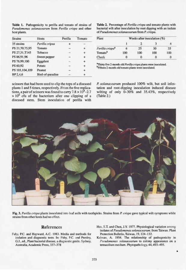
Table 1. Pathogenicity to perilla and tomato of strains of PselldomoMs solanacearum from Perilla crispa and other host plants.
Strains Hosts Perilla Tomato
35 strains Perilla cripsa + PS 21,70,75,95 Tomato + PS 27.31,T145 Tobacco + PS 86.91.96 Sweet pepper + PS 76,99.100 Eggplant + PS 60,92 Potato + PS 103,104,109 Peanut + BP2,4,6 Bird.of-paradise +
scissors that had been used to etip the tops of a diseased plants I and 5 times, respectively. From the five replications. a pair of scissors was fou nd to carry 7.8 x 10⁴-2.7 x 10⁶ cfu of the bacterium after one clipping of a diseased stem. Stem inoculation of perilla with
Table 2. Percentage of Perilla crispa and tomato plants with bacterial wilt after inoculation by root dipping with an isolate of PseudomOMs solanacearum from P. crispa.
Plant Weeks after inoculation (%)
I 2 3 4
Perilla crispaa 6 25 30 35
Tomatob 100 100 100 100
Check 0 0 0 0
"Thirty five 2-month-old Perilla crispa plants were inoculated. !>Piftcen 2-month-old tomato plants were inoculated.
P. solanacearum produced 100% wilt, but soil infestation and root-dipping inoculation induced disease wilting of only 0-30% and 35.43%, respectively (Table 2.)
Fig. 3. Pailla crisp a plants inoculated into leafaxils with toothpicks. Strains from P. crispa gave typical wilt ymptoms while strains from other hosts had no effect.
References
Pally. P.C. and Hayward, A.C. 1983. Media and methods for isolation and diagnostic tests. In: Fahy. P.C. and Persley. G.J., ed., Plant bacterial disease, a diagllOstic guide. Sydney, Australia. Academic Press, 337-378
375
Hsu, S.T. and Chen. J.Y. 1977. Physiological variation among isolates of Pseudomonas solanacearum. from Taiwan. Plant Protection Bulletin, Taiwan, 19, 124-132.
KelOlan. A. 1954. The relationship of pathogenicity in Pselldomollas solallacearum to colony appearance on a tetrawlium medium. Phytopathology. 44, 493-495.
•

Plenary Session Reports

Plenary Session
A.C. Hayward
WORK presented at this symposium has provided ample evidence of the range of variability of the bacterial wilt pathogen in genotype, phenotype and pathotype. The question should be asked to what extent the present formal and informal nomenclature successfully accommodates this range of variability and, if there is deficiency, what steps might be taken in the future to improve the situation.
There seems little doubt that at the highest level there will be further nomenclatural changes to re fl ect the genotypic diversity within the genus Pseudomonas as defi ned at present. It is 20 years since Palleroni et al. (1973) showed that Pseudomonas species were separable into 5 homology groups on the basis of DNA:rRNA homology (hybridisation); since then the phylogenetic heterogeneity of Pseudomonas has been confirmed by comparison of the total sequence of the 1540 nucleotides in the l 6SrRNA of representative species of Pseudomonas (Woese et al. 1984). Representatives of rRNA group 1II have already been distributed into new genera; it is very like ly that in the next 5 years or earlier that the species contained in rRNA group 11, including P solanacearum, will also be placed in new genera.
The sequence information presented at this symposium, together with the results of Palleroni et al. (1 973) show that within this homology group there are two clusters of related species, one including P solanacearum and P pickettii, the other P cepacia, P andropogonis and some other species (Li et al. 1993). These two sub-homology groups are equivalent to genera established elsewhere on the basis of sequence simi lari ty in the nucleotides of the 16SrRNA. However, it would be premature to make any taxonomic changes until additional sequence data are obtained for those species such as P syzygii, the cause of Sumatra disease of cloves, the blood disease bacterium 'Pseudomonas celebense' , and some others, which show either significant levels of DNA:DNA hybridisation, reaction with probes in RFLP analysis, or similarity in phenotype to P solanacearum.
The evidence from several complementary investigations has led to the belief that there may be the ba is for the e tablishment of at least two subspecies in P solanacearum. Cook et al. (1989) have shown that RFLP analysis separates all isolates of P solanacearum into two major divisions, one primarily of Asian origin the other in the Americas. Thi suggests distant evolutionary dichotomy, an idea also supported by the data on sequence of nucleotides of the l6SrRNA in isolates of P solarwcearum representing biovars 1,2, 3 and 4 (Li et al. these proceedings). However, any subspecific classification should be delayed until more detailed comparative studies have been made of larger numbers of isolates of wider geographical origin.
378

At the infrasubspecific level various infonnal systems of nomenclature have been used that are not always easy to relate to one another, and hence there has been some confusion. Differences in genotype are illustrated in the RFLP groupings of Cook et al. (1989) and in the DNA fingerprinting patterns obtained on restriction enzyme analysis (Gillings and Fahy, these proceedings). Differences in phenotype are shown in the biovar classification, and of pathogenicity and host range in races and pathotypes. It is clear that molecular methods of genotypic analysis allow far greater discrimination between strains (isolates) of P. solanacearum than do the phenotypic methods, and these powerful techniques find particular application in some epidemiological investigations and in plant quarantine. The tradi tional phenotypic methods, though simple and inexpensive to apply, often require days or weeks of incubation and the end result of the reaction may be difficult to interpret.
Biovar 2, which was previously considered to be a unifonn phenotype, has now been shown to be differentiable into three sub-phenotypes (French et al., these proceedings). Two of these are metabolic ally less active, differ in a few phenotypic properties, and equate with race 3; they probably evolved at high elevation in the Andes as pathogens of potato and subsequently were distributed worldwide to the altitudinal and latitudinal limits of distribution of the species on latently infected planting material. The metabolically more active sub-phenotype of biovar 2 has so far been found only east of the Andes in Peru and Brazil and mainly at lower elevations. The nomenclature of these sub-phenotypes is in transition. French et al. (these proceedings) have used the term biovar 2-T for the tropical strains from Peru and Brazil, whereas others have used the tenn 'N-2' for the same sub-phenotype. The tenn biovar 2-A was given to the strains of Andean origin by French et al. (the. proceeding ). The latter is perhaps the best defined entity within the ent ire P. solanacearum species complex. All isolates are found in either of two closely-related RFLP groups, 26 and 27, and biovar 2-A (race 3) isolates from whatever geographical location have also been shown to be remarkably unifonn in DNA fingerprinti ng pattern (Gi llings and Fahy, these proceedings). This unifonnity in genotype is matched by phenotypic homogeneity and degree of pathogenic pecialisation. It seems that any fu ture classification at the level of subspecies should recognise this major potato pathogen as a di ti nct and separate entity. One argument against the erection of subspecies for the two divisions of Cook et al. (1989) is that the potato pathogen would be submerged into a subspecies including pathogens specialised to other hosts such as banana. Although there i . no immediate solution to this problem the objective should be an infrasubspecific classification that is universally understood and of maximum utili ty to plant pathologists, plant breeders and others, such as plant quarantine officers, who would make use of such a classification.
References
Cook, D., Barlow, E . and Sequeira, L. 1989. Genetic di versity of Pseudomonas solmwcearum: detection of restriction fragment length polymorph isms with DNA probes that specify virulenc.e and hypersensit ive respon e. Molecular Plant-Microbe Interactions, 2. 113- 12 1.
Li, X., Dorsch. M .. Del Dot, T., Sly, L.I.. Stackebrandt. E. and Hayward. A.C. 1993. Phylogenetic studies o f the rRNA group" pseudomonads based on 16SrRNA gene sequences. Journal of Applied Bacteriology (i n press).
Palleroni, N.1. , Kunisawa. R .• Contopoulou. R. and Doudoroff, M. 1973. Nucleic acid homologies in the genus Pseudorrumas. International Journal ofSy ·tematic Bacteriology. 23. 333- 339.
Woese, C.R., Blanz. P. and Hahn, C.M. 1984. What isn't a pseudomonad: the importance of nomenclature in bacterial classi ficat ion. Systematic and Applied Microbiology, 5. 179- 195.
379

Host Resistance
E.R. French
REPORTS were presented on breeding for resistance in tomato, tobacco, groundnut, brinjal (eggplant) and pepper. The work on potato was raised in discussions. For Eucalyptus spp., selection for resistance has begun for another 'new encounter' disease along the Amazon river in Brazil where strains of biovars 1, 2, and 3 are devastating newly introduced species (could the biovar 2 be similar to the tropical strain described for potato: Bv 2 - T?).
Common to most hosts is the unavailability of adequate resistance, making it necessary to develop integrated control programs. The available tolerance is usually overcome by high temperatures around 30°C or more, or by high inoculum potential (present at times as systemic latent infection during screening procedures). It would seem desirable to utilise recombinant DNA procedures to transform plants with potentially superior genes; however, this has proven elusive so far in work with potatoes. The use of hybrids in breeding for tobacco resistance is noteworthy.
Tomato breeding has seen much effort over several decades with only modest or temporary gains. because of unavailability of general resistance and linkage of resistance with small fruit character. It would seem that even more collaboration is needed between scientists and institutions if significant progress is to be attained in future. It is essential to widely test germplasm to detect, and subsequently accumulate, genes for most strains of the pathogen. Marker assisted selection using molecular techniques would accelerate this process.
The genetics of heritability of resistance in all the crops mentioned is not adequately known, though types of resistance have been described for tomato (and potato) and some genomic regions described for tomato for one strain.
There was general agreement that screening for resistance must be done, or at least culminated, in the field under natural conditions; although efficient prescreening under controlled conditions can be useful.
Resistance to bacterial wilt alone is inadequate. It must be combined with resistance to other diseases and pests (especially nematodes that interact with P solanacearum), and with quality and yield characteristics.
The effectiveness of integrated control of bacterial wilt of potato was shown in Nepal, where mustard was shown to be undesirable in rotation. Similarly, the integrated control of wilt of groundnut in India using a resistance component which is also sought in Uganda, showed promise.
The importance of seed transmission of P solanacearum on tomato was shown in Nepal.
380

Molecular Basis of Virulence and Pathogenicity
Fundamental research is often difficult to fund, its eventual usefulness not always being predictable, but it is the essence of progress. Results of progress were presented on pathogenicity genes (hrp), polylacturonase as a component of disease, relation of exopolysaccharides to virulence and the reversibility of phenotype conversion.
Disease Management: Biological and Cultural Methods
The integrated management of tobacco wilt was presented for Australia, and for the U.S.A in the Host Resistance session.
New factors in the control of potato wilt in the warm tropics-soil amendments, antagonism by P cepacia-were unveiled.
The importance of soil type in programs for integrated control in a tropical climate, as well as the colonisation of roots as a factor in biological control were covered for Guadeloupe, Taiwan and in a general review paper. Fundamental research continues to be needed in these areas. Induced resistance needs to be further explored.
381

No. 28
No. 29
No. 30
No. 31
No. 32
No. 33
No. 34
No. 35
No. 36
No. 37
No. 38
No. 39
No. 40
No. 41
No. 42
No. 43
No. 44
ACIAR Proceedings Series Recent Titles
Tropical tree seed research: proceedings of an international workshop held at the Forestry Training Centre, Gympie, Queensland, Australia, 21-24 August, 1989. J.W. Tumbull (ed.), 156 p. 1990.
Sulfur fertilizer policy for lowland and upland rice cropping systems in Indonesia: proceedings of a seminar held at Jakarta, Indonesia 18-20 July, 1989. Graeme Blair and Rod Lefroy (ed.) 142 p., 1990. Tuna baitfish in the lndo-Pacific region: proceedings of a workshop, Honiara, Solomon Islands, 11-13 December, 1989. SJ.M. Blaber and J.W. CopJand (ed.) 211 p., 1990.
Bacterial wilt of groundnut: proceedings of an ACIARlICRISAT collaborative planning meeting held at Genting Highlands, Malaysia 18-19 March, 1990. KJ. Middleton and A.C. Hayward (ed.) 58 p., 1990.
Forages and plantation crops: proceedings of a workshop held at Sanur Beach. Bali, Indonesia, 27-29 June 1990. H.M. Shelton and W.W. Stilr (ed.) 168 p., 1991.
Technologies for sustainable agriculture on marginal uplands in Southeast Asia: proceedings of a seminar held at Ternate, Cavite, Philippines, 10-14 December 1990. Graeme Blair and Rod Lefroy (ed.) 128 p., 1991.
Buffalo and goats in Asia: genetic diversity and its application. Proceedings of a workshop, Kuala Lumpur, Malaysia, 10-14 February 1991. N.M. Tulloh (ed.), 144 p., 1991. Advances in tropical acacia research: proceedings of a workshop held at Bangkok. Thailand, 11-15 February 1991. J.W. Turnbull (ed.), 234 p., 1991.
Fungi and mycotoxins in stored products: proceedings of an international conference held at Bangkok, Thailand, 23-26 April 1991. B.R. Champ, E. Highley, A.D. Hocking, and J.I. PiU (ed.), 270 p., 1991.
Breeding technologies for tropical acacias. Proceedings of a workshop held in Tawau, Sabah, Malaysia, 1-4 July 1991. L.T. Carron and K.M. Aken (ed.), 132 p., 1992.
Forages on red soils in China: proceedings of a workshop, Lengshuitan, Hunan Province, People's Republic of China, 22-25 April 1991. P.M. Home, D.A. McLeod andJ.M. Scott (ed.), 141 p., 1992.
Newcastle disease in village chickens: control with thermostable oral vaccines: proceedings of an international workshop held in Kuala Lumpur, Malaysia, 6-10 October 1991. P.D. Spradhrow (ed.), 181 p., 1992.
Peanut improvement: a case study in Indonesia. Proceedings of an ACIARI AARD/QDPI collaborative review meeting held at Malang, East Java, Indonesia, 19-23 August 1991. G.C. Wright and K.C. Middleton (ed.), 108 p., 1992. A search for strategies for sustainable dryland cropping in semi-arid eastern Kenya: proceedings of a symposium held in Nairobi, Kenya, 10-11 December 1990. M.E. Probert (ed.), 138 p., 1992.
Productive use of saline land. Proceedings of a workshop, Perth, Western Australia, 10-14 May 1991. Neil Davidson and Richard Galloway (ed.), 123 p., 1993.
PasteureIlosis in production animals. An international workshop at Bali, Indonesia, 10-13 August 1992. B.E. Spencer, R.B. Johnson, D. Hoffmann and L. Lehane (ed.), 256 p.,1993.
Bovine ephemeral fever and related rhabdoviruses. Proceedings of the 1st international symposium, Beijing, PRC, 25-27 August 1992. T.D. St George, M.F. Uren, P.L. Young and D. Hoffmann (ed.), in press.