Ribosome association primes the stringent factor Rel for tRNA ...
14
444–457 Nucleic Acids Research, 2021, Vol. 49, No. 1 Published online 16 December 2020 doi: 10.1093/nar/gkaa1187 Ribosome association primes the stringent factor Rel for tRNA-dependent locking in the A-site and activation of (p)ppGpp synthesis Hiraku Takada 1,2,* , Mohammad Roghanian 1,2 , Julien Caballero-Montes 3 , Katleen Van Nerom 3 , Steffi Jimmy 1,2 , Pavel Kudrin 4 , Fabio Trebini 1 , Rikinori Murayama 5 , Genki Akanuma 6 , Abel Garcia-Pino 3,7,* and Vasili Hauryliuk 1,2,4,* 1 Department of Molecular Biology, Ume˚ a University, SE-901 87 Ume ˚ a, Sweden, 2 Laboratory for Molecular Infection Medicine Sweden (MIMS), Ume ˚ a University, SE-901 87 Ume ˚ a, Sweden, 3 Cellular and Molecular Microbiology, Facult´ e des Sciences, Universit´ e Libre de Bruxelles (ULB), Building BC, Room 1C4 203, Boulevard du Triomphe, 1050 Brussels, Belgium, 4 University of Tartu, Institute of Technology, 50411 Tartu, Estonia, 5 Akita Prefectural Research Center for Public Health and Environment, 6-6 Senshu-Kubotamachi, Akita, 010-0874, Japan, 6 Department of Life Science, Graduate School of Science, Gakushuin University, Tokyo, Japan and 7 WELBIO, Avenue Hippocrate 75, 1200 Brussels, Belgium Received November 02, 2020; Revised November 18, 2020; Editorial Decision November 19, 2020; Accepted November 20, 2020 ABSTRACT In the Gram-positive Firmicute bacterium Bacillus subtilis, amino acid starvation induces synthesis of the alarmone (p)ppGpp by the RelA/SpoT Homolog factor Rel. This bifunctional enzyme is capable of both synthesizing and hydrolysing (p)ppGpp. To de- tect amino acid deficiency, Rel monitors the aminoa- cylation status of the ribosomal A-site tRNA by di- rectly inspecting the tRNA’s CCA end. Here we dis- sect the molecular mechanism of B. subtilis Rel. Off the ribosome, Rel predominantly assumes a ‘closed’ conformation with dominant (p)ppGpp hydrolysis ac- tivity. This state does not specifically select deacy- lated tRNA since the interaction is only moderately affected by tRNA aminoacylation. Once bound to the vacant ribosomal A-site, Rel assumes an ‘open’ conformation, which primes its TGS and Helical do- mains for specific recognition and stabilization of cognate deacylated tRNA on the ribosome. The tRNA locks Rel on the ribosome in a hyperactivated state that processively synthesises (p)ppGpp while the hydrolysis is suppressed. In stark contrast to non- specific tRNA interactions off the ribosome, tRNA- dependent Rel locking on the ribosome and activa- tion of (p)ppGpp synthesis are highly specific and completely abrogated by tRNA aminoacylation. Bind- ing pppGpp to a dedicated allosteric site located in the N-terminal catalytic domain region of the enzyme further enhances its synthetase activity. INTRODUCTION Alarmone nucleotides guanosine pentaphosphate (pppGpp) and tetraphosphate (ppGpp), collectively referred to as (p)ppGpp, regulate metabolism, virulence, stress responses and antibiotic tolerance in the vast ma- jority of bacterial species (1–4). The cellular levels of (p)ppGpp are controlled by members of the RelA/SpoT Homolog (RSH) protein family (5). These enzymes both synthesize (p)ppGpp by transferring the pyrophosphate group of ATP onto the 3 ribose position of either GDP or GTP, and degrade the alarmone by hydrolysing the nucleotide back to GDP or GTP, releasing inorganic pyrophosphate in the process. RSH factors fall into two categories: ‘small’ single do- main RSHs and ‘long’ multi-domain RSHs (5). In the ma- jority of bacterial species, long RSHs are represented by a single ribosome-associated bifunctional enzyme called Rel that is capable of both synthesizing and degrading (p)ppGpp. In the lineage to Beta- and Gammaproteobacte- ria, duplication and functional divergence of the ancestral ribosome-associated Rel gave rise to a pair of specialized factors––the namesakes of the RSH protein family––RelA and SpoT (5,6). RelA is a synthesis-only enzyme incapable of hydrolysing (p)ppGpp (7), while SpoT and Rel are bi- * To whom correspondence should be addressed. Tel: +46 907 850 807; Fax: +46 907 850 807; Email: [email protected] Correspondence may also be addressed to Hiraku Takada. Tel: +46 761 023 344; Fax: +46 761 023 344; Email: [email protected] Correspondence may also be addressed to Abel Garcia-Pino. Tel: +32 2 650 53 77; Fax: +32 2 650 53 77; Email: [email protected] Present address: Pavel Kudrin, Department of Molecular Biology and Genetics, Aarhus University, 8000 Aarhus C, Denmark. C The Author(s) 2020. Published by Oxford University Press on behalf of Nucleic Acids Research. This is an Open Access article distributed under the terms of the Creative Commons Attribution-NonCommercial License (http://creativecommons.org/licenses/by-nc/4.0/), which permits non-commercial re-use, distribution, and reproduction in any medium, provided the original work is properly cited. For commercial re-use, please contact [email protected] Downloaded from https://academic.oup.com/nar/article/49/1/444/6039922 by guest on 12 July 2022
-
Upload
khangminh22 -
Category
Documents
-
view
1 -
download
0
Transcript of Ribosome association primes the stringent factor Rel for tRNA ...

444–457 Nucleic Acids Research, 2021, Vol. 49, No. 1 Published online 16 December 2020doi: 10.1093/nar/gkaa1187
Ribosome association primes the stringent factor Relfor tRNA-dependent locking in the A-site andactivation of (p)ppGpp synthesisHiraku Takada 1,2,*, Mohammad Roghanian 1,2, Julien Caballero-Montes 3,Katleen Van Nerom3, Steffi Jimmy 1,2, Pavel Kudrin 4, Fabio Trebini1, Rikinori Murayama5,Genki Akanuma6, Abel Garcia-Pino 3,7,* and Vasili Hauryliuk 1,2,4,*
1Department of Molecular Biology, Umea University, SE-901 87 Umea, Sweden, 2Laboratory for Molecular InfectionMedicine Sweden (MIMS), Umea University, SE-901 87 Umea, Sweden, 3Cellular and Molecular Microbiology,Faculte des Sciences, Universite Libre de Bruxelles (ULB), Building BC, Room 1C4 203, Boulevard du Triomphe,1050 Brussels, Belgium, 4University of Tartu, Institute of Technology, 50411 Tartu, Estonia, 5Akita PrefecturalResearch Center for Public Health and Environment, 6-6 Senshu-Kubotamachi, Akita, 010-0874, Japan,6Department of Life Science, Graduate School of Science, Gakushuin University, Tokyo, Japan and 7WELBIO,Avenue Hippocrate 75, 1200 Brussels, Belgium
Received November 02, 2020; Revised November 18, 2020; Editorial Decision November 19, 2020; Accepted November 20, 2020
ABSTRACT
In the Gram-positive Firmicute bacterium Bacillussubtilis, amino acid starvation induces synthesis ofthe alarmone (p)ppGpp by the RelA/SpoT Homologfactor Rel. This bifunctional enzyme is capable ofboth synthesizing and hydrolysing (p)ppGpp. To de-tect amino acid deficiency, Rel monitors the aminoa-cylation status of the ribosomal A-site tRNA by di-rectly inspecting the tRNA’s CCA end. Here we dis-sect the molecular mechanism of B. subtilis Rel. Offthe ribosome, Rel predominantly assumes a ‘closed’conformation with dominant (p)ppGpp hydrolysis ac-tivity. This state does not specifically select deacy-lated tRNA since the interaction is only moderatelyaffected by tRNA aminoacylation. Once bound tothe vacant ribosomal A-site, Rel assumes an ‘open’conformation, which primes its TGS and Helical do-mains for specific recognition and stabilization ofcognate deacylated tRNA on the ribosome. The tRNAlocks Rel on the ribosome in a hyperactivated statethat processively synthesises (p)ppGpp while thehydrolysis is suppressed. In stark contrast to non-specific tRNA interactions off the ribosome, tRNA-dependent Rel locking on the ribosome and activa-tion of (p)ppGpp synthesis are highly specific andcompletely abrogated by tRNA aminoacylation. Bind-
ing pppGpp to a dedicated allosteric site located inthe N-terminal catalytic domain region of the enzymefurther enhances its synthetase activity.
INTRODUCTION
Alarmone nucleotides guanosine pentaphosphate(pppGpp) and tetraphosphate (ppGpp), collectivelyreferred to as (p)ppGpp, regulate metabolism, virulence,stress responses and antibiotic tolerance in the vast ma-jority of bacterial species (1–4). The cellular levels of(p)ppGpp are controlled by members of the RelA/SpoTHomolog (RSH) protein family (5). These enzymes bothsynthesize (p)ppGpp by transferring the pyrophosphategroup of ATP onto the 3′ ribose position of either GDPor GTP, and degrade the alarmone by hydrolysing thenucleotide back to GDP or GTP, releasing inorganicpyrophosphate in the process.
RSH factors fall into two categories: ‘small’ single do-main RSHs and ‘long’ multi-domain RSHs (5). In the ma-jority of bacterial species, long RSHs are represented bya single ribosome-associated bifunctional enzyme calledRel that is capable of both synthesizing and degrading(p)ppGpp. In the lineage to Beta- and Gammaproteobacte-ria, duplication and functional divergence of the ancestralribosome-associated Rel gave rise to a pair of specializedfactors––the namesakes of the RSH protein family––RelAand SpoT (5,6). RelA is a synthesis-only enzyme incapableof hydrolysing (p)ppGpp (7), while SpoT and Rel are bi-
*To whom correspondence should be addressed. Tel: +46 907 850 807; Fax: +46 907 850 807; Email: [email protected] may also be addressed to Hiraku Takada. Tel: +46 761 023 344; Fax: +46 761 023 344; Email: [email protected] may also be addressed to Abel Garcia-Pino. Tel: +32 2 650 53 77; Fax: +32 2 650 53 77; Email: [email protected] address: Pavel Kudrin, Department of Molecular Biology and Genetics, Aarhus University, 8000 Aarhus C, Denmark.
C© The Author(s) 2020. Published by Oxford University Press on behalf of Nucleic Acids Research.This is an Open Access article distributed under the terms of the Creative Commons Attribution-NonCommercial License(http://creativecommons.org/licenses/by-nc/4.0/), which permits non-commercial re-use, distribution, and reproduction in any medium, provided the original workis properly cited. For commercial re-use, please contact [email protected]
Dow
nloaded from https://academ
ic.oup.com/nar/article/49/1/444/6039922 by guest on 12 July 2022

Nucleic Acids Research, 2021, Vol. 49, No. 1 445
functional (8,9). The (p)ppGpp synthesis activity of RelAand Rel is induced by so-called ‘starved’ ribosomal com-plexes (i.e. ribosomes accommodating deacylated tRNA inthe A-site) (8,10). While deacylated tRNA dramatically sta-bilizes the association of Rel/RelA with the ribosome (11–13), RelA has significant affinity to the vacant ribosomal A-site as well. Indeed, Loveland et al., in addition to reportingstructures of RelA associated with starved ribosomes, havealso solved a structure of RelA bound to the ribosome with-out A-site tRNA (Structure 1, PDB 5KPS) (14). In the caseof RelA, the pppGpp product is a potent inducer of the en-zyme’s synthesis activity (15). The mechanistic basis of thisregulatory mechanism is currently unexplored, and it is un-known whether bifunctional RSH Rel is similarly regulatedby pppGpp.
Long RSHs are comprised of an enzymatic N-terminalmulti-domain region (NTD) and a regulatory C-terminalmulti-domain region (CTD) (Figure 1). The NTD is sub-divided into (p)ppGpp hydrolase (HD) and (p)ppGppsynthetase (SYNTH) catalytic domains, while the regula-tory CTD is comprised of the TGS (ThrRS, GTPase andSpoT), Helical (equivalent to alpha-helical as per Love-land et al. (14)), ZFD (Zinc Finger Domain; equivalentto CC, Conserved Cysteine as per Atkinson et al. (5) andRIS, Ribosome-InterSubunit as per Loveland et al. (14))and RRM (RNA recognition motif; equivalent to ACT, as-partokinase, chorismate mutase and TyrA as per Atkinsonet al. (5)). Experiments with Rel from Streptococcus dys-galactiae subsp. equisimilis (16,17), Mycobacterium tuber-culosis (18,19), Caulobacter crescentus (20), Staphylococcusaureus (21), Thermus thermophilus (22) and Bacillus subtilis(12) have established that (i) the synthetic and hydrolytic ac-tivities of the NTD are mutually exclusive, (ii) CTD sup-presses the NTD synthetic activity in cis through autoinhi-bition, and (iii) the CTD is essential for Rel regulation bythe ribosomal complexes.
Our understanding of the relationship between the en-zymatic activity of RelA/Rel and the factors’ interactionswith starved ribosomal complexes is largely based on ex-periments with E. coli RelA. A family of related ‘hopping’models was put forward (23–25). According to the origi-nal ‘hopping’ model, each act of (p)ppGpp synthesis fuelsthe displacement of RelA from the ribosome, upon whichthe factor ‘hops’ to the next starved complex (25). Singlemolecule tracking experiments using RelA C-terminal fu-sions with photoswitchable fluorescent proteins led to theformulation of the ‘extended hopping’ (23) and the ‘shorthopping time’ (24) models. The former model suggests thatupon activation by starved ribosomes RelA processivelysynthesizes (p)ppGpp off the ribosome and the latter statesthat upon activation, RelA spends prolonged periods oftime catalytically active in association with starved riboso-mal complexes. Finally, there are two ‘flavours’ of the ‘shorthopping time’ model: RelA could first bind the vacant A-site and then recruit the deacylated tRNA (14,15) or formthe RelA:tRNA complex off the ribosome and deliver dea-cylated tRNA to the A-site, similarly to how elongationfactor EF-Tu delivers aminoacylated tRNA (26,27). Prin-cipally, one could also imagine a ‘tRNA-first’ mechanism,with deacylated tRNA binding first to the vacant A-sitefollowed by the RelA association with the complex. How-
ever, this scenario was deemed unlikely given the structureof RelA locked on the starved ribosome (14).
The proposed delivery of deacylated tRNA to the ribo-some by RelA/Rel is a controversial topic. In the case ofM. tuberculosis Rel, binding to tRNA is readily detectible(8,18). While binding of tRNA to E. coli RelA is not de-tected by EMSA (15,27) unless the complex is crosslinkedby formaldehyde (27), stable tRNA association was re-ported in the case of a truncated version of E. coli RelA con-taining just the TGS and part of the Helical domain (27).This suggests a possibility that in the full-length protein thetRNA interaction interface is not accessible and becomesaccessible only upon ribosomal recruitment or truncationof the protein. Moreover, addition of A-site non-cognatedeacylated tRNA in excess did not significantly inhibit RelAactivation by starved complexes, as it would be expectedif the factor would be sequestered in a complex with non-cognate tRNA off the ribosome (15). Importantly, the orig-inal report proposing tRNA delivery by E. coli RelA re-lied on the UV-induced crosslinking and analysis of cDNAs(CRAC) approach and the authors provided no biochemi-cal or biophysical evidence of the RelA:tRNA complex for-mation (26). While CRAC is an extremely sensitive tech-nique for detection of interactions, it is inherently unable toestimate the affinities or establish the sequence of bindingevents. The TGS and Helical domains of RelA form multi-ple contacts with deacylated A-site tRNA (14,28,29) whileon the ribosome. However, it is unclear if these domains areaccessible when RelA/Rel is off the ribosome.
Studies of the physiological effects of (p)ppGpp in theGram-positive Firmicute bacterium B. subtilis have beenfundamental to our understating of (p)ppGpp-mediatedtranscriptional regulation (30) and regulation of nucleotidemetabolism (31). However, mechanistic understanding of B.subtilis Rel is limited (12,32). Using our reconstituted bio-chemical system (12), we have dissected the relationship be-tween, on the one hand, Rel’s association with deacylatedtRNA and the ribosome and, on the other hand, (p)ppGppsynthesis and hydrolysis by the enzyme, and probed theroles of individual domains. We establish that the specificrecognition of deacylated tRNA takes place after Rel as-sociates with the vacant ribosomal A-site, and the strengthof the interaction with deacylated tRNA fine-tunes the sta-bility of the interaction of Rel/RelA enzymes with starvedribosomes. In the case of B. subtilis Rel, the factor forms astable complex with starved ribosomes and this complex isnot actively dissociated by processive synthesis of ppGpp.We demonstrate that pppGpp enhances synthetase activityof both B. subtilis Rel and E. coli RelA by binding to a ded-icated allosteric site located in the enzymatic NTD regionof the factor.
MATERIALS AND METHODS
Detailed description of experimental procedures is providedin Supplementary methods. All bacterial strains and plas-mids used in this study are listed in Supplementary Tables1 and 2. All proteins were expressed, purified and charac-terized using SUMO-tagging strategy described earlier forE. coli RelA (33) and B. subtilis Rel (12) (SupplementaryFigure S1A-G). The monomeric nature of 50 nM B. sub-
Dow
nloaded from https://academ
ic.oup.com/nar/article/49/1/444/6039922 by guest on 12 July 2022

446 Nucleic Acids Research, 2021, Vol. 49, No. 1
Figure 1. Molecular recognition of the deacylated A/R-tRNA by the stringent factor. The domain composition as well as the truncated versions of theenzymes (B. subtilis Rel: �RRM 1–645, �ZFD-RRM 1–583 and NTD 1–373; E. coli RelA: �RRM 1-657, �ZFD-RRM 1–592 and NTD 1–385) usedfor functional studies are highlighted, as well as functionally important residues D264 and H420 of B. subtilis Rel. The 3′ CCA end of the partially A-siteaccommodated and highly distorted deacylated tRNA (A/R-tRNA) is shown as spheres. The 3D model was constructed from using partial structure of B.subtilis Rel in complex with starved ribosomes (RDB accession number 6HTQ) and a swap dimeric state of C-terminally truncated B. subtilis Rel (6YXA)(32).
tilis Rel and 100 nM E. coli RelA was confirmed by massphotometry (34) using Refeyn OneMP instrument (RefeynLtd.) (Supplementary Figure S1H and I).
Enzymatic assays3H-pppGpp/3H-ppGpp synthesis assays were performed at37◦C in HEPES:Polymix buffer (5 mM Mg2+ final concen-tration) as described earlier (12) using either E. coli RelAand B. subtilis Rel as well as (i) 70S initiation complexesassembled using either E. coli or B. subtilis and (ii) deacy-lated E. coli tRNAVal or tRNAPhe. Either 300 �M 3H-GTPor 300 �M 3H-GDP (both from PerkinElmer) as well as 1mM ATP were used as substrates. 3H-pppGpp/3H-ppGpphydrolysis assays were performed using either 300 �M 3H-pppGpp or 300 �M 3H-ppGpp as substrate as describedearlier (12). Individual quenched timepoints were spottedPEI-TLC plates (Macherey-Nagel), nucleotides resolved in1.5 M KH2PO4 pH 3.5 buffer, the TLC plates were driedand cut into sections as guided by UV-shadowing and 3Hradioactivity was quantified by scintillation counting. En-zyme turnover was estimated by linear regression thoughindividual kinetic points making sure that only the pointson the linear kinetic regime are used (<50% substrate toproduct conversion). The error bars represent standard de-viations of the turnover estimates using four data points;each experiment was performed at least two times.
Sucrose gradient fractionation and Western blotting
Sample preparation i: lysates: B. subtilis samples were pre-pared as described earlier (12) and supplemented with nu-cleotides GTP / GDP (0.5 mM) and ATP/AMPCPP (1mM) as indicated on the figure legends and used for ana-lytical ultracentrifugation and Western blotting.
Sample preparation ii: lysates: MG1655 relA::HTF E. colistrain (26) was grown in liquid LB media at 37◦C. At OD600of 0.2 the culture was treated 20 min with mupirocin addedto final concentration of 70 �M, and the samples were pro-cessed as described for B. subtilis lysates.
Sample preparation iii: reconstituted system: 20 �l reac-tion mixtures containing 500 nM B. subtilis 70S IC(MVF),140 nM Rel, E. coli tRNAVal (all in HEPES:Polymix buffer,5 mM Mg2+ final concentration) were incubated at 37◦C for5 min and loaded onto sucrose gradients.
Sucrose gradient fractionation: clarified cell lysates (orreconstituted ribosomal complexes) were loaded onto10–35% sucrose gradients in HEPES:Polymix buffer pH7.5 (5 mM Mg2+ final concentration, supplementedwith nucleotides GTP:Mg2+/GDP:Mg2+ (0.5 mM) andATP:Mg2+/AMPCPP:Mg2+ (1 mM) as indicated on the fig-ure legends), subjected to centrifugation (36 000 rpm for 3h at 4◦C, SW-41Ti Beckman Coulter rotor) and analysedusing Biocomp Gradient Station (BioComp Instruments)with A260 and A280 as a readout.
Western blotting: experiments were performed as de-scribed earlier (12). Ribosomal protein L3 of the 50S ribo-somal subunit was detected using anti-L3 primary antibod-ies (a gift from Fujio Kawamura (35)) combined with goatanti-rabbit IgG-HRP secondary antibodies.
Electrophoretic mobility shift assay (EMSA)
Before performing the experiment, stockmRNA(MVF) (5′-GGCAAGGAGGAGAUAAGAAUGGUUUUCUAAUA-3′) was incubated for 2min at 60◦C to denature possible secondary structures.Reaction mixtures (10 �l) in HEPES:Polymix buffer(36) with 5 mM Mg2+ were assembled by adding E. colieither tRNAVal/tRNAi
Met (0.1 �M final concentration)
Dow
nloaded from https://academ
ic.oup.com/nar/article/49/1/444/6039922 by guest on 12 July 2022

Nucleic Acids Research, 2021, Vol. 49, No. 1 447
or mRNA (0.1 �M final concentration) or both (0.1 �MtRNAVal/tRNAi
Met and 1 �M mRNA(MVF) competitor),followed by the addition of Rel. After incubation for 5min at 37◦C, 4 �l of 50% sucrose was added per sample,and the samples were electrophoretically resolved on a12% Tris:Borate:EDTA gel at 4◦C (160–180V) for 1–1.5h. Gels were stained with SYBR Gold nucleic acid stain(Life Technologies) for 30 min, followed by visualizationusing a Typhoon Trio Variable Mode Imager (AmershamBiosciences). Bands were quantified using ImageJ (37). Theefficiency of complex formation (effective concentration,EC50 ± standart deviation) was calculated using the 4PLmodel (Hill equation) as per Sebaugh (38) using eight datapoints; each experiment was performed at least two times.
Isothermal titration calorimetry (ITC)
Dialyzed B. subtilis RelNTD/E. coli RelANTD was concen-trated in Sartorius Ultrafiltration Centrifugal Concentra-tors (cut-off 30 kDa) at 3000 × g to a final concentrationof 35 �M. 10 mM ppGpp and pppGpp (both from JenaBioscience) were diluted to 450 �M in ITC buffer. Non-hydrolysable ATP analogue AMPCPP (ApCpp, Jena Bio-Science) and GDP were diluted with ITC buffer to 100and 100 �M, respectively, and mixed with the protein priorto the experiment. 300 �M EDTA was incubated with B.subtilis RelNTD overnight at 4◦C and supplemented withAMPCPP and GDP prior to the experiment. The sampleswere degassed and equilibrated at titration temperature andthe ITC measurement were performed using Affinity ITCcalorimeter (TA instruments) at 20◦C. The stirring rate wasset to 75 rpm and a constant injection volume of 2 �l oftitrant (ppGpp or pppGpp) was injected into the cell (177�l of B. subtilis RelNTD) with an injection interval time of250 s. For the heat exchange measurements RelNTD proteinwas concentrated to 150 �M and 2.1 mM stock solutionsof GDP and GTP were used. The stirring rate was set to300 rpm and the measurements were performed at 20, 25and 30◦C. All data were processed and analysed using theNanoAnalyse and Origin software packages.
HPLC-based nucleotide quantification
HPLC-based nucleotide quantification was performed asper Varik et al. (39). Wild type MG1655 and MG1655relA::HTF E. coli (26) strains were grown in MOPS mini-mal media at 37◦C until OD600 0.5 and challenged with 150�g/ml of mupirocin (3× the MIC).
RESULTS AND DISCUSSION
Off the ribosome the NTD region of B. subtilis Rel non-specifically binds RNA
When Rel/RelA is associated with the starved ribosomalcomplex, the TGS and Helical domains form specific con-tacts with the ribosome and tRNA (14,28,29,32) (Figure1). To probe the accessibility of Rel’s TGS and Helical do-mains for interaction with deacylated tRNA off the ribo-some, we purified native, full-length RNA-free untagged B.subtilis Rel as well as a set of C-terminally truncated vari-ants: lacking RRM (Rel�RRM, amino acids 1–645), lacking
both RRM and ZFD (Rel�ZFD-RRM, 1–583) and lacking allof the regulatory CTD domains (RelNTD, 1–373). For thesake of simplicity, throughout the text B. subtilis Rel––themain focus of the current study––is referred to simply as‘Rel’.
As a specificity control for tRNA binding studies we sub-stituted a conserved histidine residue (H420E) in the TGSdomain. The corresponding histidine residue in E. coli RelA(H432) forms a stacking interaction with the 3′ CCA endof the uncharged A-site tRNA (28). The H432E substitu-tion abrogates RelA’s functionality by abolishing RelA acti-vation by deacylated tRNA (12,26). We have shown a sim-ilar loss-of-function effect of the H420E substitution in B.subtilis Rel (12). Therefore, the H420E substitution is an ex-cellent tool to probe the specificity of Rel’s interaction withdeacylated tRNA.
We used electrophoretic mobility shift assays (EMSA) tostudy complex formation between native deacylated E. colitRNAVal and B. subtilis Rel: full-length and C-terminallytruncated, both wild-type and H420E variants. While anal-ogous EMSAs failed to detect a stable complex betweenRelA and deacylated tRNA (15,27), a Rel:tRNA complexis readily observable (Figure 2A and Supplementary Fig-ure S2). In the absence of a non-specific RNA competitor,full-length Rel forms a complex with EC50 of 0.5 �M. Sur-prisingly, the H420E substitution decreases the affinity onlytwice (from EC50 0.5 to 1.0 �M), suggesting a lack of speci-ficity (Figure 2A and Supplementary Figure S2AB).
The sequential deletion of RRM and ZFD domains in-creases the affinity to tRNAVal (EC50 of 0.3 and 0.2 �M),and even upon deletion of the entire CTD, RelNTD affin-ity for tRNAVal remains virtually unaffected (EC50 0.7�M) (Figure 2B, black filled trace, and SupplementaryFigure S2D–F). Consistent with the isolated NTD bind-ing tRNAVal, while the H420E substitution in the full-length and �ZFD-RRM backgrounds does decrease thefactor’s affinity to tRNA, it does not abrogate the inter-action completely (Figure 2B, compare empty and filledtraces). To further characterize the non-specific compo-nent of the Rel:tRNA interaction, we used single-strandedmRNA(MVF) as a competitor. In the presence of a 10-foldexcess of mRNA over tRNA, the affinity for tRNA dropsabout two-fold (Figure 2B, green traces, and SupplementaryFigure S3). At the same time, Rel binds both RNA specieswith a similar affinity, EC50 of 0.5 �M (Figure 2B and C).Using the set of truncated Rel variants described above, welocalized the source of this non-specific affinity for mRNAto Rel’s NTD region (Figure 2C and Supplementary FigureS4).
Taken together, these results suggest that when tested inthe absence of ribosomes, complex formation between Reland tRNA has a clear non-specific component, which is me-diated by the protein’s NTD region. This result is clearly atodds with the highly specific recognition of tRNA by theTGS domain in the cellular context as well as in the pres-ence of starved ribosomal complexes, both of which are ab-rogated by the H420E substitution (12). The ability of E.coli RelA compromised in ribosomal binding by deletionof both RRM and ZFD domains to crosslink to tRNA inCRAC experiments was used as an argument for tRNA de-livery by RelA to the ribosome (26). However, as we show
Dow
nloaded from https://academ
ic.oup.com/nar/article/49/1/444/6039922 by guest on 12 July 2022

448 Nucleic Acids Research, 2021, Vol. 49, No. 1
Figure 2. The isolated TGS-Helical region of B. subtilis Rel, but not the full-length protein, can specifically recognize the tRNA 3′ CCA end. Complexformation between increasing concentrations of B. subtilis Rel (wild-type or mutants, H420E mutants shown as empty circles) and either 0.1 �M E. colitRNAVal or synthetic mRNA(MVF) was monitored by EMSA. Representative full EMSA gels as well as EC50 quantifications are provided as Supplemen-tary Figures S2–S5. (A) Complex formation between E. coli tRNAVal and either wild-type or the H420E Rel mutant in the absence of a non-specific RNAcompetitor. (B) Complex formation between C-terminally truncated Rel variants and E. coli tRNAVal. (C) Complex formation between mRNA(MVF)and full-length Rel, Rel NTD fragment or Rel TGS-Helical fragment. (D) Complex formation between E. coli tRNAVal and either the Rel TGS domainor the Rel TGS-Helical fragment. Complex formation between acetylated and deacylated E. coli tRNAVal and either isolated TGS-Helical fragment (E)or full-length (F) Rel. Analogous experiments using E. coli tRNAi
Met are presented on Supplementary Figure S6.
here, these interactions lack specificity and therefore are un-likely to represent a productive on-path intermediate in themechanism of stringent factor activation. To deconvoluteRNA binding by the NTD from that mediated by the CTD,we next characterized the isolated Rel TGS-Helical region(amino acid positions 374–583) as well as the TGS domainalone (amino acid positions 374–469).
The isolated TGS-Helical region of Rel specifically recog-nizes the tRNA 3′ CCA end
While we do not detect formation of a stable complex in thecase of TGS alone, the TGS-Helical fragment of Rel bindstRNAVal with EC50 of 0.8 �M (Figure 2D and Supple-mentary Figure S5AB). In stark contrast to the full-length
protein, this interaction is completely abrogated upon in-troduction of the H420E substitution, suggesting that forthese isolated regions (unlike the full-length protein), com-plex formation is driven by a specific recognition of the 3′CCA tRNA end. Importantly, unlike the full-length enzymeand the NTD, the TGS-Helical fragment does not bindmRNA(MVF), further reinforcing the specificity of this in-teraction (Figure 2C and Supplementary Figure S4C). Fi-nally, aminoacylation of tRNAVal completely abrogates theinteraction with the isolated TGS-Helical domains (Fig-ure 2E and Supplementary Figure S5EF), while the affin-ity of the full-length Rel remains largely unaffected (Figure2F and Supplementary Figure S3GH). To rule out tRNA-specific effects, we performed an analogous set of bindingassays with full-length Rel and the isolated TGS-Helical
Dow
nloaded from https://academ
ic.oup.com/nar/article/49/1/444/6039922 by guest on 12 July 2022

Nucleic Acids Research, 2021, Vol. 49, No. 1 449
domains using both charged and deacylated E. coli initia-tor tRNAi
Met (Supplementary Figure S6). The results arein excellent agreement with our experiments with tRNAVal:while tRNAi
Met binding to full-length Rel is largely insensi-tive to the H420E substitution and tRNA aminoacylation,complex formation with isolated TGS-Helical domains isstrictly dependent on deacylated tRNAi
Met and is abrogatedby the H420E substitution.
Our results demonstrate that, unlike the full-length pro-tein, the isolated Rel TGS-Helical region is highly specificin its recognition of the tRNA 3′ CCA end. Taking into ac-count cryo-EM studies dissecting RelA recruitment to theribosome (14,28,29) and previous mechanistic studies (16),we hypothesize that it is ribosome binding that drives thetransition of Rel from the ‘closed’ conformation, unable tospecifically sense and bind tRNA, to an ‘open’ conforma-tion in which the TGS-Helical region is primed to specif-ically recognize the deacylated 3′ CCA end. Therefore, wenext investigated the effects of deacylated tRNA on the in-teraction of Rel with the ribosome.
Full-length Rel specifically recognizes the 3′ CCA of deacy-lated tRNA on the ribosome
To probe the specificity of tRNA-dependent recruitment ofRel to the ribosome, we performed 3H-pppGpp synthesisassays in the presence of 70S initiation complexes as wellas increasing concentrations of either A-site cognate dea-cylated tRNAVal or non-cognate tRNAi
Met (Figure 3A).The activation of Rel’s synthetic activity by tRNAVal is po-tent (EC50 of 41 ± 7 nM) and specific: we detect no ac-tivation in the presence of tRNAi
Met. Since non-cognatetRNA does not activate Rel in the presence of initiationcomplexes, formation of the stable non-cognate tRNA:Relcomplex off the ribosome is expected to compromise theribosome-dependent activation since the latter is strictly de-pendent on tRNA being cognate. However, even when thenon-cognate tRNAPhe is added in a 50-fold excess of overcognate tRNAVal, we observe only a moderate inhibitoryeffect (30 %), demonstrating efficient selection of the cog-nate tRNA on the ribosome (Figure 3B). We have earlierperformed analogous experiments with E. coli RelA withsimilar results (15).
To directly probe tRNA recruitment to the ribosome, wenext resolved on sucrose gradients either purified stringentfactor combined with reconstituted B. subtilis 70S initiationcomplexes (Figure 3C) or lysates of B. subtilis cells treatedwith the antibiotic mupirocin that induces acute isoleucinestarvation (Figure 3D). We detected native, untagged Rel byimmunoblotting with polyclonal anti-Rel antiserum (12).Deacylated tRNA promotes efficient and specific recruit-ment of Rel to the ribosome in both experimental systems(Figure 3C and D). In stark contrast with the non-specificRel:tRNA interaction off the ribosome, tRNA-mediatedlocking of Rel on the ribosome is highly specific and is ef-ficiently abrogated both by tRNA aminoacylation (Figure3C; reconstituted system) and the H420E substitution (Fig-ure 3C and D; both systems). The latter result is in agree-ment with the CRAC experiments detecting no RelA:rRNAcrosslinks upon amino acid starvation of E. coli expressingthe H432E RelA variant (26).
Next, we tested Rel recruitment upon mupirocin treat-ment in a B. subtilis strain lacking the ribosomal protein L11(�rplK), which is crucial for activation of E. coli RelA bystarved ribosomal complexes (7,25,40), as well as the func-tionality of C. crescentus (41) and B. subtilis (12) Rel. Ingood agreement with earlier results (25,41), the lack of L11does not perturb Rel’s recruitment to the ribosome (Figure3D). This demonstrates that while association of Rel withstarved complexes is driven by deacylated tRNA, recruit-ment to the ribosome is not sufficient for activation of Rel’ssynthetic activity.
Finally, we tested a synthesis-compromised D264G Relvariant (42). Surprisingly, D264G Rel is not stably recruitedto the ribosome when acute amino acid starvation is in-duced by mupirocin (Figure 3D). Note that the centrifu-gation approach can only reliably detect stable complexes;for instance, while Rel lacking the C-terminal RRM do-main is efficiently activated by starved ribosomes, stable ri-bosomal recruitment was not observed upon the mupirocinchallenge (12). A possible explanation is that by drivingthe protein into the SYNTHOFF HDON state, this substitu-tion compromises the binding of the stringent factor to theribosome.
Deacylated tRNA locks Rel on the ribosome in stationaryphase in B. subtilis
As bacteria enter the stationary phase and nutrients becomelimiting, the ribosome-associated RSHs RelA/Rel are acti-vated and the intracellular concentration of (p)ppGpp in-creases (39). We characterized Rel association with ribo-somes in B. subtilis throughout the growth curve, collectingthe samples at OD600 of 0.2, 1.1, 2.1 and 4.8 (Figure 3G).In the last two samples, the bulk of Rel is recruited to ribo-somes and the 100S ribosome dimer peak is prominent. Thelatter is a sign of high (p)ppGpp levels: expression of the ri-bosome dimerization factor HPF is under positive stringentcontrol (43) resulting in formation of 100S ribosome dimersupon entry to the stationary phase (44).
Processive (p)ppGpp synthesis by ribosome-associated Reldoes not efficiently dislodge Rel from the ribosome
Next, we reassessed the ‘hopping’ models by testing theeffects of nucleotide substrates for (p)ppGpp synthesis onthe interaction of wild-type Rel with starved ribosomes incell lysates (Figure 3E). We supplemented both the lysatesand the corresponding sucrose gradients with guanosinesubstrates (0.5 mM GTP or GDP), both added aloneor together with either ATP or its non-hydrolysable ana-logue, �,�-methyleneadenosine 5′-triphosphate (AMPCPP,1 mM). Importantly, since disrupted cells constitute <1%of the final gradient volume, the carryover of the intracellu-lar nucleotides is negligible. This set of conditions was usedto discriminate between the effects of active (p)ppGpp syn-thesis (GTP/GDP supplemented with ATP) and substratebinding per se (all the other conditions), since the formerwas suggested to actively fuel the dissociation of E. colistringent factor RelA from the ribosome (25). In the pres-ence of the GDP substrate, the Western blot signal of Rel isspread out in the ribosomal fractions, with further addition
Dow
nloaded from https://academ
ic.oup.com/nar/article/49/1/444/6039922 by guest on 12 July 2022

450 Nucleic Acids Research, 2021, Vol. 49, No. 1
Figure 3. Rel is stably recruited to the starved ribosomal complex to drive processive (p)ppGpp production. SYNTH activity of Rel assayed in the presenceof (A) 70S IC(MVF) as well as increasing concentrations of cognate tRNAVal or non-cognate tRNAi
Met or (B) starved ribosomal complexes (70S IC andsub-saturating concentration of cognate tRNAVal, 0.2 �M) as well increasing concentrations of non-cognate tRNAPhe. (C) Sucrose gradient centrifugationand immunobloting analyses probing against either Rel or ribosomal protein L3 of reconstituted Rel:ribosome complexes. Reaction mixtures containing0.5 �M IC (MVF) and 140 nM of wild-type or H420E Rel protein were supplemented with either 2 �M tRNAVal or 2 �M Val-tRNAVal, incubated at37◦C for 5 min and resolved on 10 to 35% sucrose gradients in HEPES:Polymix buffer (pH 7.5, 5 mM Mg2+). (D) Polysome profile and Rel immunoblotanalyses probing Rel’s interaction with the ribosomes with or without the induction of acute isoleucine starvation by antibiotic mupirocin. Wild-type Relwas expressed in either wild-type 168 B. subtilis or in �L11 strain (VHB47). Rel H420E variant defective in CCA recognition was expressed from thenative chromosomal locus in wild-type B. subtilis (VHB68). Synthesis-inactive D264G was ectopically expressed in �rel B. subtilis under the control of
Dow
nloaded from https://academ
ic.oup.com/nar/article/49/1/444/6039922 by guest on 12 July 2022

Nucleic Acids Research, 2021, Vol. 49, No. 1 451
of ATP or AMPCPP having only a minor effect. Notably wesee no difference between ATP and AMPCPP. In the case ofGTP, Rel is detected both in the light fractions (free protein)and in the 70S ribosomal peak. Just as in the case of GDP,further addition of either ATP or AMPCPP does not have asignificant effect. Taken together, these results suggest thatthe catalytic activity – or the lack of it (ATP or its substitu-tion ATP for AMPCPP) – does not play a significant role inRel association with starved ribosomes, as one would expectif acts of (p)ppGpp synthesis would drive the departure ofRel from the ribosome. There is, however, a clear differencebetween the effect of GDP and GTP on Rel association withribosomes. This effect could arise from the GTP-dependentaction of other A-site binding factors present in lysates, e.g.translational GTPases or different conformations inducedby each nucleotide.
A potential drawback of sucrose gradient fractiona-tion experiments is that the SYNTH-competence of theribosome-associated Rel is not established simultaneouslywith immunodetection. Therefore, we have additionallyprobed the connection between ribosomal association and(p)ppGpp synthesis through enzymatic assays. We firstperformed a competition experiment. While keeping theconcentration of wild-type Rel constant, we titrated ei-ther SYNTH-inactive D264G Rel (42) or the SYNTH-competent H420E variant compromised in recognition ofthe tRNA CCA (12) and assayed 3H-pppGpp production inthe presence of starved ribosomes (Supplementary FigureS7). Both D264G and H420E Rel are compromised in theirinteraction with starved ribosomes (Figure 3D), and, whenadded 1:1 with wild-type Rel, do not inhibit (p)ppGpp syn-thesis. However, 10-fold excess of the catalytically-inactiveD264G variant strongly inhibits production 3H-pppGpp,indicative of D264G Rel outcompeting wild-type Rel onstarved ribosomes. Conversely, 10-fold excess of H420G Reldecreases the synthesis rate by merely 3.5-fold. This is prob-ably due to, first, a competitive advantage of the wild-typeprotein which can be stabilized by tRNA, and, second, thatthe H420E variant still can be activated by the 70S, but notby deacylated tRNA (12).
To further discriminate between ‘hopping’ and proces-sive synthesis on the ribosome, we titrated wild-type Rel inour reconstituted biochemical system. When the reactionturnover is calculated per starved ribosomal complex (Fig-ure 3F, red trace), the enzymatic activity reaches a plateauwhen the concentration of Rel is equal to that of the ribo-somes (0.5 �M). At higher Rel concentrations the efficiencyof 3H-pppGpp production in the reconstituted system doesnot increase. Importantly, when turnover is calculated perRel molecule, it decreases, consistent with only (stably)ribosome-bound Rel molecules being activated (Figure 3F,black trace). This behaviour is consistent with Rel proces-sively synthesizing (p)ppGpp while associated with starvedcomplexes rather than the enzyme spending prolonged pe-
riods off the ribosome in a catalytically active state upondeparture from the ribosome. In the latter case one couldexpect that, acting catalytically, one starved ribosomal com-plex would fully activate several Rel molecules; this does notseem to be the case.
The hydrolysis substrate pppGpp does not promote Rel disso-ciation from starved ribosomal complexes
Given the well-documented antagonistic allosteric couplingbetween the SYNTH and HD catalytic domains of Rel en-zymes (17,22), we tested the effect of the hydrolysis sub-strate pppGpp on Rel’s interaction with ribosomes. How-ever, due to the significant volume of sucrose gradients (12mL) it is not feasible to perform the experiment in a waythat makes the substrate available for the enzyme as it mi-grates though the gradient. Therefore, we resorted to sup-plementing only the lysate with 1 mM pppGpp. We seeno effect on Rel’s association with the ribosome (Supple-mentary Figure S8). There are at least two possible expla-nations for this result. First, pppGpp could be diluted inthe test tube and thus lose its effect. Second, the associa-tion of Rel with the starved complexes could, by promot-ing its synthesis activity, inhibit pppGpp binding to theHD domain, thus counteracting the potential effect of thissubstrate.
The synthetase activity of Rel is activated by pppGpp bindingto an allosteric site in the NTD region
Next, we characterized the effects of substrates (GDPor GTP) as well as regulators (ribosomal complexes,tRNA, ppGpp and pppGpp) on the synthesis activityof Rel. Since Mn2+ is universally essential for hydroly-sis activity of long RSHs such as M. tuberculosis Rel(8), S. equisimilis Rel (16) T. thermophilus Rel (45) andE. coli SpoT (46), we characterized the synthesis activ-ity of the full-length Rel in the absence of divalent man-ganese ions in order to avoid possible underestimationof the synthesis efficiency due to concomitant (p)ppGpphydrolysis.
While the ribosome stimulates Rel’s synthesis activity 5-to 10-fold, and the ultimate activator – the starved com-plex – has a significantly stronger effect, ∼50-fold (Fig-ure 4A and B). In good agreement with our earlier resultswith E. coli RelA (15), deacylated tRNA by itself has nosignificant effect. As with Rel enzymes from S. equisimilis(16) and M. tuberculosis (19,47), B. subtilis Rel is moder-ately more efficient in converting GTP to pppGpp than con-verting GDP to ppGpp. Conversely, E. coli RelA prefersthe GDP substrate (15,47). The preference for GTP likelyplays a regulatory role since it is GTP consumption, ratherthan direct (p)ppGpp binding to RNA polymerase thateffectuates the transcriptional regulation upon the strin-gent response in B. subtilis (30,48). Finally, similarly to
←−−−−−−−−−−−−−−−−−−−−−−−−−−−−−−−−−−−−−−−−−−−−−−−−−−−−−−−−−−−−−−−−−−−−−−−−−−−−−−−−−−−−−−−−−−−Phy-spank promotor (VHB156), and expression was induced by 1 mM IPTG. (E) Effects of nucleotide substrates on Rel’s association with starved ribosomesgenerated by B. subtilis by treatment with mupirocin. (F) Synthase activity of Rel activated by starved ribosomal complexes (0.5 �M 70S IC(MVF) and2 �M tRNAVal) as a function increasing concentrations of Rel. Error bars represent standard deviations of the turnover estimates by linear regressionand each experiment was performed at least three times. (G) Ribosomal association of Rel as a function of B. subtilis growth phase. All experiments wereperformed with B. subtilis grown in liquid LB media at 37◦C.
Dow
nloaded from https://academ
ic.oup.com/nar/article/49/1/444/6039922 by guest on 12 July 2022

452 Nucleic Acids Research, 2021, Vol. 49, No. 1
Figure 4. Role of individual domains in the regulation of Rel’s (p)ppGpp synthesis and hydrolysis activities by tRNA, ribosomes, starved ribosomalcomplexes and pppGpp. Synthase activity of 140 nM wild-type and C-terminally truncated Rel assayed in the presence of 1 mM ATP (A–C) and 0.3 mMof either 3H-labeled GDP (A) or GTP (B and C). Hydrolase activity of 140 nM wt and C-terminally truncated Rel variants assayed in the presence of0.3 mM of 3H-labeled pppGpp (D–F). As indicated on the figure, the reaction mixtures were supplemented with combinations of 0.5 �M IC (MVF) andtRNAVal (2 �M: A-site) as well as 100 �M ppGpp (A, B and D) or pppGpp (A–C). All experiments were performed in HEPES:Polymix buffer, pH 7.5at 37◦C in the presence of either 5 mM Mg2+ (A–C), increasing concentrations of Mn2+ (D) or 5 mM Mg2+ supplemented with 1 mM Mn2+ (E and F).Error bars represent standard deviations of the turnover estimates by linear regression and each experiment was performed at least three times.
E. coli RelA (15), while both ppGpp and pppGpp stimu-late B. subtilis Rel synthetic activity, ppGpp has a weakereffect.
Stimulation of the synthesis activity of B. subtilis Rel bypppGpp implies complex formation between the alarmoneand the protein. We used isothermal titration calorimetry(ITC) to study the binding of pppGpp to the NTD-onlyvariant of Rel which is dramatically more soluble than thefull-length protein and, just like the full-length protein, isactivated by pppGpp (Supplementary Figure S9A). The RelNTD region binds pppGpp with an affinity of 10.6 ± 0.9�M (Table 1 and Supplementary Figure S9B). Importantly,the interaction has a stoichiometry close to unity, i.e. oneRel molecule binds one pppGpp molecule. To test the pos-sibility of the heat signal being reflective of the alarmonebinding to one of the two active sites, we titrated pppGppinto RelNTD in the presence of saturating concentrationsof GDP and a non-hydrolysable ATP analogue AMPCPP(to prevent (p)ppGpp biding in the catalytic pocket of theSYNTH domain), or GDP and AMPCPP combined withEDTA (to remove the Mn2+ ion from the catalytic site of theHD domain and prevent the potential binding of (p)ppGppby this site). In both cases, both the affinity (KD) or the sto-ichiometry (n) of pppGpp binding remained largely unper-turbed. Taken together, our results suggest the existence of
a dedicated pppGpp-binding allosteric site located in theNTD region of Rel.
Starved ribosomal complexes actively induce Rel’s synthe-sis activity rather than merely release the auto-inhibition byCTD
Next, we used enzymatic assays in a reconstituted biochem-ical system to probe the roles of the individual domains insensing the ribosome and A-site deacylated tRNA. Dele-tion of the RRM domain had modest effect on activationby starved complexes and pppGpp (Figure 4C). Progressiveremoval of RRM and ZFD domains abrogates activationby A-site tRNA, although activation by the initiation com-plex itself remains detectable. In good agreement with ear-lier results for M. tuberculosis Rel (18,19), when tested in theabsence of the ribosomes, B. subtilis Rel NTD has a synthe-sis activity that is only about two-fold higher than the full-length protein. Thus the data do not support the autoinhi-bition model sensu stricto, where NTD synthesis activity issuppressed by the CTD, and release of this inhibition uponinteraction with the starved ribosome explains the mecha-nism of Rel/RelA activation (16,49,50). Since the release ofthe autoinhibition of the NTD by the CTD does not ac-count for the full activity of Rel observed in the presence
Dow
nloaded from https://academ
ic.oup.com/nar/article/49/1/444/6039922 by guest on 12 July 2022

Nucleic Acids Research, 2021, Vol. 49, No. 1 453
Table 1. Thermodynamic parameters of pppGpp binding to NTD domain region fragments of B. subtilis Rel and E. coli RelA as determined by ITC
Titration KD, �M �G, kcal mol−1 �H, kcal mol−1 –T�S, kcal mol−1 n
pppGpp into RelNTD 10.6 ± 0.9 − 6.67 ± 0.01 − 2.2 ± 0.8 − 4.5 ± 0.4 1.1 ± 0.1pppGpp into RelNTD + AMPCPP + GDP 9 ± 1 − 6.74 ± 0.07 − 1.8 ± 0.5 − 4.9 ± 1 0.9 ± 0.2pppGpp into RelNTD + AMPCPP + GDP + EDTA 10 ± 2 − 6.73 ± 0.01 − 2.4 ± 0.8 − 4.3 ± 0.5 1.0 ± 0.1pppGpp into RelANTD 6.9 ± 0.9 − 6.92 ± 0.02 − 2.8 ± 0.5 − 4.1 ± 0.5 0.9 ± 0.5pppGpp into RelANTD + AMPCPP + GDP +EDTA
11 ± 1 − 6.76 ± 0.01 − 14 ± 1 7.6 ± 0.5 0.9 ± 0.2
The binding affinities were determined from fitting a single interaction model to the experimental ITC data. Data represent mean values ± standarddeviations.
of starved ribosomes, our results point toward ribosomalcomplexes playing an active role in induction of synthesisin addition to the release of auto-inhibition.
The ribosome and tRNA inhibit (p)ppGpp hydrolysis by Rel
The synthetic and hydrolytic activities of Rel are mutuallyexclusive (16,17,22). This motivated us to directly test theeffects of the ligands that induce the synthetic activity (ri-bosomes, tRNA) on Rel’s hydrolysis activity. The hydro-lase activity of the enzyme tested alone is strictly Mn2+-dependent and peaks at 1 mM of Mn2+ (Figure 4D, emptyblack circles). Notably, when we tested the effects of increas-ing Mn2+ concentration on 3H-pppGpp production by Relactivated by starved complexes, only a modest decrease innet SYNTH activity was detected (Figure 4D, filled red cir-cles). This result is in good agreement with suppression ofRel’s HD activity by recruitment to the ribosome (see be-low).
We tested the effects of tRNA, ribosomes and starvedcomplexes (Figure 4E). In good agreement with earlier re-sults with M. tuberculosis Rel (8), tRNA inhibits the hy-drolysis activity of B. subtilis Rel, and the inhibition is ab-rogated when the interaction with the tRNA CCA end isdisrupted by the H420E substitution. Both initiation andstarved complexes inhibit hydrolysis, and the H420E sub-stitution does not overcome this effect, suggesting that theH420E Rel does bind to the ribosome, although can notbe stabilized by the deacylated tRNA. Therefore, we con-clude that it is the ribosome itself that inhibits hydrol-ysis. Synthesis-inactive D264G Rel has elevated hydroly-sis activity, consistent with the antagonistic allosteric cou-pling between SYNTH and HD domains (17,22). This ac-tivity is inhibited by ribosomes, albeit, in agreement com-promised ribosomal association of the D264G-subsitutedprotein, less efficiently than in the case of wild-typeRel.
Next, we tested the HD activity of our C-terminally trun-cated Rel variants, both in the presence and absence oftRNAVal (Figure 4F). Progressive deletion of both RRMand ZFD leads to induction of the hydrolysis activity (Fig-ure 4F), while the synthesis activity is compromised (Figure4C). Reduction of Rel to an NTD fragment lacking the reg-ulatory CTD near-completely abrogates the hydrolysis ac-tivity, in good agreement with our microbiological (12) andITC experiments (Supplementary Figure S9), as well as bio-chemical studies of S. equisimilis (16) and C. crescentus (20)Rel enzymes. The inhibitory effect of tRNAVal is lost onlywhen Rel is reduced to its hydrolytically near-inactive NTD.
Taken together, our biochemical results demonstrate thatthe CTD-mediated transduction of the regulatory stimulithat induce (p)ppGpp synthesis inhibits the hydrolysis ac-tivity – and vice versa. This regulatory strategy in the full-length protein utilizes the antagonistic allosteric couplingwithin the NTD enzymatic core of the protein and effi-ciently prevents futile enzymatic activity.
E. coli RelA does not stably associate with starved ribosomalcomplexes due to low affinity to deacylated tRNA
Since E. coli RelA is, arguably, the most well-studied longRSH, we complemented our investigations of B. subtilis Relwith a set of experiments with the E. coli factor. Despitemultiple contacts between tRNA and RelA on starved ribo-somal complexes (14,28,29), our earlier EMSAs using full-length E. coli RelA failed to detect a stable complex withdeacylated tRNA off the ribosome (15). At the same time,Kushwaha and colleagues have recently reported nM-rangetRNA affinity of the E. coli RelA fragment containing theTGS domain and part of the Helical domain (27). There-fore, we tested tRNA binding to a set of C-terminal trun-cations of E. coli RelA (Supplementary Figure S10A), aswell as the isolated TGS-Helical fragment (SupplementaryFigure S10B). However, we did not detect stable complexformation in either of the cases. While the reason for thisdiscrepancy is unclear, our EMSAs strongly suggest that E.coli RelA is a considerably weaker tRNA binder than B. sub-tilis Rel.
Since deacylated tRNA is the driving force promoting as-sociation of Rel and RelA with the ribosome, this impliesthat E. coli RelA association with starved ribosomes is alsoless stable. To detect RelA by Western blotting we used anE. coli strain encoding RelA C-terminally tagged with His6-TEV-FLAG3 (HTF) on the chromosome. This tagged RelAdisplays wild-type-like functionality in live cells (26) (Sup-plementary Figure S11). While the bulk of RelA is shiftedto denser fractions upon amino acid starvation, the proteindoes not form a stable complex (Figure 5A), demonstratingthat indeed E. coli RelA is a significantly weaker binder ofstarved complexes compared to B. subtilis Rel.
The pppGpp-binding site is conserved between B. subtilis Reland E. coli RelA
To test if, similarly to B. subtilis RelNTD, the allostericpppGpp binding site of E. coli RelA is also located in theNTD region, we performed a set of enzymatic assays with
Dow
nloaded from https://academ
ic.oup.com/nar/article/49/1/444/6039922 by guest on 12 July 2022

454 Nucleic Acids Research, 2021, Vol. 49, No. 1
Figure 5. E. coli RelA NTD region contains the allosteric regulatory site that binds pppGpp. (A) MG1655 E. coli (relA::HTF) expressing C-teminallyHTF-tagged RelA was grown in LB liquid medium at 37◦C. To induce amino acid starvation bacterial cultures exponentially growing at OD600 0.2 weretreated for 20 min with mupirocin added to final concentration of 70 �M that completely abolishes bacterial growth. (B and C) Synthesis activity ofwild-type E. coli RelA as well as RelA�RRM (1–657), RelA�ZFD (1–592), and RelANTD (1–385) mutants by assayed in absence of ribosomes, in presenceof 70S initiation complexes and 70S initiation complexes supplemented with cognate deacylated tRNAVal. Experiments were performed in the presenceof either 100 �M ppGpp (B) or pppGpp (C). Error bars represent standard deviations of the turnover estimates by linear regression and each experimentwas performed at least three times.
C-terminally truncated E. coli RelA variants in the pres-ence of either ppGpp (the weaker activator allosteric acti-vator of RelA (15)) or pppGpp (the stronger activator al-losteric activator of RelA (15)) (Figure 5B and C). Sincethe ppGpp alarmone is produced in situ from 3H-GDP, wecompared the effects of the two alarmones rather than omit-ting the alarmone altogether. While B. subtilis Rel lackingthe RRM domain behaves very similarly to the wild-typeprotein (Figure 4C), deletion of the RRM domain stronglycompromises activation of E. coli RelA by the A-site tRNAof the starved complex (Figure 5B). A likely explanationis that the low affinity of RelA to starved complexes ren-ders it more sensitive to destabilization of the complex bydeletion of the RRM domain. Notably, we do not observean increase in its synthetic activity for RelANTD lacking theautoinhibitory CTD region as compared to the full-lengthprotein.
All of the truncated variants of E. coli RelA are consis-tently more SYNTH-active in the presence of pppGpp and,similarly to Rel, this holds even for the NTD-only version.The strength of the effect varies from 2- to 32-fold depend-ing on the construct and whether the protein is tested aloneor in the presence of 70S initiation or starved complexes. Di-rect measurements by ITC confirmed that pppGpp binds toRelANTD with an affinity of 6.9 ± 0.9 �M, and this interac-tion is insensitive to the addition of GDP, AMPCPP andEDTA, (Table 1 and Supplementary Figure S9EF). Thisstrongly suggests that the interaction is not mediated by theactive site, consistent with the alarmone stimulating, ratherthan inhibiting RelA’s enzymatic activity as it would be ex-pected in the case of direct competition with substrates.Taken together, these results demonstrate that as with B.subtilis Rel, the pppGpp-binding allosteric regulatory siteof E. coli RelA is also located in the NTD region of the en-zyme.
Model of Rel regulation by starved ribosomal complexes
The results presented in this study are compatible with arecent CRAC study of E. coli RelA (26). While the cryo-
EM analysis of RelA bound to starved ribosomal complexsuggested that delivery of deacylated tRNA to the riboso-mal A-site in a complex with RelA is structurally unlikely(14), crosslinking of tRNA to RelA lacking RRM and ZDFdomains was used as an argument for tRNA:RelA com-plex formation off the ribosome (26). While these trunca-tions, indeed, compromise the association with and activa-tion of Rel/RelA by starved ribosomal complexes (Figures4C and 5BC), they do not abrogate the interaction withthe ribosome, as evident from the increased crosslinkingto the Sarcin-Ricin Loop, SRL, upon starvation (26). Si-multaneously, removal of RRM and ZDF increases Rel’saffinity to tRNA (Figure 2D), likely by rendering the TGSdomain more accessible and promoting crosslinking totRNA. Likewise, abrogation of both tRNA and rRNAcrosslinks by the H432E (E. coli RelA numbering) sub-stitution compromising interaction with the CCA end ofuncharged A-site tRNA does not necessitate formation ofthe specific tRNA:RelA complex either: the lack of tRNAand rRNA crosslinks is readily explained by the instabil-ity of the mutant RelA:tRNA:ribosome complex (Figure3C and D) which is strongly stabilized by the A-site tRNA(11–13).
We propose the following model that rationalizes previ-ously published studies (12,14,16,17,21,22,25–29) and ourcurrent results (Figure 6). In the absence of amino acidstarvation Rel is not ribosome-associated (Figure 3D). Offthe ribosome, Rel adopts a ‘closed’ conformation in whichtRNA-binding TGS and Helical domains are sequestered(Figure 6A). In this ‘default’ state the protein has net hydro-lase activity, i.e. HDON SYNTHOFF (Figure 4AB and E).Upon amino acid starvation ‘closed’ Rel could form a rela-tively weak (EC50 0.5 �M) complex with deacylated tRNAoff the ribosome (Figure 6B) which suppresses the HDactivity (HDOFF SYNTHOFF) in H420-dependent manner(Figure 4E). However – at least in the test tube – Rel:tRNAinteraction has a strong non-specific component which ismediated by RelNTD region (Figure 2B) and, therefore, itsphysiological relevance is unclear. Amino acid starvation
Dow
nloaded from https://academ
ic.oup.com/nar/article/49/1/444/6039922 by guest on 12 July 2022
Nucleic Acids Research, 2021, Vol. 49, No. 1 455
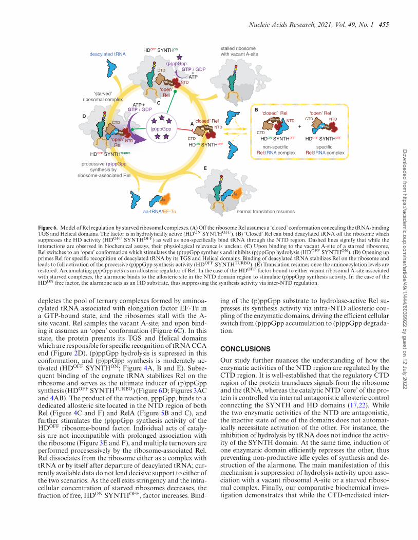
Figure 6. Model of Rel regulation by starved ribosomal complexes. (A) Off the ribosome Rel assumes a ‘closed’ conformation concealing the tRNA-bindingTGS and Helical domains. The factor is in hydrolytically active (HDON SYNTHOFF). (B) ‘Closed’ Rel can bind deacylated tRNA off the ribosome whichsuppresses the HD activity (HDOFF SYNTHOFF) as well as non-specifically bind tRNA through the NTD region. Dashed lines signify that while theinteractions are observed in biochemical assays, their physiological relevance is unclear. (C) Upon binding to the vacant A-site of a starved ribosome,Rel switches to an ‘open’ conformation which stimulates the (p)ppGpp synthesis and inhibits (p)ppGpp hydrolysis (HDOFF SYNTHON). (D) Opening upprimes Rel for specific recognition of deacylated tRNA by its TGS and Helical domains. Binding of deacylated tRNA stabilizes Rel on the ribosome andleads to full activation of the processive (p)ppGpp synthesis activity (HDOFF SYNTHTURBO). (E) Translation resumes once the aminoacylation levels arerestored. Accumulating pppGpp acts as an allosteric regulator of Rel. In the case of the HDOFF factor bound to either vacant ribosomal A-site associatedwith starved complexes, the alarmone binds to the allosteric site in the NTD domain region to stimulate (p)ppGpp synthesis activity. In the case of theHDON free factor, the alarmone acts as an HD substrate, thus suppressing the synthesis activity via inter-NTD regulation.
depletes the pool of ternary complexes formed by aminoa-cylated tRNA associated with elongation factor EF-Tu ina GTP-bound state, and the ribosomes stall with the A-site vacant. Rel samples the vacant A-site, and upon bind-ing it assumes an ‘open’ conformation (Figure 6C). In thisstate, the protein presents its TGS and Helical domainswhich are responsible for specific recognition of tRNA CCAend (Figure 2D). (p)ppGpp hydrolysis is supressed in thisconformation, and (p)ppGpp synthesis is moderately ac-tivated (HDOFF SYNTHON; Figure 4A, B and E). Subse-quent binding of the cognate tRNA stabilizes Rel on theribosome and serves as the ultimate inducer of (p)ppGppsynthesis (HDOFF SYNTHTURBO) (Figure 6D; Figures 3ACand 4AB). The product of the reaction, pppGpp, binds to adedicated allosteric site located in the NTD region of bothRel (Figure 4C and F) and RelA (Figure 5B and C), andfurther stimulates the (p)ppGpp synthesis activity of theHDOFF ribosome-bound factor. Individual acts of cataly-sis are not incompatible with prolonged association withthe ribosome (Figure 3E and F), and multiple turnovers areperformed procesessively by the ribosome-associated Rel.Rel dissociates from the ribosome either as a complex withtRNA or by itself after departure of deacylated tRNA; cur-rently available data do not lend decisive support to either ofthe two scenarios. As the cell exits stringency and the intra-cellular concentration of starved ribosomes decreases, thefraction of free, HDON SYNTHOFF, factor increases. Bind-
ing of the (p)ppGpp substrate to hydrolase-active Rel su-presses its synthesis activity via intra-NTD allosteric cou-pling of the enzymatic domains, driving the efficient cellularswitch from (p)ppGpp accumulation to (p)ppGpp degrada-tion.
CONCLUSIONS
Our study further nuances the understanding of how theenzymatic activities of the NTD region are regulated by theCTD region. It is well-established that the regulatory CTDregion of the protein transduces signals from the ribosomeand the tRNA, whereas the catalytic NTD ‘core’ of the pro-tein is controlled via internal antagonistic allosteric controlconnecting the SYNTH and HD domains (17,22). Whilethe two enzymatic activities of the NTD are antagonistic,the inactive state of one of the domains does not automat-ically necessitate activation of the other. For instance, theinhibition of hydrolysis by tRNA does not induce the activ-ity of the SYNTH domain. At the same time, induction ofone enzymatic domain efficiently represses the other, thuspreventing non-productive idle cycles of synthesis and de-struction of the alarmone. The main manifestation of thismechanism is suppression of hydrolysis activity upon asso-ciation with a vacant ribosomal A-site or a starved riboso-mal complex. Finally, our comparative biochemical inves-tigation demonstrates that while the CTD-mediated inter-
Dow
nloaded from https://academ
ic.oup.com/nar/article/49/1/444/6039922 by guest on 12 July 2022

456 Nucleic Acids Research, 2021, Vol. 49, No. 1
action with the starved ribosome is essential for full acti-vation of the Rel SYNTH domain, the connection betweenthe activity and ribosomal binding is somewhat ‘loose’, witha broad range of acceptable ribosomal affinities for longRSHs. While E. coli RelA is a weak tRNA binder with acorrespondingly low affinity to starved ribosomes, B. sub-tilis Rel has a significantly higher tRNA affinity and is stablyrecruited to the ribosome upon amino acid starvation. Wehypothesize that the stable high-affinity interaction of Relwith starved ribosomal complexes is essential for efficientsuppression of the hydrolysis activity upon amino acid star-vation. In the case of the synthesis-only RelA this fail-safemechanism is not needed, and the protein is evolutionaryoptimized for efficient sampling of the ribosomal popula-tion though dissociation and re-binding.
SUPPLEMENTARY DATA
Supplementary Data are available at NAR Online.
ACKNOWLEDGEMENTS
We are grateful to Protein Expertise Platform (PEP) atUmea University and Mikael Lindberg for constructingplasmids, Gemma C. Atkinson for insightful comments onthe manuscript, Tomas de Garay and Refeyn Ltd. for assis-tance in collecting mass photometry data, Kenn Gerdes andKristoffer Skovbo Winther for sharing MG1655 relA::HTFE. coli strain (26).
FUNDING
European Regional Development Fund through the Cen-tre of Excellence for Molecular Cell Technology (to V.H.);Molecular Infection Medicine Sweden (MIMS) (to V.H.);Swedish Research council [2017-03783 to V.H.]; RagnarSoderberg foundation (to V.H.); Umea Centre for Micro-bial Research (UCMR) [postdoctoral grant 2017 to H.T.];MIMS Excellence by Choice Postdoctoral Fellowship Pro-gramme [postdoctoral grant 2018 to M.R.]; Fonds Na-tional de Recherche Scientifique [FRFS-WELBIO CR-2017S-03, FNRS CDR J.0068.19, FNRS-PDR T.0066.18];Joint Programming Initiative on Antimicrobial Resistance[JPI-EC-AMR -R.8004.18]; Program ‘Actions de RechercheConcertee’ 2016–2021 and Fonds d’Encouragement a laRecherche (FER) of ULB; Fonds Jean Brachet and theFondation Van Buuren (to A.G.P.); the Fund for Researchin Industry and Agronomy (FRIA) from the Fonds dela Recherche Scientifique of Belgium (FNRS) (to J.C.M.).Funding for open access charge: Swedish Research council[2017-03783].Conflict of interest statement. None declared.
REFERENCES1. Hauryliuk,V., Atkinson,G.C., Murakami,K.S., Tenson,T. and
Gerdes,K. (2015) Recent functional insights into the role of(p)ppGpp in bacterial physiology. Nat. Rev. Microbiol., 13, 298–309.
2. Gaca,A.O., Colomer-Winter,C. and Lemos,J.A. (2015) Many meansto a common end: the intricacies of (p)ppGpp metabolism and itscontrol of bacterial homeostasis. J. Bacteriol., 197, 1146–1156.
3. Liu,K., Bittner,A.N. and Wang,J.D. (2015) Diversity in (p)ppGppmetabolism and effectors. Curr. Opin. Microbiol., 24, 72–79.
4. Ronneau,S. and Hallez,R. (2019) Make and break the alarmone:regulation of (p)ppGpp synthetase/hydrolase enzymes in bacteria.FEMS Microbiol. Rev., 43, 389–400
5. Atkinson,G.C., Tenson,T. and Hauryliuk,V. (2011) The RelA/SpoThomolog (RSH) superfamily: distribution and functional evolution ofppGpp synthetases and hydrolases across the tree of life. PLoS One,6, e23479.
6. Mittenhuber,G. (2001) Comparative genomics and evolution of genesencoding bacterial (p)ppGpp synthetases/hydrolases (the Rel, RelAand SpoT proteins). J. Mol. Microbiol. Biotechnol., 3, 585–600.
7. Shyp,V., Tankov,S., Ermakov,A., Kudrin,P., English,B.P.,Ehrenberg,M., Tenson,T., Elf,J. and Hauryliuk,V. (2012) Positiveallosteric feedback regulation of the stringent response enzyme RelAby its product. EMBO Rep., 13, 835–839.
8. Avarbock,D., Avarbock,A. and Rubin,H. (2000) Differentialregulation of opposing RelMtb activities by the aminoacylation stateof a tRNA.ribosome.mRNA.RelMtb complex. Biochemistry, 39,11640–11648.
9. Xiao,H., Kalman,M., Ikehara,K., Zemel,S., Glaser,G. and Cashel,M.(1991) Residual guanosine 3′,5′-bispyrophosphate synthetic activityof relA null mutants can be eliminated by spoT null mutations. J. Biol.Chem., 266, 5980–5990.
10. Haseltine,W.A. and Block,R. (1973) Synthesis of guanosine tetra-and pentaphosphate requires the presence of a codon-specific,uncharged transfer ribonucleic acid in the acceptor site of ribosomes.Proc. Natl. Acad. Sci. U.S.A., 70, 1564–1568.
11. Agirrezabala,X., Fernandez,I.S., Kelley,A.C., Carton,D.G.,Ramakrishnan,V. and Valle,M. (2013) The ribosome triggers thestringent response by RelA via a highly distorted tRNA. EMBO Rep.,14, 811–816.
12. Takada,H., Roghanian,M., Murina,V., Dzhygyr,I., Murayama,R.,Akanuma,G., Atkinson,G.C., Garcia-Pino,A. and Hauryliuk,V.(2020) The C-terminal RRM/ACT domain is crucial for fine-tuningthe activation of ‘long’ RelA-SpoT Homolog enzymes by ribosomalcomplexes. Front. Microbiol., 11, 277.
13. Beljantseva,J., Kudrin,P., Jimmy,S., Ehn,M., Pohl,R., Varik,V.,Tozawa,Y., Shingler,V., Tenson,T., Rejman,D. et al. (2017) Molecularmutagenesis of ppGpp: turning a RelA activator into an inhibitor.Sci. Rep., 7, 41839.
14. Loveland,A.B., Bah,E., Madireddy,R., Zhang,Y., Brilot,A.F.,Grigorieff,N. and Korostelev,A.A. (2016) Ribosome*RelA structuresreveal the mechanism of stringent response activation. Elife, 5,e17029.
15. Kudrin,P., Dzhygyr,I., Ishiguro,K., Beljantseva,J., Maksimova,E.,Oliveira,S.R.A., Varik,V., Payoe,R., Konevega,A.L., Tenson,T. et al.(2018) The ribosomal A-site finger is crucial for binding andactivation of the stringent factor RelA. Nucleic Acids Res., 46,1973–1983.
16. Mechold,U., Murphy,H., Brown,L. and Cashel,M. (2002)Intramolecular regulation of the opposing (p)ppGpp catalyticactivities of Rel(Seq), the Rel/Spo enzyme from Streptococcusequisimilis. J. Bacteriol., 184, 2878–2888.
17. Hogg,T., Mechold,U., Malke,H., Cashel,M. and Hilgenfeld,R. (2004)Conformational antagonism between opposing active sites in abifunctional RelA/SpoT homolog modulates (p)ppGpp metabolismduring the stringent response. Cell, 117, 57–68.
18. Jain,V., Saleem-Batcha,R., China,A. and Chatterji,D. (2006)Molecular dissection of the mycobacterial stringent response proteinRel. Protein Sci., 15, 1449–1464.
19. Avarbock,A., Avarbock,D., Teh,J.S., Buckstein,M., Wang,Z.M. andRubin,H. (2005) Functional regulation of the opposing (p)ppGppsynthetase/hydrolase activities of RelMtb from Mycobacteriumtuberculosis. Biochemistry, 44, 9913–9923.
20. Ronneau,S., Caballero-Montes,J., Coppine,J., Mayard,A.,Garcia-Pino,A. and Hallez,R. (2018) Regulation of (p)ppGpphydrolysis by a conserved archetypal regulatory domain. NucleicAcids Res., 47, 843–854.
21. Gratani,F.L., Horvatek,P., Geiger,T., Borisova,M., Mayer,C., Grin,I.,Wagner,S., Steinchen,W., Bange,G., Velic,A. et al. (2018) Regulationof the opposing (p)ppGpp synthetase and hydrolase activities in abifunctional RelA/SpoT homologue from Staphylococcus aureus.PLoS Genet., 14, e1007514.
22. Tamman,H., Van Nerom,K., Takada,H., Vandenberk,N., Scholl,D.,Polikanov,Y., Hofkens,J., Talavera,A., Hauryliuk,V., Hendrix,J. et al.
Dow
nloaded from https://academ
ic.oup.com/nar/article/49/1/444/6039922 by guest on 12 July 2022

Nucleic Acids Research, 2021, Vol. 49, No. 1 457
(2020) A nucleotide-switch mechanism mediates opposing catalyticactivities of Rel enzymes. Nat. Chem. Biol., 16, 834–840.
23. English,B.P., Hauryliuk,V., Sanamrad,A., Tankov,S., Dekker,N.H.and Elf,J. (2011) Single-molecule investigations of the stringentresponse machinery in living bacterial cells. Proc. Natl. Acad. Sci.U.S.A., 108, E365–E373.
24. Li,W., Bouveret,E., Zhang,Y., Liu,K., Wang,J.D. and Weisshaar,J.C.(2016) Effects of amino acid starvation on RelA diffusive behavior inlive Escherichia coli. Mol. Microbiol., 99, 571–585.
25. Wendrich,T.M., Blaha,G., Wilson,D.N., Marahiel,M.A. andNierhaus,K.H. (2002) Dissection of the mechanism for the stringentfactor RelA. Mol. Cell, 10, 779–788.
26. Winther,K.S., Roghanian,M. and Gerdes,K. (2018) Activation of thestringent response by loading of RelA-tRNA complexes at theribosomal A-Site. Mol. Cell, 70, 95–105.
27. Kushwaha,G.S., Bange,G. and Bhavesh,N.S. (2019) Interactionstudies on bacterial stringent response protein RelA with unchargedtRNA provide evidence for its prerequisite complex for ribosomebinding. Curr. Genet., 65, 1173–1184.
28. Brown,A., Fernandez,I.S., Gordiyenko,Y. and Ramakrishnan,V.(2016) Ribosome-dependent activation of stringent control. Nature,534, 277–280.
29. Arenz,S., Abdelshahid,M., Sohmen,D., Payoe,R., Starosta,A.L.,Berninghausen,O., Hauryliuk,V., Beckmann,R. and Wilson,D.N.(2016) The stringent factor RelA adopts an open conformation onthe ribosome to stimulate ppGpp synthesis. Nucleic Acids Res., 44,6471–6481.
30. Krasny,L. and Gourse,R.L. (2004) An alternative strategy forbacterial ribosome synthesis: Bacillus subtilis rRNA transcriptionregulation. EMBO J., 23, 4473–4483.
31. Kriel,A., Bittner,A.N., Kim,S.H., Liu,K., Tehranchi,A.K., Zou,W.Y.,Rendon,S., Chen,R., Tu,B.P. and Wang,J.D. (2012) Direct regulationof GTP homeostasis by (p)ppGpp: a critical component of viabilityand stress resistance. Mol. Cell, 48, 231–241.
32. Pausch,P., Abdelshahid,M., Steinchen,W., Schafer,H., Gratani,F.L.,Freibert,S.A., Wolz,C., Turgay,K., Wilson,D.N. and Bange,G. (2020)Structural basis for regulation of the Opposing (p)ppGpp synthetaseand hydrolase within the stringent response orchestrator rel. CellRep., 32, 108157.
33. Turnbull,K.J., Dzhygyr,I., Lindemose,S., Hauryliuk,V. andRoghanian,M. (2019) Intramolecular interactions dominate theautoregulation of Escherichia coli stringent factor RelA. Front.Microbiol., 10, 1966.
34. Tamara,S., Franc,V. and Heck,A.J.R. (2020) A wealth ofgenotype-specific proteoforms fine-tunes hemoglobin scavenging byhaptoglobin. Proc. Natl. Acad. Sci. U.S.A., 117, 15554–15564.
35. Murina,V., Kasari,M., Takada,H., Hinnu,M., Saha,C.K.,Grimshaw,J.W., Seki,T., Reith,M., Putrins,M., Tenson,T. et al. (2019)ABCF ATPases involved in protein synthesis, ribosome assembly and
antibiotic resistance: structural and functional diversification acrossthe tree of life. J. Mol. Biol., 431, 3568–3590.
36. Antoun,A., Pavlov,M.Y., Tenson,T. and Ehrenberg,M.M. (2004)Ribosome formation from subunits studied by stopped-flow andRayleigh light scattering. Biol. Proced. Online, 6, 35–54.
37. Schneider,C.A., Rasband,W.S. and Eliceiri,K.W. (2012) NIH Imageto ImageJ: 25 years of image analysis. Nat. Methods, 9, 671–675.
38. Sebaugh,J.L. (2011) Guidelines for accurate EC50/IC50 estimation.Pharm. Stat., 10, 128–134.
39. Varik,V., Oliveira,S.R.A., Hauryliuk,V. and Tenson,T. (2017)HPLC-based quantification of bacterial housekeeping nucleotidesand alarmone messengers ppGpp and pppGpp. Sci. Rep., 7, 11022.
40. Parker,J., Watson,R.J. and Friesen,J.D. (1976) A relaxed mutant withan altered ribosomal protein L11. Mol. Gen. Genet., 144, 111–114.
41. Boutte,C.C. and Crosson,S. (2011) The complex logic of stringentresponse regulation in Caulobacter crescentus: starvation signalling inan oligotrophic environment. Mol. Microbiol., 80, 695–714.
42. Nanamiya,H., Kasai,K., Nozawa,A., Yun,C.S., Narisawa,T.,Murakami,K., Natori,Y., Kawamura,F. and Tozawa,Y. (2008)Identification and functional analysis of novel (p)ppGpp synthetasegenes in Bacillus subtilis. Mol. Microbiol., 67, 291–304.
43. Tagami,K., Nanamiya,H., Kazo,Y., Maehashi,M., Suzuki,S.,Namba,E., Hoshiya,M., Hanai,R., Tozawa,Y., Morimoto,T. et al.(2012) Expression of a small (p)ppGpp synthetase, YwaC, in the(p)ppGpp(0) mutant of Bacillus subtilis triggers YvyD-dependentdimerization of ribosome. Microbiologyopen, 1, 115–134.
44. Akanuma,G., Kazo,Y., Tagami,K., Hiraoka,H., Yano,K., Suzuki,S.,Hanai,R., Nanamiya,H., Kato-Yamada,Y. and Kawamura,F. (2016)Ribosome dimerization is essential for the efficient regrowth ofBacillus subtilis. Microbiology, 162, 448–458.
45. Van Nerom,K., Tamman,H., Takada,H., Hauryliuk,V. andGarcia-Pino,A. (2019) The Rel stringent factor from Thermusthermophilus: crystallization and X-ray analysis. Acta Crystallogr. FStruct. Biol. Commun., 75, 561–569.
46. Heinemeyer,E.A., Geis,M. and Richter,D. (1978) Degradation ofguanosine 3′-diphosphate 5′-diphosphate in vitro by the spoT geneproduct of Escherichia coli. Eur. J. Biochem., 89, 125–131.
47. Sajish,M., Kalayil,S., Verma,S.K., Nandicoori,V.K. and Prakash,B.(2009) The significance of EXDD and RXKD motif conservation inRel proteins. J. Biol. Chem., 284, 9115–9123.
48. Ratnayake-Lecamwasam,M., Serror,P., Wong,K.W. andSonenshein,A.L. (2001) Bacillus subtilis CodY repressesearly-stationary-phase genes by sensing GTP levels. Genes Dev., 15,1093–1103.
49. Gropp,M., Strausz,Y., Gross,M. and Glaser,G. (2001) Regulation ofEscherichia coli RelA requires oligomerization of the C-terminaldomain. J. Bacteriol., 183, 570–579.
50. Schreiber,G., Metzger,S., Aizenman,E., Roza,S., Cashel,M. andGlaser,G. (1991) Overexpression of the relA gene in Escherichia coli.J. Biol. Chem., 266, 3760–3767.
Dow
nloaded from https://academ
ic.oup.com/nar/article/49/1/444/6039922 by guest on 12 July 2022