Pheromonal bile acid 3-ketopetromyzonol sulfate primes the neuroendocrine system in sea lamprey
13
RESEARCH ARTICLE Open Access Pheromonal bile acid 3-ketopetromyzonol sulfate primes the neuroendocrine system in sea lamprey Yu-Wen Chung-Davidson 1 , Huiyong Wang 1 , Michael J Siefkes 1,2 , Mara B Bryan 1,3 , Hong Wu 1,4 , Nicholas S Johnson 1,5 and Weiming Li 1* Abstract Background: Vertebrate pheromones are known to prime the endocrine system, especially the hypothalamic- pituitary-gonadal (HPG) axis. However, no known pheromone molecule has been shown to modulate directly the synthesis or release of gonadotropin releasing hormone (GnRH), the main regulator of the HPG axis. We selected sea lamprey (Petromyzon marinus) as a model system to determine whether a single pheromone component alters the output of GnRH. Sea lamprey male sex pheromones contain a main component, 7α, 12α, 24-trihydroxy-5α-cholan-3-one 24-sulfate (3 keto-petromyzonol sulfate or 3kPZS), which has been shown to modulate behaviors of mature females. Through a series of experiments, we tested the hypothesis that 3kPZS modulates both synthesis and release of GnRH, and subsequently, HPG output in immature sea lamprey. Results: The results showed that natural male pheromone mixtures induced differential steroid responses but facilitated sexual maturation in both sexes of immature animals (χ 2 = 5.042, dF = 1, p < 0.05). Exposure to 3kPZS increased plasma 15α-hydroxyprogesterone (15α-P) concentrations (one-way ANOVA, p < 0.05) and brain gene expressions (genes examined: three lamprey (l) GnRH-I transcripts, lGnRH-III, Jun and Jun N-terminal kinase (JNK); one-way ANOVA, p < 0.05), but did not alter the number of GnRH neurons in the hypothalamus in immature animals. In addition, 3kPZS treatments increased lGnRH peptide concentrations in the forebrain and modulated their levels in plasma. Overall, 3kPZS modulation of HPG axis is more pronounced in immature males than in females. Conclusions: We conclude that a single male pheromone component primes the HPG axis in immature sea lamprey in a sexually dimorphic manner. Keywords: Pheromone, Priming, HPG axis, GnRH, Steroid, Sexual dimorphism Background The term “pheromones” was first introduced by Karlson and Lüscher (1959) to describe the substances involved in chemical communication among conspecifics [1]. Pheromones can be classified as releasers, primers, or more recently, signalers and modulators [2-6]. These functions are not mutually exclusive. Primer pheromones are exem- plified by their effects on the onset of puberty, the length of estrous cycles in females, the success or failure of preg- nancy, and shifts in hormone levels in mice [6]. Male pheromones have a direct impact on female sexual desire, menstrual cycles and ovulation in humans [7]. About 20% of women who have smelled male underarm secretions have an advanced onset of next luteinizing hormone (LH) pulse [8]. These primer pheromones are all putative pheromones since their chemical structures have yet to be determined. Sex pheromones exert priming effects via the HPG axis that links environmental inputs to reproductive outputs [3]. Pheromone extracts or pheromone components have * Correspondence: [email protected] 1 Department of Fisheries and Wildlife, Michigan State University, 13 Natural Resources Building, 480 Wilson Road, East Lansing, MI 48824, USA Full list of author information is available at the end of the article © 2013 Chung-Davidson et al.; licensee BioMed Central Ltd. This is an Open Access article distributed under the terms of the Creative Commons Attribution License (http://creativecommons.org/licenses/by/2.0), which permits unrestricted use, distribution, and reproduction in any medium, provided the original work is properly cited. Chung-Davidson et al. BMC Neuroscience 2013, 14:11 http://www.biomedcentral.com/1471-2202/14/11
-
Upload
michiganstate -
Category
Documents
-
view
0 -
download
0
Transcript of Pheromonal bile acid 3-ketopetromyzonol sulfate primes the neuroendocrine system in sea lamprey
Chung-Davidson et al. BMC Neuroscience 2013, 14:11http://www.biomedcentral.com/1471-2202/14/11
RESEARCH ARTICLE Open Access
Pheromonal bile acid 3-ketopetromyzonol sulfateprimes the neuroendocrine system in sealampreyYu-Wen Chung-Davidson1, Huiyong Wang1, Michael J Siefkes1,2, Mara B Bryan1,3, Hong Wu1,4,Nicholas S Johnson1,5 and Weiming Li1*
Abstract
Background: Vertebrate pheromones are known to prime the endocrine system, especially the hypothalamic-pituitary-gonadal (HPG) axis. However, no known pheromone molecule has been shown to modulate directly thesynthesis or release of gonadotropin releasing hormone (GnRH), the main regulator of the HPG axis. We selectedsea lamprey (Petromyzon marinus) as a model system to determine whether a single pheromone component altersthe output of GnRH.Sea lamprey male sex pheromones contain a main component, 7α, 12α, 24-trihydroxy-5α-cholan-3-one 24-sulfate(3 keto-petromyzonol sulfate or 3kPZS), which has been shown to modulate behaviors of mature females. Througha series of experiments, we tested the hypothesis that 3kPZS modulates both synthesis and release of GnRH, andsubsequently, HPG output in immature sea lamprey.
Results: The results showed that natural male pheromone mixtures induced differential steroid responses butfacilitated sexual maturation in both sexes of immature animals (χ2 = 5.042, dF = 1, p < 0.05). Exposure to 3kPZSincreased plasma 15α-hydroxyprogesterone (15α-P) concentrations (one-way ANOVA, p < 0.05) and brain geneexpressions (genes examined: three lamprey (l) GnRH-I transcripts, lGnRH-III, Jun and Jun N-terminal kinase (JNK);one-way ANOVA, p < 0.05), but did not alter the number of GnRH neurons in the hypothalamus in immatureanimals. In addition, 3kPZS treatments increased lGnRH peptide concentrations in the forebrain and modulatedtheir levels in plasma. Overall, 3kPZS modulation of HPG axis is more pronounced in immature males than infemales.
Conclusions: We conclude that a single male pheromone component primes the HPG axis in immature sealamprey in a sexually dimorphic manner.
Keywords: Pheromone, Priming, HPG axis, GnRH, Steroid, Sexual dimorphism
BackgroundThe term “pheromones” was first introduced by Karlsonand Lüscher (1959) to describe the substances involvedin chemical communication among conspecifics [1].Pheromones can be classified as releasers, primers, or morerecently, signalers and modulators [2-6]. These functionsare not mutually exclusive. Primer pheromones are exem-plified by their effects on the onset of puberty, the length of
* Correspondence: [email protected] of Fisheries and Wildlife, Michigan State University, 13 NaturalResources Building, 480 Wilson Road, East Lansing, MI 48824, USAFull list of author information is available at the end of the article
© 2013 Chung-Davidson et al.; licensee BioMeCreative Commons Attribution License (http:/distribution, and reproduction in any medium
estrous cycles in females, the success or failure of preg-nancy, and shifts in hormone levels in mice [6]. Malepheromones have a direct impact on female sexual desire,menstrual cycles and ovulation in humans [7]. About 20%of women who have smelled male underarm secretionshave an advanced onset of next luteinizing hormone (LH)pulse [8]. These primer pheromones are all putativepheromones since their chemical structures have yet to bedetermined.Sex pheromones exert priming effects via the HPG axis
that links environmental inputs to reproductive outputs[3]. Pheromone extracts or pheromone components have
d Central Ltd. This is an Open Access article distributed under the terms of the/creativecommons.org/licenses/by/2.0), which permits unrestricted use,, provided the original work is properly cited.

Chung-Davidson et al. BMC Neuroscience 2013, 14:11 Page 2 of 13http://www.biomedcentral.com/1471-2202/14/11
been shown to affect gonadotropin (GTH) or LH surge inmany species [6,9,10]. However, no known pheromonecomponent has been shown to directly induce GnRHrelease [9-12]. In fish, most studies of GnRH or GTH re-lease have focused on seasonal changes and very fewstudies pay attention to the daily cycle or pulsatility ofGnRH or GTH.Sea lamprey provides an advantageous vertebrate
model to examine the mechanisms by which a phero-mone component primes the HPG axis since lampreyGnRH peptides are well characterized [13], and severalpheromone molecules have been identified in this spe-cies [14-17]. Sea lamprey occupy a key position close tothe root of the vertebrate phylogenetic tree [18,19]. Theydevelop through three distinct life stages [20,21]. Larvalsea lamprey spends several years in burrows as benthicfilter feeders in the stream. After going through meta-morphosis [22], the resulting parasitic juveniles enterlarge lakes or the ocean to feed on host fish. After 1.5 to2 years, the adults cease feeding in the early spring andmigrate into rivers to spawn and then die [20,21]. Forparasitic sea lamprey, sexual maturation is halted untilthey cease feeding and begin an upstream spawning mi-gration [23]. It is not clear how lGnRH-I and -III regu-late the final maturation and whether there is adifferential expression of hormones or prehormonesduring this stage. Males arrive at the spawning groundfirst, build a nest, and complete the final sexual matur-ation. Mature males release sex pheromones containing3kPZS through the gills at the onset of spermiation [24].It is known that 3kPZS stimulates olfactory receptorneurons in the olfactory epithelium [25], and inducesupstream movement and searching behavior toward thenest in ovulatory females [14,26-28]. Downstream fromthe nests, immature animals are also exposed to 3kPZS,the possible effects of which have not been examined.We hypothesized that 3kPZS exerts priming effects by
altering the HPG output in immature sea lamprey. Totest this hypothesis, we first examined the effect of nat-ural pheromone mixtures on sexual maturation of im-mature lamprey. We then examined the dose responseand the time course of synthesized 3kPZS on plasma15α-P concentrations, brain gene expressions of neur-onal activation markers (Jun and JNK) and lGnRH-I and-III, and lGnRH peptide concentrations in the forebrain,hindbrain, and plasma. The results confirmed thatwaterborne 3kPZS modulated the synthesis and releaseof lGnRH-I and -III in immature sea lamprey.
ResultsNatural male pheromone mixtures facilitate sexualmaturationExposure to spermiating male washings (SMW, natu-ral male pheromone mixtures) facilitated reproductive
maturation in immature males and females (χ2 = 5.042,dF = 1, p < 0.05; Table 1). On average, immature femalesexposed to SMW began to ovulate in 21 days, whereasimmature females exposed to prespermiating malewashings (PSMW, no pheromone control) did not startto ovulate until day 40. For immature males, the averagetime to spermiate when exposed to SMW was 17 days;in contrast, when exposed to PSMW, none of the imma-ture males produced milt by day 40. Immature malelamprey appeared to mature faster (17 ± 3 days) thanimmature females (21 ± 8 days) after SMW exposure.
Sex difference in steroid responses to natural malepheromone mixturesSMW exposure for 24 h altered plasma 15α-P concent-rations in immature sea lamprey (one-way ANOVA,p < 0.05; Figure 1). There were seasonal effects and sexdifferences in plasma 15α-P concentrations after exposureto SMW. Plasma 15α-P concentrations increased in im-mature males but decreased or showed no effect in imma-ture females (Figure 1). Immature males showed anincrease in 15α-P concentrations in response to SMWexposure early in the spawning season (May and June)while immature females did not show the decrease inresponse to SMW until mid-season (June) and theresponse dropped abruptly after July (Figure 1).
Exposure to 3kPZS increased plasma 15α-P in malesImmature males showed elevated plasma 15α-P concent-rations after continuous exposure to a wide range of3kPZS for 4 h up to 8 h (2- to 4-fold increase, Figure 2).Exposure to 10-10 M 3kPZS for 4 h was the most effectivetreatment to increase plasma 15α-P concentration (4-foldchange, Figure 1). 3kPZS had no effect on plasma 15α-Pconcentrations in immature females (Figure 2). There wasno apparent dose effect of 3kPZS within the rangeexamined. In fact, higher concentration (10-9 M) of 3kPZSappeared to show longer latency in elevating plasma15α-P compared to lower concentration (10-11 M or10-10 M, Figure 2).
Induction of forebrain gene expression after 3kPZSexposure in males3kPZS was most effective at 10-11 M with 24 h exposuretime, inducing 7- to 21-fold increase in forebrain geneexpressions. As the concentration of 3kPZS increased,the peak of gene expressions shifted to earlier timepoints (Figure 3) but the magnitude decreased.Exposure to 10-11 M 3kPZS increased all mRNA
transcripts examined in the forebrain of immature males(Figure 3). All three known splice variants of lGnRH-ImRNA were altered by 3kPZS exposure. GAP49 transcriptsincreased after exposure for 8 h (4-fold), 24 h (8-fold), and48 h (4-fold). Increases in GAP58 transcripts occurred

Table 1 Natural pheromones facilitate sexual maturation
Treatment Animals Days to mature (Mean ± S.E.M) Percentage of maturation No. of animals (Immature/Mature)
PSMW POF 40 16.7 5/1
PSMW PSM ND 0 6/0
SMW POF 21 ± 8 50 3/3
SMW PSM 17 ± 3 50 3/3
Exposure to water conditioned by mature male sea lampreys (spermiating male washings, SMW) facilitated reproductive maturation in immature males andfemales. On average, immature females (POF) exposed to SMW began to ovulate in 21 days, whereas POF exposed to immature male washings (PSMW) did notovulate until day 40. For immature males (PSM), the average time to spermiate after exposure to SMW was 17 days; in contrast, when exposed to PSMW, none ofthe PSMs had spermiated by day 40. χ2 = 5.042, dF = 1, p < 0.05 (Combine POF and PSM). ND: Not determined.
Chung-Davidson et al. BMC Neuroscience 2013, 14:11 Page 3 of 13http://www.biomedcentral.com/1471-2202/14/11
earlier and lasted longer after exposure for 4 h (4-fold),8 h (4-fold), 24 h (13-fold), and 48 h (5-fold). GAP50transcripts only increased after 24 h exposure (21-fold).lGnRH-III transcripts increased after exposure for 24 h(7-fold) and 48 h (5-fold). Jun transcripts increased afterexposure for 8 h (5-fold), 24 h (14-fold), and 48 h (7-fold).JNK transcripts increased after exposure for 8 h (2-fold)and 24 h (3-fold).Exposure to 10-10 M 3kPZS also increased all mRNA
transcripts examined in the forebrain of immature males(Figure 3). GAP49 transcripts increased after exposurefor 4 h (2-fold), 8 h (4-fold), and 24 h (3-fold). GAP58transcripts increased after exposure for 8 h (3-fold) and24 h (2-fold). GAP50 transcripts only increased after expo-sure for 8 h (5-fold). lGnRH-III transcripts increased after
FemaleControl
FemaleSMW
Pla
sma
15α-
P (
pg
/ml)
0
1000
2000
3000
4000Pre (May) Post (May)
Pre (June)
Post (June)
Pre (July) Post (July)
* *
Figure 1 Sex difference in seasonal steroidal responses after natural pwashings (SMW) for 24 h decreased plasma 15α-hydroxyprogesterone (15αsame treatment increased 15α-P concentrations in immature males in May(data not shown) had no effect on 15α-P levels in either sex. Females did n(May), but as the season progressed in June and July, plasma 15α-P concen15α-P after SMW treatment was greater earlier in the spawning season (Maplasma 15α-P concentrations were significant (p < 0.05). * Statistically signi(p < 0.05).
exposure for 8 h (3-fold), 24 h (3-fold), and 48 h (2-fold).Jun transcripts increased the earliest, after exposure for 2 h(2-fold), 8 h (3-fold), and 24 h (3-fold). JNK transcriptsincreased after exposure for 8 h (2-fold) and 24 h (2-fold).Exposure to 10-9 M 3kPZS only increased lGnRH-I tran-
script variants, lGnRH-III and Jun mRNA concentrationsin the forebrain of immature males (Figure 3). GAP49transcripts increased after exposure for 4 h (2-fold), 8 h(4-fold), and 24 h (3-fold). GAP58 transcripts increasedlater after exposure for 8 h (3-fold) and 24 h (2-fold).GAP50 transcripts only increased after 8 h exposure(5-fold). lGnRH-III transcripts increased after exposure for8 h (3-fold), 24 h (3-fold), and 48 h (2-fold). Jun transcriptsrose the earliest, after exposure for 2 h (2-fold), 8 h (3-fold),and 24 h (3-fold).
MaleControl
MaleSMW
*
*
heromone exposure in sea lamprey. Exposure to mature male-P) concentrations in immature females in June and July while theand June. Lake Huron water (control) and immature male washingsot have detectable 15α-P in their plasma early in the spawning seasontrations became detectable. In immature males, changes in circulatingy) but diminished in later in the season (July). Sex differences inficant between pre- and post-treatment level in the same group

Exposure Time0h 2h 4h 8h 24h 48h
Δ P
lasm
a15α
-P (
pg
/ml)
-2000
0
2000
4000
**
*
*
(B) Male
Exposure Time0h 2h 4h 8h 24h 48h
ΔPla
sma
15α-
P (
pg
/ml)
-100
-50
0
50
100 10-11M 3kPZS10-10M 3kPZS 10-9M 3kPZS
10-11M 3kPZS10-10M 3kPZS 10-9M 3kPZS
(A) Female
Figure 2 Sex difference in steroidal responses after exposure to synthesized pheromone component in sea lamprey. Exposure to 3kPZSincreased plasma 15α-hydroxyprogesterone (15α-P) concentrations in immature male but not in immature female sea lamprey. Δ15α-P = (post-treatment15α-P level) - (pre-treatment 15α-P level). * Statistically different from 0 h control group (p < 0.05).
Chung-Davidson et al. BMC Neuroscience 2013, 14:11 Page 4 of 13http://www.biomedcentral.com/1471-2202/14/11
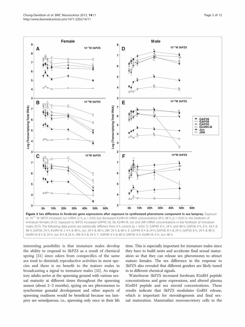
Sex difference in forebrain gene expression after 3kPZSexposureImmature females showed increases in Jun (2-fold) andJNK (2-fold) expression rapidly (2 h) after 10-11 M3kPZS exposure (Figure 3), whereas immature maleshad more delayed responses (≥ 4 h, Figure 3). Exposureto 10-10 M or 10-9 M 3kPZS had no effect on forebraingene expression in immature females (Figure 3).
Differential effect of 3kPZS on hindbrain gene expressionIn the brain stem of immature males, 3kPZS seemedto be most effective at 10-10 M in increasing geneexpressions, and the response appeared to be phasicwith an earlier peak (2 h) and a delayed peak (48 h) atthe time points examined. On the other hand, in thebrain stem of immature females, only 10-10 M 3kPZSdecreased GAP50 expression after 2 h (2-fold), 8 h(3-fold) and 48 h (2-fold) exposure (Figure 4). 3kPZS atother concentrations examined showed no effect on hind-brain gene expression in immature females (Figure 4).The brain stem of immature males showed more
pronounced gene expression changes than the forebrainafter exposure to 10-11 M 3kPZS. lGnRH-III transcriptsincreased after 2 h (19-fold) and 48 h (55-fold) exposure(Figure 4). Jun transcripts increased after 48 h exposure(99-fold, Figure 4). JNK transcripts increased after 2 h(2-fold), 8 h (2-fold), 24 h (2-fold), and 48 h (4-fold) ex-posure (Figure 4).At 10-10 M 3kPZS increased GAP49 transcripts after
8 h exposure (2-fold). Jun transcripts increased after 2 h(9-fold) and 48 h (8-fold) exposure. Prolonged exposureto 10-10 M 3kPZS (48 h) increased GAP58 (1122-fold),lGnRH-III (826-fold), and JNK (2-fold) transcripts in thebrain stem of immature males (Figure 4).At 10-9 M 3kPZS increased lGnRH-III transcripts after
8 h (379-fold) and 48 h (1052-fold) exposure (Figure 4).Jun transcripts increased after exposure for 2 h (12-fold)and 48 h (16-fold, Figure 4). JNK transcripts increased
after exposure for 2 h (2-fold), 4 h (2-fold), 8 h (2-fold),and 48 h (2-fold, Figure 4). Prolonged exposure to 10-9 M3kPZS (48 h) decreased GAP49 (7-fold) but increasedGAP58 (2735-fold) expressions in the brain stem of imma-ture males (Figure 4).
Differential effect of 3kPZS on forebrain and plasmalGnRH peptide concentrationsExposure to 10-10 M 3kPZS increased lGnRH-I and -IIIpeptide concentrations in the forebrain (Figures 5 & 6)but had no effect in the brain stem of immature males(Additional file 1: Figures S1& S2). Plasma lGnRH-I and-III peptide concentrations pulsed at the short timecourse after 3kPZS exposure. 3kPZS increased plasmalGnRH-I and -III peptide concentrations when the con-trol level was low, but this effect was inhibited when thecontrol level was high (Figures 5 & 6).
No effect of 3kPZS on the number of lGnRH neurons inthe hypothalamusThe distribution of lGnRH mRNA, observed in the insitu hybridization experiments, was similar to thatobserved by Reed et al. [29], and was found mainly inthe preoptic-hypothalamic area (Additional file 1: FigureS3). Immunocytochemistry using antibodies specific forlGnRH-I and -III demonstrated that the distributions oflGnRH-I or lGnRH-III immunoreactivities in lampreybrain (Additional file 1: Figures S4 & S5) were consistentwith data described by Nozaki et al. [30]. Exposure to3kPZS had no effect on the number of lGnRH-I or -IIIimmunoreactive cells in the preoptic-hypothalamic area.
DiscussionTo our knowledge, this is the first study of the dose re-sponse and time course of the priming effects of a singlepheromone component on GnRH synthesis and release.The synthesized male pheromone molecule affects bothsexes but induces more dramatic responses in males. An
3
4
5
6
7
8
9
*
Lo
g (
Tran
scri
pts
/ng
To
tal R
NA
)
3
4
5
6
7
0h 10h 20h 30h 40h 50h 60h3
4
5
6
7
A
B
C
**
Female
10-11M 3kPZS
10-10M 3kPZS
10-9M 3kPZS
10-11M 3kPZS
10-10M 3kPZS
10-9M 3kPZS
GAP49GAP58GAP50lGnRH-IIIJunJNK
3
4
5
6
7
8
Lo
g (
Tran
scri
pts
/ng
To
tal R
NA
)
2
3
4
5
6
7
0h 10h 20h 30h 40h 50h 60h3
4
5
6
7
**
*
****
**
**
*
**
***
* *
**
* *
* **
*
* *
D
E
F
Male
*
*
*
*
Figure 3 Sex difference in forebrain gene expressions after exposure to synthesized pheromone component in sea lamprey. Exposureto 10-11 M 3kPZS increased Jun mRNA (2 h, p < 0.05) but decreased lGnRH-III mRNA concentrations (8 h, 48 h, p < 0.05) in the forebrain ofimmature females (A-C). Exposure to 3kPZS increased GAP49, 50, 58, lGnRH-III, Jun and JNK mRNA concentrations in the forebrain of immaturemales (D-E). The following data points are statistically different from 0 h control (p < 0.05). D. GAP49: 8 h, 24 h, and 48 h; GAP58: 4 h, 8 h, 24 h &48 h; GAP50: 24 h; lGnRH-III: 2 4 h & 48 h; Jun: 24 h & 48 h; JNK: 24 h & 48 h. E. GAP49: 8 h & 24 h; GAP58: 8 h & 24 h; GAP50: 8 h, 24 h & 48 h;lGnRH-III: 8 h & 24 h; Jun: 8 h & 24 h; JNK: 8 h & 24 h. F. GAP49: 4 h & 48 h; GAP58: 4 h; lGnRH-III: 4 h; Jun: 48 h.
Chung-Davidson et al. BMC Neuroscience 2013, 14:11 Page 5 of 13http://www.biomedcentral.com/1471-2202/14/11
interesting possibility is that immature males developthe ability to respond to 3kPZS as a result of chemicalspying [31] since odors from conspecifics of the samesex tend to diminish reproductive activities in most spe-cies and there is no benefit to the mature males inbroadcasting a signal to immature males [32]. As migra-tory adults arrive at the spawning ground with various sex-ual maturity at different times throughout the spawningseason (about 2–3 months), spying on sex pheromones tosynchronize gonadal development and other aspects ofspawning readiness would be beneficial because sea lam-prey are semelparous, i.e., spawning only once in their life
time. This is especially important for immature males sincethey have to build nests and accelerate final sexual matur-ation so that they can release sex pheromones to attractmature females. The sex difference in the response to3kPZS also revealed that different genders are likely tunedin to different chemical signals.Waterborne 3kPZS increased forebrain lGnRH peptide
concentrations and gene expressions, and altered plasmalGnRH peptide and sex steroid concentrations. Theseresults indicate that 3kPZS modulates GnRH release,which is important for steroidogenesis and final sex-ual maturation. Mammalian neurosecretory cells in the

1
2
3
4
5
6
Lo
g (
Tra
nsc
rip
ts/n
g T
ota
l RN
A)
1
2
3
4
5
0h 10h 20h 30h 40h 50h 60h1
2
3
4
5
* * *
A
B
C
10-11M 3kPZS
10-10M 3kPZS
10-9M 3kPZS
10-11M 3kPZS
10-10M 3kPZS
10-9M 3kPZS
Female
*
****
GAP49GAP58GAP50lGnRH - IIIJunJNK
2
3
4
5
6
7
8
*
*
*
***
1
Lo
g (
Tra
nsc
rip
ts/n
g T
ota
l RN
A)
2
3
4
5
6
***
**
*
*
*
1
0h 10h 20h 30h 40h 50h 60h0
1
2
3
4
5
6
7
*
*
*
**
* * *
*
D
E
F
Male
*
*
Figure 4 Sex difference in hindbrain gene expressions after exposure to synthesized pheromone component in sea lamprey. Exposureto 10-10 M 3kPZS decreased GAP50 mRNA (2 h, 8 h, 24 h, and 48 h, p < 0.05) and JNK mRNA (2 h, 4 h, 8 h, and 24 h, p < 0.05) concentrations inthe brain stem of immature females (A-C). Exposure to 3kPZS increased GAP49 & 58, lGnRH-III, Jun and JNK mRNA concentrations in the brainstem of immature males (D-E). The following data points are statistically different from 0 h control (p < 0.05). D. lGnRH-III: 2 h & 48 h; Jun: 48 h;JNK: 2 h, 8 h, 24 h & 48 h. E. GAP58: 48 h; lGnRH-III: 48 h; Jun: 2 h & 48 h; JNK: 2 h, 4 h & 48 h. F. GAP49: 8 h & 24 h; GAP58: 48 h; lGnRH-III: 8 h &48 h; JNK: 2 h & 8 h.
Chung-Davidson et al. BMC Neuroscience 2013, 14:11 Page 6 of 13http://www.biomedcentral.com/1471-2202/14/11
hypothalamus display pulsatile activities [33], and ovula-tion is induced by a LH (one of the mammalian counter-parts of GTH) surge which is preceded by a surge ofGnRH [LH releasing hormone (LHRH) in mammals]secretion into the brain portal system [34-36]. Parallel tothis preovulatory increase in GnRH secretion is an in-crease in GnRH gene expression, as indicated by expres-sion of the immediate-early genes (c-fos and c-jun) [37,38]and elevated GnRH mRNA [39,40], and increased levels ofnewly synthesized GnRH [41]. Lampreys and teleost
fishes, however, lack the portal system in the brain, andGnRH neurons either terminate directly in the pituitarygland or release GnRH directly into the third ventricle[42,43]. Teleost fishes have well-characterized GTH withsimilar function and regulatory mechanisms as its mam-malian counterpart [LH and follicle-stimulating hormone(FSH)]. A cDNA encoding lamprey GTH subunit β hasbeen cloned, but its function has not been validated [44].We found differential expression of lGnRH-I and -III
transcripts in sea lamprey brain exposed to 3kPZS.

(A) Plasma lGnRH-I
15 min 30 min 1h 2h
Co
nce
ntr
atio
n (
pg
/ml)
0
50
100
150
200
250
Control 3kPZS
* * *
*
(B) Forebrain lGnRH-I
15 min 30 min 1h 2h
pg
/mg
Tis
sue
0
2
4
6
8
10Control3kPZS
*
Figure 5 Differential effects of synthesized pheromone component on plasma and forebrain lGnRH-I concentrations in male sealamprey. (A) Plasma lGnRH-I concentrations showed various effects after 10-10 M 3kPZS exposure. Exposure to 10-10 M 3kPZS increased lGnRH-Iconcentrations in the forebrain (B) but had no effect in the brain stem (data not shown).
Chung-Davidson et al. BMC Neuroscience 2013, 14:11 Page 7 of 13http://www.biomedcentral.com/1471-2202/14/11
Lampreys are the most primitive vertebrates for which mul-tiple GnRH neurohormones are involved in pituitary-reproductive activity [45,46]. Both lGnRH-I and -III havebeen shown to induce gonadal maturation, steroidogenesis,and spermiation or ovulation in adult sea lamprey [45-49].In immunocytochemical studies, both immunoreactive-lGnRH-I and -III can be found in the cell bodies of the ros-tral hypothalamus and preoptic area in larval and adult sealamprey [30,44,50,51]. lGnRH-III was considered the moreactive form during development and gonadal maturation[52]. Our results indicated that lGnRH-III concentrationswere more responsive to pheromone exposure. However,lGnRH-I may be more important in inducing behavioralresponses in mature females, given that the transcripts ofits three splice variants fluctuated in the brainstem afterpheromone exposure while lGnRH-III transcripts stayedconstant.It is interesting that three lGnRH-I splice variants also
showed differential expressions after pheromone expos-ure. Lamprey GnRH-I precursor was the first identifiedagnathan GnRH to contain the same tripartite structurewith the signal peptide, GnRH decapeptide, and GnRH-
(A) Plasma lGnRH-III
15 min 30 min 1h 2h
Co
nce
ntr
atio
n (
pg
/ml)
0
1000
2000
3000
4000Control 3kPZS
** *
*
(
Figure 6 Differential effects of synthesized pheromone component olamprey. (A) Plasma lGnRH-III concentrations showed various effects afterconcentrations in the forebrain (B) but had no effect in the brain stem (da
associated peptide (GAP) as gnathostome GnRHprecursors [52]. Unlike other known vertebrate GnRHprecursors, which typically have one or two splice variants,three distinct splice variants were isolated and sequenced inlampreys [52]. The lGnRH-I splice variants, GAP49,GAP50 and GAP58, differed in the length of the GAP cod-ing sequence [52]. The relative abundance of these splicevariants in our results followed a magnitude decrease in theorder of GAP58, GAP50, and GAP49. According to thechanges after pheromone exposure, GAP50 seemed to beimportant for forebrain function whereas GAP49 and 58may be associated with brainstem function in both sexes.However, forebrain GAP49 and brainstem GAP50 mayserve additional functions in immature females [53].Corresponding to its gene expression level in male sea
lamprey, lGnRH-III is the most prominent lGnRHpeptides in the brain and plasma. Its concentration isaround 20 times more than lGnRH-I. We also foundthat 3kPZS altered the pulsatile pattern of GnRHconcentrations in plasma. Pulsatile release of GnRH is acommon neurobiological feature in vertebrates. It is au-tonomous within the GnRH neuronal network without
B) Forebrain lGnRH-III
15 min 30 min 1h 2h
pg
/mg
Tis
sue
0
50
100
150
200Control3kPZS
*
*
n plasma and forebrain lGnRH-III concentrations in male sea10-10 M 3kPZS exposure. Exposure to 10-10 M 3kPZS increased lGnRH-IIIta not shown).

Chung-Davidson et al. BMC Neuroscience 2013, 14:11 Page 8 of 13http://www.biomedcentral.com/1471-2202/14/11
spontaneous pacemaker cells [54]. It is interestingthat menstrual synchrony in humans is mediated bypheromones through two opposing effects of the samecompounds [55]. Axillary compounds (putative humanpheromones) from donor women in the follicular phaseshortened both the time to ovulation and the length ofthe menstrual cycle in recipients and those in the ovula-tory phase delayed ovulation and lengthened the totalcycle [55]. We found that when GnRH level is low inthe control group, 3kPZS increased GnRH peptideconcentrations in the plasma whereas when GnRH levelis high in the control group, 3kPZS showed oppositeeffects. There may be some similarity in the controlmechanism involved in this process.Pulsatile GnRH regulates the gonadotropin subunit genes
in a differential manner, with faster frequencies (8 to60 min pulse intervals) favoring mammalian LH β subunitgene expression and slower frequencies (≥ 120 min pulseintervals) favoring the expression of FSH β subunit [56].The mechanism is through the activation of GnRH recep-tor and its signaling cascades including JNK and its sub-strate JUN [57-61]. We found that 3kPZS induced GnRHrelease within 15 min, and Jun and JNK gene expressionafter 2 h exposure, suggesting that 3kPZS can modulateGnRH release and its signaling cascades.The detection of lGnRH peptides in the plasma was a
surprise since many previous attempts using HPLC andradioimmunoassays had failed to detect lGnRH peptides insea lamprey plasma [62-65]. However, high-affinity bindingsites for lGnRH peptides were detected in lamprey gonads,and GnRH appears to stimulate steroidogenesis independ-ent of the pituitary [48,66]. Previous reports also noticedthat the concentrations of brain GnRH peptides in lampreyoccur at higher concentrations than seen in othervertebrates [62,67], suggesting that there may be a higherproduction or metabolism (or both) of each of the lGnRHpeptides compared to gnathostomes [65]. Taken together,lGnRH peptides likely serve as hormones that are releasedinto the third ventricle and then into the blood stream, andact on target organ such as gonads. Therefore, our resultsprovide an additional explanation for GnRH binding affinityin peripheral organs [66]. Furthermore, our results supporta possible peripheral endocrine function of lGnRHpeptides, which may bypass the pituitary gland in the HPGaxis.The acquisition of a HPG axis was a seminal event in ver-
tebrate evolution leading to the neuroendocrine control ofmany biological functions [68]. GnRH is a critical neuro-peptide that acts through the HPG axis to regulate verte-brate reproduction. However, invertebrate GnRH peptidesseem to exert a wide range of central and peripheralfunctions not limited to reproduction [69]. In addition,GnRH may even function as spawning pheromone ininvertebrates [70]. The presence of lGnRH peptides in sea
lamprey plasma indicates that the function of lGnRHpeptides may not be fully centralized. It is likely that sealamprey GnRH peptides function in an evolutionary transi-tional state where they not only act on the HPG axis, butalso exert hormonal functions through systemic circulation.Furthermore, environmental factors such as a single phero-mone component (i.e. 3kPZS) can modulate the HPG axisthrough olfaction. The considerable specialization of GnRHin the HPG axis as a reproductive activator may be aphenomenon specific to jawed vertebrates (gnathostomes)(Figure 7).
Conclusions3kZPS is a primer pheromone in sea lamprey that exertsits functions through the HPG axis via GnRH release.
MethodsAnimalsSea lamprey were collected by agents of the US Fish andWildlife Service Marquette Biological Station (Marquette,MI) and Department of Fisheries and Oceans Canada SeaLamprey Control Centre (Sault Ste. Marie, ON). Phero-mone treatments of animals were conducted at the USGeological Survey Great Lakes Science Center HammondBay Biological Station (Millersburg, MI). For each set ofexperiments, all test subjects were captured from thesame stream on the same day to reduce variation in ma-turity. Standard operating procedures for transporting,maintaining, handling, anesthetizing, and euthanizing sealamprey were approved by the Institutional Committeeon Animal Use and Care of Michigan State University(AUF#05/09-088-00). Ethical guidelines were followedthroughout the course of this research. Pheromone expo-sure time in the following experiments was chosenaccording to our previous publications [14,53,71,72].
Pheromone exposure experiment 1Twelve immature males were held in separate tanks(100 gallon; 16°C; water replenishment 1 L min-1), andacclimated for 2 days. 16°C was within the optimaltemperature range for lamprey reproduction [73]. Animalswere randomly assigned to two treatment groups (6/group):water from a tank (pheromone source tank) containing5 mature males (spermiating male washings, SMW) or5 immature males (prespermiating male washings, PSMW).Every two days, animals were checked for spermiation. Thenumber of days to reach spermiation was recorded. Thesame experiment was repeated using 12 immature females(checking for egg release), and data were analyzed with theχ2 test.
Pheromone exposure experiment 2Two hundred and fifty-two immature females and maleswere acclimated as described in experiment 1, and assigned

Hypothalamus GnRH Neurons
Pheromone Stimulation
Pituitary
Olfactory Bulb
GnRH I, II, III
GTH?
GonadGnRH I, II, III 15α-P?Other Steroids?
Sea Lamprey(Agnathan)
Reproduction
Hypothalamus GnRH Neurons
Pheromone Stimulation
Pituitary
Accessory & MainOlfactory Bulb
GnRH I
LH
Gonad SteroidHormones
Mammal(Gnathostome)
FSH
Reproduction
Gonad
Pheromone StimulationGnRH as Pheromone?
CNS
Invertebrate
Reproduction
GnRH-like?
PeripheralOrgans
GnRH-like?
GnRH-like?
Figure 7 A schematic diagram of the evolutionary transition between pheromone and GnRH control from invertebrates to sealamprey (agnathan) to mammals (gnathostome).
Chung-Davidson et al. BMC Neuroscience 2013, 14:11 Page 9 of 13http://www.biomedcentral.com/1471-2202/14/11
to two treatment groups (control or SMW). At 0 h (imme-diately before introducing the odorants) and 24 h (comple-tion of odorant treatment), 1 ml of blood was drawn toobtain plasma according to previously established protocolsand stored at −80°C until use [71,72]. Plasma samples wereassayed for immunoreactive 15α-P using HPLC andradioimmunoassays [71,72]. The same experiment wasrepeated in the summers of 2001, 2002 and 2003. Datawere pooled for each month (May, June and July) andanalyzed by One-way ANOVA for repeated measurements(before and after pheromone exposures). If treatmenteffects were detected (p < 0.05), Fisher’s PLSD post hoc testswere followed to determine which treatment induced ap-parent changes in the parameters measured.
Pheromone exposure experiment 3To examine the time course of hormonal responses of im-mature sea lamprey exposed to 3kPZS at a wide range ofconcentrations, 108 immature males were acclimated for2 days (6/tank; test tank: 200 L, 16°C, water replenishment1 L min-1). Each tank was randomly assigned a treatmentof 10-11 M, 10-10 M or 10-9 M 3kPZS. Immediately beforeand after pheromone exposure, 1 ml blood was drawn forplasma 15α-P analyses [71,72]. One-way ANOVA was usedto compare the changes in plasma 15α-P concentration[Δ15α-P = (post-treatment 15α-P level) - (pre-treatment15α-P level)] among groups treated with the same phero-mone concentration but sampled at different time points.
Fisher’s PLSD post hoc tests were performed if the ANOVAshowed significant effects (p < 0.05).At 0 (control), 2, 4, 8, 24 and 48 h after pheromone
exposure, animals were euthanized with 0.5% MS222(Sigma, St. Louis, MO). Eight brain samples were snapfrozen and analyzed for transcripts of lGnRH-I (GAP49,GAP50, and GAP58) [74] and -III, jun, JNK, and 40Sribosomal RNA. Six brain samples from each group werefixed in 4% paraformaldehyde (in 0.1 M phosphate buf-fer, pH 7.4) for immunocytochemistry (ICC) and in situhybridization (ISH) analyses. From each preserved brain,serial transverse sections of 20 μm were collected.Neighboring sections were processed for lGnRH-I or -IIIICC [53], or lGnRH mRNA ISH. Negative controls(deprived of primary antibody for ICC or using senseprobe for ISH) were processed simultaneously in eachexperiment. ISH and ICC results were examined byinvestigators with no knowledge of the experimentalcondition. The number of immunoreactive cells in thepreoptic-hypothalamic area was counted [53], and theaverage number in a square area (0.0625 mm2) wasused for statistical analyses. Brain nuclei were identifiedaccording to sea lamprey brain atlas by Nieuwenhuysand Nicholson [75]. The nonparametric Kruskal-Wallistest was used to compare the number of lGnRH-I or -IIIimmunoreactive neurons among treatment groups.Parallel experiments were conducted with immaturefemales.

Chung-Davidson et al. BMC Neuroscience 2013, 14:11 Page 10 of 13http://www.biomedcentral.com/1471-2202/14/11
Pheromone exposure experiment 4Since 3kPZS had no dramatic effects in immature femalesand the gene expression changed after 2 h exposure, weonly examined the time course of GnRH release in imma-ture males exposed to vehicle (5 ppm methanol) or 10-10
M 3kPZS. 64 immature males were acclimated as describedabove. Immediately and at 15 min, 30 min, 1 h or 2 h afterpheromone exposure, animals were euthanized and bloodwas drawn by cardiac puncture for plasma lGnRH-I and -III analyses using UPLC-MS/MS (method described below).Brain samples were snap-frozen and analyzed for lGnRH-Iand -III using ultra performance liquid chromatographycoupled with tandem mass spectrometry (UPLC-MS/MS).
In situ Hybridization used digoxigenin-labeled anti-sense and sense RNA probes generated by in vitro Tran-scription Systems (Promega BioSciences, San Luis Obispo,CA, USA). Sections were treated with proteinase K atroom temperature (R.T.) for 10 min followed by 4%paraformaldehyde (0.1 M phosphate buffer, pH 7.4) for15 min. Sections were incu- bated in prehybridization so-lution [50% formamide, hybridization salt (150 mM NaCl,50 mM EDTA, 50 mM PIPES), Denhardt’s solution(Sigma, St. Louis, MO, USA), 2.5 μg/ml calf thymus DNA,2.5 μg/ml poly-adenosine, 0.2% sodium dodecyl sulfate(SDS), and 0.1% diethylpyrocarbonate] at 42°C for 2 h,and then in antisense or sense RNA probe in hybridizationsolution (prehybridization solution with 5% dextran sul-fate) at 60°C overnight (about 18 h). Sections were rinsedwith 4× SSC (150 mM NaCl and 15 mM sodium citrate),followed by 2× SSC (with 0.3% Tween-20) at 68°C for15 min 3 times, 0.2× SSC (with 0.3% Tween-20) at 68°Cfor 15 min 3 times, 0.1× SSC (with 0.3% Tween-20)at R.T. for 15 min, and then 0.1 M phosphate buffersaline (PBS, pH 7.4). Sections were incubated in alka-line phosphatase-conjugated sheep-anti-digoxigenin Fabfragments (1:1000; Roche Applied Science, Indianapolis,IN, USA) with normal sheep serum (in 0.1 M PBS with0.3% Tween-20) at 5°C overnight (about 18 h). Sectionswere rinsed in 0.1 M Tris buffer saline (TBS, pH 9.5) for15 min 3 times, followed by nitroblue tetrazolium chlorideand 5-bromo-4-chloro-3 indolyl phosphate substrate(Roche) for 1 h, rinsed in PBS (pH 7.4) for 15 min 3 times,counter stained with Nuclear Fast Red (Vector Laborato-ries, Inc., Burlingame, CA, USA) for 5 min, and coverslippedwith Vector AQ mounting media.
Real time quantitative RTQ-PCR followed the pro-cedure described by Chung-Davidson et al. [53]. Syn-thetic oligos were used as standards and run on thesample plate. 40S ribosomal RNA was used as an in-ternal standard and confirmed to have no change in theexpression level in all experiments. RTQ-PCR data(among groups treated with same pheromone
concentration but sampled at different time points) wereanalyzed by one-way ANOVA followed by Fisher’s PLSDpost hoc tests if the ANOVA showed significant timeeffects (p < 0.05). The sequence for primers and TaqManRGB probe (Applied Biosystems) for each mRNA waslisted below. 40S ribosomal RNA: 5' primer (5'ACCTACGCAGGAACAGCTATGAC3'), probe (5'ATCTCGAGCAGCTGAA3'), 3' primer (5'CGACGAATTCCACCACATTG3'). Jun: 5' primer (5'CATGGCCGCAAACTTTGG3'), probe (5'CACGAACCTGACCAGC3'), 3' primer(5'CCACCTCCCTGCTGATGCT3'). JNK: 5' primer (5'TCAGGCGTGTGGCCAAGT3'), probe (5'CCATGACTTGATCGAATGT3'), 3' primer (5'GAATCAAATTGAGAACGCAAACG3'). Lamprey GnRH-III: 5' primer (5'TGACACGAACCCTGTCAATGA3'), probe (5'ATGCCCTCGCTGTGGT3'), 3' primer (5'ACAAAGGGTCTAAGAGACGTCACA3'). To analyze three transcripts of lGnRH-I, we used the same 5' primer (5'TGAATTACGCGCAGCACTACTC3') and probe (5'TGGAATGGAAACCCGG3'), but the 3' primers were designed so that theywere located at the splice junction of each correspondingtranscript (GAP49: 5'CTCCTCCAGGTCTCGTTTGC3';GAP50: 5'CTCCTGCTCCAGGTCTCGTT3'; GAP58: 5'CCAGCTCTCGTGTGTGACTGA3').
UPLC-MS/MS analyses of lGnRHlGnRH-I standard was custom synthesized from GenScriptUSA Inc. (Piscataway, NJ, USA). lGnRH-III and LHRHwere purchased from BAChem Americas, Inc. (Torrance,CA, USA). Each compound was dissolved in 50% metha-nol/H2O (v/v) to make 1 mg/ml stock solution, and storedat −20°C until use. Subsequent dilutions were made in 50%methanol/H2O. Calibration standard spiking solutions wereprepared in a range from 0.01 to 10 ng/ml by spiking ap-propriate stock solutions to brain tissue or plasma extracts.Internal standard (LHRH) solution (50 ng/ml) wasprepared in 50% methanol/H2O and 20 μl was added toeach sample (1 ng per sample).Brain samples were weighed and homogenized with
400 μl of 1% formic acid in cold (−20°C) acetonitrilewith 1 ng internal standard. 1 ml of 1% formic acid incold (−20°C) acetonitrile was added to the homogenate,incubated at −20°C for 15 min, and centrifuged at15,800 × g for 20 min at 4°C. The supernatant wastransferred to a new tube, freeze-dried using a CentriVapCold Trap Concentrator (Labconco Co., Kansas City,MO, USA), and reconstituted in 1 ml water solutionwith 3% acetic acid and 1% TFA. 500 μl plasma sampleswere processed in a similar procedure without thehomogenization step. The reconstituted supernatant wastransferred to a SPE HLB cartridge preconditioned with3 ml methanol followed by 3 ml loading solution (water/acetic acid/TFA, 96:3:1, v/v), washed with 3 ml loadingsolution and followed by 3 ml loading solution/methanol
Chung-Davidson et al. BMC Neuroscience 2013, 14:11 Page 11 of 13http://www.biomedcentral.com/1471-2202/14/11
(70:30, v/v). Samples were eluted with 3 ml 3% aceticacid/ solution: methanol (30:70, v/v), freeze-dried andreconstituted in 100 μl 3% acetic acid/methanol (50:50,v/v), vortexed for 30 seconds and transferred to glassautosampler vials for UPLC-MS/MS analyses.A Waters Xevo TQ-S mass spectrometry was coupled to
an H-Class UPLC system with a Waters BEH C18 column(1.0 × 50 mm, 1.7 μm particle size) and oven temperatureat 35°C. The injection volume was 10 μl, and the UPLCflow rate is 0.15 ml/min with a gradient (mobile phase A:0.1% formic acid in water; B: 0.1% formic acid inacentonitrile): initial, 88% A and 12% B; 0.5 min 88% A,4 min 65% A, 7 min 1% A, 8 min 1% A, 8 min 88% A, and9 min 88% A. Mass spectra were acquired usingelectrospray ionization in positive ion mode and MRM.The capillary voltage, cone voltage, and rf lens setting were3.20 kV, 64 V, and 0.3, respectively. The flow rates of conegas and desolvation gas were 20 and 400 L/h, respectively.The source temperature and desolvation temperaturewere 150 and 500°C. Collision-induced dissociationemployed argon as collision gas at a manifold pressureof 2 × 10⁻³ mbar, and collision energies and source conepotentials were optimized for each transition using WatersQuanOptimize software. Data were acquired with MassLynx4.1 and QuanLynx softwares.
Additional file
Additional file 1: Figure 1. 3kPZS exposure had no effect on hindbrainlGnRH-I concentrations in immature male sea lamprey. Data arepresented as mean ± S.E.M. Figure 2. 3kPZS exposure had no effect onhindbrain lGnRH-III concentrations in immature male sea lamprey. Dataare presented as mean ± S.E.M. Figure 3. 3kPZS exposure did notchange the number of lamprey (l) GnRH-positive neurons in thehypothalamus. Lamprey (l) GnRH in situ hybridization (ISH) showedpositive cells (blue stain) in the preoptic area of immature female andmale sea lamprey. 20 μm Transverse sections were counterstained withnuclear fast red (pink stain). Scale bar: 20 μm. 3 V: third ventricle.Figure 4. 3kPZS exposure did not change the number of lamprey (l)GnRH-Iimmunoreactive neurons in the hypothalamus. lGnRH-I-immunoreactive neurons (red stain) are located in the preoptic area ofimmature female and male sea lamprey. 20 μm transverse sections werecounterstained with hematoxylin (blue/purple stain). Scale bar: 50 μm.3 V: third ventricle. Figure 5. 3kPZS exposure did not change the numberof lamprey (l) GnRH-IIIimmunoreactive neurons in the hypothalamus. lGnRH-III-immunoreactive neurons (red stain) are located in the preoptic area ofimmature female and male sea lamprey. 20 μm transverse sections werecounterstained with hematoxylin (blue/purple stain). Scale bar: 50 μm. 3 V:third ventricle.
AbbreviationsHPG: Hypothalamic-pituitary-gonadal; GnRH: Gonadotropin-releasinghormone; 3kPZS: 3-keto petromyzonol sulfate; 15α-P: 15α-hydroxyprogesterone; lGnRH-I: lamprey GnRH-I; lGnRH-III: lamprey GnRH-III;JNK: Jun N-terminal kinase; LH: Luteinizing hormone; GTH: Gonadotropin;SMW: Spermiating male washing; PSMW: Prespermiating male washing;LHRH: Luteinizing hormone-releasing hormone; FSH: Follicle-stimulatinghormone; ICC: Immunocytochemistry; ISH: In situ hybridization; RTQ-PCR: Real-time quantitative PCR; UPLC-MS/MS: Ultra-performance liquidchromatography coupled with tandem mass spectrometry.
Competing interestsThe authors declare that they have no competing interests.
Authors’ contributionsYWCD designed the study, carried out the sample collection and allexperimental studies, performed the statistical analyses and wrote themanuscript. H Wang developed UPLC-MS/MS methods and collected data inPheromone Exposure Experiment 4. MJS participated in PheromoneExposure Experiment 1 and 2. MBB participated in Pheromone ExposureExperiment 2 and 3. NSJ and H Wu participated in Pheromone ExposureExperiment 3. WL conceived of the study, participated in its design andcoordination and helped to write the manuscript. All authors read andapproved the final manuscript.
AcknowledgementsWe thank Dr. Stacia A. Sower for her generous gifts of lamprey GnRHantibodies. We thank all summer assistants, especially David Partyka, JosephBednark, Christine N. Bedore, and Aaron Smuda, for their help in samplecollection. We thank the staff of US Fish and Wildlife Service MarquetteBiological Station (Marquette, MI) and Department of Fisheries and OceanCanada Sea Lamprey Control Centre (Sault St. Marie, ON, Canada) forsupplying animals and (Contribution 1729) US Geological Survey Great LakesScience Center Hammond Bay Biological Station (Millersburg, MI) forproviding animal housing facility. This study is supported by NSF grantIOB0517491, NIH grant 5R24GM83982 and the Great Lakes FisheryCommission to WL.
Author details1Department of Fisheries and Wildlife, Michigan State University, 13 NaturalResources Building, 480 Wilson Road, East Lansing, MI 48824, USA. 2Presentaddress: Great Lakes Fishery Commission, 2100 Commonwealth Blvd., Suite100, Ann Arbor, MI 48105, USA. 3Present address: Energy BiosciencesInstitute, University of California, 130 Calvin Laboratory, MC 5230, Berkeley,CA 94720, USA. 4Present address: Department of Microbiology &Immunology, School of Medicine, Emory University, Rollins Research CenterG214, 201 Dowman Drive, Atlanta, Georgia 30322, USA. 5Present address:USGS, Great Lakes Science Center, Hammond Bay Biological Station, 11188Ray Road, Millersburg, MI 49759, USA.
Received: 11 September 2012 Accepted: 15 January 2013Published: 20 January 2013
References1. Karlson O, Lüscher M: ‘Pheromones’: a new term for a class of biologically
active substances. Nature 1959, 183:55–56.2. Brennan PA, Zufall F: Pheromonal communication in vertebrates. Nature
2006, 444:308–315.3. Rekwot PI, Ogwu D, Oyedipe EO, Sekoni VO: The role of pheromones and
biostimulation in animal reproduction. Anim Reprod Sci 2001, 65:157–170.4. Stowers L, Marton TF: What is a pheromone? Mammalian pheromones
reconsidered. Neuron 2005, 46:699–702.5. Wyatt T: Pheromones and signature mixtures: defining species-wide
signals and variable cues for identity in both invertebrates andvertebrates. J Comp Physiol A 2010, 196:685–700.
6. Wysocki CJ, Preti G: Facts, fallacies, fears, and frustrations with humanpheromones. Anat Rec Part A 2004, 281A:1201–1211.
7. Motofei IG: A dual physiological character for sexual function: libido andsexual pheromones. J Compil BJUI 2009, 104:1702–1708.
8. Preti G, Wysocki CJ, Barnhart KT, Sondheimer SJ, Leyden JJ: Male axillaryextracts contain pheromones that affect pulsatile secretion of luteinizinghormone and mood in women recipients. Biol Reprod 2003, 68:2107–2113.
9. Murata K, Wakabayashi Y, Kitago M, Ohara H, Watanabe H, Tamogami S,Warita Y, Yamagishi K, Ichikawa M, Takeuchi Y, Okamura H, Mori Y:Modulation of gonadotrophin-releasing hormone pulse generatoractivity by the pheromone in small ruminants. J Neuroendocrinol 2009,21:346–350.
10. Okamura H, Murata K, Sakamoto K, Wakabayashi Y, Ohkura S, Takeuchi Y,Mori Y: Male effect pheromone tickles the gonadotrophin-releasinghormone pulse generator. J Neuroendocrinol 2010, 22:825–832.
Chung-Davidson et al. BMC Neuroscience 2013, 14:11 Page 12 of 13http://www.biomedcentral.com/1471-2202/14/11
11. Yu KL, Peng C, Peter RE: Changes in brain levels of gonadotropin-releasing hormone and serum levels of gonadotropin and growthhormone in goldfish during spawning. Can J Zool 1989, 69:182–188.
12. Zhang W, Stacey NE: A steroidal pheromone and spawning stimuli act viadifferent neuroendocrine mechanisms to increase gonadotropin andmilt volume in male goldfish Carassius auratus. Gen Comp Endocrinol1997, 105:228–238.
13. Sower SA: The endocrinology of reproduction in lampreys andapplications for lamprey sterilization. J Great Lakes Res 2003, 29:50–65.
14. Li W, Scott AP, Siefkes MJ, Yan HG, Liu Q, Yun SS, Gage DA: Bile acidsecreted by mate sea lamprey that acts as a sex pheromone. Science2002, 296:138–141.
15. Li K, Siefkes MJ, Brant CO, Li W: Isolation and identification ofpetromyzesterosterol, a polyhydroxysteroid from sexually mature malesea lamprey (Petromyzon marinus L.). Steroids 2012, 77:806–810.
16. Sorensen PW, Fine JM, Dvornikovs V, Jeffrey CS, Shao F, Wang J, Vrieze LA,Anderson KR, Hoye TR: Mixture of new sulfated steroids functions as amigratory pheromone in the sea lamprey. Nat Chem Biol 2005, 1:324–328.
17. Yun SS, Scott AP, Li W: Pheromones of the male sea lamprey, Petromyzonmarinus L.: structural studies on a new compound, 3-keto allocholic acid,and 3-keto petromyzonol sulfate. Steroids 2003, 68:297–304.
18. Hardisty MW: The significance of lampreys for biological research.Endeavour 1983, 7:110–115.
19. Kuratani S, Kuraku S, Murakami Y: Lamprey as an evo-devo model: lessonsfrom comparative embryology and molecular phylogenetics. Genesis2002, 34:175–183.
20. Applegate VC: Natural history of the sea lamprey (Petromyzon marinus) inMichigan. In US Fish and Wildlife Service Special Science Report on FisheryService 1950, 55:237.
21. Hardisty MW, Potter IC: The general biology of adult lampreys. In Thebiology of lampreys. Volume 1. Edited by Hardisty MW, Potter IC. New York:Academic Press; 1971:127–206.
22. Youson JH, Potter IC: A description of the stages in the metamorphosis ofthe anadromous sea lamprey, Petromyzon marinus L. Can J Zool 1979,57:1808–1817.
23. Potter IC, Gill HS: Adaptive radiation of lampreys. J Great Lakes Res 2003,29(Suppl 1):95–112.
24. Siefkes MJ, Scott AP, Zielinski B, Yun S-S, Li W: Male sea lampreys,Petromyzon marinus L., excrete a sex pheromone from gill epithelia.Biol Reprod 2003, 69:125–132.
25. Siefkes MJ, Li W: Electrophysiological evidence for detection anddiscrimination of pheromonal bile acids by the olfactory epithelium offemale sea lampreys (Petromyzon marinus). J Comp Physiol A 2004,190:193–199.
26. Johnson NS, Yun S-S, Thompson HT, Brant CO, Li W: A synthesizedpheromone induces upstream movement in female sea lamprey andsummons them into traps. PNAS 2009, 106:1021–1026.
27. Li W, Scott AP, Siefkes MJ, Yun SS, Zielinski BS: A male pheromone in thesea lamprey (Petromyzon marinus): an overview. Fish Physiol Biochem2003, 28:259–262.
28. Siefkes MJ, Winterstein SR, Li W: Evidence that 3-keto petromyzonolsulphate specifically attracts ovulating female sea lamprey, Petromyzonmarinus. Anim Behav 2005, 70:1037–1045.
29. Reed KL, MacIntyre JK, Tobet SA, Trudeau VL, MacEachern L, Rubin BS,Sower SA: The spatial relationship of γ-aminobutyric acid (GABA)neurons and gonadotropin-releasing hormone (GnRH) Neurons in larvaland adult sea lamprey, Petromyzon marinus. Brain Behav Evol 2002,60:1–12.
30. Nozaki M, Ominato K, Gorbman A, Sower SA: The distribution of lampreyGnRH-III in brains of adult sea lampreys (Petromyzon marinus). Gen CompEndocrinol 2000, 118:57–67.
31. Sørensen PW, Scott AP: The evolution of hormonal sex pheromones in teleostfish: poor correlation between the pattern of steroid release by goldfish andolfactory sensitivity suggests that these cues evolved as a result of chemicalspying rather than signal specialization. Acta Physiol Scand 1994, 152:191–205.
32. Koyama S: Primer: effects by conspecific odors in house mice: a newperspective in the study of primer effects on reproductive activities.Horm Behav 2004, 46:303–310.
33. Misgeld U, Zeilhofer HU, Swandulla D: Synaptic modulation of oscillatoryactivity of hypothalamic neuronal networks in vitro. Cell Mol Neurobiol 1998,18:29–43.
34. Caraty A, Evans NP, Fabre-Nys CJ, Karsch FJ: The preovulatorygonadotropin-releasing hormone surge: a neuroendocrine signal forovulation. J Reprod Fertil Suppl 1995, 49:245–255.
35. Christian CA, Moenter SM: Critical roles for fast synaptic transmission inmediating estradiol negative and positive feedback in the neural controlof ovulation. Endocrinol 2008, 149:5500–5508.
36. Ping L, Mahesh VB, Bhat GK, Brann DW: Regulation of gonadotropin-releasing hormone and luteinizing hormone secretion by AMPAreceptors. Evidence for a physiological role of AMPA receptors in thesteroid-induced luteinizing hormone surge. Neuroendocrinol 1997,66:246–253.
37. Lee WS, Smith MS, Hoffman GE: Luteinizing hormone-releasing hormoneneurons express Fos protein during proestrous surge of luteinizing hormone.PNAS 1990, 87:5163–5167.
38. Lee WS, Abbud R, Smith MS, Hoffman GE: LHRH neurons express c-Junprotein during proestrous surge of luteinizing hormone. Endocrinol 1992,130:3101–3103.
39. Park OK, Gugneja S, Mayo KE: Gonadotropin-releasing hormone geneexpression during the rat estrous cycle: effect of pentobarbital andovarian steroids. Endocrinol 1990, 127:365–372.
40. Zoeller RT, Young WS: Changes in cellular levels of messenger ribonucleic acidencoding gonadotropin-releasing hormone in the anterior hypothalamus offemale rats during the estrous cycle. Endocrinol 1988, 123:1688–1689.
41. Gore AC, Roberts JL: Regulation of gonadotropin-releasing hormone geneexpression in the rat during the luteinizing hormone surge. Endocrinol1995, 136:889–896.
42. King JC, Sower SA, Anthony EL: Neuronal systems immunoreactive withantiserum to lamprey gonadotropin-releasing hormone in the brain ofPetromyzon marinus. Cell Tissue Res 1988, 253:1–8.
43. Zohar Y, Muñoz-Cueto JA, Elizur A, Kah O: Neuroendocrinology ofreproduction in teleost fish. Gen Comp Endocrinol 2010, 165:438–455.
44. Sower SA, Moriyama S, Kasaraha M, Takahashi A, Nozaki M, Uchida K,Dahlstrom JM, Kawauchi H: Identification of sea lamprey GTHβ-like cDNAand its evolutionary implications. Gen Comp Endocrinol 2006, 148:22–32.
45. Sower SA: Gonadotropin-releasing hormone in primitive fishes. Prog ClinBiol Res 1990, 342:73–78.
46. Sower SA: Neuroendocrine control of reproduction in lampreys. Fish PhysiolBiochem 1990, 8:365–374.
47. Deragon KL, Sower SA: Effects of lamprey gonadotropin-releasing hormone-IIIon steroidogenesis and spermiation in male sea lampreys. Gen CompEndocrinol 1994, 95:363–367.
48. Gazourian L, Derragon KL, Chase CF, Pati D, Habibi HR, Sower SA:Characteristics of GnRH binding in the gonads and effects oflamprey GnRH-I and -III on reproduction in the adult sea lamprey.Gen Comp Endocrinol 1997, 108:327–339.
49. Sower SA, Chiang YC, Lovas S, Conlon JM: Primary structure and biologicalactivity of a third gonadotropin-releasing hormone from lamprey brain.Endocrinol 1993, 132:1125–1131.
50. Tobet SA, Nozaki M, Youson JH, Sower SA: Distribution of lampreygonadotropin releasing hormone-III (GnRH-III) in brains in larval lampreys(Petromyzon marinus). Cell Tissue Res 1995, 279:261–267.
51. Wright GM, McBurney KM, Youson JH, Sower SA: Distribution of lampreygonadotropin-releasing hormone in the brain and pituitary of larval,metamorphic and adult sea lamprey, Petromyzon marinus. Can J Zool1994, 72:48–53.
52. Sower SA, Kawauchi H: Update: brain and pituitary hormones oflampreys. Comp Biochem Physiol 2001, 129:291–302.
53. Chung-Davidson Y-W, Bryan MB, Teeter J, Bedore CN, Li W:Neuroendocrine and behavioral responses to weak electric fields inadult sea lampreys (Petromyzon marinus). Horm Behav 2008,54:34–40.
54. Dellovade T, Schwanzel-Fukuda M, Gordan J, Pfaff D: Aspects of GnRHneurobiology conserved across vertebrate forms. Gen Comp Endocrinol1998, 112:276–282.
55. Stern K, McClintock MK: Regulation of ovulation by human pheromones.Nature 1998, 392:177–179.
56. Burger LL, Haisenleder DJ, Dalkin AC, Marshall JC: Regulation of gonadotropinsubunit gene transcription. J Mol Endocrinol 2004, 33:559–584.
57. Bonfil D, Chuderland D, Kraus S, Shahbazian D, Friedberg I, Seger R, Naor Z:Extracellular signal-regulated kinase, Jun N-terminal kinase, p38, and c-Src are involved in gonadotropin-releasing hormone-stimulated activity

Chung-Davidson et al. BMC Neuroscience 2013, 14:11 Page 13 of 13http://www.biomedcentral.com/1471-2202/14/11
of the glycoprotein hormone follicle-stimulating hormone β-subunitpromoter. Endocrinol 2004, 145:2228–2244.
58. Burger LL, Haisenleder DJ, Aylor KW, Marshall JC: Regulation of Lhb andEgr1 gene expression by GnRH pulses in rat pituitaries is both c-Jun N-terminal kinase (JNK)- and extracellular signal-regulated kinase (ERK)-dependent. Biol Reprod 2009, 81:1206–1215.
59. Haisenleder DJ, Burger LL, Walsh HE, Stevens J, Aylor KW, Shupnik MA,Marshall JC: Pulsatile gonadotropin-releasing hormone stimulation ofgonadotropin subunit transcription in rat pituitaries: evidence for theinvolvement of Jun N-terminal kinase but not p38. Endocrinol 2008,149:139–145.
60. Harris D, Bonfil D, Chuderland D, Kraus S, Seger R, Naor Z: Activation ofMAPK cascades by GnRH: ERK and Jun N-terminal kinase are involved inbasal and GnRH-stimulated activity of the glycoprotein hormone LHβ-subunit promoter. Endocrinol 2002, 143:1018–1025.
61. Yokoi T, Ohmichi M, Tasaka K, Kimura A, Kanda Y, Hayakawa J, Tahara M,Hisamoto K, Kurachi H, Murata Y: Activation of the luteinizing hormone βpromoter by gonadotropin-releasing hormone requires c-Jun NH2-terminal protein kinase. J Biol Chem 2000, 275:21639–21647.
62. Fahien CM, Sower SA: Relationship between brain gonadotropin-releasinghormone and final reproductive period of the adult male sea lamprey,Petromyzon marinus. Gen Comp Endocrinol 1990, 80:427–437.
63. Millar RP, King JA: Structure and functional evolution of gonadotropin-releasing hormone. Int Rev Cytol 1987, 106:149–182.
64. Nozaki M, Gorbman A, Sower SA: Diffusion between the neurophysis andthe adenohypophysis of lampreys, Petromyzon marinus. Gen CompEndocrinol 1994, 96:385–391.
65. Sower SA, Balz E, Aquilina-Beck A, Kavanaugh SI: Seasonal changes of brainGnRH-I, -II, and -III during the final reproductive period in adult maleand female sea lamprey. Gen Comp Endocrinol 2011, 170:276–282.
66. Gazourian L, Evans EL, Hanson L, Chase CF, Sower SA: The effects oflamprey GnRH-I, -III and analogs on steroidogenesis in the sea lamprey(Petromyzon marinus). Aquaculture 2000, 188:147–165.
67. Bolduc TG, Sower SA: Changes in brain gonadotropin-releasing hormone,plasma estradiol 17-beta, and progesterone during the finalreproductive cycle of the female sea lamprey, Petromyzon marinus. J ExpZool 1992, 264:55–63.
68. Kavanaugh SI, Nozaki M, Sower SA: Origins of gonadotropin-releasinghormone (GnRH) in vertebrates: identification of a novel GnRH in a basalvertebrate, the sea lamprey. Endocriol 2008, 149:3860–3869.
69. Zhang L, Tello JA, Zhang W, Tsai P-S: Molecular cloning, expressionpattern, and immunocytochemical localization of a gonadotropin-releasing hormone-like molecule in the gastropod mollusk, Aplysiacalifornica. Gen Comp Endocrinol 2008, 126:201–209.
70. Gorbman A, Whiteley A, Kavanaugh S: Pheromonal stimulation ofspawning release of gametes by gonadotropin releasing hormone in thechiton, Mopalia sp. Gen Comp Endocrinol 2003, 131:62–65.
71. Bryan MB, Scott AP, Cerny I, Young BA, Li W: 15 Alpha-hydroxyprogesterone in male sea lampreys, Petromyzon marinus L.Steroids 2004, 69:473–481.
72. Bryan MB, Young BA, Close DA, Semeyn J, Robinson TC, Bayer J, Li W:Comparison of synthesis of 15 alpha-hydroxylated steroids in males of fourNorth American lamprey species. Gen Comp Endocrinol 2006, 146:149–156.
73. Hanson LH, Manion PJ: Sterility method of pest-control and its potentialrole in an integrated sea lamprey (Petromyzon marinus) control program.Can J Fish Aquat Sci 1980, 37:2108–2117.
74. Suzuki K, Gamble RL, Sower SA: Multiple transcripts encoding lampreygonadotropin-releasing hormone-I precursors. J Mol Endocrinol 2000,24:365–376.
75. Nieuwenhuys R, Nicholson C: Lampreys, Petromyzontoidea. In The CentralNervous System of Vertebrates. Volume 1. Edited by Nieuwenhuys R, tenDonkelaar HJ, Nicholson C. Heidelberg: Springer-Verlag; 1998:397–495.
doi:10.1186/1471-2202-14-11Cite this article as: Chung-Davidson et al.: Pheromonal bile acid 3-ketopetromyzonol sulfate primes the neuroendocrine system in sealamprey. BMC Neuroscience 2013 14:11.
Submit your next manuscript to BioMed Centraland take full advantage of:
• Convenient online submission
• Thorough peer review
• No space constraints or color figure charges
• Immediate publication on acceptance
• Inclusion in PubMed, CAS, Scopus and Google Scholar
• Research which is freely available for redistribution
Submit your manuscript at www.biomedcentral.com/submit