
Potassium phosphite primes defense responses in potato against Phytophthora infestans
8
Please cite this article in press as: Machinandiarena MF, et al. Potassium phosphite primes defense responses in potato against Phytophthora infestans. J Plant Physiol (2012), http://dx.doi.org/10.1016/j.jplph.2012.05.005 ARTICLE IN PRESS G Model JPLPH-51493; No. of Pages 8 Journal of Plant Physiology xxx (2012) xxx–xxx Contents lists available at SciVerse ScienceDirect Journal of Plant Physiology jou rn al h o mepage: www.elsevier.de/jplph Potassium phosphite primes defense responses in potato against Phytophthora infestans Milagros Florencia Machinandiarena ∗ , María Candela Lobato, Mariana Laura Feldman, Gustavo Raúl Daleo , Adriana Balbina Andreu Instituto de Investigaciones Biológicas, CONICET-UNMDP, Funes 3250 CC1245, 7600 Mar del Plata, Argentina a r t i c l e i n f o Article history: Received 19 December 2011 Received in revised form 17 May 2012 Accepted 17 May 2012 Keywords: Defense responses Phosphites Phytophthora infestans Potato Priming a b s t r a c t Although phosphite is widely used to protect plants from pathogenic oomycetes on a wide range of hor- ticultural crops, the molecular mechanisms behind phosphite induced resistance are poorly understood. The aim of this work was to assess the effects of potassium phosphite (KPhi) on potato plant defense responses to infection with Phytophtora infestans (Pi). Pathogen development was severely restricted and there was also an important decrease in lesion size in infected KPhi-treated leaves. We demonstrated that KPhi primed hydrogen peroxide and superoxide anion production in potato leaves at 12 h post- inoculation with Pi. Moreover, the KPhi-treated leaves showed an increased and earlier callose deposition as compared with water-treated plants, beginning 48 h after inoculation. In contrast, callose deposition was not detected in water-treated leaves until 72 h after inoculation. In addition, we carried out RNA gel blot analysis of genes implicated in the responses mediated by salicylic (SA) and jasmonic acid (JA). To this end, we examined the temporal expression pattern of StNPR1 and StWRKY1, two transcription factors related to SA pathway, and StPR1 and StIPII, marker genes related to SA and JA pathways, respectively. The expression of StNPR1 and StWRKY1 was enhanced in response to KPhi treatment. In contrast, StIPII was down regulated in both KPhi- and water-treated leaves, until 48 h after infection with Pi, suggesting that the regulation of this gene could be independent of the KPhi treatment. Our results indicate that KPhi primes the plant for an earlier and more intense response to infection and that SA would mediate this response. © 2012 Elsevier GmbH. All rights reserved. Introduction Induced resistance (IR) is defined as the mechanism that, upon abiotic or biotic stimuli, plants can increase their level of resistance against a future stress. Based on differences in signaling path- ways and spectra of effectiveness, IR is classified in various types. Among them, the classic form is systemic acquired resistance (SAR), and occurs on distal parts of the plant after a localized infection by a necrotizing pathogen and is controlled by a signal pathway that involved salicylic acid (SA) accumulation and the defense regulatory protein NPR1 (Spoel et al., 2009; Vlot et al., 2009). Down- stream and upstream of NPR1, several WRKY transcription factors play important roles in the regulation of SA-dependent defense Abbreviations: DAB, diaminobenzidine; hpi, hours post-inoculation; IR, induced resistance; JA, jasmonic acid; KPhi, potassium phosphite; NBT, nitrobluetetra- zolium; Pi, Phytophthora infestans; PRs, pathogenesis-related proteins; ROS, reactive oxygen species; SA, salicylic acid. ∗ Corresponding author at: Instituto de Investigaciones Biológicas, Funes 3250 4 ◦ , Nivel, Argentina. Tel.: +54 223 4753030x14; fax: +54 223 4724143. E-mail address: [email protected] (M.F. Machinandiarena). responses (Chen and Chen, 2000; Yu et al., 2001; Wang et al., 2006; Van der Ent et al., 2009). Another type, induced systemic resistance (ISR) is induced by non pathogenic bacteria and in Arabidopsis it shown to be independent of SA but requires jasmonic acid (JA) and ethylene (ET). The IR does not necessarily require direct activation of defense responses, but can also result from a sensitization of the tissue to express basal defense mechanisms more rapidly and strongly upon pathogen or insect attack. This capacity for augmented defense expression is called “priming”, so this primed state appears to be an immune system that offers protection to a wide spec- trum of stresses caused by biotic or abiotic agents (Beckers and Conrath, 2007; Goellner and Conrath, 2008). Primed responses include an oxidative burst that consists in a rapid accumulation of reactive oxygen species (ROS), the deposition of cell wall rein- forcement components such as callose and lignin, and the induction of pathogenesis-related proteins (PRs) (Ahn et al., 2007; Taheri and Tarighi, 2010). Moreover, it has also been suggested that this responses would be a consequence of the accumulation of signaling components that enhance the transcription of defense genes after stress recognition (Conrath et al., 2006). 0176-1617/$ – see front matter © 2012 Elsevier GmbH. All rights reserved. http://dx.doi.org/10.1016/j.jplph.2012.05.005
-
Upload
independent -
Category
Documents
-
view
1 -
download
0
Transcript of Potassium phosphite primes defense responses in potato against Phytophthora infestans
G
J
Pi
M,
I
a
ARRA
KDPPPP
I
aawAabtrsp
rzo
N
0h
ARTICLE IN PRESS Model
PLPH-51493; No. of Pages 8
Journal of Plant Physiology xxx (2012) xxx– xxx
Contents lists available at SciVerse ScienceDirect
Journal of Plant Physiology
jou rn al h o mepage: www.elsev ier .de / jp lph
otassium phosphite primes defense responses in potato against Phytophthoranfestans
ilagros Florencia Machinandiarena ∗, María Candela Lobato, Mariana Laura Feldman, Gustavo Raúl DaleoAdriana Balbina Andreunstituto de Investigaciones Biológicas, CONICET-UNMDP, Funes 3250 CC1245, 7600 Mar del Plata, Argentina
r t i c l e i n f o
rticle history:eceived 19 December 2011eceived in revised form 17 May 2012ccepted 17 May 2012
eywords:efense responseshosphiteshytophthora infestansotatoriming
a b s t r a c t
Although phosphite is widely used to protect plants from pathogenic oomycetes on a wide range of hor-ticultural crops, the molecular mechanisms behind phosphite induced resistance are poorly understood.The aim of this work was to assess the effects of potassium phosphite (KPhi) on potato plant defenseresponses to infection with Phytophtora infestans (Pi). Pathogen development was severely restricted andthere was also an important decrease in lesion size in infected KPhi-treated leaves. We demonstratedthat KPhi primed hydrogen peroxide and superoxide anion production in potato leaves at 12 h post-inoculation with Pi. Moreover, the KPhi-treated leaves showed an increased and earlier callose depositionas compared with water-treated plants, beginning 48 h after inoculation. In contrast, callose depositionwas not detected in water-treated leaves until 72 h after inoculation. In addition, we carried out RNA gelblot analysis of genes implicated in the responses mediated by salicylic (SA) and jasmonic acid (JA). Tothis end, we examined the temporal expression pattern of StNPR1 and StWRKY1, two transcription factors
related to SA pathway, and StPR1 and StIPII, marker genes related to SA and JA pathways, respectively.The expression of StNPR1 and StWRKY1 was enhanced in response to KPhi treatment. In contrast, StIPIIwas down regulated in both KPhi- and water-treated leaves, until 48 h after infection with Pi, suggestingthat the regulation of this gene could be independent of the KPhi treatment. Our results indicate thatKPhi primes the plant for an earlier and more intense response to infection and that SA would mediatethis response.ntroduction
Induced resistance (IR) is defined as the mechanism that, uponbiotic or biotic stimuli, plants can increase their level of resistancegainst a future stress. Based on differences in signaling path-ays and spectra of effectiveness, IR is classified in various types.mong them, the classic form is systemic acquired resistance (SAR),nd occurs on distal parts of the plant after a localized infectiony a necrotizing pathogen and is controlled by a signal pathwayhat involved salicylic acid (SA) accumulation and the defense
Please cite this article in press as: Machinandiarena MF, et al. Potassium
infestans. J Plant Physiol (2012), http://dx.doi.org/10.1016/j.jplph.2012.05.0
egulatory protein NPR1 (Spoel et al., 2009; Vlot et al., 2009). Down-tream and upstream of NPR1, several WRKY transcription factorslay important roles in the regulation of SA-dependent defense
Abbreviations: DAB, diaminobenzidine; hpi, hours post-inoculation; IR, inducedesistance; JA, jasmonic acid; KPhi, potassium phosphite; NBT, nitrobluetetra-olium; Pi, Phytophthora infestans; PRs, pathogenesis-related proteins; ROS, reactivexygen species; SA, salicylic acid.∗ Corresponding author at: Instituto de Investigaciones Biológicas, Funes 3250 4◦ ,ivel, Argentina. Tel.: +54 223 4753030x14; fax: +54 223 4724143.
E-mail address: [email protected] (M.F. Machinandiarena).
176-1617/$ – see front matter © 2012 Elsevier GmbH. All rights reserved.ttp://dx.doi.org/10.1016/j.jplph.2012.05.005
© 2012 Elsevier GmbH. All rights reserved.
responses (Chen and Chen, 2000; Yu et al., 2001; Wang et al., 2006;Van der Ent et al., 2009). Another type, induced systemic resistance(ISR) is induced by non pathogenic bacteria and in Arabidopsis itshown to be independent of SA but requires jasmonic acid (JA) andethylene (ET).
The IR does not necessarily require direct activation of defenseresponses, but can also result from a sensitization of the tissue toexpress basal defense mechanisms more rapidly and strongly uponpathogen or insect attack. This capacity for augmented defenseexpression is called “priming”, so this primed state appears tobe an immune system that offers protection to a wide spec-trum of stresses caused by biotic or abiotic agents (Beckers andConrath, 2007; Goellner and Conrath, 2008). Primed responsesinclude an oxidative burst that consists in a rapid accumulationof reactive oxygen species (ROS), the deposition of cell wall rein-forcement components such as callose and lignin, and the inductionof pathogenesis-related proteins (PRs) (Ahn et al., 2007; Taheri
phosphite primes defense responses in potato against Phytophthora05
and Tarighi, 2010). Moreover, it has also been suggested that thisresponses would be a consequence of the accumulation of signalingcomponents that enhance the transcription of defense genes afterstress recognition (Conrath et al., 2006).
ING Model
J
2 al of P
tcAppaemmteArbtbDr12mR(2
t2bsbpcit2oe2mftlp
mmfmwStR
M
B
nscTw
ARTICLEPLPH-51493; No. of Pages 8
M.F. Machinandiarena et al. / Journ
Potato late blight, caused by the oomycete pathogen Phytoph-hora infestans (Pi), is the most important disease of this crop,onsidered as a serious threat to tuber production. Therefore, inrgentina and other regions of the world, potato production is notossible without the use of fungicides. Their use not only increaseroduction costs but also generates environmental and health dam-ge (Cooke et al., 2011). In this scenario, it is clear the need of annvironmental and economic more sustainable late blight controlethod. An innovative strategy, within integrated crop manage-ent (ICM), is the use of biocompatible chemical compounds
hat enhance disease resistance in plants through the IR (Daayft al., 2000; Shibuya and Minami, 2001; Altamiranda et al., 2008).mong them, phosphites (inorganic salts of phosphorous acid) haveeceived particular attention, and they have been described as capa-le of controlling crop diseases caused by oomycetes and bacteriahrough both, a direct effect on the pathogen and an indirect effecty stimulating host defense responses (Deliopoulos et al., 2010).irect effects include the inhibition of mycelial growth and the
eduction or alteration of the pathogen metabolism (Grant et al.,990; Guest and Grant, 1991; Wilkinson et al., 2001; King et al.,010). The indirect effect involves the stimulation of plant defenseechanisms such as the enhanced production of phytoalexins and
OS, the induction of PRs and the reinforcement of the cell wallGuest and Grant, 1991; Lobato et al., 2008, 2011; Pilbeam et al.,011; Eshraghi et al., 2011).
In our lab, the effect of phosphite applications directly to seedubers and foliage of potato plants has been studied (Lobato et al.,008). Phosphites reduced seed tuber disease symptoms causedy the oomycete Pi, but also by Fusarium solani and Rhizoctoniaolani infections. Protection in foliage against Pi was also obtainedy foliar applications of calcium phosphite and potassium phos-hite (KPhi). In addition, foliar applications of KPhi to field grownrops resulted in post-harvest tubers with a reduced susceptibil-ty to Pi, F. solani and Erwinia carotovora infections, suggesting thathis compound induced a systemic defense response (Lobato et al.,011). These phosphites were able to induce PRs, phytoalexins andther defense related enzymes in the foliage and tubers which couldxplain, at least in part, the protection observed (Lobato et al., 2008,011). Interestingly, KPhi was able to prime the accumulation ofolecules involved in defense reaction only in Pi-infected tubers
rom plants with foliar KPhi treatments (Lobato et al., 2011). Despitehe common use of phosphites, their complex mechanism under-ying their ability to protect plants against infections by differentathogens remains unclear.
Taken together, our previous results suggest that phosphitesight act inducing plant defense responses through a primingechanism, increasing the level of potato resistance against a
uture pathogen challenge. In order to elucidate the molecularechanisms underlying these responses, the aim of the presentork was to analyze if KPhi primes defense responses mediated by
A, analyzing the expression of marker genes and key regulators ofhis pathway, and the induction of cellular defense responses likeOS accumulation and callose deposition.
aterials and methods
iological material
For foliage assays, Solanum tuberosum seed tubers (cv. Ken-ebec) were planted in pots containing a pasteurized mixture of
Please cite this article in press as: Machinandiarena MF, et al. Potassium
infestans. J Plant Physiol (2012), http://dx.doi.org/10.1016/j.jplph.2012.05.0
oil: vermiculite (3:1, v/v). Pots were maintained under greenhouseonditions (18 ◦C day-night temperature, 16 h of light per day).hese growing conditions were applied to all foliage experimentshich were performed at least three times each.
PRESSlant Physiology xxx (2012) xxx– xxx
Phytophthora infestans (Pi) (Mont.) De Bary (race R2 R3 R6 R7 R9,mating type A2) (Andreu et al., 2010) was grown on potato tuberslices of cv. Spunta at 18 ◦C for seven days. Mycelia were harvestedin sterile water and stimulated to release zoospores by incubationat 4 ◦C for 6 h. After filtration through a 15 �m nylon filter cloth,the sporangial suspension was observed under light microscopefor quantification before being used as inoculum.
Chemical treatment
KPhi (Afital Potassium Phosphite, Agro-EMCODI SA) was appliedto the foliage at 10 mL per plant (3 L/ha) by using an atomizer (ESACSA) operating at 200 kPa, 21 days after emergence. The dose uti-lized was 1% (v/v) of the commercial product. Control plants weresprayed with water.
Pathogen challenge
Three days after KPhi or water treatment, two leaflets perplant were detached from ten plants per treatment. These leafletswere immediately placed on a wet filter paper in Petri dishesand artificially inoculated with a zoospore suspension (2 × 10⁴
zoospores/mL) of Pi either by spray (RNA extraction/foliage pro-tection assay) or drops (15 �L each, histochemical assays). Theinoculated leaflets were then incubated at 18 ◦C. At different timespost-inoculation, leaves were used for RNA extraction or histo-chemical assays.
Seven days post-inoculation, typical leaf disease symptomswere observed (Hooker, 1980).
RNA isolation
At four time points (0, 24, 48 and 72 h) post-inoculation withPi, total RNA from each treatment was isolated using Tri-Reagent(Molecular Research Center Inc., Cincinnati, OH, USA) accordingto the manufacturer’s instructions. RNA concentration was eval-uated by measuring the absorbance at 260 nm and its integrity wasvisualized by 1% agarose gel electrophoresis.
Relative quantification of Pi in potato leaves
Approximately 2 �g of total RNA (DNA-free), were used forfirst-strand cDNA synthesis using the M-MLV Reverse transcriptaseenzyme (Promega) according to the manufacturer’s instructions.
The ITS region of Pi (Gen Bank accession number JF834703) wasused to generate the primer pair ITS1-R/ITS2-F (listed in Table 1).Amplifications were performed with an automated thermal cycler(Thermo) in a 25 �L reaction volume containing 125 �M of the fourdNTPs (Promega), 0.5 �M of each primer, 1 U of GoTaq DNA poly-merase (Promega), template cDNA and water. The reaction mixturewas subjected to 30 cycles at the following incubations: 30 s denat-uration at 95 ◦C (120 s for the first cycle), 30 s annealing at 55 ◦C and60 s extension at 72 ◦C (10 min for the final cycle). Stactin was usedas the internal control standard for reverse transcription PCR inthe same samples. Primers for this gene are listed in Table 1. PCRproducts were analyzed by gel electrophoresis in a 1.5% horizontalagarose gel in TAE buffer (40 mM Tris–acetate, 1 mM EDTA, pH 8)in the presence of ethidium bromide. DNA bands were visualizedusing a UV transilluminator.
Histochemical detection of reactive oxygen species (ROS) andcallose
phosphite primes defense responses in potato against Phytophthora05
ROS detectionThe in situ accumulation of ROS, hydrogen peroxide (H2O2)
and superoxide anion (O2−) was determined by histochemical
ARTICLE IN PRESSG Model
JPLPH-51493; No. of Pages 8
M.F. Machinandiarena et al. / Journal of Plant Physiology xxx (2012) xxx– xxx 3
Table 1Primers used for the generation of probes for RNA gel blot analysis and for RT-PCR analysis.
Stgene GenBank accession number Reverse primer Forward primer
StNPR1 AY615281 5′-CGCATCTCTCTCTCCAAAACAT-3′ 5′-GAGCTTCTCACXTCATTGCGTT-3′
StPR1 AY050221 5′-CATGAACATATGGTACGTGGAA-3′ 5′-CCTAAAGCAAAATGGGGTTG-3′
StWRKY1 AY615273 5′-ATGGGATGTGAATGCATGC-3′ 5′-CGGGTTCTTGGGACTAATG-3′′ TAGC ′ ′ ′
CGACTACTC
apsa(LD13taiim
ac
C
auaefmlsirI
R
s1wSdwtlappRQaS
f
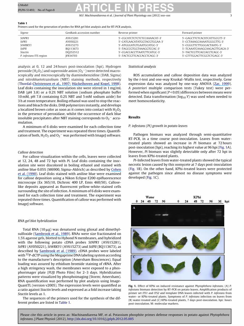
against the pathogen since almost no disease symptoms weredeveloped (Fig. 1C).
Fig. 1. Effect of KPhi on induced resistance against Phytophthora infestans. (A) P.
StIPII BQ113673 5 -Stactin DQ252512 5′-P. infestans ITS region JF834703 5′-
nalysis at 0, 12 and 24 hours post-inoculation (hpi). Hydrogeneroxide (H2O2) and superoxide anion (O2
−) were detected macro-copically and microscopically by diaminobenzidine (DAB, Sigma)nd nitrobluetetrazolium (NBT) staining methods, respectivelyThordal-Christensen et al., 1997; Hückelhoven and Kogel, 1998).eaf disks containing the inoculation site were stirred in 1 mg/mLAB (pH 3.8) or a 0.2% NBT solution (sodium phosphate buffer0 mM, pH 7.8 containing 0.2% NBT and 5 mM sodium azide) for
h at room temperature. Boiling ethanol was used to stop the reac-ions and bleach the disks. DAB polymerizes instantly, and develops
localized brown color as soon as it comes into contact with H2O2n the presence of peroxidase, whilst the occurrence of dark bluensoluble precipitates after NBT staining corresponds to O2
− accu-ulation.A minimum of 6 disks were examined for each collection time
nd treatment. The experiment was repeated three times. Quantifi-ation of both, H2O2 and O2
− was performed with ImageJ software.
allose detectionFor callose visualization within the cells, leaves were collected
t 12, 24, 48 and 72 hpi with Pi. Leaf disks containing the inoc-lation site were discolored in boiling ethanol and stained withniline blue 0.05% (M6900, Sigma–Aldrich) as described by Cohent al. (1990). Leaf disks stained with aniline blue were examinedor callose deposition using a Nikon Eclipse E200 epifluorescence
icroscope (Ex 365/10, Dichroic 400 LP, Emis 460/50). Callose-ike deposits appeared as fluorescent yellow-white-stained cellsurrounding the site of infection. A minimum of 6 disks were exam-ned for each collection time and treatment. The experiment wasepeated three times. Quantification of callose was performed withmageJ software.
NA gel blot hybridization
Total RNA (10 �g) was denatured using glioxal and dimethyl-ulfoxide (Sambrook et al., 1989). RNAs were size fractionated on.5% agarose gels, blotted to Hybond N membranes, and hybridizedith the following potato cDNA probes StNPR1 (AY615281),
tPR1 (AY050221), StWRKY1 (AY615273) and StIPII (BQ113673), asescribed by Sambrook et al. (1989). cDNA probes were labeledith 32P-dCTP using the Megaprime DNA labeling system according
o the manufacturer’s description (Amersham Biosciences). Equaloading was assured by ethidium bromide staining of rRNA. After
high stringency wash, the membranes were exposed to a phos-horimager plate (FUJI Photo Film) for 2–3 days. Hybridizationatterns were visualized by phosphoimaging (Storm; Amersham).NA quantification was performed by plot analysis using Image-uantTL (version v2005). The expression levels were quantified as
Please cite this article in press as: Machinandiarena MF, et al. Potassium
infestans. J Plant Physiol (2012), http://dx.doi.org/10.1016/j.jplph.2012.05.0
ratio against Stactin levels and expressed as a fold increase takingtactin levels as 1.
The sequences of the primers used for the synthesis of the dif-erent probes are listed in Table 1.
CGTGGTAAAGGTCCAC-3 5 -TCAAATCAAAGCAAGAGTTGAGA-3CACCTTAATCTTCATGC-3′ 5′-TACTCGTTCACCACCTCAGC-3′
GTTCACCACCTCAGC-3′ 5′-GTTTGGACTTCGGTCTGAGC-3′
Statistical analysis
ROS accumulation and callose deposition data was analyzedby the t-test and one-way Kruskal–Wallis test, respectively. Geneexpression data was analyzed by one-way ANOVA (Zar, 1999).A posteriori multiple comparison tests (Tukey test) were per-formed when significant (P < 0.05) differences between means weredetected. Data transformation (log10 Y) was used when needed tomeet homoscedasticity.
Results
P. infestans (Pi) growth in potato leaves
Pathogen biomass was analyzed through semi-quantitativeRT-PCR, in a time course post-inoculation. Leaves from water-treated plants showed an increase in Pi biomass at 72 hourspost-inoculation (hpi), reaching its highest value at 96 hpi (Fig. 1A).However, Pi biomass was slightly detectable only after 72 hpi inleaves from KPhi-treated plants.
Pi-Infected leaves from water-treated plants showed the typicalnecrotic lesion caused by this oomycete at 7 days post-inoculation(Fig. 1B). On the other hand, KPhi-treated leaves were protected
phosphite primes defense responses in potato against Phytophthora05
infestans biomass detection by RT-PCR on potato leaves. Amplification products ofprimer set ITS1 and ITS2 and template DNA leaves infected with P. infestans fromwater- or KPhi-treated plants. Symptoms of P. infestans infection on leaves from(B) water-treated and (C) KPhi-treated plants, 7 days post-inoculation. hpi: hourspost-inoculation. M: molecular markers.
ARTICLE IN PRESSG Model
JPLPH-51493; No. of Pages 8
4 M.F. Machinandiarena et al. / Journal of Plant Physiology xxx (2012) xxx– xxx
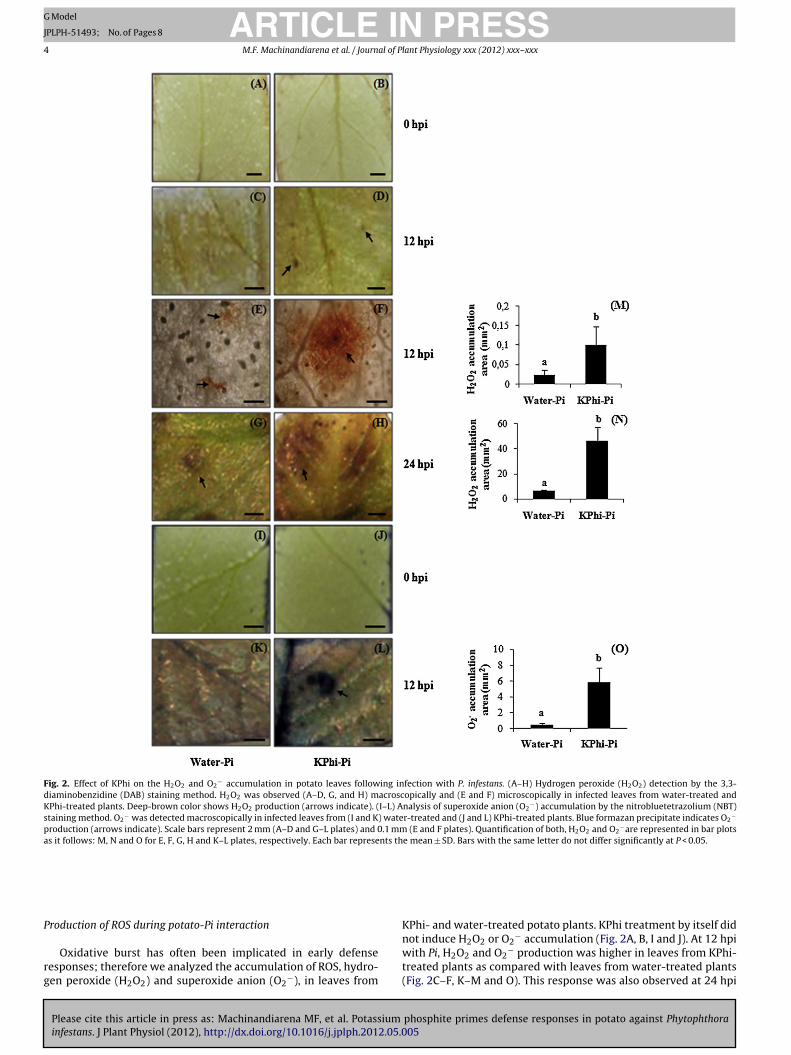
Fig. 2. Effect of KPhi on the H2O2 and O2− accumulation in potato leaves following infection with P. infestans. (A–H) Hydrogen peroxide (H2O2) detection by the 3,3-
diaminobenzidine (DAB) staining method. H2O2 was observed (A–D, G, and H) macroscopically and (E and F) microscopically in infected leaves from water-treated andKPhi-treated plants. Deep-brown color shows H2O2 production (arrows indicate). (I–L) Analysis of superoxide anion (O2
−) accumulation by the nitrobluetetrazolium (NBT)staining method. O2
− was detected macroscopically in infected leaves from (I and K) water-treated and (J and L) KPhi-treated plants. Blue formazan precipitate indicates O2−
production (arrows indicate). Scale bars represent 2 mm (A–D and G–L plates) and 0.1 mm (E and F plates). Quantification of both, H2O2 and O2−are represented in bar plots
as it follows: M, N and O for E, F, G, H and K–L plates, respectively. Each bar represents the mean ± SD. Bars with the same letter do not differ significantly at P < 0.05.
P
rg
roduction of ROS during potato-Pi interaction
Please cite this article in press as: Machinandiarena MF, et al. Potassium
infestans. J Plant Physiol (2012), http://dx.doi.org/10.1016/j.jplph.2012.05.0
Oxidative burst has often been implicated in early defenseesponses; therefore we analyzed the accumulation of ROS, hydro-en peroxide (H2O2) and superoxide anion (O2
−), in leaves from
KPhi- and water-treated potato plants. KPhi treatment by itself did
phosphite primes defense responses in potato against Phytophthora05
not induce H2O2 or O2− accumulation (Fig. 2A, B, I and J). At 12 hpi
with Pi, H2O2 and O2− production was higher in leaves from KPhi-
treated plants as compared with leaves from water-treated plants(Fig. 2C–F, K–M and O). This response was also observed at 24 hpi
ING Model
J
al of P
fip
C
tflo(7(
Ap
dpopStBecpewteo
Ii
JmoftlleKrto
D
tirltp
bpa
ARTICLEPLPH-51493; No. of Pages 8
M.F. Machinandiarena et al. / Journ
or H2O2 (Fig. 2G, H and N). However, no differences were detectedn O2
− accumulation between leaves from KPhi- and water-treatedlants at this time (not shown).
allose deposition during potato-Pi interaction
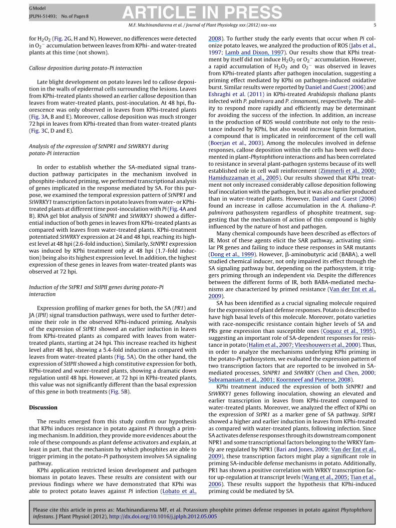
Late blight development on potato leaves led to callose deposi-ion in the walls of epidermal cells surrounding the lesions. Leavesrom KPhi-treated plants showed an earlier callose deposition thaneaves from water-treated plants, post-inoculation. At 48 hpi, flu-rescence was only observed in leaves from KPhi-treated plantsFig. 3A, B and E). Moreover, callose deposition was much stronger2 hpi in leaves from KPhi-treated than from water-treated plantsFig. 3C, D and E).
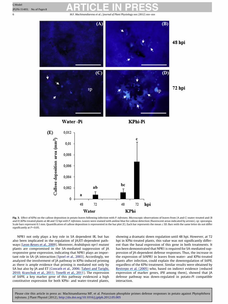
nalysis of the expression of StNPR1 and StWRKY1 duringotato-Pi interaction
In order to establish whether the SA-mediated signal trans-uction pathway participates in the mechanism involved inhosphite-induced priming, we performed transcriptional analysisf genes implicated in the response mediated by SA. For this pur-ose, we examined the temporal expression pattern of StNPR1 andtWRKY1 transcription factors in potato leaves from water- or KPhi-reated plants at different time post-inoculation with Pi (Fig. 4A and). RNA gel blot analysis of StNPR1 and StWRKY1 showed a differ-ntial induction of both genes in leaves from KPhi-treated plants asompared with leaves from water-treated plants. KPhi-treatmentotentiated StWRKY expression at 24 and 48 hpi, reaching its high-st level at 48 hpi (2.6-fold induction). Similarly, StNPR1 expressionas induced by KPhi treatment only at 48 hpi (1.7-fold induc-
ion) being also its highest expression level. In addition, the highestxpression of these genes in leaves from water-treated plants wasbserved at 72 hpi.
nduction of the StPR1 and StIPII genes during potato-Pinteraction
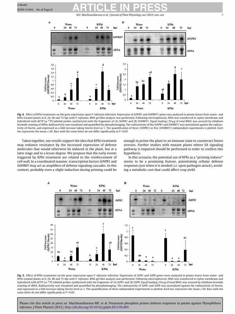
Expression profiling of marker genes for both, the SA (PR1) andA (IPII) signal transduction pathways, were used to further deter-
ine their role in the observed KPhi-induced priming. Analysisf the expression of StPR1 showed an earlier induction in leavesrom KPhi-treated plants as compared with leaves from water-reated plants, starting at 24 hpi. This increase reached its highestevel after 48 hpi, showing a 5.4-fold induction as compared witheaves from water-treated plants (Fig. 5A). On the other hand, thexpression of StIPII showed a high constitutive expression for both,Phi-treated and water-treated plants, showing a dramatic downegulation until 48 hpi. However, at 72 hpi in KPhi-treated plants,his value was not significantly different than the basal expressionf this gene in both treatments (Fig. 5B).
iscussion
The results emerged from this study confirm our hypothesishat KPhi induces resistance in potato against Pi through a prim-ng mechanism. In addition, they provide more evidences about theole of these compounds as plant defense activators and explain, ateast in part, that the mechanism by which phosphites are able torigger priming in the potato-Pi pathosystem involves SA signalingathway.
Please cite this article in press as: Machinandiarena MF, et al. Potassium
infestans. J Plant Physiol (2012), http://dx.doi.org/10.1016/j.jplph.2012.05.0
KPhi application restricted lesion development and pathogeniomass in potato leaves. These results are consistent with ourrevious findings where we have demonstrated that KPhi wasble to protect potato leaves against Pi infection (Lobato et al.,
PRESSlant Physiology xxx (2012) xxx– xxx 5
2008). To further study the early events that occur when Pi col-onize potato leaves, we analyzed the production of ROS (Jabs et al.,1997; Lamb and Dixon, 1997). Our results show that KPhi treat-ment by itself did not induce H2O2 or O2
− accumulation. However,a rapid accumulation of H2O2 and O2
− was observed in leavesfrom KPhi-treated plants after pathogen inoculation, suggesting apriming effect mediated by KPhi on pathogen-induced oxidativeburst. Similar results were reported by Daniel and Guest (2006) andEshraghi et al. (2011) in KPhi-treated Arabidopsis thaliana plantsinfected with P. palmivora and P. cinnamomi, respectively. The abil-ity to respond more rapidly and efficiently may be determinantfor avoiding the success of the infection. In addition, an increasein the production of ROS would contribute not only to the resis-tance induced by KPhi, but also would increase lignin formation,a compound that is implicated in reinforcement of the cell wall(Boerjan et al., 2003). Among the molecules involved in defenseresponses, callose deposition within the cells has been well docu-mented in plant-Phytophthora interactions and has been correlatedto resistance in several plant-pathogen systems because of its wellestablished role in cell wall reinforcement (Zimmerli et al., 2000;Hamiduzzaman et al., 2005). Our results showed that KPhi treat-ment not only increased considerably callose deposition followingleaf inoculation with the pathogen, but it was also earlier producedthan in water-treated plants. However, Daniel and Guest (2006)found an increase in callose accumulation in the A. thaliana–P.palmivora pathosystem regardless of phosphite treatment, sug-gesting that the mechanism of action of this compound is highlyinfluenced by the nature of host and pathogen.
Many chemical compounds have been described as effectors ofIR. Most of these agents elicit the SAR pathway, activating simi-lar PR genes and failing to induce these responses in SAR mutants(Dong et al., 1999). However, �-aminobutyric acid (BABA), a wellstudied chemical inducer, not only impaired its effect through theSA signaling pathway but, depending on the pathosystem, it trig-gers priming through an independent via. Despite the differencesbetween the different forms of IR, both BABA-mediated mecha-nisms are characterized by primed resistance (Van der Ent et al.,2009).
SA has been identified as a crucial signaling molecule requiredfor the expression of plant defense responses. Potato is described tohave high basal levels of this molecule. Moreover, potato varietieswith race-nonspecific resistance contain higher levels of SA andPRs gene expression than susceptible ones (Coquoz et al., 1995),suggesting an important role of SA-dependent responses for resis-tance in potato (Halim et al., 2007; Vleeshouwers et al., 2000). Thus,in order to analyze the mechanisms underlying KPhi priming inthe potato-Pi pathosystem, we evaluated the expression pattern oftwo transcription factors that are reported to be involved in SA-mediated processes, StNPR1 and StWRKY (Chen and Chen, 2000;Subramaniam et al., 2001; Koornneef and Pieterse, 2008).
KPhi treatment induced the expression of both StNPR1 andStWRKY1 genes following inoculation, showing an elevated andearlier transcription in leaves from KPhi-treated compared towater-treated plants. Moreover, we analyzed the effect of KPhi onthe expression of StPR1 as a marker gene of SA pathway. StPR1showed a higher and earlier induction in leaves from KPhi-treatedas compared with water-treated plants, following infection. SinceSA activates defense responses through its downstream componentNPR1 and some transcriptional factors belonging to the WRKY fam-ily are regulated by NPR1 (Bari and Jones, 2009; Van der Ent et al.,2009), these transcription factors might play a significant role inpriming SA-inducible defense mechanisms in potato. Additionally,
phosphite primes defense responses in potato against Phytophthora05
PR1 has shown a positive correlation with WRKY transcription fac-tor up-regulation at transcript levels (Wang et al., 2005; Tian et al.,2006). These results support the hypothesis that KPhi-inducedpriming could be mediated by SA.
ARTICLE IN PRESSG Model
JPLPH-51493; No. of Pages 8
6 M.F. Machinandiarena et al. / Journal of Plant Physiology xxx (2012) xxx– xxx
Fig. 3. Effect of KPhi on the callose deposition in potato leaves following infection with P. infestans. Microscopic observations of leaves from (A and C) water-treated and (Ba h anilS he bars
awprtaaS2oc
nd D) KPhi-treated plants at 48 and 72 hpi with P. infestans. Leaves were stained witcale bars represent 0.1 mm. Quantification of callose deposition is represented in tignificantly at P < 0.05.
NPR1 not only plays a key role in SA dependent IR, but haslso been implicated in the regulation of JA/ET-dependent path-ays (Leon-Reyes et al., 2009). Moreover, Arabidopsis npr1 mutantlants are compromised in the SA-mediated suppression of JAesponsive gene expression, indicating that NPR1 plays an impor-ant role in SA–JA interaction (Spoel et al., 2003). Accordingly, wenalyzed the involvement of JA pathway in KPhi-induced primings there is ample evidence that priming is mediated not only by
Please cite this article in press as: Machinandiarena MF, et al. Potassium
infestans. J Plant Physiol (2012), http://dx.doi.org/10.1016/j.jplph.2012.05.0
A but also by JA and ET (Conrath et al., 2006; Taheri and Tarighi,010; Kravchuk et al., 2011; Tonelli et al., 2011). The expressionf StIPII, a key marker gene of this pathway evidenced a highonstitutive expression for both KPhi- and water-treated plants,
ine blue for callose detection (fluorescent areas indicated by arrows). sp: sporangia. plot (E). Each bar represents the mean ± SD. Bars with the same letter do not differ
showing a dramatic down regulation until 48 hpi. However, at 72hpi in KPhi-treated plants, this value was not significantly differ-ent than the basal expression of this gene in both treatments. Ithas been demonstrated that NPR1 is required for SA-mediated sup-pression of JA-dependent defense responses. Thus, the increase inthe expression of StNPR1 in leaves from water- and KPhi-treatedplants after infection, could explain the downregulation of StIPII,regardless of the KPhi treatment. Similar results were obtained by
phosphite primes defense responses in potato against Phytophthora05
Restrepo et al. (2005) who, based on indirect evidence (reducedexpression of marker genes, IPII among them), showed that JAdefense pathway was down-regulated in potato-Pi compatibleinteraction.
ARTICLE IN PRESSG Model
JPLPH-51493; No. of Pages 8
M.F. Machinandiarena et al. / Journal of Plant Physiology xxx (2012) xxx– xxx 7
Fig. 4. Effect of KPhi treatments on the gene expression upon P. infestans infection. Expression of StNPR1 and StWRKY1 genes was analyzed in potato leaves from water- andKPhi-treated plants at 0, 24, 48 and 72 hpi with P. infestans. RNA gel blot analysis was performed. Following electrophoresis, RNA was transferred to nylon membrane andhybridized with dCTP-[a-32P]-labeled probes synthesized with the fragments of (A) StNPR1 and (B) StWRKY1. Equal loading (10 �g of total RNA) was assured by ethidiumb agingt tificatb t P < 0
mmltcSc
FKhsas
romide staining of rRNA. Radioactivity was visualized and quantified by phosphoimivity of Stactin, and expressed as a fold increase taking Stactin level as 1. The quanar represents the mean ± SD. Bars with the same letter do not differ significantly a
Taken together, our results support the idea that KPhi treatmentay enhance resistance by the increased expression of defenseolecules that would otherwise be induced in the plant, but at a
ater stage and to a lesser degree. We propose that the early eventsriggered by KPhi treatment are related to the reinforcement of
Please cite this article in press as: Machinandiarena MF, et al. Potassium
infestans. J Plant Physiol (2012), http://dx.doi.org/10.1016/j.jplph.2012.05.0
ell wall. In a coordinated manner, transcription factors StNPR1 andtWRKY may act as amplifiers of defense signaling cascades. In thisontext, probably even a slight induction during priming could be
ig. 5. Effect of KPhi treatments on the gene expression upon P. infestans infection. ExpPhi-treated plants at 0, 24, 48 and 72 hpi with P. infestans. RNA gel blot analysis was peybridized with dCTP-[a-32P]-labeled probes synthesized with the fragments of (A) StPR1taining of rRNA. Radioactivity was visualized and quantified by phosphoimaging. The rand expressed as a fold increase taking Stactin level as 1. The quantification of three indeame letter do not differ significantly at P < 0.05.
. The radioactivity of the StNPR1 and StWRKY1 was normalized against the radioac-ion of three (StNPR1) or five (StWRKY1) independent experiments is plotted. Each.05.
enough to prime the plant to an immune state to counteract futurestresses. Further studies with mutant plants where SA signalingpathway is impaired should be performed in order to confirm thishypothesis.
In this scenario, the potential use of KPhi as a “priming inducer”
phosphite primes defense responses in potato against Phytophthora05
seems to be a promising feature, potentiating cellular defenseresponses just when it is needed (i.e. upon pathogen attack), avoid-ing a metabolic cost that could affect crop yield.
ression of StPR1 and StIPII genes were analyzed in potato leaves from water- andrformed. Following electrophoresis, RNA was transferred to nylon membrane and
and (B) StIPII. Equal loading (10 �g of total RNA) was assured by ethidium bromidedioactivity of StPR1 and StIPII was normalized against the radioactivity of Stactin,pendent experiments is plotted. Each bar represents the mean ± SD. Bars with the
ING Model
J
8 al of P
A
gPtUMCi
R
A
A
A
B
B
B
C
C
C
C
C
D
D
D
D
E
G
G
G
H
H
H
ARTICLEPLPH-51493; No. of Pages 8
M.F. Machinandiarena et al. / Journ
cknowledgments
This work was supported by Consejo Nacional de Investi-aciones Científicas y Técnicas (CONICET), Agencia Nacional deromoción Científica y Tecnológica (ANPCYT), Comisión de Inves-igaciones Científicas de la Provincia de Buenos Aires (CIC), andniversidad Nacional de Mar del Plata (UNMdP). MachinandiarenaF, Feldman ML and Andreu AB are established researchers from
ONICET. Daleo GR is an established researcher of CIC. Lobato MCs a fellow of CONICET.
eferences
hn I-P, Kim S, Lee Y-H, Suh S-C. Vitamin B1-induced priming is dependent onhydrogen peroxide and the NPR1 gene in Arabidopsis. Plant Physiol 2007;143:838–48.
ltamiranda EAG, Andreu AB, Daleo GR, Olivieri FP. Effect of �-aminobutyric acid(BABA) on protection against Phytophthora infestans throughout the potato cropcycle. Australas Plant Pathol 2008;37:421–7.
ndreu AB, Caldiz DO, Forbes GA. Phenotypic description of resistance toPhythophtora infestans in processing potatoes in Argentina. Am J Potato Res2010;87:177–87.
ari R, Jones JD. Role of plant hormones in plant defence responses. Plant Mol Biol2009;69:473–88.
eckers GJM, Conrath U. Priming for stress resistance: from the lab to the field. CurrOpin Plant Biol 2007;10:425–31.
oerjan W, Ralph J, Baucher M. Lignin biosynthesis. Annu Rev Plant Biol2003;54:519–46.
hen C, Chen Z. Isolation and characterization of two pathogen- and salicylic acid-induced genes encoding WRKY DNA-binding proteins from tobacco. Plant MolBiol 2000;42:387–96.
ohen Y, Eyal H, Hanania J. Ultrastructure, autofluorescence, callose depositionand lignification in susceptible and resistant muskmelon leaves infected withthe powdery mildew fungus Sphaerotheca fuliginea. Physiol Mol Plant Pathol1990;36:191–204.
onrath U, Beckers GJ, Flors V, García-Agustín P, Jakab G, Mauch F, et al. Priming:getting ready for battle. Mol Plant Microbe Interact 2006;19:1062–71.
ooke LR, Schepers HTAM, Hermansen A, Bain RA, Bradshaw NJ, Ritchie F, et al.Epidemiology and integrated control of potato late blight in Europe. Potato Res2011;54:183–222.
oquoz J-L, Buchala A, Meuwly P, Métraux J-P. Arachidonic acid induces localbut not systemic synthesis of salicylic acid and confers systemic resistance inpotato plants to Phytophthora infestans and Alternaria solani. Phytopathology1995;85:1219–24.
aayf F, Ongena M, Boulanger RN, El Hadrami I, Bélanger RR. Induction of phenoliccompounds in two cultivars of cucumber by treatment of healthy and powderymildew-infected plants with extracts of Reynoutria sachalinensis. J Chem Ecol2000;26:1579–93.
aniel R, Guest D. Defence responses induced by potassium phosphonate in Phy-tophthora palmivora-challenged Arabidopsis thaliana. Physiol Mol Plant Pathol2006;67:194–201.
eliopoulos T, Kettlewell PS, Hare MC. Fungal disease suppression by inorganic salts:a review. Crop Prot 2010;29:1059–75.
ong H, Delaney TP, Bauer DW, Beer SV. Harping induces disease resistance in Ara-bidopsis through the systemic acquired resistance pathway mediated by salicylicacid and the NIM1 gene. Plant J 1999;20:207–15.
shraghi L, Anderson J, Aryamanesh N, Shearer B, McComb J, Hardy GEStJ, O’BrienPA. Phosphite primed defence responses and enhanced expression of defencegenes in Arabidopsis thaliana infected with Phytophthora cinnamomi. Plant Pathol2011;60:1086–95.
oellner K, Conrath U. Priming: it’s all the world to induced disease resistance. EurJ Plant Pathol 2008;121:233–42.
rant BR, Dunstan RH, Griffith JM, Niere JO, Smillie RH. The mechanism ofphosphonic (phosphorous) acid action in Phytophthora. Australas Plant Pathol1990;19:115–21.
uest D, Grant B. The complex action of phosphonates as antifungal agents. Biol Rev1991;66:159–87.
alim V, Eschen-Lippold L, Altmann S, Birschwilks M, Scheel D, Rosahl S. Salicylicacid is important for basal defense of Solanum tuberosum against Phytophthorainfestans. Mol Plant Microbe Interact 2007;20:1346–52.
amiduzzaman MM, Jakab G, Barnavon L, Neuhaus J-M, Mauch-Mani B. �-
Please cite this article in press as: Machinandiarena MF, et al. Potassium
infestans. J Plant Physiol (2012), http://dx.doi.org/10.1016/j.jplph.2012.05.0
Aminobutyric acid-induced resistance against Downy Mildew in grapevine actsthrough the potentiation of callose formation and jasmonic acid signaling. MolPlant Microbe Interact 2005;18:819–29.
ooker WJ. Compendio de Enfermedades de la Papa. St. Paul, MN, USA: The AmericanPhytopathological Society; 1980. p. 56–60.
PRESSlant Physiology xxx (2012) xxx– xxx
Hückelhoven R, Kogel KH. Tissue-specific superoxide generation at interaction sitesin resistant and susceptible near-isogenic barley lines attacked by the pow-dery mildew fungus (Erysiphe graminis f. sp. hordei). Mol Plant Microbe Interact1998;11:292–300.
Jabs T, Tschöpe M, Colling C, Hahlbrock K, Scheel D. Elicitor-stimulated ion fluxesand O2
− from the oxidative burst are essential components in triggering defensegene activation and phytoalexin synthesis in parsley. Proc Natl Acad Sci USA1997;94:4800–5.
King M, Reeve W, Van der Hoek MB, Williams N, McComb J, O’Brien PA, Hardy GE.Defining the phosphite-regulated transcriptome of the plant pathogen Phytoph-thora cinnamomi. Mol Genet Genomics 2010;284:425–35.
Koornneef A, Pieterse CMJ. Cross talk in defense signaling. Plant Physiol2008;146:839–44.
Kravchuk Z, Vicedo B, Flors V, Camanes G, González-Bosch C, García-Agustín P. Prim-ing for JA-dependent defenses using hexanoic acid is an effective mechanism toprotect Arabidopsis against B. cinerea. J Plant Physiol 2011;168:359–66.
Lamb C, Dixon RA. The oxidative burst in plant disease resistance. Annu Rev PlantPhysiol 1997;48:251–75.
Leon-Reyes A, Spoel SH, De Lange ES, Abe H, Kobayashi M, Tsuda S, et al. Ethylenemodulates the role of nonexpressor of pathogenesis-related genes1 in cross talkbetween salicylate and jasmonate signaling. Plant Physiol 2009;149:1797–809.
Lobato MC, Olivieri FP, González Altamiranda EA, Wolski EA, Daleo GR, Caldiz DO,Andreu AB. Phosphite compounds reduce disease severity in potato seed tubersand foliage. Eur J Plant Pathol 2008;122:349–58.
Lobato MC, Machinandiarena MF, Tambascio C, Dosio GAA, Caldiz DO, Daleo GR,et al. Effect of foliar applications of phosphite on post-harvest potato tubers. EurJ Plant Pathol 2011;130:155–63.
Pilbeam RA, Howard K, Shearer BL, Hardy GESJ. Phosphite stimulated histologicalresponses of Eucalyptus marginata to infection by Phytophthora cinnamomi. TreesStruct Funct 2011;25:1121–31.
Restrepo S, Myers KL, del Pozo O, Martin GB, Hart AL, Buell CR, et al. Gene pro-filing of a compatible interaction between Phytophthora infestans and Solanumtuberosum suggests a role for carbonic anhydrase. Mol Plant Microbe Interact2005;18:913–22.
Sambrook J, Fritsch EF, Maniatis T. Molecular cloning: a laboratory manual, vol. I,2nd ed. Cold Spring Harbor Laboratory Press; 1989.
Shibuya N, Minami E. Oligosaccharide signalling for defence responses in plant.Physiol Mol Plant Pathol 2001;59:223–33.
Spoel SH, Koornneef A, Claessens SM, Korzelius JP, Van Pelt JA, Mueller MJ, et al. NPR1modulates cross talk between salicylate- and jasmonate-dependent defensepathways through a novel function in the cytosol. Plant Cell 2003;15:760–70.
Spoel SH, Mou Z, Tada Y, Spivey NW, Genschik P, Dong X. Proteasome-mediatedturnover of the transcription coactivator NPR1 plays dual roles in regulatingplant immunity. Cell 2009;137:860–72.
Subramaniam R, Desveaux D, Spickler C, Michnick S, Brisson N. Direct visualizationof protein interactions in plant cells. Nat Biotechnol 2001;19:769–72.
Taheri P, Tarighi S. Riboflavin induces resistance in rice against Rhizoctonia solanivia jasmonate-mediated priming of phenylpropanoid pathway. J Plant Physiol2010;167:201–8.
Thordal-Christensen H, Zhang Z, Wei Y, Collinge DB. Subcellular localization of H2O2
in plants. H2O2 accumulation in papillae and hypersensitive response during thebarley-powdery mildew interaction. Plant J 1997;11:1187–94.
Tian ZD, Liu J, Wang BL, Xie CH. Screening and expression analysis of Phytophthorainfestans induced genes in potato leaves with horizontal resistance. Plant CellRep 2006;25:1094–103.
Tonelli ML, Furlan A, Taurian T, Castro S, Fabra A. Peanut priming induced by bio-control agents. Physiol Mol Plant Pathol 2011;75:100–5.
Van der Ent S, Van Hulten M, Pozo MJ, Czechowski T, Udvardi MK, Pieterse CMJ, TonJ. Priming of plant innate immunity by rhizobacteria and �-aminobutyric acid:differences and similarities in regulation. New Phytol 2009;183:419–31.
Vleeshouwers V, Van Dooijeweert W, Govers F, Kamoun S, Colon LT. Does basalPR gene expression in Solanum species contribute to non-specific resistance toPhytophthora infestans? Physiol Mol Plant Pathol 2000;57:35–42.
Vlot AC, Dempsey DMA, Klessig DF. Salicylic acid, a multifaceted hormone to combatdisease. Annu Rev Phytopathol 2009;47:177–206.
Wang B, Liu J, Tian Z, Song B, Xie C. Monitoring the expression patterns of potatogenes associated with quantitative resistance to late blight during Phytophthorainfestans infection using cDNA microarrays. Plant Sci 2005;169:1155–67.
Wang D, Amornsiripanitch N, Dong X. A genomic approach to identify regulatorynodes in the transcriptional network of systemic acquired resistance in plants.PLoS Pathog 2006;2:1042–50.
Wilkinson CJ, Shearer BL, Jackson TJ, Hardy GESJ. Variation in sensitivity of westernAustralian isolates of Phytophthora cinnamomi to phosphite in vitro. Plant Pathol2001;50:83–9.
Yu D, Chen C, Chen Z. Evidence for an important role of WRKY DNA binding proteins
phosphite primes defense responses in potato against Phytophthora05
in the regulation of NPR1 gene expression. Plant Cell 2001;13:1527–40.Zar JH. Biostatistical analysis. 4th ed. Prentice-Hall: Upper Saddle River; 1999.Zimmerli L, Jakab G, Metraux JP, Mauch-Mani B. Potentiation of pathogen-specific
defense mechanisms in Arabidopsis by �-aminobutyric acid. Proc Natl Acad SciUSA 2000;97:12920–5.


![L'identification des enfants. Un concept utile pour l'analyse des primes socialisations [article dans Sociologie, 2015]](https://static.fdokumen.com/doc/165x107/63233cb2078ed8e56c0ac3fa/lidentification-des-enfants-un-concept-utile-pour-lanalyse-des-primes-socialisations.jpg)

