Verticillium wilt and Phytophthora blight of chile pepper - CORE
102
Retrospective eses and Dissertations Iowa State University Capstones, eses and Dissertations 1960 Verticillium wilt and Phytophthora blight of chile pepper Roy Minoru Nakayama Iowa State University Follow this and additional works at: hps://lib.dr.iastate.edu/rtd Part of the Agriculture Commons , and the Plant Pathology Commons is Dissertation is brought to you for free and open access by the Iowa State University Capstones, eses and Dissertations at Iowa State University Digital Repository. It has been accepted for inclusion in Retrospective eses and Dissertations by an authorized administrator of Iowa State University Digital Repository. For more information, please contact [email protected]. Recommended Citation Nakayama, Roy Minoru, "Verticillium wilt and Phytophthora blight of chile pepper " (1960). Retrospective eses and Dissertations. 2623. hps://lib.dr.iastate.edu/rtd/2623
-
Upload
khangminh22 -
Category
Documents
-
view
1 -
download
0
Transcript of Verticillium wilt and Phytophthora blight of chile pepper - CORE

Retrospective Theses and Dissertations Iowa State University Capstones, Theses andDissertations
1960
Verticillium wilt and Phytophthora blight of chilepepperRoy Minoru NakayamaIowa State University
Follow this and additional works at: https://lib.dr.iastate.edu/rtd
Part of the Agriculture Commons, and the Plant Pathology Commons
This Dissertation is brought to you for free and open access by the Iowa State University Capstones, Theses and Dissertations at Iowa State UniversityDigital Repository. It has been accepted for inclusion in Retrospective Theses and Dissertations by an authorized administrator of Iowa State UniversityDigital Repository. For more information, please contact [email protected].
Recommended CitationNakayama, Roy Minoru, "Verticillium wilt and Phytophthora blight of chile pepper " (1960). Retrospective Theses and Dissertations.2623.https://lib.dr.iastate.edu/rtd/2623

VERTICILLIUM WILT AND PHYTOPHTHORA BLIGHT OF CHILE PEPPER
by
Roy Minoru Nakayama
A Dissertation Submitted to the
Graduate Faculty in Partial Fulfillment of
The Requirements for the Degree of
DOCTOR OF PHILOSOPHY
Major Subject: Plant Pathology
Approved:
In Charge of Major Work
Head of Major Department
Iowa State University Of Science and Technology
Ames, Iowa
1960
Signature was redacted for privacy.
Signature was redacted for privacy.
Signature was redacted for privacy.

11
TABLE OF CONTENTS
Page
INTRODUCTION 1
PART I: VERTICILLIUM WILT 3
Literature Review 3
Investigations 6
Symptoms of the disease 6 The causal organism 9 Materials and methods 13 Experimental results 16
PART II: . PHYTOPHTHORA BLIGHT 27
Literature Review 27
Investigations 38
Symptoms of the disease 38 The causal organism 42 Materials and methods. . . 48 Experimental results 55
DISCUSSION 78
SUMMARY 90
LITERATURE CITED 95
ACKNOWLEDGEMENTS 99

1
INTRODUCTION
The disease of chile pepper commonly called chile wilt or
Fusarium wilt, caused by Fusarlum annuum L. wes first reported
to occur in New Mexico in 1908 by Leonlan (1919). Since that
time, wilted plants have been observed in practically all
chile pepper fields in the areas of the state where this crop
is grown. This disease is considered to be the most serious
disease of the crop with losses depending on the age of the
plants at the time of attack. Severity of the disease ranges
from a few to 100 per cent wilted plants per field. Fusarlum
annuum has been reported to invade only the cortex of roots,
with no evidence of discoloration of vascular tissue, and no
spread into the above ground parts of the plant.
Phvtophthora cansicl L., causal agent of another disease
of chile peppers in the state, has been reported to produce
no root infection but to attack the stem at or above the soil
line, as well as the leaves and fruit.
The recommended control measures for Fusarlum wilt have
not been uniformly effective, and upon a more critical exam
ination of diseased plants occurring in fields over the state,
a consistency 1 Ti symptoms of the disease was not observed.
On many wilted plants, previously described symptoms of chile
wilt were not observed. A Verticlllium was isolated from
these plants, and indicated that this fungus could also be
involved.

2
These observations resulted in the initiation of a more
thorough Investigation of the wilt diseases of chile pepper.
The results of these studies are reported in two parts: those
related to Vertlcllllum and those related to Phvtophthore.
In the studies reported here, the term chile pepper is
referred to members of Capsicum frutescens L. ver. Iongum
Sendt., and bell or sweet peppers to members of Ç. frutescens
L. var. gros sum Sendt. This species designation is based on
the works of Bailey ( 192-3) in which he considers all horti
cultural peppers to be forms of the species Capsicum frutes
cens L. and in which he considers Capsicum annuum L. to be
synonymous.

3
PART I: VERTICILLIUM WILT
Literature Review
The first record of a Vertlcllllum causing disease on
Capsicum frutescens L. was made by Curzi (1925) from Italy.
He described a typical hadromycosis on pepper caused by a
fungus to which he gave the name, Verticilllum tracheiphllum.
He reported the fungus to be widespread in Italy. The first
recorded occurrence of Vertlcllllum wilt of peppers in the
United States was in 1937 from California by Rudolph and
Snyder (1937). They identified the causal agent to be Ver
tlcllllum albo-atrum Reinke and Berth., and reported that s
20 per cent loss was incurred in the field in which it was
first observed. Later during the same year Dunlap (1937) re
ported that 70 per cent or more of the plants were affected
by the same fungus in widely separated pepper fields In Con
necticut. Snyder and Rudolph (1939) made more extensive
studies on the occurrence of the disease and concluded that
Vertlcllllum wilt was an Important disease of peppers in Cal
ifornia in fields where it appeared.
Since the initial report of Vertlcllllum wilt of peppers,
Clccarone (1953) reported that V. albo-atrum is widespread
throughout Italy. Elenkov (1955) reported this same fungus
to cause dwarfing and losses up to 90 per cent in some fields
during the 1951 season In Bulgaria. A species of Verticilllum

4
was reported by Chamberlain (1942) to affect 5 per cent of
the plants in a pepper planting in Ontario, Canada. Later,
McKeen (1949) reported a wilt, caused by V. dahliae. of 60 per
cent of plants in a small field in the same area. The disease
appeared midway through the growing season and many plants
died before any fruits were harvested. The remainder of the
plants were badly stunted and produced a poor crop. Woollams
(i960) reported the isolation of V. albo-atrum from diseased
pepper plants in the province of British Columbia. Two years
later Verticilllum wilt was reported to occur on several farms
in three different growing areas in the same province (Wool-
lams, 1952). Infection varied among fields from slight to
over 75 per cent diseased plants. By 1957, Verticilllum wilt
was reported (Conners et. aJL. , 1957) to be widespread in this
province. Ten to 100 per cent of the plants were affected,
with an average of 20 per cent infected plants in the fields.
Attempts to infect pepper plants by inoculation with
strains of V. albo-atrum Isolated from hosts other than
peppers have not always been successful- Bewley (1922) pro
duced symptoms on pepper plants with an isolate from diseased
tomato plants. On the other hand, Haenseler (1922) reported
negative results from inoculation attempts of pepper plants
with an isolate from eggplant. Baines (1945) was unable to
Infect peppers with Isolates from Viburnum, peppermint and
chrysanthemum.

5
Kendrlck and Middleton (1959) used isolates of V. albo-
atrum from a wide range of host plants in an experiment to
detect differences in pathogenicity to pepper. They reported
that isolates from chrysanthemum, rose and pepper were all
significantly pathogenic on pepper, but the pepper isolate
had a severity index three times greater than isolates from
the other two hosts. Isolates from potato, okra, tomato and
six other hosts were not significantly pathogenic to pepper.
In another experiment by the same authors, an attempt was made
to rate the relative pathogenicity of five different Isolates
of V. albo-atrum. Isolates from avocado, muskmelon, okra,
tomato and pepper were tested on two varieties of chile pepper
and one variety of sweet pepper. The pepper, muskmelon and
okra isolates were pathogenic to all three varieties of
peppers. The pepper isolate produced severe stunting and sub
sequent death of okra, but failed to infect tomato. In these
studies, it was also reported that stunting of pepper plants
within a given period of time was more severe when 30-day old
plants were inoculated than when 60-day old plants were used.
Studies dealing with the possibility' that differences in
reaction of varieties of peppers to V. albo-atrum might exist
were conducted by Elenkov (1957) in Bulgaria. Of the 115
varieties of peppers tested by root dipping in a suspension
of the fungus, only two hybrids of a local variety by Cali
fornia Wonder were resistant. Of the pungent varieties, a

6
local paprika variety was most resistant and the cherry pepper
was most susceptible.
Temperature studies with isolates of V. albo-atrum from
diseased pepper plants were conducted by Kendrlck and Middle-
ton (1959) on potato dextrose agar in tubes. The maximum
daily growth increase occurred at 24° C. Mo growth occurred
below 9° or above 36° C. after seven days. All isolates grew
well at temperatures ranging from 12° to 30° C. No signifi
cant difference in disease severity existed between 15° and
30° C. At 35° C., symptoms of wilt were slight on one vari
ety, whereas no symptoms were observed on the other varieties
used in the test.
Investigations
Symptoms of the disease
The first symptom on plants infected with Vertlcllllum is
a drooping of leaves on a few branches or of the entire niant.
The leaf edges roll inward, exposing the lighter colored lower
surface. On some leaves, poorly defined chlorotic areas of
the interveinal tissue occur. On the branches that do not
show wilting, the leaves may appear healthy, or more generally
show a dull green or even a light yellow color (Fig. l-B,C).
After a few days, the wilted leaves appear tightly rolled and
have turned a. light brown color upon drying (Fig. 1-D,E) .
Leaves and the very Immature fruit remain lightly attached to

Fig. 1. Symptoms on chile pepper plants naturally infected with V. albo-atrum
A. Healthy chile pepper plant
B. Infected plant showing two branches with wilted leaves
C. Infected plant with wilted leaves on all branches except one
D. Midseason infection of chile pepper plant; note light fruit production
E. Early season infection of chile pepper plant
F. Internal discoloration of root from infected plant; no internal discoloration of vascular tissues of root of healthy plant at right


9
the plant until mechanically disturbed, but the more mature
fruit remain attached to the plant for the remainder of the
season. The outer surface of roots of such plants anpear
normal and no evidence of cortical discoloration can be ob
served. However, a light brown discoloration of the water
conducting tissues may be observed in the roots and in the
stems (Fig. 1-F).
On plants that were attacked early in the season, the
leaves were not wilted, but were smaller in size and dull
green in color. Also few fruit were set and these were
smaller in size than normal. Leaves on some niants showed
irregular patches of chlorotic tissue which gave the leaf a
mottled appearance. Extensive necrosis of interveinal tissue
extending from the blade edge was not commonly observed, but
considerable leaf fall occurred from plants that showed this
type of leaf symptom.
The distribution of infected plants within a given field
was generally in irregularly defined areas. Plants in all
stages of infection from just wilting to dead plants were ob
served, and occurred either singly or as several successive
plants within a row and Interrupted with healthy plants.
The causal organism
Identification A Verticllllum was isolated from
diseased chile pepper plants collected in northern New Mexico

10
during the 1956 season, and again from the same locality and
also from fields in the central and southern parts of the
state during 1958. Cultures of all isolates produced a thick
carbonaceous crust of black microsclerotia covered with a
white thin mass of verticillately branched conidiophores.
Macroscopically and microscopically, cultures of all three
isolates appeared similar and were considered to be of the
same species.
The pathogen used in these studies was identified as
Verticilllum albo-atrum Reinke and Berthold since its morpho
logical characteristics agreed with the original description
of that fungus by Reinke and Berthold (1879) and further sub
stantiated by the works of Rudolph (1931) and Presley (1941).
Location of fungus in plant tissue VertlclIlium was
isolated from roots, stem, petiole and peduncle of fruit of
diseased plants. Mycelium was observed in the water conduct
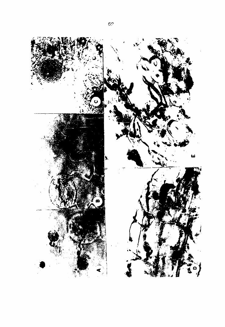
ing tissues of diseased roots (Fig. P-C,D) . The dark occlu
sions and tyloses in vascular tissue of roots (Fig. ?-A,B)
were not as extensive as those observed in the stems.
When the lower portion of 30-day old chile pepper niant
roots were immersed and grown in flasks containing Vertlcil-
llum in sterile distilled water, the pathogen was isolated
from the stem 85 to 90 millimeters above the inoculum when the
leaves showed wilting seven to nine days after inoculation.
On plants that did not show wilting, the pathogen was isolated

Fig. 2 . Cross sections of secondary roots from V. albo-atrum infected plants
A. Section showing tyloses in vessels
B. Enlarged section from A, showing tyloses and occlusions in xylem tissue
C.D. Mycelium in vessels and trachae


13
from root sections taken 10 to 15 millimeters above the in
oculum .
Materials and methods
Materials The variety of chile pepper used in all
experiments was New Mexico Experiment Station No. 6. Seed
was obtained from the Horticulture Department, New Mexico
State University.
The isolate of Vertlcllllum albo-atrum used in all ex
periments was cultured from naturally infected chile pepper
plants collected in the Espanola Valley in northern Mew
Mexico.
Routine isolation techniques were employed in attempts
to detect the presence of Vertlclllium in plant material. The
plant material was washed with a detergent in tap water and
then cut into approximately one-half inch sections and inter
mittently agitated in a 15 per cent solution of Clorox for
five minutes. This was followed by a thorough rinse in
sterile distilled water. The sections were then split
longitudinally and a small portion of tissue was aseptically
removed and planted on Difco potato dextrose agar in Petri
plates. Growth of the pathogen from diseased material was
observed after six days incubation at 25° C.
Temperature effect on growth of the fungus Studies
on effect of temperature on daily colony diameter increase of

14
Vertlcllllum were conducted in standard 90 millimeter Petri
plates containing 20 milliliters Difco potato dextrose agar.
A three millimeter diameter plug taken from a two-week old
culture of the fungus on potato dextrose agar was used as
the inoculum in each plate. The plates were held at room
temperature for six hours to insure establishment of the
fungus and then placed in incubators with temperatures con
trolled at 5°, 10°, 15°, 20°, 25°, 30° and 33°-35° C. Super
ficial growth of the fungus was measured at 24-hour intervals
from the edge of the plug of inoculum to the edge of the
colony at four designated locations approximately 90 degrees
from each other.
Growth of Verticllllum in autoclaved soil or in sand in
the absence of host plants was studied by placing 100 grams
of moist soil or sand in standard Petri dishes. Plates were
inoculated in the center with a three millimeter diameter
plug taken from a two-week old culture of the pathogen on
potato dextrose agar. The plates were incubated at 25° C. and
samples of soil were taken at four locations each 10 milli
meters away from the inoculum at seven-day intervals and
plated on potato dextrose agar to determine the presence of
the fungus.
Laboratory inoculation studies on plants Studies were
made to determine the distance from point of inoculation at
which Vertlclllium could be Isolated when leaves wilt.

15
Thirty-day old chile pepper plants grown in autoclaved soil
were removed and the roots thoroughly washed in running tap
water. Two plants were held in position in each 1?5 milli
liter Erienmeyer flask by means of two grooves in a cork.
The lower third of the roots were immersed in 50 ml. of water
containing a one centimeter square block taken from a two-
week old culture of the fungus. When leaves showed wilting,
the plant was removed and successive five millimeter sections
from the level of inoculum to the top of the plant were re
moved and plated on potato dextrose agar.
To investigate spread of Vertlcllllum by root contact in
water in the absence of soil, 30-day old chile pepper plants
were prepared and placed into 125 milliliter Erlenmeyer flasks
as described in the experiment to study advance of mycelium
at time of wilting. However, care was taken to insure contact
of roots of the two plants only at the region that was im
mersed in the sterile distilled water in the absence of in
oculum. Wound inoculation of stem was made at the cotyledon-
ary leaf node with a small mass of mycelium and spores taken
from the surface of a two-week old culture of Vertlcllllum
on potato dextrose agar.
Seed transmission studies In studies on seed trans
mission of Vertlcllllum a composite sample of seed taken from
plants infected with Verticllllum were used. Seed were inter
mittently agitated for five minutes in a 15 per cent solution

16
of Clorox containing five per cent of 95 per cent ethyl alco
hol. The seed coat was removed aseptically from two groups
of 100 seeds each and plated on potato dextrose agar. Three
additional groups of 100 seeds each were surface disinfected
and were plated on agar without removing the seed coat. Ten
seeds were placed in each Petri plate and were incubated at
25° C. for six days.
Inoculation of plants in greenhouse In greenhouse
inoculation studies, a. one-centimeter square block taken from
a two-week old culture of the pathogen on potato dextrose agar
was placed on a one-inch layer of soil in the bottom of a pot.
The intact contents of a pot containing healthy plants were
placed directly on the inoculum. In this manner, plants of
different ages were inoculated at the same time with a minimum
of root injury.
Experimental results
Temperature effect on growth The effect of various
temperatures as measured by increase in diameter of colony
was studied on potato dextrose agar in standard Petri plates.
Data are presented in Table 1. No measurable growth was ob
served at the end of 24 hours at any of the temperature
levels, nor was growth observed at the end of 120 hours at
5° C., or at the end of 72 hours at 10° C. The greatest
average increase in diameter of colony per °4-hour period

17
Table 1. Growth of V. albo-atrum on solid media at seven temperatures ranging from 5° to 35° C.
Diameter of colony (mm .) after Av. increase 24 hours
48 hours
72 hours
96 hours
120 hours
144 hours
per 24-hour period (mm.)
5° 0 0 0 0 0 Trace
10° 0 0 0 1.0 1.2 1.8 0.10
15° 0 Trace 2.0 3.2 4.8 6.6 1.10
20° 0 1.8 6.2 10.0 13.2 16.4 2.73
25° 0 2.6 6.4 11.2 14.0 17.0 2.83
30° 0 3.6 5.0 5.8 7.6 9.0 1.50
33-35° 0 0 1.0 1.2 1.2 2.2 0.36
occurred in plates held at 20° and 25° 0.
Seed transmission Studies were made to determine if
chile pepper seeds from plants infected with Vertlcllllum
could serve as a means of dissemination of the pathogen. Two
groups of 100 seeds each were surface disinfected and the seed
coat removed, and three groups of 100 seeds each were surface
disinfected and all were placed on potato dextrose agar in
Petri plates. No evidence of Vertlcllllum was observed after
24 days at 25° C. Germination of seeds from infected plants
averaged 89 per cent, whereas seed from disease-free plants
averaged 92 per cent.
Growth of pathogen in soil and sand In studies con

ducted in the laboratory to determine the growth of Verti
cllllum in moist autoclaved soil and In sand in the absence
of a host plant, the pathogen was not detected at a distance
of 10 millimeters from the inoculum even after 28 days at
25° C.
Transmission by root contact In studies to demon
strate whether Vertlcllllum can be transmitted to healthy
plants by root contact, one of a pair of 30-day old plants
in each flask was wound inoculated at the cotyledonary leaf
node. Eight days after inoculation, leaves began to wilt on
the inoculated plant, but no symptoms were observed on the
roots. Twelve to 15 days after inoculation, roots as well as
the stem were light brown and the top of the plant was com
pletely wilted. Wilting was observed on the nonlnoculated
plants 18 to 23 days after the other member was inoculated.
By this time, Verticllllum was fruiting freely on the surface
of the dead roots and the stem inside the flask.
Persistence of pathogen In plant material Platings of
tissue from diseased chile pepper stems and roots stored at
room temperatures for periods of one and two years produced
colonies of Verticllllum. The pathogen was also isolated
from woody portions of diseased plants which had remained in
the field for 12 months. In all studies, healthy plants
when inoculated with both diseased plant material and a pure
culture of the pathogen produced symptoms of the disease.

19
Inoculation of -niants In greenhouse An experiment was
set up to determine the effect of Vertlcllllum when placed in
contact with roots of plants 10, 75 and 120 days after emer
gence. The data recorded are presented in Table 2. Eighteen
days after inoculation, one of the 10-day old plants showed
leaf chlorosis and mottle, and similar symptoms were observed
on 75- and 120-day old plants 20 days after inoculation.
Twenty-four days after inoculation, 10, 11 and seven plants
showed symptoms on the 10-, 75- and 120-day old plants, re
spectively . At the termination of the experiments, 40 days
after inoculation, 19, 23 and 16 of the 10-, 75- and 120-day
old plants, respectively, showed symptoms that varied from
leaves just wilting to dead plants (Fig. 3-A,B,D). Regrowth
from the lower nodes occurred on plants six to eight days
after top leaves were severely wilted (Fig. 3-C). Root cortex
Table 2. Summary of results from two inoculation experiments using V. albo-atrum on chile peppers at three stages of growth
Age of plants Number Number showing symptoms at time of of after Inoculation inoculation plants 18 days 24 days 30 days 40 days
10 40 1 10 14 19
75 40 0 11 17 23
120 40 0 7 12 16

Fig. 3. Symptoms on chile pepper plsnts inoculated with V. albo-atrum in the greenhouse
A. Plants 10 days old at time of inoculation; photo taken 40 days after inoculation and 20 days after first symptoms were observed; one plant in the pot did not show symptoms
B. Symptoms 20 days after inoculation of 75-day old plants
C. Symptoms 65 days after inoculation of 75-day old plants; regrowth observed 26 days after inoculation
D. Symptoms 40 days after inoculation of IPO-day old plants; first symptoms appeared 98 days after inoculation

21

22
of diseased plants was not discolored. It was also noted
that leaf shedding did occur on diseased plants but was con
fined to the lowermost four or five nodes.
When 120-day old plants were inoculated by burying the
inoculum adjacent to each plant, wilting was first observed
16 days after inoculation. Of the 30 plants inoculated in
this manner, 21 showed symptoms 30 days after inoculation.
Of the nine plants that showed no symptoms, three showed dis
colored flecks in the vascular tissue of roots, whereas six
did not. All plants that showed leaf symptoms also showed
vascular discoloration in the roots and stems.
Tissue from plants that showed no symptoms of Vertlcll
llum when plated on potato dextrose agar did not produce
colonies, whereas the pathogen was recovered from niants that
expressed symptoms.
Inoculation of chile pepper with isolates from other
hosts Studies were made on the response of chile pepper
plants to Vertlcllllum isolates from cotton, potato, tomato
and chile plants. One-centimeter square blocks were taken
from two-week old cultures of the different isolates grown
on potato dextrose agar. These were placed in contact with
the roots of 10-day old healthy chile pepper plants. Symptoms
were observed 16 days after inoculation on plants growing in
pots containing the isolate from chile pepper. Sixty-five
days after inoculation, 38 of 40 plants in pots inoculated

23
with this same isolate showed symptoms. Of these, 13 were
dead, and 25 expressed a range of symptoms from just wilting
leaves to death of the upper portion of the plant accompanied
by regrowth from the lower nodes. No visible effect on the
growth of plants was observed up to the time symptoms were
expressed.
No symptoms on leaves and no vascular discolorations
were observed in 65 days on chile plants inoculated with iso
lates of Vertlcllllum from cotton, potato or tomato.
In another experiment, 30- and 120-day old chile pepper
plants were inoculated with Vertlcllllum from the same four
hosts. A one-centimeter square block of inoculum was placed
in a hole, made with a sterilized cork borer approximately two
inches deep in the center of each pot containing growing
plants. Some root wounding did result from this method of
inoculation. Symptoms on 30-day old niants inoculated with
the chile pepper isolate were first observed after 19 days,
and at the end of 48 days, 11 of 16 plants showed symptoms
of Vertlcilllum infection that ranged from a faint leaf mottle
to death of the upper portion of the plant. One niant was
completely dead at the end of this period. No leaf symptoms
nor vascular discoloration was observed on 30-day old plants
growing in pots infested with the Isolates from cotton, potato
or tomato.
Leaf symptoms were observed on 120-day old plants 17

24
days after the soil was infested with the isolate from chile
peppers. After 48 days, 14 of the 16 plants showed symptoms
ranging from mottled leaves to plants showing dead tops and
considerable regrowth. Three of the 14 plants were dead after
this period. As was the case with 30-day old plants, no leaf
symptoms or vascular discoloration was observed on any of the
plants growing in pots infested with isolates from the other
three hosts.
Vvhen 30-day old plants growing in autoclsved soil were
removed and the washed roots were immersed in flasks contain
ing either Isolates from cotton or from chile pepper, first
symptoms were observed on a plant growing in a flask contain
ing the chile pepper isolate. Ten days after inoculation,
10 of the 16 plants showed symptoms and 13 days after inocu
lation, all 16 plants showed symptoms ranging from wilted
leaves to dead plants. By the fifteenth day, all niants were
dead and the stem and root portion within the flask was cover
ed with conidiophores of Vertlclllium.
The first sign of wilting was observed after 20 days
on two of 16 plants growing in flasks inoculated with the
cotton Isolate. Forty-five days after inoculation, seven of
16 plants showed symptoms, and of these, three were dead.
Cross inoculation studies A suspension made of
Vertlclllium isolated from chile pepper and from cotton was
poured on the soil surface of pots containing 30-day old chile

25
pepper plants and 24-day old cotton plants. Six of 70 chile
pepper plants when root wounded and inoculated with the chile
isolate showed slight mottling and drooping leaves eight days
after inoculation. Twenty days after Inoculation, -3-3 of the
wounded and eight of 70 nonwounded plants showed symptoms,
whereas no plants when either wounded or nonwounded and in
oculated with the cotton isolate showed wilting. However,
plants that were wounded showed some stunting and leaves
showed a dull green cast, as compared to the normal glossy
green color in the wounded, but noninoculated pots.
Of the 20 cotton plants wound inoculated with the chile
isolate, only three showed a slight mottle on the cotyledonary
leaves after 25 days, and no symptoms were observed on the
plants that were inoculated without root wounding. However,
13 days after wound inoculation with the cotton Isolate, four
of 20 plants showed mottled cotyledonary leaves, and after
25 days, 11 of 20 wounded and six of 20 noninjured cotton
plants showed symptoms of Vertlclllium wilt.
Effect on bell pepper An experiment was conducted
to study the effect of chile pepper, cotton, tomato and potato
isolates of Vertlclllium on 45-day old bell pepper plants
(variety, California Wonder). Chile pepper plants of the same
age were similarly inoculated by placing the inoculum in con
tact with the roots. Thirty days after inoculation, 13 bell
pepper and 15 chile pepper plants of 40 plants each showed

26
symptoms ranging from slight leaf mottle to death of the
upper portion of the plant. Regrowth was Just beginning to
appear on some of these plants. Symptoms were similar on the
two hosts infected with the chile pepper isolate, and first
symptoms were observed 18 days after inoculation on chile and
20 days after inoculation on bell pepper plants. No leaf
symptoms were observed on either bell pepper or chile pepper
plants growing in pots containing Vertlclllium isolates from
cotton, tomato or potato.

27
PART II: PHYTOPHTHORA BLIGHT
Literature Review
The first recorded observation of s Phytophthors disease
of chile pepper (Capsicum frutescens L.) was by Leonian in New
Mexico where it was prevalent. Leonian (192?) described the
disease as a stem and fruit blight, and determined the causal
fungus to which he gave the name Phytophthora capslcl. No
detailed mention was made of the extent or the losses incurred
by the disease. Since 1922, the fungus has been reported
to be the cause of stem blight in various sections of the
United States and in foreign countries. Weber (193?) ob
served that in a. bell pepper field in Florida, the infected
areas were small and confined to one section of the field.
No mention was made of the extent of the disease within the
locality in which it was observed until Bodine (1935) re
ported the disease on peppers to be severe in a few fields of
a pepper growing area of Colorado where it caused damping off
of seedlings in the seed bed as well as a blight of mature
plants in the field. The occurrence of a destructive root
rot of bell or sweet pepper was reported in California by
Tompkins and Tucker ( 1941b) . Bretz ( 1944) observed a. planting
in Missouri in which 10 per cent of the plants had been killed
and 50 per cent of the remaining stand showed lesions. In a
report by Leyendecker ( 1947) of ail epiphytotic of chile pepper

28
blight in southern New Mexico, 50 to 60 per cent of green
fruit was reported to be infected in several plantings. In
general, it was estimated that 15 to 20 per cent of the
diseased plants wes girdled at the soil line end had died.
In one field, up to 85 per cent of the niants observed was
so affected. Sinclair .et si. (1958) renorted that a Phyto
phthora species considered to be P. capslci caused a stem
and leaf blight in several bell pepper fields in Louisiana.
The occurrence and the importance of Phytophthora capslci
L. on pepper plants in other countries appear to be somewhat
similar to that reported from the United States. Tucker
(1931) isolated the fungus from chile pepper plants in Puerto
Rico. Sarejanni (1936) found a serious collar rot of the same
species in Greece, caused by P. capslci. At least 70 ner cent
of the crop was reported to be affected. Occasional attacks
by the fungus were observed on fruits and young plants in
the seed bed. A serious disease of chile pepper in Argentina
was reported by Godoy (1939) in which it was observed that
chile pepper fields in a given farming area were not uniformly
infected. Extent of the disease ranged from a trace in some
fields to total destruction of the crop in other fields.
Severe losses of young plants in the seed bed were observed
in one locality. Malaguti and Pontis (1950a) reported wide
spread occurrence of chile pepper blight in Venezuela since
1949. They also stated that under proper conditions for

29
disease development, a total destruction of the crop may
occur. Do Amaral (1952) observed a Phytophthora blight for
the first time in Brazil where it destroyed a stand of Capsi
cum. Trotter (1924) reported a wilt and desiccation of chile
pepper plants in Italy by a fungus that he identified as
Phytophthora omnivora. A few years later Curzi (1927) re
ported a foot rot of chile pepper in Italy caused by a
Phytophthora species. He concluded that morphologically this
fungus was more similar to the members of the Phaseoli group
of Rosenbaum than to the Cactorum group. He named the fungus
Phytophthora hydrophlla. Tucker (1931) concluded that P.
hvdrophlla did not differ from P. capslci L. and considered
the former a synonym.
Several reports of the occurrence of Phytophthora. capslci
on other species of crop plants have been recorded from the
United States and other countries. Generally, reports of
its occurrence on these crops have preceded or followed re
ports of this organism to be the cause of disease on pepper
plants from these same areas. Up until 1933 when Tucker
(1933) presented his report of the distribution of the genus
Phytophthora. P. capslci had been reported as the cause of
disease of peppers from only two states in the United States
and from Italy. Since 1933, P. capslci has been reported as
the cause of a rot of honeydew fruits (Cucumls melo L. var.
lnodorus Naud.) from California. (Tompkins and Tucker, 1937).

30
Kreutzer (1937) reported that P. capslci infected 100 per
cent of the cucumber (Cucumls satlvus L.) fruits in an eight-
acre field in southeastern Colorado. A few years later an
outbreak of this disease on cucumber and tomato fruits was
reported from the same state (Kreutzer et al., 1940). In
addition to the hosts on which P. capslci had been reported
by other workers in Colorado, Kreutzer and Bryant (1946) re
ported that the fungus had also caused field losses in the
state on eggplant, honeydew melon, cantaloupe fruit, squash
and on watermelon fruits and vines. Complete loss of a ship
ment of watermelon (Cjtrullus vulgaris Schrad.) due to P.
capslci infection was reported by Wiant and Tucker (1940) at
the terminal shipping point. Tompkins and Tucker (1941a)
and Critopoulos (1954) reported the same fungus to be the
cause of a tomato fruit rot in California. Tompkins and
Tucker (1941b) reported not only a fruit rot, but also a
leaf and stem blight of pumpkin and Critopoulos (19 55) re
ported a foot rot of young and mature tomato plants occurring
in the same state. Crossan et al. (1954) reported complete
loss of some squash plantings in North Carolina resulting
from infection of fruit, leaves and stems by P. capslci.
A buckeye rot of tomato fruits and a fruit rot of squash
caused by P. capslci was reported to occur in Venezuela for
the first time (Malaguti and Pontis, 1950b). An epiphytotic
on eggplant (Solanum melongena L.) caused by P. capslci was

31
reported from the same country by Pontis and Rodriguez (1953).
Pontls (1945) observed a fruit rot and vine blight of souash
in Argentina caused by the same fungus.
Isolates from each host in each of the above reports
were Inoculated by wound injection or soil infestation and
produced death of young and mature pepper plants.
The most extensive study on the effect of P. capslci in
oculations on hosts other than Capsicum frutescens L. was
conducted by Tucker (1931). In this study, P. capslci was
listed as pathogenic on potato tubers ; virulently pathogenic
on tomato fruits and plants, eggplant fruits and seedlings,
but not on mature plants; on Rlcinus species, cacao and papaw
(Carica) seedlings and on apple fruits. He listed the fungus
as nonpathogenic on Citrus (grapefruit) seedlings, tobacco
plants or on green cotton bolls.
Numerous workers have reported the symptoms of the
disease caused by Phytophthora capslci L. on chile or on bell
peppers to be a leaf and stem blight and a fruit rot. Most
of the authors made no mention of attack of the roots of
pepper plants by this fungus. Leonian (19PP) stated that
the fungus invaded the roots from stem Inoculations only on
plants that were grown under undue stress in the greenhouse.
His attempts to inoculate plant roots in the field or green
house by use of the fungus grown on culture or by the use of
diseased plant material were negative. He stated that under

32
ordinary field and greenhouse conditions the fungus did not
cause a root rot. Godoy (1939) reported the isolation of
the fungus from stems, fruits and seed of diseased niants,
but that it was not isolated from the roots. Critopoulos
(1955) reported that infection in pepper plants by an isolate
of P. capslci from tomato spread very little or not at all in
the root but extended upward into the stem. Rot of the m^in
root of tomato plants was observed, however.
What appears to be the first record of a Phytophthora
disease of pepper characterized by wilting due to root infec
tion was from Italy (Trotter, 19P4). In this report he iden
tified a species of Phytophthora to be P. omnlvora which
caused a wilt and collar constriction on chile pepper and
eggplant seedlings. This was followed by a report (Curzl,
1927) of a root rot disease of chile pepper in Italy caused
by P. hydrophlla. later determined by Tucker ( 19-31) to be
synonymous with P. capslci L. The first intensive study on
the effect of P. capslci on pepper plants was made by Tomp
kins and Tucker (1941b). They describe the symptoms as
blackish-brown discolorations of the invaded cortex of the
roots followed by a decay that eventually Involved all tis
sues. The cortex sloughed off upon pulling the plant from
the soil. They isolated P. capslci from recently infected
roots and underground portions of the stem. Cross an et^ al.
(1954) reported wilting of pepper plants after 14 days when

33
s suspension of the fungus was poured over the roots and the
plants then repotted. More recently, Bazan (1958) reported
a wilt of chile pepper in Peru caused by a species of Phyto
phthora which he Identified as Phytophthora cltrophthora.
Infection of the roots as well as the main and secondary
stems was observed.
Infection of pepper plants by P. capslci is reported by
various authors to take place through the roots, stems, leaves
or fruits of pepper plants. Penetration studies on Capsicum
frutescens L. are very limited. Leonian (l9?2) stated that
P. capslci can Invade sound chile pepper fruit tissue and suc
culent stem tips. Under conditions in southern New Mexico,
the first visible symptoms were observed ?4 hours after in
oculation. Weber (1932) reported that when a suspension of
sporangia was sprayed on injured and noninjured plants and
held in a moist chamber for 30 hours, 100 per cent of the
plants were diseased after three days.
There have been reports of somewhat more extensive work
on the mode of penetration of P. capslci on other crops.
Penetration of sound tissue by the germ tube of the zoospore
is generally reported. According to Simonds and Kreutzer
(1944), swarmspores germinated in two to three hours and
formed invasion hyphae that penetrated the epidermal cells
of tomato fruit directly, followed by growth of hyphae in
these cells. Kreutzer and Bryant (1945) reported that tomato

34
fruit can become infected in as short a time as 60 minutes
after swarmspores are released, provided that favorable tem
perature and moisture conditions exist. Infection studies on
summer squash leaves by Crossan et. al. ( 1954) revealed that
infection can take place within 80 minutes after inoculation.
They observed that zoospores germinated and formed what is
termed as pseudo-appressorium at the end of the germ tube.
Beneath the appressorium-like swelling, infection hyphae pene
trated directly through the epidermis.
The control measures recommended for the disease of
peppers caused by P. capslci always included the use of
disease-free seed to avoid infection of young plants in the
seed bed or in the field. It is generally assumed by the
various authors that the fungus is carried in a viable state
from season to season on or within the seed of peppers.
Leonian (1922) observed that seeds beneath diseased lesions
on the pods were in various stages of infection. When seeds
from these pods were planted on nutrient agar they gave rise
to mycelium of the pathogen. He further observed that some
of these seeds when plated on nutrient agar germinated in
spite of the fungus. The young sprouts, however, were soon
destroyed by the hyphae. From these studies, he concluded
that infected seeds constitute important agents of dissemina
tion of the fungus. Godoy (1939) reported that the fungus
mycelium is present in the integuments and aleurone layers

35
of pepper seeds. Leyendecker (1954) reported that 81 of 840
apparently healthy chile pepper pods contained P. capslci.
When seeds from diseased honeydew melons were placed
directly after removal upon agar media, 18 of ?4 seeds gave
rise to colonies of P. capslci (Tompkins and Tucker, 1937).
Further studies revealed that if seed from the same fruit
was removed and held in a dry container at room temperature
for a period of four months, P. capslci was not recovered.
From this evidence, they concluded that P. capslci is not
transmitted by honeydew melon seed.
Phytophthora capslci is also generally assumed to be a
soil borne pathogen. Bodine (1935) reported that P. capslci
can remain alive in soil in cold frames for two years and
possibly longer. He stated that the fungus is soil borne
and thrives in the soil vegetatively by mycelium or by
spores. Critopoulos (1954), in his studies on the carryover
of P. capslci in soil in pots In the greenhouse, found that
if soil in pots containing plants killed by the fungus were
kept moist, the fungus was recovered after five months. He
was able to recover P. capslci if soil was kept dry for a
period of two months, but could not recover the fungus after
six months.
The high optimum temperature requirement of Phytophthora
capslci when grown on culture media was reported by most
workers. Generally, they are agreed that the temperature at

36
which greatest rate of diameter growth is attained on various
media in tubes or in Petri pistes is between 25° and 30° G.
Tucker (1931) reported no growth at 5° C., some growth was
observed at 35° C., snd best growth was observed st temper
atures between 25° and 30° C. He also reported that when
cultures were held at 5° C. for four days, and then placed
at 25° C., no growth was observed. Wiant and Tucker (1940)
reported that no growth was observed at 7.2° or at 36.6° C.,
and the lowest temperature at which growth was observed was
7.6° C.
Crossan et al. ( 1954) observed a trace of growth at 12°
C., and the amount of growth was progressively greater up to
28° C. Maximum growth occurred between 28° and 32° C., with
no growth occurring at 36° C. According to Critopoulos
(1955) no growth was observed at 4° or 41° C.; some growth
occurred at 35° C., and the greatest measurement in growth
after 72 hours occurred at 26° C. He further observed that
P. capslci did not survive after remaining for 72 hours at
4° or at 41° C.
Kreutzer and Bryant (1946) reported that sporangia were
produced at temperatures ranging from 20° to 30° C., the
greatest number being produced at 25° C. No sporangia were
produced at 10°, 15°, 18° or 35° C.
The conditions under which the disease occurs in the
field and the distribution pattern of diseased plants within

37
a field is reported. Leonian (19??) stated that the disease
generally appeared when the warm rainy season began and con
tinued until late fall. Tucker (1931) reported that the
disease was present in Puerto Rico during wet weather. In
Colorado, the disease was reported to be most severe and re
peated losses occurred on heavy, wet soils in parts of the
field where water tended to accumulate (Bodine, 1935). Tomp
kins and Tucker (1941b) reported the development of a destruc
tive root rot of peppers and pumpkins caused by the fungus
in areas of the field where water accumulated due to poor
drainage. They did not observe the disease in areas of the
same field where proper drainage was provided. In New Mexico,
Leyendecker (194 7) reported that the year in which an epi-
phytotic of pepper blight was observed, infection took place
during a 10-day period of unusually high humidity. Measur
able rainfall was recorded on six of these 10 days. Malagutl
and Pontis (1950a) reported that during dry summers, a collar
and root rot disease is observed, whereas under humid condi
tions a blight may cause a total failure of the crop.
The conditions favoring disease development in other
hosts of P. capslci are reported to be similar to those favor
ing disease development on peppers.

38
Investigations
Symptoms of the disease
Symptoms of Phytophthora disease on field and greenhouse
grown plants The first symptoms of Phytophthora capslci
infection on chile pepper was drooping of leaves on whet
otherwise appeared to be healthy plants. Chlorosis, stunt
ing, or leaf shedding wes not observed prior to wilting of
the leeves. The leef edges roll inward (Fig. 4-C) , exposing
the lighter colored lower surface and the entire plant appear
ed similar to that observed when roots were mechanically de
tached from the top. The dry wrinkled light green leaves
remained attached to the plant and after one to two weeks
gradually turned a light brown color (Fig. 4-E). The nearly
mature green fruit turned light red and the surface was
wrinkled in contrast to the smooth, dark red appearance of
fruit matured on healthy plants. These fruit, as well as a
majority of the leaves, remained attached to the dead plant
for the remainder of the season (Fig. 4-F,G).
The distribution of diseased plants within a given field
was generally confined to the lower portions of the field
where water tended to collect. A few to almost all plants
in all stages of Infection could be observed within such
areas. Conspicuously wilted plants occurred singly between
healthy plants, or several adjacent wilted plants were

Fig. 4. Symptoms on chile pepper plants naturally infected with P. capslci
A. Healthy plant approximately 120 days after planting
B. Phytophthora infected mummified pods ; healthy mature pod at right
C. Infected plant four days after first symptoms appeared
D. Root from infected plant shown in C
E. Infected plant showing dried leaves two weeks after first symptoms appeared
F. Infected plant four weeks after first symptoms appeared
G. Infected plant six weeks after first symptoms appeared

40

41
observed within the row, with no evidence of infection on
plants in adjacent rows.
A dark blackish-brown discoloration of the cortex of from
one to a few of the larger lateral roots and always sn en
circlement of the main root at or near the crown was observed
on plants that showed first signs of wilt. In most plants,
the discolored area was observed to extend one to one and
one-half Inches on the stem above the point of attachment of
the uppermost root (Fig. 4-D) . Cortical discoloration a few
Inches above ground was observed on plants that were in the
more advanced stages of the disease. The discolored root
cortex readily sloughed off when these plants were pulled from
the soil. A light discoloration of the surface of the woody
cylinder was observed beneath the lesions, but did not extend
deeper than the surface layers of cells. The tip portion of
some lateral roots appeared healthy even though the cortex
was discolored on these roots at the point of attachment to
the main root.
In the greenhouse, plants infected with the pathogen
showed symptoms similar to those observed on naturally in
fected plants in the field. Although seedling Infection was
not observed in the field, greenhouse Inoculated 10-day old
plants showed severe damping-off symptoms (Fig. 11-A).
Green fruit that were one-half to three-fourths grown
when infected were characterized by a very firm, moderately

42
wrinkled, bleached white appearance. The wrinkled surface
was not as evident, however, on green fruit that was more
mature when infection occurred (Fig. 4-E) . The interior of
these pods contained a mass of cottony mycelium of the path
ogen, and seeds were shriveled end brown.
The causal organism
Isolation and identification A Phytophthora was iso
lated from diseased chile pepper plants in four widely sep
arated chile growing areas in New Mexico- A culture isolated
from diseased chile pepper plants grown on the New Mexico
State University Horticulture Farm at University Park, N.M.,
was chosen to be used in these studies because all Isolations
were alike morphologically and produced similar symptoms on
inoculated chile peppers and because the first isolation of
this organism In New Mexico was made here by Leonian in 1922.
The pathogen used in the studies reported here was iden
tified to be Phytophthora capslci Leonian. This was based on
the similarity of morphological characters of the pathogen
to those given in the original description of the species by
Leonian (192?), and the ability of the fungus to infect and
kill young and mature pepper plants which was considered by
Tucker ( 19-31) as further basis for separation of this from
other species of Phytophthora.
Description Mycelium was predominantly nonseptate

43
when young; however, septs were sometimes observed in older
mycelium. Submerged mycelium in corn meal sgar appeared es
irregular swellings (Fig. 5-F). Sporengiophores were branched
and often did not appear different from the mycelium, although
occesionally regular swellings were observed (Fig. 5-C,D) .
Sporangia were borne terminally or occasionally Intercalary
(Fig. 5-B) and were varied in shape from spherical to ovoid
or irregularly elongate. One to as many es three prominent
papillae were observed on a sporangium (Fig. 5-A,E). Mean
size of sporangia was 45.1 by 29.7 microns, with extremes of
16.5 to 90.0 microns in length end 14.7 to 57.0 microns in
width. Zoospore size ranged between 6.0 end 11.0 microns in
diameter with a mean of 8.1 microns. Oospore walls were reg
ular and dark brown with a mean size of 31.6 microns and a
range of 92.5 to 36.0 microns. Antheridia were emphlgynous
and persistent (Fig. 5-G) .
Oospore production wes very sparse and was observed on
a four month culture on lima bean agar and on a four-week
old culture on soybean dextrose agar.
Germination of sporangia was predominantly by zoospores
(Fig. 6-B), but germination by germ tube and the formation
of either mycelium (Fig. 6-A,B) or secondary sporengie (Fig.
6-E) also occurred. Zoospores germinated by germ tube (Fig.
6—B ) .
Locetlon In plant tissue Fungous mycelium wes observed

Fig. 5. Observations on morphological characters of P. capslci
A. Variability In size and shape of sporangia taken from infected leaf in distilled water
B. Intercalary sporangium
C.D. Sporangiophores showing regular swellings
E. Kultipapillste sporangia
F. Irregular swellings on mycelium in submerged growth In corn meal agar
G-. Oospore showing persistent amphlgynous antheridium; from four week culture on soybean dextrose agar

45

Fig. 6. Germinating fruiting bodies of P. capslci
A. Intercalary sporangium germinating by germ tube
B. Sporangium germinating by germ tube (empty sporangia germinated by either germ tube or liberation of zoospores)
C. Sporangia containing zoospores, some partially liberated
D. Zoospores germinating by germ tube (stained with cotton-blue In lacto-phenol)
E. Sporangium germinating by succession of sporangia


48
to be both inter- and intracellular in epidermal cells (Fig.
9-B) sud in parenchyma cells of the cortex (Fig. 9-E) . En
larged fungous bodies, some approximately the shape of spor
angia , were observed in epidermal cells.
Materials and methods
Materials The chile pepper seed used in all experi
ments was obtained from the Horticulture Department, New
Mexico State University, and was the variety designated as
New Mexico Experiment Station No. 6.
The culture of Fhytophthora used in all experiments was
isolated from diseased chile pepper niants grown on the Mew
Mexico State University Experimental Farm, University Park,
N. M.
Isolation technlcues Isolation of Phytophthora from
diseased plant material was attempted by the use of routine
methods, but considerable difficulty was encountered. Either
contaminants densely overgrew the excised plant material in
the plates or the use of surface disinfectants (15 per cent
Clorox solution or a 1:1000 bichloride of mercury solution)
did not consistently result in fungus growth from sections
taken from the same piece of diseased plant material. Two
isolation procedures were employed in connection with these
studies, depending on the part of the plant from which an
attempt was made to isolate the fungus. In attempts to iso

49
late the fungus from the above ground parts of the plant,
stem sections were selected in the general area of junction
of the discolored and green tissue. The stem was first washed
with a detergent in tap water. Then a tangential slice of
the cortex just below the epidermis was made with a disin
fected scalpel. A piece of the discolored cortex was planted
on either Difco lima bean agar or Difco corn meal agar in
Petri pistes. The plates were then incubated in a 95° C.
chamber and sufficient fungus growth was evident after 30
hours. Growth of this fungus on water agar or under solid
ified water agar was very poor. No growth at all was observed
in some plates even after 48 hours.
In isolating from plant roots or from the soil, detached
green tomato fruits were used. Green tomato fruits one-half
to three-quarters mature of the variety, Pearson, were thor
oughly washed with a. detergent and tap water, and then placed
in a 15 per cent solution of Clorox for five minutes. The
fruits were then rinsed in distilled water and placed in a
closed container without adding moisture. The area in which
the inoculation wound was to be made was swabbed with 95 per
cent ethyl alcohol. A wound approximately four millimeters
in depth was made tangent!ally beneath the epidermis with
the point of a sterilized scalpel. The root material or a
.small quantity of soil was stuffed into the wound and the
inoculated fruits were then placed in a ?5° C. incubator.

50
After approximately 30 hours, a water-soaked prea two to
three millimeters in width was observed on the surface of the
fruit around the point where the material was nlaced. After
an additional 12 to 24 hours, a water-soaked area, sometimes
with a tint of brown, extended in a somewhat circular nattern
for a distance of one to two centimeters from the point of in
oculation. Aseptic removal of the epidermis in the area of
the junction of the water-soaked and green tomato fruit tissue
and plating of fruit material from this area gave, in many
cases, a pure culture of Phvtophthora. The use of fruit of
noncoloring varieties of apnle also gave similar results.
Preparation of Inoculum The inoculum used in studies
associated with Phvtophthora were seven-day old cultures on
corn meal agar or on lima been agar incubated at 25° C.
Sporangia production for studies on mode of penetration of
Phvtophthora on detached chile pepper leaves and roots was
promoted by mascerating five small chile pepner leaves in 15
cc of sterile distilled water and this suspension was poured
over a 10-day old culture of the fungus growing on soybean
dextrose agar. After 48 to 60 hours, an abundant production
was observed.
Temperature effect on growth of the fungus Media
used in all the experiments on the effect of temperature on
daily increase in diameter growth of Phvtophthora mycelium
consisted of a mixture of one-half the recommended amount of

51
Difco potato dextrose agar and one-half the recommended amount
of Difco corn meal agar. Preliminary work showed that the
mixture of corn meal agar and potato dextrose agar produced
colonies with a more regular outline than colonies grown on
corn meal agar alone, thus facilitating the measurements at
each 24-hour period. Twenty milliliters of media were poured
into standard 90 millimeter Petri plates.
An agar plug three millimeters in diameter and with a
depth produced by ?0 ml. of medium in a standard Petri plate
was placed at approximately the center of these plates after
the media had solidified. The plug was taken from a five-day
old culture of Phvtophthora on corn meal agar. The plates
were held at room temperature for a period of six hours to in
sure establishment of the fungus. Five plates were placed at
each temperature in incubators with temperatures controlled at
5°, 10°, 15°, 20°, 25°, 30°, 33 to 35°, and 36° C., and re
peated three times. Growth for each time period was measured
from the edge of the plug to the edge of the colony at four
designated locations approximately 90 degrees from each other.
Dry weight yield of Phvtophthora grown in a liquid medium
at different temperatures was also studied. The medium used
consisted of 20 gm. Difco malt extract, 10 gm. dextrose and
1000 ml. distilled water. Five replicates of Inoculated and
one noninoculated control flask were used at each temperature
level. All flasks were incubated at the temperature used for

52
diameter growth studies, and after 21 days, total dry weight
growth was determined by stendard procedures.
Inoculation studies on plants grown in the greenhouse
The technique employed in placing Phvtophthora in contact
with chile pepper plants depended on the objectives of the
particular experiment. Wound inoculations of stem or fruit
were made to study effect and rate of growth on above-ground
parts of the chile plants. The surface was swabbed with 95
per cent ethyl alcohol end the flattened tip of an ordinary
dissecting needle was inserted tangential]y in the cortex or
fruit just below the epidermis for a depth of approximately
two millimeters. A small amount of mycelium was inserted
into this wound and placed as deep as possible. Attached
leaves were also inoculated in a similar manner, except the
wound was not as deep. It was found that when the mycelium
was inserted in this manner, no material was necessary to
cover the wound to prevent drying. Control plants were treat
ed in a similar manner except no inoculum was used.
In an attempt to insure that only the roots of pepper
plants were exposed to Phvtophthora with a minimum of injury,
the entire contents of pots containing growing plants were
repotted into pots of one and one-half to two inches larger
in diameter. Pots that were to contain the inoculum were
prepared by placing autoclaved soil to a depth of approxi
mately one inch in the bottom of the pot. A one centimeter

53
square piece of inoculum, taken from a seven-day old culture
of the fungus on corn meal agar, was placed on the surface
of the one inch layer of soil. The undisturbed contents of
a pot of test plants were set on the piece of inoculum. Auto-
clsved soil was then added to fill in the sides without break
ing up the shape of the original contents. Control plants
were handled in the same manner except that an agar block
without the fungus was used.
In an attempt to determine the effect of the fungus on
noninjured niants when applied on the surface of the soil,
a suspension of the fungus was poured directly on the surface
of soil in cots, and watered once during each 24-hour period.
In all the inoculation studies, data recorded consisted
of effect of fungus on roots or stems, the time that leaves
first showed signs of flaccidity, and the time of top wilting
in relation to extent of visible cortex discoloration.
Inoculation of plants in the laboratory Studies to
determine whether Phvtophthora can invade and kill pepper
plants through root infection and the progress of symptoms
on roots in the absence of soil were made in the laboratory.
Forty-five day old chile pepper plants grown in autoclaved
soil were removed and the roots thoroughly washed in running
tap water. A plant was held in position in a °50 ml. Erlen-
meyer flask by means of a groove in the cork. The lower third
of the roots were immersed in 75 cc. of sterile distilled

54
water containing a one centimeter square block of a corn meal
agar culture of the pathogen. The cork was fitted with a
glass tube so that water could be added to maintain the level
of the inoculum (Fig. 10-A).
Seed transmission studies Studies were made on the
carryover of Phvtophthora in seed of chile pepper. Seed was
taken from inoculated fruit and surface disinfected in a 15
per cent Clorox solution containing five per cent of 95 per
cent ethyl alcohol and intermittently agitated for five min
utes, then rinsed in two changes of sterile distilled water.
The seed coat from two groups of 100 seeds each was removed
aseptically and plated on corn meal agar. Seed from three
additional groups of 100 seed each were placed on corn meal
agar without removing the seed coat. All lots were incubated
at 25° to 28° C.
Leaf penetration studies In studies on penetration
of leaves by P. capslcl. the surface of detached chile pepper
leaves were washed in sterile distilled water end placed in
Petri plates containing enough sterile distilled water so that
the cut end of the petiole was immersed. A suspension of
sporangia, some in the process of liberating zoospores was
placed in an approximately one centimeter square well bordered
with petrolatum. At 60 minute intervals, water was drained
and the section within the square was placed immediately In
a clearing solution containing equal parts glacial acetic

55
acid and 95 per cent ethyl alcohol. After °4 hours, cleared
leaf sections were stained with a dilute acid fuchsin in
lacto-phenol for two minutes, rinsed and then mounted in
clear lacto-phenol. Sporangia and zoospore contents and germ
tubes stained light red in color.
Experimental results
Effect of temperature on growth of the pathogen In
preliminary studies, no growth of Phvtophthora was evident in
Czapek1 s sucrose nitrate solution.
The average diameter of colony at each 94-hour period
following inoculation and the average increase in diameter
of colony per 24-hour period at each temperature were recorded
and these data are presented in Fig. 7. Total dry weight
yield determinations were made after 21 days at the different
temperatures and these data pre presented in Fig. S On solid
media, no growth was observed at 15° C. until after 24 hours
or until after 96 hours at 10° C. No growth occurred on
solid media in six days nor in liquid media in 21 days at
either 5° or 36° C. When the plates held at 5° C. for 168
hours were placed at 25° C., the fungus grew as fast as those
originally incubated at 25° C. When the plates were held as
little as six days at 36° C., no growth was observed when they
were placed at 25° C. Effect of temperature on growth as
measured by increase in diameter of colony indicated that the

Fig. 7. Effect of temperature on growth of P. capsici on corn meal-potato dextrose agar
A. Diameter of colony after each P4-hour period during six days incubation at six different temperatures (no growth recorded at 5 or 36° G.)
B. Average Increase in colony diameter per ?4-hour period for seven different temperatures (computed from data in A)

7 2
6 8
6 4
6 0
5 6
5 2
4 8
4 4
4 0
3 6
3 2
2 8
2 4
2 0
I 6
I 2
8
4
0
3 0 ° '
3 3 - 3 5 °
2 4 4 8 7 2 9 6 1 2 0 1 4 4
T! ME IN HOURS AFTER INOCULATION
E E
Q O CE LU Q.
C\J
CE LU CL
LU tZ) < W CE O
CE LU
16
12
2 2
JZL lA
/
en o
kd 5 1 0 1 5 2 0 2 5 3 0 3 3 - 3 5 3 6
TEMPERATURE,DEGREES CENTIGRADE

Fig. S. Total dry weight yield of P. cspslci in mslt extract-dextrose liquid medium after 21 days at seven different temperature levels (no growth recorded at 5° or 36° C.)

59
5 1 0 1 5 2 0 2 5 3 0 3 6
TEMPERATURE,DEGREES CENTIGRADE

60
25° and -30° C. temperatures were optimum (Fig. 7-E) .
When four-day old cultures of Phvtophthora in Petri
plates were placed at 60° C., the pathogen was not viable
after six hours. When placed at -8° C., the pathogen was not
viable after four days.
Leaf penetration When a suspension containing spor
angia and zoospores was placed on the surface of detached
chile pepper leaves in a. humidity chamber for 60 minutes,
zoospore germination occurred, but penetration of the leaf
was not observed. However, after 120 minutes, zoospore germ
ination and germ tube penetration was observed directly through
the epidermis (Fig. 9-A,B) as well as through the stomates
(Fig. 9-A). The germ tubes from zoospores grew for distances
up to four times the diameter of the spore before entering a
stomate; sometimes germ tubes from zoospores grew away from
a near-by stomate. A club-shaped swelling half again the
diameter of the germ tube was sometimes observed on germ tubes
that penetrated the epidermis directly (Fig. 9-B), whereas
other penetrations were made without the appearance of such
an enlargement (Fig. 9-A). No swellings were observed in the
germ tubes that entered the stomata. Direct penetration of
the epidermis by a germ tube from a sporangium was also ob
served (Fig. 9-C).
Growth of pathogen in soil Cultures of Phvtophthora
capslci were placed in moist autoclaved soil and in sand in

Fig. 9. Leaf penetration and location of P. capslcl in chile pepper stem
A. Direct penetration of epidermis by zoospore germ tube (upper) and entrance through stompte (lower) of chile pepper leaf
B. Direct penetration of chile pepper leaf epidermis by zoospore germ tube; note club-shaped enlargement on germ tube
0. Direct penetration of chile pepper leaf epidermis by germ tube from a sporangium
D. Intracellular mycelium of P. capslcl in epidermal cells of a chile pepper stem
E. Intracellular mycelium of P. capslcl in parenchyma cells in the cortex of a chile pepper stem

Petri plates and incubated at 25° C. At the end of -32 days
the pathogen was not detected by taking soil or sand at a
distance of 10 millimeters from the piece of inoculum and
placing it adjacent to pepper seedlings growing on corn meal
agar in Petri plates. However, when the original niece of
inoculum was removed and similarly plated, the pathogen was
viable.
Seed transmission Seed from diseased chile pepper
pods were stored at room temperatures for periods of three,
six and 12 months. The seed coats were removed from 200
seeds taken from each lot after disinfection and then elated
to determine the presence of Phytophthora. Three hundred
seeds from each lot were surface disinfected and nlated on
corn meal agar without removing the seed coat. The pathogen
was not detected after 14 days by either procedure. Germina
tion was low, however, with an average of 68 per cent seed
lings from diseased pods as compared to 92 per cent from seed
taken from healthy pods.
When seeds were taken from Phytophthora infected nods 30
days after inoculation and plated immediately on corn meal
agar, all 200 seeds gave rise to colonies of the pathogen.
Eighty-one per cent germinated, but were killed within 30
hours.
Persistence in diseased plant material Diseased plant
material and soil taken from around diseased plants were

64
stored at room temperatures for three, five, IP and ?6
months. The two techniques used to demonstrate the viability
of the fungus were: l) Placement of diseased plant material
or the possibly contaminated soil into wounds made in green
tomato fruit and 2) placing these materials adjacent to roots
of healthy pepper plants growing in pots containing auto-
clsved soil. Even after 60 days no infection was found on
green tomato fruit or on chile pepper plants exposed to either
material. The pathogen was recovered, however, by the same
technique from diseased pepper stem and fruit after storage
at room temperature for 45 days.
Inoculation of plants in the laboratory When 45-day
old chile pepper plants were placed in flasks and the lower
one-third of the root system was immersed in sterile distilled
water containing a one centimeter square block of inoculum,
a light brown discoloration extending up the roots for an aver
age of 15 millimeters above the level of inoculum was observed
after 50 hours. Leaves at this time showed no signs of wilt
ing. After 60 hours, discoloration was noted in the region
of the lower portion of the main root, an average of 35 milli
meters above the inoculum. When the range of discoloration
involved the lower half to almost the point of uppermost root
attachment, the lower leaves began to wilt. This was 84 hours
after inoculation. The uppermost point of the discolored
area was an average of 55 millimeters above the level of In
oculum. Discoloration progressed upward in the stem at an

65
average rate of 12 millimeters per 24-hour period (Fig. 10-B),
and the plant died on an average of six to seven days after
inoculation.
Leaf infection The effect of leaf infection by Phyto
phthora was studied on a set of leaves wound inoculated in
the midrib near the base of the blade and also at a point
midway between the tip and the base• Ten attached leaves
were selected for each inoculation. Infection was observed
24 hours after inoculation and appeared as a yellowing of the
leaf blade portion in the vicinity of the point of inoculation.
The progress of infection in the blade tissue could not be
followed due to a general yellowing of the entire tip portion
of the leaf. The leaf edges rolled inward and a collapse of
the midrib and petiole occurred within 60 hours. After four
days, all leaves when inoculated in the midrib at the center
of the blade dehisced, whereas all but two dehisced when in
oculated at the base of the blade. Phytophthora was recovered
from the midrib and adjacent blade portion of dehisced leaves.
Of the two leaves that remained attached to the plant, the
pathogen advanced into the stem from only one. Stem dis
coloration and progress of infection was similar to that ob
served when stems were infected. The other leaf remained
attached to the stem for a period of five days, and no evi
dence of stem infection from it was observed.
Fruit infection In order to study the effect of fruit

Fig. 10. Arrangement of plants within flasks to study effect of P. capslcl on chile pepper plants by the root immersion method of inoculation
A. Chile pepper plants in varions stages of infection (control flask is at left)
B. Infected plants from flasks shown in A, eight days after inoculation (plant from noninoculated flask is at right)

67

68
maturity upon disease development, flowers were tagged at
time of pollination. Ten pods 10- to 12-days after pollina
tion and 10 pods 16- to 19-âays after pollination were wound
inoculated equidistant from the calyx and blossom end with a
small mass of mycelium taken from a seven-day old culture on
lima bean agar.
Advance of the fungus, as determined by the water-soaked
appearance of fruit tissue, was recorded. No evidence of
infection was evident until at least -30 hours after inocula
tion , when an area of water-soaked tissue two to three milli
meters in diameter was observed around the ooint of inocula
tion. Infection spread rapidly at an average rate of 15 to
20 millimeters a day until the entire fruit was invaded four
days after inoculation.
On the 10 to 12 day old fruit, the infected tissue dried
rapidly and the surface of the pod was tightly wrinkled and
appeared almost white. Six days after inoculation, all fruit
were mummified and the interior was filled with a cottony
mass of mycelium of the pathogen. By this time, the calyx
and peduncle were infected and brown in color, the tissue
shriveled and eight of the ten fruit fell from the plant.
The pathogen invaded the stem from the two infected pods that
remained attached and stem infection was similar to that re
sulting from direct inoculation of the stem.
The rate of spread of infection in the 16- to 19-day old

69
pods were similar to that observed on the more immature pods.
However, only three of these more mature pods dehisced when
the fruit and peduncle were completely invaded. Stem infec
tion resulted from each of the seven pods that remained
attached to the plant. In one pot in which pods on one of
the ten plants were inoculated, the stem discoloration prog
ressed downward below the soil level; and 18 days after the
pod was inoculated, three adjacent plants were dead. All 10
plants' were dead 22 days after the pods were inoculated, and
P. capslci was isolated from all of the plants.
Effect on plants of different ages In an experiment
to study the development of Phytophthora blight on plants in
oculated at different ages, 40 each of 10-, 75- and 120-day
old plants were grown in autoclaved soil in pots in the green
house. By the method of placing only the roots in contact
with the pathogen, a few wilted leaves were evident on 10-day
old and 75-day old plants six days after inoculation. No
discoloration of stem above the crown was evident at this
time. Data presented in Table 3 show that eight days after
inoculation, 23, 20 and six of the 10-, 75- and 120-day old
plants, respectively, had wilted leaves. By this time, dis
coloration on the stem above the crown was quite evident on
10-day old plants and had advanced on some plants to a dis
tance of 10 to 15 millimeters. A large number of these plants
had fallen over and after 15 days, all 40 plants showed char-

70
Table 3. Effect of inoculating chile pepper plants with P. capslci at the root extremities and at the crown using plants 10, 75 and 120 days old
Age of plant at time of inoculation
Number of
plants Number wilted after inoculation
Age of plant at time of inoculation
Number of
plants 6 days 8 days 15 days 21 days
Root inoculation
10 40 7 23 40
75 40 3 18 40
120 40 0 6 17 40
Surface inoculation
10 40 17 32 40
75 40 12 29 40
120 40 2 18 40
acteristic symptoms of damping off (Fig. 11-A). Also, after
the same period of time, 90 of the 75-day old plants and six
of the 120-day old plants had wilted leaves. Soon after
slight wilting was observed on the 75-day old plants, the
lower leaves began to dehisce and this progressed upward until
the plant died. The uppermost dried young leaves remained
attached to the dead plant, however (Fig. 11-B). Discolora
tion of cortex of 75-day old plants was observed above the
crown two days after first signs of wilt. At the end of 15
days, all 40 plants were wilted. On the 120-day old plants,
leaf shedding was confined to the lowermost two or three

Fig. 11. Symptoms on chile pepper plants inoculated with P. capslci in the greenhouse
A. Infection on 10-day old plants 14 days after inoculation (control pot at left)
B. Symptoms 14 days after roots of 75-day old plants were Inoculated : note leaf fall and discoloration at base (control pot at left)
C. Symptoms 20 days after IPO-day old niants were inoculated


73
leaves. Discoloration of the stem on the 120-day old niants
was observed two to three days after leaves showed wilting.
First symptoms were observed six days after Inoculation and
all 40 plants had wilted 21 days after inoculation (Fig.
11-C) . Discoloration on the stems of these plants progressed
upward to a distance of 20 to 25 millimeters but the dis
colored areas were not observed to progress after the unln-
vaded cortex had dried. All leaves remained attached to the
plant for some time. Phvtonhthora could not be isolated from
the dry stems 15 millimeters above the farthest advance of
discoloration on the cortex.
On most of the small secondary roots of niants that were
just beginning to wilt the cortex was light brown in color
and was easily removed from the stele. The main root at the
crown showed a darker discoloration and some, but not all,
lateral roots were affected. After the plant h^d wilted, all
roots appeared to be invaded by the nathogen.
In another experiment, 10-, 75- and 120-day old plants
growing in pots were inoculated by pouring a suspension of
the fungus on the surface of the soil. First signs of wilt
ing were observed four days after inoculation on 10-day old
plants and five days after inoculation on the 75- and 120-day
old plants. Discoloration of the cortex of the stem above
soil level was observed one to two days after leaves showed
first signs of wilting. Data presented in Tpble 3 show that

74
after eight days, 35, ?9 and 19 of the 10-, 75- and 120-day
old plants, respectively, were wilted and all plants were
wilted 15 days after inoculation. Discoloration on some
stems was observed 40 millimeters above the surface of the
soil. Symptoms expressed by the plants in this experiment
were similar to those in which Infection resulted from inocu
lation of roots at the bottom of the pot.
Inoculation of other reported hosts When Phvtophthora
was buried one-half inch deep in the center of pots containing
•30-day old healthy tomato plants (variety, Pearson), symptoms
appeared eight days later. Tomato leaves first showed signs
of wilting, and within 10 to 17 days after inoculation, a
dark blackish discoloration of the stem just above the soil
line was observed (Fig. 1?-A). Twenty-four days after inocu
lation, 73 of 100 plants showed symptoms ranging from wilted
leaves to death of the plant.
Thirty-day old bell pepper (variety, California Wonder),
yellow crookneck squash (Cucurblta pepo L. ver. melopepo
Alef.) and sugar pie pumpkin (Cucurblta pepo L.) plants were
Inoculated by placing small blocks taken from a seven-day old
culture of the pathogen on lima bean agar adjacent to roots
of these plants grown in flats. Squash plants showed a watery
decay of the stem at the soil line five days after inocula
tion, whereas pumpkin, bell pepper and chile pepper plants
showed symptoms six days after inoculation. All plants were

Fig. 1 2 . Symptoms of P. capslcl infection on chile pepper and other host plants inoculated in the greenhouse
A. Symptoms on 30-day old tomato plants (variety, Pearson) 24 days after inoculation (healthy plant on left)
B. Left to right: Squash, bell pepper, chile pepper and pumpkin 15 days after 30-day old plants were inoculated with P. capsici

76

77
killed 15 days after inoculation (Fig. 1 P - B ) . Bell nepper
plants showed symptoms almost identical to those on chile
pepper.
When 120-day old bell pepper plants were inoculated by
placing the inoculum on the surface of the soil, symptoms
were observed seven days later. When fruit and stem were
wound inoculated, symptoms were similar to those produced on
chile peppers, except that bell pepper fruits, because of
their thicker flesh, did not mummify as soon after infection
as did chile pepper fruits.
Comparison with Fusarlum inoculations Four different
species of Fusarlum isolated from diseased chile pepper roots
from which Phvtophthora capslcl was also isolated, and an
isolate of Fusarlum solanl (Martius) Appel and Wollenweber
were used in these studies. Wound inoculation of each of
these species into stems of 75- and 120-day old chile pepper
plants failed to produce symptoms after 60 days. Symptoms
were not observed on chile pepper plants after 72 days when
these fungi were placed in contact with injured or noninjured
roots of 30-, 75- and 120-day old plants.

78
DISCUSSION
The evidence obtained in these studies show that either
Phvtophthora cansici Leonien or Verticlllium albo-atrum Reinke
and Berthold, isolated from diseased chile pepper plants in
New Mexico, can cause a severe disease on either wounded or
nonwounded 10-, 30-, 45-, 75- and 120-day old plants.
The general above-ground appearance of plants infected
with either pathogen is similar in that plants first show a
drooping of the leaves that soon roll inward, exposing the
lighter colored lower surface. The dry leaves are light
brown one to two weeks after symptoms are first observed, and
when not mechanically disturbed, will remain attached to the
plant for the remainder of the season. These diseased plants
were observed to occur either singly or in scattered groups
in irregularly defined areas commonly at the lower ends of
poorly drained fields. Both diseases can cause severe damage
during the higher temperature periods of the growing season.
When diseased plants were closely examined, it was ob
served that most plants infected with V. albo-atrum had one
or a few branches with nonwilted, somewhat lighter colored
leaves while the remainder of the leaves were approaching
the permanent wilting point. All leaves on plants infected
with P. capslcl wilt within a few hours after initial symptoms
are observed. Also discoloration of the vascular tissue in
the stem is characteristic, while plants infected with P.

79
capsicl do not show this discoloration.
Phytophthora blight and Verticlllium wilt produce differ
ent symptoms on the roots of chile pepper plants. With Phyto-
phthora blight cortex rot of roots and underground stems of
plants with just wilted leaves is blackish-brown. Even at
this early stage the discoloration is usually on a few lateral
roots and always encircles the main root (Fig. 1-3-B) . In
contrast the cortex of roots of plants infected with V. albo-
atrum appear healthy (Fig. 13-3) . The tips of some roots on
plants in advanced stages of Verticlllium infection may show
discolored cortex that is not very noticeable, whereas plants
in the advanced stages of Phvtophthora infection will show
considerable, if not total, decay of the cortex of all roots.
The other distinguishing characteristic of the two
diseases on chile pepper plants is the presence or absence
of discoloration in the vascular tissue. No evidence of
vascular discoloration was observed on plants infected with
P. capsicl. while plants infected with V. albo-atrum showed
discoloration ranging from a slight flecking to almost a com
plete light brown color of the entire woody portions (Fig.
3.3-E) . In studies conducted in the greenhouse, Verticlllium
was always Isolated from plants that showed vascular dis
coloration, whereas the pathogen was not isolated from plants
that did not show discolored tissue.
Both Verticlllium wilt and Phytophthora blight have been

Fig. 13. Symptoms of Verticlllium wilt and Phytophthora blight on naturally infected plants
A. Field view showing extent of the diseases on chile peppers ; plants in this field showed symptoms typical of Verticlllium wilt and Phytophthora blight
B. Roots from Phvtophthora Infected plant at left; from Verticlllium infected plant at right
C. Root from healthy plant
D. Root from Phvtophthora infected plant showing discoloration of invaded cortex and underground portion of stem
E. Root from Verticlllium Infected plant showing brown color of vascular tissue ; healthy root on right

81

82
observed on adjacent plants in a field (Fig. 13-A), and the
causal agents of each disease were also isolated from the
same plant. The total effect when the two pathogens Infected
a single plant did not appear different from when each had
infected separate plants. However, under seasonal conditions
of high temperature and normal moisture, Verticlllium was
considered to be severe, whereas Phytophthora blight was con
sidered to be insignificant.
Phytophthora blight has not been observed in New Mexico
in the early part of the season when soil temperature ranged
from 12° to Iff C., even in areas in fields that continually
received excess water. However, the disease was commonly
observed in these areas later in the season when temperatures
were much higher. The optimum temperature for the pathogen
when grown on solid media was considered to be between 25° and
30° C. If one considers this growth as an indication of the
activity of the pathogen when in contact with the host, then
it could be concluded that the high soil temperatures were
necessary for disease to develop in these areas where water
tends to accumulate.
Verticlllium wilt, on the other hand, has been observed
somewhat earlier in the season than Phytophthora blight, but
the disease has not been observed on young seedlings in the
field. The optimum temperature range for growth of Verti
clllium on culture media is between 20° and 25° C., and this

83
may in part explain the presence of disease in the fields at
a somewhat earlier date than Phytophthore blight.
Spread of either P. capsicl or V. albo-atrum by growth
through autoclaved soil or sand in the absence of a host plant
was very little, if any, even after 3? days. When inoculum
was placed in the bottom of a pot and the contents of another
pot containing healthy pepper plants were placed directly on
the inoculum, P. capsicl was recovered from the stems approxi
mately 90 millimeters from the point of inoculation eight to
ten days later, and V. albo-atrum some 18 to PO days later.
Similar results were obtained when only the lower one-third
of the plant roots were exposed to the inoculum in flasks in
the laboratory. Based on these studies, it can be concluded
that the distribution of both pathogens in the soil is depend
ent on live chile pepper roots.
When the stem or fruit of only one of several plants in
a pot was inoculated with P. capsicl. the pathogen grew toward
the roots and a few days later infected the noninoculated
plants. This method of spread of the disease can account for
the observations in the field where three to four adjacent
plants spaced approximately 15 inches apart, and up to 14 suc
cessive plants spaced app roximately six inches apart, were
diseased. No diseased plants were observed in adjacent rows.
In the flask inoculation studies, both Verticlllium and
Phvtophthora were observed to fruit freely on the surface of

84
the dead roots and stem inside the flask.
On the basis of these studies on distribution of P. cap
sicl in the soil in the presence of live pepper roots and
sporulation on surface of diseased roots, it seems probable
that even in a field where a small per cent of diseased plants
were observed prior to a period of flooding and unusually
high rainfall, the amount of inoculum would be sufficient for
100 per cent infection which is often observed.
It is commonly reported in the literature that P. capsicl
is seed borne in seeds from diseased pods. In these studies
the pathogen was recovered from seed taken from infected nods
30 days after inoculation, and also from diseased stems and
fruit after storage at room temperature for 45 days. However,
the pathogen was not recovered, not only from seeds, but from
diseased stems, roots, fruit or from soil taken from around
diseased pepper plants when these were stored at room tempera
ture for three, five, IP and P6 months. No recovery studies
were made from soil or diseased plant material stored for long
periods of time in a moist condition, but Crltopoulos (1955)
reported that P. capsicl was recovered after five months when
soil containing diseased plants was kept continually moist.
If this could be applied to conditions existing in the field,
then the carryover of P. capsicl from season to season could
be explained. However, it has been observed that in fields
in which 100 per cent diseased plants occurred during the pre

85
vious season, less than one per cent diseased plants were
observed when chile pepper was again planted the following
year. In view of this, it appears that additional studies on
exactly what takes place in the field from season to season
are needed to offer a. tenable explanation on this phase of
the disease cycle.
Carryover studies on V. albo-atrum showed that the path
ogen can remain viable in diseased pepper stems and roots for
at least two years after storage at room temperature. Attempts
to isolate the pathogen from a sample of 500 seeds taken from
diseased plants were negative. When Rudolph (1944) plated
26,786 tomato seeds from Verticlllium infected plants, only
two gave colonies of the pathogen. Therefore, it appears
that where only 500 seeds were tested, one can only conclude
that V. albo-atrum is not readily isolated from seed taken
from diseased chile pepper plants.
Attempts to infect healthy chile pepper plants grown in
the greenhouse with four species of Fusarlum isolated from
diseased chile plants were all negative. A species of Fusar
lum isolated from diseased chile pepper plants in New Mexico
and identified as Fusarlum annuum n. sp. by Leonian (1919)
is reported to be a widespread and destructive disease of
chile peppers in the state. It appears that if the fungus
was of such widespread occurrence, that the chances of ob
taining a pathogenic isolate would be great. However, when

86
four species of Fusarlum were either wound inoculated into
stems of 75- and 120-day old chile plants, or by both wound
ing and nonwounding of roots on 45- and 75-day old plants,
no symptoms were observed after 72 days.
In the original paper on P. capsicl by Leonian (19??),
it is stated that this fungus does not attack roots on normal
greenhouse and field grown plants, even when placed in direct
contact. In the studies reported here, infection was readily
obtained when P. capsicl was placed in contact with roots of
seedlings up to 120-day old plants. Further evidence that
infection does take place through the roots was obtained in
the flask culture where only the lower one-third of the root
system was immersed in the inoculum.
Isolation techniques attempted in these studies show that
when diseased plant roots are surface disinfected with 15 per
cent solution of Clorox or 1:1000 bichloride of mercury and
plated on potato dextrose agar or on corn meal agar, the
pieces of root are overgrown with contaminants, and Phvto
phthora either failed to grow or was difficult to find even
with knowledge of its possible presence. Half of a root
lesion plated by these methods often resulted in failure to
give P. capsicl • The pathogen was obtained from the other
half when methods suitable for Phvtophthora were used. In
plates overgrown with contaminants, Phvtophthora could usually
be found after an extensive search, but the most obvious

87
organisms were species of Fusarlum. It cm be understood that
if one were not aware of the presence of P. capsicl. the
fungus could easily be overlooked, and the most consistently
observed fungus might be thought to cause disease. It is un
fortunate that cultures from the original isolate of Fusarlum
annuum L. were not available for comparative studies at this
time.
Fusarlurn annuum L. is reported to cause wilt of chile
peppers in Arizona, Colorado, Louisiana, 1'aryland, Missis
sippi, New Jersey, Oklahoma, Texas as well as in New Mexico
(Weiss and O'Brien, 1953). The symptoms of Fusarlum wilt
described in the original paper by Leonian (1919) are a rapid
wilting, first of the lower, then of the upper leaves, fol
lowed by a general browning and drying of the foliage. Roots
of plants that showed first signs of wilt are characterized
by a browned or blackened appearance of a part, or more com
monly the whole taproot as well as a portion of the lateral
roots. It is also reported that the stem below ground may
also show a blackened discoloration, and that the affected
cortex of roots on plants in advanced stages of infection
readily sloughed off when pulled from the soil. Leonian also
reported that diseased plants appeared singly or in groups
commonly at the lower ends of poorly drained fields.
It is clearly evident that Fusarlum annuum. in the report
by Leonian (1919), and Phvtophthora capsicl used 3n these

88
studies both invsded the cortex of roots and stems of chile
pepper plants and caused a rapid death of the plant. The
negative results obtained when species of Fusarium isolated
from plants showing such symptoms, and the reproduction of
the disease on healthy chile pepper plants inoculated with
P. capsicl isolated from the same tissue, strongly indicates
that Phvtophthora capsicl is the real cause of the reported
Fusarium wilt in New Mexico, and that perhaps P. capsicl or
Verticlllium albo-atrum. because of the similarity of symptoms
in the early stages of the disease, might also be Involved
in the states from which Fusarlum wilt is reported. A more
thorough investigation of diseased plant material from these
locations would certainly present a clearer picture on the
exact cause of the disease reported to be caused by Fusarlum
annuum L.
Attempts at control measures were not investigated in
these studies. Based on the assumption that Fusarium wilt
reported in the literature and Phytophthora blight reported
in this paper are the same disease caused by Phvtophthora
capsicl. the control measures outlined for Fusarium wilt
(Garcia, 19-34; Crawford, 19-34) might apply only under normal
seasonal conditions in fields where P. capsicl alone occurs.
The control measures for Fusarium wilt include planting on
high ridges and to avoid planting in fields with heavy soils
and especially those that lack proper drainage. This practice

89
has reduced the incidence of Fusarium wilt except in seasons
of abnormally high rainfall.
The effect of moisture, such as good drainage may not be
so important in the control of Verticlllium wilt as It is
with Phytophthora blight. It was observed that this Verticll
lium wilt was more severe in a part of a field where practices
recommended for control of Fusarium wilt were employed than
in an adjoining part in the same field where such practices
were not followed. It does not appear that the practice of
planting on ridges alone will give satisfactory control of the
disease.
A review of the literature on P. capsicl indicates that
there is no apparent resistance in the varieties and strains
of bell and chile peppers to this organism. There are, how
ever, some reported observations of apparent degrees of re
sistance in chile and bell peppers to V. albo-atrum. Kendrick
and Middleton (1959) reported that in their studies, bell and
chile peppers reacted differently to isolates of V. albo-atrum
from other host plants, thus indicating that different strains
of the pathogen exist.
A thorough testing of the varieties and strains of
peppers by inoculation with pure cultures of P. capsicl and
V. albo-atrum and a study of the effects of crop sequence
should both be considered in future investigations regarding
control measures.

90
SUMMARY
Fusarium wilt was considered the most importent disease
of chile pepper in New Mexico. Fusarlum annuum was described
by Leonian (1919) as causing a cortex rot of the roots and
crown. Later, Phy to phthora capsicl (Leonian, 19?!?) was de
scribed as causing a cortex rot of the stem, a fruit rot, and
leaf infection. It was thought that P. capsicl effected the
above-ground portion of the chile pepper plants while, in a
similar way, F. annuum affected the below-ground portion.
In these studies, P. capsicl. isolated from any part of
the chile pepper niant including the roots, produced all the
symptoms attributed to both organisms and ell the Fusarlum
isolates were non-pathogenic. Two diseases were found to be
important in the pepper growing areas of New Mexico, Verti
clllium wilt and Phytophthore. blight.
Both Phvtophthora capsicl Leonian end Verticlllium albo-
atrum Reinke and Berth, were isolated from diseased chile
pepper plants in four widely separated pepper growing areas
of New Mexico. When wounded or nonwounded roots of healthy
pepper plants were inoculated, symptoms similar to those
observed in the field were produced.
Phvtophthora capsicl and Verticlllium albo-atrum Infected
plants have & number of symptoms in common such as drooping
and inward rolling of leaves followed by rapid drying, most
of the dry, light brown leaves end wrinkled fruit remaining

91
attached to the plant for the remainder of the season.
However, there are important differences in symptoms.
Verticlllium wilt is a vascular disease; Phytophthora blight
is not. Plants infected with V. albo-strum showed no dis
coloration on the external surface of roots or stems, but a
discoloration in the vascular tissue that varied from a
slight flecking to a light brown appearance of the entire
woody tissue. The pathogen was isolated from stem, roots,
petioles and peduncle of fruit of infected plants.
Plants infected with P. capsicl showed no discoloration
of the vascular tissue in either roots or stems, but the stem
above and below ground, the main root and some or all of the
lateral roots showed a blackish-brown discoloration and decay
of the cortex. The pathogen was isolated only from these
discolored areas and from infected fruit.
Time of appearance of symptoms on greenhouse grown
plants varied from four to eight days after inoculation with
P. capsicl and from 17 to Pi3 days after inoculation with V.
albo-atrum.
P. capsicl caused a rapid death of the entire plant,
whereas V. albo-atrum infected plants were generally not so
affected. In Verticlllium wilt, death of the leaves and stems
above ground was observed, but roots generally remained alive,
and re-growth from the basal four to six nodes commonly
occurred.

92
Seed from fruit infected with P. capsici 30 deys after
inoculation and stems 45 days after inoculation yielded col
onies of the pathogen. After the materials concerned were
stored at room temperature for three months, P. capslci was
not isolated from seed from diseased plants, from diseased
stems or roots, or from soil from around diseased plants.
V. albo-atrum was not isolated from a lot of 500 seeds
taken from diseased plants, but was recovered from roots and
stems stored at room temperatures for two years.
Infection of detached pepper leaves by zoospores was ob
served to be by direct penetration of the epidermal cells
and also through stomates. Infection occurred between 60 and
120 minutes after inoculation. Direct penetration of epi
dermal cells of leaves was also observed by .germ tubes from
sporangia.
Inoculation of plants 10-, 30-, and 120-days old with
V. albo-atrum isolates from tomato, potato and cotton gave
negative results, whereas inoculations with the chile pepper
isolate gave Infection of 11 of 16 plants 30 days old and 13
of 16 plants 120 days old and 38 of 40 plants 10 days old.
When only the cotton isolate was compared with the chile
pepper isolate on plants placed in flasks, all 16 plants
inoculated with the chile pepper isolate wilted after 13
days, and seven of 16 plants inoculated with the cotton iso
late showed wilting after 45 days.

93
Cross inoculation studies in the greenhouse using 30-day
old chile pepper and 24-day old cotton plants showed 42 of
140 chile pepper and three of 40 cotton plants wilted when
inoculated with the pepper isolate. Twenty days after in
oculation, 17 of 40 cotton plants wilted and chile pepper
plants were stunted, but none wilted when inoculated with the
cotton isolate.
In the Verticilllum inoculation studies, all chile plants
that showed leaf wilting also showed vascular discoloration,
and a. small number of those that showed no leaf symptoms did
show vascular discoloration. Tissue from all plants that
showed vascular discoloration yielded colonies of V. albo-
atrum. whereas those that did not show vascular discoloration
did not yield the pathogen.
In the field, diseased plants were observed to occur
either singly or together in small scattered groups within an
irregularly defined area generally at the lower end of poorly
drained fields. P. capsicl infected plants were observed to
reach 100 per cent occurrence In some fields. Similarly,
Verticlllium wilt reached 80 per cent.
Wound inoculation of chile pepper roots and stems with
four species of Fusarlum isolated from diseased plant material
that also produced colonies of P. capsicl all gave negative
results even after 72 days.
A species of Fusarlum pathogenic to chile pepper plants

94
could not be isolated from plants showing symptoms typical of
those described for the disease reported to be caused by
Fusarlum annuum L.

95
LITERATURE CITED
Bailey, L. H. Gentes Herbarum, I. 1993:128-129.
Baines, R. C. 1945. Verticlllium wilt and. die-beck of Viburnum. Phytopath. 35:145-147.
Bazan de Segura, Consuelo• 1958. La merchitez o wilt del Aji. Estacion Expt. Agr. de "La Molina." Bui• 105.
Bev/ley, W. F. 1922. "Sleepy disease" of the tomato. Ann. April. Biol. 9:116-134.
Bodine, E. W. 1935. Blight of peppers. Colo. Agr. Expt. Sta. Press Bui. 85.
Bretz, T. W. 1944. Report on diseases of tomatoes, eggplant and pepper. Plant Disease Reporter. 28:763.
Chamberlain, G. C. 1942. Twenty-second annual report of the Canadian plant disease survey. 1942:50.
Ciccarone, A. 1953. Disease outbreaks on economic plants in Italy. Food and Agriculture Organization Plant Prot. Bui. 1:49-51.
Conners, I. L., R. A. Shoemaker and D. VJ. Creelman. 1957. Thirty-second annual report of the Canadian plant disease survey. 1957:38.
Crawford, R. F. 1934. The etiology and control of chile wilt, produced by Fusarlum annuum. New Mex. Agr. Expt. Sta. Bui. 223.
Crltopoulos, P. D. 1954. Symptoms on tomato fruit incited by three Phvtophthora species. Phytopath. 44:551.
. 1955. Foot rot of tomato incited by Phvtophthora capsicl. Bui. Torrey Bot. Club. 82:168-182.
Crossan, D. F., F. A. Haasis and D. E. Ellis. 1954. Phvtophthora blight of summer squash. Phytopath. 38:557-559.
Curzi, M. 1925. II parassitismo del "Verticlllium trache-Iphllum Curzi" e la dlffuslone della tracheoverticil-liosi del peperone en Italia. Riv. Patol. Veg. 15: 145-160.

96
. 1927. L1eziologia délia "cancrena pedale" del Capsicum annuum. Rlv. Patol. Veg. 17:1-19.
Do Amaral, J. F. 1952. Requeima do Plmentao• Biologico. 18:150-161.
Dunlap, A. A. 19-37. Rust on teosinte and wilt of pepper in Connecticut. Plant Disease Reporter. 21:456.
Elenkov, E. 1955. Morphological changes in sweet peppers affected by Verticlllium wilt. (Translated title.) Bui. Plant Prot., Sophia. 3:39-41. (Original not available for examination; abstracted in Review of Applied Mycology. 37:4-3 5. 1958.)
. 1957. On the relation between sweet pepper varieties end Verticlllium wilt. (Translated title.) Bui. Plant Prot., Sophia. 6:32—37. (Original not available for examination ; abstracted in Review of Applied Mycology. 37:435. 1958.)
Garcia, Fabian. 1934. Reduction of chile wilt by cultural methods. New Mex. Agr. Expt. Sta. Bui. 216.
Godoy, E. F. 1939. El "mildew o tizon" del pimiento pro-ducido por la Phvtophthora capsicl en la Republica Argentina. Rev. Fac. Agron., La Plata. 54:235-280.
Hpenseler, C. C. 1922. Studies on eggplant wilt. New Jers Agr. Expt. Sta. 42nd Ann. Rept. 1922:469-472.
Kendrick, J. B., Jr. and J. T. Middle ton. 1959. Influence of soil temperature and strains of the pathogen on severity of Verticlllium wilt of pepper. Phytopath. 49:23-28.
Kreutzer, "w. A. 1937. A Phvtophthora rot of cucumber fruit (Abstr.) Phytopath. 27:955.
, E. W. Bodine and L. W. Durrell. 1940. Cucurbit diseases and rot of tomato fruit caused by Phvtophthora capsicl. Phytopath. 30:972-976.
and L. R. Bryant. 1945. Certain aspects of the epiphytology and control of tomato fruit rot caused by Phvtophthora capsicl Leonian. Phytopath. 36:3̂ 9—339.
Leyendecker, P. J. 1947. An eplphytotic of pepper blight caused by Phvtophthora capsicl in southern New Mexico. Plant Disease Reporter. 31:421-422.

97
. 1954. Fungi associated with internal contamination of sundried chile in New Mexico. Bui. Torrey Bot. Club. 81:400-404.
Leonian, L. H. 1919. Fusarium wilt of chile pepper. New Mex. Agr. Expt. Sta. Res. Bui. 121.
• 1922. Stem and fruit blight of peppers caused by Phvtophthora capslci sp. nov. Phytopath. 12:401-408.
Malaguti, G. and R. E. Pontis. 1950a. El 1tizon1 o 1 mildew1 del Pimiento en Venezuela. Agric. Venez. 14:4-5.
and . 1950b. Phytophthora capsicl en Venezuela. Riv. Agr. Subtrop. 44:4-12.
McKeen, C. D. 1949. Twenty-ninth annual report of the Canadian plant disease survey. 1949:5-3.
Pontis, R. E. 1945. Phvtophthora capsicl en frutos de "Zapalito de tronco." Riv. Argent. Agron. 12:17-21.
and Rodriguez, A. 1953. Una podredumbre de los frutas de la Berenjena en Venezuela causada por Phvtophthora capsicl Leonian. Agron. Trop. Mpracay. 3:117-119.
Presley, J. T. 1941. Saltants from a monosporic culture of Verticlllium albo-atrum. Phytopath. -31:1135-1139.
Reinke, J. and G. Berthold. 1879. Die Zersetzung der Ksrtoffel durch Pilze. Untersuch. Bot. Lab. Univ. Gottingen. 1:1-100.
Rudolph, B. A. 1931. Verticlllium hadromycosis. Hilgardia. 5:201-360.
. 1944. The importance of tomato seed in the dissemination of Verticlllium wilt in California. Phytopath. 34:622-630.
and VJ. C. Snyder. 1937. Verticlllium wilt of pepper. Plant Disease Reporter. 21:404.
Sprejannl, J. A. 19-35. La pourriture du collet des solanees cultlvees et la classification du genre Phvtophthora. (Athens) Inst. Phytopath. Benakl Ann. 2:35-52.

98
Simonds, A. 0. and W.- A. Kreutzer. 1944. Infection phenomena, in tomato fruit rot caused by Phvtophthora capsicl. Phytopath. 34:81-3-817.
Sinclair, J. B., N. L. Horn and E. C. Tims. 19 58. Pepper diseases in Louisiana. Plant Disease Reporter. 4P:984.
Snyder, W. C. and B. A. Rudolph. 1939. Verticlllium wilt of pepper, Capsicum annuum. Phytopath. 89:359-36?.
Tompkins, C. M. and C. M. Tucker. 1937. Phvtophthora rot of honeydew melon. Jour. Agr. Res. 54:933-944.
and . 1941a. Buckeye rot of tomato in California. Jour. Agr. Res. 6?:467-474.
and . 1941b. Root rot of pepper and pumpkin caused by Phvtophthora capsicl. J our. Agr. Res. 63:417-426.
Trotter, A. 1924. "Cancrena pedale" del Peperone e Melanzana nella Campania (Capsicum annuum e Solanum melongena). Riv. Fa.toi. Veg. 14:125-130.
Tucker, C. M. 1931. Taxonomy of the genus Phvtophthora de Bary• Mo. Agr. Expt. Sta• Res. Bui. 153.
. 1933. The distribution of the genus Phvtophthora. Mo. Agr. Expt. Sta. Res. Bui. 184.
Weber, G. F. 1932. Blight of peppers in Florida caused by Phytophthora capsicl. Phytopath. 82:775-784.
Weiss, Freeman and Muriel J. O'Brien. 1953. Index of plant diseases in the United States. Plant Disease Survey Special Publ. Part V:109 5.
Wlent, J. S. and C. M. Tucker. 1940. A rot of winter queen watermelon caused by Phvtophthora capsicl. Jour. Agr. Res. 60: 73-88.
Wooliams, G. E. 1950. Thirtieth annual report of the Canadian plant disease survey. 1950:63.
. 1952. Thirty-second annual report of the Canadian plant disease survey. 1952:54.

99
ACKNOWLED GEMENTS
The author wishes to. express his sincere appreciation to
Dr. P. J. Leyendecker for suggesting the problem and for his
helpful advice during the investigations, and to Dr. C. S.
Reddy for his very helpful suggestions and criticisms offered
during the writing of the thesis. The author is grateful to
the New Mexico State University for use of its facilities
and for the cooperation of various members of its staff.
Appreciation is also expressed to my wife for technical
assistance and to Dr. L. H. Tiffany for her many helpful com
ments during the writing of the thesis.