Detection of Fusarium wilt pathogens of Psidium guajava L. in soil using culture independent PCR...
6
ORIGINAL ARTICLE Detection of Fusarium wilt pathogens of Psidium guajava L. in soil using culture independent PCR (ciPCR) Rupesh K. Mishra a,b , Brajesh K. Pandey a , M. Muthukumar a , Neelam Pathak b , Mohammad Zeeshan b, * a Molecular Plant Pathology Laboratory, Division of Crop Protection, Central Institute for Subtropical Horticulture, Lucknow 227 107, India b Department of Biotechnology, Integral University, Lucknow 226 026, India Received 24 July 2012; revised 29 October 2012; accepted 31 October 2012 Available online 10 November 2012 KEYWORDS Culture independent PCR; Fusarium species; Guava wilt; ITS sequences Abstract Traditional culturing methods take a long time for identification of pathogenic isolates. A protocol has been developed for the detection of Fusarium from soil samples in the early stage of infection. Seventeen soil samples from different locations were collected before the onset of rains to find out the presence of Fusarium spp. population present in the soil of guava orchards and to correlate its presence with incidence of wilt. A PCR based method was developed for the molecular characterization of Fusarium using Fusarium spp. specific primer. DNA extracted by this method was free from protein and other contaminations and the yield was sufficient for PCR amplification. The primer developed in this study was amplifying 230 bp in all infected samples while not in healthy soil. The specificity and sensitivity of primer were tested on several Fusarium spp. and found that this primer was amplifying 10 6 dilution of the fungal DNA. The present study facilitates the rapid detection of Fusarium spp. from infected soil samples of guava collected from different agro- climatic regions in India. A rapid detection method for pathogens and a diagnostic assay for disease would facilitate an early detection of pathogen and lead to more effective control strategies. ª 2012 King Saud University. Production and hosting by Elsevier B.V. All rights reserved. 1. Introduction Guava (Psidium guajava Linn.), is considered as nutrient rich sources for humans globally as it contains vitamin C, pectin, calcium, phosphorous and trace elements. It has been grown in all regions of India while good quality of guava is produced in Allahabad, Uttar Pradesh. Due to the wide occurrence of microbial pathogens the pro- duction is now decreasing drastically as about 177 different pathogens including fungi, bacteria, algae, nematodes and epiphyte, causing various pre and post-harvest diseases, are re- ported on various parts of guava plant (Misra and Prakash, 1990). Fusarium spp., one of the most important pathogens which causes wilt disease of guava (P. guajava L.) is a major threat to guava cultivation (Misra and Pandey, 1996; Misra, 2006). Varied chemical and non-chemical control measures have been applied to control the Fusarium spp., which has re- sulted in heterogeneity among the isolates (Misra, 2006; Misra * Corresponding author. Tel.: +91 0522 2890812. E-mail address: [email protected] (M. Zeeshan). Peer review under responsibility of King Saud University. Production and hosting by Elsevier Saudi Journal of Biological Sciences (2013) 20, 51–56 King Saud University Saudi Journal of Biological Sciences www.ksu.edu.sa www.sciencedirect.com 1319-562X ª 2012 King Saud University. Production and hosting by Elsevier B.V. All rights reserved. http://dx.doi.org/10.1016/j.sjbs.2012.10.007
-
Upload
independent -
Category
Documents
-
view
0 -
download
0
Transcript of Detection of Fusarium wilt pathogens of Psidium guajava L. in soil using culture independent PCR...

Saudi Journal of Biological Sciences (2013) 20, 51–56
King Saud University
Saudi Journal of Biological Sciences
www.ksu.edu.sawww.sciencedirect.com
ORIGINAL ARTICLE
Detection of Fusarium wilt pathogens of Psidium guajava L.
in soil using culture independent PCR (ciPCR)
Rupesh K. Mishra a,b, Brajesh K. Pandey a, M. Muthukumar a, Neelam Pathak b,
Mohammad Zeeshan b,*
a Molecular Plant Pathology Laboratory, Division of Crop Protection, Central Institute for Subtropical Horticulture,Lucknow 227 107, Indiab Department of Biotechnology, Integral University, Lucknow 226 026, India
Received 24 July 2012; revised 29 October 2012; accepted 31 October 2012Available online 10 November 2012
*
E
Pe
13
ht
KEYWORDS
Culture independent PCR;
Fusarium species;
Guava wilt;
ITS sequences
Corresponding author. Tel.
-mail address: zee_bot_auin
er review under responsibilit
Production an
19-562X ª 2012 King Saud
tp://dx.doi.org/10.1016/j.sjbs
: +91 05
d@rediff
y of King
d hostin
Universit
.2012.10.0
Abstract Traditional culturing methods take a long time for identification of pathogenic isolates.
A protocol has been developed for the detection of Fusarium from soil samples in the early stage of
infection. Seventeen soil samples from different locations were collected before the onset of rains to
find out the presence of Fusarium spp. population present in the soil of guava orchards and to
correlate its presence with incidence of wilt. A PCR based method was developed for the molecular
characterization of Fusarium using Fusarium spp. specific primer. DNA extracted by this method
was free from protein and other contaminations and the yield was sufficient for PCR amplification.
The primer developed in this study was amplifying �230 bp in all infected samples while not in
healthy soil. The specificity and sensitivity of primer were tested on several Fusarium spp. and found
that this primer was amplifying 10�6 dilution of the fungal DNA. The present study facilitates the
rapid detection of Fusarium spp. from infected soil samples of guava collected from different agro-
climatic regions in India. A rapid detection method for pathogens and a diagnostic assay for disease
would facilitate an early detection of pathogen and lead to more effective control strategies.ª 2012 King Saud University. Production and hosting by Elsevier B.V. All rights reserved.
1. Introduction
Guava (Psidium guajava Linn.), is considered as nutrient rich
sources for humans globally as it contains vitamin C, pectin,calcium, phosphorous and trace elements. It has been grown
22 2890812.
mail.com (M. Zeeshan).
Saud University.
g by Elsevier
y. Production and hosting by Else
07
in all regions of India while good quality of guava is producedin Allahabad, Uttar Pradesh.
Due to the wide occurrence of microbial pathogens the pro-
duction is now decreasing drastically as about 177 differentpathogens including fungi, bacteria, algae, nematodes andepiphyte, causing various pre and post-harvest diseases, are re-ported on various parts of guava plant (Misra and Prakash,
1990). Fusarium spp., one of the most important pathogenswhich causes wilt disease of guava (P. guajava L.) is a majorthreat to guava cultivation (Misra and Pandey, 1996; Misra,
2006). Varied chemical and non-chemical control measureshave been applied to control the Fusarium spp., which has re-sulted in heterogeneity among the isolates (Misra, 2006; Misra
vier B.V. All rights reserved.

Table 1 Soil samples collected from different guava orchards.
Sample ID Location Wilt (%)
W-1 1st Block, CISH, Lucknow (UP) 70–80
W-2 3rd Block, CISH, Lucknow (UP) 50–70
W-3 3rd Block, CISH, Lucknow (UP) 100
W-4 RB road campus, CISH, Lucknow (UP) 70–75
W-5 RB road campus, CISH, Lucknow (UP) 100
W-6 RB road campus, CISH, Lucknow (UP) 100
W-7 Puskar (Rajasthan) 100
W-8 Puskar (Rajasthan) 80–90
W-9 Puskar (Rajasthan) 100
W-10 Muzaffarnagar (UP) 100
W-11 Muzaffarnagar (UP) 100
W-12 Bihar sample (Bihar) 90
W-13 Bihar sample (Bihar) 100
W-14 Allahabad sample (UP) 60–70
W-15 Allahabad sample (UP) 100
W-16 Meadow orchard CISH, Lucknow 100
W-17 Meadow orchard CISH, Lucknow 100
52 R.K. Mishra et al.
and Gupta, 2007). Fusarium is a cosmopolitan soil borne fun-
gus that colonizes into the vascular system of the host plantand thereby blocks the movement of water to the upper partof the host plant, which in turn causes yellowing, wilting andfinally death of the host plant. It is very difficult to manage this
disease in the early infection of the pathogen because symp-toms are not visualized during the early stage of infection(Pandey et al., 2010). But recently molecular tools have
become valuable for specific detection of the pathogen in theearly stage of infection and analyzing of microbial populationsor communities (Kumar and Anandaraj, 2006; Louws et al.,
1999). Specific taxonomic groups can be identified anddetected by using nucleic acid probes without culturing mi-crobes. Previously, studies on the development of microbial
communities required their isolation from soil samples, fol-lowed by a series of morphological and biochemical tests toidentify them. Furthermore, culture dependent communitystructure analysis produces spatial and heavily biased results
(Fatima et al., 2011). Molecular techniques allow to accessthe metabolic potential of microorganisms via the isolationof DNA from environmental samples, i.e., without the applica-
tion of microbial culture techniques such as PCR using patho-gen specific probes or oligo primers to detect the pathogens(Louws et al., 1999).
Various procedures for extracting microbial DNA from soilhave been reported and these techniques employ extensivepurification steps to ensure that the DNA is suitable forPCR (Tsai and Olson, 1991; Holben, 1994; Zhou et al., 1996;
Miller et al., 1999; Roose-Amsaleg et al., 2001). Thus, theselection of an appropriate DNA extraction and purificationprocedure remains a major problem in the application of
molecular techniques for studying of soil and sediment micro-bial communities.
In the present investigation an attempt was made to isolate
fungal DNA from soil and to detect guava wilt pathogen‘Fusarium spp.’ in soil using specific primer. We describedthe development of PCR primer derived from ITS sequences
for the specific detection of Fusarium spp. from soil. The spec-ificity and sensitivity of the reaction were tested on a range ofwild Fusarium species. The sensitivity of the PCR assay wasdetermined, and the PCR protocols were tested for their ability
to detect Fusarium in diseased soil samples and Fusarium iso-lates collected in the field.
2. Materials and methods
2.1. Sample collection and maintenance
Seventeen soil samples from different locations (Table 1) were
collected before the onset of rains to find out the presence ofFusarium spp. population present in the soil of guava orchardsand to correlate its presence with incidence of wilt. Fusarium
spp. could be isolated from all the locations. All isolates werestored on potato dextrose agar (PDA) at 4 �C and maintainedin collection at the Department of Molecular Plant Pathology,Central Institute for Subtropical Horticulture, Rehmankhera,
Lucknow, UP, India.
2.2. DNA extraction method
DNA was prepared by the modified method of Fatima et al.(2011). Approximately 500 mg of soil samples were suspended
in 0.5 ml DNA extraction buffer containing 200 mM Tris–HCl(pH 8.0), 0.02 M Na2EDTA (pH 8.0), 5 M NaCl, 10% SDS,10% CTAB, 10 ll of Proteinase K (10 mg/ml) and 1 M manni-tol in centrifuge tubes and incubated at 65 �C in a water bath
for 1 h with occasional stirring and homogenizing the slurryhorizontally at 37 �C on a vortex mixture for 10 min. Thiswas followed by centrifugation at 12,000 rpm for 15 min at
4 �C. The supernatant was extracted with an equal volume ofphenol–chloroform–isoamyl alcohol (25:24:1) followed by cen-trifugation at 12,000 rpm at 4 �C. Aqueous layer of PCI was
precipitated with 1/10th volume of 3 M sodium acetate (pH5.2) and 2 volumes of ethanol and the pellet was recoveredby centrifugation at 12,000 rpm and dried, dissolved in 50 llof sterile water or 1· TE buffer (10 mM Tris–HCl and 1 mMEDTA, pH 8.0), and used as a template for PCR amplificationor stored at �20 �C until use.
2.3. Quantification of DNA
The concentration of the DNA was determined by following
the absorbance at 260 nm (Sambrook et al., 1989). Sampledilution was adjusted to get the absorbance between 0.1 and1.0. The ratio of the readings at 260 nm and 280 nm provides
an estimate of the purity of DNA with respect to contaminantsthat absorb UV.
2.4. PCR amplification of 18S rDNA regions
The 18S rDNA regions were amplified using 18SF (50-ATT-GGAGGGCAAGTCTGGTG-30) and 18SR (50-CCGATC
CCTAGTCGGCATAG-30) primer pair (Einsele et al., 1997).Amplification was carried out in 25 ll reaction mixture con-taining 2.5 ll of 1· PCR buffer, 2.5 mM of MgCl2, 0.5 mM
of each dNTPs, 0.5 lM of each primers (10 pmol), 1.25U ofTaq polymerase (Fermentas), 5% (v/v) of DMSO (Sigma–Al-drich Inc. USA) and 1 ll (1:10 dilution) of community DNA.
Amplification was performed with an Eppendorf ThermalCycler in a program comprising of 34 cycles of denaturationat 94 �C for 60 s, annealing at 53 �C for 60 s, and extensionat 72 �C for 1.5 min with an initial denaturation of 5 min at
Detection of Fusarium wilt pathogens of Psidium guajava L. in soil using culture independent PCR (ciPCR) 53
94 �C before cycling and final extension of 5 min at 72 �C after
cycling. The PCR products were analyzed by 1.2% agarose gelelectrophoresis.
2.5. Taxon specific primer designing
Prior to primer designing, the core parameters used in the pri-mer design include the following: (1) the primer length is be-
tween 18 bp and 25 bp, (2) the percentage of GC is between35% and 60%, (3) the Tm of the primers is over 40 �C, whichwas calculated using standard PCR conditions. Species-specific
primers were designed using CLUSTALW based on the DNAsequence retrieve from the NCBI database. The specificity ofthe primers for Fusarium spp. was also validated through
BLAST searching the primer sequences against the NCBI se-quence database (http://www.ncbi.nlm.nih.gov/BLAST).
2.6. Specific detection of Fusarium species
PCR amplification for detection of Fusarium spp. in soil wasperformed using the DNA isolated from soil as template.
Reaction volume 25 ll contains 2.5 ll of 1· PCR buffer,2.5 mM of MgCl2, 0.5 mM of each dNTPs, 5% (v/v) of DMSO(Sigma–Aldrich Inc. USA), 0.5 lM of each primers ITS1F (50-
CCAGAGGACCCCCTAACTCT-30) and ITS1R (50-GCCT-GAGGGTTGTAATGACG-30), 1.25U of Taq polymerase(Fermentas) and 1 ll (1:10 dilution) of community DNA.Amplification was performed with an Eppendorf Thermal Cy-
cler. PCR was performed by 34 cycles of denaturation at 94 �Cfor 60 s, annealing at 52 �C for 45 s, and extension at 72 �C for60 s with an initial denaturation of 5 min at 94 �C before cy-
cling and final extension of 5 min at 72 �C after cycling. Thegel was stained with Ethidium bromide and photographedon an UV transilluminator.
3. Results and discussion
3.1. Survey of wilt disease
A total of 17 soil samples were collected from different agro-ecological regions in India. Out of 17 samples, six samples wereselected due to some of samples from same regions thus, it canbe considered as same soil types and agroclimatic conditions.
The wilt diseases were periodically recorded and Fusariumoxysporum f. sp. psidii were identified from all the locations(Mishra et al., 2012). The percentage of wilt symptoms in
guava were periodically recorded and given in Table 1 (Misraand Pandey, 2000).
Figure 1 DNA extracted from six soil samples of wilted guava plant,
Hind III) (Fermentas). Lanes 1–6: Soil samples.
3.2. DNA extraction and PCR amplification of 18S rDNA
The main objective of soil DNA extraction is that the soilDNA should be free from those PCR inhibitors or the concen-
tration of those inhibitors must be low enough so that they donot interfere in the activity of DNA polymerase used in PCR.Soil DNA when isolated directly would accumulate impurities
from soil that are potential inhibitors of restriction enzymes orpolymerase enzyme (Tsai and Olson, 1992). DNA extractionfrom soil has three requirements: extraction of high molecularweight DNA; extraction of DNA free from inhibitors for sub-
sequent molecular biological manipulations to be performed;and representative lysis of microorganisms within the sample(Yeates et al., 1998). The extracted DNA from six soil samples
were used for PCR amplification and also obtained 500 bpbands in all samples using universal primers based on 18SrDNA sequences (Figs. 1 and 2). No variations were observed
in PCR products of these samples. A series of dilutionsthat contain the 500 bp DNA template were made to evalu-ate the sensitivity of the universal primer. All the diluted
templates gave the target band compared to the negative(blank) control.
3.3. Quantification and digestion of soil DNA by TaqI and MspI
The protocol standardized in the present work yielded DNAwith A260/280 ratio ranging from 1.53 to 2.14 and A260/230
ratio ranging from 1.27 to 1.97 (Table 2). A high 260/230 ratio(>2) is indicative of pure DNA, while a low ratio is indicativeof humic acid contamination. The yield of soil DNA was rang-
ing from 19.20 to 40.82 lg/g of soil samples by this methodwhich proves an efficient extraction method. None of the crudeextracts could be digested by EcoR I or Hind III. Furthermore,although a reduction in the brownish color of the crude DNA
was observed after gel filtration, this DNA still could not bedigested by EcoR I or Hind III (Tien et al., 1999). As we as-sume that there is enough purity in community DNA thus, it
can be completely digested by EcoR I or Hind III. It may bethe high concentration of humic acid in soils that may inhibitrestriction enzymes. A total of six community DNAs were
digested using two restrictions such as EcoR I and Hind III.It was completely digested, and indicated that these DNA sam-ples were highly pure (Fig. 3a and b).
The yield of DNA ranging from 19.20 to 40.82 lg/g of soilsamples by this method therefore proved an efficient extractionmethod like described by Roh et al. (2006). The humic materi-als in soil have similar size and charge characteristics to DNA
resulting in their co-purification, evident by the extractions
Lane M: DNA size marker (DNA double digested with EcoRI and

Figure 2 18S rDNA PCR amplification of uncultured fungi, Lane M: 100 bp DNA Ladder (Fermentas), Lanes 1–6 soil samples, Lane 7:
Positive control, Lane N: Negative control.
Table 2 Amount and purity of DNA extracted from different
soil samples.
Sample ID Purity Yield of DNA (lg/g)
A260/A280 A260/A230
W-1 1.84 ± 0.07 1.27 ± 0.00 26.56
W-2 1.86 ± 0.05 1.97 ± 0.02 40.82
W-3 1.87 ± 0.05 1.91 ± 0.07 35.62
W-4 1.85 ± 0.04 1.95 ± 0.00 38.64
W-5 1.53 ± 0.01 1.78 ± 0.01 33.03
W-6 2.13 ± 0.12 1.37 ± 0.02 19.20
54 R.K. Mishra et al.
being brown in color (Holben, 1994). Humic contaminantsalso interfere in DNA quantification since this exhibit absor-bance at both 230 nm and at 260 nm, the latter used to quan-titate DNA (Liesack et al., 1997; Olson, 1992). Zhou et al.
(1996) have reported 0.91 and 1.35 for A260/230 and A260/280, respectively. While More et al. (1994) reported soilDNA yield as high as 11.8 and 5.2 lg g�1 in the bead beating
and freeze thawing methods, respectively. The bead beatingdirect lysis method described by Yeates et al. (1998) yieldedDNA between 15 and 23.5 lg/g of soil. Extraction methods
using small soil samples ranging from 5 to 100 mg of soil haveextracted 9–25 lg/g (Porteous and Armstrong, 1991), 12 lg/g(Tsai and Olson, 1992), 1–100 lg/g (Porteous et al., 1994)
and 2.5–26.9 lg/g of soil (Zhou et al., 1996).
3.4. Specific detection of Fusarium spp. in soil samples
PCR-based assays have already been applied to microbial ecol-ogy and environmental sciences for detection and monitoringof microorganisms in rhizosphere, soils and diagnose plant
diseases. In the present study, culture independent PCR
Figure 3 (a and b) Lane M: DNA size marker (DNA double diges
undigested soil DNA, Lanes la, 2a, 3a, 4a, 5a, 6a, TaqI digested soil DN
techniques were used for the specific detection of Fusarium spe-cies. Out of 17 soil samples, six were selected from differentlocations (Table 1) and used to evaluate the specificity and sen-
sitivity of the newly developed primer.Among these samples one of them (W-1) was collected from
the rhizosphere of partially wilted host plant that shows wilt
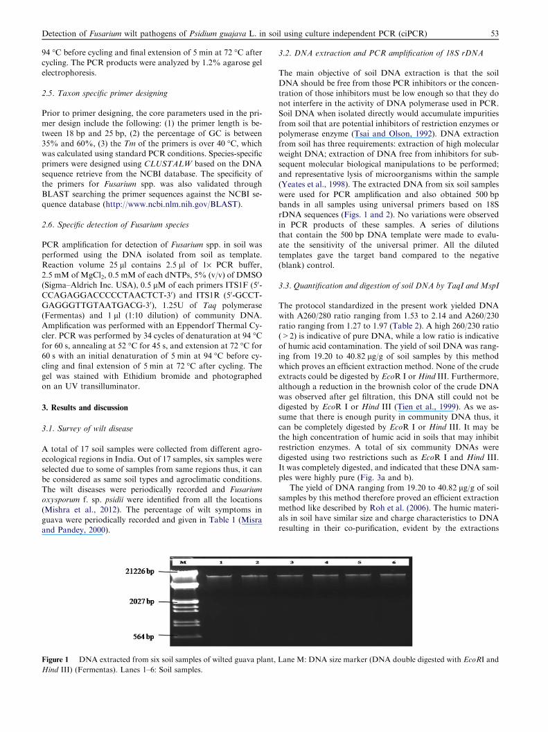
symptoms and five of them (W-3, W-5, W-7, W-10 andW-13) were collected from the rhizosphere of completelywilted host plant. All six samples produced the target amplicon
size of 230 bp (Fig. 4) which showed the presence of Fusariumspp. in the soil. Specificity and sensitivity of the primer areevaluated with several Fusarium isolates. The predicted ampli-
con size of 230 bp amplifies in all Fusarium isolates. No ampli-fication was observed in control sample (Fig. 4). To determinethe sensitivity of the newly designed primers based on ITSregion they were tested with serial dilutions of genomic
DNA from cultured Fusarium species. This PCR amplificationwas obtained up to 10�6 dilutions of DNA sample. ITS regionwas sequenced and found the homology with F. oxysporum
(Mishra et al., 2012; Pandey et al., 2010). According to the re-sults we can make a conclusion that this primer is sensitiveenough to produce the target fragment even from one copy
of the template DNA.According to the specific detection results we can say that
the primer we used was sensitive enough to detect the infection
of Fusarium spp. in soil. Specificity and sensitivity of theprimer pair were investigated using Fusarium isolates fromthese soil samples and we found 230 bp amplicon in all theFusarium isolates tested. DNA detection tests from soil have
been developed for several pathogens like Phytophthora species(Stammler and Seemuller, 1994; Grote et al., 2002; Hussainet al., 2005; Bilodeau et al., 2007), Ralstonia solanacearum (Ku-
mar and Anandaraj, 2006), Phytophthora cinnamomi (O’Brien,2008), Histoplasma capsulatum (Reid and Schafer, 1999) andAgrobacterium tumefaciens (Yang et al., 2011).
ted with EcoRI and Hind III) (Fermentas), Lanes 1, 2, 3, 4, 5, 6
A and Lanes 1b, 2b, 3b, 4b, 5b, 6b, soil DNA digested with MspI.
Figure 4 Specificity of PCR based assay for the detection of Fusarium spp. in soil samples. LaneM: 100 bp DNALadder (Biochem), Lane
1: W-1, Lane 2: W-3, Lane 3: W-5, Lane 4: W-7, Lane 5: W-10, Lane 6: W-13 soil samples, Lane N: control (DNA from healthy plant).
Detection of Fusarium wilt pathogens of Psidium guajava L. in soil using culture independent PCR (ciPCR) 55
In present study, all these community DNAs were com-
pletely digested. Similarly, a number of reports are availableon humic acids that pose a considerable problem and interferein enzymatic manipulations of DNA and various protocols
have been published in the past for the successful isolationof PCR amplifiable DNA from soil (Yeates and Gillings,1998; Amorim et al., 2008). Due to the low level of contamina-
tion, DNA extracted by this method was easily digested byrestriction enzymes TaqI and MspI and this DNA can be suc-cessfully employed for metagenomic studies of soil from wiltedguava plant.
4. Conclusion
The PCR assay reported here is a sensitive, specific, efficient,very convenient and easy to be developed in actual diagnosistechnique for detecting Fusarium spp. from the soil. Because
this PCR assay also has significant practical applications inthe detection of Fusarium infected soils as well as isolates, itconstitutes a powerful tool for controlling the dispersion of
Fusarium and developing disease control strategies. Detectionof Fusarium spp. from soil is a time consuming process by tra-ditional isolation methods, which can delay disease manage-
ment decisions. The PCR detection method reported herecan provide a definitive diagnosis of this pathogen in soilswithin hours, and can be used to more accurately survey theoccurrence and distribution of the pathogen in soil. Moreover,
the molecular markers developed in this study have potentialto overcome the problems associated with existing methodsand can even be used by non-expert biologists.
Acknowledgements
We are thankful to Director and Head, Division of CropProtection, Central Institute for Subtropical Horticulture,Rehmankhera, Lucknow for their encouragement and sup-
port. We gratefully acknowledged the Indian Council of Agri-cultural Research for Financial support in the form of SeniorResearch Fellowship and Vice Chancellor, Integral University
for necessary facilities.
References
Amorim, J.H., Macena, T.N.S., Lacerda-Junior, G.V., Rezende, R.P.,
Dias, J.C.T., Brendel, M., Cascardo, J.C.M., 2008. An improved
extraction protocol for metagenomic DNA from a soil of the
Brazilian Atlantic Rainforest. Genet. Mol. Res. 7, 1226–1232.
Bilodeau, G.J., Levesque, C.A., de Cock, A.W.A.M., Duchaine, C.,
Bri‘ere, S., Uribe, P., Martin, F.N., Hamelin, R.C., 2007. Molec-
ular detection of Phytophthora ramorum by real-time polymerase
chain reaction using TaqMan, SYBR Green, and molecular
beacons. Phytopathol 97, 632–642.
Einsele, H., Hebart, H., Roller, G., Loffler, J., Rothenhofer, I., Muller,
C.A., Bowden, R.A., van Burik, J., Engelhard, D., Schumacher,
U., Kanz, L., 1997. J. Clin. Microbiol. 35, 1353.
Fatima, F., Chaudhary, I., Ali, J., Rastogi, S., Pathak, N., 2011.
Microbial DNA extraction from soil by different methods and its
PCR amplification. Biochem. Cell. Arch. 11, 1.
Grote, D., Olmos, A., Kofoet, A., Tuset, J., Bertolini, E., Cambra, M.,
2002. Specific and sensitive detection of Phytophthora nicotianae
by simple and nested-PCR. Eur. J. Plant Pathol. 108, 197–207.
Holben, W.E., 1994. Isolation and purification of bacterial DNA from
soil. Soil Sci. 5, 727–751.
Hussain, S., Lees, A.K., Duncan, J.M., Cooke, D.E.L., 2005.
Development of a species-specific and sensitive detection assay
for Phytophthora infestans and its application for monitoring of
inoculums in tubers and soil. Plant Pathol. 54, 373–382.
Kumar, A., Anandaraj, M., 2006. Method for isolation of soil DNA
and PCR based detection of ginger wilt pathogen, Ralstonia
solanacearum. Ind. Phytopathol. 59, 154–160.
Liesack, W.P., Janssen, H., Rainey, F.A., Ward-Rainey, N.L.,
Stackebrandt, E., 1997. Microbial diversity in soil: the need for a
combined approach using molecular and cultivation techniques.
Mod. soil Microbiol., 375–439.
Louws, F.J.W., Rademaker, J.L., de Bruijn, F.J., 1999. The three D’s of
PCR based genomic analysis of phytobacteria: diversity, detection,
and disease diagnosis. Ann. Rev. Phytopathol. 37, 81–125.
Miller, D.N., Bryant, J.E., Madsen, E.L., Ghiorse, W.C., 1999.
Evaluation and optimization of DNA extraction and purification
procedures for soil and sediment samples. Appl. Environ. Micro-
biol. 65, 4715–4724.
Misra, A.K., Gupta, V.K., 2007. Variability in Fusarium solani-a
causal organism of wilt of guava. CISH Newsl. 8, 2.
Misra, A.K., 2006. Wilt of guava-a disease of national importance.
Ind. Phytopathol. 59, 269–280.
Misra, A.K., Pandey, B.K., 1996. Present status of wilt disease of
guava. In: Disease Scenario in Crop Plants. International Books
and Periodical Supply Service, New Delhi, pp. 61–70.
Misra, A.K., Prakash, O.M., 1990. Guava diseases – (an annotated
bibliography 1907–1990). Bishen Singh Mahendra Pal Singh,
Dehradun, 132p.
Misra, A.K., Pandey, B.K., 2000. Progressive natural wilting of guava
plants during different months. Ind. Phytopathol. 53, 423–427.
Mishra, R.K., Pandey, B.K., Pandey, A., Mohd. Zeeshan., Pathak N.,
2012. Specific detection of Fusarium oxysporum f. sp. psidii (Fop)
causing wilt disease of Psidium guajava L. in India. In: National
Symposium on Microbes in Health and Agriculture, JNU, New
Delhi, pp. 87.
More, M.I., Herrick, J.B., Silva, M.C., Ghiorse, W.C., Madsen, E.L.,
1994. Quantitative cell lysis of indigenous microorganisms and

56 R.K. Mishra et al.
rapid extraction of microbial DNA from sediment. Appl. Environ.
Microbiol., 1572–1580.
O’Brien, P.A., 2008. PCR primers for specific detection of Phytoph-
thora cinnamomi. Aust. Plant Pathol. 37, 69–71.
Olson, B.H., 1992. Rapid method for separation of bacterial DNA
from humic substances in sediments for polymerase chain reaction.
Appl. Environ. Microbiol. 58, 2292–2295.
Pandey, B.K., Mishra, R.K., Pandey, A., Kamle, M., Sareen, P.,
Muthukumar, M., 2010. Designing and validation of primers aided
through bioinformatics tools for molecular genetic diversity
assessment of Guava wilt Pathogen. In: National Symposium on
Molecular Approaches for Fungal Diseases of Crop Plants. IIHR,
Bangalore, Karnataka, p. 159.
Porteous, L.A., Armstrong, J.L., 1991. Recovery of bulk DNA from
soil by a rapid, small-scale extraction method. Curr. Microbiol. 22,
345–348.
Porteous, L.A., Armstrong, J.L., Seidler, R.J., Watrud, L.S., 1994. An
effective method to extract DNA from environmental samples for
polymerase chain reaction amplification and DNA fingerprint
analysis. Curr. Microbiol. 29, 301–307.
Reid, T.M., Schafer, M.P., 1999. Direct detection of Histoplasma
capsulatum in soil suspensions by two-stage PCR. Mol. Cell Probes
13, 269–273.
Roh, C., Villatte, F., Kim, B.G., Schmid, R.D., 2006. Comparative
study of methods for extraction and purification of environmental
DNA from soil and sludge samples. Appl. Biochem. Biotechnol.
134, 97–112.
Roose-Amsaleg, C.L., Garnier-Sillam, E., Harry, M., 2001. Extraction
and purification of microbial DNA from soil and sediment samples.
Appl. Soil Ecol. 18, 47–60.
Sambrook, J., Fritsch, E.F., Maniatis, T., 1989. Molecular cloning: a
laboratory manual, 2nd ed. Cold Spring Harbor Laboratory, Cold
Spring Harbor, NY.
Stammler, G., Seemuller, E., 1994. Detection of Phytophthora
fragariae var. rubi in infected raspberry roots by PCR. In: Modern
Assays for Plant Pathogenic Fungi, pp. 135–139.
Tien, C.C., Chao, C.C., Chao, W.L., 1999. Methods for DNA
extraction from various soils: a comparison. J. Appl. Microbiol. 86,
937–943.
Tsai, Y.L., Olson, B.H., 1991. Rapid method for direct extraction of
DNA from soil and sediments. Appl. Environ. Microbiol. 57, 1070–
1074.
Tsai, Y.L., Olson, B.H., 1992. Detection of low numbers of bacterial
numbers in soils and sediments by polymerase chain reaction. Appl.
Environ. Microbiol. 58, 754–757.
Yang, Wei, Lei, Ji, Li-Rong, Tan, Shi-Mo, Li, Yan, Wang, Hong-Xia,
Liu, Yu-Ming, Luo, 2011. Sensitive and specific detection of
Agrobacterium tumefaciens in soil using a rapid polymerase chain
reaction (PCR). Afr. J. Microbiol. Res. 5, 708–713.
Yeates, C., Gillings, M.R., 1998. Rapid purification of DNA from soil
for molecular biodiversity analysis. Lett. Appl. Microbiol. 27, 49–
53.
Yeates, C., Gillings, M.R., Davison, A.D., Altavilla, N., Veal, D.A.,
1998. Methods for microbial DNA extraction from soil for PCR
amplification. Biol. Proced. Online 1, 40–47.
Zhou, J., Bruns, M.A., Tiedje, J.M., 1996. DNA recovery from soils of
diverse composition. Appl. Environ. Microbiol. 62, 316–322.