studies on sweet corn: stewart's wilt forecasting, the effect
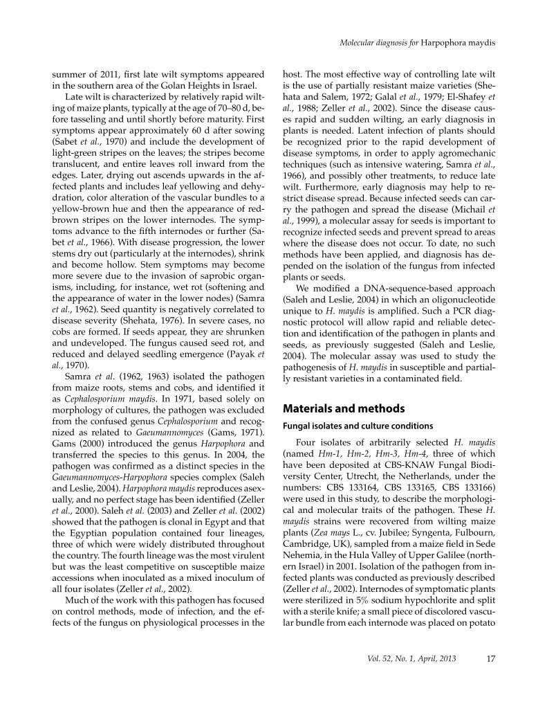
16 ISSN (print): 0031-9465 www.fupress.com/pmISSN (online): 1593-2095 © Firenze University Press
Phytopathologia Mediterranea (2013) 52, 1, 16−29
Corresponding author: O. Degani Fax: +972 4 6817410 E-mail: [email protected]
RESEARCH PAPER
Molecular diagnosis for Harpophora maydis, the cause of maize late wilt in IsraelRan DRORI1,2, amIR SHaROn3, DOROn GOLDBERG1,4, Onn RaBInOVITZ5, maGGIE LEVY2 and OfIR DEGanI1,4
1 Molecular Phytopathology Lab, Migal - Galilee Research Institute, P.O. Box 831 Kiryat-Shmona, 11016, Israel 2 Department of Plant Pathology and Microbiology, The Robert H. Smith Faculty of Agriculture, Food and Environment, The
Hebrew University of Jerusalem, PO Box 12, Rehovot, 76100 Israel3 Department of Plant Sciences, Tel Aviv University, Ramat Aviv, Tel Aviv, 69978, Israel4 Tel-Hai College, Upper Galilee, 12210, Israel5 Israel Ministry of Agriculture and Rural Development, Consultation Service (Shaham), PO Box 28, Bet-Dagan, 50250, Israel
Summary. Late wilt of maize, caused by the fungus Harpophora maydis, is one of the most important fungal diseases in Egypt. The disease has also been reported from India, Hungary, Spain and Portugal. The pathogen survives for long periods in soil. Late wilt is currently controlled using maize varieties with reduced sensitivity, but virulent variants of the fungus may threaten these varieties. Common disease symptoms have been documented over 20 years in Upper Galilee (northern Israel), particularly the Hula Valley. Recently, prevalence of the disease has increased. This is the first confirmed report of the direct and primary cause of the disease in Israel. Isolates of the pathogen obtained from wilting maize plants were morphologically identical to those of strains found in Egypt and India. We modified a molecular method as a diagnostic assay of disease progress in an infested field in north-ern Israel. The assay identified the pathogen 50 d after seeding, before the emergence of disease symptoms, both in susceptible and partially resistant host plants. DNA assessment was consistent with the disease progress in a susceptible maize variety, and the pathogen also spread in partially resistant plants that showed no symptoms. Seeds of apparently healthy, partially resistant plants may therefore also spread the disease. A modified molecular method presented here is a preliminary step in developing a seed health assay.
Key words: Harpophora maydis, late wilt, maize, fungal, molecular diagnosis.
IntroductionLate wilt is a vascular disease of maize (Zea mays
L.) that is caused by the soilborne and seedborne fungus Harpophora maydis (Samra et al.) W. Gams (Samra et al., 1963; Gams, 2000). The disease was first described in Egypt in 1960 (Sabet et al., 1961), and the pathogen was named Cephalosporium maydis, with the disease described as the worst on maize in that country (El-Assiuty et al., 1988). In susceptible varieties, the disease affected 70% of the plants and
caused 40% loss of grain yield (Labib et al., 1975). The disease has spread rapidly since its discovery. In 1979, it was reported in most maize-growing areas of Egypt, with up to 100% of the plants being infected on some farms (Galal et al., 1979). Late wilt of maize was subsequently reported in India (Payak et al., 1970), Hungary (Pecsi and Nemeth, 1998), Portugal and Spain (Molinero-Ruiz et al., 2011). Thus, the geo-graphical distribution of this fungus is expanding, and its recognition is increasing. Indeed, maize wilt-ing with symptoms of late wilt has been observed for over 20 years in the northern part of Israel; however, the disease was only diagnosed in 2002 (A. Sharon, personal communication). In recent years, the dis-ease has worsened, and spread toward the center of Israel, to the Jezreel Valley and to Beit She’an. In the
17Vol. 52, No. 1, April, 2013
Molecular diagnosis for Harpophora maydis
summer of 2011, first late wilt symptoms appeared in the southern area of the Golan Heights in Israel.
Late wilt is characterized by relatively rapid wilt-ing of maize plants, typically at the age of 70–80 d, be-fore tasseling and until shortly before maturity. First symptoms appear approximately 60 d after sowing (Sabet et al., 1970) and include the development of light-green stripes on the leaves; the stripes become translucent, and entire leaves roll inward from the edges. Later, drying out ascends upwards in the af-fected plants and includes leaf yellowing and dehy-dration, color alteration of the vascular bundles to a yellow-brown hue and then the appearance of red-brown stripes on the lower internodes. The symp-toms advance to the fifth internodes or further (Sa-bet et al., 1966). With disease progression, the lower stems dry out (particularly at the internodes), shrink and become hollow. Stem symptoms may become more severe due to the invasion of saprobic organ-isms, including, for instance, wet rot (softening and the appearance of water in the lower nodes) (Samra et al., 1962). Seed quantity is negatively correlated to disease severity (Shehata, 1976). In severe cases, no cobs are formed. If seeds appear, they are shrunken and undeveloped. The fungus caused seed rot, and reduced and delayed seedling emergence (Payak et al., 1970).
Samra et al. (1962, 1963) isolated the pathogen from maize roots, stems and cobs, and identified it as Cephalosporium maydis. In 1971, based solely on morphology of cultures, the pathogen was excluded from the confused genus Cephalosporium and recog-nized as related to Gaeumannomyces (Gams, 1971). Gams (2000) introduced the genus Harpophora and transferred the species to this genus. In 2004, the pathogen was confirmed as a distinct species in the Gaeumannomyces-Harpophora species complex (Saleh and Leslie, 2004). Harpophora maydis reproduces asex-ually, and no perfect stage has been identified (Zeller et al., 2000). Saleh et al. (2003) and Zeller et al. (2002) showed that the pathogen is clonal in Egypt and that the Egyptian population contained four lineages, three of which were widely distributed throughout the country. The fourth lineage was the most virulent but was the least competitive on susceptible maize accessions when inoculated as a mixed inoculum of all four isolates (Zeller et al., 2002).
Much of the work with this pathogen has focused on control methods, mode of infection, and the ef-fects of the fungus on physiological processes in the
host. The most effective way of controlling late wilt is the use of partially resistant maize varieties (She-hata and Salem, 1972; Galal et al., 1979; El-Shafey et al., 1988; Zeller et al., 2002). Since the disease caus-es rapid and sudden wilting, an early diagnosis in plants is needed. Latent infection of plants should be recognized prior to the rapid development of disease symptoms, in order to apply agromechanic techniques (such as intensive watering, Samra et al., 1966), and possibly other treatments, to reduce late wilt. Furthermore, early diagnosis may help to re-strict disease spread. Because infected seeds can car-ry the pathogen and spread the disease (Michail et al., 1999), a molecular assay for seeds is important to recognize infected seeds and prevent spread to areas where the disease does not occur. To date, no such methods have been applied, and diagnosis has de-pended on the isolation of the fungus from infected plants or seeds.
We modified a DNA-sequence-based approach (Saleh and Leslie, 2004) in which an oligonucleotide unique to H. maydis is amplified. Such a PCR diag-nostic protocol will allow rapid and reliable detec-tion and identification of the pathogen in plants and seeds, as previously suggested (Saleh and Leslie, 2004). The molecular assay was used to study the pathogenesis of H. maydis in susceptible and partial-ly resistant varieties in a contaminated field.
Materials and methodsFungal isolates and culture conditions
Four isolates of arbitrarily selected H. maydis (named Hm-1, Hm-2, Hm-3, Hm-4, three of which have been deposited at CBS-KNAW Fungal Biodi-versity Center, Utrecht, the Netherlands, under the numbers: CBS 133164, CBS 133165, CBS 133166) were used in this study, to describe the morphologi-cal and molecular traits of the pathogen. These H. maydis strains were recovered from wilting maize plants (Zea mays L., cv. Jubilee; Syngenta, Fulbourn, Cambridge, UK), sampled from a maize field in Sede Nehemia, in the Hula Valley of Upper Galilee (north-ern Israel) in 2001. Isolation of the pathogen from in-fected plants was conducted as previously described (Zeller et al., 2002). Internodes of symptomatic plants were sterilized in 5% sodium hypochlorite and split with a sterile knife; a small piece of discolored vascu-lar bundle from each internode was placed on potato
Phytopathologia Mediterranea18
R. Drori et al.
dextrose yeast agar (PDYA) (potato dextrose agar (PDA) + 0.2% yeast extract). As noted by Saleh et al. (2003), recovery of H. maydis is difficult, even from heavily infested material, due to its slow growth and the abundance of other, more rapidly growing fungi, particularly Fusarium spp. Cultures were propagated from single conidia and each isolate was identified as H. maydis according to cultural and microscopic characteristics (Samra et al., 1963). The cultures were also identified using a polymerase chain reaction (PCR) protocol, as described by Saleh and Leslie (2004).
DNA from each isolate was extracted using the A200 primer set that amplifies a specific H. maydis segment (Saleh and Leslie, 2004) (see below), and was sequenced and aligned against the GenBank database. All isolates were grown on PDA (Difco, Detroit, MI, USA) at 28°C in complete darkness or in light from cool-white fluorescent tubes (Philips, Eindhoven, The Netherlands). To prepare inocula, cultures were grown in potato dextrose broth (PDB) or liquid complete medium (CM) (Leach et al., 1982) at 28°C in the dark, on a rotary shaker at 150 rpm.
Pathogenicity on maize
To satisfy Koch’s postulates we randomly select-ed another five H. maydis isolates from our collec-tion, obtained from the same field plants at the same location and on the same day as the first four isolates used in the morphological and molecular study. The five isolates were identical, both morphologically and in their unique molecular DNA fragment, to the former strains. Cultures were each propagated from single conidia and the identity of each isolate was verified by PCR. Each isolate was grown in 50 mL corn broth (boiled solution of corn leaves) for 7 d, the mycelium was harvested, and equal amounts of my-celium from each isolate were mixed, blended and used for the seed or soil inoculation.
For seed inoculation, cv. Jubilee maize seeds were soaked overnight in an inoculum suspension at room temperature. For soil inoculation, 200 g steri-lized barley seeds were soaked overnight in H. may-dis inoculum suspension. These barley seeds were only used to spread the pathogen in the soil, as done previously with sorghum seeds (Zeller et al., 2002). A control treatment included barley seeds soaked in an equal volume of sterile water. Experiments were per-formed in 10 L capacity pots with commercial, non-
sterilized soil (40% peat, 40% loam, 10% vermiculite, 10% compost, w/w). Inoculated seeds were planted at 2 cm depth, six seeds per pot. For soil inoculation, the top 20 cm of the soil was removed from each pot and mixed with sterilized inoculated barley seeds, then returned to the pot. Four maize plants were left in each pot after seedling emergence. All plants from seed inoculation and soil inoculation experiments were maintained in a greenhouse with varying tem-perature and light regime.
One plant was sampled from each pot 47 d after planting (before emergence of the first symptoms), and at 2 week intervals thereafter, i.e. 61 d after planting (when the first symptoms were expected to appear) and at 75 d (at the end of the growth peri-od). Pathogen recovery was assayed from the lower (30 cm) and upper (70 cm) parts of the stem of each plant, as previously described (Zeller et al., 2002). In summary, the experiment included five fungal iso-lates (replications), three treatments (seed and soil inoculation and untreated plants), and three sam-pling dates, i.e. a total 45 plants were sampled. We repeated the experiment twice, so a total of 90 plants were tested in both experiments.
Adaptation of molecular diagnosis
DNA was obtained with the Extract-N-amp plant PCR kit (Sigma, Rehovot, Israel), according to the manufacturer’s instructions, from 7-d-old cultures grown under the above-described conditions. PCR was performed to amplify a specific H. maydis seg-ment (Saleh and Leslie, 2004) with a Rapidcycler (Idaho Technology, Salt Lake City, UT, USA). An ex-isting molecular assay (Saleh and Leslie, 2004) was modified by choosing a new set of primers (A200 and Am42/43), using a different reaction mixture, and al-tering the cycling conditions as described below. This new sequence is a major part of a larger AFLP frag-ment that was previously proved to be species-specif-ic (Zeller et al., 2000; Saleh et al., 2003). The new prim-er set was chosen after a series of preliminary experi-ments among a few primer combinations (data not shown), and was found to be the most efficient for our purpose of monitoring late wilt. The A200 primer set (A200a [forward primer]: 5’-CCGACGCCTAAAAT-ACAGGA-3’, A200b [reverse primer: 5’-GGGCTTTT-TAGGGCCTTTTT-3’]) amplifies a specific H. may-dis segment (Saleh and Leslie, 2004). The Am42/43 primer set (Am42 [anti sense primer]: 5’-CAACTAC-

19Vol. 52, No. 1, April, 2013
Molecular diagnosis for Harpophora maydis
GAGCTTTTTAACTGC-3’, Am43 (sense primer): 5’-CAAATTACCCAATCCCGACAC-3’) amplifies eukaryotic ribosomal DNA (18S rRNA gene product, rDNA [Fromont-Racine et al., 2003]) and was used for total estimation of the plant and fungal DNA and as a positive control. Reaction mixtures contained, in a total volume of 20 µL: 1 µL primers (20 µM of each primer), 4 µL Red Load Taq Master (Larova, Teltow, Germany), 3 µL DNA sample and 12 µL double-dis-tilled water (DDW). Cycling conditions for all primer pairs were 94°C for 2 min, followed by 35 cycles of 94°C for 30 s, 55°C for 30 s, 72°C for 1 min, and a final step of 72°C for 5 min. After PCR, a 200-bp ampli-fied DNA band was identified by electrophoresis on an agarose gel. All fungal isolates used in this work (nine isolates) were proven to be H. maydis using mo-lecular characterization.
DNA purification and sequencing
Following the PCR, DNA was extracted and puri-fied from the electrophoresis gel using a HiYieldTM Gel/PCR DNA Extraction Kit (RBC Bioscience, Tai-wan, China). The purified DNA was sequenced (Hy Labs, Rehovot, Israel), and aligned against the Gen-Bank database using NCBI (National Center for Bio-technology Information, MD, USA, Web site: http://www.ncbi.nlm.nih.gov) BLASTn 2.2.17 (Altschul et al., 1997), where high homology (>95%) indicates that more than 95% of the nucleotides are identical in the sequence comparison, and where the expect (E) value (between 0 and 1) close to “0” indicates “sig-nificant” matching.
Field experiment assessing resistance of maize varieties
A field observation of H. maydis pathogenesis in two different host cultivars was conducted during the spring and summer of 2008. The experiment was performed in the south area (No. 10) of a maize field at Kibbutz Naot Mordechai in the Hula Valley (Up-per Galilee, northern Israel), which was infected with the late wilt pathogen. These fields were located near Sede Nehemia maize fields (from which the isolates used in this study had been taken). All northern Hula Valley fields are infected with the late wilt pathogen, and the Kibbutz Naot Mordechai field was chosen for the field evaluation of disease severity for practical reasons. Fungal isolates from this field were not in-
cluded in this study, but they are identical, both mor-phologically and molecularly, in their unique DNA fragment, to the Sede Nehemia field isolates. The experimental area included two plots (Figure 3, up-per panel), each containing three rows. Each row was 250 m long and contained six to seven maize plants m-1. This area was part of a large maize field used for grain production and the other rows nearby were replicates or planted with other hybrids not sampled. The field was watered with a frontal irrigation sys-tem (600 mm per growth season in total).
Two representative cultivars were selected for pathogenicity tests: the susceptible cultivar Jubilee of sweet corn and the partially resistant cultivar Roy-alty, from Pop Vriend Seeds B.V., Andijk, The Neth-erlands (both supplied by Eden Seeds, Reut, Israel). These varieties had previously been tested for sus-ceptibility to late wilt (Israel Northern R&D, Migal - Galilee Research Institute, Kiryat Shmona, Israel, unpublished data).
Seeds were sown on April 14, 2008 with a distance of 96.5 cm between rows. Plants were hand-pollinat-ed on June 13, 2008 and the field was harvested on July 8, 2008. Three to four plants were arbitrarily col-lected from each maize variety at weekly intervals, from day 22 after sowing onwards. Sampling was done on day 22, 30, 36, 43, 50, 57, 64, 72 and 78 after seeding. DNA was extracted separately, as described above, immediately after harvesting the samples, from the root, stem and leaves of each plant and kept at -20°C for later analysis.
The field experiment was repeated in the summer of the following year (2009), in the nearby Kibbutz Hulata maize field (also in the Hula Valley). The ex-perimental area was again part of a large maize field used for grain production and was watered with a frontal irrigation system (500 mm per growth season in total). Each plot contained three rows. Rows were 15 m long and contained six to seven maize plants per meter. Each of the two representative cultivars, Jubilee and Royalty, were grown in six equally sized plots, ar-ranged in the field using a randomized block design.
Seeding was performed on July 1, 2009, with a distance of 96.5 cm between rows. Plants were polli-nated on August 20, 2009, and the field was harvest-ed on September 13, 2009, 74 d after planting. Three plants from each maize variety were randomly sam-pled in approximately 10 d intervals from day 35 on-wards, 35, 46, 56, 67 and 72 days after planting. The fifth sampling was conducted at a shorter interval of

Phytopathologia Mediterranea20
R. Drori et al.
5 d because the field was harvested 2 d later. Imme-diately after sampling, DNA was extracted in three independent replications, from roots, stem, leaves and cobs of each plant, and kept at -20°C for later evaluation. In recent years it was impossible to grow healthy cv. Jubilee maize in the experimental area; a comparison with normal yield had to be based on measurements performed for maize planted in dis-ease-free fields in the Hula Valley (2000 and 2002), and in Beit Shean (2005) (Israel Northern R&D, Migal - Galilee Research Institute, Kiryat Shmona, Israel, unpublished data). Although the Beit Shean region is located south of the Hula Valley, the average total yield in these two regions was the same at the time of evaluation and we therefore include them together.
Molecular diagnosis of Harpophora maydis pathogenesis in the field
Different plant tissues (root, stem, leaf and seed) were sterilized separately with 70% ethanol and then washed with autoclaved DDW. The plants were ground separately in a mortar with pestle and DNA was extracted. All DNA samples were kept at -20°C for later inspection. At the end of the growth period, all DNA samples were analyzed together. A 200-bp PCR-amplified DNA band, specific for H. maydis (Zeller et al., 2002, with the modifications described here) was identified on an agarose gel by electropho-resis. Tina 2.10 g software (Raytest, Straubenhardt, Germany) was used for semi-quantitative analysis of DNA bands. The agarose gel was scanned, and band optical density was measured and normalized against that of the total DNA of the plant and fun-gus, represented by the optical density of the riboso-mal DNA bands (18s rDNA) (Fromont-Racine et al., 2003), which were also used as a positive control.
Molecular seed-health assay for Harpophora maydis infection and infestation
To examine the ability of the pathogen to infect maize seeds in vitro, we inoculated seeds by soaking in a fungal suspension and detected the presence of fun-gus in the inner tissues afterwards. Ten seeds of each maize variety (without any seed treatment with fungi-cides or insecticides) were dipped in 1% (v/v) sodium hypochlorite for 3 min, and then placed in a 250-mL Erlenmeyer flask with 20 mL autoclaved DDW. Five 6-mm-diam. agar disks from 7-d-old H. maydis colo-
nies (grown on PDA at 28°C in the dark) were added to each flask. Harpophora maydis-colonized autoclaved maize seeds were used as positive controls while un-treated seeds (soaked in water) were used as negative controls. At 3 d intervals from the inoculation, a sam-ple of three seeds from each treatment was washed with a 70% ethanol solution and then with autoclaved DDW. The seeds were ground to powder using liquid nitrogen, and DNA was obtained with the Extract-N-amp plant PCR kit (Sigma, Rehovot, Israel), according to the manufacturer’s instructions. The purified DNA was used as template for the PCR reaction. Samples of three seeds each from naturally infected plants of sweet corn at eating stage, from the two field ex-periments, at Kibbutz Naot Mordechai in 2008 and at Kibbutz Hulata in 2009, were also inspected using the DNA-based assay for H. maydis. We repeated this experiment twice. All amplified DNA samples were identified by electrophoretic gel analysis.
ResultsMorphological and microscopic characterization of the Harpophora maydis isolates
Cultures
Four isolates of H. maydis, recovered from wilting field plants collected at Upper Galilee in northern Is-rael (Hula Valley), were characterized separately in this work. The pathogen grew well on solid and in liquid media. On CM and PDA solid media, the colo-nies reached 90 mm diam. within 8 d at 28°C under continuous cool-white fluorescent lighting. Colo-nies grew significantly faster in the dark than light-grown colonies (P<0.05, data not shown), covering a 90 mm diam. plate within 6 d. Colonies were flat, white at first (Figure 1A), then turning gray to black with age (Figure 1B). The margins of the older (10 to 21 d) colonies had a “rhizoidal” or “hyphal rope” appearance (Figure 1B) and they contained typical sclerotium-like bodies (Figure 1C), aggregates of thick-walled, dark cells (Figure 1C). The sclerotial bodies were similar to those reported previously by Samra et al. (1963). Margins of hyphae and “hyphal ropes” were often diverted in a certain direction, as noted by Samra et al. (1962, 1963) (Figure 1B and D).
Microscopic morphology
Hyphae were 2–4.5 µm in width, hyaline, septate, branched and decumbent (Figure 1D). Sporulation

21Vol. 52, No. 1, April, 2013
Molecular diagnosis for Harpophora maydis
occurred within a few days of transferring colonies to new plates. Conidiophores were also hyaline, sep-tate and branched (Figure 1E), and each carried four to seven single-celled, oval to cylindrical, hyaline, smooth-walled conidia (Figure 1F). Conidia were 8.5–25 µm long (average 15 µm) and 5–7 µm wide. Conidia germinated rapidly, usually via bipolar germ tubes, but sometimes one or three germ tubes
were formed (Figure 1G). Anastomosis of germ tubes often occurred.
DNA-based identification
Identification of H. maydis with the molecular assay was verified by sequencing. All four isolates examined presented high homology (>95%) to pre-viously described (Saleh and Leslie, 2004) H. may-
Figure 1. Morphological and microscopic characterization of Harpophora maydis strain Hm-3. A, A young colony (6 d old on PDA); B, mature colony (12 d old) exhibiting a “rhizoidal” or “hyphal rope” appearance; C, Sclerotium-like bodies formed in old cultures (44 d old) of H. maydis on PDA, seen as spots from the back of the Petri dish (C1), magnified on the culture surface (C2), and at high magnification in a slide preparation (C3); D, bent marginal hyphae; E, two conidiophores; F, conidia; G, germinating conidia after 6 h of incubation. All cultures were grown at 28°C under continuous light from cool-white fluorescent tubes. The bar markers represent 10 µm.

Phytopathologia Mediterranea22
R. Drori et al.
dis amplification fragment length polymorphism (AFLP) oligonucleotides (NCBI, nucleotide blast: E-value 4e-79 to 1e-79).
Pathogenicity on maize
Koch’s postulates were satisfied for field isolates on the late wilt susceptible maize cv. Jubilee. Inocu-lation experiments (soil and seed) were performed with five H. maydis isolates from infected plants. Only the plants in the soil-inoculated treatment were symptomatic and infested (Figure 2). No symptoms
were observed in any of the other treatments 47 d after sowing. Harpophora maydis was isolated from the lower part of the stems in three out of five plants from the soil treatment, but not from any of the oth-er samples. At 61 d after sowing, the plants in the inoculated soil showed severe wilting symptoms; these plants did not flower, in contrast to plants in the control and seed-inoculation treatments which all flowered, and some developed cobs (Figure 2). The pathogen was isolated from all five plants in the soil-inoculation treatment, both from the stem bases and from the nodes closest to the first cob, but not
A B
CFigure 2. Testing Koch’s postulates for Harpophora maydis in a pathogenicity test. Severe disease symptoms appeared in maize plants grown in inoculated soil (B), while in non-inoculated soil (A) the plants developed normally, photographed 61 d after sowing; C, disease symptoms appearing on the stem as seen from the side (diseased plant on the right) and in cross section (diseased plant in lower section).

23Vol. 52, No. 1, April, 2013
Molecular diagnosis for Harpophora maydis
from plants in the other treatments (seed inoculation and untreated plants). At 75 d, all of the plants in the soil-inoculation treatment were dead. Develop-ment of plants in the seed-inoculation treatment was normal. These plants were similar in size and fresh weight to the non-inoculated plants and did not de-velop wilting symptoms.
Molecular diagnostic of Harpophora maydis pathogenesis in the field
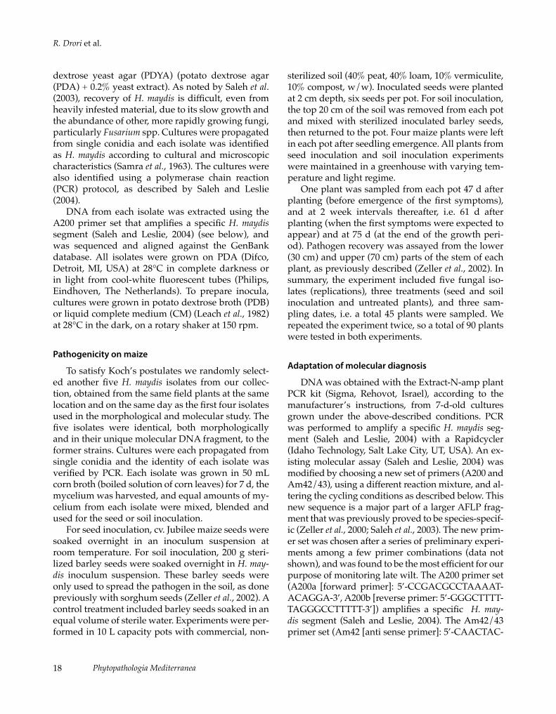
To examine disease progression from 20 d post-sowing until maturity, we conducted a field experi-ment in the Kibbutz Naot Mordechai maize field, in the spring-summer of 2008. The sampling program included two representative maize varieties: cv. Roy-alty, a partially late-wilt-resistant variety, and the sus-ceptible cv. Jubilee. Disease symptoms in cv. Jubilee plants progressed in the same way as in the amount of fungal DNA. The DNA diagnosis first identi-fied the fungus in the roots and stems of cv. Jubilee plants 50 d after sowing and 7 d before appearance of the first symptoms (Figures 3 and 4). The optical density of the pathogen-specific DNA band, nor-malized against that of the ribosomal DNA (rDNA) bands, was used for semi-quantitative analysis of the pathogen DNA. The total amount of fungal DNA in-creased from day 50 onwards, and peaked in stems at 3.2-fold its initial level 72 d after sowing (Figure 4). Fungal DNA in the roots was recorded on day 57 and 72 after sowing (Figure 4).
Approximately 2 months after sowing, early signs of the disease began to appear. One week later (day 64), fungal DNA decreased in the roots but in-creased significantly in the stems of cv. Jubilee plants (Figure 4). On day 72, the fungal DNA in the roots, stems and leaves reached its highest recorded levels. At that age, 12 d after pollination, many of the cv. Jubilee plants were already diseased and dry (Figure 3).
In the partially resistant cv. Royalty, the fungus was clearly identified in all parts of the plants from day 50 onwards, although the amount of DNA meas-ured was less than in cv. Jubilee and less variable (Figure 4). In stems of cv. Royalty plants, the total amount of fungal DNA increased 1.5-fold between 50 and 72 d post-sowing, but no signs of disease were visible. On day 78, the fungus was identified in kernels of sweet corn at eating stage, in both cv. Jubilee and cv. Royalty (data shown only for the 2009
repeat of the experiment, Figure 5, lower panel). Crop yield was estimated after harvesting (Figure 3, lower panel). In the disease-free fields (control, Fig-ure 3, lower panel, insert), cv. Royalty produced 18% more yield than cv. Jubilee. This difference increased more than fourfold, reaching 84% in a diseased field (Figure 3, lower panel). Infected cv. Jubilee cobs were poorly developed (Figure 5A). Furthermore, more than two-thirds of the cv. Jubilee cobs (73%) were class B (cob weight less than 250 g), whereas only 3% of the cv. Royalty cobs were in this class (Figure 3, lower panel).
In the summer of 2009, a similar field test was performed in the nearby Kibbutz Hulata maize field (Figure 5), and the findings were similar to those of the 2008 Kibbutz Naot Mordechai field study (data not shown). This repetition supported the main con-clusions of our summer 2008 observations, which were:• First identification of the pathogen in the roots 56
d after sowing, before emergence of first symp-toms.
• A consistent increase in pathogen DNA in the host tissues during the growth period until about 3 weeks after pollination, spreading from the roots to the stems and leaves, and eventually to the cobs and grains.
• The appearance of relatively small amounts of fungal DNA in the leaves.
• The identification of fungal DNA in the tissues of cv. Royalty plants (including in the kernels at eat-ing stage) in lesser amounts than in cv. Jubilee.
• Increase in fungal DNA was delayed by 2 weeks in the partially resistant plants.
• Cv. Royalty produced 55% more yield than cv. Ju-bilee in a diseased field. Seventy three percent of the cv. Jubilee cobs were poorly developed (Fig-ure 5A) and classified as class B (cob weight less than 250 g), whereas only 28% of the cv. Royalty cobs were in this class.
Molecular seed-health assay for Harpophora maydis
Our preliminary molecular seed-health assay for H. maydis includes H. maydis-infested seeds (inocu-lum soaked seeds) and naturally infected maize ker-nels at the eating stage of sweet corn. This method was applied to diagnose the crops in 2008 and 2009. Seeds were inoculated in vitro with H. maydis and taken for DNA assay every 3 d. The pathogen DNA

Phytopathologia Mediterranea24
R. Drori et al.
Figure 3. Field experiment for assessing cultivar resistance to Harpophora maydis in an infected sweet corn field in the Hula Valley (Upper Galilee, northern Israel). Aerial photograph (upper panel) of the maize field 75 d after sowing, wilted cv. Jubilee plants (red box) and cv. Royalty plants (blue box). Disease progression in cv. Jubilee plants from 50 to 72 d after sowing is shown in the middle panel. Maize yield (lower panel) includes all maize cobs collected from the experimental field (in kg m-2); Class A yield (cobs weighing more than 250 g) and Class B yield (cobs weighing less than 250 g, see Figure 5A). Insert in lower panel shows total average yield of cv. Royalty and Jubilee in the same region, the Hula Valley (2000, 2002), and Beit Shean (2005), in the last years before the spread of late wilt disease to this area.
25Vol. 52, No. 1, April, 2013
Molecular diagnosis for Harpophora maydis
was clearly identified in freshly harvested immature sweet maize cv. Jubilee kernels, 74 d after sowing and 24 d after pollination (Figure 5). Kernels from apparently healthy and resistant cv. Royalty field plants at the developmental stage were also found to be infected (Figure 5), for the first time 6 d after seeds were infested. We repeated this experiment twice.
DiscussionThe occurrence of H. maydis, the causal agent of
maize late wilt, is reported for Israel. This pathogen
is soilborne and seedborne, and is a major threat to maize production in Egypt and other countries. Although the disease was diagnosed several years ago in Israel (A. Sharon, 2002, unpublished), this is the first formal report of H. maydis in this country. Pathogenicity of the Israeli H. maydis isolates was confirmed by completing Koch’s postulates (Figure 2), and the pathogen was characterized by its colony morphology and microscopic traits (Figure 1). The morphological and microscopic characteristics of the pathogen were identical to those of previously de-scribed strains found in Egypt and India (Samra et
rDNA
Specific
Days
Days
Figure 4. Molecular diagnosis of late wilt in maize plant samples (cv. Jubilee and Royalty) using PCR amplification of the unique Harpophora maydis oligonucleotide (upper panel) and rDNA (lower panel). Electrophoresis results show the pro-gression of the disease in roots, stems and leaves, from 43 d post-sowing until maturity. Marker - 200-bp DNA size marker (100 bp DNA ladder, NEB, Ornat, Rehovot, Israel); DDW - double-distilled water used as a template in the PCR mixture to ensure absence of DNA contamination; and H. maydis DNA - isolate Hm-3, used as positive control.

Phytopathologia Mediterranea26
R. Drori et al.
Figure 5. Molecular maize seed health assay for Harpophora maydis (cv. Royalty and Jubilee) in a Kibbutz Hulata, Israel maize field in summer of 2009 (upper panel): A, infected cv. Jubilee cob with shrunken and undeveloped seeds (defined as Class B cob), harvested 74 d after sowing; B, C, cv. Royalty plants, 56 and 67 d after sowing; D, E, cv. Jubilee at the same dates, showing late wilt symptoms. In the lower panel, untreated seed (healthy untreated seeds used as a negative control), infested seed (healthy seeds deliberately infested with H. maydis), field seed (randomly selected seeds from naturally in-fected maize plants presented in the upper panel, 74 d after sowing), cv. Royalty sterilized (autoclaved ‘Royalty’ seeds used as a positive control to demonstrate the ability to detect fungal DNA under the experimental conditions), DDW (double distilled water), H. maydis DNA, specific (amplification to identify the H. maydis-specific oligonucleotide), and rDNA (18S eukaryotic ribosomal DNA amplified).
27Vol. 52, No. 1, April, 2013
Molecular diagnosis for Harpophora maydis
al., 1963; Payak et al., 1970). Final confirmation was achieved by PCR-based DNA analysis (Figures 4 and 5) (Saleh and Leslie, 2004).
We have been closely inspecting maize plants that were developed in infested ground during 4 years (growing seasons). First emergence and progression of the disease symptoms are subject to environmental and physiological changes. A low water potential is one of the most important factors enhancing disease progression. In Egypt early sowing of corn reduced late wilt (El-Shafey et al., 1988), but in India this oc-curred with late summer planting (Singh and Sirad-hana, 1988). Frequent watering or saturated soils re-duced late wilt (Samra et al., 1966). These results can be partially explained by the sensitivity of H. maydis to low oxygen conditions (Samra et al., 1966). The disease progressed in greenhouse plants in a similar way as in the Kibbutz Naot Mordechai field. The first symptoms of wilting appeared in the pathogenicity assay 61 d after sowing and 57 d after sowing in the field. At 75 d after sowing, all pot plants in the soil-inoculation treatment had died; in the field on day 72 after sowing, many of the cv. Jubilee plants were diseased and dry. It is curious why H. maydis did not grow out in maize plants after seed inoculation. Per-haps the fungus was inoculated for a short time and remained superficial on the seeds, where it is sub-jected to numerous antagonists in the soil. Domsch and Gams (1968) and Pope and Jackson (1973) found that Gaeumannomyces graminis was amongst the most sensitive fungal pathogens to soil antagonists.
Another environmental factor that might affect the fungus is light. Fungi, like other organisms, re-spond to light (Rodríguez-Romero et al., 2010). For example, asexual reproduction (conidiation) in fungi is tightly regulated by light and nutrient availability. We found that H. maydis is sensitive to light (data not shown), its growth being significantly reduced by light. This finding is consistent with the pathogen’s life style as it is usually found in the soil or in the vascular tissues of host plants.
At present, the most efficient means of control-ling late wilt is with resistant germplasm (El-Shafey et al., 1988). However, there is little information in the literature on development of the pathogen in ap-parently asymptomatic maize varieties. Zeller et al. (2000) identified four AFLP primer pairs that could be used as markers to determine the distribution of H. maydis lineages and to identify new lineages in field populations. We used the identification of this
H. maydis-unique nucleotide sequence, proved ear-lier to be species-specific ( Zeller et al., 2000; Saleh et al., 2003), as a monitoring tool (Figures 4 and 5). The modified PCR method detected the fungus in plant tissues and was sensitive enough to reveal variations in the amount of DNA above threshold levels, ac-cording to band intensity in assay gels.
Our molecular findings on disease progression in host plants are consistent with previous literature observations (Samra et al., 1963; Payak et al., 1970; Pecsi and Sabet et al., 1970; Nemeth, 1998; Michail et al., 1999). According to Sabet et al. (1970), infection takes place during the first 3 weeks of growth. The roots are penetrated and the fungus first appears in the xylem 21 d after sowing. After 35 d, the pathogen reaches the first stem internode above the ground, and the fourth internode after 49 d (Sabet et al., 1970). At that stage (50 d from sowing), we detected small but clearly identifiable amounts of fungal DNA in various plant parts (Figure 4). When tassels first emerged (day 63), Sabet et al. (1970) found the fun-gus throughout the length of the stalks, although there was less of fungus toward the top of the plants. At this plant age we found DNA levels at their great-est in stems of the susceptible variety (Figure 4), and the first disease symptoms appeared shortly there-after. The first symptom is a moderately rapid wilt-ing of affected plants (Samra et al., 1963), progressing upwards. Leaves turn a dull green, then lose their color and become dry as if suffering from dehydra-tion (Figure 3, 72 d after sowing). Sabet et al. (1970) detected the fungus 84 d after sowing in the cob stalks. We identified fungal DNA at 78 d, in kernels of cv. Jubilee (Figure 5).
Relatively constant DNA levels of the pathogen were identified in the partially resistant cv. Royalty throughout the growth period. Although this vari-ety displayed no disease symptoms and grew well, even in heavily infested soils, the relatively minor amounts of pathogen DNA (Figure 4) imply that H. maydis does not become well established. It is un-clear how the host has restricted the ability of the pathogen to grow and induce disease symptoms.
We used the same molecular method as previous authors in developing a molecular seed health test in maize for H. maydis. Seed health testing is important for research purposes, the development of reduced sensitivity maize varieties and to evaluate control treatments. Earlier research (Samra et al., 1963; Fathi, 1966) reported that the pathogen can, in some cases,
Phytopathologia Mediterranea28
R. Drori et al.
be isolated from the seeds of freshly harvested as well as stored maize, suggesting that it may be seedborne. Although infected seeds do not show discernible ex-ternal symptoms and cannot be identified visually, H. maydis can be cultured from infected seeds by plating them on PDYA after soaking in 1% hypochlorite for 3 min, incubating at 20°C under 12-h cycles of alternat-ing near-ultraviolet light and darkness, and examin-ing after 24 h (Michail et al., 1999). Identification of cultures is accomplished by spore morphology and pathogenicity tests. Michail et al. (1999) demonstrat-ed that H. maydis can be seedborne using an isola-tion-based method and detecting H. maydis in the ear branches, cobs, seeds, husks and silks of naturally infected maize varieties. These researchers reported that H. maydis is internally and externally seedborne in maize. The infection percentage in the embryos (6.3%) was greater than in the endosperms (5%) and seed coats (4%) of the 13 tested seed samples, with the exception of seeds of one variety in which the fungus was confined only on the seed coats (Michail et al., 1999). The traditional method of detecting H. maydis in seeds requires more than 36 h and is inef-ficient for large-scale seed health testing.
Our results demonstrate the potential use of a molecular assay, proved earlier to be species-specific (Zeller et al., 2000; Saleh et al., 2003), to test healthy, deliberately infested and naturally infected kernels of sweet maize (Figure 5). This method is an impor-tant initial step towards the goal of efficient seed health testing. Several additional validation steps are still required before the molecular assay presented here can be introduced as a tool to be used for regu-latory purposes. These include additional trials with multiple seed lots and other organisms.
Developing a molecular seed health assay is even more relevant given that H. maydis can survive for several months in plant seeds. In India, Singh and Siradhana (1987b) reported that the pathogen can survive for up to 2 months in seeds stored in the laboratory, and for 10 months in seeds stored at high temperature and low humidity (Singh and Sirad-hana, 1987a). They also predicted longer survival at low temperatures. The pathogen can also persist on maize stubble for 12 to 15 months (Sabet et al., 1970; Singh and Siradhana, 1987b). The ability of the pathogen to become established and survive in stored maize seeds (even from apparently healthy parental plants) stresses the urgent need to develop new ways to monitor seed health and control dis-
ease spread. The confirmed reports of maize late wilt in Hungary (1994) (Pecsi and Nemeth, 1998), Israel (2002, reported here), Spain (2003) and Portu-gal (2008, Molinero-Ruiz et al., 2011) demonstrate the potential for disease spread and risk. Indeed, Pecsi and Nemeth (1998) assumed that late wilt reached Hungary through the import of infected seeds. To the best of our knowledge, there are no effective dis-infestation treatments for late-wilt-infected seeds.
AcknowledgmentsWe thank Dr. Benjamin A. Horwitz (Technion-
Israel Institute of Technology, Israel) for his advice, Moran Werner, Adi Avidav, Dar Dvir and Einav Rat-zon (Tel-Hai College, Israel) for technical assistance, and Svetlana Yom-Din (Migal - Galilee Research Institute, Israel) for many helpful suggestions. This work was supported by a grant from Israel North-ern R&D (2007) and a research grant from the Israel Plant Council, Ministry of Agriculture (2008, 2009).
Literature citedAltschul S.F., T.L. Madden, A.A. Schaffer, J. Zhang, Z. Zhang,
W. Miller and D.J. Lipman, 1997. Gapped BLAST and PSI-BLAST: a new generation of protein database search pro-grams. Nucleic Acids Research 25, 3389–3402.
Domsch K.H. and W. Gams, 1968. Die Bedeutung vorfruchtab-hängiger Verschiebungen in der Bodenmikroflora. II. Ant- agonistische Einflüsse auf pathogene Bodenpilze. Phyto-pathologische Zeitschrift 63,165–176.
El-Assiuty E.M., H.A. El-Shafey, A.-S. Ismael and Z.M. Fahmy, 1988. Pathogenic variation in Cephalosporium maydis. Phy-topathology 88, S25.
El-Shafey H.A., F.A. El-Shorbagy, I.I. Khalil and E.M. El-As-siuty, 1988. Additional sources of resistance to the late-wilt disease of maize caused by Cephalosporium maydis. Agricul-ture Research Review, Egypt 66, 221–230.
Fathi S.M. 1966. Some Studies on Seed and Seedling Disease of Corn. M. Sc. Thesis, Agriculture, Ain Shams, Egypt.
Fromont-Racine M., B. Senger, C. Saveanu and F. Fasiolo, 2003. Ribosome assembly in eukaryotes. Gene 313, 17–42.
Galal A.A., M.M. El-Rouby and A.M. Gad, 1979. Genetic analysis of resistance to late wilt (Cephalosporium maydis) in variety crosses of maize. Zeitschrift für Planzenzüchtung 83, 176–183.
Gams W., 1971. Cephalosporium-artige Schimmelpilze (Hyphomy-cetes). G. Fischer Verlag. Stuttgart, D, 262 pp.
Gams W., 2000. Phialophora and some similar morphologically little-differentiated anamorphs of divergent ascomycetes. Studies in Mycology 45, 187–199.
Labib H.A., M.F. Abdel-Rahim, A. Salem and A. Abdel-Fattah, 1975. D.C. 19, a new maize hybrid seed resistant to late-

29Vol. 52, No. 1, April, 2013
Molecular diagnosis for Harpophora maydis
wilt disease caused by Cephalosporium maydis. Agriculture Research Review 53, 1–4.
Leach J., B.R. Lang and O.C. Yoder, 1982. Methods for selec-tion of mutants and in vitro culture of Cochliobolus heteros-trophus. Microbiology 128, 1719–1729.
Michail S.H., M.S. Abou-Elseoud and S. Nour Eldin Mona, 1999. Seed health testing of corn for Cephalosporium maydis. Acta Phytopathologica et Entomologica Hungarica 34, 35–42.
Molinero-Ruiz M.L., J.M. Melero-Vara and A. Mateos, 2011. Cephalosporium maydis, the cause of late wilt in maize, a pathogen new to Portugal and Spain. Plant Disease 94, 379–379.
Payak M.M., S. Lal, J. Lilaramani and B. L. Renfro, 1970. Cepha-losporium maydis – a new threat to maize in India. Indian Phytopathology 23, 562–569.
Pecsi S. and L. Nemeth, 1998. Appearance of Cephalosporium maydis Samra, Sabet and Hingorani in Hungary. Medede-lingen Faculteit Landbouwkundige en Toegepaste Biologische Wetenschappen, Universiteit Gent 63, 873–877.
Pope A.M.S. and R.M. Jackson, 1973. Effects of wheatfield soil on inocula of Gaeumannomyces graminis var. tritici J. Walker in relation to take-all decline. Soil Biology and Biochemistry 5, 881–890.
Rodríguez-Romero J., M. Hedtke, C. Kastner, S. Muller and R. Fischer, 2010. Fungi, hidden in soil or up in the air: light makes a difference. Annual Review of Microbiology 64, 585–610.
Sabet K.A., A.S. Samra, M.K. Hingorani and I.M. Mansour, 1961. Stalk and root rots of maize in the United Arab Re-public. FAO Plant Protection Bulletin 9, 121–125.
Sabet K.A., A.S. Samra and N.A. Dawood. 1966. Combined infection with stalk-rot fungi. In: Investigations on stalk-rot disease of maize in U.A.R. (A.S. Samra, K.A. Sabet, ed.), Min-istry of Agriculture, Government Printing Offices, Cairo, Egypt.
Sabet K.A., A.M. Zaher, A.S. Samra and I.M. Mansour, 1970. Pathogenic behaviour of Cephalosporium maydis and C. acremonium. Annals of Applied Biology 66, 257–263.
Saleh A.A. and J.F. Leslie, 2004. Cephalosporium maydis is a dis-tinct species in the Gaeumannomyces-Harpophora species complex. Mycologia 96, 1294–1305.
Saleh A.A., K.A. Zeller, A.-S.M. Ismael, Z.M. Fahmy, E.M. El-Assiuty and J.F. Leslie, 2003. Amplified fragment length polymorphism diversity in Cephalosporium maydis from Egypt. Phytopathology 93, 853–859.
Samra A.S., K.A. Sabet and M.K. Hingorani, 1962. A new wilt disease of maize in Egypt. Plant Disease Reporter 46, 481–483.
Samra A.S., K.A. Sabet and M.K. Hingorani, 1963. Late wilt disease of maize caused by Cephalosporium maydis. Phyto-pathology 53, 402–406.
Samra A.S., K.A. Sabet and M.F. Abdel-Rahim. 1966. Effect of soil conditions and cultural practices on infection with stalk rots. In: Investigations on Stalk-rot Disease of Maize (A.S. Samra, K.A. Sabet, ed.), Ministry of Agriculture, Government Printing Offices, Cairo, Egypt.
Shehata F.A., 1976. The inheritance of resistence to late wilt caused by Cephalosporium maydis in some corn lines. M. Sc. thesis, Faculty of Agriculture, Al-Azhar, Cairo, Egypt.
Shehata A.H. and A.M. Salem, 1972. Genetic analysis of resist-ance to late-wilt of maize caused by Cephalosporium may-dis. Sabrao Journal 4, 1–5.
Singh S.D. and B.S. Siradhana, 1987a. Influence of some envi-ronmental conditions on the development of late wilt of maize induced by Cephalosporium maydis. Indian Journal of Mycology and Plant Pathology 17, 1–5.
Singh S.D. and B.S. Siradhana, 1987b. Survival of Cephalospori-um maydis, incitant of late wilt of maize. Indian Journal od Mycolology and Plant Pathology 17, 83–85.
Singh S.D. and B.S. Siradhana, 1988. Date of sowing in rela-tion to late wilt disease of maize. Indian Phytopathology 41, 489–491.
Zeller K.A., J.E. Jurgenson, E.M. El-Assiuty and J.F. Leslie, 2000. Isozyme and amplified fragment length polymor-phisms from Cephalosporium maydis in Egypt Phytopara-sitica 28, 121–130.
Zeller K.A., M.I. Abou-Serie, E.M. El-Assiuty, Z.M. Fahmy, F.M. Bekheet and J.F. Leslie, 2002. Relative competitive-ness and virulence of four clonal lineages of Cephalospori-um maydis from Egypt toward greenhouse-grown maize. Plant Disease 86, 373–378.
Accepted for publication: May 13, 2012
Copyright © 2022 FDOKUMEN