Molecular Characterization of Tomato Spotted Wilt Virus Defective Interfering RNAs and Detection of...
15
Molecular Characterization of Tomato Spotted Wilt Virus Defective Interfering RNAs and Detection of Truncated L Proteins Alice K. Inoue-Nagata,* ,1 Richard Kormelink,* Jean-Yves Sgro,² Tatsuya Nagata,* Elliot W. Kitajima,‡ Rob Goldbach,* and Dick Peters* ,2 *Department of Virology, Wageningen Agricultural University, Binnenhaven 11, 6709 PD, Wageningen, The Netherlands; ²Institute for Molecular Virology, University of Wisconsin, 1525 Linden Drive, Madison, Wisconsin 53706; and ‡NAP/Department of Phytopathology, ESALQ, C.P. 9, 13418-900, Piracicaba, SP, Brazil Received December 15, 1997; returned to author for revision January 12, 1998; accepted June 4, 1998 Junction sites of 25 different defective interfering (DI) RNAs of tomato spotted wilt virus (TSWV) were characterized. The DI RNAs varied in size from 2.0 to 5.2 kilobases (kb) and contained a single internal deletion. The absence of DI RNAs smaller than 2 kb suggested a size constraint for the survival of TSWV DI RNAs. This hypothesis was reinforced by the finding of a dimeric DI RNA formed by two 1.6-long monomers linked head to tail. Three types of junction sites were found, one type originating from a simple deletion; the second type contained a few extra nucleotides of unknown origin; and the third type contained a stretch of three to five nucleotides, originally occurring at both sides of the deletion and of which one was deleted. In 19 of the 25 DI RNAs studied, the original reading frame was maintained, suggesting a selective preference of DI RNAs with translational potency. Truncated proteins encoded by these DI RNAs were indeed detected in the nucleocapsid preparations. Folding studies of the complete L RNA revealed that the calculated minimal energy of folding was at 16°C lower than at 23°, indicating a higher stability of this molecule at low temperatures. The results suggest an involvement of locally folded secondary structures in the process of deletion, rather than the requirement of certain sequences around the deletion point. The DI RNA generation in TSWV is essentially, as discussed, similar to the process of RNA recombination described in many viruses. © 1998 Academic Press INTRODUCTION Defective interfering (DI) RNAs are generated from the original viral genome by deletions and/or rearrange- ments and have a replicative advantage over wild type genomes (Huang and Baltimore, 1970; Perrault, 1981). Whereas their generation has for animal viruses been studied extensively (Huang, 1973; Perrault, 1981; Barret and Dimmock, 1986; Schlesinger, 1988), their ubiquity for plant viruses was only confirmed during the last decade (Hillman et al., 1987; Roux et al., 1991; Graves et al, 1996). Recently, the generation and accumulation of DI RNAs of tomato spotted wilt virus (TSWV), a negative stranded plant virus belonging to the genus Topovirus was re- ported (Resende et al., 1991, 1992; Inoue-Nagata et al., 1997). The TSWV genome consists of two (S and M) ambisense RNA segments and one (L) negative sense RNA segment with respective sizes of 2916, 4821, and 8897 nucleotides (de Haan et al., 1990, 1991; Kormelink et al., 1992). These RNAs have complementary termini and form a complex with the nucleocapsid (N) protein, referred to as ribonucleoprotein (RNP) particles, with a pseudo-circular structure (de Haan et al., 1989; Peters et al., 1991). Purified virus preparations contain four struc- tural proteins, the N protein (29 kDa) encoded by the S RNA, two glycoproteins (78 and 58 kDa) encoded by the M RNA, and small amounts of the L protein (330 kDa) encoded by the L RNA and representing the putative viral polymerase (Mohamed et al., 1973; Tas et al., 1977; de Haan et al., 1991; van Poelwijk et al., 1993). During serial mechanical inoculation of TSWV isolates in susceptible hosts, DI RNAs of TSWV are readily gen- erated (Resende et al., 1991; Inoue-Nagata et al., 1997). Growth of TSWV-infected plants at low temperatures and use of high inoculum concentrations positively affect their generation and accumulation. All DI RNAs of TSWV studied so far resulted from a single internal deletion within the L RNA, and their occurrence is usually asso- ciated with symptom attenuation in the inoculated plant (Resende et al., 1991, 1992; Inoue-Nagata et al., 1997). In other bunyaviruses, DI RNAs are poorly studied with the exception of Bunyamwera virus (Kascsak and Lyons, 1978; Patel and Elliott, 1992; Scallan and Elliott, 1992). Similarly to TSWV, they are all derived from the L RNA, and some of them are shown to be associated with persistent infections (Patel and Elliott, 1992; Scallan and Elliott, 1992). For three TSWV DI RNAs, the nucleotide sequence of 1 Present address: CENARGEN/EMBRAPA, SAIN Parque Rural, C.P. 02372, CEP 70770±900. Brası ¬lia, DF, Brazil. Fax: 55±61-340±3624. 2 To whom correspondence and reprint requests should be ad- dressed. Fax: 31±317-484820. E-mail: [email protected]. VIROLOGY 248, 342±356 (1998) ARTICLE NO. VY989271 0042-6822/98 $25.00 Copyright © 1998 by Academic Press All rights of reproduction in any form reserved. 342
Transcript of Molecular Characterization of Tomato Spotted Wilt Virus Defective Interfering RNAs and Detection of...

Molecular Characterization of Tomato Spotted Wilt Virus DefectiveInterfering RNAs and Detection of Truncated L Proteins
Alice K. Inoue-Nagata,*,1 Richard Kormelink,* Jean-Yves Sgro,† Tatsuya Nagata,*Elliot W. Kitajima,‡ Rob Goldbach,* and Dick Peters*,2
*Department of Virology, Wageningen Agricultural University, Binnenhaven 11, 6709 PD, Wageningen, The Netherlands;†Institute for Molecular Virology, University of Wisconsin, 1525 Linden Drive, Madison, Wisconsin 53706;
and ‡NAP/Department of Phytopathology, ESALQ, C.P. 9, 13418-900, Piracicaba, SP, Brazil
Received December 15, 1997; returned to author for revision January 12, 1998; accepted June 4, 1998
Junction sites of 25 different defective interfering (DI) RNAs of tomato spotted wilt virus (TSWV) were characterized. TheDI RNAs varied in size from 2.0 to 5.2 kilobases (kb) and contained a single internal deletion. The absence of DI RNAs smallerthan 2 kb suggested a size constraint for the survival of TSWV DI RNAs. This hypothesis was reinforced by the finding of adimeric DI RNA formed by two 1.6-long monomers linked head to tail. Three types of junction sites were found, one typeoriginating from a simple deletion; the second type contained a few extra nucleotides of unknown origin; and the third typecontained a stretch of three to five nucleotides, originally occurring at both sides of the deletion and of which one wasdeleted. In 19 of the 25 DI RNAs studied, the original reading frame was maintained, suggesting a selective preference of DIRNAs with translational potency. Truncated proteins encoded by these DI RNAs were indeed detected in the nucleocapsidpreparations. Folding studies of the complete L RNA revealed that the calculated minimal energy of folding was at 16°C lowerthan at 23°, indicating a higher stability of this molecule at low temperatures. The results suggest an involvement of locallyfolded secondary structures in the process of deletion, rather than the requirement of certain sequences around the deletionpoint. The DI RNA generation in TSWV is essentially, as discussed, similar to the process of RNA recombination describedin many viruses. © 1998 Academic Press
INTRODUCTION
Defective interfering (DI) RNAs are generated from theoriginal viral genome by deletions and/or rearrange-ments and have a replicative advantage over wild typegenomes (Huang and Baltimore, 1970; Perrault, 1981).Whereas their generation has for animal viruses beenstudied extensively (Huang, 1973; Perrault, 1981; Barretand Dimmock, 1986; Schlesinger, 1988), their ubiquity forplant viruses was only confirmed during the last decade(Hillman et al., 1987; Roux et al., 1991; Graves et al, 1996).
Recently, the generation and accumulation of DI RNAsof tomato spotted wilt virus (TSWV), a negative strandedplant virus belonging to the genus Topovirus was re-ported (Resende et al., 1991, 1992; Inoue-Nagata et al.,1997). The TSWV genome consists of two (S and M)ambisense RNA segments and one (L) negative senseRNA segment with respective sizes of 2916, 4821, and8897 nucleotides (de Haan et al., 1990, 1991; Kormelinket al., 1992). These RNAs have complementary terminiand form a complex with the nucleocapsid (N) protein,referred to as ribonucleoprotein (RNP) particles, with a
pseudo-circular structure (de Haan et al., 1989; Peters etal., 1991). Purified virus preparations contain four struc-tural proteins, the N protein (29 kDa) encoded by the SRNA, two glycoproteins (78 and 58 kDa) encoded by theM RNA, and small amounts of the L protein (330 kDa)encoded by the L RNA and representing the putative viralpolymerase (Mohamed et al., 1973; Tas et al., 1977; deHaan et al., 1991; van Poelwijk et al., 1993).
During serial mechanical inoculation of TSWV isolatesin susceptible hosts, DI RNAs of TSWV are readily gen-erated (Resende et al., 1991; Inoue-Nagata et al., 1997).Growth of TSWV-infected plants at low temperatures anduse of high inoculum concentrations positively affecttheir generation and accumulation. All DI RNAs of TSWVstudied so far resulted from a single internal deletionwithin the L RNA, and their occurrence is usually asso-ciated with symptom attenuation in the inoculated plant(Resende et al., 1991, 1992; Inoue-Nagata et al., 1997). Inother bunyaviruses, DI RNAs are poorly studied with theexception of Bunyamwera virus (Kascsak and Lyons,1978; Patel and Elliott, 1992; Scallan and Elliott, 1992).Similarly to TSWV, they are all derived from the L RNA,and some of them are shown to be associated withpersistent infections (Patel and Elliott, 1992; Scallan andElliott, 1992).
For three TSWV DI RNAs, the nucleotide sequence of
1 Present address: CENARGEN/EMBRAPA, SAIN Parque Rural, C.P.02372, CEP 70770–900. Brasılia, DF, Brazil. Fax: 55–61-340–3624.
2 To whom correspondence and reprint requests should be ad-dressed. Fax: 31–317-484820. E-mail: [email protected].
VIROLOGY 248, 342–356 (1998)ARTICLE NO. VY989271
0042-6822/98 $25.00Copyright © 1998 by Academic PressAll rights of reproduction in any form reserved.
342

the region surrounding the junction sites was deter-mined (Resende et al., 1992). In these three DI RNAsstudied, a small sequence repeat at both release andreinitiation sides of the deletion site was observed. Oneof these repeats was lost after the junction event. Inaddition, two of these three DI RNAs showed a frameshiftin the ORF of the L RNA. Despite this frameshift, theoriginal ORF was potentially restored by the use of asecondary start codon, suggesting an active ORF. How-ever, the expression of these ORF’s was not furtherinvestigated by the authors.
In this report, a large number of TSWV DI RNAs aredescribed, including the analysis of their junction sitesand their translatibility with the aim to recognise com-mon molecular features. Based on the results obtainedand the predicted secondary structure of the complete LRNA, a possible mechanism of DI RNA generation inTSWV is discussed.
RESULTS
Nucleotide sequence of DI RNA molecules
Distinct DI RNAs of TSWV were generated by serialmechanical inoculations under different inoculation con-ditions in various hosts (Fig. 1). To gain more insight intothe mechanism underlying the generation of these mol-ecules, they were cloned and the nucleotide sequence ofthe junction sites determined. The obtained sequenceswere compared with the nucleotide sequence of thewild-type TSWV BR-01 L RNA (de Haan et al., 1991) usedfor generation of the DI RNAs. In total 25 DI RNA mole-cules were isolated from 22 plant lines, which wereoriginally inoculated with wild-type (wt) TSWV (Inoue-Nagata et al., 1997).
DI RNAs were amplified using primers specific to bothterminal ends of the L RNA, cloned into a sequencingvector, and further analyzed by restriction enzyme diges-tion, and nucleotide sequence determination of the re-gion surrounding the junction sites. The obtained data
revealed that all DI RNAs found contained one largeinternal deletion (Table 1). However, the presence ofadditional very small deletions or insertions in the L RNAcan not be completely ruled out. To exclude the possi-bility that artificial DI cDNA fragments arose as a resultof RT-PCR, a control reaction was performed on wt TSWVRNA using the same pair of primers. This reaction gaverise to a few fragments in low amounts that did notcomigrate with any of the DI RNA fragments analyzed(data not shown).
The size of DI RNAs varied between 2075 [DI RNA16(15)-1] and 5191 (5–3) nts (Table 1). Most of the DIRNAs analyzed (20 of 25) had shorter 59 than 39 terminalends of the viral complementary (vc) strand of L RNA(Table 1, column 7). The shortest 59 end, found in DI Pe-2(Table 1, Pe-2), consisted of the first 93 nts, hence retain-ing the start codon of the L ORF (located at position 34).On the other hand, DI RNAs with short 39 terminal endscould also be found, notably in DI Be-1. The deletion inthis DI RNA occurred between nt 3228–8732 (Table 1,Be-1), which included the stop codon of the L ORF(position 8661).
Northern analyses of total RNA of a few plant linesincubated at 16°C showed the presence of differentlysized DI RNAs after consecutive passages [Fig. 1, 16(5)and 16(15)]. DI RNAs, differing in size and sequence,were found between the 5th and 15th passage in lines16–1 and 16–3 [Table 1, 16(5)-1 and 16(15)-1, 16(5)-3, and16(15)-3], whereas the 3.4-kb DI RNA in line 16–2 re-mained present and dominating during the examinedpassages [Fig. 1; Table 1, 16(5)-2 and 16(15)-2]. Theseresults suggested that some DI RNAs, once formed,could persist in a TSWV isolate throughout several pas-sages, while others were possibly outcompeted by moreefficiently replicating DI RNA molecules. The occasionalpresence of at least two distinct DI RNAs in the sameplant line, all in large, but different amounts, was occa-sionally observed [Fig. 1, 16(5)-3 and 5–2] and shows thatthey can temporarily coexist.
FIG. 1. Northern blot hybridization of total RNA extracts from TSWV DI RNAs infected plants using probes specific to TSWV L RNA. DI RNAs weregenerated following serial inoculations of TSWV in N. rustica plants incubated at 16, 30, or 23°C TSWV (labeled at the top of the panel), and in tomato(To), sweet-pepper (Pe), N. benthamiana (Be), and Datura stramonium (Da) plants at 23°C (Inoue-Nagata et al., 1997). Plant lines incubated at 16°Cwere tested after the 5th [16(5)] and 15th passage [16(15)]. For all other plant lines, only the 15th passage was used. Inoculation of plants incubatedat 23°C was performed with 5- and 50-fold diluted inoculum. Three individual plants were used for each condition (third row, 1, 2, and 3), and onlythose plants showing DI RNA accumulation are shown. Position of wt L RNA and the size range of DI L RNA molecules are indicated at the left ofthe panel.
343DI RNAs CHARACTERIZED IN TSWV

The occurrence of a DI RNA with a dimeric structure
Northern blot analysis of DI Da-2 showed a single RNAspecies of ca. 3.0 kb (Fig. 1). However, PCR amplificationof cDNA from this isolate resulted in two distinct DNAfragments of 3.2 and 1.6 kb (data not shown). Completenucleotide sequence determination of both moleculesshowed that the larger DI RNA (3184 nts) was formed bytwo copies of the smaller DI RNA (1592 nts) linked head-to-tail. Each monomeric sequence had a deletion be-tween nt 893 and 8204 and an insertion of UUUUUA atthis junction site. The linkage point between the twomonomers had perfect 39 and 59 ends of the L RNA
sequence (Fig. 2A), by which a new XbaI site (TCTAGA)was created. This linkage site was confirmed by PCRamplification using the first strand DNA as template andprimers pDH003 (identical to nt 8339–8345 of vc L RNA)and ZUP406 (complementary to nt 78–94 of vc L RNA).The particular localization of these primers (Fig. 2A)enabled amplification of a fragment of ca. 600 bp. Directsequencing of this fragment showed that the DI RNApresent in Da-2 was formed by a homogenous popula-tion of the dimer.
To confirm whether Da-2 was a true dimer, a Northernblot analysis was made using four different probes (Fig.
TABLE 1
Characterization of Junction Sites of TSWV DI RNAs
DI-RNA Sizea Ib IIc IIId Framee 59 39f
16 (5)-1g 2600 X in -....1125 7423 ......--16 (5)-2g 3424 X shift -.. 503 5977 ////////////////////////--16 (5)-3g 2216 X in -..... 1164 7846 .....--16(15)-1h 2075 X in -... 784 7607 ......--16(15)-2h 3424 X shift* -.. 503 5977 ////////////////////////--16(15)-3h 4301 X in -................ 4021 8618 ..--
30-3 5009 A in -.. 624 4514 ..................--
5-2 2429 UUUUA in -... 681 7155 .......--5-3 5191 X shift* -... 660 4367 //////////////////////////////////////--
50-1 3017 5 in -........ 7789 .....--50-2 2492 X in* -.... 1051 7457 ......--50-3U 4073 G in -.239 5065 ................--50-3L-1 3767 3 in -.107 5238 ...............--50-3L-2 3769 UUAUG shift -. 322 5456 /////////////////////////////--
To-2 3500 GC in -.. 397 5797 .............--To-3 2672 X in* -... 682 6908 ........--
Pe-1 3980 X in* -.. 538 5456 ..............--Pe-2 3136 UUU shift -.93 5858 //////////////////////////--Pe-3 2099 UG in* -. 354 7155 .......--
Be-1 3392 3 —* -............. 3227 8733 --Be-3 3488 X in -.... 1710 7120. .......--
Da-1-1 3961 UUUUU shift* -. 153 5095 ////////////////////////////////--Da-1-2 3965 3 in -. 143 5075 ................--Da-2i 3184 UUUUUUA in -.... 892 8205 ...--Da-3 2816 17 in* -.. 434 6533 ..........--Total 25 11 10 4Percent 100 44 40 16
a Size in nucleotides of the complete DI RNA.b Type I junction site: deletion without any insertion or deletion at the junction site.c Type II junction site: unknown sequence added at the point of the junction. The extra sequence is shown, except for DI Da-3, which had an
insertion of 17 bases (Fig. 3B).d Type III junction site: repeated sequence is present at the junction site, one of them was lost. The number of repeated bases is shown.e in, the deletion maintained the original reading frame downstream of the junction site; shift, the deletion caused a frame shift; *, purified particles
tested by Western blotting for the presence of truncated L proteins (Fig. 4).f Schematic view of DI RNAs in the vc strand of the L RNA; ., in frame sequence for the L ORF; /, out of frame sequence for the L ORF; -, noncoding
sequence. Numbers indicate position of junction sites.g (5), 5th passage of N. rustica lines incubated at 16°C.h (15), 15th passage of N. rustica lines incubated at 16°C.iThis monomer occurred as a dimer (Fig. 2).
344 INOUE-NAGATA ET AL.
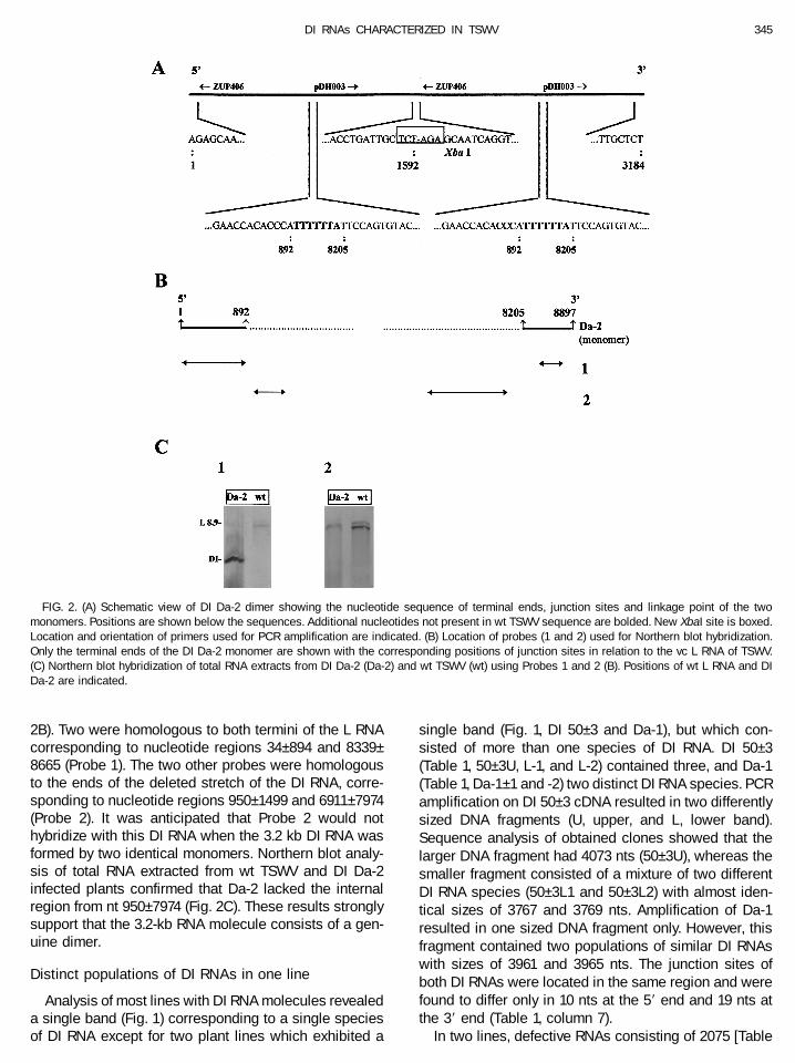
2B). Two were homologous to both termini of the L RNAcorresponding to nucleotide regions 34–894 and 8339–8665 (Probe 1). The two other probes were homologousto the ends of the deleted stretch of the DI RNA, corre-sponding to nucleotide regions 950–1499 and 6911–7974(Probe 2). It was anticipated that Probe 2 would nothybridize with this DI RNA when the 3.2 kb DI RNA wasformed by two identical monomers. Northern blot analy-sis of total RNA extracted from wt TSWV and DI Da-2infected plants confirmed that Da-2 lacked the internalregion from nt 950–7974 (Fig. 2C). These results stronglysupport that the 3.2-kb RNA molecule consists of a gen-uine dimer.
Distinct populations of DI RNAs in one line
Analysis of most lines with DI RNA molecules revealeda single band (Fig. 1) corresponding to a single speciesof DI RNA except for two plant lines which exhibited a
single band (Fig. 1, DI 50–3 and Da-1), but which con-sisted of more than one species of DI RNA. DI 50–3(Table 1, 50–3U, L-1, and L-2) contained three, and Da-1(Table 1, Da-1–1 and -2) two distinct DI RNA species. PCRamplification on DI 50–3 cDNA resulted in two differentlysized DNA fragments (U, upper, and L, lower band).Sequence analysis of obtained clones showed that thelarger DNA fragment had 4073 nts (50–3U), whereas thesmaller fragment consisted of a mixture of two differentDI RNA species (50–3L1 and 50–3L2) with almost iden-tical sizes of 3767 and 3769 nts. Amplification of Da-1resulted in one sized DNA fragment only. However, thisfragment contained two populations of similar DI RNAswith sizes of 3961 and 3965 nts. The junction sites ofboth DI RNAs were located in the same region and werefound to differ only in 10 nts at the 59 end and 19 nts atthe 39 end (Table 1, column 7).
In two lines, defective RNAs consisting of 2075 [Table
FIG. 2. (A) Schematic view of DI Da-2 dimer showing the nucleotide sequence of terminal ends, junction sites and linkage point of the twomonomers. Positions are shown below the sequences. Additional nucleotides not present in wt TSWV sequence are bolded. New XbaI site is boxed.Location and orientation of primers used for PCR amplification are indicated. (B) Location of probes (1 and 2) used for Northern blot hybridization.Only the terminal ends of the DI Da-2 monomer are shown with the corresponding positions of junction sites in relation to the vc L RNA of TSWV.(C) Northern blot hybridization of total RNA extracts from DI Da-2 (Da-2) and wt TSWV (wt) using Probes 1 and 2 (B). Positions of wt L RNA and DIDa-2 are indicated.
345DI RNAs CHARACTERIZED IN TSWV
1, 16(15)-1] and 2099 nts (Table 1, Pe-3) were found byPCR, whereas Northern blot analysis on total RNA ofboth plant lines (Fig. 1) only showed the presence of adefective RNA species of ca. 4 kb, i.e., larger than thoseobtained by the PCR studies. All attempts to amplify thelarger RNA molecules using several internal primer com-binations failed (data not shown). These results indi-cated that in the 15th passage of these lines, the 2075and 2099 nts defective RNA species were present,though below Northern blot detection levels. Most likely,they were preferentially PCR amplified due to theirsmaller size and might not represent true interferingmolecules.
In the lines 16(5)-3 and 5–2, two major DI RNAs (Fig. 1)with approximate sizes of 2 and 4 kb were detected. PCRamplification of 16(5)-3 and 5–2 cDNAs using primersspecific to both ends of L RNA resulted in single DNAfragments of 2.2 and 2.4 kb, respectively, correspondingin size to the smaller RNA species found [Table 1, 16(5)-3and 5–2]. The larger RNA could not be PCR amplified,even when primers directed to deleted regions of thesmaller DI RNA were used (data not shown), thereforeonly the smaller DI RNA molecule was characterized inthese lines.
Analysis of the junction sites of DI RNAs
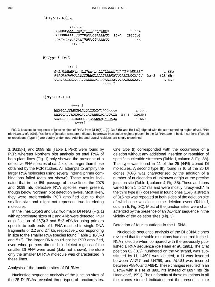
Nucleotide sequence analysis of the junction sites ofthe 25 DI RNAs revealed three types of junction sites.
One type (I) corresponded with the occurrence of adeletion without any additional insertion or repetition ofspecific nucleotide stretches (Table 1, column 3; Fig. 3A).This type was found in 11 of the 25 (44%) cloned DImolecules. A second type (II), found in 10 of the 25 DIclones (40%), was characterized by the addition of anumber of nucleotides of unknown origin at the precisejunction site (Table 1, column 4; Fig. 3B). These additionsvaried from 1 to 17 nts and were mostly ‘‘uracyl-rich.’’ Inthe third type (III), observed in four clones (16%), a stretchof 3–5 nts was repeated at both sides of the deletion siteof which one was lost in the deletion event (Table 1,column 5; Fig. 3C). Most of the junction sites were char-acterized by the presence of an ‘‘AU-rich’’ sequence in thevicinity of the deletion sites (Fig. 3).
Detection of four mutations in the L RNA
Nucleotide sequence analysis of the DI cDNA clonesrevealed that four stable mutations had occurred in the LRNA molecule when compared with the previously pub-lished L RNA sequence (de Haan et al., 1991). The C atposition 82 (C82), numbered on the vc strand, was sub-stituted by U, U4691 was deleted, a U was insertedbetween A4767 and U4768, and AUUU was insertedbetween A8840 and A8841. These changes resulted in anL RNA with a size of 8901 nts instead of 8897 nts (deHaan et al., 1991). The uniformity of these mutations in allthe clones studied indicated that the present isolate
FIG. 3. Nucleotide sequence of junction sites of RNAs from DI 16(5)-1 (A), Da-3 (B), and Be-1 (C) aligned with the corresponding region of vc L RNA(de Haan et al., 1991). Positions of junction sites are indicated by arrows. Nucleotide regions present in the DI RNAs are in bold. Insertions (Type II)or repetitions (Type III) are doubly underlined. Adenine and uracyl residues are singly underlined.
346 INOUE-NAGATA ET AL.

BR-01 used as the inoculum source differed slightly fromthe original one. Other minor point mutations were oc-casionally found throughout the sequenced molecules,but were, unlike those described above, not invariablypresent.
Detection of truncated L proteins encodedby DI RNA molecules
Analysis of DI RNAs revealed that 19 of 25 clones hadan in-frame deletion within the original ORF of the Lprotein (‘‘in’’ in Table 1, column 6). These DI RNAs, there-fore, could potentially encode a truncated L protein withthe original amino- (N) and carboxyl-terminal (C) aminoacids. When the deletion caused a frameshift in this ORF(‘‘shift’’ in Table 1, column 6), a first stop codon waspresent already a few codons downstream of the junc-tion site in all remaining six DI RNAs (data not shown).
To study the potentially translational expression ofthese truncated ORFs, nucleocapsids isolated from viralparticles of nine DI RNA (Table 1, column 6, asterisk)infected plants were analyzed by Western blotting. IntactL protein, of predicted size of 330 kDa, was detected inall preparations, and distinct smaller proteins in most ofthem (Fig. 4). The latter reacted strongly and specificallywith the antibodies to the N or C regions of the L protein,indicating that these smaller, L-derived proteins con-tained both original terminal regions and must have beenexpressed from DI RNAs. Such truncated proteins wereonly detected for DI RNAs with an in frame deletion(Table 1, column 6), and not in nucleocapsid preparationsof DI 16–2 [16(15)-2], 5–3 and Da-1, in which the deletionhad caused a frameshift in the downstream region of thisORF after the junction site (Table 1, column 6).
The estimated sizes of the truncated proteins found inDI 50–2, To-3, Pe-1, Be-3, and Da-3 (Fig. 4) were in goodagreement with the expected sizes derived from theirsequence (85, 93, 142, 124, and 98 kDa, respectively).
Only one truncated protein, i.e., DI Be-1 (123 kDa), re-acted solely with the antibody against the N-terminal partof the L protein (Fig. 4). The nucleotide sequence alreadyshowed that the encoding DI RNA lost almost its entire 39end of the L ORF (Table 1, Be-1), thus, explaining theabsence of a positive reaction with antibodies specific tothe C region of the L protein. The amount of the truncatedprotein relative to intact L protein varied with the DI RNAisolate. High amounts of truncated L proteins were foundin the nucleocapsid preparations of DI 50–2 and Be-3,whereas lower amounts were found in preparations of DITo-3, Pe-1, Be-1 and Da-3. In To-3, two truncated proteinsof slightly different sizes, but occurring in approximatelyequal amounts, were found. The second protein wasmost likely encoded by a distinct DI RNA present in theplant line, though not clearly visualized on Northern blot(Fig. 1, To-3) and not amplified by RT-PCR. Besides thetruncated L proteins, which could readily be detected inmost preparations, proteins giving a faint reaction wereoccasionally observed, e.g., 5–3, To-3, Be-3 (Fig. 4). Theorigin of these proteins, including the second proteinfound in To-3, remains to be studied.
Secondary structure of the L RNA
DI RNAs of TSWV are more efficiently generated atlower temperatures (Inoue-Nagata et al., 1997). To studya possible correlation between the folding of the RNAand the generation of DI RNAs, the L RNA, corrected forthe new mutations found (8901 nts), was folded at both16 and 23°C using firstly its positive polarity (vc strand).The optimal folding was different at these temperatures(Figs. 5A and 5B). At 16°C, 5932 nts (66.6%) were in-volved in base pairing (1714 A:U, 943 G:C, and 309 G:Ubase pairs), while 5836 nts (65.6%) were at 23°C (1692A:U, 934 G:C, and 292 G:U base pairs). Most of the pairednts assembled in 675 stems of length 2 or more for the16°C fold and 648 at 23°C. More than half of these stems
FIG. 4. Western blot analysis of purified nucleocapsid preparations from N. rustica plants infected with TSWV containing DI RNAs. Healthy (H) andwt TSWV (wt) infected plants were used as controls. Wt L protein is seen in all preparations (330), except for H. Identical reactions were obtainedwhen using antibodies against the N and C region of L protein (only one of them is shown), except for Be-1, in which anti-N (N) and anti-C reactionsare shown. Size marker is on the left.
347DI RNAs CHARACTERIZED IN TSWV

FIG
.5.C
ount
ercl
ockw
ise
two-
dim
ensi
onal
repr
esen
tatio
nof
the
optim
alfo
ldin
gof
TSW
Vpo
sitiv
e(v
c)L
RN
Ast
rand
at16
(A)
and
23°C
(B)
and
ofne
gativ
e(v
)st
rand
at16
°C(C
).D
elet
ion
poin
tsar
ein
dica
ted
for
the
59en
dpo
sitio
n(g
ray)
and
39en
dpo
sitio
n(b
lack
)fo
rea
chcl
one
(Tab
le1)
.The
base
sw
ithlo
wes
tP-
num
valu
esar
edr
awn
inbl
ack,
allo
ther
base
sar
egr
ay.
348 INOUE-NAGATA ET AL.

FIG
.5—
Con
tinue
d
349DI RNAs CHARACTERIZED IN TSWV

FIG
.5—
Con
tinue
d
350 INOUE-NAGATA ET AL.

(340 stems of length 2 or more) were common to bothfolds. Some of these common stems were grouped andcould fold with the same pattern, see for example re-gions of Fig. 5 around nt 2000, 5300, 6000, or 8900. Thelatter showed the 59 and 39 ends in close contact, con-necting the first 59 14 nts to the last 39 nts with thelongest helix of the whole fold at either 16 or 23°C andextended by four helices to base 41 (59) and 8870 (39).This long stem was further forked and prolonged withmore helices bringing 59 to 39 together. The 59 and 39 endlong range stems harbored very low P-num values (thenumber of all possible pairing partners for each base) inboth folds, and most clearly at 23°C, an indication thatthe 59 and 39 ends are likely to be found paired in themajority of the RNA molecules. The minimal energy cal-culated for folding was lower at 16°C (22859.1 kcal) thanat 23°C (22350.2 kcal), suggesting a higher stability ofthe RNA at lower temperatures because the RNA canform a larger number of base pairs at the lower temper-ature. Observed P-num values for each fold (see Mate-rials and Methods) ranged from 1 (nt C5, C9, A10, A8894,and G8897) to 1103 (base U4924) for the 16°C fold andfrom 1 (nt C5, C9, A10, and G8897) to 1033 (base U8625)for the 23°C fold. Overall, P-num values in both foldswere high (for 16 and 23°C folds average P-num valueswere 205 6 143 and 213 6 146, means 6 SD), suggest-ing a large variability in the pairing pattern for suboptimalstructures for each temperature fold (data not shown).However, 3.3% of the nucleotides at 16°C but 18.2% at23°C were at normalized P-num value lower than 3% ofthe maximum P-num value at infinite energy, suggestingthat the corresponding regions could be found pairedmore often at 23°C than at 16°C (see darker blacksegments on Figs. 5A and 5B).
Considering that the negative (viral) strand fold wouldnot represent a symmetrical folding of the positive (vc)one for thermodynamic reasons and also because of thepossibility of G–U pairs, the negative polarity was foldedat the temperature of 16°C serving as a model to com-pare the secondary structure of both strands (Fig. 5C). Inthe calculated optimal folding of the negative strand at16°C, 5694 nts (64.0%) were base-paired (1714 A:U, 896G:C, and 236 G:U base pairs) in 612 stems of length 2 ormore. The total energy of this optimal structure was22585.7 kcal. The lesser amount of bases engaged inbase-pairing observed in the negative polarity foldingmay be a reflection of the skew in base composition.Overall the base composition is 1/3 GC and 2/3 AU forboth polarities. For both 16°C folds, the number of A:Ubase pairs was identical (1714) while the number of basepairs involving a G was reduced in the negative polaritywhich has only 1293 Gs (14.5%) vs 1692 (19%) for thepositive polarity. The observed P-num values rangedfrom a value of 1 (nt C5, C9, A10, A8894, and G8897),located in a 14 nucleotide terminal repeat common topositive and negative strands at both ends, to a maxi-
mum value of 466 (base G758) and were on averagelower than that of positive folds (mean of 124 with 75 ofstandard deviation). In the negative strand, 41% of thebases had a normalized P-num values lower than 3% ofthe maximum P-num value at infinite energy. Baseswithin the negative strand at 16°C appeared thereforemore limited in the number of pairing partners they canchoose compared with the positive strand, suggesting alarger number of stable conformations at this tempera-ture (see darker black segments on Fig. 5C).
Within the structures obtained, the position of thedeletion (release and reinitiation) points from all se-quenced DI RNAs were positioned in the optimally foldedstructure (Figs. 5A–5C). Analysis of the location of thesepoints in the structure revealed that they were concen-trated in regions where the RNA was mostly foldedlocally, i.e., not involved in long range base pairing.These deletion points did not show a particular correla-tion with low P-num regions unlike results from corona-virus studies (Rowe et al., 1997). The release and reini-tiation points were located in clusters which were distanteach other when analyzing in only two dimensions.These deletion points were located either in singlestranded loop regions or in double stranded stems.
DISCUSSION
Recently, a large series of DI RNAs of TSWV wasgenerated during serial mechanical inoculations insusceptible plants (Inoue-Nagata et al., 1997). To elu-cidate the process of DI RNA formation in TSWV, thenucleotide sequence of the junction site of 25 of theseDI RNAs was determined. The data obtained showedthat these DI RNAs were formed by a single internaldeletion ranging from 42 to 77% of the complete L RNAsegment. Thus, only DI RNAs with intact 59 and 39ends were found, similarly to those previously found intospoviruses (Resende et al., 1991, 1992), Bunyamweravirus (Patel and Elliott, 1992), as well as in influenzavirus (Nayak et al., 1982; Jennings et al., 1983). Thisresult indicates that the cis-acting sequences requiredfor replication of the TSWV L RNA molecule arepresent in both the 59 and 39 ends of the molecule. Acloser analysis of the smallest terminal ends of the DIRNAs revealed that these essential sequences weremost likely within the last 165 nts of the 39 end (Table1, Be-1) and, in analogy, within the first 71 nt9 of the 59end (DI NL-11 3.3 kb, Resende et al., 1992).
The smallest DI RNA detected, 16(5)-3, had a size of2216 nts. It exhibited a clear replicative advantage overthe wt TSWV L RNA as could be concluded from the largeamount of defective RNA detected by Northern blot hy-bridization studies [Fig. 1, 16(5)-3]. This contrasts to otherL specific defective RNAs smaller than 2100 nts, whichwere isolated during PCR cloning but that could not bedetected on Northern blots [Table 1, 16(15)-1 and Pe-3].
351DI RNAs CHARACTERIZED IN TSWV

These RNAs most likely represent defective noninterfer-ing RNAs newly generated during replication of the iso-lates, and which are preferentially amplified by PCR.Because no DI RNAs smaller than 2.0 kb have beenreported for tospoviruses (Resende et al., 1992), a sizeconstraint may exist for their survival. This might berelated to RNA replication or stability, as suggested forturnip crinkle carmovirus DI RNAs (Zhang and Simon,1994). The strongest evidence for such size constraintswas the finding of a dimeric DI RNA. Its low fitness tosurvive as a monomeric DI RNA may be reflected by ourinability to detect the monomer on Northern blots while itwas the dominant form amplified by RT-PCR. Dimer RNAmolecules functioning as DI or satellite RNA have alsopreviously been reported in tombusviruses (Dalmay etal., 1995; Havelda et al., 1995; Finnen and Rochon, 1995)and in carmoviruses (Simon et al., 1988; Cascone et al.,1990). The mechanism leading to the formation of dimersis still not known (Carpenter et al., 1991; Finnen andRochon, 1995). However, the most accepted hypothesisassumes that the polymerase, after finishing the replica-tion, reinitiates another round of polymerization withoutrelease of the nascent strand by switching to the 39 endof the same or another template molecule (Carpenter etal., 1991; Dalmay et al., 1995). In our studies, the difficul-ties encountered to clone the RNAs observed in DI16(15)-1, Pe-3, and the larger RNAs of 16(5)-3 and 5–2may be related to dimer formation in these isolates, butthis needs further investigation.
The comparison of the nucleotide sequences of atleast two clones of each DI RNA indicated that possiblymost of them grew as a single dominant DI RNA species.However, the coexistence of at least two distinct DIRNAs in the same line suggests that the DI RNAs occuras a population of heterogenous DI RNAs even thoughone species is often largely dominant.
Based on the obtained results, no answers can begiven on the question whether small TSWV DI RNAs doevolve from larger ones or whether they are being inde-pendently generated. The latter possibility seems morelikely, as supported by the appearance of larger ratherthan smaller DI RNA molecules compared with the orig-inally present ones. A thorough study on DI RNA gener-ation within one plant line, though, is required to answerthe aforementioned question, and give more informationon the heterogeneity of DI RNA populations.
Maintenance of a reading frame was demonstrated to beessential for the survival of DI RNAs in several viruses(Kuge et al., 1986; White et al., 1992; Pogany et al., 1995,1997; van der Most et al., 1995). In our studies, the L ORFwas maintained in 19 of 25 TSWV DI RNAs, which sug-gested a preferential selection of those DI RNAs with an inframe deletion. Preliminary attempts to detect the truncatedL protein in crude sap preparation from TSWV infectedtissue failed (data not shown). However, truncated proteinswith expected sizes could be detected successfully in pu-
rified virus particle preparations. This indicated that thenucleocapsid (ribonucleoprotein complex, RNP) bindingdomain was still retained in these truncated proteins andthat they were not selected out from the particles, in con-trast with the situation in Bunyamwera (Patel and Elliott,1992) and influenza virus (Akkina et al., 1984a,b) for whichtruncated proteins were not found in the particles. Theoccurrence, within the virus particle, of a truncated proteinencoded by DI RNA Be-1 in the virus particle, in which theentire C-terminal region of the original L protein was ab-sent, represents evidence that the nucleocapsid bindingdomain is in the N-terminal extremity of the L protein be-tween amino acids positions 1 and 1065. The presence ofa truncated L protein in Da-3 particle preparations, harbor-ing a small N-terminal region of about 133 amino acids,would even suggest that the binding domain is presentbetween amino acids 1 and 133. This result argues againstthe role of the acidic domain of the C-terminal region in Land NSM protein (nonstructural protein encoded by M RNA)in the binding to nucleocapsids suggested previously(Kormelink et al., 1992, 1994).
A major effect of the temperature of incubation on thegeneration/accumulation of DI RNA molecules has recentlybeen demonstrated (Inoue-Nagata et al., 1997). The possi-bility to analyze a large number of junction sites from DIRNAs generated at distinct temperatures prompted us topredict the secondary structure of the complete L RNA atthe temperature of 16 and 23°C for the positive (vc) strandand at 16°C for the negative (viral) strand (Fig 5). As ex-pected, all three structures differed in calculated minimalenergy, though the predicted conformation retained com-mon local foldings (e.g., Figs. 5A and 5B: around nt 2000,5300, 6000, and 8900; or Fig. 5C: around nt 6900, 3600,2900, and 1). This result suggests that these foldings arestable and may be present in the RNA structure irrespectiveof the temperature. The deletion sites on the folded RNA atboth temperatures seem to be concentrated in regionspredicted to be in local folding configurations, either insingle or double stranded regions. These results are inagreement with the increased occurrence of recombina-tions in highly base-paired regions of a number of viruses(Cascone et al., 1993; Nagy and Bujarski, 1993; Carpenter etal., 1995; Dzianott et al., 1995; White and Morris, 1995;Havelda et al., 1997). The global minimum energy of eachstructure is an average of the overall favorable and unfa-vorable thermodynamic and energy considerations for thecomplete molecule. The lower free minimum energy calcu-lated for 16°C could be an indication that at this tempera-ture the molecule can form more energetically favorablestructures that might facilitate the release of the polymer-ase complex. This hypothesis could explain why DI RNAswere readily generated in plants incubated at a lower tem-perature (Inoue-Nagata et al., 1997). Alternative explana-tions can reside in significant changes in the physiology ofthe plant and thus in the replicating machinery of the virusor in the greater stability of DI RNAs at lower temperatures.
352 INOUE-NAGATA ET AL.

However, the latter hypothesis is less plausible since incu-bation of TSWV DI RNAs infected plants at higher temper-atures does not filter out the DI RNAs from the TSWV isolate(data not shown). Most of the 59 junction points (Table 1,column 7) were located in the first (around nt 1) or secondbranch (around nt 600) of positive polarity or in the first(around nt 1) or last folded branch (around nt 8200) ofnegative polarity structure. This result supports the hypoth-esis that these regions are hot spots of occurrence ofpolymerase stopping and release hence originating dele-tions or inversely as structures favoring the landing of thepolymerase-nascent strand complex. Although no conclu-sion can be drawn from our data as to which strand polarityis the siege of deletion events it is noteworthy that, in thepositive polarity, 21 of the 25 junctions (84%) are within thefirst 1200 bases while the deletion points at the 39 end aremore evenly distributed along the 39 half of the molecule.The concentration of deletion points at the 59 end of thepositive polarity strand could reflect the early release of thepolymerase as it tries to duplicate the positive strand fromthe 39 end of the negative strand template. The occurrenceof only type I junction site was observed in all DI RNAsgenerated at 16°C, but whether there was a correlation withthe RNA folding remains unknown. The lowest P-num val-ues for an energy bracket of 12 kcal were located within thefirst and the last dozen bases at the extremities suggestingthat the 59–39repeats have evolved to pair together. Thecircularization of the RNA may thus be the result of select-ing against alternative pairing partners elsewhere in thesequence (Palmenberg and Sgro, 1997). This result con-firms the panhandle conformation of purified nucleocapsidsseen in the electron microscope (Peters et al., 1991). One ofthe limitations of using this secondary structure predictionis that the folding is based on a naked RNA sequence andnot on the folding of the RNP complex. However, the use ofa variety of enzymatic and chemical probes for studying theRNA conformation strongly indicated that the RNA is ex-posed outside of the RNP structure in influenza virus (Jen-nings et al., 1983; Baudin et al., 1994). Assuming that RNPsof TSWV are structured identically, RNA–RNA interactionsforming a structure similar to that predicted of naked RNAmay represent a real situation. The confirmation of thereliability of the secondary structure awaits further studiesusing biochemical and biophysical techniques (e.g., Raikaret al., 1988). However, the strong clustering of the deletionsites at the 59 end of vc L RNA (or inversely, at the 39 end ofthe v L RNA) in a region with intricate locally folded complexstructures suggested the potential usefulness of the usedtechnique to predict the conformation of the RNA in nature.Our results suggest that secondary structure can affect theefficiency of replication particularly at lower temperaturesat which the RNA molecules could fold energetically stablemotifs putatively disrupting the polymerase complex.
Nucleotide sequence analysis of the junction sitesof TSWV DI RNAs revealed three types of deletions.Most junction sites did not show any repeated se-
quences or acquisition of additional nucleotides. Thisindicates that TSWV DI RNAs are mostly generated bynonhomologous recombination events, as only 16%homologous sequences were found surrounding thedeletion site. However, analysis of each of the junctionsites found revealed strong similarities with aberranthomologous recombination seen in brome mosaic bro-movirus (Nagy and Bujarski, 1996), like the presenceof AU-rich sequences close to the deletion site and theaddition of extra nts at the junction site, most of themrich in uracyl (Cascone et al., 1990, 1993; White andMorris, 1994). Many models, developed to explain theoccurrence of recombinations and deletions during DIRNA formation, are based on the ‘‘copy-choice’’ model.In this model the polymerase dissociates carrying thenascent chain following to precise realignment andcontinuation of replicase activity on a new template oron a new site in the same template. It was originallyproposed by Cooper et al. (1974) for recombination inpolioviruses and later corroborated by Lazzarini et al.(1981), Perrault (1981), and Lai (1992). We believe thatin TSWV a strongly folded RNA promotes occasionaltranscriptional pausing (Mills et al., 1978) and tem-plate switching (Cooper et al., 1974), thus generatingdeletions in the RNA genome. The finding of two DIRNAs from one plant line with closely located dele-tions sites (Table 1, Da-1–1 and -2) suggested that alarge variety of DI RNAs are indeed generated but notisolated due to a strong selective pressure. The highfrequency of generation of TSWV DI RNAs supportsthe great importance of recombinatorial events in RNAevolution and virus adaptation.
MATERIALS AND METHODS
Virus isolates containing DI RNAs
DI RNAs of TSWV were generated in Capsicum an-nuum, Datura stramonium, Lycopersicon esculentum,Nicotiana benthamiana, and N. rustica plants by serialmechanical inoculations (Inoue-Nagata et al., 1997) ofthe wt TSWV isolate BR-01 (de Avila et al., 1993). Leafmaterial collected from plants infected with TSWV DIRNA was stored at 270°C until analysis.
Cloning and sequencing of DI RNAs
Total RNA was extracted from leaf material infectedwith TSWV containing DI RNAs according to the methodof de Vries et al. (1982), except for N. benthamiana plants,in which the method of Logermann et al. (1987) wasused. First strand cDNA was synthesized using primerpDH001 (Table 2), and prior to PCR amplification andsubsequent cloning, samples were treated with 1 U ofRNase H (GIBCO-BRL) at 37°C for 20 min. PCR amplifi-cation was performed according to manufacturer’s rec-ommendations (Stratagene) using Taq Extender PCR Ad-
353DI RNAs CHARACTERIZED IN TSWV

ditive (Stratagene). Primers used for amplification aredescribed in Table 2. Each cycle consisted of 30 s ofdenaturation at 94°C, 1 min of annealing at 52°C andextension at 72°C for 3–6 min, depending on the size ofthe DI RNA. After amplification, the DNA was gel purifiedusing Glassmax (GIBCO BRL) and ligated into pGEM-T(Promega), pUC 19 or pSK, as described by Sambrook etal. (1989). The location of the junction sites were deter-mined by restriction enzyme analysis and PCR amplifi-cation using internal primers. The sequence of the re-gion around the junction site was determined using aninternal primer (Table 2) on an ABI automatic sequencer.Nucleotide sequences were compiled and analyzed us-ing programs of the GCG package (Devereux et al., 1984,GCG, Madison, WI) and DNAsis (Hitachi Software Engi-neering, Japan). The sequence of each fragment ana-lyzed was based on at least two DI cDNA clones.
Secondary structure prediction of the TSWV L RNA
RNA secondary structure folding of the 8901 nucleo-tides (nts) vc strand of TSWV L RNA (accession numberD01230) was predicted according to Zuker (1989) withversion 2.2 of the stand-alone MFOLD program (ftp://snark.wustl.edu). The program was compiled and run ona Silicon Graphics (Mountain View, CA, USA) Crimsonworkstation with 256 MB of RAM. Each run requiredabout 1 week of uninterrupted computation.
MFOLD calculated the global minimum free energyfollowing thermodynamic rules and parameters (Jaegeret al., 1989). By pairing each nucleotide with all possiblepartners in the sequence, MFOLD also simultaneously
determined all suboptimal RNA secondary structureswith less optimal free energy.
MFOLD assumed a temperature of 37°C, thereforeappropriate thermodynamics tables for 23 and 16°Cwere calculated with the program NEWTEMP distributedwith MFOLD.
P-num (Jaeger et al., 1989) was extracted for all struc-tures within 112 kcal of the global minimum free energyand normalized with respect to base composition, se-quence length and the number of pairing partners foreach base at infinite energy (Palmenberg and Sgro, 1997and unpublished data).
Graphic representations were generated for the globalminimum free energy calculated by MFOLD using theprogram NAVIEW (Bruccoleri and Heinrich, 1988) asadapted by Zuker (1989; ftp://snark.wustl.edu).
Northern blot analysis using DIG-labeled probes
The presence of wt L and DI RNA molecules wasfollowed by Northern blot hybridizations using total RNAextracts from systemically infected leaves. Total RNAextraction and subsequent agarose gel electrophoresiswere done essentially as described by Inoue-Nagata etal. (1997). Northern blot hybridization was performed withDIG-labeled probes, generated by standard PCR includ-ing 1 mM DIG-11-dUTP (Boehringer Mannheim) usingprimers of 17–19 nts located at both sides of the fragmentof interest. After hybridization, the filters were incubatedwith alkaline-phosphatase anti-DIG polyclonal antibod-ies (Boehringer Mannheim) and subsequently developedusing NBT/BCIP (GIBCO-BRL) as substrate.
TABLE 2
Primers Used to Amplify and Sequence TSWV DI RNAs
Primer Positiona Amplificationb Sequencingc
pDH001 1–17 Pe-1,Be-1d
pDH002 8897–8880 5-2,50-1,50-2,50-3,Pe-3,Da-1,Da-3L1 34–77 50-3L2 8665–8626 5-3,16(15)-1,16(15)-2,16(15)-3,30-3,To-2,
To-3,Pe-2,Pe-3,Be-3,Da-2,Da-316(15)-3
M9 586–604 16(15)-1,30-3,Da-2J33 6548–6531 16(5)-2J39 8396–8378 16(5)-1,16(5)-3A1 7974–7958 16(5)-3,50-1A2 7455–7434 16(5)-1,5-2, Pe-3,Be-3A3 951–968 50-2A4 370–387 16(5)-2,5-3, 16(15)-2,To-3,Pe-1,Da-3SK vector 50-3,Da-1KS vector Pe-2,Pe-3T7 vector To-2,Be-1,Da-2T3 vector Be-1,Da-2Reverse vector Pe-2
a Position of the primer in the L RNA molecule (de Haan et al., 1991).b All DI RNAs were amplified with pDH001 as the first primer and a second primer as mentioned in the table.c Junction sites of these DI RNAs were determined using the primer in the first column.d Clones Pe-1 and Be-1 could be amplified using only the primer pDH001 due to the high complementarity of the L RNA terminal ends.
354 INOUE-NAGATA ET AL.

Detection of TSWV L RNA translation products
To detect wt and truncated L protein species, Westernimmunoblot studies were performed on nucleocapsidsreleased from particles of TSWV DI RNAs. Viral nucleo-capsids were extracted by homogenizing systemicallyinfected N. rustica leaves in 0.1 M potassium phosphatebuffer, pH 7.0, containing 0.01 M sodium sulfite. Then thisextract was centrifuged at 13,000 g for 15 min. Theresulting pellet, which contained the particle fraction,was resuspended in resuspension buffer (0.01 M potas-sium phosphate buffer, pH 7.0, containing 0.01 M sodiumsulfite) plus 1% Nonidet-P40 to release nucleocapsidsfrom the virus particles. Plant debris and starch wereeliminated by centrifugation at 13,000 g for 15 min. Nu-cleocapsids were then precipitated by centrifugation at185,000 g for 1 h through a 30% sucrose cushion. Thepellet was resuspended in resuspension buffer (10 mlbuffer/1 g starting leaf material) and stored at 270°C.
Nucleocapsid preparations containing approximately30 mg (5 ml) of proteins were subjected to denaturingelectrophoresis in a 5–15% gradient SDS-polyacrylamidegel according to Laemmli (1970) buffered with Tris pH8.8. Proteins were transferred to a PVDF membrane(Immobilon) by a semi-dry transfer blot (Transblot SD,Bio-Rad) according to manufacturer’s recommendations.Filters were blocked with 5% nonfat milk in 0.01 M po-tassium phosphate buffer, pH 7.2, containing 0.14 MNaCl, and 0.05% Tween 20 (PBS-T). Filters were thenincubated with antibodies directed to the amino- (aminoacids positions 1–287) or carboxyl-terminal (amino acidspositions 2293–2875) ends of the L protein (van Poelwijket al., 1993) in PBS-T. The antigen-IgG complex formedwas detected using horseradish conjugated anti-rabbitantibodies, and visualized using the ECL detection kit ofAmersham.
ACKNOWLEDGMENTS
Alice K. Inoue-Nagata was a doctoral student of University of Brasılia(Brazil) and was supported by a fellowship from the National ResearchCouncil of Brazil (CNPq). Jean-Yves Sgro is supported by a grant fromthe Lucille P. Markey Charitable Trust. We thank Marcel Prins andErwin Cardol for helpful discussions.
REFERENCES
Akkina, R. K., Chambers, T. M., and Nayak, D. P. (1984a). Expression ofdefective-interfering influenza virus specific transcripts and polypep-tides in infected cells. J. Virol. 51, 395–403.
Akkina, R. K., Chambers, T. M., and Nayak, D. P. (1984b). Mechanism ofinterference by defective-interfering particles of influenza virus: Dif-ferential reduction of intracellular synthesis of specific polymeraseproteins. Virus Res. 1, 687–702.
Barret, A. D. T., and Dimmock, N. J. (1986). Defective interfering virusesand infections of animals. Curr. Top. Microbiol. Immunol. 128, 55–84.
Baudin, F., Bach, C., Cusack, S., and Ruigrok, R. W. H. (1994). Structureof influenza virus RNP. I. Influenza virus nucleoprotein melts second-ary structure in panhandle RNA and exposes the bases to thesolvent. EMBO J. 13, 3158–3165.
Bruccoleri, R. E., and Heinrich, G. (1988). An improved algorithm fornucleic acid secondary structure display. Comput. Appl. Biosci. 4,167–173.
Carpenter, C. D., Cascone, P. J., and Simon, A. E. (1991). Formation ofmultimers of linear satellite RNAs. Virology 183, 586–594.
Carpenter, C. D., Oh, J. W., Zhang, C., and Simon, A. E. (1995). Involve-ment of a stem-loop structure in the location of junction sites in viralRNA recombination. J. Mol. Biol. 245, 608–622.
Cascone, P. J., Carpenter, C. D., Li, X. H., and Simon, A. E. (1990).Recombination between satellite RNAs of turnip crinkle virus. EMBOJ. 9, 1709–1715.
Cascone, P. J., Haydar, T. F., and Simon, A. E. (1993). Sequences andstructures required for recombination between virus-associateRNAs. Science 260, 801–805.
Cooper, P. D., Steiner-Pryor, A., Scotti, P. D., and Delong, D. (1974). Onthe nature of poliovirus genetic recombinants. J. Gen. Virol. 23,41–49.
Dalmay, T., Zittya, G., and Burgyan, J. (1995). Generation of defectiveinterfering RNA dimers of cymbidium ringspot tombusvirus. Virology207, 510–517.
de Avila, A. C., de Haan, P., Smeets, M. L. L., Resende, R. de O.,Kormelink, R., Kitajima, E. W., Goldbach, R. W., and Peters, D. (1993).Distinct levels of relationships between tospovirus isolates. Arch.Virol. 128, 211–227.
de Haan, P., Kormelink, R., Resende, R. de O., van Poelwijk, F., Peters,D., and Goldbach, R. (1991). Tomato spotted wilt virus L RNA encodesa putative RNA polymerase. J. Gen. Virol. 71, 2207–2216.
de Haan, P., Wagenmakers, L., Peters, D., and Goldbach, R. W. (1989).Molecular cloning and terminal sequence determination of the S andM RNA of tomato spotted wilt virus. J. Gen. Virol. 70, 3469–3473.
de Haan, P., Wagenmakers, L., Peters, D., and Goldbach, R. W. (1990).The S RNA segment of tomato spotted wilt virus has an ambisensecharacter. J. Gen. Virol. 71, 1001–1007.
Devereux, J., Haeberli, P., and Smithies, O. (1984). A comprehensive setof sequence analysis programs for the VAX. Nucleic Acids Res. 12,387–395.
de Vries, S. C., Springer, J., and Wessala, J. G. H. (1982). Diversity ofabundant mRNA sequences and patterns of protein synthesis inetiolated and greened pea seedlings. Planta 156, 129–135.
Dzianott, A., Flasinski, S., and Bujarski, J. J. (1995). Foreign complemen-tary sequences facilitate genetic RNA recombination in brome mo-saic virus. Virology 208, 370–375.
Finnen, R. L., and Rochon, D. M. (1995). Characterization and biologicalactivity of DI RNA dimers formed during cucumber necrosis coinfec-tions. Virology 207, 282–286.
Graves, M. V., Pogany, J., and Romero, J. (1996). Defective interferingRNAs and defective viruses associated with multipartite RNA virusesof plants. Sem. Virol. 7, 399–408.
Havelda, Z., Dalmay, T., and Burgyan, J. (1995). Localization of cis-actingsequences essential for cymbidium ringspot tombusvirus defectiveinterfering RNA replication. J. Gen. Virol. 76, 2311–2316.
Havelda, Z., Dalmay, T., and Burgyan, J. (1997). Secondary structure-dependent evolution of cymbidium ringspot virus defective interfer-ing RNA. J. Gen. Virol. 78, 1227–1234.
Hillman, B. I., Carrington, J. C., and Morris, T. J. (1987). A defectiveinterfering RNA that contains a mosaic of a plant virus genome. Cell51, 427–433.
Huang, A. S. (1973). Defective interfering viruses. Annu. Rev. Microbiol.27, 101–117.
Huang, A. S., and Baltimore, D. (1970). Defective viral particles and viraldisease processes. Nature 226, 325–327.
Inoue-Nagata, A. K., Kormelink, R., Nagata, T., Kitajima, E. W., Goldbach,R., and Peters, D. (1997). Temperature and host effects on the gen-eration of tomato spotted wilt virus defective interfering RNAs. Phy-topathology 87, 1168–1173.
Jaeger, J. A., Turner, D. H., and Zuker, M. (1989). Improved predictions of
355DI RNAs CHARACTERIZED IN TSWV

secondary structures for RNA. Proc. Natl. Acad. Sci. USA 86, 7706–7710.
Jennings, P. A., Finch, J. T., Winter, G., and Robertson, J. S. (1983). Doesthe higher order structure of the Influenza virus ribonucleoproteinguide sequence rearrangements in influenza viral RNA? Cell 34,619–627.
Kascsak, R. J., and Lyons, M. J. (1978). Bunyamwera virus. II. Thegeneration and nature of defective interfering particles. Virology 89,539–546.
Kormelink, R., de Haan, P., Meurs, C., Peters, D., and Goldbach, R.(1992). The nucleotide sequence of the M segment of tomato spottedwilt virus, a bunyavirus with two ambisense RNA segments. J. Gen.Virol. 73, 2795–2804.
Kormelink, R., Storms, M., van Lent, J., Peters, D., and Goldbach, R.(1994). Expression and subcellular location of the NSm protein oftomato spotted wilt virus (TSWV), a putative viral movement protein.Virology 200, 56–65.
Kuge, S., Saito, I., and Nomoto, A. (1986). Primary structure of poliovirusdefective-interfering particle genomes and possible generationmechanisms of the particles. J. Mol. Biol. 192, 473–487.
Laemmli, U. K. (1970). Cleavage of structural proteins during the as-sembly of the head of bacteriophage T4. Nature 227, 680–685.
Lai, M. M. C. (1992). RNA recombination in animal and plant viruses.Microbiol. Rev. 56, 61–79.
Lazzarini, R. A., Keene, J. D., and Schubert, M. (1981). The origin ofdefective interfering particles of the negative-strand RNA viruses.Cell 26, 145–154.
Logermann, J., Schell, J., and Willmitzer, L. (1987). Improved method forthe isolation of RNA from plant tissues. Anal. Biochem. 163, 16–20.
Mills, D. R., Dobkin, C., and Kramer, F. R. (1978). Template-determined,variable rate of RNA chain elongation. Cell 15, 541–550.
Mohamed, N. A., Randles, J. W., and Francki, R. I. B. (1973). Proteincomposition of tomato spotted wilt virus. Virology 56, 12–21.
Nagy, P. D., and Bujarski, J. J. (1993). Targeting the site of RNA-RNArecombination in brome mosaic viruses with antisense sequences.Proc. Natl. Acad. Sci. USA 90, 6390–6394.
Nagy, P. D., and Bujarski, J. J. (1996). Homologous RNA recombinationin brome mosaic virus: AU-rich sequences decrease the accuracy ofcrossovers. J. Virol. 70, 415–426.
Nayak, D. P., Chambers, T. M., and Akkina, R. K. (1982). Structure ofdefective-interfering RNAs of Influenza viruses and their role ininterference. In ‘‘The Influenza Viruses’’ (R. M. Krug, Ed.), pp. 269–317.Plenum Press, New York.
Palmenberg, A. C., and Sgro, J.-Y. (1997). Topological organization ofpicornaviral genomes: Statistical prediction of RNA structural sig-nals. Sem. Virol. 8, 231–241.
Patel, A. H., and Elliott, R. M. (1992). Characterization of Bunyamweravirus defective interfering particles. J. Gen. Virol. 73, 389–396.
Perrault, J. (1981). Origin and replication of defective interfering parti-cles. Curr. Top. Microbiol. Immunol. 93, 151–207.
Peters, D., de Avila, A. C., Kitajima, E. W., Resende, R. de O., de Haan,P., and Goldbach, R. W. (1991). An overview of tomato spotted wiltvirus. in Virus-Thrips-Plant interactions of tomato spotted wilt virus. In‘‘Proceedings of USDA Workshop, Beltsville, MD’’ (H.-T. Hsu and R. H.Lawson, Eds.), pp. 1–14. National Technology Information Service,Springfield, VA.
Pogany, J., Romero, J., Huang, Q., Sgro, J.-Y., Shang, H., and Bujarski, J. J.(1995). De novo generation of defective interfering-like RNAs in broadbean mottle bromovirus. Virology 212, 574–586.
Pogany, J., Romero, J., and Bujarski, J. J. (1997). Effect of 59 and 39terminal sequences, overall length, and coding capacity on theaccumulation of defective RNAs associated with broadbean mottlevirus in planta. Virology 228,: 236–243.
Raikar, A., Rubino, H. M., and Lockard, R. E. (1988). Chemical probing ofadenine residues within the secondary structure of rabbit ribosomalRNA. Biochemistry 27, 582–592.
Resende, R. de O., de Haan, P., de Avila, A. C., Kitajima, E. W., Kormelink,R., Goldbach, R., and Peters, D. (1991). Generation of envelope anddefective interfering RNA mutants of tomato spotted wilt virus bymechanical passage. J. Gen. Virol. 72, 2375–2383.
Resende, R. de O., de Haan, P., van de Vossen, E., de Avila, A. C.,Goldbach, R., and Peters, D. (1992). Defective interfering L RNAsegments of tomato spotted wilt virus retain both genome terminiand have extensive internal deletions. J. Gen. Virol. 73, 2509–2516.
Roux, L., Simon, A. E., Holland, J. J. (1991). Effects of defective interferingviruses on virus replication and pathogenesis in vitro and in vivo. Adv.Virus Res. 40, 181–211.
Rowe, C. L., Fleming, J. O., Nathan, M. J., Sgro, J.-Y., Palmenberg, A. C.,and Baker, S. C. (1997). Generation of coronavirus spike deletionvariants by high-frequency recombination at regions of predictedRNA secondary structure. J. Virol. 71, 6183–6190.
Sambrook, J., Fritsch, E. F., and Maniatis, T. (1989). ‘‘Molecular Cloning.A Laboratory Manual,’’ 2nd ed. Cold Spring Harbor Laboratory Press,Cold Spring Harbor, NY.
Scallan, M. F., and Elliott, R. M. (1992). Defective RNAs in mosquito cellspersistently infected with Bunyamwera virus. J. Gen. Virol. 73, 53–60.
Schlesinger, S. (1988). The generation and amplification of defectiveinterfering RNAs. In ‘‘RNA Genetics’’ (E. Domingo, J. J. Holland, and P.Ahlquist, Eds.), Vol. 2, pp. 167–185. CRC Press, Boca Raton, FL.
Simon, A. E., Engel, H., Johnson, R. P., and Howel, S. H. (1988).Identification of regions affecting virulence, RNA processing andinfectivity in the virulent satellite of turnip crinkle virus. EMBO J. 7,2645–2651.
Tas, P. W. L., Boerjan, M. L., and Peters, D. (1977). The structuralproteins of tomato spotted wilt virus. J. Gen. Virol. 36, 267–279.
van der Most, R., Luytjes, W., Rutjes, S., and Spaan, W. J. M. (1995).Translation but not the encoded sequence is essential for the effi-cient propagation of the defective interfering RNAs of the coronavirusmouse hepatitis virus. J. Virol. 69, 3744–3751.
van Poelwijk, F., Boye, K., Oosterling, R., Peters, D., and Goldbach, R.(1993). Detection of the L protein of tomato spotted wilt virus. Virology197, 468–470.
White, K. A., Bancroft, J. B., and Mackie, G. A. (1992). Coding capacitydetermines in vivo accumulation of a defective RNA of clover yellowmosaic virus. J. Virol. 66, 3069–3076.
White, K. A., and Morris, T. J. (1994). Nonhomologous RNA recombina-tion in tombusviruses: Generation and evolution of defective inter-fering RNAs by stepwise deletions. J. Virol. 68, 14–24.
White, K. A., and Morris, T. J. (1995). RNA determinants of junction siteselection in RNA virus recombinants and defective interfering RNAs.RNA 1, 1029–1040.
Zhang, C., and Simon, A. E. (1994). Effect of template size on accumu-lation of defective interfering RNAs in protoplasts. J. Virol. 68, 8466–8469.
Zuker, M. (1989). On finding all suboptimal foldings of an RNA molecule.Science 244, 48–52.
356 INOUE-NAGATA ET AL.