Multiwalled Carbon Nanotubes Hinder Microglia Function Interfering with Cell Migration and...
10
Microglial cells undergo a dose-dependent cell division arrest and apoptosis when treated with multiwalled carbon nanotubes (MWCNTs). It is shown that how MWCNTs severely interfere with both cell migration and phagocytosis in live microglia. These results provide strategic clues of how to biocompatibilize MWCNTs to reduce the damage in brain macrophages to develop new nanodrugs based on MWCNTs. FULL PAPER Carbon Nanotubes J. C. Villegas, L. Álvarez-Montes, L. Rodríguez-Fernández, J. González, R. Valiente, M. L. Fanarraga* ........... x–xx Multiwalled Carbon Nanotubes Hinder Microglia Function Interfering with Cell Migration and Phagocytosis
-
Upload
independent -
Category
Documents
-
view
1 -
download
0
Transcript of Multiwalled Carbon Nanotubes Hinder Microglia Function Interfering with Cell Migration and...

Microglial cells undergo a dose-dependent cell division arrest and apoptosis when treated with multiwalled carbon nanotubes (MWCNTs). It is shown that how MWCNTs severely interfere with both cell migration and phagocytosis in live microglia. These results provide strategic clues of how to biocompatibilize MWCNTs to reduce the damage in brain macrophages to develop new nanodrugs based on MWCNTs.
FULL PAPER
Carbon Nanotubes
J. C. Villegas, L. Álvarez-Montes, L. Rodríguez-Fernández, J. González, R. Valiente, M. L. Fanarraga* ........... x–xx
Multiwalled Carbon Nanotubes Hinder Microglia Function Interfering with Cell Migration and Phagocytosis

www.advhealthmat.dewww.MaterialsViews.com
FULL P
APER
© 2013 WILEY-VCH Verlag GmbH & Co. KGaA, Weinheim 1wileyonlinelibrary.com
Multiwalled Carbon Nanotubes Hinder Microglia Function Interfering with Cell Migration and Phagocytosis
Juan C. Villegas , Laura Álvarez-Montes , Lidia Rodríguez-Fernández , Jesús González , Rafael Valiente , and Mónica L. Fanarraga *
DOI: 10.1002/adhm.201300178
Prof. J. C. Villegas Departamento de Anatomía y Biología Celular Universidad de Cantabria-IFIMAV 39011 , Santander , Spain L. Álvarez-Montes Unidad de Microscopía Láser, IFIMAV 39011 , Santander , Spain Dr. L. Rodríguez-Fernández SERMET , Universidad de Cantabria 39005 , Santander , Spain Dr. J. González MALTA-Consolider Team CITIMAC, Facultad de Ciencias Universidad de Cantabria 39005 , Santander , Spain Dr. R. Valiente Departamento de Física Aplicada Facultad de Ciencias Universidad de Cantabria 39005 , Santander , Spain Dr. M. L. Fanarraga Departamento de Biología Molecular Universidad de Cantabria-IFIMAV 39011 , Santander , Spain E-mail: [email protected]
The intranasal drug delivery route provides exciting expectations regarding the application of engineered nanomaterials as nano-medicines or drug-delivery vectors into the brain. Among nanomaterials, multiwalled CNTs (MWCNTs) are some of the best candidates for brain cancer therapy since they are well known to go across cellular barriers and display an intrinsic ability to block cancer cell proliferation triggering apoptosis. This study reveals that microglial cells, the brain macrophages and putative vehicles for MWCNTs into the brain, undergo a dose-dependent cell division arrest and apoptosis when treated with MWCNTs. Moreover, it is shown that MWCNTs severely interfere with both cell migration and phagocytosis in live microglia. These results lead to a re-evaluation of the safety of inhaled airborne CNTs and provide strategic clues of how to biocompatibilize MWCNTs to reduce brain macrophage damage and to develop new nanodrugs.
oxide, [ 4 ] silver, [ 5 ] cerium oxide, [ 6,7 ] ferric [ 8 ] and copper [ 9 ] oxide, or titanium dioxide [ 10 ] nanoparticles (NPs) have been detected in the brain after exposure by inhalation. Moreover, some of these nanomaterials have also been associated with neurotox-icity and neurodegeneration after intra-nasal administration. [ 9–12 ] On the other hand, the intranasal administration route is so fast and direct that could be exploited to introduce nanomedicines into the brain to treat neurodegenerative diseases or brain cancer, avoiding the blood brain bar-rier (BBB).
This entry route to the central nervous system is still poorly understood, but according to the literature the nose-to-brain delivery is mediated by the olfactory bulb (OB) resident brain macrophages, known
as microglial cells. [ 13 ] These cells are continuously surveying the brain parenchyma, [ 14,15 ] and are strongly chemoattracted by for-eign bodies, invading pathogens and tumors. [ 16 ] After intranasal administration of nanomaterials, microglial cells are fi rst recruited to the OB, where they phagocyte the inhaled particles, distributing throughout the brain parenchyma afterwards. [ 17,18 ] Thus, micro-glia are key cells to vehicle nanotherapeutics into the brain.
Among all nanomaterials, carbon nanotubes (CNTs) are highly appealing in nanotechnology for their very interesting mechanical, thermal, and electronic properties. CNTs can go across cellular barriers, including the BBB, and can interact with tissues in living organisms and enter the cells. Thus, they have an enormous potential in medicine as nano-delivers to introduce genes or drugs into the brain. [ 19–22 ]
CNTs have been claimed not to be inherently cytotoxic to human cells [ 23 ] but might produce biosynthetic interactions that can interfere with different cellular mechanisms. In particular, these 1D nanostructures have been reported to interact with DNA (2 nm diameter), actin microfi laments (7 nm), and micro-tubules (24 nm). However, not all CNTs produce identical cel-lular effects suggesting intrinsic differences among these fi bers. In particular, multi-walled CNTs (MWCNTs) display structural similarities to the protofi laments that constitute the microtu-bules (8 nm), [ 24,25 ] and have been shown to intermingle with these inside the cells, assembling biosynthetic fi laments that cause severe proliferation defects that trigger cell death. [ 25 ] On the other hand, single-walled CNTs (SWCNTs) produce cortical actin reorganization and multinucleation in HeLa cells. [ 23,26 ] These two different effects reveal that different species of nanotubes
1 . Introduction
The intranasal route allows some chemicals, small particles, and nanomaterials to enter the brain more effi ciently than administered systemically. [ 1–3 ] Among others, manganese
Adv. Healthcare Mater. 2013, DOI: 10.1002/adhm.201300178

www.MaterialsViews.com
FULL
PAPER
www.advhealthmat.de
2 wileyonlinelibrary.com © 2013 WILEY-VCH Verlag GmbH & Co. KGaA, Weinheim
could selectively interact with either microtubules or actin fi la-ments. This interaction occurs because biological and synthetic fi laments share numerous properties. They i) display a similar length-to-width ratio, ii) they both self-assemble, iii) have all a high resiliency, and fi nally iv) tend to form bundles that increase their strength. But, on the contrary to CNTs, microtubules and actin fi laments are highly dynamic polymers that continuously undergo assembly and disassembly processes, and this dynamic behavior is crucial to play their roles in cell biology.
Here, we examine the cytoskeletal changes that occur in microglia when exposed to MWCNTs and the derived conse-quences. Our aim is to establish the effects of MWCNTs on the viability and function (migration and phagocytosis) of these brain macrophages.
2 . Results and Discussion
2.1 . MWCNTs Characterization and Resuspension
MWCNTs were synthesized, purifi ed, characterized, and dis-persed as described elsewhere. [ 25 ] Transmission electron micros-copy (TEM) imaging of the as-prepared MWCNTs revealed that these CNTs displayed 3 to 12 walls, with outer diameter ranging from 5 to 15 nm, and inner diameter between 2.5 and 10 nm respectively ( Figure 1 ). The Raman spectroscopy char-acterization of the as-prepared MWCNTs revealed a number of well-characterized MWCNTs peaks. [ 27,28 ] The dispersive dis-order induced D band at 1330 cm −1 , the tangential G band at 1586 cm −1 , and the D′ band at 1614 cm −1 were observed. These were assigned using the symmetry analysis to the longitudinal optical (LO) mode close to Brillouin zone center ( Γ ). Both TEM
and Raman spectroscopy techniques ruled out impurities or contaminants in these MWCNTs samples. [ 29 ]
We prepared a stable dispersion of MWCNTs in horse serum (HS) to be added to the culture media. HS is a standard additive in the medium and typically contains proteins and lipids shown to interact with the surface of various types of CNTs, including this particular type of MWCNTs. A stock resuspension of 60 μ g mL −1 of MWCNTs in HS was prepared as reported else-where (see the Experimental Section and other work). [ 25 ] The 60 μ g mL −1 stock solution was used as a ratio 1:10 or 1:100 (v/v) dilution into the cell culture medium where MWCNTs aggrega-tion or precipitation were not observed during the course of the experiments. Resuspended MWCNTs were stable for several days at 4 °C, and at least for 70 h at 37 °C diluted in culture medium.
2.2 . Low MWCNTs Dosages Induce Cytoskeletal-Related Cell Division Defects, While High Dosages Produce S-Phase Cell Cycle Arrest
Intracellular MWCNTs were confi rmed by confocal Raman scattering and TEM ( Figure 2 ). For the study, we have used a cell line model named BV2, an immortalized murine micro-glial cell line, commonly used in many macrophage studies and which has been previously shown to effi ciently internalize MWCNTs. [ 30 ] The use of a cell line model in these experiments improves reproducibility and reduces primary cell culture vari-ables (such as cell diversity, cell death due to tissue handling, individual animal differences, etc.). To obtain the Raman scat-tering spectra, the laser beam was focused on a spot of about 1 μ m 2 localized within the cytoplasm of a 70 h MWCNT-treated
cell (Figure S1, Supporting Information, blue spot). Typical G and D peaks, together with fi ngerprints corresponding to the cellular proteins and the growing substrate (boro-silicate glass), were observed (Figure 2 a, red, indicated as “glass substrate”). These later peaks were also observable in control cells treated with HS (Figure 2 a, black). Intra-cellular MWCNTs were also detectable by TEM on sections of 70 h MWCNT-treated cells (Figure 2 b and Supporting Informa-tion, Figure S2). Although identifying single intracellular MWCNTs by TEM in cellular sections is highly improbable at the working dosages used, we were able to distinguish individual MWCNTs in various cell cyto-plasms (Figure 2 b), and also occasionally crossing the plasma membrane of microglia (Figure S2b, Supporting Information). Intra-cellular bundles of MWCNTs similar to those previously characterized for other cell lines were also observed (Figure S2c, Supporting Information). [ 25 ] The fact that MWCNTs were observed individually in these cells indicates that MWCNTs were effectively dispersed in our culture medium and that these fi bers were not engulfed as a micro-aggregates.
Figure 1. a) Room temperature Raman spectrum of the as-prepared MWCNTs using the 647 nm line of an Ar-Kr laser. The main peaks are labeled as D, G, and 2D (see text). b) HRTEM image of the as-prepared MWCNTs. Both techniques corroborate the absence of undesired impurities.
Adv. Healthcare Mater. 2013, DOI: 10.1002/adhm.201300178

www.MaterialsViews.com
FULL P
APER
www.advhealthmat.de
3wileyonlinelibrary.com© 2013 WILEY-VCH Verlag GmbH & Co. KGaA, Weinheim
We investigated the phenotypes of these cells treated for 70 h with a fi nal 0.6 μ g mL −1 MWCNTs dose dispersed in the culture medium. For this purpose, cells were fi xed, stained for nuclear DNA (blue channel) and actin (red channel), and examined by confocal microscopy. Projection images of treated microglia revealed that many cells in the culture contained fragmented DNA, indicative of cell death by apoptosis ( Figure 3 a, empty arrows). More interestingly, there were a large number of cells apparently arrested in metaphase, displaying supernumerary chromosomes (Figure 3 a, fi lled arrows). This fi nding, known as polyploidy, often results in aberrant cell divisions leading to multi-nucleation or aneuploidy.
We next characterized by fl ow cytometry the distribution and total amount of DNA per cell. This is a broadly used technique to perform quantitative and qualitative analysis of the total DNA content per cell, in approximately 10 000 cells per experiment, allowing the simultaneous determination of the percentages of cells at each stage of the cell cycle, and the percentage of apop-totic cells (see further details in the Experimental Section). [ 31 ]
Figure 3 b (left panel) displays the cell cycle profi les for the different microglial cell pop-ulations (the rough cell populations have been colored for simplicity). These profi les reveal that, in control cultures, most micro-glial cells were not undergoing cell division (approximately 70% of the total cell popula-tion, labeled as G1 and colored in red). Early dividing cells, undergoing DNA synthesis (S stage, yellow peak), represented approxi-mately 15%–20%. And fi nally, dividing cells, containing twice the total DNA per cell (G2 stage, blue peak) represented less than 10% of the total control cells population. In con-trast, cells exposed to MWCNTs displayed dose-dependent cell cycle distribution changes. Low MWCNT dosages (0.6 μ g mL −1 ) produced a patent misplacement of the cell cycle fl ow cytometry pattern, suggesting the existence of cells containing several nuclei or polyploidy (purple and green peaks) (Figure 3 b). These fi ndings indicated that MWCNTs could hinder cell division at the fi nal stages of mitosis, allowing chromo-somal duplication, but interfering with ana-phase, telophase, or cytokinesis. [ 32–36 ] While, on the other hand, high MWCNT concentra-tions in the culture medium (6 μ g mL −1 ) pro-duced an earlier mitotic arrest at the S phase of the cell cycle (Figure 3 b, bottom-left graph, yellow peak) and a severe DNA-fragmentation effect, a typical feature of apoptosis. We con-clude this double cell-killing effect depends on the actual intracellular MWCNT concen-trations. While low MWCNT numbers would mostly interfere with actin or microtubules, high MWCNT concentrations would invade the whole cell, including the nucleus. [ 37 ] As CNTs have been reported to inhibit DNA syn-thesis [ 37 ] and to destabilize the DNA struc-
ture, [ 38 ] MWCNTs entry in the nucleus would interfere with DNA during replication, at the “S” (synthesis) stage of the cell cycle, leading to cell dead (Figure 3 b). Thus, in both instances MWCNTs could trigger apoptosis, fi rstly, through the activation of the spindle assembly checkpoint, arresting cells in mitosis, and secondly, inhibiting DNA synthesis.
2.3 . Microglia Cell Migration is Severely Affected by MWCNTs
In macrophages, actin fi laments play crucial roles in cell migra-tion and phagocytosis, [ 40 ] thus we investigated the effect of MWCNTs in both processes. We fi rst quantifi ed cell speed and motility in 70 h MWCNT-incubated cells performing time-lapse video microscopy. Manual tracking analysis ( Figure 4 and Sup-porting Information, Video S1,S2) revealed an average speed in control cells of 0.72 μ m min −1 (SD = 3.4), twice the speed of that observed for MWCNT-treated cells (0.31 μ m min −1 ) (SD = 2.6). Correspondingly, while control cells traveled an average
Figure 2. a) Raman scattering experiments performed on the cytoplasm of control 70 h con-trol and MWCNT-treated microglial cells (black and red profi les, respectively). The spectrum displays the typical fi ngerprints of MWCNTs, together with feature peaks that correspond to cellular proteins and the growing substrate (borosilicate glass coverslip) (see Figure S1, Sup-porting Information). b) TEM section of a microglial cell (Inset #1). Individual MWCNTs are observed inside the cell cytoplasm, within the nuclear cleft (Inset #2). Two intracellular fi la-ments displaying different diameters are indicated, i) a microtubule (empty arrow, 24,3 nm), ii) and a MWCNT (fi lled arrow, 7,85 nm).
Adv. Healthcare Mater. 2013, DOI: 10.1002/adhm.201300178

www.MaterialsViews.com
FULL
PAPER
www.advhealthmat.de
4 wileyonlinelibrary.com © 2013 WILEY-VCH Verlag GmbH & Co. KGaA, Weinheim
Figure 3. a) Confocal microscopy projection images of 70 h MWCNT-treated BV2 cells stained for nuclear DNA (blue channel) and actin (red channel). DNA deposits indicative of fragmentation and cell death by apoptosis is observed (Inset #3, empty arrows). Several cells arrested in the middle of mitosis (metaphase) and displaying polyploidy (highly increased number of chromosomes) are indicated (fi lled arrows). b) Flow-cytometry cell cycle profi les of microglia labeled with Hoechst. Different cell cycle stages have been outlined and colored as indicated. (top graph) Typical cell cycle of microglia control cells. Non-dividing cells (G1, red peak) represent approximately 70% of the total cell population. (mid, bottom graphs) Flow-cytometry cell cycles of cultures incubated with fi nal concentrations of 0.6 μ g mL −1 and 6 μ g mL −1 (low and high dose, respectively) of MWCNTs in the medium. Low MWCNT dosages produced a patent increase in polyploidy/multi-nucleation (3.5% of the cells) and a signifi cant rise in the apoptotic rate (gray). These charts also reveal how large MWCNT concentrations in the culture medium severely interfere with the DNA synthesis process, leading to a patent S cell cycle arrest (37.5% compared with 16.4% in controls, yellow peak) and to a massive cell death by apoptosis (gray). (Plots: X axis: total DNA content per event; Y axis: number of total events).
Adv. Healthcare Mater. 2013, DOI: 10.1002/adhm.201300178

www.MaterialsViews.com
FULL P
APER
www.advhealthmat.de
5wileyonlinelibrary.com© 2013 WILEY-VCH Verlag GmbH & Co. KGaA, Weinheim
total distance of 157 μ m during 3.5 h (SD = 34), MWCNT-treated microglia only moved a total average distance of 65 μ m (SD = 43) (Figure S3, Supporting Information). The
statistical comparison between these two sets of values revealed that MWCNTs signifi cantly interfered with microglial cell speed and motility (with t -Student parameters of 11.7 and 11.4, respectively, in both cases degrees of freedom DF = 96 and p = 10 −10 ). Similar speed differences were also observed in BV2 cells infected microglia in a 22 h video (see below). As far as we know, this is the fi rst evidence in the literature demonstrating that CNTs interfere with cell migration.
2.4 . Microglia Cell Phagocytosis is Also Hindered by MWCNTs
As explained above, phagocytic processes regardless of the organism or specifi c molecules concerned, are driven by a fi nely controlled rearrangement of the actin cytoskeleton. [ 40 ] Thus, to further confi rm that MWCNTs were interfering with the actin fi laments we examined the phagocytic capacity of these macrophages when exposed to a bacterial infection. MWCNT-treated and control BV2 cell cultures were inoculated with low numbers of Listeria monocytogenes in the medium, a bacterium that causes the infection listeriosis. The microglial–bacterial interaction was followed by time-lapse video microscopy during 22 h. Figure 5 a (top) shows how control microglial cells did not allow bacteria to grow in the culture plate (Video S3, Sup-porting Information) while cultures that had been previously incubated with MWCNTs for 70 h, displayed a conspicuous bacterial growth 22 h post-infection (Insets #4, and #5, arrow; Video S4, Supporting Information). Quantifi cation of bacterial growth (Figure 5b) corroborated these fi ndings while revealing a dose-dependent effect. Therefore, these fi ndings demonstrate that MWCNTs also impede the phagocytic capacity of microglia supporting the hypothesis that MWCNTs could interfere with actin fi lament dynamics.
2.5 . MWCNTs Induce Microglial Multinucleation in Primary Cultures
We also investigated in vitro if microglia were the target cells for MWCNTs in the OB. For this purpose we dissected, dissoci-ated, and cultured murine OB cellular components. These cells, namely neurons, macroglia, and microglia, were exposed to 0.6 μ g mL −1 MWCNTs in the culture medium for 70 h before fi xation and processing (see further details in the Experimental section). Figure 6 shows the effects of the addition of MWCNTs in OB nerve cells in vitro . Confocal microscopy imaging revealed no obvious degenerative changes (such as DNA frag-mentation of axonal retraction) in OB neurons at these incuba-tion times, suggesting that MWCNTs were not toxic to neurons at these concentrations and exposure times.
On the other hand, microglial cells in MWCNT-treated cul-tures displayed a clear amoeboid morphology, characteristic of activated macrophages and were heavily loaded with MWCNTs as observed by differential interference contrast (DIC) micros-copy (Figure 6 , Inset #6). The large cytoplasmic vacuoles fi lled with black deposits indicated MWCNTs accumulation in the microglial cell cytoplasm (Inset #6, black arrow). Con-focal microscopy imaging also showed a patent remodeling
Figure 4. a) Sample 3.5 h tracking trajectories of control and MWCNT-treated microglial cells. Trajectories from different cells are labeled in different colors (See also Figure S3, Supporting Information). b) Repre-sentation of individual cell speeds for control cells (right) and MWCNT-treated microglial cells (left). (See also the corresponding time-lapse videos S1 and S2 available as Supporting information).
Adv. Healthcare Mater. 2013, DOI: 10.1002/adhm.201300178

www.MaterialsViews.com
FULL
PAPER
www.advhealthmat.de
6 wileyonlinelibrary.com © 2013 WILEY-VCH Verlag GmbH & Co. KGaA, Weinheim
Figure 5. Microglial response to bacterial infection after 70 h incubation with MWCNTs. a) Single microscopy images of a 22 h time-lapse video obtained of microglial cell cultures infected with L. monocytogenes at t = 0 and t = 22 h post infection (See also time-lapse video S3 and S4 available as Supporting Information). Insets #4 and #5 show bacterial growth in the culture 22 h post infection (arrow). b) Representation of the bacterial growth kinetics in these cultures at the indicated times. Control cells show a highly effi cient bacterial clearance capacity, while BV2 microglial cells treated with both low and high dosages of MWCNTs for 70 h in the culture medium were signifi cantly less successful destroying bacteria.
of the actin cytoskeleton where no fi lamentous actin (F-actin) was observed. Interestingly, microglial cell bi-nucleation (blue arrows) was also observed. These results confi rm that, just as reported in vivo, [ 13,16 ] in vitro OB microglial cells are also the target cells for MWCNTs and behave as highly active mac-rophages engulfi ng these nanofi laments.
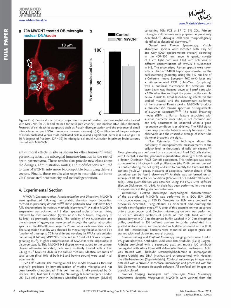
Finally, microglial multi-nucleation was also investigated and reproduced in primary cultures of purifi ed brain micro-glia. Figure 7 a shows a confocal microscopy projection image
of three microglial cells, each displaying two nuclei after 70 h incubation with MWCNTs. Quantifi cation of mono-nucleated versus multi-nucleated cells demonstrated a statistically sig-nifi cant rise of 22% in multi-nucleation (a signifi cant drop of mono-nucleated cells) in MWCNT-treated cultures (Figure 7b). Since microglia cell fusion was not observed in time-lapse live cell videos, we concluded that this phenotype was the result of a late cell division defect. MWCNTs could be interacting bio-mimetically with the mitotic spindle microtubules as described
Adv. Healthcare Mater. 2013, DOI: 10.1002/adhm.201300178

www.MaterialsViews.com
FULL P
APER
www.advhealthmat.de
7wileyonlinelibrary.com© 2013 WILEY-VCH Verlag GmbH & Co. KGaA, Weinheim
Figure 6. Confocal microscopy projection images of cells cultured from OB and treated with HS or MWCNTs for 70 h. No obvious degenerative changes such as nuclear DNA fragmen-tation are observed in neurons (empty arrows). Conversely, microglial cells appeared highly vacuolated. (#6). A disorganized microtubule distribution and binucleation (blue fi lled arrows) are observed. (Inset #6, right) Same cell observed by differential interference contrast (DIC) containing large deposits of MWCNTs in the cell cytoplasm (black arrow). A control untreated microglial cell is shown on the bottom right panel.
for HeLa cells, [ 25 ] or with the actin fi laments of the contractile ring, inhibiting the pro-gression of the cell cleavage and impeding the separation of the two daughter cells. The inhibition of the actin contractile ring, as described for other systems, [ 26,35–37 ] typically produces a late G2 arrest that makes cells to terminate cell division abruptly bearing two nuclei. Hence, these results also indicate that in brain purifi ed macrophages these MWCNTs interact with actin, stabilizing these fi laments in a similar fashion to that described for microtubules in HeLa cells. [ 25 ] In addition, the patent reorganization of the cytoplasmic actin fi laments [ 26 ] and the exist-ence of small cytoplasmic compact DNA aggregates, just as observed for the BV2 cell line, confi rmed apoptosis in these pri-mary cultures of brain microglia (Figure 7 a, arrows).
3 . Conclusions
The development of drugs that can enter the brain bypassing the BBB is one of the present challenges in medicine and a key obstacle to treat malignant brain tumors or neurodegenerative diseases. The intranasal administration route, ideal for these pur-poses, depends on microglia as key players in the distribution of nanomaterials through the brain, since these cells are strongly attracted by foreign materials and tumors cells or invading pathogens. Among nanomaterials, MWCNTs could represent ideal candidates for brain cancer therapy. These fi bers are well known to go across cellular barriers and dis-play an intrinsic ability to block cancer cell proliferation triggering apoptosis by inter-acting with intracellular nanofi laments such as microtubules or actin. However, our work demonstrates that these properties are a double edge sword since MWCNTs severely impeded microglial vehiculization of carbon nanotubes towards the tumor site after intra-nasal administration.
On the other hand, these properties might result interesting when considering local brain pre- or post-surgical adjuvant therapy. There are several studies that suggest that microglia and brain macrophages are a hall-mark of human malignant brain tumors and signifi cantly contribute to the clinical course and bad prognosis of brain tumors. [ 41 ] Thus, local MWCNT therapy would mostly inter-fere with the tumor macrophages rather than other local cells (Figure 6 ), producing
Adv. Healthcare Mater. 2013, DOI: 10.1002/adhm.201300178
www.MaterialsViews.com
FULL
PAPER
www.advhealthmat.de
8 wileyonlinelibrary.com © 2013 WILEY-VCH Verlag GmbH & Co. KGaA, Weinheim
containing 10% FCS at 37 °C, 5% CO 2 . Primary microglial cell cultures were prepared as previously described. [ 43 ] Microglial cells were morphologically identifi ed as described elsewhere. [ 43 ]
Optical and Raman Spectroscopy : Visible absorption spectra were recorded with Cary 50 and Cary 6000i spectrometers (Varian) operating in the 400–800 nm range. A quartz cuvette of 1 cm light path was fi lled with solutions of different concentrations of MWCNTS suspended in HS. The unpolarized Raman spectra were taken with a Horiba T64000 triple spectrometer in the backscattering geometry, using the 647 nm line of a Coherent Innova Spectrum 70C Ar-Kr laser and a nitrogen-cooled CCD (Jobin-Yvon Symphony) with a confocal microscope for detection. The laser beam was focused down to 1 μ m 2 spot with a 100× objective and kept the power on the sample below 2 mW to avoid laser-heating effects on the probed material and the concomitant softening of the observed Raman peaks. MWCNTs produce a characteristic Raman spectrum distinguishable of SWCNTs spectrum. [ 27,28 ] The radial breathing modes (RBM), a Raman feature associated with a small diameter inner tube, is not common and can only sometimes be observed when a good resonance condition is established. The RBM signal from large diameter tubes is usually too weak to be observable and the ensemble average of inner tube diameter broadens the signal.
Flow Cytometry : This technique allows the possibility of multiparameter measurements at the cellular level in thousands of cells per second. [ 31 ]
Flow cytometry was performed on a suspension of fi xed BV2 cells stained with Hoechst, a dye that produces a quantitative staining of DNA, using a Becton Dickinson FACS CantoII equipment. This technique was used to determine a blockage in cell proliferation (the DNA content per cell is doubled during the cell cycle) and also to quantify the fractional DNA content (“sub-G1” peak), indicative of apoptosis. Further details of the technique can be found elsewhere. [ 31 ] Analysis was performed on an average of 10 000 cells per condition (HS-control or HS-MWCNT-treated cells). Data quantifi cation was obtained using the FACS Diva software (Becton Dickinson, NJ, USA). Analysis has been performed in three sets of experiments at the given concentrations.
Transmission Electron Microscopy : Morphological characterization of the as-produced MWCNTs was performed in a JEOL JEM 2100 microscope operating at 120 kV. Samples for TEM were prepared as previously described, using ethanol as dispersant and omitting the sample centrifugation steps. [ 25 ] A drop of this suspension was adsorbed onto a Lacey copper grid. Electron microscopy on cells was performed on 70 nm Araldite sections of pellets of BV2 cells fi xed with 1% glutaraldehyde in 0.12 m phosphate buffer, washed in 0.12 m phosphate buffer, post-fi xed in 1% buffered osmium tetroxide, dehydrated in a graded acetone series and embedded in Araldite observed with a JEOL JEM 1011 microscope. Sections were mounted on copper grids and stained with lead citrate and uranyl acetate.
Immunostaining and Confocal Microscopy Imaging : Cells were fi xed in 1% glutaraldehyde. Antibodies used were anti- α -tubulin (B512) (Sigma-Aldrich) combined with a secondary goat anti-mouse IgG antibody conjugated with Alexa Fluor 488 (Molecular Probes, Invitrogen). Actin was stained with Phalloidin–Tetramethylrhodamine B isothiocyanate (Sigma-Aldrich) and DNA (nucleus and chromosomes) with Hoechst dye (Bis-benzimide) (Sigma-Aldrich). Confocal microscopy images were obtained with a Nikon A1R confocal microscope and processed with the NIS-Elements Advanced Research software. All confocal cell images are pseudo-colored.
Live-Cell Imaging Techniques and Time-Lapse Video Microscopy Experiments. Bacterial Phagocytosis : MWCNTs were washed from the
anti-tumoral effects in situ as shown for other tumors, [ 42 ] while preserving intact the microglial immune-function in the rest of brain parenchyma. These results also provide new clues about the dosages, administration routes, and modifi cations required to turn MWCNTs into more biocompatible brain drug delivery vectors. Finally, these results also urge to reconsider air-borne CNT associated neurotoxicity and neurodegeneration.
4 . Experimental Section MWCNTs Characterization, Functionalization, and Dispersion : MWCNTs
were synthesized following the catalytic chemical vapor deposition method as previously described. [ 29 ] These particular MWCNTs have been fully characterized by various methods elsewhere. [ 25 ] A stable MWCNTs suspension was obtained in HS after repeated cycles of vortex mixing followed by mild sonication (cycles of 2 s for 5 times, frequency of 38 kHz) as previously described. The stability of the suspension and the existence of aggregates were tested through the optical absorption spectra in the 400–800 nm range for 20 min after each sonication cycle. The suspension stability was checked by measuring the absorbance as a function of time up to 70 h for different wavelengths. [ 25 ] A stock solution containing 0.140 mg MWCNTs dispersed in 2.3 mL of HS was prepared (a 60 μ g mL −1 ). Higher concentrations of MWCNTs were impossible to disperse steadily. This MWCNT-HS dispersion was added to the culture. Unless otherwise indicated, cells were routinely treated with a fi nal 0.6 μ g mL −1 MWCNT dose in the culture medium. Identical amounts of total serum (fi nal 10% of both HS and bovine serum) were used in all experiments.
BV2 Cell Cultures : The microglial cell line model known as BV2 was originally produced by immortalization of murine microglia and has been broadly characterized. This cell line was kindly provided by Dr. Pocock, UCL, National Hospital for Neurology & Neurosurgery, London UK. BV2 cells grow in Dulbecco’s Modifi ed Eagle's Medium (DMEM)
Figure 7. a) Confocal microscopy projection images of purifi ed brain microglial cells treated with MWCNTs for 70 h and stained for actin (red channel) and nuclear DNA (blue channel). Features of cell death by apoptosis such as F-actin disorganization and the presence of small intracellular compact DNA masses are observed (arrows). b) Quantifi cation of the percentages of mono-nucleated versus multi-nucleated cells revealed a signifi cant increase ( t = 4.72; p = 3 × 10 −5 ; degrees of freedom, DF = 39) in microglial cell multi-nucleation in primary brain cultures treated with MWCNTs.
Adv. Healthcare Mater. 2013, DOI: 10.1002/adhm.201300178

www.MaterialsViews.com
FULL P
APER
www.advhealthmat.de
9wileyonlinelibrary.com© 2013 WILEY-VCH Verlag GmbH & Co. KGaA, Weinheim
[9] L. Zhang , R. Bai , Y. Liu , L. Meng , B. Li , L. Wang , L. Xu , L. Le Guyader , C. Chen , Nanotoxicology 2012 , 5 , 562 .
[10] G. Oberdörster , A. Elder , A. Rinderknecht , J. Nanosci. Nanotechnol. 2009 , 9 , 4996 .
[11] J. Wang , C. Chen , Y. Liu , F. Jiao , W. Li , F. Lao , Y. Li , B. Li , C. Ge , G. Zhou , Y. Gao , Y. Zhao , Z. Chai , Toxicol. Lett. 2008 , 183 , 72 .
[12] T. T. Win-Shwe , H. Fujimaki , Int. J. Mol. Sci. 2011 , 12 , 6267 . [13] X. Zhuang , X. Xiang , W. Grizzle , D. Sun , S. Zhang , R. C. Axtell , S. Ju ,
J. Mu , L. Zhang , L. Steinman , D. Miller , H. G. Zhang , Mol. Ther. 2011 , 19 , 1769 .
[14] A. Nimmerjahn , F. Kirchhoff , F. Helmchen , Science 2005 , 308 , 1314 . [15] L. J. Smithson , M. D. Kawaja. J. Neurosci. Res. J. Neurosci. Res. 2010 ,
88 , 858 . [16] F. Lazarini , M. M. Gabellec , N. Torquet , P. M. Lledo , J. Neurosci.
2012 , 32 , 3652 . [17] Y. Wang , B. Wang , M. T. Zhu , M. Li , H. J. Wang , M. Wang ,
H. Ouyang , Z. F. Chai , W. Y. Feng , Y. L. Zhao , Toxicol. Lett. 2011 , 205 , 26 .
[18] L. R. Hanson , W. H. Frey , J. Neuroimmune Pharmacol. 2007 , 2 , 81 . [19] A. Bianco , K. Kostarelos , M. Prato , Curr. Opin. Chem. Biol. 2005 , 9 ,
674 . [20] J. M. Schnorr , T. M. Swager , Chem. Mater. 2011 , 23 , 646 . [21] J. Ren , S. Shen , D. Wang , Z. Xi , L. Guo , Z. Pang , Y. Qian , X. Sun ,
X. Jiang , Biomaterials 2012 , 33 , 3324 . [22] X. Luo , C. Matranga , S. Tan , N. Alba , X. T. Cui , Biomaterials 2011 ,
32 , 6316 . [23] H. N. Yehia , R. K. Draper , C. Mikoryak , E. K. Walker , P. Bajaj ,
I. H. Musselman , M. C. Daigrepont , G. R. Dieckmann , P. Pantano , J. Nanobiotechnol. 2007 , 5 , 8 .
[24] C. Z. Dinu , S. S. Bale , G. Zhu , J. S. Dordick Small 2009 5 , 310 . [25] L. Rodriguez-Fernandez , R. Valiente , J. Gonzalez , J. C. Villegas ,
M. L. Fanarraga , ACS Nano 2012 , 6 , 6614 . [26] B. D. Holt , P. A. Short , A. D. Rape , Y. L. Wang , M. F. Islam ,
K. N. Dahl , ACS Nano 2010 , 4 , 4872 . [27] E. F. Antunes , A. O. Lobo , E. J. Corat , V. J. Trava-Airoldi , A. A. Martin ,
C. Verissimo , Carbon 2006 , 44 , 2202 . [28] S. Dresselhaus , G. Dresselhaus , R. Saito , A. Jorio , Phys. Rep. 2005 ,
409 , 47 . [29] E. Flahaut , Ch. Laurent , A. Peigney , Carbon 2005 , 43 , 375 . [30] B. Kateb , V. Yamamoto , D. Alizadeh , L. Zhang , H. M. Manohara ,
M. J. Bronikowski , B. Badie , Methods Mol. Biol. 2010 , 651 , 307 . [31] D. Wlodkowic , W. Telford , J. Skommer , Z. Darzynkiewicz , Methods
Cell Biol. 2011 , 103 , 55 . [32] L. Gonzalez , I. Decordier , M. Kirsch-Volders , Biochem. Soc. Trans.
2010 , 38 , 1691 . [33] D. Cimini , D. Fioravanti , C. Tanzarella , F. B. Degrassi , Chromosoma
1998 , 107 , 479 . [34] M. Pardo , P. Nurse , Science 2003 , 6 , 1569 . [35] S. N. Martineau , P. R. Andreassen , R. L. Margolis , J. Cell Biol. 1995 ,
131 , 191 . [36] M. Bottini , S. Bruckner , K. Nika , N. Bottini , S. Belluci , A. Magrini ,
A. Bargamaschi , T. Mustelin , Toxicol. Lett. 2006 , 160 , 121 . [37] M. Desouza , P. W. Gunning , J. R. Stehn , Bioarchitecture 2012 , 1 , 75 . [38] W. P. Roos , B. Kaina , Trends Mol. Med. 2006 , 12 , 440 . [39] X. Li , Y. Peng , J. Ren , X. Qu , Proc. Natl. Acad. Sci. U.S.A. 2006 , 103 ,
19658 . [40] R. C. May , L. M. Machesky , J. Cell Sci. 2001 , 114 , 1061 . [41] J. R. Engler , A. E. Robinson , I. Smirnov , J. G. Hodgson , M. S. Berger ,
N. Gupta , C. D. James , A. Molinaro , J. R. Phillips , PLoS One 2012 , 7 , e43339 .
[42] M. Yang , J. Meng , X. Cheng , J. Lei , H. Guo , W. Zhang , H. Kong , H. Xu , Theranostics 2012 , 2 , 258 .
[43] M. L. Fanarraga , J. Villegas , G. Carranza , R. Castaño , J. C. Zabala , Exp. Cell Res. 2009 , 315 , 535 .
culture before live-cell imaging. The cell migration speed and total track lengths were both measured on 3.5 h time-lapse movies obtained at 5 frames per min. Cells were incubated with MWCNTs during 70 h and then washed before live cell imaging. This was performed on a Nikon Eclipse Ti live-cell station, using a 20× Nikon N.A. 0.45. Cell tracking analysis was performed with the NIS-Elements software. Trajectory identifi cation was performed manually on at least 200 individual cells per condition frame by frame. Infected cultures were imaged during 22 h (at 5 frames per min). Quantifi cations in Figure 5 b were made on random images fi elds obtained at the indicated times with the NIS-Elements software. BV2 control and treated cell cultures were incubated with L. monocytogenes to provoke microglial activation and bacterial phagocytosis. Microglial cultures were exposed to bacteria for 1 h without antibiotic. Next, a bacteriostatic gentamicin dose was added to the cultures, allowing a minor time-dependent bacterial recovery. Bacterial density in cultures was measured in three random fi elds of video images taken at the indicated time points in three different cultures fi elds. Bacterial cells were quantifi ed using the NIS-Elements software in 3 ROIs of 12 mm 2 per photogram at the indicated times.
Statistical Analysis : Unpaired two-tailed Student’s t -tests were used for the statistical analyses of the differences between control and treated cell results. Three sets of experiments were used for each studied condition. SD stands for standard deviation.
Supporting Information Supporting Information is available from the Wiley Online Library or from the author.
Acknowledgements The authors thank Prof. J. C. Zabala for his support and helpful scientifi c discussions. The authors thank Dr. Alvarez for her help with the infection experiments and Dr. Flahaut for providing the MWCNTs. We are grateful to the Nikon A1R Laser Microscopy Unit of the IFIMAV Institute for the electron microscopy and confocal/time-lapse microscopy. This work has been supported by the Spanish MINECO under Projects ref. MAT2012–38664-C02–01; BFU2010–18948, MALTA Consolider-Ingenio ref. CSD2007–00045.
Received: May 10, 2013 Published online:
[1] M. Fazil , S. Shadab Baboota , J. K. Sahni , J. Ali , J. Drug Targeting 2012 , 20 , 97 .
[2] T. Sakane , S. Yamashita , N. Yata , H. Sezaki , J. Drug Targeting 1999 , 7 , 233 .
[3] M. Fazil , S. Md , S. Haque , M. Kumar , S. Baboota , J. K. Sahni , J. Ali , Eur. J. Pharm. Sci. 2012 , 47 , 6 .
[4] A. Elder , R. Gelein , V. Silva , T. Feikert , L. Opanashuk , J. Carter , R. Potter , A. Maynard , Y. Ito , J. Finkelstein , G. Oberdörster , Environ. Health Perspect. 2006 , 114 , 1172 .
[5] M. B. Genter , N. C. Newman , H. G. Shertzer , S. F. Ali , B. Bolon , Toxicol. Pathol. 2012 , 40 , 1004 .
[6] A. Cimini , B. D’Angelo , S. Das , R. Gentile , E. Benedetti , V. Singh , A. M. Monaco , S. Santucci , S. Seal , Acta Biomater. 2012 , 8 , 2056 .
[7] L. Geraets , A. G. Oomen , J. D. Schroeter , V. A. Coleman , F. R. Cassee , Toxicol. Sci. 2012 , 127 , 463 .
[8] B. Wang , W. Y. Feng , M. Wang , J. W. Shi , F. Zhang , H. Ouyang , Y. L. Zhao , Z. F. Chai , Y. Y. Huang , Y. N. Xie , H. F. Wang , J. Wang , Biol. Trace Elem. Res. 2007 , 118 , 233 .
Adv. Healthcare Mater. 2013, DOI: 10.1002/adhm.201300178