Fabrication of Mesoporous Silica Shells on Solid ... - CiteSeerX

Therapeutics, Targets, and Chemical Biology
CancerResearch
Sustained Small Interfering RNA Delivery byMesoporous Silicon Particles
Takemi Tanaka1,9, Lingegowda S. Mangala2, Pablo E. Vivas-Mejia11, René Nieves-Alicea1, Aman P. Mann1,Edna Mora2,5,10,11, Hee-Dong Han2, Mian M.K. Shahzad2,8, Xuewu Liu1,9, Rohan Bhavane1, Jianhua Gu1,Jean R. Fakhoury1,9, Ciro Chiappini9, Chunhua Lu2, Koji Matsuo2, Biana Godin1, Rebecca L. Stone2,Alpa M. Nick2, Gabriel Lopez-Berestein3,4,6,13, Anil K. Sood2,3,6, and Mauro Ferrari1,4,6,7,9
Abstract
Authors' AEngineerinDepar tme4ExperimenRNA InterAndersonUniversity;College ofEngineeri10DepartmComprehen
Note: SupResearch O
T. Tanaka a
CorresponScience Ce500-2470;
doi: 10.115
©2010 Am
www.aacr
RNA interference (RNAi) is a powerful approach for silencing genes associated with a variety of pathologicconditions; however, in vivo RNAi delivery has remained a major challenge due to lack of safe, efficient, andsustained systemic delivery. Here, we report on a novel approach to overcome these limitations using a multi-stage vector composed of mesoporous silicon particles (stage 1 microparticles, S1MP) loaded with neutralnanoliposomes (dioleoyl phosphatidylcholine, DOPC) containing small interfering RNA (siRNA) targetedagainst the EphA2 oncoprotein, which is overexpressed in most cancers, including ovarian. Our deliverymethods resulted in sustained EphA2 gene silencing for at least 3 weeks in two independent orthotopic mousemodels of ovarian cancer following a single i.v. administration of S1MP loaded with EphA2-siRNA-DOPC.Furthermore, a single administration of S1MP loaded with-EphA2-siRNA-DOPC substantially reduced tumorburden, angiogenesis, and cell proliferation compared with a noncoding control siRNA alone (SKOV3ip1, 54%;HeyA8, 57%), with no significant changes in serum chemistries or in proinflammatory cytokines. In summary,we have provided the first in vivo therapeutic validation of a novel, multistage siRNA delivery system forsustained gene silencing with broad applicability to pathologies beyond ovarian neoplasms. Cancer Res; 70(9);3687–96. ©2010 AACR.
Introduction
RNA interference (RNAi) therapy is emerging as a treat-ment modality of exceptional promise, in view of its versatileapplication to the silencing of any gene with a known se-quence and especially those that are not drugable by existingapproaches such as small-molecule inhibitors. However,in vivo systemic administration of RNAi has remained a ma-jor challenge due to its short half-life (1), lack of ability topenetrate the plasma membrane (2), and potential toxicity
ffiliations: 1Department of Nanomedicine and Biomedicalg, University of Texas Health Science Center at Houston;nts of 2Gynecolog ic Oncology, 3Cancer B io logy,tal Therapeutics, and 5Surgical Oncology, and 6Center forference and Non-Coding RNA, University of Texas M.D.Cancer Center; 7Department of Bioengineering, Riceand 8Department of Obstetrics and Gynecology, BaylorMedicine, Houston, Texas; 9Department of Biomedical
ng, Universi ty of Texas Austin, Austin, Texas; andent of Surgery and 11The University of Puerto Ricosive Cancer Center, San Juan, Puerto Rico
plementary data for this article are available at Cancernline (http://cancerres.aacrjournals.org/).
nd L.S. Mangala contributed equally to this work.
ding Author: Mauro Ferrari, The University of Texas Healthnter, 1825 Pressler Street, Houston, TX 77030. Phone: 713-Fax: 713-500-2462; E-mail: [email protected].
8/0008-5472.CAN-09-3931
erican Association for Cancer Research.
journals.org
(3, 4). Nanoparticle-based delivery systems have been pro-posed to address these concerns. The validity of RNAi thera-peutics has been shown in animal models (3, 5–7) and morerecently in human clinical trials (8, 9). For all of the potentialsmall interfering RNA (siRNA) delivery advantages they en-gender, nanoparticles also have some limitations includingtheir potential for rapid clearance (10), instability in serum(11), and systemic toxicity, especially to the liver (3). More-over, most of the current RNAi delivery approaches requirefrequent injections (12, 13), which can be a substantial im-pediment to patient treatment due to impaired enrollmenton clinical trials and decreased patient compliance (14).Thus, development of safe, easy to administer, and efficientdelivery systems that achieve sustained target gene silencingis of substantial clinical importance.Previously, we have shown that siRNA incorporated in
neutral nanoliposomes (30–40 nm in diameter) composedof dioleoyl phosphatidylcholine (DOPC) led to therapeuticgene modulation in several orthotopic cancer models withno overt toxicities (12, 13). Although our lipid-based siRNAdelivery platform holds substantial promise for clinical trans-lation, as is the case for other nanocarriers, our method cur-rently requires twice weekly injections to achieve continuousgene silencing. We sought to develop a biocompatible ap-proach that would allow for the sustained delivery of siRNAresulting in continuous gene silencing, therapeutic efficacy atnontoxic doses, and ease of administration. To attain thesegoals, we have developed a multistage delivery approach
3687

Tanaka et al.
3688
(Fig. 1A) composed of two biodegradable and biocom-patible carriers: the first-stage carriers are mesoporousmicroscale biodegradable silicon particles (stage 1 micro-particles: S1MP; ref. 15), allowing for the loading and releaseof second-stage nanocarriers (DOPC nanoliposomal siRNA:siRNA-DOPC) in a sustained manner. Here, we providethe first evidence that a single administration of multistagesiRNA-DOPC delivery resulted in sustained in vivo genesilencing for 3 weeks, significant antitumor effect in twoorthotopic mouse models of human ovarian cancer with noobservable concurrent toxicity.
Materials and Methods
Fabrication of porous silicon particles. Porous siliconparticles were fabricated by electrochemical etching ofsilicon wafers as previously described (15). The physicaldimension and pore size of S1MP were verified by high-resolution scanning electron microscope. The porositywas verified by nitrogen absorption analysis as previouslydescribed (15).Surface chemistry of S1MP. Surface of the S1MP was
hydroxylated in oxygen plasma (O2100sccm 50W). Thepositively charged amine groups were introduced on thesurface by silanization with 9% v/v 3-aminopropyltriethoxy-silane in isopropanol as previously described (15). Thesurface charge was measured by Zeta Pals (BrookhavenInstruments).Liposomal siRNA formulation. The siRNA liposomes were
prepared as previously described (13) by mixing 1,2 dioleoyl-
Cancer Res; 70(9) May 1, 2010
sn-glycero-3-phosphocholine with siRNA (20:1, mol/mol)in the presence of excess t-butanol. Control siRNAs (5′-AATT-CTCCGAACGTGTCACGT-3′) and Alexa 555–tagged controlsiRNA were purchased from Qiagen and siRNAs againstEphA2 (5′-AATGACATGCCGATCTACATG-3′) were pur-chased from Sigma (16). Dry film of DOPC liposomes were re-constituted by adding water followed by briefly sonication.Assembly of multistage vector. Amino functionalized
S1MP (1 × 10⁷ particles, 5 μg silicon) were mixed with t-butanol,and dried and mixed with DOPC nanoliposomes containingfluorescently labeled siRNA. The mixture was briefly sonicatedand then spun down to remove any unincorporated freeDOPC-siRNA. Fluorescence intensity of the supernatants aswell as the precipitants were measured at Ex 555/Em 592to assess loading efficacy.Cell lines and culture. The human epithelial ovarian
cancer cell lines such as HeyA8-Lc and Skov3ip1-Lc wereused for these studies. Cells were maintained in RPMI1640 supplemented with 15% fetal bovine serum and 0.1%gentamicin sulfate as described as previously (13). All ex-periments were performed with 70% to 80% confluentcultures.Orthotopic in vivo model of ovarian cancer and tissue
processing. Female athymic nude mice (NCr-nu; 4- to 5-week-old) were purchased from Taconic Farms and maintained aspreviously described (13). Institutional Animal Care and UseCommittee approval was obtained before all animal studies.All mouse studies were approved by the M.D. Anderson Can-cer Center Institutional Animal Care and Use Committee.Orthotopic nude mouse models of ovarian carcinoma (17)were used for this study as previously described.
Figure 1. Assembly of S1MP-siRNA-DOPC. A, concept of multistage delivery system. B to D, Scanning electron microscopic images of S1MP at differentmagnifications. E, loading of Alexa555-siRNA-DOPC to the S1MP. After the loading, fluorescence from unincorporated Alexa555-siRNA-DOPC wasmeasured to assess the loading efficacy. S1MP loaded with Alexa555-siRNA-DOPC were dissolved in 0.25% tetramethylammonium hydroxide and theloaded siRNA were separated by gel electrophoresis and visualized with SYBR Gold. F, release kinetics of Alexa555-siRNA-DOPC from the S1MP.The Alexa555-siRNA-DOPC–loaded S1MP were incubated in 10% FBS and the supernatant was separated to measure fluorescent intensity atEx544/Em590 at different time points.
Cancer Research

Sustained siRNA Delivery
Western blot analysis. Western blot was performed aspreviously described (13). Primary antibody against EphA2was detected with horseradish peroxidase–conjugated IgG(Amersham) and developed using an enhanced chemilumi-nescence detection kit (Pierce). Membranes were tested forβ-actin to confirm equal loading.Immunohistochemistry. Ki67 and EphA2 staining were
performed using formalin-fixed, paraffin-embedded tumorsections (8-μm thickness) as previously described (8). Forthe evaluation of cell proliferation, the proliferative indiceswere determined by counting the number of Ki67-positivecells out of total number of cells in five randomly selectedhigh-power fields exclusive of necrotic areas at ×100 mag-nification. The data were expressed as percentage of Ki67-positive cells. For the measurement of microvessel density,the proliferative indices were determined by counting thenumber of CD31-positive vessels in five randomly selectedhigh-power fields exclusive of necrotic areas at ×100 magni-fication. All staining was quantified by two investigators in ablinded fashion.Inductively coupled plasma atomic emission spectroscopy
analysis for silicon content. The organs were lysed, spundown, and then analyzed for silicon content using inductivelycoupled plasma atomic emission spectroscopy. We detectedsilicon at 250.69, 251.43, 251.61, and 288.158 nm and Yttrium(1 ppm) was used as an internal control for normalization.All results were expressed as percent of injected dose.Tissue transmission electron microscope (TEM). The
sections (100 nm) were imaged in a JEOL 1200 transmissionelectron microscope at 60 kV with digital images collectedusing a 1 k × 1 k Gatan BioScan camera Model 792.Sample size justification and statistical analysis. We
planed for n = 10 mice per treatment depending on the typeof experiment. For an effect size (ratio of fixed effect and re-sidual SD) of 0.65, this sample size was sufficient to provide80% power for a test at significance level of 0.05. As part ofpreliminary analysis, we validated the normality assumptionand proceeded with a nonparametric test as appropriate.The Mann-Whitney test was performed to compare thetumor sizes and tumor nodule number among differenttreatment groups.In vivo safety study (blood chemistry and cytokine).
Plasma samples were collected from retro-orbital sinusand further analyzed for blood chemistry [lactate dehydro-genase (LDH), blood urea nitrogen (BUN), and creatinine]and proinflammatory cytokine production. Equivalentamounts of silicic acid (degradation product of silicon par-ticles) and saline were used as negative controls. Cytokinelevels in the supernatants were determined by ELISA kitsaccording to the manufacturer's instructions (Bio-Rad).
Results
Assembly of multistage system loaded with neutralnanoliposomal siRNA (DOPC-siRNA). We fabricated S1MPusing electrochemical etching and photolithography as previ-ously described (15). These methods allow for control over thesize, shape, surface properties, overall porosity, and pore size
www.aacrjournals.org
of the S1MP. For this study, we used half-dome shaped S1MPsthat were uniform in size with diameter of 1.6 ± 0.1 μm (Fig.1A and C) and had straight pores in the center with averagepore size of 26 nm (Fig. 1D). The size and shape for the S1MPwere selected in accordance with the mathematical vector de-sign criteria (18, 19). In particular, the S1MP design was cho-sen to enhance the probability of their margination andendothelial adhesion in capillary circulation. The S1MP werefunctionalized with an amine group to provide for a positivesurface charge (8.0 mV) to enhance the charge-based interac-tion of negatively charged DOPC-siRNA (−2.9 mV) within thepores of the S1MP (Supplementary Fig. S1). We first examinedthe loading capacity of the S1MP to achieve the maximumloading of siRNA-DOPC. DOPC nanoliposomes containingAlexa555-labeled siRNA (Alexa555-siRNA-DOPC) were usedfor direct measurement of loading. Alexa555-siRNA-DOPCwas efficiently incorporated into the S1MP in a concentra-tion-dependentmanner (Fig. 1E). Gel electrophoresis also sup-ported siRNA-DOPC dose-dependent loading as well as theintegrity of loaded siRNA (Fig. 1E). Under this loading con-dition, a maximum 2.0 pg of siRNA were loaded into a sin-gle S1MP (Fig. 1E). Effective loading of alexa555-siRNA-DOPC was achievable when dried and positively chargedS1MP was used (Supplementary Fig. S1), suggesting that theliposomes are loaded into the porous structure by capillary ac-tion. Approximately 35% of Alexa555-siRNA-DOPC werereleased from the S1MP within the first 3 hours (presum-ably associated with release from the surface and shallowpores), followed by gradual release of the remainingamount from the S1MP over 14 days (Fig. 1F).Effect of multistage delivery on siRNA tumor uptake and
tissue distribution. To determine the duration and extent ofsiRNA-DOPC delivery in vivo, we first compared siRNA dis-tribution and tumor uptake using the S1MP loaded withAlexas555-siRNA-DOPC versus Alexas555-siRNA-DOPCalone. Mice with HeyA8 orthotopic tumors (15 days afteri.p. inoculation of tumor cells) were given an i.v. injection ofeither S1MP loaded with a single dose of Alexa555-siRNA-DOPC (5 μg siRNA) or a single dose of Alexa555-siRNA-DOPCalone (5 μg siRNA). At day 10, siRNA fluorescence was almostabsent in the tumors from mice treated with Alexa555-siRNA-DOPC, whereas fluorescence remained for >10 days in thetumor from mice injected with S1MP-Alexa555-siRNA-DOPC(Supplementary Fig. S2A). Overall, a single injection of theS1MP loaded with siRNA-DOPC resulted in enhanced siRNAuptake into the tumor parenchyma by 6-fold compared withsiRNA-DOPC alone. No significant difference in Alexa555-siRNA uptake into other organs such as kidney or liver wasnoted (Supplementary Fig. S2B and C).In vivo delivery of siRNA-DOPC using the multistage
system. Based on the sustained delivery observed in the ex-periments noted above, we next investigated the durationand extent of gene silencing. We focused on EphA2 (receptortyrosine kinase in the ephrin family; ref. 20) because EphA2is overexpressed in >70% of ovarian cancers and is associatedwith poor patient outcome (21). We first evaluated theimmunostimulatory potential of EphA2 siRNA and/or theDOPC nanoliposomes because immunologic effects, especially
Cancer Res; 70(9) May 1, 2010 3689
Tanaka et al.
3690
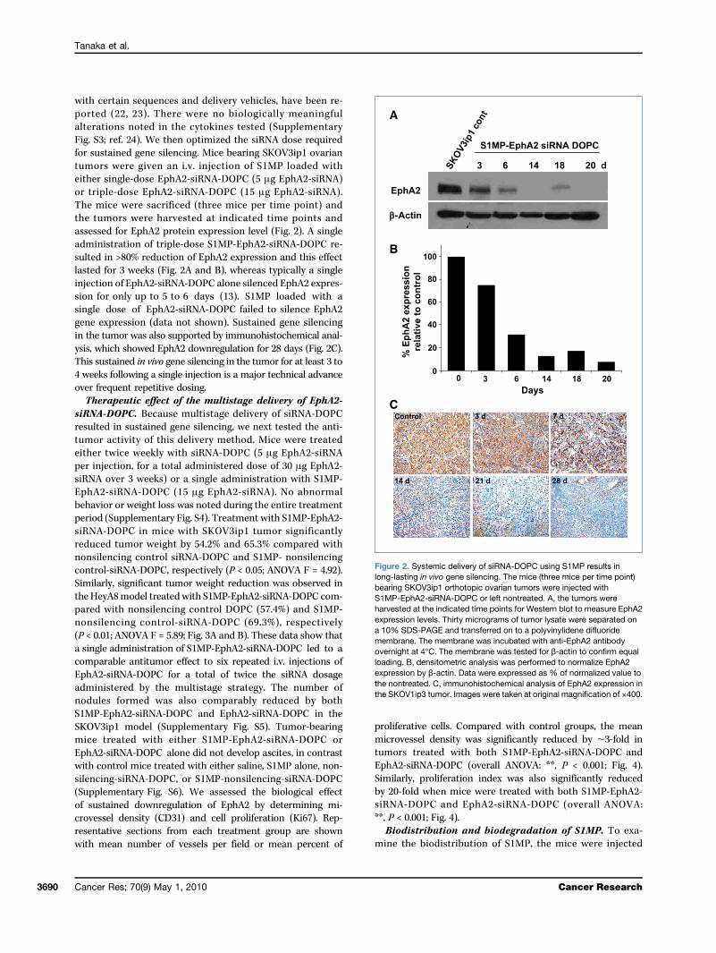
with certain sequences and delivery vehicles, have been re-ported (22, 23). There were no biologically meaningfulalterations noted in the cytokines tested (SupplementaryFig. S3; ref. 24). We then optimized the siRNA dose requiredfor sustained gene silencing. Mice bearing SKOV3ip1 ovariantumors were given an i.v. injection of S1MP loaded witheither single-dose EphA2-siRNA-DOPC (5 μg EphA2-siRNA)or triple-dose EphA2-siRNA-DOPC (15 μg EphA2-siRNA).The mice were sacrificed (three mice per time point) andthe tumors were harvested at indicated time points andassessed for EphA2 protein expression level (Fig. 2). A singleadministration of triple-dose S1MP-EphA2-siRNA-DOPC re-sulted in >80% reduction of EphA2 expression and this effectlasted for 3 weeks (Fig. 2A and B), whereas typically a singleinjection of EphA2-siRNA-DOPC alone silenced EphA2 expres-sion for only up to 5 to 6 days (13). S1MP loaded with asingle dose of EphA2-siRNA-DOPC failed to silence EphA2gene expression (data not shown). Sustained gene silencingin the tumor was also supported by immunohistochemical anal-ysis, which showed EphA2 downregulation for 28 days (Fig. 2C).This sustained in vivo gene silencing in the tumor for at least 3 to4 weeks following a single injection is a major technical advanceover frequent repetitive dosing.Therapeutic effect of the multistage delivery of EphA2-
siRNA-DOPC. Because multistage delivery of siRNA-DOPCresulted in sustained gene silencing, we next tested the anti-tumor activity of this delivery method. Mice were treatedeither twice weekly with siRNA-DOPC (5 μg EphA2-siRNAper injection, for a total administered dose of 30 μg EphA2-siRNA over 3 weeks) or a single administration with S1MP-EphA2-siRNA-DOPC (15 μg EphA2-siRNA). No abnormalbehavior or weight loss was noted during the entire treatmentperiod (Supplementary Fig. S4). Treatment with S1MP-EphA2-siRNA-DOPC in mice with SKOV3ip1 tumor significantlyreduced tumor weight by 54.2% and 65.3% compared withnonsilencing control siRNA-DOPC and S1MP- nonsilencingcontrol-siRNA-DOPC, respectively (P < 0.05; ANOVA F = 4.92).Similarly, significant tumor weight reduction was observed inthe HeyA8model treatedwith S1MP-EphA2-siRNA-DOPC com-pared with nonsilencing control DOPC (57.4%) and S1MP-nonsilencing control-siRNA-DOPC (69.3%), respectively(P < 0.01; ANOVA F = 5.89; Fig. 3A and B). These data show thata single administration of S1MP-EphA2-siRNA-DOPC led to acomparable antitumor effect to six repeated i.v. injections ofEphA2-siRNA-DOPC for a total of twice the siRNA dosageadministered by the multistage strategy. The number ofnodules formed was also comparably reduced by bothS1MP-EphA2-siRNA-DOPC and EphA2-siRNA-DOPC in theSKOV3ip1 model (Supplementary Fig. S5). Tumor-bearingmice treated with either S1MP-EphA2-siRNA-DOPC orEphA2-siRNA-DOPC alone did not develop ascites, in contrastwith control mice treated with either saline, S1MP alone, non-silencing-siRNA-DOPC, or S1MP-nonsilencing-siRNA-DOPC(Supplementary Fig. S6). We assessed the biological effectof sustained downregulation of EphA2 by determining mi-crovessel density (CD31) and cell proliferation (Ki67). Rep-resentative sections from each treatment group are shownwith mean number of vessels per field or mean percent of
Cancer Res; 70(9) May 1, 2010
proliferative cells. Compared with control groups, the meanmicrovessel density was significantly reduced by ∼3-fold intumors treated with both S1MP-EphA2-siRNA-DOPC andEphA2-siRNA-DOPC (overall ANOVA: **, P < 0.001; Fig. 4).Similarly, proliferation index was also significantly reducedby 20-fold when mice were treated with both S1MP-EphA2-siRNA-DOPC and EphA2-siRNA-DOPC (overall ANOVA:**, P < 0.001; Fig. 4).Biodistribution and biodegradation of S1MP. To exa-
mine the biodistribution of S1MP, the mice were injected
Figure 2. Systemic delivery of siRNA-DOPC using S1MP results inlong-lasting in vivo gene silencing. The mice (three mice per time point)bearing SKOV3ip1 orthotopic ovarian tumors were injected withS1MP-EphA2-siRNA-DOPC or left nontreated. A, the tumors wereharvested at the indicated time points for Western blot to measure EphA2expression levels. Thirty micrograms of tumor lysate were separated ona 10% SDS-PAGE and transferred on to a polyvinylidene difluoridemembrane. The membrane was incubated with anti-EphA2 antibodyovernight at 4°C. The membrane was tested for β-actin to confirm equalloading. B, densitometric analysis was performed to normalize EphA2expression by β-actin. Data were expressed as % of normalized value tothe nontreated. C, immunohistochemical analysis of EphA2 expression inthe SKOV1ip3 tumor. Images were taken at original magnification of ×400.
Cancer Research

Sustained siRNA Delivery
with Alexa555-siRNA-DOPC loaded S1MP i.v. and the majororgans including liver, kidney, spleen, lung, and heart wereharvested 4 hours after the injection. Silicon content analysisshowed that S1MP were primarily accumulated in the liver(53%) and spleen (11% of total injected dose), and therewas no to minimal S1MP accumulation observed in the kid-
www.aacrjournals.org
ney, lung, and heart (Fig. 5A). To provide further evidencethat the S1MP allows sustained siRNA release, S1MP conju-gated with FITC (green fluorescence) were loaded withAlexa555-siRNA-DOPC (red fluorescence) and i.v. injected in-to mice. The liver and spleen were harvested 20 days afterinjection for histologic evaluation. Alexa555-siRNA-DOPC re-mained associated with S1MP as evidenced by the yield ofyellow fluorescence, but not with macrophages (pink) inthe liver and spleen at 20 days after injection (SupplementaryFig. S7A and B), indicating that Alexa555-siRNA-DOPC re-mains intact. The S1MP were distributed throughout theliver sinusoids and the red pulp in the spleen (Fig. 5B). His-topathologic analyses of tissue sections with H&E stainshowed no obvious inflammatory infiltration in these organs.In the liver, the S1MPs were distributed throughout thesinusoids and (Fig. 5B) some were taken up by Kupffer cells(Supplementary Fig. S7A). In the spleen, the S1MPs werepresent in the splenic cords (Fig. 5B) and some were takenup by f4/80-positive macrophages (Supplementary Fig. S7B).Transmission electron microscopic analyses of the liver andspleen sections confirmed that S1MP remained within sinu-soidal spaces in the liver and red pulp in the spleen (Fig. 5C).Silicon contents in the spleen were reduced by 80% in thefirst 2 weeks and cleared by the 3rd week (Fig. 6A). In con-trast, only 55% of S1MP was cleared from the liver in the first2 weeks and ∼25% of the injected S1MP remained in the liver3 weeks after injection (Fig. 6B), suggesting that S1MP deg-radation kinetics are different in each organ. Scanning elec-tron microscopic images of S1MP showed enlargement ofpore size at day 7 and reduction of overall size by day 14(Fig. 6C). In some cases, bulky surface erosion was noted(data not shown). Collectively, these data suggest that theS1MP serve as a reservoir to protect siRNA-DOPC from deg-radation, release them over time by a mechanism that com-bines degradation of the carrier material, and hindereddiffusion through the nanoscopic pore.Biocompatibility of S1MP. We next tested the biocom-
patibility of multistage delivery system in both an acuteand chronic setting in FBV/N mice following the i.v. admi-nistration of therapeutic dose of S1MP-EphA2-siRNA-DOPC(15 μg EphA2-siRNA). Blood chemistry showed that S1MPsdid not cause a significant increase in any of the parameterstested (Supplementary Fig. S8A–F). Despite significant accu-mulation of the S1MP in the liver and spleen, plasma LDHlevels from the liver and spleen remained unchanged (Sup-plementary Fig. S8A and D). Similarly, tissue LDH levels inthe liver and spleen were not changed at both conditionstested (data not shown), indicating that accumulation ofthe S1MP in the liver and spleen did not cause tissue injury.Similar to blood chemistry, the values of cytokines testedwere not significantly higher in the mice injected withDOPC-loaded S1MP than in the saline-injected controlanimals in both acute and chronic setting, although someS1MP were taken up by macrophages (SupplementaryFig. S8G and H). These data indicate that S1MP-siRNA-DOPCis not toxic to the kidney, liver, and spleen and that nosignificant levels of inflammatory cytokines were inducedby the S1MP or its degradation products.
Figure 3. Therapeutic efficacy of sustained EphA2-siRNA-DOPCdelivery by S1MP. Nude mice were injected i.p. with SKOV3ip1 or HeyA8cells and randomly allocated to one of six treatment groups (n = 10):(a) saline, (b) S1MP, (c) nonsilencing control siRNA-DOPC,(d) S1MP-nonsilencing control-siRNA-DOPC, (e) EphA2-siRNA-DOPC,(f) S1MP-EphA2-siRNA-DOPC. SiRNA-DOPC was i.v. injected biweeklyat a dose of 5 μg siRNA. S1MP-EphA2-siRNA-DOPC was injected ina single administration in 3 wks at a dose of 15 μg siRNA. The mice wereinjected with saline biweekly in the rest of treatment period. When controlanimals (saline- and nonsilencing siRNA–treated mice) began to appearmoribund (4–5 wks after cell injection), all animals in an experiment weresacrificed and mouse weight, tumor weight (wt), tumor number, ascitesvolume, and tumor location were recorded. Columns, mean tumor weightfrom SKOV3ip1 (A) or HeyA8 cells (B); bars, SD.
Cancer Res; 70(9) May 1, 2010 3691

Tanaka et al.
3692
Discussion
The key findings from this work are that a single injectionof multistage delivery system comprised mesoporous siliconparticles loaded with liposomal siRNA against oncogene,EphA2, resulted in a sustained gene silencing for up to atleast 3 weeks in ovarian tumor, concomitant with a sub-stantial reduction of tumor burden with no to minimumtoxicity. Many oncoproteins as well as their distinct roleshave been identified in the past few decades; however, manyof them are “undrugable” with traditional approaches suchas small-molecule inhibitors because their crystal structuresare unknown. We targeted oncoprotein EphA2 (tyrosinekinase receptor in the ephrin family), which plays roles inangiogenesis (25) and cell proliferation (26), and is difficultto target with conventional approaches. EphA2 is absent in
Cancer Res; 70(9) May 1, 2010
the normal tissue but is overexpressed in different types oftumor including ovarian (27), breast (28), lung (29), andmelanoma (30), with strong association with poor survival,advanced-stage, or high-metastatic potential (31). Althoughthe dual roles of EphA2 as a pro-oncogenic or antioncogenicprotein have been suggested (32), a recent study revealedthat EphA2 mediates cell migration and invasion in the ab-sence of ephrin (a cognate ligand for EphA2) through Aktphosphorylation (33). It has also been reported that EphA2overexpression is frequently accompanied by the loss ofephrin, further supporting the pro-oncogenic role of EphA2in cancer (34). Therefore, a disruption of the biological func-tions of EphA2 is a highly attractive molecular-targeted the-rapy approach (35).Our data shown that the multistage delivery of liposo-
mal siRNA (single injection of 15 μg EphA2-siRNA) led
Figure 4. Effect of sustained siRNA delivery on angiogenesis and cell proliferation. Tumors from animals with the SKOV3ip1 ovarian tumor wereexamined for microvessel density (CD31) and cell proliferation (Ki67). Representative sections from each treatment group are shown (final magnification,×100 for CD31 and ×400 for Ki67), with mean number of vessels per field or mean % of proliferative cells, summarized in the graph at the bottom.Five different fields per slides, at least three individual tumors per treatment group were examined. Both microvessel density (MVD; A and C) and cellproliferation (B and D) were significantly reduced in tumor when treated with repeated administration of EphA2-siRNA-DOPC and a single administrationof the S1MP-EphA2-siRNA-DOPC. (overall ANOVA: **, P < 0.001).
Cancer Research

Sustained siRNA Delivery
to comparable antitumor effect to repeated i.v. siRNA-DOPCinjections (six injections of 5 μg EphA2-siRNA each), thushalving the siRNA dosage required to achieve an equivalentantitumor effect. Although we are focusing on EphA2 in thisstudy, this platform could allow for highly adjustable person-alized molecular targeted therapies with a suitable combina-tion of RNAi depending on the molecular signature of eachtumor type with “tunable” release kinetics of siRNA to con-trol cancer cell growth. Many efforts have been reported to de-liver RNAi; viral vectors (36), chemically modified siRNA (6),and nonviral nanocarriers including cationic liposome (37)and polycation-based carriers (38) have shown effective genesilencing effects in vitro and in vivo. However, immunogenicity,toxicity, safety concern, and unfavorable pharmacokinetics re-main generally unsolved problems for these delivery strategies.Traditional approaches to the improvement of pharmacokinet-ics include surface functionalization of nanocarrier with poly-ethylene glycol (PEG) to avoid reticuloendothelial systemuptake (39). PEG, however, does not prevent renal excretionand degradation of nanocarriers. In addition, PEGylation accel-erates the clearance of nanocarrier from the circulation wheninjected repeatedly (40). Thus, with the aim of providing stabil-ity to nanocarriers containing biologically active payload and ofachieving sustainedgene silencing, in this study, we used meso-porous silicon particles (S1MP) as a carrier of DOPC neutralnanoliposomes. The serum half-life (t1/2) of systemically injectedDOPC nanoliposomes is ∼2.7 hours.12 The combinatorial mul-
12 R.A. Nieves, T. Tanaka, C. Rodriguez-Aguayo, T. Madden, A.M. Tari, A.K.Sood, M. Ferrari, G. Lopez-Berestein, unpublished data.
www.aacrjournals.org
tistage delivery system we used in this study significantly im-proved the pharmacokinetics of the DOPC liposome andresulted in 3-week-long sustained gene silencing of EphA2 inthe tumor. It is likely that S1MP protect DOPC- siRNA fromdegradation and renal excretion due to their entrapment withinthe capillary spaces within the liver and spleen (Fig. 5). Slowrelease is most likely attained due to the gradual degradationof S1MP with subsequent release of siRNA-DOPC that reachesto the tumor.Porous silicon is an attractive material (41), which has
been investigated for possible applications in drug deliveryby loading of nanoscale payload such as protein (42), en-zymes (43), drugs (41), and nanocarriers (15). The surfacechemistry on the porous silicon will play a critical role indetermining the loading efficacy and release kinetics basedon the type of interaction of payload and porous surface(41). We used positively charged S1MP for enhanced en-trapment of the negatively charged (−2.9 mV) DOPC-siRNA.The electrostatic interactions between the pore wall sur-face and liposomes are likely to contribute to the sustainedrelease of the liposomes from the porous structure as theS1MP degrades.Mesoporous silicon is biocompatible (44, 45) and has re-
ceived Food and Drug Administration approval for use inbrachytherapy and drug delivery from implants (BioSilicon;ref. 46). The two delivery carrier materials used in this study,mesoporous silicon andDOPCneutral liposome, are highly bio-degradable, biocompatible, and combine suitably to yield time-sustained delivery with no liver and renal toxicity in both in theacute and the chronic setting, at a dose of 25 μg of mesoporoussilicon (Supplementary Fig. S8). In a separate study, we showed
Figure 5. Biodistribution of S1MP. A, silicon content analysis of organs isolated from mice (n = 6) following i.v. injection of S1MP-EphA2-siRNA-DOPC.The major organs including heart, lung, liver, kidney, and spleen were harvested 4 h after the i.v. injection. Silicon amounts were measured byinductively coupled plasma atomic emission spectroscopy and expressed as % of injected dose per organ (% ID). n.d., not detectable. B, H&E staining ofthe liver and spleen sections. The liver and spleen were harvested from the mice injected with S1MP-EphA2-siRNA-DOPC and fixed with formalin.Five-micrometer paraffin sections were stained with H&E to identify the localization of S1MP in the organs. Arrows, S1MP (final magnification, ×1,000).C, transmission electron microscopic images of S1MP in the liver and spleen. S1MP remained within the sinusoidal space in the liver (red arrow). K, Kupffercell; R, RBC; SS, sinusoidal space; H, hepatocyte; M, macrophage (original magnification, ×10,000).
Cancer Res; 70(9) May 1, 2010 3693

Tanaka et al.
3694
the safe systemic administration of 250 μg of mesoporous(10-fold the dose used here) through both i.p. and i.v. routesin mice.13 The multistage delivery system used in this studydid not result in the production of the proinflammatory cyto-kines tested, unlike other delivery system that relies on the hostimmunity (47). Our data indicate that the antitumor effect me-diated by the S1MP loaded with DOPC-siRNA was due to theDOPC-siRNA-EphA2 mediated gene silencing of EphA2 andnot due to immunostimulatory effects.Porous silicon is known to degrade completely into
orthosilicic acid over time, which is readily excreted from
13 T. Tanaka, B. Godin, R. Bhavane, R.A. Nieves, J. Gu, X. Liu, C. Chiappini,J.R. Fakhoury, S. Amra, A. Ewing, Q. Li, M. Ferrari, unpublished data.
Cancer Res; 70(9) May 1, 2010
the body. The mechanism of sustained liposomal siRNA deliv-ery is likely to rely on essentially two unique characteristicsand tailorable characteristics of the S1MP: biodegradationand biodistribution. Biodegradation is controlled by the over-all porosity and pore size of the particles (48), which in turn isexquisitely controllable by choice of material processing para-meters (41). The biodistribution of the particles is controlledby their size, shape, surface, and bulk properties (49), whichare also precisely controlled during the particle fabricationprocesses (15). In view of the different degradation kineticsin different organs, and in particular within the liver and thespleen, altering their respective accumulations will leadto different, adjustable kinetics of release of the nanolipo-some-encapsulated siRNA. Possible mechanisms involved inthe geometry-dependent organ sequestration of particlesare capillary size, capillary architecture, hemodynamics, andendocytosis (49).In conclusion, in this study, we showed that a single
injection of multistage delivery system comprised ofmesoporous silicon particles loaded with nanoliposomalsiRNA against oncogene, EphA2, resulted in: (a) sustainedgene silencing for up to at least 3 weeks in ovarian tumor;(b) substantial reduction of tumor burden; (c) substantialdecrease of angiogenesis and cellular proliferation; (d) noproduction of ascites; and (e) no detected toxicity associ-ated with the S1MP vectors, the neutral nanoliposomes,or the therapeutic siRNA. To the best of our knowledge,this is the first study to achieve these collective goals in vivoand, in particular, in two orthotopic models of ovarian can-cer. These finding encourage the development of a “library”of targets and drugs that can be further tailored towardspecific genetic abnormalities in cancer.
Disclosure of Potential Conflicts of Interest
Commercialization rights on the intellectual property presented in this ar-ticle have been acquired by Leonardo Biosystems Inc. from the title holder, theUniversity of Texas Health Science Center in Houston. M. Ferrari is the found-ing scientist of Leonardo Biosystems, and hereby discloses a financial interestin the company. The other authors disclosed no potential conflicts of interest.
Acknowledgments
We thank Matthew Landry for assistance with manuscript preparation.
Grant Support
Department of Defense (W81XWH-07-2-0101, W81XWH-09-1-0212) andNIH (R01CA128797, R33CA122864), the State of Texas's Emerging TechnologyFund, National Aeronautics and Space Administration (NNJ06HE06A), theOvarian Cancer Research Fund Program Project Development Grant, the Uni-versity of Texas M.D. Anderson Cancer Center Ovarian Cancer Specialized Pro-gram of Research Excellence (P50 CA083639), the NIH (CA110793, CA109298),the Zarrow Foundation, the Betty Ann Asche Murray Distinguished Professor-ship, the Baylor WRHR grant (HD050128) and the GCF Molly-Cade ovariancancer research grant (M.M.K. Shahzad), and the Alliance for NanoHealth.
The costs of publication of this article were defrayed in part by the paymentof page charges. This article must therefore be hereby marked advertisement inaccordance with 18 U.S.C. Section 1734 solely to indicate this fact.
Received 10/23/2009; revised 02/09/2010; accepted 02/26/2010; publishedonline 04/29/2010.
Figure 6. Biodegradation of S1MP. The mice (n = 5) were injected withS1MP-Alexa555-siRNA-DOPC and the liver and spleen were harvested atindicated time points. The organs were processed for silicon contentsanalysis and SEM images. A and B, inductively coupled plasma analysiswas performed to determine the silicon contents in the liver and spleen atdifferent time points. Silicon (Si) amounts were expressed as % ofinjected dose per organs (% ID). C, the S1MPs were isolated from the liverand spleen homogenates and further cleaned by sonication for SEManalysis. SEM images show time-dependent biodegradation of S1MP inthe liver and spleen at day 7 and 15. Original magnification, ×50,000).
Cancer Research

Sustained siRNA Delivery
References
1. Bumcrot D, Manoharan M, Koteliansky V, Sah DW. RNAi thera-peutics: a potential new class of pharmaceutical drugs. Nat ChemBiol 2006;2:711–9.
2. Aagaard L, Rossi JJ. RNAi therapeutics: principles, prospectsand challenges. Adv Drug Deliv Rev 2007;59:75–86.
3. Heidel JD, Yu Z, Liu JY, et al. Administration in non-humanprimates of escalating intravenous doses of targeted nanopar-ticles containing ribonucleotide reductase subunit M2 siRNA.Proc Natl Acad Sci U S A 2007;104:5715–21.
4. Desai AA, Schilsky RL, Young A, et al. A phase I study of anti-sense oligonucleotide GTI-2040 given by continuous intravenousinfusion in patients with advanced solid tumors. Ann Oncol 2005;16:958–65.
5. Frank-Kamenetsky M, Grefhorst A, Anderson NN, et al. TherapeuticRNAi targeting PCSK9 acutely lowers plasma cholesterol inrodents and LDL cholesterol in nonhuman primates. Proc Natl AcadSci U S A 2008;105:11915–20.
6. Morrissey DV, Lockridge JA, Shaw L, et al. Potent and persistentin vivo anti-HBV activity of chemically modified siRNAs. Nat Bio-technol 2005;23:1002–7.
7. Zimmermann TS, Lee AC, Akinc A, et al. RNAi-mediated genesilencing in non-human primates. Nature 2006;441:111–4.
8. Kim DH, Rossi JJ. Strategies for silencing human disease using RNAinterference. Nat Rev Genet 2007;8:173–84.
9. Davis ME. The first targeted delivery of siRNA in humans via a self-assembling, cyclodextrin polymer-based nanoparticle: from conceptto clinic. Mol Pharm 2009;6:659–68.
10. Longmire M, Choyke PL, Kobayashi H. Clearance propertiesof nano-sized particles and molecules as imaging agents: con-siderations and caveats. Nanomedicine 2008;3:703–17.
11. Zhang Y, Bradshaw-Pierce EL, Delille A, Gustafson DL,Anchordoquy TJ. In vivo comparative study of lipid/DNA com-plexes with different in vitro serum stability: effects on bio-distribution and tumor accumulation. J Pharm Sci 2008;97:237–50.
12. Merritt WM, Lin YG, Spannuth WA, et al. Effect of interleukin-8 gene silencing with liposome-encapsulated small interferingRNA on ovarian cancer cell growth. J Natl Cancer Inst 2008;100:359–72.
13. Landen CN, Jr., Chavez-Reyes A, Bucana C, et al. TherapeuticEphA2 gene targeting in vivo using neutral liposomal small interferingRNA delivery. Cancer Res 2005;65:6910–8.
14. Lara PN, Jr., Higdon R, Lim N, et al. Prospective evaluation ofcancer clinical trial accrual patterns: identifying potential barriers toenrollment. J Clin Oncol 2001;19:1728–33.
15. Tasciotti E, Liu XW, Bhavane R, et al. Mesoporous silicon particlesas a multistage delivery system for imaging and therapeutic appli-cations. Nat Nanotechnol 2008;3:151–7.
16. Halder J, Kamat AA, Landen CN, Jr., et al. Focal adhesion kinasetargeting using in vivo short interfering RNA delivery in neutralliposomes for ovarian carcinoma therapy. Clin Cancer Res 2006;12:4916–24.
17. Thaker PH, Yazici S, Nilsson MB, et al. Antivascular therapyfor orthotopic human ovarian carcinoma through blockade ofthe vascular endothelial growth factor and epidermal growth factorreceptors. Clin Cancer Res 2005;11:4923–33.
18. Decuzzi P, Ferrari M. The adhesive strength of non-sphericalparticles mediated by specific interactions. Biomaterials 2006;27:5307–14.
19. Decuzzi P, Lee S, Decuzzi M, Ferrari M. Adhesion of microfabricatedparticles on vascular endothelium: a parametric analysis. AnnBiomed Eng 2004;32:793–802.
20. Stein E, Lane AA, Cerretti DP, et al. Eph receptors discriminatespecific ligand oligomers to determine alternative signaling com-plexes, attachment, and assembly responses. Gene Dev 1998;12:667–78.
21. Lin YG, Han LY, Kamat AA, et al. EphA2 overexpression is asso-ciated with angiogenesis in ovarian cancer. Cancer 2007;109:332–40.
www.aacrjournals.org
22. Judge AD, Sood V, Shaw JR, Fang D, McClintock K, MacLachlan I.Sequence-dependent stimulation of the mammalian innate im-mune response by synthetic siRNA. Nat Biotechnol 2005;23:457–62.
23. Robbins MA, Rossi JJ. Sensing the danger in RNA. Nat Med 2005;11:250–1.
24. Mangala LS, Zuzel V, Schmandt R, et al. Therapeutic targetingof ATP7B in ovarian carcinoma. Clin Cancer Res 2009;15:3770–80.
25. Ogawa K, Pasqualini R, Lindberg RA, Kain R, Freeman AL,Pasquale EB. The ephrin-A1 ligand and its receptor, EphA2, areexpressed during tumor neovascularization. Oncogene 2000;19:6043–52.
26. Cheng N, Brantley DM, Liu H, et al. Blockade of EphA re-ceptor tyrosine kinase activation inhibits vascular endothelialcell growth factor-induced angiogenesis. Mol Cancer Res 2002;1:2–11.
27. Thaker PH, Deavers M, Celestino J, et al. EphA2 expression isassociated with aggressive features in ovarian carcinoma. ClinCancer Res 2004;10:5145–50.
28. Zelinski DP, Zantek ND, Stewart JC, Irizarry AR, Kinch MS. EphA2overexpression causes tumorigenesis of mammary epithelial cells.Cancer Res 2001;61:2301–6.
29. Kinch MS, Moore MB, Harpole DH, Jr. Predictive value of the EphA2receptor tyrosine kinase in lung cancer recurrence and survival.Clin Cancer Res 2003;9:613–8.
30. Easty DJ, Hill SP, Hsu MY, et al. Up-regulation of ephrin-A1 duringmelanoma progression. Int J Cancer 1999;84:494–501.
31. Kertesz N, Krasnoperov V, Reddy R, et al. The soluble extracel-lular domain of EphB4 (sEphB4) antagonizes EphB4-2 interaction,modulates angiogenesis, and inhibits tumor growth. Blood 2006;107:2330–8.
32. Miao H, Wei BR, Peehl DM, et al. Activation of EphA receptortyrosine kinase inhibits the Ras/MAPK pathway. Nat Cell Biol2001;3:527–30.
33. Miao H, Li DQ, Mukherjee A, et al. EphA2 mediates ligand-dependent inhibition and ligand-independent promotion of cellmigration and invasion via a reciprocal regulatory loop with Akt.Cancer Cell 2009;16:9–20.
34. Dodelet VC, Pasquale EB. Eph receptors and ephrin ligands:embryogenesis to tumorigenesis. Oncogene 2000;19:5614–9.
35. Lee JW, Han HD, Shahzad MM, et al. EphA2 immunoconjugateas molecularly targeted chemotherapy for ovarian carcinoma. J NatlCancer Inst 2009;101:1193–205.
36. Xia H, Mao Q, Paulson HL, Davidson BL. siRNA-mediatedgene silencing in vitro and in vivo. Nat Biotechnol 2002;20:1006–10.
37. Schiffelers RM, Ansari A, Xu J, et al. Cancer siRNA therapy bytumor selective delivery with ligand-targeted sterically stabilizednanoparticle. Nucleic Acids Res 2004;32:e149.
38. Bitko V, Musiyenko A, Shulyayeva O, Barik S. Inhibition ofrespiratory viruses by nasally administered siRNA. Nat Med 2005;11:50–5.
39. Wang S, Lee RJ, Cauchon G, Gorenstein DG, Low PS. Delivery ofantisense oligodeoxyribonucleotides against the human epidermalgrowth factor receptor into cultured KB cells with liposomes conju-gated to folate via polyethylene glycol. Proc Natl Acad Sci U S A1995;92:3318–22.
40. Laverman P, Carstens MG, Boerman OC, et al. Factors affect-ing the accelerated blood clearance of polyethylene glycol-liposomes upon repeated injection. J Pharmacol Exp Ther 2001;298:607–12.
41. Anglin EJ, Cheng L, Freeman WR, Sailor MJ. Porous silicon indrug delivery devices and materials. Adv Drug Deliv Rev 2008;60:1266–77.
42. Foraker AB, Walczak RJ, Cohen MH, Boiarski TA, Grove CF,Swaan PW. Microfabricated porous silicon particles enhanceparacellular delivery of insulin across intestinal Caco-2 cell mono-layers. Pharm Res 2003;20:110–6.
Cancer Res; 70(9) May 1, 2010 3695
Tanaka et al.
3696
43. Delouise LA, Miller BL. Enzyme immobilization in porous silicon:quantitative analysis of the kinetic parameters for glutathione-S-transferases. Anal Chem 2005;77:1950–6.
44. Prestidge CA, Barnes TJ, Lau CH, Barnett C, Loni A, Canham L.Mesoporous silicon: a platform for the delivery of therapeutics.Expert Opin Drug deliv 2007;4:101–10.
45. Park JH, Gu L, von Maltzahn G, Ruoslahti E, Bhatia SN, Sailor MJ.Biodegradable luminescent porous silicon nanoparticles for in vivoapplications. Nat Mater 2009;8:331–6.
46. Zhang K, Loong SL, Connor S, et al. Complete tumor responsefollowing intratumoral 32P BioSilicon on human hepatocellular and
Cancer Res; 70(9) May 1, 2010
pancreatic carcinoma xenografts in nude mice. Clin Cancer Res2005;11:7532–7.
47. Cubillos-Ruiz JR, Engle X, Scarlett UK, et al. Polyethylenimine-basedsiRNA nanocomplexes reprogram tumor-associated dendritic cellsvia TLR5 to elicit therapeutic antitumor immunity. J Clin Invest2009;119:2231–44.
48. Canham LT. Bioactive silicon structure fabrication through nanoetch-ing techniques. Adv Mater 1995;7:1033–7.
49. Decuzzi P, Pasqualini R, Arap W, Ferrari M. Intravascular delivery ofparticulate systems: Does geometry really matter? Pharm Res 2009;26:235–43.
Cancer Research
Copyright © 2022 FDOKUMEN