Recent advances of mesoporous materials in sample preparation
12
Journal of Chromatography A, 1228 (2012) 193–204 Contents lists available at SciVerse ScienceDirect Journal of Chromatography A jou rn al h om epage: www.elsevier.com/locat e/chroma Review Recent advances of mesoporous materials in sample preparation Liang Zhao, Hongqiang Qin, Ren’an Wu ∗ , Hanfa Zou ∗∗ National Chromatographic R&A Center, CAS Key Laboratory of Separation of Science for Analytical Chemistry, Dalian Institute of Chemical Physics, Chinese Academy of Sciences, Dalian 116023, China a r t i c l e i n f o Article history: Available online 22 September 2011 Keywords: Review Advance Mesoporous material Sample preparation Enrichment a b s t r a c t Sample preparation has been playing an important role in the analysis of complex samples. Mesoporous materials as the promising adsorbents have gained increasing research interest in sample preparation due to their desirable characteristics of high surface area, large pore volume, tunable mesoporous channels with well defined pore-size distribution, controllable wall composition, as well as modifiable surface properties. The aim of this paper is to review the recent advances of mesoporous materials in sample preparation with emphases on extraction of metal ions, adsorption of organic compounds, size selective enrichment of peptides/proteins, specific capture of post-translational peptides/proteins and enzymatic reactor for protein digestion. © 2011 Elsevier B.V. All rights reserved. Contents 1. Introduction ... . . . . . . . . . . . . . . . . . . . . . . . . . . . . . . . . . . . . . . . . . . . . . . . . . . . . . . . . . . . . . . . . . . . . . . . . . . . . . . . . . . . . . . . . . . . . . . . . . . . . . . . . . . . . . . . . . . . . . . . . . . . . . . . . . . . . . . . 193 2. Synthesis of mesoporous materials . . . . . . . . . . . . . . . . . . . . . . . . . . . . . . . . . . . . . . . . . . . . . . . . . . . . . . . . . . . . . . . . . . . . . . . . . . . . . . . . . . . . . . . . . . . . . . . . . . . . . . . . . . . . . . . . . . 194 3. Application of mesoporous materials in sample preparation ... . . . . . . . . . . . . . . . . . . . . . . . . . . . . . . . . . . . . . . . . . . . . . . . . . . . . . . . . . . . . . . . . . . . . . . . . . . . . . . . . . . . . 195 3.1. Extraction of metal ions . . . . . . . . . . . . . . . . . . . . . . . . . . . . . . . . . . . . . . . . . . . . . . . . . . . . . . . . . . . . . . . . . . . . . . . . . . . . . . . . . . . . . . . . . . . . . . . . . . . . . . . . . . . . . . . . . . . . . . 195 3.2. Adsorption of organic compounds ... . . . . . . . . . . . . . . . . . . . . . . . . . . . . . . . . . . . . . . . . . . . . . . . . . . . . . . . . . . . . . . . . . . . . . . . . . . . . . . . . . . . . . . . . . . . . . . . . . . . . . . . . 195 3.3. Enzymatic reactor for protein digestion . . . . . . . . . . . . . . . . . . . . . . . . . . . . . . . . . . . . . . . . . . . . . . . . . . . . . . . . . . . . . . . . . . . . . . . . . . . . . . . . . . . . . . . . . . . . . . . . . . . . . 197 3.4. Size-selective enrichment of endogenous peptides and proteins ... . . . . . . . . . . . . . . . . . . . . . . . . . . . . . . . . . . . . . . . . . . . . . . . . . . . . . . . . . . . . . . . . . . . . . . . . 197 3.5. Specific capture of post-translational peptides and proteins . . . . . . . . . . . . . . . . . . . . . . . . . . . . . . . . . . . . . . . . . . . . . . . . . . . . . . . . . . . . . . . . . . . . . . . . . . . . . . . . 200 4. Conclusion . . . . . . . . . . . . . . . . . . . . . . . . . . . . . . . . . . . . . . . . . . . . . . . . . . . . . . . . . . . . . . . . . . . . . . . . . . . . . . . . . . . . . . . . . . . . . . . . . . . . . . . . . . . . . . . . . . . . . . . . . . . . . . . . . . . . . . . . . . . . 203 Acknowledgements . . . . . . . . . . . . . . . . . . . . . . . . . . . . . . . . . . . . . . . . . . . . . . . . . . . . . . . . . . . . . . . . . . . . . . . . . . . . . . . . . . . . . . . . . . . . . . . . . . . . . . . . . . . . . . . . . . . . . . . . . . . . . . . . . . 203 References . . . . . . . . . . . . . . . . . . . . . . . . . . . . . . . . . . . . . . . . . . . . . . . . . . . . . . . . . . . . . . . . . . . . . . . . . . . . . . . . . . . . . . . . . . . . . . . . . . . . . . . . . . . . . . . . . . . . . . . . . . . . . . . . . . . . . . . . . . . 203 1. Introduction In spite of the rapid development of various technologies in analytical chemistry, sample preparation is still a crucial step to achieve higher sensitivity and/or better specificity for the analysis of various analytes, especially for complex sample and trace level analytes [1]. Recently, mesoporous materials are gaining increasing research interest in sample preparation because of their desir- able characteristics such as high surface area, large pore volume, tunable mesoporous channels with well defined pore-size distri- bution, controllable wall composition as well as modifiable surface properties. ∗ Corresponding author. Tel.: +86 411 84379828; fax: +86 411 84379617. ∗∗ Corresponding author. Tel.: +86 411 84379610; fax: +86 411 84379620. E-mail addresses: [email protected] (R. Wu), [email protected] (H. Zou). According to the nomenclature by International Union of Pure and Applied Chemistry (IUPAC), porous materials can be classi- fied into three categories: microporous material with pore size below 2 nm, macroporous material with pore size above 50 nm, and the mesoporous material with pore size between 2 and 50 nm. Due to the distinct mesopore structure, mesoporous materials have demonstrated their unique advantages in sample preparation: (1) mesoporous materials have highly ordered and size-controlled mesoporous structures which enable the size-selective adsorp- tion of small molecules but the size-exclusion of larger molecules, providing the molecular weight cutoff in sample enrichment. For instance, the size-selective enrichment of endogenous serum pep- tides could be achieved by mesoporous materials due to the size-exclusion of mesoporous structure against the larger proteins in serum [2]. Though microporous material can also provide size selectivity for guest molecules, the main drawback is the limita- tion of mass diffusion [3]; (2) mesoporous materials have extremely 0021-9673/$ – see front matter © 2011 Elsevier B.V. All rights reserved. doi:10.1016/j.chroma.2011.09.051
-
Upload
independent -
Category
Documents
-
view
3 -
download
0
Transcript of Recent advances of mesoporous materials in sample preparation
R
R
LND
a
AA
KRAMSE
C
1
aaoaratbp
0d
Journal of Chromatography A, 1228 (2012) 193– 204
Contents lists available at SciVerse ScienceDirect
Journal of Chromatography A
jou rn al h om epage: www.elsev ier .com/ locat e/chroma
eview
ecent advances of mesoporous materials in sample preparation
iang Zhao, Hongqiang Qin, Ren’an Wu ∗, Hanfa Zou ∗∗
ational Chromatographic R&A Center, CAS Key Laboratory of Separation of Science for Analytical Chemistry, Dalian Institute of Chemical Physics, Chinese Academy of Sciences,alian 116023, China
r t i c l e i n f o
rticle history:vailable online 22 September 2011
a b s t r a c t
Sample preparation has been playing an important role in the analysis of complex samples. Mesoporousmaterials as the promising adsorbents have gained increasing research interest in sample preparation due
eywords:eviewdvanceesoporous material
ample preparation
to their desirable characteristics of high surface area, large pore volume, tunable mesoporous channelswith well defined pore-size distribution, controllable wall composition, as well as modifiable surfaceproperties. The aim of this paper is to review the recent advances of mesoporous materials in samplepreparation with emphases on extraction of metal ions, adsorption of organic compounds, size selectiveenrichment of peptides/proteins, specific capture of post-translational peptides/proteins and enzymaticreactor for protein digestion.
nrichment© 2011 Elsevier B.V. All rights reserved.
ontents
1. Introduction . . . . . . . . . . . . . . . . . . . . . . . . . . . . . . . . . . . . . . . . . . . . . . . . . . . . . . . . . . . . . . . . . . . . . . . . . . . . . . . . . . . . . . . . . . . . . . . . . . . . . . . . . . . . . . . . . . . . . . . . . . . . . . . . . . . . . . . . . . 1932. Synthesis of mesoporous materials . . . . . . . . . . . . . . . . . . . . . . . . . . . . . . . . . . . . . . . . . . . . . . . . . . . . . . . . . . . . . . . . . . . . . . . . . . . . . . . . . . . . . . . . . . . . . . . . . . . . . . . . . . . . . . . . . . 1943. Application of mesoporous materials in sample preparation . . . . . . . . . . . . . . . . . . . . . . . . . . . . . . . . . . . . . . . . . . . . . . . . . . . . . . . . . . . . . . . . . . . . . . . . . . . . . . . . . . . . . . . 195
3.1. Extraction of metal ions . . . . . . . . . . . . . . . . . . . . . . . . . . . . . . . . . . . . . . . . . . . . . . . . . . . . . . . . . . . . . . . . . . . . . . . . . . . . . . . . . . . . . . . . . . . . . . . . . . . . . . . . . . . . . . . . . . . . . . 1953.2. Adsorption of organic compounds . . . . . . . . . . . . . . . . . . . . . . . . . . . . . . . . . . . . . . . . . . . . . . . . . . . . . . . . . . . . . . . . . . . . . . . . . . . . . . . . . . . . . . . . . . . . . . . . . . . . . . . . . . . 1953.3. Enzymatic reactor for protein digestion . . . . . . . . . . . . . . . . . . . . . . . . . . . . . . . . . . . . . . . . . . . . . . . . . . . . . . . . . . . . . . . . . . . . . . . . . . . . . . . . . . . . . . . . . . . . . . . . . . . . . 1973.4. Size-selective enrichment of endogenous peptides and proteins . . . . . . . . . . . . . . . . . . . . . . . . . . . . . . . . . . . . . . . . . . . . . . . . . . . . . . . . . . . . . . . . . . . . . . . . . . . 1973.5. Specific capture of post-translational peptides and proteins . . . . . . . . . . . . . . . . . . . . . . . . . . . . . . . . . . . . . . . . . . . . . . . . . . . . . . . . . . . . . . . . . . . . . . . . . . . . . . . . 200
4. Conclusion. . . . . . . . . . . . . . . . . . . . . . . . . . . . . . . . . . . . . . . . . . . . . . . . . . . . . . . . . . . . . . . . . . . . . . . . . . . . . . . . . . . . . . . . . . . . . . . . . . . . . . . . . . . . . . . . . . . . . . . . . . . . . . . . . . . . . . . . . . . . 203Acknowledgements . . . . . . . . . . . . . . . . . . . . . . . . . . . . . . . . . . . . . . . . . . . . . . . . . . . . . . . . . . . . . . . . . . . . . . . . . . . . . . . . . . . . . . . . . . . . . . . . . . . . . . . . . . . . . . . . . . . . . . . . . . . . . . . . . . 203References . . . . . . . . . . . . . . . . . . . . . . . . . . . . . . . . . . . . . . . . . . . . . . . . . . . . . . . . . . . . . . . . . . . . . . . . . . . . . . . . . . . . . . . . . . . . . . . . . . . . . . . . . . . . . . . . . . . . . . . . . . . . . . . . . . . . . . . . . . . 203
. Introduction
In spite of the rapid development of various technologies innalytical chemistry, sample preparation is still a crucial step tochieve higher sensitivity and/or better specificity for the analysisf various analytes, especially for complex sample and trace levelnalytes [1]. Recently, mesoporous materials are gaining increasingesearch interest in sample preparation because of their desir-ble characteristics such as high surface area, large pore volume,
According to the nomenclature by International Union of Pureand Applied Chemistry (IUPAC), porous materials can be classi-fied into three categories: microporous material with pore sizebelow 2 nm, macroporous material with pore size above 50 nm,and the mesoporous material with pore size between 2 and 50 nm.Due to the distinct mesopore structure, mesoporous materials havedemonstrated their unique advantages in sample preparation: (1)mesoporous materials have highly ordered and size-controlled
unable mesoporous channels with well defined pore-size distri-ution, controllable wall composition as well as modifiable surfaceroperties.
∗ Corresponding author. Tel.: +86 411 84379828; fax: +86 411 84379617.∗∗ Corresponding author. Tel.: +86 411 84379610; fax: +86 411 84379620.
E-mail addresses: [email protected] (R. Wu), [email protected] (H. Zou).
021-9673/$ – see front matter © 2011 Elsevier B.V. All rights reserved.oi:10.1016/j.chroma.2011.09.051
mesoporous structures which enable the size-selective adsorp-tion of small molecules but the size-exclusion of larger molecules,providing the molecular weight cutoff in sample enrichment. Forinstance, the size-selective enrichment of endogenous serum pep-tides could be achieved by mesoporous materials due to the
size-exclusion of mesoporous structure against the larger proteinsin serum [2]. Though microporous material can also provide sizeselectivity for guest molecules, the main drawback is the limita-tion of mass diffusion [3]; (2) mesoporous materials have extremely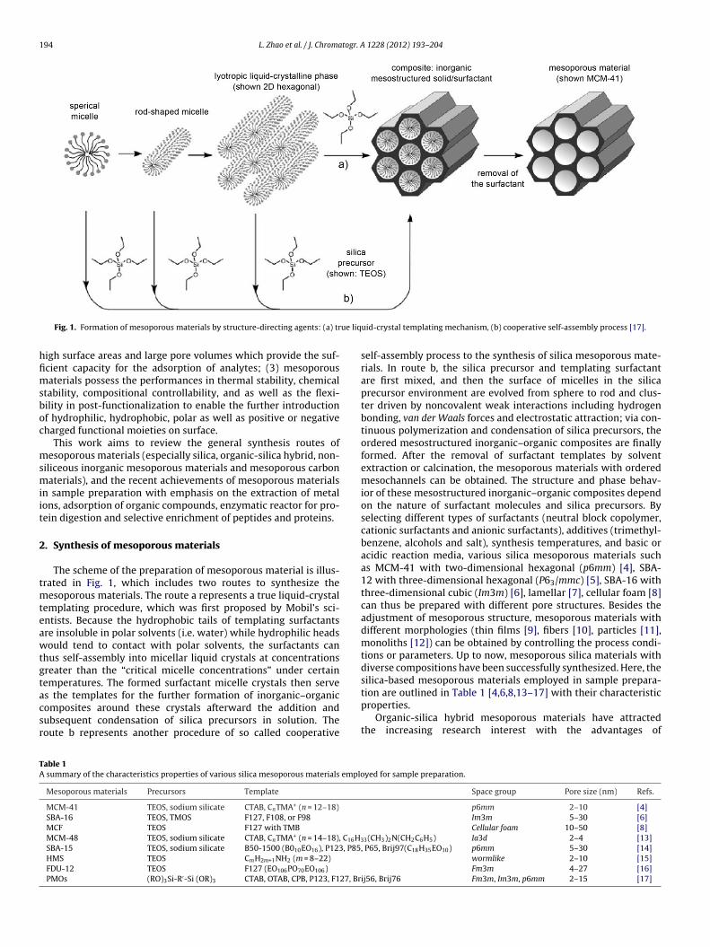
194 L. Zhao et al. / J. Chromatogr. A 1228 (2012) 193– 204
ue liq
hfimsboc
msmiit
2
tmteawtgtacsr
TA
Fig. 1. Formation of mesoporous materials by structure-directing agents: (a) tr
igh surface areas and large pore volumes which provide the suf-cient capacity for the adsorption of analytes; (3) mesoporousaterials possess the performances in thermal stability, chemical
tability, compositional controllability, and as well as the flexi-ility in post-functionalization to enable the further introductionf hydrophilic, hydrophobic, polar as well as positive or negativeharged functional moieties on surface.
This work aims to review the general synthesis routes ofesoporous materials (especially silica, organic-silica hybrid, non-
iliceous inorganic mesoporous materials and mesoporous carbonaterials), and the recent achievements of mesoporous materials
n sample preparation with emphasis on the extraction of metalons, adsorption of organic compounds, enzymatic reactor for pro-ein digestion and selective enrichment of peptides and proteins.
. Synthesis of mesoporous materials
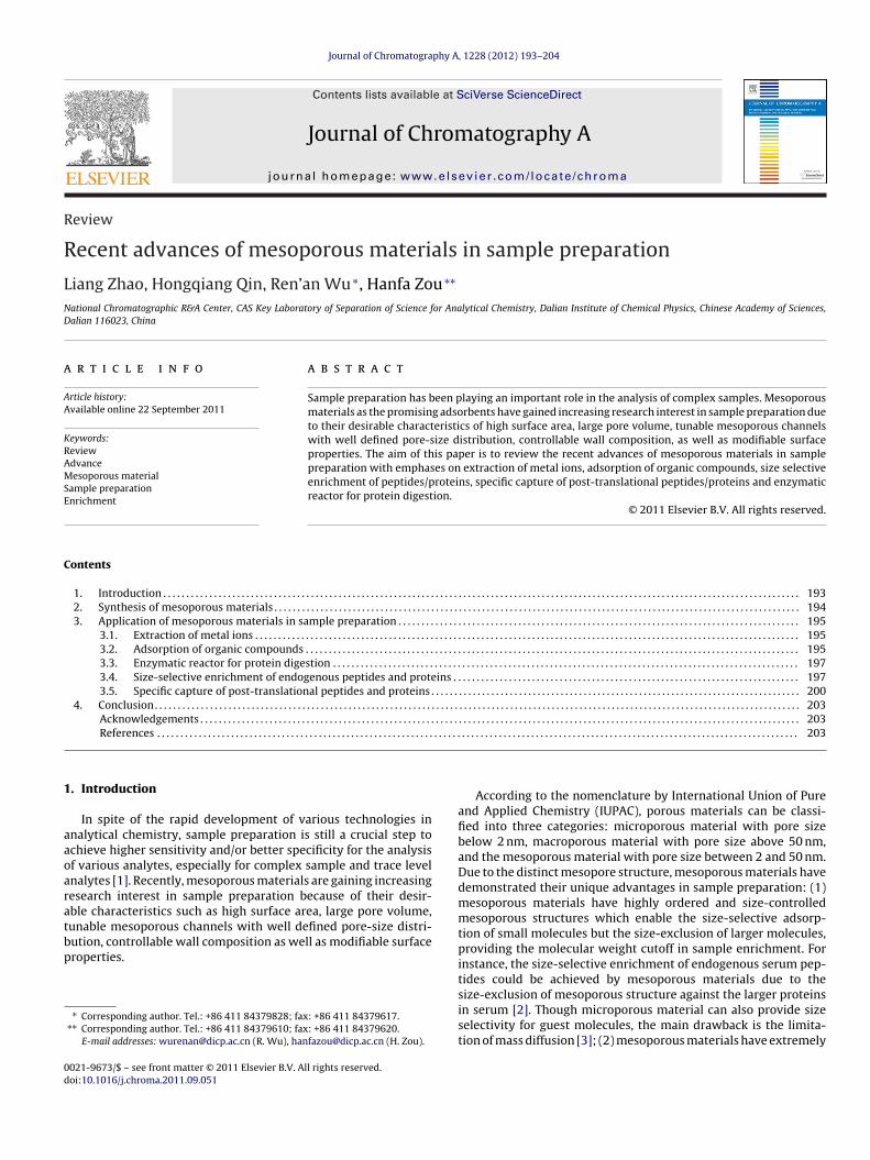
The scheme of the preparation of mesoporous material is illus-rated in Fig. 1, which includes two routes to synthesize the
esoporous materials. The route a represents a true liquid-crystalemplating procedure, which was first proposed by Mobil’s sci-ntists. Because the hydrophobic tails of templating surfactantsre insoluble in polar solvents (i.e. water) while hydrophilic headsould tend to contact with polar solvents, the surfactants can
hus self-assembly into micellar liquid crystals at concentrationsreater than the “critical micelle concentrations” under certainemperatures. The formed surfactant micelle crystals then serve
s the templates for the further formation of inorganic–organicomposites around these crystals afterward the addition andubsequent condensation of silica precursors in solution. Theoute b represents another procedure of so called cooperativeable 1 summary of the characteristics properties of various silica mesoporous materials emplo
Mesoporous materials Precursors Template
MCM-41 TEOS, sodium silicate CTAB, CnTMA+ (n = 12–18)
SBA-16 TEOS, TMOS F127, F108, or F98
MCF TEOS F127 with TMB
MCM-48 TEOS, sodium silicate CTAB, CnTMA+ (n = 14–18), C16H3
SBA-15 TEOS, sodium silicate B50-1500 (B010EO16), P123, P85HMS TEOS CmH2m+1NH2 (m = 8–22)
FDU-12 TEOS F127 (EO106PO70EO106)
PMOs (RO)3Si-R′-Si (OR)3 CTAB, OTAB, CPB, P123, F127, Br
uid-crystal templating mechanism, (b) cooperative self-assembly process [17].
self-assembly process to the synthesis of silica mesoporous mate-rials. In route b, the silica precursor and templating surfactantare first mixed, and then the surface of micelles in the silicaprecursor environment are evolved from sphere to rod and clus-ter driven by noncovalent weak interactions including hydrogenbonding, van der Waals forces and electrostatic attraction; via con-tinuous polymerization and condensation of silica precursors, theordered mesostructured inorganic–organic composites are finallyformed. After the removal of surfactant templates by solventextraction or calcination, the mesoporous materials with orderedmesochannels can be obtained. The structure and phase behav-ior of these mesostructured inorganic–organic composites dependon the nature of surfactant molecules and silica precursors. Byselecting different types of surfactants (neutral block copolymer,cationic surfactants and anionic surfactants), additives (trimethyl-benzene, alcohols and salt), synthesis temperatures, and basic oracidic reaction media, various silica mesoporous materials suchas MCM-41 with two-dimensional hexagonal (p6mm) [4], SBA-12 with three-dimensional hexagonal (P63/mmc) [5], SBA-16 withthree-dimensional cubic (Im3m) [6], lamellar [7], cellular foam [8]can thus be prepared with different pore structures. Besides theadjustment of mesoporous structure, mesoporous materials withdifferent morphologies (thin films [9], fibers [10], particles [11],monoliths [12]) can be obtained by controlling the process condi-tions or parameters. Up to now, mesoporous silica materials withdiverse compositions have been successfully synthesized. Here, thesilica-based mesoporous materials employed in sample prepara-
tion are outlined in Table 1 [4,6,8,13–17] with their characteristicproperties.Organic-silica hybrid mesoporous materials have attractedthe increasing research interest with the advantages of
yed for sample preparation.
Space group Pore size (nm) Refs.
p6mm 2–10 [4]Im3m 5–30 [6]Cellular foam 10–50 [8]
3(CH3)2N(CH2C6H5) Ia3d 2–4 [13], P65, Brij97(C18H35EO10) p6mm 5–30 [14]
wormlike 2–10 [15]Fm3m 4–27 [16]
ij56, Brij76 Fm3m, Im3m, p6mm 2–15 [17]

L. Zhao et al. / J. Chromatogr. A 1228 (2012) 193– 204 195
F CM-
cmsuopscmfs
saassiscsih4amsHp
mds
3p
3
patdtat[e
ig. 2. Synthetic scheme: (a) preparation of (3-aminopropyl) triethoxysilane (APS)-M
ompositing functional organic groups in silica. There are threeajor approaches to incorporate organic moieties into silica
ubstrates [17]: (i) grafting organic groups onto pore surfacesing organosilanes, (ii) co-condensation of silica precursors andrganosilanes, (iii) condensations of organic bridged silylatedrecursors. Via the aforementioned approaches, organic groupsuch as C–C multiple bonds, thiols, sulfonic and carboxylic acids,yano, amine or aromatic groups have been incorporated intoesoporous materials [17,18]. Due to improved mechanical per-
ormance and thermal stability, the hybrid mesoporous materialshould be promising in sample preparation.
Rather than the silica-based mesoporous materials, non-iliceous inorganic mesoporous materials (such as metal oxidesnd pure metal) and mesoporous carbon materials have also beenpplied in separation science. There are usually two approaches toynthesize these materials. One is self-assembling approach usingoft-templates, similar to the synthesis of silica based materials,norganic precursors (metal alkoxides or chlorides, carbon precur-ors) are usually used for self-assembly with amphiphilic blockopolymer templates via chemical interactions including electro-tatic attraction, hydrogen bonding, and hydrophobic/hydrophilicnteractions [19]; another is using nanocasting strategy that takesighly ordered silica mesoporous materials (such as SBA-15, MCM-8) as ‘hard template’ and the inorganic precursors as the fillinggents to fill up the mesochannels of ‘hard template’ to cast theesostructured non-siliceous inorganic materials. Various non-
iliceous inorganic mesoporous materials including ZrO2, TiO2,fO2, Gr2O3, Au, Pd, CMK-1 and CMK-3 have been successfullyrepared by these methods [18,20–22].
Since extensive expatiations on the development of mesoporousaterials have been reviewed elsewhere [17–20,23], we hereafter
iscuss the application of mesoporous materials with emphasis onample preparation.
. Application of mesoporous materials in samplereparation
.1. Extraction of metal ions
Hazardous metals can get into food chains through variousathways with the consequence of causing fatal diseases as theccumulated metal ions in organs up to their certain limita-ions [24]. Various technologies have been developed for theetermination of hazardous metals in polluted area, such ashe spectrophotometry [25], electrochemical analysis [26], atomic
dsorption spectrometry (AAS) [27], electrothermal vaporiza-ion inductively coupled plasma mass spectrometry (ETV-ICP-MS)28], inductively coupled plasma optical emission spectrom-try/mass spectrometry (ICP-OES/MS) [29,30], and inductively41 and (b) preparation of 2,4-dihydroxybenzaldehyde (4-OHsal)/APS-MCM-41 [29].
coupled plasma atomic emission spectrometer (ICP-AES) [31].However, due to the complexity of sample matrix and the tracelevel of target metal ion in sample, it is often difficult to determinemetal ions by these techniques directly. The effective extractionof hazardous metal ions from complex sample matrices is thusnecessary. The solid phase extraction (SPE) is a widely appliedtechnique with advantages of fast and simple operation, highenrichment performance, easy automation, cost effective etc. Thedistinctive characteristics of mesoporous materials (large surfacearea and pore volume) makes ordered mesoporous materials theideal adsorbents for SPE.
2,4-Dihydroxybenzaldehyde functionalized mesoporous silica(MCM-41) has been applied as an adsorbent for the extraction ofberyllium ion (Be2+) from aqueous solution, followed by the detec-tion of ICP-OES (as shown in Fig. 2) [29]. Under the optimizedcondition, the functionalized MCM-41 performed as a bidentateunivalent anionic ligand to form chelate complex with Be2+. Thisfunctionalized MCM-41 adsorbent had the ability to selectivelyenrich Be2+ from a mixed metal ion solution containing interferen-tial metal ions of Cu2+, Zn2+, Co3+ with their concentrations 10,000times higher than that of Be2+. The maximum adsorption capacityof 34 mg g−1 could be achieved with a good reusability. Comparedto other preconcentration methods (such as conventional SPE, pre-cipitation and micelle-mediated), the method of using mesoporousmaterials demonstrated much lower LOD (0.3 ng L−1). Besides, avariety of organic function groups have been introduced on thesurface of mesoporous materials for the capture of different metalions. Ordered mesoporous Al2O3 materials or mesoporous titaniumfilm have also been used as capillary microextraction coating for thepreconcentration of trace metal ions. Table 2 summarizes the pre-concentration of metal ions by various functionalized mesoporousmaterials [26,30,32–38].
In addition, the ionic imprinting technique is an effectiveapproach for the selective extraction of target analytes, which cangenerate plenty of specific recognition sites on mesoporous poly-mer materials via polymerization of cross-linking and functionalmonomers in the presence of the target analyte. For instance, Hoaiet al. [39] has prepared copper (II) ion-imprinted mesoporous poly-mer materials for the extraction of Cu (II) ions. The results showedthat the Cu (II) imprinted material had a higher affinity to copperion rather than other metal ions, such as Ni, Zn. In comparison withcommercial materials, the copper (II) ion-imprinted mesoporouspolymer material demonstrated at least 10 times higher selectivity.
3.2. Adsorption of organic compounds
Volatile organic compounds (VOCs) are carbon-based chemicalsin atmospheric environments. Many VOCs are demonstrated to beeither toxic or even carcinogenic [40]. Since the concentrations of

196 L. Zhao et al. / J. Chromatogr. A 1228 (2012) 193– 204
Table 2A summary of the functionalized mesoporous materials employed for preconcentration of metal ion.
Mesoporous materials Functional groups Metal ion Adsorption capacity Detection limit Detection methods Refs.
Mesoporous silica Tetraacetamide derivative of cyclam Pb2+ 2.7 × 10⁻⁹ M Electroanalysis [26]Mesoporous silica 3-Aminopropyl Cr5+ 4.35 mmol g−1 1.2 pg mL−1 AAS [27]Mesoporous TiO2 V, Cr and Cu 1.1–6.3 pg mL−1 ETV-ICP-MS [28]MCM-41 2,4-Dihydroxybenzaldehyde Be2+ 34 mg g−1 0.3 pg mL−1 ICP-OES [29]Mesoporous Al2O3 As3+, Cr3+, As5+, Cr5+ 0.7–74 pg mL−1 ICP-MS [30]MCM-41 Thiophene-2-carbaldehyde Pd2+ 5.0 mg g−1 0.2 ng mL−1 ICP-AES [31]SBA-15 5-Mercapto-1-methyltetrazole Zn2+ 0.96 ± 0.01 mmol g−1 8.0 × 10⁻⁹ M FAAS [32]SBA-15 2-Mercaptopyrimidine Cd2+ 0.99 ± 0.03 mmol g−1 FAAS [33]Mesoporous silica Chitosan V, Cu, Pb, Cd and Hg 12.2–22.9 mg g−1 0.05–0.96 ng mL−1 ICP-OES [34]MCM-41 5-Nitro-2-furaldehyde U5+, Th4+ 47–49 mg g−1 0.3 ng mL−1 ICP-OES [35]Mesoporous TiO2 Dimercaptosuccinic acid As3+, Sb3+, As5+, Sb5+ 0.10–0.15 ng mL−1 ICP-OES [36]
Vubsm(egCbcvdctopttpa
pusfmc1(ht
rimAthsmmdaTf(tdi
detected by circular dichroism and optical polarimetry [47]. Withthe three dimensional imprinted chiral mesostructures, CMPPynanoparticles display remarkable chiral selectivity and selectiveadsorption in the mesochannels. Using these chiral nanoparticles,
Mesoporous silica 3-(2-Aminoethylami-no) propyl As3+,As5+
SBA-15 Ethylenediamine Cd2+, Pb2+
OCs in air are at ppb or sub-ppb (v/v) levels, which are usuallynder the detection limits of modern gas chromatography (GC)ased methods, the pre-concentration is generally of a necessarytep to match the detection limits of applied analytical instru-ents. Based on hydrophobic interaction, a mesoporous material
MCM-41) with the pore size of 2.9 nm was applied in the in-linenrichment of VOCs (using C2–C12 as a standard gas mixture) foras chromatography coupled with flame ionization detection [41].ompared to commercial available carbon molecular sieves (car-on adsorbents) for capturing C3–C12, MCM-41 material exhibitsomparable adsorption ability for C8–C12 other than for the highlyolatile smaller compounds (C3–C7). In addition, the MCM-41epicts the lower minimum desorption temperature (150 ◦C) asompared to that of carbon adsorbents (300 ◦C). By adjusting theemperature to sub-ambient temperatures (−20 ◦C), a full rangef VOCs from C4–C12 could be efficiently trapped by MCM-48 withore size of 3.7 nm at a higher recovery. The highly efficient adsorp-ion of VOCs at sub-ambient temperatures, the lower desorptionemperature as well as the low memory effect have made meso-orous materials the promising in-line enrichment media for VOCsnalysis by gas chromatography [42].
Powdered activated carbons (ACs) with an important meso-orous volume and distinct surface chemistry characteristics aresed as adsorbent phases to purify triazinic herbicides (atrazine,imazine and terbutylazine) in environmental water matrices,ollowed by liquid desorption and high performance liquid chro-
atography with diode array detection [43]. Under optimizedonditions (extraction time, pH, ionic strength), recovery around00%, detection limits about 0.1 �g L−1, and suitable linearity1.0–12.0 �g L−1) have been achieved. This proposed methodologyas been proved to be a suitable way to monitor traces levels ofhese three herbicides in water matrices.
Likewise the molecular imprinting technology for the prepa-ation of the molecularly imprinted polymers (MIPs), a surfacemprinting method has also been applied in the synthesis of
esoporous material for the selective analysis of target analytes.s shown in Fig. 3, some organic moieties can be incorporated into
he skeleton of mesoporous material via the organic–inorganicybrid sol–gel process by using the certain organic groups bridgedilsesquioxanes. Because of the molecular affinity of the organicoieties on the skeleton of organic–inorganic hybrid mesoporousaterials, these surface imprinted mesoporous materials have
emonstrated the enhanced selectivity and sensitivity to the targetnalytes, which are analogous to the imprinted organic moieties.rammell et al. [44] realized the use of nanoporous organosilicasor the rapid preconcentration and extraction of trinitrotoluene
TNT) for electrochemical analysis and demonstrate the effect ofemplate-directed molecular imprinting on TNT adsorption. Theyeveloped two types of TNT imprinted nanoporous organosil-cas for the preconcentration of TNT prior to electrochemical
10.3 mg g−1 0.05 ng mL−1 ICP-OES [37]360 ± 1.4100.0 ± 0.6 mg g−1 AAS [38]
analysis. One is benzene (BENZ)-imprinted hybridorganic–inorganic having high BET surface area (980 m2/g)and large pore volume (0.714 cm3/g) with highly ordered meso-pore structure; another is the diethylbenzene (DEB)-imprintedhybrid organic–inorganic one possessing amorphous pore struc-ture with BET surface area of 35 m2/g and small pore volumeof 0.0331 cm3/g. In a comparison, the DEB-imprinted materialwas 7 times more efficient than the nonimprinted one in thecapture of TNT from aqueous contaminated soil samples. Inaddition, the limit of detection of the BENZ-bridged mesoporousmaterials (30 ppb) is much lower than that of the DEB-bridgedadsorbents (90 ppb) in the enrichment of TNT from 500 �L sample.This surface imprinting technique could be adopted to analyzeother target analytes of interest by using corresponding organiccompounds (diethylbenzene, porphyrins analog) as bridge moietyin the synthesis of hybrid inorganic–organic mesoporous materials[45,46].
Spherical chiral mesoporous polypyrrole (CMPPy) nanoparti-cles have been synthesized by incorporating chirality into themesochannels and applied to recognize chiral molecules, which are
Fig. 3. General synthetic pathway to surface imprinted hybrid mesoporous materi-als that are constructed from bissilylated organic bridging units. R = organic bridge,which has affinity for the analytes analog to the organic moieties [17].

L. Zhao et al. / J. Chromatogr. A
Fso
pscc
3
bb“bestesc
sigittctmwhacaibctp2oaiuit
st
ig. 4. Diagram of protein adsorption in mesoporous materials with different poreizes: the matched pore size (middle); while, the larger pore size (left) or smallerne (right) [48].
ure chiral molecules (l-valine) from a racemic mixture could betereoselectively recognized and separated. In this way, varioushiral materials can be synthesized in this simple, controllable andost effective way.
.3. Enzymatic reactor for protein digestion
Proteomics have attracted much attention in recent yearsecause of its ability to elucidate the key roles of proteins iniological process and discover potential biomarkers [1,47]. Theshot-gun” proteomics requires protein digestion (in most casesy trypsin) before mass spectrometry analysis. The conventionalnzymatic digestion is in free solution, which often suffers from theerious interference of the autodigestion of enzymes. Protein diges-ion by the immobilized enzyme has been considered a promisingnzymatic digestion technology due to the advantages in enzymetability, digestion efficiency, enzyme recycling and free of productontamination.
For enzyme reactor, it is important to choose the proper poreize and spatial structures, as well as the surface properties includ-ng hydrophilicity, hydrophobicity, charge status, different organicroups etc, since these factors may affect the activity and stability ofncluded enzyme. As shown in Fig. 4 [48], if the pore size is smallerhan the dimensions of enzyme, the enzyme would adsorb ontohe external surface, resulting in low stability and low adsorptionapacity; when the pore size of mesoporous is sufficiently larger forhe entrapment of biomolecules, enzyme would penetrate into the
esochannels rather than adsorb onto the outer surface. If enzymesere reversibly immobilized by weak physical adsorptions, such asydrogen bonding, hydrophobic attraction or electrostatic inter-ction, the adsorbed enzymes would leach out the mesoporoushannels. By covalently binding enzymes on reactive groups such asldehyde, epoxide and thiol groups, or by cross-linking the enzymesnside the mesoporous channels, the immobilization stability coulde greatly enhanced and the covalent or cross-linked enzymesan be recycled for the repeat usage. Hyeon et al. reported thathe immobilized trypsin remained its 89% initial activity in meso-orous materials by cross-linking with glutaraldehyde even after9 iterative cycles of enzyme reaction [49]. Besides, the balancef the hydrophobic and hydrophilic interaction between enzymend material is also known to impact the stability and activity ofmmobilized enzyme [48,50]. Rotello et al. reported that the denat-ration extent of chymotrypsin could be considerably reduced by
ntroducing of hydrophilic ethylene glycol as space arm between
he surface and the immobilized enzyme [51,52].To achieve the rapid digestion, large sequence coverage and highensitivity for broad range proteins with diverse physical proper-ies, Shui et al. [53] developed a novel proteolytic nanoreactor using
1228 (2012) 193– 204 197
a mesoporous silica of FDU-12 with a unique three-dimensionalnanopore structure. This nanoreactor integrated substrate enrich-ment, “reagent-free” protein denaturation, and efficient proteolyticdigestion in the mesoporous space of FDU-12. In this design, pro-tein substrates were first captured by the mesoporous materialof FDU-12 and then were concentrated from solution. Followingby pH change and applying trypsin, the denaturation and con-current proteolysis of broad-range proteins could be efficientlyachieved, resulting in the identifications of a broad range of diverseproteins with high sequence coverage by mass spectrometry. Sim-ilarly, trypsin immobilization on SBA-15 or amine and cyanofunctionalized SBA-15 materials have also been applied for pro-tein proteolysis [54–56]. Casadonte et al. [54] found that the aminefunctionalized SBA-15 showed higher digestion efficiency thanunmodified SBA-15 with the same pore diameters. Also, the pro-teolytic efficiency of functionalized SBA-15 was 1000 times fasterthan that of using conventional free solution method. Interestingly,the digestion performance could be enhanced by increasing thelength of the amine group, which seemed to attribute to the sta-bilization effect of the longer amine group to enzyme. Though theadsorption of enzyme on material via electrostatic interaction issimple, the risk of enzyme leaching out is still a concern.
The analysis of low-molecular-weight (LMW) proteins is oftenvery difficult, because the separation of LMW proteins from com-plex protein sample by various separation techniques is timeconsuming and would resulted in sample loss. Recently, a size-selective digestion of LMW proteins was developed by using amesoporous silica material [57]. As shown in Fig. 5, trypsin wascovalently immobilized on the thiol-modified SBA-15 materialand confined in mesochannels of SBA-15 with the pore size ofca. 5.7 nm. Due to the size-exclusion interaction of mesopores ofthe thiol-modified SBA-15 material to the big size protein of BSA(5.0 × 7.0 × 7.0 nm3), the material could exclude high-molecular-weight (HMW) protein of BSA but capture LMW proteins (<5.7 nm)of cytochrome C, lysozyme, myoglobin for giving the chance tocarry out the enzymatic digestion by the immobilized trypsin. Bythis size-selective digestion, the LMW proteins could be selectivelydigested by the immobilized trypsin but not the HMW proteins.
3.4. Size-selective enrichment of endogenous peptides andproteins
Peptidomics, referring to all of LMW peptides or proteins, isdefined as the systematic analysis of endogenous peptides andsmall proteins in biological sample (such as body fluids, celllysate, and tissue extract) at a defined time points and/or loca-tions [58–60]. Peptides from these biological samples may havethe potential to serve as biomarkers, indicating of progression froma normal to a diseased status [61]. Thus, peptidomics based onmass spectrometry technology has been established and emergedas a promising strategy to characterize the native peptides orsmall proteins, resulting in comprehensive understanding of nativepeptides/proteins in biological processes. However, due to the com-plexity and high dynamic range of endogenous peptides and theinterference from high concentrations of proteins, salts and lipidsin biological samples, the ability to extract peptides/small pro-teins from the complex biological samples as well as enhancingthe sensitivity toward peptides/small proteins at low abundance,remains a great challenging task before MS analysis. Thus, variousmethods (ultrafiltration [62], solid-phases extraction [63], selec-tive electrophoresis and continuous elution electrophoresis [64],organic solvent precipitation [65]) have been developed to isolate
peptides/small proteins before MS analysis. Centrifugal ultrafil-tration with accurate MW cutoff is one widely used method forpeptide enrichment based on size-exclusion effect. Unfortunately,it would take a long time to operate and co-concentrate some small
198 L. Zhao et al. / J. Chromatogr. A 1228 (2012) 193– 204
Cyt c,
mcl
mbpsbtethwp
oRmsomcssiTfcihe2pii
Fig. 5. Schematic procedure of size-selective proteolysis of small proteins (
olecule contaminants (such as salt) if a huge amount of biologi-al sample is applied, resulting in low enrichment efficiency, whichimited its practical application [62].
Mesoporous materials are attractive candidates for the enrich-ent of peptides. The efficiency of size-dependent separation
ased on mesoporous materials is determined by the relation ofore size to the diameter of target molecules. Biomolecules whoseizes are smaller than the pore size of mesoporous material cane captured in the pore channel, while biomolecules larger thanhe pore size are excluded by the pore channel. Besides the sizeffect, hydrophobic and electrostatic interactions between pep-ides/proteins and materials along with other factors including 3Dydrodynamic dimensions of the protein, hydrogen bonding withater may also affect the adsorption of biomolecules on meso-orous materials [66].
Various functionalized mesoporous materials have been devel-ped for the enrichment of peptides from biological samples.ecently, Tian et al. [2] developed highly ordered mesoporousaterials possessing different pore size (2, 8, 12 nm) to adsorb
tandard protein lysozyme, and found MCM-41 with the pore sizef 2 nm could be used to selectively and effectively enrich low-olecular-weight peptides/proteins from human plasma with a
utoff of 12 kDa, based on the combined effect of hydrophobic andize-exclusion interactions. The typical MALDI-TOF MS results arehown in Fig. 6. The unique property of mesoporous material madet superior adsorbent for enrichment of peptides or small proteins.o improve the enrichment efficiency and capacity, Tian et al. [60]urther synthesized different functionalized MCM-41 with strongation-exchange and strong anion-exchange properties. As shownn Fig. 7, by combination of anion-exchange, cation-exchange andydrophobic mechanisms, endogenous peptides from mouse liverxtract were selectively enriched and consequently analyzed by
D nano-LC/MS/MS. Because the hydrophobic interaction betweeneptides and mesoporous materials can be enhanced by introduc-ng organic groups (–CH2–) on the walls of mesoporous channelsn the synthesis of periodic mesoporous organosilica (PMO)
lysozyme or myoglobin) from a high-molecular-weight protein (BSA) [57].
materials, Wan et al. [67] reported amino group (NH2) function-alized PMO materials (NH2-PMO) with an organo-bridged (–CH2–)hybrid wall composition. Due to the charge status, PMO (−25.2 mV)and NH2-PMO (+39.0 mV) can enrich positively charged and nega-tively charged peptides, respectively. As a result, 69.4% (25 of 36)of peptides with pI ≥ 6 were enriched by PMO and 80% (21 of 28) ofpeptides with pI ≤ 6 were captured by NH2-PMO. Thus, more com-prehensive and complementary information can be collected bycombination of these two types of functionalized PMO materials.
Hu et al. [68] synthesized mesoporous silica (MPS) thin filmwith various pore size distribution, pore structure, and connec-tivity. The effect of the pore structure of mesoporous silica on theenrichment efficiency and specificity of low molecular-weight fromhuman plasma was investigated. It was observed that because ofthe increased pore connectivity and the reduced steric hindrance,the mesoporous silica with 3D cubic nanostructure or 3D honey-comb hexagonal nanostructure exhibited superior performance incapture of LMW peptides than the MPS with 2D hexagonal nano-structure, in despite of similar pore size distribution and the samemass cutoff. Nevertheless, with the same 3D nanostructure of MPS,the recovery of LMW proteins would also be affected by pore size(3, 4, 6 and 7 nm) of MPS.
In order to facilitate the enrichment process, magnetic meso-porous silica mesoporous materials have been also developed forthe magnetic separation. As shown in Fig. 8, Chen et al. [69] reporteda facile and low-cost way to synthesize Fe3O4@nSiO2@mSiO2microspheres, which were applied in the selective enrichmentof low-concentration peptides. By combining the magnetic prop-erty and mesoporous silica shell structure, the process of peptideenrichment is convenient, fast and efficient, which has beendemonstrated in the enrichment of endogenous peptides fromrat brain extract based on the hydrophobic interaction between
hydrophobic peptides and siloxane bridge groups on inner wallof mesoporous channels. Since the prepared Fe3O4@nSiO2@mSiO2microspheres could only be used to capture hydrophobic peptidesrather than hydrophilic peptides, Cu2+ functionalized magnetic
L. Zhao et al. / J. Chromatogr. A 1228 (2012) 193– 204 199
Fig. 6. MALDI-TOF MS analysis of human plasma (a, c) after and (b, d) before treated by MCM-41 with a-cyano-4-hydroxycinnamic acid as MALDI matrix. Analysis in themolecular-weight range of (a, b) 1–15 kDa and (c, d) 10–100 kDa [2].
Fig. 7. Schematic overview of (a) the preparation of SCX-MCM-41 and SAX-MCM-41 materials; (b) the work flow for endogenous peptide enrichment and identification; theendogenous peptides from mouse liver are enriched by three kinds of mesoporous materials before analysis with 2D nano-LC/MS/MS [60].

200 L. Zhao et al. / J. Chromatogr. A 1228 (2012) 193– 204
F atic iu
mneaiha
ashrntRnsCsIteiroit[[mad
3
tsp
ig. 8. (a) The synthesis route of Fe3O4@nSiO2@mSiO2 microspheres and (b) schemsing Fe3O4@nSiO2@mSiO2 microspheres [69].
esoporous silica microspheres (Fe3O4@mSiO2–Cu2+) with mag-etic core and mesoporous silica shell were developed for thenrichment of hydrophilic peptide via the immobilized metal ionffinity chromatography (IMAC) [70]. The enrichment mechanisms that the large amount of immobilized Cu2+ could interact withydrophilic peptides via the coordination between copper (II) ionnd carboxylic and amino groups of peptides.
By mimicking biological membrane, mesoporous materials arelso used to size selective separation of biological macromolecules,uch as proteins. The efficiency of size-based is dependent on theydrodynamic diameter of proteins in a given electrolyte. It iseported that proteins can be passed through the mesoporous chan-els with the pore diameters as approximately three times largerhan that of target proteins [71]. With the anodization process,oy et al. [66] prepared a TiO2 membrane with defined mesochan-els (average pore size of 8-12 nm) for the size selective proteineparation. Three proteins with different Stokes radii [cytochrome
(1.63 nm), BSA (3.62 nm) and �-galactosidase (6.86 nm)] areelected to evaluate the efficiency of size-dependent separation.t was found that cytochrome C and BSA could pass throughhe mesochannels while the large molecule �-galactosidase arexcluded. Clogging of mesochannels is a sticky problem occurringn all size separation devices if the pore opening is in the sizeange of the smallest excluded proteins. Therefore, a main meritf these TiO2 membranes is their photocatalytic activity, whichs able to effective declogging of these membranes when appliedo separate proteins. Mesoporous silica coated carbon nanotubes72], mesoporous polymer coated copper hydroxide nanostrands73], magnetic silica nanospheres with highly ordered periodic
esostructure [74] and lysozyme imprinted polymer beads [75]re also synthesized for the selective separation of proteins withifferent molecular sizes.
.5. Specific capture of post-translational peptides and proteins
Protein phosphorylation is one of the most important post-ranslational modification involved in many biological processes,uch as cellular growth, division and signaling [76,77]. Aberranthosphorylation has been known to be related to underlying
llustration of fast and convenient enrichment protocol for endogenous peptide by
many human diseases, especially human cancer [78]. To achievedetailed understand on the reversible phosphorylation-controlledbiological processes, it is therefore necessary to characterize thephosphorylated peptides and their sites. Mass spectrometry basedmethods has been proved to be a powerful tool for the analysisof phosphorylated peptides [79,80]. However, because of the lowabundance of phosphorylated peptides, interference from exces-sive amounts of nonphosphorylated peptides and the low efficiencyin ionization of negative charged phosphorylated peptides, it isstill a great challenge on the large scale analysis of phosphorylatedpeptides. Therefore, it demands to effectively isolate and enrichphosphorylated peptides or proteins from biological samples priorto MS analysis.
One of the most widely employed methods to enrich phos-phopeptides is affinity-based chromatography, which is dividedinto two categories. The first one is metal oxide affinity chro-matography (MOAC) using titanium dioxide (TiO2) [81], zirconiumdioxide (ZrO2) [82], and ferric oxide (Fe2O3) [83]; the second one isimmobilized metal ion affinity chromatography (IMAC), which haschelating ligand (iminodiacetic acid (IDA) or nitrilotriacetic acid(NTA)) on chromatographic adsorbents for immobilizing metal ion(Fe3+ or Ga3+) [84,85].
For MOAC, metal oxides display amphoteric properties basedon unsatisfied valences. In acidic solution, metal oxides behaveas a Lewis acid with positively charged metal ions, which can bepreferentially and reversibly exchanged with negatively chargedmolecules (such as phosphate, carboxylate, sulfate groups); whilein basic solutions, the negative charged molecules can be elutedfrom the adsorbents. Mesoporous metal oxides with well orderedporosity are stable over a wide pH range, and provide many activesites for binding phosphate groups, which may be translated intoeven larger binding capacity than micro or nano metal oxides. Thus,mesoporous metal oxides may have more superior performance inphosphoproteomics.
Tang et al. [86] reported that mesoporous TiO2 microspheres
were applied as potential MOAC adsorbents for purification ofphosphopeptides. The surface area of mesoporous TiO2 micro-spheres (84.98 m2/g) with the diameter of about 1.0 �m is almosttwo times of commercial TiO2 nanoparticles (a diameter of 90 nm)
togr. A 1228 (2012) 193– 204 201
a(TtneF4FpuntadacTfifiIpfisrmpatrpscla
dsmaaiswTrmisFhpp
csbt
pbpmsc
Fig. 9. (a) 3D view of the MALDI-TOF-MS profiling of phosphopeptides enrichedfrom sera of hepatocellular carcinoma cancer (HCC) patients and healthy persons.
L. Zhao et al. / J. Chroma
nd is much larger than that of smooth TiO2 microspheres3.35 m2/g), so these superior properties provide mesoporousiO2 microspheres with greater binding capacity and higher cap-ure efficiency than that of smooth TiO2 microspheres and TiO2anoparticles in phosphoproteome analysis. As an adsorbent fornrichment of phosphopeptides, Han et al. [83] found mesoporouse2O3 microspheres with an averaged inter-particle pore size of8 nm can eliminate the shadow effect, because mesoporous ofe2O3 microspheres are large enough to freely release trappedeptides (<2 nm) from the inner-pore surface to the outer surfacender laser irradiation. Lu et al. [87] synthesized mesoporous TiO2anocrystal clusters with modification of hydrophilic and nega-ive outer surface, three dimensional pores, submicrometer sizend high ratio of surface area to volume, making the clusters moreispersible in water solution and can enhance the adsorbent moreccessible to the phosphopeptides, resulting in much larger bindingapacity, higher selectivity and higher sensitivity than that of solidiO2 spheres using � or �-casein as model protein. Nelson et al. [88]rst reported mesoporous ZrO2 metal oxides showed higher speci-city and efficiency for phosphopeptides than that of commercial
MAC and ZrO2 nanoparticles packed tip, and then evaluated theerformance among mesoporous TiO2, HfO2 and ZrO2 metal oxidesor enrichment of phosphopeptides from tryptic digests of �-caseinn another report [89]. The results showed that the enrichmentpecificity (>99%) using mesoporous HfO2 and ZrO2 is fairly compa-able, and is higher than that of using mesoporous TiO2; moreover,esoporous TiO2 and HfO2 tend to effectively enrich multiple
hosphorylated peptides while mesoporous TiO2, HfO2 and ZrO2ppeared to be similar affinity toward mono phosphorylated pep-ides in general; interestingly, mesoporous HfO2 and ZrO2 can beepeatedly used in the enrichment of phosphopeptides with com-arable performance to that of freshly prepared materials after aimple solution regeneration procedure. Similarly, zirconium layeroated mesoporous silica microspheres (MCF) [90] or titaniumayer coated magnetic hollow mesoporous silica microspheres [91]re also applied to enrich phosphopeptides from standard proteins.
However, sample loss during various steps of enrichment proce-ure (centrifugation or elution) is unavoidable. In order to reduceample loss and facilitate the sample preparation in the enrich-ent procedure, on-target and magnetic enrichment strategies are
lso taken. MALDI plate was modified by mesoporous anatase TiO2nd employed to analyze phosphopeptides [92], the enrichments directly operated on the modified MALDI plate and the non-pecifically peptides unbound to the plate can be easily removedith washing buffers. Using this strategy, mesoporous anatase
iO2 spots can be able to enrich very low and substoichiomet-ic amounts (≈3 fmol) of phosphopeptides; a thin stripe made ofesoporous TiO2 sintered onto a conductive glass surface, which
s analogous to thin layer chromatography, was used to enrich andeparate multi- and monophosphorylated peptides [93]. Moreover,e3O4@SiO2@mCeO2 with a magnetic core and mesoporous shellave also been applied to simultaneously enrich and label phos-hopeptides (dephosphorylation) due to its affinity and catalysisroperties [94].
The mesoporous metal oxides have indeed shown strong appli-ation in efficient enrichment of phosphopeptides. However, largecale analysis of phosphopeptides may not be achieved by MOAC-ased methods because of the presence of steric hindrance and alsohe not so well biocompatibility inorganic metal oxides.
IMAC is the most well-known affinity method in phospho-roteome analysis, which is based on strong specific interactionetween metal ion on adsorbents and phosphate groups of
hosphopeptides. However, conventional IMAC adsorbents areodified with IDA or NTA as ligands to immobilize Ga3+ or Fe3+, andtill lack enough specificity to phosphopeptides due to the signifi-antly unselective co-enrichment of highly acidic peptides, which
(b) Partial least-squares discriminant analysis score plot showing the separationbetween the HCC cancer and healthy groups [96].
results in low selectivity and sensitivity for targeted phosphopep-tides. In our lab, Fe3+ and Zr4+ phosphate functionalized periodicmesoporous organosilicas with ordered 2D hexagonal mesostruc-tures, high specific surface area and large pore volume have beenapplied to enrich phosphopeptides, which showed higher intensi-ties and signal/noise ratios of the enriched phosphopeptides thanthat of commercial POROS 20 loaded with the same metal ion [22].Ti4+ incorporated hexagonal mesoporous silica (Ti-HMS) with rela-tive high Ti-content (2 and 8 mol%) are successfully synthesized inour lab for the enrichment of phosphopeptides [95]. It was foundthat the Ti-HMS with higher content (8%) of titanium could effec-tively enrich three phosphopeptides from �-casein; while, Ti-HMSwith lower content (2%) of titanium could only enrich one mul-tiple phosphorylated peptides due to the relatively low contentof titanium incorporated in the silica framework of Ti-HMS. Huet al. [96] also synthesized titanium phosphate modified highlyordered mesoporous silica microparticles and applied to enrichphosphopeptides from �-casein as well as human serum. Becauseof high surface area, large pore volume and ordered mesoporous ofthe synthesized material, the detection limit for phosphopeptidesenrichment from �-casein and standard phosphopeptide spikedhuman serum can be as low as 1.25 fmol analyzed by MALDI-TOFMS. Based on size-exclusion and adsorption effect by mesoporous
silica particles, four endogenous phosphopeptides were effectivelyenriched from human serum. By combination of the direct use ofMALDI profiling and isotope labeling, as shown in Fig. 9, it was
202 L. Zhao et al. / J. Chromatogr. A 1228 (2012) 193– 204
F nanocu OF M
fedrtb
raAsassnOdIT
ig. 10. Illustration of (a) the procedure for the fabrication of the mesoporous TiO2
sing the mesoporous TiO2 clusters and the elutes were analyzed by CE or MALDI-T
ound that four endogenous phosphopeptides were differentiallyxpressed between cancer and health persons, thus making theeveloped approach as a potential biomarker discovery for clinicalesearch. This approach is effective for the profiling of phosphopep-ides in complex biological samples and the discovery of biomarkerased on functional mesoporous materials.
The above reported techniques based on affinity chromatog-aphy are all used to enrich phosphopeptides at peptide levelnd indirectly provide information about phosphorylated protein.s shown in Fig. 10, Lu et al. [97] described the preparationelf-assembled TiO2 nanocrystal clusters with different aver-ged pore sizes of 2.3, 2.5 and 3.5 nm, and their application onelective enrichment of phosphorylated proteins from a mixedolution of three proteins (one phosphorylated �-casein, twoon-phosphorylated horseradish peroxidase and �-lactoglobin).
nly �-casein was detected by capillary electrophoresis with UVetection at 200 nm when using the mesoporous TiO2 clusters.n comparison, TiO2 solid spheres are failed to trap �-casein.hese results indicated that trapping of phosphorylated protein
Fig. 11. Postsynthetic steps (left) of ordered mesoporous di-boroni
rystal clusters and (b) the selective enrichment process of phosphorylated proteinsS [97].
not only depended on the affinity of TiO2 but also the mesoscalepores.
Protein glycosylation is also a most common post-translationalmodification and plays very important role in cell communication,signaling and cell adhesion [98]. However, comprehensive stud-ies on protein glycosylation have been complicated by the diversestructure of protein glycans. Though various strategies such aslectin affinity chromatography [99], hydrophilic interaction liq-uid chromatography [100], hydrazide chemistry [101] have beendeveloped, a sensitive, quick and applicable method for enrichmentof glycopeptides is still needed. As shown in Fig. 11, Xu et al. [102]synthesized a novel boronic acid functionalized mesoporous silicamaterial (FDU-12-GA). Because of its prominent features of highsurface area, large pore volume, and narrow distribution of regu-lar pore size, a high percentage (up to 32 wt%) of grafted organics
has been incorporated into the material. Glycopeptides with therecovery of 83.5% can be effectively and specifically enriched fromcomplex tryptic peptide mixture in 15 min, while the commercialboronic acid functionalized magnetic beads would take 60 min; byc acid functionalized FDU-12 (denoted as FDU-12-GA) [102].

togr. A
ttt
4
psahwpadfienct
atloadttbtpsioamnvwTolnYpigtosee
A
dGDH((n
L. Zhao et al. / J. Chroma
he elimination of the suppression effect from nonglycopeptides,he limit of detection for glycopeptides is greatly enhanced by closeo 2 orders of magnitude.
. Conclusion
Mesoporous materials are promising adsorbents in samplereparation due to their distinct physicochemical properties inurface area and pore structure as well as the versatile avail-bility in surface functionalization to provide the hydrophobic,ydrophilic, positively and negatively charged surface to interactith analytes. The combination of the size-exclusion effect of meso-ores against big size molecules and the adsorption of small sizenalytes inside the mesopores actually mimics the effect of multi-imensional chromatographic separation of integrating molecularltration (by tunable highly ordered mesopore) and solid-phasextraction (by internal hydrophobic, hydrophilic, positively andegatively charged inner surface, etc.) of analytes on-beads, whichan greatly simplify the sample preparation procedure by avoidinghe multi-step filtration, washing, extraction and elution.
Although mesoporous materials have demonstrated uniquedvantages in sample preparation, some issues associated withheir synthesis and application are yet to be resolved. First, theow stability of mesoporous silica materials under strong acidr basic conditions will inhibit their application as solid phasedsorbents at some extreme pH conditions. Second, the pore sizeistribution of mesoporous materials could not be exactly con-rolled as we designed. Third, post modification is an effective wayo endow mesoporous materials with various functional groups,ut this severe way only provides uneven functional groups onhe external/internal surface and may decrease or even block theorosity. Organic groups can be homogeneously introduced to theurface of mesoporous materials, but the mesostructural order-ng would be affected with the increase content of organosilanesr organic bridged silylated precursors, resulting in decrease ofdsorption amount. Fourth, conventional synthesized mesoporousaterials are usually very bulky, and the orientation of mesochan-
els run along the long axis, so the length of mesochannels isery long, it may take a long time to fully adsorb biomolecules,hich would be not favorable for the quick analytical requirement.
hus, ideal mesoporous adsorbents should possess high densityf functional groups, short and accessible mesoporosity, control-able morphology (such as 3D cubic nanostructure or 2D hexagonalanostructure), fast molecular diffusion and adsorption kinetics.et, mesoporous metal oxides, carbon or polymer based meso-orous materials with different properties require in-depth studies
n sample preparation, especially for biological samples such aslycans, peptides, lipids and other post-translational proteins. Fur-hermore, multifunctional mesoporous materials with magnetic,ptical, electronic properties and tailored morphology (such as poreize, mesoporous structure, wall compositions, and surface prop-rties) are also crucial factors in enhancement of selectivity andfficiency in sample preparation.
cknowledgements
This work was supported by the National Natural Science Foun-ation of China (Nos. 20735004, 20875089), the Creative Researchroup Project by NSFC (21021004), the State Key Basic Researchevelopment Program of China (No. 2007CB914102), the National
igh Technology Research and Development Program of ChinaNo. 2008AA02Z211), the Key Program of Knowledge InnovationKSCX2-YW-G-049) and the Hundred Talent Program of the Chi-ese Academy of Sciences.
1228 (2012) 193– 204 203
References
[1] N.L. Anderson, N.G. Anderson, Mol. Cell. Proteomics 1 (2002) 845.[2] R. Tian, H. Zhang, M. Ye, X. Jiang, L. Hu, X. Li, X. Bao, H. Zou, Angew. Chem. Int.
Ed. 46 (2007) 962.[3] Y. Tao, H. Kanoh, L. Abrams, K. Kaneko, Chem. Rev. 106 (2006) 896.[4] C.T. Kresge, M.E. Leonowicz, W.J. Roth, J.C. Vartuli, J.S. Beck, Nature 359 (1992)
710.[5] Q.S. Huo, D.I. Margolese, G.D. Stucky, Chem. Mater. 8 (1996) 1147.[6] D.Y. Zhao, Q.S. Huo, J.L. Feng, B.F. Chmelka, G.D. Stucky, J. Am. Chem. Soc. 120
(1998) 6024.[7] T. Kimura, T. Kamata, M. Fuziwara, Y. Takano, M. Kaneda, Y. Sakamoto, O.
Terasaki, Y. Sugahara, K. Kuroda, Angew. Chem. Int. Ed. 39 (2000) 3855.[8] P. Schmidt-Winkel, W.W. Lukens, D.Y. Zhao, P.D. Yang, B.F. Chmelka, G.D.
Stucky, J. Am. Chem. Soc. 121 (1999) 254.[9] D.Y. Zhao, P.D. Yang, D.I. Margolese, B.F. Chmelka, G.D. Stucky, Chem. Com-
mun. (1998) 2499.[10] F. Marlow, M.D. McGehee, D.Y. Zhao, B.F. Chmelka, G.D. Stucky, Adv. Mater.
11 (1999) 632.[11] Q.S. Huo, J.L. Feng, F. Schuth, G.D. Stucky, Chem. Mater. 9 (1997) 14.[12] N.A. Melosh, P. Lipic, F.S. Bates, F. Wudl, G.D. Stucky, G.H. Fredrickson, B.F.
Chmelka, Macromolecules 32 (1999) 4332.[13] A. Monnier, F. Schuth, Q. Huo, D. Kumar, D. Margolese, R.S. Maxwell, G.D.
Stucky, M. Krishnamurty, P. Petroff, A. Firouzi, M. Janicke, B.F. Chmelka, Sci-ence 261 (1993) 1299.
[14] D. Zhao, J. Feng, Q. Huo, N. Melosh, G.H. Fredrickson, B.F. Chmelka, G.D. Stucky,Science 279 (1998) 548.
[15] P.T. Tanev, T.J. Pinnavaia, Science 267 (1995) 865.[16] J. Fan, C. Yu, F. Gao, J. Lei, B. Tian, L. Wang, Q. Luo, B. Tu, W. Zhou, D. Zhao,
Angew. Chem. Int. Ed. 42 (2003) 3146.[17] F. Hoffmann, M. Cornelius, J. Morell, M. Froba, Angew. Chem. Int. Ed. 45 (2006)
3216.[18] Z. Yang, Y. Lu, Z. Yang, Chem. Commun. (2009) 2270.[19] C.D. Liang, Z.J. Li, S. Dai, Angew. Chem. Int. Ed. 47 (2008) 3696.[20] M. Tiemann, Chem. Eur. J. 13 (2007) 8376.[21] S. Jun, S.H. Joo, R. Ryoo, M. Kruk, M. Jaroniec, Z. Liu, T. Ohsuna, O. Terasaki, J.
Am. Chem. Soc. 122 (2000) 10712.[22] P.Y. Wang, L. Zhao, R. Wu, H. Zhong, H.F. Zou, J. Yang, Q.H. Yang, J. Phys. Chem.
C 113 (2009) 1359.[23] Y. Wan, D. Zhao, Chem. Rev. 107 (2007) 2821.[24] N. Coen, C. Mothersill, M. Kadhim, E.G. Wright, J. Pathol. 195 (2001) 293.[25] A. Afkhami, A.R. Zarei, Anal. Sci. 20 (2004) 1711.[26] S. Goubert-Renaudin, M. Moreau, C. Despas, M. Meyer, F. Denat, B. Lebeau, A.
Walcarius, Electroanalysis 21 (2009) 1731.[27] M. Kim, J. Stripeikis, M. Tudino, Spectrochim. Acta B 64 (2009) 500.[28] Y.W. Wu, B. Hu, W.L. Hu, Z.C. Jiang, B.Y.Z. Li, J. Hazard. Mater. 42 (2007) 467.[29] S.R. Yousefi, F. Shemirani, M.R. Jamali, M. Salavati-Niasari, Microchim. Acta
169 (2010) 241.[30] W.L. Hu, F. Zheng, B. Hu, J. Hazard. Mater. 151 (2008) 58.[31] M.R. Jamali, Y. Assadi, F. Shemirani, M. Salavati-Niasari, Talanta 71 (2007)
1524.[32] D. Pérez-Quintanilla, A. Sánchez, I. del Hierro, M. Fajardo, I. Sierra, J. Hazard.
Mater. 166 (2009) 1449.[33] D.n. Pérez-Quintanilla, I. del Hierro, M. Fajardo, I. Sierra, J. Mater. Chem. 16
(2006) 1757.[34] D.H. Chen, B. Hu, C.Z. Huang, Talanta 78 (2009) 491.[35] S.R. Yousefi, S.J. Ahmadi, F. Shemirani, M.R. Jamali, M. Salavati-Niasari, Talanta
80 (2009) 212.[36] C.Z. Huang, B. Hu, Z.C. Jiang, Spectrochim. Acta B 62 (2007) 454.[37] D.H. Chen, C.Z. Huang, M. He, B. Hu, J. Hazard. Mater. 164 (2009) 1146.[38] L. Hajiaghababaei, A. Badiei, M.R. Ganjali, S. Heydari, Y. Khaniani, G.M. Ziarani,
Desalination 266 (2011) 182.[39] N.T. Hoai, D.K. Yoo, D. Kim, J. Hazard. Mater. 173 (2010) 462.[40] H. Peng, J. Wang, Z. Shen, D. Wu, Y. Guan, Analyst 136 (2011) 586.[41] T.M. Wu, G.R. Wu, H.M. Kao, J.L. Wang, J. Chromatogr. A 1105 (2006) 168.[42] Y.C. Su, H.M. Kao, J.L. Wang, J. Chromatogr. A 1217 (2010) 5643.[43] N.R. Neng, A.S. Mestre, A.P. Carvalho, J.M.F. Nogueira, Talanta 83 (2011) 1643.[44] S.A. Trammell, M. Zeinali, B.J. Melde, P.T. Charles, F.L. Velez, M.A. Dinderman,
A. Kusterbeck, M.A. Markowitz, Anal. Chem. 80 (2008) 4627.[45] B.J. Johnson, B.J. Melde, P.T. Charles, D.C. Cardona, M.A. Dinderman, A.P.
Malanoski, S.B. Qadri, Langmuir 24 (2008) 9024.[46] B.J. Johnson, B.J. Melde, C. Thomas, A.P. Malanoski, I.A. Leska, P.T. Charles, D.A.
Parrish, J.R. Deschamps, Sensors 10 (2010) 2315.[47] R.S. Tirumalai, K.C. Chan, D.A. Prieto, H.J. Issaq, T.P. Conrads, T.D. Veenstra,
Mol. Cell. Proteomics 2 (2003) 1096.[48] C.H. Lee, T.S. Lin, C.Y. Mou, Nano Today 4 (2009) 165.[49] J. Kim, J. Lee, H.B. Na, B.C. Kim, J.K. Youn, J.H. Kwak, K. Moon, E. Lee, J. Park,
A. Dohnalkova, H.G. Park, M.B. Gu, H.N. Chang, J.W. Grate, T. Hyeon, Small 1(2005) 1203.
[50] D. Goradia, J. Cooney, B.K. Hodnett, E. Magner, J. Mol. Catal. B: Enzym. 32(2005) 231.
[51] R. Hong, N.O. Fischer, A. Verma, C.M. Goodman, T. Emrick, V.M. Rotello, J. Am.Chem. Soc. 126 (2004) 739.
[52] C.C. You, M. De, V.M. Rotello, Org. Lett. 7 (2005) 5685.[53] W.Q. Shui, J. Fan, P.Y. Yang, C.L. Liu, J.J. Zhai, J. Lei, Y. Yan, D.Y. Zhao, X. Chen,
Anal. Chem. 78 (2006) 4811.

2 togr. A
04 L. Zhao et al. / J. Chroma[54] F. Casadonte, L. Pasqua, R. Savino, R. Terracciano, Chem. Eur. J. 16 (2010) 8998.[55] L. Qiao, Y. Liu, S.P. Hudson, P.Y. Yang, E. Magner, B.H. Liu, Chem. Eur. J. 14
(2008) 151.[56] C. Zuo, W.J. Yu, X.F. Zhou, D.Y. Zhao, P.Y. Yang, Rapid Commun. Mass Spectrom.
20 (2006) 3139.[57] Q.H. Min, R.A. Wu, L.A. Zhao, H.Q. Qin, M.L. Ye, J.J. Zhu, H.F. Zou, Chem. Com-
mun. 46 (2010) 6144.[58] L.D. Fricker, J. Lim, H. Pan, F.Y. Che, Mass Spectrom. Rev. 25 (2006) 327.[59] M. Soloviev, P. Finch, Proteomics 6 (2006) 744.[60] R.J. Tian, L.B. Ren, H.J. Ma, X. Li, L.H. Hu, M.L. Ye, R. Wu, Z.J. Tian, Z. Liu, H.F.
Zou, J. Chromatogr. A 1216 (2009) 1270.[61] K. Egeblad, C.H. Christensen, M. Kustova, Chem. Mater. 20 (2008) 946.[62] H. Tammen, I. Schulte, R. Hess, C. Menzel, M. Kellmann, T. Mohring, P. Schulz-
Knappe, Proteomics 5 (2005) 3414.[63] J. Villanueva, J. Philip, D. Entenberg, C.A. Chaparro, M.K. Tanwar, E.C. Holland,
P. Tempst, Anal. Chem. 76 (2004) 1560.[64] S. Camerini, M.L. Polci, L.A. Liotta, E.F. Petricoin, W. Zhou, Proteomics Clin.
Appl. 1 (2007) 176.[65] O. Chertov, A. Biragyn, L.W. Kwak, J.T. Simpson, T. Boronina, V.M. Hoang, D.A.
Prieto, T.P. Conrads, T.D. Veenstra, R.J. Fisher, Proteomics 4 (2004) 1195.[66] P. Roy, T. Dey, K. Lee, D. Kim, B. Fabry, P. Schmuki, J. Am. Chem. Soc. 132 (2010)
7893.[67] J.J. Wan, K. Qian, J. Zhang, F. Liu, Y.H. Wang, P.Y. Yang, B.H. Liu, C.Z. Yu, Lang-
muir 26 (2010) 7444.[68] Y. Hu, A. Bouamrani, E. Tasciotti, L. Li, X.W. Liu, M. Ferrari, ACS Nano 4 (2010)
439.[69] H.M. Chen, S.S. Liu, H.L. Yang, Y. Mao, C.H. Deng, X.M. Zhang, P.Y. Yang, Pro-
teomics 10 (2010) 930.[70] S.S. Liu, H.M. Chen, X.H. Lu, C.H. Deng, X.M. Zhang, P.Y. Yang, Angew. Chem.
Int. Ed. 49 (2010) 7557.[71] S.F. Yu, S.B. Lee, M. Kang, C.R. Martin, Nano Lett. 1 (2001) 495.[72] M. Zhang, Y.P. Wu, X.Z. Feng, X.W. He, L.X. Chen, Y.K. Zhang, J. Mater. Chem.
20 (2010) 5835.[73] X.S. Peng, J. Jin, I. Ichinose, Adv. Funct. Mater. 17 (2007) 1849.[74] L. Zhang, S.Z. Qiao, Y.G. Jin, H.G. Yang, S. Budihartono, F. Stahr, Z.F. Yan, X.L.
Wang, Z.P. Hao, G.Q. Lu, Adv. Funct. Mater. 18 (2008) 3203.[75] L. Qin, X.W. He, W. Zhang, W.Y. Li, Y.K. Zhang, J. Chromatogr. A 1216 (2009)
807.[76] T. Hunter, Cell 100 (2000) 113.[77] M. Mann, O.N. Jensen, Nat. Biotechnol. 21 (2003) 255.
1228 (2012) 193– 204
[78] H. Jumaa, R.W. Hendriks, M. Reth, Annu. Rev. Immunol. 23 (2005) 415.[79] P.H. Huang, F.M. White, Mol. Cell 31 (2008) 777.[80] D.T. McLachlin, B.T. Chait, Curr. Opin. Chem. Biol. 5 (2001) 591.[81] M.R. Larsen, T.E. Thingholm, O.N. Jensen, P. Roepstorff, T.J. Jorgensen, Mol.
Cell. Proteomics 4 (2005) 873.[82] H. Zhou, R. Tian, M. Ye, S. Xu, S. Feng, C. Pan, X. Jiang, X. Li, H. Zou, Electrophore-
sis 28 (2007) 2201.[83] L. Han, Z. Shan, D.H. Chen, X.J. Yu, P.Y. Yang, B. Tu, D.Y. Zhao, J. Colloid Interface
Sci. 318 (2008) 315.[84] C. Pan, M. Ye, Y. Liu, S. Feng, X. Jiang, G. Han, J. Zhu, H. Zou, J. Proteome Res. 5
(2006) 3114.[85] M.C. Posewitz, P. Tempst, Anal. Chem. 71 (1999) 2883.[86] J. Tang, P. Yin, X.H. Lu, D.W. Qi, Y. Mao, C.H. Deng, P.Y. Yang, X.M. Zhang, J.
Chromatogr. A 1217 (2010) 2197.[87] Z. Lu, J. Duan, L. He, Y. Hu, Y. Yin, Anal. Chem. 82 (2010) 7249.[88] C.A. Nelson, J.R. Szczech, Q.G. Xu, M.J. Lawrence, S. Jin, Y. Ge, Chem. Commun.
(2009) 6607.[89] C.A. Nelson, J.R. Szczech, C.J. Dooley, Q. Xu, M.J. Lawrence, H. Zhu, S. Jin, Y. Ge,
Anal. Chem. 82 (2010) 7193.[90] H.H. Wan, J.Y. Yan, L. Yu, X.L. Zhang, X.Y. Xue, X.L. Li, X.M. Liang, Talanta 82
(2010) 1701.[91] J.H. Wu, X.S. Li, Y. Zhao, Q.A. Gao, L. Guo, Y.Q. Feng, Chem. Commun. 46 (2010)
9031.[92] A. Eriksson, J. Bergquist, K. Edwards, A. Hagfeldt, D. Malmstrom, V. Agmo
Hernandez, Anal. Chem. 82 (2010) 4577.[93] A. Eriksson, J. Bergquist, K. Edwards, A. Hagfeldt, D. Malmstrom, V.A. Hernan-
dez, Anal. Chem. 83 (2011) 761.[94] G. Cheng, J.-L. Zhang, Y.-L. Liu, D.-H. Sun, J.-Z. Ni, Chem. Commun. 47 (2011)
5732.[95] Y. Zhang, C. Chen, H.Q. Qin, R.A. Wu, H.F. Zou, Chem. Commun. 46 (2010) 2271.[96] L. Hu, H. Zhou, Y. Li, S. Sun, L. Guo, M. Ye, X. Tian, J. Gu, S. Yang, H. Zou, Anal.
Chem. 81 (2009) 94.[97] Z.D. Lu, M.M. Ye, N. Li, W.W. Zhong, Y.D. Yin, Angew. Chem. Int. Ed. 49 (2010)
1862.[98] L. Lehle, S. Strahl, W. Tanner, Angew. Chem. Int. Ed. 45 (2006) 6802.
[99] J. Hirabayashi, Glycoconj. J. 21 (2004) 35.[100] Y. Wada, M. Tajiri, S. Yoshida, Anal. Chem. 76 (2004) 6560.[101] Y. Tian, Y. Zhou, S. Elliott, R. Aebersold, H. Zhang, Nat. Protoc. 2 (2007) 334.[102] Y.W. Xu, Z.X. Wu, L.J. Zhang, H.J. Lu, P.Y. Yang, P.A. Webley, D.Y. Zhao, Anal.
Chem. 81 (2009) 503.