Complete genome sequence analysis of an iridovirus isolated from the orange-spotted grouper,...
20
Complete genome sequence analysis of an iridovirus isolated from the orange-spotted grouper, Epinephelus coioides Ling Lu ¨ a,1 , Song Y. Zhou a,1 , Cheng Chen a , Shao P. Weng a,b , Siu-Ming Chan b , Jian G. He a, * a State Key Laboratory for Biocontrol, School of Life Sciences, Zhongshan University, Guangzhou 510275, P. R. China b Department of Zoology, The University of Hong Kong, Hong Kong, P. R. China Received 25 February 2005; returned to author for revision 9 March 2005; accepted 11 May 2005 Available online 20 June 2005 Abstract Orange-spotted grouper iridovirus (OSGIV) was the causative agent of serious systemic diseases with high mortality in the cultured orange-spotted grouper, Epinephelus coioides. Here we report the complete genome sequence of OSGIV. The OSGIV genome consists of 112,636 bp with a G + C content of 54%. 121 putative open reading frames (ORF) were identified with coding capacities for polypeptides varying from 40 to 1168 amino acids. The majority of OSGIV shared homologies to other iridovirus genes. Phylogenetic analysis of the major capsid protein, ATPase, cytosine DNA methyl transferase and DNA polymerase indicated that OSGIV was closely related to infectious spleen and kidney necrosis virus (ISKNV) and rock bream iridovirus (RBIV), but differed from lymphocytisvirus and ranavirus. The determination of the genome of OSGIV will facilitate a better understanding of the molecular mechanism underlying the pathogenesis of the OSGIV and may provide useful information to develop diagnosis method and strategies to control outbreak of OSGIV. D 2005 Elsevier Inc. All rights reserved. Keywords: OSGIV; Iridoviruses; Genome; Sequence analysis Introduction Iridoviruses are icosahedral cytoplasmic DNA virus that can infect invertebrates and poikilothermic vertebrates including the insects, fishes, amphibians, and reptiles (Williams, 1996). The viral genomes are both circularly permuted and terminally redundant which is a unique feature among eukaryotic virus genomes (Darai et al., 1983, 1985; Delius et al., 1984; Goorha and Murti, 1982). Additionally, the iridoviruses infect vertebrates that have highly methy- lated genomes (Darai et al., 1983; Tidona and Darai, 1997; Willis and Granoff, 1980). Currently, the entire genomes of nine iridoviruses have been completely sequenced. These viruses include lymphocystis disease virus 1 (LCDV-1, the type species of the genus Lymphocystivirus ; Tidona and Darai, 1997; accession no. L63545), infectious spleen and kidney necrosis virus (ISKNV; He et al., 2001; accession no. AF371960), Chilo iridescent virus (CIV, the type species of the genus Iridovirus ; Jakob et al., 2001; accession no. AF303741), tiger frog virus (TFV; He et al., 2002; accession no. AF389451), Ambystoma tigrinum virus (ATV; Jancovich et al., 2003; accession no. AY150217), lympho- cystis disease virus isolated in China (LCDV-C; Zhang et al., 2004; accession no. AY380826), frog virus 3 (FV3, the type species of the genus Ranavirus ; Tan et al., 2004; accession no. AY548484), Singapore grouper iridovirus (SGIV; Song et al., 2004; accession no. AY521625), and rock bream iridovirus (RBIV; Do et al., 2004; accession no. AY532606). Most of these viruses can infect low vertebrates except CIV, which is isolated from insects. Based on the Seventh Report of the International Committee on Taxonomy of Virus (ICTV), the family Iridoviridae has been subdivided into four genera, including Iridovirus , Chloriridovirus , Ranavirus , and Lymphocystis- virus (van Regenmortel et al., 1999). Another type of iridoviruses from affected fish, which belong to neither lymphocystivirus nor ranavirus, can cause enlargement of cells in many tissues, especially in the spleen and kidney of 0042-6822/$ - see front matter D 2005 Elsevier Inc. All rights reserved. doi:10.1016/j.virol.2005.05.021 * Corresponding author. Fax: +86 20 84036215. E-mail address: [email protected] (J.G. He). 1 These authors contributed equally to this work. Virology 339 (2005) 81 – 100 www.elsevier.com/locate/yviro
Transcript of Complete genome sequence analysis of an iridovirus isolated from the orange-spotted grouper,...
www.elsevier.com/locate/yviro
Virology 339 (20
Complete genome sequence analysis of an iridovirus isolated from the
orange-spotted grouper, Epinephelus coioides
Ling Lua,1, Song Y. Zhoua,1, Cheng Chena, Shao P. Wenga,b, Siu-Ming Chanb, Jian G. Hea,*
aState Key Laboratory for Biocontrol, School of Life Sciences, Zhongshan University, Guangzhou 510275, P. R. ChinabDepartment of Zoology, The University of Hong Kong, Hong Kong, P. R. China
Received 25 February 2005; returned to author for revision 9 March 2005; accepted 11 May 2005
Available online 20 June 2005
Abstract
Orange-spotted grouper iridovirus (OSGIV) was the causative agent of serious systemic diseases with high mortality in the cultured
orange-spotted grouper, Epinephelus coioides. Here we report the complete genome sequence of OSGIV. The OSGIV genome consists of
112,636 bp with a G + C content of 54%. 121 putative open reading frames (ORF) were identified with coding capacities for polypeptides
varying from 40 to 1168 amino acids. The majority of OSGIV shared homologies to other iridovirus genes. Phylogenetic analysis of the
major capsid protein, ATPase, cytosine DNA methyl transferase and DNA polymerase indicated that OSGIV was closely related to infectious
spleen and kidney necrosis virus (ISKNV) and rock bream iridovirus (RBIV), but differed from lymphocytisvirus and ranavirus. The
determination of the genome of OSGIV will facilitate a better understanding of the molecular mechanism underlying the pathogenesis of the
OSGIV and may provide useful information to develop diagnosis method and strategies to control outbreak of OSGIV.
D 2005 Elsevier Inc. All rights reserved.
Keywords: OSGIV; Iridoviruses; Genome; Sequence analysis
Introduction
Iridoviruses are icosahedral cytoplasmic DNA virus that
can infect invertebrates and poikilothermic vertebrates
including the insects, fishes, amphibians, and reptiles
(Williams, 1996). The viral genomes are both circularly
permuted and terminally redundant which is a unique feature
among eukaryotic virus genomes (Darai et al., 1983, 1985;
Delius et al., 1984; Goorha and Murti, 1982). Additionally,
the iridoviruses infect vertebrates that have highly methy-
lated genomes (Darai et al., 1983; Tidona and Darai, 1997;
Willis and Granoff, 1980). Currently, the entire genomes of
nine iridoviruses have been completely sequenced. These
viruses include lymphocystis disease virus 1 (LCDV-1, the
type species of the genus Lymphocystivirus; Tidona and
Darai, 1997; accession no. L63545), infectious spleen and
kidney necrosis virus (ISKNV; He et al., 2001; accession
0042-6822/$ - see front matter D 2005 Elsevier Inc. All rights reserved.
doi:10.1016/j.virol.2005.05.021
* Corresponding author. Fax: +86 20 84036215.
E-mail address: [email protected] (J.G. He).1 These authors contributed equally to this work.
no. AF371960), Chilo iridescent virus (CIV, the type
species of the genus Iridovirus; Jakob et al., 2001; accession
no. AF303741), tiger frog virus (TFV; He et al., 2002;
accession no. AF389451), Ambystoma tigrinum virus (ATV;
Jancovich et al., 2003; accession no. AY150217), lympho-
cystis disease virus isolated in China (LCDV-C; Zhang et
al., 2004; accession no. AY380826), frog virus 3 (FV3, the
type species of the genus Ranavirus; Tan et al., 2004;
accession no. AY548484), Singapore grouper iridovirus
(SGIV; Song et al., 2004; accession no. AY521625), and
rock bream iridovirus (RBIV; Do et al., 2004; accession no.
AY532606). Most of these viruses can infect low vertebrates
except CIV, which is isolated from insects.
Based on the Seventh Report of the International
Committee on Taxonomy of Virus (ICTV), the family
Iridoviridae has been subdivided into four genera, including
Iridovirus, Chloriridovirus, Ranavirus, and Lymphocystis-
virus (van Regenmortel et al., 1999). Another type of
iridoviruses from affected fish, which belong to neither
lymphocystivirus nor ranavirus, can cause enlargement of
cells in many tissues, especially in the spleen and kidney of
05) 81 – 100

L. Lu et al. / Virology 339 (2005) 81–10082
fish. He et al. (2001) tentatively referred them as cell
hypertrophy iridoviruses. In 2003, Chinchar et al. proposed
a new genus, Megalocystivirus, to the International Com-
mittee on Taxonomy of Virus (ICTV). The proposed
classification system of the family Iridoviridae included
Iridovirus, Chloriridovirus, Ranavirus, Lymphocystisvirus,
and Megalocystivirus (personal communication).
In recent years, megalocystiviruses have attracted much
research attention because of their ecological and economic
impact on wild and cultured fishes. Megalocystiviruses are
well-known causative agents of many serious systemic
diseases occurred in economically important freshwater
and marine fish worldwide. Fishes infected by the
megalocystivirus include the red sea bream, Pagrus major
(Inouye et al., 1992); sea bass, Lateolabrax sp. (Nakajima
and Sorimachi, 1995); brown-spot grouper, Epinephelus
tauvina (Chua et al., 1994); Malabar grouper, E. malabar-
icus (Danayadol et al., 1996); angelfish, Pterophyllum sca-
lare (Rodge et al., 1997); grouper, Epinephelus sp. (Chou et
al., 1998); tilapia, Oreochromis niloticus (McGrogan et al.,
1998); mandarin fish, Siniperca chuatsi (He et al., 2000);
African lampeye, Aplocheilichthys normani (Sudthongkong
et al., 2001); dwarf gourami, Colisa lalia (Sudthongkong et
al., 2002); red drum, Sciaenop socellata (Weng et al., 2002);
rock bream, Oplegnathus fasciatus (Jung and Oh, 2000);
large yellow croaker, Larimichthys crocea (Chen et
al., 2003); and turbot, Scophthalmus maximus (Shi et al.,
2004).
With the rapid development of grouper culture, outbreaks
of viral diseases occurred frequently in cultured orange-
spotted grouper (Epinephelus coioides) in the culture farms
of Guangdong Province, China. The causative agent was
confirmed to be an iridovirus named orange-spotted grouper
iridovirus (OSGIV). OSGIV is closely related to ISKNV,
RBIV, and red sea bream iridovirus (RSIV) based on
morphology, histopathology, epidemiology, and some
nucleotide sequences information. As the disease was
important to the orange-spotted grouper cultures, we have
performed sequence analysis and molecular characterization
of the OSGIV complete genome. We also performed
phylogenetic analysis of the OSGIV proteins with that of
other iridoviruses and discussed the taxonomic position of
OSGIV.
Table 1
Summary of genomic information for 10 sequenced iridoviruses
Virus Genus Genome size (bp) G + C content (%)
OSGIV Unassigned 112636 54
RBIV Unassigned 112080 53
ISKNV Unassigned 111362 54.8
FV3 Ranavirus 105903 55
TFV Ranavirus 105057 55
ATV Ranavirus 106332 54
SGIV Ranavirus 140131 48.64
LCDV-1 Lymphocystivirus 102653 29.1
LCDV-C Lymphocystivirus 186247 27.25
CIV Iridovirus 212482 28.6
Results and discussion
Determination of the viral genomic DNA sequence
Because no reliable cell lines could be used for the
propagation and isolation of OSGIV, PCR was performed
using primers of ISKNV to detect/identify diseased
grouper. Sequencing of the PCR products revealed that
the major capsid protein (MCP), ribonucleotide reductase
small chain (RNRS), and cytosine DNA methyl transferase
(DMet) of OSGIV shared 95%, 94%, and 95% identities to
those of ISKNV at the nucleotide level (data not shown).
We then developed a PCR approach to amplify OSGIV
genome with the primers from nucleotide sequence of
ISKNV (AF371960). The amplified PCR products were
about 1000–1200 bp in length. Moreover, large numbers
of overlapping primer pairs were designed to fill gaps and
to confirm the sequence. The PCR products were purified
and the DNA sequences were determined by a PCR
sequencing kit (Applied Biosystems, Inc.). With this
procedure, about 6� coverage of OSGIV genome sequence
was accomplished.
The OSGIV genome contained a double-stranded DNA
consisting of 112,636 bp with a G + C content of 54%.
Among the sequenced iridoviruses, the size of the OSGIV
genome was similar to that of ISKNV (111,362 bp) (He et
al., 2001), RBIV (112,080 bp) (Do et al., 2004), FV3
(105,903 bp) (Tan et al., 2004), TFV (105,057 bp) (He et
al., 2002), ATV (106,332 bp) (Jancovich et al., 2003), and
LCDV-1 (102,653 bp) (Tidona and Darai, 1997), and
slightly smaller than that of SGIV (140,131 bp) (Song et
al., 2004) and LCDV-C (186,250 bp) (Zhang et al., 2004),
but a much smaller than that of the invertebrate iridovirus,
CIV (212,482 bp) (Jakob et al., 2001). The G + C content
of the OSGIV genome was similar to those of ISKNV
(54.8%), RBIV (53%), FV3 (55%), TFV (55%), ATV
(54%), and SGIV (48.64%), but much higher than that of
LCDV-1 (29.1%), LCDV-C (27.25%), and CIV (28.6%)
(Table 1).
The OSGIV genome also contained numerous short
direct, inverted, and palindromic repetitive sequences. A
highly direct repetitive region was identified at position
24187 to 24642 bp in the OSGIV genome, which was also
No. of ORFs ORF size (aa) Year determined Accession no.
121 40–1168 2004 AY894343
118 50–1253 2004 AY532606
124 40–1208 2001 AF371960
98 50–1293 2004 AY548484
106 40–1294 2002 AF389451
96 32–1294 2003 AY150217
162 41–1268 2004 AY521625
195 40–1199 1997 L63545
240 40–1193 2004 AY380826
468 40–2432 2001 AF303741
L. Lu et al. / Virology 339 (2005) 81–100 83
found in ISKNV (He et al., 2001). In this region, there are
14 copies of a 12-bp repetitive sequence. The biological
function of these repetitive sequences remained unknown.
Coding capacity of the OSGIV genome
Prediction of potential ORFs by the DS GENE 1.5 viral
gene prediction program (Accelrys Inc.) and NCBI ORF
finder (http://www.ncbi.nlm.nih.gov/gorf/gorf.html). About
121 presumptive ORFs were identified encoding polypep-
tides ranging from 40 to 1168 amino acid residues (Table
2, Fig. 1). In 121 ORFs, 16 were also found in other
iridoviral genomes (Table 2, italic), and 13 of the 16 ORFs
had been assigned similarity or putative function based on
homologies with other viral or cellular genes. The relative
positions of the putative ORFs in the genome were shown
in Fig. 1.
The 121 predicted ORFs accounted for 91% of the
genetic information in the OSGIV genome, and these ORFs
were present on both strands (42% forward, 58% reverse)
(Fig. 1). OSGIV had a relatively compact arrangement of
ORFs. The average distance in 106 non-overlapping ORFs
was about 96 bp with a smallest distance of 2 bp and a
maximum distance of 676 bp. Moreover, 33 putative
conserved domains or signatures were identified in the
NCBI CD-Search database (Table 2).
There are 15 pairs of overlapping ORFs in the OSGIV
genome. Of these 15 pairs of ORFs, 12 pairs had an
overlapping of 1–7 bp. This is similar to ISKNV (ORF
39R and 40L, 45L and 46L, 80L and 81R, 86L and 87R;
He et al., 2001), RBIV (45L and 46L, 57L and 58L; Do et
al., 2004), ATV (ORF 6R and 6bR, 43R and 43bR, 61R
and 61bR, etc.; Jancovich et al., 2003), and SGIV (ORF
1L and 2R, 7L and 8L, 12L and 13R, etc.; Song et al.,
2004). However, 2 pairs of OSGIV ORFs (ORF 11L and
12L, 87R and 88R) were a fragmented form of ISKNV
ORFs (10L and 88R) and RBIV ORFs (11L and 84R),
respectively.
OSGIV resembles two other megalocystiviruses (i.e.,
ISKNV and RBIV) in overall genome structure, and the
average identity of the homologous ORFs was 97% to
RBIVand 90% to ISKNVat the amino acid level. Moreover,
28 homologous genes were completely matched in size and
orientation with the OSGIV, RBIV, and ISKNV (Table 2,
marked with a superscript g).
Sequence similarities to proteins in databases
The deduced gene products of the 121 ORFs were
compared to amino acid sequence in NCBI BLAST program
(http://www.ncbi.nlm.nih.gov/BLAST/). Thirty-nine ORFs
showed significant homology to functionally characterized
proteins from other species. These proteins included
structural proteins and enzymes involved in virus replica-
tion, transcription, protein modification, and virus–host
interaction (Table 2).
DNA replication and repair
Some OSGIV ORFs encoded putative gene products
involved DNA replication, modification, and processing,
such as DNA polymerase (ORF 22R), DNA repair protein
RAD2 (ORF 30L), cytosine DNA methyl transferase (ORF
48L), SNF2 family helicase (ORF 63L), putative replication
factor (ORF 60L), and D5 family NTPase (ORF 106L).
These ORFs showed 95–100% identity to those of ISKNV,
RBIV, or RSIV.
OSGIV ORF 48L encoded a homologue of cytosine
DNA methyl transferase. High levels of methylation of
cytosine at CpG residues were identified in iridoviruses
from vertebrate hosts, such as FV3 (Willis and Granoff,
1980), LCDV-1 (Wanger et al., 1985), EHNV (Eaton et al.,
1991), ISKNV (Deng et al., 2001), and TFV (Lu et al.,
2001). Methylation was important in the packaging of DNA
into virions (Essani, 1990). There was evidence that FV3
DNA methylase can methylate a broad range of natural and
synthetic DNAs in vitro (Willis et al., 1984), and have
endonuclease activity (Essani, 1990). When the FV3 DNA
methylase was absent, the virus can survive, but they lack
viral endonuclease activity (Essani, 1990). In other iridovi-
ruses, the deduced gene products of cytosine DNA methyl
transferase existed in the genomes of RBIV, ISKNV, ATV,
TFV, LCDV-1, and LCDV-C. The function of OSGIV ORF
48L was unknown. Whether it made viral DNA methylation
and appeared an endonuclease activity required further
investigation.
The predicted amino acid sequence of OSGIV 106L
shared high homology to D5 family NTPase. D5 family
NTPase is required for viral DNA replication. It is a nucleic
acid independent nucleoside triphosphatase. The vaccinia
virus D5 gene encodes a 90-kDa protein that is transiently
expressed at early time after infection (Evans et al., 1995).
Members of D5 protein family were also found in other
iridoviruses. D5 protein of OSGIV D5 is 103 kDa and
possesses a characteristic extended type A site of the purine
nucleotide-binding motif found in NTP-hydrolyzing
enzymes. It might have an NTPase activity and play some
role in virus DNA replication or deoxyribonucleotide
metabolism.
Transcription and nucleotide metabolism
Proteins involved in DNA transcription were found in the
OSGIV genome. They were the two largest subunits of the
DNA-dependent RNA polymerase (ORF 31L and 36R),
transcription elongation factor SII (ORF 32L), mRNA
capping enzyme (ORF 64L), and ribonuclease III (ORF
85R). Most of them showed high amino acid identities (94–
99%) to those of ISKNV, RBIV, or RSIV. However, the
mRNA capping enzyme of OSGIV shared relatively low
identity (76%) to that of RBIV.
The putative OSGIV gene products that were related to
nucleotide metabolism comprised of ribonucleotide reduc-

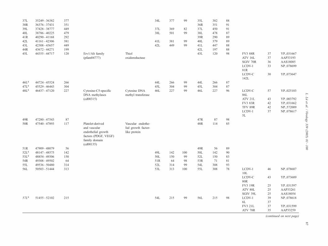
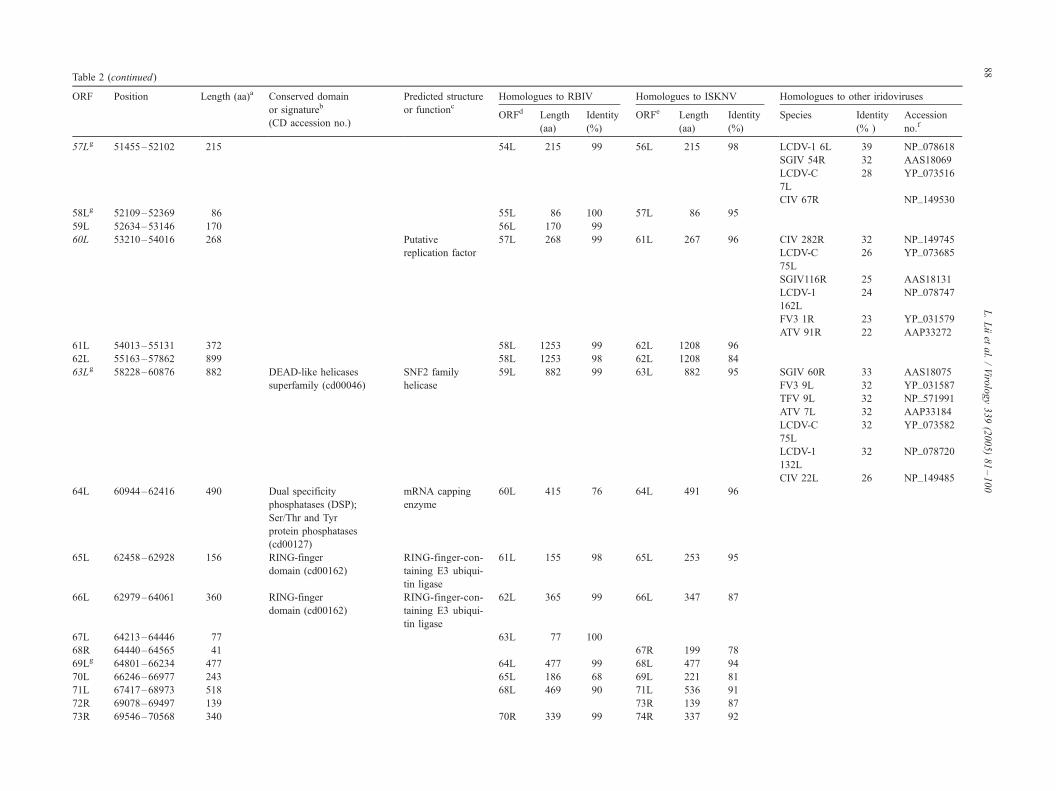
Table 2
Potential open reading frames of the OSGIV genome
ORF Position Length (aa)a Conserved domain
or signatureb
(CD accession no.)
Predicted structure
or functioncHomologues to RBIV Homologues to ISKNV Homologues to other iridoviruses
ORFd Length
(aa)
Identity
(%)
ORFe Length
(aa)
Identity
(%)
Species Identity
(% )
Accession
no.f
1Lg 134–1270 378 Transmembrane amino
acid transporter protein
(pfam01490)
Transmembrane
amino acid
transporter protein
1L 378 99 1L 378 95
2R 1394–1789 131 2L 216 89
3R 1849–2064 71 3R 71 100
4L 2110–2613 167 4L 167 100 3L 185 78
5L 2632–2808 58 5L 58 100
6L 2883–3548 221 Catalytic domain of Catalytic domain 6L 221 100 5L 251 95 LCDV-C 36 YP_073653
CTD-like phosphatases of CTD-like 148L
(smart00577) phosphatase SGIV 61R 35 AAS18076
LCDV-1 33 NP_078678
82L
FV3 37R 31 YP_031615
ATV 64R 29 AAP33244
CIV 335R 29 NP_149818
7Lg 3793–5154 453 Iridovirus major capsid Major capsid 7L 453 99 6L 453 98 SBIV 100 AAP74203
protein (pfam04451) protein GSDIV 100 AAP37443
RSIV 99 AAP74204
ALIV 98 AAP37442
DGIV 98 AAP37441
LCDV-C 46 YP_025102
43L
LCDV-1 46 NP_044812
147L
TFV 96R 44 NP_572010
ATV 14L 44 YP_003785
RRV 44 YP_003785
EHNV 44 AAO32315
CIV 274L 44 NP_149737
FV3 90R 43 YP_031669
BIV 43 AAO32316
SGIV 72R 42 YP_164167
8Lg 5171–6628 485 Myristylated 8L 485 99 7L 485 97 RSIV 94 BAC66967
membrane protein LCDV-C158R 33 YP_073663
SGIV 88L 31 AAS18103
FV3 53R 30 YP_031631
ATV 51L 30 AAP33230
LCDV-167L 29 NP_078686
CIV 118L 28 NP_078665
9R 6699–8246 515 9R 515 99 8R 525 88 RSIV 93 BAC66966
10Rg 8342–8503 53 10R 53 100 9R 53 92
11L 8662–8895 77 11L 130 98 10L 130 94 LCDV-1 32 NP_078686
91R
LCDV-C 28 YP_073651
L.Luet
al./Viro
logy339(2005)81–100
84

145R
12L 8849–9055 68 11L 130 75 10L 130 72
13L 9052–9312 86 11L 86 99
14Rg 9331–9663 110 RING-finger-containing
ubiquitin ligase (COG5540)
RING-finger-
containing
ubiquitin ligase
12R 110 100 12R 110 96
15R 9670–11067 465 Serine/threonine protein Serine/threonine 13R 465 100 13R 461 89 CIV 380R 36 NP_149561
kinases, catalytic domain protein kinases SGIV150R 34 AAS18165
(cd00180) LCDV-C 31 YP_073682
178L
ATV 47L 28 AAP33226
FV3 57R 27 YP_031636
LCDV-1 25 NP_078729
43L
16R 11322–12296 324 14R 324 100 14R 319 95
17Rg 12302–13093 263 15R 263 100 15R 263 95
18Lg 13151–13738 195 16L 195 99 16L 195 90
19L 13753–14088 111 17L 112 99
20R 14094–14351 85 18L 79 91
21L 14431–14607 58 19L 58 100
22Rg 14623–17469 948 DNA polymerase DNA polymerase 20R 948 99 19R 948 97 RSIV 98 BAA28669
type-B family; DNA LCDV-C 37 YP_073706
-directed DNA 203L
polymerase (cd00145) LCDV-1 35 NP_078724
135R
SGIV128R 36 YP_164223
FV3 60R 35 YP_031639
TFV 63R 35 NP_572000
ATV 44L 35 YP_003817
CIV 37L 30 AAD48150
23L 17533–17715 60 21L 60 100 20L 62 87
24L 17851–17973 40 21L 40 95
25L 18063–19715 550 Appr-1W-p processing
enzyme (smart00506)
Putative
phosphatase
22L 536 75 22L 499 64 RSIV 93 AAQ07955
26R 19788–22922 1044 Laminin-type epidermal
growth factor-like
domain (cd00055)
Laminin-type
epidermal growth
factor-like protein
23R 805 75 23R 856 64 RSIV 83 AAQ07956
27Rg 23207–24145 312 Ribonucleotide reductase,
R2/beta subunit (RNRR2)
(cd01049)
Ribonucleotide
reductase small
chain
26R 312 99 24R 312 96 RSIV 99 BAA82755
28R 24169–24696 175 27R 127 66
29L 24693–25013 106 26L 107 87
30Lg 25035–25931 298 Xeroderma pigmentosum DNA repair 28L 298 99 27L 298 98 RSIV 99 BAA82754
G N- and I-regions protein RAD2 ATV 10L 33 AAP33187
(XPGN, XPGI) (cd00128) FV3 95R 32 YP_031674
(continued on next page)
L.Luet
al./Viro
logy339(2005)81–100
85

ORF Position Length (aa)a Conserved domain
or signatureb
(CD accession no.)
Predicted structure
or functioncHomologues to RBIV Homologues to ISKNV Homologues to other iridoviruses
ORFd Length
(aa)
Identity
(%)
ORFe Length
(aa)
Identity
(%)
Species Identity
(% )
Accession
no.f
30Lg 25035–25931 298 Xeroderma pigmentosum DNA repair 28L 298 99 27L 298 98 RSIV 99 BAA82754
G N- and I-regions protein RAD2 TFV 100R 32 NP_572012
(XPGN, XPGI) (cd00128) SGIV 97L 30 AAS18112
LCDV-1 30 NP_078767
191R
LCDV-C 28 YP_073674
169R
CIV 369L 22 NP_149832
31L 25948–29454 1168 RNA polymerase I Largest subunit of 29L 1168 99 28L 1159 96 RSIV 98 BAA82753
subunit A N-terminus the DNA- TFV 8R 42 NP_571990
(smart00663); dependent RNA FV3 8R 39 YP_031586
RNA polymerase II, large polymerase ATV 6R 39 AAP33183
subunit (KOG0260) SGIV104L 38 AAS18119
LCDV-C 37 YP_025105
191R
LCDV-1 36 NP_078624
16L
32L 29461–29682 73 C2C2 Zinc finger; Transcription 29L 73 94 FV3 81R 41 YP_031660
nucleic-acid-binding elongation factor TFV 86R 41 NP_572006
motif in transcriptional SII ATV 24L 41 AAP33201
elongation factor TFIIS LCDV-1 36 NP_078754
and RNA polymerases 171R
(smart00440); TFIIS, CIV 349L 29 NP_149812
transcription factor
S-II (TFIIS) (pfam01096)
SGIV 85R 27 AAS18100
33Rg 29973–30215 80 30R 80 99 31R 80 85
34R 30310–30942 210 Deoxynucleotide Deoxyribonucleo- 31R 210 100 32R 203 88 ATV 19L 28 AAP33196
kinases (COG1428) side kinase FV3 85R 27 YP_031664
LCDV-C 26 YP_073536
27R
LCDV-1 26 NP_078725
136R
SGIV 67L 25 AAS18082
CIV 143R 24 NP_149606
35L 31033–31935 300 32L 313 99 33L 313 90
36R 32018–35176 1052 RNA polymerase DNA-directed 33R 1053 98 34R 1044 96 LCDV-C 43 YP_073534
Rpb2, domain 6 RNA polymerase 25R
(pfam00562) II second largest LCDV-1 43 NP_078633
subunit-like 25L
protein SGIV 73L 41 AAS18088
FV3 62L 40 YP_031641
TFV 65L 40 NP_572001
ATV 43R 39 AAP33221
Table 2 (continued)L.Luet
al./Viro
logy339(2005)81–100
86
37L 35249–36382 377 34L 377 99 35L 382 88
38R 36376–37431 351 36R 351 91
39L 37428–38777 449 37L 369 82 37L 450 91
40L 38786–40225 479 38L 501 99 38L 478 87
41R 40290–41168 292 39R 290 89
42L 41161–42306 381 41L 381 99 40L 379 89
43L 42308–43657 449 42L 449 99 41L 447 88
44R 43672–44271 199 42L 197 88
45L 44355–44717 120 Erv1/Alt family Thiol 43L 120 98 FV3 88R 37 YP_031667
(pfam04777) oxidoreductase ATV 16L 37 AAP33193
SGIV 70R 36 AAS18085
LCDV-1 33 NP_078699
81R
LCDV-C 30 YP_073647
142L
46Lg 44724–45524 266 44L 266 99 44L 266 87
47Lg 45529–46443 304 45L 304 99 45L 304 87
48Lg 46437–47120 227 Cytosine-C5-specific Cytosine DNA 46L 227 99 46L 227 96 LCDV-C 57 YP_025103
DNA methylases methyl transferase 86L
(cd00315) ATV 21L 43 YP_003792
FV3 83R 42 YP_031662
TFV 89R 42 NP_572009
LCDV-1 37 NP_078617
5L
49R 47280–47543 87 47R 87 98
50R 47540–47893 117 Platelet-derived
and vascular
endothelial growth
factors (PDGF, VEGF)
family domain
(cd00135)
Vascular endothe-
lial growth factor-
like protein
48R 114 85
51R 47909–48079 56 49R 56 89
52Lg 48147–48575 142 49L 142 100 50L 142 90
53Lg 48854–49306 150 50L 150 99 52L 150 83
54R 49308–49502 64 51R 64 98 53R 71 81
55L 49536–50480 314 52L 314 99 54L 308 93
56L 50503–51444 313 53L 313 100 55L 308 78 LCDV-1 46 NP_078687
10L
LCDV-C 43 YP_073689
80R
FV3 19R 25 YP_031597
ATV 80L 25 AAP33261
SGIV 39L 25 AAS18054
57Lg 51455–52102 215 54L 215 99 56L 215 98 LCDV-1 39 NP_078618
6L 37
FV3 21L 37 YP_031599
ATV 78R 35 AAP33259
(continued on next page)
L.Luet
al./Viro
logy339(2005)81–100
87
ORF Position Length (aa)a Conserved domain
or signatureb
(CD accession no.)
Predicted structure
or functioncHomologues to RBIV Homologues to ISKNV Homologues to other iridoviruses
ORFd Length
(aa)
Identity
(%)
ORFe Length
(aa)
Identity
(%)
Species Identity
(% )
Accession
no.f
57Lg 51455–52102 215 54L 215 99 56L 215 98 LCDV-1 6L 39 NP_078618
SGIV 54R 32 AAS18069
LCDV-C 28 YP_073516
7L
CIV 67R NP_149530
58Lg 52109–52369 86 55L 86 100 57L 86 95
59L 52634–53146 170 56L 170 99
60L 53210–54016 268 Putative 57L 268 99 61L 267 96 CIV 282R 32 NP_149745
replication factor LCDV-C 26 YP_073685
75L
SGIV116R 25 AAS18131
LCDV-1 24 NP_078747
162L
FV3 1R 23 YP_031579
ATV 91R 22 AAP33272
61L 54013–55131 372 58L 1253 99 62L 1208 96
62L 55163–57862 899 58L 1253 98 62L 1208 84
63Lg 58228–60876 882 DEAD-like helicases SNF2 family 59L 882 99 63L 882 95 SGIV 60R 33 AAS18075
superfamily (cd00046) helicase FV3 9L 32 YP_031587
TFV 9L 32 NP_571991
ATV 7L 32 AAP33184
LCDV-C 32 YP_073582
75L
LCDV-1 32 NP_078720
132L
CIV 22L 26 NP_149485
64L 60944–62416 490 Dual specificity
phosphatases (DSP);
Ser/Thr and Tyr
protein phosphatases
(cd00127)
mRNA capping
enzyme
60L 415 76 64L 491 96
65L 62458–62928 156 RING-finger
domain (cd00162)
RING-finger-con-
taining E3 ubiqui-
tin ligase
61L 155 98 65L 253 95
66L 62979–64061 360 RING-finger
domain (cd00162)
RING-finger-con-
taining E3 ubiqui-
tin ligase
62L 365 99 66L 347 87
67L 64213–64446 77 63L 77 100
68R 64440–64565 41 67R 199 78
69Lg 64801–66234 477 64L 477 99 68L 477 94
70L 66246–66977 243 65L 186 68 69L 221 81
71L 67417–68973 518 68L 469 90 71L 536 91
72R 69078–69497 139 73R 139 87
73R 69546–70568 340 70R 339 99 74R 337 92
Table 2 (continued)L.Luet
al./Viro
logy339(2005)81–100
88

74L 70669–70938 89 71L 89 99 75L 88 94
75L 70940–73912 990 72L 998 95 76L 990 96 CIV 295L 37 NP_149758
FV3 41R 25 YP_031619
ATV 69R 25 AAP33249
LCDV-C 24 YP_073738
235R
SGIV 57L 23 AAS18072
LCDV-1 22 NP_078748
163R
76R 73930–75264 444 Ankyrin repeats
(cd00204)
Ankyrin repeat-
containing protein
77R 444 92
77Rg 75261–75725 154 75R 154 100 78R 154 97
78L 75727–75951 74 76L 74 100 79L 73 95
79R 76039–76512 157 77R 157 100
80Rg 76525–77022 165 78R 165 99 81R 165 97
81Lg 77071–78177 368 79L 368 99 82L 368 95
82R 78202–78600 132 80R 132 98
83L 78627–79931 434 81L 434 99 84L 451 86
84R 80018–80548 176 82R 176 100 85R 200 77
85R 80978–81775 265 Ribonuclease Ribonuclease III 83R 265 98 87R 256 96 SGIV 84L 34 AAS18099
III family LCDV-C 31 YP_073691
(smart00535); 187R
dsRNA-specific LCDV-1 30 NP_078726
ribonuclease 137R
(COG0571) FV3 80L 30 YP_031659
TFV 85L 30 NP_572005
ATV 25R 29 AAP33202
CIV 142R 23 NP_149605
86R 81878–82279 133 84R 640 84 88R 667 82
87R 82234–83805 523 84R 640 97 88R 667 92
88L 84000–84134 44 91L 79 90
89R 84810–84995 61 92R 91 96
90Lg 84992–85918 308 86L 308 99 93L 308 94
91Lg 85928–86428 166 87L 166 99 94L 166 96
92L 86453–87616 245 88L 387 99 95L 386 91
93L 87624–88361 245 96L 270 89 LCDV-C 30 YP_073606
100L
FV3 12L 29 YP_031590
ATV 87R 28 AAP33268
LCDV-1 26 NP_078701
108L
SGIV118R 24 AAS18133
CIV 287R 21 NP_149750
94L 88366–88857 163 90L 162 99 LCDV-1 25 NP_078659
59L
LCDV-C 19 YP_073662
157R
(continued on next page)
L.Luet
al./Viro
logy339(2005)81–100
89

ORF Position Length (aa)a Conserved domain
or signatureb
(CD accession no.)
Predicted structure
or functioncHomologues to RBIV Homologues to ISKNV Homologues to other iridoviruses
ORFd Length
(aa)
Identity
(%)
ORFe Length
(aa)
Identity
(%)
Species Identity
(% )
Accession
no.f
95Lg 88906–89229 107 RING-finger
domain (cd00162)
RING-finger
domain-containing
E3 protein
91L 107 100 99L 107 83
96L 89377–90024 215 92L 215 99 100L 181 89
97L 89993–90508 171 101L 171 94
98R 90577–92022 481 Ankyrin repeats
(cd00204)
Ankyrin repeat-
containing protein
94R 481 100 102R 480 91
99R 92029–92442 137 Src homology 2
domains (cd00173)
Suppressor of
cytokine signaling
protein
103R 133 80
100R 92501–93280 259 96R 259 99 104R 258 90
101R 93282–93653 123 97R 123 100 105R 121 97
102R 93747–94634 295 HINT (histidine
triad nucleotide-binding
protein) subgroup
(cd01277)
HINT protein 98R 295 100
103L 94676–95548 290 99L 273 99 106L 339 78
104L 95799–95936 45 107L 71 88
105R 95992–96276 94 100R 94 85 108R 49 77
106L 96298–99060 920 Predicted ATPase D5 family NTPase 101L 921 99 109L 921 97 LCDV-C 36 YP_073585
(COG3378); Poxvirus 80L
D5 protein-like SGIV 52L 36 AAS18067
(pfam03288) FV3 22R 35 YP_031600
ATV 77L 35 AAP33258
LCDV-1 34 NP_078717
128L
CIV 184R 28 NP_149647
107R 99113–99268 51 110R 51 90
10 8L 99265–99885 206 Tnf receptor-associated
factor 2 domain
(cd00270);
MATH (cd00121)
Tumor necrosis
factor type 2
receptor-
associated protein
102L 298 91 111L 296 83
109R 100183–100926 247 Proliferating cell 103R 146 98 112R 247 97 LCDV-1 31 NP_078615
nuclear antigen 3L
LCDV-C 28 YP_073700
197L
SGIV 68L 28 AAS18083
ATV 20L 24 AAP33197
FV3 84R 23 YP_031663
110R 100998–101414 138 113R 117 96
Table 2 (continued)L.Luet
al./Viro
logy339(2005)81–100
90

111L 101594–104041 815 CAP10, putative Tyrosine kinase 106L 358 95 114L 941 81 FV3 27R 43 YP_031605
lipopolysaccharide- TFV 29R 43 NP_571995
modifying enzyme ATV 58R 43 AAP33237
(smart00672) SGIV 81L 42 AAS18093
LCDV-C 24 YP_073677
173R
CIV 179R 23 NP_149902
LCDV-1 20 NP_078770
195R
112R 104476–105486 336 Immediate early 108L 243 89 115R 336 97 LCDV-1 33 NP_078648.1
protein ICP-46 47L
LCDV-C 25 YP_073667.1
162R
TFV 97R 24 NP_572011.1
FV3 91R 23 YP_031670.1
ATV 13L 22 AAP33190.1
SGIV162L 20 AAS18177.1
CIV 393L 20 NP_149856.1
113R 105547–106701 384 110R 477 95 116R 482 86
114Lg 106978–107652 224 Early 31 kDa 111L 224 99 117L 224 95 FV3 23R 24 YP_031603
protein TFV 25R 24 NP_571993
LCDV-C 24 YP_025101
34L
LCDV-1 23 NP_078713
122R
ATV 55R 23 AAP33234
SGIV 6R 22 AAS18021
115L 107986–109299 437 Ankyrin repeats
(cd00204)
Ankyrin repeat-
containing protein
112L 454 98 118L 456 93
116R 109369–109656 95 RING-finger
domain (cd00162)
RING-finger
domain-containing
E3 protein
113R 57 98 119R 95 95
117R 109687–110193 168 114R 129 84 120R 169 93
118L 110214–110849 211 115L 149 88 121L 216 65
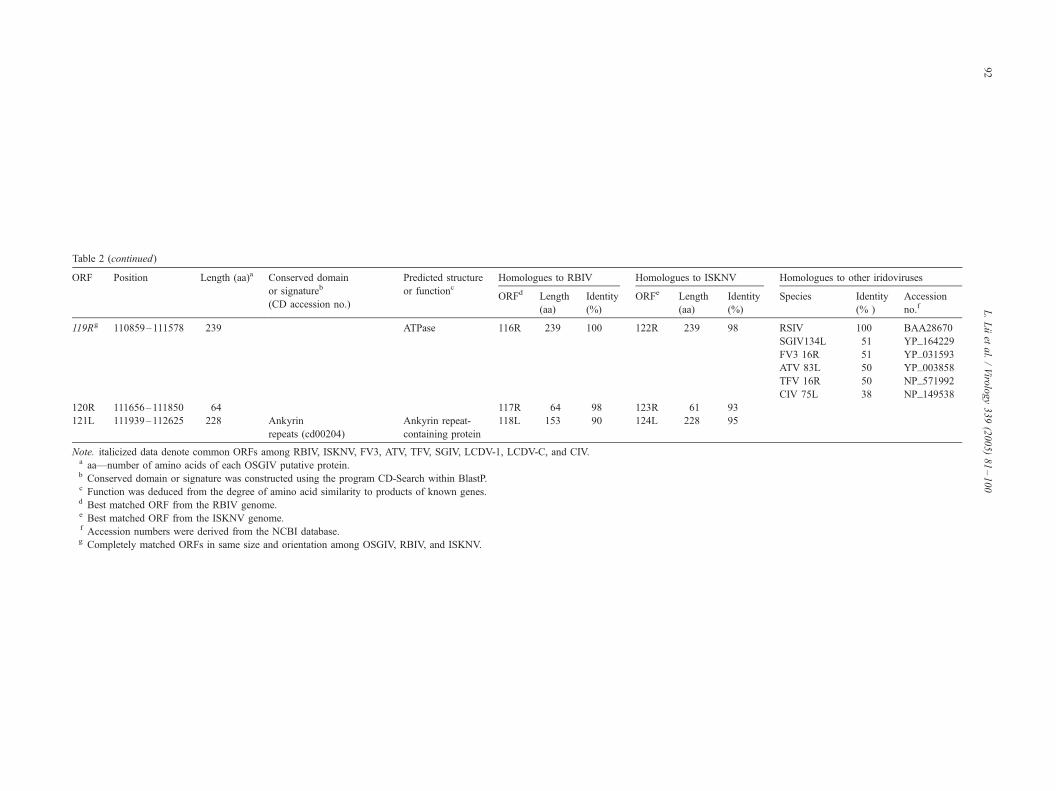
119Rg 110859–111578 239 ATPase 116R 239 100 122R 239 98 RSIV 100 BAA28670
SBIV 100 BAA96406
LBIV 100 AAL68654
TGIV 100 BAA96407
GSIV 100 AAL68653
LYCIV 99 AAO16492
ALIV 99 BAA96408
DGIV 99 AAP74205
GIV 54 AAL68652
LCDV-C 54 YP_073620
114L
LCDV-1 54R 53 NP_078656
(continued on next page)
L.Luet
al./Viro
logy339(2005)81–100
91
ORF Position Length (aa)a Conserved domain
or signatureb
(CD accession no.)
Predicted structure
or functioncHomologues to RBIV Homologues to ISKNV Homologues to other iridoviruses
ORFd Length
(aa)
Identity
(%)
ORFe Length
(aa)
Identity
(%)
Species Identity
(% )
Accession
no.f
119Rg 110859–111578 239 ATPase 116R 239 100 122R 239 98 RSIV 100 BAA28670
SGIV134L 51 YP_164229
FV3 16R 51 YP_031593
ATV 83L 50 YP_003858
TFV 16R 50 NP_571992
CIV 75L 38 NP_149538
120R 111656–111850 64 117R 64 98 123R 61 93
121L 111939–112625 228 Ankyrin
repeats (cd00204)
Ankyrin repeat-
containing protein
118L 153 90 124L 228 95
Note. italicized data denote common ORFs among RBIV, ISKNV, FV3, ATV, TFV, SGIV, LCDV-1, LCDV-C, and CIV.a aa—number of amino acids of each OSGIV putative protein.b Conserved domain or signature was constructed using the program CD-Search within BlastP.c Function was deduced from the degree of amino acid similarity to products of known genes.d Best matched ORF from the RBIV genome.e Best matched ORF from the ISKNV genome.f Accession numbers were derived from the NCBI database.g Completely matched ORFs in same size and orientation among OSGIV, RBIV, and ISKNV.
Table 2 (continued)
L.Luet
al./Viro
logy339(2005)81–100
92

Fig. 1. Organization of the OSGIV genome. Arrows that indicated the location, orientation, and putative size represent each ORF. White arrows represent ORFs
with predicted function to other organisms, and black arrows represent ORFs with unknown function.
L. Lu et al. / Virology 339 (2005) 81–100 93
tase small subunit (ORF 27R) and deoxyribonucleoside
kinase (ORF 34R). DNA is made of four deoxyribonucleo-
side triphosphates, provided by the de novo and salvage
pathways. The key enzyme of the de novo pathway is
ribonucleotide reductase. ORF 27R of OSGIV encoded a
ribonucleotide reductase small subunit, which may catalyze
a rate-limiting reaction in which ribonucleoside diphosphates
are converted to their corresponding deoxyribonucleoside
diphosphates. The precursors of deoxyribonucleoside tri-
phosphates (dNTPs) are required for DNA synthesis and
repair (Lammers and Follmann, 1983). ORF 34R encoded a
homologue of deoxyribonucleoside kinase, the key enzymes
of salvage pathways, which phosphorylate deoxyribonucleo-
sides to the corresponding deoxyribonucleoside monophos-
phates (Arner and Eriksson, 1995; Lammers and Follmann,
1983).
Protein modification
OSGIV contained three genes with putative protein
modification function. These genes included a serine/
threonine protein kinase (ORF 15R), a thiol oxidoreductase
(ORF 45L), and a tyrosine kinase (ORF 111L).
ORF 45L encoded a thiol oxidoreductase with Erv1/Alr
domain (Erv1, essential for respiration and vegetative
growth; Alr, augmenter of liver regeneration). The Erv1/
Alr family is a large family encoded by cytoplasmic DNA
viruses and all eukaryotes (Senkevich et al., 2000). It
includes the Saccharomyces cerevisiae Erv1 protein, which
is required for mitochondrial biogenesis, and it’s the
mammalian homologues hematopoietin (named ALR). Both
Erv1p and full-length ALR are located in the mitochondrial
intermembrane. The conserved domain of the ERV1/ALR
protein consists of about 100 amino acid residues and
contains an invariant thiol active-site motif C-X-X-C
(Senkevich et al., 2000). The functions of many ERV1/
ALR proteins remain unknown, but they may represent a
ubiquitous class of cellular thiol oxidoreductases. The C-X-
X-C motif of the ERV1 domain is the redox-active disulfide
bridge of secreted egg-white sulfhydryl oxidase, a member
of the quiescin family (Senkevich et al., 1997). The deduced
OSGIV Erv1/Alr protein consisted of 120 amino acid
residues, and it contained a thiol active-site motif C-K-T-
C, which suggested that it might participate in thiol-
disulfide metabolism and protein folding.
Host-related function
OSGIV contained a significant number of putative genes
that exhibit similarity to cellular genes and other iridovirus
genes. There were five ORFs predicted to encode RING
finger proteins (RFPs) (ORF 14R, 65L, 66L, 95L, and
116R). They were highly homologous to those of ISKNV
(ORF 12R, 65L, 66L, 99L, and 119R). The study on Ring

L. Lu et al. / Virology 339 (2005) 81–10094
finger protein showed that the four RFPs of ISKNV (ORF
12R, 65L, 66L, and 111L) acted as the E3 enzyme in the
presence of ubiquitin activating enzyme (E1), ubiquitin,
zincion, and specific E2 (UbcH5 subfamily). The RING
domain of RFP in ISKNV (ORF 66L) was proved to be
essential for the activity of E3 enzyme by the mutational
analysis (unpublished data). The functions of the RFPs in
OSGIV remained unknown, but these RFPs in OSGIV may
have E3 activity considering their high homology (83–96%
amino acids identities) to those in ISKNV (Table 2).
ORF 99R of OSGIV encoded a Src homology 2 (SH2)
domain-containing protein. SH2 domain is involved in recog-
nition of phosphorylated tyrosine (pTyr). It binds to pTyr-
containing ligands via two surface pockets, a pTyr and a
hydrophobic-binding pocket, allowing proteins with SH2
domains to localize to tyrosine-phosphorylated sites. Some
SH2 domain-containing proteins (such as SLP-76, Shb, and
SHIP) play a role in phosphoinositide 3-kinase (PI3K)
signaling pathways (Shim et al., 2004; Lu et al., 2002; Rauh
et al., 2003). ORF 50R encoded a protein that contained
vascular endothelial growth factor (VEGF) family domain.
Recent studies showed that VEGF could stimulate the PI3K
signaling pathways and inhibit apoptosis in the viral-infected
host cells (Brader and Eccles, 2004; Gelinas et al., 2002;
Takahashi et al., 2003). Further investigation is needed to
determinewhether bothORF 50R andORF 99R participate in
the PI3K signaling pathways in the viral-infected host cells.
In addition, some important putative genes involved in
host-related function were found in the OSGIV genome,
such as ankyrin repeat motifs (ORF 76R, 98R, 115L, and
121L), the C-terminal domain (CTD)-like phosphatase
(ORF 6L), proliferating cell nuclear antigen (ORF 10R),
laminin-type epidermal growth factor (EGF)-like domain
(ORF 26R), and tumor necrosis factor receptor-associated
factor (TRAF, ORF 108L).
Putative membrane-associated proteins
The ORFs of OSGIV were analyzed for the presence of
putative transmembrane domains (TMs). One or more
putative TMs (1–11) were found in 5 ORFs (ORF 1L,
5L, 52L, 53L, and 94L) with the computer software
TMHMM 2.0 (Krogh et al., 2001). These proteins contain
a putative TM probably associated with membrane struc-
tures. These hydrophobic domains may be involved in
protein–protein interactions that are necessary for the
formation of the nucleocapsid globular subunits (van Hulten
et al., 2001).
Other proteins
OSGIV also encoded proteins homologous to a phos-
phatase (ORF 113R), an ATPase (ORF 119R), a protein
containing HINT (histidine triad nucleotide-binding protein)
domain (ORF 102R), and a structural protein (major capsid
protein, ORF 7L).
ORF 102R encoded a protein containing a HINT domain.
HINT belongs to a superfamily of histidine triad (HIT)
hydroxylases that act on alpha-phosphate of ribonucleotides.
HINT consists of small homodimerizing proteins charac-
terized by conserved histidine residues His-X-His-X-His-X-
X (X, a hydrophobic amino acid) near their C-terminal ends
(Brenner, 2002). HINT is the most widely conserved feature
of the HIT superfamily, and HINT homologues are present
in a wide variety of organisms in the metazoan, plant,
fungus, bacterium, and virus. Although the biochemical
function has not been characterized for many of the
members of HINT, the proteins from yeast have been
shown to be involved in secretion, peroxisome formation,
and gene expression (Bieganowski et al., 2002). The
deduced product of OSGIV ORF 102R was 295 amino
acids in length with a ‘‘H-A-H-A’’ region near its C-terminal
end, and it may be involved in gene expression in the viral-
infected host cells.
Relationship of OSGIV to ISNNV and RBIV
The comparative analysis of the OSGIV, RBIV, and
ISKNV revealed many homologues (Table 2). Many ORFs
of the three-iridoviral genomes including the conserved
genes and other ORFs of unknown function were similar in
size, structure, and composition. 98 and 109 ORFs of
OSGIV had homologies to that of RBIV (66–100%) and
ISKNV (64–99%) at the amino acid level, respectively (Do
et al., 2004; He et al., 2001). Comparing OSGIV and RBIV,
98 ORFs of OSGIV had homologies (average 97%) to those
of RBIV; 63 of the 98 ORFs had same size and the identities
varied from 85% to 100%; 35 of the 98 ORFs were differed
in size with identities varied from 66% to 99%. Comparing
OSGIV with ISKNV, 109 ORFs showed homologies to
those of ISKNV with average identity of 90%; 43 of 109
ORFs had the same size and the identities vary from 83% to
99%; 66 of 109 ORFs had different sizes and their identities
vary from 64% to 96%.
DNA dot matrix analyses of OSGIV genomic DNAwith
itself (Fig. 2A), the ISKNV genome (Fig. 2B) and the RBIV
genome (Fig. 2C) were performed using DS GENE 1.5
(Accelrys Inc.). The results revealed that the gene order
between OSGIV and ISKNV, OSGIV, and RBIV was
markedly conserved. The arrowhead at Fig. 2A showed
repeat sequences indicated by the short parallel lines offset
from the consensus diagonal. A highly direct repetitive
region (24187–24642 bp) was identified from Fig. 2A,
which was consistent with the result using the GeneQuest
program (Lasergene). Despite the high sequence similarity
among OSGIV, ISKNV, and RBIV, Fig. 2B showed several
insertions or deletions between OSGIV and ISKNV
indicated by gaps in the consensus line, and they were
mainly in the OSGIV genome nucleotides position 18500–
23500, 54000–62000, 69000–69500, 75800–76600, and
93500–94750 bp. The insertions or deletions were also
observed between OSGIV and RBIV (Fig. 2C), and they

Fig. 2. Dot matrix plots comparing the OSGIV genome (vertical axis) with the ISKNV, RBIV genome, and itself (horizontal axis). The horizontal axes
represent (A) the OSGIV genome; (B) the ISKNV genome; (C) the RBIV genome. The complete genomic sequences were aligned using DS GENE 1.5. The
arrowheads indicate repeat sequences (A), insertions, or deletions (B and C).
L. Lu et al. / Virology 339 (2005) 81–100 95
were mainly located in the OSGIV genome nucleotides
position 21500–23000, 52000–57000, 67000–69000,
82000–85000, and 105100–107700 bp. But when com-
pared Fig. 2B with Fig. 2C, the gaps of Fig. 2C were less
than those of Fig. 2B, which suggested that OSGIV was
more colinear to RBIV than to ISKNV.
The similarity among OSGIV, RBIV, and ISKNV indi-
cated a close structural and functional relationship. Addi-
tionally, the differences in the size and homology among
ORFs of three viruses may result in the differences in the
natural hosts andmortality. For example, in a recent study, we
have shown that OSGIV could infect the fresh water
mandarin fish that is the natural host of ISKNV. The mortality
of mandarin fish infected by OSGIV was about 40%
(unpublished data), but it could reach 100% if it was infected
by ISKNV (He et al., 2000).
Relationship of OSGIV to other iridoviruses
Iridoviral sequences available in data banks were com-
pared to the complete sequence of OSGIV, and the result
showed that the putative gene products of OSGIV shared
high homology to the corresponding viral proteins of other
iridoviruses (Table 2). There were some homologous genes
in the OSGIV, ISKNV, RBIV, TFV, FV3, ATV, SGIV, LCDV-
1, and LCDV-C genomes. These included genes for the DNA

L. Lu et al. / Virology 339 (2005) 81–10096
polymerase (DdDP), the DNA methyl transferase (DMet),
the SNF2-like helicase, the XPG/RAD2-type nuclease
(RAD2), the two large subunit of DNA-dependent RNA
polymerase (DdRPI and DdRPII), the RNase III (RIII), the
ATPase and the major capsid protein (MCP) involved in virus
replication, transcription, modification, and structural com-
position. Fig. 3 showed the comparison of OSGIV with other
eight vertebrate iridoviruses. Since the origin of replication
was unknown, the start codon (ATG) of MCPs gene was
chosen as the first base for all viral genomes. However, as
MCP of OSGIV, RBIV, ISKNV, ATV, LCDV-1, and LCDV-
C were present on antisense strands of the genomes, we
shifted sense and antisense strands on these viruses in order to
get the same nucleotide order on MCPs individually. It was
obvious that these homologous genes of OSGIV, RBIV, and
ISKNV were located at similar map position and colinearity
was detected in the three virus genomes. In contrast, these
gene orders on the OSGIV genome were significant different
with those of ranaviruses and lymphocystiviruses. This figure
indicated that OSGIV was more closely related to RBIV and
ISKNV, and markedly differed from members of the genus
Ranavirus and Lymphocystivirus.
Phylogenetic analysis
In order to determine the phylogenetic relationship of
OSGIV to other iridoviruses, the amino acid sequence of the
Fig. 3. Organization comparison of some conserved genes of the OSGIV, RBIV,
indicates the genome of the corresponding iridovirus, and the squares indicate th
(DdDP), the DNA methyl transferase (DMet), the SNF2-like helicase (Helicase)
dependent RNA polymerase (DdRPI and DdRPII), the RNase III (RIII), the AT
forward is represented by an open square, and solid squares correspond to the re
major capsid protein (MCP) was used in an alignment with
other iridoviruses from GenBank. MCP is a suitable target
for the phylogenetic studies as it is highly conserved in
iridoviruses (Tidona et al., 1998; Hyatt et al., 2000). The
amino acid sequence of MCP share > 98% identity with
OSGIV, RBIV, and ISKNV, respectively, but it shared low
identity (< 47%) to that of ranaviruses and lymphocystivi-
ruses. It is the predominant structural component of the
virus particles comprising 40–50% of the total particle
polypeptide (Williams, 1996). The neighbor-joining tree
(Saitou and Nei, 1987) of MCP was constructed and it
assigned the iridoviruses to four groups: group I, ranavi-
ruses, including FV3, TFV, ATV, RRV, BIV, EHNV, and
SGIV; group II, lymphocystiviruses, including LCDV-1 and
LCDV-C; group III, megalocystiviruses, including OSGIV,
ISKNV, RBIV, RSIV, SBIV, GSDIV, ALIV, and DGIV;
group IV, the insect iridoviruses, including CIV, CzIV, SIV,
TIV, and WIV; PBCV-1 was used as an outgroup (Fig. 4A).
The results illuminated that OSGIV was more closely
related to RBIV, ISKNV, RSIV, SBIV, GSDIV, ALIV, and
DGIV as compared to the Ranavirus and Lymphocystivi-
ruses. Additionally, the highly conserved full-length protein
sequences of the ATPase, cytosine DNA methyl transferase,
and DNA polymerase from the iridoviruses were used for
phylogenetic analysis (Figs. 4B–D). These trees also
supported the view that OSGIV was more closely related
to RBIV and ISKNV.
ISKNV, FV3, TFV, ATV, SGIV, LCDV-1, and LCDV-C genomes. The line
e approximate size of the conserved genes including the DNA polymerase
, the XPG/RAD2-type nuclease (RAD2), the two largest subunit of DNA-
Pase, and the major capsid protein (MCP). The direction of transcription
verse direction.
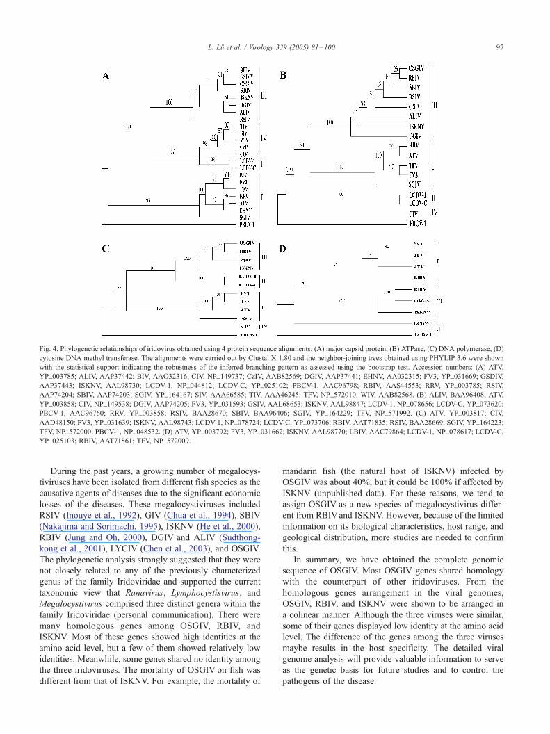
Fig. 4. Phylogenetic relationships of iridovirus obtained using 4 protein sequence alignments: (A) major capsid protein, (B) ATPase, (C) DNA polymerase, (D)
cytosine DNA methyl transferase. The alignments were carried out by Clustal X 1.80 and the neighbor-joining trees obtained using PHYLIP 3.6 were shown
with the statistical support indicating the robustness of the inferred branching pattern as assessed using the bootstrap test. Accession numbers: (A) ATV,
YP_003785; ALIV, AAP37442; BIV, AAO32316; CIV, NP_149737; CzIV, AAB82569; DGIV, AAP37441; EHNV, AA032315; FV3, YP_031669; GSDIV,
AAP37443; ISKNV, AAL98730; LCDV-1, NP_044812; LCDV-C, YP_025102; PBCV-1, AAC96798; RBIV, AAS44553; RRV, YP_003785; RSIV,
AAP74204; SBIV, AAP74203; SGIV, YP_164167; SIV, AAA66585; TIV, AAA46245; TFV, NP_572010; WIV, AAB82568. (B) ALIV, BAA96408; ATV,
YP_003858; CIV, NP_149538; DGIV, AAP74205; FV3, YP_031593; GSIV, AAL68653; ISKNV, AAL98847; LCDV-1, NP_078656; LCDV-C, YP_073620;
PBCV-1, AAC96760; RRV, YP_003858; RSIV, BAA28670; SBIV, BAA96406; SGIV, YP_164229; TFV, NP_571992. (C) ATV, YP_003817; CIV,
AAD48150; FV3, YP_031639; ISKNV, AAL98743; LCDV-1, NP_078724; LCDV-C, YP_073706; RBIV, AAT71835; RSIV, BAA28669; SGIV, YP_164223;
TFV, NP_572000; PBCV-1, NP_048532. (D) ATV, YP_003792; FV3, YP_031662; ISKNV, AAL98770; LBIV, AAC79864; LCDV-1, NP_078617; LCDV-C,
YP_025103; RBIV, AAT71861; TFV, NP_572009.
L. Lu et al. / Virology 339 (2005) 81–100 97
During the past years, a growing number of megalocys-
tiviruses have been isolated from different fish species as the
causative agents of diseases due to the significant economic
losses of the diseases. These megalocystiviruses included
RSIV (Inouye et al., 1992), GIV (Chua et al., 1994), SBIV
(Nakajima and Sorimachi, 1995), ISKNV (He et al., 2000),
RBIV (Jung and Oh, 2000), DGIV and ALIV (Sudthong-
kong et al., 2001), LYCIV (Chen et al., 2003), and OSGIV.
The phylogenetic analysis strongly suggested that they were
not closely related to any of the previously characterized
genus of the family Iridoviridae and supported the current
taxonomic view that Ranavirus, Lymphocystisvirus, and
Megalocystivirus comprised three distinct genera within the
family Iridoviridae (personal communication). There were
many homologous genes among OSGIV, RBIV, and
ISKNV. Most of these genes showed high identities at the
amino acid level, but a few of them showed relatively low
identities. Meanwhile, some genes shared no identity among
the three iridoviruses. The mortality of OSGIV on fish was
different from that of ISKNV. For example, the mortality of
mandarin fish (the natural host of ISKNV) infected by
OSGIV was about 40%, but it could be 100% if affected by
ISKNV (unpublished data). For these reasons, we tend to
assign OSGIV as a new species of megalocystivirus differ-
ent from RBIVand ISKNV. However, because of the limited
information on its biological characteristics, host range, and
geological distribution, more studies are needed to confirm
this.
In summary, we have obtained the complete genomic
sequence of OSGIV. Most OSGIV genes shared homology
with the counterpart of other iridoviruses. From the
homologous genes arrangement in the viral genomes,
OSGIV, RBIV, and ISKNV were shown to be arranged in
a colinear manner. Although the three viruses were similar,
some of their genes displayed low identity at the amino acid
level. The difference of the genes among the three viruses
maybe results in the host specificity. The detailed viral
genome analysis will provide valuable information to serve
as the genetic basis for future studies and to control the
pathogens of the disease.

L. Lu et al. / Virology 339 (2005) 81–10098
Materials and methods
Fish
Diseased orange-spotted grouper were obtained from the
aquatic farms in Huidong, Guangdong Province, China, in
September 2002 when the outbreak of the disease occurred.
The diseased fish showed some signs including reduced
feeding activity, lethargy, and a darkened body with atypical
swimming behavior at the edge of cages in moribund fish.
These fish had enlarged spleen and kidney cells checked by
histopathology. The spleens and kidneys were removed
from diseased fish and the viruses were observed by light
and electron microscopy. Meanwhile, some samples were
stored at �80 -C, then were examined by PCR to further
confirm the disease.
Virus and viral DNA
Spleen and kidney from the diseased fish were rinsed 3
times with PBS buffer (pH 7.4) and pulverized by a mortar
and pestle in liquid nitrogen. The powdered tissue was
homogenized with glass tissue blender in ten volumes of
PBS buffer (pH 7.4) on ice. After centrifuged at 3500 � g
for 10 min at 4 -C, the supernatant was pelleted at
35,000 � g for 30 min at 4 -C. The virus pellet was
resuspended with TMP buffer (100 mM Tris–HCl, pH 7.5,
10 mM MgCl2, 1 mM PMSF) incubated with DNase I and
RNase A at 37 -C for 15 min. Then the virus was layered on
20–50% (w/w) sucrose gradient and further purified by
centrifugation for 2 h at 60,000�g in an SW41 Ti rotor
(Beckman). Viral DNA was extracted by incubating virions
in 0.5 mg/ml proteinase K and 0.5% SDS at 55 -C for 3 h.
After extractions with phenol-chloroform, the DNA was
precipitated with ethanol (Sambrook et al., 1989).
PCR amplification and sequencingx
The complete genomic DNA of OSGIVwas sequenced by
PCR method. As OSGIV showed high homology to ISKNV,
the primers were derived from the DNA sequence of ISKNV
in GenBank/EMBL/DDBJ database (AF371960). Amplified
PCRproductswere about 1000–1200bp in length, andprimer
pairs with overlapping sequence were designed and used in
order to fill gaps and verify the sequence. The PCR products
were purified using QIAquick PCR purification kit (QIA-
GEN). DNA sequencing was carried out using BigDye
Terminator Kit (Applied Biosystems, Inc.) on an ABI PRISM
377DNAsequencer (AppliedBiosystems, Inc.). The software
DataCollection and SequenceAnalysis (AppliedBiosystems,
Inc.) was applied to create the contigs assemble the genome.
Computer-assisted analyses
Genomic DNA composition, structure, and homologous
regions were analyzed using DNASTAR (Lasergene). The
ORFs and their amino acid sequences were predicted using
DS GENE 1.5 (Accelrys Inc.) and NCBI ORF finder (http://
www.ncbi.nlm.nih.gov/gorf/gorf.html). Protein database
searches were conducted using the BLASTP at NCBI Web
site (http://www.ncbi.nlm.nih.gov/). Alignment of amino
acid sequences of the major capsid protein, ATPase,
cytosine DNA methyl transferase, and DNA polymerase
were obtained by Clustal-X 1.80 (Thompson et al., 1994).
The phylogenetic trees were generated using the PHYLIP
package (Felsenstein, 1995) and the TreeView (Page, 1996).
The DNA dot matrix was obtained using DS GENE 1.5
(Accelrys Inc.). Prediction of transmembrane domains
(TMs) was performed using TMHMM 2.0 (http://
www.cbs.dtu.dk/services/TMHMM-2.0/) (Krogh et al.,
2001).
Virus abbreviations
The names of viruses were abbreviated in this article as
follow: ALIV, African lampeye iridovirus; ATV, A. tigrinum
virus; BIV, Bohle iridovirus; CIV, Chilo iridescent virus;
CzIV, Costelytra zealandica iridescent virus; DGIV, dwarf
gourami iridovirus; EHNV, epizootic hemorrhagic disease
virus; FV3, frog virus 3; GIV, grouper iridovirus; GSDIV,
grouper sleepy disease iridovirus; GSIV, giant seaperch
iridovirus; ISKNV, infectious spleen and kidney necrosis
virus; LBIV, largemouth bass iridovirus; LCDV-1, lympho-
cystis disease virus 1; LCDV-C, lymphocystis disease virus
isolated in China; LYCIV, large yellow croaker iridovirus;
OSGIV, orange-spotted grouper iridovirus PBCV-1, Para-
mecium bursaria Chlorella virus 1; RBIV, rock bream
iridovirus; RRV, Regina ranavirus; RSIV, red sea bream
iridovirus; SBIV, sea bass iridovirus; SGIV, Singapore
grouper iridovirus; SIV, Simulium iridescent virus; TFV,
tiger frog virus; TGIV, grouper iridovirus in Taiwan; TIV,
Tipula iridescent virus; and WIV, Wiseana iridescent virus.
Nucleotide sequence accession number
The nucleotide sequence reported in this paper will
appear in the GenBank/EMBL/DDBJ database with the
accession number AY894343.
Acknowledgments
This study was supported by grants from the National
Science Foundation of China (30271030, 30325035),
National ‘‘863’’ project of China (2001AA626030), and
Natural Science Foundation of Guangdong Province of
China (200230002).
References
Arner, E.S.J., Eriksson, S., 1995. Mammalian deoxyribonucleoside kinases.
Pharmacol. Ther. 67, 155–186.

L. Lu et al. / Virology 339 (2005) 81–100 99
Bieganowski, P., Garrison, P.N., Hodawadekar, S.C., Faye, G., Barnes,
L.D., Brenner, C., 2002. Adenosine monophosphoramidase activity of
Hint and Hnt1 supports function of Kin28, Ccl1, and Tfb3. J. Biol.
Chem. 277, 10852–10860.
Brader, S., Eccles, SA., 2004. Phosphoinositide 3-kinase signalling path-
ways in tumor progression, invasion and angiogenesis. Tumori 90, 2–8.
Brenner, C., 2002. Hint, Fhit, and Galt: function, structure, evolution, and
mechanism of three branches of the histidine triad superfamily of
nucleotide hydrolases and transferases. Biochemistry 41, 9003–9014.
Chen, X.H., Lin, K.B., Wang, X.W., 2003. Outbreaks of an iridovirus
disease in maricultured large yellow croaker, Larimichthys crocea
(Richardson), in China. J. Fish Dis. 26, 615–619.
Chou, H.Y., Hsu, C.C., Peng, T.Y., 1998. Isolation and characterization of a
pathogenic iridovirus from cultured grouper (Epinephelus sp.) in
Taiwan. Fish Pathol. 33, 201–206.
Chua, H.C., Ng, M.L., Woo, J.J., Wee, J.Y., 1994. Investigation of
outbreaks of a novel disease, FSleepy Grouper Disease_, affecting the
brown-spotted grouper, Epinephelus tauvina Forskal. J. Fish Dis. 17,
417–427.
Danayadol, Y., Direkbusarakom, S., Boonyaratpalin, S., Miyazaki, T.,
Miyata, M., 1996. An outbreak of iridovirus-like infection in brown-
spotted grouper (Epinephelus malabaracus) cultured in Thailand.
AAHRI Newslett. 5, 6.
Darai, G., Anders, K., Koch, H.G., Delius, H., Gelderblom, H., Samalecos,
C., Flugel, R.M., 1983. Analysis of the genome of fish lymphocystis
disease virus isolated directly from epidermal tumours of pleuronectes.
Virology 126, 466–479.
Darai, G., Delius, H., Clarke, J., Apfel, H., Schnitzler, P., Flugel, R.M.,
1985. Molecular cloning and physical mapping of the genome of fish
lymphocystis disease virus. Virology 146, 292–301.
Delius, H., Darai, G., Flugel, R.M., 1984. DNA analysis of insect iridescent
virus 6: evidence for circular permutation and terminal redundancy. J.
Virol. 49, 609–614.
Deng, M., He, J.G., Weng, S.P., Zeng, K., Zeng, Z., Long, Q.X., 2001.
Purification and genomic analysis of infectious spleen and kidney
necrosis virus (ISKNV) from mandarinfish. J. fisheries of China
(Abstract in English) 25, 238–243.
Do, J.W., Moon, C.H., Kim, H.J., Ko, M.S., Kim, S.B., Son, J.H., Kim, J.S.,
An, E.J., Kim, M.K., Lee, S.K., Han, M.S., Cha, S.J., Park, M.S., Park,
M.A., Kim, Y.C., Kim, J.W., Park, J.W., 2004. Complete genomic DNA
sequence of rock bream iridovirus. Virology 325, 351–363.
Eaton, B.T., Hyatt, A.D., Hengstberger, S., 1991. Epizootic haemato-
poietic necrosis virus: purification and classification. J. Fish Dis. 14,
157–169.
Essani, K., 1990. The DNA methylase of frog virus 3. In: Darai, G. (Ed.),
Molecular Biology of Iridoviruses. Kluwer, Boston, pp. 163–172.
Evans, E., Klemperer, N., Ghosh, R., Traktman, P., 1995. The vaccinia virus
D5 protein, which is required for DNA replication, is a nucleic acid-
independent nucleoside triphosphatase. J. Virol. 69, 5253–5361.
Felsenstein, J., 1995. PHYLIP (Phylogeny Inference Package) Version 3.6.
University of Washington.
Gelinas, D.S., Bernatchez, P.N., Rollin, S., Bazan, N.G., Sirois, M.G., 2002.
Immediate and delayed VEGF-mediated NO synthesis in endothelial
cells: role of PI3K, PKC and PLC pathways. Br. J. Pharmacol. 137,
1021–1030.
Goorha, R., Murti, K.G., 1982. The genome of frog virus 3, an animal DNA
virus, is circularly permuted and terminally redundant. Proc. Natl. Acad.
Sci. U.S.A. 79, 248–252.
He, J.G., Wang, S.P., Zeng, K., Huang, Z.J., Chan, S.M., 2000. Systemic
disease caused by an iridovirus-like agent in cultured mandarin fish,
Siniperca chuatsi (Basilewsky), in China. J. Fish Dis. 23, 219–222.
He, J.G., Deng, M., Weng, S.P., Li, Z., Zhou, S.Y., Long, Q.X., Wang,
X.Z., Chan, S.M., 2001. Complete genome analysis of the mandarin
fish infectious spleen and kidney necrosis iridovirus. Virology 291,
126–139.
He, J.G., Lu, L., Deng, M., He, H.H., Weng, S.P., Wang, X.H., Zhou, S.Y.,
Long, Q.X., Wang, X.Z., Chan, S.M., 2002. Sequence analysis of the
complete genome of an iridovirus isolated from the tiger frog. Virology
292, 185–197.
Hyatt, A.D., Gould, A.R., Zupanovic, Z., Cunningham, A.A., Hengst-
berger, S., Whittington, R.J., Kattenbelt, J., Coupar, B.E.H., 2000.
Comparative studies of piscine and amphibian iridoviruses. Arch.
Virol. 145, 301–331.
Inouye, K., Yamano, K., Maeno, Y., Nakajima, K., Matsuoka, M., Wada, Y.,
Sorimachi, M., 1992. Iridovirus infection of cultured red sea bream,
Pagrus major. Fish Pathol. 27, 19–27.
Jakob, N.J., Muller, K., Bahr, U., Darai, G., 2001. Analysis of the first
complete DNA sequence of an invertebrate iridovirus: coding
strategy of the genome of Chilo iridescent virus. Virology 286,
182–196.
Jancovich, J.K., Mao, J.H., Chinchar, V.G., Wyatt, C., Case, S.T., Kumar,
S., Valente, G., Subramanian, S., Davidson, E.W., Collins, J.P., Jacobs,
B.L., 2003. Genomic sequence of a ranavirus (family Iridoviridae)
associated with salamander mortalities in North America. Virology 316,
90–103.
Jung, S.J., Oh, M.J., 2000. Iridovirus-like infection associated with high
mortalities of striped beakperch, Oplegnathus fasciatus (Temmink et
Schlegel), in southern coastal areas of the Korean peninsula. J. Fish Dis.
23, 223–226.
Krogh, A., Larsson, B., von Heijne, G., Sonnhammer, E.L., 2001.
Predicting transmembrane protein topology with a hidden Markov
model: application to complete genomes. J. Mol. Biol. 305,
567–580.
Lammers, M., Follmann, H., 1983. The ribonucleotide reductases—a
unique group of metalloenzymes essential for cell proliferation. Struct.
Bonding 54, 27–91.
Lu, L., He, J.G., He, H.H., Deng, M., Wang, X.H., Weng, S.P., 2001.
Purification and enzyme analysis of a virus from Frog (Rana tigrina
rugulosa). Acta Scientiarum Naturalium Universitatis Sunyatseni
(Abstract in English) 40, 91–95.
Lu, L., Holmqvist, K., Cross, M., Welsh, M., 2002. Role of the Src
homology 2 domain-containing protein Shb in murine brain endothe-
lial cell proliferation and differentiation. Cell Growth Differ. 13,
141–148.
McGrogan, D.G., Ostland, V.E., Byrne, P.J., Ferguson, H.W., 1998.
Systemic disease involving an iridovirus-like agent in cultured tilapia,
Oreochromis niloticus L. J. Fish Dis. 21, 149–152.
Nakajima, K., Sorimachi, M., 1995. Production of monoclonal antibodies
against red sea bream iridovirus. Fish Pathol. 30, 47–52.
Page, R.D., 1996. Tree View: an application to display phylogenetic trees
on personal computers. Comput. Appl. Biosci. 12, 357–358.
Rauh, M.J., Kalesnikoff, J., Hughes, M., Sly, L., Lam, V., Krystal, G.,
2003. Role of Src homology 2-containing-inositol 5V-phosphatase(SHIP) in mast cells and macrophages. Biochem. Soc. Transact. 31,
286–291.
Rodge, H.D., Kobs, M., Macartney, A., Frerichs, G.N., 1997. Systemic
iridovirus infection in freshwater angelfish, Pterophyllum scalare
(Lichtenstein). J. Fish Dis. 20, 69–72.
Sambrook, J., Fritsch, E.F., Maniatis, T., 1989. Extraction, purification, and
analysis of message RNA from eukaryotic cells. Molecular cloning: A
Laboratory Manual. Cold Spring Harbor Laboratory Press, Plainview,
New York.
Saitou, N., Nei, M., 1987. The neighbor-joining method: a new method for
reconstructing phylogenetic trees. Mol. Biol. Evol. 4, 406–425.
Senkevich, T.G., Koonin, E.V., Bugert, J.J., Darai, G., Moss, B., 1997. The
genome of molluscum contagiosum virus: analysis and comparison with
other poxviruses. Virology 233, 19–42.
Senkevich, T.G., White, C.L., Koonin, E.V., Moss, B., 2000. A viral
member of the ERV1/ALR protein family participates in a cytoplasmic
pathway of disulfide bond formation. Proc. Natl. Acad. Sci. U.S.A. 97,
12068–12073.
Shi, C.Y., Wang, Y.G., Yang, S.L., Huang, J., Wang, Q.Y., 2004. The first
report of an ridovirus-like gent infection in farmed turbot, Scophthalmus
maximus, in China. Aquaculture 236, 11–25.

L. Lu et al. / Virology 339 (2005) 81–100100
Shim, E.K., Moon, C.S., Lee, G.Y., Ha, Y.J., Chae, S.K., Lee, J.R., 2004.
Association of the Src homology 2 domain-containing leukocyte
phosphoprotein of 76 kD (SLP-76) with the p85 subunit of phosphoi-
nositide 3-kinase. FEBS Lett. 575, 35–40.
Song, W.J., Qin, Q.W., Qiu, J., Huang, C.H., Wang, F., Hew, C.L., 2004.
Functional genomics analysis of Singapore grouper iridovirus:
complete sequence determination and proteomic analysis. J. Virol.
78, 12576–12590.
Sudthongkong, C., Miyata, M., Miyazaki, T., 2001. Iridovirus disease in
two ornamental tropical freshwater fishes: African lampeye and dwarf
gourami. Dis. Aquat. Org. 48, 163–173.
Sudthongkong, C., Miyata, M., Miyazaki, T., 2002. Viral DNA sequence
of genes encoding the ATPase and the major capsid protein of
tropical iridovirus isolates which are pathogenic to fishes in Japan,
South China Sea and Southeast Asian countries. Arch. Virol. 147,
2089–2109.
Takahashi, M., Matsui, A., Inao, M., Mochida, S., Fujiwara, K., 2003.
ERK/MAPK-dependent PI3K/Akt phosphorylation through VEGFR-1
after VEGF stimulation in activated hepatic stellate cells. Hepatol. Res.
26, 232–236.
Tan, W.G.H., Barkman, T.J., Chinchar, V.G., Essani, K., 2004. Comparative
genomic analyses of frog virus 3, type species of the genus Ranavirus
(family Iridoviridae). Virology 323, 70–84.
Thompson, J.D., Higgins, D.G., Gibson, T.J., 1994. CLUSTALW: improv-
ing the sensitivity of progressive multiple sequence alignment through
sequence weighting, position-specific gap penalties and weight matrix
choice. Nucleic Acids Res. 22, 4673–4680.
Tidona, C.A., Darai, G., 1997. The complete DNA sequence of lympho-
cystis disease virus. Virology 230, 207–216.
Tidona, C.A., Schnitzler, P., Kehm, R., Darai, G., 1998. Is the major capsid
protein of iridoviruses a suitable target for the study of viral evolution?
Virus Genes 16, 59–66.
van Hulten, M.C.W., Witteveldt, J., Peters, S., Kloosterboer, N., Tarchini,
R., Fiers, M., Sandbrink, H., Lankhorst, R., Vlak, K., 2001. The white
spot syndrome virus DNA genome sequence. Virology 286, 7–22.
van Regenmortel, M.H., Fauquet, C.M., Bishop, D.H.L., Carstens, E.B.,
Estes, M.K., Lemon, S.M., Maniloff, J., Mayo, M.A., McGeoch, D.J.,
Pringle, C.R., Wickner, R.B., 1999. Virus Taxonomy-Seventh Report of
the International Committee on Taxonomy of Viruses. Academic Press,
New York.
Weng, S.P., Wang, Y.Q., He, J.G., Deng, M., Lu, L., Guan, H., Liu, .J.,
Chan, Y.J., 2002. Outbreaks of an iridovirus in red drum, Sciaenops
ocellata (L.), cultured in southern China. J. Fish Dis. 25, 681–685.
Williams, T., 1996. The iridoviruses. Adv. Virus Res. 46, 345–412.
Willis, D.B., Granoff, A., 1980. Frog virus 3 DNA is heavily methylated at
CpG sequences. Virology 107, 250–257.
Willis, D., Foglesong, D., Granoff, A., 1984. Nucleotide sequence of an
immediate-early frog virus 3 gene. J. Virol. 53, 905–912.
Wanger, H., Simon, D., Werner, E., Gelderblom, H., Darai, G., 1985.
Methylation pattern of DNA of fish lymphocystis disease virus. J. Virol.
53, 1005–1007.
Zhang, Q.Y., Xiao, F., Xie, J., Li, Z.Q., Gui, J.F., 2004. Complete genome
sequence of lymphocystis disease virus isolated from China. J. Virol.
78, 6982–6994.