Cloning of an orange-spotted grouper Epinephelus coioides heat shock protein 90AB (HSP90AB) and...
14
Cloning of an orange-spotted grouper (Epinephelus coioides) Mx cDNA and characterisation of its expression in response to nodavirus Young-Mao Chen a , Yong-Lin Su a , John Han-You Lin b , Huey-Lang Yang a,b,c , Tzong-Yueh Chen a,b,c, * a Institute of Biotechnology, College of Science, National Cheng Kung University, Tainan, 701, Taiwan b Agriculture Biotechnology Research Center, National Cheng Kung University, Tainan, 701, Taiwan c Center for Bioscience and Biotechnology, National Cheng Kung University, Tainan, 701, Taiwan Received 15 September 2004; revised 1 February 2005; accepted 1 April 2005 Available online 20 June 2005 Abstract Molecular cloning and nucleotide sequencing of cDNA encoding an orange-spotted grouper (Epinephelus coioides) homolog of Mx (‘‘OsgMx’’) was conducted and its possible role in fish immunity was analysed. Similar to mammalian Mx, the OsgMx are members of a family of interferon-inducible genes that are expressed by cells in response to nodavirus and iridovirus naturally-infected. Expression of OsgMx mRNA was noticeably upregulated in all tissues by nodavirus naturally-infected grouper. The transcription of OsgMx gene increased 6 h after intramuscular injection of nodavirus experimentally-infected fish and peaked at 72 h in their brains. Analysis of the 5#-flanking sequence of the gene shows that as in pufferfish and zebrafish, the OsgMx promoter contains two potential interferon-stimulated response element (ISRE) responsible for the induction of interferon-inducer polyinosinic-polycytidylic acid (Poly[I:C]). Transient transfection of grouper cells in gfp-reporter gene assays shows that the activation of the grouper Mx promoter fragment by Poly[I:C] is sufficient to allow the expression of green fluorescent protein (GFP). These results may provide a possible regulated pathway against nodavirus. Ó 2005 Elsevier Ltd. All rights reserved. Keywords: Orange-spotted grouper; Mx protein; Interferon-stimulated response element (ISRE); Nodavirus * Corresponding author. Tel.: C886 6 2757575x65622; fax: C886 6 276 6505. E-mail address: [email protected] (T.-Y. Chen). 1050-4648/$ - see front matter Ó 2005 Elsevier Ltd. All rights reserved. doi:10.1016/j.fsi.2005.04.001 Fish & Shellfish Immunology 20 (2006) 58e71 www.elsevier.com/locate/fsi
-
Upload
independent -
Category
Documents
-
view
1 -
download
0
Transcript of Cloning of an orange-spotted grouper Epinephelus coioides heat shock protein 90AB (HSP90AB) and...

Fish & Shellfish Immunology 20 (2006) 58e71
www.elsevier.com/locate/fsi
Cloning of an orange-spotted grouper (Epinepheluscoioides) Mx cDNA and characterisation of its
expression in response to nodavirus
Young-Mao Chen a, Yong-Lin Su a, John Han-You Lin b,Huey-Lang Yang a,b,c, Tzong-Yueh Chen a,b,c,*
a Institute of Biotechnology, College of Science, National Cheng Kung University, Tainan, 701, Taiwanb Agriculture Biotechnology Research Center, National Cheng Kung University, Tainan, 701, Taiwanc Center for Bioscience and Biotechnology, National Cheng Kung University, Tainan, 701, Taiwan
Received 15 September 2004; revised 1 February 2005; accepted 1 April 2005
Available online 20 June 2005
Abstract
Molecular cloning and nucleotide sequencing of cDNA encoding an orange-spotted grouper (Epinephelus coioides)homolog of Mx (‘‘OsgMx’’) was conducted and its possible role in fish immunity was analysed. Similar to mammalianMx, the OsgMx are members of a family of interferon-inducible genes that are expressed by cells in responseto nodavirus and iridovirus naturally-infected. Expression of OsgMx mRNA was noticeably upregulated in all tissues
by nodavirus naturally-infected grouper. The transcription of OsgMx gene increased 6 h after intramuscular injectionof nodavirus experimentally-infected fish and peaked at 72 h in their brains. Analysis of the 5#-flanking sequence of thegene shows that as in pufferfish and zebrafish, the OsgMx promoter contains two potential interferon-stimulated
response element (ISRE) responsible for the induction of interferon-inducer polyinosinic-polycytidylic acid (Poly[I:C]).Transient transfection of grouper cells in gfp-reporter gene assays shows that the activation of the grouper Mxpromoter fragment by Poly[I:C] is sufficient to allow the expression of green fluorescent protein (GFP). These results
may provide a possible regulated pathway against nodavirus.� 2005 Elsevier Ltd. All rights reserved.
Keywords: Orange-spotted grouper; Mx protein; Interferon-stimulated response element (ISRE); Nodavirus
* Corresponding author. Tel.: C886 6 2757575x65622; fax: C886 6 276 6505.
E-mail address: [email protected] (T.-Y. Chen).
1050-4648/$ - see front matter � 2005 Elsevier Ltd. All rights reserved.
doi:10.1016/j.fsi.2005.04.001

59Y.-M. Chen et al. / Fish & Shellfish Immunology 20 (2006) 58e71
1. Introduction
Piscine Mx is an IFN-induced cytoplasmic protein [1] with antiviral activity against a number of RNAviruses including orthomyxoviruses [2] and rhabdovirus. It belongs to a family of conserved largeGTPases [3] and to the superfamily of dynamin-like GTPases [4], which are both involved in a widerange of intracellular transport processes [5]. These GTPases represent force-generating enzymes that useenergy from GTP hydrolysis to induce conformational changes required for their mechanochemicalfunction [6].
Recently the cDNA of Mx has been cloned and characterised in certain fish: rainbow trout [7,8],Atlantic salmon [9], Japanese flounder [10], Atlantic halibut [11], pufferfish [12], gilthead sea bream [13],channel catfish [14], goldfish [15], and zebrafish [16]. Each Mx protein contains the tripartite GTP-binding motifs which bind and hydrolyze GTP for antiviral function [17]. Adjacent to the conservedtripartite GTP-binding motifs, Mx proteins have a second highly conserved region that is also found inthe Drosophila biological dynamin domains. The dynamin superfamily of GTPase is involved inendocytosis and vesicle transport [18]. In addition, since leucine zippers have been implicated in thedimerisation of a number of proteins including the Jun-Fos heterodimerisation, the polymerisation ofhuman Mx has been attributed to the leucine zipper motif in the carboxy terminal end of all Mxproteins [19].
In fish, the function of these Mx genes remains to be elucidated, especially their role in theenhancement of natural resistance against viral infection. It has been shown that the three differenttypes of Mx in trout have no apparent antiviral activity against rhabdovirus and infectioushematopoietic necrosis virus (IHNV) [8]. Recently, it has been established that Japanese flounder Mxproteins have antiviral function as has been found to be the case in higher organisms [20]. In Atlanticsalmon, its Mx1 protein has been found to inhibit the virus-induced cytopathic effect (CPE), viralprotein synthesis, and transcription of viral RNA [21]. The interferon of zebrafish cell line (ZF4), whichconstitutionally expressed zebrafish interferon cDNA, has been shown to significantly inhibit againsta fish rhabdovirus [22]. These results indicate that piscine interferon establishes an antiviral state in cellsvia the Mx proteins as does mammalian Mx. Over the years, piscine nodavirus has devastated thegrouper (Epinephelus spp) culture industry in Taiwan and other Asian countries and a betterunderstanding of fish natural defense mechanisms against such pathogens is needed. It is hoped thatknowledge of grouper Mx protein will contribute to the understanding gene function associated withinterferon-signaling molecules in grouper. Furthermore, future studies are needed to identify all themolecular components of the interferon system, which cause the unanticipated high sensitivity ofgrouper to protection against nodavirus, the major pathogen for the grouper culture industry.
Over the past years, piscine nodavirus, a member of the Betanodavirdae family, is the causative agentof viral nervous necrosis or fish encephalitis that produces high mortalities in hatchery-reared larvaeand juveniles of marine fishes in Taiwan, Japan, Australia, and Europe [23,24]. This virus is anunenveloped, icosahedral capsid (25e30 nm in diameter), and the genome is composed of bipartite,single-stranded, positive-sense RNA molecules [25]. Betanodavirus is neuropathogenic and inflictsconspicuous damage characterised by vacuolation and degeneration of neurons throughout the centralnervous system [26]. However, infection in older red-spotted grouper and adults are generally mild andoften go unrecognised suggesting that adult groupers have an efficient defense mechanism againstnodaviruses. Mechanism of the piscine interferon protection against nodavirus at the molecular level isnot well understood and the well studied interferon-inducible antiviral proteins, such as double-strandedRNA-dependent protein kinase, Mx and ribonuclease L, have not been studied in cultured grouper.Here, the evidence that OsgMx in grouper can respond to the nodavirus to induce Mx transcription ispresented, and we also show that the grouper Mx promoter contained the ISRE element and respondedto Poly[I:C].

60 Y.-M. Chen et al. / Fish & Shellfish Immunology 20 (2006) 58e71
2. Materials and methods
2.1. Fish maintenance and collection
Orange-spotted grouper (Epinephelus coioides) were obtained from spawning of broodstock kept inconcrete tanks. Spawning occurred within the holding tanks with reproductive cycles controlled throughtemperature manipulation. Juvenile grouper (average weight 0.8 g) were reared to 40e45 days post-hatching (dph) in concrete tanks. Juveniles of 40 dph were fed a combination of live brine shrimp nauplii.Before each experiment started, the fish were acclimatised for one week at 25e26 �C filled with air-pumpedcirculating seawater. The orange-spotted grouper that had shown the most severe mortality were obtainedfrom hatchery farms in southern Taiwan, and used for RT-PCR nodavirus examination.
2.2. RNA isolation and RT-PCR
Total RNA was isolated from 40e45 days post-hatching nodavirus-infected orange-spotted grouper,Epinephelus coioides, following the single-step acid guanidinium thiocyanate-phenol-chlorofrom extractionmethod [27]. For reverse-transcription, extracted cellular total RNA (5 mg) as template was incubated at 42�C for 30 min in 20 ml 1X reaction buffer that contained 2 U Moloney Murine Leukemia Virus (M-MLV)reverse transcriptase (Promega), 0.4 U RNasin (Boehringer Mannheim Biochemicals), 0.25 mM dNTP and4 mM oligo(dT)15 primer. The procedure of RT-PCR to amplify the Mx cDNA fragments, pairs ofdegenerate primers were designed according to the consensus sequence of mammalian GTP-bindingdomains.
2.3. Cloning and sequencing
cDNA subtraction was performed essentially following the manufacturer’s protocol using a PCR-SelectcDNA subtraction Kit (Clontech). Briefly, first-strand cDNA were synthesised from 2 mg of poly(AC)RNAs isolated from the juvenile fish (0.6e0.8 g) of nodavirus-infected grouper for the tester and fromcontrol juvenile healthy grouper for the driver. Following second-strand cDNA synthesis, both tester anddriver cDNAs were digested with Rsa1, and adaptors 1 and 2 ligated to the tester only. The tester cDNAwas hybridised twice with excess driver cDNA. After hybridisation, differentially expressed cDNA wereselectively amplified using nucleotide sequences on both adaptor 1 and 2 as primers for suppression PCRamplification. Nested PCR amplicons were subcloned into a pGEM-T easy Cloning Kit (Promega). The1.2-kb PCR product amplified from the subtracted plasmid library using PCR-based subtractivehybridisation and the degenerate primers Mx-GTPaseA (5#-AARRTKCGBCCYTGYATCGAC-3#) andMx-GTPaseB (5#-CCWGGBAGYTCYCTTCCMCGGTA-3#), where RZA or G, YZC or T, MZA orC, WZA or T, BZC, G or T (1 min at 94 �C, at 1 min 55 �C, and 1 min 30 s at 72 �C, 35 cycles, the lastcycle was followed by extension for 5 min at 72 �C) was gel-purified and cloned using the pGEM-T easyCloning Kit (Promega). The specific PCR primers designed from a tripartite guanosine-5#-triphosphate(GTP)-binding motif and signature of the dynamin family were OSG-Mx-s213, 5#-CTGCCTGCTATCGCCGTGATAGG-3# and OSG-Mx-a1177, 5#-CTGATGGCATCCTGAGT-3#. ThePCR reaction products were electrophoresed on a 1% agarose gel. This amplified a product of 964 bpswhich was then sequenced (Mission Biotech). Multiple alignments of determined nucleotide and deducedamino acid sequences were constructed with the Lasergne program (DNASTAR). A pair of b-actin specificprimer, b-actin-s 5#-AGCCAACAGGGAGAAGATGACCC-3# and b-actin-a 5#-TGATCCACATCTGCTGGAAG-3#, were used as positive controls in RT-PCR experiments.

61Y.-M. Chen et al. / Fish & Shellfish Immunology 20 (2006) 58e71
2.4. Rapid amplification of cDNA ends polymerase chain reaction (RACE-PCR)
RACE-PCR was carried out as described by Frohman et al. [28]. Based on the verified sequence of the1.2 kbps fragment of Mx cDNA from the subtracted plasmid library, two primers OSG-5race-Mx-a213 (5#-CCTATCACGGCGATAGCAGGCAG-3#) and OSG-3race-Mx-s1177(5#-ACTCAGGATGCCATCAG-3#) were used for PCR amplification and cloning for the 5# end and 3# end, respectively. Two adapterprimers for both ends were provided in the Marathon cDNA Amplication Kit (Clontech, CA, USA). TheRACE-PCR thermal cycle profile was as follows: 94 �C for 1 min; 30 s at 94 �C, 4 min at 72 �C, 10 min at72 �C for thirty cycles; extention at 72 �C for 10 min. The amplified fragment was verified with subcloningin to pCR II vectors for sequencing. To minimise the polymerisation errors during PCR, a proofreadingpolymerase was used in PCR reactions.
2.5. Construction of Mx promoter-gfp fusion plasmid, cell transfection and reporter gene assay
Genomic DNA of grouper was purified from muscle by the Phenol Chloroform method [29] and used toconstruct a genomic library using the Universal Genome Walker Kit (Clontech, CA, USA) according to themanufacturer’s instructions. A promoter region of OsgMx, corresponding to nucleotides �752 to C118(relative to the ATG shown in Fig. 2A) was amplified from genomic DNA of grouper by PCR using thefollowing primer: 5#-AGGAGAAGAGAGTGATCACTGC-3# and 5#-CCTATCACGGCGATAGCAGGCAG-3#. The PCR product was cloned and sequenced as described above. TRANSFAC database [30]was used to search for the potential transcription factor-binding motif in the OsgMx promoter sequence.For Mx promoter activity, the pEGFP-1 plasmid (Clontech) provided the vector backbone for theconstruction of the pMxp-EGFP fusion plasmid. The vector, pEGFP-1, is a promoterless vector that isused to monitor transcription from different promoters inserted into the multiple cloning site (MCS)located upstream of the GFP coding sequence. To generate the pMxp-EGFP fusion plasmid, the promoterregion of OsgMx, corresponding to nucleotides �752 to C1 (relative to the ATG position) was ligated intothe EcoRI site upstream of the GFP cassette in pEGFP-1. The OsgMx gene was cloned into pcDNA3.1/CT-GFP-TOPO� of the eukaryotic expression vector (Invitrogen) to generate pcDNA3.1Mx-GFP vector,producing grouper Mx-GFP protein so that it was under the control of the cytomegalovirus (CMV)promoter as a positive control. Grouper cells (GF-1), BCRC960094 obtained from Bioresources Collectionand Research Center in Taiwan (CCRC), was grown at 28 �C in antibiotic-free L15 medium (LifeTechnologies) and supplemented with 5% v/v heat-inactivated fetal bovine serum (FBS). The GF-1 cells(5!105) in six-well plates were transiently transfected (Lipofectamine; Gibco-BRL) with pMxp-EGFPvectors (1 mg) containing grouper Mx promoter fragments and 0.5 mg pCMV-bGal. For co-transfection ofGF-1 cells, 1 mg of CMV promoter driven expression plasmids for grouper Mx-GFP protein(pcDNA3.1Mx-GFP) or promoterless vector (pEGFP-1). Total DNA (1 mg) were combined with 10 mLof transfection reagent in 100 mL of serum-free medium and applied to 5!105 cells in six-well platescontaining 2 mL of complete medium in each well. After 24 h culture, GFP-transfected cells were examinedfor GFP expression by using a fluorescence microscope with a fluorescein isothiocyanate filter andnormalised to b-galactosidase expression.
2.6. RT-PCR analysis of grouper Mx gene expression
The fish were maintained in tanks supplied with running fresh air at 25 �C and fed daily with live naupliiof the brine shrimp (Artemia spp) at the onset of the experiments. In experiment 1, duplicate group of fivefish were injected intramuscularly (i.m.) with 10 ml of nodavirus at a titre of 105 TCID50 mL�1, 10 ml ofpoly[I:C] (100 mg per fish), or LPS at 10 mg mL�1. PBS-injected fish were used as control. The Mx geneexpression time course after intramuscular injection of nodavirus experimentally-infected fish was analysed

62 Y.-M. Chen et al. / Fish & Shellfish Immunology 20 (2006) 58e71
as follows: juvenile fish of 40e45 dph (0.6e0.8 g) were obtained from a commercial fish farm (Tainan,Taiwan) and acclimatised to laboratory conditions for 1 week. Brain material from five fish was tested forthe presence of carrier nodavirus by RT-PCR and shown to be virus negative prior to treatments. At eachtime point, five fish were sacrificed for sampling from each group. The following tissues were also collectedfrom five fish of group before challenge for the analysis of tissue expression: brain, eye, gill, heart, liver,spleen, kidney, muscle, and intestine. Tissues were kept in a �70 �C until preparation of RNA. Samples(average weight 80 mg) of each tissue from five fish were pooled. The brain was excised 6, 12, 24, 48, 72, and96 h after nodavirus experimentally-infection and total RNA extracted with the single-step acidguanidinium thiocyanate-phenol-chloroform extraction method. In experiment 2, the orange-spottedgrouper were obtained from disease outbreaks in hatchery farms, one group was nodavirus, iridovirus andVibro anguillarum naturally-infected grouper. The pooled tissues were rapidly frozen with liquid nitrogen.As a control, brain from saltwater UV-treated injection was excised and used to extract RNA. First-strandcDNA was synthesised using total RNA (5 mg) and cDNA (250 ng) as a template for the PCR. Thenucleotide sequences of forward and reverse primers were as follows: OSG-Mx-s213, 5#-CTGCCTGCTATCGCCGTGATAGG-3# (forward primer) and OSG-Mx-a1177, 5#-CTGATGGCATCCTGAGT-3# (reverse primer). Actin gene expression was analysed as an internal marker using the followingprimers: b-actin-s 5#-AGCCAACAGGGAGAAGATGACCC-3# (forward primer) and b-actin-a 5#-TGATCCACATCTGCTGGAAG-3# (reverse primer). PCR was conducted under the following conditions:1 min at 94 �C, at 1 min at 55 �C, and 1 min 30 s at 72 �C, 35 cycles; the last cycle was followed byextension for 5 min at 72 �C. To study the tissue-specific Mx gene expression, we excised the brain, eye, gill,heart, spleen, liver, intestine, kidney and muscle from healthy and nodavirus naturally-infected grouper.Total RNA was extracted from these tissues and PCR conducted as described previously.
3. Results
3.1. Molecular cloning and nucleotide sequencing of grouper Mx cDNA
A partial cDNA clone (1.2 kb) for OsgMx was obtained by PCR-based subtractive hybridisation andRT-PCR. The amino acid sequence deduced from the nucleotide sequence of the PCR fragment showedhomology with the N-terminal sequence Mx genes from zebrafish, catfish, pufferfish, rainbow trout,Atlantic salmon, catfish and Japanese flounder. A comparison of the sequence of grouper MxcDNA withthose of the fish Mx indicates that the RT-PCR fragment contained a tripartite guanosine-5#-triphosphate(GTP)-binding coding region and a 5-untranslated region. Degenerate RT-PCR and 3# rapid amplificationof the cDNA end (3#RACE) were conducted to determine the nucleotide sequence of the grouper MxcDNA 3# region. Nucleotide sequencing of the resulting 3#RACE fragment shows that the deduced aminoacid sequence contains a conserved leucine zipper domain for protein-protein interaction. Nucleotidesequences of this fragment also indicate that it contained a translation termination codon. These resultstherefore suggest that the full-length OsgMx gene has been cloned from nodavirus-infected grouper mRNAand it is1881 bp in length and encodes a protein of 626 amino acids (Fig. 1).
3.2. Analysis of grouper Mx promoter
The promoter-UTR5# region sequence was investigated by genome walking. The PCR product containsa genomic fragment of about 871 bp of the grouper Mx which was homologous to a recently publishedgenomic sequence of the pufferfish and the zebrafish Mx gene [12,16]. The sequences obtained for theproduct is shown in Fig. 2A. Searching the transcription factor database (TRANSFAC) reveals severalpotential transcription factor binding sites that include a NFkB binding site, positions �598 to �588

63Y.-M. Chen et al. / Fish & Shellfish Immunology 20 (2006) 58e71
(relative to the ATG) that is conserved between promoters of human and rat immune-relative gene. Twopotential ISREs sites are located in the grouper gene, one at position �178 to �166 (ISRE1), and the otherat position �162 to �150 (ISRE2), and with almost complete sequence identity compared to the functionalISRE of the zebrafish Mx promoter [16]. The sites, TGAAATGAAAGT and GGAAACGAAACC,strongly match the tandem consensus sequence of (G/A/T)GAAAN(1e2)GAAA(G/C)(A/T/C) common toestablished STAT target gene [31]. For functional characterisation, the putative 752 bp Mx promoter wasinserted into the pEGFP-1-gfp reporter vector and transiently co-transfected into GF-1 cell. Moreover, toexamine the inducibility and transcriptional activity of the Mx promoter, expression vectors containinggrouper Mx promoter (pMxp-EGFP) and CMV promoter are capable of driving the Mx upstream of thegfp reporter gene (pcDNA3.1Mx-GFP). The relative to expression of GFP driven by these promoters wereassessed in GF-1. As shown in Fig. 2B, the induction of 752 bp Mx fragment was achieved by treating thecells at 24 h post-transfection with dsRNA poly[I:C]. These results show that the 752 bp Mx fragmentexhibits the promoter activity and could be inducible by poly[I:C].
3.3. Differential expression of Mx gene in a variety of grouper tissues investigated by RT-PCR
The expression of the OsgMx gene from healthy fish was assessed by RT-PCR using various groupertissues such as brain, eye, gill, heart, liver, spleen, intestine, kidney and muscle, as shown in Fig. 3. It
Fig. 1. The complete nucleotide and deduced amino acid sequences of the grouper (Epinephelus coioides) OsgMx gene. The tripartite
GTP-binding motifs are underlined and the dynamin family signature consensus sequence which is located at amino acid positions 58e
67 indicates gray box. The leucine residues of the leucine zipper repeats are shown in both italics and underlined at the carboxy-
terminal region. The stop (TAG) codon is indicated with an asterisk. The positions of deduced amino acids are indicated in bold
numbers. This nucleotide sequence data reported in this paper have been submitted to the GenBank nucleotide sequence databases and
have been assigned the accession no. AY574372.

Fig. 2. Functional characterisation of the grouper Mx promoter. (A). Partial nucleotide sequence of the grouper (Epinephelus coioides)
OsgMx promoter. Sequencing the 752-bp 5#-flanking region of the grouper OsgMx gene has harbored the putative promoter. The
potential transcription factor binding sites are indicated double-underlined and indicated above. ISRE, interferon-stimulated response
element; NFkB, nuclear factor kappa B. (B). Transient transfection assays of the grouper Mx promoter without and with poly[I:C]
induction. The grouper GF-1 cells (5!105) were co-transfected with 1 mg each of pMxp-EGFP and expression plasmid
(pcDNA3.1Mx-GFP producing the grouper Mx-GFP; or pEGFP-1, the promoterless plasmid as a negative control). In all
transfections, pCMV-bGal (2 mg) was included to normalise the data to transfection efficiency. After 24 h of incubation, GFP and bGal
activities were measured.

65Y.-M. Chen et al. / Fish & Shellfish Immunology 20 (2006) 58e71
expressed constitutively at higher level in eye, gill, and heart, but at low level in healthy brain, kidney,spleen, liver, intestine, and muscle. This distribution of OsgMx tissue expression is fairly similar to theexpression pattern shown in Japanese flounder and pufferfish. Furthermore, grouper Mx was significantlytranscripted and its transcription strongly induced by nodavirus naturally-infected fish throughout alltissues (Fig. 3).
3.4. Induction of Mx mRNA by nodavirus and iridovirus
To determine whether OsgMx expression was induced in brain tissue by virus infection or poly[I:C]treatment, orange-spotted grouper were obtained from hatchery farms with disease outbreaks. One groupwas nodavirus, iridovirus and Vibro anguillarum naturally-infected fish, and another group wasexperimentally-challenged fish which had been injected intramuscularly with poly[I:C], a known interferoninducer in fish. Saltwater, PBS and LPS were included for comparison. Juvenile fish of 40e45 dph (0.6e0.8g) with five fish per group were intramuscularly injected with 10 ml of nodavirus at a titre of 105 TCID50
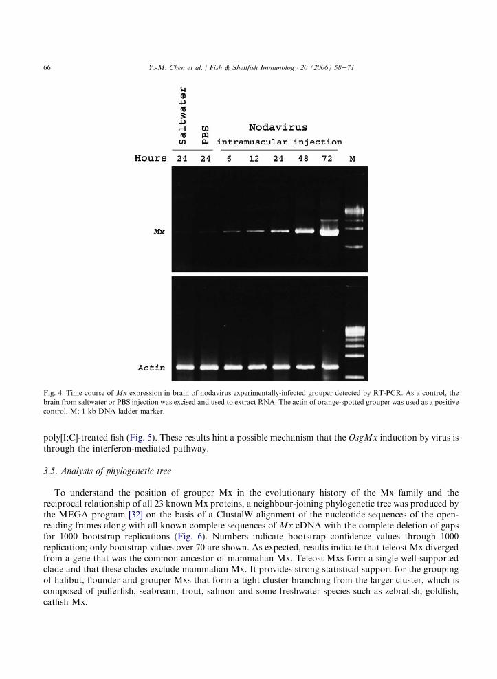
mL�1. The results demonstrate that intramuscular injection of nodavirus into grouper produces an increaseof Mx mRNA at 6 h in the brain, peaking at 72 h after injection (Fig. 4). In addition, no grouper infectedwith the pathogen Vibro anguillarum or LPS (at 10 mg mL�1) by injection shows Mx expression intheir brain. The OsgMx induction effects of nodavirus and iridovirus were comparable to those of
Fig. 3. RT-PCR revealing the overall distribution of OsgMx expression among different tissues. Expression of OsgMx mRNA healthy
grouper, and nodavirus naturally-infected grouper of severe mortality were obtained from hatchery farms in southern Taiwan.
Amplifications of Mx cDNA fragments were accomplished using equal number of PCR cycles. PCR products were obtained from
brain, eye, gill, heart, liver, spleen, intestine, kidney, and muscle. The b-actin of orange-spotted grouper was used as a positive control.
M; 1 kb DNA ladder marker.
66 Y.-M. Chen et al. / Fish & Shellfish Immunology 20 (2006) 58e71
poly[I:C]-treated fish (Fig. 5). These results hint a possible mechanism that the OsgMx induction by virus isthrough the interferon-mediated pathway.
3.5. Analysis of phylogenetic tree
To understand the position of grouper Mx in the evolutionary history of the Mx family and thereciprocal relationship of all 23 known Mx proteins, a neighbour-joining phylogenetic tree was produced bythe MEGA program [32] on the basis of a ClustalW alignment of the nucleotide sequences of the open-reading frames along with all known complete sequences of Mx cDNA with the complete deletion of gapsfor 1000 bootstrap replications (Fig. 6). Numbers indicate bootstrap confidence values through 1000replication; only bootstrap values over 70 are shown. As expected, results indicate that teleost Mx divergedfrom a gene that was the common ancestor of mammalian Mx. Teleost Mxs form a single well-supportedclade and that these clades exclude mammalian Mx. It provides strong statistical support for the groupingof halibut, flounder and grouper Mxs that form a tight cluster branching from the larger cluster, which iscomposed of pufferfish, seabream, trout, salmon and some freshwater species such as zebrafish, goldfish,catfish Mx.
Fig. 4. Time course of Mx expression in brain of nodavirus experimentally-infected grouper detected by RT-PCR. As a control, the
brain from saltwater or PBS injection was excised and used to extract RNA. The actin of orange-spotted grouper was used as a positive
control. M; 1 kb DNA ladder marker.

67Y.-M. Chen et al. / Fish & Shellfish Immunology 20 (2006) 58e71
4. Discussion
A cDNA from orange-spotted grouper, Epinephelus coioides, homolog of Mx has been identified andcharacterised. The grouper Mx is a 626 amino acid composed of four main functional domains: an amino-terminal tripartite GTP-binding motif GDQSSGKS, DLPG, TKPD, the dynamin family signatureLPRGSGIVTR, a Centra Interaction Domain (CID) and a carboxy-terminal leucine zipper motif. Theamino acid sequence had approximately 51 and 83% identity to the sequences of human and rainbow troutMx cDNA, respectively. Significant similarity between OsgMx and the vertebrate Mx family is limited tothe centra interaction domain and includes residues identified in human Mx as critical for GTPase activity,the dynamin family signature, and leucine zipper for protein-protein interaction. Accordingly, the deducedamino acid sequences of the grouper Mx GTPase domain shares 79e88% identity to those of the other fishMx genes and 66e69% identity to those of mammalian Mx genes. The CID region of grouper Mx proteinis less well-conserved showing 70e78% identity with those of the fish Mx proteins and only 36e45%
Fig. 5. Differential effect of nodavirus on transcription of OsgMx gene in the grouper. Effects of nodavirus, iridovirus, and Vibro
anguillarum naturally-infected grouper; poly[I:C] (100 mg per fish) and LPS (at 10 mg mL�1), duplicate group of five fish were
intramuscular injection on Mx expression in grouper brain.

68 Y.-M. Chen et al. / Fish & Shellfish Immunology 20 (2006) 58e71
sequence identity with those of mammalian Mx genes. Similar to the mammalian Mx leucine zipper motifsequences, a putative grouper leucine zipper motif is also present. The molecular basis of the antiviralactivity of Mx against a broad range of negative-sense RNA virus have been revealed by the extensivestudies of interferon-mediated antiviral protein expression in a variety of vertebrates, including mammalsand various fish species [20,22,31].
This study extended data to OsgMx and describes the structural and functional properties of the grouperMx promoter. Following the prediction of transcription factor binding motifs using computer programmes,TRANSFAC, binding motifs in the 5# region of OsgMx accumulate in a 752-bp nucleotide stretch 5#upstream of the translation initiation codon ATG. This region exhibited a promoter activity, which couldbe induced by treatment with poly[I:C]. Two sequences that conform to the most recent ISRE consensus, 5#A/GGTTTCN(1e2)TTTCC/T 3# are present in the grouper Mx promoter region [33]. The only other fishMx promoters cloned to date are those of rainbow trout, fugu and zebrafish [12,16,33]. The rainbow troutMx promoter contains only one ISRE, where the grouper, zebrafish, and fuguMx promoters each containtwo. The high degree of conservation that can be extended to the mammalian homolog Mx indicates anessential role for these promoter elements. Interestingly, the humanMxA promoter also has multiple ISREsinvolved in interferon-inducible pathway. Of the three ISREs found in the human MxA promoter, twoproximal ISREs contributed to interferon-inducibility cooperatively, whereas the distal ISRE is
Fig. 6. Neighbour-joining tree of Mx family of different organisms. The tree is based on an alignment corresponding to the full length
of the Mx protein. The numbers on the branches are bootstrap values. The GenBank accession codes of the sequence designations. The
accession numbers of sequences retrieved in this study are as follows: orange-spotted grouper (Epinephelus coioides) Mx, AY574372;
channel catfish (Ictalurus punctatus) Mx, AY095349; Japanese flounder (Paralichthys olivaceus) Mx, AB110446; Atlantic halibut
(Hippoglossus hippoglossus) Mx, AF245513; Fugu rubripes (Takifugu rubripes) Mx, AF525215; gilthead seabream (Sparus aurata) Mx,
AF491302; zebrafish (Danio rerio) Mx, AF533769; goldfish (Carassius auratus) Mx, AY303813; rainbow trout (Oncorhynchus mykiss)
Mx1, U30253; Mx2, U47945; Mx3, U47946; Atlantic salmon (Salmo salar) Mx1, U66475; Mx2, U66476; Mx3, U66477; human MxA,
M30817; MxB, M30818; mouse Mx1, M12279; Mx2, AB029920; rat Mx1, X52711; Mx2, X52712; Mx3, X52713; sheep (Ovis aries) Mx,
AF399856; cow (Bos taurus) Mx, U88329.

69Y.-M. Chen et al. / Fish & Shellfish Immunology 20 (2006) 58e71
non-functional [34]. Concentrating the presence of multiple ISREs enhances the sensitivity of the promoterto induction by interferon. As expected from the high identity with the corresponding consensus sequences,it was demonstrated that OsgMx gene regulation is highly conserved between fish and mammals [33]. Thepresent data show that, as found in all known Mx, OsgMx is involved in an interferon-dependent proteinpathway whose function is required to protect the host in cells infected by piscine virus infection. Thus, theantiviral activity of mammalian Mx could be an ancient and conserved function for this protein. However,there are intriguing differences between the mammalian and piscine versions of Mx. Search in thetranscription factor database (TRANSFAC) reveals that two potential sites involved in the regulation bySTAT and NFkB regulator are present in this region [35]. Similar to the bovine Mx1 gene, the promoter ofgrouper Mx contains highly conserved sequences that could be the binding sites for ISRE and NFkB [36].Work is ongoing to more clearly understand how these elements are influenced. Additionally, one on-goingattempts to identify other regulatory elements, for example, the endogenous enhancer that controls theexpression of OsgMx.
Expression of the grouper Mx gene has shown that it is induced in vivo upon treatment with interferoninducers such as piscine virus, nodavirus, iridovirus, poly[I:C], but not in the infecting-bacteria. To assesswhether the OsgMx mRNAs observed in various grouper tissues are truly inducible, total RNAs isolatedfrom healthy grouper have been analysed in parallel with nodavirus naturally-infected grouper. Comparedto nodavirus naturally-infected grouper, higher levels of Mx expression have been seen in eyes, gill andheart in healthy control grouper. On the other hand, significant expression have been seen in eyes, brain,gill, heart, liver, spleen, kidney and intestine of nodavirus naturally-infected or healthy grouper followingnodavirus experimentally-challenged fish. Transcription of OsgMx mRNA in healthy grouper is restrictedto eyes, gill and heart. Moreover, two additional bands are produced from the eyes and gill RNA on thehealthy grouper. Fish Mx is mainly distributed in gill which is rich in leukocytes, and in eye where the riskof pathogen invasion is high. It is possible that grouper Mx plays an important role in host defencemechanisms against infectious diseases, and developing grouper can respond to proper signals to modulateMx gene activities. This finding is consistent with the highly conserved pattern of Mx induction previouslyreported in flounder and pufferfish.
This finding of OsgMx and its possible role in virus induction provides a new model for the study of thefunction of a key antiviral gene. Thus far, antiviral activity has been established only for Mx proteins inmouse, rat, human, and chicken against several viruses [37,38]. The antiviral activity of fish Mx proteinsremains unclear. Trobridge et al. [8] reported that, in rainbow trout, three different types of Mx have noapparent antiviral activity against rhabdovirus IHNV. Although a challenge experiment has been alsoperformed to show an effect on resistance against infectious salmon anaemia virus ISAV, and resistancewould correlate with expression of Atlantic salmon Mx protein induced by poly[I:C], yet there is no clearevidence that the Mx protein is involved in the inhibition of ISAV [39]. Nevertheless, syntheticoligodeoxynucleotides containing CpG motifs have been shown to induce short-term expression of MxmRNA in Atlantic salmon and to provide a degree of protection against challenge with IPNV [40]. This isthe first report that the induction of Mx expression in Atlantic salmon parr was investigated followinginjection of vibrio bacterin, composed of Vibrio anguillarum serotypes I and II-ordalii, at two differenttemperatures in comparison with the potent Mx inducer poly[I:C] [41,42]. Some of study aimed to identifysubstances which are already known to be immunostimulants in fish as being stimulatory for Mx. On theother hand, a study reported that the replication of fish rhabdoviruses has been reduced in a fish cell linetransfected with Japanese flounder Mx proteins [20]. Recently, CHSE-214 cells constitutively expressingsalmon Mx1 showed a severely reduced IPNV-induced cytopathic effect and inhibited virus proteinsynthesis [21]. In our results of promoter assay (Fig. 2B) and the OsgMx induction effects of poly[I:C]-treated fish (Fig. 5), the grouper Mx also promoter could be induced by poly[I:C]. And this induction maybe through the highly conserved binding sites for ISRE in the grouper Mx promoter. Therefore, given thatmany aspects of the conserved nature of the OsgMx gene correlate between fish and mammals, it is possible

70 Y.-M. Chen et al. / Fish & Shellfish Immunology 20 (2006) 58e71
that the grouper Mx protein might be similarly regulated by an interferon-dependent defence systemagainst viruses.
Acknowledgements
We are grateful to Mr. Fu-Ping Chang and Mr. Nai-Heng Yu for critical comments on the manuscript.We thank Dr. Cho-Fat Hui, Institute of Zoology, Academia Sinica, for helpful comments on thismanuscript. This research was supported by National Science Council and the National Science andTechnology Program for Agricultural Biotechnology (NSC92-2317-B006-008).
References
[1] Leong JAC, Trobridg GD, Kim CH, Johnson M, Simon B. Interferon-inducible Mx proteins in fish. Immunol Rev 1998;
166:349e63.
[2] Falk K, Namork E, Rimstad E, Mjaaland S, Dannevig BH. Characterization of infectious salmon anemia virus, an orthomyxo-
like virus isolated from Atlantic salmon (Salmo salar L.). J Virol 1997;71:9016e23.[3] Kochs G, Trost M, Janzen C. MxA GTPase: oligomerization and GTP-dependent interaction with viral RNP target structures.
Methods 1998;15:255e63.
[4] Arnheiter H, Frese M, Kambadur R, Meier E, Haller O. Mx transgenic mice-animal models of health. Curr Top Microbiol
Immunol 1996;206:119e47.[5] van der Bliek AM. Functional diversity in the dynamin family. Trends Cell Biol 1999;9:96e102.
[6] Praefcke GJK, Geyer M, Schwemmle M, Kalbitzer HR, Herrmann C. Nucleotide-binding characteristics of human guanylate-
binding protein 1 (hGBP1) and identification of the third GTP-binding motif. J Mol Biol 1999;292:321e32.[7] Trobridge GD, Leong JA. Characterization of a rainbow trout Mx gene. J Interferon Cytokine Res 1995;15:691e702.
[8] Trobridge GD, Chiou PP, Leong JA. Cloning of the rainbow trout (Oncorhynchus mykiss) Mx2 and Mx3 cDNAs and
characterization of trout Mx protein expression in salmon cells. J Virol 1997;71:5304e11.
[9] Robertsen B, Trobridg GD, Leong JA. Molecular cloning of double-stranded RNA inducible Mx genes from Atlantic salmon
(Salmo salar L.). Dev Comp Immunol 1997;21:397e412.
[10] Lee JY, Hirono I, Aoki T. Cloning and analysis of expression of Mx cDNA in Japanese flounder, Paralichthys olivaceus. Dev
Comp Immunol 2000;24:407e15.
[11] Jensen V, Robertsen B. Cloning of an Mx cDNA from Atlantic halibut (Hippoglossus hippoglossus) and characterization of Mx
mRNA expression in response to double-stranded RNA or infectious pancreatic necrosis virus. J Interferon Cytokine Res
2000;20:701e10.
[12] Yap WH, Tay A, Brenner S, Venkatesh B. Molecular cloning of the pufferfish (Takifugu rubripes) Mx gene and functional
characterization of its promoter. Immunogenetics 2003;54:705e13.
[13] Tafalla C, Aranguren R, Secombes CJ, Figueras A, Novoa B. Cloning and analysis of expression of a gilthead sea bream (Sparus
aurata) Mx cDNA. Fish Shellfish Immunol 2004;16:11e24.
[14] Plant KP, Thune RL. Cloning and characterization of a channel catfish (Ictalurus punctatus) Mx gene. Fish Shellfish Immunol
2004;16:391e405.
[15] Zhang Y, Zhang Q, Xu D, Hu C, Gui J. Identification of antiviral-relevant genes in the cultured fish cells induced by UV-
inactivated virus. Chin Sci Bull 2003;48:581e8.
[16] Altmann ST, Mellon MT, Johnson MC, Paw BH, Trede NS, Zon LI, et al. Cloning and characterization of an Mx gene and its
corresponding promoter from the zebrafish, Danio rerio. Dev Comp Immunol 2004;28:295e306.
[17] Pitossi F, Blank A, Schroder A, Schwarz A, Hussi P, Schwemmle M, et al. A functional GTP-binding motif is necessary for
antiviral activity of Mx proteins. J Virol 1993;67:6726e32.
[18] Chen MS, Obar RA, Schroeder CC, Austin TW, Poodry CA, Wasdsworth SC, et al. Multiple forms of dynamin are encoded by
shibire, a Drosophila gene involved in endocytosis. Nature 1991;351:583e6.
[19] O’Shea EK, Rutkowski R, Kim PS. Mechanism of specificity in the Fos-Jun oncoprotein heterodimer. Cell 1992;68:699e708.
[20] Caipang CMA, Hirono I, Aoki T. In vitro inhibition of fish rhabdoviruses by Japanese flounder, Paralichthys olivaceus Mx.
Virology 2003;317:373e82.
[21] Larsen R, Rokenes TP, Robertsen B. Inhibition of infectious pancreatic necrosis virus replication by Atlantic salmon Mx1
protein. J Virol 2004;78:7938e44.

71Y.-M. Chen et al. / Fish & Shellfish Immunology 20 (2006) 58e71
[22] Altmann SM, Mellon MT, Distl DL, Kim CH. Molecular and functional analysis of an interferon gene from the zebrafish, Danio
rerio. J Virol 2003;77:1992e2002.
[23] Munday BL, Nakai T. Special topic review: nodaviruses as pathogens in larval and juvenile marine finfish. World J Microbiol
Biotechnol 1997;13:375e81.
[24] Skliris GP, Krondiris JV, Sideris DC, Shinn AP, Starkey WG, Richards RH. Phylogenetic and antigenic characterization of new
fish nodavirus isolates from Europe and Asia. Virus Res 2001;75:59e67.
[25] Iwamoto T, Mise K, Mori K, Arimoto M, Nakai T, Okuno T. Establishment of an infectious RNA transcription system for
Striped jack nervous necrosis virus, the type species of the betanodaviruses. J Gen Virol 2001;82:2653e62.
[26] Mori K, Nakai T, Arimoto M, Mushiake K, Furusawa I. Properties of a new virus belonging to Nodaviridae found in larval
striped jack (Pseudocaranx dentex) with nervous necrosis. Virology 1992;187:368e71.
[27] Chomezynski P, Sacchi N. Single-step method of RNA isolation by acid guanidiniumthiocyanate-phenol-chloroform extraction.
Anal Biochem 1987;162:156e9.
[28] Frohman MA, Dush MK, Martin GR. Rapid production of full-length cDNA from rare transcripts: Amplification using a single
gene-specific oligonucleotide primer. Proc Natl Acad Sci USA 1988;85:8998e9002.[29] Sambrook J, Fritsch EF, Maniatis T. Molecular Cloning: A Laboratory Manual, vols. 1e3. New York: Cold Spring Harbor
Laboratory Press; 1989.
[30] Wingender E, Chen X, Hehl R, Karas H, Liebich I, Matys V, et al. TRANSFAC: an integrated system for gene expression
regulation. Nucleic Acids Res 2000;28:316e9.[31] Collet B, Boudinot P, Benmansour A, Secombes CJ. An Mx1 promoter-reporter system to study interferon pathways in rainbow
trout. Dev Comp Immunol 2004;28:793e801.
[32] Kumar S, Tamura K, Nei M. MEGA: molecular evolutionary genetics analysis software for microcomputers. Comput Appl
Biosci 1994;10:189e91.[33] Collet B, Secombes CJ. The rainbow trout (Oncorhynchus mykiss) Mx1 promoter. Structural and functional characterization. Eur
J Biochem 2001;268:1577e84.
[34] Nakade K, Handa H, Nagata K. Promoter structure of the MxA gene that confers resistance to influenza virus. FEBS Letters
1997;418:315e8.
[35] Katze MG, He Y, Gale Jr M. Viruses and interferon: a fight for supremacy. Nat Rev Immunol 2002;2:675e87.
[36] Gerardin JA, Baise EA, Pire GA, Leroy MPP, Desmecht DJM. Genomic structure, organisation, and promoter analysis of the
bovine (Bos taurue) Mx1 gene. Gene 2004;326:67e75.[37] Asano A, Jin HK, Watanabe T. Mouse Mx2 gene: organization, mRNA expression and the role of the interferon-response
promoter in its regulation. Gene 2003;306:105e13.
[38] Schumacher B, Bernasconi D, Schultz U, Staeheli P. The chicken Mx promoter contains an ISRE motif and confers interferon
inducibility to a reporter gene in chick and monkey cells. Virology 1994;203:144e8.[39] Sommer AI, Mennen S. Propagation of infectious salmon anaemia virus in Atlantic salmon, Salmo salar L., head kidney
macrophages. J Fish Dis 1996;19:179e83.
[40] Jorgensen JB, Johansen LH, Steiro K, Johansen A. CpG DNA induces protective antiviral immune responses in Atlantic salmon
(Salmo salar L.). J Virol 2003;77:11471e9.
[41] Salinas I, Lockhart K, Bowden TJ, Collet B, Secombes CJ, Ellis AE. An assessment of immunostimulants as Mx inducers in
Atlantic salmon (Salmo salar L.) parr and the effect of temperature on the kinetics of Mx responses. Fish Shellfish Immunol
2004;17:159e70.[42] Acosta F, Lockhart K, Gahlawat SK, Real F, Ellis AE. Mx expression in Atlantic salmon (Salmo salar L.) parr in response to
Listonella anguillarum bacterin, lipopolysaccharide and chromosomal DNA. Fish Shellfish Immunol 2004;17:255e63.