Coeliac disease-associated risk variants in TNFAIP3 and REL implicate altered NF- B signalling
6
Coeliac disease-associated risk variants in TNFAIP3 and REL implicate altered NF-kB signalling G Trynka, 1 A Zhernakova, 2 J Romanos, 1 L Franke, 1,3 K A Hunt, 3 G Turner, 4 M Bruinenberg, 1 G A Heap, 3 M Platteel, 1 A W Ryan, 4 C de Kovel, 2 G K T Holmes, 5 P D Howdle, 6 J R F Walters, 7 D S Sanders, 8 C J J Mulder, 9 M L Mearin, 10 W H M Verbeek, 9 V Trimble, 4 F M Stevens, 11 D Kelleher, 4 D Barisani, 12 M T Bardella, 13,14 R McManus, 4 D A van Heel, 3 C Wijmenga 1,2 c Two supplementary files giving additional data for the SNPs in this study are published online only at http://gut.bmj. com/content/vol58/issue8 1 Genetics Department, University Medical Centre and University of Groningen, The Netherlands; 2 Complex Genetics Section, Department of Biomedical Genetics, University Medical Centre Utrecht, The Netherlands; 3 Institute of Cell and Molecular Science, Barts and The London School of Medicine and Dentistry, London, UK; 4 Departments of Clinical Medicine, Institute of Molecular Medicine, Trinity College Dublin, Ireland; 5 Department of Gastroenterology, Derbyshire Royal Infirmary, Derby, UK; 6 Academic Medical Unit, Leeds Institute of Molecular Medicine, St James’s University Hospital, Leeds, UK; 7 Gastroenterology Section, Imperial College London, Hammersmith Hospital, London, UK; 8 Department of Gastroenterology and Liver Unit, Royal Hallamshire Hospital, Sheffield, UK; 9 Department of Gastroenterology, VU Medical Center, Amsterdam, The Netherlands; 10 Department of Paediatric Gastroenterology, Leiden University Medical Center, Leiden, The Netherlands; 11 Department of Medicine, National University of Ireland, Galway, Ireland; 12 Department of Experimental Medicine, Faculty of Medicine, University of Milano- Bicocca, Monza, Italy; 13 Fondizione IRCCS Ospedale Maggiore Policlinico, Mangiagalli e Regina Elena, Milan, Italy; 14 Department of Medical Sciences, University of Milan, Italy Correspondence to: Professor C Wijmenga, Department of Genetics, University Medical Centre Groningen and University of Groningen, Internal post HPC CB50, PO Box 30001, 9700 RB Groningen, The Netherlands; [email protected] GTr and AZ contributed equally to the paper. Revised 6 February 2009 Accepted 12 February 2009 Published Online First 24 February 2009 ABSTRACT Objective: Our previous coeliac disease genome-wide association study (GWAS) implicated risk variants in the human leucocyte antigen (HLA) region and eight novel risk regions. To identify more coeliac disease loci, we selected 458 single nucleotide polymorphisms (SNPs) that showed more modest association in the GWAS for genotyping and analysis in four independent cohorts. Design: 458 SNPs were assayed in 1682 cases and 3258 controls from three populations (UK, Irish and Dutch). We combined the results with the original GWAS cohort (767 UK cases and 1422 controls); six SNPs showed association with p,1 6 10 204 and were then genotyped in an independent Italian coeliac cohort (538 cases and 593 controls). Results: We identified two novel coeliac disease risk regions: 6q23.3 (OLIG3-TNFAIP3) and 2p16.1 (REL), both of which reached genome-wide significance in the combined analysis of all 2987 cases and 5273 controls (rs2327832 p = 1.3 6 10 208 , and rs842647 p = 5.2 6 10 207 ). We investigated the expression of these genes in the RNA isolated from biopsies and from whole blood RNA. We did not observe any changes in gene expression, nor in the correlation of genotype with gene expression. Conclusions: Both TNFAIP3 (A20, at the protein level) and REL are key mediators in the nuclear factor kappa B (NF-kB) inflammatory signalling pathway. For the first time, a role for primary heritable variation in this important biological pathway predisposing to coeliac disease has been identified. Currently, the HLA risk factors and the 10 established non-HLA risk factors explain ,40% of the heritability of coeliac disease. Coeliac disease is a common intestinal inflamma- tory disorder, characterised by intolerance to diet- ary gluten protein from wheat, and related proteins from barley and rye. It is the best understood human leucocyte antigen (HLA)- associated disorder. Coeliac disease is rather special because it shares its pathogenesis with other autoimmune diseases (such as type 1 diabetes (T1D) and rheumatoid arthritis (RA)) and with intestinal inflammatory diseases (such as Crohn’s disease). Shared genetic risk factors for both coeliac disease and Crohn’s disease, as well as for coeliac disease and autoimmunity have been reported. 1–4 To search for genetic risk factors, we recently performed a genome-wide association study (GWAS) in coeliac disease, followed by replication of the 1020 most strongly associated single nucleotide polymorphisms (SNPs) in case–control cohorts from three populations. These studies led to the discovery of eight new non-HLA loci. 15 The results led to three observations: 1 c only three of the confirmed loci were presented by SNPs located in the top-100 associated signals in the GWAS, whereas three more associated SNPs ranked below the top-500, and one SNP ranked as low as 1004 in the initial GWAS 2 c seven of the eight new loci contained immune- related genes, four of which are cytokines or cytokine receptors (IL2/IL21, IL18RAP, IL12A, and the CCR1/CCR3 cluster locus) 3 c four of the new loci (IL2-IL21, IL18RAP, CCR3 and SH2B3) are shared by other autoimmune and inflammatory disorders 1–3 5 6 These three observations prompted us to start a second replication study in which we followed up even lower ranking SNPs from our coeliac disease GWAS, and focused on the involvement of the immune pathways in the pathogenesis of coeliac disease (fig 1). We therefore enriched our SNP set with SNPs that showed association to coeliac disease in our GWAS and that mapped to the immune-related genes (mostly interleukins and their receptors). In addition, we sought shared autoimmune and inflammatory genes by investi- gating an overlap between SNPs associated in our coeliac disease GWAS and to either T1D, RA or Crohn’s disease in the Wellcome Trust Case Control Consortium (WTCCC) GWAS data. 7 With such a study design we successfully identified two novel, genome-wide significant, coeliac disease loci: the intergenic region on 6q23.3 located in the proximity of the TNFAIP3 gene, and 2p16.1, mapping to the second intron of the REL gene. Both loci indicate an as yet unrecognised role for the nuclear factor kappa B (NF-kB) signalling pathway in the pathogenesis of coeliac disease. MATERIALS AND METHODS Subject DNA DNA was extracted from whole blood, except for the 1958 cohort control samples, which were lymphoblastoid cell line DNA, and 374 cases and 176 controls from the UK2 collection, which were Oragene saliva DNA. Whole-genome amplified (WGA) blood DNA was used for 194 Irish cases Small intestine 1078 Gut 2009;58:1078–1083. doi:10.1136/gut.2008.169052
Transcript of Coeliac disease-associated risk variants in TNFAIP3 and REL implicate altered NF- B signalling

Coeliac disease-associated risk variants in TNFAIP3and REL implicate altered NF-kB signalling
G Trynka,1 A Zhernakova,2 J Romanos,1 L Franke,1,3 K A Hunt,3 G Turner,4
M Bruinenberg,1 G A Heap,3 M Platteel,1 A W Ryan,4 C de Kovel,2 G K T Holmes,5
P D Howdle,6 J R F Walters,7 D S Sanders,8 C J J Mulder,9 M L Mearin,10
W H M Verbeek,9 V Trimble,4 F M Stevens,11 D Kelleher,4 D Barisani,12 M T Bardella,13,14
R McManus,4 D A van Heel,3 C Wijmenga1,2c Two supplementary filesgiving additional data for theSNPs in this study are publishedonline only at http://gut.bmj.com/content/vol58/issue8
1 Genetics Department,University Medical Centre andUniversity of Groningen, TheNetherlands; 2 Complex GeneticsSection, Department ofBiomedical Genetics, UniversityMedical Centre Utrecht, TheNetherlands; 3 Institute of Celland Molecular Science, Barts andThe London School of Medicineand Dentistry, London, UK;4 Departments of ClinicalMedicine, Institute of MolecularMedicine, Trinity College Dublin,Ireland; 5 Department ofGastroenterology, DerbyshireRoyal Infirmary, Derby, UK;6 Academic Medical Unit, LeedsInstitute of Molecular Medicine,St James’s University Hospital,Leeds, UK; 7 GastroenterologySection, Imperial College London,Hammersmith Hospital, London,UK; 8 Department ofGastroenterology and Liver Unit,Royal Hallamshire Hospital,Sheffield, UK; 9 Department ofGastroenterology, VU MedicalCenter, Amsterdam, TheNetherlands; 10 Department ofPaediatric Gastroenterology,Leiden University Medical Center,Leiden, The Netherlands;11 Department of Medicine,National University of Ireland,Galway, Ireland; 12 Department ofExperimental Medicine, Faculty ofMedicine, University of Milano-Bicocca, Monza, Italy;13 Fondizione IRCCS OspedaleMaggiore Policlinico, Mangiagallie Regina Elena, Milan, Italy;14 Department of MedicalSciences, University of Milan, Italy
Correspondence to:Professor C Wijmenga,Department of Genetics,University Medical CentreGroningen and University ofGroningen, Internal post HPCCB50, PO Box 30001, 9700 RBGroningen, The Netherlands;[email protected]
GTr and AZ contributed equallyto the paper.
Revised 6 February 2009Accepted 12 February 2009Published Online First24 February 2009
ABSTRACTObjective: Our previous coeliac disease genome-wideassociation study (GWAS) implicated risk variants in thehuman leucocyte antigen (HLA) region and eight novel riskregions. To identify more coeliac disease loci, we selected458 single nucleotide polymorphisms (SNPs) that showedmore modest association in the GWAS for genotyping andanalysis in four independent cohorts.Design: 458 SNPs were assayed in 1682 cases and 3258controls from three populations (UK, Irish and Dutch). Wecombined the results with the original GWAS cohort (767UK cases and 1422 controls); six SNPs showedassociation with p,1610204 and were then genotyped inan independent Italian coeliac cohort (538 cases and 593controls).Results: We identified two novel coeliac disease riskregions: 6q23.3 (OLIG3-TNFAIP3) and 2p16.1 (REL), bothof which reached genome-wide significance in thecombined analysis of all 2987 cases and 5273 controls(rs2327832 p = 1.3610208, and rs842647p = 5.2610207). We investigated the expression of thesegenes in the RNA isolated from biopsies and from wholeblood RNA. We did not observe any changes in geneexpression, nor in the correlation of genotype with geneexpression.Conclusions: Both TNFAIP3 (A20, at the protein level)and REL are key mediators in the nuclear factor kappa B(NF-kB) inflammatory signalling pathway. For the firsttime, a role for primary heritable variation in this importantbiological pathway predisposing to coeliac disease hasbeen identified. Currently, the HLA risk factors and the 10established non-HLA risk factors explain ,40% of theheritability of coeliac disease.
Coeliac disease is a common intestinal inflamma-tory disorder, characterised by intolerance to diet-ary gluten protein from wheat, and relatedproteins from barley and rye. It is the bestunderstood human leucocyte antigen (HLA)-associated disorder. Coeliac disease is rather specialbecause it shares its pathogenesis with otherautoimmune diseases (such as type 1 diabetes(T1D) and rheumatoid arthritis (RA)) and withintestinal inflammatory diseases (such as Crohn’sdisease). Shared genetic risk factors for both coeliacdisease and Crohn’s disease, as well as for coeliacdisease and autoimmunity have been reported.1–4
To search for genetic risk factors, we recentlyperformed a genome-wide association study(GWAS) in coeliac disease, followed by replication
of the 1020 most strongly associated singlenucleotide polymorphisms (SNPs) in case–controlcohorts from three populations. These studies ledto the discovery of eight new non-HLA loci.1 5 Theresults led to three observations:1
c only three of the confirmed loci were presentedby SNPs located in the top-100 associatedsignals in the GWAS, whereas three moreassociated SNPs ranked below the top-500,and one SNP ranked as low as 1004 in theinitial GWAS2
c seven of the eight new loci contained immune-related genes, four of which are cytokines orcytokine receptors (IL2/IL21, IL18RAP, IL12A,and the CCR1/CCR3 cluster locus)3
c four of the new loci (IL2-IL21, IL18RAP, CCR3and SH2B3) are shared by other autoimmuneand inflammatory disorders1–3 5 6
These three observations prompted us to start asecond replication study in which we followed upeven lower ranking SNPs from our coeliac diseaseGWAS, and focused on the involvement of theimmune pathways in the pathogenesis of coeliacdisease (fig 1). We therefore enriched our SNP setwith SNPs that showed association to coeliacdisease in our GWAS and that mapped to theimmune-related genes (mostly interleukins andtheir receptors). In addition, we sought sharedautoimmune and inflammatory genes by investi-gating an overlap between SNPs associated in ourcoeliac disease GWAS and to either T1D, RA orCrohn’s disease in the Wellcome Trust CaseControl Consortium (WTCCC) GWAS data.7
With such a study design we successfullyidentified two novel, genome-wide significant,coeliac disease loci: the intergenic region on6q23.3 located in the proximity of the TNFAIP3gene, and 2p16.1, mapping to the second intron ofthe REL gene. Both loci indicate an as yetunrecognised role for the nuclear factor kappa B(NF-kB) signalling pathway in the pathogenesis ofcoeliac disease.
MATERIALS AND METHODSSubject DNADNA was extracted from whole blood, except forthe 1958 cohort control samples, which werelymphoblastoid cell line DNA, and 374 cases and176 controls from the UK2 collection, which wereOragene saliva DNA. Whole-genome amplified(WGA) blood DNA was used for 194 Irish cases
Small intestine
1078 Gut 2009;58:1078–1083. doi:10.1136/gut.2008.169052

and 18 Dutch cases. Genotype cluster theta values for WGADNA were similar to blood DNA, for a small fraction of markersthe intensity (R) was lower.
Detailed characteristics of UKGWAS, UK2, Irish, Dutch andItalian samples are provided in table 1 and previously publishedstudies.1 5 8 Informed consent was obtained from all subjects.
SNP selectionThree groups of SNPs were selected for the genotyping:c To follow up our GWAS SNPs were selected with p values
between p.0.000275 and p,0.004 (indicated as SNP-group1 in table 2 and supplementary data 1; also indicated ascategory ‘‘WGAtop2000_noWTCCCassoc’’ in supplemen-tary data 1) (n = 300).
c SNPs from immune-related genes associated in GWAS withp,0.05 (indicated as SNP-group 2 in table 2 and supplemen-tary data 1; also indicated as category ‘‘WGArepli_ImmuneGenes’’ in supplementary data 1) (n = 55).
c SNPs within the top-3000 coeliac–GWAS ranking(p,0.009), which showed also association to either type 1diabetes (T1D), rheumatoid arthritis (RA) or Crohn’sdisease in the WTCCC study with p,0.05 (n = 103). Forcalculation of association in the WTCCC cohort we usedimputed genotypes (WTCCC data accessed on 21November 2007). T1D, RA and Crohn’s cases werecompared to the blood donor WTCCC control cohort(n = 1500). The 1958 birth cohort was excluded from theWTCCC analysis, as the majority of these samples over-lapped with the coeliac disease–GWAS control cohort. ThisSNP category is indicated as SNP-group 3 in table 2 andsupplementary data 1; also indicated as category‘‘WGAtop3000associatedWTCCC’’ in supplementary data 1).
Golden Gate genotypingGenotyping of all samples was performed following themanufacturer’s protocol. Genotyping data and clustering wasperformed in BeadStudio. Clustering clouds were manuallyinvestigated and adjusted if necessary. Four hundred and fifty-eight SNPs were included in the genotype analysis. Ten SNPswith ,95% call rate, because of poor amplification or poorgenotype cloud clustering, were excluded. For the top-6associated SNPs we investigated clustering in subgroups ofblood, saliva and lymphoblastoid cell line DNAs. All groupsshowed similar patterns and comparable theta values. All platesincluded one duplicate sample to control for plate swaps. SixSNPs were out of Hardy–Weinberg equilibrium (p.0.001) andwere excluded from further analysis. In total, 1682 cases and3258 controls from the three populations (replication cohort 1;R1) were successfully genotyped for 442 SNPs.
Genotype concordanceA single control DNA sample was included in each 96-wellplate. Genotype concordance for this sample was 99.9% for 45replicates of 442 SNPs.
Additional genotype quality controlPairwise comparisons of identity-by-descent were made for allsamples (UK2, Irish and Dutch) using PLINK v1.02. We detectedthe same proportion of first-degree relatives as described byHunt et al and excluded one sample from each pair of first-degree relatives from the entire dataset in the current study.1 Allof the top association findings were in Hardy–Weinbergequilibrium in controls.
Figure 1 Scheme representing thesingle nucleotide polymorphisms (SNPs)selected for the second coeliac genome-wide association study (GWAS) follow-upstudy. WTCCC, Wellcome Trust CaseControl Consortium.
Table 1 Subjects included in our replication study
UKGWAS UK2 Dutch Irish Italian
TotalPhase I – GWAS Replication cohort 1Replicationcohort 2
Coeliac cases 767 724 514 444 538 2987
Male/female 213/554 169/555 168/346 142/302 132/406
Controls 1422 1398 900 960 593 5273
Male/female 720/702 464/934 550/350 284/676 225/368
TOTAL 2189 2122 1414 1404 1131 8260
GWAS, genome-wide association study.
Small intestine
Gut 2009;58:1078–1083. doi:10.1136/gut.2008.169052 1079
Taqman genotypingReplication cohort 2 (Italian population) was genotyped usingTaqMan probes and primers developed by Applied Biosystems,on an ABI 7900HT system (Applied Biosystems, Nieuwerkerk a/d IJssel, the Netherlands). Genotyping was performed followingthe manufacturer’s specifications. DNA samples were processedin 384-well plates and each plate contained eight negativecontrols and 16 genotyping controls (four duplicates of fourdifferent samples obtained from the Centre d’Etude duPolymorphisme Humain (CEPH), Paris, France).
Genotype statistical analysisCochran–Mantel–Haenszel allele count x2 association testswere performed using PLINK9 with four clusters: UKGWAS(Infinium assay), UK2, IRISH and DUTCH (Golden Gate assay)collections. All p values are two-tailed. The Cochran–Mantel–Haenszel allele count x2 association test implicated in SPSS v 15was used for combined analysis of association of the coeliacdisease cohort (including R2 (Italian) samples), and for analysisof the combined autoimmune cohort. Using Tarone’s statistic inSPSS v 15, we tested for heterogeneity of odds ratios betweenthe different coeliac disease cohorts for the SNPs reported intable 2. The odds ratios differed significantly between cohorts(p = 0.007) only for rs1160542. Haplotypes and linkage dis-equilibrium (LD) blocs were defined and analysed usingHaploview v4.1.10
Intestinal biopsy expression analysisThe duodenal tissue biopsies from 12 healthy controls, 12untreated coeliac individuals with Marsh III and 12 treatedcoeliac individuals with Marsh 0 were collected in RNAlater(Applied Biosystems/Ambion, Austin, Texas, USA). RNA wasextracted using TRIzol (Invitrogen, Carlsbad, California, USA)and glass beads, hybridised to HumanRef-8v2 arrays andanalysed as previously described1
Quality control of whole blood PAXgene dataThe whole blood PAXgene expression data from 110 uniquecoeliac individuals, who were also genotyped in the UKGWAS,was isolated, hybridized to the Illumina HumanRef-8v2 arraysand analysed as previously described.11
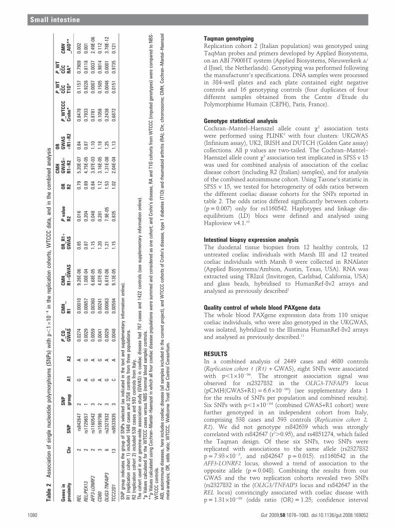
RESULTSIn a combined analysis of 2449 cases and 4680 controls(Replication cohort 1 (R1) + GWAS), eight SNPs were associatedwith p,1610204. The strongest association signal wasobserved for rs2327832 in the OLIG3-TNFAIP3 locus(pCMH(GWAS+R1) = 6.6610206) (see supplementary data 1for the results of SNPs per population and combined results).Six SNPs with p,1610204 (combined GWAS+R1 cohort) werefurther genotyped in an independent cohort from Italy,comprising 538 cases and 593 controls (Replication cohort 2,R2). We did not genotype rs842639 which was stronglycorrelated with rs842647 (r2.0.95), and rs4851274, which failedthe Taqman design. Of these six SNPs, two SNPs werereplicated with associations to the same allele (rs2327832p = 7.9361025, and rs842647 p = 0.015). rs1160542 in theAFF3-LONRF2 locus, showed a trend of association to theopposite allele (p = 0.048). Combining the results from ourGWAS and the two replication cohorts revealed two SNPs(rs2327832 in the (OLIG3/TNFAIP3 locus and rs842647 in theREL locus) convincingly associated with coeliac disease withp = 1.31610208 (odds ratio (OR) = 1.25; confidence intervalTa
ble
2A
ssoc
iatio
nof
sing
lenu
cleo
tide
poly
mor
phis
ms
(SN
Ps)
with
p,16
102
4in
the
repl
icat
ion
coho
rts,
WTC
CC
data
,an
din
the
com
bine
dan
alys
is
Gen
esin
prox
imit
yC
hrS
NP
SN
Pgr
oup
A1
A2
P_C
D_
GW
AS
CM
H_
R1
CM
H_
R1+
GW
AS
OR
_R1+
GW
AS
Pva
lue
R2
OR
R2
CM
HG
WA
S+
R1+
R2
OR
GW
AS
+R1+
R2
P_W
TCC
CC
rohn
*
P_W
TC
CC
T1D
*
P_W
TC
CC
RA
*C
MH
_AID
**
REL
2rs
8426
472
GA
0.02
740.
0001
09.
26E-
060.
850.
016
0.79
5.20
E-07
0.84
0.84
780.
1157
0.79
090.
002
REL
/PEX
132
rs17
2965
71
AG
0.00
290.
0065
71.
00E-
040.
870.
204
0.89
4.75
E-05
0.87
0.79
330.
9226
0.81
180.
001
AFF
3-LO
NR
F22
rs11
6054
23
GA
0.00
590.
0036
06.
68E-
051.
150.
048
0.84
3.97
E-03
1.10
0.87
810.
0007
0.00
372.
49E-
06
CD
803
rs15
9979
62
AG
0.00
410.
0024
14.
07E-
051.
200.
281
1.12
3.16
E-05
1.18
0.10
560.
1505
0.98
140.
112
OLI
G3-
TNFA
IP3
6rs
2327
832
3G
A0.
0029
0.00
063
6.61
E-06
1.21
7.9E
-05
1.53
1.31
E-08
1.25
0.24
380.
0046
0.00
013.
78E-
12
TCC
22D
113
rs95
3393
53
CA
0.00
480.
0059
49.
13E-
051.
150.
835
1.02
2.04
E-04
1.13
0.68
720.
0151
0.97
350.
121
SN
Pgr
oup
indi
cate
sth
egr
oup
ofS
NPs
sele
cted
(as
indi
cate
din
the
text
and
supp
lem
enta
ryin
form
atio
non
line)
.R
1(r
eplic
atio
nco
hort
1)in
clud
ed16
48ca
ses
and
3258
cont
rols
from
thre
epo
pula
tions
.R
2(r
eplic
atio
nco
hort
2)in
clud
ed53
8ca
ses
and
593
cont
rols
from
Italy
.Th
eco
hort
used
inou
rge
nom
e-w
ide
asso
ciat
ion
stud
y(G
WA
S)
inco
elia
cdi
seas
eha
d76
7ca
ses
and
1422
cont
rols
(see
supp
lem
enta
ryin
form
atio
non
line)
.*p
Val
ues
calc
ulat
edfo
rth
eW
TCC
Cca
ses
vers
usN
BS
(nor
mal
bloo
dsa
mpl
e)co
ntro
ls.
**p
Val
ues
calc
ulat
edus
ing
Coc
hran
–Man
tel–
Hae
nsze
lin
whi
chal
lfou
rco
elia
cdi
seas
epo
pula
tions
wer
esu
mm
edan
dco
nsid
ered
ason
eco
hort
;and
Cro
hn’s
dise
ase,
RA
and
T1D
coho
rts
from
WTC
CC
(impu
ted
geno
type
s)w
ere
com
pare
dto
NB
S-
WTC
CC
cont
rols
.A
ID,a
utoi
mm
une
dise
ases
,her
ein
clud
esco
elia
cdi
seas
e(a
llsa
mpl
esin
clud
edin
the
curr
ent
proj
ect)
,and
WTC
CC
coho
rts
ofC
rohn
’sdi
seas
e,ty
pe1
diab
etes
(T1D
)an
drh
eum
atoi
dar
thrit
is(R
A);
Chr
,chr
omos
ome;
CM
H,C
ochr
an–M
ante
l–H
aens
zel
met
a-an
alys
is;
OR
,od
dsra
tio;
WTC
CC
,W
ellc
ome
Trus
tC
ase
Con
trol
Con
sort
ium
.
Small intestine
1080 Gut 2009;58:1078–1083. doi:10.1136/gut.2008.169052
(CI), 1.15 to 1.34) and p = 5.2610207 (OR = 0.84; CI, 0.78 to0.90), respectively.
In addition, to search for independent association signals incoeliac disease we performed haplotype analysis with 85 coeliac-GWAS genotyped SNPs encompassing the OLIG3-TNFAIP3 region(chromosome 6; bp137851837–138241110, NCBI build36) usingthe 767 coeliac cases and 1422 controls included in our GWASstudy. However, none of the other single SNPs or haplotypes wasmore strongly associated than rs2327832 (data not shown).
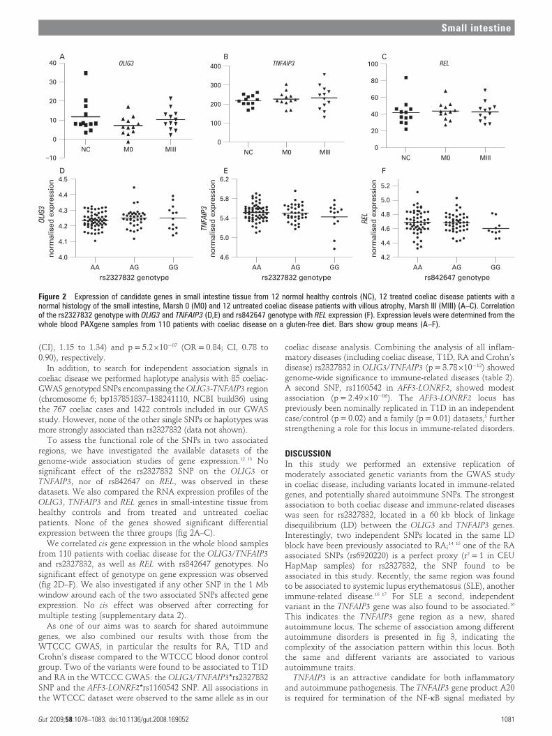
To assess the functional role of the SNPs in two associatedregions, we have investigated the available datasets of thegenome-wide association studies of gene expression.12 13 Nosignificant effect of the rs2327832 SNP on the OLIG3 orTNFAIP3, nor of rs842647 on REL, was observed in thesedatasets. We also compared the RNA expression profiles of theOLIG3, TNFAIP3 and REL genes in small-intestine tissue fromhealthy controls and from treated and untreated coeliacpatients. None of the genes showed significant differentialexpression between the three groups (fig 2A–C).
We correlated cis gene expression in the whole blood samplesfrom 110 patients with coeliac disease for the OLIG3/TNFAIP3and rs2327832, as well as REL with rs842647 genotypes. Nosignificant effect of genotype on gene expression was observed(fig 2D–F). We also investigated if any other SNP in the 1 Mbwindow around each of the two associated SNPs affected geneexpression. No cis effect was observed after correcting formultiple testing (supplementary data 2).
As one of our aims was to search for shared autoimmunegenes, we also combined our results with those from theWTCCC GWAS, in particular the results for RA, T1D andCrohn’s disease compared to the WTCCC blood donor controlgroup. Two of the variants were found to be associated to T1Dand RA in the WTCCC GWAS: the OLIG3/TNFAIP3*rs2327832SNP and the AFF3-LONRF2*rs1160542 SNP. All associations inthe WTCCC dataset were observed to the same allele as in our
coeliac disease analysis. Combining the analysis of all inflam-matory diseases (including coeliac disease, T1D, RA and Crohn’sdisease) rs2327832 in OLIG3/TNFAIP3 (p = 3.78610212) showedgenome-wide significance to immune-related diseases (table 2).A second SNP, rs1160542 in AFF3-LONRF2, showed modestassociation (p = 2.49610206). The AFF3-LONRF2 locus haspreviously been nominally replicated in T1D in an independentcase/control (p = 0.02) and a family (p = 0.01) datasets,5 furtherstrengthening a role for this locus in immune-related disorders.
DISCUSSIONIn this study we performed an extensive replication ofmoderately associated genetic variants from the GWAS studyin coeliac disease, including variants located in immune-relatedgenes, and potentially shared autoimmune SNPs. The strongestassociation to both coeliac disease and immune-related diseaseswas seen for rs2327832, located in a 60 kb block of linkagedisequilibrium (LD) between the OLIG3 and TNFAIP3 genes.Interestingly, two independent SNPs located in the same LDblock have been previously associated to RA;14 15 one of the RAassociated SNPs (rs6920220) is a perfect proxy (r2 = 1 in CEUHapMap samples) for rs2327832, the SNP found to beassociated in this study. Recently, the same region was foundto be associated to systemic lupus erythematosus (SLE), anotherimmune-related disease.16 17 For SLE a second, independentvariant in the TNFAIP3 gene was also found to be associated.16
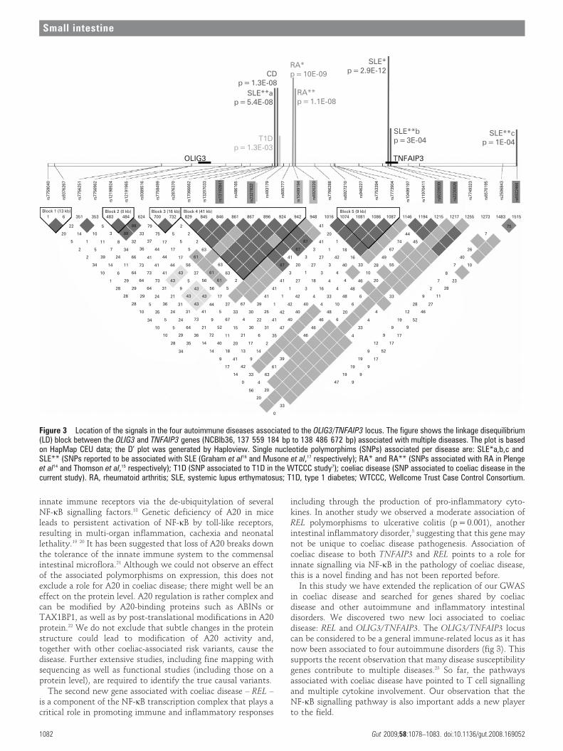
This indicates the TNFAIP3 gene region as a new, sharedautoimmune locus. The scheme of association among differentautoimmune disorders is presented in fig 3, indicating thecomplexity of the association pattern within this locus. Boththe same and different variants are associated to variousautoimmune traits.
TNFAIP3 is an attractive candidate for both inflammatoryand autoimmune pathogenesis. The TNFAIP3 gene product A20is required for termination of the NF-kB signal mediated by
Figure 2 Expression of candidate genes in small intestine tissue from 12 normal healthy controls (NC), 12 treated coeliac disease patients with anormal histology of the small intestine, Marsh 0 (M0) and 12 untreated coeliac disease patients with villous atrophy, Marsh III (MIII) (A–C). Correlationof the rs2327832 genotype with OLIG3 and TNFAIP3 (D,E) and rs842647 genotype with REL expression (F). Expression levels were determined from thewhole blood PAXgene samples from 110 patients with coeliac disease on a gluten-free diet. Bars show group means (A–F).
Small intestine
Gut 2009;58:1078–1083. doi:10.1136/gut.2008.169052 1081
innate immune receptors via the de-ubiquitylation of severalNF-kB signalling factors.18 Genetic deficiency of A20 in miceleads to persistent activation of NF-kB by toll-like receptors,resulting in multi-organ inflammation, cachexia and neonatallethality.19 20 It has been suggested that loss of A20 breaks downthe tolerance of the innate immune system to the commensalintestinal microflora.21 Although we could not observe an effectof the associated polymorphisms on expression, this does notexclude a role for A20 in coeliac disease; there might well be aneffect on the protein level. A20 regulation is rather complex andcan be modified by A20-binding proteins such as ABINs orTAX1BP1, as well as by post-translational modifications in A20protein.22 We do not exclude that subtle changes in the proteinstructure could lead to modification of A20 activity and,together with other coeliac-associated risk variants, cause thedisease. Further extensive studies, including fine mapping withsequencing as well as functional studies (including those on aprotein level), are required to identify the true causal variants.
The second new gene associated with coeliac disease – REL –is a component of the NF-kB transcription complex that plays acritical role in promoting immune and inflammatory responses
including through the production of pro-inflammatory cyto-kines. In another study we observed a moderate association ofREL polymorphisms to ulcerative colitis (p = 0.001), anotherintestinal inflammatory disorder,3 suggesting that this gene maynot be unique to coeliac disease pathogenesis. Association ofcoeliac disease to both TNFAIP3 and REL points to a role forinnate signalling via NF-kB in the pathology of coeliac disease,this is a novel finding and has not been reported before.
In this study we have extended the replication of our GWASin coeliac disease and searched for genes shared by coeliacdisease and other autoimmune and inflammatory intestinaldisorders. We discovered two new loci associated to coeliacdisease: REL and OLIG3/TNFAIP3. The OLIG3/TNFAIP3 locuscan be considered to be a general immune-related locus as it hasnow been associated to four autoimmune disorders (fig 3). Thissupports the recent observation that many disease susceptibilitygenes contribute to multiple diseases.23 So far, the pathwaysassociated with coeliac disease have pointed to T cell signallingand multiple cytokine involvement. Our observation that theNF-kB signalling pathway is also important adds a new playerto the field.
Figure 3 Location of the signals in the four autoimmune diseases associated to the OLIG3/TNFAIP3 locus. The figure shows the linkage disequilibrium(LD) block between the OLIG3 and TNFAIP3 genes (NCBIb36, 137 559 184 bp to 138 486 672 bp) associated with multiple diseases. The plot is basedon HapMap CEU data; the D’ plot was generated by Haploview. Single nucleotide polymorphims (SNPs) associated per disease are: SLE*a,b,c andSLE** (SNPs reported to be associated with SLE (Graham et al16 and Musone et al,17 respectively); RA* and RA** (SNPs associated with RA in Plengeet al14 and Thomson et al,15 respectively); T1D (SNP associated to T1D in the WTCCC study7); coeliac disease (SNP associated to coeliac disease in thecurrent study). RA, rheumatoid arthritis; SLE, systemic lupus erthymatosus; T1D, type 1 diabetes; WTCCC, Wellcome Trust Case Control Consortium.
Small intestine
1082 Gut 2009;58:1078–1083. doi:10.1136/gut.2008.169052

NF-kB is a transcription complex that plays a key role inregulating the cellular immune response to infections, stress,cytokines and other stimuli. Activation of NF-kB in variousinflammatory disorders, including asthma, arthritis and inflam-matory bowel disease (IBD) has been described.24
Coeliac disease can now be added to the list of complex disordersthat show association to the 6q23 region. This strengthens theimportance of A20 in controlling inflammation in autoimmunediseases and points to A20 as an attractive candidate for drugtargeting, as suggested recently by Coornaert et al.22
How the newly discovered genes interplay with thepreviously identified coeliac loci can only be speculated. Onthe one hand, activation of the NF-kB complex leads tooverexpression of inflammatory cytokines and, together withpreviously identified cytokines and cytokine receptor genes suchas CCR5, RGS1, IL12A and IL18RAP, this pathway would beimportant in fine-tuning the immune response. On the otherhand, the NF-kB pathway may play an independent role in theinnate mechanisms of disease development. Strikingly, genesinvolved in the innate immune response have recently beenassociated with various autoimmune diseases, suggesting a rolefor microbial and viral triggers in disease development. In coeliacdisease an increased frequency of rotaviral infections have beenobserved, suggesting that viral infections may, for example,trigger an innate immune response.25
It is interesting that the knockdown studies of A20 indendritic cells show a shift in the subset of activated T cells,hyperactivation of cytotoxic T lymphocytes and T helper cells,and suppression of regulatory T cells. This shift results fromenhanced expression of co-stimulatory signals and proinflam-matory cytokines when inhibiting A2026 and would fit in celiacdisease being a Th1 mediated disease.
These two novel loci can be added to the list of eight known,non-HLA, genetic risk factors for coeliac disease that have asmaller risk effect than HLA. Extending the list of commonvariants that account for coeliac disease will improve the geneticprognosis of patients and may help to predict the likelihood ofindividuals from the at-risk group developing coeliac disease.
Acknowledgements: We thank J Swift, P Kumar, D P Jewell, S P L Travis,L Dinesen and K Moriarty for collection of UK-GWAS and additional coeliac casesamples. We acknowledge use of DNA from the British 1958 Birth Cohort collection,funded by the UK Medical Research Council grant G0000934 and the Wellcome Trustgrant 068545/Z/02. We thank H van Someren and F Mulder for clinical databasemanagement, E Oostrom, R van ‘t Slot and the genotyping facilities at UMCG and UMCUtrecht (the Netherlands) for technical assistance. We thank C Feighery andJ McPartlin for sample collection. Irish control DNA was supplied by the Irish BloodTransfusion Service/Trinity College Dublin Biobank. We thank all coeliac and controlindividuals for participating in this study. We thank J Senior for critically reading themanuscript.
Funding: The study was supported by grants from Coeliac UK (to DAvH); the CoeliacDisease Consortium (an innovative cluster approved by the Netherlands GenomicsInitiative and partly funded by the Dutch Government, grant BSIK03009 to CW); theEuropean Union (STREP 036383); the Netherlands Organization for Scientific Research(VICI grant 918.66.620 to CW); the Science Foundation Ireland; the Higher EducationAuthority PRTLI; The Irish Health Research Board; and the Wellcome Trust (GR068094MAClinician Scientist Fellowship to DAvH; New Blood Fellowship to RMcM).
Competing interests: None.
Ethics approval: Ethics approval was from Oxfordshire REC B or East London and theCity REC 1 (UKGWAS, UK2), the Medical Ethical Committee of the University MedicalCentre Utrecht (Dutch), the Institutional Ethics Committee of St James’s Hospital (Irish)and the Ethics Committee of the Fondazione IRCCS Ospedale Maggiore Policlinico,Mangiagalli e Regina Elena (Italian).
REFERENCES1. Hunt KA, Zhernakova A, Turner G, et al. Newly identified genetic risk variants for
celiac disease related to the immune response. Nat Genet 2008;40:395–402.2. Zhernakova A, Alizadeh BZ, Bevova M, et al. Novel association in chromosome
4q27 region with rheumatoid arthritis and confirmation of type 1 diabetes point to ageneral risk locus for autoimmune diseases. Am J Hum Genet 2007;81:1284–8.
3. Zhernakova A, Festen EM, Franke L, et al. Genetic analysis of innate immunity inCrohn’s disease and ulcerative colitis identifies two susceptibility loci harboringCARD9 and IL18RAP. Am J Hum Genet 2008;82:1202–10.
4. Zhernakova A, van Diemen CC, Wijmenga C. Detecting shared pathogenesis fromthe shared genetics of immune-related diseases. Nat Rev Genet 2009;10:43–55.
5. van Heel DA, Franke L, Hunt KA, et al. A genome-wide association study for celiacdisease identifies risk variants in the region harboring IL2 and IL21. Nat Genet2007;39:827–9.
6. Todd JA, Walker NM, Cooper JD, et al. Robust associations of four newchromosome regions from genome-wide analyses of type 1 diabetes. Nat Genet2007;39:857–64.
7. Consortium WTCC. Genome-wide association study of 14,000 cases of sevencommon diseases and 3,000 shared controls. Nature 2007;447:661–78.
8. Romanos J. Six new celiac disease loci replicated in an Italian population confirmassociation to celiac disease. J Med Genet 2009;46;60–3.
9. Purcell S, Neale B, Todd-Brown K, et al. PLINK: a tool set for whole-genome associationand population-based linkage analyses. Am J Hum Genet 2007;81:559–75.
10. Barrett JC, Fry B, Maller J, et al. Haploview: analysis and visualization of LD andhaplotype maps. Bioinformatics 2005;21:263–5.
11. Heap GA, Trynka G, Jansen RC, et al. Complex nature of SNP genotype effects ongene expression in primary human leucocytes. BMC Med Genomics 2009;2:1.
12. Dixon AL, Liang L, Moffatt MF, et al. A genome-wide association study of globalgene expression. Nat Genet 2007;39:1202–7.
13. Stranger BE, Nica AC, Forrest MS, et al. Population genomics of human geneexpression. Nat Genet 2007;39:1217–24.
14. Plenge RM, Cotsapas C, Davies L, et al. Two independent alleles at 6q23 associatedwith risk of rheumatoid arthritis. Nat Genet 2007;39:1477–82.
15. Thomson W, Barton A, Ke X, et al. Rheumatoid arthritis association at 6q23. NatGenet 2007;39:1431–3.
16. Graham RR, Cotsapas C, Davies L, et al. Genetic variants near TNFAIP3 on 6q23 areassociated with systemic lupus erythematosus. Nat Genet 2008;40:1059–61.
17. Musone SL, Taylor KE, Lu TT, et al. Multiple polymorphisms in the TNFAIP3 regionare independently associated with systemic lupus erythematosus. Nat Genet2008;40:1062–4.
18. Sun SC. Deubiquitylation and regulation of the immune response. Nat Rev Immunol2008;8:501–11.
19. Boone DL, Turer EE, Lee EG, et al. The ubiquitin-modifying enzyme A20 is requiredfor termination of Toll-like receptor responses. Nat Immunol 2004;5:1052–60.
20. Lee EG, Boone DL, Chai S, et al. Failure to regulate TNF-induced NF-kappaB and celldeath responses in A20-deficient mice. Science 2000;289:2350–4.
21. Turer EE, Tavares RM, Mortier E, et al. Homeostatic MyD88-dependent signalscause lethal inflamMation in the absence of A20. J Exp Med 2008;205:451–64.
22. Coornaert B, Carpentier I, Beyaert R. A20: Central gatekeeper in inflammation andimmunity. J Biol Chem 2009;284:8217–21.
23. Xavier RJ, Rioux JD. Genome-wide association studies: a new window intoimmune-mediated diseases. Nat Rev Immunol 2008;8:631–43.
24. Perkins ND. Integrating cell-signalling pathways with NF-kappaB and IKK function.Nat Rev Mol Cell Biol 2007;8:49–62.
25. Stene LC, Honeyman MC, Hoffenberg EJ, et al. Rotavirus infection frequency and riskof celiac disease autoimmunity in early childhood: a longitudinal study.Am J Gastroenterol 2006;101:2333–40.
26. Song XT, Evel-Kabler K, Shen L, et al. A20 is an antigen presentation attenuator, andits inhibition overcomes regulatory T cell-mediated suppression. Nat Med2008;14:258–65.
Small intestine
Gut 2009;58:1078–1083. doi:10.1136/gut.2008.169052 1083