Heparin-binding protein improves prediction of severe sepsis in the emergency department
Upload
independentCategory
view
0download
0
Modulation of the Heparanase-inhibiting Activity of Heparinthrough Selective Desulfation, Graded N-Acetylation, andGlycol Splitting*
Received for publication, December 17, 2004Published, JBC Papers in Press, January 12, 2005, DOI 10.1074/jbc.M414217200
Annamaria Naggi‡, Benito Casu‡§, Marta Perez‡, Giangiacomo Torri‡, Giuseppe Cassinelli‡,Sergio Penco¶, Claudio Pisano¶, Giuseppe Giannini¶, Rivka Ishai-Michaeli�,and Israel Vlodavsky**
From the ‡G. Ronzoni Institute for Chemical and Biochemical Research, via G. Colombo, 81, 20133 Milan, Italy,¶Sigma-Tau Research Department, 00040 Pomezia, Rome, Italy, �Department of Oncology, Hadassah-HebrewUniversity Medical Center, Jerusalem 91120, Israel, and **Cancer and Vascular Biology Research Center,The Bruce Rappaport Faculty of Medicine, Technion, Haifa 31096, Israel
Heparanase is an endo-�-glucuronidase that cleavesheparan sulfate (HS) chains of heparan sulfate proteogly-cans on cell surfaces and in the extracellular matrix(ECM). Heparanase, overexpressed by most cancer cells,facilitates extravasation of blood-borne tumor cells andcauses release of growth factors sequestered by HSchains, thus accelerating tumor growth and metastasis.Inhibition of heparanase with HS mimics is a promisingtarget for a novel strategy in cancer therapy. In thisstudy, in vitro inhibition of recombinant heparanase wasdetermined for heparin derivatives differing in degrees of2-O- and 6-O-sulfation, N-acetylation, and glycol splittingof nonsulfated uronic acid residues. The contemporane-ous presence of sulfate groups at O-2 of IdoA and at O-6 ofGlcN was found to be non-essential for effective inhibitionof heparanase activity provided that one of the two posi-tions retains a high degree of sulfation. N-Desulfation/N-acetylation involved a marked decrease in the inhibi-tory activity for degrees of N-acetylation higher than 50%,suggesting that at least one NSO3 group per disaccharideunit is involved in interaction with the enzyme. On theother hand, glycol splitting of preexisting or of both pre-existing and chemically generated nonsulfated uronic ac-ids dramatically increased the heparanase-inhibiting ac-tivity irrespective of the degree of N-acetylation. IndeedN-acetylated heparins in their glycol-split forms inhibitedheparanase as effectively as the corresponding N-sulfatedderivatives. Whereas heparin and N-acetylheparins con-taining unmodified D-glucuronic acid residues inhibitedheparanase by acting, at least in part, as substrates, theirglycol-split derivatives were no more susceptible to cleav-age by heparanase. Glycol-split N-acetylheparins did notrelease basic fibroblast growth factor from ECM andfailed to stimulate its mitogenic activity. The combinationof high inhibition of heparanase and low release/potenti-ation of ECM-bound growth factor indicates that N-acety-lated, glycol-split heparins are potential antiangiogenicand antimetastatic agents that are more effective thantheir counterparts with unmodified backbones.
Heparanase is a mammalian endo-�-D-glucuronidase thatcleaves heparan sulfate (HS)1 chains at a limited number ofsites (1–3). Cloning of the heparanase cDNA by several groups(1–6) suggests that a single functional HS-degrading endogly-cosidase is expressed in mammalian cells. The enzyme is syn-thesized as a latent 65-kDa precursor that undergoes proteo-lytic cleavage, yielding 8- and 50-kDa subunits thatheterodimerize to form a highly active enzyme (7, 8). Hepara-nase enzymatic activity participates in degradation and remod-eling of the extracellular matrix (ECM), facilitating, amongother activities, cell invasion associated with cancer metasta-sis, angiogenesis, and inflammation (1–3, 9). Heparanase up-regulation has been documented in a variety of human tumorscorrelating, in some cases, with increased vascular density andpoor postoperative survival (10–13). Heparanase overexpres-sion has also been noted in several other pathologies such ascirrhosis (14), nephrosis (15), and diabetes (16). In addition toits intimate involvement in the egress of cells from the bloodstream, heparanase activity releases from the ECM and tumormicroenvironment a multitude of HS-bound growth factors,cytokines, chemokines, and enzymes that affect cell and tissuefunction, most notably angiogenesis (17, 18). These observa-tions, the anticancerous effect of heparanase gene silencing(ribozyme and small interfering RNA) (19) and of heparanase-inhibiting molecules (non-anticoagulant species of heparin andother sulfated polysaccharides) (20, 21), and the unexpectedidentification of a predominant functional heparanase (1–3)suggest that the enzyme is a promising target for developmentof new anticancer drugs.
HS and the structurally related heparin are present in mostanimal species. They are glycosaminoglycans constituted byrepeating disaccharide units of a uronic acid (either D-glucu-ronic acid (GlcA) or L-iduronic acid (IdoA)) and D-glucosamine(either GlcNAc or D-glucosamine N-sulfate (GlcNSO3)) andbear sulfate substituents in various positions (22–25). Al-though derived from the common biosynthetic precursor N-acetylheparosan (-GlcA-GlcNAc)n, HS and heparin have differ-
* This work was performed, in part, under the framework of theEuropean Union Project HEPARANASE (Contract QLK3-CT-2002-02049). The costs of publication of this article were defrayed in part bythe payment of page charges. This article must therefore be herebymarked “advertisement” in accordance with 18 U.S.C. Section 1734solely to indicate this fact.
§ To whom correspondence should be addressed. E-mail: [email protected].
1 The abbreviations used are: HS, heparan sulfate; HSPG, heparansulfate proteoglycan; ECM, extracellular matrix; FGF-2, basic fibro-blast growth factor; FGF-1, acidic fibroblast growth factor; IdoA, L-iduronic acid; GlcA, D-glucuronic acid; GlcN, D-glucosamine; GlcNAc,N-acetyl-D-glucosamine; GlcNSO3, D-glucosamine N-sulfate; LMWH,low molecular weight heparin; GPC-HPLC, gel permeation chromatog-raphy-high performance liquid chromatography; NA, N-acetylated;NAH, N-acetylheparin; NS, N-sulfated; gs, glycol-split; RO-H, reducedoxyheparin; PBS, phosphate-buffered saline; OdeS, O-desulfated; GalA,L-galacturonic acid; M� w, weight average molecular weight.
THE JOURNAL OF BIOLOGICAL CHEMISTRY Vol. 280, No. 13, Issue of April 1, pp. 12103–12113, 2005© 2005 by The American Society for Biochemistry and Molecular Biology, Inc. Printed in U.S.A.
This paper is available on line at http://www.jbc.org 12103
by guest on July 29, 2016http://w
ww
.jbc.org/D
ownloaded from

ent structures: HS is less sulfated and more heterogeneousthan heparin. The two glycosaminoglycans also have differentlocations in tissues: whereas HS is a component of the ECMand of the surface of most cells, heparin is stored in granules ofmast cells and co-released with histamine into the circulationupon cellular degranulation mainly in cases of allergic andinflammatory reactions and anaphylactic stress. On the otherhand, exogenous heparin is widely used as an anticoagulantand antithrombotic drug and is of increasing interest for noveltherapeutical applications (24–27).
As an analog of the natural substrate of heparanase, heparinis commonly considered to be a potent inhibitor of heparanase(20, 21, 28–31). This activity is attributed, in part, to its highaffinity interaction with the enzyme and limited degradation,serving as an alternative substrate. Early reports (20, 21, 30,31) showed that heparin and some chemically modified speciesof heparin as well as other sulfated polysaccharides (22, 32)that inhibit tumor cell heparanase also inhibit experimentalmetastasis in animal models, while other related compoundsthat lack heparanase-inhibiting activity fail to exert an anti-metastatic effect (20–22, 30–32). Regardless of the mode ofaction, heparin and low molecular weight heparin (LMWH)were reported to exert a beneficial effect in cancer patients (33),stimulating research on the potential use of modified, non-anticoagulant species of heparin and HS in cancer therapy.
Screening of heparin derivatives has permitted the identifi-cation of some of the structural features of heparin associatedwith inhibition of the enzyme. As a general trend, the hepara-nase-inhibiting activity increases with increasing degrees ofO-sulfation. However, N-sulfates seems to exert little effectsince they can be replaced by N-acyl (N-acetyl, N-succinyl, orN-hexanoyl) groups without substantial loss of inhibitory ac-tivity (20, 34). No significant differences were found betweenthe currently used unfractionated heparins and low molecularweight heparins and a tetradecasaccharidic fragment (34). 2-O-Desulfated derivatives were shown to retain the inhibitoryactivity, whereas N-desulfated, N-acetylated derivatives dis-played a reduced activity (35). In the present study, relation-ships between structure and heparanase-inhibiting activity ofheparin were studied using a larger number of heparins andheparin derivatives, including some with various degrees of6-O-sulfation of GlcN and 2-O-sulfation of IdoA residues as wellas “glycol-split” derivatives obtained by controlled periodateoxidation/borohydride reduction of natural (36) or partially2-O-desulfated heparins (37, 38). Glycol splitting of C-2–C-3bonds of nonsulfated uronic acid residues was suggested tointerfere with the biological interactions of heparin by provid-ing flexible joints between protein binding sequences (37–39).When framing heparin sequences that bind FGF-2, glycol-splitresidues were shown not to impair the binding to FGF-2. How-ever, they prevented activation of FGF-2 and FGF-2-inducedangiogenic activity (37, 38). The present study shows thatglycol splitting enhances the heparanase-inhibiting activity ofheparin. Based on the observation that N-acetyl groups do notprevent and may even assist recognition by heparanase (40, 41)and taking into account that N-acetylheparin, as opposed toheparin, does not release angiogenic factors from ECM (34), weprepared and tested heparins with various degrees of N-acety-lation/N-sulfation together with some of their glycol-split de-rivatives. N-Acetylated, glycol-split heparins were shown toinhibit heparanase more efficiently than the correspondingnon-glycol-split N-acetylated heparins.
EXPERIMENTAL PROCEDURES
Materials
All chemicals were of reagent grade from Sigma and were used assupplied. Heparins were commercial preparations from pig mucosa
(H-1 to H-3 from Laboratori Derivati Organici, Trino Vercellese, Italy,and H-6 from Hepar), from beef mucosa (H-4 and H-5, LaboratoriDerivati Organci), and from beef lung (H-7, The Upjohn Co.). Thecorresponding contents of major sulfate groups, as evaluated by 13CNMR spectroscopy (42) and expressed as mole percent of IdoA2SO3,GlcNSO3, and GlcN(SO3 or Ac)6SO3 per disaccharide unit based onquantification of underlined sulfate groups, were: H-1: 69, 89, 79; H-2:68, 85, 82; H-3: 64, 85, 82; H-4: 62, 89, 60; H-5: 66, 92, 60; H-6: 65, 86,82; and H-7: 86, 98, 95. The weight average molecular weights (M� w, byGPC-HPLC (43)) were: H-1, 14,200; H-2, 18,100; H-3, 19,600; H-4,18,800; H-5, 18,200; H-6, 23,200; and H-7, 21,600. Sample desalting wascarried out by dialysis against water with 1000-Da cut-off tubes or byfractionation on a 2.5 � 100-cm Sephadex G-25 column (AmershamBiosciences) using 10% ethanol in water as eluent and UV detection at210 nm. Molecular weight determinations were performed by GPC-HPLC on a Viscotex instrument equipped with a VE1121 pump, Rheo-dyne valve (100 �l), and triple detector array 302 equipped with IR,viscosimeter, and 90° light-scattering systems. Two 300 � 7.8-mm TSKGMPWXL Viscotek columns were used with 0.1 M NaNO3 as eluent(flow, 0.6 ml/min). Samples were dissolved in the eluent solution at theconcentration of 15 mg/ml (43).
NMR spectra were recorded at 500 MHz for 1H and 125 MHz for 13Cwith a Bruker AMX spectrometer equipped with a 5-mm 1H/X inverseprobe. The spectra were obtained at 45 °C from D2O solutions (15mg/0.5 ml D2O, 99.99% D). Chemical shifts, given in parts per milliondown field from sodium-3-(trimethylsilyl)propionate, were measuredindirectly with reference to acetone in D2O (� 2.235 for 1H and � 30.20for 13C). The 13C NMR spectra were recorded at 300 or 400 MHz with aBruker AC-300 or AMX-400 spectrometer.
Recombinant Human Heparanase
Recombinant enzymatically active heparanase was purified fromheparanase-transfected Chinese hamster ovary cells (4). Briefly Chi-nese hamster ovary cells were harvested with trypsin and centrifuged,and the cell pellet was suspended in 20 mM citrate-phosphate buffer pH5.4. The suspension was subjected to four cycles of freeze/thaw (�70/37 °C, 5 min each), the cell extract was centrifuged (18,000 rpm, 15 min,2–8 °C), and the supernatant was collected and filtered through a0.45-�m filter. The filtrate was applied onto a Source 15 S column(Amersham Biosciences) equilibrated with 20 mM phosphate buffer, pH6.8. The column was washed (20 mM phosphate buffer, pH 6.8, followedby 20 mM phosphate buffer, pH 8.0), and heparanase was eluted with alinear gradient (0–35%) of 8 column volumes of 1.5 M NaCl in 20 mM
phosphate buffer, pH 8.0. Active fractions were pooled and applied ontoa Fractogel EMD SO3
� (Merck) column equilibrated with 20 mM citrate-phosphate buffer, pH 5.4. Heparanase was eluted with a linear gradient(0–22%) of 1 column volume followed by 10 column volumes (22–25%) of1.5 M NaCl in 20 mM phosphate buffer, pH 8.0. Finally heparanaseeluted from the Fractogel column was applied onto a HiTrap heparincolumn (Amersham Biosciences) equilibrated with 20 mM phosphatebuffer, pH 8.0, and eluted with a linear gradient of 1 column volume(0–20%) and 15 column volumes (20–28%) of 1.5 M NaCl in 20 mM
phosphate buffer, pH 8.0. Eluted fractions were analyzed by gradientSDS-PAGE, stained with Gelcode® (Pierce), and pooled according totheir purity. An at least 90% pure, highly active heparanase prepara-tion was obtained, containing the active 50- and 8-kDa heparanasesubunits and, to a lower extent, the 65-kDa proheparanase (8). Activerecombinant human heparanase was also produced in insect cells asdescribed previously (7). The construct encoding the 8- and 50-kDaheparanase subunits was kindly provided by Dr. E. McKenzie (OxfordGlycoSciences Ltd., Abingdon, Oxon, UK) (7). Similar results wereobtained with both preparations.
Preparation of Heparin Derivatives6-O-Desulfated Heparins
Procedure A—An extensively 6-O-desulfated heparin also partially(�15%) 2-O-desulfated (716OdeS-H(A) where the superscript denotesthe degree of 6-O-desulfation), M� w 16,000, was prepared according toNagasawa et al. (44), starting from the pyridinium salt of heparin H-1,under solvolytic conditions (10 mg/ml in Me2SO:water 9:1) at 100 °C for2.5 h followed by resulfation of free amino groups with sulfur trioxide-trimethylamine complex in alkaline aqueous medium (45).
Procedure B—6-O-Desulfated-heparins (776OdeS-H(B), M� w 19,000;736OdeS-H(B), M� w 17,700; and 466OdeS-H(B), M� w 20,400) were pre-pared according to Matsuo et al. (46) by O-desulfation through activa-tion with N-methyl-N-(trimethylsilyl)trifluoroacetamide or N,O-bis (tri-methylsilyl)acetamide without N-desulfation. Heparin H-1 (200 mg)
Heparin-derived Heparanase Inhibitors12104
by guest on July 29, 2016http://w
ww
.jbc.org/D
ownloaded from

was converted into its pyridinium salt and soaked in pyridine (20 ml).After addition of 4 ml of N-methyl-N-(trimethylsilyl)trifluoroacetamide,the solution was heated for 4 h at 80 °C to yield 736OdeS-H or for 8 h at60 °C to yield 776OdeS-H. Heparin (H-1) was converted into its pyridin-ium salt and soaked in pyridine (30 ml). After addition of 6 ml ofN,O-bis(trimethylsilyl)acetamide, the solution was heated for 2 h at60 °C to yield 466OdeS-H.
2-O-Desulfated Heparins
Procedure A—2-O-Desulfated heparin in the IdoA form (H, IdoA(A),M� w 17,700) was prepared according to Jaseja et al. (47). Heparin (500mg) was simply dissolved in 500 ml of 0.1 M NaOH, and the solution wasfrozen and lyophilized. The residue dissolved in 500 ml of distilledwater was dialyzed, and the product was isolated by evaporation underreduced pressure. Its 13C NMR spectrum closely corresponded to theone reported in the literature (48), indicating an essentially completeconversion of the original IdoA2SO3 residues into IdoA residues.
Procedure B—2-O-Desulfated heparin in the GalA form (H, GalA(B),M� w 12,600) was prepared by a modification of methods used by Jasejaet al. (47) and Rej and Perlin (49) essentially as described previously(48). Heparin (500 mg) was dissolved in 10 ml of 1 M NaOH and thenheated at 85 °C for 1 h. After cooling below 30 °C, the solution wasbrought to pH 7 with 0.1 M HCl and heated at 70 °C for 48 h to give(after cooling, dialysis, and freeze-drying) the GalA derivative with atypical 13C NMR spectrum (48).
N-Acetylated Heparins
N-Acetylated heparins (xNAH, where the superscript x denotes thedegree of N-acetylation as referred to total GlcN) were prepared bytime-controlled N-desulfation under solvolytic conditions (44). Brieflythe pyridinium salt of heparin was stirred at 20–25 °C in Me2SO:water(9:1) for different times (30, 60, 90, 100, and 120 min and 8 h) to obtainintermediates with different degrees of N-desulfation, which upon N-acetylation with acetic anhydride in alkaline aqueous medium(NaHCO3, 4 °C, 2 h) (50) gave 29NAH (M� w 22,000), 39NAH (M� w 21,000),50NAH (M� w 21,000), 58NAH (M� w 21,000), and 70NAH (M� w 22,000),92NAH (M� w 13,700), and 100NAH (M� w 15,700).
Glycol-split Heparins and Glycol-split N-Acetylated Heparins
Glycol-split heparins and glycol-split N-acetylated heparins wereprepared by exhaustive periodate oxidation and borohydride reductionof heparin and N-acetylheparins, respectively, without (36) or with (37,38) prior partial 2-O-desulfation. For the first series of glycol-splitN-acetylheparins, 250-mg samples of H-1, 29NAH, 39NAH, 50NAH,70NAH, and 100NAH were dissolved in 6 ml of H2O, and 6 ml of 0.1 M
NaIO4 were added to the solutions. The solutions were stirred at 4 °Cfor 16 h in the dark. The reactions were stopped by adding 1 ml ofethylene glycol, and the solutions were dialyzed through 1000-Da cut-off tubes for 16 h. Solid sodium borohydride (60 mg) was added to theretentate solutions in several portions under stirring. After 2–3 h thepH was adjusted to 4 with 0.1 M HCl, and the solutions were neutralizedwith 0.1 M NaOH. After desalting and dialysis, the final products wererecovered by freeze-drying to yield RO-H (M� w 15,700), 26NA,RO-H (M� w
17,000), 40NA,RO-H (M� w 16,000), 53NA,RO-H (M� w 11,250), 67NA,RO-H(M� w 15,000), and 100NA,RO-H (M� w 20,200). For the second series ofN-acetylated, glycol-split heparins (NAH,gs), 250-mg samples of H-1,29NAH, 39NAH, 58NAH, and 70NAH were dissolved in 5 ml of 1 M NaOHand then heated at 60 °C for 30 min. After cooling below 30 °C, thesolutions were brought to pH 7 with 0.1 M HCl and heated at 70 °C for48 h to give (after cooling, dialysis, and freeze-drying) partial conver-sion of IdoA2SO3 to GalA. Products were treated as described above toyield the corresponding glycol-split derivatives H,52gs (M� w 11,000),29NAH,60gs (M� w 6,000), 43NAH,60gs (M� w 8,500), 57NAH,64gs (M� w 9,500),and 70NAH,59gs (M� w 9,300). The glycol-splitting (gs) percentages wereevaluated by integration of the anomeric 13C NMR signals at 106.5 ppm(A) and at 102 ppm (B), corresponding to the split uronic acid residuesand 2-O-sulfated iduronic acid residues, respectively; gs � (A/(A � B)) �100. Products obtained without generation of additional nonsulfateduronic acid residues had a content of glycol-split residues (mainlyarising from GlcA) of 24 � 1% and are designated as “reduced oxyhe-parins” (RO-H) (36). Products obtained by glycol splitting of both thepreexisting and the newly generated nonsulfated uronic acids (IdoA orGalA) were designated as H,xgs (or NAH,xgs if derived from N-acetyl-heparins) where the superscript x indicates the percentage of glycol-split uronic acid.
Low Molecular Weight Derivatives
Low molecular weight derivatives of H-1, H,44gs, and 50NAH,25gs(50NA,RO-H) were prepared by nitrous acid depolymerization of thecorresponding polysaccharides (51). A solution of polysaccharide (4 g)was dissolved in 65 ml of H2O and cooled at 4 °C. After the addition of75 mg of NaNO2 the pH was adjusted to 2 with 0.1 M HCl. The solutionwas stirred at 4 °C for 20 min, and then the pH was brought to 7. SolidNaBH4 (1 g) was added in several portions under stirring. After 2–3 h,the pH was adjusted to 4 with 0.1 M HCl, and the solution was neutral-ized with 0.1 M NaOH. The products (low molecular weight H-1, 6,500;LMWH,49gs, 6,300; LMWH,49gs, 3,000; and 50NA,RO-H, 5,400) ob-tained by precipitation with 3 volumes of ethanol were dissolved inwater and recovered by freeze-drying. The depolymerization degreesand the corresponding molecular weight values were determined byintegration of the 13C NMR signals at 98–107 and 82, 85, and 87 ppm,corresponding to total C-1 and C-2, C-3, and C-5 of the anhydromannitolunit, respectively. The percentage of glycol splitting, expressed as gly-col-split residues referred to total uronic acids, was evaluated by inte-gration of the 13C NMR signals at 106.5 and 102 ppm, corresponding toC-1 of the split uronic residues and 2-O-sulfated iduronic residues,respectively.
Cells
Cultures of bovine corneal endothelial cells were established fromsteer eyes and maintained in Dulbecco’s modified Eagle’s medium (1 gof glucose/liter) supplemented with 5% newborn calf serum, 10% fetalcalf serum, and 1 ng/ml FGF-2 as described previously (4, 52). Conflu-ent cell cultures were dissociated with 0.05% trypsin and 0.02% EDTAin phosphate-buffered saline (PBS) and subcultured at a split ratio of1:8 (52).
Preparation of Dishes Coated with ECM
Bovine corneal endothelial cells were plated into 35-mm tissue cul-ture dishes at an initial density of 2 � 105 cells/ml and cultured asdescribed above except that 4% dextran T-40 was included in thegrowth medium (4, 52). On day 12, the subendothelial ECM was ex-posed by dissolving the cell layer with PBS containing 0.5% TritonX-100 and 20 mM NH4OH followed by four washes with PBS (52). TheECM remained intact, free of cellular debris, and firmly attached to theentire area of the tissue culture dish. To produce sulfate-labeled ECM,Na2
35SO4 (Amersham Biosciences) was added (25 �Ci/ml) on days 2 and5 after seeding, and the cultures were incubated with the label withouta medium change and processed as described previously (4, 52). Nearly80% of the ECM radioactivity was incorporated into HSPG.
Heparanase Inhibition Activity
Heparin species were tested for their ability to inhibit heparanaseusing metabolically sulfate-labeled ECM as a substrate (28, 29). Brieflysulfate-labeled ECM coating the surface of 35-mm culture dishes wasincubated (4 h, 37 °C, pH 6.0) with recombinant human heparanase (40ng/ml) in the absence and presence of three concentrations (0.2, 1.0, 5.0�g/ml) of each heparin species. The reaction mixture contained 50 mM
NaCl, 1 mM dithiothreitol, 1 mM CaCl2, and 10 mM phosphate-citratebuffer, pH 6.0. To evaluate the occurrence of proteoglycan degradation,the incubation medium was collected and applied for gel filtration onSepharose 6B columns (0.9 � 30 cm). Fractions (0.2 ml) were elutedwith PBS at a flow rate of 5 ml/h and counted for radioactivity. Theexcluded volume (Vo) was marked by blue dextran, and the total in-cluded volume (Vt) was marked by phenol red. Nearly intact HSPGs areeluted from Sepharose 6B just after the void volume (Kav � 0.2, frac-tions 1–10), while HS degradation fragments are eluted toward the Vt ofthe column (peak II, 0.5 � Kav � 0.8, fractions 15–35) (4, 28, 29).Labeled fragments eluted in peak II were shown to be degradationproducts of HS as they were 5–6-fold smaller than intact HS chains ofHSPGs, resistant to further digestion with papain and chondroitinaseABC, and susceptible to deamination by nitrous acid (29). Heparanaseactivity � Kav � total cpm in peak II. Recovery of labeled materialapplied on the column ranged from 85 to 95% in different experiments.Each experiment was performed at least three times, and the variationin elution positions (Kav values) did not exceed �15%.
Release of ECM-bound FGF-2
ECM-coated wells (4-well plates) were incubated with iodinatedFGF-2 (1–2 � 105 cpm/ng, 1.5–2.5 � 104 cpm/0.25 ml/well, 3 h, 24 °C),and the unbound FGF-2 was removed by four washes with PBS con-taining 0.02% gelatin (34). The ECM was then incubated (3 h, 24 °C)with the various heparins and modified heparins, and aliquots (0.25 ml)
Heparin-derived Heparanase Inhibitors 12105
by guest on July 29, 2016http://w
ww
.jbc.org/D
ownloaded from

of the incubation medium were counted in a �-counter to determine theamount of released material. The remaining ECM was washed twicewith PBS and solubilized with 1 N NaOH, and the radioactivity wascounted in a �-counter (34). The percentage of released 125I-FGF-2 wascalculated from the total ECM-associated radioactivity. “Spontaneous”release of 125I-FGF-2 in the presence of incubation medium alone was7–12% of the total ECM-bound FGF-2 (34). Each experiment was per-formed three to five times, yielding similar results.
Stimulation of FGF-2 Mitogenic Activity
A cytokine-dependent, heparan sulfate-deficient lymphoid cell line(BaF3) engineered to express fibroblast growth factor receptor-1 (53,54) was applied to investigate the effect of heparin derivatives onFGF-2-mediated cell proliferation. These cells (clone F32) respond toFGF-2 only in the presence of exogenously added heparin, HS, or somemodified species of heparin. Briefly F32 cells (2 � 104/well) were platedinto 96-well microtiter plates in the presence of 2.5 or 5.0 ng/ml FGF-2and increasing concentrations of the test compound in a total volume of250 �l. Forty-eight hours later, 1 �Ci of [3H]thymidine was added perwell, and the cells were incubated for an additional 6 h and collectedwith a cell harvester. Incorporated thymidine was determined by liquidscintillation counting using a TopCount microplate counter (53, 54).
Gel Permeation Analysis of NAH and �50% Glycol-split Heparin(H,52gs) before and after Digestion with Heparanase
2 mg of each compound were incubated for 48 h at 37 °C in 40 mM
phosphate-citrate buffer, pH 5.8, with or without 4 �g of recombinantheparanase in a total volume of 50 �l. The samples were lyophilized,then redissolved in 0.5 ml of water, and analyzed by GPC-HPLC using300 � 7.8-mm TSK PW 2000 and PW 3000 columns.
RESULTS
Preparation (and Schematic Presentation) of ChemicallyModified Species of Heparin—The relationship between sulfa-tion patterns and heparanase-inhibiting activity of heparinspecies with unmodified backbones was studied using O-desul-fated heparin derivatives and N-desulfated, N-acetylated he-parins of various degrees of substitution prepared startingfrom a well characterized pig mucosal heparin (H-1) usingestablished procedures with slight modifications. 6-O-Desulfa-tion was accomplished using two different procedures, the first(procedure A) involving solvolytic desulfation (44) and the sec-ond (procedure B) through activated silyl acetamides (46). Asreported (46), attempts to obtain extensively 6-O-desulfatedheparins using procedure A also involved partial 2-O-desulfa-tion (10–15%, by NMR analysis). Therefore, the extensively6-O-desulfated heparin H,776OdeS was obtained only via pro-cedure B. 2-O-Desulfation of heparin was also performed usingtwo different procedures, leading to different products. Thefirst (procedure A), involving lyophilization of alkaline solu-tions, quantitatively removes the 2-OSO3 groups retaining theIdoA residues in their original configuration (47). The secondone (procedure B), which involves heating of alkaline solutions,is more easily controlled to generate also partially 2-O-desul-fated heparins (37) and to convert the 2-O-sulfated IdoA resi-dues into GalA residues (47–49). Glycol-split derivatives wereprepared by periodate oxidation/borohydride reduction of bothunmodified heparin and partially 2-O-desulfated heparins asdescribed previously (37). The same procedure was applied toobtain glycol-split derivatives of N-acetylheparins of variousdegrees of N-acetylation and 2-O-desulfation. Details are givenunder “Experimental Procedures.”
Fig. 1 shows the general scheme for the preparation of de-rivatives of heparin, N-acetylheparins, and the correspondingglycol-split derivatives. Scheme 1 shows the formulas of hepa-rin and heparin derivatives. The structure of a heparin chain isschematized as composed of N-acetylated (NA), N-sulfated(NS), and mixed NA/NS domains; the prevalent sequences arethose of the trisulfated disaccharide (formula 1) (24). For sim-plicity, only chains containing the antithrombin binding regionare presented, although this region is contained in only about
one-third of the chains of heparin. The prevalent structure of6-O-desulfated heparins is shown in formula 2, and that of2-O-desulfated heparins retaining the IdoA configuration isshown in formula 3. Formula 4 shows the prevalent structureof heparins that underwent inversion of configuration to GalAduring 2-O-desulfation of IdoA. Fully N-acetylated heparinsare represented by the general formula 5. Partially N-acety-lated heparins have different percentages of N-acetyl groups;the NSO3 groups are the complement to 100% N-substitution.Glycol splitting is depicted in Fig. 1 in two different ways. Fig.1A refers to heparin and fully N-acetylated heparin (NAH)glycol-split without any previous modification of their struc-tures. The corresponding glycol-split products are referred to asRO-H (36) and “N-acetyl-reduced oxyheparins” (NA,RO-H), re-spectively. Fig. 1B refers to heparin and NAH (in the example,50% N-acetylated heparin 50NAH) that were glycol-split afterremoval of 2-OSO3 groups to reach a total content of 50%nonsulfated uronic acid residues; the nonmodified residueswere IdoA2SO3 (37). De-O-sulfation of one of two IdoA residuesfollowed by glycol splitting gave heparins with prevalent struc-ture 6, corresponding to sequences of polypentasulfated trisac-charides separated by glycol-split uronic acid residues (37).Low molecular weight species of heparin and representativeheparin derivatives were obtained by controlled nitrous aciddepolymerization (51) of heparin, 50% glycol-split heparin, andthe RO derivative of 50% N-acetylated heparin.
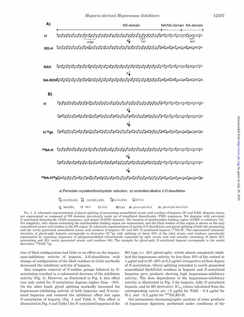
All compounds were analyzed by one- and two-dimensional1H and 13C NMR spectroscopy (37). Analytical data, expressedas relative molar content of 6-OSO3, 2-OSO3, and NSO3 groupsare summarized in Table I. Superscripts on abbreviations for6-O-desulfated (6OdeS), 2-O-desulfated (2OdeS), NA, and gsheparins represent relative percentages of 6-O- and 2-O-desul-fation, N-acetylation, and glycol splitting, respectively.
Heparanase Inhibition by Heparin Derivatives—Typicalheparanase inhibition curves showing the gel filtration profilesof sulfate-labeled degradation fragments released by hepara-nase from metabolically labeled ECM in the absence (control)and presence of 1 �g/ml unmodified heparin and fully N-acety-lated heparin are presented in Fig. 2. Inhibition is reflected bythe decreased amounts and Kav values of HS fragments re-leased from ECM and eluted as peak II (fractions 20–35) incomparison with control incubation of the ECM with recombi-nant heparanase in the absence of inhibitors (28, 29). Hepara-nase activity is calculated as the total amount of cpm eluted inpeak II multiplied by the Kav (i.e. elution position) of thesefragments. The heparanase-inhibitory activity (expressed aspercent inhibition of heparanase) of almost all heparins atconcentrations of 5, 1, and 0.2 �g/ml is shown in Table I. Mostof the data represent the average of several separate experi-ments (numbers indicated). Standard deviations, indicated foreach heparin, were usually lower than 5 for the most activecompounds and did not exceed 20 as a mean for the less effec-tive ones.
Data in Table I confirm that heparin is a strong inhibitor ofheparanase (�70% inhibition at 1 �g/ml). No significant differ-ences in inhibitory activity were found among H-1 and otherheparin preparations from pig mucosa, beef mucosa, and beeflung (data not shown) despite significant differences in theirsulfation patterns (detailed under “Experimental Procedures”).Also activity differences found between the parent heparin andits low molecular weight species as well as between glycol-split50NAH and its low molecular weight species were not signifi-cant. On the other hand, well defined significant differences inheparanase-inhibiting activity were associated with specificchemical modifications of heparin. As illustrated in Fig. 3,whereas either 6-O-desulfation or 2-O-desulfation with reten-
Heparin-derived Heparanase Inhibitors12106
by guest on July 29, 2016http://w
ww
.jbc.org/D
ownloaded from
tion of IdoA configuration had little or no effect on the hepara-nase-inhibitory activity of heparin, 2-O-desulfation withchange of configuration of the IdoA residues to GalA markedlydecreased the inhibitory activity of heparin.
Also complete removal of N-sulfate groups followed by N-acetylation resulted in a substantial decrease of the inhibitoryactivity (Fig. 3). However, as illustrated in Fig. 4, this effectwas only noted for N-acetylation degrees higher than �50%.On the other hand, glycol splitting markedly increased theheparanase-inhibiting activity of both heparins and N-acety-lated heparins and restored the inhibitory effect lost uponN-acetylation of heparin (Fig. 4 and Table I). This effect isillustrated in Fig. 4 and Table I for N-acetylated heparins of the
RO type (i.e. 25% glycol-split), which almost completely inhib-ited the heparanase activity (to less than 10% of the control at1 �g/ml and to 20–30% at 0.2 �g/ml) irrespective of their degreeof N-acetylation. Glycol splitting extended to newly generatednonsulfated IdoA/GalA residues in heparin and N-acetylatedheparins gave products showing high heparanase-inhibitoryactivity. The dose dependence of the heparanase-inhibitoryactivity is illustrated in Fig. 5 for heparin, fully N-acetylatedheparin, and its RO derivative. IC50 values calculated from thecorresponding curves are �5 �g/ml for NAH, �0.4 �g/ml forH-1, and �0.2 �g/ml for 100NA,RO-H.
Gel permeation chromatographic analysis of some productsof heparanase digestion, performed under conditions of the
FIG. 1. A, schematic representation of glycol splitting of preexisting nonsulfated uronic acid residues of heparin (H) and NAH. Heparin chainsare represented as composed of NS domains prevalently made up of trisulfated disaccharide (TSD) sequences, NA domains with prevalentN-acetylated disaccharide (NAD) sequences, and mixed (NA/NS) domains. The location of antithrombin binding region (ATBR) is arbitrary (24).For simplicity, only chains containing the antithrombin binding region are represented, and the GlcA residue of this region is shown as the onlynonsulfated uronic acid residue in the NS region. B, schematic representation of partial 2-O-desulfation and glycol splitting of both the preexistingand the newly generated nonsulfated uronic acid residues of heparin (H) and 50% N-acetylated heparin (50NA-H). The represented schematicstructure of glycol-split heparin corresponds to derivative H,50gs with splitting of about 50% of the total uronic acid residues, prevalentlyrepresented by repeating sequences of polypentasulfated trisaccharide separated by split uronic acid and actually consisting of about 25%preexisting and 25% newly generated uronic acid residues (38). The example for glycol-split N-acetylated heparin corresponds to the modelderivative 50NAH,50gs.
Heparin-derived Heparanase Inhibitors 12107
by guest on July 29, 2016http://w
ww
.jbc.org/D
ownloaded from

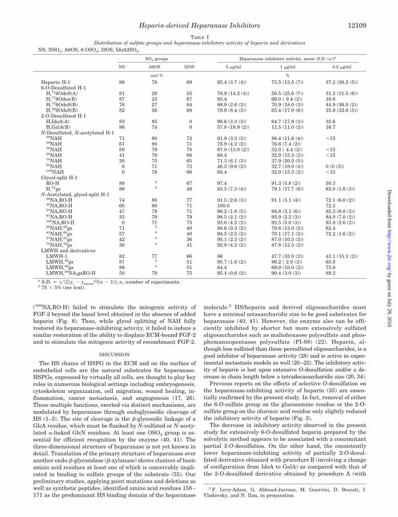
enzyme inhibition assay, indicated that whereas heparin (notshown) and N-acetylheparin are cleaved by heparanase (aspreviously shown for heparin) (40), their glycol-split deriva-tives are not susceptible to cleavage as illustrated for fullyN-acetylated, RO-heparin in Fig. 6A and 52% glycol-split hep-arin (H,52gs) in Fig. 6B.
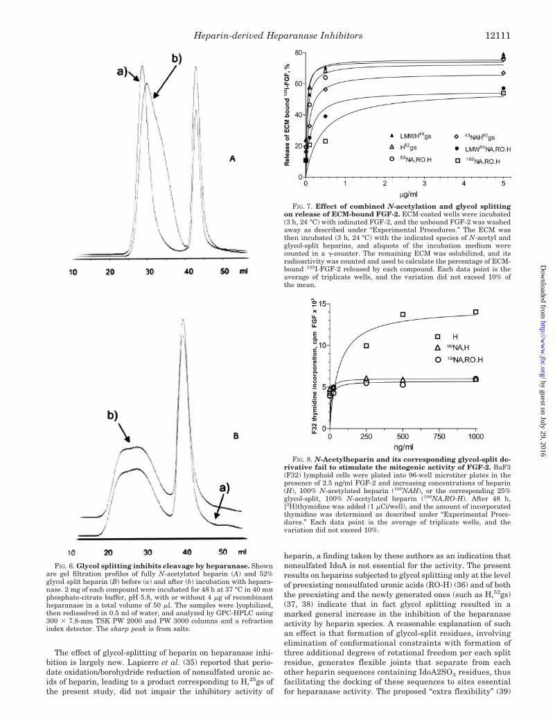
Effect of Modified Heparins on Release of ECM-bound FGF-2and Stimulation of FGF-2 Mitogenic Activity—Some of theheparin derivatives were tested for their capacity to releaseFGF-2 from ECM (18, 34). As demonstrated in Fig. 7, dose-response curves of the FGF-2-releasing activity of glycol-splitheparin (H,52gs) and its corresponding low molecular weightderivative (LMWH,49gs) were almost superimposable to thosereported for heparin (34), indicating that glycol splitting doesnot substantially modify the FGF-2-releasing properties of hep-
arin. Also the curves of the RO derivative and of heparin aresuperimposable (data not shown). Fig. 7 also shows that glycol-split, N-acetylated heparins behave similarly to non-glycol-split NAH (34) in that they release ECM-bound FGF-2 consis-tently less than unmodified heparin. 100NAH (not shown) and100NA,RO-H exhibited the lowest FGF-2-releasing activityamong the tested compounds, yielding only about twice thespontaneous release observed in presence of the buffer(PBS) alone.
The ability of heparin, 100NAH, and 100NA,RO-H to promotethe mitogenic activity of recombinant FGF-2 was investigatedusing cytokine-dependent, heparan sulfate-deficient lymphoidcells (BaF3) engineered to express fibroblast growth factorreceptor-1 (53, 54). Unlike heparin, both fully N-acetylatedheparin (100NAH) and its glycol-split counterpart molecule
SCHEME 1. Formulas of heparin andheparin derivatives.
Heparin-derived Heparanase Inhibitors12108
by guest on July 29, 2016http://w
ww
.jbc.org/D
ownloaded from
(100NA,RO-H) failed to stimulate the mitogenic activity ofFGF-2 beyond the basal level obtained in the absence of addedheparin (Fig. 8). Thus, while glycol splitting of NAH fullyrestored its heparanase-inhibiting activity, it failed to induce asimilar restoration of the ability to displace ECM-bound FGF-2and to stimulate the mitogenic activity of recombinant FGF-2.
DISCUSSION
The HS chains of HSPG in the ECM and on the surface ofendothelial cells are the natural substrates for heparanase.HSPGs, expressed by virtually all cells, are thought to play keyroles in numerous biological settings including embryogenesis,cytoskeleton organization, cell migration, wound healing, in-flammation, cancer metastasis, and angiogenesis (17, 26).These multiple functions, exerted via distinct mechanisms, aremodulated by heparanase through endoglycosidic cleavage ofHS (1–3). The site of cleavage is the �-glycosidic linkage of aGlcA residue, which must be flanked by N-sulfated or N-acety-lated �-linked GlcN residues. At least one OSO3 group is es-sential for efficient recognition by the enzyme (40, 41). Thethree-dimensional structure of heparanase is not yet known indetail. Translation of the primary structure of heparanase overanother endo-�-glycosidase (�-xylanase) shows clusters of basicamino acid residues at least one of which is conceivably impli-cated in binding to sulfate groups of the substrate (55). Ourpreliminary studies, applying point mutations and deletions aswell as synthetic peptides, identified amino acid residues 158–171 as the predominant HS binding domain of the heparanase
molecule.2 HS/heparin and derived oligosaccharides musthave a minimal octasaccharidic size to be good substrates forheparanase (40, 41). However, the enzyme also can be effi-ciently inhibited by shorter but more extensively sulfatedoligosaccharides such as maltohexaose polysulfate and phos-phomannopentaose polysulfate (PI-88) (22). Heparin, al-though less sulfated than these persulfated oligosaccharides, is agood inhibitor of heparanase activity (28) and is active in exper-imental metastasis models as well (20–22). The inhibitory activ-ity of heparin is lost upon extensive O-desulfation and/or a de-crease in chain length below a tetradecasaccharidic size (20, 34).
Previous reports on the effects of selective O-desulfation onthe heparanase-inhibiting activity of heparin (35) are essen-tially confirmed by the present study. In fact, removal of eitherthe 6-O-sulfate group on the glucosamine residue or the 2-O-sulfate group on the iduronic acid residue only slightly reducedthe inhibitory activity of heparin (Fig. 3).
The decrease in inhibitory activity observed in the presentstudy for extensively 6-O-desulfated heparin prepared by thesolvolytic method appears to be associated with a concomitantpartial 2-O-desulfation. On the other hand, the consistentlylower heparanase-inhibiting activity of partially 2-O-desul-fated derivative obtained with procedure B (involving a changeof configuration from IdoA to GalA) as compared with that ofthe 2-O-desulfated derivative obtained by procedure A (with
2 F. Levy-Adam, G. Abboud-Jarrous, M. Guerrini, D. Beccati, I.Vlodavsky, and N. Ilan, in preparation.
TABLE IDistribution of sulfate groups and heparanase-inhibitory activity of heparin and derivatives
NS, NSO3; A6OS, 6-OSO3; I2OS, IdoA2SO3.
SO3 groups Heparanase inhibitory activity, mean (S.D. (n))a
NS A6OS I2OS 5 �g/ml 1 �g/ml 0.2 �g/ml
mol % %
Heparin H-1 89 79 69 95.4 (3.7 (4)) 73.5 (13.5 (7)) 37.2 (20.2 (5))6-O-Desulfated H-1
H,716OdeS(A) 81 29 55 78.9 (14.2 (4)) 50.5 (25.6 (7)) 31.3 (31.5 (6))H,776Odes(B) 87 23 67 93.4 66.0 ( 9.4 (2)) 16.6H,736OdeS(B) 78 27 64 88.9 (2.6 (2)) 70.9 (18.0 (3)) 44.9 (36.0 (2))H,466OdeS(B) 82 56 68 79.9 (8.4 (3)) 63.4 (17.0 (6)) 35.9 (32.6 (5))
2-O-Desulfated H-1H,IdoA(A) 83 85 0 98.6 (3.3 (3)) 64.7 (17.8 (3)) 35.6H,GalA(B) 86 74 0 57.8 (18.9 (2)) 11.5 (11.0 (2)) 16.7
N-Desulfated, N-acetylated H-129NAH 71 80 72 91.9 (3.3 (3)) 88.4 (11.6 (4)) �1539NAH 61 80 71 78.9 (4.2 (2)) 76.6 (7.4 (2))50NAH 50 79 70 87.0 (13.0 (2)) 52.0 ( 4.4 (2)) �1558NAH 41 79 68 68.4 32.9 (15.3 (2)) �1570NAH 30 75 65 71.3 (6.1 (3)) 37.9 (20.2 (5))92NAH 8 71 73 46.2 (9.6 (2)) 32.7 (18.0 (4)) 0 (0 (2))100NAH 0 78 66 68.4 32.9 (15.3 (2)) �15
Glycol-split H-1RO-H 89 b 67 97.4 91.3 (5.8 (2)) 50.3H,52gs 89 b 48 83.5 (7.3 (4)) 79.1 (17.7 (6)) 62.0 (1.6 (3))
N-Acetylated, glycol-split H-126NA,RO-H 74 80 77 91.5 (2.6 (3)) 91.1 (3.1 (4)) 72.1 (6.0 (2))40NA,RO-H 60 80 71 100.0 72.353NA,RO-H 47 79 71 98.2 (1.8 (5)) 94.8 (3.1 (6)) 85.3 (6.8 (3))67NA,RO-H 33 79 79 98.5 (2.1 (2)) 93.9 (2.2 (3)) 84.0 (7.0 (2))100NA,RO-H 0 71 75 93.0 (4.2 (2)) 92.5 (5.0 (3)) 93.8 (2.6 (2))29NAH,60gs 71 b 40 88.6 (0.3 (2)) 79.6 (13.0 (2)) 62.443NAH,60gs 57 b 40 94.5 (2.5 (2)) 70.1 (17.1 (2)) 72.2 (1.6 (2))57NAH,64gs 42 b 36 95.1 (2.3 (2)) 87.0 (10.5 (2))70NAH,59gs 30 b 41 92.9 (4.2 (2)) 87.8 (12.5 (2))
LMWH and derivativesLMWH-1 82 77 66 86 47.7 (35.0 (2)) 43.1 (15.3 (2))LMWH,49gs 87 b 51 95.7 (1.8 (2)) 86.2 ( 2.8 (3)) 65.8LMWH,49gs 89 b 51 84.4 69.9 (10.0 (2)) 73.8LMWH,50NA,gsRO-H 50 79 75 95.4 (0.6 (2)) 90.4 (3.0 (3)) 89.2
a S.D. � ((yi � ymean)2/(n � 1)); n, number of experiments.b 75 � 5% (see text).
Heparin-derived Heparanase Inhibitors 12109
by guest on July 29, 2016http://w
ww
.jbc.org/D
ownloaded from

retention of the IdoA configuration) is likely associated withdifferent conformational properties of IdoA and GalA. IdoAresidues have been demonstrated to be endowed with a uniqueconformational flexibility (“plasticity”). Such a plasticity, asso-ciated with different equienergetic conformations of IdoA resi-dues, all coexisting in a rapid dynamic equilibrium, can cur-rently explain the better protein binding capacity andassociated biological properties of IdoA-containing sequencesas compared with the more rigid GlcA-containing ones (56).Based on simple conformational criteria, GalA is expected tohave very much the same conformational rigidity as GlcA.
Replacement of N-sulfate groups with N-acetyl groups doesnot completely suppress the heparanase-inhibitory activity ofheparin (34); its activity is reduced to about one-third (35) asconfirmed by the present study. However, it is noteworthy thata substantial decrease in heparanase-inhibiting activity wasonly observed for degrees of N-acetylation higher than about50%. As shown in Table I and illustrated in Fig. 4, for degreesof N-acetylation lower than 40%, the inhibitory activity re-mained essentially the same as that of heparin. This is taken asan indication that only one-half of the NSO3 groups of heparinare essential for complete inhibition of heparanase. The ac-cepted conformation of the NS domains of heparin is repre-sented by helices where sets of three sulfate groups (NSO3,2SO3, and 6SO3) alternate on each side of the chain (57). Theobservation that only one of two N-sulfate groups is required toinhibit heparanase and the assumption that the N-desulfation/N-acetylation reaction occurs randomly along the NS domainswould accordingly suggest that heparin and its derivativeswith unmodified backbones bind the enzyme only from one sideof the chain.
FIG. 2. Representative heparanase inhibition curves for hep-arin and N-acetylheparin. Sulfate-labeled ECM was incubated (4 h,37 °C, pH 6.0) with recombinant human heparanase (40 ng/ml) in theabsence (control) and presence of 1 �g/ml heparin (H-1) or NAH. Sul-fate-labeled degradation fragments released into the incubation me-dium were analyzed by gel filtration on Sepharose 6B. The figure showspeak II (corresponding to fractions 20–35) used to calculate percentresidual activity of the enzyme (see “Experimental Procedures”).
FIG. 3. Heparanase inhibitory activity of O-desulfated speciesof heparin. Sulfate-labeled ECM was incubated (4 h, 37 °C, pH 6.0)with recombinant heparanase (40 ng/ml) in the absence (contr.) andpresence of 1 (right bars) or 5 (left bars) �g/ml unmodified heparin(Hep.), NAH, or each of the indicated specifically desulfated heparins(6OdeS-H, 2OdeS-H, and H,GalA). Sulfate-labeled degradation frag-ments released into the incubation medium were analyzed by gel fil-tration on Sepharose 6B. Kav of peak II (see Fig. 2), calculated for eachcompound, was multiplied by the total cpm eluted in peak II. Resultsare presented as percentage of control. Residual heparanase activity �Kav � total cpm in peak II (percentage of control).
FIG. 4. Inhibition of heparanase by N-acetylheparins and thecorresponding 25% glycol-split (RO) derivatives. Sulfate-labeledECM was incubated (4 h, 37 °C, pH 6.0) with heparanase (40 ng/ml) inthe presence of 1 �g/ml NAH with an increased percentage of N-acetylation (% NAc) or with the corresponding 25% glycol-split deriva-tives (NA,RO-H). Sulfate-labeled material released into the incubationmedium was analyzed by gel filtration, and heparanase enzymaticactivity (Kav � total cpm in peak II) is presented as percentage of the100% activity obtained in the absence of inhibitor.
FIG. 5. Dose dependence of heparanase inhibition by heparin,N-acetylheparin, and 25% glycol-split (RO) N-acetylheparin. Sul-fate-labeled ECM was incubated (4 h, 37 °C, pH 6.0) with heparanase (40ng/ml) in the absence or presence of increasing concentrations of heparin(H), 100% N-acetylheparin (100NAH), or the corresponding 25% glycol-split (RO), 100% N-acetylheparin (100NA,RO-H). Sulfate-labeled materialreleased into the incubation medium was analyzed by gel filtration.Heparanase enzymatic activity (total cpm in peak II � Kav) is presentedas percentage of the activity (100%) obtained in the absence of heparin.
Heparin-derived Heparanase Inhibitors12110
by guest on July 29, 2016http://w
ww
.jbc.org/D
ownloaded from
The effect of glycol-splitting of heparin on heparanase inhi-bition is largely new. Lapierre et al. (35) reported that perio-date oxidation/borohydride reduction of nonsulfated uronic ac-ids of heparin, leading to a product corresponding to H,25gs ofthe present study, did not impair the inhibitory activity of
heparin, a finding taken by these authors as an indication thatnonsulfated IdoA is not essential for the activity. The presentresults on heparins subjected to glycol splitting only at the levelof preexisting nonsulfated uronic acids (RO-H) (36) and of boththe preexisting and the newly generated ones (such as H,52gs)(37, 38) indicate that in fact glycol splitting resulted in amarked general increase in the inhibition of the heparanaseactivity by heparin species. A reasonable explanation of suchan effect is that formation of glycol-split residues, involvingelimination of conformational constraints with formation ofthree additional degrees of rotational freedom per each splitresidue, generates flexible joints that separate from eachother heparin sequences containing IdoA2SO3 residues, thusfacilitating the docking of these sequences to sites essentialfor heparanase activity. The proposed “extra flexibility” (39)
FIG. 6. Glycol splitting inhibits cleavage by heparanase. Shownare gel filtration profiles of fully N-acetylated heparin (A) and 52%glycol split heparin (B) before (a) and after (b) incubation with hepara-nase. 2 mg of each compound were incubated for 48 h at 37 °C in 40 mM
phosphate-citrate buffer, pH 5.8, with or without 4 �g of recombinantheparanase in a total volume of 50 �l. The samples were lyophilized,then redissolved in 0.5 ml of water, and analyzed by GPC-HPLC using300 � 7.8-mm TSK PW 2000 and PW 3000 columns and a refractionindex detector. The sharp peak is from salts.
FIG. 7. Effect of combined N-acetylation and glycol splittingon release of ECM-bound FGF-2. ECM-coated wells were incubated(3 h, 24 °C) with iodinated FGF-2, and the unbound FGF-2 was washedaway as described under “Experimental Procedures.” The ECM wasthen incubated (3 h, 24 °C) with the indicated species of N-acetyl andglycol-split heparins, and aliquots of the incubation medium werecounted in a �-counter. The remaining ECM was solubilized, and itsradioactivity was counted and used to calculate the percentage of ECM-bound 125I-FGF-2 released by each compound. Each data point is theaverage of triplicate wells, and the variation did not exceed 10% ofthe mean.
FIG. 8. N-Acetylheparin and its corresponding glycol-split de-rivative fail to stimulate the mitogenic activity of FGF-2. BaF3(F32) lymphoid cells were plated into 96-well microtiter plates in thepresence of 2.5 ng/ml FGF-2 and increasing concentrations of heparin(H), 100% N-acetylated heparin (100NAH), or the corresponding 25%glycol-split, 100% N-acetylated heparin (100NA,RO-H). After 48 h,[3H]thymidine was added (1 �Ci/well), and the amount of incorporatedthymidine was determined as described under “Experimental Proce-dures.” Each data point is the average of triplicate wells, and thevariation did not exceed 10%.
Heparin-derived Heparanase Inhibitors 12111
by guest on July 29, 2016http://w
ww
.jbc.org/D
ownloaded from

induced by these joints reinforces the binding driving influ-ence of the already existing intrinsic conformational plastic-ity of iduronate residues. More notably, glycol splitting alsoincreased the heparanase-inhibiting activity of N-acetylatedheparins, even that of fully N-acetylated heparin, whoseheparanase binding capacity is very weak when their back-bone is unmodified.
Glycol splitting involves substantial loss of the anticoagulantactivity of heparin (36). It is now clear that the main reason forsuch an effect is the cleavage of C-2–C-3 bonds of the GlcAresidue of the pentasaccharidic sequence (Fig. 1, formula 7).This residue in its unmodified form is essential for binding toantithrombin, and whenever it is glycol-split (as in sequence 8),the heparin affinity for antithrombin is completely lost (Ref. 24and references therein).
The heparanase inhibiting properties of glycol-split, exten-sively N-acetylated heparins cannot be explained by the modeldiscussed previously where only sulfate groups (perhaps inaddition to the uronate carboxyl groups) on the same side of theheparin helix are involved in binding to the enzyme. Such amodel is compatible with heparanase binding of heparins thatcontain no more than 50% N-acetyl groups. Retention of stronginhibitory activity upon removal all NSO3 groups and theirsubstitution with nonpolar N-acetyl groups followed by glycolsplitting of nonsulfated uronic acids implies that efficient dock-ing to heparanase, favored by the flexible joints generated byglycol splitting, occurs with a conformation different from thatadopted by heparin with a degree of N-acetylation lowerthan 50%.
Conformational polymorphism is not uncommon in heparinsequences bound to proteins. Thus, x-ray studies showed thatheparin sequences may bind FGF-1 in more than one, equallyfavored conformation (58). Increasing evidence is being accu-mulated that the plasticity of iduronate residues combinedwith some rotational freedom of both uronic acid and aminosugar residues around the glycosidic linkages favors severalpossibilities of binding to basic domains of proteins (59). Asexpected (37, 39), glycol splitting appears to further increasethe molecular flexibility of heparin chains as determined bysmall angle x-ray scattering (60) and NMR spectroscopy sup-ported by molecular modeling studies (61).
It appears that heparin contains both recognition/cleavageand inhibition sites for heparanase and that its inhibition ofthe enzyme also involves competition (40). Size profiling of HSdegradation products by heparanase in the presence of N-acetylheparin (Fig. 6A) showed that the fully N-acetylated de-rivative NAH is also cleaved by the enzyme. On the contrary,the gel filtration profile obtained in the presence of glycol-splitheparin is practically superimposable to that obtained in theabsence of the enzyme (Fig. 6B), indicating that modification ofGlcA residues totally abolishes cleavage by the enzyme.
HS assembles ligands and receptors into ternary signalingcomplexes, best exemplified by the FGF-fibroblast growth fac-tor receptor-heparin complex (62, 63). Following cleavage byheparanase, the multitude of polypeptides sequestered andregulated by HS (17) become bioavailable (1, 18), and thisrequires a tight regulation of their activity, applying, amongother approaches, modified species of heparin and HS. Thepresent results on the effect of glycol splitting on the ability ofheparins to release FGF-2 from ECM extend previous observa-tions on the effect of O-sulfation and N-acetylation on thisproperty (34). As illustrated in Fig. 7, glycol-split derivatives ofboth heparin (including LMWH) and NAH did not substan-tially modify the ability to displace FGF-2 from ECM. In otherwords, while both native and glycol-split heparins efficientlyreleased FGF-2 from ECM, their N-acetylated counterparts
exhibited a markedly reduced ability to displace FGF-2, reflect-ing the essential involvement of N-sulfate groups in this inter-action. This observation supports the finding that glycol-splitheparins bind FGF-2 (37) and vascular endothelial growthfactor (64) with very much the same affinity as unmodifiedheparins. However, since H,50gs inhibits dimerization of FGF-2(37), it is conceivable that the ECM-bound growth factor isreleased by glycol-split heparins in an inactive form. Moreoverwe demonstrated that unlike native heparin and LMWH, NAHand even more so glycol-split NAH (100NAH,25gs, i.e. NA,RO-H)failed to stimulate FGF-2-mediated proliferation of HS-defi-cient lymphoid cells (Fig. 8). The remarkable heparanase-in-hibitory activity of N-acetylated, glycol-split heparins togetherwith the low levels of FGF-2 that they release from ECM andtheir inability to stimulate the mitogenic activity of FGF-2indicates this class of chemically modified heparins as potentialantiangiogenic and antimetastatic agents. These compoundsalso markedly inhibit wound angiogenesis in transgenic miceoverexpressing the heparanase gene (65). Furthermore ourpreliminary experiments show that some of these heparin de-rivatives effectively abolish experimental lung colonization ofintravenously administered B16-BL6 mouse melanoma cells(Ref. 66).
Retrospective analyses suggest that treatment of venousthromboembolism in cancer patients with LMWHs is associ-ated with additional benefits in terms of their survival (33).The experiments presented in this study were undertaken todevelop heparin-based molecules for efficient inhibition ofheparanase activity. Polysulfated chains such as those of hep-arin are expected to envelop the basic clusters of heparanaseand compete with its binding to HS. Confirming previous find-ings (34), we showed that such an activity is retained by a lowmolecular weight heparin. Moreover we also demonstratedsuch a retention for three representative low molecular weightglycol-split derivatives. Further studies are planned to deter-mine, for each type of derivative, the shortest chains as well asthe shortest IdoA2SO3-containing sequences still showing sig-nificant inhibition of the enzyme and to elucidate whether thecarboxylate groups of glycol-split residues participate in bind-ing and inactivation of heparanase.
In conclusion, we applied desulfation strategies and con-trolled glycol splitting to remove sulfate groups not necessarilyinvolved in heparanase recognition and inhibition and to im-prove the molecular flexibility and biological interactions ofheparin. Generation of specific heparanase-inhibiting com-pounds such as those described in this study is important notonly as a proof of concept but also as a promising approach todevelop heparin-based anticancer lead compounds devoid ofside effects.
REFERENCES
1. Vlodavsky, I., and Friedmann, Y. (2001) J. Clin. Investig. 108, 341–3472. Parish, C. R., Freeman, C., and Hulett, M. D. (2001) Biochim. Biophys. Acta
1471, M99–M1083. Dempsey, L. A., Brunn, G. T., and Platt, J. L. (2000) Trends Biol. Sci. 25,
349–3514. Vlodavsky, I., Friedmann, Y., Elkin, M., Aingorn, H., Atzmon, R., Ishai-
Michaeli, R., Spector, L., and Pecker, I. (1999) Nat. Med. 5, 793–8025. Hulett, M. D., Freeman, C., Hamdorf, B. J., Baker, R. T., Harris, M. J., and
Parish, C. R. (1999) Nat. Med. 5, 803–8096. Toyoshima, M., and Nakajima, M. (1999) J. Biol. Chem. 274, 24153–241607. McKenzie, E., Young, K., Hircock, M., Bennett, J., Bhaman, M., Felix, R.,
Turner, P., Stamps, A., McMillan, D., Saville, G., Ng, S., Mason, S., Snell,D., Schofield, D., Gong, H., Townsend, R., Gallagher, J., Page, M., Parekh,R., and Stubberfield, C. (2003) Biochem. J. 373, 423–435
8. Levy-Adam, F., Miao, H. Q., Heinrikson, R. L., Vlodavsky I., and Ilan, N.(2003) Biochim. Biophys. Res. Commun. 308, 885–891
9. Vlodavsky, I., Eldor, A., Haimovitz-Friedman, A., Matzner, Y., Ishai-Michaeli,R., Lider, O., Naparstek, Y., Cohen, I. R., and Fucks, Z. (1992) InvasionMetastasis 12, 112–127
10. Koliopanos, A., Friess, H., Kleeff, J., Shi, X., Liao, Q., Pecker, I., Vlodavsky, I.,Zimmermann, A., and Buchler, M. W. (2001) Cancer Res. 61, 4655–4659
11. El-Assal, O. N., Yamanoi, A., Ono, T., Kohno, H., and Nagasue, N. (2001) Clin.
Heparin-derived Heparanase Inhibitors12112
by guest on July 29, 2016http://w
ww
.jbc.org/D
ownloaded from

Cancer Res. 7, 1299–130512. Gohji, K., Hirano, H., Okamoto, M., Kitazawa, S., Toyoshima, M., Dong, J.,
Katsuoka, Y., and Nakajima, M. (2001) Int. J. Cancer 95, 295–30113. Sato, T., Yamaguchi, A., Goi, T., Hirono, Y., Takeuchi, K., Katayama, K., and
Matsukawa, S. (2004) J. Surg. Oncol. 87,174–18114. Xiao, Y., Kleeff, J., Shi, X., Buchler, M. W., and Friess, H. (2003) Hepatol. Res.
26, 192–19815. Levidiotis, V., Kanellis, J., Ierino, F. L., and Power, D. A. (2001) Kidney Int. 60,
1287–129616. Katz, A., Van-Dijk, D. J., Aingorn, H., Erman, A., Davies, M., Darmon, D.,
Hurvitz, H., and Vlodavsky, I. (2002) Isr. Med. Assoc. 4, 996–100217. Bernfield, M., Gotte, M., Park, P. W., Reizes, O., Fitzgerald, M. L., Lincecum,
J., and Zako, M. (1999) Annu. Rev. Biochem. 68, 729–77718. Elkin, M., Ilan, N., Ishai-Michaeli, R., Friedmann, Y., Papo, O., Pecker, I., and
Vlodavsky, I. (2001) FASEB J. 15, 1661–166319. Edovitsky, E., Elkin, M., Zcharia, E., Peretz, T., and Vlodavsky, I. (2004)
J. Natl. Cancer Inst. 96, 1219–123020. Vlodavsky, I., Mohsen, M., Lider, O., Svahn, C. M., Ekre, H. P., Vigoda, M., and
Peretz, T. (1994) Invasion Metastasis 14, 290–30221. Nakajima, M., Irimura, T., and Nicolson, G. L. (1988) J. Cell. Biochem. 36,
157–16722. Parish, C. R., Freeman, C., Brown, K. J., Francis, D. J., and Cowden, W. B.
(1999) Cancer Res. 59, 3433–344123. Kjellen, L., and Lindahl, U. (1991) Annu. Rev. Biochem. 60, 443–47524. Casu, B., and Lindahl, U. (2001) Adv. Carbohydr. Chem. Biochem. 57, 159–20825. Lindahl, U. (2000) Glycoconj. J. 17, 597–60526. Lever, R., and Page, C. P. (2002) Nat. Rev. Drug Discov. 1, 140–14827. Linhardt, R. J. (2003) J. Med. Chem. 46, 2551–256428. Bar-Ner, M., Eldor, A., Wasserman, L., Matzner, Y., and Vlodavsky, I. (1987)
Blood 70, 551–55729. Vlodavsky, I., Fuks, Z., Bar-Ner, M., Ariav, Y., and Schirrmacher, V. (1983)
Cancer Res. 43, 2704–271130. Irimura, T., Nakajima, M., and Nicolson, G. L. (1986) Biochemistry 25,
5322–532831. Parish, C. R., Coombe, D. R., Jakobsen, K. B., Bennett, F. A., and Underwood,
P. A. (1987) Int. J. Cancer 40, 511–51832. Miao, H. Q., Elkin, M., Aingorn, E., Ishai-Michaeli, R., Stein, C. A., and
Vlodavsky, I. (1999) Int. J. Cancer 83, 424–43133. Altinbas, M., Coskun, H. S., Er, O., Ozkan, M., Eser, B., Unal, A., Cetin, M.,
and Soyuer, S. A. (2004) J. Thromb. Haemostasis 2, 1266–127134. Ishai-Michaeli, R., Svahn, C. M., Weber, M., Chajek-Shaul, T., Korner, G.,
Ekre, H.-P., and Vlodavsky, I. (1992) Biochemistry 31, 2080–208835. Lapierre, F., Holme, K., Lam, L., Tressler, R. J., Storm, N., Wee, J., Stack,
R. J., Castellot, J., and Tyrrell, D. J. (1996) Glycobiology 6, 355–36636. Casu, B., Diamantini, G., Fedeli, G., Mantovani, M., Prino, G., Oreste, P.,
Pescador, R., Torri, G., and Zoppetti, G. (1985) Arzneim.-Forsch. (Drug Res.)36, 637–642
37. Casu, B., Guerrini, M., Guglieri, S., Naggi, A., Perez, M., Torri, G., Cassinelli,G., Ribatti, D., Carminati, P., Giannini, G., Penco, S., Pisano, C., Belleri, M.,Rusnati, M., and Presta, M. (2004) J. Med. Chem. 47, 838–848
38. Casu, B., Guerrini, M., Naggi, A., Perez, M., Torri, G., Ribatti, D., Carminati,C., Giannini, G., Penco, S., Pisano, C., Belleri, M., Rusnati, M., and Presta,M. (2002) Biochemistry 41, 10519–10528
39. Casu, B. (1990) in New Trends in Hemostasis (Harenberg, J., Heene, D. L.,Stehle, G., and Schettler, G., eds) pp. 2–11, Springer Verlag, Heidelberg,Germany
40. Sandback-Pikas, D. S., Li, J.-p., Vlodavsky, I., and Lindahl, U. (1998) J. Biol.Chem. 273, 18770–18777
41. Okada, Y., Yamada, S., Toyoshima, M., Dong, J., Nakajima, M., and Sugahara,K. (2002) J. Biol. Chem. 277, 42488–42495
42. Casu, B., Guerrini, M., Naggi, A., Torri, G., De Ambrosi, L., Boveri, G.,Gonella, S., Cedro, A., Ferro, L., Lanzarotti, E., Paterno, M., Attolini, M.,and Valle, M. G. (1996) Arzneim.-Forsch. (Drug Res.) 46, 472–477
43. Bertini, S., Bisio, A., Torri, G., Bensi, D., and Terbojevich, M. (2005) Biomac-romolecules 6, 168–173
44. Nagasawa, K., Inoue, Y., and Kamata, T. (1977) Carbohydr. Res. 58, 47–5545. Lloyd, A. G., Emberg, G., and Fowler, L. J. (!971) Biochem. Pharmacol. 20, 63746. Matsuo, M., Takano, R., Kamei-Hayashi, K., and Hara, S. (1993) Carbohydr.
Res. 241, 209–21547. Jaseja, M., Rej, R. N., Sauriol, F., and Perlin, A. S. (1989) Can. J. Chem. 67,
1449–145648. Piani, S., Casu, B., Marchi, E. G., Torri, G., and Ungarelli, F. (1993) J. Car-
bohydr. Chem. 12, 507–52149. Rej, R. N., and Perlin, A. S. (1990) Carbohydr. Res. 200, 437–44750. Levvy, G. A., and McAllan, A. (1959) Biochem. J. 73, 127–15951. Cifonelli, J. C. (1968) Carbohydr. Res. 8, 233–24252. Vlodavsky, I. (1999) in Current Protocols in Cell Biology, Vol. 1, pp.
10.14.11–10.14.14, John Wiley & Sons, New York53. Ornitz, D. M., Yayon, A., Flanagan, J. G., Svahn, C. M., Levi, E., and Leder, P.
(1992) Mol. Cell. Biol. 12, 240–24754. Miao, H.-Q., Ornitz, D. M., Eingorn, E., Ben-Sasson, S. A., and Vlodavsky, I.
(1997) J. Clin. Investig. 99, 1565–157555. Hulett, M. D., Hornby, J. R., Ohms, S. J., Zuegg, J., Freeman, C., Gready, J. E.,
and Parish, C. R. (2000) Biochemistry 39, 15659–1566756. Casu, B., Provasoli, A., Petitou, M., and Sinay, P. (1988) Trends Biol. Sci. 13,
221–22557. Mulloy, B., Forster, M. J., Jones, C., and Davies, D. B. (1993) Biochem. J. 293,
840–85858. DiGabriele, A. D., Lax, I., Chen, D. I., Svahn, C. M., Jaje, M., Schlessinger, J.,
and Hendrickson, W. A. (1998) Nature 393, 812–81759. Mulloy, B., and Forster, M. J. (2000) Glycobiology 10, 1147–115660. Korramian, B. A. (1987) in Industrial Polysaccharides (Stivala, S. S.,
Crescenzi, V., and Dea, I. C. M., eds) pp. 339–347, Gordon and Breach, NewYork
61. Hricovini, M., Guerrini, M., Naggi, A., Torri, G., and Casu, B. (2003) 12thEuropean Carbohydrate Symposium, Grenoble, France, July 6–11, 2003,Abstr. OC061, Eurocarb 12
62. Schlessinger, J., Plotnikov, A. N., Ibrahimi, O. A., Eliseenkova, A. V., Yeh,B. K., Yayon, A., Linhardt, R. J., and Mohammadi, M. (2000) Mol. Cell. 6,743–750
63. Kwan, C. P., Venkataraman, G., Shriver, Z., Raman, R., Liu, D., Qi, Y.,Vartivovsaki, L., and Sasisekharan, R. (2000) J. Biol. Chem. 276,23421–23429
64. Pisano, C., Aulicino, C., Vesci, L., Casu, B., Naggi, A., Torri, G., Ribatti, D.,Belleri, M., Rusnati, M., and Presta, M. (2005) Glycobiology 15, 1C–6C
65. Zcharia, E., Zilka, R., Yaar, A., Yacoby-Zeevi, O., Zetser, A., Metzger, S., Sarid,R., Naggi, A., Casu, B., Ilan, N., Vlodavsky, I., and Abramovitch, R. (2005)FASEB J. 19, 211–221
66. Vlodavsky, I., Zcharia, E., Goldshmidt, O., Eshell, R., Katz, B.-Z., Minucci, S.,Kovalchuck, O., Penco, S., Pisano, C., Naggi, A., and Casu, B. (2003)Pathophysiol. Haemostasis Thromb. 33, Suppl. 1, 59–61
Heparin-derived Heparanase Inhibitors 12113
by guest on July 29, 2016http://w
ww
.jbc.org/D
ownloaded from
VlodavskySergio Penco, Claudio Pisano, Giuseppe Giannini, Rivka Ishai-Michaeli and Israel
Annamaria Naggi, Benito Casu, Marta Perez, Giangiacomo Torri, Giuseppe Cassinelli,-Acetylation, and Glycol SplittingNDesulfation, Graded
Modulation of the Heparanase-inhibiting Activity of Heparin through Selective
doi: 10.1074/jbc.M414217200 originally published online January 12, 20052005, 280:12103-12113.J. Biol. Chem.
10.1074/jbc.M414217200Access the most updated version of this article at doi:
Alerts:
When a correction for this article is posted•
When this article is cited•
to choose from all of JBC's e-mail alertsClick here
http://www.jbc.org/content/280/13/12103.full.html#ref-list-1
This article cites 63 references, 16 of which can be accessed free at
by guest on July 29, 2016http://w
ww
.jbc.org/D
ownloaded from
Copyright © 2022 FDOKUMEN