Static electric fields interfere in the viability of cells exposed to ionising radiation
Upload
independentCategory
view
0download
0
BioMed CentralMolecular Cancer
ss
Open AcceResearchEnhanced levels of Hsulf-1 interfere with heparin-binding growth factor signaling in pancreatic cancerJunsheng Li1,2, Jörg Kleeff*1, Ivane Abiatari1, Hany Kayed1, Nathalia A Giese1, Klaus Felix1, Thomas Giese3, Markus W Büchler1 and Helmut Friess1Address: 1Department of General Surgery, University of Heidelberg, Heidelberg, Germany, 2Department of General Surgery, Zhong-Da Hospital, Southeast University, Nanjing, China and 3Institute of Immunology, University of Heidelberg, Heidelberg, Germany
Email: Junsheng Li - [email protected]; Jörg Kleeff* - [email protected]; Ivane Abiatari - [email protected]; Hany Kayed - [email protected]; Nathalia A Giese - [email protected]; Klaus Felix - [email protected]; Thomas Giese - [email protected]; Markus W Büchler - [email protected]; Helmut Friess - [email protected]
* Corresponding author
pancreatic cancergrowth factorssulfataseproteoglycans
AbstractHsulf-1 is a newly identified enzyme, which has the ability to decrease the growth of hepatocellular,ovarian, and head and neck squamous cell carcinoma cells by interfering with heparin-bindinggrowth factor signaling. Since pancreatic cancers over-express a number of heparin-binding growthfactors and their receptors, the expression and function of this enzyme in pancreatic cancer wasanalyzed.
Results: Pancreatic cancer samples expressed significantly (22.5-fold) increased Hsulf-1 mRNAlevels compared to normal controls, and Hsulf-1 mRNA was localized in the cancer cellsthemselves as well as in peritumoral fibroblasts. 4 out of 8 examined pancreatic cancer cell linesexpressed Hsulf-1, whereas its expression was below the level of detection in the other cell lines.Stable transfection of the Hsulf-1 negative Panc-1 pancreatic cancer cell line with a full length Hsulf-1 expression vector resulted in increased sulfatase activity and decreased cell-surface heparan-sulfate proteoglycan (HSPG) sulfation. Hsulf-1 expression reduced both anchorage-dependent and-independent cell growth and decreased FGF-2 mediated cell growth and invasion in this cell line.
Conclusion: High expression of Hsulf-1 occurs in the stromal elements as well as in the tumorcells in pancreatic cancer and interferes with heparin-binding growth factor signaling.
IntroductionPancreatic cancer is one of the most aggressive humanmalignancies with an overall five-year survival rate of lessthen 5% [1]. Although the reasons for the aggressivegrowth behavior of pancreatic cancer are not completely
understood, recent molecular biological studies haverevealed several factors that are involved in the pathogen-esis of pancreatic cancer. These include genetic changes,such as k-ras, p53, p16, and Smad4 mutations [2], as well
Published: 07 April 2005
Molecular Cancer 2005, 4:14 doi:10.1186/1476-4598-4-14
Received: 28 March 2005Accepted: 07 April 2005
This article is available from: http://www.molecular-cancer.com/content/4/1/14
© 2005 Li et al; licensee BioMed Central Ltd. This is an Open Access article distributed under the terms of the Creative Commons Attribution License (http://creativecommons.org/licenses/by/2.0), which permits unrestricted use, distribution, and reproduction in any medium, provided the original work is properly cited.
Page 1 of 14(page number not for citation purposes)

Molecular Cancer 2005, 4:14 http://www.molecular-cancer.com/content/4/1/14
as epigenetic alterations, such as overexpression of anumber of growth factors and their receptors [3,4].
Membrane-associated heparin-sulfate proteoglycans(HSPGs) are thought to play an important role in manyaspects of cellular physiology including growth factor sig-naling. HSPGs are required for the optimal activity ofheparin-binding growth factors, such as for examplefibroblast growth factors (FGFs) [5,6]. One member of theHSPG family, glypican-1 is over-expressed in pancreaticcancer and influences heparin binding growth factor sign-aling in this disease [7,8]. The heparan-sulfate (HS) chainsof HSPGs seem to interact with the ligands (e.g. FGF-2)and high-affinity FGF-receptors, to increase ligand-recep-tor binding and signaling [9]. The enzyme Hsulf-1 is arecently identified human sulfatase, which exhibits aryl-sulfatase activity [10]. Hsulf-1 expression is down-regu-lated in ovarian cancers, and lost in a proportion of livercancers [11,12]. Absence or low levels of Hsulf-1 in hepa-tocellular, ovarian, and head and neck squamous cell car-cinoma cell lines were associated with up-regulation ofheparin-binding growth factor signaling [11-13]. SinceHSPGs such as glypican-1 play an important role in pan-creatic cancer and since Hsulf-1 can influence the sulfa-tion state and the biological function of HSPGs, theexpression and functional role of Hsulf-1 was analyzed inpancreatic cancer.
ResultsHsulf-1 mRNA expression in pancreatic tissuesUtilizing DNA arrays the expression of nine sulfatase fam-ily members in pancreatic cancer, pancreatic cancer metas-tasis, chronic pancreatitis and the normal pancreas wasscreened. This analysis revealed that Hsulf-1 was signifi-cantly over-expressed in pancreatic cancer and chronicpancreatitis compared to normal pancreatic tissues. Thus,Hsulf-1 mRNA expression levels were increased 9.1-foldin primary pancreatic cancer, 4.5-fold in pancreatic cancer
metastasis, and 3.4-fold in CP tissues compared to normalpancreatic tissues. In contrast, there were only minor orno changes in the mRNA levels of the other members ofthe sulfatase family (Table 1). In order to better quantifyHsulf-1 expression quantitative RT-PCR was carried out innormal pancreatic tissue samples (n = 19), chronic pan-creatitis (n = 22) and pancreatic cancer tissue samples (n= 31). The samples from normal tissues had a mean (+/-SEM) number of Hsulf-1 transcripts/µl of 114 ± 23, whileHsulf-1 mRNA levels increased in both chronic pancreati-tis and pancreatic cancer, with mean (+/- SEM) transcriptslevels of 2054 ± 911 in chronic pancreatitis and 2566 ±420 in pancreatic cancer. 10 of 22 (45%) CP and 22 of 31(71%) pancreatic cancer tissue samples displayed highercopy numbers of Hsulf-1 mRNA than the highest Hsulf-1mRNA level observed in normal pancreatic tissue samples(Figure 1).
Localization of Hsulf-1 in pancreatic tissuesTo identify the local expression pattern of Hsulf-1 in thenormal pancreas, chronic pancreatitis and pancreatic can-cer tissues, in situ hybridization analysis was carried out.Weak Hsulf-1 mRNA expression was observed in the aciniof normal and chronic pancreatitis tissues. Hsulf-1 mRNAwas localized in the smooth muscle cells and the endothe-lium of blood vessels, as well as in fibroblasts of the con-nective tissue (Figure 2 A–C). In addition, Hsulf-1 mRNAexpression was present in tubular complexes of chronicpancreatitis tissues (Figure 2 D). In pancreatic cancer tis-sues, Hsulf-1 mRNA was mainly expressed in tubularcomplexes (Figure 2 E), in the cancer cells themselves(Figure 2 F, G) as well as in fibroblasts of the connectivetissue (Figure 2 F).
Hsulf-1 expression in pancreatic cancer cell linesQRT-PCR analysis was carried out in 8 cultured pancreaticcancer cell lines. This analysis revealed relatively highexpression of Hsulf-1 mRNA in Su-8686 and moderate
Table 1: Expression of different sulfatases in normal pancreas (No), chronic pancreatitis (CP), primary pancreatic cancer (CA) and pancreatic cancer metastasis (Mx) tissues as determined by DNA array analysis.
Fold change
Ca vs No Mx vs No CP vs No
hSulf-1 9.1 4.5 3.4Arylsulfatase A 0.6 0.8 0.9Arylsulfatase B 1.8 1.6 1.8Arylsulfatase C 1.6 1.8 1.6Arylsulfatase D 0.5 0.2 0.8Arylsulfatase E 0.5 0.6 0.7galactosamine (N-acetyl)-6-sulfate sulfatase 0.8 0.8 0.9glucosamine (N-acetyl)-6-sulfatase 2.5 2.2 1.6iduronate 2-sulfatase 1.0 1.2 1.0
Page 2 of 14(page number not for citation purposes)

Molecular Cancer 2005, 4:14 http://www.molecular-cancer.com/content/4/1/14
expression in T3M4, Colo-357, and BxPc-3 pancreaticcancer cell lines. In the other cell lines, Hsulf-1 expressionwas below the detection level (Figure 3 A). Panc-1 pancre-atic cancer cells were selected for Hsulf-1 transfection,since Hsulf-1 expression was below the level of detectionin this cell line. To confirm successful transfection ofPanc-1 cells with the full-length Hsulf-1 construct, North-ern blot analysis was carried out using a Hsulf-1 antisenseriboprobe. A total of number 36 clones were screened, ofwhich 10 clones clearly expressed Hsulf-1 mRNA. TwoHsulf-1 positive clones (sulf-26 and sulf-38) were selectedfor use in further experiments and compared to emptyvector-transfected (EV) and non transfected wild type(WT) Panc-1 cells (Figure 3 B). To confirm the expressionof Hsulf-1 in the positive clones on the protein level,immunoblotting was performed. Since the Hsulf-1expression plasmid contained a c-myc tag [10], it was pos-sible to detect the expression of the Hsulf-1-myc fusionprotein in the selected clones by immunoblot analysiswith an anti-c-myc antibody (Figure 3 C). To determinethe activity of the expressed sulfatase, cellular extracts pre-pared from both control (wild type and empty vector) andtransfected clones (sulf-26, sulf-38) were analyzed. 4-Methylumbelliferyl-sulfate, which represents a substratefor a variety of sulfatases, including cellular steroidsulfatases, was used as the substrate for sulfatase activity.Upon transfection, sulfatase activity was most promi-nently increased in clone sulf-38 (Figure 3 D). However,since this assay could not differentiate between different
sulfatases, high sulfatase activity was also observed in thecontrol cells. Therefore, to further confirm successfultransfection and increased Hsulf-1 activity, immunofluo-rescence with the 10E4 anti-HSPG monoclonal antibody,which recognizes N-sulfated glucosamine-containingHSPGs, was carried out. Prominent staining of the cellmembrane was observed in both wild type (Figure 4A)and empty vector Panc-1 cells (Figure 4 C). In contrast,markedly diminished staining of the cell membrane wasobserved in the two Hsulf-1 expressing clones (Figure 4 B,D), indicating that Hsulf-1 desulfates HSPGs at the cellsurface.
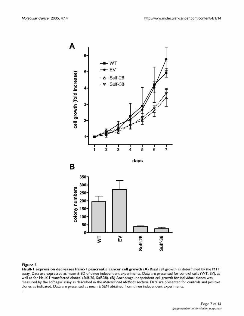
Functional consequences of Hsulf-1 expression in Panc-1 pancreatic cancer cellsNext, the effects of Hsulf-1 expression on the growth ofPanc-1 cells were assessed. Analysis of basal growthrevealed that the 2 control clones displayed an averageexponential doubling time of 50.3 ± 3.2 hours, which wassignificantly shorter compared to the 2 Hsulf-1 transfectedclones (68.2 ± 4.3 hours) (Figure 5 A). To determineanchorage-independent growth rates, soft agar assayswere carried out. The 2 control clones showed an averagecolony number of 223 as compared to 31 colonies for theHsulf-1 transfected cells (Figure 5 B).
Hsulf-1 decreases FGF-2 mediated cell proliferation and signaling in Panc-1 pancreatic cancer cellsIt has been shown previously that a variety of growth fac-tors such as FGF-2, EGF, HB-EGF, and IGF-1 are overexpressed in pancreatic cancer and that they have thepotential to act as mitogens for pancreatic cancer cell lines[3,4]. Therefore, we further investigated whether over-expression of Hsulf-1 could modulate the function ofthese growth factors in pancreatic cancer cells. Two Hsulf-1 transfected clones and two controls (wild type andempty vector) were selected to perform growth assays inthe presence or absence of different doses of the indicatedgrowth factors. Hsulf-1 expression significantly attenu-ated FGF-2 (50 ng/ml) induced cell growth by around50%, from +28.0 ± 3.8% in controls to +14.4 ± 1.0 % inHsulf-1 clones (Figure 6). In contrast, there was nodifference in the response towards IGF-1, EGF or HB-EGFin the control versus Hsulf-1 expressing cells. Since Hsulf-1 expression reduced FGF-2 but not EGF or HB-EGFinduced cell proliferation, next we sought to investigatewhether Hsulf-1 expression would influence FGF-2 andEGF/HB-EGF downstream signaling. EGFR phosphoryla-tion was not changed in response to EGF or HB-EGF inHsulf-1 expressing clones compared to controls (Figure 7A). In addition, there was also no difference of EGF andHB-EGF induced MAPK phosphorylation between controlcells and positive clones (Figure 7 A). In contrast, controlcells showed increased MAPK phosphorylation after FGF-2 stimulation, while this FGF-2 induced phosphorylation
Expression of Hsulf-1 mRNA in pancreatic tissues and cell linesFigure 1Expression of Hsulf-1 mRNA in pancreatic tissues and cell lines Quantification of Hsuf-1 mRNA levels in the normal pancreas, chronic pancreatitis (CP), pancreatic can-cer tissues (PC), by real time QRT-PCR as described in the Material and Methods section. Values are normalized to housekeeping genes (cyclophilin B and HRPT), and presented as single values and mean (horizontal line).
1
10
100
1000
10000
normal cancerCP
hS
UL
F1
mR
NA
leve
ls(c
op
ies/
µl)
Page 3 of 14(page number not for citation purposes)

Molecular Cancer 2005, 4:14 http://www.molecular-cancer.com/content/4/1/14
Expression and localization of Hsulf-1 mRNA in pancreatic tissuesFigure 2Expression and localization of Hsulf-1 mRNA in pancreatic tissues In situ hybridization was performed as described in Material and Methods section. Hsulf-1 localization in: normal pancreatic acini (A); blood vessels (B), nerves (C), and tubular complexes (D) of CP; tubular complexes (E), cancer cells (F) of pancreatic cancer. Note the staining in the control section probed with the antisense riboprobe (G), compared with absent staining in the sense-probed section (H).
A B
C
G
D
E F
H
Page 4 of 14(page number not for citation purposes)

Molecular Cancer 2005, 4:14 http://www.molecular-cancer.com/content/4/1/14
Expression of Hsulf-1 in pancreatic cancer cell linesFigure 3Expression of Hsulf-1 in pancreatic cancer cell lines (A) Quantification of Hsulf-1 mRNA levels in pancreatic cancer cell lines by real time QRT-PCR as described in the Materials and Methods section. Values are normalized to housekeeping genes (cyclopilin B and HRPT), and presented as mean ± SD. (B/C) Panc-1 cells were stable transfected with a Hsulf-1 sense expres-sion plasmid as described in the Material and Methods section. (B) Hsulf-1 sense RNA expression in Panc-1 cells was verified by Northern blot analysis using a radiolabeled Hsulf-1 antisense riboprobe. A sample Northern blot of 2 controls and 2 trans-fected clones is shown. (C) Expression of c-myc tagged Hsulf-1 (arrow) by immunoblot analysis as described in the Materials and Methods section. Equal loading of the protein samples was confirmed using anti-γ-tubulin antibodies. (D) Sulfatase activity was measured as described in Material and Methods section in control and positive transfected clones. Data are expressed as relative fluorescence and presented as mean ± SD.
0
100000
200000
300000
Aspc-1
BxPc-3
Capan-1
Colo-357
Mia-PaCa-2
Panc-1
SU-8686
T3M4
hSULF1mRNAlevels
(copies/µl)
1
10
100
1000
hSULF1
WT
EV
Sulf-26
Sulf-38
hSULF1
γγγγ-tubulin
WT
EV
Sulf-26
Sulf-38
B
A
D
sulfataseactivity
C
WT
EV
Sulf-26
Sulf-38
Page 5 of 14(page number not for citation purposes)

Molecular Cancer 2005, 4:14 http://www.molecular-cancer.com/content/4/1/14
was markedly attenuated in Hsulf-1 transfected cells (Fig-ure 7 B). Next, the basal and FGF-2 induced invasioncapacity of tumor cells was analyzed. This analysisrevealed a significant reduction in the invasiveness ofFGF-2 exposed Hsulf-1 expressing cells compared toHsulf-1 negative clones. As demonstrated in Figure 7C,FGF-2 (10 µg/ml) significantly stimulated the invasion ofthe control cells by +83.4 ± 24.2% after 24 h of incuba-tion. In contrast, the invasion ability of Hsulf-1 positivecells was significantly less stimulated (+27.4 ± 35.5%) byexposure to FGF-2 (Figure 7 C).
Effects of Hsulf-1 expression on chemosensitivityPancreatic cancers exhibit variable degrees of chemother-apy resistance. To determine whether Hsulf-1 expressionmight influence the sensitivity of Panc-1 cells to chemo-therapeutic agents, cells were treated with gemcitabine, 5-FU, or actinomycin-D and the GI50 concentration wascalculated. The GI50 concentration of gemcitabine in thePanc-1 WT control cells was 3.9 nM, and in EV Panc-1cells approximately 50 nM. Interestingly Hsulf-1 express-ing cells exhibited GI50 values of more than 100 nM(Figure 8). In contrast no significant changes were
Hsulf-1 decreases the sulfation of cell surface HSPGsFigure 4Hsulf-1 decreases the sulfation of cell surface HSPGs Immunofluorescence was performed as described in Material and Methods section with a specific 10E4 anti-HSPG monoclonal antibody, which recognizes native heparan-sulfate containing the N-sulfated glucosamine moiety. WT (A), EV (C), Sulf-26 (B), Sulf-38 (D).
A B
C D
Page 6 of 14(page number not for citation purposes)
Molecular Cancer 2005, 4:14 http://www.molecular-cancer.com/content/4/1/14
Hsulf-1 expression decreases Panc-1 pancreatic cancer cell growthFigure 5Hsulf-1 expression decreases Panc-1 pancreatic cancer cell growth (A) Basal cell growth as determined by the MTT assay. Data are expressed as mean ± SD of three independent experiments. Data are presented for control cells (WT, EV), as well as for Hsulf-1 transfected clones. (Sulf-26, Sulf-38). (B) Anchorage-independent cell growth for individual clones was measured by the soft agar assay as described in the Material and Methods section. Data are presented for controls and positive clones as indicated. Data are presented as mean ± SEM obtained from three independent experiments.
cell
gro
wth
(fo
ldin
crea
se)
A
1 2 3 4 5 6 7
1
2
3
4
5
6
WTEV
Sulf-26Sulf-38
0
50
100
150
200
250
300
350
WT
EV
Su
lf-2
6
Su
lf-3
8
colo
ny
nu
mb
ers
Bdays
Page 7 of 14(page number not for citation purposes)

Molecular Cancer 2005, 4:14 http://www.molecular-cancer.com/content/4/1/14
observed in the sensitivity of Hsulf-1 positive clones andcontrol cells towards 5-FU and actinomycin-D.
DiscussionSulfatases are a family of enzymes that catalyse the hydrol-ysis of sulfate ester bonds from a wide variety of com-pounds. They are classified into arylsulfatases and
nonarysulfatases according to their ability to hydrolysethe sulfate ester bonds of aromatic compounds such as p-nitrocatechol sulfate and 4-methylumbelliferyl sulfate[14]. Hsulf-1 is a newly identified member of the sulfatasefamily, which exhibits arysulfatase activity and removessulfate from the C-6 position of glucosamine within thespecific sub regions of intact heparin [10]. In the present
Effects of Hsulf-1 expression on growth factor induced proliferationFigure 6Effects of Hsulf-1 expression on growth factor induced proliferation. Clones were cultured in 1% FBS medium and incubated with increasing concentrations of the indicated growth factors for 72 h. Cell growth was measured by the MTT assay. Percent growth stimulation was determined by comparison with control cell growth. Values shown are the mean ± SEM obtained from three independent experiments.
0 10 20 30 40 5090%
100%
110%
120%
130%
140%
WT
EGF (ng/ml)
cell
gro
wth
(%
of
con
tro
l)
0 10 20 30 40 5090%
100%
110%
120%
130%
140%
HB-EGF (ng/ml)
0 10 20 30 40 5090%
100%
110%
120%
130%
140%
EVSulf-26Sulf-38
IGF-1 (ng/ml)
cell
gro
wth
(%
of
con
tro
l)
FGF2 (ng/ml)
0 10 20 30 40 5090%
100%
110%
120%
130%
140%
Page 8 of 14(page number not for citation purposes)

Molecular Cancer 2005, 4:14 http://www.molecular-cancer.com/content/4/1/14
(A-B). Effects of Hsulf-1 on EGF/HB-EGF and FGF-2 induced receptor phosphorylation and MAPK phosphorylationFigure 7(A-B). Effects of Hsulf-1 on EGF/HB-EGF and FGF-2 induced receptor phosphorylation and MAPK phosphor-ylation. Panc-1 pancreatic cancer cells were cultured in 1% FBS medium overnight and then incubated with 10 µg/ml of the indicated growth factors for 10 min. Phosphorylation of MAP kinase and receptor phosphorylation (EGFR) was determined by immunoblotting with antibodies specific for phospho-p44/42 MAPK and phospho-EGFR. Equal loading was determined by re-blotting the membranes with an γ-tubulin antibody. The figure is representative of three independent experiments. (C) Hsulf-1 attenuates FGF-2 stimulated invasion. An in vitro cell invasion assay was performed using 8 µM filters coated with Matrigel as described in the Material and Methods section. Panc-1 cells (1.25 × 10⁵) were seeded onto the filters in 1% serum overnight, and then treated as indicated for 24 h. The values shown are the mean ± SEM obtained from three independent experiments.
EV Sulf-26 WT Sulf-38
p-EGFRcontrol
EGF
HB-EGF
control
EGF
HB-EGF
control
EGF
HB-EGF
control
EGF
HB-EGF
p-MAPK
WT EV Sulf-26 Sulf-38
p-MAPK
γγγγ-tubulin
invadingcells(%
ofcontrol)
0%
50%
100%
150%
200%
WT EV Sulf-26 Sulf-38
control
FGF2
control
FGF2
control
FGF2
control
FGF2
B
A
C
control
FGF2
control
FGF2
control
FGF2
control
FGF2
Page 9 of 14(page number not for citation purposes)

Molecular Cancer 2005, 4:14 http://www.molecular-cancer.com/content/4/1/14
study a significant up-regulation of Hsulf-1 in primarypancreatic cancer, pancreatic metastasis and CP comparedto normal pancreatic tissues was demonstrated, whereasthere was no significant difference in the expression ofother members of the sulfatase family in these tissues.This indicates that Hsulf-1 might play a specific role in thepathogenesis and evolution of CP and pancreatic cancer.Normal pancreatic tissues are composed mainly of ahomogenous population of acinar cells (and a lowpercentage of ductal and islet cells), whereas both CP andpancreatic cancer tissues contain a variable amount ofdesmoplastic areas, inflammatory cells, degeneratingacini, tubular complexes (and cancer cells). Thus, theobserved wide range of expression of Hsulf-1 mRNA inboth CP and pancreatic cancer tissues is most likely due to
the different individual composition of these tissues. Toconfirm this hypothesis, in situ hybridization was utilizedto localize Hsulf-1 mRNA expression in normal, CP andpancreatic cancer tissues. This analysis demonstrated thatHsulf-1 mRNA expression was weakly present in normalacinar cells, and at high levels in the endothelium andsmooth muscle cells of blood vessels, as well as in fibrob-lasts and tubular complexes in CP tissues and additionallyin the malignant cells in pancreatic cancer tissues.
The observed increased levels of Hsulf-1 in pancreatic can-cer tissues seem to be in contrast to the down-regulationof Hsulf-1 in HCC and ovarian tumors [11,12]. However,while in ovarian cancer markedly diminished levels wereobserved in approximately 75% of the cases [11], the
Effects of Hsulf-1 on the sensitivity towards chemotherapeutic agentsFigure 8Effects of Hsulf-1 on the sensitivity towards chemotherapeutic agents. Panc-1 cells were cultured in complete medium and incubated in the absence (control) or presence of increasing concentrations of the indicated drugs for 48 h. Cell growth was measured by the MTT assay. Percent growth inhibition was determined by comparison with control cell growth. Values shown are the mean ± SEM obtained from three independent experiments.
0 25 50 75 100
-100
-50
0
50
100
5-FU (µM) gemcitabine (nM)
actinomycin D (ng/ml)
cell
gro
wth
(%
of
con
tro
l)ce
llg
row
th (
% o
f co
ntr
ol)
0 10 20 30 40 500
25
50
75
100
125
WTEVSulf-26Sulf-38
0 25 50 75 1000
25
50
75
100
125
Page 10 of 14(page number not for citation purposes)
Molecular Cancer 2005, 4:14 http://www.molecular-cancer.com/content/4/1/14
percentage was much smaller in HCCs (30%) [12], sug-gesting that reduced Hsulf-1 expression is not universallyobserved in all tumor types. It has been hypothesized thatenhanced expression of Hsulf-1 is related to c-myc ampli-fication in HCCs [12]. It could be speculated that also inpancreatic cancer high Hsulf-1 levels are related to c-mycamplification [15], at least in a subset of tumors. Anotherinteresting aspect is the generally low Hsulf-1 expressionlevel in cultured cancer cell lines. Thus, Hsulf-1 expressionis absent in 71% of ovarian cancer cell lines [11], in 82%of HCC cell lines [12], and in 50% of pancreatic cancercell lines (present study).
To evaluate the functional importance of Hsulf-1 in pan-creatic cancer cells, Panc-1 cells, which do not expressHsulf-1 at detectable levels, were stably transfected with aHsulf-1 expression vector. Over-expression of Hsulf-1 inPanc-1 cells resulted in reduced anchorage-dependent and-independent cell growth, suggesting an importantgrowth regulatory role of this gene in pancreatic cancer.These tumors are characterized by enhanced expression ofa variety of growth factors and their receptors, which havethe capacity to influence different cellular functions, suchas cell proliferation, migration and angiogenesis [3,4].Some of these growth factors are heparin-binding growthfactors, such as FGFs, VEGF and HB-EGF. We hypothe-sized that Hsulf-1 expression would attenuate the effectsof these growth factors by desulfation of HSPGs resultingin a growth disadvantage as suggested for other tumors[11-13]. FGF-2 stimulated cell proliferation was attenu-ated by the expression of Hsulf-1. Nonetheless, FGF-2 stillinduced growth in Hsulf-1 expressing cells, but to a lesserextent compared with control cells. It is conceivable thatthe HSPG/FGF receptor complex can facilitate FGF-2 sign-aling, but may not be strictly required for binding of FGF-2 to its receptor; it only increases the affinity of the FGF-2/FGF receptor interaction to a certain degree. Hsulf-1expression in Panc-1 cells also partially blocked FGF-2induced MAPK phosphorylation and invasion, furthersupporting the hypothesis that Hsulf-1 interferes withFGF-2 signaling in pancreatic cancer cells. In contrast, nodifference between Hsulf-1 expressing and control cellswas observed upon stimulation with HB-EGF – anotherheparin-binding growth factor- as well as EGF and IGF-1suggesting that these growth factors and their receptors donot require sulfated HSPGs for effective signaling. Theobservation that Hsulf-1 expression does not interferewith HB-EGF signaling in pancreatic cancer cells is in con-trast to recent studies in ovarian cancer cells [11], suggest-ing cell type specific differences.
Previously, it has been shown that Hsulf-1 expressionenhances cisplatin-induced apoptosis in HCC cell lines[12]. In the present study, we did not observe increasedsensitivity towards chemotherapeutic agents in Hsulf-1
expressing versus control cells. In contrast, Hsulf-1expressing Panc-1 cells were more resistant to gemcitabinethan the control cells, thereby suggesting that Hsulf-1over-expression might confer increased chemoresistanceto pancreatic cancer cells and thus provide them with agrowth advantage. However, the reason behind this effectis currently not known and requires further analysis.
In conclusion, Hsulf-1 is up-regulated in pancreatic cancerand chronic pancreatitis compared to normal pancreatictissues, mainly due to over-expression in the desmoplasticand cancerous tissue elements. Expression of Hsulf-1 inPanc-1 cells negatively influences growth and invasion byattenuating FGF-2 signaling, suggesting that Hsulf-1 playsa specific role in the pathogenesis of pancreatic cancer.Further experimental approaches, especially in vivo stud-ies, will help to assess in more detail the role of thisenzyme in human pancreatic cancer.
Materials and methodsMaterialsDMEM, trypsin-EDTA, and penicillin-streptomycin werepurchased from Invitrogen (Mannheim, Germany); FBSfrom PAN Biotech (Aidenbach, Germany); Gene screenhybridization transfer membranes from PerkinElmer LifeScience (Boston, MA, USA); 32P CTP from AmershamPharmacia Biotech (Freiburg, Germany); Lipofectaminereagent™ and TRIzol Reagent from Invitrogen (Karlsruhe,Germany); RiboMAX™ Large Scale RNA production sys-tem, antibiotic G-418 sulfate from Promega (Mannheim,Gemany); Gemcitabine-Hydrochloride from Lilly (EliLilly and Company Limited, Hampshire, UK); Recom-binant human FGF-2, Recombinant human IGF-1,Recombinant human HB-EGF from R&D systems (Wies-baden-Nordenstadt, Germany); p-EGFR (Tyr 1173)antibody from Santa Cruz Biotechnology (Santa Cruz,CA, USA), p-P44/42 MAPK (Thr202/Tyr204) antibodiesfrom Cell Signaling Technology (Frankfurt, Germany);EGF from Upstate Biotechnology (Hamburg, Germany);monoclonal anti-Heparan sulfate (10-E4 epitope) fromSeikagaju Corporation (Tokyo, Japan); Labeled goat anti-mouse IgM antibodies from Molecular Probes (Leiden,Netherlands); Anti-c-Myc antibody from Invitrogen (Karl-sruhe, Germany); Anti-goat IgG HRP- linked antibodies,anti-mouse IgG HRP- linked antibodies, anti-rabbit IgGHRP- linked antibodies and ECL immunoblottingdetection reagents from Amersham Biosciences (Freiburg,Germany); complete mini-EDTA-free protease inhibitorcocktail tablets from Roche (Mannheim, Germany);DIFCO Noble agar from DIFCO Laboratories (Detroit,MI,) All other reagents were from Sigma (Munich,Germany).
Page 11 of 14(page number not for citation purposes)
Molecular Cancer 2005, 4:14 http://www.molecular-cancer.com/content/4/1/14
DNA arrayThe GeneChip® HG-U95Av2 array used in this study wasfabricated by Affymetrix Inc. (Santa Clara, CA). Poly (A)+
RNA isolation, cDNA synthesis, cRNA in vitro transcrip-tion, product purification and fragmentation wasperformed as described [16,17]. Hybridization of the frag-mented in vitro transcription product to oligonucleotidearrays was performed according to the manufacturerinstructions (Affymetrix Inc.).
Patients and tissues collection31 pancreatic cancer samples were obtained from patients(median age 62.5 years; range, 41–78 years), who under-went pancreatic resections for pancreatic cancer at theUniversity Hospitals of Berne (Switzerland) and Heidel-berg (Germany). 22 chronic pancreatitis samples wereobtained from patients who underwent resection forchronic pancreatitis (median age 44 years range 22–66years). 19 normal human pancreatic tissue samples wereobtained from previously healthy individuals through anorgan donor program (median age 45 years range 20–74years). Immediately upon surgical removal, tissue sam-ples were either snap-frozen in liquid nitrogen and thenmaintained at -80°C until use (for RNA extraction) orfixed in 5% formalin and embedded in paraffin after 24 h.All studies were approved by the Ethics Committees of theUniversity of Heidelberg, and the University of Bern. Writ-ten informed consent was obtained from all patients.
Cell culturePancreatic cancer cell lines were routinely grown inDMEM medium (Panc-1 and Mia-PaCa-2) or RPMImedium (Aspc-1, BxPc-3, Capan-1, Colo-357, SU-8686and T3M4) supplemented with 10% fetal calf serum(FCS), 100 U/ml penicillin, and100 µg/ml streptomycin(complete medium). Cells were maintained at 37°C in ahumid chamber with 5% CO2 and 95% air atmosphere.
Real-time quantitative polymerase chain reaction (QRT-PCR)All reagents and equipment for mRNA and cDNA prepa-ration were purchased from Roche (Roche Applied Sci-ence, Mannheim, Germany). mRNA was prepared byautomated isolation using the MagNA Pure LC instrumentand isolation Kit I (for cells) and Kit II (for tissues). RNAwas reverse transcribed into cDNA using the 1st StrandcDNA Synthesis Kit for RT-PCR (AMV) according to themanufacturer's instructions. QRT-PCR was performedwith the Light Cycler Fast Start DNA SYBR Green kit asdescribed previously [18]. The number of specific tran-scripts was normalized to housekeeping genes (cyclophi-lin B and hypoxanthine guaninephosphoribosyltransferase, HPRT), and presented asadjusted transcripts / µl cDNA. All primers were obtainedfrom Search-LC (Heidelberg, Germany).
In situ hybridizationSpecific human Hsulf-1 riboprobes were generated byreverse-transcription polymerase chain reaction using thefollowing primer pairs: Hsulf-1: sense, 5'-ACT GTA CCAATC GGC CAG AG-3'; antisense, 5'-CCT CCT TGA ATGGGT GAA GA -3'. The resulting polymerase chain reactionproducts were subcloned into the pGEM-T easy vector(Promega GmbH, Mannheim, Germany) containing pro-moters for DNA-dependent SP6 and T7 RNA polymerases.The authenticity of the subcloned Hsulf-1 fragment wasconfirmed by sequencing (Qiagen GmbH, Hilden, Ger-many). Plasmids were linearized using SpeI and NcoIrestriction enzymes. T7 and SP6 RNA polymerases wereused to construct sense and antisense complementaryRNA riboprobes. Biotin complementary RNA labeling wasperformed using the biotin RNA labeling kit according tothe manufacturer's instructions (Roche Diagnostics, Man-nheim, Germany). Tissue sections (3 µm) were deparaffi-nized, rehydrated with 1× phosphate-buffered saline, andincubated in 0.2 M/L HCl for 20 minutes at roomtemperature. After rinsing the slides in 2× standard salinecitrate, sections were treated with proteinase K (RocheDiagnostics) at a concentration of 25 µg/ml for 15 min-utes at 37°C. After postfixation with 4% paraformalde-hyde in phosphate-buffered saline for 5 minutes andwashing in 2× standard saline citrate, samples wereacetylated in 2.5% acetic anhydride and 1.5% trieth-anolamine for 10 minutes. Subsequently, sections wereprehybridized at 78°C for 2 hours in 50% formamide, 4×standard saline citrate, 2× Denhardt's reagent, and 250 µgRNA/ml. Hybridization was performed overnight at 78°Cin 50% formamide, 4× standard saline citrate, 2× Den-hardt's reagent, 500 µg RNA/ml, and 10% dextran sulfate.The final concentration of the biotin-labeled probes was0.8 ng/µl. After hybridization, excess probe was removedby washing the slides 3 times in Dako stringent wash solu-tion (Dako) at 78°C for 15 minutes. The samples werethen incubated with streptavidin alkaline phosphataseconjugate (Dako) for 30 minutes at room temperature.For the color reaction, 5-bromo-4-chloro-3-indolyl phos-phate/nitroblue tetrazolium substrate (Dako) was used.
Stable transfectionThe Hsulf-1 expression plasmid (pcDNA 3.1/myc-His)[10] was a kindly provided by S.D. Rosen (University ofCalifornia, San Francisco). Panc-1 pancreatic cancer cellswere stably transfected with the Hsulf-1 plasmid and withthe empty control vector using the lipofectamine reagent[7,8]. Briefly, after reaching confluence, cells were split1:10 into selection medium (complete medium supple-mented with 1200 µg/ml G418) and single clones wereisolated after 2–4 weeks. After expansion, cells fromeachindividual clone were screened for the expression ofHsulf-1 by Northern blot analysis. Parental Panc-1 pan-creatic cancer cells were also transfected with an empty
Page 12 of 14(page number not for citation purposes)

Molecular Cancer 2005, 4:14 http://www.molecular-cancer.com/content/4/1/14
expression vector carrying the neomycin-resistance geneas a control. Positive clones were routinely grown in selec-tion medium.
RNA extraction and Northern Blot analysisThe pGEMT-easy vector containing the Hsulf-1 fragmentwas linearized with SpeI, and a 32P -labeled riboprobe wassynthesized with the RiboMAX™ large scale RNA produc-tion system kit using T7 polymerase and 32P CTP. TotalRNA was extracted by the single step-acid guanidiniumthiocyanate phenol chloroform method [7,8]. RNA (15µg/lane) was size fractionated on 1.2 % agarose/1.8 M for-maldehyde gels. Gels were stained with ethidium bromidefor verification of RNA integrity and loading equivalency.Fractionated RNA was transferred onto Genescreenmembranes and cross linked by UV irradiation. Blots werethen prehybridized for 12 h at 65°C in 50% formamide,1% SDS, 5 × Denhardt's, 100 µg/ml salmon sperm DNA,50 mM Na2PO4, pH 7.4, 10% dextran, 75 mM NaCl and5 mM EDTA. Blots were then hybridized for 24 h at 65°Cin the presence of 32P CTP labeled riboprobe, rinsed twicewith 2 × SSC and washed twice with 0.2 × SSC/2%SDS at65°C for 20 min, respectively. All blots were exposed at -80°C to Kodak BiomaxMS films with Kodak-intensifyingscreens.
Immunoblot analysisCell culture monolayers were washed twice with ice-coldPBS and lysed with lysis buffer (50 mM Tris-HCl, 100 mMNaCl, 2 mM EDTA,1% SDS) containing one tablet ofcomplete mini-EDTA-free protease inhibitor cocktail (in10 ml buffer). Protein concentration was determined bythe BCA protein assay (Pierce Chemical Co, Rockford,IL,). Cell lysates (30 µg/lane) were separated on SDS-poly-acrylamide gels and electroblotted onto nitrocellulosemembranes. Membranes were then incubated in blockingsolution (5% nonfat- milk in 20 mM Tris-HCl, 150 mMNaCl, 0.1% Tween-20), followedby incubation with theindicated antibodies at 4°C overnight. The membraneswere then washed in blocking solution and incubatedwithHRP-conjugated secondary antibodiesfor 1 hour at roomtemperature. Antibody detection was performedby anenhanced chemiluminescence reaction.
Sulfatase assayTo assay sulfatase activity in whole cell extracts, cells werelysed in lysis buffer (10 mM HEPES, 150 mM NaCl, 1%NP-40, 10% glycerol, 1.0 mM PMSF, and 1 mM EGTA),and the lysates were incubated on ice for 10 minutes. 4-Methylumbelliferyl-sulfate was used as the substrate. Celllysates with 100 µg protein were diluted with SIE (250mmol/l sucrose, 3 mmol/L imidazole, 0.1% absolute eth-anol, pH 7.4) to a total volume of 100 µl. One hundred µlof 1 µmol/l 4-methylumbelliferylsulfate was added toeach tube, mixed, and incubated at 37°C for 12 hours. 2
ml of stop solution (50 mM glycine, 5 mM EDTA, pH10.4) was then added and mixed, and released 4-methyl-umbelliferone was measured using a fluorometer (excita-tion wavelength 360 nm, emission wavelength 460 nm).
Immunofluorescence assayCells were grown in complete medium overnight in 10-well chambers, washed with PBS, fixed with 4% formalde-hyde/PBS for 20 min at room temperature (RT), incu-bated in ice-cold methanol for 5 min (RT) andsubsequently in acetone for 2 min (RT). Cells were incu-bated overnight with an anti-mouse antibody that recog-nizes negative heparan sulfate that contains the N-sulfatedglusamine residue (10E-4 mAb; 1:30 dilution) (SeikagakuCorporation Tokyo, Japan). Cells were then washed 3times with PBS, incubated with Alexa Fluor labeled goatanti-mouse IgM antibodies (Molecular Probes, Leiden,Netherlands) for 1 hour at room temperature, washedwith PBS and mounted with DAPI and anti-fadingmedium (Gel/mountTM, Abcam, Cambridgeshire, UK).Confocal microscopic analysis was performed using theSpectral Confocal Microscope Leica TCS SL (Leica Micro-systems GmbH, Heidelberg, Germany).
Proliferation assayAnchorage-dependent cell growth was determined by the3-(4,5-methylthiazol-2-yl)-2,5-diphenyltetrazolium bro-mide (MTT) colorimeric growth assay [7]. Briefly, 2,000cells/well were plated in 96-well plates and cultured for upto 7 days. Each day cell growth was determined by addingMTT solution (50 µg /well) for 4 hours. Cellular MTT wassolubilized withacidic isopropanol and optical densitywas measured at 570 nm. The doubling time was calcu-lated for the exponential growth phase. To measure andcalculate the GI50 of the chemotherapeutic agent (the con-centration that causes 50% cell growth inhibition), gradedconcentrations of drugs were added to triplicate wells andGI50 was calculated using the formula 100 x (T-T0)/(C-T0)= 50, where T is the optical density of the test well after 48hours period of exposure to drugs, T0 is the optical densityat time zero, and C is the control optical density after 48hours [19]. All experiments were performed in triplicate.
Soft agar assayCells (1 × 10³/well) were suspended in 3 ml of 0.3% DifcoNoble agar supplemented with complete culture medium.This suspension was layered over 1.5 ml of 0.5% agar-medium base layer in 12-well plates. After 14 days, cellswere stained with MTT (400 µg/well) for 24 h and colo-nies larger than 0.05 mm were counted.
Invasion assay8 µm filters were coated with Matrigel and placed inchambers. Cells (1.25 × 10⁴) cells were cultured in DMEMmedium containing 1% FBS overnight. FGF-2 was added
Page 13 of 14(page number not for citation purposes)
Molecular Cancer 2005, 4:14 http://www.molecular-cancer.com/content/4/1/14
Publish with BioMed Central and every scientist can read your work free of charge
"BioMed Central will be the most significant development for disseminating the results of biomedical research in our lifetime."
Sir Paul Nurse, Cancer Research UK
Your research papers will be:
available free of charge to the entire biomedical community
peer reviewed and published immediately upon acceptance
cited in PubMed and archived on PubMed Central
yours — you keep the copyright
Submit your manuscript here:http://www.biomedcentral.com/info/publishing_adv.asp
BioMedcentral
to the top chambers. After a subsequent 24 h incubationat 37°C, non-invaded cells were scraped off, and the cellsthat migrated to the lower surface of the filter inserts werefixed with 25% acetic acid and 75% methanol for 10 minand stained with 1% Toluidine blue in 1% sodium boratesolution. The invasion index was expressed as the ratio ofthe percent invasion of the treated cells over the percentinvasion of the control cells.
Statistical analysisResults were expressed as mean ± SEM, unless indicatedotherwise. For statistical analysis, the Student's t test wasused. Significance was defined as p < 0.05.
Competing interestsThe author(s) declare that they have no competinginterests.
Authors' contributionsJ.L, I.A, H.K carried out the in situ hybridization, immu-noblotting, cell proliferation, invasion assays, and softagar experiments. N.A.G. and T.G carried out the QRT-PCR analysis. J.K, M.W.B. and H.F. conceived the studyand participated in its design and coordination. J.K. andK.F. drafted the manuscript. All authors read andapproved the final version.
References1. Jemal A, Tiwari RC, Murray T, Ghafoor A, Samuels A, Ward E, Feuer
EJ, Thun MJ: Cancer statistics, 2004. CA Cancer J Clin 2004, 54:8-29.2. Li D, Xie K, Wolff R, Abbruzzese JL: Pancreatic cancer. Lancet
2004, 363:1049-1057.3. Korc M: Role of growth factors in pancreatic cancer. Surg Oncol
Clin N Am 1998, 7:25-41.4. Ozawa F, Friess H, Tempia-Caliera A, Kleeff J, Buchler MW: Growth
factors and their receptors in pancreatic cancer. Teratog Car-cinog Mutagen 2001, 21:27-44.
5. Filmus J, Selleck SB: Glypicans: proteoglycans with a surprise. JClin Invest 2001, 108:497-501.
6. Filmus J: Glypicans in growth control and cancer. Glycobiology2001, 11:19R-23R.
7. Kleeff J, Ishiwata T, Kumbasar A, Friess H, Buchler MW, Lander AD,Korc M: The cell-surface heparan sulfate proteoglycan glypi-can-1 regulates growth factor action in pancreatic carci-noma cells and is overexpressed in human pancreaticcancer. J Clin Invest 1998, 102:1662-1673.
8. Kleeff J, Wildi S, Kumbasar A, Friess H, Lander AD, Korc M: Stabletransfection of a glypican-1 antisense construct decreasestumorigenicity in PANC-1 pancreatic carcinoma cells. Pan-creas 1999, 19:281-288.
9. Schlessinger J, Plotnikov AN, Ibrahimi OA, Eliseenkova AV, Yeh BK,Yayon A, Linhardt RJ, Mohammadi M: Crystal structure of a ter-nary FGF-FGFR-heparin complex reveals a dual role forheparin in FGFR binding and dimerization. Mol Cell 2000,6:743-750.
10. Morimoto-Tomita M, Uchimura K, Werb Z, Hemmerich S, Rosen SD:Cloning and characterization of two extracellular heparin-degrading endosulfatases in mice and humans. J Biol Chem2002, 277:49175-49185.
11. Lai J, Chien J, Staub J, Avula R, Greene EL, Matthews TA, Smith DI,Kaufmann SH, Roberts LR, Shridhar V: Loss of HSulf-1 up-regu-lates heparin-binding growth factor signaling in cancer. J BiolChem 2003, 278:23107-23117.
12. Lai JP, Chien JR, Moser DR, Staub JK, Aderca I, Montoya DP, Mat-thews TA, Nagorney DM, Cunningham JM, Smith DI, Greene EL,Shridhar V, Roberts LR: hSulf1 Sulfatase promotes apoptosis of
hepatocellular cancer cells by decreasing heparin-bindinggrowth factor signaling. Gastroenterology 2004, 126:231-248.
13. Lai JP, Chien J, Strome SE, Staub J, Montoya DP, Greene EL, Smith DI,Roberts LR, Shridhar V: HSulf-1 modulates HGF-mediatedtumor cell invasion and signaling in head and neck squamouscarcinoma. Oncogene 2004, 23:1439-1447.
14. Parenti G, Meroni G, Ballabio A: The sulfatase gene family. CurrOpin Genet Dev 1997, 7:386-391.
15. Zojer N, Fiegl M, Mullauer L, Chott A, Roka S, Ackermann J, RadererM, Kaufmann H, Reiner A, Huber H, Drach J: Chromosomal imbal-ances in primary and metastatic pancreatic carcinoma asdetected by interphase cytogenetics: basic findings and clin-ical aspects. Br J Cancer 1998, 77:1337-1342.
16. Friess H, Ding J, Kleeff J, Liao Q, Berberat PO, Hammer J, BuchlerMW: Identification of disease-specific genes in chronic pan-creatitis using DNA array technology. Ann Surg 2001,234:769-778.
17. Tureci O, Bian H, Nestle FO, Raddrizzani L, Rosinski JA, Tassis A,Hilton H, Walstead M, Sahin U, Hammer J: Cascades of transcrip-tional induction during dendritic cell maturation revealed bygenome-wide expression analysis. FASEB J 2003, 17:836-847.
18. Li J, Kleeff J, Guweidhi A, Esposito I, Berberat PO, Giese T, BuchlerMW, Friess H: RUNX3 expression in primary and metastaticpancreatic cancer. J Clin Pathol 2004, 57:294-299.
19. Li J, Kleeff J, Giese N, Buchler MW, Korc M, Friess H: Gefitinib('Iressa', ZD1839), a selective epidermal growth factorreceptor tyrosine kinase inhibitor, inhibits pancreatic cancercell growth, invasion, and colony formation. Int J Oncol 2004,25:203-210.
Page 14 of 14(page number not for citation purposes)
Copyright © 2022 FDOKUMEN