BITC Sensitizes Pancreatic Adenocarcinomas to TRAIL-Induced Apoptosis
12
Wright State University CORE Scholar Neuroscience, Cell Biology & Physiology Faculty Publications Neuroscience, Cell Biology & Physiology 1-20-2010 BITC Sensitizes Pancreatic Adenocarcinomas to TIL-Induced Apoptosis Christina A. Wicker Ravi P. Sahu Kashmira Kulkarni-Datar Sanjay K. Srivastava omas L. Brown Wright State University - Main Campus, [email protected] Follow this and additional works at: hp://corescholar.libraries.wright.edu/ncbp Part of the Medical Cell Biology Commons , Medical Neurobiology Commons , Medical Physiology Commons , Neurosciences Commons , and the Physiological Processes Commons is Article is brought to you for free and open access by the Neuroscience, Cell Biology & Physiology at CORE Scholar. It has been accepted for inclusion in Neuroscience, Cell Biology & Physiology Faculty Publications by an authorized administrator of CORE Scholar. For more information, please contact [email protected]. Repository Citation Wicker, C. A., Sahu, R. P., Kulkarni-Datar, K., Srivastava, S. K., & Brown, T. L. (2010). BITC Sensitizes Pancreatic Adenocarcinomas to TIL-Induced Apoptosis. Cancer Growth and Metastasis, 2009 (2), 45-55. hp://corescholar.libraries.wright.edu/ncbp/32
Transcript of BITC Sensitizes Pancreatic Adenocarcinomas to TRAIL-Induced Apoptosis

Wright State UniversityCORE ScholarNeuroscience, Cell Biology & Physiology FacultyPublications Neuroscience, Cell Biology & Physiology
1-20-2010
BITC Sensitizes Pancreatic Adenocarcinomas toTRAIL-Induced ApoptosisChristina A. Wicker
Ravi P. Sahu
Kashmira Kulkarni-Datar
Sanjay K. Srivastava
Thomas L. BrownWright State University - Main Campus, [email protected]
Follow this and additional works at: http://corescholar.libraries.wright.edu/ncbp
Part of the Medical Cell Biology Commons, Medical Neurobiology Commons, MedicalPhysiology Commons, Neurosciences Commons, and the Physiological Processes Commons
This Article is brought to you for free and open access by the Neuroscience, Cell Biology & Physiology at CORE Scholar. It has been accepted forinclusion in Neuroscience, Cell Biology & Physiology Faculty Publications by an authorized administrator of CORE Scholar. For more information,please contact [email protected].
Repository CitationWicker, C. A., Sahu, R. P., Kulkarni-Datar, K., Srivastava, S. K., & Brown, T. L. (2010). BITC Sensitizes Pancreatic Adenocarcinomasto TRAIL-Induced Apoptosis. Cancer Growth and Metastasis, 2009 (2), 45-55.http://corescholar.libraries.wright.edu/ncbp/32

Cancer Growth and Metastasis 2009:2 45–55
This article is available from http://www.la-press.com.
© the author(s), publisher and licensee Libertas Academica Ltd.
This is an open access article. Unrestricted non-commercial use is permitted provided the original work is properly cited.
Open AccessFull open access to this and thousands of other papers at
http://www.la-press.com.
Cancer Growth and Metastasis 2009:2 45
Cancer Growth and Metastasis
O r i G i n A L r e s e A r C h
BITc sensitizes pancreatic Adenocarcinomas to TRAIL-induced Apoptosis
Christina A. Wicker1,3, ravi P. sahu2,3, Kashmira Kulkarni-Datar1,3, sanjay K. srivastava2 and Thomas L. Brown1
1Department of neuroscience, Cell Biology, and Physiology, Boonshoft school of Medicine, Wright state University, Dayton, Oh, UsA. 2Department of Biomedical sciences and Cancer Biology Center, Texas Tech University health sciences Center, school of Pharmacy, Amarillo, TX, UsA. 3These authors contributed equally to the manuscript. email: [email protected], [email protected]
Abstract: Pancreatic adenocarcinoma is an aggressive cancer with a greater than 95% mortality rate and short survival after diagnosis. Chemotherapeutic resistance hinders successful treatment. This resistance is often associated with mutations in codon 12 of the K-Ras gene (K-Ras 12), which is present in over 90% of all pancreatic adenocarcinomas. Codon 12 mutations maintain Ras in a constitutively active state leading to continuous cellular proliferation. Our study determined if TRAIL resistance in pancreatic adenocarcinomas with K-Ras 12 mutations could be overcome by first sensitizing the cells with Benzyl isothiocyanate (BITC). BITC is a component of cruciferous vegetables and a cell cycle inhibitor. BxPC3, MiaPaCa2 and Panc-1 human pancreatic adenocarcinoma cell lines were examined for TRAIL resistance. Our studies show BITC induced TRAIL sensitization by dual activation of both the extrinsic and intrinsic apoptotic pathways.
Keywords: TRAIL resistance, K-RAS, BITC, pancreatic adenocarcinoma, chemotherapeutic resistance

Wicker et al
46 Cancer Growth and Metastasis 2009:2
IntroductionPancreatic cancer is the fourth leading cause of cancer-related deaths in the United States.1,2 Pancre-atic cancer includes exocrine, neuroendocrine, and adenocarcinoma forms, with adenocarcinoma being the most common and having the least favorable prog-nosis. There are three predominant treatment options; surgery, radiation, and chemotherapy. Pancreatic ade-nocarcinomas are largely resistant to radiation and chemotherapy and often inoperable. The combination of late detection and chemotherapeutic resistance in pancreatic cancers is responsible for a greater than 95% mortality rate.1,2
TRAIL (TNF-related apoptosis-inducing ligand) is a potential chemotherapeutic agent. TRAIL death receptors are highly expressed on the surface of trans-formed cancer cells but generally absent on most normal cells.3 The cytotoxic effects of TRAIL are pronounced on cancerous cells and trigger apoptosis, but leave most noncancerous tissues unaffected.4–6 Chemotherapeutic resistance to TRAIL-induced apop-tosis has been associated with mutations in codon 12 of the K-Ras gene (K-Ras12).4,5
Over ninety percent of pancreatic adenocarcinomas harbor a mutation within codon 12 of the K-Ras gene.7,8 Although other mutations, such as p53, p16, and SMAD 4 have been reported in pancreatic adenocarcinomas, mutations of K-Ras are shown to be an initiating factor in the formation of pancreatic cancers.8 Ras is a G protein that regulates migration, cytoskeletal formation, apoptosis, and cellular prolif-eration, predominantly through the MAP kinase signal transduction pathways.9–11 Ras cycles between an inactive GDP-bound state and an active GTP- bound state.12–16 Endogenous GTPase activity is responsi-ble for the inactivation of Ras, thus inhibiting Ras-mediated signaling.17,18 Mutations within codon 12 of K-Ras inhibit this endogenous GTPase activity, thereby maintaining Ras in its GTP-bound active state.13 Pre-vious studies have shown that constitutive activation of Ras leads to continuous cellular proliferation.
Benzyl isothiocyanate (BITC) has been shown to inhibit cell cycle progression.19 BITC is present in cruciferous plants and is a member of the isothiocya-nate family, which have been found to be protective against carcinogenesis.20–22 BITC has recently been shown to induce G2/M cell cycle arrest by decreasing Cdk1, CyclinB1 and Cdc25B protein levels.19,23,24 In
high doses, BITC produces the formation of reactive oxygen species and can induce cell death.19,24,25
Two major pathways, the extrinsic (death receptor) pathway and the intrinsic (mitochondrial) pathway mediate apoptosis.3 The extrinsic cell death pathway begins with external death receptors on the cell sur-face. When ligands such as TNF alpha, Fas, or TRAIL bind to their specific receptor, intercellular signaling results in the cleavage and activation of caspase 8.26,27 Caspase 8 can then cleave effector caspase 3, inducing apoptosis directly.28 In addition, in several cell types, caspase 8 can also trigger the activation of the intrinsic cell death pathway via cleavage of Bid, a proapoptotic protein.29 Truncated Bid (t-Bid) is capable of inter-acting with other pro-apoptotic proteins leading to loss of mitochondrial membrane integrity, which has previously been shown to trigger the release of cyto-chrome C.29,30 Cytochrome C release is associated with the activation of caspase 9 and subsequently leads to the activation of effector caspase 3.31–34
BxPC3, MiaPaCa2 and Panc-1 cell lines were cho-sen for the current study because these cell lines rep-resent the most common mutations found in human pancreatic adenocarcinomas (Table 1).35–37 BxPC3 cells, isolated from human pancreatic adenocarci-noma, are tumorigenic but are wildtype at codon 12 of the K-Ras gene. Panc-1 cells, harbor a glycine to aspartate amino acid change within codon 12, and MiaPaCa2 cells, a glycine to cysteine amino acid change. Our results indicate that BITC sensitizes chemotherapeutically-resistant human pancreatic ade-nocarcinoma cells to TRAIL-induced apoptosis via the activation of dual apoptotic pathways.
Materials and MethodsMaterialsPanc-1 and BxPC3 cells were purchased from ATCC. Dr. B. Weissman of the University of North Carolina, Chapel Hill School of Medicine kindly provided MiaPaCa2 cells. Goat-anti-rabbit (W401B) and goat-anti-mouse (W402B) HRP-conjugated secondary anti-bodies were purchased from Promega. The primary antibodies used were: rabbit polyclonal anti-XIAP (Cell Signaling Cat # 2042), rabbit polyclonal PARP (Cell Signaling Cat # 9541S), mouse monoclonal cas-pase 8 (Cell Signaling Cat # 9746), mouse monoclo-nal active caspase 9 (Upstate Biotech Cat # 05-572), and rabbit polyclonal Bid (Cell Signaling Cat # 2002).

TrAiL resistance in pancreatic adenocarcinomas
Cancer Growth and Metastasis 2009:2 47
Mouse monoclonal pan actin was a kind gift from Dr. J. Lessard at Cincinnati Children’s Hospital. Rabbit polyclonal active caspase 3 antibody was a kind gift from Dr. K. Tomaselli at Idun Pharmaceuticals. For apoptotic death quantitation, the Roche Cell Death ELISA Assay was used (Roche Cat # 1774425). Human recombinant TRAIL was purchased from R&D Systems (Cat # 375-TEC). Apoptosis-inducing, anti-Fas antibody (clone CH-11) was purchased from Millipore. BITC was purchased from Sigma-Aldrich (Cat # 252492). Nonmethylated Q-VD-OPh (Cat # A0004) was a kind gift from Apoptrol, LLC.
Cell cultureBxPC3, MiaPaCa2, and Panc-1 cells were maintained in RPMI 1640 with L-glutamine, supplemented with 5% heat-inactivated fetal bovine serum and 1% antibiotic/antimycotic. Cells were maintained at 5% CO2 and 37 °C. All cell lines were maintained at 1 × 10⁵ cells/ml.
Treatment protocolFor collection of whole cell lysates used in Western blot analysis, cells were plated at 2 × 10⁵ cells/ml and allowed to attach overnight. TRAIL was solubilized in 0.1% BSA/water. A stock solution of BITC (100 mM) was prepared in DMSO and subsequently diluted in fresh medium so that the final concentration of DMSO was less than 0.1%. An equal volume of DMSO was added for the vehicle controls. Both BITC alone and BITC with TRAIL groups were treated with a final concentration of 5 µM BITC for 18 hrs. The follow-ing morning, vehicle and BITC alone groups were treated with 5 µl 0.1% BSA, whereas in the TRAIL and BITC combined group, TRAIL was added to a final concentration of 10 ng/ml TRAIL for 6 hrs. Total
treatment time with BITC was 24 hrs. Cells were then collected for whole cell lysates. In cells treated with Q-VD-OPh, cells were preincubated for 30 minutes with the pan-caspase inhibitor, prior to beginning the experimental treatments. Jurkat human T cells (1 × 10⁶ cells/ml) were treated with 100 ng/ml of apoptosis-inducing anti-Fas or 1 µg/ml Actinomycin D for 4 hrs as positive controls.
Western blotsWhole cell lysates were collected in CHAPS buffer (50 mM PIPES pH 6.5, 2 mM EDTA pH 8.0, 0.1% CHAPS) containing protease inhibitor, sonicated, and centrifuged at 12,000 rpm for 10 minutes at 4 °C. Pro-tein concentrations were determined using the Brad-ford assay.38 Cell lysates were treated with reducing sample buffer, and boiled for 5 minutes at 100 °C. Proteins were resolved on an SDS polyacrylamide gel via electrophoresis.31–34 Separated proteins were subsequently transferred onto a PVDF membrane in Western transfer buffer overnight at 0.1 A. Proteins were visualized using Ponceau S staining and the membrane was then blocked at room temperature for one hour in PBS blocking buffer (1X PBS, 5% (w/v) non-fat dry milk, and 0.05% Tween-20) for caspase 3 and 9 and Bid. Blots were blocked in TBS blocking buffer (1X TBS, 5% (w/v) non-fat dry milk, and 0.10% Tween-20) for XIAP, PARP and caspase 8. Blots were probed with antibodies specific for XIAP, cleaved PARP, active caspase 8 and 9 at (1:1,000) overnight at 4 °C. Actin was incubated at (1:16,000) for 40 minutes at room temperature (RT) and active caspase 3 at (1:4,000) for 2 hours at RT. Blots were then incubated with Pierce SuperSignal® West Pico Chemiluminescent Substrate following the manufac-turer’s recommendations and exposed to X-ray film.
Cell death eLisAFor cell death ELISA analysis, cells were plated at 1 × 10⁵ cells/ml. Vehicle and TRAIL alone groups were treated with a final concentration of 0.2% DMSO. BITC and BITC with TRAIL groups were treated for 18 hrs at a final concentration of 5 µM BITC. The following morning, the vehicle and BITC groups were treated with 2 µl 0.1% BSA. Cells treated with TRAIL were incubated with a final concentration of 10 ng/ml TRAIL for 6 hrs. Total treatment time with BITC was 24 hrs. Cells were then collected for analysis with the cell death
Table 1. K-Ras 12 mutations in Bxpc3, Miapaca2, and panc-1 human pancreatic cancer cell Lines. Observed nucleotide and amino acid changes within codon 12 of K-ras in BxPC3, MiaPaCa2, and Panc-1 cell lines.
cell line K-Ras 12 nucleotide change
Amino acid change
Reference
BxPC3 GGT none; Gly (37) MiaPaCa2 GGT→TGT Gly→Cys (35)Panc-1 GGT→GAT Gly→Asp (35)

Wicker et al
48 Cancer Growth and Metastasis 2009:2
ELISA assay following the manufacturer’s protocol. Roche Cell Death ELISA Assay was used to detect the fold increase in apoptotic cell death.29,30
statistical analysisStatistical analysis was performed using a one-way ANOVA. Results were considered to be statistically significant at p 0.05. All experiments were per-formed independently, a minimum of 3 times.
ResultsResistance to TRAIL-induced apoptosis has previ-ously been reported in pancreatic cancer cells.32 To determine the optimal dosage of TRAIL needed to induce apoptosis, we first treated Panc-1 cells with increasing doses of 1, 10, and 100 ng/ml TRAIL for 6 hours (Fig. 1A).6,24 TRAIL-induced apoptosis was evaluated using the cell death ELISA assay. A dose responsive increase in apoptosis was observed with increasing amounts of TRAIL. There was no sig-nificant increase in apoptosis with 1 ng/ml TRAIL compared to the vehicle treated cells. A significant 1.5 fold increase in apoptotic death was observed with 10 ng/ml TRAIL when compared to the vehicle alone and the 100 ng/ml TRAIL showed a 1.95 fold increase in cell death (P 0.05). Since a significant increase in apoptosis was observed at 10 ng/ml TRAIL and as there was no significant difference in apoptotic death between 10 ng/ml TRAIL or 100 ng/ml TRAIL; 10 ng/ml TRAIL was chosen for all further studies.
We determined the optimal duration for TRAIL treatment. Panc-1 and BxPC3 cells were treated with 10 ng/ml TRAIL for 6, 15 or 24 hours and apoptosis was analyzed using the cell death ELISA assay (Fig. 1B). Compared to the vehicle, there was a 7.04-fold increase in cell death at 6 hrs in BxPC3 cells, at 15 hrs there was a 4.53 fold increase in cell death, and at 24 hrs there was a 3.76 fold increase in cell death. In Panc-1 cells, at 6 hrs there was a 2.76 fold increase, at 15 hrs there was a 0.96 fold increase in cell death, and at 24 hrs there was a 1.05 fold increase in cell death. Higher levels of TRAIL-induced apoptosis were observed in wildtype K-Ras 12 BxPC3 cells, than in the K-Ras 12 mutant Panc-1 cell line. The greatest increase in apoptotic death in both cell lines was observed at 6 hours and this time point was chosen for all further experiments.
BITC is known to induce cell cycle arrest in human pancreatic cancer cells.18,22,24 We analyzed the effect
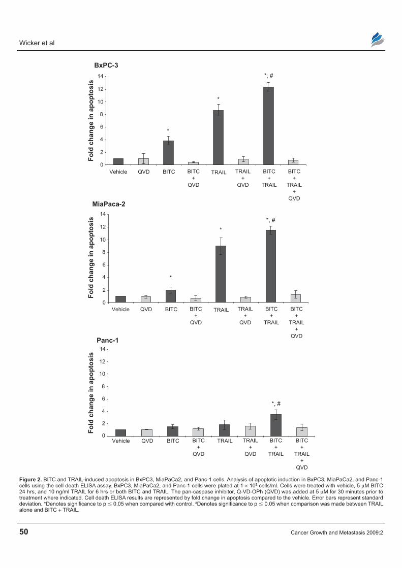
of BITC on TRAIL-induced apoptosis in BxPC3, MiaPaCa2 and Panc-1 cells by the cell death ELISA assay. Cells were treated with vehicle, BITC alone, TRAIL alone, or BITC in combination with TRAIL. Because BITC is a known cell cycle inhibitor, the fold change in apoptosis was normalized to cell num-ber to accurately reflect the amount of apoptosis. The normalized cell death ELISA results indicate that BITC alone caused a modest increase in apoptosis in BxPC3, MiaPaCa2 and Panc-1 cell lines (Fig. 2). BxPC3 and MiaPaCa2 cells were the most sensitive to TRAIL-induced apoptosis with a greater than 8-fold increase in apoptosis compared to vehicle. Panc-1 cells were the most resistant to TRAIL with only a 1.24 fold increase in apoptosis.
BxPC3 cells had a 3.84 fold increase in apopto-sis upon treatment with BITC alone, an 8.65 fold increase was observed with TRAIL alone, and a 12.39 fold increase was seen when cells were treated with BITC combined with TRAIL. MiaPaCa2 cells had a 2.08 fold increase in apoptosis upon treatment with BITC, a 9.12 fold increase with TRAIL alone, and an 11.72 fold increase with BITC combined with TRAIL. Panc-1 cells underwent a 1.49 fold increase in apoptosis upon treatment with BITC, a 1.82 fold increase with TRAIL alone, and a 3.45 fold increase with BITC combined with TRAIL compared to vehicle. Compared to TRAIL treatment alone, BITC combined with TRAIL produced a significant increase in apoptotic cell death in all three cell lines examined. In addition, treatment of BxPC3, MiaPaCa2, and Panc-1 cells with non-methylated Q-VD-OPh, a pan caspase inhibitor, prevented apoptosis in all three cell lines examined (Fig. 2).31
Western blot analysis was performed to identify the apoptotic pathways involved in the BITC and TRAIL-induced apoptosis of pancreatic adenocarci-noma cells. BxPC3, MiaPaCa2 and Panc-1 cells were treated with vehicle, BITC, TRAIL, or BITC com-bined with TRAIL and examined for activation of the extrinsic apoptotic cell death pathway (Fig. 3). In BxPC3 cells, MiaPaCa2, and Panc-1 cells, vehicle or BITC alone did not activate initiator caspase 8. Treat-ment with TRAIL alone induced the cleavage of the inactive proform of caspase 8, indicating the activation of caspase 8, although no further increase in caspase 8 cleavage was observed with the combination of BITC and TRAIL in all three cell lines (Fig. 3A).

TrAiL resistance in pancreatic adenocarcinomas
Cancer Growth and Metastasis 2009:2 49
TRAIL dose at 6 hrs
Times course for 10 ng/ml TRAIL treatment
Fo
ld c
han
ge
in a
po
pto
sis
TRAIL concentration
Fo
ld c
han
ge
in a
po
pto
sis 3
A
B
2.5
2
1.5
1
0.5
0
9
8
7
6
5
4
3
2
1
0
6 hrs
Time
15 hrs
BxPC3
Panc-1
24 hrs
Vehicle
Vehicle
1 ng/ml 10 ng/ml 100 ng/ml
*
*
Figure 1. Time and dose response of Panc-1 and BxPC3 cells to TrAiL. A) Panc-1 cells were plated 1 × 10⁵ cells/ml. Cells were treated with vehicle, 1, 10 and 100 ng/ml TrAiL for 24 hours and the dose response was analyzed by cell death eLisA assay, B) BxPC3 and Panc-1 cells were plated at 1 × 10⁵ cells/ml. Cells were treated with 10 ng/ml TrAiL for 6, 15 or 24 hours and the optimal time course was analyzed using the cell death eLisA. The results are represented by fold increase in apoptosis compared to vehicle. Error bars represent standard deviation. *Denotes significance to p 0.05 when compared with control.
X-linked inhibitor of apoptosis protein (XIAP) is a prosurvival protein and a direct target of caspase 8.42 Thus if caspase 8 becomes cleaved (and thus activated), caspase 8 will cleave its targets. We examined the XIAP protein to infer the extent of caspase 8 activation by visualizing the loss of the intact 53 kDa XIAP pro-tein in BxPC3, MiaPaCa2, and Panc-1 cells (Fig. 3B). No detectable XIAP cleavage was observed in BxPC3 cells treated with BITC alone, TRAIL alone, or BITC combined with TRAIL. In Panc-1 cells, addition of BITC resulted in the loss of intact XIAP. TRAIL alone, however, was unable to induce XIAP cleavage.
BITC combined with TRAIL led to a reduction in intact XIAP compared to the vehicle. In MiaPaCa2 cells, there was no major cleavage of XIAP with vehi-cle, BITC, and TRAIL alone. Treatment of TRAIL, combined with BITC in MiaPaCa2 cells, did trig-ger the loss of mature XIAP protein, indicative of caspase 8 cleavage. The combined treatment of BITC with TRAIL led to a reduction in intact XIAP pro-tein in MiaPaCa2 and Panc-1 cell lines compared to TRAIL alone.
In many cell types, the extrinsic (death receptor) path-way is known to activate caspase 8 and simultaneously
Wicker et al
50 Cancer Growth and Metastasis 2009:2
Fo
ld c
han
ge
in a
po
pto
sis
Vehicle QVD
BxPC-3
BITC BITC+
QVD
TRAIL+
QVD
BITC+
TRAIL
BITC+
TRAIL+
QVD
TRAIL
*
*
*, #14
12
10
8
6
4
2
0
Fo
ld c
han
ge
in a
po
pto
sis
Vehicle QVD
MiaPaca-2
BITC BITC+
QVD
TRAIL+
QVD
BITC+
TRAIL
BITC+
TRAIL+
QVD
TRAIL
*
*
14
12
10
8
6
4
2
0
Fo
ld c
han
ge
in a
po
pto
sis
Vehicle QVD
Panc-1
BITC BITC+
QVD
TRAIL+
QVD
BITC+
TRAIL
BITC+
TRAIL+
QVD
TRAIL
14
12
10
8
6
4
2
0
*, #
*, #
Figure 2. BiTC and TrAiL-induced apoptosis in BxPC3, MiaPaCa2, and Panc-1 cells. Analysis of apoptotic induction in BxPC3, MiaPaCa2, and Panc-1 cells using the cell death eLisA assay. BxPC3, MiaPaCa2, and Panc-1 cells were plated at 1 × 10⁵ cells/ml. Cells were treated with vehicle, 5 µM BiTC 24 hrs, and 10 ng/ml TrAiL for 6 hrs or both BiTC and TrAiL. The pan-caspase inhibitor, Q-VD-OPh (QVD) was added at 5 µM for 30 minutes prior to treatment where indicated. Cell death eLisA results are represented by fold change in apoptosis compared to the vehicle. error bars represent standard deviation. *Denotes significance to p 0.05 when compared with control. #Denotes significance to p 0.05 when comparison was made between TrAiL alone and BiTC + TrAiL.

TrAiL resistance in pancreatic adenocarcinomas
Cancer Growth and Metastasis 2009:2 51
trigger the activation of the intrinsic (mitochondrial) pathway via cleavage of the protein, Bid, resulting in truncated Bid (t-Bid).29,30 BxPC3, MiaPaCa2 and Panc-1 were cells treated with vehicle, BITC alone, TRAIL alone, or BITC combined with TRAIL and examined for Bid cleavage (Fig. 3C). The loss of intact 22 kDa Bid is indicative of t-Bid formation and the activation of the intrinsic pathway. Vehicle or BITC alone did not induce Bid cleavage in BxPC3 cells. TRAIL alone produced a complete loss of the inactive proform of Bid as did TRAIL combined with BITC. Similar to BxPC3 cells, MiaPaCa2 cells were absent of Bid cleavage upon treatment with vehicle or BITC alone, however, a substantial loss of intact Bid was observed when cells were treated with TRAIL alone or BITC combined with TRAIL. In Panc-1 cells, treat-ment with BITC alone did not reduce intact Bid protein; however, a progressive increase in cleavage was seen upon treatment with TRAIL alone and a complete loss of intact Bid was observed with BITC combined with TRAIL.
Truncation, and therefore activation, of Bid has been shown to induce the intrinsic cell death pathway.33,35 We sought to determine whether TRAIL combined with BITC activated the intrinsic, mitochondrial cell death pathway (Fig. 4). BxPC3, MiaPaCa2 and Panc-1 cells were examined for the activation of caspase 9, the initiator caspase for the intrinsic apoptotic death pathway (Fig. 4A). In BxPC3 cells treated with BITC alone, no cleavage of caspase 9 was observed. Upon treatment with TRAIL alone, there was a decrease of the proform and detection of an activated caspase 9 cleavage product. BITC in combination with TRAIL slightly increased the amounts of cleaved active caspase 9. In MiaPaCa2 cells, there was no appear-ance of the cleaved, active caspase 9 fragment upon treatment of the vehicle, BITC alone, or TRAIL alone. TRAIL combined with BITC resulted a dra-matic decrease in inactive procaspase 9, however, little cleaved caspase 9 could be detected. In Panc-1 cells, there was no cleavage of caspase 9 with BITC alone. TRAIL-induced a decrease in the proform and
Caspase 8
Proform (57 kDa)
Cleaved (45/43 kD)
MiaPaCa 2 Panc 1 BxPC 3 Jurkat
MiaPaCa 2 Panc 1 BxPC 3 Jurkat
MiaPaca 2 Panc 1 BxPC 3 Jurkat
V B B BT TT BT BT BT −V V +
A
B
C
V B B BT TT BT BT BT −V V +
V B B BT TT BT BT BT −V V +
Pan actin
Bid (22 kDa)
XIAP (53 kDa)
Pan actin
Pan actin
*
Figure 3. Analysis of the extrinsic apoptotic Pathway in human pancreatic cancer cells. Activation of caspase 8 was evaluated in BxPC3, MiaPaCa2 and Panc-1 cell lines upon treatment with vehicle (V), 5 µM BiTC for 24 hrs (B), 10 ng/ml TrAiL (T) for 6 hrs or both 5 µM BiTC and 10 ng/ml TrAiL (BT). Total treatment time was 24 hours. Whole cell lysates were collected, separated, and analyzed by Western blotting described in the Methods and Materials. The blots were probed with antibodies to initiator caspase 8 A), XiAP B), or Bid c) and reprobed with actin to ensure equal loading. Jurkat human T cells treated with vehicle (-), anti-Fas antibody (A, B) (+), or hrTrAiL (c) (+) were used as negative and positive controls, respectively.

Wicker et al
52 Cancer Growth and Metastasis 2009:2
an increase in the active cleaved fragment. BITC combined with TRAIL produced no further increase in cleaved caspase 9.
Because both the extrinsic and intrinsic cell death pathways trigger effector caspase cleavage, we eval-uated the effect of BITC and TRAIL on the major effector caspase, caspase 3 (Fig. 4B). In BxPC3 cells, vehicle and BITC alone show no significant loss of inactive procaspase 3. Treatment with TRAIL pro-duced the appearance of the cleavage product sug-gesting activation. TRAIL in combination with BITC also produced cleaved caspase 3, similar to that of TRAIL alone. MiaPaCa2 cells showed no cleavage of procaspase 3 when treated with vehicle or BITC alone. TRAIL treatment led to a decrease
in the proform, however the cleaved form was only weakly detected. The combined treatment of BITC and TRAIL in MiaPaCa2 cells resulted in the loss of procaspase 3, but the cleaved form of caspase 3 was not detected. In Panc-1 cells, there was little difference between vehicle and BITC alone with no procaspase 3 cleavage. TRAIL alone triggered the cleavage of caspase 3 and this cleavage was further increased when Panc-1 cells were treated with BITC in combination with TRAIL.
To confirm the activation of caspase 3, we exam-ined PARP, a direct substrate of activated caspase 3 (Fig. 4C). In BxPC3 cells, there is no appearance of cleaved PARP upon treatment with vehicle or BITC. TRAIL treatment alone induced a minor amount of
MiaPaCa 2
MiaPaCa 2
MiaPaCa 2
Panc 1
Panc 1
Panc 1
BxPC 3
BxPC 3
BxPC 3
JurkatCaspase 9
Caspase 3
Proform (47 kDa)
Cleaved (37 kDa)
Cleaved (19 kDa)
Proform (35 kDa)
Jurkat
Jurkat
A
B
C
V V VB B BT T TBT BT BT −
−
+
+V
V V
V FasV
V VB
B
B
B
B BT
T
T
T
T TBT BT
BT
BT
BT
BT
CleavedPARP(89 kDa)
Actin
Actin
Cleaved PARP (89 kDa)
Figure 4. Analysis of the intrinsic apoptotic pathway in human pancreatic cancer cells. Activation of caspase 9 was evaluated in BxPC3, MiaPaCa2 and Panc-1 cell lines upon treatment with vehicle (V), 5 µM BiTC for 24 hrs (B), 10 ng/ml TrAiL (T) for 6 hrs or both 5 µM BiTC and 10 ng/ml TrAiL (BT). Total treatment time was 24 hours. Whole cell lysates were collected, separated, and analyzed by Western blotting described in the Methods and Materials. The blots were probed with antibodies to initiator caspase 9 A), caspase 3 B), or PArP c). Arrows indicated procaspase 9 and the cleaved/active fragment. Asterisk (*) indicates an unrelated, crossreactive band. Jurkat human T cells treated with vehicle (-), actinomycin (+) (A, B), or anti-fas (+) (c) were used as negative and positive controls, respectively.

TrAiL resistance in pancreatic adenocarcinomas
Cancer Growth and Metastasis 2009:2 53
PARP cleavage of PARP. A large increase in PARP cleavage was detected upon treatment with BITC combined with TRAIL. In MiaPaCa2 cells, PARP remained intact with vehicle alone and a small amount of the cleaved product was observed in cells treated with BITC alone. TRAIL increased the appearance of cleaved PARP and TRAIL with BITC led to even further PARP cleavage in MiaPaCa2 cells. In Panc-1 cells, there was no appearance of cleaved PARP upon treatment of vehicle or BITC alone. However, upon treatment of TRAIL, the appearance of the cleaved PARP was observed. TRAIL combined with BITC further increased PARP cleavage in Panc-1 cells com-pared to TRAIL alone.
DiscussionPancreatic cancer is a one of the leading causes of death in the United States and chemotherapeutic resistance and high mortality in pancreatic cancer are associated with point mutations within codon 12 of K-Ras.39,40 K-Ras regulates cellular proliferation in most cell types and mutations within codon 12 disrupt the normal on-off cycling leading to constitutive signaling and proliferation.14 This study determined if the cell cycle inhibitor, BITC, could sensitize pancre-atic adenocarcinoma cell lines with mutated K-Ras to TRAIL-induced apoptosis.
The cell death ELISA assay was used to quanti-tate apoptotic death in the pancreatic adenocarcinoma cell lines. We determined that 10 ng/ml TRAIL for 6 hrs was the optimal treatment to induce apoptosis in Panc-1 cells. The time course ELISA showed a gradual decline in apoptosis from 6 hrs to 24 hours (Fig. 1B). Previous studies in our lab have shown that cells in culture can undergo secondary necrosis with extended treatment periods with cell death inducers.30 The cell death ELISA assay can exclude necrotic death and specifically measure apoptotic death and this is a likely explanation for the decrease in cell death that was observed at later time points. BxPC3 cells were very sensitive to TRAIL-induced apopto-sis compared to resistant Panc-1 cells. This could be attributed to their K-Ras12 status, as BxPC3 cells are K-Ras 12 WT and Panc-1 cells are K-Ras 12 mutant.
When the cell death ELISA results were normal-ized, the data indicated that BITC significantly sen-sitizes all three human pancreatic cancer cell lines to TRAIL-induced apoptosis. K-Ras 12 wild type,
BxPC3 cells undergo the greatest degree of apop-tosis, as expected. The K-Ras 12 mutant MiaPaCa2 cells were moderately sensitive to TRAIL. This sug-gests that the K-Ras 12 (gly→cys) mutation seen in MiaPaCa2 cells may not be dominant for chemo-therapeutic resistance. Alternatively, there are likely to be other potential mutations in these cell lines that may also contribute to determining overall resistance. Panc-1 cells were resistant to TRAIL-induced apopto-sis, but could be sensitized to TRAIL when treated in combination with BITC. Our results demonstrate that BITC combined with TRAIL significantly increases apoptosis in these human pancreatic cancer cell lines compared to TRAIL alone.
Activation of caspase 8 was observed, as TRAIL is reported to signal through the death receptor path-way.41 Initiator caspase 8 cleavage and activation can signal directly and induce apoptosis by cleaving effector caspase 3. In all three cell lines, caspase 8 cleavage did occur upon exposure to TRAIL, but no further increase was observed when combined with BITC. This may occur as TRAIL may have activated the maximal amount of caspase 8 activity and there-fore BITC was unable to further sensitize the cells.
As a target of caspase 8, XIAP cleavage was exam-ined.41,42 In all three cell lines examined, BITC alone or TRAIL alone moderately reduced intact XIAP. The combination of BITC and TRAIL produced moder-ate cleavage of XIAP in BxPC3, but substantial XIAP cleavage in Panc-1 and MiaPaCa2 cells. The intact XIAP seen in BxPC3 cells, with the combined treatment of BITC with TRAIL, may be due to these cells being TRAIL sensitive and therefore full effects were exhib-ited upon treatment of TRAIL alone. Alternatively, the reduction in XIAP may be related to reduced transcrip-tion due to cell cycle inhibition via BITC.
Bid cleavage was studied as it is a direct target of caspase 8 and its truncated form can initiate the intrinsic cell death pathway. A near complete loss of intact Bid was observed in MiaPaCa2 and BxPC3 treated with TRAIL. Intact Bid still remained upon TRAIL treatment alone in Panc-1 cells. This suggests that Panc-1 cells are more resistant to TRAIL-induced apoptosis than BxPC3 cells or MiaPaCa2, however, the combined treatment of BITC with TRAIL led to a complete loss of intact Bid. In Panc-1 cells, BITC sensitization to TRAIL-induced apoptosis via BID cleavage may be a mechanism to overcome the
Wicker et al
54 Cancer Growth and Metastasis 2009:2
resistance to TRAIL, and a potential target for further analysis. The resistance seen in Panc-1 cells com-pared to the other cell lines evaluated may suggest that particular K-Ras 12 mutations may be associated with different levels of chemotherapeutic resistance and this is currently under investigation.
Truncated Bid can lead to the activation of the intrinsic cell death pathway and caspase 9.29,30 In BxPC3 cells, caspase 9 cleavage was increased upon treatment with combined BITC and TRAIL relative to TRAIL alone. MiaPaCa2 cells had no increase in activated caspase 9 with combined treatment but do show a decrease in the caspase 9 proform, suggesting some sensitization by BITC. Despite no substantial increase in cleaved caspase 9 with combined treat-ment, the results suggest that MiaPaCa2 cells under-went increased apoptosis as indicated by the loss of procaspase 9 protein and the cell death ELISA assay. Panc-1 cells are more resistant to TRAIL-induced apoptosis, as suggested by lower levels of activated caspase 9 compared to BxPC3 cells.
Both the extrinsic and intrinsic cell death pathway lead to the activation of the main effector caspase, caspase 3.5,29–31 BxPC3, MiaPaCa2 and Panc-1 cell lines all show some activation of caspase 3 with TRAIL treatment. MiaPaCa2 cells show the least amount of active caspase 3. However, a substantial decrease in the caspase 3 proform suggests lim-ited resistance to TRAIL. All three cell lines show increased activation of caspase 3 when treated with BITC in combination with TRAIL, compared to TRAIL alone. BxPC3 cells show the least amount of change between TRAIL alone and the combination of BITC and TRAIL. This may be because these cell lines are very TRAIL sensitive and its full effect is seen with TRAIL alone. Cleavage of PARP supports the levels of caspase 3 activation.
In conclusion, our results indicate that BITC sensitizes BxPC3, MiaPaCa2, and Panc-1 human pancreatic cancer cell lines to TRAIL-induced apoptosis via dual apoptotic pathways and the induction of several key apoptotic proteins, namely caspases 8, 9 and 3 as well as there respective substrates XIAP, Bid and PARP. BITC has also recently been shown to sensitize human pancreatic cancer cells to gamma irradiation therapy.43 Furthermore, the inhibition of STAT-3 and its signaling pathway in human pancre-atic cancer cells has been suggested as contributing to
the inhibition of cell growth and induction of apopto-sis.44 A recent report indicates that STAT-3 is in the mitochondria and plays an important role in maintain-ing the electron transport chain.45 Therefore, BITC’s ability to sensitize cells to apoptosis may lie in the ability to significantly dampen mitochondrial elec-tron transport and alter the prosurvial:proapoptotic homeostatic balance and these studies are currently under investigation. Our results also indicate that Panc-1 cells are more resistant to TRAIL than BxPC3 and MiaPaCa2 cells, possibly suggesting that certain K-Ras 12 mutations lead to increased TRAIL resis-tance. Our data suggest that BITC may be of value for the treatment of chemotherapeutically-resistant mutant K-Ras 12 pancreatic adenocarcinomas, as BITC sensitizes human pancreatic cancer cells to TRAIL-induced apoptosis.
AcknowledgementsSpecial thanks to Chanel Keoni, Mary K. Leonard, and Amanda Vince for excellent technical assistance. Special thanks to Dr. J. Lessard for the gift of pan actin antibody, Dr. B. Weissman for MiaPaCa2 cells, Dr. K. Tomasselli for the caspase 3 antibody, and Apoptrol, LLC for the Q-VD-OPh NM. This research was funded in part by NIH R01 grant CA 106953, awarded to S.K.S. by the National Cancer Institute.
DisclosuresThe authors report no conflicts of interest.
References 1. American Cancer Society. Cancer facts and figures 2008. http://www.
cancer.org/docroot/STT/content/STT_1x_Cancer_Facts__Figures_2009.asp?from=fast
2. Ries LAG, Melbert D, Krapcho M, et al. SEER Cancer Statistics Review, 1975–2005. National Cancer Institute, Bethesda, MD, 2008.
3. Sheridan JP, Marsters SA, Pitti RM, et al. Control of TRAIL-induced apoptosis by a family of signaling and decoy receptors. Science. 1997; 277:818–21.
4. Koschny R, Walczak H, Ganten TM. The promise of TRAIL-potential and risks of a novel anticancer therapy. J Mol Med. 2007;85:923–35.
5. Shankar S, Srivastava RK. Enhancement of therapeutic potential of TRAIL by cancer chemotherapy and irradiation: Mechanisms and clinical implications. Drug Resist Updat. 2004;7:139–56.
6. Vogler M, Durr K, Jovanovic M. Debatin KM, Fulda S. Regulation of TRAIL-induced apoptosis by XIAP in pancreatic carcinoma cells. Oncogene. 2007;26:248–57.
7. Bardeesy N, DePinho RA. Pancreatic cancer biology and genetics. Nat Rev Cancer. 2002;2:897–909.
8. Jaffee EM, Hruban RH, Canto M, Kern SE. Focus on pancreas cancer. Cancer Cell. 2002;2:25–8.
9. Boguski MS, McCormick F. Proteins regulating ras and its relatives. Nature. 1993;366:643–54.

publish with Libertas Academica and every scientist working in your field can
read your article
“I would like to say that this is the most author-friendly editing process I have experienced in over 150
publications. Thank you most sincerely.”
“The communication between your staff and me has been terrific. Whenever progress is made with the manuscript, I receive notice. Quite honestly, I’ve never had such complete communication with a
journal.”
“LA is different, and hopefully represents a kind of scientific publication machinery that removes the
hurdles from free flow of scientific thought.”
Your paper will be:• Available to your entire community
free of charge• Fairly and quickly peer reviewed• Yours! You retain copyright
http://www.la-press.com
TrAiL resistance in pancreatic adenocarcinomas
Cancer Growth and Metastasis 2009:2 55
10. Campbell SL, Khosravi-Far R, Rossman KL, Clark GJ, Der CJ. Increasing complexity of ras signaling. Oncogene. 1998;17:1395–413.
11. Hoshino R, Chatani Y, Yamori T, et al. Constitutive activation of the 41–43-kDa mitogen-activated protein kinase\signaling pathway in human tumors. Oncogene. 1999;18:813–22.
12. Barbacid M. Ras genes. Annu Rev Biochem. 1987;56:779–827.13. Bos JL. Ras oncogenes in human cancer: A review. Cancer Res. 1989;
49:4682–9.14. Shields JM, Pruitt K, McFall A, Shaub A, Der CJ. Understanding ras:
‘it ain’t over ‘til it’s over’. Trends Cell Biol. 2000;10:147–54.15. Wolfman A. Ras isoform-specific signaling: Location, location, location.
Sci STKE 2001: PE2. 2001.16. Omerovic J, Hammond DE, Clague MJ, Prior IA. Ras isoform abundance
and signaling in human cancer cell lines. Oncogene. 2008;27:2754–62.17. Clark GJ, Quilliam LA, Hisaka MM, Der CJ. Differential antagonism of ras
biological activity by catalytic and src homology domains of ras GTPase activation protein. Proc Natl Acad Sci U S A. 1993;90:4887–91.
18. Bourne HR, Sanders DA, and McCormick F. The GTPase superfamily: Conserved structure and molecular mechanism. Nature. 1991;349:117–27.
19. Zhang R, Loganathan S, Humphreys I, Srivastava SK. Benzyl isothiocyanate-induced DNA damage causes G2/M cell cycle arrest and apoptosis in human pancreatic cancer cells. J Nutr. 2006;136:2728–34.
20. Srivastava SK, Xiao D, Lew KL, et al. Allyl isothiocyanate, a constituent of cruciferous vegetables, inhibits growth of PC-3 human prostate cancer xenografts in vivo. Carcinogenesis. 2003;24:1665–70.
21. Nakamura Y, Miyoshi N. Cell death induction by isothiocyanates and their underlying molecular mechanisms. Biofactors. 2006;26:123–34.
22. Miyoshi N, Uchida K, Osawa T, Nakamura Y. Benzyl isothiocyanate modi-fies expression of the G2/M arrest-related genes. Biofactors. 2004;21:23–6.
23. Nakamura Y, Kawakami M, Yoshihiro A, et al. Involvement of the mito-chondrial death pathway in chemopreventive benzyl isothiocyanate-induced apoptosis. J Biol Chem. 2002;277:8492–9.
24. Srivastava SK, Singh SV. Cell cycle arrest, apoptosis induction and inhibition of nuclear factor kappa B activation in anti-proliferative activity of benzyl isothiocyanate against human pancreatic cancer cells. Carcinogenesis. 2004;25:1701–9.
25. Singh SV, Srivastava SK, Choi S, et al. Sulforaphane-induced cell death in human prostate cancer cells is initiated by reactive oxygen species. J Biol Chem. 2005;280:19911–24.
26. Ashkenazi A, Dixit VM. Apoptosis control by death and decoy receptors. Curr Opin Cell Biol. 1999;11:255–60.
27. Kamachi M, Le TM, Kim SJ, Geiger ME, Anderson P, Utz PJ. Human autoimmune sera as molecular probes for the identification of an autoantigen kinase signaling pathway. J Exp Med. 2002;196:1213–25.
28. Porter AG, Janicke RU. Emerging roles of caspase-3 in apoptosis. Cell Death Differ. 1999;6:99–104.
29. Tang D, Lahti JM, Kidd VJ. Caspase-8 activation and bid cleavage contribute to MCF7 cellular execution in a caspase-3-dependent manner during staurosporine-mediated apoptosis. J Biol Chem. 2000;275:9303–7.
30. Kulkarni K, Selesniemi K, Brown TL. Interferon-gamma sensitizes the human salivary gland cell line, HSG, to tumor necrosis factor-alpha induced activation of dual apoptotic pathways. Apoptosis. 2006;11:2205–15.
31. Caserta T, Smith A, Gultice A, Reedy M, Brown T. Q-VD-OPh, a broad spectrum caspase inhibitor with potent antiapoptotic properties. Apoptosis. 2003;8:345–52.
32. Khanbolooki S, Nawrocki ST, Arumugam T, et al. Nuclear factor-kappaB maintains TRAIL resistance in human pancreatic cancer cells. Mol Cancer Ther. 2006;5:2251–60.
33. Brown TL, Patil S, Cianci CD, Morrow JS, Howe PH. Transforming growth factor beta induces caspase 3-independent cleavage of alphaII-spectrin (alpha-fodrin) coincident with apoptosis. J Biol Chem. 1999;274:23256–62.
34. Brown TL, Patil S, Basnett RK, Howe PH. Caspase inhibitor BD-fmk distinguishes transforming growth factor beta-induced apoptosis from growth inhibition. Cell Growth Differ. 1998;9:869–75.
35. Moore PS, Sipos B, Orlandini S, et al. Genetic profile of 22 pancre-atic carcinoma cell lines. analysis of K-ras, p53, p16 and DPC4/Smad4. Virchows Arch. 2001;439:798–802.
36. Hruban RH, van Mansfeld AD, Offerhaus GJ, et al. K-ras oncogene activation in adenocarcinoma of the human pancreas. A study of 82 carcinomas using a combination of mutant-enriched polymerase chain reaction analysis and allele-specific oligonucleotide hybridization. Am J Pathol. 1993;143:545–54.
37. Nagata Y, Abe M, Motoshima K, Nakayama E, Shiku H. Frequent glycine-to-aspartic acid mutations at codon 12 of c-ki-ras gene in human pancreatic cancer in japanese. Jpn J Cancer Res. 1990;81:135–40.
38. Bradford MM. A rapid and sensitive method for the quantitation of micro-gram quantities of protein utilizing the principle of protein-dye binding. Anal Biochem. 1976;72:248–54.
39. Kim J, Reber HA, Dry SM, et al. Unfavourable prognosis associated with K-ras gene mutation in pancreatic cancer surgical margins. Gut. 2006;55:1598–605.
40. Lievre A, Bachet JB, Le Corre D, et al. KRAS mutation status is predictive of response to cetuximab therapy in colorectal cancer. Cancer Res. 2006; 66:3992–5.
41. Rudner J, Jendrossek V, Lauber K, Daniel PT, Wesselborg S, Belka C. Type I and type II reactions in TRAIL-induced apoptosis—results from dose-response studies. Oncogene. 2005;24:130–40.
42. Deveraux QL, Takahashi R, Salvesen GS, Reed JC. X-linked IAP is a direct inhibitor of cell-death proteases. Nature. 1997;388:300–4.
43. Sahu RP, Epperly MW, Srivastava SK. Benzyl isothiocyanate sensitizes human pancreatic cancer cells to radiation therapy. Front Biosci (Elite Ed). 2009 Jun 1;1:568–76.
44. Sahu RP, Srivastava SK. The role of STAT-3 in the induction of apoptosis in pancreatic cancer cells by benzyl isothiocyanate. J Natl Cancer Inst. 2009;101(3):176–93.
45. Wegrzyn J, Potla R, Chwae YJ, et al. Function of mitochondrial Stat3 in cellular respiration. Science. 2009;323(5915):793–7.