Solution Structures of Chemoenzymatically Synthesized Heparin and Its Precursors
21
Solution Structures of Chemoenzymatically Synthesized Heparin and Its Precursors Zhenqing Zhang † , Scott A. McCallum ‡ , Jin Xie † , Lidia Nieto § , Francisco Corzana ⊥ , Jesús Jiménez-Barbero § , Miao Chen || , Jian Liu || , and Robert J. Linhardt *,†,‡ Departments of Chemistry and Chemical Biology, Chemical and Biological Engineering, and Biology and Center for Biotechnology and Interdisciplinary Studies, Rensselaer Polytechnic Institute, Troy, New York 12180, Centro de Investigaciones Biológicas (CSIC), Ramiro Maeztu 9, 28040 Madrid, Spain, Universidad de La Rioja UA-CSIC, Madre de Dios 5, 26006 Logroño, Spain, and Division of Medicinal Chemistry and Natural Products, University of North Carolina, Chapel Hill, North Carolina 27599 Abstract We report the first chemoenzymatic synthesis of the stable isotope-enriched heparin from a uniformly labeled [ 13 C, 15 N]N-acetylheparosan (-GlcA(1,4)GlcNAc-) prepared from E. coli K5. Glycosaminoglycan (GAG) precursors and heparin were formed from N-acetylheparosan by the following steps: chemical N-deacetylation and N-sulfonation leading to N-sulfoheparosan (-GlcA (1,4)GlcNS-); enzyme-catalyzed C 5 -epimerization and 2-O-sulfonation leading to undersulfated heparin (-IdoA2S(1,4)GlcNS-); enzymatic 6-O-sulfonation leading to the heparin backbone (- IdoA2S(1,4)GlcNS6S-); and selective enzymatic 3-O-sulfonation leading to the anticoagulant heparin, containing the GlcNS6S3S residue. Heteronuclear, multidimensional nuclear magnetic resonance spectroscopy was employed to analyze the chemical composition and solution structure of [ 13 C, 15 N]N-acetylheparosan, precursors, and heparin. Isotopic enrichment was found to provide well-resolved 13 C spectra with the high sensitivity required for conformational studies of these biomolecules. Stable isotope-labeled heparin was indistinguishable from heparin derived from animal tissues and is a novel reagent for studying the interaction of heparin with proteins. Introduction Heparin and heparan sulfate (HS) participate in many important biological processes, including blood anticoagulation, viral and bacterial infection and entry, angiogenesis, inflammation, cancer, and development. 1–3 Heparin/HS carry out their biological functions primarily by their interaction with proteins, in which sulfo and carboxyl groups electrostatically interact or hydrogen-bond with basic amino acids of the target protein. 1 In a number of cases, heparin/ HS has been demonstrated to bind specifically and with high affinity to proteins, regulating E-mail: [email protected]. † Departments of Chemistry and Chemical Biology and Chemical and Biological Engineering, Rensselaer Polytechnic Institute. ‡ Department of Biology and Center for Biotechnology and Interdisciplinary Studies, Rensselaer Polytechnic Institute. § Centro de Investigaciones Biológicas (CSIC). ⊥ Universidad de La Rioja UA-CSIC. || Division of Medicinal Chemistry and Natural Products, University of North Carolina. Supporting Information Available: HPLC-ESI-MS disaccharide analysis of N-acetylheparosan; CT-HSQC NMR of N-acetylheparosan and N-sulfoheparosan; 3D-HCCH-COSY of N-acetylheparosan, N-sulfoheparosan, and undersulafted heparin; 3D HCCH-NOESY of N-acetylheparosan, N-sulfoheparosan, undersulfated heparin, and heparin; φ/ψ plots of N-sulfoheparosan and undersulfated heparin; stereoview of heparin helix; disaccharide composition of heparin and anticoagulant heparin; key proton-pair distances by tar-MD for N-sulfoheparosan and undersulfated heparin. This information is available free of charge via the Internet at http://pubs.acs.org. JA8026345 NIH Public Access Author Manuscript J Am Chem Soc. Author manuscript; available in PMC 2009 February 6. Published in final edited form as: J Am Chem Soc. 2008 October 1; 130(39): 12998–13007. doi:10.1021/ja8026345. NIH-PA Author Manuscript NIH-PA Author Manuscript NIH-PA Author Manuscript
Transcript of Solution Structures of Chemoenzymatically Synthesized Heparin and Its Precursors

Solution Structures of Chemoenzymatically Synthesized Heparinand Its Precursors
Zhenqing Zhang†, Scott A. McCallum‡, Jin Xie†, Lidia Nieto§, Francisco Corzana⊥, JesúsJiménez-Barbero§, Miao Chen||, Jian Liu||, and Robert J. Linhardt*,†,‡Departments of Chemistry and Chemical Biology, Chemical and Biological Engineering, and Biologyand Center for Biotechnology and Interdisciplinary Studies, Rensselaer Polytechnic Institute, Troy,New York 12180, Centro de Investigaciones Biológicas (CSIC), Ramiro Maeztu 9, 28040 Madrid,Spain, Universidad de La Rioja UA-CSIC, Madre de Dios 5, 26006 Logroño, Spain, and Division ofMedicinal Chemistry and Natural Products, University of North Carolina, Chapel Hill, North Carolina27599
AbstractWe report the first chemoenzymatic synthesis of the stable isotope-enriched heparin from a uniformlylabeled [13C,15N]N-acetylheparosan (-GlcA(1,4)GlcNAc-) prepared from E. coli K5.Glycosaminoglycan (GAG) precursors and heparin were formed from N-acetylheparosan by thefollowing steps: chemical N-deacetylation and N-sulfonation leading to N-sulfoheparosan (-GlcA(1,4)GlcNS-); enzyme-catalyzed C5-epimerization and 2-O-sulfonation leading to undersulfatedheparin (-IdoA2S(1,4)GlcNS-); enzymatic 6-O-sulfonation leading to the heparin backbone (-IdoA2S(1,4)GlcNS6S-); and selective enzymatic 3-O-sulfonation leading to the anticoagulantheparin, containing the GlcNS6S3S residue. Heteronuclear, multidimensional nuclear magneticresonance spectroscopy was employed to analyze the chemical composition and solution structureof [13C,15N]N-acetylheparosan, precursors, and heparin. Isotopic enrichment was found to providewell-resolved 13C spectra with the high sensitivity required for conformational studies of thesebiomolecules. Stable isotope-labeled heparin was indistinguishable from heparin derived fromanimal tissues and is a novel reagent for studying the interaction of heparin with proteins.
IntroductionHeparin and heparan sulfate (HS) participate in many important biological processes, includingblood anticoagulation, viral and bacterial infection and entry, angiogenesis, inflammation,cancer, and development.1–3 Heparin/HS carry out their biological functions primarily by theirinteraction with proteins, in which sulfo and carboxyl groups electrostatically interact orhydrogen-bond with basic amino acids of the target protein.1 In a number of cases, heparin/HS has been demonstrated to bind specifically and with high affinity to proteins, regulating
E-mail: [email protected].†Departments of Chemistry and Chemical Biology and Chemical and Biological Engineering, Rensselaer Polytechnic Institute.‡Department of Biology and Center for Biotechnology and Interdisciplinary Studies, Rensselaer Polytechnic Institute.§Centro de Investigaciones Biológicas (CSIC).⊥Universidad de La Rioja UA-CSIC.||Division of Medicinal Chemistry and Natural Products, University of North Carolina.Supporting Information Available: HPLC-ESI-MS disaccharide analysis of N-acetylheparosan; CT-HSQC NMR of N-acetylheparosanand N-sulfoheparosan; 3D-HCCH-COSY of N-acetylheparosan, N-sulfoheparosan, and undersulafted heparin; 3D HCCH-NOESY ofN-acetylheparosan, N-sulfoheparosan, undersulfated heparin, and heparin; φ/ψ plots of N-sulfoheparosan and undersulfated heparin;stereoview of heparin helix; disaccharide composition of heparin and anticoagulant heparin; key proton-pair distances by tar-MD forN-sulfoheparosan and undersulfated heparin. This information is available free of charge via the Internet at http://pubs.acs.org.JA8026345
NIH Public AccessAuthor ManuscriptJ Am Chem Soc. Author manuscript; available in PMC 2009 February 6.
Published in final edited form as:J Am Chem Soc. 2008 October 1; 130(39): 12998–13007. doi:10.1021/ja8026345.
NIH
-PA Author Manuscript
NIH
-PA Author Manuscript
NIH
-PA Author Manuscript
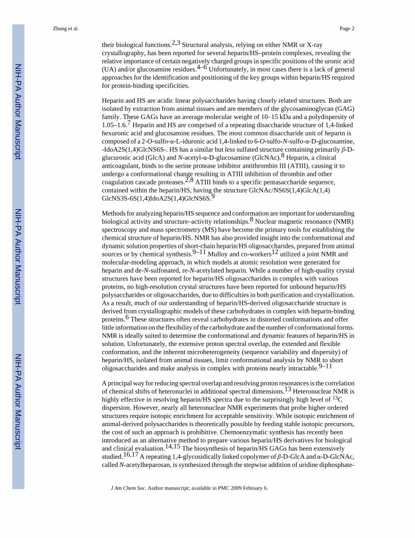
their biological functions.2,3 Structural analysis, relying on either NMR or X-raycrystallography, has been reported for several heparin/HS–protein complexes, revealing therelative importance of certain negatively charged groups in specific positions of the uronic acid(UA) and/or glucosamine residues.4–6 Unfortunately, in most cases there is a lack of generalapproaches for the identification and positioning of the key groups within heparin/HS requiredfor protein-binding specificities.
Heparin and HS are acidic linear polysaccharides having closely related structures. Both areisolated by extraction from animal tissues and are members of the glycosaminoglycan (GAG)family. These GAGs have an average molecular weight of 10–15 kDa and a polydispersity of1.05–1.6.7 Heparin and HS are comprised of a repeating disaccharide structure of 1,4-linkedhexuronic acid and glucosamine residues. The most common disaccharide unit of heparin iscomposed of a 2-O-sulfo-α-L-iduronic acid 1,4-linked to 6-O-sulfo-N-sulfo-α-D-glucosamine,-IdoA2S(1,4)GlcNS6S-. HS has a similar but less sulfated structure containing primarily β-D-glucuronic acid (GlcA) and N-acetyl-α-D-glucosamine (GlcNAc).8 Heparin, a clinicalanticoagulant, binds to the serine protease inhibitor antithrombin III (ATIII), causing it toundergo a conformational change resulting in ATIII inhibition of thrombin and othercoagulation cascade proteases.2,8 ATIII binds to a specific pentasaccharide sequence,contained within the heparin/HS, having the structure GlcNAc/NS6S(1,4)GlcA(1,4)GlcNS3S-6S(1,4)IdoA2S(1,4)GlcNS6S.9
Methods for analyzing heparin/HS sequence and conformation are important for understandingbiological activity and structure–activity relationships.8 Nuclear magnetic resonance (NMR)spectroscopy and mass spectrometry (MS) have become the primary tools for establishing thechemical structure of heparin/HS. NMR has also provided insight into the conformational anddynamic solution properties of short-chain heparin/HS oligosaccharides, prepared from animalsources or by chemical synthesis.9–11 Mulloy and co-workers12 utilized a joint NMR andmolecular-modeling approach, in which models at atomic resolution were generated forheparin and de-N-sulfonated, re-N-acetylated heparin. While a number of high-quality crystalstructures have been reported for heparin/HS oligosaccharides in complex with variousproteins, no high-resolution crystal structures have been reported for unbound heparin/HSpolysaccharides or oligosaccharides, due to difficulties in both purification and crystallization.As a result, much of our understanding of heparin/HS-derived oligosaccharide structure isderived from crystallographic models of these carbohydrates in complex with heparin-bindingproteins.6 These structures often reveal carbohydrates in distorted conformations and offerlittle information on the flexibility of the carbohydrate and the number of conformational forms.NMR is ideally suited to determine the conformational and dynamic features of heparin/HS insolution. Unfortunately, the extensive proton spectral overlap, the extended and flexibleconformation, and the inherent microheterogeneity (sequence variability and dispersity) ofheparin/HS, isolated from animal tissues, limit conformational analysis by NMR to shortoligosaccharides and make analysis in complex with proteins nearly intractable.9–11
A principal way for reducing spectral overlap and resolving proton resonances is the correlationof chemical shifts of heteronuclei in additional spectral dimensions.13 Heteronuclear NMR ishighly effective in resolving heparin/HS spectra due to the surprisingly high level of 13Cdispersion. However, nearly all heteronuclear NMR experiments that probe higher orderedstructures require isotopic enrichment for acceptable sensitivity. While isotopic enrichment ofanimal-derived polysaccharides is theoretically possible by feeding stable isotopic precursors,the cost of such an approach is prohibitive. Chemoenzymatic synthesis has recently beenintroduced as an alternative method to prepare various heparin/HS derivatives for biologicaland clinical evaluation.14,15 The biosynthesis of heparin/HS GAGs has been extensivelystudied.16,17 A repeating 1,4-glycosidically linked copolymer of β-D-GlcA and α-D-GlcNAc,called N-acetylheparosan, is synthesized through the stepwise addition of uridine diphosphate-
Zhang et al. Page 2
J Am Chem Soc. Author manuscript; available in PMC 2009 February 6.
NIH
-PA Author Manuscript
NIH
-PA Author Manuscript
NIH
-PA Author Manuscript

activated sugars. Bacteria, including Escherichia coli and Pasteurella multocida, alsobiosynthesize N-acetylheparosan.18,19 During heparin/HS biosynthesis in animal cells, theN-acetylheparosan linear homocopolymer is modified sequentially through the action of N-deacetylase/N-sulfotransferase, C5-epimerase, and 2-, 6-, and 3-O-sulfotransferases (OSTs).Complete or nearly complete modification of a chain results in heparin, while partialmodification results in HS.16,17 These biosynthetic enzymes have been cloned, expressed,and used for controlled synthesis of heparin/HS.15,20
N-Acetylheparosan and its derivatives have been the subject of recent interest in an effort toprepare heparin-like therapeutics from non-animal sources.14,21 Enzymatic sulfonation ofthese derivatives offers products that closely resemble the heparins isolated from animaltissues, suggesting a utility in the development of new pharmaceuticals.15,20,22Unfortunately, despite the decreased heterogeneity of these heparin derivatives, structuralstudies using NMR are still plagued by the low natural abundance of heteronuclei.Heteronuclear NMR has been extremely beneficial in solving the solution structures of proteinsand nucleic acids.23,24 Studies on 13C-labeled chondroitin sulfate oligosaccharides25 andstable isotope-labeled hyaluronan and hyaluronan oligosaccharides26 suggested to us that asimilar approach might be possible for heparin. As a demonstration, the solution structures ofN-acetylheparosan, heparin, and intermediate GAGs have been analyzed here usingheteronuclear NMR and molecular dynamics (MD) with time-averaged restraints (Tar-MD).
Experimental SectionPreparation of [13C,15N]N-Acetylheparosan
13C,15N-isotopically labeled N-acetylated heparosan (-GlcA(1,4)GlcNAc-) was prepared byfermentation of E. coli K5 strain in minimal media containing 13C-D-glucose and 15NH4Cl.
Expression of Recombinant HS Biosynthetic Enzymes and Heparin LyasesThe catalytic domains of human C5-epimerase, hamster 2-OST, hamster 6-OST-1, mouse 6-SOT-3, and mouse 3-OST-1 were recombinately expressed in E. coli and purified.20 Heparinlyases I, II, and III were cloned from the genomic DNA of Flavobacterium heparinum. Theexpression of the recombinant heparin lyases was also carried out for E. coli.28
Preparation of Heparin and Its Precursors13C,15N-Labeled N-acetylheparosan (5 mg) was N-deacetylated using NaOH, neutralized withHCl, and N-sulfonated with (CH3)3N · SO3.29 Three modification steps were used toconvert 13C,15N-labeled N-sulfoheparosan to anticoagulant heparin: C5-epimerization/2-O-sulfonation, 6-O-sulfonation, and 3-O-sulfonation. Each step was performed in buffer in thepresence of a PAPs regeneration system.15
NMR1H and 13C NMR, 2D 1H–13C HMQC, and constant-time HSQC and 3D HCCH-COSY,HCCH-TOCSY, and (H)CCH-TOCSY spectra were recorded on 13C,15N-labeled N-acetylheparosan, N-sulfoheparosan, undersulfated heparin, and heparin samples in D2O. Theproton and carbon chemical shifts were calibrated against 2,2-dimethyl-2-silapentane-5-sulfonic acid (DSS). A series of 3D 13C-separated NOE spectra were also acquired for eachsample at mixing times of 20, 40, and 100 ms.
Disaccharide Composition Analysis of Anticoagulant HeparinHeparin and anticoagulant heparin (20 μg, respectively) were incubated in 50 mM sodiumphosphate buffer, pH 7.0, with heparin lyase I, II, and III mixture (100 m-units) at 37 °C for
Zhang et al. Page 3
J Am Chem Soc. Author manuscript; available in PMC 2009 February 6.
NIH
-PA Author Manuscript
NIH
-PA Author Manuscript
NIH
-PA Author Manuscript
10 h. Completely digested products were heated in a boiling water bath for 10 min to halt thereaction. The denatured protein was removed by centrifugation at 12000g for 10 min, anddisaccharides were analyzed by HPLC-ESI-MS.30
APTT Activity and ATIII-Binding13C,15N-Labeled heparin and anticoagulant heparin (100–1500 ng) were subjected to APTTassay, and anticoagulant activity was calculated from a standard curve prepared using the sameconcentrations of pharmaceutical heparin.31 The 3-O-[35S] sulfated, anticoagulant heparin wasincubated with ATIII and captured using ConA-Sepharose. Binding of 35S-labeledanticoagulant heparin to ATIII was determined by scintillation counting.
Molecular Dynamics SimulationsFor all the MD simulations, with or without time-averaged restraints (tar), a heptasaccharide(residues labeled 1–7 with 1 being a nonreducing end residue), prepared from the repeatingdisaccharide of each GAG structure, was employed, starting with a GlcNAc residue at thenonreducing end. Two independent simulations were performed for undersulfated heparin andheparin, one with the IdoA in the 1C4 conformation and one with the IdoA in the 2SO skewboat geometry.
Tar-MD Simulations in Explicit Water with NOE-Based RestraintsTar-MD simulations were performed with AMBER32 to 6.0 (parm94),33 implemented withGLYCAM 06 parameters34 and with parameters computed for the sulfo and sulfamo groups,35 to accurately simulate the conformational behavior of the sugar moiety and the sulfo andsulfamo groups, respectively. The tar-MD used 1805, 1977, 1993, and 2135 water moleculesfor N-acetylheparosan, N-sulfoheparosan, undersulfated heparin, and heparin, respectively. Inall cases, the starting geometries were generated from the available data12 deposited in theProtein Data Bank (pdb code 1hpn) and modified accordingly. Final trajectories were run usingan exponential decay constant of 400 ps and a simulation length of 4 ns. Experimental cross-relaxation rates were derived from the NOESY-HSQC experiments acquired with 20, 40, and100 ms mixing times. The experimental distances and experimental NOEs were compared tothose computed for the tar-derived ensembles and for single conformers of the modelheptasaccharides using the isolated spin-pair approximation and a full-matrix relaxationapproach with software developed in-house. An isotropic reorientation model was inadequateand unable to simultaneously accommodate the experimental intra- and inter-residue NOEsfor the different residues. Thus, a symmetric top model with correlation time values τ⊥= 8 nsand τ|| = 0.16 ns was employed for heparin and the related molecules, as described by Mulloyand co-workers.12
Molecular Modeling without Restraints in Explicit WaterThe solute molecule was first immersed in a TIP3P36 bath of water molecules with the LEAPmodule. The simulation was performed using periodic boundary conditions and the particle-mesh Ewald approach37 to introduce long-range electrostatic effects. The SHAKE algorithmfor hydrogen atoms, which allows using a 2 fs time step, was also employed. Finally, a 9 Åcutoff was applied to Lennard-Jones interactions (see Supporting Information for details).
Results and DiscussionPreparation of Uniformly Labeled [13C,15N]N-Acetylheparosan
[13C,15N]N-Acetylheparosan was afforded by fermentation of E. coli K5 strain on mediumcontaining uniformly labeled 13C-D-glucose and 15NH4Cl (Figure 1A). The average molecularweight of the [13C,15N]N-acetylheparosan was ~75 kDa,15 and its isotopic purity was 94%
Zhang et al. Page 4
J Am Chem Soc. Author manuscript; available in PMC 2009 February 6.
NIH
-PA Author Manuscript
NIH
-PA Author Manuscript
NIH
-PA Author Manuscript

based on disaccharide analysis using high-performance liquid chromatography (HPLC) anddetection with electrospray ionization mass spectrometry (ESI-MS) (see SupportingInformation).
Preparation of Uniformly Labeled Heparin and Its Precursors13C,15N-Labeled heparin and its precursors were prepared by a stepwise chemoenzymaticsynthesis in milligram amounts from [13C,15N]N-acetylheparosan. The initial chemical N-deacetylation/N-sulfonation steps result in a partial depolymerization N-acetylheparosan,reducing the average chain length of all of the precursors to ~20 disaccharide units,15 andultimately giving a heparin molecular weight of ~12 kDa. The percent conversion of each step(Figure 1A) was calculated on the basis of disaccharide analysis (see Supporting Information).15,20
Treatment with 2-OST and C5-Epi afforded an undersulfated heparin that was treated with 6-OST to afford heparin. Finally, treatment with 3-OST-1 results in an anticoagulant heparin.The presence of 13C,15N-label permitted the unambiguous assignment of all proton and carbonresonances. It is notable that, while excess enzyme and PAPS cofactor were used in each step,none of the enzymatic modifications were complete. The final products, both heparin andanticoagulant heparin, however, resemble the pharmaceutical heparin products extracted fromanimal tissues38 in disaccharide composition (see Supporting Information), with 86–89% oftheir structure consisting of the trisulfated, IdoA2S(1,4)GlcNS6S, disaccharide repeating unit.
Chemical Structure Analysis of Uniformly Labeled Heparin and Its Precursors1D, 2D, and 3D NMR were applied to confirm the structures of N-acetylheparosan, N-sulfoheparosan, undersulfated heparin, and heparin with significant improvement inresolution. 1H–13C HMQC, 1H–13C constant-time (CT)-HSQC, 3D HCCH-COSY, HCCH-TOCSY, (H)CCH-TOCSY, and 13C-separated NOESY spectra were applied to unambiguouslyassign the spectra (Table 1, the GlcN signals are designated N1–N6 and the GlcA/IdoA signalsare designated G/I1–G/I6). NMR assignments for N-acetylheparosan and heparin, found in theliterature,12,39 were comparable to those presented in Table 1. Examples of 1H–13C HMQCspectra of N-acetylheparosan, N-sulfoheparosan, undersulfated heparin, and heparin are shownin Figure 1B–E (see Supporting Information). The HMQC spectra show both highly degenerateproton and well-dispersed carbon resonances. A high degree of chemical homogeneity isevident and the differences in the chemical structures of these GAGs are clearly reflected inthese spectra. Chemical differences at the chain termini were not observable since theycomprise low relative concentrations (<5%) as a result of the high chain length. The 3D-HCCH-COSY (Figure 2) spectra were instrumental in the assignment of severely overlapped protonsignals that could not be resolved in homonuclear spectra. Even in cases where overlap in boththe 1H and 13C dimensions was observed, such as for N3, N4, and N5 of N-acetylheparosanand N-sulfoheparosan, constant-time encoding of 13C chemical shifts was effective at resolvingand aiding in the assignment of cross-peaks. The chemical shifts of 15N andcorresponding 1H were assigned on the basis of 1H–15N HSQC (Supporting Information).The 1H and the corresponding 15N signals were observed at 8.26 and 123.25 ppm, and theyshifted upfield to 5.92 and 92.5 ppm, respectively, after N-deacetylation and N-sulfonation.
The percent conversions of 97% (99%) for N-deacetylation and N-sulfonation, 87% (93%) forC5-epimerization and 2-O-sulfonation, and 90% (95%) for 6-O-sulfonation, obtained fromNMR and (disaccharide analysis, Figure 1A), were comparable to those calculated from thesespectra. The chemoenzymatic process used in GAG synthesis results in a polydisperse productcontaining minor structural heterogeneity as a result of incomplete modification. Thus, theassignments obtained (Table 1) correspond to the major disaccharide-repeating unit present ineach GAG. While saccharide residues at the reducing and nonreducing ends of the GAG chains
Zhang et al. Page 5
J Am Chem Soc. Author manuscript; available in PMC 2009 February 6.
NIH
-PA Author Manuscript
NIH
-PA Author Manuscript
NIH
-PA Author Manuscript
can also contribute to heterogeneity, NMR did not readily observe these end groups, as theycorresponded to <5% of the saccharidic units. The percent conversion of heparin toanticoagulant heparin, resulting from 3-O-sulfonation, was too low to be unambiguouslyconfirmed and/or quantified by NMR or by disaccharide analysis.
Heparin structure varies between species and organs; for example, porcine intestinal and bovinelung heparins (both pharmaceutical heparins) have distinctly different structures.1,38 Ontreatment with heparin lyases, individual chains can afford as many as many as eight differentdisaccharides and several resistant oligosaccharides to as few as one repeating disaccharide, -IdoA2S(1,4)GlcNS6S-. This microheterogeneity of animal-derived heparin results fromvariability of the activity, specificity, and accessibility of enzymes in the Golgi at the time ofbiosynthesis. Heparan sulfate, a closely related polysaccharide, has an even more complexstructure than heparin, and it is often an impurity in pharmaceutical heparin isolated fromanimal tissues. Chemoenzymatically synthesized heparin contains no heparan sulfate impurityand no residual linkage region impurity, and since it is lacking N-acetyl-substitutedglucosamine residues it shows a simpler disaccharide composition than pharmaceuticalheparin.
Uniform 13C/15N isotopic labeling facilitated the NMR-based structural analysis of the GAGs.The well-dispersed 13C-spectra enabled sensitive application of a wide array ofmultidimensional heteronuclear experiments by overcoming limitations resulting from severeoverlap of proton resonances that have historically hindered the interpretation of GAG NMRdata. One-bond 1H–15N couplings in N-acetylheparosan were sensitively detected, but rapidsolvent exchange in N-sulfoheparosan decreased their value in the structural assignment ofN-sulfonated GAGS. In the future we anticipate that 15N labeling will be important in the NMRstudies of these GAGs due to the sensitivity of 15N chemical shift to the localenvironment. 15N dispersion can be instrumental in overcoming spectral degeneracies bycombining 15N- and 13C-separated dimensions within an NMR experiment. This dualheteronuclear labeling strategy should facilitate sequence-specific assignment and providestructural data at atomic resolution for heparin oligosaccharides in the future, particularly whenused for heparin–protein complexes.
Characterization of Anticoagulant HeparinIn the transformation of heparin to anticoagulant heparin, the small number of sites undergoing3-O-sulfonation afforded no detectable change by NMR. To determine percentconversion,13C,15N heparin was treated with 3-OST-1 and [35S]PAPS, affording 3-O-[35S]sulfo-13C,15N anticoagulant heparin. Measurement of [35S] showed that 1 μg (83 pmol) ofheparin incorporated 115 pmol of [35S]sulfo group, representing ~1.5 3-O-[35S] sulfo groups/chain. This corresponds to the sulfonation of ~7.5% of heparin’s GlcN-3-hydroxyl groups.
The ATIII binding activity of this anticoagulant heparin was also determined.20 After 3-OST-1modification, scintillation counting demonstrated that only 35.2% of the 35S-labeledanticoagulant heparin chains bound to ATIII, confirming that the 3-OST-1 modified heparinbinds to ATIII.
Anticoagulant activity was next assessed using activated thromboplastin time (APTT).40 Theactivity of chemoenzymatically synthesized heparin was 20 ± 6 U/mg, whilechemoenzymatically anticoagulant heparin was 180 ± 15 U/mg, comparable to the 170 U/mgdisplayed by a standard pharmaceutical heparin. Previously, we had reported anti-Xa and anti-IIa activities comparable to those of pharmaceutical heparin for heparin chemoenzymaticallysynthesized without isotopic labeling.15
Zhang et al. Page 6
J Am Chem Soc. Author manuscript; available in PMC 2009 February 6.
NIH
-PA Author Manuscript
NIH
-PA Author Manuscript
NIH
-PA Author Manuscript

While chemoenzymatically synthesized heparin showed only four disaccharide peaks inHPLC-ESI-MS (see Supporting Information), anticoagulant heparin showed additional (4%)heparin lyase-resistant peaks as a result of the incorporation of 3-O-sulfo groups. 35Sincorporation studies confirmed that ~7.5% of the GlcN residues were 3-O-sulfonated and thatapproximately one-third of these chains bind with high affinity to ATIII, closely resemblingthe percentage of chains in pharmaceutical heparin with high affinity for ATIII.38 APTT assayshowed the activity of anticoagulant heparin to be comparable to that of standardpharmaceutical heparin.
Conformational Analysis of Heparin and Its PrecursorsFrom the fully assigned spectra (Table 1) and quantitative NOEs (Table 2), the conformationof the starting N-acetylheparosan, N-sulfoheparosan, undersulfated heparin, and heparin wereexamined with experimentally restrained MD simulations using a protocol41 similar to thatrecently applied by our group on a dermatan sulfate oligosaccharide.42 Existing models ofGAGs and related glycans were used to evaluate NOE cross-peak assignments and degeneracy.43 Once the assignment process was completed, the intensities of the NOE cross-peaks at thevarious mixing times were normalized to the corresponding diagonal peaks. The interprotondistances were deduced from a full-matrix relaxation approach on model chair and skew-boatgeometries.44 Spin-diffusion was essentially absent at 20 ms NOE mixing time and gave avery minor contribution at 40 ms NOE mixing time. A maximum error of 10%, estimated fromspectral signal-to-noise, resulted from noncorrected spin-diffusion contributions and resonanceoverlap, problems largely eliminated by our 3D 13C-separated approach. The observed datacorrespond to an average for all the glycosidic linkages of the GAG and should show identicalensemble-averaged behavior on the long time scale required for measuring NOE values.
Tar-MD simulations were employed to generate 3D models of the different GAGs. Forcomparison purposes, the same simulations were also performed without time-averagedrestraints (7–9 ns of simulation time), providing similar results. Tar-MD45–48 simulationswere performed for heparin and its precursors to obtain a conformer ensemble having distancescomparable to those estimated by NMR. The NOE-derived distances were included as timeaveraged distance constraints in two different trajectories using either ⟨r−6⟩−1/6 or ⟨r−3⟩−1/3
restraints to cope with different internal motion timescales. In all cases, tar-MD trajectorieswere collected in the presence of explicit water, and unrestrained MD simulations were alsocomputed for comparison purposes.
Results from the tar-MD simulations for N-acetylheparosan (Table 3 and Figure 3A) showclose agreement, in numerical terms, between the distances found in the refined models andexperimental data using a 4 ns tar-MD simulation with explicit water. The φ/ψ distributionmaps indicate that the glycosidic linkages cover a similar area of the allowable conformationalspace, independent of their position in the sequence (Figure 3A). In terms of the glycosidicangle φ, only exoanomeric orientations were adopted, demonstrating the goodness of theemployed force field. Only a very minor proportion (<2%) of non-exoanomeric conformerswere predicted for the linkage between residues 2 and 3 of the heptasaccharide sequenceundergoing MD simulation. Both GlcNAc(1,4)GlcA and GlcA(1,4)GlcNAc linkages showconformers with a negative-ψ angle highly populated with only minor incursions for the anti-ψ and positive-ψ values. Analogous φ/ψ plots are also provided heparin (Table 4 and Figure3B) and for N-sulfoheparosan, and undersulfated heparin (see Supporting Information). Theensemble shows that the conformational space accessible for each glycosidic linkage covers awell-defined area of the total available φ/Ψ energy surface. Flexibility is apparent in N-acetylheparosan, which cannot be defined by a single, rigid chain model, with a behavior indeedsimilar to that reported for hyaluronic acid (HA), a similar structure having a -4)GlcA(1,3)GlcNAc(1- repeating disaccharide unit, with almost four sugar rings per helix turn.49 The
Zhang et al. Page 7
J Am Chem Soc. Author manuscript; available in PMC 2009 February 6.
NIH
-PA Author Manuscript
NIH
-PA Author Manuscript
NIH
-PA Author Manuscript
reported standard deviations of the glycosidic linkages in this polymer are ~13°, while in ourtar-MD approach for N-acetylheparosan, the estimated maximum deviations for φ and ψ anglesfrom the major value are ~35°.
MD simulations were similarly performed on all GAG precursors and heparin. Asuperimposition of 20 conformers, randomly collected from the tar-MD simulations, is shownin Figure 4. After discarding data from the first nanosecond, the root-mean-square deviation(rmsd) of the skeleton atoms of all the sampled structures was 2.1 Å, compared to the averagedstructure. The unrestrained simulations showed a very similar behavior. The simulation beganwith all seven saccharide residues 1–7 of the heptasaccharide in the 4C1 form. While the NMRdata were acquired on a polysaccharide of approximately 20 disaccharide units, our simulationswere performed on heptasaccharide models of the polysaccharides. Therefore, special attentionwas paid to the motion of the central residues, as a paradigm for the motion of the disaccharideentities within the intact polysaccharide. At the end of the simulation no interconversion intoother ring forms was observed for either GlcNAc or GlcA residues. In contrast, in the case ofthe IdoA residues, interconversion between 1C4 and 2SO forms was observed in both therestrained and unrestrained simulations. These results are consistent with observations thatGlcNAc and GlcNS residues were solely in the 4C1 conformation based on 3JH–H values.5Additionally, the pyranose form of GlcA also has been shown to prefer the more stable 4C1form.50 Tar-MD simulations were also used here to examine the conformation of N-sulfoheparosan. The ring geometries and the φ/ψ values of N-sulfoheparosan in the generatedstructural models were nearly identical to those of N-acetylheparosan resulting, in a similarlocal and global conformation (Figure 4B, also see Supporting Information). The length (N–N distance between the GlcN residues at positions 1 and 13) and shape of the two chains weresimilar when oligosaccharides having 13 residues were modeled from the more populatedvalues of the tar-MD Φ/Ψ distributions of the central residues for each structure (Figure 5).The lengths of heparosan and N-sulfoheparosan were 57.4 and 58.4 Å, differing by only 1.0Å. The pairwise rmsd between the 13-mer N-acetylheparosan and N-sulfoheparosan chainswas 4.9 Å, and the pairwise rmsd for the central trisaccharide was 1.0 Å. Thus, it appears thatN-sulfonation does not modify the conformation of this GAG (Figure 5A). Neither anti-conformer nor non-exo conformer was found in the final ensemble of N-sulfoheparosan, andthe models showed even less conformational variation (rmsd 1.9 Å) than that of N-acetylheparosan. The observed increase in the ensemble-averaged H1GlcA–H4GlcNS distanceresults from an increase in positive ψ-conformers for this linkage, as can be observed in thecorresponding curves in Figure 3A for heparosan (5 ± 65°) and those in Figure 3B for N-sulfoheparosan (25 ± 60°). Epimerization of the GlcA C5 position and 2-O-sulfonation affordsundersulfated heparin. The structural impact of these modifications was explored through thetar-MD simulations on undersulfated heparin (Figure 4C, also see Supporting Information). Insolution, IdoA2S pyranose exhibits both the 2SO and 1C4 forms in equilibrium.50,51 As ameans of evaluating bias in the resulting structures, dual sets of simulations were initiated withIdoA2S in the 2SO and half in the 1C4 conformer. Both sets of simulations converged within4 ns, with an rmsd value of 2.4 Å. The estimated NOE-based H2IdoA2S–H5IdoA2S distanceis 3.8Å(± 10%). Since the idealize dH2IdoA2S–H5IdoA2S distances for the 1C4 and 2SOconformers are 4.0 and 2.4 Å, respectively, this value is consistent with a large, >85%population of the 1C4 conformer of IdoA2S. There was a decrease in the length of thecorresponding 13-mer chains, from 58.4 Å for N-sulfoheparosan to 51 Å for undersulfatedheparin with all of the IdoA2S residues in the 1C4 conformation (Figure 5). Nevertheless,deviations from an idealized helical geometry are found throughout the resulting ensemblemembers.
Heparin’s repeating unit was next afforded by 6-O-sulfonation of the undersulfated heparin.In contrast to its undersulfated precursor, when the force field matched the H1GlcNS6S–H4IdoA2S and H1GlcNS6S–H3IdoA2S distances, the NOE-based H2IdoA2S–H5IdoA2S
Zhang et al. Page 8
J Am Chem Soc. Author manuscript; available in PMC 2009 February 6.
NIH
-PA Author Manuscript
NIH
-PA Author Manuscript
NIH
-PA Author Manuscript
distance was calculated at 2.8 Å (±10%), a value that, according to the idealized values givenabove, is consistent with >80% of the IdoA2S being in the 2SO form, with an rmsd value of2.2 Å. These results clearly demonstrate that the addition of a 6-O-sulfo group onto GlcNSresidues markedly shifts the equilibrium of adjacent IdoA2S residues from the 1C4 tothe 2SO conformer. The balance of the 1C4 to 2SO equilibrium in an IdoA residue within a GAGchain had been previously shown to depend on both its own substitution with a 2-O-sulfo groupand the substitution of the adjacent GlcN residues.12,52 Mulloy and co-workers50 showedthat the overall conformation of the chain (i.e., the optimal angles around the inter-residueglycosidic linkages) is not greatly affected by the position of the 1C4 to 2SO equilibrium. Ourobservations are similar with only a very minor 0.7 Å difference between the lengths of the13-mer chains built using 1C4 and 2SO geometries for the IdoA2S. The helical structure of thechain only becomes apparent in the fully modified heparin (Figures 4D and 5B and seeSupporting Information). While the global structures of the members of the tar-MD heparinensemble deviate from the idealized geometry in models proposed by Mulloy and co-workers,12 there is good agreement at a local structural level. The ring geometries and glycosidiclinkages in the tar-refined heparin ensemble are consistent with previous structural studiesperformed on short heparin oligosaccaharides.10,11 Medium-range to long-range structuralordering is also consistent with the previous studies and, in the current study, heparin forms aleft-handed helix with rotational and translational parameters similar to those of short heparinoligosaccharides. Therefore, the key helical parameters in heparin, including helical width,groove depth, and axial rise, are not significantly influenced by overall chain length. Localconformational flexibility is evident in the tar-refined ensemble, consistent with previousreported observations. However, this flexibility does not appear to result in accordion-likestretching along the helical axis (Figure 5). Some rotational freedom about the glycosidiclinkages was observed in the heparin ensemble, and this fact results in small inter-residuedeviations that would be expected to propagate along the extended heparin structure, resultingin long-range disorder or bending of the helical axis. These global distortions are representativeof the conformational space explored in solution by the dynamic heparin polysaccharide.
The structure of heparin has been extensively studied since its discovery.7,8 The advent ofNMR was instrumental in defining heparin’s major uronic acid residue as IdoA and has greatlyimproved our understanding of heparin’s primary structure.53 NMR-based conformationalanalyses of heparin and heparin oligosaccharides have resulted in an improved understandingof its ring conformations and their flexibility.9–11,54 Molecular modeling coupled withNMR12 has led to structures widely used for analyzing heparin binding to proteins.4–6However, NMR has had limited success in high-resolution structure determination of theheparin molecule portions within these complexes. X-ray crystallography has offered anindependent method to look at heparin’s structure. Despite the publication of a number ofcrystal structures of heparin oligosaccharides complexed with heparin-binding proteins,6unbound heparin has only been studied at low resolution in fiber structures.55
Future studies will be required to map the fine structural features of heparin and compare theseto various pharmaceutical heparins prepared from animal tissues. Additional studies will focuson the use of uniformly stable isotope-labeled GAGs as binding partners for various heparin-binding proteins. In such studies, it should be possible to study conformational changes in thebound and unbound GAG using NMR. Moreover, it should be possible to prepare structurallyhomogeneous oligosaccharide binding partners56 uniformly labeled with NMR-sensitiveisotopes that enable high-resolution structural determination of these complexes. This shouldafford the improved understanding of heparin structure–activity relationships required for thedesign of a novel and specific class of heparin-based therapeutic agents.
Zhang et al. Page 9
J Am Chem Soc. Author manuscript; available in PMC 2009 February 6.
NIH
-PA Author Manuscript
NIH
-PA Author Manuscript
NIH
-PA Author Manuscript

Supplemental MaterialsRefer to Web version on PubMed Central for supplementary material.
AcknowledgementsNational Institutes of Health Grants GM38060 and HL62244 (to R.J.L.) and AI50050 (to J.L.), and the Ministry ofEducation and Science of Spain for Grant CTQ-2006-10874-C02-01 (to J.J.-B.) and for Ramón-y-Cajal contract (toF.C.) are gratefully acknowledged for supporting this work.
References1. Capila I, Linhardt RJ. Angew Chem, Int Ed 2002;41:391–412.2. Munoz EM, Linhardt RJ. Arterioscler Thromb Vasc Biol 2004;24:1549–1557. [PubMed: 15231514]3. Raman R, Sasisekharan V, Sasisekharan R. Chem Biol 2005;12:267–277. [PubMed: 15797210]4. Jin L, Abrahams JP, Skinner R, Petitou M, Pike RN, Carrell RW. Proc Natl Acad Sci USA
1997;94:14683–14688. [PubMed: 9405673]5. Guerrini M, Guglieri S, Beccati D, Torri G, Viskov C, Mourier P. Biochem J 2006;399:191–198.
[PubMed: 16796563]6. Mulloy B, Linhardt RJ. Curr Opin Struct Biol 2001;11:623–628. [PubMed: 11785765]7. Casu B. Adv Carbohydr Chem Biochem 1985;43:51–134. [PubMed: 3913287]8. Linhardt RJ. J Med Chem 2003;46:2551–2554. [PubMed: 12801218]9. Ragazzi M, Ferro DR, Perly B, Sinaÿ P, Petitou M, Choay J. Carbohydr Res 1990;195:169–185.
[PubMed: 2331700]10. Mikhailov D, Linhardt RJ, Mayo KH. Biochem J 1997;328:51–61. [PubMed: 9359833]11. Angulo J, Hricovíni M, Gairi M, Guerrini M, de Paz JL, Ojeda R, Martín-Lomas M, Nieto PM.
Glycobiology 2005;15:1008–1015. [PubMed: 15958415]12. Mulloy B, Forster MJ, Jones C, Davies DB. Biochem J 1993;293:849–858. [PubMed: 8352752]13. Homans SW. Biochem Soc Trans 1998;26:551–560. [PubMed: 10047781]14. Lindahl U, Li JP, Kusche-Gullberg M, Salmivirta M, Alaranta S, Veromaa T, Emeis J, Roberts I,
Taylor C, Oreste P, Zoppetti G, Naggi A, Torri G, Casu B. J Med Chem 2005;48:349–352. [PubMed:15658847]
15. Chen J, Avci FY, Munoz EM, McDowell LM, Chen M, Pedersen LC, Zhang L, Linhardt RJ, Liu J.J Biol Chem 2005;280:42817–42825. [PubMed: 16260789]
16. Lindahl U, Feingold DS, Roden L. Trends Biochem Sci 1986;11:221–225.17. Esko JD, Selleck SB. Annu Rev Biochem 2002;71:435–471. [PubMed: 12045103]18. Navia JL, Riesenfeld J, Vann WF, Lindahl U, Rodén L. Anal Biochem 1983;135:134–140. [PubMed:
6367539]19. DeAngelis PL, Gunay NS, Toida T, Mao WJ, Linhardt RJ. Carbohydr Res 2002;337:1547–1552.
[PubMed: 12350324]20. Chen J, Jones CL, Liu J. Chem Biol 2007;14:986–993. [PubMed: 17884631]21. Casu B, Grazioli G, Razi N, Guerrini M, Naggi A, Torri G, Oreste P, Tursi F, Zoppetti G, Lindahl
U. Carbohydr Res 1994;263:271–284. [PubMed: 7805054]22. Kuberan B, Beeler DL, Lech M, Wu ZL, Rosenberg RD. J Biol Chem 2003;278:52613–52621.
[PubMed: 14519763]23. Westler WM, Stockman BJ, Hosoya Y, Miyake Y, Kainosho M, Markley JL. J Am Chem Soc
1988;110:6255–6258.24. Nikonowicz EP, Pardi A. Nature 1992;355:184–186. [PubMed: 1370345]25. Yu F, Wolff JJ, Amster IJ, Prestegard JH. J Am Chem Soc 2007;129:13288–13297. [PubMed:
17924631]26. Almond A, DeAngelis PL, Blundell CD. J Mol Biol 2006;358:1256–1269. [PubMed: 16584748]27. Vann WF, Schmidt MA, Jann B, Jann K. Eur J Biochem 1981;116:359–364. [PubMed: 7018909]
Zhang et al. Page 10
J Am Chem Soc. Author manuscript; available in PMC 2009 February 6.
NIH
-PA Author Manuscript
NIH
-PA Author Manuscript
NIH
-PA Author Manuscript

28. Duncan MB, Liu M, Fox C, Liu J. Biochem Biophys Res Commun 2006;339:1232–1237. [PubMed:16343444]
29. Nadkarni VD, Toida T, Van Gorp CL, Schubert RL, Weiler JM, Hansen KP, Caldwell EEO, LinhardtRJ. Carbohydr Res 1996;290:87–96. [PubMed: 8805784]
30. Warda M, Zhang F, Radwan M, Zhang Z, Kim N, Kim YN, Linhardt RJ, Han J. Glycoconjugate J2008;25:441–450.
31. Pearlman DA, Case DA, Caldwell JW, Ross WR, Cheatham TE III, DeBolt S, Ferguson D, SeibelG, Kollman P. Comput Phys Commun 1995;91:1–41.
32. Kollman, PA. University of California; San Francisco: 1999.33. Cornell WD, Cieplak P, Bayly CI, Gould IR, Merz KM, Ferguson DM, Spellmeyer DC, Fox T,
Caldwell JW, Kollman PA. J Am Chem Soc 1995;117:5179–5197.34. Woods RJ, Dwek RA, Edge CJ, Fraser-Reid B. J Phys Chem B 1995;99:3832–3846.35. Lan, J. PhD Thesis. University of Edinburgh; 2007.36. Jorgensen WL, Chandrasekhar J, Madura JD, Impey RW, Klein ML. J Chem Phys 1983;79:926–935.37. Darden T, York D, Pedersen L. J Chem Phys 1993;98:10089–10092.38. Linhardt RJ, Gunay NS. Semin Thrombos Hemostas 1999;25:5–16.39. Guerrini M, Naggi A, Guglieri S, Santarsiero R, Torri G. Anal Biochem 2005;337:35–47. [PubMed:
15649373]40. Murugesan S, Park TJ, Yang H, Mousa S, Linhardt RJ. Langmuir 2006;22:3461–3463. [PubMed:
16584210]41. Corzana F, Busto JH, Jiménez-Osés G, García de Luis M, Asensio JL, Jiménez-Barbero J, Peregrina
JM, Avenoza A. J Am Chem Soc 2007;129:9458–9467. [PubMed: 17616194]42. Silipo A, Zhang Z, Cañada FJ, Molinaro A, Linhardt RJ, Jiménez-Barbero J. ChemBioChem
2007;9:240–252. [PubMed: 18072186]43. Hricovíni, M.; Nieto, PM.; Torri, G. NMR spectroscopy of glycoconjugates. Jimenez-Barbero, J.;
Peters, T., editors. Wiley-VCH; Weinheim: 2002. p. 189-230.44. Neuhaus, D.; Williamson, MP., editors. The nuclear Overhauser effect in structural and
conformational analysis. Wiley; New York: 1989.45. Pearlman DA. J Biomol NMR 1994;4:1–16.46. Torda AE, Scheek RM, van Gunsteren WF. J Mol Biol 1990;214:223–235. [PubMed: 2370663]47. Torda AE, Scheek RM, van Gunsteren WF. Chem Phys Lett 1989;157:289–294.48. Forster M, Jones C, Mulloy B. J Mol Graphics 1989;7:196–201.49. Almond A, Deangelis PL, Blundell CD. J Mol Biol 2006;358:1256–1269. [PubMed: 16584748]50. Mulloy B, Forster MJ. Glycobiology 2000;10:1147–1156. [PubMed: 11087707]51. Sanderson PN, Huckerby TN, Nieduszynski IA. Biochem J 1987;243:175–181. [PubMed: 3038077]52. van Boeckel CA, van Aelst SF, Wagenaars GN, Mellema JR, Paulsen H, Peters T, Pollex A, Sinnwell
V. Recl Trav Chim Pays-Bas 1987;106:19–29.53. Perlin AS, Mackie DM, Dietrich CP. Carbohydr Res 1971;18:185–194. [PubMed: 5151386]54. Casu B, Petitou M, Provasoli M, Sinaÿ P. Trends Biochem Sci 1988;13:221–225. [PubMed: 3076283]55. Atkins ED, Nieduszynski IA. Adv Exp Med Biol 1975;52:19–37. [PubMed: 1124697]56. Kuberan B, Lech MZ, Beeler DL, Wu ZL, Rosenberg RD. Nat Biotechnol 2003;21:1343–1346.
[PubMed: 14528313]
Zhang et al. Page 11
J Am Chem Soc. Author manuscript; available in PMC 2009 February 6.
NIH
-PA Author Manuscript
NIH
-PA Author Manuscript
NIH
-PA Author Manuscript

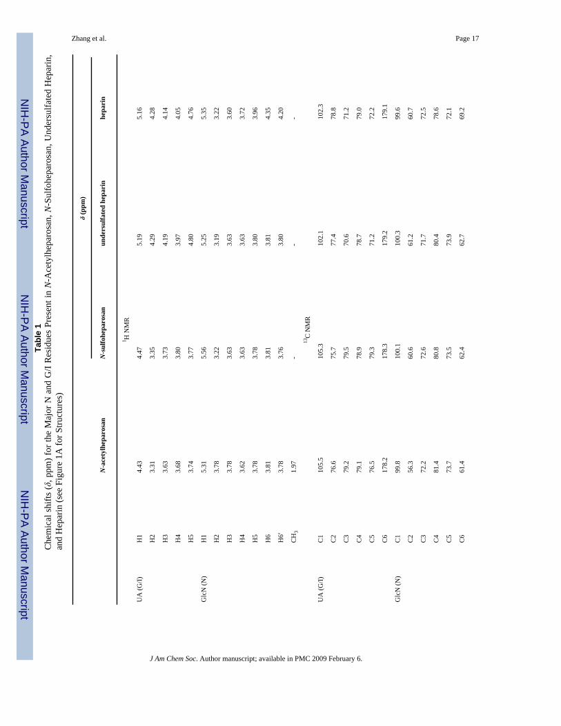
Figure 1.(A) Scheme of stepwise chemoenzymatic synthesis of 13C,15N-labeled anticoagulant heparin.The major saccharide units are shown (with the exception of anticoagulant heparin whichshows a minor fully modified sequence). The GlcN residues are labeled N and the GlcA/IdoAresidues are labeled G/I. HMQC spectra of (B) 13C,15N-labeled N-aceylheparosan (blue),(C) 13C,15N-labeled N-sulfoheparosan (red), (D) 13C,15N-labeled undersulfated heparin(green), and (E) 13C,15N-labeled heparin (purple).
Zhang et al. Page 12
J Am Chem Soc. Author manuscript; available in PMC 2009 February 6.
NIH
-PA Author Manuscript
NIH
-PA Author Manuscript
NIH
-PA Author Manuscript

Figure 2.Strip plots from the 3D-HCCH-COSY spectrum of 13C,15N-labeled heparin. Panels A–Eillustrate a sequential walk from C2 of GlcN to C6 of GlcN; panels F–I under C2 of IdoA toC5 of IdoA.
Zhang et al. Page 13
J Am Chem Soc. Author manuscript; available in PMC 2009 February 6.
NIH
-PA Author Manuscript
NIH
-PA Author Manuscript
NIH
-PA Author Manuscript

Figure 3.φ/ψ distribution for (A) N-acetylheparosan and (B) heparin. In the tar-MD simulation aheptasaccharide (saccharide residues 1–7) is used. Each panel shows the φ/ψ plot betweenadjacent saccharide residues.
Zhang et al. Page 14
J Am Chem Soc. Author manuscript; available in PMC 2009 February 6.
NIH
-PA Author Manuscript
NIH
-PA Author Manuscript
NIH
-PA Author Manuscript

Figure 4.Superimposition of 20 conformers randomly taken from the tar-MD simulations for each GAG.The three central residues have been chosen for the superimposition to show the propagationof the flexibility around the GAG chain: (A) N-acetylheparosan, (B) N-sulfoheparosan, (C)undersulfated heparin, and (D) heparin. Carbon is shown in green, oxygen in red, nitrogen inblue, and sulfur in yellow.
Zhang et al. Page 15
J Am Chem Soc. Author manuscript; available in PMC 2009 February 6.
NIH
-PA Author Manuscript
NIH
-PA Author Manuscript
NIH
-PA Author Manuscript
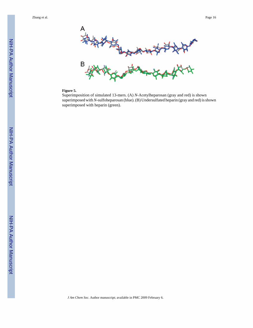
Figure 5.Superimposition of simulated 13-mers. (A) N-Acetylheparosan (gray and red) is shownsuperimposed with N-sulfoheparosan (blue). (B) Undersulfated heparin (gray and red) is shownsuperimposed with heparin (green).
Zhang et al. Page 16
J Am Chem Soc. Author manuscript; available in PMC 2009 February 6.
NIH
-PA Author Manuscript
NIH
-PA Author Manuscript
NIH
-PA Author Manuscript
NIH
-PA Author Manuscript
NIH
-PA Author Manuscript
NIH
-PA Author Manuscript
Zhang et al. Page 17Ta
ble
1C
hem
ical
shi
fts (δ
, ppm
) for
the
Maj
or N
and
G/I
Res
idue
s Pr
esen
t in
N-A
cety
lhep
aros
an, N
-Sul
fohe
paro
san,
Und
ersu
lfate
d H
epar
in,
and
Hep
arin
(see
Fig
ure
1A fo
r Stru
ctur
es)
δ (p
pm)
N-a
cety
lhep
aros
anN
-sul
fohe
paro
san
unde
rsul
fate
d he
pari
nhe
pari
n
1 H N
MR
UA
(G/I)
H1
4.43
4.47
5.19
5.16
H2
3.31
3.35
4.29
4.28
H3
3.63
3.73
4.19
4.14
H4
3.68
3.80
3.97
4.05
H5
3.74
3.77
4.80
4.76
Glc
N (N
)H
15.
315.
565.
255.
35
H2
3.78
3.22
3.19
3.22
H3
3.78
3.63
3.63
3.60
H4
3.62
3.63
3.63
3.72
H5
3.78
3.78
3.80
3.96
H6
3.81
3.81
3.81
4.35
H6′
3.78
3.76
3.80
4.20
CH
31.
97-
--
13C
NM
R
UA
(G/I)
C1
105.
510
5.3
102.
110
2.3
C2
76.6
75.7
77.4
78.8
C3
79.2
79.5
70.6
71.2
C4
79.1
78.9
78.7
79.0
C5
76.5
79.3
71.2
72.2
C6
178.
217
8.3
179.
217
9.1
Glc
N (N
)C
199
.810
0.1
100.
399
.6
C2
56.3
60.6
61.2
60.7
C3
72.2
72.6
71.7
72.5
C4
81.4
80.8
80.4
78.6
C5
73.7
73.5
73.9
72.1
C6
61.4
62.4
62.7
69.2
J Am Chem Soc. Author manuscript; available in PMC 2009 February 6.
NIH
-PA Author Manuscript
NIH
-PA Author Manuscript
NIH
-PA Author Manuscript
Zhang et al. Page 18
δ (p
pm)
N-a
cety
lhep
aros
anN
-sul
fohe
paro
san
unde
rsul
fate
d he
pari
nhe
pari
n
CH
323
.9-
--
CO
176.
6-
--
J Am Chem Soc. Author manuscript; available in PMC 2009 February 6.

NIH
-PA Author Manuscript
NIH
-PA Author Manuscript
NIH
-PA Author Manuscript
Zhang et al. Page 19Ta
ble
2Ex
perim
enta
l Key
Pro
ton-
Pair
Dis
tanc
es (Å
) As D
educ
ed fr
om th
e NO
E-B
ased
3D
NO
ESY
-HSQ
C E
xper
imen
ts E
mpl
oyin
g th
e Iso
late
dSp
in-P
air A
ppro
xim
atio
n (A
s Des
crib
ed in
the
Expe
rimen
tal S
ectio
n) fr
om T
hree
Diff
eren
t NO
ESY
Exp
erim
ents
, at 2
0, 4
0 an
d 10
0 m
sM
ixin
g Ti
mes
(Est
imat
ed E
xper
imen
tal E
rror
is 1
0%)
H1
Glc
A–H
4 G
lcN
Ac
H1
Glc
A–H
3 G
lcN
Ac
H1
Glc
A–H
6 G
lcN
Ac
H1
Glc
A–H
6 G
lcN
Ac
H1
Glc
NA
c–H
3 G
lcA
H1
Glc
NA
c–H
4 G
lcA
N-a
cety
lhep
aros
an2.
54.
52.
7a2.
7a2.
73.
6
N-s
ulfo
hepa
rosa
n2.
74.
52.
5a2.
5a2.
73.
8
H1
IdoA
–H4
Glc
NS
H1
IdoA
–H3
Glc
NS
H1
IdoA
–H6
Glc
NS
H1
IdoA
–H6
Glc
NS
H1
Glc
NS–
H3
IdoA
H1
Glc
NS–
H4
IdoA
unde
rsul
fate
d he
parin
2.5
4.5
2.7a
2.7a
2.5
2.7
hepa
rin2.
44.
52.
6a3.
1a2.
82.
8
a No
ster
eosp
ecifi
c as
sign
men
t was
per
form
ed, a
nd th
e re
stra
int w
as in
clud
ed in
the
tar-
MD
sim
ulat
ions
to th
e co
rres
pond
ing
C-6
car
bon
atom
(add
ing
0.8
Å).
J Am Chem Soc. Author manuscript; available in PMC 2009 February 6.
NIH
-PA Author Manuscript
NIH
-PA Author Manuscript
NIH
-PA Author Manuscript
Zhang et al. Page 20Ta
ble
3R
esul
ts o
f the
Tar
-MD
Sim
ulat
ions
(⟨r−
6 ⟩−1
/6/⟨
r−3 ⟩−1
/3 A
vera
ging
) for
the
Diff
eren
t Gly
cosi
dic
Link
ages
of N
-Ace
tylh
epar
osan
a
H1
Glc
A–H
4 G
lcN
Ac
H1
Glc
A–H
3 G
lcN
Ac
H1
Glc
A–H
6a Glc
NA
cH
1 G
lcA
–H6b G
lcN
Ac
H1
Glc
NA
c–H
4 G
lcA
H1
Glc
NA
c–H
3 G
lcA
1→2
--
--
2.5/
2.5
3.3/
3.4
2→3
2.5/
2.6
3.6/
4.4
2.6/
2.9
2.6/
2.9
--
3→4
--
--
2.5/
2.5
3.4/
3.4
4→5
2.4/
2.6
4.4/
4.4
2.7/
3.0
2.6/
2.9
--
5→6
--
--
2.5/
2.5
3.3/
3.3
6→7
2.6/
2.5
3.0/
4.2
2.7/
2.7
2.7/
2.6
--
MD
-ave
rage
2.5/
2.6
3.5/
4.3
2.6/
2.9
2.6/
2.8
2.5/
2.5
3.3/
3.4
expe
rimen
tal
2.5
4.5
2.7b
2.7b
2.7
3.6
a The e
xper
imen
tal k
ey p
roto
n-pa
ir di
stan
ces (
Å) a
s ded
uced
from
the N
OE-
base
d 3D
NO
ESY
-HSQ
C ex
perim
ents
wer
e im
plem
ente
d as
stru
ctur
al re
stra
ints
with
a 10
% m
argi
n, u
sing
a fla
t wel
l pot
entia
l.
b No
ster
eosp
ecifi
c as
sign
men
t was
per
form
ed a
nd th
e re
stra
int w
as se
t to
the
corr
espo
ndin
g C
-6 c
arbo
n at
om (a
ddin
g 0.
8 Å
).
J Am Chem Soc. Author manuscript; available in PMC 2009 February 6.

NIH
-PA Author Manuscript
NIH
-PA Author Manuscript
NIH
-PA Author Manuscript
Zhang et al. Page 21Ta
ble
4R
esul
ts o
f the
Tar
-MD
Sim
ulat
ions
(⟨r−
6 ⟩−1
/6/⟨
r−3 ⟩−1
/3 A
vera
ging
) for
the
Diff
eren
t Gly
cosi
dic
Link
ages
of H
epar
ina
H1
IdoA
– H
4G
lcN
SH
1 Id
oA–
H3
Glc
NS
H1
IdoA
– H
6G
lcN
SH
1 Id
oA–
H6
Glc
NS
H1
Glc
NS–
H4
IdoA
H1
Glc
NS–
H3
IdoA
1→2
--
--
2.6/
2.6
2.6/
2.6
2→3
2.6/
2.6
4.5/
4.5
2.3/
2.2
3.7/
3.7
--
3→4
--
--
2.6/
2.6
2.5/
2.6
4→5
2.6/
2.6
4.5/
4.5
2.3/
2.2
2.9/
3.7
--
5→6
--
--
2.6/
2.7
2.5/
2.5
6→7
2.4/
2.6
4.4/
4.4
2.3/
2.3
3.7/
3.8
--
MD
-ave
rage
2.5/
2.6
4.4/
4.5
2.3/
2.2
3.2/
3.7
2.6/
2.6
2.5/
2.6
expe
rimen
tal
2.4
4.5
2.6b
3.2b
2.8
2.8
a The
expe
rimen
tal k
ey p
roto
n-pa
ir di
stan
ces (
Å) a
s ded
uced
from
the
NO
E-ba
sed
3D N
OES
Y-H
SQC
exp
erim
ents
wer
e im
plem
ente
d as
tar-
cons
train
ts w
ith a
10%
mar
gin.
b No
ster
eosp
ecifi
c as
sign
men
t was
per
form
ed a
nd th
e re
stra
int w
as se
t to
the
corr
espo
ndin
g C
-6 c
arbo
n at
om (a
ddin
g 0.
8 Å
).
J Am Chem Soc. Author manuscript; available in PMC 2009 February 6.