Delineation of Two Clinically and Molecularly Distinct Subgroups of Posterior Fossa Ependymoma
Molecularly designed surfaces for blood deheparinizationusing an immobilized heparin-binding peptide
M. Cristina L. Martins,1 Scott A. Curtin,2,3 Sidonio C. Freitas,1,4 Pedro Salgueiro,1
Buddy D. Ratner,2,3 Mario A. Barbosa1,41INEB - Instituto de Engenharia Biomedica, Divisao de Biomateriais, Universidade do Porto, Rua do Campo Alegre,823, 4150-180, Porto, Portugal2Department of Bioengineering, University of Washington, Seattle, Washington 98195, USA3Department of Chemical Engineering, University of Washington, Seattle, Washington 98195, USA4Departamento de Engenharia Metalurgica e Materiais, Faculdade de Engenharia, Universidade do Porto, Porto, Portugal
Received 18 July 2007; revised 3 September 2007; accepted 12 September 2007Published online 19 February 2008 in Wiley InterScience (www.interscience.wiley.com). DOI: 10.1002/jbm.a.31849
Abstract: Systemic heparinization, used during haemo-dialysis to prevent blood clotting on the extracorporealcircuit, leads to a high incidence of hemorrhagic complica-tions. The adverse reactions associated with heparinneutralization using protamine sulphate justify the devel-opment of an alternative system for blood dehepariniza-tion. The main objective of this work is to design nano-structured surfaces with the capacity to bind heparin fromblood in a selective way. A heparin-binding polypeptide,composed of L-lysine and L-leucine (pKL), was synthesizedand immobilized, in different concentrations, onto self-assembled monolayers (SAMs) terminated with tetra(ethy-lene-glycol) (EG4 SAMs). Immobilization was performedusing a fixed concentration of pKL after surface activationto different degrees using a range of CDI (N,N0-carbonyl-diimidazole) concentrations. Results demonstrated that thepresence of pKL increases heparin adsorption to EG4-
SAMs, independently of the pKL concentration and theway of immobilization (adsorption or covalent bound). Se-lectivity towards heparin was successfully achieved onSAMs with low concentrations of immobilized pKL (9–17% of pKL). Surfaces were characterized using ellipsome-try, contact angle measurements, Fourier transform infra-red reflection absorption spectroscopy (IRAS), atomic forcemicroscopy, and X-ray photoelectron spectroscopy. Hepa-rin adsorption was assessed using IRAS and N-sulpho-nate-35S-heparin. Therefore, this study could give a goodcontribution for the design of blood deheparinization devi-ces. � 2008 Wiley Periodicals, Inc. J Biomed Mater Res88A: 162–173, 2009
Key words: heparin; self-assembled monolayers; surfacefunctionalization; peptide immobilization; protein adsorp-tion
INTRODUCTION
Thrombus formation is one of the major complica-tions associated with blood contact medical devices.Heparin is used as an anticoagulant in surgical pro-cedures involving the cardiovascular system as wellas in extra-corporeal therapies, such as heart-lungoxygenation and haemodialysis. However, the use ofheparin has been associated with a high incidence ofhemorrhagic complications in patients undergoinghaemodialysis.1 To address this problem, protamin
sulphate, a low molecular weight protein rich inbasic amino acids such arginine, is routinely admin-istered as a clinical heparin antagonist. Unfortu-nately, the use of protamin can cause severe, and insome situations fatal cardiovascular responses.2 Onepossible strategy to avoid heparin associated compli-cations is to use a filter assembled at the distal endof the extracorporeal blood circuit during haemodial-ysis to remove heparin from the bloodstream. Thedevelopment of filters to remove heparin has beeninvestigated using immobilized protamine,3 poly-lysine,4 and enzymes to generate low heparin frag-ments minimizing its anti-coagulant properties.5
However, none of these approaches demonstratedthe high efficiency needed for clinical application.
The main objective of this work is to molecularlydesign surfaces that can bind heparin from blood ina selective way. The knowledge obtained with thiswork will be transposed to the preparation of filtersfor heparin. To understand the heparin adsorption
Correspondence to: M.C.L. Martins; e-mail: [email protected] grant sponsor: The Portuguese Foundation for
Science and Technology (FCT); contract grant number:POCI/CTM/55644/2004Contract grant sponsor: UWEB; contract grant number:
NSF Grant EEC-9529161
� 2008 Wiley Periodicals, Inc.

mechanism at the molecular scale, stable modelswith a well-defined surface structure must be used.Self-assembled monolayers (SAMs) of alkanethiolateson gold are good models for that purpose since theyare well-ordered organic surfaces that permit controlover the properties of the interface.6
To improve heparin adsorption and selectivity, aheparin ligand was immobilized onto SAMs termi-nated with tetra(ethylene-glycol) (EG4 SAMs), sincethis terminal group is known to resist nonspecificprotein adsorption.7 The ligand used to bind heparinwas a polypeptide composed of L-lysine and L-leu-cine (pKL) which was synthesized by us, based onthe consensus sequences present in heparin-bindingproteins. These sequences are composed of basic anduncharged or hydrophobic amino acid clusters.8
MATERIALS AND METHODS
pKL synthesis
Poly(L-lysine, L-leucine) (pKL) was synthesized usingring-opening polymerization of amino acid N-carboxyany-drides (NCAs).
a-Amino acid N-carboxyanhydride (NCA) synthesis
Materials and instrumentation
Tetrahydrofuran (THF) and hexanes were dried usingan MBraun MB-SPS by purging with nitrogen and passagethrough molecular sieve columns prior to use. Infrared(FTIR) measurements were taken on a PerkinElmer 1720FTIR spectrophotometer calibrated using polystyrene film.1H NMR spectra were acquired on a Bruker AVANCE 300MHz spectrometer.
General polypeptide synthesis
All amino acid NCA monomers were prepared usingpreviously described protocols.9 Polymerizations of NCAmonomers were performed using butyl amine initiator,with purified yields of copolypeptides in the range of 75–85%. Copolypeptides were purified, and then characterizedusing FTIR according to previously described methods.9
Polymerization of NCAs
Detailed synthesis of representative copolypeptides poly(N-e-benzyloxycarbonyl-L-lysine)25-ran-poly(L-leucine)25,p(Z)KL
In a nitrogen atmosphere dry box, N-e-benzyloxycar-bonyl-L-lysine-N-carboxyanhydride (Z-Lys NCA) (3.5 g,11.0 mmol) was dissolved in THF (100 mL) and L-leucine-
N-carboxyanhydride (Leu NCA) (1.8 g, 11.0 mmol) wasdissolved in THF (50 mL) placed in a 250 mL round bot-tom flask with stir bar. A tert-butyl amine initiator solution(33 mg in 5 mL of THF) was then added to the flask. Theflask was then sealed and allowed to stir in the dry boxfor 24 h at 228C. An aliquot (100 lL) was removed and an-alyzed by FTIR to confirm that all the Z-Lys NCA and LeuNCA had been consumed. Outside of the dry box, thecopolypeptide was then precipitated by adding the THFsolution to methanol (500 mL), and then isolated by cen-trifugation. The polymer pellet was then soaked in metha-nol (50 mL) for 2 h before a second centrifugation to givethe protected copolymer. After drying under vacuum forseveral hours we obtained a white powder (3.64 g, 78.1%yield).
HBr/TFA protocol-deprotection of Cbz-lysine
Poly(L-lysine-HBr)25-ran-poly(L-leucine)25, pKL
A 100 mL round-bottom flask was charged with p(Z)KL(3.6 mg) and TFA (5 mL). The flask was placed in an icebath and allowed to stir for 15 min, which allowed thepolymer to dissolve and the contents of the flask to cool to08C. At this point, HBr (8 mL of a 33% solution in HOAc)was added dropwise and the solution was then allowed tostir in the ice bath for 3 h. After this time, diethyl ether(200 mL) was added in order to precipitate the product.The mixture was centrifuged to isolate the solid precipi-tate, and the product was subsequently washed withdiethyl ether (100 mL) several times to yield a white solid.After drying the sample in air, it was resuspended inwater and the solution was placed in a dialysis bag(MWCO 5 2000 Da). The sample was dialyzed against DIwater for 3 days (water changed every 8 h). After dialysis,the sample was lyophilized to give the product as a whitefluffy powder (2.98 g, 77.0% yield).
pKL immobilization on EG4-SAMs
Gold substrates
Gold substrates (1 3 1 cm2 and 0.5 3 0.5 cm2) were pre-pared as described elsewhere.10 Briefly, chromium (5 nm)and gold (25 nm) films were deposited by ion beam sput-tering onto silicon wafers. The thin layer of chromium wasused to improve adhesion of gold to silicon.
Preparation of EG4-SAMs
(1-Mercapto-11-undecyl)tetra(ethylene glycol) thiol(SH��(CH2)11��O��(CH2��CH2��O)4��H; EG4-thiol; 99%,Asemblon, Inc., Redmond, WA) was used as received. Goldsubstrates were immersed in the alkanethiol solutions pre-pared in ethanol (Merck, 99.8%) with a final concentration of0.1 mM. Incubation was performed at room temperatureover 24 h in a nitrogen environment. After 24 h incubation
MOLECULARLY DESIGNED SURFACES FOR BLOOD DEHEPARINIZATION 163
Journal of Biomedical Materials Research Part A

time, the monolayers were washed three times in fresh etha-nol and dried with a stream of pure argon.
Activation of the OH groups of EG4-SAMs usingCDI (N,N0-carbonyldiimidazole)
To prepare EG4-SAMs with different degrees of activa-tion, SAMs were immersed in solutions of CDI (0, 0.03,0.3, 3, and 30 mg/mL) in anhydrous THF. Reaction wasperformed for 2 h at room temperature and under staticconditions. After reaction, activated SAMs were washedwith anhydrous THF and dried with argon. All the proc-esses were performed under dry nitrogen atmosphereinside a glove box.
Immobilization of a pKL onto activated EG4-SAMs
Activated SAMs were immersed in a solution of 1 mg/mLof pKL in sodium borate (borax, 0.01M, pH 5 9.15) for 24 hat 100 rpm and 408C. As a control, the same reaction was per-formed without pKL (only borax solution).
After immobilization, SAMs were washed with sodiumdodecyl sulfate (1% SDS in ultrapure water) (2 times over5 min at 100 rpm and 408C) and ultrapure water (2 timesin an ultrasonic bath over 2 min). After washing, SAMswere dried with a stream of pure argon. The pKL immobi-lization reaction is shown in Scheme 1.
Surface characterization
X-ray photoelectron spectroscopy
X-ray photoelectron spectroscopy (XPS) measurementswere carried out on a VG Scientific ESCALAB 200A (UK)spectrometer using magnesium Ka (1253.6 eV) as the radi-ation source. The photoelectrons were analyzed at a takeoff angle of 558. Survey spectra were collected over a rangeof 0–1150 eV with an analyzer pass energy of 50 eV. High-resolution C(1s), O(1s), S(2p), N(1s), and Au(4f) spectra
were collected with an analyzer pass energy of 20 eV. Thebinding energy (BE) scales were referenced by setting theAu4f7/2 BE to 84.0 eV. All the spectra were fitted using anXPS peak fitting program (XPSPEAK Version 4.1).
Fourier transform infrared reflectionabsorption spectroscopy
Infrared reflection absorption spectroscopy (IRAS) meas-urements were performed on a FTIR spectrophotometerfrom PerkinElmer, model 2000, coupled with VeeMax IIAccessory (PIKE) and a liquid-nitrogen-cooled MCT detec-tor. The instrument was continuously purged with drynitrogen for 5 min before data collection and during meas-urements to eliminate water vapor absorption. Spectrawere collected using the 808 grazing angle reflection mode.The incident light was p-polarized. For each SAM, a goldsurface was used as the background. For each sample andreference, 200 scans were collected with 4 cm21 resolution.
Contact angle measurements
Contact angle measurements were performed with acontact angle measuring system from Data Physics, modelOCA 15, equipped with a video CCD-camera and SCA 20software. The equipment incorporates an electronic syringeunit with a gas-tight 500 lL dosing syringe (Hamilton).SAMs were placed in a closed, thermostated chamber satu-rated with the liquid sample in order to prevent evapora-tion of the liquid from the drop. Measurements were car-ried out using the sessile drop method with distilled anddeionized water at 258C. After 4 lL drop deposition,images were taken every 2 s over 300 s. The contact anglefor each time was calculated using the ellipse method. Thewater contact angle for each SAM was calculated byextrapolating the time dependent curve to zero. Threereplicates were used.
Ellipsometry
Ellipsometry measurements were performed using animaging ellipsometer, model EP,3 from Nanofilm SurfaceAnalysis. This ellipsometer was operated in a polarizer-compensator-sample-analyzer (PCSA) mode (null ellipsom-etry). The light source was a solid-state laser with a wave-length of 532 nm. The gold substrate refractive index (n) andextinction coefficient (k) were determined by using a deltaand psi spectrum with a variation of angle between 668 and768. These measurements were made in four zones to correctfor any instrument misalignment. To determine the thick-ness of SAMs, the same kind of spectrum was used and nand k for the organic layer were set as 1.45 and zero, respec-tively. Results are the average of four measurements oneach of the three samples for each type of SAM.
Atomic force microscopy
The atomic force microscopy (AFM) measurements wereperformed using a PicoPlus scanning probe microscopeinterfaced with a Picoscan 2500 controller from Molecular
Scheme 1. Scheme for the covalent attachment of pKLonto EG4-SAMs via CDI linking chemistry. During the firststep of the reaction, the terminal OH groups of the EG4-SAM reacts with CDI forming imidazolyl-carbamategroups which during the second reaction step will couplewith the amine groups of the pKL (terminal amine orlysine residues).
164 MARTINS ET AL.
Journal of Biomedical Materials Research Part A
Imaging. Each sample was imaged with a 10 3 10 lm2
piezoscanner. The instrument was operated with a 10 3 10lm2 piezoscanner, in tapping mode, in air and at roomtemperature. A silicon cantilever (type FM from MolecularImaging) was used with a spring constant of 1.2–3.5 N/m.The images and the surface roughness (root mean square;Sq) were obtained from scanned areas of 2500 3 2500 nm2
on five randomly chosen locations of each sample.
Heparin adsorption studies
Infrared spectroscopy
Heparin adsorption studies were performed using hepa-rin (Sigma; H-3393) dissolved in PBS with a final concen-tration of 1 mg/mL. Competitive adsorption tests wereperformed using heparin (1 mg/mL) dissolved in 1%human plasma (prepared in PBS). Human plasma wasobtained from the Portuguese Blood Institute after centrif-ugation of whole blood at 3900 rpm for 10 min at 228C.Whole blood was obtained from nonmedicated healthydonors and collected to a bag with CPDA1 anticoagulant(Macopharma). Adsorption and competitive tests were per-formed at 378C, over a 30 min period. After adsorption thesamples were washed three times with 2 mL PBS, blowndry with a stream of argon and dried for over 1 h in avacuum oven at room temperature. IRAS analyses wereperformed as described above.
Radiolabeled heparin (S35-Heparin)
N-sulphonate-35S-heparin was obtained from GE Health-care Europe GmbH. 35S-Heparin was added to unlabeledheparin, in PBS or 1% human plasma, in order to obtain afinal activity of 6 3 107 cpm/mL and a final heparin con-centration of 1 mg/mL.
Heparin adsorption tests were performed at 378C over30 min. The SAMs were placed in a 24-well tissue cultureplate (Sarsted) with the surface facing up. Paper wettedwith PBS was placed on the cover of the tissue cultureplate in order to maintain moisture. A 7 lL drop of hepa-rin solution was pipetted onto each SAM. After biomole-cule adsorption, SAMs were rinsed four times with 2 mLof PBS. The beta activities were counted using a lumines-cence counter (1450 LSC & Luminescence Counter;MicroBeta1Trilux; PerkinElmer) with the samples placedin 24-well plate (Wallac Oy, 1450-402) with 0.9 mL of scin-tillation liquid (Optiphase ‘Hisafe’ 3; PerkinElmer). Threereplicates were used in two experimental sets. The countsfrom each sample were averaged and the surface concen-tration was calculated using the equation:
Heparin ðmg=m2Þ ¼ Counts ðcpmÞ � Csolution ðmg=mLÞAsolution ðcpm=mLÞ � SA ðm2Þ
where the counts are the radioactivity measurements fromthe SAMs, Csolution and Asolution are the concentration andthe specific activity of the heparin solution, respectively,
and SA is the contact surface area of the drop. Surfacearea of the drop (SA) was calculated using the drop sur-face contact diameter obtained using the contact anglemeasuring system software described above, for the sameconditions used during the heparin adsorption test.
Statistical analysis
The statistical significance of differences between meanvalues was determined using ANOVA for analysis of var-iance. Values of p < 0.05 were considered statistically sig-nificant.
RESULTS
pKL synthesis
1H NMR and FTIR spectra of synthesized p(Z)KLand pKL were similar to literature data for samplesof (Z)KL and pKL, respectively.9 The FTIR spectros-copy verified the complete consumption and incor-poration of the monomers into the pKL resulting in25 L-lysines and 25 L-leucine residues per chain. Thetheoretical molecular weight of the pKL should beabout 8500.
pKL immobilization on EG4-SAMs
Activation of the OH groups of EG4-SAMs usingCDI (N,N0-carbonyldiimidazole)
The process of activation of the OH groups of theEG4-SAMs was followed using ellipsometry, contactangle measurements, IRAS and XPS. EG4-SAMswere activated using different concentrations of CDIin order to create surfaces with different amounts ofimmobilized pKL.
Figure 1 shows the thickness and water contactangle of EG4-SAMs before and after activation withdifferent concentrations of CDI, measured by ellips-ometry and contact angle measurements, respec-tively.
The average thickness of EG4-SAMs increasesfrom 2.5 to 3.2 nm with increase of the concentrationof CDI in solution from 0 to 30 mg/mL. The increasein average thickness is most likely related to theincrease in the concentration of functionalized EG4chains, rather than reflecting a true layer thickening.It constitutes an indirect evidence for the effectivebinding of CDI to EG4. An increase of the hydropho-bicity of the surfaces with increase of the CDI con-centration was also observed since the water contactangle shifts from 35.68 6 0.18 (CDI 5 0 mg/mL) to46.98 6 0.48 (CDI 5 30 mg/mL).
MOLECULARLY DESIGNED SURFACES FOR BLOOD DEHEPARINIZATION 165
Journal of Biomedical Materials Research Part A

Figure 2 illustrates the IRAS spectra of EG4-SAMsafter activation with different concentrations of CDI.Imidazolyl-carbamate groups (Scheme 1) of the acti-vated EG4-SAMs can be detected by the infraredabsorption bands at: 1750 cm21 (asymmetric stretchof the CDI carbonyls), 1460 cm21 (CH2 scissor defor-mations), 1394, and 1246 cm21 (C��N stretches).11
There is an increase of the intensity of the character-
istics absorption bands of the imidazolyl-carbamategroups with increase of the amount of CDI in solu-tion. Figure 3 shows the ratio between nitrogen andgold of EG4-SAMs before and after activation withCDI calculated from the XPS high resolution spec-trum of nitrogen and gold. The presence of a nitro-gen peak (400 eV) can be used to detect the forma-tion of the carbamate group which is created by theactivation of the OH groups of EG4-SAMs with CDI.This graph demonstrates a gradual increase of theratio N/Au as the concentration of CDI increase.These results indicate that EG4-SAMs with differentdegrees of activation were successfully prepared.
Immobilization of a pKL onto activated EG4-SAMs
Immobilization of pKL on activated EG4-SAMswas followed by IRAS, AFM, XPS, water contactangle measurements and ellipsometry.
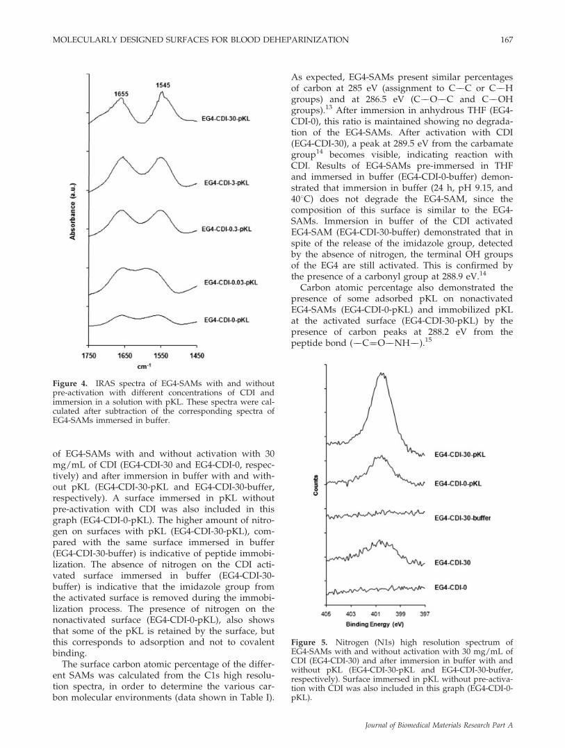
Figure 4 shows IRAS spectra of EG4-SAMs withand without pre-activation with different CDI con-centrations and after immersion in a pKL solution.These spectra were calculated after subtraction ofthe corresponding spectra of EG4-SAMs immersedin buffer. They show the amide I (1690 6 45 cm21)and amide II (1540 6 60 cm21) absorption bands,characteristic of peptides.12 The increase of the inten-sity of these bands indicates a raise of the amount ofpKL immobilized, which is directly related to EG4activation (CDI concentration). Although low, theseabsorption bands are also present on the nonacti-vated EG4-SAM (CDI-0) showing some pKL adsorp-tion (not covalently bound).
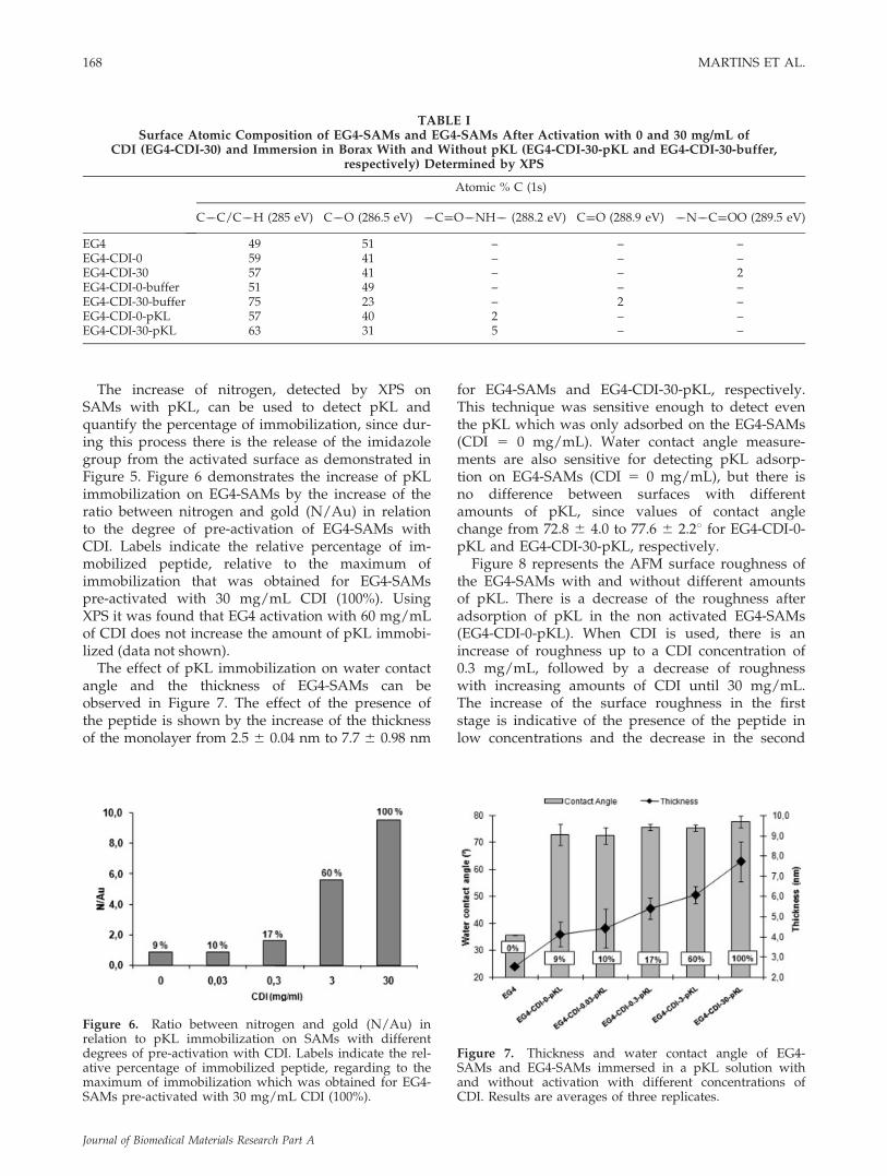
Survey spectra (data not shown) determined usingXPS reveal the presence of only the desired mono-layer and substrate elements (Au, C, S, O, and N),thus indicating the absence of contamination. Figure5 presents the XPS nitrogen high resolution spectra
Figure 1. Thickness and water contact angle of EG4-SAMs before and after activation with different concentra-tions of CDI. Results are averages of three replicates.
Figure 2. IRAS spectrum of EG4-SAMs after activationwith different concentrations of CDI.
Figure 3. Nitrogen (N1s) high resolution spectrum ofEG4-SAMs before and after activation with different con-centrations of CDI.
166 MARTINS ET AL.
Journal of Biomedical Materials Research Part A
of EG4-SAMs with and without activation with 30mg/mL of CDI (EG4-CDI-30 and EG4-CDI-0, respec-tively) and after immersion in buffer with and with-out pKL (EG4-CDI-30-pKL and EG4-CDI-30-buffer,respectively). A surface immersed in pKL withoutpre-activation with CDI was also included in thisgraph (EG4-CDI-0-pKL). The higher amount of nitro-gen on surfaces with pKL (EG4-CDI-30-pKL), com-pared with the same surface immersed in buffer(EG4-CDI-30-buffer) is indicative of peptide immobi-lization. The absence of nitrogen on the CDI acti-vated surface immersed in buffer (EG4-CDI-30-buffer) is indicative that the imidazole group fromthe activated surface is removed during the immobi-lization process. The presence of nitrogen on thenonactivated surface (EG4-CDI-0-pKL), also showsthat some of the pKL is retained by the surface, butthis corresponds to adsorption and not to covalentbinding.
The surface carbon atomic percentage of the differ-ent SAMs was calculated from the C1s high resolu-tion spectra, in order to determine the various car-bon molecular environments (data shown in Table I).
As expected, EG4-SAMs present similar percentagesof carbon at 285 eV (assignment to C��C or C��Hgroups) and at 286.5 eV (C��O��C and C��OHgroups).13 After immersion in anhydrous THF (EG4-CDI-0), this ratio is maintained showing no degrada-tion of the EG4-SAMs. After activation with CDI(EG4-CDI-30), a peak at 289.5 eV from the carbamategroup14 becomes visible, indicating reaction withCDI. Results of EG4-SAMs pre-immersed in THFand immersed in buffer (EG4-CDI-0-buffer) demon-strated that immersion in buffer (24 h, pH 9.15, and408C) does not degrade the EG4-SAM, since thecomposition of this surface is similar to the EG4-SAMs. Immersion in buffer of the CDI activatedEG4-SAM (EG4-CDI-30-buffer) demonstrated that inspite of the release of the imidazole group, detectedby the absence of nitrogen, the terminal OH groupsof the EG4 are still activated. This is confirmed bythe presence of a carbonyl group at 288.9 eV.14
Carbon atomic percentage also demonstrated thepresence of some adsorbed pKL on nonactivatedEG4-SAMs (EG4-CDI-0-pKL) and immobilized pKLat the activated surface (EG4-CDI-30-pKL) by thepresence of carbon peaks at 288.2 eV from thepeptide bond (��C¼¼O��NH��).15
Figure 4. IRAS spectra of EG4-SAMs with and withoutpre-activation with different concentrations of CDI andimmersion in a solution with pKL. These spectra were cal-culated after subtraction of the corresponding spectra ofEG4-SAMs immersed in buffer.
Figure 5. Nitrogen (N1s) high resolution spectrum ofEG4-SAMs with and without activation with 30 mg/mL ofCDI (EG4-CDI-30) and after immersion in buffer with andwithout pKL (EG4-CDI-30-pKL and EG4-CDI-30-buffer,respectively). Surface immersed in pKL without pre-activa-tion with CDI was also included in this graph (EG4-CDI-0-pKL).
MOLECULARLY DESIGNED SURFACES FOR BLOOD DEHEPARINIZATION 167
Journal of Biomedical Materials Research Part A
The increase of nitrogen, detected by XPS onSAMs with pKL, can be used to detect pKL andquantify the percentage of immobilization, since dur-ing this process there is the release of the imidazolegroup from the activated surface as demonstrated inFigure 5. Figure 6 demonstrates the increase of pKLimmobilization on EG4-SAMs by the increase of theratio between nitrogen and gold (N/Au) in relationto the degree of pre-activation of EG4-SAMs withCDI. Labels indicate the relative percentage of im-mobilized peptide, relative to the maximum ofimmobilization that was obtained for EG4-SAMspre-activated with 30 mg/mL CDI (100%). UsingXPS it was found that EG4 activation with 60 mg/mLof CDI does not increase the amount of pKL immobi-lized (data not shown).
The effect of pKL immobilization on water contactangle and the thickness of EG4-SAMs can beobserved in Figure 7. The effect of the presence ofthe peptide is shown by the increase of the thicknessof the monolayer from 2.5 6 0.04 nm to 7.7 6 0.98 nm
for EG4-SAMs and EG4-CDI-30-pKL, respectively.This technique was sensitive enough to detect eventhe pKL which was only adsorbed on the EG4-SAMs(CDI 5 0 mg/mL). Water contact angle measure-ments are also sensitive for detecting pKL adsorp-tion on EG4-SAMs (CDI 5 0 mg/mL), but there isno difference between surfaces with differentamounts of pKL, since values of contact anglechange from 72.8 6 4.0 to 77.6 6 2.28 for EG4-CDI-0-pKL and EG4-CDI-30-pKL, respectively.
Figure 8 represents the AFM surface roughness ofthe EG4-SAMs with and without different amountsof pKL. There is a decrease of the roughness afteradsorption of pKL in the non activated EG4-SAMs(EG4-CDI-0-pKL). When CDI is used, there is anincrease of roughness up to a CDI concentration of0.3 mg/mL, followed by a decrease of roughnesswith increasing amounts of CDI until 30 mg/mL.The increase of the surface roughness in the firststage is indicative of the presence of the peptide inlow concentrations and the decrease in the second
TABLE ISurface Atomic Composition of EG4-SAMs and EG4-SAMs After Activation with 0 and 30 mg/mL of
CDI (EG4-CDI-30) and Immersion in Borax With and Without pKL (EG4-CDI-30-pKL and EG4-CDI-30-buffer,respectively) Determined by XPS
Atomic % C (1s)
C��C/C��H (285 eV) C��O (286.5 eV) ��C¼¼O��NH�� (288.2 eV) C¼¼O (288.9 eV) ��N��C¼¼OO (289.5 eV)
EG4 49 51 – – –EG4-CDI-0 59 41 – – –EG4-CDI-30 57 41 – – 2EG4-CDI-0-buffer 51 49 – – –EG4-CDI-30-buffer 75 23 – 2 –EG4-CDI-0-pKL 57 40 2 – –EG4-CDI-30-pKL 63 31 5 – –
Figure 6. Ratio between nitrogen and gold (N/Au) inrelation to pKL immobilization on SAMs with differentdegrees of pre-activation with CDI. Labels indicate the rel-ative percentage of immobilized peptide, regarding to themaximum of immobilization which was obtained for EG4-SAMs pre-activated with 30 mg/mL CDI (100%).
Figure 7. Thickness and water contact angle of EG4-SAMs and EG4-SAMs immersed in a pKL solution withand without activation with different concentrations ofCDI. Results are averages of three replicates.
168 MARTINS ET AL.
Journal of Biomedical Materials Research Part A
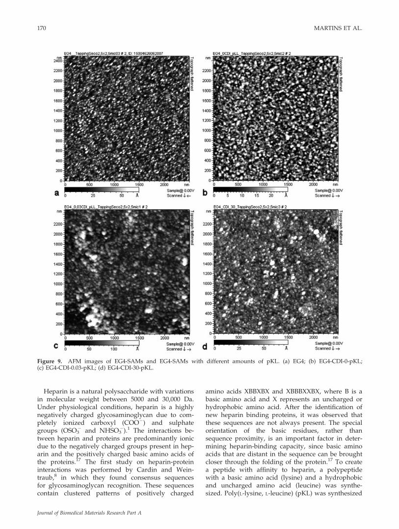
stage demonstrated the presence of a relative closepacked peptide monolayer. The decrease of theroughness after adsorption of the peptide (EG4-CDI-0-pKL), compared with nonfunctionalized EG4-SAMs, is explained by a ‘‘smoothing out’’ effect ofthe adsorbed peptide, since our sputtered gold sub-strates were not perfectly smooth: Sq 5 1.136 0.18 nm.Figure 9 demonstrate the AFM images of EG4-SAMswith and without different amounts of pKL.
Heparin adsorption on SAMs
Infrared spectroscopy
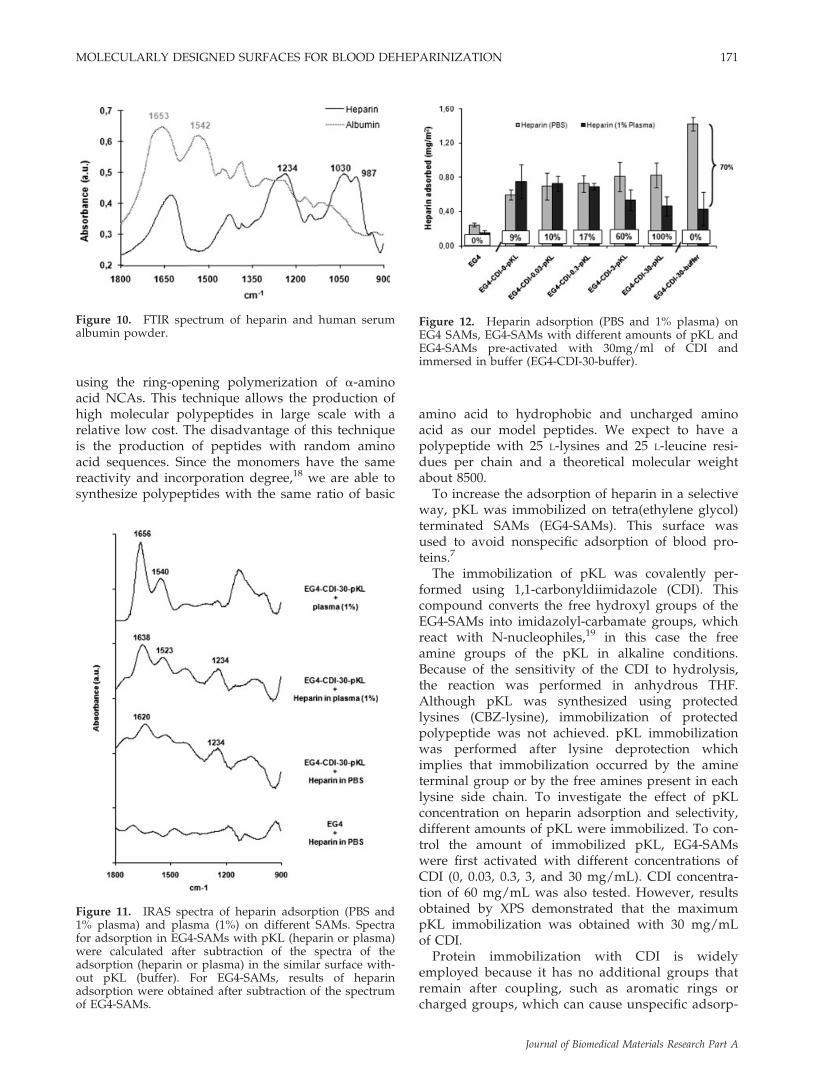
Figure 10 shows an FTIR spectrum of heparin andhuman serum albumin powder. The heparin spec-trum shows the typical absorption bands of the sul-phate groups (��OSO3
2) that can be used to distin-guish heparin from proteins. The absorption ataround 1230 and 1030 cm21 are associated with thestretching vibrations of the SO2 group.16 The typicalabsorption bands of the amine groups present onheparin was observed at 1620 cm21. However, thispeak may overlap with the amide groups of proteinsand was not used to detect heparin on competitiveadsorption with proteins. The albumin spectrumshows two intense absorption bands at 1653 and1526 cm21 that can be associated, as already de-scribed for peptides, with the characteristics absorp-tion bands from the amide I and amide II groupsof proteins.12,16 These absorption bands were used todetect protein adsorption using infrared spectroscopy.
Figure 11 shows the IRAS spectra of heparinadsorption from PBS on EG4-SAMs and on SAMswith pKL. Results were calculated after subtraction
of the spectrum of the surface without pKL (buffer)from the spectrum of heparin adsorption on SAMswith pKL. Concerning the characteristic absorptionbands of heparin at 1234 and 1620 cm21, the resultsdemonstrate that heparin adsorption is only detectedon SAMs with pKL. In the presence of competitiveproteins (1% plasma), this surface maintains itscapacity to adsorb heparin, although some proteinadsorption is detected by the increase of the inten-sity of a peak at 1523 cm21 and the shift of the peakat 1620–1638 cm21. Plasma adsorption on SAMswith pKL was used as a control.
Radiolabeled heparin (S35-heparin)
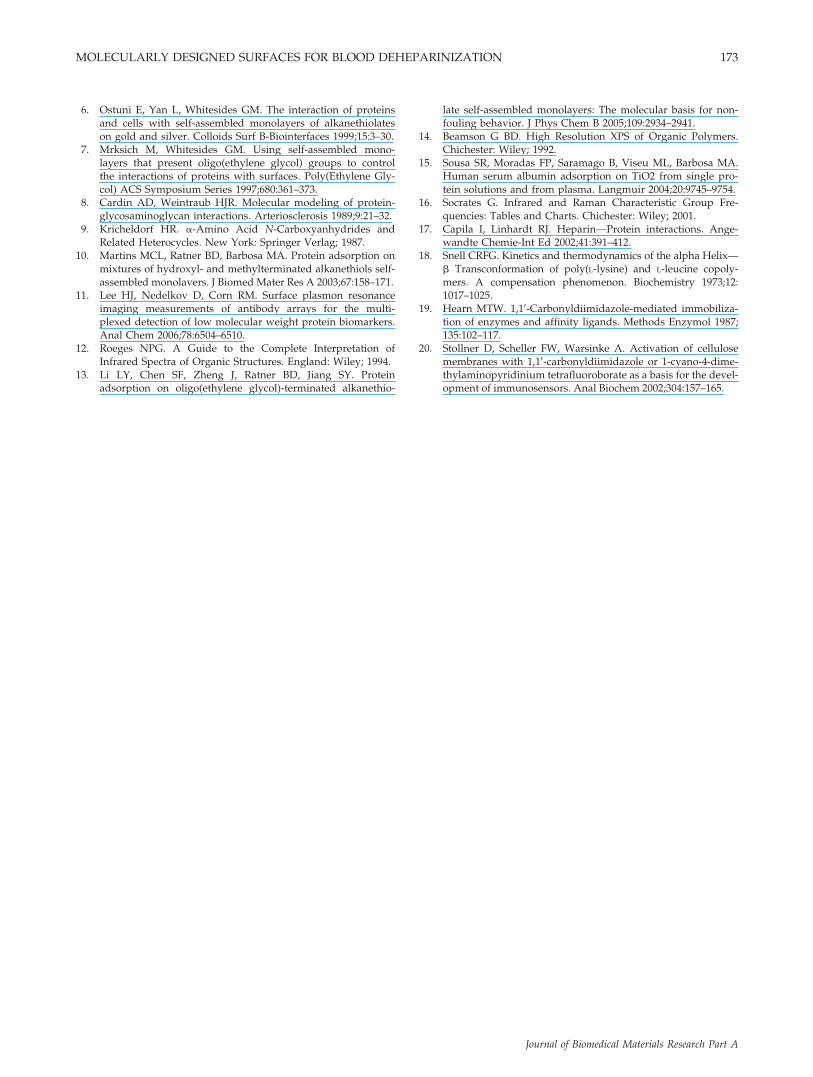
Heparin adsorption on EG4 SAMs incubated inPBS and in 1% plasma, EG4-SAMs with pKL afterpre-activation with different concentrations of CDIand EG4-SAMs pre-activated with 30 mg/mL of CDIand immersed in buffer (EG4-CDI-30-buffer) isshown in Figure 12. These results demonstrate thatthe presence of pKL induces heparin adsorption onEG4-SAMs. However, the amount of immobilizedpKL does not influence the adsorption of heparin(0.6 6 0.06 to 0.8 6 0.15 mg/m2 for EG4-CDI-0-pKLand EG4-CDI-30-pKL, respectively). There is no stat-istically significant difference (p < 0.05; Tukey)between heparin adsorption on SAMs with pKL. Theeffect of the pKL concentration was only detectedduring competitive adsorption studies between hep-arin and plasma proteins. It was observed that lowconcentration of pKL favors the selectivity of heparinadsorption. Higher selectivity to heparin is only veri-fied on SAMs containing pKL, under the followingconditions: nonactivated and activated with 0.03 and0.3 mg/mL of CDI (p < 0.05; ANOVA-Dunnett C).pKL increases the percentage of heparin in solutionthat is binding to the surface from 0.05 (EG4) to0.11–0.15. In the presence of the plasma proteins,this percentage is only maintained for SAMs with 0,0.03, and 0.3 mg/mL of CDI.
These results also show that the highest amount ofheparin was detected on SAMs pre-activated withCDI and immersed in buffer. Nevertheless, in thepresence of competitive proteins (1% plasma) theadsorption of heparin on this surface is reduced by70%, showing a nonspecific adsorption process.
DISCUSSION
In the present work, we have used SAMs of alka-nethiols on gold as model surfaces to study theeffect of an immobilized synthetic polypeptide onthe adsorption of heparin, including the degree ofselective adsorption.
Figure 8. AFM surface roughness (Root Mean Square; Sq)of the EG4-SAMs and EG4-SAMs with different amountsof pKL.
MOLECULARLY DESIGNED SURFACES FOR BLOOD DEHEPARINIZATION 169
Journal of Biomedical Materials Research Part A
Heparin is a natural polysaccharide with variationsin molecular weight between 5000 and 30,000 Da.Under physiological conditions, heparin is a highlynegatively charged glycosaminoglycan due to com-pletely ionized carboxyl (COO2) and sulphategroups (OSO3
2 and NHSO32).1 The interactions be-
tween heparin and proteins are predominantly ionicdue to the negatively charged groups present in hep-arin and the positively charged basic amino acids ofthe proteins.17 The first study on heparin-proteininteractions was performed by Cardin and Wein-traub,8 in which they found consensus sequencesfor glycosaminoglycan recognition. These sequencescontain clustered patterns of positively charged
amino acids XBBXBX and XBBBXXBX, where B is abasic amino acid and X represents an uncharged orhydrophobic amino acid. After the identification ofnew heparin binding proteins, it was observed thatthese sequences are not always present. The specialorientation of the basic residues, rather thansequence proximity, is an important factor in deter-mining heparin-binding capacity, since basic aminoacids that are distant in the sequence can be broughtcloser through the folding of the protein.17 To createa peptide with affinity to heparin, a polypeptidewith a basic amino acid (lysine) and a hydrophobicand uncharged amino acid (leucine) was synthe-sized. Poly(L-lysine, L-leucine) (pKL) was synthesized
Figure 9. AFM images of EG4-SAMs and EG4-SAMs with different amounts of pKL. (a) EG4; (b) EG4-CDI-0-pKL;(c) EG4-CDI-0.03-pKL; (d) EG4-CDI-30-pKL.
170 MARTINS ET AL.
Journal of Biomedical Materials Research Part A
using the ring-opening polymerization of a-aminoacid NCAs. This technique allows the production ofhigh molecular polypeptides in large scale with arelative low cost. The disadvantage of this techniqueis the production of peptides with random aminoacid sequences. Since the monomers have the samereactivity and incorporation degree,18 we are able tosynthesize polypeptides with the same ratio of basic
amino acid to hydrophobic and uncharged aminoacid as our model peptides. We expect to have apolypeptide with 25 L-lysines and 25 L-leucine resi-dues per chain and a theoretical molecular weightabout 8500.
To increase the adsorption of heparin in a selectiveway, pKL was immobilized on tetra(ethylene glycol)terminated SAMs (EG4-SAMs). This surface wasused to avoid nonspecific adsorption of blood pro-teins.7
The immobilization of pKL was covalently per-formed using 1,1-carbonyldiimidazole (CDI). Thiscompound converts the free hydroxyl groups of theEG4-SAMs into imidazolyl-carbamate groups, whichreact with N-nucleophiles,19 in this case the freeamine groups of the pKL in alkaline conditions.Because of the sensitivity of the CDI to hydrolysis,the reaction was performed in anhydrous THF.Although pKL was synthesized using protectedlysines (CBZ-lysine), immobilization of protectedpolypeptide was not achieved. pKL immobilizationwas performed after lysine deprotection whichimplies that immobilization occurred by the amineterminal group or by the free amines present in eachlysine side chain. To investigate the effect of pKLconcentration on heparin adsorption and selectivity,different amounts of pKL were immobilized. To con-trol the amount of immobilized pKL, EG4-SAMswere first activated with different concentrations ofCDI (0, 0.03, 0.3, 3, and 30 mg/mL). CDI concentra-tion of 60 mg/mL was also tested. However, resultsobtained by XPS demonstrated that the maximumpKL immobilization was obtained with 30 mg/mLof CDI.
Protein immobilization with CDI is widelyemployed because it has no additional groups thatremain after coupling, such as aromatic rings orcharged groups, which can cause unspecific adsorp-
Figure 10. FTIR spectrum of heparin and human serumalbumin powder.
Figure 11. IRAS spectra of heparin adsorption (PBS and1% plasma) and plasma (1%) on different SAMs. Spectrafor adsorption in EG4-SAMs with pKL (heparin or plasma)were calculated after subtraction of the spectra of theadsorption (heparin or plasma) in the similar surface with-out pKL (buffer). For EG4-SAMs, results of heparinadsorption were obtained after subtraction of the spectrumof EG4-SAMs.
Figure 12. Heparin adsorption (PBS and 1% plasma) onEG4 SAMs, EG4-SAMs with different amounts of pKL andEG4-SAMs pre-activated with 30mg/ml of CDI andimmersed in buffer (EG4-CDI-30-buffer).
MOLECULARLY DESIGNED SURFACES FOR BLOOD DEHEPARINIZATION 171
Journal of Biomedical Materials Research Part A

tion effects.20 It was assumed that complete hydro-lysis of CDI-activated SAMs occurs under alkalineconditions within 20–24 h.20 XPS results, namelynitrogen high resolution spectra (Fig. 5) demon-strated the absence of nitrogen from the surface ofEG4-CDI-30 after immersion in buffer (borax, pH9.15, 408C) over a 24 h period. This is indicative ofthe imidazole group release during the immersionof the surfaces in the buffer. However, the carbonhigh resolution spectra of the SAMs functionalizedwith 30 mg/mL of CDI (EG4-CDI-30-buffer) dem-onstrated the presence of peaks at 288.9 eV, whichare assigned to carbonyl groups that were notreleased from the activated surface. These resultsdemonstrated that the procedure used was not effi-cient for the complete hydrolysis of CDI-activatedSAMs, which could induce unspecific adsorption onheparin and blood proteins. This explains ourchoice to control the first part of the reaction, i.e.activation, using different concentrations of CDIand a single concentration of pKL, instead of satu-rating the surface with high concentrations of CDI(30 mg/mL) and using different concentrations ofpKL, This strategy proved effective in view of theselectivity obtained with the lower concentrationsof CDI (Fig. 12).
Contact angle measurements, ellipsometry, IRASand XPS results demonstrated that different concen-trations of CDI can be used to activate the EG4-SAMs to different degrees. The utilization of variousCDI concentrations allowed the immobilization of arange of percentages of pKL concentrations on theEG4-SAMs. Results from IRAS, ellipsometry andXPS provide evidence in support of an augmentationof the amount of pKL with increasing CDI concen-trations. These results were confirmed by AFM. Thistechnique indicates a uniform distribution of pKL onthe SAMs surfaces.
Although all the data obtained are indicative of acovalent immobilization of pKL, since there is directproportionality between the amount of immobilizedpKL and degree of surface activation, some pKLadsorption was also observed on surfaces not acti-vated with CDI. This is probably indicative of theconditions used during pKL immobilization, namelythe basic environment and 408C, may alter the non-fouling capacity of EG4-SAMs, thus allowing somebinding of pKL. Degradation of this surface was notdetected by XPS, which might be another reason forthe observed effect.
The presence of pKL, even in the adsorbed state,increases heparin adsorption compared with EG4-SAMs. The increase of pKL concentration does notincrease the amount of heparin adsorption. How-ever, the increase of pKL concentration (activationwith 3 and 30 mg/mL of CDI) decreases the selectiv-ity of the surfaces to heparin. Two possible explana-
tions have to be considered at this stage. First, surfa-ces with high concentrations of pKL may not exposea large number of amino acid sequences, due to asteric hindrance effect of closely packed chains. Sec-ond, if the lysines are bound to activated OH groupsfewer of them would be exposed on the surface,thereby reducing the number of selective adsorptionsites. The low selectivity of surfaces with a high con-centration of pKL is not likely related to partial acti-vation of OH groups, otherwise the adsorption frompure heparin solutions would be higher.
CONCLUSIONS
In the present work, a heparin-binding peptide(pKL) was successful synthesized and immobilizedon EG4-SAMs under different concentrations. Immo-bilization was performed using a fixed concentrationof pKL after surface activation to different degreesusing CDI. This strategy aims to prevent free acti-vated groups on the pKL surface from inducing non-specific protein adsorption.
Results demonstrated that the presence of pKLincreases heparin adsorption to EG4-SAMs, inde-pendently of the pKL concentration and the way ofimmobilization (adsorption or covalent bound). Se-lectivity to heparin was successfully reached for lowconcentrations of pKL. At high concentrations, pKLmay not expose a large number of amino acidsequences, either due to a steric hindrance effect ofclosely packed chains or because high degrees ofCDI activation can bind several lysines of pKL, thusreducing the number of selective adsorption sites toheparin.
The authors thank the Portuguese Blood Institute (IPS)for donating the human plasma and M. Manuela Bras andAna L. Ribeiro (INEB) for AFM analysis.
References
1. Dailey J. Blood. Ipswich, MA, USA: Medical ConsultingGroup; 1998.
2. Section V. Chronic intermittent haemodialysis and preventionof clotting in the extracorporal system. Nephrol DialysisTransplant 2002;17:63–71.
3. Zhang YH, Singh VK, Yang VC. Novel approach for optimiz-ing the capacity and efficacy of a protamine filter for clinicalextracorporeal heparin removal. Asaio J 1998;44:M368–M373.
4. Tao W, Deyo DJ, Brunston RL, Vertrees RA, ZwischenbergerJB. Extracorporeal heparin adsorption following cardiopulmo-nary bypass with a heparin removal device—An alternativeto protamine. Critical Care Med 1998;26:1096–1102.
5. Ameer GA, Barabino G, Sasisekharan R, Harmon W, CooneyCL, Langer R. Ex vivo evaluation of a Taylor–Couette flow,immobilized heparinase I device for clinical application. ProcNatl Acad Sci USA 1999;96:2350–2355.
172 MARTINS ET AL.
Journal of Biomedical Materials Research Part A
6. Ostuni E, Yan L, Whitesides GM. The interaction of proteinsand cells with self-assembled monolayers of alkanethiolateson gold and silver. Colloids Surf B-Biointerfaces 1999;15:3–30.
7. Mrksich M, Whitesides GM. Using self-assembled mono-layers that present oligo(ethylene glycol) groups to controlthe interactions of proteins with surfaces. Poly(Ethylene Gly-col) ACS Symposium Series 1997;680:361–373.
8. Cardin AD, Weintraub HJR. Molecular modeling of protein-glycosaminoglycan interactions. Arteriosclerosis 1989;9:21–32.
9. Kricheldorf HR. a-Amino Acid N-Carboxyanhydrides andRelated Heterocycles. New York: Springer Verlag; 1987.
10. Martins MCL, Ratner BD, Barbosa MA. Protein adsorption onmixtures of hydroxyl- and methylterminated alkanethiols self-assembled monolavers. J Biomed Mater Res A 2003;67:158–171.
11. Lee HJ, Nedelkov D, Corn RM. Surface plasmon resonanceimaging measurements of antibody arrays for the multi-plexed detection of low molecular weight protein biomarkers.Anal Chem 2006;78:6504–6510.
12. Roeges NPG. A Guide to the Complete Interpretation ofInfrared Spectra of Organic Structures. England: Wiley; 1994.
13. Li LY, Chen SF, Zheng J, Ratner BD, Jiang SY. Proteinadsorption on oligo(ethylene glycol)-terminated alkanethio-
late self-assembled monolayers: The molecular basis for non-fouling behavior. J Phys Chem B 2005;109:2934–2941.
14. Beamson G BD. High Resolution XPS of Organic Polymers.Chichester: Wiley; 1992.
15. Sousa SR, Moradas FP, Saramago B, Viseu ML, Barbosa MA.Human serum albumin adsorption on TiO2 from single pro-tein solutions and from plasma. Langmuir 2004;20:9745–9754.
16. Socrates G. Infrared and Raman Characteristic Group Fre-quencies: Tables and Charts. Chichester: Wiley; 2001.
17. Capila I, Linhardt RJ. Heparin—Protein interactions. Ange-wandte Chemie-Int Ed 2002;41:391–412.
18. Snell CRFG. Kinetics and thermodynamics of the alpha Helix—b Transconformation of poly(L-lysine) and L-leucine copoly-mers. A compensation phenomenon. Biochemistry 1973;12:1017–1025.
19. Hearn MTW. 1,10-Carbonyldiimidazole-mediated immobiliza-tion of enzymes and affinity ligands. Methods Enzymol 1987;135:102–117.
20. Stollner D, Scheller FW, Warsinke A. Activation of cellulosemembranes with 1,10-carbonyldiimidazole or 1-cyano-4-dime-thylaminopyridinium tetrafluoroborate as a basis for the devel-opment of immunosensors. Anal Biochem 2002;304:157–165.
MOLECULARLY DESIGNED SURFACES FOR BLOOD DEHEPARINIZATION 173
Journal of Biomedical Materials Research Part A
Copyright © 2022 FDOKUMEN