The “CPC Clip Motif”: A Conserved Structural Signature for Heparin-Binding Proteins
8
The ‘‘CPC Clip Motif’’: A Conserved Structural Signature for Heparin-Binding Proteins Marc Torrent 1,2 *, M. Victo ` ria Nogue ´s 1 , David Andreu 2 , Ester Boix 1 1 Department of Biochemistry and Molecular Biology, Biosciences Faculty, Universitat Auto ` noma de Barcelona, Cerdanyola del Valle `s, Spain, 2 Department of Experimental and Health Sciences, Universitat Pompeu Fabra, Barcelona Biomedical Research Park, Barcelona, Spain Abstract Glycosaminoglycans (GAGs) are essential molecules that regulate diverse biological processes including cell adhesion, differentiation, signaling and growth, by interaction with a wide variety of proteins. However, despite the efforts committed to understand the molecular nature of the interactions in protein-GAG complexes, the answer to this question remains elusive. In the present study the interphases of 20 heparin–binding proteins have been analyzed searching for a conserved structural pattern. We have found that a structural motif encompassing one polar and two cationic residues (which has been named the CPC clip motif) is conserved among all the proteins deposited in the PDB. The distances between the a carbons and the side chain center of gravity of the residues composing this motif are also conserved. Furthermore, this pattern can be found in other proteins suggested to bind heparin for which no structural information is available. Hence we propose that the CPC clip motif, working like a staple, is a primary contributor to the attachment of heparin and other sulfated GAGs to heparin-binding proteins. Citation: Torrent M, Nogue ´s MV, Andreu D, Boix E (2012) The ‘‘CPC Clip Motif’’: A Conserved Structural Signature for Heparin-Binding Proteins. PLoS ONE 7(8): e42692. doi:10.1371/journal.pone.0042692 Editor: Eugene A. Permyakov, Russian Academy of Sciences, Institute for Biological Instrumentation, Russian Federation Received April 25, 2012; Accepted July 11, 2012; Published August 6, 2012 Copyright: ß 2012 Torrent et al. This is an open-access article distributed under the terms of the Creative Commons Attribution License, which permits unrestricted use, distribution, and reproduction in any medium, provided the original author and source are credited. Funding: MT is the recipient of a postdoctoral grant from Alianza Cuatro Universidades (Spain). Work supported by the European Union (HEALTH-F3-2008- 223414), the Spanish Ministry of Science and Innovation (BIO2008-04487-CO3-02, BFU2009- 09371, FEDER funds) and the Generalitat de Catalunya (SGR2009-494, SGR2009-795). The funders had no role in study design, data collection and analysis, decision to publish, or preparation of the manuscript. Competing Interests: The authors have declared that no competing interests exist. * E-mail: [email protected] Introduction Glycosaminoglycans (GAGs) are negatively charged polysac- charides, with molecular weights ranging from 10 to 100 kDa, composed of repeating units of uronic acid (D-glucuronic or L- iduronic acid) and amino sugars (D-galactosamine or D-glucos- amine). D-glucosamine-containing GAGs, like heparin, are named glucosaminoglycans [1]. GAGs have central biological functions including wound healing [2], anti-coagulation [3], cell signaling, development and angiogenesis [4], tumor progression and metastasis [5] and can even play an important role in amyloid-related diseases [6]. In particular, heparin can preclude blood clotting and is mainly used as anti-coagulant for the treatment of thrombosis, thrombophle- bitis and embolism [3]. Additionally, GAGs are involved in cell proliferation and diseases such as rheumatoid arthritis, inflamma- tory bowel disease and infections associated with inflammatory responses [7] and can hinder HIV-1 or herpes simplex virus activity through binding to the viral surface glycoproteins [8]. In this context, heparin and heparan sulfate (HS) have been found to bind a wide variety of proteins with diverse functions, including growth factors, thrombin, chemokines and viral proteins [1,9]. Unfortunately, despite the growing pharmaceutical interest in protein-sugar interactions, structural requirements for GAG binding are still not well characterized [1,10,11]. Cardin and Weintraub analyzed the structure of heparin- binding proteins and proposed that typical binding sites contain the sequence-based motif XBBXBX or XBBBXXBX, where B is a lysine or arginine (rarely His) and X a hydropathic residue such as Ala, Gly, Ile, Leu or Tyr [12]. In this way, a conserved sequence (XBBBXXBBBXXBBXBX) was similarly proposed for the Von Willebrand factor and a TXXBXXTBXXXTBB sequence was ascribed to a and b fibroblast growth factors (aFGF, bFGF) and transforming growth factor b-1 (TGFb-1) [13]. Heparin-binding domains always contain cationic residues, which bind to anionic (carboxylate, sulfate) groups in heparin through electrostatic and hydrogen-bonding interactions. It has been reported that a precise spacing of cationic clusters is required, and efficient heparin-binding peptides were designed on this basis [14]. However, the specificity of these interactions remains elusive and is still poorly understood [11]. Here we report a novel structural signature for heparin-binding proteins, which is conserved in all such protein structures available in the Protein Data Bank (PDB). The motif involves two cationic residues (Arg or Lys) and a polar residue (preferentially Asn, Gln, Thr, Tyr or Ser, more rarely Arg or Lys), with fairly conserved distances between the a carbons and the side chain center of gravity, defining a clip-like structure where heparin would be lodged. This structural motif is highly conserved and can be found in many proteins with reported heparin binding capacity. Methods Protein structure analysis Protein three-dimensional structures were obtained from the Protein Data Bank (www.pdb.org). The summary of ligand interactions with IDS (2-O-sulfo-a-L-iduronic acid) and SGN (N,O6-disulfo-glucosamine) has been performed using the PDBe- PLoS ONE | www.plosone.org 1 August 2012 | Volume 7 | Issue 8 | e42692
Transcript of The “CPC Clip Motif”: A Conserved Structural Signature for Heparin-Binding Proteins

The ‘‘CPC Clip Motif’’: A Conserved Structural Signaturefor Heparin-Binding ProteinsMarc Torrent1,2*, M. Victoria Nogues1, David Andreu2, Ester Boix1
1 Department of Biochemistry and Molecular Biology, Biosciences Faculty, Universitat Autonoma de Barcelona, Cerdanyola del Valles, Spain, 2 Department of
Experimental and Health Sciences, Universitat Pompeu Fabra, Barcelona Biomedical Research Park, Barcelona, Spain
Abstract
Glycosaminoglycans (GAGs) are essential molecules that regulate diverse biological processes including cell adhesion,differentiation, signaling and growth, by interaction with a wide variety of proteins. However, despite the efforts committedto understand the molecular nature of the interactions in protein-GAG complexes, the answer to this question remainselusive. In the present study the interphases of 20 heparin–binding proteins have been analyzed searching for a conservedstructural pattern. We have found that a structural motif encompassing one polar and two cationic residues (which hasbeen named the CPC clip motif) is conserved among all the proteins deposited in the PDB. The distances between the acarbons and the side chain center of gravity of the residues composing this motif are also conserved. Furthermore, thispattern can be found in other proteins suggested to bind heparin for which no structural information is available. Hence wepropose that the CPC clip motif, working like a staple, is a primary contributor to the attachment of heparin and othersulfated GAGs to heparin-binding proteins.
Citation: Torrent M, Nogues MV, Andreu D, Boix E (2012) The ‘‘CPC Clip Motif’’: A Conserved Structural Signature for Heparin-Binding Proteins. PLoS ONE 7(8):e42692. doi:10.1371/journal.pone.0042692
Editor: Eugene A. Permyakov, Russian Academy of Sciences, Institute for Biological Instrumentation, Russian Federation
Received April 25, 2012; Accepted July 11, 2012; Published August 6, 2012
Copyright: � 2012 Torrent et al. This is an open-access article distributed under the terms of the Creative Commons Attribution License, which permitsunrestricted use, distribution, and reproduction in any medium, provided the original author and source are credited.
Funding: MT is the recipient of a postdoctoral grant from Alianza Cuatro Universidades (Spain). Work supported by the European Union (HEALTH-F3-2008-223414), the Spanish Ministry of Science and Innovation (BIO2008-04487-CO3-02, BFU2009- 09371, FEDER funds) and the Generalitat de Catalunya (SGR2009-494,SGR2009-795). The funders had no role in study design, data collection and analysis, decision to publish, or preparation of the manuscript.
Competing Interests: The authors have declared that no competing interests exist.
* E-mail: [email protected]
Introduction
Glycosaminoglycans (GAGs) are negatively charged polysac-
charides, with molecular weights ranging from 10 to 100 kDa,
composed of repeating units of uronic acid (D-glucuronic or L-
iduronic acid) and amino sugars (D-galactosamine or D-glucos-
amine). D-glucosamine-containing GAGs, like heparin, are named
glucosaminoglycans [1].
GAGs have central biological functions including wound
healing [2], anti-coagulation [3], cell signaling, development and
angiogenesis [4], tumor progression and metastasis [5] and can
even play an important role in amyloid-related diseases [6]. In
particular, heparin can preclude blood clotting and is mainly used
as anti-coagulant for the treatment of thrombosis, thrombophle-
bitis and embolism [3]. Additionally, GAGs are involved in cell
proliferation and diseases such as rheumatoid arthritis, inflamma-
tory bowel disease and infections associated with inflammatory
responses [7] and can hinder HIV-1 or herpes simplex virus
activity through binding to the viral surface glycoproteins [8].
In this context, heparin and heparan sulfate (HS) have been
found to bind a wide variety of proteins with diverse functions,
including growth factors, thrombin, chemokines and viral proteins
[1,9]. Unfortunately, despite the growing pharmaceutical interest
in protein-sugar interactions, structural requirements for GAG
binding are still not well characterized [1,10,11].
Cardin and Weintraub analyzed the structure of heparin-
binding proteins and proposed that typical binding sites contain
the sequence-based motif XBBXBX or XBBBXXBX, where B is a
lysine or arginine (rarely His) and X a hydropathic residue such as
Ala, Gly, Ile, Leu or Tyr [12]. In this way, a conserved sequence
(XBBBXXBBBXXBBXBX) was similarly proposed for the Von
Willebrand factor and a TXXBXXTBXXXTBB sequence was
ascribed to a and b fibroblast growth factors (aFGF, bFGF) and
transforming growth factor b-1 (TGFb-1) [13].
Heparin-binding domains always contain cationic residues,
which bind to anionic (carboxylate, sulfate) groups in heparin
through electrostatic and hydrogen-bonding interactions. It has
been reported that a precise spacing of cationic clusters is required,
and efficient heparin-binding peptides were designed on this basis
[14]. However, the specificity of these interactions remains elusive
and is still poorly understood [11].
Here we report a novel structural signature for heparin-binding
proteins, which is conserved in all such protein structures available
in the Protein Data Bank (PDB). The motif involves two cationic
residues (Arg or Lys) and a polar residue (preferentially Asn, Gln,
Thr, Tyr or Ser, more rarely Arg or Lys), with fairly conserved
distances between the a carbons and the side chain center of
gravity, defining a clip-like structure where heparin would be
lodged. This structural motif is highly conserved and can be found
in many proteins with reported heparin binding capacity.
Methods
Protein structure analysisProtein three-dimensional structures were obtained from the
Protein Data Bank (www.pdb.org). The summary of ligand
interactions with IDS (2-O-sulfo-a-L-iduronic acid) and SGN
(N,O6-disulfo-glucosamine) has been performed using the PDBe-
PLoS ONE | www.plosone.org 1 August 2012 | Volume 7 | Issue 8 | e42692

MOTIF web server (http://www.ebi.ac.uk/pdbe-site/pdbemotif/)
and statistically evaluated using the Prism software. To define the
CPC clip motif, the location of protein-ligand interfaces has been
performed using the PISA web server (http://www.ebi.ac.uk/msd-
srv/prot_int/pistart.html) and the residues involved in heparin
binding were analyzed to outline a conserved motif. To further
check the contacts detected by PISA, a manual inspection of the
interacting residues was performed using Pymol (DeLano
Scientific, San Carlos, CA), defining a 3.5 A cut-off distance for
hydrogen bond interactions and a 4.5 A for electrostatic
interactions. The analysis of a-carbon distances has also been
calculated using Pymol. Chemokine domain structural modeling
has been performed using the automated Swiss Model server
(http://swissmodel.expasy.org/). Amino acid conservation was
evaluated using the ConSurf web server (http://consurf.tau.ac.il/).
Sequence alignments were drawn using ESPript (http://espript.
ibcp.fr/ESPript/ESPript/) and figures were prepared using
Pymol.
DatasetsThe discovery set contains 20 proteins crystallized with heparin
analogs and were used to define the CPC clip motif (Table S1). All
proteins included share less than 25% of sequence similarity with
the remaining dataset, except for the pairs: 1FQ9/1E0O, 1AXM/
1BFB and 1G5N/2HYU. The later were included because
different binding sites were detected for those structures, probably
reflecting complementary binding sites for larger heparin mole-
cules. The testing dataset contains 48 proteins; 24 proteins with
experimental evidence of heparin binding capacity not included in
the discovery set (,50% sequence similarity; positive testing
dataset) and 24 proteins with neither described not expected
heparin binding activity (negative testing dataset; five negative
testing datasets were designed). To select the structures belonging
to the negative dataset, we have numbered each structure in the
SPASM database [15] (23746 proteins) and selected 24 proteins
among them using a random number generation function.
Proteins with potential heparin binding capacity (e.g. growth
factors, viral capsid proteins, etc. or proteins related to the
discovery set) were excluded. Proteins in the negative dataset were
ensured to have similar size distribution than in the positive dataset
(p,0.05).
CPC enrichment in heparin binding proteinsTo test the CPC motif enrichment in heparin-binding proteins,
the SPASM algorithm [15] has been used to search for CPC motif
hits in the testing dataset (48 proteins) allowing 2.5 A of RMSD.
The process has been repeated five times with five independent
negative testing datasets. In the SPASM computational algorithm,
the residues conforming the motif are represented by its Ca atom
and its side-chain center of gravity. SPASM calculates the
distances between these pseudo-atoms, and identifies all sets of
identical (or similar) residues for each protein database. After-
wards, the hits obtained were classified based on their database
origin to determine the enrichment fraction.
Molecular docking simulationsDocking simulations were conducted with AutoDock 4.2
(Scripps Research Institute. La Jolla, CA). The ligand used was
the heparin dodesaccharide characterized by NMR and deposited
in the PDB (code 1HPN). In all crystal structures used, water
molecules were removed from the structure. Hydrogen atoms and
atomic partial charges (using the Gasteiger method) were added
using Autodock Tools. Protein was kept rigid. Heparin was
allowed to bend by selecting ten degrees of freedom along the
oligosaccharide backbone (Figure S1A). The interaction of a probe
group, corresponding to each type of atom found in the ligand,
within the whole protein structure, was computed at 0.500 A grid
positions in a box centered in the protein.
The docking was accomplished using 100 Lamarkian genetic
algorithms (LGA) runs and the initial position of the ligand was
random. The number of individuals in populations was set to 150.
The maximum number of energy evaluations that the genetic
algorithm should make was 2500000. Maximum number of
generations was 27000. The number of top individuals that are
guaranteed to survive into the next generation was 1. Rates of
gene mutation and crossover were 0.02 and 0.80, respectively.
Following docking, all structures generated for the same
compound were subjected to cluster analysis, cluster families
being based on a tolerance of 10 A for an all-atom root mean
square (RMS) deviation from a lower energy structure. A second
docking stage was performed to increase performance and
complete exploration of conformational space. Thus, the global
minimum structure found in the previous run was subjected to
redocking under the same conditions and to 2 A cluster analysis,
using a box centered in the ligand with a 0.375 A of grid spacing.
The global minimum structure found in this second-stage docking
was considered as the final result.
To better estimate the binding energy for the complex, a
heparin disaccharide (H3S, obtained from the PDB, code 1U4M)
was docked using the parameters described before within a 50 A3
grid box centered in the CPC motif detected. The heparin
disaccharide was allowed to bend by allowing two degrees of
freedom along its backbone (Figure S1B).
Automated motif findingThe coordinates of the five identified canonic CPC clip motifs
(C-Asn-C, C-Gln-C, C-Ser-C, C-Thr-C and C-Tyr-C, where C is
a cationic residue, Arg or Lys) identified have been subjected to
analysis using the SPASM algorithm (Lys and Arg were considered
equivalent as cationic residues), then analyzed with SAVANT [16]
and finally filtered using DEJANA [16]. SAVANT algorithm
performs an all-atom least squares superpositioning of the query
motif on each hit found by SPASM. To analyze the gene-related
distribution of the proteins found with SPASM, PICR (http://
www.ebi.ac.uk/Tools/picr/) has been used to convert PDB to
SwissProt identifiers. The database generated was inspected using
PANTHER (http://www.pantherdb.org/) and the genes identi-
fied were classified by gene function and GO annotation. CD-HIT
was used to avoid check protein similarity (http://weizhong-lab.
ucsd.edu/cdhit_suite/).
Results and Discussion
Structural analysis of heparin-binding proteinsAt present, 20 non-redundant three-dimensional protein struc-
tures in complex with heparin disaccharides or oligosaccharides
have been deposited in the PDB. Using the PDBeMOTIF server
we have examined them to identify the protein primary amino
acid residues that interact with the two main components of
heparin, N,O6-disulfo-glucosamine (SGN) and 2-O-sulfo-a-L-
iduronic acid (IDS). We have inspected the amino acid side
chains for hydrogen bonding, electrostatic and van der Waals
interactions with heparin. The results (Figure 1) confirm that Arg
and Lys are essential, making most of the electrostatic and
hydrogen-bonding interactions with both IDS and SGN.
A high contribution for hydrogen bonding is also observed for
Asn and Gln and, less frequently for Tyr, Ser and Thr. In fact,
hydrogen-bonding contacts have been described to be the major
Heparin Binding Signature
PLoS ONE | www.plosone.org 2 August 2012 | Volume 7 | Issue 8 | e42692

contribution to heparin interaction in the bFGF [17] and the
natriuretic peptide [18], together with electrostatic interactions. It
can also be observed that Asn is preferred over Gln in SGN
binding whereas the opposed trend is found for IDS, suggesting a
favored role for the longer side chain of Gln in IDS interaction.
Our analysis also shows that Van der Waals interactions are
mainly confined to the side chains of either polar or cationic, with
surprisingly little contribution from aromatic or hydrophobic
residues.
According to Cardin-Weintraub, cationic Lys and Arg, on the
one hand, and Ala, Gly, Ile, Leu or Tyr, on the other hand, would
make the strongest contribution to heparin binding [12,13]. Our
analysis, however, shows that such composition underestimates the
role of polar residues (mainly Gln and Asn) in heparin recognition
(Figure 1).
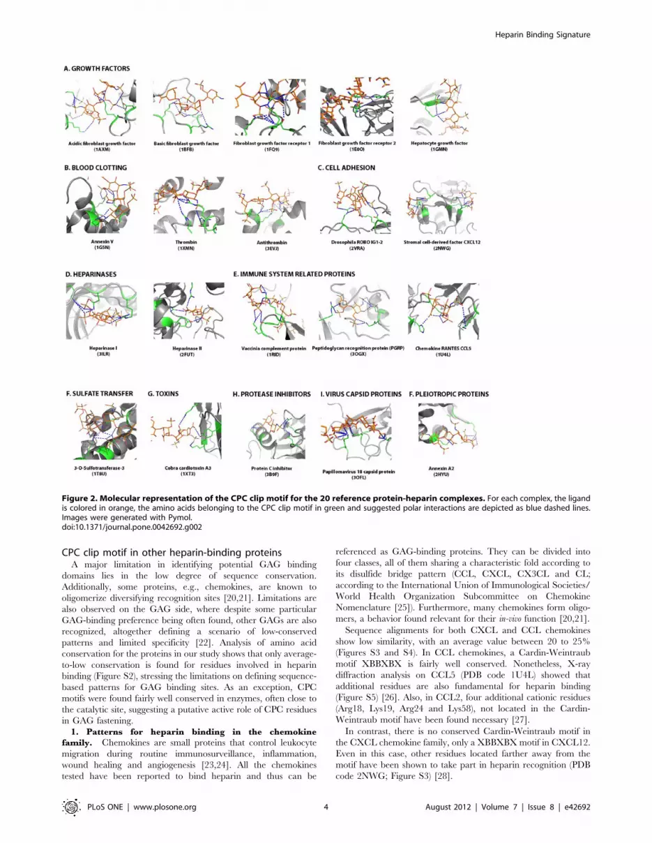
Definition of the CPC clip motifWe have used the PDBePISA server [19] to characterize
hydrogen-bonding contacts in the ligand-protein interfaces of the
discovery dataset. This analysis shows Arg and Lys residues
making the primary hydrogen-bonding contacts. Polar residues,
for their part, could fine-tune the precise recognition of GAGs.
More specifically, detailed inspection of the interacting residues
reveals a conserved pattern that comprises one polar and two
positively charged residues (Figure 2) whose spatial arrangement
allows defining regular distances between cationic (C and C9) and
polar (P) residue a-carbons and side-chain center of gravity
(Figure 3). Average measured distances are 6.061.8 A (PC),
11.661.6 A (PC9) and 11.462.4 A (CC9) for Ca and 6.061.9 A
(PC), 10.661.8 A (PC9) and 10.762.0 A (CC9) for side-chains
center of gravity. Hence our analysis suggests that a structural
rather than a sequence pattern appears to be conserved in
heparin-binding proteins. Additional contacts involving both side-
chain and main-chain atoms can be found in the identified
interfaces. These would provide complementary contacts for a
fuller fastening of heparin. Thus, the cation-polar-cation (CPC)
motif outlined above could be regarded as the minimum structural
requirement for heparin binding in proteins. We should also
consider that heparin-binding sites could be located in monomers
but also in oligomeric interfaces. The CPC motif can thus be
shared by two monomers. For example, in structure 1AXM a
monomeric and a dimeric binding site can be described where one
monomer contains one side of the motif (P and C residues) and the
other the remaining residue (C9).
To test whether heparin-binding proteins are indeed enriched in
the CPC clip motif, we have automatically searched for the motif
in the testing dataset, which contains 48 proteins not related to the
discovery set (24 proteins with experimental evidence of heparin
binding and 24 proteins with no evidence and no expectation to
bind heparin) using the SPASM algorithm. The results obtained
show that, heparin-binding proteins are certainly enriched in the
motif (Figure 3).
Figure 1. Summary of side-chain amino acid interactions for protein-heparin complexes deposited in the PDB. Molecular contactswere inspected for (A) SGN and (B) IDS, the two major components of heparin. The fraction of contacts is represented for each amino acid.doi:10.1371/journal.pone.0042692.g001
Heparin Binding Signature
PLoS ONE | www.plosone.org 3 August 2012 | Volume 7 | Issue 8 | e42692
CPC clip motif in other heparin-binding proteinsA major limitation in identifying potential GAG binding
domains lies in the low degree of sequence conservation.
Additionally, some proteins, e.g., chemokines, are known to
oligomerize diversifying recognition sites [20,21]. Limitations are
also observed on the GAG side, where despite some particular
GAG-binding preference being often found, other GAGs are also
recognized, altogether defining a scenario of low-conserved
patterns and limited specificity [22]. Analysis of amino acid
conservation for the proteins in our study shows that only average-
to-low conservation is found for residues involved in heparin
binding (Figure S2), stressing the limitations on defining sequence-
based patterns for GAG binding sites. As an exception, CPC
motifs were found fairly well conserved in enzymes, often close to
the catalytic site, suggesting a putative active role of CPC residues
in GAG fastening.
1. Patterns for heparin binding in the chemokine
family. Chemokines are small proteins that control leukocyte
migration during routine immunosurveillance, inflammation,
wound healing and angiogenesis [23,24]. All the chemokines
tested have been reported to bind heparin and thus can be
referenced as GAG-binding proteins. They can be divided into
four classes, all of them sharing a characteristic fold according to
its disulfide bridge pattern (CCL, CXCL, CX3CL and CL;
according to the International Union of Immunological Societies/
World Health Organization Subcommittee on Chemokine
Nomenclature [25]). Furthermore, many chemokines form oligo-
mers, a behavior found relevant for their in-vivo function [20,21].
Sequence alignments for both CXCL and CCL chemokines
show low similarity, with an average value between 20 to 25%
(Figures S3 and S4). In CCL chemokines, a Cardin-Weintraub
motif XBBXBX is fairly well conserved. Nonetheless, X-ray
diffraction analysis on CCL5 (PDB code 1U4L) showed that
additional residues are also fundamental for heparin binding
(Figure S5) [26]. Also, in CCL2, four additional cationic residues
(Arg18, Lys19, Arg24 and Lys58), not located in the Cardin-
Weintraub motif have been found necessary [27].
In contrast, there is no conserved Cardin-Weintraub motif in
the CXCL chemokine family, only a XBBXBX motif in CXCL12.
Even in this case, other residues located farther away from the
motif have been shown to take part in heparin recognition (PDB
code 2NWG; Figure S3) [28].
Figure 2. Molecular representation of the CPC clip motif for the 20 reference protein-heparin complexes. For each complex, the ligandis colored in orange, the amino acids belonging to the CPC clip motif in green and suggested polar interactions are depicted as blue dashed lines.Images were generated with Pymol.doi:10.1371/journal.pone.0042692.g002
Heparin Binding Signature
PLoS ONE | www.plosone.org 4 August 2012 | Volume 7 | Issue 8 | e42692

The CPC clip motif is well defined in both CCL5 and CXCL12
crystal structures and appears to be preserved throughout the
whole CCL and CXCL chemokine families (Figures S5 and S6).
Modeling simulations were used for all proteins with no structural
data in the PDB. Residues not included in the Cardin-Weintraub
motif appear well conserved (particularly in the CXCL family),
hence supporting the hypothesis that structural patterns are
actually contributing. Ca distances are also preserved in the case
of CXCL chemokine family, with measured values (6.962.3 A,
12.061.6 A, 14.862.3 A) within the set reference intervals. For
CC chemokines, distances appear to be well preserved although
mean values (10.961.9 A, 17.262.5 A, 23.963.1 A) differ con-
siderably from the reference ones, a deviation that may come from
inherent lack of precision of protein models. Indeed, for CXC
chemokines CPC residues are mainly located in regions with
defined secondary structure whereas in the CC series, CPC
residues are found in highly flexible loops, difficult to evaluate by
in-silico protein modeling.
Lymphotactin (Ltn, XCL1/XCL2), the representative member
of the C chemokine family, involved in the recruitment of T and
NK cells [29], has a single disulfide bond and is conformationally
heterogeneous, switching between a conserved chemokine fold,
named Ltn10 [30], and an unrelated dimeric structure, Ltn40
[31]. This reversible interconversion alters the heparin-binding site
allowing Ltn40 to bind tighter to heparin than Ltn10 [32].
Since a structure for Ltn complexed to heparin or heparin
derivatives is unavailable, we have conducted molecular docking
simulations (using 1HPN heparin dodesaccharide as a ligand) to
find putative heparin binding sites. Our results (Figure 4A,B)
suggest that both, the Ltn10 (PDB code 1J8I) and Ltn40 (PDB
code 2JP1) can tightly bind heparin (Table 1) through a cationic
surface, as described for most chemokines. In this model, Arg and
Lys residues provide the main interactions (Arg23, Lys25, Lys42,
Arg 43, Lys46, Lys66) and one polar residue (Ser22) is close
enough to make hydrogen-bonding contact with the ligand. These
results are supported both by NMR and heparin-sepharose
chromatography. Particularly, Arg23 and Arg43 are the ones
undergoing stronger NMR chemical shifts upon heparin binding
[33] and can be viewed as defining a CPC clip motif together with
the polar residue Ser22 in both folds (Figure 4A,B), with Cadistances (3.8 A, 11.7 A and 8.4 A for Ltn10 and 3.8 A, 11.7 A
and 12.0 A for Ltn40) consistent with reference values.
Finally, the single member of the CX3C chemokine class,
named chemical domain of fractalkine (CDF or CX3CL1) and
involved in the capture and activation of leukocytes [34], binds to
heparin with similar strength as other chemokines and displays a
similar electrostatic surface [35]. There is no structure of CDF
complexed to heparin mimetics, thus we have again resorted to
docking simulations. As shown in Figure 4C, the binding surface of
CDF is highly cationic and comprises several lysine residues,
(Arg37, Arg47, Lys 59). Again, a CPC clip motif can be describedFigure 3. Statistical analysis of the Ca and side chain center ofgravity distances between the amino acids conforming theCPC clip motif. (A) Schematic representation of the CPC clip motif,composed of one polar (P) and two cationic residues (namely C and C9,being C the closest to the polar residue). Image was generated withPymol. (B) Measured PC, PC9 and CC9 distances for the 20 referenceproteins described. (C) Enrichment of CPC clip motif in heparin-bindingproteins. The negative and positive testing databases were analyzed bySPASM and a cumulative frequency histogram plotting the number ofhits per residue is depicted. The positive testing dataset is colored ingreen and the negative dataset in blue. Each point in the negativedataset represents the average of five independent tests and errors barsare depicted. See the Materials and Methods section for furtherinformation.doi:10.1371/journal.pone.0042692.g003
Figure 4. Molecular docking simulation of lymphotactin andfractalkine heparin-binding sites. The figure displays the proteinelectrostatic potential (left) and the protein cartoon highlighting in redthe CPC clip motif (right) of lymphotactin Ltn10 (A) and Ltn40 (B) andCDF fractalkine domain (C). CPC residues are colored in blue (cationic)and magenta (polar). Heparin dodecasaccharide ligand used in dockingsimulations is colored in orange. PDB codes: 1J8I (Ltn10), 2JP1 (Ltn40),1B2T (fractalkine) and 1HPN (heparin ligand).doi:10.1371/journal.pone.0042692.g004
Heparin Binding Signature
PLoS ONE | www.plosone.org 5 August 2012 | Volume 7 | Issue 8 | e42692

in this surface (Table 1), comprising residues Gln31, Arg37 and
Arg47, with measured distances 7.7 A, 11.6 A and 16.8 A.
In summary, the CPC clip motif has been found to correctly
describe the heparin-binding sites of chemokines, providing
additional clues on the characterization of discontinuous motives.
2. New insights into the heparin-binding site of human
amyloid b protein. The extracellular accumulation of amyloid
b proteins in neuritic plaques is one of the hallmarks of
Alzheimer’s disease [36]. Amyloid Ab protein can bind to many
macromolecules, including heparin. Although Ab can self-aggre-
gate to form amyloid fibrils in vitro, its binding to heparin enhances
amyloid aggregation and fibril formation. It has been shown that
the sulfate moiety is necessary for the growing of amyloid
aggregates, as no fibrils are observed in the presence of hyaluronic
acid (HA), a non-sulfated GAG [37]. Low-molecular-weight
heparins (LMWHs) can reverse the process of amyloidosis, by
inhibiting fibril formation and blocking the formation of b-plated
structures, underlining a possible therapeutic approach [38].
We have assessed the binding surface of Ab to heparin by
molecular docking simulation on two crystallized fragments of Ab(Ab28–123, PDB code 1MWR and Ab460–569, PDB code
1TKN), both found to bind heparin [39,40]. As in the case of
chemokines and other heparin-binding proteins, both regions are
highly cationic (Figure 5).
In Ab28–123, the main interacting residues correlate with a
Cardin-Weintraub motif XBBXBX (namely 98CKRGRK103)
found critical for heparin binding [39]. However, other polar
residues that contribute to heparin binding (Asn46, His44, Ser54,
Ser57 and Thr59) are also found, and a CPC clip motif defined by
residues Thr59, Arg100 and Arg102, with distance values of
5.7 A, 8.0 A and 6.0 A can be proposed for this region, the last
value however found out from the reference values.
Our docking studies also suggest that the Ab460–569 region,
reported as crucial for heparin binding [40], contains a CPC clip
motif defined by residues Arg468, Asn475 and Lys496 (distances
8.9 A, 10.3 A and 10.2 A). Other residues in this region found to
interact are Thr478, His489, Arg 495, Arg499, Lys503 and Lys510.
These residues do not define any Cardin-Weintraub motif, despite
the importance of the region for heparin binding. In particular, our
docking results suggest that the binding site located in the Ab460–
569 region would bind tightly heparin whereas the site located in
Ab28–123 could be a complementary binding site (Table 1).
Searching for other proteins containing the CPC clipmotif
By means of the SPASM algorithm, we have analyzed the PDB
database to find the described CPC motif in proteins reported to
bind heparin but for which no structural information on their
binding domain is available. SPASM uses a fast search process
based on differences between atomic positions. We have analyzed
the five canonic CPC motifs identified (i.e., C-Asn-C, C-Gln-C, C-
Ser-C, C-Thr-C and C-Tyr-C) to find other proteins potentially
able to bind heparin. However, detection of small structural motifs
is complex and sometimes lacks specificity due to the size of the
database. To further refine our search, SAVANT has been used to
perform an all-atom least squares superpositioning of the CPC
pattern and the SPASM hits. We used CD-HIT to avoid including
Table 1. Average binding energies for the docked heparin-protein complexesa.
PDB code CPC motif Average free energy of binding (kcal/mol)b
1F2L Gln31, Arg37, Arg47 29.25/25.95
1J8I Ser22, Arg23, Arg43 218.41/28.29
1MWP Thr59, Arg100, Arg102 24.22/24.91
1TKN Asn475, Arg468, Lys496 29.95/29.21
2JP1 Ser22, Arg23, Arg43 220.04/27.54
aAll docking simulations have been carried out using Autodock with a heparin dodesaccharide or disaccharide allowing ten or two degrees of freedom respectively(Figure S1). See Materials and Methods section for further information.bFirst value refers to dodesaccharide docking whereas second value refers to the disaccharide.doi:10.1371/journal.pone.0042692.t001
Figure 5. Molecular docking simulation of Ab28–123 andAb460–569 heparin-binding sites. The figure displays the proteinelectrostatic potential (left) and the protein cartoon highlighting in redthe CPC clip motif (right) of (A) Ab28–123 and (B) Ab460–569. CPCresidues are colored in blue (cationic) and magenta (polar). Heparindodecasaccharide ligand used in docking simulations is colored inorange. PDB codes: 1MWR (Ab28–123), 1TKN (Ab460–569) and 1HPN(heparin ligand).doi:10.1371/journal.pone.0042692.g005
Heparin Binding Signature
PLoS ONE | www.plosone.org 6 August 2012 | Volume 7 | Issue 8 | e42692

similar proteins and PANTHER to analyze and classify the
function of proteins identified by SPASM. From these proteins, the
main portion is related to binding proteins and enzymes (.75%).
Around 20% are related to primary metabolism, from which a
40% is dedicated to metabolites containing sugars derivatives.
Regarding function, many of them have characteristic roles found
in heparin-binding proteins like cell communication, adhesion
and/or proliferation (Figure S7).
While the above method could be useful to detect new proteins
with no previously reported heparin binding, it tends to unveil a
large number of candidates that require subsequent filtering, and
therefore is only suitable as a complementary searching tool. In
conclusion, we have found a structural motif conserved in heparin-
binding proteins that provide a cationic surface environment to fix
heparin. This motif would act as a staple for polymeric GAG
substrates and provides useful clues on why GAG binding proteins
display considerable sequence diversity.
Supporting Information
Figure S1 Representation of heparin dodesaccharide(A, PDB code 1HPN) and disaccharide (B, PDB code1U4M) molecules used in docking simulations. Allowed
torsions in the simulation are colored in red whereas fixed bonds
are colored in green.
(JPG)
Figure S2 Representation of the amino acid conserva-tion for the 20 reference protein-heparin complexes.Ligands are colored in orange and amino acid residues are colored
by conservation as depicted in the scale provided at the bottom of
the image; residues were colored in yellow when now enough
information was available. Images were computed using Consurf
and generated with Pymol.
(JPG)
Figure S3 Sequence alignment of human CXCL chemo-kines. Conserved residues are colored in red; the putative
XBBXBX motif is highlighted in orange. Green arrows indicate
the two CPC clip motives found in CXCL chemokines.
(JPG)
Figure S4 Sequence alignment of human CCL chemo-kines. Conserved residues are colored in red; the putative
XBBXBX motif is highlighted in orange. Green arrows indicate
the CPC clip motif found in CCL chemokines.
(JPG)
Figure S5 Molecular representation of the CPC clipmotif for the CXCL chemokine complexes. For each
complex, the ligand is colored in blue, the amino acids belonging
to the reference CPC clip motif (CXCL12; PDB code 2NWG) are
colored in red and the CPC residues corresponding to the modeled
chemokine are colored in green. Residues not matching with the
CPC description are colored in olive green. Images were
generated with Pymol.
(JPG)
Figure S6 Molecular representation of the CPC clipmotif for the CCL chemokine complexes. For each
complex, the ligand is colored in blue, the amino acids belonging
to the reference CPC clip motif (CCL5; PDB code 1U4L) are
colored in red and the CPC residues corresponding to the modeled
chemokine are colored in green. Residues not matching with the
CPC description are colored in olive green. Images were
generated with Pymol.
(JPG)
Figure S7 Function distribution of the genes that encodefor the putative heparin-binding regions detected bySPASM. The list of proteins obtained by SPASM (PDB codes)
was translated to a SwissProt identifier list using PICR (http://
www.ebi.ac.uk/Tools/picr/). The database generated was in-
spected using PANTHER (http://www.pantherdb.org/) and the
genes identified were classified by GO annotation.
(JPG)
Table S1 List of proteins included in the reference dataset.
(DOCX)
Author Contributions
Conceived and designed the experiments: MT EB. Performed the
experiments: MT. Analyzed the data: MT MVN DA EB. Contributed
reagents/materials/analysis tools: DA EB. Wrote the paper: MT MVN
DA EB.
References
1. Gandhi NS, Mancera RL (2008) The structure of glycosaminoglycans and their
interactions with proteins. Chem Biol Drug Des 72: 455–482.
2. Peplow PV (2005) Glycosaminoglycan: a candidate to stimulate the repair of
chronic wounds. Thromb Haemost 94: 4–16.
3. Casu B, Guerrini M, Torri G (2004) Structural and conformational aspects of
the anticoagulant and anti-thrombotic activity of heparin and dermatan sulfate.
Curr Pharm Des 10: 939–949.
4. Kovensky J (2009) Sulfated oligosaccharides: new targets for drug development?
Curr Med Chem 16: 2338–2344.
5. Liu D, Shriver Z, Qi Y, Venkataraman G, Sasisekharan R (2002) Dynamic
regulation of tumor growth and metastasis by heparan sulfate glycosaminogly-
cans. Semin Thromb Hemost 28: 67–78.
6. Kisilevsky R, Ancsin JB, Szarek WA, Petanceska S (2007) Heparan sulfate as a
therapeutic target in amyloidogenesis: prospects and possible complications.
Amyloid 14: 21–32.
7. Young E (2008) The anti-inflammatory effects of heparin and related
compounds. Thromb Res 122: 743–752.
8. Rider CC (1997) The potential for heparin and its derivatives in the therapy and
prevention of HIV-1 infection. Glycoconj J 14: 639–642.
9. Mulloy B, Linhardt RJ (2001) Order out of complexity–protein structures that
interact with heparin. Curr Opin Struct Biol 11: 623–628.
10. Hileman RE, Fromm JR, Weiler JM, Linhardt RJ (1998) Glycosaminoglycan-
protein interactions: definition of consensus sites in glycosaminoglycan binding
proteins. Bioessays 20: 156–167.
11. Raghuraman A, Mosier PD, Desai UR (2006) Finding a needle in a haystack:
development of a combinatorial virtual screening approach for identifying high
specificity heparin/heparan sulfate sequence(s). J Med Chem 49: 3553–3562.
12. Cardin AD, Weintraub HJ (1989) Molecular modeling of protein-glycosamino-
glycan interactions. Arteriosclerosis 9: 21–32.
13. Capila I, Linhardt RJ (2002) Heparin-protein interactions. Angew Chem Int Ed
Engl 41: 391–412.
14. Fromm JR, Hileman RE, Caldwell EE, Weiler JM, Linhardt RJ (1997) Pattern
and spacing of basic amino acids in heparin binding sites. Arch Biochem Biophys
343: 92–100.
15. Kleywegt GJ (1999) Recognition of spatial motifs in protein structures. J Mol
Biol 285: 1887–1897.
16. Madsen D, Kleywegt GJ (2001) Interactive motif and fold recognition in protein
structures. J Appl Cryst 35: 137–139.
17. Thompson LD, Pantoliano MW, Springer BA (1994) Energetic characterization
of the basic fibroblast growth factor-heparin interaction: identification of the
heparin binding domain. Biochemistry 33: 3831–3840.
18. Bae J, Desai UR, Pervin A, Caldwell EE, Weiler JM, et al. (1994) Interaction of
heparin with synthetic antithrombin III peptide analogues. Biochem J 301 (Pt 1):
121–129.
19. Krissinel E, Henrick K (2007) Inference of macromolecular assemblies from
crystalline state. J Mol Biol 372: 774–797.
20. Handel TM, Johnson Z, Crown SE, Lau EK, Proudfoot AE (2005) Regulation
of protein function by glycosaminoglycans–as exemplified by chemokines. Annu
Rev Biochem 74: 385–410.
Heparin Binding Signature
PLoS ONE | www.plosone.org 7 August 2012 | Volume 7 | Issue 8 | e42692

21. Weber C, Koenen RR (2006) Fine-tuning leukocyte responses: towards a
chemokine ‘interactome’. Trends Immunol 27: 268–273.
22. Raman R, Sasisekharan V, Sasisekharan R (2005) Structural insights into
biological roles of protein-glycosaminoglycan interactions. Chem Biol 12: 267–
277.
23. Mackay CR (2001) Chemokines: immunology’s high impact factors. Nat
Immunol 2: 95–101.
24. Sallusto F, Baggiolini M (2008) Chemokines and leukocyte traffic. Nat Immunol
9: 949–952.
25. Nomenclature IUoISWHOSoC (2001) Chemokine/chemokine receptor no-
menclature. J Leukoc Biol 70: 465–466.
26. Shaw JP, Johnson Z, Borlat F, Zwahlen C, Kungl A, et al. (2004) The X-ray
structure of RANTES: heparin-derived disaccharides allows the rational design
of chemokine inhibitors. Structure 12: 2081–2093.
27. Lau EK, Paavola CD, Johnson Z, Gaudry JP, Geretti E, et al. (2004)
Identification of the glycosaminoglycan binding site of the CC chemokine,
MCP-1: implications for structure and function in vivo. J Biol Chem 279:
22294–22305.
28. Murphy JW, Cho Y, Sachpatzidis A, Fan C, Hodsdon ME, et al. (2007)
Structural and functional basis of CXCL12 (stromal cell-derived factor-1 alpha)
binding to heparin. J Biol Chem 282: 10018–10027.
29. Yoshida T, Imai T, Kakizaki M, Nishimura M, Takagi S, et al. (1998)
Identification of single C motif-1/lymphotactin receptor XCR1. J Biol Chem
273: 16551–16554.
30. Kuloglu ES, McCaslin DR, Kitabwalla M, Pauza CD, Markley JL, et al. (2001)
Monomeric solution structure of the prototypical ‘C’ chemokine lymphotactin.
Biochemistry 40: 12486–12496.
31. Tuinstra RL, Peterson FC, Kutlesa S, Elgin ES, Kron MA, et al. (2008)
Interconversion between two unrelated protein folds in the lymphotactin nativestate. Proc Natl Acad Sci U S A 105: 5057–5062.
32. Volkman BF, Liu TY, Peterson FC (2009) Chapter 3. Lymphotactin structural
dynamics. Methods Enzymol 461: 51–70.33. Peterson FC, Elgin ES, Nelson TJ, Zhang F, Hoeger TJ, et al. (2004)
Identification and characterization of a glycosaminoglycan recognition elementof the C chemokine lymphotactin. J Biol Chem 279: 12598–12604.
34. Haskell CA, Cleary MD, Charo IF (1999) Molecular uncoupling of fractalkine-
mediated cell adhesion and signal transduction. Rapid flow arrest of CX3CR1-expressing cells is independent of G-protein activation. J Biol Chem 274: 10053–
10058.35. Hoover DM, Mizoue LS, Handel TM, Lubkowski J (2000) The crystal structure
of the chemokine domain of fractalkine shows a novel quaternary arrangement.J Biol Chem 275: 23187–23193.
36. Herrup K (2010) Reimagining Alzheimer’s disease–an age-based hypothesis.
J Neurosci 30: 16755–16762.37. Valle-Delgado JJ, Alfonso-Prieto M, de Groot NS, Ventura S, Samitier J, et al.
(2010) Modulation of Abeta42 fibrillogenesis by glycosaminoglycan structure.FASEB J 24: 4250–4261.
38. Ariga T, Miyatake T, Yu RK (2010) Role of proteoglycans and glycosaminoglycans
in the pathogenesis of Alzheimer’s disease and related disorders: amyloidogenesisand therapeutic strategies–a review. J Neurosci Res 88: 2303–2315.
39. Small DH, Nurcombe V, Reed G, Clarris H, Moir R, et al. (1994) A heparin-binding domain in the amyloid protein precursor of Alzheimer’s disease is
involved in the regulation of neurite outgrowth. J Neurosci 14: 2117–2127.40. Wang Y, Ha Y (2004) The X-ray structure of an antiparallel dimer of the human
amyloid precursor protein E2 domain. Mol Cell 15: 343–353.
Heparin Binding Signature
PLoS ONE | www.plosone.org 8 August 2012 | Volume 7 | Issue 8 | e42692