Thoracic Endovascular Aortic Repair (TEVAR) First in Patients ...
Upload
independentCategory
view
5download
0
Heparanase Alters Arterial Structure, Mechanics and RepairFollowing Endovascular Stenting in Mice
Aaron B. Baker1, Adam Groothuis1, Michael Jonas1,2, David S. Ettenson1, Tarek Shazly1,Eyal Zcharia3, Israel Vlodavsky3, Philip Seifert1, and Elazer R. Edelman1,21 Harvard-MIT Division of Health Sciences and Technology, Massachusetts Institute of Technology,Cambridge, MA2 Cardiovascular Division, Brigham and Women’s Hospital, Harvard Medical School, Boston, MA3 Cancer and Vascular Biology Research Center, Bruce Rappaport Faculty of Medicine, Technion,PO Box 9649, Haifa, 31096, Israel
AbstractHeparan sulfate proteoglycans (HSPGs) are potent regulators of vascular remodeling and repair.Heparanase is the major enzyme capable of degrading heparan sulfate in mammalian cells. Here weexamined the role of heparanase in controlling arterial structure, mechanics and remodeling. In vitrostudies supported that heparanase expression in endothelial cells serves as a negative regulator ofendothelial inhibition of vascular smooth muscle cell (vSMC) proliferation. Arterial structure andremodeling to injury were also modified by heparanase expression. Transgenic mice overexpressingheparanase had increased arterial thickness, cellular density and mechanical compliance.Endovascular stenting studies in Zucker rats demonstrated increased heparanase expression in theneointima of obese, hyperlipidemic rats in comparison to lean rats. The extent of heparanaseexpression within the neointima strongly correlated with the neointimal thickness following injury.To test the effects of heparanase overexpression on arterial repair, we developed a novel murinemodel of stent injury using small diameter self-expanding stents. Using this model we found thatincreased neointimal formation and macrophage recruitment occurs in transgenic miceoverexpressing heparanase. Taken together, these results support a role for heparanase in theregulation of arterial structure, mechanics and repair.
Keywordsheparanase; vascular remodeling; restenosis; stenting; arterial compliance
IntroductionVascular smooth muscle cell (vSMC) proliferation and hypertrophy are commonpathophysiological mechanisms underlying clinical cardiovascular disorders includinghypertension, atherosclerosis and restenosis1–3. Arterial structure is maintained by a dynamicinterplay between growth inhibitory factors produced primarily by endothelial cells and growthstimulatory factors produced principally by vSMCs, inflammatory cells, and dysfunctionalendothelial cells4. Disease states can compromise endothelial function and disturb this balance
Correspondence to: Aaron B. Baker, Massachusetts Institute of Technology, 77 Massachusetts Avenue, Building E25, Room 442,Cambridge, MA 02139, [email protected]: None.
NIH Public AccessAuthor ManuscriptCirc Res. Author manuscript; available in PMC 2010 February 13.
Published in final edited form as:Circ Res. 2009 February 13; 104(3): 380–387. doi:10.1161/CIRCRESAHA.108.180695.
NIH
-PA Author Manuscript
NIH
-PA Author Manuscript
NIH
-PA Author Manuscript

leading to local inflammation, thrombosis and vasoconstriction5. Heparan sulfateproteoglycans (HSPGs) derived from endothelial cells are potent regulators of vSMC growthand vascular remodeling6, 7. Both heparin and endothelial cell-derived HSPGs are potentinhibitors of vSMC proliferation and mitogenesis8–10. This regulation is dependent on theoverall health and growth state of the endothelial cells. Subconfluent cultures of endothelialcells stimulate vSMC growth whereas postconfluent cultures inhibit vSMC growth6. Further,these endothelial-derived HSPGs are essential to inhibiting and resolving the neointimalresponse to vascular injury7.
While there is evidence that HSPGs participate in the control of vascular remodeling, it remainsunclear how these molecules are regulated in context of disease and injury. Heparanase is themajor mammalian enzyme capable of digesting heparan sulfate chains. This enzyme is an endo-β-D-glucuronidase that cleaves at a specific motif in the heparan sulfate chains to createfragments 10–20 sugar units long and biologically active11, 12. Heparanase has been intenselystudied for its role in angiogenesis and cancer metastasis13, 14, yet the role of this enzyme inregulating arterial structure and remodeling remains poorly defined. We hypothesized thatheparanase plays a key role in regulating arterial structure, mechanics and vascular remodeling.In the present work, we show that heparanase expression in endothelial cells serves as anegative feedback regulator of paracrine inhibition of vSMC proliferation. Miceoverexpressing the human heparanase transgene had increased arterial thickening, cellularityand arterial compliance. We found that stent-induced vascular injury in obese, hyperlipidemicrats had increased neointimal heparanase expression and that the magnitude of this increasedirectly related to the extent of neointimal expansion. Finally, we developed a novel minimally-invasive murine arterial stenting model that demonstrated increased neointimal formation inresponse to endovascular stenting in transgenic heparanase mice.
Materials and MethodsCell Culture
Human aortic vSMCs and human umbilical cord vascular endothelial cell (HUVEC) lines werepurchased from Cambrex (Walkersville, MD). Cells were maintained in MCDB-131 media(Invitrogen, Carlsbad, CA) supplemented with EGM-2 growth supplements (Cambrex). Cellswere cultured at 37°C under 5% CO2 and were used between passages three and five.
Transfection and siRNA VectorsFour short hairpin RNA expression vectors were screened for gene silencing activity towardsthe heparanase gene. The following sequences were used in the pRS expression vector(Origene, Rockville, MD): (1) TTATGTGGCTGGATAAATTGGGCCTGTCA, (2)GTGGTGATGAGGCAAGTATTCTTTGGAGC, (3)TCGTTCCTGTCCGTCACCATTGACGCCAA, (4)GTTCAAGAACAGCACCTACTCAAGAAGCT. Overexpression of heparanase andsyndecan was performed using vectors with constitutive gene expression under control of theCMV promoter (Origene).
Cell Lysis and Western BlottingCells were lysed in 1 ml of lysis buffer containing 20 mM Tris, 150 mM NaCl, 1% TritonX-100, 1% deoxycholate, 0.1% SDS, 1 mM sodium orthovanadate, 50mM NaF, 2 mM PMSF,and complete protease inhibitor cocktail (Roche, Nutley, NJ). After 10 minutes of incubation,the plates were scraped and the lysates pipetted into a centrifuge tube. The samples were thencentrifuged at 14,000 g for 15 min prior to western blotting. Samples were run on 4–15%polyacrylamide gradient gels and transferred to PVDF membranes. The membranes wereblocked for 1 hr in 5% non-fat milk in PBS with 0.01% tween-20 (PBST) and exposed to a
Baker et al. Page 2
Circ Res. Author manuscript; available in PMC 2010 February 13.
NIH
-PA Author Manuscript
NIH
-PA Author Manuscript
NIH
-PA Author Manuscript

heparanase primary antibody (1:200, Cell Sciences, Canton, MA) at 4°C overnight in 1% non-fat milk. The membranes were washed with PBST, incubated at room temperature for 2 hrswith a 1:3500 dilution of a horse radish peroxidase linked secondary antibody and detectedusing chemiluminescence (Perkin Elmer, Boston, MA).
Immunocytochemical StainingCells were washed three times in PBS and fixed for 5 min with methanol at −20°C. The cultureswere then blocked with 20% goat serum in PBS for 45 min at room temperature. A primaryantibody heparanase (Cell Science, Canton, MA) was added at 1:100 dilution in PBS containing1% BSA. A secondary antibody conjugated with Alexa Fluor 594 or Oregon Green dyes(Invitrogen) was added at 1:200 dilution in PBS-BSA solution. The plates were thencoverslipped, mounted using a DAPI containing mounting medium (Vector Laboratories,Burlingame, CA) and visualized with fluorescence microscopy.
Metabolic Labeling, Isolation and Analysis of ProteoglycansTo metabolically label the glycosaminoglycans, 80 μCi of 3H-glucosamine was added to eachplate. The conditioned media was collected and combined with guanidine-HCl to a finalconcentration of 4 M. The cell layers were then washed three times with cold PBS and lysed.This solution was then brought to a final concentration of 4 M guanidine-HCl. To obtainextracellular matrix bound proteoglycans, the plates were washed three times in PBS andextracted for 48 hrs with a solution of 50 mM sodium acetate (pH = 6.0), 4 M guanidine-HCl,2% triton x-100 and protease inhibitors.
The isolated proteoglycans from conditioned media, extracellular matrix and cell surface weredesalted into Buffer A (20 mM Tris, 8 M Urea, pH = 8.0) using a HiTrap desalting column(Amersham Biosciences, Piscataway, NJ). The proteoglycans were separated from otherproteins by fractionation on a 1 ml HiTrap Q ion exchange column (Amersham) with a linearsalt gradient from 0 to 2 M NaCl. One ml fractions were collected and aliquots of these sampleswere counted using liquid scintillation. Aliquots were subjected to digestion with 0.6 U/ml ofprotease free chondroitinase ABC (Seikagaku, Japan) for 4 hours. Control samples weresubjected to digestion conditions without the addition of enzyme.
Immunohistochemical StainingThe abdominal aorta from rats was formalin fixed and sectioned using standard methods. Thesections were heated for ten minutes in a 60°C oven, deparaffinized in xylene, and rehydrated.Antigen retrieval was performed by placing the slides in 10mM citrate buffer (pH = 6.0) andheating in the microwave for 10 min. The samples were allowed to cool for 20 min and werethen incubated in 3% hydrogen peroxide for 10 min. The samples were rinsed 3 times withPBS with 0.01% tween-20 (PBST) between each of the following steps. The sections wereblocked with 20% normal goat serum for 45 minutes at room temperature. Primary antibodieswere diluted in PBS containing 1% BSA, applied to slides, and incubated in a humid chamberovernight at 4°C. Secondary antibody staining at detection was performed using the LSAB 2kit (DakoCytomation, Carpinteria, CA) according to the manufacturer’s instructions. An AECsubstrate (DakoCytomation) was used for detection of the HRP conjugate. The samples werecounterstained in Mayer’s hematoxylin for 3 min, washed with tap water, and mounted inaqueous mounting medium (DakoCytomation).
Histochemical AnalysisAortae were harvested from wild type and heparanase transgenic mice. The aortae were fixedin neutral buffered formalin at 4°C overnight, paraffin embedded and sectioned using standardmethods. The aortae were stained with hemotoxylin and eosin or with an elastin Movat stain
Baker et al. Page 3
Circ Res. Author manuscript; available in PMC 2010 February 13.
NIH
-PA Author Manuscript
NIH
-PA Author Manuscript
NIH
-PA Author Manuscript

as previously described15. Arterial thickness, circumference and nuclei were counted on threeaortic sections from four mice for each group of animals using Adobe Photoshop (Adobe, SanJose, CA).
Smooth Muscle Cell Proliferation AssayHuman smooth muscle cells were passaged into 48-well plates at low density. Smooth musclecells were serum starved in 0.5% calf serum for 24 hours. The cells were then washed twicein culture media with no growth supplements and endothelial cell conditioned media wasapplied. After 24 hours 1 μCi of 3H-thymidine was added. Twenty-four hours later, the cellswere washed three times with PBS at 4°C and then incubated with 10% TCA for 30 min. Thecultures were then washed twice in 95% ethanol and solubilized in 1 ml of 0.25 M NaOH with0.1% SDS for 1 hour. The samples were added to scintillation cocktail and radioactivity wasmeasured using a liquid scintillation counter.
Animal Models of Endovascular Stent InjuryAll experimental procedures and protocols used in this investigation were reviewed andapproved by the Animal Care and Use Committee of the Massachusetts Institute of Technologyand conformed to the “Guiding Priniciples in the Care and Use of Animals” of the AmericanPhysiological Society and the NIH Guide for the Care and Use of Laboratory Animals. Zuckerobese (Crl: (ZUC)-faBR) and Zucker lean rats were used in an animal model of vascular injuryand stenting in the presence of metabolic syndrome and insulin resistance16. At the time ofstenting the rats were 12 and 14 weeks old for the obese and lean rats, respectively. The ratswere anesthetized using isofluorane, given a 100 U/kg dose of heparin, and a small incisionwas made to expose the right femoral artery. This artery was ligated and an arteriotomy wasperformed proximal to the ligature. A 0.014” angioplasty guidewire was passed into the aortaand an 9-mm long endovascular stent (Nirflex; Medinol Inc., Tel Aviv, Israel) mounted on a15 × 2.5-mm angioplasty balloon (Crossail; Guidant Inc., Santa Clara, CA) was passed intothe abdominal aorta. The stent was deployed with a 15 second inflation at 8 atm inflationpressure. Post-stenting the animals were given aspirin via drinking water at an approximatedose of 5 mg/kg/day16. After 14 days the stents were harvested for analysis.
Wild-type and heparanase transgenic mice were derived directly from those previouslydescribed17. The animals were given aspirin via drinking water for 24 hrs prior to stentimplantation and thereafter. Immediately prior to stenting the mice were given a subcutaneousdose of heparin. Vascular access was obtained through the right femoral artery in a mannersimilar to that used for the rat studies. A coronary stent system with a 1.5 mm constraineddiameter and 12 mm length was used (CardioMind, Sunnyvale, CA). The catheter was passedinto the abdominal aorta through the femoral artery. The stent was deployed by removing anexternal sheath, allowing the self expanding stent to deploy. The average diameter of the aorticlumen in the mice was 1.3 mm and the deployed stent diameter is 1.5 mm, leading to 15.4%dilation in on stent deployment. After seven days the stents were explanted and analysed byhistochemical and immunochemical staining as previously described16, 18. To ensure that thedifferences in response to injury were not a result of altered blood pressure, the arterial bloodpressure in the mice was measured using a tail cuff blood pressure monitor (Holliston, MA).The average blood pressure for each group was 113.6±4.2 and 106.8±3.6 mmHg for wild typeand heparanase transgenic mice, respectively (n = 4, p = ns).
Mechanical Testing of AortaePrior to mechanical testing aortae from heparanase transgenic and wild-type mice wereharvested and adventitial tissue removed. The longitudinal compliance and ultimate tensilestrength of whole arteries was measured using an ElectroForce Biodynamic Test Instrument(Bose Corporation, Framingham, MA). The samples were measured in uniaxial tensile
Baker et al. Page 4
Circ Res. Author manuscript; available in PMC 2010 February 13.
NIH
-PA Author Manuscript
NIH
-PA Author Manuscript
NIH
-PA Author Manuscript

configuration using a 22 N load cell (Interface Inc., Scottsdale, AZ) with a displacement rateof 0.05 mm/s and maximal displacement of 4.0 mm. WinTest 3.0 software (Bose) was used torecord data during the testing. Circumferential testing was performed on aortic ring samplescannulated with two sutures and tested using the Biodynamic Test Instrument. To examinemechanical properties of the elastin networks we digested aortae in 0.1N NaOH at 75°C for 1hour. The solution was neutralized and removed with multiple washes with PBS. Thisprocedure has been shown to remove cellular components and extracellular matrix but leavethe elastin network intact19.
StatisticsAll results are shown as mean ± standard error of the mean. An ANOVA followed by Student-Newman–Keuls post hoc test. A two-tailed p value <0.05 was considered statistical significantwas used to make comparisons between groups of continuous variables. The Pearson productmoment correlation statistic was used as a measure of correlation between variables.
ResultsHeparanase provides negative feedback control to paracrine growth inhibition of vSMCs byendothelial cells
Endothelial cells were transfected with an expression vector for heparanase (pHPA), one offour siRNA vectors targeted to the heparanase (siHPA-1 to -4), a siRNA control vector (pRS)or an empty overexpression vector (pcDNA). After 2 days in culture, the cells were lysed andassayed for heparanase by immunoblotting to confirm alterations in heparanase expression(Figure 1a). The most effective siRNA sequence (siHPA-3) was selected and used in furtherstudies. This siRNA reduced heparanase expression to 13.2±7.2% from 100.0±18.7% forcontrol cells (n=3, p<0.05). Transfected cells under similar conditions were fixed,immunostained for heparanase and imaged using fluorescent microscopy (Figure 1b and Figure1c). The effect of heparanase on the glycosaminoglycans of endothelial cells was measuredthrough metabolic labeling and analysis by ion exchange chromatography. In these graphs theearly peaks represent predominantly protein while later peaks are from highly chargedglycosaminoglycans (i.e. fractions 9 through 20). These studies demonstrated that enhancedendothelial expression of heparanase led to reduction in both cell surface and soluble heparansulfate glycosaminoglycans (Figure 1d). For the surface HSPGs the relative area of theglycosaminoglycan peak was 100.0±6.4%, 36.2±3.3% and 154.9±4.9% for pRS, pHPA andsiHPA transfected cells, respectively (p<0.05 for comparisons between all groups).Conversely, reduction of heparanase expression increased heparan sulfate in cell surface andconditioned media fractions. Notably, there was also a large reduction in cell surface proteinwith heparanase overexpression, likely due to heparanase-induced shedding of syndecans20.
Endothelial cells produce soluble factors that inhibit vSMC proliferation. This process isessential for arterial health and requires endothelial cell derived HSPGs6, 7. We tookconditioned media from endothelial cells and applied it to vSMCs in culture to examine therole of heparanase in controlling endothelial inhibition of vSMC proliferation. Endothelial cellstransfected with a control vector inhibited vSMC proliferation to about half of the baselinevSMC proliferation in growth media (Figure 1e). This effect was absent in cells overexpressingheparanase and markedly enhanced in cells with reduced heparanase levels (Figure 1e).Previous studies have implicated the release of soluble factors, in particular FGF-2, as aneffector mechanism of heparanase tissue action21, 22. However, in our experiments FGF-2levels remained relatively constant irrespective of heparanase expression (data not shown).
Baker et al. Page 5
Circ Res. Author manuscript; available in PMC 2010 February 13.
NIH
-PA Author Manuscript
NIH
-PA Author Manuscript
NIH
-PA Author Manuscript

Heparanase overexpression leads to arterial thickening, increased arterial compliance andincidence of spontaneous aneurysm
Transgenic mice overexpressing heparanase were used to examine the effects of heparanaseexcess on arterial structure and mechanics. The aortae of mice with the heparanase transgenehad increased medial thickness in comparison to wild-type controls (Figure 2a and 2b). Thisthickening appeared to be due to enhanced cellular density rather than hypertrophy in theheparanase transgenic animals when compared to their wild-type counterparts (Figure 2c).Aortae were harvested from both groups and mechanical properties of the aorta were measuredusing a mechanical testing device with environmental control. Heparanase overexpression ledto a profound alteration in the the force-displacement relationship of the aorta. This led to anearly three-fold reduction in longitudinal arterial stiffness (Figure 3a) and ultimate tensilestrength (Figure 3b). We also measured circumferential stiffness and ultimate strength andfound a similar reduction in arterial mechanical properties for the heparanase transgenic mice(Figure 3c and 3d). To test whether heparanase expression affected the integrity of the elastinnetwork, arteries were digested with NaOH at elevated temperature. This treatment has beenshown to destroy most cellular and extracellular matrix components while leaving the elastinnetwork intact. We mechanical tested the remaining elastin networks and found that theheparanase transgenic mice had elastin with dramatically reduced mechanical integrity (Figure3e and 3f). Consistent with the reduced stiffness and reduction in ultimate strength, localizedspontaneous aneurysms were found in the transgenic heparanase mice but not in the wild type(Figure 3g).
Heparanase expression is increased in stent injury on obese, hyperlipidemic Zucker rats andis associated with increased intimal thickness
In the clinical setting, diabetic patients are at increased risk of atherosclerosis and restenosiswith both bare metal and drug-eluting stents23–25. We examined the expression of heparanasein stent induced restenosis by implanting stents in the aortae of normal and obese Zucker ratsusing minimally invasive femoral access surgery16. The obese Zucker rat is a model of type IIdiabetes with obesity, insulin resistance, hyperinsulinemia, hypertriglyceridemia andhypercholesterolemia. Following stent injury and restenosis we found that obese Zucker ratshad increased neointimal thickness and expression of heparanase (Figure 4a–4c). Theexpression of heparanase within the neointima correlated strongly with the final neointimalarea, giving a correlation of R=0.50 (p < 0.001) for lean rats and R=0.77 (p < 0.001) for fattyrats (Figure 4d).
Heparanase overexpression enhances neointimal proliferation and macrophage recruitmentin response to endovascular stent implantation
To examine arterial repair in the presence of heparanase overexpression we developed aminimally invasive method of stent placement in mice. Using a novel small diameter stentingsystem (CardioMind, Sunnyvale, CA) we performed minimally invasive endovascular stentingof transgenic mice overexpressing heparanase. This method takes advantage of self expandingstents that have a diameter identical to that of the catheter wire prior to deployment. Nitinolself expanding stents were deployed in the abdominal aorta of wild type and heparanasetransgenic mice via femoral access (Figure 5a). In the heparanase transgenic mouse theneointimal lesion was rich in macrophages, staining strongly for the Mac-1 surface marker(Figure 5b). Heparanase transgenic mice had markedly increased neointimal formation withincreased neointimal area (Figure 5c) and intima to media ratio (Figure 5d) with no significantchange in medial area (Figure 5e). We examined release of monocyte chemoattractant protein-1(MCP-1) one hour after vascular injury and found increased amounts of MCP-1 in theheparanase transgenic mice (Figure 5f).
Baker et al. Page 6
Circ Res. Author manuscript; available in PMC 2010 February 13.
NIH
-PA Author Manuscript
NIH
-PA Author Manuscript
NIH
-PA Author Manuscript

DiscussionDespite the discovery of heparanase-like activity in endothelial cells nearly two decadesago26, the functional role of this expression in arterial biology has been explored by only alimited number of studies. Previous studies have shown that heparanase is expressed inendothelial cells and can be regulated by high glucose27 and oxidized lipoproteins28. However,the functional significance of these findings to arterial biology was unknown. The current studyadds to these findings by defining a functional role for heparanase in controlling paracrineinhibition of vSMCs by endothelial cells. Endothelial inhibition of vSMCs is an essentialprocess for maintaining arterial health and its loss is a precursor to atherosclerosis, restenosisand arterial thickening. We and others have shown that HSPGs, specifically perlecan, areessential for endothelial inhibition of vSMC proliferation8–10. In this study, we demonstratedthat overexpression of heparanase can abolish this effect and, further, that knock down ofheparanase expression enhances endothelial cell inhibition of vSMC growth. We found thatheparanase expression within endothelial cells led to degradation of surface and solubleheparan sulfate chains. The heparan sulfate motif recognized by heparanase may be critical toinhibition of vSMCs. Thus, even with small changes in total HSPGs, the inhibitory propertiesmay be dramatically reduced. This finding implies that inhibitors of heparanase may bepotential therapeutics for diseases that compromise endothelial dysfunction and induceaberrant vascular remodeling.
One known mechanism of heparanase activity is the cleavage of extracellular matrix HSPGsleading to a release of matrix bound growth factors13. Heparanase isolated from platelets hasbeen shown to release FGF-2 and increase cell proliferation22. Here we examined the effectsof endothelial cell expression of heparanase in contrast to direct application of the activeenzyme. This allows for the endogenous control mechanism for regulating heparanase activityin endothelial cells to remain intact while increasing gene expression. The levels of FGF-2 inthe conditioned media of samples under the various treatments were constant, implying adifferential mechanism in between to the direct application of active heparanase protein incomparison to increased gene expression.
Diabetes, infection and inflammation can lead to arterial states with heightened heparanaseexpression27, 28. We examined the functional outcome of increased heparanase expression onthe arterial structure and mechanical properties. Transgenic mice overexpressing heparanasehad aortic thickening and increased aortic cellular density. In addition, aortic stiffness andultimate strength were both decreased and this was accompanied by increased incidence ofspontaneous aneurysm. These demonstrate that excessive heparanase expression cancompromise the mechanical integrity of the aorta. Arterial stiffness is maintained by elastinfiber integrity whereas ultimate strength is predominantly a function of the collagen networkwithin the artery. Alterations in both of these properties would suggest a multifactorialmechanism for heparanase within this system. Heparanase can act on the cell surface HSPGsyndecan-1 which serves as an adhesion receptor for collagen I29. In addition, HSPGs areimportant for elastin and collagen fiber assembly30 and heparanase may compromise both ofthese processes. We digested the aortae to leave on the elastin network intact and thenperformed circumferential stress testing. These results reveal that there is a reduction instiffness of the elastin network with heparanase overexpression.
We examined the role of heparanase in two models of stent induced vascular injury and repair.Endovascular stent placement in the obese Zucker rat led to increased neointimal thicknessand neointimal heparanase expression. Our studies revealed a strong correlation betweenneointimal heparanase expression and neointimal thickness. Previous studies have made useof potential heparanase inhibitors in the context of neointimal formation. Heparin and PI-88(a synthetic heparin analogue) have been used in animal models to reduce neointimal
Baker et al. Page 7
Circ Res. Author manuscript; available in PMC 2010 February 13.
NIH
-PA Author Manuscript
NIH
-PA Author Manuscript
NIH
-PA Author Manuscript
formation31, 32. Both of these compounds can inhibit heparanase activity but also have otheractivities including growth factor binding and direct activity on vSMCs. A neutralizingantibody to heparanase has also been used to reduce neointima formation following carotidballoon injury33. In our studies, we used a novel murine stent injury model which provideddeep vascular injury, permanent stretch and in-dwelling device providing injury similar toclinical interventions. Our results showed increased macrophage infiltration in heparanasetransgenic mice in concert with enhanced intimal formation. Consistent with these findings,we also found that the concentration of MCP-1 was increased in the transgenic heparanasemice following acute vascular injury. The cellular production of MCP-1 is decreased byheparin/heparan sulfate34 and this may be a potential mechanism for heparanase causing anincrease in MCP-1 following vascular injury. The murine model of endovascular stentingrepresents a significant advancement in the animal models of vascular injury, allowing forclinically relevant stent-induced injury in the powerful genetic models available in mice. Stentinjury provides deeper vascular injury, thus generating a distinct inflammatory response andendothelial dysfunction not present in balloon or wire injury models.
Taken together, our results demonstrate that heparanase is a potent regulator of vascularremodeling, both on the level of paracrine regulation of vascular homeostasis and as an effectormolecule in vascular response to injury. Our study suggests that heparanase can act throughmultiple mechanisms to alter vascular remodeling and response to injury. Heparanase can serveas a control point allowing endothelial cells to modulate between inhibition and stimulation ofvascular smooth muscle cells. Further, overexpression of heparanase can alter elastin fiberintegrity as well as increase response to vascular injury through enhancing macrophagerecruitment. A common unifying feature of these processes is the involvement of heparansulfate and, thus, vulnerability to disruption by heparanase. Consequently, aberrant heparanasemay serve as a common pathophysiological mechanism governing vascular remodeling underdifferent pathological disease states. While effective and specific small molecule inhibitors ofheparanase have long been sought after for the treatment of cancer, our study indicates thatthese molecules would also be useful in disease states leading to pathophysiologic arterialremodeling.
AcknowledgmentsWe gratefully acknowledge the technical assistance of G. Wong and CardioMind Corporation for the kind donationof small caliber endovascular stents.
Sources of Funding
This work was supported by US National Institutes of Health (grant R01 HL67246) to E.R.E as well as WhitakerFoundation and Philip Morris Postdoctoral Research Fellowships to A.B.B.
References1. Cohen JD. Overview of physiology, vascular biology, and mechanisms of hypertension. J Manag Care
Pharm 2007;13:S6–8. [PubMed: 17605504]2. Libby P. Atherosclerosis: disease biology affecting the coronary vasculature. Am J Cardiol
2006;98:3Q–9Q.3. Weintraub WS. The pathophysiology and burden of restenosis. Am J Cardiol 2007;100:3K–9K.4. Karnovsky, MJ.; Edelman, ER. Heparin/heparan sulphate regulation of vascular smooth muscle
behaviour. In: Page, CP.; Black, JL., editors. Airways and Vascular Remodeling in Asthma andCardiovascular Disease: Implications for Therapeutic Intervention. New York: Academic Press; 1994.p. 45-70.
5. Michiels C. Endothelial cell functions. J Cell Physiol 2003;196:430–443. [PubMed: 12891700]
Baker et al. Page 8
Circ Res. Author manuscript; available in PMC 2010 February 13.
NIH
-PA Author Manuscript
NIH
-PA Author Manuscript
NIH
-PA Author Manuscript

6. Ettenson DS, Koo EW, Januzzi JL, Edelman ER. Endothelial heparan sulfate is necessary but notsufficient for control of vascular smooth muscle cell growth. J Cell Physiol 2000;184:93–100.[PubMed: 10825238]
7. Nugent MA, Nugent HM, Iozzo RV, Sanchack K, Edelman ER. Perlecan is required to inhibitthrombosis after deep vascular injury and contributes to endothelial cell-mediated inhibition of intimalhyperplasia. Proc Natl Acad Sci U S A 2000;97:6722–6727. [PubMed: 10841569]
8. Castellot JJ Jr, Addonizio ML, Rosenberg R, Karnovsky MJ. Cultured endothelial cells produce aheparinlike inhibitor of smooth muscle cell growth. J Cell Biol 1981;90:372–379. [PubMed: 7287812]
9. Edelman ER, Nugent MA, Karnovsky MJ. Perivascular and intravenous administration of basicfibroblast growth factor: vascular and solid organ deposition. Proc Natl Acad Sci U S A 1993;90:1513–1517. [PubMed: 8434012]
10. Nugent MA, Karnovsky MJ, Edelman ER. Vascular cell-derived heparan sulfate shows coupledinhibition of basic fibroblast growth factor binding and mitogenesis in vascular smooth muscle cells.Circ Res 1993;73:1051–1060. [PubMed: 8222077]
11. Elkin M, Ilan N, Ishai-Michaeli R, Friedmann Y, Papo O, Pecker I, Vlodavsky I. Heparanase asmediator of angiogenesis: mode of action. Faseb J 2001;15:1661–1663. [PubMed: 11427519]
12. Vlodavsky I, Goldshmidt O, Zcharia E, Metzger S, Chajek-Shaul T, Atzmon R, Guatta-Rangini Z,Friedmann Y. Molecular properties and involvement of heparanase in cancer progression and normaldevelopment. Biochimie 2001;83:831–839. [PubMed: 11530216]
13. Vlodavsky I, Abboud-Jarrous G, Elkin M, Naggi A, Casu B, Sasisekharan R, Ilan N. The impact ofheparanese and heparin on cancer metastasis and angiogenesis. Pathophysiol Haemost Thromb2006;35:116–127. [PubMed: 16855356]
14. Vlodavsky I, Friedmann Y, Elkin M, Aingorn H, Atzmon R, Ishai-Michaeli R, Bitan M, Pappo O,Peretz T, Michal I, Spector L, Pecker I. Mammalian heparanase: gene cloning, expression andfunction in tumor progression and metastasis. Nat Med 1999;5:793–802. [PubMed: 10395325]
15. Baker AB, Ettenson DS, Jonas M, Nugent MA, Iozzo RV, Edelman ER. Endothelial cells providefeedback control for vascular remodeling through a mechanosensitive autocrine TGF-beta signalingpathway. Circ Res 2008;103:289–297. [PubMed: 18583708]
16. Jonas M, Edelman ER, Groothuis A, Baker AB, Seifert P, Rogers C. Vascular neointimal formationand signaling pathway activation in response to stent injury in insulin-resistant and diabetic animals.Circ Res 2005;97:725–733. [PubMed: 16123336]
17. Zcharia E, Metzger S, Chajek-Shaul T, Aingorn H, Elkin M, Friedmann Y, Weinstein T, Li JP, LindahlU, Vlodavsky I. Transgenic expression of mammalian heparanase uncovers physiological functionsof heparan sulfate in tissue morphogenesis, vascularization, and feeding behavior. Faseb J2004;18:252–263. [PubMed: 14769819]
18. Simon DI, Dhen Z, Seifert P, Edelman ER, Ballantyne CM, Rogers C. Decreased neointimal formationin Mac-1(−/−) mice reveals a role for inflammation in vascular repair after angioplasty. J Clin Invest2000;105:293–300. [PubMed: 10675355]
19. Vesely I. The role of elastin in aortic valve mechanics. J Biomech 1998;31:115–123. [PubMed:9593204]
20. Yang Y, Macleod V, Miao HQ, Theus A, Zhan F, Shaughnessy JD Jr, Sawyer J, Li JP, Zcharia E,Vlodavsky I, Sanderson RD. Heparanase enhances syndecan-1 shedding: a novel mechanism forstimulation of tumor growth and metastasis. J Biol Chem 2007;282:13326–13333. [PubMed:17347152]
21. Ishai-Michaeli R, Eldor A, Vlodavsky I. Heparanase activity expressed by platelets, neutrophils, andlymphoma cells releases active fibroblast growth factor from extracellular matrix. Cell Regul1990;1:833–842. [PubMed: 2088528]
22. Myler HA, West JL. Heparanase and platelet factor-4 induce smooth muscle cell proliferation andmigration via bFGF release from the ECM. J Biochem (Tokyo) 2002;131:913–922. [PubMed:12038989]
23. Cutlip DE, Chauhan MS, Baim DS, Ho KK, Popma JJ, Carrozza JP, Cohen DJ, Kuntz RE. Clinicalrestenosis after coronary stenting: perspectives from multicenter clinical trials. J Am Coll Cardiol2002;40:2082–2089. [PubMed: 12505217]
Baker et al. Page 9
Circ Res. Author manuscript; available in PMC 2010 February 13.
NIH
-PA Author Manuscript
NIH
-PA Author Manuscript
NIH
-PA Author Manuscript
24. Lemos PA, Hoye A, Goedhart D, Arampatzis CA, Saia F, van der Giessen WJ, McFadden E, SianosG, Smits PC, Hofma SH, de Feyter PJ, van Domburg RT, Serruys PW. Clinical, angiographic, andprocedural predictors of angiographic restenosis after sirolimus-eluting stent implantation in complexpatients: an evaluation from the Rapamycin-Eluting Stent Evaluated At Rotterdam CardiologyHospital (RESEARCH) study. Circulation 2004;109:1366–1370. [PubMed: 14993127]
25. Mathew V, Gersh BJ, Williams BA, Laskey WK, Willerson JT, Tilbury RT, Davis BR, Holmes DRJr. Outcomes in patients with diabetes mellitus undergoing percutaneous coronary intervention inthe current era: a report from the Prevention of REStenosis with Tranilast and its Outcomes(PRESTO) trial. Circulation 2004;109:476–480. [PubMed: 14732749]
26. Godder K, Vlodavsky I, Eldor A, Weksler BB, Haimovitz-Freidman A, Fuks Z. Heparanase activityin cultured endothelial cells. J Cell Physiol 1991;148:274–280. [PubMed: 1880155]
27. Han J, Woytowich AE, Mandal AK, Hiebert LM. Heparanase upregulation in high glucose-treatedendothelial cells is prevented by insulin and heparin. Exp Biol Med (Maywood) 2007;232:927–934.[PubMed: 17609509]
28. Chen G, Wang D, Vikramadithyan R, Yagyu H, Saxena U, Pillarisetti S, Goldberg IJ. Inflammatorycytokines and fatty acids regulate endothelial cell heparanase expression. Biochemistry2004;43:4971–4977. [PubMed: 15109255]
29. Beauvais DM, Burbach BJ, Rapraeger AC. The syndecan-1 ectodomain regulates alphavbeta3integrin activity in human mammary carcinoma cells. J Cell Biol 2004;167:171–181. [PubMed:15479743]
30. Buczek-Thomas JA, Chu CL, Rich CB, Stone PJ, Foster JA, Nugent MA. Heparan sulfate depletionwithin pulmonary fibroblasts: implications for elastogenesis and repair. J Cell Physiol 2002;192:294–303. [PubMed: 12124775]
31. Khachigian LM, Parish CR. Phosphomannopentaose sulfate (PI-88): heparan sulfate mimetic withclinical potential in multiple vascular pathologies. Cardiovasc Drug Rev 2004;22:1–6. [PubMed:14978514]
32. Edelman ER, Adams DH, Karnovsky MJ. Effect of controlled adventitial heparin delivery on smoothmuscle cell proliferation following endothelial injury. Proc Natl Acad Sci U S A 1990;87:3773–3777.[PubMed: 2339120]
33. Myler HA, Lipke EA, Rice EE, West JL. Novel heparanase-inhibiting antibody reduces neointimaformation. J Biochem (Tokyo) 2006;139:339–345. [PubMed: 16567398]
34. Douglas MS, Ali S, Rix DA, Zhang JG, Kirby JA. Endothelial production of MCP-1: modulation byheparin and consequences for mononuclear cell activation. Immunology 1997;92:512–518.[PubMed: 9497493]
Baker et al. Page 10
Circ Res. Author manuscript; available in PMC 2010 February 13.
NIH
-PA Author Manuscript
NIH
-PA Author Manuscript
NIH
-PA Author Manuscript

Figure 1.Heparanase knock down and overexpression modulates endothelial inhibition of vascularsmooth muscle cell (vSMC) proliferation. (a) Expression of heparanase identified by Westernblotting in cells transfected with a vector expressing siRNA specific for mammalian heparanase(siHPA). Four siHPA were evaluated and the one that most effectively reduced heparanasewas used in all subsequent experiments. A scrambled sequence (pRS) served as control. (b)Immunocytochemically identified heparanase (Bar = 10 μm) in cells transfected with pHPA,siHPA or pRS. (c) Quantification of cellular staining for heparanase in transfected cells. Valuesare based on measurement of intensity of the average of ten cells from five independentexperiments (n=5). *Statistically significant difference from control group (p < 0.05). (d)Measurement of heparanase’s effect on total glycosaminoglycans and heparan sulfate inendothelial cells. Endothelial cell HSPGs were metabolically labeled using 3H-glucosamine.Cell lysates and conditioned media were then analyzed for glycosaminoglycan content usingion exchange chromatography. Heparanase overexpression caused a reduction in cellular andsoluble HSPGs in endothelial cells. Conversely, reduction of heparanase expression inendothelial cells by siRNA led to an increase in both cellular and soluble HSPGs. Plots are theaverage of three independent analyses. Black line = pRS; red line = pHPA; blue line = siHPA.(e) Heparanase regulates endothelial inhibition of vSMC proliferation. Conditioned mediafrom endothelial cells that were transfected with pcDNA, pHPA, siHPA or pRS vectors washarvested and applied to cultures of vSMCs. Bars represent the relative growth of vSMC inthe presence of conditioned media of endothelial cells transfected with the indicated vectorsas measured by 3H-thymidine incorporation. *Statistically significant difference betweengroups (p < 0.05).
Baker et al. Page 11
Circ Res. Author manuscript; available in PMC 2010 February 13.
NIH
-PA Author Manuscript
NIH
-PA Author Manuscript
NIH
-PA Author Manuscript

Figure 2.Transgenic mice overexpressing heparanase demonstrate altered arterial structure and cellulardensity. Thoracic aortae were harvested from transgenic and wild-type animals (n = 8 for eachgroup), formalin fixed, paraffin embedded and sectioned. Aortic sections were taken from fouranimals of each group at three different locations on the aorta. (a) Histological analysis ofheparanase transgenic (HPA Tg) and wild-type (WT) mice stained with H&E and elastic Movatpentachrome stain (EMP). (b) Aortic thickness is increased in heparanase transgenicmice. *Statistically significant difference between samples (n = 4, p < 0.05). Bar = 50 μm.
Baker et al. Page 12
Circ Res. Author manuscript; available in PMC 2010 February 13.
NIH
-PA Author Manuscript
NIH
-PA Author Manuscript
NIH
-PA Author Manuscript
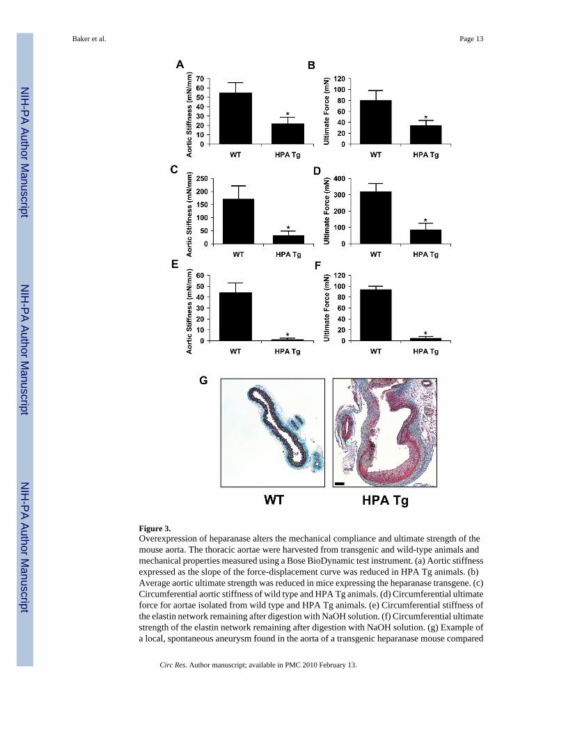
Figure 3.Overexpression of heparanase alters the mechanical compliance and ultimate strength of themouse aorta. The thoracic aortae were harvested from transgenic and wild-type animals andmechanical properties measured using a Bose BioDynamic test instrument. (a) Aortic stiffnessexpressed as the slope of the force-displacement curve was reduced in HPA Tg animals. (b)Average aortic ultimate strength was reduced in mice expressing the heparanase transgene. (c)Circumferential aortic stiffness of wild type and HPA Tg animals. (d) Circumferential ultimateforce for aortae isolated from wild type and HPA Tg animals. (e) Circumferential stiffness ofthe elastin network remaining after digestion with NaOH solution. (f) Circumferential ultimatestrength of the elastin network remaining after digestion with NaOH solution. (g) Example ofa local, spontaneous aneurysm found in the aorta of a transgenic heparanase mouse compared
Baker et al. Page 13
Circ Res. Author manuscript; available in PMC 2010 February 13.
NIH
-PA Author Manuscript
NIH
-PA Author Manuscript
NIH
-PA Author Manuscript

to a wild type control. The sample was stained with elastic Movat’s pentachrome stain. Bar =50 μm.
Baker et al. Page 14
Circ Res. Author manuscript; available in PMC 2010 February 13.
NIH
-PA Author Manuscript
NIH
-PA Author Manuscript
NIH
-PA Author Manuscript

Figure 4.Intimal thickening and heparanase expression are increased with stenting in obese,hyperlipidemic Zucker rats. Endovascular stenting was performed through minimally invasivefemoral access. (a) Histochemical staining and immunohistochemical staining of lean andobese Zucker rat aorta, 14 days after stent placement (n=16). EVG = Elastic Verhoff VanGeisen staining. Bar = 50 μm. (b) Analysis of neointimal thickness in lean and obese Zuckerrats. (c) Quantitative analysis of heparanase in the neointima of lean and obese Zucker rats. (d)Heparanase expression and neointimal proliferation in response to endovascular stenting areenhanced in obese Zucker rats (■) compared to lean controls (○).The expression of heparanasewithin the neointima correlated strongly with the final neointimal area, giving a correlation ofR=0.50 (p < 0.001) for lean rats and R=0.77 (p < 0.001) for fatty rats. *Statistically significantdifference between samples (p < 0.05).
Baker et al. Page 15
Circ Res. Author manuscript; available in PMC 2010 February 13.
NIH
-PA Author Manuscript
NIH
-PA Author Manuscript
NIH
-PA Author Manuscript

Figure 5.Abdominal stenting of heparanase transgenic and wild type mice was performed using femoralaccess with small diameter self-expanding stents. Stents were harvested after 14 days andprocessed for histological analysis in resin sections. (a) Lateral x-ray of stent placement in theabdominal aorta of the mouse. The stent is visible just below the spine (marked with arrow).(b) Neointimal formation in response to vascular injury with endovascular staining is increasedin heparanase transgenic mice. Staining for Mac-1 was increased in heparanase transgenicmice. Bar = 50 μm. (c–e) Morphological analysis of the stented arteries showed increasedintimal area, intima to medial ratio and no change in media area for heparanase transgenic
Baker et al. Page 16
Circ Res. Author manuscript; available in PMC 2010 February 13.
NIH
-PA Author Manuscript
NIH
-PA Author Manuscript
NIH
-PA Author Manuscript

mice. (f) Concentration of MCP-1 in arterial lysates as measured by ELISA assay. The arterieswere harvested one hour following arterial injury (n=4). *Statistically significant differencebetween all samples (p < 0.05).
Baker et al. Page 17
Circ Res. Author manuscript; available in PMC 2010 February 13.
NIH
-PA Author Manuscript
NIH
-PA Author Manuscript
NIH
-PA Author Manuscript
Copyright © 2022 FDOKUMEN