Optimization schemes for endovascular repair with parallel ...
Upload
independentCategory
view
0download
0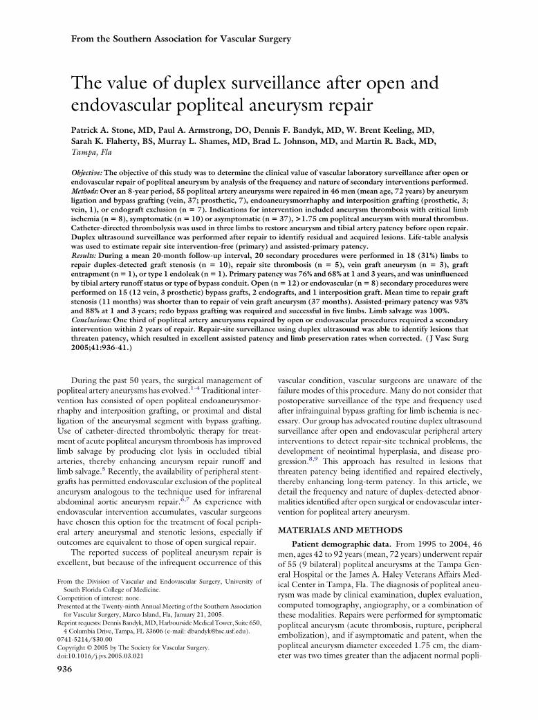
From the Southern Association for Vascular Surgery
The value of duplex surveillance after open andendovascular popliteal aneurysm repairPatrick A. Stone, MD, Paul A. Armstrong, DO, Dennis F. Bandyk, MD, W. Brent Keeling, MD,Sarah K. Flaherty, BS, Murray L. Shames, MD, Brad L. Johnson, MD, and Martin R. Back, MD,Tampa, Fla
Objective: The objective of this study was to determine the clinical value of vascular laboratory surveillance after open orendovascular repair of popliteal aneurysm by analysis of the frequency and nature of secondary interventions performed.Methods: Over an 8-year period, 55 popliteal artery aneurysms were repaired in 46 men (mean age, 72 years) by aneurysmligation and bypass grafting (vein, 37; prosthetic, 7), endoaneurysmorrhaphy and interposition grafting (prosthetic, 3;vein, 1), or endograft exclusion (n � 7). Indications for intervention included aneurysm thrombosis with critical limbischemia (n � 8), symptomatic (n � 10) or asymptomatic (n � 37), >1.75 cm popliteal aneurysm with mural thrombus.Catheter-directed thrombolysis was used in three limbs to restore aneurysm and tibial artery patency before open repair.Duplex ultrasound surveillance was performed after repair to identify residual and acquired lesions. Life-table analysiswas used to estimate repair site intervention-free (primary) and assisted-primary patency.Results: During a mean 20-month follow-up interval, 20 secondary procedures were performed in 18 (31%) limbs torepair duplex-detected graft stenosis (n � 10), repair site thrombosis (n � 5), vein graft aneurysm (n � 3), graftentrapment (n � 1), or type 1 endoleak (n � 1). Primary patency was 76% and 68% at 1 and 3 years, and was uninfluencedby tibial artery runoff status or type of bypass conduit. Open (n � 12) or endovascular (n � 8) secondary procedures wereperformed on 15 (12 vein, 3 prosthetic) bypass grafts, 2 endografts, and 1 interposition graft. Mean time to repair graftstenosis (11 months) was shorter than to repair of vein graft aneurysm (37 months). Assisted-primary patency was 93%and 88% at 1 and 3 years; redo bypass grafting was required and successful in five limbs. Limb salvage was 100%.Conclusions: One third of popliteal artery aneurysms repaired by open or endovascular procedures required a secondaryintervention within 2 years of repair. Repair-site surveillance using duplex ultrasound was able to identify lesions thatthreaten patency, which resulted in excellent assisted patency and limb preservation rates when corrected. (J Vasc Surg
2005;41:936-41.)During the past 50 years, the surgical management ofpopliteal artery aneurysms has evolved.1-4 Traditional inter-vention has consisted of open popliteal endoaneurysmor-rhaphy and interposition grafting, or proximal and distalligation of the aneurysmal segment with bypass grafting.Use of catheter-directed thrombolytic therapy for treat-ment of acute popliteal aneurysm thrombosis has improvedlimb salvage by producing clot lysis in occluded tibialarteries, thereby enhancing aneurysm repair runoff andlimb salvage.5 Recently, the availability of peripheral stent-grafts has permitted endovascular exclusion of the poplitealaneurysm analogous to the technique used for infrarenalabdominal aortic aneurysm repair.6,7 As experience withendovascular intervention accumulates, vascular surgeonshave chosen this option for the treatment of focal periph-eral artery aneurysmal and stenotic lesions, especially ifoutcomes are equivalent to those of open surgical repair.
The reported success of popliteal aneurysm repair isexcellent, but because of the infrequent occurrence of this
From the Division of Vascular and Endovascular Surgery, University ofSouth Florida College of Medicine.
Competition of interest: none.Presented at the Twenty-ninth Annual Meeting of the Southern Association
for Vascular Surgery, Marco Island, Fla, January 21, 2005.Reprint requests: Dennis Bandyk, MD, Harbourside Medical Tower, Suite 650,
4 Columbia Drive, Tampa, FL 33606 (e-mail: [email protected]).0741-5214/$30.00Copyright © 2005 by The Society for Vascular Surgery.
doi:10.1016/j.jvs.2005.03.021936
vascular condition, vascular surgeons are unaware of thefailure modes of this procedure. Many do not consider thatpostoperative surveillance of the type and frequency usedafter infrainguinal bypass grafting for limb ischemia is nec-essary. Our group has advocated routine duplex ultrasoundsurveillance after open and endovascular peripheral arteryinterventions to detect repair-site technical problems, thedevelopment of neointimal hyperplasia, and disease pro-gression.8,9 This approach has resulted in lesions thatthreaten patency being identified and repaired electively,thereby enhancing long-term patency. In this article, wedetail the frequency and nature of duplex-detected abnor-malities identified after open surgical or endovascular inter-vention for popliteal artery aneurysm.
MATERIALS AND METHODS
Patient demographic data. From 1995 to 2004, 46men, ages 42 to 92 years (mean, 72 years) underwent repairof 55 (9 bilateral) popliteal aneurysms at the Tampa Gen-eral Hospital or the James A. Haley Veterans Affairs Med-ical Center in Tampa, Fla. The diagnosis of popliteal aneu-rysm was made by clinical examination, duplex evaluation,computed tomography, angiography, or a combination ofthese modalities. Repairs were performed for symptomaticpopliteal aneurysm (acute thrombosis, rupture, peripheralembolization), and if asymptomatic and patent, when thepopliteal aneurysm diameter exceeded 1.75 cm, the diam-
eter was two times greater than the adjacent normal popli-
JOURNAL OF VASCULAR SURGERYVolume 41, Number 6 Stone et al 937
teal artery, and mural thrombus was imaged. Associatedmedical conditions included hypertension in 18 (33%) pa-tients, hyperlipidemia in 15 (27%) patients, and diabetesmellitus in 12 (22%) patients. Twenty-three (42%) patientshad had either prior abdominal aortic aneurysm repair orthe diagnosis of a small �5 cm abdominal aortic aneurysmthat was under ultrasound surveillance. Other peripheralaneurysms diagnosed in the study group included femoralaneurysm (n � 6), iliac aneurysm (n � 5), and thoracicaorta aneurysm (n � 2).
Indication and operative procedure. Thirty-seven(67%) of the repaired popliteal aneurysms were asymptom-atic and were repaired electively (Table I). Eight patientspresented with symptomatic popliteal aneurysms (throm-bosis, n � 6; embolization, n � 2) requiring emergenttreatment, including the use of catheter-directed throm-bolysis in three limbs to restore patency in the aneurysmand tibial artery runoff. Types of repair used in this groupincluded vein bypass grafts (n � 7) and endovascular stent-graft (n � 1). Ten patients had elective repair of a poplitealaneurysm with limb symptoms that included limb pain (n �4), claudication (n � 4), or deep venous thrombosis (n � 2).
Two types of open surgical repair were used: aneurysmexclusion by proximal and distal ligation and bypass graft-ing via a medial-surgical approach (n � 44), and poplitealendoaneurysmorrhaphy with interposition grafting via aposterior popliteal fossa approach (n � 4). Polytetrafluoro-ethylene (PTFE) prosthetic conduits (6 mm diameter)were used in 7 bypass and 3 interposition grafting proce-dures because of inadequate saphenous vein conduit, notsecondary to urgency of procedure. The common femoralartery was the inflow site of 7 (15%) of 48 bypass grafts,with the distal superficial femoral artery used in 41 (85%)limbs. The graft outflow was to the distal popliteal artery in38 (79%) limbs or an infrageniculate (tibial-peroneal trunk,n � 4; peroneal, n � 3; posterior tibial, n � 2; dorsalispedis, n � 1) artery in 10 (21%) limbs.
Since 2001, seven patients, including six who were ofhigh surgical risk (ischemic cardiomyopathy, 5; end-stagerenal disease, 1) were considered for endograft treatment ofthe popliteal aneurysm if specific anatomic criteria weremet, including aneurysmal segment confined to the above-knee popliteal artery, mild axial tortuosity, two-vessel tibialartery runoff, and distal stent-graft seal zone of at least 1 cmat or above the knee joint space. Endovascular exclusionwas performed in seven limbs using 1-3 overlapping BostonScientific 10- or 12-mm-diameter Wall graft (n � 5) orW. L. Gore 10-mm-diameter ViaBahn (n � 2) endopros-thesis segments. Selection of the endograft type used wasbased on surgeon preference. The procedure of poplitealaneurysm exclusion was performed via open femoral arteryexposure and antegrade delivery of the endograft over aguidewire. Angiographic monitoring of stent-graft deploy-ment was performed to ensure adequate proximal and distallanding site coverage, and to evaluate adequacy of aneu-rysm exclusion, ie, no type I endoleak.
In the absence of contraindications, a perioperative
antithrombotic regimen consisting of aspirin (325 mg),clopidogrel (75 mg), dextran-40 (500 mL intravenous at25 mL/h) was administered after both endovascular andopen surgical repair. Long-term antiplatelet therapy wasprescribed in all patients.
Duplex ultrasound surveillance protocol. Patientswere enrolled in a surveillance protocol that included clin-ical assessment (patient history, physical examination) ofarterial limb circulation, measurement of Doppler-derivedankle systolic pressure (ankle-brachial index), and colorduplex scanning.10 Testing was performed in an Intersoci-etal Commission for Accreditation of Vascular Laborato-ries–accredited vascular laboratory before hospital dis-charge, 3 to 4 weeks later, and subsequently every 3 monthsfor the first 2 years, and then at 6-month intervals. Theentire popliteal aneurysm repair was imaged, including theinflow and distal arteries, and peak systolic velocities (PSVs)were recorded along the vein/prosthetic graft segments todetect stenosis or identify the development of an abnormallow flow (PSV decrease � 30 cm/s compared with previousstudies; value �45 cm/s) graft flow. When a stenosis wasidentified, PSV and the velocity ratios (Vr) across the stenosiswere recorded. Criteria for repair of duplex-detected stenosisincluded PSV � 300 cm/s and Vr � 3.5.11 Vein graftaneurysms were diagnosed based on real-time B-mode andcolor Doppler imaging by the findings of greater than twotimes diameter focal dilation of the vein graft conduit withmural thrombus. If duplex testing identified a graft stenosisthat did not meet criteria for intervention (PSV � 300cm/s), the interval of graft surveillance was reduced to 4 to6 weeks. The assessment of endovascular repairs was similarto that for open surgical repairs, but also included color/power Doppler imaging of the aneurysm sac for endoleak.Duplex criteria for reintervention to correct secondaryrepair-site abnormalities were identical to those used for theinitial procedure.
Treatment of duplex-detected graft stenosis oraneurysm. Graft or repair site stenoses meeting criteria forintervention were treated by either endovascular (percuta-neous transluminal balloon angioplasty [PTA]) or opensurgical repair. The type of intervention used was based ontime from original procedure, lesion length, and vesseldiameter.10,11 PTA was used to treat focal (�2 cm length)stenosis, in larger (3.5 mm or greater) caliber vessels, thatdeveloped 3 months or more after the aneurysm repair
Table I. Clinical presentation of 55 popliteal aneurysmsin 46 patients relative to need for emergent or electiverepair
Emergent repairIschemic pain (rest pain) 7 (13%)Tissue loss/gangrene 1 (2%)
Elective repairAsymptomatic with mural thrombus 37 (67%)Painful mass 4 (7%)Claudication 4 (7%)Deep venous thrombosis 2 (4%)
procedure. Surgical repair was selected if the above criteria

JOURNAL OF VASCULAR SURGERYJune 2005938 Stone et al
were not met or if duplex scanning detected persistentstenosis (PSV � 200 cm/s) after PTA. In general, thenature of the abnormality dictated the type of surgicalrepair performed: stenosis was treated by either patch an-gioplasty, interposition grafting, or placement of jumpgraft; and vein graft aneurysm was repaired using interpo-sition graft replacement. Intraprocedural duplex evaluationwas performed after correction of the graft lesion. Duplexfindings indicating a technically adequate repair includedwidely patent lumen by real-time color Doppler imaging,PSV � 180 cm/s, and Vr � 2 at the repair site.
Data analysis. A retrospective review of patient hospi-tal, vascular clinic, and vascular laboratory records wasconducted. Adverse patient outcomes, including death,repair site thrombosis, graft revision, and amputation, wererecorded throughout a follow-up interval that ranged from3 to 84 months (mean, 20 months). Popliteal repair pri-mary, assisted-primary, and secondary patency and limbsalvage were estimated as recommended by the reportingstandards committee of the Society for Vascular Surgeryusing life-table analysis. �2 analysis was used to determinewhether differences in the number of secondary proceduresoccurred within various treatment groups.
RESULTS
Thirty-day outcomes. No patient died as a result ofan open or endovascular popliteal aneurysm repair. Intra-operative and predischarge duplex scans were interpreted asnormal in all patients, resulting in no repair site revisionsperformed before discharge. Secondary surgical site proce-dures were required in 6 (13%) patients, including evacua-tion of incision hematoma (n � 3), surgical débridementand wound closure for Szilagyi grade II infection (n � 2),or lymphocele repair (n � 1).
A nonringed PTFE common femoral-to-distal poplitealbypass thrombosed (technical error caused by graft kinking)after discharge, and was revised by replacement with a ringed6-mm-diameter PTFE conduit. Two endovascular Wall graft(10- and 12-mm-diameter) repairs required revision: en-dograft extension to treat a type I endoleak detected by duplexultrasound at the first postoperative clinic evaluation, andopen conversion of symptomatic endograft thrombosis usingfemoral-to-posterior tibial artery saphenous vein bypass graft-ing. No forefoot or limb amputations were performed.
Outcomes after 30 days. Duplex surveillance identi-fied 14 graft abnormalities in 12 surgical reconstructions,including 10 graft (vein, 9; PTFE, 1) stenoses, 3 vein graftaneurysms, and 1 vein graft entrapped by the medial gas-trocnemius muscle. Only two graft stenoses were symp-tomatic with the patients having symptoms of calf claudi-cation. The distal anastomotic region was the mostcommon (n � 6) site of a duplex-detected stenosis (PSV �300 cm/s), followed by midgraft (n � 2) and proximal (n� 2) vein graft locations. Seven graft stenoses met criteriafor treatment by percutaneous balloon angioplasty, and theremaining three were treated by open surgical repair (vein-patch angioplasty, 2; interposition graft, 1). All vein graft
aneurysms were repaired using interposition vein grafting.Mean time to intervention for an acquired hemodynami-cally significant graft stenosis was 11 � 12 months (median,7 months; range 1.5 to 41 months) compared with 37 (15,25, and 60) months for the development vein graft aneu-rysm. The primary patency, ie, intervention-free patency,rate was 88% at 6 months, 76% at 1 year, and 68% at 3 years(Fig). No interventions were performed for enlargement orfor symptoms related to the ligated/excluded poplitealartery aneurysm. Duplex imaging of the ligated aneurysmin 44 limbs treated with bypass grafting showed no flow orsignificant enlargement with time.
Recurrent stenosis developed in two of seven revisedgrafts after PTA, whereas no open surgical repair developedrecurrent stenosis during a mean follow-up of 11 months(range, 2 to 27 months). One graft stenosis initially treatedwith PTA required surgical revision by jump bypass graft-ing 4 months later, whereas the other was successfullytreated by reperforming the PTA 31 months later. Theprimary-assisted patency rate was 96% at 6 months, 93% at1 year, and 88% at 3 years (Fig). No graft failed because ofinfection, and limb preservation was 100% at 5 years.
Although all seven endovascular repairs were successfulat the time of hospital discharge, two patients requiredsurgical intervention for endoprosthesis thrombosis within2 months of aneurysm exclusion. The two modes of devicefailure were thought to be caused by inadequate fixation ina tortuous aneurysm resulting in stent-graft migration, andextension of the stent-graft into the distal popliteal artery totreat a type 1 endoleak with subsequent device kinkingassociated with knee flexion. In the remaining five patientswith endovascular repairs, two have limited follow-up (3months) but duplex scans are normal, and the other threehave normal duplex scans at 8, 21, and 37 months.
During the follow-up period (mean, 20 months; range,3 to 98 months), four (9%) patients died of cardiac (n � 3)or end-stage renal (n � 1) disease. Three patients who hadundergone an open (n � 2) or an endovascular poplitealaneurysm repair presented at the scheduled surveillanceappointment with graft occlusion at 2, 6, and 26 months.Limb-threatening ischemia was present in one patient,resulting in emergent thrombolytic therapy before redovein bypass grafting. The remaining two graft occlusionswere managed by redo bypass grafting, and all remainpatent at last follow-up. Overall, five popliteal repairsthrombosed (three bypass grafts and two endografts, fourof which occurred in limbs with two or more tibial arteryrunoffs). Secondary procedures (PTA or surgical repair) tomaintain a patent bypass were performed in 15% of limbswith single vessel runoff and 29% of limbs with two or morepatent tibial arteries. Overall, graft revision and failure rateswere not influenced by clinical presentation (symptomaticvs asymptomatic), type of conduit, site of distal graft anas-tomosis, grafting procedure, or number of patent tibialarteries (Table II).
DISCUSSION
Duplex surveillance identified an abnormality in the
popliteal repair in approximately one third of repairs, in-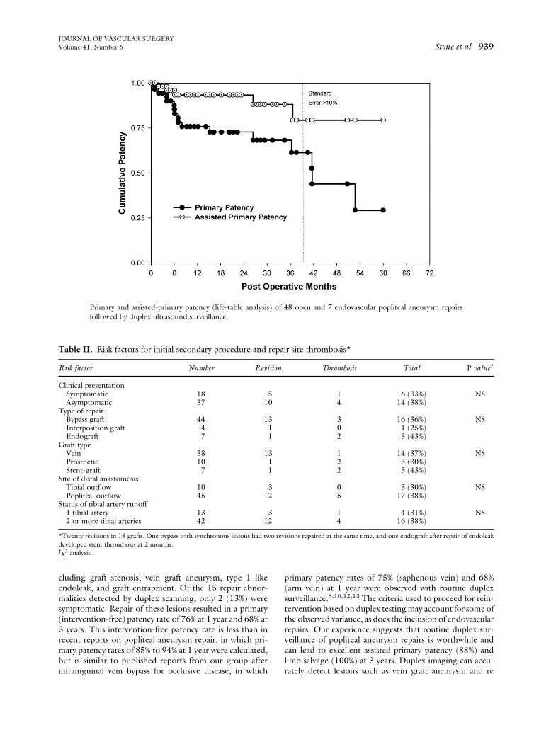
JOURNAL OF VASCULAR SURGERYVolume 41, Number 6 Stone et al 939
cluding graft stenosis, vein graft aneurysm, type 1–likeendoleak, and graft entrapment. Of the 15 repair abnor-malities detected by duplex scanning, only 2 (13%) weresymptomatic. Repair of these lesions resulted in a primary(intervention-free) patency rate of 76% at 1 year and 68% at3 years. This intervention-free patency rate is less than inrecent reports on popliteal aneurysm repair, in which pri-mary patency rates of 85% to 94% at 1 year were calculated,but is similar to published reports from our group after
Primary and assisted-primary patency (life-table analysisfollowed by duplex ultrasound surveillance.
Table II. Risk factors for initial secondary procedure and
Risk factor Number Revi
Clinical presentationSymptomatic 18Asymptomatic 37 1
Type of repairBypass graft 44 1Interposition graft 4Endograft 7
Graft typeVein 38 1Prosthetic 10Stent-graft 7
Site of distal anastomosisTibial outflow 10Popliteal outflow 45 1
Status of tibial artery runoff1 tibial artery 132 or more tibial arteries 42 1
*Twenty revisions in 18 grafts. One bypass with synchronous lesions had twdeveloped stent thrombosis at 2 months.†�2 analysis.
infrainguinal vein bypass for occlusive disease, in which
primary patency rates of 75% (saphenous vein) and 68%(arm vein) at 1 year were observed with routine duplexsurveillance.8,10,12,13 The criteria used to proceed for rein-tervention based on duplex testing may account for some ofthe observed variance, as does the inclusion of endovascularrepairs. Our experience suggests that routine duplex sur-veillance of popliteal aneurysm repairs is worthwhile andcan lead to excellent assisted-primary patency (88%) andlimb salvage (100%) at 3 years. Duplex imaging can accu-
8 open and 7 endovascular popliteal aneurysm repairs
r site thrombosis*
Thrombosis Total P value†
1 6 (33%) NS4 14 (38%)
3 16 (36%) NS0 1 (25%)2 3 (43%)
1 14 (37%) NS2 3 (30%)2 3 (43%)
0 3 (30%) NS5 17 (38%)
1 4 (31%) NS4 16 (38%)
isions repaired at the same time, and one endograft after repair of endoleak
) of 4
repai
sion
50
311
311
32
32
o rev
rately detect lesions such as vein graft aneurysm and re

JOURNAL OF VASCULAR SURGERYJune 2005940 Stone et al
stenosis after angioplasty, focal lesions that can be repairedbased on duplex findings alone.
Popliteal artery aneurysm, although an infrequent vas-cular condition, is the most common site for a nativeperipheral aneurysm, accounting for 70% to 80% of allcases.3,4 Clinical presentation rarely (�2%) involves rup-ture, but manifestations of limb ischemia caused by throm-bosis, peripheral embolization, concomitant atherosclero-sis obliterans, or pain attributable to compression ofadjacent nerves and veins are present in 40% of patients.3,5-7
The majority (85% in these series) of popliteal aneurysmrepairs can be performed as elective procedures, allowingtime for vascular imaging to assess aneurysm extent, con-comitant tibial artery occlusive disease, and the presence ofbilateral disease. An aggressive approach toward repair ofpopliteal aneurysms is recommended because delay in in-tervention risks thrombosis and limb loss.4 Reports fromthe 1950s documented amputation rates of 20%,1 butmajor limb amputation associated with popliteal aneurysmrepair has decreased because of the availability of catheter-directed thrombolytic therapy, experience with bypass totibial arteries, and enhanced clinical awareness of this con-dition leading to ultrasound imaging of the popliteal artery.In general, repair is recommended for asymptomatic,patent popliteal aneurysms � 1.8 to 2 cm in diameter ifmural thrombus is also present.2-4,9
When clinical presentation involves acute aneurysmthrombosis or peripheral embolization producing limb-threatening ischemia, emergent angiography is recom-mended, and if warranted, use of catheter-directed throm-bolytic therapy to restore tibial artery patency when nooutflow vessel is visualized. The success of thrombolytictherapy in these circumstances was documented by Martyet al, who reported recovery of tibial artery runoff in 77% ofpatients.5 The success of popliteal aneurysm repair has beenlinked to the severity of tibial artery occlusive disease with a5-year patency rate of 65% when only single-vessel tibialartery runoff is present. In our series, the presence of singletibial artery patency, present in 13 (24%) of 55 limbs, wasnot predictive of repair thrombosis or the need for revision.Because of the small number (n � 10) of PTFE andendovascular (n � 7) grafts performed in this series, acomparison of repair patency relative to tibial runoff statusor vein bypass grafting using Kaplan-Meier outcome anal-ysis was not possible.
The anatomy of the aneurysmal popliteal segment dic-tates the type of repair. Bypass grafting with proximal anddistal aneurysm ligation is the most common procedure,and vein conduits are preferred especially for the treatmentof long aneurysmal segments or when severe atheroscle-rotic occlusive disease is present. For focal popliteal aneu-rysm, either open interposition bypass grafts or endograft-ing can be performed. The endovascular approach isappealing because of its reduced operative time and mor-bidity, especially in high-surgical-risk patients, comparedwith open surgical bypass via a medial or posterior ap-proach. The results of small (�25) series of stent-graft
exclusion of popliteal aneurysms indicate reduced primarypatency (40% to 67% at 1 year) compared with open repair(80% to 90% at 1 year).7,8,12-14 Endograft thrombosis wasthe primary mode of failure, and this was also true in ourseries, in which two of seven endografts failed because ofthrombosis. The risk for stent-graft thrombosis may bedecreased by careful patient selection based on strict ana-tomic criteria and improvement in stent-graft design andflexibility. Experience gleaned from the seven endovascularprocedures performed has led to a refinement in selectioncriteria. At present, we would consider endograft repair forpopliteal aneurysms in the absence of significant SFA andtibial occlusive disease, when the stent-graft does extendbelow the knee joint and the total treated aneurysmalsegment is 20 cm or less and without significant tortuosity.The decision to proceed with endovascular repair is alsoinfluenced by patient factors such as adequate saphenousvein conduit, severe medical comorbidities, and the pres-ence of ideal anatomy for stent-graft exclusion of the pop-liteal aneurysm.
Our audit of popliteal aneurysm repair indicated thatopen and endovascular interventions are prone to bothtechnical and acquired problems that influence repair func-tion and patency. Use of duplex ultrasound surveillanceboth in the early postoperative period and in the long termcan optimize long-term patency because the majority oflesions identified and corrected were asymptomatic. Resid-ual flow in a popliteal aneurysm ligated both proximally anddistally was not detected, and symptomatic enlargementwas not observed. Intraluminal stent-graft exclusion of apopliteal aneurysm has the potential for providing long-term durability in patients with appropriate anatomy, butapplication of this endovascular intervention requires fur-ther study.
REFERENCES
1. Gifford RW Jr, Hines EA, Janes JM. An analysis and follow-up study ofone hundred popliteal aneurysms. Surgery 1953;33:284-93.
2. Szilagyi DE, Schwartz RL, Reddy HD. Popliteal arterial aneurysms:their natural history and management. Arch Surg 1981;116:724-8.
3. Shortell CK, DeWeese JA, Ouriel K, Green RM. Popliteal aneurysms: a25-year surgical experience. J Vasc Surg 1991;14:771-9.
4. Halliday AW, Wolfe JH, Taylor PR. The management of poplitealaneurysm: the importance of early surgical repair. Ann R Coll Surg1991;73:253-7.
5. Marty B, Wicky S, Ris HB, Meuller X, Fisher A, Hayoz D, VonSegesserLK. Success of thrombolysis as a predictor of outcome in acute throm-bosis of poplitel aneurysms. J Vasc Surg 2002;35:487-93.
6. Henry M, Amor M, Kloraris C, Tzvetanov K, Buniet JM, Amicabile C,Drawin T. Percutaneous endovascular treatment of peripheral aneu-rysms. J Cardiovasc Surg 2000;41:871-83.
7. Gerasimidis T, Sfyroeras G, Papazoglou K, Trellopoulous G, Ntinas A,Karamonos D. Endovascular treatment of popliteal artery aneurysms.Eur J Vasc Endovasc Surg 2003;26:506-11.
8. Gupta AK, Bandyk DF, Cheanvechai D, Johnson BL. Natural history ofinfrainguinal vein graft stenosis relative to bypass grafting technique. JVasc Surg 1997;25:211-25.
9. Ascher E, Markevich N, Schutzer RW, Kallajuri S, Jacob T, HingoraniAP. Small popliteal artery aneurysm: are they clinically significant? J VascSurg 2003;37:755-60.
10. Armstrong PA, Bandyk DF, Wilson JS, Shames ML, Johnson BL, Back
MR. Optimizing infrainguinal arm vein bypass patency with duplex
JOURNAL OF VASCULAR SURGERYVolume 41, Number 6 Stone et al 941
ultrasound surveillance and endovascular therapy. J Vasc Surg2004;40:724-31.
11. Bandyk DF. Infrainguinal vein bypass graft surveillance: how to do it,when to intervene, and is it cost effective? J Am Coll Surg 2002;194(Suppl 1):S40-52.
12. Lilly MP, Flinn WR, McCarthy WJ III, Courtney DF, Yao JST, BerganJJ.The effect of distal arterial anatomy on the success of popliteal aneurysm
13. Aulivola B, Hamdan AD, Hile CN, Sheahan MG, Skillman JJ, Camp-bell DR, et al. Popliteal artery aneurysms: a comparison of outcomes inelective versus emergent repair. J Vasc Surg 2004;39:1171-7.
14. Howell M, Krajcer J, Dietrich EB. Wallgraft endoprosthesis for thepercutaneous treatement of femoral and popliteal artery aneurysms. JEndovascular Ther 2002;9:76-81.
repair. J Vasc Surg 1988;7:653-60. Submitted Jan 17, 2004; accepted Mar 7, 2005.
Copyright © 2022 FDOKUMEN