Endovascular Treatment of Carotid Cavernous Fistulas
15
EndovascularTreatment of Carotid Cavernous Fistulas Joseph J. Gemmete, MD a, *, Sameer A. Ansari, MD, PhD a , Dheeraj Gandhi, MD b,c This article provides an overview of direct and indirect carotid cavernous fistulas (CCFs) with emphasis on the recent advances in endovascular techniques available for treatment. It first briefly discusses the classification, etiology and pathology, clinical presentation, and diagnostic imaging of direct and indirect CCFs. Additionally, it provides a brief description of the medical management and surgical treatment of direct and indirect CCFs. The subsequent sections present a detailed discussion of the various endo- vascular techniques available for the treatment of direct and indirect CCFs with a brief discussion of the complications and results of treatment. CLASSIFICATION CCFs can be classified based on etiology (trau- matic or spontaneous), rate of flow (high versus low flow), or the angiographic architecture (direct or indirect). The most commonly used classifica- tion scheme established by Barrow and colleagues 1 divides the CCFs into four types, depending on the arterial supply. Direct (type A fistulas) are direct communications between the internal carotid artery (ICA) and the cavernous sinus, usually associated with high flow rates. Indi- rect fistulas (types B, C, and D) are dural arteriove- nous fistulas (DAVFs) fed by the meningeal arteries of the ICA, the external carotid artery (ECA), or both. Type B fistulas are relatively uncommon and are supplied only by the dural branches of the ICA. Type C fistulas are supplied solely by the dural branches of the ECA. The most common indirect CCF is a type D fistula, which is supplied by the meningeal branches of both the ICA and ECA. ETIOLOGY AND PATHOLOGY Direct (type A) fistulas usually are traumatic, caused by motor vehicle accidents or penetrating injuries, 2 and generally affect young males. Approximately 20% of type A CCFs may be spon- taneous, resulting from the rupture of either a cavernous ICA aneurysm or a weakened ICA vessel wall coursing through the cavernous sinus. 3 Debrun 4 described the site of CCF origin by dividing the cavernous ICA into five arbitrary segments. He found the most common site of occurrence is the proximal horizontal portion of the ICA near the artery of the inferior cavernous sinus (40%). CCFs occur with decreasing frequency at the junction of the horizontal and posterior vertical ascending cavernous segment (28%), at the origins from the posterior vertical ascending segment itself (20%), and distally near the anterior genu and the clinoidal segment (12%). Van Dellen 5 considers CCFs to be part of a continuum of injury to the arterial and venous a Division of Interventional Neuroradiology, Department of Radiology, University of Michigan Health System, 1500 E. Medical Center Dr., B1D330, Ann Arbor, MI 48109 0030, USA b Department of Radiology, Johns Hopkins Hospital, Baltimore, MD, USA c Department of Neurosurgery, Johns Hopkins Hospital, Baltimore, MD, USA * Corresponding author. E-mail address: [email protected] (J.J. Gemmete). KEYWORDS Carotid cavernous fistula Endovascular treatment Classification Etiology and pathology Clinical presentation Neuroimag Clin N Am 19 (2009) 241–255 doi:10.1016/j.nic.2009.01.006 1052-5149/09/$ – see front matter ª 2009 Elsevier Inc. All rights reserved. neuroimaging.theclinics.com
-
Upload
independent -
Category
Documents
-
view
0 -
download
0
Transcript of Endovascular Treatment of Carotid Cavernous Fistulas

EndovascularTreatmentof Carotid CavernousFistulas
JosephJ. Gemmete, MDa,*, Sameer A. Ansari, MD, PhDa,Dheeraj Gandhi, MDb,cKEYWORDS� Carotid cavernous fistula � Endovascular treatment� Classification � Etiology and pathology� Clinical presentation
This article provides an overview of direct andindirect carotid cavernous fistulas (CCFs) withemphasis on the recent advances in endovasculartechniques available for treatment. It first brieflydiscusses the classification, etiology andpathology, clinical presentation, and diagnosticimaging of direct and indirect CCFs. Additionally,it provides a brief description of the medicalmanagement and surgical treatment of directand indirect CCFs. The subsequent sectionspresent a detailed discussion of the various endo-vascular techniques available for the treatment ofdirect and indirect CCFs with a brief discussionof the complications and results of treatment.
om
CLASSIFICATION
CCFs can be classified based on etiology (trau-matic or spontaneous), rate of flow (high versuslow flow), or the angiographic architecture (director indirect). The most commonly used classifica-tion scheme established by Barrow andcolleagues1 divides the CCFs into four types,depending on the arterial supply. Direct (type Afistulas) are direct communications between theinternal carotid artery (ICA) and the cavernoussinus, usually associated with high flow rates. Indi-rect fistulas (types B, C, and D) are dural arteriove-nous fistulas (DAVFs) fed by the meningeal arteriesof the ICA, the external carotid artery (ECA), or
a Division of Interventional Neuroradiology, Department1500 E. Medical Center Dr., B1D330, Ann Arbor, MI 4810b Department of Radiology, Johns Hopkins Hospital, Balc Department of Neurosurgery, Johns Hopkins Hospital,* Corresponding author.E-mail address: [email protected] (J.J. Gemmete).
Neuroimag Clin N Am 19 (2009) 241–255doi:10.1016/j.nic.2009.01.0061052-5149/09/$ – see front matter ª 2009 Elsevier Inc. All
both. Type B fistulas are relatively uncommonand are supplied only by the dural branches ofthe ICA. Type C fistulas are supplied solely bythe dural branches of the ECA. The most commonindirect CCF is a type D fistula, which is suppliedby the meningeal branches of both the ICA andECA.
ETIOLOGYAND PATHOLOGY
Direct (type A) fistulas usually are traumatic,caused by motor vehicle accidents or penetratinginjuries,2 and generally affect young males.Approximately 20% of type A CCFs may be spon-taneous, resulting from the rupture of eithera cavernous ICA aneurysm or a weakened ICAvessel wall coursing through the cavernous sinus.3
Debrun4 described the site of CCF origin bydividing the cavernous ICA into five arbitrarysegments. He found the most common site ofoccurrence is the proximal horizontal portion ofthe ICA near the artery of the inferior cavernoussinus (40%). CCFs occur with decreasingfrequency at the junction of the horizontal andposterior vertical ascending cavernous segment(28%), at the origins from the posterior verticalascending segment itself (20%), and distally nearthe anterior genu and the clinoidal segment (12%).
Van Dellen5 considers CCFs to be part ofa continuum of injury to the arterial and venous
of Radiology, University of Michigan Health System,9 0030, USAtimore, MD, USABaltimore, MD, USA
rights reserved. neur
oim
agin
g.th
ecli
nics
.c

Gemmete et al242
vessel wall. An incomplete tear can result ina pseudoaneurysm of the cavernous carotid arterythat can compress the venous sinusoids. A trueCCF, however, occurs when the arterial tear iscomplete and there is associated breach of thewalls of venous sinusoids.
Flow rates in type A fistulas are variable anddepend on the size of the ostium and venousdrainage. Complete steal of ICA flow to CCFoccurs in approximately 5% of cases at diagnosis.Most fistula ostia measure 2.6 mm in diameter,4
typically small enough to be treated with detach-able balloons with a mean volume of 0.28 cm3,equivalent to an inflated balloon dimension of7 mm � 9 mm. Bilateral CCFs occur in approxi-mately 1% to 2% of patients who have traumaticCCFs.6,7
Spontaneous direct (type A) CCFs are morecommon in older women, although a few havebeen reported in children.8,9 They usually arecaused by the rupture of a cavernous aneurysmor by the spontaneous rupture of a congenitallyweakened, atherosclerotic, or diseased artery.Predisposition to spontaneous direct CCFs havebeen shown in fibromuscular dysplasia, Ehlers-Danlos syndrome, and pseudoxanthomaelasticum.10,11
Indirect (types B, C, or D) CCFs have a predilec-tion for spontaneous occurrence in postmeno-pausal women. Sinus thrombosis, hypertension,and diabetes have been suggested as predispos-ing factors.12,13 Trauma is less commonly associ-ated with symptomatic indirect CCFs.14
Congenital indirect CCFs have been reported inchildren, including infants as young as 5 weeksof age.15
The prevalence of types B, C, and D CCFs isuncertain: their frequency in a referral populationis not representative of their occurrence in thegeneral population.
CLINICAL PRESENTATION
The classic presentation for a direct, high-flowCCF is the sudden development of a clinical triad:exophthalmos, bruit, and conjunctival chemosis.Direct CCFs can develop following a traumatictear of the cavernous segment of the ICA and/orrupture of a cavernous ICA aneurysm.7,16,17
Complete disruption of the ICA wall allows highlypressurized arterial blood to be transmitteddirectly into the cavernous sinus and theophthalmic veins, leading to venous hypertension.The manifestations of venous hypertensioninclude ocular signs (Fig. 1) and symptoms (prop-tosis, chemosis, conjunctival injection, cranialnerve pareses, and visual deficits), bleeding
(from mouth, nose, or ears), and cerebral compli-cations (intracranial hemorrhage, increased intra-cranial pressure, and steal phenomena).18,19 Fivepercent of patients develop intracranial hemor-rhage, and 1% to 2% manifest life-threateningepistaxis. Epistaxis can be either acute or remotefrom the initial trauma caused by rupture of a pseu-doaneurysmal cavernous sinus varix.
Compared with direct CCFs, indirect fistulashave a gradual onset, generally with a milder clin-ical presentation. Indirect fistulas usually arelow-flow, acquired lesions that result from sinusthrombosis leading to venous congestion. Subse-quently, abnormal arteriovenous shuntingdevelops through the recanalized dural veins.20
DAVFs of the cavernous sinus often do notdemonstrate the classic triad of symptoms.Patients who have these fistulas have chronicallyred eyes because of tortuous arterialization ofthe conjunctival veins. An ocular bruit may ormay not be present with these lesions.
Unlike direct high-flow fistulas, most sponta-neous indirect DAVFs improve, and many healwith medical management.21,22
DIAGNOSTIC IMAGING
CT and MR imaging often are used in the initialwork-up of a possible CCF. CT findings in CCFsinclude proptosis, enlargement of the extraocularmuscles, enlargement and tortuosity of the supe-rior ophthalmic vein, and enlargement of the ipsi-lateral cavernous sinus. MR imaging findings inCCFs are similar to those seen on CT with theaddition of orbital edema and abnormal flow voidsin the affected cavernous sinus.23 In the setting ofa high-flow fistula and retrograde cortical venousreflux, MR or CT studies may reveal dilatation ofleptomeningeal and cortical veins. In patientswho have cerebral venous congestion andelevated intracranial pressures, cerebral edemaand/or hemorrhage may be encountered.
Digital subtraction angiography is essential inconfirming the diagnosis, classifying the fistula,and delineating the venous drainage pathways.Specifically, conventional angiography best char-acterizes the flow rate of the fistula and clearlydistinguishes between direct and indirect fistulas(exact anatomic location of ICA tear versus duralICA/ECA feeders). Moreover, it helps to assessthe draining venous pathways (anterior versusposterior), cortical venous reflux, venous stenosis,or occlusions that could limit transvenous accessto the cavernous sinus.
A complete and detailed diagnostic angiogramis recommended for planning either endovascularor surgical treatment. Selective ICA and ECA

Fig. 1. (A, B) Photographs of a patient’s eye before treatment of a traumatic, direct carotid cavernous fistulademonstrate proptosis, ptosis, chemosis, and arterialization of the conjunctival veins. (C, D) Same eye photo-graphed after endovascular treatment demonstrates complete resolution of the clinical findings.
Endovascular Treatment of Carotid Cavernous Fistulas 243
injections allow accurate classification of thefistula: direct fistula from the ICA or indirect duralfistula supplied by branches of the ICA/ECA. Inaddition, vertebral artery injections are helpful infully appreciating the intracranial collateral circula-tion and circle of Willis in case ICA sacrifice mustbe considered as an option.
In evaluating direct CCFs, localizing the rent inthe ICA can be challenging because of the highflow–related washout of intra-arterial contrastand instantaneous opacification of the cavernoussinus. Angiographic high-frame-rate imaging (> 5frames/second) and rapid contrast injection rates(7 or 8 mL/second) may assist in evaluating themorphology of high-flow fistulas. If these tech-niques fail to identify the site of the fistulouscommunication accurately, specific maneuversto decrease the flow rate across the fistula maybe attempted. The Mehringer-Hieshima maneuverconsists of injecting the ipsilateral ICA and manualcompression of the ipsilateral common carotidartery while filming at a slower frame rate. Use ofthis maneuver slows the rate of opacification ofthe fistula and thereby allows better delineationof the fistula site. Another maneuver is the Hubermaneuver, which involves injection of the
ipsilateral vertebral artery with manual compres-sion of the affected carotid artery.24 With thismaneuver, the fistula is opacified through a poste-rior communicating artery if patent.
High-frame-rate imaging and magnified views ofthe head and neck allow detailed vascularmapping for anticipated transarterial and/or trans-venous approaches. Delayed angiographicsequences incorporate the venous phase andhelp evaluate the patency of the major venousdrainage pathways and the presence of corticalvenous reflux and venous tortuosity and/orstenosis.
MEDICALMANAGEMENT
Unlike high-flow direct CCFs, low-flow indirect ordural CCFs are not associated with increasedmortality or significant risk for intracranial hemor-rhage. Even in anterior draining fistulas that maylead to ocular manifestations, approximately20% to 50% of dural CCFs heal spontaneouslywithin days to months after symptomatic presen-tation. Therefore, an accepted practice is to treatthe patient’s ocular symptoms medically withprism therapy or patching for diplopia, topical

Gemmete et al244
agents for elevated intraocular pressure, lubrica-tion for proptosis-related keratopathy, and/orsystemic corticosteroids if needed.25
Furthermore, manual external carotid compres-sion therapy may be initiated as a noninvasivetreatment for indirect CCFs. This type of therapyis particularly effective in patients harboringfistulas in the anterior cavernous sinus and thosewho have relatively lower ocular pressures anda short interval between symptom onset and initi-ation of treatment.26 The patient is instructed to sitin a chair or lie in bed, compressing the carotidartery and jugular vein with the contralateral handfor a period of 10 seconds, four to six times eachhour. Intermittent self-administered manualcarotid-jugular compression alone can result ina cure in 30% of patients.21
Contraindications to manual carotid compres-sion include hypertensive carotid sinus syndrome,atherosclerotic stenosis, ulceration of the carotidartery, and a history of cerebral ischemia, becausepatients who have these anomalies cannottolerate the transient occlusion of the ipsilateralICA. The therapy must be discontinued if visualfunction shows progressive decline, the ocularpressure exceeds 25 mm Hg, or patients experi-ence unbearable orbital pain.26
Signs of ocular morbidity alter a conservativemedical approach toward surgical or endovascu-lar intervention. Patients who have progressivevisual decline, diplopia, optic disc edema refrac-tory to medication, proliferative retinopathy,increasing intraocular pressures, headaches,intraparenchymal hemorrhage, angiographicevidence of retrograde (cortical) venous drainage,or significant cosmetic deformity resulting from thefistula are offered definitive endovasculartreatment.22
SURGICALTREATMENT
Early treatment for CCF consisted of varioussurgical approaches. Trapping of the fistula by liga-tion of the cervical and intracranial ICA wasdescribed in the 1930s. Alternatively, carotid sacri-fice was performed via embolization using differentmaterials delivered by direct carotid exposure.
Although surgical trapping with ligation of the ICAis still considered an effective treatment for directCCFs, sacrifice of the ICA is performed sparinglybecause of a significant risk of cerebral infarctioneven after successful balloon test occlusionstudies. Furthermore, endovascular treatmentnow can offer similar results with a less invasiveapproach, avoiding the difficult surgical drillingneeded for exposure of the clinoidal segment ofthe ICA.
In 1974, Parkison27 reported successful treat-ment of 9 of 11 patients by surgical exposureand packing of the cavernous sinus with preserva-tion of the ICA. Although this surgical technique isuseful for both direct and indirect CCFs, its role islimited if endovascular treatment can be per-formed because of its associated morbidity fromcranial nerve deficits and residual fistulas.
Rarely, orbital surgery or decompression can beoffered in cases that that do not respond to endo-vascular/surgical treatment or to those in whichelevated intraocular pressures persist despiteclosure of the fistula.25
ENDOVASCULAR TREATMENT
Recent advances in endovascular technologyhave made a number of different treatment optionsfor CCFs currently available. The exact methodchosen in each case depends on the anatomy ofthe fistula and operator/institutional preferences(Fig. 2).
Direct fistulas occur from a tear in the cavernoussegment of the ICA (Fig. 2A) or, less commonly,from the intracavernous rupture of an ICA aneu-rysm. The goal of treatment in direct CCFs is toocclude the site of communication between theICA and the cavernous sinus while preserving thepatency of the ICA. This goal can be accomplishedwith either transarterial obliteration of the fistulawith a detachable balloon (Figs. 2B and 3), trans-arterial or transvenous obliteration of the ipsilateralcavernous sinus with coils or other embolic mate-rials (Fig. 2C), or deployment of a covered stentacross the fistula (Fig. 2D). Rarely, if the defect islarge and cannot be repaired, the ICA may needto be sacrificed or trapped.
Indirect fistulas consist of small dural arteriove-nous shunts between the meningeal branches ofthe ICA, the ECA, or both and the cavernous sinus.The goal of treatment in this condition is to inter-rupt the fistulous communications and decreasethe pressure in the cavernous sinus. This goalcan be accomplished by occluding the arterialbranches supplying the fistula (transarterial embo-lization) or, more commonly, by occluding thecavernous sinus that harbors the fistulous commu-nications (transvenous embolization).
The following sections provide a brief overviewof the various endovascular options for the treat-ment of CCFs.
Transarterial Methods
In 1974, Serbinenko28 reported the first case ofsuccessful embolization of a CCF from an endo-vascular approach using a detachable balloon.Subsequently, Debrun and colleagues29 reported

Fig. 2. Artist’s rendition of a direct CCF. (A) The tear in the proximal horizontal cavernous segment of the internalcarotid artery is shown with direct communication to a distended cavernous sinus. (B) Detachable balloon embo-lization is an elegant and effective option for the treatment. The balloon (shown in blue) is floated across thefistula and is inflated against the defect in the artery. (C) Stent-assisted coil embolization of direct CCF: schematicdiagram of placement of an intracranial porous stent across the arterial defect and closure of the fistula withdetachable coils. (D) An alternative option for endovascular management is to cover the arterial defect witha covered stent graft. These stents are relatively stiff and therefore are difficult to use when proximal arterialtortuosity is present. (Courtesy of L. Gregg, Baltimore, MD; with permission. Copyright ª Lydia Gregg 2009.)
Endovascular Treatment of Carotid Cavernous Fistulas 245
their successful treatment in 12 of 17 patientsusing detachable balloons. By the 1980s, detach-able balloons were widely accepted as the treat-ment of choice for direct CCFs, although mostballoons used in the United States were importedfrom abroad. The Food and Drug Administration(FDA) approved a detachable balloon system forperipheral vessel occlusion in 1981, but problemswith the detachment system led to its withdrawalfrom the market in 1991. The first FDA-approveddetachable balloon for intracranial use was notapproved until 1998. Unfortunately, the balloonwas removed from the United States market in2003 because of problems with the valve mecha-nism, however, it continues to be available inmany other parts of the world, however.
If available, the ideal treatment for a high-flow,direct CCF remains transarterial obliteration ofthe fistula with a detachable balloon.30,31 Theballoon offers the advantage of being able to beflow-directed through the fistula and into the
cavernous sinus. The balloon is inflated toa volume larger than the orifice of the fistula toprevent its retrograde prolapse into the ICA andthen is detached. This approach constitutes aninexpensive, simple, but elegant endovasculartreatment for direct CCFs (see Figs. 2B and 3).
Occasionally, technical problems have beenencountered with detachable balloon emboliza-tion, such as failure of flow-directed advancementfrom the ICA into the cavernous sinus or difficultypassing the balloon through the rent in the ICA.Additionally, early detachment/deflation of theballoon and occasional rupture of the ballooncaused by contact with bone fragments haveoccurred.7,32
Transarterial embolization with coils or otherembolic material now is the mainstay of endovas-cular treatment for high-flow direct CCFs, giventhe unavailability of detachable balloons.Commonly used embolic agents include detach-able platinum coils, n-butyl cyanoacrylate
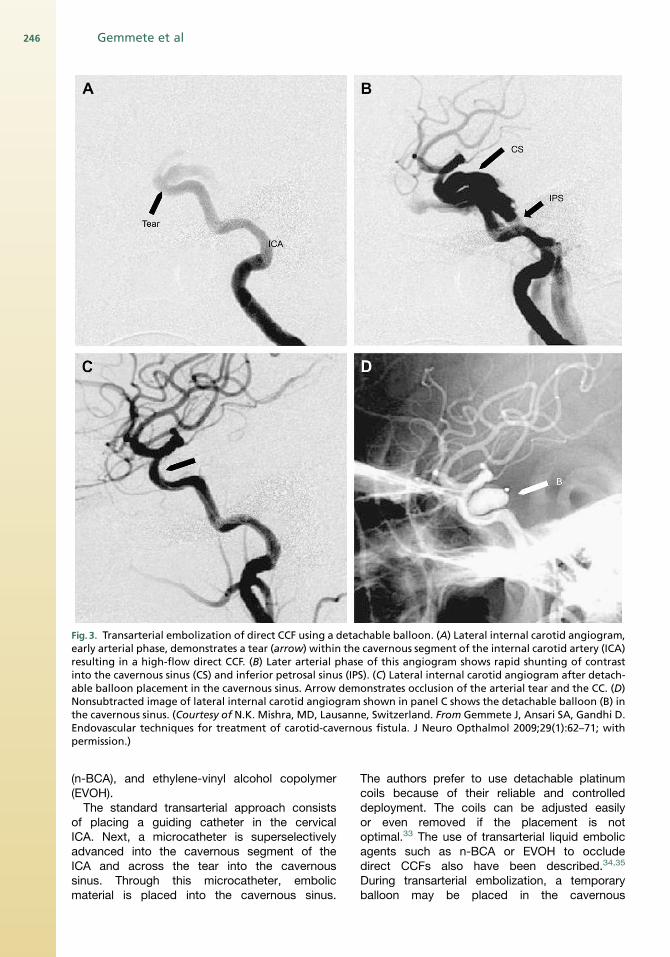
Fig. 3. Transarterial embolization of direct CCF using a detachable balloon. (A) Lateral internal carotid angiogram,early arterial phase, demonstrates a tear (arrow) within the cavernous segment of the internal carotid artery (ICA)resulting in a high-flow direct CCF. (B) Later arterial phase of this angiogram shows rapid shunting of contrastinto the cavernous sinus (CS) and inferior petrosal sinus (IPS). (C) Lateral internal carotid angiogram after detach-able balloon placement in the cavernous sinus. Arrow demonstrates occlusion of the arterial tear and the CC. (D)Nonsubtracted image of lateral internal carotid angiogram shown in panel C shows the detachable balloon (B) inthe cavernous sinus. (Courtesy of N.K. Mishra, MD, Lausanne, Switzerland. From Gemmete J, Ansari SA, Gandhi D.Endovascular techniques for treatment of carotid-cavernous fistula. J Neuro Opthalmol 2009;29(1):62–71; withpermission.)
Gemmete et al246
(n-BCA), and ethylene-vinyl alcohol copolymer(EVOH).
The standard transarterial approach consistsof placing a guiding catheter in the cervicalICA. Next, a microcatheter is superselectivelyadvanced into the cavernous segment of theICA and across the tear into the cavernoussinus. Through this microcatheter, embolicmaterial is placed into the cavernous sinus.
The authors prefer to use detachable platinumcoils because of their reliable and controlleddeployment. The coils can be adjusted easilyor even removed if the placement is notoptimal.33 The use of transarterial liquid embolicagents such as n-BCA or EVOH to occludedirect CCFs also have been described.34,35
During transarterial embolization, a temporaryballoon may be placed in the cavernous

Endovascular Treatment of Carotid Cavernous Fistulas 247
segment of the ICA (across the site of the tear)to protect the parent vessel and to preventmigration of the embolic material into the distalintracranial circulation.
CCFs caused by a small tear in the ICA can betreated with detachable balloons or coils asdescribed. If there is a fairly large rent in the artery,however, the coils or balloon may herniate (ormigrate) through the defect into the parent vessel.This retrograde herniation of embolic material cancause vessel occlusion and thromboemboliccomplications.
Recently, dedicated, intracranial, self-expand-ing stents have become available. These stentsare approved by the FDA for coil embolization ofwide-necked intracranial aneurysms. In the settingof direct CCFs, however, they can provide valu-able scaffolding to reconstruct a severely injuredintracranial ICA (see Fig. 2C).
When deployed across a traumatic tear, stentscreate a barrier between the ICA and thecavernous sinus, preventing retrograde herniationof coils into the parent artery.36 These devicesallow initial reconstruction of the damagedsegment of the ICA and then controlled depositionof coils into the cavernous sinus through eithera transarterial or transvenous approach. Usingthis technique, a direct CCF with severe injury tothe ICA now can be occluded while preservingthe ICA.
Direct CCFs caused by extensive injury to theICA may not be amenable to endovascular occlu-sion with preservation of the parent artery. In thesecases, occlusion of the arterial segment bearingthe fistula may be the only viable option for treat-ment. If time permits, and the patient is coopera-tive, a temporary balloon test occlusion study ofthe involved ICA is recommended before perma-nent occlusion. Some risk of ischemic complica-tions remain after ICA sacrifice despitea successful balloon test occlusion study.
To prevent retrograde flow from the supraclinoidICA into the fistula, vessel occlusion is initiatedcranial to the site of the suspected tear. Usingmultiple coils, the cavernous ICA then is occludedat the level of and caudal to the site of the fistula.This technique may be life saving in a patient whohas extensive and unstable injuries. Recently,hydrogel-coated detachable coils have been intro-duced that expand on contact with blood, a favor-able property especially in resilient high-flowfistulas that require high volumetric packing of thecavernous sinus. Furthermore, these coils mayfacilitate vessel sacrifice, decreasing the proce-dure and fluoroscopy times.37 Use of a vascularplug for arterial sacrifice also has been reported.38
Although the plug is easy to deploy and is effective,
the navigation of the vascular plug into the distalICA is currently difficult.
Placement of a stent or graft covered with poly-fluorotetraethylene is an alternative treatmentoption if ICA sacrifice is not desired or is unaccept-able because of an unsuccessful balloon testocclusion study (Figs. 2D and 4). Covered stentgrafts can be extremely useful for the immediateobliteration of a direct CCF and other fistulas.Additionally, they may decrease the risk ofischemic stroke by preserving the involved arterywhile simultaneously sealing the site of the fistula.A few reports have described the successful use ofa covered stent graft for the treatment of CCFs.39–41
Currently, the FDA has not approved any coveredstent for intracranial use in the United States. Thedisadvantages of the stents include stiff and high-profile construction that make it difficult to navi-gate them into the distal ICA, risk of endoleaks,possibility of coverage of vital perforators, andlack of long-term safety data.
Transarterial embolization of indirect low-flowCCFs generally is cumbersome because of thesmall size, tortuous anatomy, and multiplicity ofarterial feeders. The transvenous approach oftenis simpler and carries a high rate of success. Thevenous approach may fail in a small percentageof patients, however; in these cases, transarterialembolization still can be a viable alternative.
Transarterial techniques involve distal catheteri-zation of the small meningeal branches supplyingthe fistula. Ideally, superselective microcatheterplacement is performed with microcatheter tip asclose as possible to the point of fistulous commu-nication. Once a satisfactory microcatheter posi-tion is achieved, liquid embolic agents areinjected under fluoroscopic control with the goalof occluding the fistulous connections and pene-trating the cavernous sinus. Although coils andparticulate agents have been used, these agentsused alone cannot cause permanent occlusion ofthe fistula.
The most commonly used agent for transarterialembolization is n-BCA glue, a monomeric liquidadhesive approved in the United States for use inpresurgical embolization of cerebral arteriovenousmalformations.42 The viscosity and polymerizationtime of n-BCA is controlled by the addition of Lip-iodol. The advantages of n-BCA include its throm-bogenic nature and permanent occlusion of theinjected feeders. Drawbacks of this agent includeits rapid polymerization time (a few seconds),adhesive nature (risk of catheter retention), anda relatively long learning curve for its optimal use.
EVOH is another useful liquid embolic agent,recently approved by the FDA for the preoperativeembolization of brain arteriovenous

Fig. 4. High-flow, posttraumatic CCF treated by a combination of transarterial and transvenous approaches. (A, B)Axial CT scan demonstrates proptosis, preseptal edema, and a dilated SOV (arrow). (C) Anteroposterior and (D)lateral left internal carotid angiograms from the same patient. Note the high-flow direct CCF from a tear withinthe cavernous segment of the ICA filling the cavernous sinus (arrow), SOV, and pterygoid venous plexus (arrow-head). (E) Spot fluoroscopic image during the placement of a covered stent within the cavernous segment of theICA (arrows). (F) Control angiogram from the same patient after placement of a covered stent in the cavernoussegment reveals significantly reduced opacification of the fistula. Slight residual shunting remained even afterthe placement of this stent, however. (G) The residual fistula was obliterated via coil embolization (arrows iden-tify the subtraction artifact caused by the coils) of the cavernous sinus using a transvenous IPS approach. This finalcontrol angiogram shows complete closure of the fistula.
Gemmete et al248
malformations, that also may be used for transar-terial embolization of indirect CCFs. EVOH isa nonadhesive liquid embolic agent with a lavalikeflow pattern. It is supplied in ready-to-use vialswith mixture of EVOH, dimethyl sulfoxide solvent(DMSO), and tantalum. Currently 6% and 8%EVOH concentrations (dissolved in DMSO) areavailable in the United States. When the mixturecontacts aqueous media, such as blood, DMSOrapidly diffuses away from the mixture causingin-situ precipitation, solidification of the polymer,
and the formation of a spongy embolus. Polymer-ization occurs more slowly than with n-BCA, andbecause EVOH is nonadherent to the walls of thevessel or microcatheter, it allows prolonged injec-tion times while decreasing the chances of perma-nent microcatheter retention. Because of theslower polymerization times, the ability to makecontrolled injections over minutes, and the abilityto direct and push the embolic agent into thedesired location, the use of EVOH may allow betterand more distal penetration of the nidus or fistula
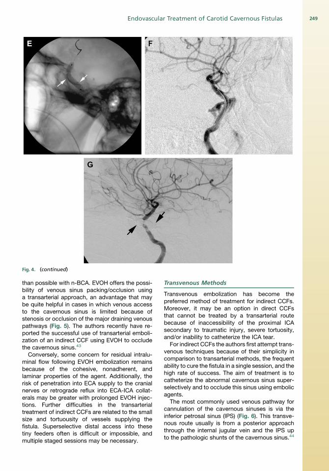
Fig. 4. (continued)
Endovascular Treatment of Carotid Cavernous Fistulas 249
than possible with n-BCA. EVOH offers the possi-bility of venous sinus packing/occlusion usinga transarterial approach, an advantage that maybe quite helpful in cases in which venous accessto the cavernous sinus is limited because ofstenosis or occlusion of the major draining venouspathways (Fig. 5). The authors recently have re-ported the successful use of transarterial emboli-zation of an indirect CCF using EVOH to occludethe cavernous sinus.43
Conversely, some concern for residual intralu-minal flow following EVOH embolization remainsbecause of the cohesive, nonadherent, andlaminar properties of the agent. Additionally, therisk of penetration into ECA supply to the cranialnerves or retrograde reflux into ECA-ICA collat-erals may be greater with prolonged EVOH injec-tions. Further difficulties in the transarterialtreatment of indirect CCFs are related to the smallsize and tortuousity of vessels supplying thefistula. Superselective distal access into thesetiny feeders often is difficult or impossible, andmultiple staged sessions may be necessary.
Transvenous Methods
Transvenous embolization has become thepreferred method of treatment for indirect CCFs.Moreover, it may be an option in direct CCFsthat cannot be treated by a transarterial routebecause of inaccessibility of the proximal ICAsecondary to traumatic injury, severe tortuosity,and/or inability to catheterize the ICA tear.
For indirect CCFs the authors first attempt trans-venous techniques because of their simplicity incomparison to transarterial methods, the frequentability to cure the fistula in a single session, and thehigh rate of success. The aim of treatment is tocatheterize the abnormal cavernous sinus super-selectively and to occlude this sinus using embolicagents.
The most commonly used venous pathway forcannulation of the cavernous sinuses is via theinferior petrosal sinus (IPS) (Fig. 6). This transve-nous route usually is from a posterior approachthrough the internal jugular vein and the IPS upto the pathologic shunts of the cavernous sinus.44

Fig.5. Indirect typeD CCF presentingwith right-sidedproptosis, chemosis, and periorbitaledema. (A) Right ICAangio-gram, lateral view, reveals inferolateral trunk (arrow) and meningohypophyseal trunk (arrowhead) minimally opaci-fying the cavernous sinus. (B) Lateral view of right external carotid angiogram demonstrating multiple small arterialfeeders (arrowheads) from the right internal maxillary artery filling a septated, irregular cavernous sinus. The inferiorpetrosal, superior petrosal, andthe circular sinuswereoccluded.Althoughthe SOV was patent,a severe stenosis at thejunction of the SOV with the angular vein prevented its catheterization. (C) Multiple attempts were made to cathe-terize the cavernous sinus without any success. Subsequently, a microcatheter was placed in the right internal maxil-lary artery, and an angiogram was performed via the microcatheter (anteroposterior view shown here). The CCFfeeders are indicated by arrowheads and the cavernous sinus by an arrow. (D) The CCF was embolized with EVOHvia a transarterial approach. The EVOH penetrated from the distal internal maxillary artery into the arterial branchesfeeding the fistula and subsequently into the cavernous sinus. This postembolization control angiogram of the rightECA demonstrates complete occlusion of the fistula. The EVOH cast is seen here as a subtraction artifact (arrowheads).
250
If the IPS is occluded or absent, access into thecavernous sinus can be obtained from an anteriorapproach through the superior ophthalmic vein(SOV) via the facial vein.45 Other percutaneoustransvenous approaches include the pterygoidvenous plexus, superior petrosal sinus, corticalveins, or the contralateral IPS or SOV with accessinto the ipsilateral cavernous sinus through thecircular sinus.46,47
Alternatively, in extremely difficult cases ofvenous occlusion, stenosis, or marked tortuosity,combined surgical and endovascular approachesmay be needed to access the cavernous sinus.Direct transorbital puncture or indirect puncturethrough the superior or inferior ophthalmic vein
(SOV/IOV) allows straightforward access to thecavernous sinus.48 Surgical access also may beobtained into the SOV, superficial middle cerebralvein, or sphenoparietal sinus leading to thecavernous sinus. Following catheterization of thecavernous sinus, disconnection of the venousoutflow from the feeding arteries at the level ofthe fistula can be initiated with detachable coils(Fig. 7) or liquid embolic agents.
Embolic materials include coils, n-BCA, andEVOH, can be used either alone or in combination.Again, the advantages of coils include their radio-pacity, ease of use, and the ability to redeploy orremove the devices if the initial placement is notoptimal. However, there may be difficulty in
Fig. 6. An artist’s rendition of the skull base dural sinuses looking from above demonstrates multiple possiblepathways to again access into the cavernous sinus for transvenous occlusion of a CCF. If the inferior petrosal sinusis patent, it provides a relatively straightforward access to the cavernous sinus. Other access routes include theSOV, IOV, superior petrosal sinus, and the basilar venous plexus. (Courtesy of L. Gregg, Baltimore, MD; withpermission. Copyright ª Lydia Gregg 2009.)
Endovascular Treatment of Carotid Cavernous Fistulas 251
achieving adequate volumetric packing orcomplete occlusion, especially in septatedcavernous sinuses. Moreover, the reported ratesof cranial nerve paresis are higher with coil embo-lization, probably because of their mass effect.
Consequently, transvenous liquid embolicagents are being used increasingly, either aloneor in combination with platinum coils.49,50 Liquidembolic agents can readily permeate differentsinus compartments, allowing complete occlusionof the fistula. In a series of 14 patients in whichn-BCA was used either alone or in conjunctionwith coils, Wakhloo and colleagues49 reportedthe technique to be safe and effective for the treat-ment of complex indirect CCFs in symptomaticpatients.
As described earlier, EVOH is a new liquidembolic agent that may be used in the transve-nous embolization of direct or indirect CCFs. Atthis time, only a few case reports describe thesuccessful transvenous treatment of indirect
CCFs with EVOH.50 It must be remembered thatEVOH has a propensity for retrograde filling ofarterial feeders and must be used cautiously. Theinadvertent penetration of EVOH into ECAbranches supplying the cranial nerves can poten-tially result in cranial nerve paralysis. Similarly,there is the potential for EVOH to migrate into themeningeal branches of the ICA and thereby refluxretrogradely into the ICA. Therefore, careful atten-tion should be paid to the EVOH cast as the embo-lization progresses. Control angiograms should beperformed if there is any doubt regarding reflux ofthis agent into the arterial feeders supplying thefistula. Given its favorable properties, EVOH prob-ably will be used more widely in the future for thetransvenous treatment of CCFs.
Endovascular Outcomes
The reported success rate of detachable balloonembolizations for direct CCFs is 88% to

Fig.7. Transvenous embolization of an indirect CCF using detachable coils. (A) Axial source image from a time-of-flightMR angiographic image reveals an enlarged left cavernous sinus, a finding consistent with a clinically known CCF. (B)Lateral view of the left common carotid angiogram confirms a type D CCF, filling from the meningeal branches of theleft ICA (LICA) and left ECA (LECA). (C) Lateral view of the selective left external carotid angiogram demonstrates thebranches of the internal maxillary artery (IMAX) and the middle meningeal artery (MMA) contributing to the CCF. (D)The inferior and superior petrosal sinuses as well the circular sinus were occluded in this patient. The SOV was cathe-terized via superficial temporal vein (STV), and access to the cavernous sinus was obtained. This lateral view of the leftexternal carotid angiogramdemonstrates themicrowire at theconfluenceof the left SOVand the cavernous sinus (CS).The microwire courses through the STVand the SOV. AV denotes the angular vein. (E) Lateral projection shows detach-able coils within the left cavernous sinus (CS). (F) Lateral view of a left common carotid angiogram (LCCA) after coilembolization of the cavernous sinus demonstrates complete occlusion of the CCF.
Gemmete et al252

Endovascular Treatment of Carotid Cavernous Fistulas 253
99%.6,7,51 Kobayashi and colleagues52 achieved80% aneurysmal and 55% posttraumatic CCFclosure with a detachable balloon. Higashida andcolleagues6 treated more than 200 traumaticCCFs , achieving complete occlusion of the fistulain 99% of patients and preserving the parent arteryin 88% of patients. Gupta and colleagues53
achieved complete occlusion in 86.3% of fistulas,nearly total occlusion in 11.0%, and ICA preserva-tion in 98%. The percentage of patients experi-encing morbidity associated with embolizationsof direct CCFs, such as ICA occlusion or wors-ening of ocular palsy, has ranged from 10% to40%.7,50,54,55
The reported complete cure rate for indirectCCFs is 70% to 90% with a complication rate of2.3% to 5%.22,56–58 Meyers and colleagues58
reported on endovascular treatment and clinicaloutcome in 135 patients who had indirect CCFsover a 15-year period. Endovascular treatmentwas performed in 133 patients (98%), and clinicalfollow-up was available in all 135 patients (meanduration of follow-up 56 � 4.3 months). Angio-graphic follow-up was performed in 72 patients(54%) who had ongoing symptoms or a history offistula with high-risk angiographic features. Ata mean follow-up of 56 months, 121 patients(90%) were clinically cured. The rate of proce-dure-related permanent morbidity was 2.3%.There was no operative mortality.
ACKNOWLEDGMENTS
The authors thank Lydia Gregg, BFA, MA,medical illustrator and research associate, Divi-sion of Interventional Neuroradiology, JohnsHopkins University, for the anatomic illustrationspresented in this article.
REFERENCES
1. Barrow DL, Spector RH, Braun IF, et al. Classification
and treatment of spontaneous carotid-cavernous
sinus fistulas. J Neurosurg 1985;62(2):248–56.
2. Locke CE. Intracranial arteriovenous aneurism or
pulsating exophthalmos. Ann Surg 1924;80(1):1–24.
3. Tomsick TA. Type A (direct) CCF: etiology, preva-
lence, and natural history. In: Tomsick T, editor.
Carotid cavernous fistula. Cincinnati (OH): Digital
Educational Publishing; 1997. p. 35–8.
4. Debrun G, Lacour P, Vinuela F, et al. Treatment of 54
carotid-cavernous fistulas. J Neurosurg 1981;55(5):
678–92.
5. Van Dellen JR. Intracavernous traumatic aneurysms.
Surg Neurol 1980;13(3):203–7.
6. Higashida RT, Halbach VV, Tsai FY, et al. Interven-
tional neurovascular treatment of traumatic carotid
and vertebral lesions: results in 234 cases. AJR
Am J Roentgenol 1989;153(3):577–82.
7. Lewis AI, Tomsick TA, Tew JM. Management of 100
consecutive direct carotid-cavernous fistulas: results
of treatment with detachable balloons. Neurosurgery
1995;36(2):239–44.
8. Gossman MD, Berlin AJ, Weinstein MA, et al.
Spontaneous direct carotid-cavernous fistula in
childhood. Ophthal Plast Reconstr Surg 1993;
9(1):62–5.
9. Debrun G, Vinuela F, Fox AJ, et al. Indications for
treatment and classification of 132 carotid-
cavernous fistulas. Neurosurgery 1988;22(2):285–9.
10. Hieshima GB, Cahan LD, Mehringer CM, et al. Spon-
taneous arteriovenous fistulas of cerebral vessels in
association with fibromuscular dysplasia. Neurosur-
gery 1986;18(4):454–8.
11. Kashiwaga S, Tsuchida E, Goto K, et al. Balloon
occlusion of spontaneous carotid- cavernous fistula
in Ehlers-Danlos syndrome type IV. Surg Neurol
1993;39(3):187–90.
12. Houser OW, Campbell JK, Campbell RJ, et al. Arte-
riovenous malformation affecting the transverse
dural venous sinus—an acquired lesion. Mayo Clin
Proc 1979;54(10):651–61.
13. Graeb DA, Dolman CL. Radiological and patholog-
ical aspects of dural arteriovenous fistulas: case
report. J Neurosurg 1986;64(6):962–7.
14. Komiyama M, Nakajima H, Nishikawa M, et al. Trau-
matic carotid cavernous sinus fistula: serial angiog-
raphy studies from the day of trauma. AJNR Am J
Neuroradiol 1998;19(9):1641–4.
15. Pang D, Kerber C, Biglan AW, et al. External carotid-
cavernous fistula in infancy: case report and review
of the literature. Neurosurgery 1981;8(2):212–8.
16. Halbach VV, Higashida RT, Hieshima GB, et al.
Transvenous embolization of direct carotid
cavernous fistulas. AJNR Am J Neuroradiol 1988;
9(4):741–7.
17. D’Angelo VA, Monte V, Scialfa G, et al. Intracerebral
venous hemorrhage in ‘‘high risk’’ carotid-cavernous
fistula. Surg Neurol 1988;30(5):387–90.
18. Vinuela F, Fox AJ, Debrun GM, et al. Spontaneous
carotid-cavernous fistulas: clinical, radiological,
and therapeutic considerations: experience with 20
cases. J Neurosurg 1984;60(5):976–84.
19. Halbach VV, Hieshima GB, Higashida RT, et al.
Carotid cavernous fistulae: indications for urgent
treatment. AJR Am J Roentgenol 1987;149(3):
587–93.
20. Kwan E, Hieshima GB, Higashida RT, et al. Interven-
tional neuroradiology in neuro-ophthalmology. J Clin
Neuroophthalmol 1989;9(2):83–97.
21. Higashida RT, Hieshima GB, Halbach VV, et al.
Closure of carotid cavernous sinus fistulae by
external compression of the carotid artery and
jugular vein. Acta Radiol Suppl 1986;369:580–3.

Gemmete et al254
22. Halbach VV, Higashida RT, Hieshima GB, et al. Dural
fistulas involving the cavernous sinus: Results of
treatment in 30 patients. Radiology 1987;163(2):
437–42.
23. Elster AD, Chen MY, Richardson DN, et al. Dilated
intercavernous sinuses: an MR sign of carotid-
cavernous and carotid-dural fistulas. AJNR Am J
Neuroradiol 1991;12(4):641–5.
24. Huber P. A technical contribution to the exact angio-
graphic localization of carotid cavernous fistulas.
Neuroradiology 1976;10(5):239–41.
25. Miller NR. Diagnosis and management of dural
carotid-cavernous sinus fistulas. Neurosurg Focus
2007;23(5):E13.
26. Kai Y, Hamada J, Morioka M, et al. Treatment of
cavernous sinus dural arteriovenous fistulae by
external manual carotid compression. Neurosurgery
2007;60(2):253–7 [discussion: 257–8].
27. Parkison D, Downs AR, Whytehead LL, et al. Carotid
cavernous fistula: direct repair with preservation of
carotid. Surgery 1975;76(6):882–9.
28. Serbinenko FA. Balloon catheterization and occlu-
sion of major cerebral vessels. J Neurosurg 1974;
41(2):125–45.
29. Debrun G, Lacour P, Caron JP, et al. Detachable
balloon and calibrated-leak balloon techniques in
the treatment of cerebral vascular lesions. J Neuro-
surg 1978;49(5):635–49.
30. Goto K, Hieshima GB, Higashida RT, et al. Treatment
of direct carotid cavernous sinus fistulae. Various
therapeutic approaches and results in 148 cases.
Acta Radiol Suppl 1986;369:576–9.
31. Teng MM, Chang CY, Chiang JH, et al. Double-
balloon technique for embolization of carotid
cavernous fistulas. AJNR Am J Neuroradiol 2000;
21(9):1753–6.
32. Norman D, Newton TH, Edwards MSB. Carotid
cavernous fistula: closure with detachable silicone
balloon. Radiology 1983;149(1):149–59.
33. Halbach VV, Higashida RT, Barnwell SL, et al. Trans-
arterial platinum coil embolization of carotid-
cavernous fistulas. AJNR Am J Neuroradiol 1991;
12(3):429–33.
34. Lv XL, Li YX, Liu AH, et al. A complex cavernous
sinus dural arteriovenous fistula secondary to
covered stent placement for a traumatic carotid
artery-cavernous sinus fistula: case report. J Neuro-
surg 2008;108(3):588–90.
35. Luo CB, Teng MM, Chang FC, et al. Transarterial
balloon-assisted n-butyl-2-cyanoacrylate emboliza-
tion of direct carotid cavernous fistulas. AJNR Am
J Neuroradiol 2006;27(7):1535–40.
36. Moron FE, Klucznik RP, Mawad ME, et al. Endovas-
cular treatment of high flow carotid cavernous fistula
by stent-assisted coil placement. AJNR Am J Neuro-
radiol 2005;26(6):1399–404.
37. Kallmes DF, Cloft HJ. The use of hydrocoil for parent
vessel occlusion. AJNR Am J Neuroradiol 2004;
25(8):1409–10.
38. Ross IB, Buciuc R. The vascular plug: a new device
for parent artery occlusion. AJNR Am J Neuroradiol
2007;28(2):385–6.
39. Madan A, Mujic A, Daniels K, et al. Traumatic
carotid-cavernous sinus fistula treated with
a covered stent. Report of two cases. J Neurosurg
2006;104(6):969–73.
40. Felber S, Henkes H, Weber W, et al. Treatment of
extracranial and intracranial aneurysms and arterio-
venous fistulae using stent grafts. Neurosurgery
2004;55(3):631–8 [discussion: 638–9].
41. Gomez F, Escobar W, Gomez AM, et al. Treatment of
carotid cavernous fistulas using covered stents:
midterm results in seven patients. AJNR Am J Neu-
roradiol 2007;28(9):1762–8.
42. Nelson PK, Russell SM, Woo HH, et al. Use of
a wedged microcatheter for curative transarterial
embolization of complex intracranial dural arteriove-
nous fistulas: indications, endovascular technique,
and outcome in 21 patients. J Neurosurg 2003;
98(3):498–506.
43. Gandhi D, Ansari SA, Cornblath WT. Successful
transarterial embolization of a Barrow type D dural
carotid-cavernous fistula with ethylene vinyl alcohol
copolymer (Onyx). J Neuro Opthalmol 2009;29(1):
9–12.
44. Klisch J, Huppertz HJ, Spetzger U, et al. Transve-
nous treatment of carotid cavernous and dural
arteriovenous fistulae: results for 31 patients and
review of the literature. Neurosurgery 2003;53(4):
836–56.
45. Biondi A, Milea D, Cognard C, et al. Cavernous
sinus dural fistulae treated by transvenous approach
through the facial vein: report of seven cases and
review of the literature. AJNR Am J Neuroradiol
2003;24(6):1240–6.
46. Mounayer C, Piotin M, Spelle L, et al. Superior
petrosal sinus catheterization for transvenous embo-
lization of a dural carotid cavernous sinus fistula.
AJNR Am J Neuroradiol 2002;23(7):1153–5.
47. Jahan R, Gobin YP, Glenn B, et al. Transvenous
embolization of a dural arteriovenous fistula of the
cavernous sinus through the contralateral pterygoid
plexus. Neuroradiology 1998;40(3):189–93.
48. White JB, Layton KF, Evans AJ, et al. Transorbital
puncture for the treatment of cavernous sinus dural
arteriovenous fistulas. AJNR Am J Neuroradiol
2007;28(7):1415–7.
49. Wakhloo AK, Perlow A, Linfante I, et al. Transvenous
n-butyl-cyanoacrylate infusion for complex dural
carotid cavernous fistulas: technical considerations
and clinical outcome. AJNR Am J Neuroradiol
2005;26(8):1888–97.

Endovascular Treatment of Carotid Cavernous Fistulas 255
50. Suzuki S, Lee DW, Jahan R, et al. Transvenous treat-
ment of spontaneous dural carotid-cavernous fistulas
using a combination of detachable coils and Onyx.
AJNR Am J Neuroradiol 2006;27(6):1346–9.
51. Debrun G, Lacour P, Vinuela F, et al. Treatment of 54
traumatic carotid-cavernous fistulas. J Neurosurg
1981;55(5):678–92.
52. Kobayashi N, Miyachi S, Negoro M, et al. Endovas-
cular treatment strategy for direct carotid-cavernous
fistulas resulting from rupture of intracavernous
carotid aneurysms. AJNR Am J Neuroradiol 2003;
24(9):1789–96.
53. Gupta AK, Purkayasta S, Krishnamoorthy T, et al.
Endovascular treatment of direct carotid cavernous
fistulae: a pictorial review. Neuroradiology 2006;
48(11):831–9.
54. Klisch K, Schipper J, Husstedt H, et al. Transsphe-
noidal computer-navigation-assisted deflation of
a balloon after endovascular occlusion of a direct
carotid cavernous sinus fistula. AJNR Am J Neurora-
diol 2001;22(3):537–40.
55. Halbach VV, Higashida RT, Dowd CF, et al. Treat-
ment of carotid cavernous fistulas associated with
Ehlers-Danlos syndrome. Neurosurgery 1990;26(6):
1021–7.
56. Picard L, Bracard S, Mallet J, et al. Spontaneous du-
ral arteriovenous fistulas. Seminars in Interventional
Radiology 1987;4:219–40.
57. Turjman F, Bascoulergue Y, Rosenberg M, et al.
Dural fistulae of the cavernous sinus treated with
embolization. Ten cases. J Neuroradiol 1992;19(4):
256–70.
58. Meyers PM, Halbach VV, Dowd CF, et al. Dural
carotid cavernous fistula: definitive endovascular
management and long-term follow-up. Am J Oph-
thalmol 2002;134(1):85–92.