Morphogenesis and clinicopathologic characteristics of recurrent carotid disease
14
Morphogenesis and clinicopathologic characteristics of recurrent carotid disease G. Patrick Clagett, M.D., Max Robinowitz, M.D., Jerry R. Youkey, M.D., Daniel F. Fisher, Jr., M.D., Richard E. Fry, M.D., Stuart I. Myers, M.D., Edward L. Lee, M.D., George J. Collins, Jr., M.D., and Renu Virmani, M.D., Dallas, Tex., and Washington, D.C. The histopathologic characteristics of primary plaques and recurrent carotid disease were studied in 32 patients. These data were related to symptoms, recurrence interval (6 to 176 months), arteriographic anatomy, and in situ operative findings. A striking predi- lection was noted for recurrent lesions to be located in the internal carotid artery near the origin, but still within the confines, of the original endarterectomy site and suture fine. A/though recurrence was frequently associated with a long primary arteriotomy, evidence of technical faults or periarterial fibrosis was rare. Early recurrent lesions (re- currence interval <36 months, n = 13) had significantly more smooth muscle cells and proteoglycans (p<0.001) than late recurrent lesions (recurrence interval >36 months, n = 19). As previously reported, features of atherosclerosis (abundant collagen, calcium deposits, and foam cells) were more pronounced in late recurrences (p<0.001). However, the histopathologic differentiation between early and late recurrent carotid disease was indistinct. A continuum was noted whereby characteristics of late recurrent lesions in- creased in proportion to recurrence interval. All recurrent lesions were easily distinguished from primary plaques in that recurrences had a less orderly arrangement of all elements and lacked the classic topographic features of advanced atherosclerosis. An important feature that differentiated primary and recurrent lesions was the presence of surface and intraplaque thrombus in 90% of recurrent lesions (p<0.001). In early recurrent disease, luminal surface thrombus was striking; this was frequently platelet-rich and showed organization devoid of neovascularity. Intraplaque thrombus was more common in late recurrent disease, consisted almost entirely of fibrin, and was often contiguous with luminal surface thrombus. No discernible relationships were noted between thrombus associated with recurrent lesions and the presence or absence of symptoms, treatment with antiplatelet agents, and hypertension. This finding suggests that thrombus was a continuous and intrinsic component of recurrent disease rather than a secondary, com- plicating feature. Recurrent carotid disease is a progressive lesion that stems from ongoing thrombogenesis occurring at the endarterectomy site. Organized thrombus and smooth muscle cell proliferation comprise the bulk of the lesion, which undergoes atherosclerotic change with time. (J VASC SURG 1986; 3:10-23.) Symptomatic recurrent carotid disease occurs in 0.6% to 3% (mean 1.2%) of patients after carotid endarterectomy. 1-4 Asymptomatic recurrent carotid From the Departments of Surgeryand Pathology, The University of Texas Health Science Center at Dallas and the Veterans Administration Medical Center at Dallas, the Department of Cardiovascular Pathology,The Armed Forces Institute of Pa- thology, Washington, D.C., and the Peripheral Vascular Sur- gery Service,Walter Reed Arm),Medical Center, Washington, D.C. Presented at the Thirty-third Scientific Meeting of the North American Chapter, International Society for Cardiovascular Surgery, Baltimore,Md., June 6-7, 1985. Reprint requests: G. Patrick Clagett, M.D.~ The University of TexasHealth Science Center at Dallas, Departmentof Surgery, 5323 Harry Hines Blvd., Dallas, TX 75235. 10 disease occurs with a much greater frequency, which varies widely (6.7% to 19% within 2 years after ca- rotid endarterectomy) depending on the noninvasive method of detection used. 5-13 A better understanding of the pathogenesis and natural history of this process is important since carotid endarterectomy is the most frequently performed noncardiac vascular operation in this country. 14 Although much information has been obtained by prospectively following patients after carotid endarterectomy with sensitive nonin- vasive tests, the findings of such series have been somewhat incomplete because of the lack of corre- lation with reoperative findings and the histopath- ologic features of retrieved recurrent lesions. In most
Transcript of Morphogenesis and clinicopathologic characteristics of recurrent carotid disease

Morphogenesis and clinicopathologic characteristics of recurrent carotid disease G. Patr ick Clagett , M.D., Max Robinowitz , M.D., Jerry R. Youkey, M.D. , Daniel F. Fisher, Jr., M.D., Richard E. Fry, M.D., Stuar t I. Myers, M.D., Edwa r d L. Lee, M.D., George J. Collins, Jr., M.D., and Ren u Virmani, M.D., Dallas, Tex., and Washington, D.C.
The histopathologic characteristics of primary plaques and recurrent carotid disease were studied in 32 patients. These data were related to symptoms, recurrence interval (6 to 176 months), arteriographic anatomy, and in situ operative findings. A striking predi- lection was noted for recurrent lesions to be located in the internal carotid artery near the origin, but still within the confines, of the original endarterectomy site and suture fine. A/though recurrence was frequently associated with a long primary arteriotomy, evidence of technical faults or periarterial fibrosis was rare. Early recurrent lesions (re- currence interval <36 months, n = 13) had significantly more smooth muscle cells and proteoglycans (p<0.001) than late recurrent lesions (recurrence interval >36 months, n = 19). As previously reported, features of atherosclerosis (abundant collagen, calcium deposits, and foam cells) were more pronounced in late recurrences (p<0.001). However, the histopathologic differentiation between early and late recurrent carotid disease was indistinct. A continuum was noted whereby characteristics of late recurrent lesions in- creased in proportion to recurrence interval. All recurrent lesions were easily distinguished from primary plaques in that recurrences had a less orderly arrangement of all elements and lacked the classic topographic features of advanced atherosclerosis. An important feature that differentiated primary and recurrent lesions was the presence of surface and intraplaque thrombus in 90% of recurrent lesions (p<0.001). In early recurrent disease, luminal surface thrombus was striking; this was frequently platelet-rich and showed organization devoid of neovascularity. Intraplaque thrombus was more common in late recurrent disease, consisted almost entirely of fibrin, and was often contiguous with luminal surface thrombus. No discernible relationships were noted between thrombus associated with recurrent lesions and the presence or absence of symptoms, treatment with antiplatelet agents, and hypertension. This finding suggests that thrombus was a continuous and intrinsic component of recurrent disease rather than a secondary, com- plicating feature. Recurrent carotid disease is a progressive lesion that stems from ongoing thrombogenesis occurring at the endarterectomy site. Organized thrombus and smooth muscle cell proliferation comprise the bulk of the lesion, which undergoes atherosclerotic change with time. (J VASC SURG 1986; 3:10-23.)
Symptomatic recurrent carotid disease occurs in 0.6% to 3% (mean 1.2%) o f patients after carotid endarterectomy. 1-4 Asymptomatic recurrent carotid
From the Departments of Surgery and Pathology, The University of Texas Health Science Center at Dallas and the Veterans Administration Medical Center at Dallas, the Department of Cardiovascular Pathology, The Armed Forces Institute of Pa- thology, Washington, D.C., and the Peripheral Vascular Sur- gery Service, Walter Reed Arm), Medical Center, Washington, D.C.
Presented at the Thirty-third Scientific Meeting of the North American Chapter, International Society for Cardiovascular Surgery, Baltimore, Md., June 6-7, 1985.
Reprint requests: G. Patrick Clagett, M.D.~ The University of Texas Health Science Center at Dallas, Department of Surgery, 5323 Harry Hines Blvd., Dallas, TX 75235.
10
disease occurs with a much greater frequency, which varies widely (6.7% to 19% within 2 years after ca- rotid endarterectomy) depending on the noninvasive method o f detection used. 5-13 A better understanding o f the pathogenesis and natural history of this process is important since carotid endarterectomy is the most frequently performed noncardiac vascular operation in this country. 14 Although much information has been obtained by prospectively following patients after carotid endarterectomy with sensitive nonin- vasive tests, the findings of such series have been somewhat incomplete because of the lack of corre- lation with reoperative findings and the histopath- ologic features of retrieved recurrent lesions. In m o s t

Volume 3 Number 1 January 1986 Recurrent carotid disease 11
reports, few patients with abnormal findings from noninvasive tests underwent reoperation. The pres- ent report focuses on thc findings at reoperation and the histopathologic features of recurrcnt lesions, and by doing so, offers insight into the nature and patho- genesis of recurrent carotid disease.
METHODS
Patients. Detailed morphologic studies were per- formed on specimens obtained from 32 patients with recurrent carotid disease who underwent reoperative carotid endarterectomy. Many of these patients were the subject of a previous report that focused on sys- temic factors for recurrent carotid disease, is The pa- tients in this report were selected because complete clinical, angiographic, operative, gross, and histo- pathologic findings were able to be correlated. Most of this information was collected prospectively as part of an ongoing inquiry into the cfinicopathologic fea- tures of recurrent carotid disease.
Morphologic analyses. At operation, careful notes and drawings were made of the location, ex- tent, and character of the recurrent carotid lesion. Of particular interest was the relationship between the recurrent disease and the primary arteriotomy suture line. In most, photographs were taken of the in situ findings that were compared with arteriographic ab- normalities. An attempt was made to remove the recurrent lesion en bloc as a single endartercctomy specimen but in many cases this was impossible be- cause of the adherent, nonhomogeneous character of these lesions. Despite this problem it was possible in most cases to orient the specimen correctly so that histopathologic findings could be correlated with an- atomic sites. After the specimens were removed, they were fixed in formalin and rephotographed before they were embedded in paraffin.
In all specimens, the qualitative cellular features were assessed by light microscopy in sections stained with hematoxylin and eosin and Movat pentachrome. The latter stains five major elements of the vascular wall and allows clear differentiation between cell cy- toplasm and nuclei, elastin, ground substance (pro- teoglycans), and collagen. Iron staining was per- formed on selected specimens to confirm the presence of hemosiderin. Specimens of special interest werc removed from paraffin blocks and prepared for ul- trastructural studies. Electron microscopic findings were correlated with light microscopic features. In some specimens, an attempt was made to identify and differentiate cells by immunoperoxidase staining for desmin (an intermediary filament marker for smooth muscle cells), vimentin (a marker for mes-
e n c h y m a l t issue) , 16 and Willebrand's factor for en- dothelial cells. Immunoperoxidase staining for fi- brinogen (fibrin) was also performed on selected specimens.
We retrieved paraffin-embedded specimens from the original endarterectomy in 20 patients and these were compared with the recurrent lesions. The pri- mary endarterectomy specimens were also compared with specimens from patients undergoing carotid en- darterectomy but who did not show evidence of re- current disease.
All microscopic sections of primary and recurrent lesions were studied by a cardiovascular pathologist (M.R.) who had no knowledge of the clinical details of the case or whether the lesion was primary or recurrent. In a blinded fashion he graded the mor- phologic features on an arbitrary scale of 0 to 4 +. These features included the presence and relative quantity of spindle cells (modified smooth muscle cells), proteoglycans, collagen, foam cells, calcifica- tion, neovascularity, and thrombus. When noting thrombus a special effort was made to determine whether the thrombus was predominantly on the lu- minal surface of the lesion or whether it appeared to originate from the interior of the plaque.
To determine the local anatomy and histologic characteristics of the arterial wall immediately fol- lowing routine carotid endarterectomy, studies were performed on 30 patients undergoing primary en- darterectomy. These patients were part of a separate study and none have had recurrent disease to date (mean follow-up less than 1 year). After the plaque and diseased media were completely removed, an as- sessment was made of the character of the arterial wall remaining, with particular attention paid to the depth of the endarterectomy plane in relation to the carotid sinus and the proximal internal carotoid ar- tery. Four planes of endarterectomy were deter- mined: (1) subintimal, (2) medial (circular fibers at base), (3) junction of media and adventitia (no cir- cular fibers at base), and (4) into adventitia (textured,
"fuzzy" Surface remaining). Drawings and photo- graphs were made and biopsies of the remaining wall were obtained to confirm the visual impressions.
RESULTS
Twenty-three men and nine women comprised the study group. Because of inclusion of patients from a Veterans Administration hospital, the female prevalence was not as high as reported previously, as The mean age (___ SEM) at the time of reoperative carotid endartercctomy was 62 -+_ 2 years (range, 50 to 73 years). Thc mean interval between primary

12 Claget~ et aL
Journal of VASCULAR
SURGEKY
\
• !:[
" Apex (3)
,.,, ICA(9)
,~ Origin ICA (8)
Bulb (3)
CCA (3)
Site of Most Stenotic Disease
Fig. 1. Sites of recurrent lesions in relation to old suture line./CA = internal carotid artery; CCA = common ca- rotid artery.
and reoperative carotid endarterectomy was 63 + 8 months (range, 6 to 176 months) and was similar for men and women. One third of the recurrent le- sions were detected within .1 year after primary ca- rotid endarterectomy and the remaining two thirds were fairly evenly distributed throughout the re- maining 15-year recurrence interval. Twenty-one pa- tients with recurrent carotid disease had focal neu- rologic symptoms and in 13 (63%) of these, the symptoms were similar to those occurring before the
-primary carotid endarterectomy. Sixteen (76%) of the patients who had symptoms had abnormal find- ings from ocular pneumoplethysmography (OPG/ Gee) examinations. Eleven patients were without symptoms at the time of detection of recurrence, and all had abnormal OPG/Gee results.
Atherosclerotic risk factors and complications were frequently encountered among these patients. Hypertension was present in 22 (69%) and a history of cigarette smoking was obtained from 30 (94%). Twenty-seven (84%) patients continued smoking af- ter primary carotid endarterectomy and most of these patients were heavy smokers. A strong familial his- tory of atherosclerosis was present in 27 (84%) and hyperlipidemia was diagnosed in 14 (44%). Two pa- tients had insulin-dependent diabetes mellitus. At the time of primary operation, all patients underwent
primary closure of the arteriotomy without a vein or prosthetic patch angioplasty. Nineteen patients (59%) had bilateral primary carotid endarterecto- roles; in 17 of these, the recurrent carotid disease was unilateral and in two the recurrences were bilateral. Both of the latter two patients were women.
Angiographic and in situ operative findings
We accurately identified the site of the most ste- notic portion of the recurrent disease in relation to the old suture line in 26 patients (Fig. 1). The re- current lesion was located at the origin, or 2 to 4 cm beyond the origin, of the internal carotid artery in 17 (65%) patients. The lesion was within the con- fines of the old suture line and primary endarterec- tomy site in all of these cases. Most of the recurrent lesions located beyond the origin of the internal ca- rotid artery (but still within the upper limits of the old suture line) were associated with a long primary arteriotomy with the suture line often extending 4 to 5 cm onto the internal carotid artery.
Recurrent disease located beyond the origin of the internal carotid artery was frequently character- ized by long, tubular, concentric thickenings of the intimal surface. These features had a characteristic appearance angiographically (Fig. 2, A). Preopera- tive evaluation of these arteriograms frequently led to the erroneous interpretation that these stenoses represented "damp injury" beyond the apex of the suture line or a focal narrowing at the apex (presum- ably because of constricting suturing technique or retained primary plaque). However, at reoperation the majority of these distal lesions were found within the confines of the primary endarterectomy site and suture line. There was no evidence of a greater con- centration of disease along the wall contiguous with the suture line. In only three patients, the predom- inant recurrent lesion was located either at the apex or just beyond the apex. In one case periarterial fi- brosis appeared to contribute to the stenosis of the internal carotid artery. Although precise measure- ments of arterial size were not obtained at reopera- tion, abnormally small internal carotid arteries were not noted except in one case.
In recurrences found at the origin of the internal carotid artery (eight patients) and at the mid-carotid bulb region (three patients), the lesions were eccen- tric and thickest posterolaterally along the outer wall. Often this was the site of the primary lesion. These primary and recurrent lesions had a similar anglo- graphic appearance (Fig. 2, C and D).
The common carotid artery was the site of re- current disease in three cases. All of these were lo-

Vokun¢ 3 Number 1 January 1986 Recurrent carotid disease 13
Fig. 2. A, Early (7 months) recurrent lesion. Concentric, smooth, tubular narrowing beyond origin of internal carotid artery was characteristic of early recurrences. Distal stenosis (top arrow) was thought to represent focal narrowing at apex of suture line or distal clamp injury. However, at reoperation this area was well within limits of primary cndarterectomy site and suture line. B, Late (168 months) recurrent lesion. Complex, irregular surface was characteristic of late recurrences. These surface irregularities represented excrescences of thrombus. C, Primary carotid stenosis at origin of internal carotid artery, and (D) recurrent lesion (11 months) at same site.
cared within millimeters of the starting point of the suture line on the common carotid artery at the site of transition between diseased intima and the primary endarterectomy. At this point a shelf of retained plaque 1 to 2 mm thick was seen, on which was an adherent layer (1 to 2 mm in thickness) of fibro- muscular proliferative tissue (Fig. 3). Both of these components contributed equally to the stenosis. This tissue had the same histologic characteristics as the neointimal fibromuscular hyperplasia noted in early recurrences (discussed later).
A striking finding was the presence of gross thrombus found in 19 (59%) of the 32 patients with recurrent carotid disease. The incidence of thrombus
found on microscopic examination was much higher. Thrombus appeared to form the bulk of the recurrent lesion in many patients. Gross thrombus was more frequently noted in patients with a recurrence interval of more than 36 months (13 [68%] of 19 patients) than in those with a recurrence interval of less than 36 months (6 [46%] of 13 patients). The character of the thrombus also depended on the age of the recurrent lesion. In patients with earlier recurrences (interval less than 36 months), the thrombotic mass tended to be a smooth-surfaced, thin, even layer ad- herent to the underlying tissue of the recurrent lesion. Often this mass was difficult to note and could be easily missed unless specifically looked for and con-

i
Clagett et al.
Journal of VASCULAR
SURGERY 14
Fig. 3. A, Schematic drawing and (B) arteriogram of findings at reoperation in patient with recurrent disease at common carotid artery (22 months). Cross-hatched area (A) represents plaque remaining after primary operation, which was covered with smooth layer of pale, white tissue that proved to be neointimal fibromuscular hyperplasia and thrombus.
Table I. Histopathologic features of primary carotid plaques and recurrent lesions
Recurrent lesions (Interval)
Feature present in Primary plaque <36 mo >36 mo moderate-to-large amounts (n = 20) (n = 13) (n = 19)
Smooth musde (spindle) cells 1 (5%) 12 (92%)* 4 (21%) Collagen 20 (100%) 1 (8%)* 18 (95%) Calcification 16 (80%) 0* 14 (74%) Foam cells 17 (85%) 2 (I5%)* 17 (89%) Neovascularity 8 (40%) I (8%)+ 10 (53%) Thrombus 8 (40%):~ i0 (77%) 19 (i00%)
*Significantly different (p < 0.001) from primary plaques and recurrent lesions >36 mo. t Significantly different (p < 0.05) from recurrent lesions >36 months and almost significantly different (p < 0.10) from primary plaques. ~:Significantly different from recurrent lesions <36 months (p <0.05) and >36 months (p < 0.001).
firmed histologically. The thrombus tended to be much more exuberant in those patients with older recurrences. At gross inspection, this material was gelatinous and friable, had an uneven surface, and extended deep into the crevices of the recurrent le- sion. A surprising finding was that there was no cor- relation between the presence or absence of focal neurologic symptoms and the presence or amount of gross thrombus. Patients without symptoms were just as likely to have large amounts of thrombotic material as those with focal symptoms.
As noted by other investigators, lesions that re- curred within 2 to 3 years of the primary endar- terectomy were pale, white, rubbery, and densely adherent to the arterial wall, which made reopera- tivc endartercctomy difficult, aa4 These lesions often had to be removed piecemeal. Although these le-
sions sometimes had an innocent appearance in that the surfaces were rdatively smooth and glistening, thrombus tended to gmooth out the surface irregu- larities. Older recurrent lesions had more o f the gross features ofatherosclcrosis in that they were yellowish, were stiff and gritty when handled (evidence of fi- brosis and calcification), and were much easier to remove by endarterectomy; most of the specimen could be removed in a single piece. These lesions had an irregular, complex surface characteristic of ulcer- ated plaques angiographically (Fig. 2, B). At in situ inspection, however, the irregular surface proved to bc mounds and excrescences of thrombotic material dissecting under a friable and unstable surface (Fig. 4). It was interesting that many of these older lesions had areas of pale, white fibrous tissue that had the identical consistency and character as younger le-

Volume 3 Number 1 January 1986 Recurrent carotid disease 15
sions. These areas were usually at the margins and the base of the older recurrent lesions (Fig. 4).
Histopathologic findings
Lesions w i t h recurrence interval o f less than 36 months. The histopathologic characteristics of primary plaques and recurrent lesions are compared in Table I. As noted by others, the features of early recurrent lesions were different from later recur- rences, and for comparative purposes, lesions recur- ring within 36 months of carotid endarterectomy were tabulated separately from those recurring at l a t e r i n t e r v a l s . 2'3'14 The 36-month point was chosen because this time interval most clearly distinguished early and late recurrent lesions.
Microscopic thrombus was associated with 77% of the early recurrent lesions (Table I) and was usually present on the surface of the lesion (Fig. 5, B, C, and'D). The superficial layers of the thrombus con- tained platelets. In some specimens the thrombus was not always apparent at gross inspection, but immu- noperoxidase staining confirmed the presence of a layer of fibrin. A transitional zone was noted between the fibromuscular proliferative process and the thrombus in many specimens, which represented or- ganization of the thrombus (Fig. 5, B and C).
Endothelium could not be identified on the sur- face of these lesions, possibly because of handling during operation and excessive storage of the spec- imen in formalin for several months. This fact also precluded accurate immunoperoxidase anti-von Wil- lebrand's factor staining. Equivocal results were also noted for immunoperoxidase staining for vimentin and desmin.
Neointimal fibromuscular hyperplasia (prolifer- ation) was characteristic of lesions found within 36 mon~ ¢ of the primary endarterectomy (Fig. 5, A, B, C, and D). This process consisted of spindle and stellate cells surrounded by proteoglycans and sparse amounts of collagen and elastic fibers. There was a virtual absence of calcification. Neovascularity, de- fined as the presence of capillaries in the lesion, was detectable in small amounts in most specimens. It was predominantly present at the base of the lesion and was abundant in only one case. Hemosiderin was seen in small focal areas in only two lesions. Foam cells (lipid-laden cells probably of modified smooth muscle cell origin) were seen in only two early re- current lesions; however, they were frequently seen in late recurrent lesions (89%). Foam cells were usu- ally seen on the surface and rarely close to the base of the lesion.
Ultrastructural examination of the early recurrent lesions showed that most of the spindle cells had
Fig. 4. In situ (A) and gross specimen (B) from patient with late (1~68 months) recurrence (arteriogram shown in Fig. 2, B). Note irregular, friable thrombus. B, Arrows at margins of lesion indicate areas ofneointimal fibromuscular hyperplasia (histologic characteristics shown in Fig. 5, E).
features of smooth muscle cells (presence of a base- ment membrane, myofilaments, and dense bodies) (Fig. 6). Rare fibroblasts were identified because of large amounts of rough endoplasmic reticulum and the absence of smooth muscle cell features. Some of the smooth muscle cells had a stellate shape. Collagen fbers, elastic fibers, and fibrin were identified in the intercellular matrix (Fig. 6).
Lesions with recurrence interval o f more than 36 months. All of the older lesions contained mi- croscopic thrombus on the surface, and, in addi- tion, many had thrombus within the lesion (Table I) (Fig. 5, E and F). In comparison to early lesions, later recurrences had extensive thrombus formation, which extended to the base of the lesion and always communicatcd with the luminal surface. The surfaces of thesc lesions often appcared fragile with areas of thrombus dissecting beneath the neointima (Fig. 5, E). Intralesion thrombus was dctectcd in 14 instances (74%) of later recurrent disease in comparison to two cases (15%) of early recurrent disease (p<0.02). Thrombus was composed almost exclusively of pure fibrin. Extensive neovascularity was often present in lcsions adjacent to the thrombi, but only a few lesions contained definite areas of acute intraplaquc hem-

16 Clagett et al.
Journal of VASCULAR
SURGERY
Fig. 5. A, Neointimal fibromuscular hyperplasia in early recurrence (8 months). Spindle and stellate-shaped cells are the same cell type (confirmed as modified smooth muscle cells by ultrastructural analysis) but cut at different angles in matrix of proteoglycans. (M0vat stain, × 60). B, Transition between organizing thrombus at luminal surface and neointimal fibro- muscular hyperplasia in early recurrent lesion (7 months) (Hematoxylin-eosin stain, × 60). C, Organization of surface thrombus on neointimal fibromuscular hyperplastic lesion (recurrence at 34 months). (Movat stain, × 60). D, Fibrin thrombus located beneath and on neointimal surface. This may represent incorporation of fibrin into recurrent lesion (recurrence at 11 months). (Movat stain, x 60.) E, Surface thrombus dissecting beneath fragile neointimal layers of late recurrent lesion (168 months). Deeper layers consist of neointimal fibromuscular hy- perplasia identical to that found in early recurrences (Movat stain, x 60). F, Late recurrent lesion (53 months) demonstrating extensive thrombus deep within substance of lesion and communicating with luminal surface. Cholesterol clefts are present (Movat stain, x 25).

Volume 3 Number 1 lanuary 1986 Recurrent carotid disease 17
Fig. 6. Ultrastructural characteristics of neointimal fibromuscular hyperplasia. A, Dividing smooth muscle cell in late recurrent lesion (108 months). Note centriole between nuclei (×24,500). B, Spindle-shaped smooth muscle cell from early recurrence (11 months) (x 11,900). C, Stellate-shaped smooth muscle cell from late recurrent lesion (161 months) (× 24,500). B M = basement membrane; DB = dense body; M = myofilament; C = col- lagen; E = elastin.
orrhage. These lesions were composed of red cells, inflammatory cells, and macrophages with and with- out hemosiderin.
Features ofatherosclerosis (foam cells, cholesterol clefts, abundant collagen, and calcium) were more pronounced in later recurrent disease (i.e., more than 36 months) (Table I). However, the histopathologic differentiation between early and late lesions was in- distinct. Focal areas of neointimal fibromuscular hy- perplasia, a distinctive feature of early lesions, was also seen in late recurrences (Fig. 5, E). These areas were found in large amounts in two patients with the longest recurrence intervals (168 and 176
-months) . These lesions corresponded to the gross findings of pale, white, fibrous tissue noted at the
margins and within the interior at the base of the recurrent lesions (Fig. 4). A temporal sequence was also noted whereby the relative amounts of neoin- timal fibromuscular hyperplasia tended to decrease with increasing recurrence interval (Fig. 7, A). In contrast, the opposite was noted for features of ath- erosclerosis. As the recurrence interval increased, the relative amounts of foam cells, cholesterol clefts, dense collagen, and calcification tended to increase (Fig. 7, B and C).
Compar ison to pr imary plaques. Recurrent le- sions were histologically distinguishable from pri- mary carotid plaques. The pathologist, who was blinded as to which were primary plaques and re- current lesions, was able to make this distinction in

18 Clagett et al.
Journal of VASCULAR
SURGERy
SMOOTH MUSCLE (spindle) CELLS
i o
0
< 2
TR
0 ®
4 - • Q "
3 - 0 , ~
i
• • • e e
• e • • •
I , I , I , I , I tl
20 40 60 80 `100
COLLAGEN
I i I , I `120 `140 `160
0
2
(x TR
0 ®
4
3 •
%
t -
I
• e e
• • • • • •
I , I , I , I , I , I , I , I 20 40 60 80 `100 '120 `140 `160
FOAM CELLS
4
0 3
0
<
m TR
(90
, \
• ee • •
• • • ee • •
20 40 60 80 100 `120 `140 "160
RECURRENCE INTERVAL
Fig. 7. Relation between recurrence interval (months) and histopathologic features. A, Neointimal fibromuscular hy- perplasia tended to decrease with increasing recurrence in- terval. B and C, Atherosclerotic features (dense collagen and foam cells) tended to increase with time of recurrence; same relation was apparent for relative amounts of calci- fication and neovascularity.
every case. Although the late recurrent lesions had components of atherosclerosis present in roughly the same amounts as primary plaques (Table I), the ath- erosclcrotic elements were arranged in a less orderly manner. Primary plaques consisted of a central core of pultaceous debris and cholesterol clefts beneath a collagen layer (fibrous cap), whereas the pultaceous material in recurrent lesions was superficial and was not covered by as dense a layer of collagen.
An important feature differentiating primary plaques and recurrent lesions was thrombus associ-
la
~dia
o f m e d i a ;n t i t ia
ilia
Fig. 8. Usual findings following routine carotid e~:":arter- ectomy illustrating depth of dissection plane into arterial wall.
ated with recurrent lesions (Table I). Only eight (40%) primary, plaques had associated thrombus (p<0.001) and in all except one case, the thrombus was located within the plaque. In primary plaques, thrombus was usually associated with areas of acute, intraplaque hemorrhage that were rare in recurrent lesions. Surface thrombus was present in two thirds of recurrent lesions compared with only one of the primary plaques (p<0.001).
Clinical correlations
In patients with recurrent carotid discasc, no cor- relation was sccn bctwecn gross or histopatholog'~: characteristics of recurrent lesions and the prescncc, absence, or type of neurologic symptoms:~pecifi - cally, thrombus was present in 16 of 21 (76%) lcsions from patients with focal neurologic symptoms and in all specimens from patients who were without symptoms. The amount of thrombus associated with the lesion and the location (intralesion or surface) also had no relationship to symptoms. In 29 patients from whom a reliable drug history was obtained, 12 (41%) were receiving regular, therapeutic aspirin (325 to 650 m g once or twice daily usually combined with dipyridamole) after primary carotid endarter- ectomy. No discernible rclationships were evident between aspirin therapy and symptomatic status or type and extent of thrombus associated with recur- rent lesions. There were also no correlations between presence or absence of hypertension, symptoms, and amount and location of thrombus.

Volume 3 Number 1 lanuary 1986 Recurrent carotid disease 19
Systemic Factors
Smoking Gender Atherosclerotic predisposition
Ongoing Thrombogenesis Neointimal Fibromuscular Hyperplasia
Complex Lesion
Local Factors
Hemodynamic Surgical Technical faults
Fig. 9. Pathogenesis of recurrent carotid disease with systemic and local factors contributing to each phase of development. Clear area = thrombus; shaded area = neointimal fibromuscular hyperplasia; dotted area = atherosclerotic degeneration (foam cells, dense collagen, cholesterol defts, and calcification).
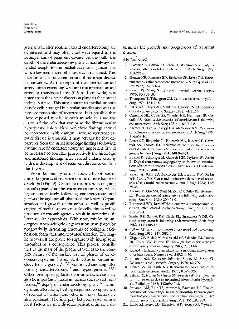
Histologic features o f arterial wall after routine -arot id endarterectomy
Fig. 8 shows the usual findings at the site im- mediatr~y following endarterectomy in 30 consecu- tive patients undergoing routine carotid endarter- ectomy. The wall remaining at the mid-bulb region and the origin of the internal carotid artery was very thin and had only traces of media. The dissection plane in these areas was between the media and ad- ventitial layers. No circular fibers of media were seen at the base, and at cross section, the wall contained elastic tissue, sparse smooth muscle cells, dense col- lagen, fibroblasts, and vasa vasorum. A transitional area was noted from the deeper dissection plane to the intimal surface in the internal carotid artery near the origin. This area was generally 0.5 to 1.0 cm wide, was located 1 to 2 cm beyond the origin, and contained adherent media of varying thickness. Bi- opsy of this area demonstrated abundant medial smooth muscle cells interspersed with collagen.
DISCUSSION Stoney and String 3 first pointed out in 1976 the
morphologic distinction between early and late re- current carotid disease and implied that early and late recurrent lesions were distinct pathologic entities. However, they admitted that it was possible that late lesions were simply early ones that had undergone atherosdcrotic degeneration with time. ~7 Cantelmo et al. 6 have adopted this point of view. Since the report by Stoney and String, several investigators have reported small series of patients with recurrent carotid disease in whom histopathologic studies were performed on retrieved specimens. 2,~8,~9 Although these studies generally confirmed the morphologic distinction between early and late recurrences, none had sufficient numbers of patients to adequately an- swer the question of whether early and late recurrent lesions were truly distinct.
The present study is unique in that it contains a large number of patients with widely varying recur-

20 Clagett et al.
Journal of VASCULAR
SURGERY
rence intervals in whom it was possible to study din- ical and angiographic features and in situ operative findings, gross morphologic characteristics, and his- topathologic features of recurrent lesions. Our data support the contention that early and late recurrent lesions are truly one and the same, that is, neointimal fibromuscular hyperplasia, which undergoes pro- gressive atherosclerotic change. The overall character of the lesion is dependent on the time of sampling (the point at which the patient has symptoms or recurrence is detected by noninvasive means and undergoes reoperation). Neointimal fibromuscular hyperplasia tended to decrease in relative amounts with time whereas atherosclerotic features increased. Although late recurrent lesions contained many features of atherosclerosis, they did not resemble the classic atheroscleroric lesions seen in primary plaques. The topography was different in that the arrangement of the elements was less orderly in re- current lesions. The changes seen in late recurrent lesions were compatible with athcrosclerotic degen- eration of a disorderly, proliferating mass of smooth muscle cells.
A striking finding in this study was the prevalence of thrombus associated with recurrent carotid le- sions. Gross and microscopic thrombus was present in more than two thirds of the early recurrences and in all of the later recurrences. In comparison, throm- bus was associated with primary plaques in only 40% of cases (p<0.001). In early recurrent disease, throm- bus was usually found in a thin layer on the surface of the neointimal fibromuscular mass and had the appearance of organizing thrombus. In late recurrent disease, thrombus was much more exuberant, was always located on the surface, but also extended deep within the substance of the lesion where it was fre- quently associated with neovascularity. This finding raised the possibility that rupture of neovessels con- tributed to the thrombus associated with later lesions. As opposed to the experience with primary carotid disease, 2°'21 w e found no discernible relationship be- tween the presence or absence of thrombus and focal neurologic symptoms in patients with recurrent dis- ease. In fact, all of our patients without symptoms had thrombus associated with their recurrent lesions. The prevalence of thrombus in recurrent lesions and the lack of correlation with symptoms suggest that thrombus formation is an intrinsic, pathogenetic component of recurrent disease and not a secondary, complicating feature.
The presence of thrombus may explain some of the noninvasive findings of investigators who found
that some early recurrent carotid lesions may underg o regression. Zierler et al.la found that 9 of 22 recurrefi~ lesions (>50% stenosis of the internal carotid artery) regressed. Other authors have also noted regression of early recurrences2 ,n It is possible that the dynam- ics of thrombus formation and lysis on the surface of a recurrent lesion account for the waxing and wan- ing hemodynamic changes noted by noninvasivc tests. If this relationship is documented, it would be of great importance to determine the factors that promote progression and regression of the throm- botic mass. From the results of this study, aspirin therapy does not appear to modify thrombus for- marion on recurrent lesions; aspirin also does not seem to protect against recurrent carotid disease. 6a5,22
The present study sought to define both the ~,"" atomic location of recurrence and the relationship of the recurrent lesion to the old suture line mad end- arterectomy site. We found that the malorlty'6f re- current lesions were found in the proximal internal carotid artery. Almost all were within the limits of the primary endarterectomy site, and they were fre- quently associated with a long primary anteriotomy suture line, extending 4 to 5 cm onto the internal carotid artery. An obvious implication of this finding is that endarterectomies extending well into the in- ternal carotid artery are predisposed to development of recurrent disease and that closure with patch an- gioplasty might prevent or delay the recurrence.
In the series reported by O'Donnell et al., 2a the earliest recurrent lesions detected by B-mode ultra- sound imaging were located in the common carotid artery. This finding raised the possibility that recur- rent lesions in this location actually represented ac- centuation of preexisting plaque. The findings in present series tend to confirm this interpretation. We invariably found a shelf of plaque ending ab%lptly at the beginning of the endarterectomy site w[}_hin mil- limeters of the starting point of the original arteri- otomy suture line in our three patients with recurrent disease at this location (Fig. 3). Superimposed on the surface of the shelf and contributing to the ste- nosis was a layer of adherent fibromuscular hyper- plasia. This finding also has technical implications in the performance of primary carotid endarterectomy. Certainly, extension of the endarterectomy proxi- mally to remove most common carotid plaque would be advisable. When this is not feasible, a patch an- gioplasty to widen the circumference of the vessel at the transitional area at which a shelf of plaque re- mains might be prudent.
The anatomic and histologic characteristics of the
Volume 3 Number 1 lanuary 1986 Recurrent carotid disease 21
a~erial wall after routine carotid endarterectomy are ~,f interest and may offer clues with regard to the pathogenesis of recurrent disease. At the bulb, the depth of the endarterectomy plane almost always ex- tended deeply to the medial-adventitial junction at which few medial smooth muscle cells remained. This location was an unc0mmon site of recurrent disease in our series. At the origin of the internal carotid artery, often extending well into the internal carotid artery, a transitional area (0.5 to 1 cm wide) was noted from the deeper dissection plane to the normal intimal surface. This area contained medial smooth muscle cells arranged in circular bundles and was the most common site of recurrence. It is possible that these exposed medial smooth muscle cells are the
:urce of the cells that comprise the fibromuscular "hyperplastic lesion. However, these findings should be interpreted with caution. Because recurrent ca- roti~clisease is unusual, it may actually be that de- partures from the usual histologic findings following routine carotid endarterectomy are important. It will be necessary to correlate prospectively the histologic and anatomic findings after carotid endarterectomy with the development of recurrent disease to confirm this theory.
From the findings of this study, a hypothesis of the pathogenesis of recurrent carotid disease has been developed (Fig. 9). Central to the process is ongoing thrombogenesis at the endarterectomy site, which begins immediately following endarterectomy and persists throughout all phases of the lesion. Organ- aization and growth of thrombus as well as prolif- eration of medial smooth muscle cells stimulated by products of thrombogenesis result in neointimal fi- .... romuscular hyperplasia. With time, this lesion un- dergoes atherosclerotic degeneration manifested by progrer~ively increasing amounts of collagen, calci- fication, foam cells, and neovascularization. The frag- ile neovessels are prone to rupture with intraplaque thrombus as a consequence. This process contrib- utes tO the mass of the plaque as well as to the com- plex nature of the surface. At all phases of devel- opment, systemic factors identified as important in- clude female gender,! ~,~s,z2 continued smoking after primary endarterectomy, ~s and hyperlipidemia) '2'23 Other predisposing factors for atherosclerosis may also be inaportant. 6 Local influences such as technical factors, ~° depth of endarterectomy plane, ~4 hemo- dynamic alterations, healing responses, completeness of reendothelization, and other unknown factors are also pertinent. The interplay between systemic and local factors in an individual patient ultimately de-
termines the growth and progression of recurrent disease.
REFERENCES 1. Crossman D, Callow AD; Stein A, Matsumoto G. Early re-
stenosis after carotid endarterectomy. Arch Surg 1978; 113:275-8.
2. Hertzer NR, Martinez BD, Benjamin SP, Beven EG. Recur- rent stenosis after carotid endarterectomy. Surg Gynecol Ob- stet 1979; 149:360-4.
3. Stoney RJ, String ST. Recurrent carotid stenosis. Surgery 1976; 80:705-10.
4. Thompson JE, Talkington CM: Carotid endarterectomy. Ann Surg 1976; 184:1-15.
5. Baker WH, Hayes AC, Mahler D, Littooy FN. Durability of carotid endarterectomy. Surge D, 1983; 94:112-5.
6. Cantelmo NL, Cutler BS, Wheeler HB, Herrmann JB, Car- dullo PA. Noninvasive detection of carotid stenosis following endarterectomy. Arch Surg 1981; 116:i005-8.
7. Kremen JE, Gee W, Kaupp HA, McDonald KM. Resteuosis or occlusion after carotid endarterectomy. Arch Surg 1979;
114:608-10. 8. Pierce GE, Iliopoulos JI, Holcomb MA, Rieder CF, Herm-
reck AS, Thomas JH. Incidence of recurrent stenosis after carotid endarterectomy determined by digital subtraction an- giography. AmJ Surg 1984; ]48:848-54.
9. Raithel D, Schweiger H, Gentsch HH, Seyferth W, Zeitler E. Digital transvenous angiography in follow-up examina- tions after carotid reconstruction: Early results. J Cardiovasc Surg 1984; 25:400-3.
10. Salvian A, Baker JD, Machleder HI, Busurtil RW, Barker WF, Moore WS. Cause and noninvasive detection of resten- osis after carotid endarterectomy. Am J Surg 1983; 146: 29-34.
11. Thomas M, Otis SM, Rush Ms ZyroffJ, Dilley RB, Bernstein EF. Recurrent carotid artery stenosis following endarterec- tomy. Ann Surg 1984; 200:74-9.
12. Turnipseed WD, BerkoffHA, Crummy A. Postoperative oc- clusion after carotid endarterectomy. Arch Surg 1980; 115:573-4.
RE, Bandyk DF, Thiele BL, Strandness [~r DE. Ca- 13. Zierler rotid artery stenosis following endarterectomy. Arch Surg 1982; 117:1408-15.
14. Callow AD. Recurrent stenosis after carotid endarterectomy. Arch Surg 1982; 117:1082-5.
15. Clagett GP, Rich NM, McDonald lYI ", Salander JM, Youkey JR, Olson DW, Hutton JE. Etiologic factors for recurrent carotid artery stenosis, Surgery 1983; 93:313-8.
16. Lazarides E. Intermediate filaments as mechanical integrators of cellular space. Nature 1980; 283:249-56.
17. Imparato AM. Discussion following Stoney RJ, string ST. Recurrent carotid stenosis. Surgery 1936; 80:709.
18. French BN, Rewcastle NB. Recurrent stenosis at site Of ca- rotid endarterectomy. Sl:roke 1977; 8:597-605.
19. Palmaz IC, Hunter G, Carson SN, French SW. Postoperative carotid restenosis due to neointimal fibromuscular hyperpla- sia. Radiology 1983; 148:699-702.
20. Imparato AM, Riles TS, Mintzer R, Baumann FG. The im- portance of hemorrhage in the relationship between gross morphologic characteristics and cerebral symptoms in 376 carotid artery plaques. Ann Surg 1983; 197:195-203.
21. Lusby RJ, Ferrel LD, Ehrenfeld WK, Stoney RJ, Wylie EJ.

22 Clagett et al.
Journal of VASCULAR
SURGERY
Carotid plaque hemorrhage: Its role in production of cerebral ischemia. Arch Surg 1982; 117:1479-88.
22. Cossman DV, Treiman RL, Foran RF, Levin PM, Cohen JL. Surgical approach to recurrent carotid stenosis. Am J Surg 1980; 140:209-11.
23. O'Donnell Jr TF, Callow AD, Scott G, Shepard AD, He~7 gerick P, Mackey WC. Ultrasound characteristics ofrecurr~.x carotid disease: Hypothesis explaining the low incidence of symptomatic recurrence. J VASC SURG 1985; 2:26-41.
DISCUSSION
Dr. Alexander W. Clowes (Seattle, Wash.). Dr. Cla- gett and his colleagues have shown that 20 of 26 instances of recurrent disease actually developed within the site of endarterectomy and not at the ends. Furthermore, there is a distinct time course; smooth muscle ceils accumulate early, collagen seems to accumulate in a middle period, and thrombus accumulates on the surface of these endarter- ectomized arteries in a late period. The thrombus is in fact on the surface and is not deep within the lesion as in the primary carotid plaque.
We have found similar results in the few endarterec- tomies we have done for recurrent disease; the dominant feature was thrombus on the surface of the recurrent lesion. We have found, with immunocytochemical markers, that smooth muscle cells are located at the bottom of the lesion and leukocyres and thrombus at the top.
In a rat carotid artery model of this process, we have noted a sequence of events leading from denudation to the formation of a thick intima mainly caused by smooth mus- cle cell proliferation.
We find that the first cellular event is medial smooth muscle cell proliferation followed by migration of cells into the intima, further proliferation, and finally accumulation of connective tissue; this kind ofreponse is similar to what happens in the endarterectomized artery. However, we do not see accumulation of thrombus on the denuded rat ca- rotid artery even at late times. This observation makes us wonder whether there is something peculiar about the hu- man carotid artery:, could the geometry of the carotid artery lead to this accumulation of thrombus? Is it not advisable to reconsider the approach to the recurrent lesion and con- sider instead resection and placement of a vein graft, which we know does not develop this accumulation of thrombus even with stenosis?
Dr, Allan D. Callow (Boston, Mass.). It is a distinct pleasure for me to comment on this detailed and elegant study. Permit me to emphasize some of the prominent features.
There is a striking incidence of gross thrombus in 59% of 32 patients and in 77% of patients at microscopic ex- amination in the early recurrent lesion. Although in many patients thrombus appeared to form the bulk of the re: current lesion, there was no correlation with focal neuro- logic symptoms. This is at variance with our experience in which patients with symptoms were more likely to have large amounts of thrombotic material than those without foca ! symptoms.
Endothelium could not be identified on the surface of
the recurrent lesions, possibly because of handling during operation or excessive length of storage of the specimen.
In two of our reoperative specimens, one taken at 33 and one at 3 months following carotid endarterectomy, no evidence of an endothelial cell is observed. We do not yet have convincing evidence that endothelium does indeed regenerate in the human carotid artery.
The authors state that the fact that the majority of recurrent lesions occur in the internal carotid artery and~yr the bifurcation is an obvious implication that endarterec- tomies extending well into the internal carotid artery are predisposed to recurrent disease and further that ~psure with patch angioplasty may prevent or delay the recurrence. This may be so, but it is an opinion and requires additional documentation. We do agree that extending the endarter- ectomy proximally as far as necessary to remove the com- mon carotid plaque, is advisable.
The authors note that the relevant amount of neoin- timal hyperplasia tended to decrease with increasing re- currence interval. The opposite was noted for those lesions featuring atherosclerosis; that is, as the recurrence interval increased, relative amounts o f foam cells, cholesterol, and other findings of atherosclerosis also tended to increase. Is this evidence indeed that the fibromuscular lesion is an early phase of atherosclerosis? Do all late lesions that have the characteristics of atherosclerosis begin as fibromuscular hyperplasia? Do all lesions of fibromuscular hyperplasia inexorably progress to the atherosclerotic type? Although the data do indeed suggest that the overall character of the lesion is dependent on the time of sampling, they do n e~ necessarily support the contention that early and late re- current disease are truly one and the same l e ~ n . This would appear to be an opinion and, ha the presd~r ~ state of our knowledge, not an established fact. It is worthy of further investigation.
Dr. Clagett (closing). I am particularly gratified to learn that Dr. Clowes, with different histologic techniques, has confirmed our finding that most recurrent lesions con- sist of organizing surface thrombus overlying smooth mus- cle proliferations undergoing various stages of atheroscle- rotic change. Dr. Clowes asks why the carotid bifurcation accumulates thrombus after endarterectomy. This phenom- enon is not seen to the same degree in most animal models and appears unique for the human carotid bifurcation. An- atomically, the bifurcation represents the point at which an elastic artery becomes a muscular artery and there is nothing unusual about the histologic features. I agree that the unique geometry of the internal carotid origin and the complex flow patterns with eddies, vortices, and low-shear

Volume 3 Number 1 lanuary 1986 Recurrent carotid disease 23
flow disturbances enhance thrombogenesis engendered by endarterectomy and extensive loss of endothelium. With regard to his technical question about how best to ap- proach recurrent carotid lesions, wc have favored reendar- terectomy and vein or prosthetic patch angioplasty. In well over 50 reoperative cases with the use of this technique, the results have been very good and there have been no second recurrences. Although resection of the bifurcation with replacement with a vein graft is logical, this would be a much more difficult operation, and we have not used it for recurrent lesions.
Dr. Callow raised a number of important issues, one of which is the question of whether the endarterectomy site becomes repaved with endothelinm. The implication, of course, is that recurrences may bc linked to failure to reendothelialize. However, I do not think we know enough hour the normal healing responses following endarter-
ectomy to make inferences about abnormal responses. A central question is whether endothelialization to any degree occur, ~'following carotid endarterectomy; the available data are scant and inconclusive. We looked hard for endothe- t i m in our specimens and used immunoperoxidase stain- hag techniques for endothelial yon Willebrand's factor. We did not see anything that resembled endothelium. How-
ever, because of problems with fixation and, possibly, spec- imen handling, we really could not say conclusively that endothelium was absent. So I think that this remains an issue that needs further investigation.
Dr. Callow also raised the point about the distinction between early and late recurrences. Our data suggest that these are the same lesions, essentially, neointimal fibro- muscular hyperplasia with superimposed, t/me-related ath- erosclerotic change. The overall character of the lesion de- pends on the time of sampling, Our evidence for this is, admittedly, indirect but strong. With increasing recurrence interval, we found a gradual decrease in the prominence of neoinfimal fibromuscular hyperplasia. Conversely, dur- ing the same time period, we found increasing atheroscle- rotic change. The temporal relationship was strong. An- other piece of indirect evidence was the finding of areas of neointimal fibromuscular hyperplasia in some late recur- rences (as late as 15 years) that were virtually indistin- guishable by all morphologic criteria (including electron microscopy) from those comprising the bulk of early re- current lesions. So, again, we thought that these were the same basic lesions that degenerated in a time-dependent fashion.