Endovascular stent-graft treatment of thoracic aortic syndromes: A 7-year experience
Upload
khangminh22Category
view
1download
0
February 2007
Supplement to Produced under an educational grant from Gore & Associates

SFAEndoluminalBypass
The peripheral vascular interventionist has a vast and expandingarmamentarium of devices and techniques available to treat superfi-cial femoral artery (SFA) disease.
The extent of SFA disease often dictates the choice of therapy.Based on clinical reports on the treatment of this vascular bed, therehas been a trend to align these various therapies and devices withthe appropriate “lesion-specific” diagnosis.
This supplement is focused on the use of an endoluminal bypassto treat the most challenging and diffuse (TASC C and D) SFAlesions. The articles are intended to offer insight to a clinical decisiontree that leads to the use of a stent graft—specifically, the GOREVIABAHN® Endoprosthesis—to restore blood flow in the most difficult SFA lesions.
4 PERCUTANEOUS ENDOVASCULAR TREATMENT OF SFA DISEASE USING THE GORE VIABAHN®ENDOPROSTHESISBy Fareed Shaikh, MD; Mohamed Djelmami-Hani, MD; Joaquin Solis , MD; Suhail Allaqaband, MD; Ramagopal Tumuluri, MD;Anjan Gupta, MD; and Tanvir Bajwa, MD
9 TIPS FOR USING THE GORE VIABAHN®ENDOPROSTHESIS IN THE SFABy John G. Adams, Jr, MD, FACS; Richard D. Coats, MD; Paul W. Humphrey, MD, FACS; Karen Althage, CFA; and Mary Chott, Research Coordinator
13 DEBUNKING THE MYTH OF FAILED LOWER-EXTREMITY ENDOPROSTHESESBy Christopher L. Wixon, MD
17 ENDOVASCULAR FEMOROPOPLITEAL BYPASS IN ACOMPLETE OCCLUSIONBy Paramjit “Romi” Chopra, MD, and Swarnam Chiramel, MS
20 TREATING SIGNIFICANT STENOSIS IN THE SFA WITHCOVERED SELF-EXPANDING STENT GRAFTSBy James B. Park, MD, FACC
22 GORE VIABAHN® ENDOPROSTHESIS RECANALIZATION OF A COMPLEX SFA LESIONBy Mark W. Mewissen, MD
Clinical images on cover courtesy of Mark W. Mewissen, MD.
Contents
2 I SUPPLEMENT TO ENDOVASCULAR TODAY I FEBRUARY 2007

The authors of this supplement provide comprehen-sive insight into endoluminal SFA bypass. With this pro-cedure, interventionists effectively re-line the SFA wallwith a smooth expanded polytetrafluoroethylene(ePTFE) lumen supported by a highly flexible stent scaf-fold. In contrast to when a bare stent is used in the SFA, astent graft, such as the GORE VIABAHN® Endoprosthesis,covers and seals off all the diseased and irregular tissue ofthe arterial wall.
More than 30 years ago, the first ePTFE surgicalbypass was performed in the SFA. Since then, millions ofabove-the-knee femoropopliteal ePTFE bypasses havebeen used to restore blood flow to the lower limb withoverall patency rates that rival vein bypass.
The advent of an ePTFE stent graft offers patients ofperipheral vascular interventionists the option of a min-imally invasive treatment that, similar to the surgicalapproach in the SFA, bypasses all the diseased tissueand plaque involved in long lesions. As with any newtreatment option, the keys to successful outcomes aredependent on numerous factors, such as proper patientselection, technical considerations, the use of ancillarydevices, and antiplatelet therapy.
As this chart of reported outcomes of the GOREVIABAHN® Endoprosthesis indicates, there has beenbroad interest over the last several years to determinethe role of stent grafts to treat the SFA. ■
1. Chopra P. Endoluminal femoropopliteal bypass using the Viabahn Stent Graft (Endograft):primary and secondary patency in 60 patients (70 limbs) with 3-year follow-up. Abstract pre-sented at: 14th Annual Advanced Interventional Management Symposium (AIMS); November13-16, 2006; New York, NY.2. Kazemi S, Djelmami-Hani M, Gupta A, et al. One year patency rate of the VIABAHN StentGraft for chronic total occlusion or long high-grade stenosis of the superficial femoral artery.Abstract presented at: 18th Annual Scientific Symposium of the Transcatheter CardiovascularTherapeutics (TCT); October 22-27, 2006; Washington, DC. Available from: AmericanJournal of Cardiology. 2006;98(suppl S1):235M-236M.3. Coats RD, Adams Jr. JG, Humphrey PW. SFA revascularization using the ViabahnEndoprosthesis. Endovasc Today. 2006;5:76-78.4. Fischer M, Schwabe C, Schulte K-L. Value of the Hemobahn/Viabahn Endoprosthesis inthe treatment of long chronic lesions of the superficial femoral artery: 6 years of experience. JEndovasc Ther. 2006;13:281-290.5. Kedora JC, Hohmann SE, Dunn TL, et al. Mid-term comparison of percutaneous ViabahnStent-Grafts versus conventional femoral-popliteal bypass in the treatment of superficialfemoral arterial occlusive disease. Abstract presented at: 2006 SVS Annual Meeting; June 1-4, 2006; Philadelphia, PA.6. Saxon RR, Coffman JM, Gooding JM, et al. Long-term patency of stent-grafts in the treat-ment of long-segment femoropopliteal artery occlusive disease. Abstract presented at: SIR31st Annual Scientific Meeting; March 30-April 4, 2006. Toronto, Ontario, Canada. Availablefrom: J Vasc Interv Radiol. 2006;17(part 2):S2.7. Zander T, Llorens R, Rostagno R, et al. Hemobahn/Viabahn endograft for long SFA lesions.Long term follow up. Abstract presented at: SIR 31st Annual Scientific Meeting. March 30-April4, 2006. Toronto, Ontario, Canada. Available from: J Vasc Interv Radiol. 2006;17(part 2):S57.8. Panetta TF. Endovascular femoropopliteal bypass with multiple stent grafts. EndovascToday. 2005;4(suppl):12-14.9. Hartung O, Otero A, Dubuc M, et al. Efficacy of Hemobahn in the treatment of superficialfemoral artery lesions in patients with acute or critical ischemia: a comparative study withclaudicants. Eur J Vasc Endovasc Surg. 2005;30:300-306.10. Bleyn J, Schol F, Vanhandenhove I, et al. Endovascular reconstruction of the superficialfemoral artery. In: Becquemin JP, Alimi YS, Watelet J, et al, eds. Controversies and Updatesin Vascular & Cardiac Surgery. Torino, Italy. Edizioni Minerva Medica. 2004;14:87-91.11. Jahnke T, Andresen R, Müller-Hülsbeck S, et al. Hemobahn stent-grafts for treatment offemoropopliteal arterial obstructions: midterm results of a prospective trial. J Vasc IntervRadiol. 2003;14:41-51.12. Turicchia GU, Cevolani M, Altini R, et al. Mid-term results in PTFE endograft treatment offemoropopliteal occlusive disease. Osp Ital Chir. 2003;9:93-96.13. Railo M, Roth WD, Edgren J, et al. Preliminary results with endoluminal femoropoplitealthrupass. Ann Chir Gynaecol. 2001;90:15-18.14. Lammer J, Dake MD, Bleyn J, et al. Peripheral arterial obstruction: Prospective study oftreatment with a transluminally placed self-expanding stent graft. Radiology. 2000;217:95-104.
REPORTED PRIMARY PATENCIES OF GORE VIABAHN® ENDOPROSTHESIS (6 MM TO 8 MM) IN THE SFA
Author Year Journal Publication/Presentation No. ofLimbs
Lesion Length (cm)
Primary Patency (years)
1 2 3 4 5
Chopra1 2006 AIM Symposium, Nov 13-16 70 20 93% 87% 72%
Kazemi2 2006 TCT Meeting, October 23-27 65 12 90%
Coats3 2006 Endovasc Today, Sept 83 89%
Fischer4 2006 J Endovasc Ther, 13:281-290 48 10.7 80% 73% 71% 64% 62%
Kedora5 2006 SVS Annual Meeting, June 1-4 50 25.6 73%
Saxon6 2006 SIR Meeting, March 31 56 13.1 84% 76% 76% 67%
Zander7 2006 SIR Meeting, April 3 31 16.6 86% 78% 78% 78%
Panetta8 2005 Endovasc Today, August 41 30.4 86% 77%
Hartung9 2005 Eur J Vasc Endovasc Surg, 30:300-206 34 10.8 85% 85%
Bleyn10 2004 Edizioni Minerva Medica, 14:87-91 67 14.3 82% 73% 68% 54% 47%
Jahnke11 2003 J Vasc Interv Radiol, 14:41-51 52 8.5 78% 74% 62%
Turicchia12 2003 Osp Ital Chir, 9:93-96 16 10 80% 80%
Railo13 2001 Annales Chirurgiae et Gynaecologiae, 90:15-18 15 8 93% 84%
Lammer14 2000 Radiology, 217:95-104 80 13.8 79%
Average/Total 708 15 84% 79% 71% 66% 55%
FEBRUARY 2007 I SUPPLEMENT TO ENDOVASCULAR TODAY I 3

4 I SUPPLEMENT TO ENDOVASCULAR TODAY I FEBRUARY 2007
SFA Endoluminal Bypass
Percutaneous EndovascularTreatment of SFA Disease Using the GORE VIABAHN®
Endoprosthesis
The prevalence and incidence of peripheral vasculardisease (PVD) continue to increase with advancingage. In response to this increase, we must considerall options for management of PVD. Aggressive risk
factor modification and a trial of an exercise program arerecommended as the first line of therapy. Pharmacotherapywith cilostazol provides additional symptom relief.1,2 Patientswho fail medical therapy and continue to have resting legpain or nonhealing ulcers are candidates for invasive treat-ment strategy. The choice of vascular intervention as thenext step in managing suprainguinal disease is clear-cut, butthe same is not true for infrainguinal disease. Surgical revas-cularization may provide better long-term results, but it isassociated with higher periprocedural morbidity and mortal-ity, making the surgical option less desirable in elderlypatients—a significant proportion of patients with PVD.
Dotter et al3 reported the first endovascular interventionin 1964. Although significant advances have been made inthe endovascular treatment of PVD during the past 40 years(ie, percutaneous transluminal angioplasty [PTA], atherecto-my, laser, stenting), long-term success has been hampered bya high rate of restenosis. PTA of the superficial femoral arter-ies (SFAs) has a high initial success rate (>90%), but therestenosis rate remains high (40%-60%).4-6 In longerlesions (>10 cm), the primary patency rate decreased to20% at 1 year.7 PTA followed by stenting prevents the prob-lems of elastic recoil and flow-limiting dissection. Althoughthe initial success of SFA stenting is high, long-term patencyis limited by increased rates of in-stent restenosis (ISR).8-11
Managing ISR remains a challenge to this day despite all ofthe modalities now available for percutaneous intervention.
The self-expanding nitinol stents have shown better 1-yearpatency rates.12 Mewissen et al13 and Kazemi et al12 showedsimilar 1-year patency rates of 76%, whereas 18-month to24-month primary patency rates decreased to 60% to 66%.Recently, Schillinger et al reported a 12-month primarypatency rate of 63% in a self-expanding stent-treated groupcompared to 37% in an angioplasty-treated group.14 Studiesin which different debulking devices were used yielded differ-ent results. A randomized trial of PTA versus atherectomyfor 2-year patency showed worse results for atherectomy.15
Jahnke et al compared PTA with atherectomy, showing noadditional benefit associated with atherectomy in 6-monthpatency.16 Laser-assisted angioplasty for patients with criticallimb ischemia was tested in a single-center trial (LACI)17 withgood results, but multiple other trials comparing laser angio-plasty versus PTA showed no advantage of laser therapy.18,19
Recently, endoprostheses have been used in treating longSFA lesions. The rationale for using a polytetrafluoroethylene(PTFE)-covered device is to prevent intimal hyperplasia, thusimproving primary patency, especially in long lesions.However, the initial reports have been mixed for PTFE-cov-ered endoprostheses. Primary patency rates of 29% to 87%have been reported in different studies.20 Use of the CraggEndoPro System endoprosthesis (Minimally Invasive Tech-nologies SARL, La Ciotat, France), an expandable nitinol stentcovered with woven polyester fabric, resulted in a low pri-mary patency in the femoropopliteal vessels (59%) and a highrate of complications, including early and late thrombosis,graft misplacement, and distal embolization.21 In an interna-tional feasibility trial, the GORE HEMOBAHN® Endoprosthesis(Gore & Associates, Flagstaff, AZ) was assessed to have a 90%
Do the procedural success and 1-year follow-up data make this
the treatment of choice?
BY FAREED SHAIKH, MD; MOHAMED DJELMAMI-HANI, MD; JOAQUIN SOLIS, MD;
SUHAIL ALLAQABAND, MD; RAMAGOPAL TUMULURI, MD; ANJAN GUPTA, MD; AND TANVIR BAJWA, MD

FEBRUARY 2007 I SUPPLEMENT TO ENDOVASCULAR TODAY I 5
SFA Endoluminal Bypass
patency at 6-month follow-up and 79% at 1-year.22
The GORE VIABAHN® Endoprosthesis (Gore &Associates) is a flexible, self-expanding, endoluminal endo-prosthesis constructed with an expandable, biocompatible
PTFE liner attached to the external nitinol stent structure. Itis now FDA approved for treatment of SFA disease. We eval-uated the potential of the GORE VIABAHN® Endoprosthesisto improve the long-term outcome of percutaneous inter-vention in complex SFA lesions.
METHODSTo assess the efficacy of the GORE VIABAHN®
Endoprosthesis, we studied patients presenting with criticallimb ischemia or lifestyle-limiting claudication. The GOREVIABAHN® Endoprosthesis was used for de novo native ves-sel disease in 98 patients. A second group of patients wasrandomized to placement of GORE VIABAHN®Endoprostheses or to atherectomy with the SilverHawkdevice (FoxHollow Technologies, Redwood City, CA). Allpatients were started on clopidogrel at 75 mg/d and aspirin325 mg/d for at least 6 months. Restenosis was defined as≥50% as determined by duplex ultrasound. The primaryendpoint was target vessel revascularization during 12months of follow-up. Secondary endpoints were clinicalimprovement of symptoms or progression to limb loss.
Procedural DetailsVascular access was achieved either from the contralater-
al common femoral artery or in an antegrade manner fromthe ipsilateral femoral artery using a modified Seldingertechnique. In the contralateral approach, a 45-cm-longsheath was advanced over the aortic bifurcation over a
TABLE 1. PATIENT CHARACTERISTICS AND RESULTSIN PATIENTS WITH DE NOVO VIABAHN® USE
Characteristics n (%), Mean±SD
Patients 81
Legs 98
Age (y) 69±9
Male 53%
Diabetes mellitus 40 (49%)
Smokers 24 (29.6%)
Fontaine class II or IIa 66 (67.3%)
Fontaine class III or IV 15 (32.6%)
Average VIABAHN®
diameter (mm)
6
Average VIABAHN® length (cm) 15 (range, 5 to 20)
Average VIABAHN® per patient 2 (range, 1 to 4)
Follow-up time (months) 16±8
6-month primary patency 98%
12-month primary patency 96%
Secondary patency 100%
TABLE 2. BASELINE CLINICAL CHARACTERISTICS
VIABAHN® n (%), Mean±SD SilverHawk Atherectomy n (%), Mean±SD P Value
Patients 23 20
Legs 29 23
Male 12 (52%) 9 (45%) .767
Age (y) 70±10 70±10 1
Diabetes mellitus 15 (65%) 10 (50%) .365
Smokers 16 (69%) 9 (45%) .13
Fontaine class III or IV 90% 90% 1
Average lesion length 10 cm 10 cm 1
Average distal runoff 2 2 1
Technical success 100% 100% 1
Complications (total)dissectionperforation
000
5 (21%)4 (17%)1 (4%)
.012
.032
.442
Adjunctive therapy (balloon or stent) 48%
Mean follow-up 12 months 12 months 1
Primary patency 90% 57% .009

6 I SUPPLEMENT TO ENDOVASCULAR TODAY I FEBRUARY 2007
SFA Endoluminal Bypass
.035-inch wire. Eight-French sheaths were used in allpatients to deliver a 5-mm- or 6-mm-diameter GOREVIABAHN® Endoprosthesis. Patients were anticoagulatedwith either bivalirudin or heparin using a weight-baseddose. Predilatation was performed in 80% of cases using an
undersized short balloon. The GORE VIABAHN®Endoprosthesis was then deployed over a stiff wire.Postdilatation was performed in all cases using a 1:1 sizedballoon at high pressures. Care was taken to avoid any bal-loon dilatation outside the endoprosthesis edges usingmagnified fluoroscopic views. Patients randomized toatherectomy underwent plaque excision using theSilverHawk atherectomy device by experienced interven-tionists. PTA or stents were used as adjunctive therapy afteratherectomy for suboptimal results.
Technical success was defined as reduction of stenosisto <20%, with restoration of brisk antegrade flow andwithout visible thrombus or dissection. Anticoagulationwas discontinued at the end of the procedure.
Follow-UpAll patients were followed clinically and underwent ankle-
brachial index with arterial duplex 1, 3, 6, and 12 monthsafter the initial intervention. Patients with abnormal duplexstudies underwent repeat angiography with reinterventionwhen necessary.
RE SULTSDe Novo Native Vessel Disease Group
Eighty-one patients received the GORE VIABAHN®Endoprosthesis for native vessel disease. A total of 167GORE VIABAHN® Endoprostheses were deployed in 98SFAs. More than 80% of interventions were performed
Figure 1. A 60-year-old man with claudication was treated with
two GORE VIABAHN® Endoprostheses in the left SFA for a chronic
total occlusion.Preprocedure image (A); postprocedure image (B).
Figure 3. A 73-year-old man with claudication and abnormal
arterial duplex ultrasound.Two GORE VIABAHN® Endo-
prostheses were placed covering the collaterals coming off
the body of the SFA, with excellent angiographic and clinical
results. Preprocedure image (A); postprocedure image (B).
Figure 2. A 75-year-old woman presented with left lower-
extremity, lifestyle-limiting claudication. Angiography revealed
a chronic total occlusion.Two GORE VIABAHN® Endoprostheses
(6 mm X 15 cm and 6 mm X 15 cm) were placed in the left SFA.
Preprocedure image (A); postprocedure image (B).

for Transatlantic Inter-Society Commission (TASC) class Cand D lesions. Technical success was 100%, with a primarypatency rate of 96% at 12-month follow-up. The baselinepatient characteristics and results are shown in Table 1.
SilverHawk Atherectomy SubgroupForty-three consecutive patients were randomized in a
GORE VIABAHN® Endoprosthesis versus SilverHawkatherectomy cohort. Twenty-three patients received GOREVIABAHN® Endoprostheses with intervention on 29 SFAs.The SilverHawk atherectomy group had 20 patients withinterventions on 23 SFAs. There were no differences in age,gender, diabetes, or smoking between the two groups. Thepresenting symptoms and lesion length were similarbetween the two groups (Table 2).
During the initial procedure, the SilverHawk atherectomygroup had significantly more complications than the GOREVIABAHN® group (21% vs 0%; P=.012). In the atherectomygroup, four (17%) cases of flow-limiting dissection and one(4%) perforation were noticed. There were no procedure-related complications in the GORE VIABAHN® group. At theend of the initial procedure, technical success was achievedin all patients in both groups. There were no deaths in eitherof the groups. The mean follow-up was 12 months. The pri-mary patency rate at 12 months was 90% in the GOREVIABAHN® group and 57% in the SilverHawk atherectomygroup (P=.009).
DISCUSSIONIn the past decade, many new technologies have
emerged to deal with atherosclerotic SFA disease.Surgical revascularization carries significant mor-bidity and other comorbidities, especially in theelderly. The success of surgical intervention is ham-pered by the limited availability of autogenousvenous conduits (which have a much better long-term patency compared to prosthetic conduits)23
in these patients.For percutaneous intervention, the incidence of
restenosis remains the Achilles’ heel of endovascu-lar intervention. Self-expanding nitinol stents doprovide excellent initial success, but long-termpatency, especially for long stents, remains subopti-mal. The anatomy of the SFA is partly to be blamedfor this increased rate of in-stent restenosis. TheSFA is unique in that it is the longest artery in thehuman body. The external forces that it has to bearare markedly influenced by its proximity to muscu-lature, continuous mobility, and the locationbetween two joints.
The use of sirolimus-eluting SMART stents(Cordis Corporation, a Johnson & Johnson compa-ny, Miami, FL) for the SFA was tested in the SIROC-
CO I and II trials. Sirolimus-eluting nitinol stents were foundto be safe and feasible, with a trend toward reducing late losscompared with uncoated stents. This apparent success was ata cost: an 18.2% incidence of stent fracture.24 Stent fracture inthe SFA is an important predictor of a need for repeat inter-ventions.25 Other endovascular techniques, including atherec-tomy or laser-assisted angioplasty, have shown no benefitover balloon angioplasty alone. In our experience, there weresignificantly more procedural complications and a reducedprimary patency rate in patients who had atherectomy com-pared to those with GORE VIABAHN® Endoprostheses.26
Although earlier studies with endoprostheses have beendisappointing,27 the results with PTFE-covered GOREVIABAHN® Endoprostheses are promising. Major advantagesof the GORE VIABAHN® Endoprosthesis include excellentlong-term patency and its stent fracture-resistant nature,making it the device of choice in long lesions of the SFA. Forbetter long-term results, two-vessel runoff below the knee ispreferred, although we have used this device in limb salvagesituations with one-vessel-below-the-knee runoff with goodresults. Advances in implantation technique and dualantiplatelet therapy have improved immediate and long-term patency. We also determined that covering the dis-eased segment of the artery in its entirety (healthy tohealthy), along with exerting extreme care to avoid ballooninjury on the outside edges of the endoprosthesis (Figures 1and 2), are crucial to reduce the chances of edge restenosis.
Special Considerations:
• Lesion located at flexion points:VIABAHN®
• Good distal runoff:VIABAHN®
• Inability to insert large-profile sheath: Self-expanding nitinol stent
• Vessel size <5 mm in diameter: PTA/self-expanding nitinol stent
The Transatlantic Inter-Society Commission (TASC) Classification:• TASC A: A single stenosis ≤3 cm long not at the origin of the superficial femoral
or distal popliteal artery.
• TASC B: A single stenosis or occlusion 3 to 5 cm long not involving the distalpopliteal; or multiple stenoses, or occlusions, each <3 cm long.
• TASC C:A single stenosis or occlusion >5 cm,or multiple lesions each 3 cm to 5 cm long.
• TASC D: Complete common femoral or superficial femoral artery occlusions, orcomplete popliteal and proximal trifurcation occlusions.
Figure 4. Algorithm tree used to determine management of
femoropopliteal disease and TASC classification.
FEBRUARY 2007 I SUPPLEMENT TO ENDOVASCULAR TODAY I 7
SFA Endoluminal Bypass

8 I SUPPLEMENT TO ENDOVASCULAR TODAY I FEBRUARY 2007
SFA Endoluminal Bypass
Although the GORE VIABAHN® Endoprosthesis device issafe, it does require large-profile sheaths (8 F or larger), mak-ing it difficult to use in elderly patients with calcified vesselsand severe PVD. Another concern is that, due to its coverednature, the device cannot be used across the profundafemoral artery to prevent occlusion of an important source ofcollaterals. The same is not true for small collaterals originat-ing from the body of the SFA. In our experience, we did notfind any clinical or procedural adverse effects by coveringthese collaterals coming off the body of the SFA (Figure 3).
CONCLUSIONSBased on our experience, with careful deployment tech-
nique, the GORE VIABAHN® Endoprosthesis had excellentlong-term results for SFA disease. We believe that currentendovascular intervention with the GORE VIABAHN®Endoprosthesis is the treatment of choice for patients withsevere SFA disease (TASC class B, C, and D). Althoughmanagement decisions may differ from case to case, wegenerally follow the algorithm tree shown in Figure 4. ■
Fareed Shaikh, MD, is from the Cardiovascular DiseaseSection, Aurora Sinai/Aurora St. Luke’s Medical Centers,University of Wisconsin Medical School in Milwaukee,Wisconsin. He has disclosed that he holds no financial inter-est in any product or manufacturer mentioned herein. Dr.Shaikh may be reached at (414) 649-3909.
Mohamed Djelmami-Hani, MD, is from the CardiovascularDisease Section, Aurora Sinai/Aurora St. Luke’s MedicalCenters, University of Wisconsin Medical School in Milwaukee,Wisconsin. He has disclosed that he holds no financial interestin any product or manufacturer mentioned herein. Dr.Djelmami-Hani may be reached at (414) 649-3909.
Joaquin Solis, MD, is from the Cardiovascular DiseaseSection, Aurora Sinai/Aurora St. Luke’s Medical Centers,University of Wisconsin Medical School in Milwaukee,Wisconsin. He has disclosed that he holds no financial inter-est in any product or manufacturer mentioned herein. Dr.Solis may be reached at (414) 649-3909.
Suhail Allaqaband, MD, is from the CardiovascularDisease Section, Aurora Sinai/Aurora St. Luke’s MedicalCenters, University of Wisconsin Medical School inMilwaukee, Wisconsin. He has disclosed that he holds nofinancial interest in any product or manufacturer mentionedherein. Dr. Allaqaband may be reached at (414) 649-3909.
Ramagopal Tumuluri, MD, is from the CardiovascularDisease Section, Aurora Sinai/Aurora St. Luke’s MedicalCenters, University of Wisconsin Medical School inMilwaukee, Wisconsin. He has disclosed that he holds nofinancial interest in any product or manufacturer mentionedherein. Dr. Tumuluri may be reached at (414) 649-3909.
Anjan Gupta, MD, is from the Cardiovascular Disease
Section, Aurora Sinai/Aurora St. Luke’s Medical Centers,University of Wisconsin Medical School in Milwaukee,Wisconsin. He has disclosed that he holds no financial inter-est in any product or manufacturer mentioned herein. Dr.Gupta may be reached at (414) 649-3909.
Tanvir Bajwa, MD, is from the Cardiovascular DiseaseSection, Aurora Sinai/Aurora St. Luke’s Medical Centers,University of Wisconsin Medical School in Milwaukee,Wisconsin. He has disclosed that he holds no financial interestin any product or manufacturer mentioned herein. Dr. Bajwamay be reached at (414) 649-3909; [email protected].
1. Dawson DL, Cutler BS, Meissner MH, et al. Cilostazol has beneficial effects in treatmentof intermittent claudication: results from a multicenter, randomized, prospective, double blindtrial. Circulation. 1998;98:678-686.2. Money SR, Herd JA, Isaacsohn JL, et al. Effect of cilostazol on walking distances inpatients with intermittent claudication caused by peripheral vascular disease. J Vasc Surg.1998;27:267-274.3. Dotter CT, Judkins MP. Transluminal treatment of arteriosclerotic obstruction: descriptionof a new technique and preliminary report of its application. Circulation. 1964;30:654-670.4. Dormandy JA, Rutherford B. Management of peripheral arterial disease (PAD). J VascSurg. 2000;3:S1-S296.5. Johnston KW. Femoral and popliteal arteries: reanalysis of results of balloon angioplasty.Radiology. 1992;183:767-771.6. Minar E, Pokrajac B, Maca T, et al. Endovascular brachytherapy for prophylaxis ofrestenosis after femoropopliteal angioplasty: results of a perspective randomized study.Circulation. 2000;102:2694-2699.7. Capek P, McLean GK, Berkowitz HD. Femoropopliteal angioplasty: factors influencinglong-term success. Circulation. 1991;83(suppl 2):170-180.8. Cejna M, Thurnher S, Illiasch H, et al. PTA versus Palmaz stent in femoropopliteal arteryobstructions: a multicenter prospective randomized study. J Vasc Interv Radiol. 2001;12:23-31.9. Vroegindeweij D, Vos LD, Tielbeek AV, et al. Balloon angioplasty combined with primarystenting versus balloon angioplasty alone in femoropopliteal obstructions: a comparativerandomized study. Cardiovasc Inter Radiol. 1997;20:420-425.10. Grimm J, Muller-Hulsbeck S, Jahnke T, et al. Randomized study to compare PTA aloneversus Palmaz stent placement for femoropopliteal lesions. J Vasc Interv Radiol.2001;12:935-942.11. Zdanowski Z, Albrechtsson U, Lundin A, et al. Percutaneous transluminal angioplastywith or without stenting for femoropopliteal occlusions? A randomized controlled study. IntAngiol. 1999;18:251-255.12. Kazemi S, Tumuluri R, Allaqaband S, et al. Percutaneous revascularization offemoropopliteal arteries with self-expanding nitinol stents: immediate and 1 year results. JAm Coll Cardiol. 2005;45(suppl):68A.13. Mewissen M. Self-expanding nitinol stents in the femoropopliteal segment: techniqueand mid-term results. Tech Vasc Intervent Radiol. 2004;7:2-5.14. Schillinger M, Sabeti S, Loewe C, et al. Balloon angioplasty versus implantation of niti-nol stents in the superficial femoral artery. N Engl J Med. 2006;354:1879-1888.15. Nakamura S, Conroy RM, Gordon IL, et al. A randomized trial of transcutaneous extractionatherectomy: intravascular ultrasound observations. J Clin Ultrasound. 1995;23:461-471.16. Jahnke T, Link J, Muller-Hulsbeck S, et al. Treatment of infrapopliteal occlusive diseaseby high-speed rotational atherectomy: initial and mid-term results. J Vasc Interv Radiol.2001;12:221-226.17. Laird JR, Biamino G, McNamara T, et al. Cryoplasty for the treatment of femoropoplitealarterial disease: extended follow-up results. J Endovasc Ther. 2006;13(suppl 2):II52-II59.18. Belli AM, Cumberland DC, Procter AE, et al. Follow-up of conventional angioplasty ver-sus laser thermal angioplasty for total femoropopliteal occlusions: results of a randomizedtrial. J Vasc Interv Radiol. 1991;2:485-488.19. Lammer J, Pilger E, Decrinis M, et al. Pulsed excimer laser versus continuous-waveNd:YAG laser versus conventional angioplasty of peripheral arterial occlusions: prospective,controlled, randomized trial. Lancet. 1992;340:1183-1188.20. Kessel DO, Wijesinghe LD, Robertson I, et al. Endovascular stent grafts for superficialfemoral artery disease: results of 1-year follow-up. J Vasc Interv Radiol. 1999;10:289-296.21. Henry M, Amor M, Cragg A, et al. Occlusive and aneurysmal peripheral arterial disease:assessment of stent-graft system. Radiology. 1996;201:717-724.22. Lammer J, Dake MD, Bleyn J, et al. Peripheral arterial obstruction: prospective study oftreatment with a transluminally-placed self-expanding stent-graft. International Trial Studygroup. Radiology. 2000;217:95-104.23. AbuRahma AF, Robinson PA, Holt SM. Prospective controlled study of polytetrafluo-roethylene versus saphenous vein in claudicant patients with bilateral above kneefemoropopliteal bypasses. Surgery. 1999;126:594-601; discussion 601-602.24. Duda SH, Pusich B, Richter G, et al. Sirolimus-eluting stents for the treatment ofobstructive superficial femoral artery disease: six-month results. Circulation.2002;106:1505-1509.25. Solis J, Allaqaband S, Bajwa T. A case of popliteal stent fracture with pseudoaneurysmformation. Catheter Cardiovasc Interv. 2006;67:319-322.26. Kazemi S, Bangash A, Gupta A, et al. Prospective, randomized comparison of Viabahnstent-graft versus SilverHawk atherectomy for de novo femoropopliteal occlusive disease. AmJ Cardiol. 2006;98:10M.27. Sapoval MR, Long AL, Raynaud AC, et al. Femoropopliteal stent placement: long-termresults. Radiology. 1992;184:833-839.

FEBRUARY 2007 I SUPPLEMENT TO ENDOVASCULAR TODAY I 9
SFA Endoluminal Bypass
Tips for Using the GOREVIABAHN® Endoprosthesis in the SFAThe GORE VIABAHN® Endoprosthesis has excellent technical success rates and
short-term durability when these techniques are followed.
BY JOHN G. ADAMS, JR, MD, FACS; RICHARD D. COATS, MD; PAUL W. HUMPHREY, MD, FACS;
KAREN ALTHAGE, CFA; AND MARY CHOTT, RESEARCH COORDINATOR
Recently, several novel techniques and productshave been used in the percutaneous treatmentof atherosclerotic occlusive disease of the lowerextremity. Several investigators have described
their experience using the GORE VIABAHN® Endo-prosthesis (Gore & Associates, Flagstaff, AZ) for endolu-minal bypass of the superficial femoral artery (SFA).1-7
The GORE VIABAHN® Endoprosthesis is a flexible, self-expanding stent graft composed of an expanded poly-tetrafluoroethylene (ePTFE) tube inside an external niti-nol support that extends along its entire length.
We began using the GORE VIABAHN® Endoprosthesisin July 2004 and have prospectively collected data sincethat time. As of August 2006, we have performed 100procedures for occlusive disease in 79 patients. Theindication for intervention was debilitating claudicationin 59% and rest pain/tissue loss in 41%. The mean age ofthe patients was 71 years and ranged from 35 years to88 years. Eighty-six procedures were limited solely tothe SFA. Technical success was 100%. Mean contrastusage was 95 mL and ranged from 35 mL to 215 mL.The complication rate was 8% and consisted of fiveminor complications (three dissections, one target-ves-sel perforation, and one episode of access-vessel bleed-ing) and three major complications (three target-vesselthromboses). The 24-month primary and secondarypatency rates were 72% and 81%, respectively. Figure 1depicts a representative patient in our series treated forSFA disease.
Technical success and short-term patency rates of theGORE VIABAHN® Endoprosthesis for use in the SFAhave been excellent. We describe what we consider tobe several of the important technical aspects of usingthe GORE VIABAHN® Endoprosthesis.
PATIENT SELECTIONTraditionally, open revascularization of the lower
extremity was reserved for patients in jeopardy of limbloss and those presenting with rest pain or tissue loss.With rare exception, patients with claudication werenot believed to be candidates for intervention becauseof the low risk of limb loss (approximately 1% to 2% peryear).8,9 Because the risks of revascularization have dra-matically decreased with percutaneous techniques andbecause we have learned that patients who cannot
Figure 1. This patient is representative of patients we have
treated using the GORE VIABAHN® Endoprosthesis.The SFA is
occluded near the origin and reconstitutes distally (A).The SFA
is widely patent without residual stenosis after insertion of two
GORE VIABAHN® Endoprostheses, measuring 6 mm X 15 cm (B).
A
B

10 I SUPPLEMENT TO ENDOVASCULAR TODAY I FEBRUARY 2007
SFA Endoluminal Bypass
exercise can do little to promote cardiovascular fitness,patients with claudication refractory to conservativemeasures are being considered for revascularization toimprove their overall cardiovascular health.
The gold standard for infrainguinal revascularizationfor occlusive disease continues to be autologous veinbypass, which yields long-term patency rates superiorto any currently available percutaneous technique.10-12
Although vein bypass remains the most durable proce-dure in our armamentarium, it is associated with amajor morbidity of 5% to 10% and a mortality of 1% to5%.10-13 We continue to use vein bypass procedures forour low-risk patients who have a life expectancy >5years. For patients who are moderate-to-high riskand/or have limited life expectancy, we consider percu-taneous revascularization as our initial approach inmanagement.
APPROACH/SHE ATH POSITIONINGWe typically use a contralateral rather than an ipsilat-
eral approach when treating lesions in the SFA. There arenumerous advantages to a contralateral, retrogradeapproach, including more working room for lesions thatinvolve the proximal SFA, smaller-caliber workingdevices placed across the common femoral artery (CFA)and SFA resulting in less risk of decreasing flow and sub-sequent target-vessel thrombosis, and decreased risk ofthrombosis that can occur with postprocedure com-pression after ipsilateral sheath removal. Instances inwhich ipsilateral, antegrade access is preferable includepatients who have undergone previous placement ofan aortoiliac or aortofemoral bifurcated graft, previousplacement of a bifurcated endograft, and in thosepatients who require an adjunctive below-the-kneepercutaneous intervention. Below-the-knee percuta-neous interventions are difficult to perform from acontralateral approach because of “trackability” and“steerability” issues.
We believe that the three major complications that
Figure 3. Branches of the profunda femoris artery (arrows) provide critical distal perfusion in this patient with severe disease of
the SFA (A). A branch of the profunda femoris artery (arrows) is visualized as it courses distally to establish a collateral route to
the distal SFA (B).The distal profunda femoris artery branch provides flow to the superior, lateral genicular artery establishing
the collateral network (arrows), which reconstitutes the distal SFA (C).
Figure 2. The tip of the sheath (arrow) is positioned in the
distal external iliac artery.
A B C

FEBRUARY 2007 I SUPPLEMENT TO ENDOVASCULAR TODAY I 11
SFA Endoluminal Bypass
we have encountered in our serieswere the direct result of obstructingflow through the CFA and SFA witha large sheath, which resulted in per-isheath thrombosis within the targetartery. We take special care to placethe working sheath no further distal-ly than the external iliac artery toavoid obstruction of flow throughthe CFA and SFA (Figure 2). In thoseinstances when we need a rigid plat-form extending into the SFA forpushability/trackability, we limitplacement of the sheath in the SFAfor 2 to 3 minutes at a time.
COLL ATER AL FLOW PRE SERVATION
In patients with chronic disease ofthe SFA, multiple collaterals are oftenpresent and are critical for distal per-fusion (Figure 3). Whereas placingbare stents across branch vesselsoften results in patency of those ves-sels, placing covered stents acrossbranch vessels results in vessel occlu-sion. The profunda femoris artery isan extremely valuable collateral pathway that usuallyreconstitutes the distal SFA or popliteal artery via one ofthe genicular arteries. Every attempt must be made topreserve the profunda femoris artery and genicularartery collateral pathway when using covered stents inthe SFA. We do not treat the proximal 2 cm of the SFAwith covered stents for fear of jeopardizing the profundafemoris artery. We preserve all major collateral geniculararteries in the distal SFA (Figure 4). We use a variety ofother techniques, such as cryoplasty or atherectomy, fortreating these areas when required. While we make everyattempt to preserve collateral vessels, we liberally covermuscular branches in the SFA that do not contribute todistal perfusion.
TARGET ARTERY PREPAR ATION/EQUIPMENT/DEPLOYMENT
As with any percutaneous intervention in the SFA,anatomical considerations for treatment include proxi-mal and distal extent of disease, location of collateralvessels, stenosis versus occlusion, and status of therunoff vessels. The primary technical goal is to place theGORE VIABAHN® Endoprosthesis across the diseasedartery with the proximal and distal ends of the endo-prosthesis positioned within arterial segments relatively
free of disease while preservingmajor collateral vessels. Most of thepatients in our series had chronicocclusion of the SFA with reconsti-tution in the distal SFA or the proxi-mal popliteal artery. Flush occlu-sions of the SFA are technically moredemanding. Disease that involvesthe SFA proximally within 2 cm of itsorigin or extends distally into thepopliteal artery often requires addi-tional, adjunctive percutaneoustechniques. We make every effort toachieve adequate runoff by aggres-sively treating any associatedpopliteal and tibial artery diseasethat is hemodynamically significantwith cryoplasty and/or atherectomywhen encountered.
Once the CFA has been accessedfrom a contralateral approach usinga crossover Tempo catheter (CordisCorporation, a Johnson & Johnsoncompany, Miami, FL) and an angled-tip Glidewire (Terumo MedicalCorporation, Somerset, NJ), a .035-inch stiff wire, such as a Jindo
(Cordis) or Amplatz Super Stiff EX (Boston ScientificCorporation, Natick, MA), is exchanged for theGlidewire. All wires must be 260 cm or longer to ensureadequate working length. A Raabe sheath (5-F, 55-cm-long, Cook Medical, Bloomington, IN) is placed acrossthe aortic bifurcation and positioned in the distal exter-nal iliac artery.
Guidewire positioning across chronic occlusions ofthe SFA can be a technically challenging exercise. Weuse a subintimal dissection technique that has beenwell described.14-16 We use an MP A1 catheter (Cordis)and a steerable angled-tip Glidewire as our initialapproach to crossing a chronic occlusion. Othercatheters that we have found to be useful in difficultcases of crossing chronic occlusions are the 4-F and 5-FGlidecaths (Terumo), the Micro Guide Catheter XP(Cordis), and the Frontrunner XP CTO Catheter(Cordis), which creates a channel for wire access usingblunt dissection to fracture the plaque. The Jindo wire isalso helpful when a stiffer platform is needed.Occasionally, re-entrance into the true lumen distally isa problem with the subintimal technique. We haveemployed the Outback LTD Re-Entry Catheter (Cordis)in such situations. The Outback catheter is a 6-F, single-lumen catheter fitted with a coaxial cannula, designed
Figure 4. The large superior, lateral
genicular artery, which provides criti-
cal collateral flow to the distal
extremity, has been preserved (small
arrow) by careful placement of the
GORE VIABAHN® Endoprosthesis
proximal to its origin (large arrow).

12 I SUPPLEMENT TO ENDOVASCULAR TODAY I FEBRUARY 2007
SFA Endoluminal Bypass
to track over a .014-inch guidewire. The distal curved-tip cannula consists of a nitinol sharp needle, activatedvia the proximal end of the catheter and used to punc-ture the true lumen of the vessel under fluoroscopicguidance. Once the true lumen has been entered withthe needle, the .014-inch guidewire is advanced throughthe cannula to gain intraluminal access. Once aguidewire has been successfully positioned across theSFA, the 5-F sheath is exchanged for a 65-cm, 8-F SuperArrow-Flex PSI set sheath (Arrow International, Inc.,Reading, PA) for insertion of a 6-mm GORE VIABAHN®Endoprosthesis. If a 7-mm GORE VIABAHN®Endoprosthesis is required, a 45-cm, 9-F Brite-tip SheathIntroducer (Cordis) is used. Again, the tip of the sheathis positioned in the distal external iliac artery.
Aggressive treatment of the target vessel with angio-plasty in the subintimal plane is required to place theGORE VIABAHN® Endoprosthesis. The endoprosthesis isuncovered and as such can be damaged if the subinti-mal channel is not well developed (we have had nodevice failures in our series). There are also theoreticalbenefits to aggressive preparation of the artery prior toplacing the endoprosthesis—the less rigorous thedilatation that is performed within the endoprosthesis,the less the theoretical risk of stent fatigue and fracture.We begin with a low-profile, 3-mm or 4-mm Ultra-ThinDiamond balloon (Boston Scientific Corporation) andperform graduated dilatation of the subintimal planeuntil the appropriately sized channel has been devel-oped. For areas of heavy calcification and high elasticrecoil, a high-pressure, noncompliant balloon such as aConquest (Bard Peripheral Vascular, Tempe, AZ) isemployed. If areas proximal or distal to the site ofplanned endoprosthesis placement require intervention,those areas are treated prior to placement of the GOREVIABAHN® Endoprosthesis.
The GORE VIABAHN® Endoprosthesis is oversized by5% to 20% in the target vessel. After the endoprosthesishas been placed, postdeployment dilatation is per-formed with a 1:1 noncompliant balloon. Great caremust be used to ensure that the balloon does notextend outside the endoprosthesis for fear of dissection.When treating long segments of the SFA, we have foundit helpful to use 8-cm-long balloons (Ultra-Thin SDS,Boston Scientific Corporation) for postdeploymentdilatation to decrease procedure times.
ANTICOAGUL ATIONAll patients who are scheduled for angiography with
or without a planned intervention are placed onaspirin therapy (81 mg/d) unless a specific contraindi-cation exists. This is continued up to, and includes
administration the morning of the intervention. Allpatients are given 2,000 units of heparin intravenouslyat the time of access sheath placement. In patientsundergoing an intervention, therapeutic anticoagula-tion is achieved by administering an additional 2,000 to4,000 units of heparin based on patient weight (50-75U/kg). Additional doses of 1,000 units of heparin aregiven each hour thereafter to maintain therapeuticanticoagulation. The heparin is not reversed at the con-clusion of the procedure unless a bleeding problem isencountered.
In patients in whom an intervention is planned, clopi-dogrel (300 mg) is administered orally the morning ofthe procedure. In patients who do not receive preoper-ative clopidogrel and undergo an intervention, 300 mgis given orally in the recovery area. Clopidogrel (75 mg)is empirically continued on a daily basis for 30 days.Data are lacking for the optimal dosage and timing ofclopidogrel therapy in peripheral vascular interventions,but there does appear to be a distinct advantage toclopidogrel plus aspirin versus aspirin alone in reducingoverall ischemic events.17
CLOSURE DEVICE SCurrently, insertion of the 6-mm GORE VIABAHN®
Endoprosthesis requires an 8-F sheath, and the 7-mmendoprosthesis requires a 9-F sheath. Early in our series,we employed manual compression of the access siteafter removal of the sheath with selective use of closuredevices. Even though our access site bleeding rate waslow, manual compression often took 30 to 45 minutes.We now use closure devices in virtually all patients inwhom a 7-F or larger access is required. We use theAngioSeal Vascular Closure Device (St. Jude Medical, St.Paul, MN). We have almost completely obviated theneed for manual compression with the use of closuredevices, with an access complication rate of <1%.Closure devices have allowed us to liberally anticoagu-late our patients before, during, and after interventions.
CONCLUSIONThe GORE VIABAHN® Endoprosthesis has been shown
to have excellent technical success rates and short-termdurability in treating SFA occlusive disease. There are,however, several technical points that need to be empha-sized. We believe that with adherence to these technicalrecommendations, the GORE VIABAHN® Endoprosthesiscan be used with acceptably low complication rates. ■
John G. Adams, Jr, MD, FACS, is from Columbia SurgicalAssociates, Columbia, Missouri. He has disclosed that he is a
(Continued on page 23)

FEBRUARY 2007 I SUPPLEMENT TO ENDOVASCULAR TODAY I 13
SFA Endoluminal Bypass
The prevalence of peripheral arterial disease(PAD), as defined by a resting ankle brachialindex ≤0.9, is estimated to range between 3%and 10% of individuals in their 6th decade of
life and may approach 20% of individuals who are olderthan 70 years of age.1 Approximately 75% of affectedindividuals are either asymptomatic or unaware of thedisease process.
The presence of PAD has been noted to be associatedwith an increased risk of coronary artery disease, cere-brovascular disease, and premature death. Progression ofthe disease is largely dependent on the activity level ofthe individual and the proper management of identifi-able cardiovascular risk factors. While the progression ofunrecognized PAD may not be as disabling as other vas-cular beds, the progression to critical limb ischemia(CLI) eventuates in amputation in approximately 2% ofindividuals.
Moreover, as a consequence of limited exercise per-formance and walking ability, individuals who have symp-toms of intermittent claudication experience a significant-ly negative impact on quality of life, which produces func-tional impairment similar to that of NYHA class II and IIIcongestive heart failure. Remarkably, of the individualswith PAD who manifest symptoms of intermittent claudi-cation, only 10% to 50% are noted to have consulted aphysician regarding their symptoms.
Despite these factors, the diagnosis of PAD has largelybeen underrecognized and underreported by the medicalprofession. While most vascular specialists would notadvocate treating individuals who are asymptomatic,many clinicians maintain a decidedly conservative stancein both asymptomatic and symptomatic patients. Thisgeneralized reluctance to recommend intervention is like-ly based upon the marginal results associated with thelimited technologies that have previously existed.
AVAIL ABLE TECHNOLOGYBecause greater than 50% of individuals with lower
extremity claudication have atherosclerotic disease con-fined to the superficial femoral artery (SFA), a flurry ofendovascular techniques has recently emerged to treatthis arterial segment. Many of these techniques offerminimally invasive therapies to individuals who werenot previously believed to be endovascular candidates,including Trans-Atlantic Inter-Society ConsensusDocument (TASC) B, C, and D lesions. Unfortunately,the enthusiasm for new technology often eclipses theclinical benefit, and a critical review of technologies ispresently warranted.
It has become increasingly clear that no single tech-nology is suitable for the full spectrum of lesions thatpresent themselves to the vagaries of peripheral vascu-lar disease, and the modern endovascular specialistmust be familiar with multiple techniques to selectproperly the most favorable technology for thepatient’s anatomy.
The GORE VIABAHN® EndoprosthesisOne of the most significant methods to arise utilizes
a vascular prosthesis delivered in a less-invasive,endovascular manner to bridge an area of chronicocclusion between two healthy segments of artery. Theonly currently FDA-approved device for this purpose isthe GORE VIABAHN® Endoprosthesis (Gore &Associates, Flagstaff, AZ). Theoretical benefits includereduction of neointimal hyperplasia, the potential toexpand the blood vessel beyond the normal limits ofthe adventitial diameter, and reduced risk of periproce-dure atheroembolization (Table 1).
Challengers of this technology, however, cite therequirement to cover collateral blood vessels that arebelieved to be critical in the subsequent prevention of
Debunking the Myth of Failed Lower-Extremity EndoprosthesesIncreased long-term patency and reduced morbidity rates support
use of the GORE VIABAHN® Endoprosthesis to treat lifestyle-limiting claudication.
BY CHRISTOPHER L. WIXON, MD

14 I SUPPLEMENT TO ENDOVASCULAR TODAY I FEBRUARY 2007
SFA Endoluminal Bypass
ischemia should the intervention fail, thereby potential-ly converting a lifestyle-limiting problem to one of CLI.
The purpose of this article is to explore the mannerby which failed SFA GORE VIABAHN® Endoprostheseshave produced progression of lower-extremity ischemicsymptoms.
CLINICAL METHODSRecords were examined in a retrospective manner.
Patients were eligible for inclusion based on previoussymptomatic, chronic lower-extremity ischemia; exis-tence of focal occlusive disease (TASC B, C, and D) ofthe SFA; and previous placement of an endovascularstent graft (Table 2). During the 4.4-year interval, 69consecutive extremities in 62 patients were treated.
The decision to proceed with intervention was basedon refractory claudication symptoms, ischemic restpain, or lower-extremity ulceration. Preprocedure imag-ing was performed using physiologic testing, duplexultrasound, computed tomography angiography, ormechanical resonance angiography. Based on contrastangiography, mean lesion length was 12 cm. Individualswith concomitant inflow or outflow disease were notexcluded from treatment. Deployment of the GOREVIABAHN® Endoprosthesis was confined to chronictotal occlusions of SFA at or above the adductor canal,as indicated by the product’s instructions for use.Potential future sites of surgical bypass were not cov-ered with stent-graft endoprostheses. A mean of 1.8GORE VIABAHN® Endoprosthesis were used per individ-ual. Concomitant adjunctive inflow and outflow proce-dures were required in 17% of patients. Technical suc-cess in treating the target lesion was achieved in 94% ofindividuals.
Of the 69 extremities that were treated, objective,duplex ultrasound follow-up data were available for 66patients during a mean follow-up period of 14 months(30 days to 4.4 years). Unless specific contraindicationexisted, individuals were treated with an antiplateletregimen consisting of either aspirin and/or clopidogrel.After implantation, patients were observed usingduplex ultrasound, and reintervention was performedfor duplex-identified stenosis as defined by peak systolicvelocity >350 cm/s or a velocity ratio of 4:1.2
RE SULTSSince initiating this
treatment methodolo-gy, eight extremitiesexperienced stent-graftthrombosis at a meaninterval of 13 monthsafter implantation. In
each case, failure was documented by either duplexultrasound or contrast angiography. Cases of stent graftthrombosis did not correlate with the length of thelesion treated (mean length thrombosis group, 12.8 cmvs mean length nonthrombosis group, 11.8 cm). Afterintervention, four individuals failed early within thepostintervention period (<3 months after intervention)and four patients failed late (>3 months after interven-tion). In the cases of stent thrombosis, patients did notpresent with acute limb-threatening ischemia, butrather, patients were noted to re-present with their pre-procedure degree of ischemia as documented byRutherford Classification (Tables 3 and 4). There wereno amputations performed in any individuals duringthe follow-up period.
To determine the etiology of the failure, angiogramsand postoperative duplex ultrasounds were examined(Figure 1). Of the four individuals who experienced earlyendoprosthesis failure, a review of the procedureangiogram demonstrated significant disease distal tothe GORE VIABAHN® Endoprosthesis. In retrospect,two individuals were believed to be poor endovascularcandidates, and they underwent subsequent bypass tothe infrageniculate popliteal artery without complica-tion. The other two early failures occurred in patients inwhom the importance of covering all hemodynamicallysignificant segments (rather than just treating only theoccluded segment) was poorly recognized. Both ofthese individuals reverted to their previous Rutherfordcategory of leg ischemia. One individual elected to
• Reduction of neointimal hyperplasia
• The potential to expand the blood vessel beyond
the normal limits of the adventitial diameter
• Reduced risk of atheroembolization
TABLE 1. THEORETICAL ADVANTAGES OF THE GORE VIABAHN® ENDOPROSTHESIS
TASC A – 0%
TASC B – 7%
TASC C – 68%
TASC D – 25%
TABLE 2. DEGREE OFOCCLUSIVE DISEASE
Class Symptoms
0 Asymptomatic
1 Mild claudication
2 Moderate claudication
3 Severe claudication
4 Ischemic rest pain
5 Mild tissue loss, nonhealing ulcer, salvageable
6 Major tissue loss, unsalvageable
TABLE 3. RUTHERFORD CLASSIFICATION OF LOWER-EXTREMITY PERIPHERAL VASCULAR DISEASE

FEBRUARY 2007 I SUPPLEMENT TO ENDOVASCULAR TODAY I 15
SFA Endoluminal Bypass
undergo reintervention with an extension of theendovascular prosthesis to cover properly all segmentsof diseased artery.
Of the four cases of late failure, one case had demon-strated clear duplex ultrasound evidence of stenosis atthe distal aspect of the stent, which was electively nottreated due to reluctance of the physician. The otherthree cases of stent graft thrombosis were not pre-dictable through either retrospective questioning of thepatient for recurrent claudication symptoms before theevent or through prospective ultrasound evaluation ofthe involved artery. None of these individuals experi-enced acute limb, threatening symptoms. Three of thefour patients underwent repeat endovascular interven-tion successfully. One individual elected not to receiveany further intervention.
DISCUSSIONThe GORE VIABAHN® Endoprosthesis became com-
mercially available in the US in January 2002 for tracheo-
bronchial applications. The indication was extended touse in the SFA in June 2005. The GORE VIABAHN®Endoprosthesis is the only endoluminal prosthetic deviceapproved by the FDA for applications in the SFA.
Given the variety of treatment modalities available forthe treatment of the SFA, recent data suggest that theGORE VIABAHN® Endoprosthesis is emerging as the pre-ferred method of treating complex TASC C and D lesions.3-5
However, several unsettled issues raise uncertaintyamong many clinicians. The most significant issue con-cerns the widely accepted perception that if the endo-prosthesis fails, the individual’s lifestyle-limiting problemmay be transformed from that of lifestyle-limiting clau-dication to that of acute limb threat. This perception isbased on previously reported data in which throm-bosed prosthetic femoropopliteal bypass grafts resultedin significant progression of limb ischemia beyond thatwhich was previously experienced.6,7 Moreover, many ofthese studies indicated that the risk of major amputa-tion was quite significant in individuals who experi-enced bypass failure.8
More recently, Dr. Frans Moll reported that the risk ofacute limb ischemia was significantly reduced in cases offailed intervention after employment of the remoteendarterectomy technique to recanalize the SFA.9 Remoteendarterectomy of the SFA has the theoretical benefit ofpreserving arterial collateral vessels, and it is from thispotential benefit that we concluded ameliorates the riskof future ischemia should the intervention fail.
In this series, we note that in all eight cases of failedintervention, the patient did not present with acute, limb-threatening ischemia beyond that which was previously
TABLE 4. CASES OF STENT GRAFT FAILURE
Patient No. Length of
Lesion (cm)
Preprocedure
Rutherford Class
Interval to
Failure (mo)
Postthrombosis
Rutherford Class
Acute Limb
Threat?
Secondary
Intervention
1 5 3 18 3 No Endovascular
salvage
2 22 2 3 d 2 No None
3 10 4 27 4 No Endovascular
salvage
4 4 5 1 5 No Infrageniculate
bypass
5 14 2 25 2 No None
6 26 3 3 3 No Infrageniculate
bypass
7 4 1 17 1 No Endovascular
salvage
8 18 5 1 4 No Endovascular
salvage
Figure 1. Four-year experience with the GORE VIABAHN®
Endoprosthesis.

16 I SUPPLEMENT TO ENDOVASCULAR TODAY I FEBRUARY 2007
SFA Endoluminal Bypass
experienced. To explore this observation more carefully,we reviewed the status of the collateral blood vessels inindividuals who had experienced stent graft thrombosis(Figure 2). In many cases, it was interesting to note that, inspite of the origin of the collateral vessel being covered bythe stent graft, re-recruitment of the collateral is notedupon thrombosis of the stent graft. The continued avail-ability of the collateral as a network through which bloodmay be shunted to the more distal circulation is onepotential hypothesis why the individuals in this series didnot develop CLI upon stent-graft thrombosis. It should benoted, however, that in this series, the mean length of dis-eased segment was approximately 13 cm. It is possible thatapplications in which significantly longer segments aretreated may not benefit from the re-recruitment of collat-eral blood vessels to the same degree.
CONCLUSIONThe disparate concepts of properly treating all hemody-
namically significant disease while trying to preserve col-lateral circulation creates opposing objectives when treat-ing complex peripheral arterial disease of the SFA with anendoprosthesis. When weighing the risks and benefitsagainst each of these contrasting concerns, it is theauthor’s opinion that one should plan for long-term pro-cedure success rather than failure. Failure to treat a lesionwholly with a sufficient-length endoprosthesis, for the
theoretical benefit of preserved collateral blood flow, willcompromise the patency of the procedure and thereforecannot be justified.
From an economic standpoint, performing the mostdurable procedure possible at the initial operation will bethe most cost-effective method in the long term.However, many physicians are sensitive to the shrinkingremuneration under CMS Ambulatory PaymentClassification (APC) and reserve the application of theendoluminal prostheses for situations in which a lesser-cost intervention has previously failed. However, from ahealth economics standpoint, the procedures that tend tobe the most expensive are those that must be repeated.10
Good judgment would indicate that if a particular modali-ty has a high rate of failure when applied to a particulardisease pattern, the increased durability of a more costlyalternative procedure would be justified.
In properly selected patient populations, patency ratescomparable to that of prosthetic femoropopliteal bypassshould be achievable.3 It must be emphasized, however,that continued favorable outcomes for such procedureswill continue to rely on proper patient selection.
The combined benefit of increased long-term patency,the reduced procedure morbidity rate associated with anendovascular approach, and the seemingly limited risk oflong-term complications (should the intervention fail) willprovide increased assurance to individuals with lifestyle-limiting claudication wishing to pursue intervention. ■
Christopher L. Wixon, MD, is Associate Professor,Department of Cardiovascular Medicine at Memorial HealthUniversity Medical Center in Savannah, Georgia. He has dis-closed that he holds no financial interest in any product ormanufacturer mentioned herein. Dr. Wixon may be reached at(912) 350-5961; [email protected].
1. Hiatt WR, Hoag S, Hamman RF. Effect of diagnostic criteria on the prevalence of arterial disease.The San Luis Valley Diabetes Study. Circulation. 1995;91:1472-1479.2. Wixon CL, Mills JL. Duplex surveillance of lower extremity vein grafts. In: Enst C, Stanley J, eds.Current Therapy in Vascular Surgery. Philadelphia: Decker; 2001;486-492.3. Kedora J, Hohmann S, Garrett W, et al. Randomized comparison of percutaneous Viabahn stent-grafts versus prosthetic femoral-popliteal bypass in the treatment of superficial femoral arterial occlu-sive disease. J Vasc Surg. 2007;45:10-16.4. Fischer M, Schwabe C, Schulte K-L. Value of the Hemobahn/Viabahn Endoprosthesis in the treat-ment of long chronic lesions of the superficial femoral artery: 6 years of experience. J Endovasc Ther.2006;13:281-290.5. Saxon RR, Coffman JM, Gooding Jm, et al. Long-term patency of stent-grafts in the treatment oflong-segment femoropopliteal artery occlusive disease. Abstract presented at: SIR 31st AnnualScientific Meeting; March 30-April 4, 2006; Toronto, Canada. Available from: J Vasc Interv Radiol.2006;17(part 2):S2.6. Aalders GJ, van Vroonhoven TJ. Polytetrafluoroethylene versus human umbilical vein in above-knee femoropopliteal bypass: six year results of a randomized clinical trial. J Vasc Surg.1992;16:816-823.7. Jackson MR, Belott TP, Dickason T, et al. The consequence of failed femoropopliteal bypass graft-ing: comparison of saphenous vein and PTFE grafts. J Vasc Surg. 2000;32:498-504.8. Wilson YG, Wyatt MG, Currie IC, et al. Preferential use of vein for above-knee femoropoplitealgrafts. Eur J Vasc Endovasc Surg. 1995;10:220-225.9. Moll F. Bypass failure is worse than debulk failure performed for limb salvage. Presentedat: 28th Charing Cross International Symposium; April 2006; London, England.10. Wixon CL, Mills JL, Westerband A, et al. An economic appraisal of lower extremitybypass graft maintenance. J Vasc Surg. 2000; 32:1-12.
Figure 2. The preprocedure angiogram demonstrates an 8-cm,
heavily calcified, chronic total occlusion of the SFA (A).
Note the location and appearance of the large collateral
proximal to the occlusion (arrow) and the 6-mm X 10-cm
GORE VIABAHN® Endoprosthesis (open arrow). Proper
endovascular treatment of the chronic occlusion necessitated
covering the large, proximal collateral (open arrows) (B).
Three years later, the patient re-presented with an occlusion
of the previously placed GORE VIABAHN® Endoprosthesis
(open arrow). Despite the origin of the collateral being cov-
ered by the stent graft, re-recruitment of the collateral is
noted upon thrombosis of the GORE VIABAHN®
Endoprosthesis (arrow) (C).
A B C

FEBRUARY 2007 I SUPPLEMENT TO ENDOVASCULAR TODAY I 17
SFA Endoluminal Bypass
Endovascular FemoropoplitealBypass in a Complete OcclusionUse of the GORE VIABAHN® Endoprosthesis to treat an SFA lesion.
BY PARAMJIT “ROMI” CHOPRA, MD, AND SWARNAM CHIRAMEL, MS
Peripheral vascular disease commonly affectsthe arteries supplying the leg and is mostlycaused by atherosclerosis, a primarily systemicinflammatory process.1-3 Intermittent claudica-
tion alone is often a symptom of severe atheroscleroticor occlusive arterial disease of the peripheral vascularsystem. Even though many patients with claudicationremain stable, about 150 to 200 per million of the pop-ulation progress to critical limb ischemia each year.4 Itis established that without revascularization, criticallimb ischemia appears as a significant risk of limb loss.5
Our earlier study has reported hemodynamic patencyrates of 97%, 93%, 89%, and 87% at 6, 12, 18, and 24months, respectively, in 60 patients in whom a GOREVIABAHN® Endoprosthesis (Gore & Associates, Flagstaff,AZ) was used to treat limb ischemia.6 This articledepicts a case report of an endovascular technique fortreating a TASC D superficial femoral artery (SFA) lesionusing the GORE VIABAHN® Endoprosthesis.
CA SE STUDYA 75-year-old woman presented with a severe,
lifestyle-limiting (half-block) claudication of the left calf.She had a known history of coronary artery disease andhypertension. Her ankle-brachial indexes (ABIs)obtained at the time were 0.8 on the right and 0.6 onthe left with abnormal waveforms. She underwent aor-tography and runoff arteriography that showed a nor-mal aorta and iliac vessels.
Left lower-extremity arteriography showed a mildnarrowing of the proximal 2 cm to 3 cm of the left SFAfollowed by complete occlusion of the remainder of theSFA. The distal SFA was reconstituted near the adductorcanal by collaterals from the profunda femoris artery.The popliteal artery showed mild but insignificantstenosis. The posterior tibial artery occluded in the midportion of the leg. The main runoff vessel to the footwas the anterior tibial artery and the peroneal artery(Figure 1).
Figure 1. Left leg arteriogram depicting complete occlusion of the SFA (A, B). Patent popliteal artery and two-vessel runoff. Note
the large collateral from the profunda femoris artery reconstituting the popliteal artery (C). Patent dorsalis pedis artery (D).
A C DB
18 I SUPPLEMENT TO ENDOVASCULAR TODAY I FEBRUARY 2007
SFA Endoluminal Bypass
Procedural ConsiderationsThe patient refused a surgical bypass and, after dis-
cussing the various options with her, she opted forrevascularization of the left lower limb by placement ofa percutaneous endograft in the left SFA.
The right common femoral artery was accessed percu-taneously, and an 8-F sheath was placed over the aorticbifurcation. The occluded SFA was recanalized with astraight 4-F glide catheter and a 0.035-inch Glidewire(Terumo Medical Systems, Somerset, New Jersey). The 4-Fglide catheter was then positioned within the true lumenof the patent popliteal artery. Contrast arteriographyconfirmed that the catheter was within the true lumen ofthe popliteal artery (Figure 2). Every effort was made toprevent injury to the intima of the popliteal artery. TheGlidewire was then exchanged for a soft-tip wire.
Angioplasty of the occluded SFA was performed.Every effort was made to prevent injury or occlusion ofthe large collateral channel that reconstituted the distalSFA at the adductor canal (Figure 3). Angioplasty
beyond this point was not performed to prevent injuryto the popliteal artery intima and to avoid future inti-mal hyperplasia and restenosis.
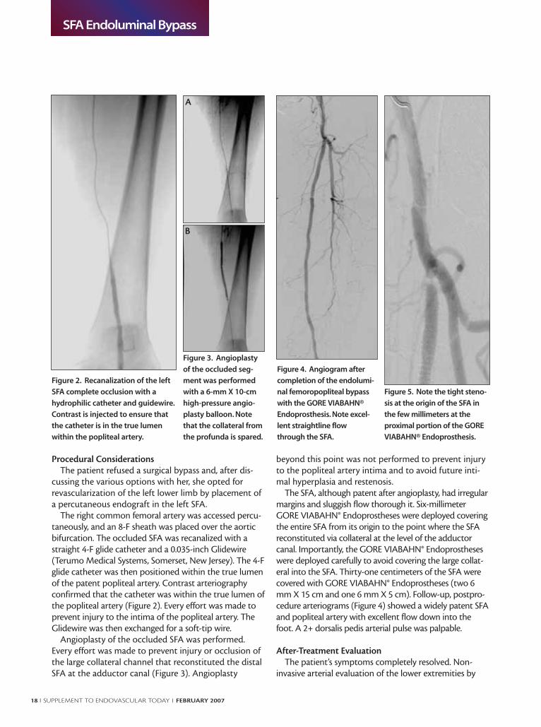
The SFA, although patent after angioplasty, had irregularmargins and sluggish flow thorough it. Six-millimeterGORE VIABAHN® Endoprostheses were deployed coveringthe entire SFA from its origin to the point where the SFAreconstituted via collateral at the level of the adductorcanal. Importantly, the GORE VIABAHN® Endoprostheseswere deployed carefully to avoid covering the large collat-eral into the SFA. Thirty-one centimeters of the SFA werecovered with GORE VIABAHN® Endoprostheses (two 6mm X 15 cm and one 6 mm X 5 cm). Follow-up, postpro-cedure arteriograms (Figure 4) showed a widely patent SFAand popliteal artery with excellent flow down into thefoot. A 2+ dorsalis pedis arterial pulse was palpable.
After-Treatment EvaluationThe patient’s symptoms completely resolved. Non-
invasive arterial evaluation of the lower extremities by
Figure 4. Angiogram after
completion of the endolumi-
nal femoropopliteal bypass
with the GORE VIABAHN®
Endoprosthesis. Note excel-
lent straightline flow
through the SFA.
Figure 3. Angioplasty
of the occluded seg-
ment was performed
with a 6-mm X 10-cm
high-pressure angio-
plasty balloon. Note
that the collateral from
the profunda is spared.
Figure 5. Note the tight steno-
sis at the origin of the SFA in
the few millimeters at the
proximal portion of the GORE
VIABAHN® Endoprosthesis.
A
B
Figure 2. Recanalization of the left
SFA complete occlusion with a
hydrophilic catheter and guidewire.
Contrast is injected to ensure that
the catheter is in the true lumen
within the popliteal artery.

FEBRUARY 2007 I SUPPLEMENT TO ENDOVASCULAR TODAY I 19
SFA Endoluminal Bypass
analysis of the Doppler analog signal and segmental limbpressure was done 2 weeks after the procedure, and theABI on the left leg had improved from 0.6 to 1.01. Shewas prescribed clopidogrel bisulphate and returned forclinical and noninvasive follow-up every 3 months.
One-Year Follow-UpAt 1-year follow-up, the patient was noted to have a
stenosis at the origin and the first few millimeters of theSFA proximal to the GORE VIABAHN® Endoprosthesis.The ABI on the left lower extremity was reduced from 1.0to 0.84. A 90% stenosis of the origin (3 mm to 4 mm) ofthe left SFA, proximal to the endograft, was noted abovethe previously placed GORE VIABAHN® Endoprosthesis(Figure 5). The entire GORE VIABAHN® Endoprosthesisand the remainder of the SFA and the popliteal arterywere widely patent with good runoff vessels.
The stenosis was treated by angioplasty with a 6-mmballoon and placement of a 6-mm X 2.5-cm GOREVIABAHN® Endoprosthesis.
Arteriography after deployment showed a widelypatent profunda with good coverage of the origin of SFAstenosis (Figure 6) with excellent flow to the foot. Thepatient’s ABI 2 weeks after the procedure had increasedfrom 0.84 to 1.0. Bilateral lower-extremity and Dopplerimaging of the arterial tree showed a widely patent leftSFA and popliteal artery.
DISCUSSIONOver the past few years, use of the GORE VIABAHN®
Endoprosthesis for the treatment of femoropopliteal dis-ease has been well documented.7-10 Previous reports haveshown that polytetrafluoroethylene-coated stents placed inthe intraluminal location may reduce intimal hyperplasia inthe peripheral vascular system.11,12 Our earlier studies havealso provided promising patency results with all grades of
TASC A to D lesions. However, the device’scapability in maintaining the long-termpatency is yet to be evaluated.
In the present case study, a TASC Dlesion is successfully treated using a GOREVIABAHN® Endoprosthesis, thus demon-strating its efficacy in treating long-seg-ment occlusive disease of the SFA. Durabletreatment of such long-segment occlusivedisease is not feasible with bare metallicstents. GORE VIABAHN® Endoprosthesespermit the placement of a percutaneousfemoropopliteal artery bypass graft safelyand effectively. However, aggressive follow-up and prophylactic measures are crucialfor achieving continued patency.
CONCLUSIONEndovascular treatment with the GORE VIABAHN®
Endoprosthesis can be regarded as an effective option fortreating long-segment occlusive disease of the SFA with highpatency rates. ■
Paramjit “Romi” Chopra, MD, is Director of MidwestInstitute for Minimally Invasive Therapies, Principal, CVIResearch Inc., in Chicago, Illinois. He has disclosed that he isa paid consultant to and receives grant/research fundingfrom Gore & Associates. Dr. Chopra may be reached at(708) 681-7888; [email protected].
Swarnam Chiramel, MS, is Research Associate, CVIResearch Inc., in Chicago, Illinois. She has disclosed that sheholds no financial interest in any product or manufacturermentioned herein. Ms. Chiramel may be reached at (708)681-7888; [email protected].
1. Beckman JA, Creager MA, Libby P. Diabetes and atherosclerosis: epidemiology, patho-physiology, and management. JAMA. 2002;287:2570-2581.2. Libby P, Theroux P. Pathophysiology of coronary artery disease. Circulation.2005;111:3481-3488.3. Garcia LA. Epidemiology and pathophysiology of lower extremity peripheral arterial dis-ease. J Endovasc Ther. 2006;13(suppl 2):II3-9.4. Beard JD. Chronic lower limb ischemia. West J Med. 2000;173:60-63.5. Dormandy JA, Rutherford RB. Management of peripheral arterial disease (PAD). TASCWorking Group. TransAtlantic Inter-Society Consensus (TASC). J Vasc Surg. 2000;31:S1-S296. 6. Chopra P. Use of the Viabahn stent graft for PVD in the femoropopliteal arterial segment.Endovascular Today (Suppl). 2005; 4S-8S.7. Bauermeister G. Endovascular stent-grafting in the treatment of superficial femoral arteryocclusive disease. J Endovasc Ther. 2001;8:315-320.8. Saxon RR, Coffman JM, Gooding JM, et al. Long-term results of ePTFE stent-graft versusangioplasty in the femoropopliteal artery: single center experience from a prospective, ran-domized trial. J Vasc Interv Radiol. 2003;14:303-311.9. Jahnke T, Andresen R, Muller-Hulsbeck S, et al. Hemobahn stent-grafts for treatment offemoropopliteal arterial obstructions: midterm results of a prospective trial. J Vasc IntervRadiol. 2003;14:41-51.10. Fischer M, Schwabe C, Schulte KL. Value of the hemobahn/viabahn endoprosthesis inthe treatment of long chronic lesions of the superficial femoral artery: 6 years of experience. JEndovasc Ther. 2006;13:281-90.11. Marin ML, Veith FJ, Cynamon J, et al. Effect of polytetrafluoroethylene covering ofPalmaz stents on the development of intimal hyperplasia in human iliac arteries. J Vasc IntervRadiol. 1996;7:651-656.12. Yuan JG, Ohki T, Marin ML, et al. The effect of nonporous PTFE-covered stents on intimalhyperplasia following balloon arterial injury in minipigs. J Endovasc Surg. 1998;5:349-358.
Figure 6. An 8-F sheath is noted in the common femoral artery (A). A wire has
been placed across the stenosis (B). A 6-mm X 2.5-cm GORE VIABAHN®
Endoprosthesis was placed in the proximal stenosis and reangioplastied to
obtain an excellent result with a patent endograft (C).
A B C

20 I SUPPLEMENT TO ENDOVASCULAR TODAY I FEBRUARY 2007
SFA Endoluminal Bypass
Treating Significant Stenosis in the SFA With Covered Self-Expanding Stent GraftsUse of the GORE VIABAHN® Endoprosthesis in significant stenosis complicated by
a significant aneurysm in the SFA.
BY JAMES B. PARK, MD, FACC
Endovascular therapy has gained widespreadacceptance for treating peripheral artery dis-ease (PAD) in several anatomical locations. Theiliac arteries are one such area, particularly in
the common iliacs and proximal external iliac areas,especially if the length of lesion is less diffuse. Greateracceptance is also being garnered for endovasculartherapy in the superficial femoral artery (SFA), espe-cially for nitinol self-expanding stents when used in rel-atively short lengths.
Among several concerns related to SFA interventionare the diffuse nature of some SFA disease and the fre-quent need to use relatively long stent lengths, at timesrequiring the treating physician to overlap multiplestents. Further complicating the treatment of SFA diseaseis the recent controversy surrounding the possibility ofstent fractures and subsequent restenosis related to thisevent. A particular area of vulnerabilityis the area through the adductor canalas the SFA continues behind the knee.In fact, the area at the adductor canal isa frequent location for SFA disease likelysecondary to the torsion and compres-sion forces on the artery itself due tothe muscular structure surrounding thiscanal. In the earlier days of endovascu-lar therapy, we believed that the area toavoid stenting due to the risk of stentcrush or stent fracture was near thebony articulation between the femurand the tibia.
We now believe that the area of criti-cal importance is really superior to thispoint leading up to the adductor canal,which is a frequent location of SFA
lesions. If it is necessary to stent this region, the ability ofthe stent to withstand the forces present in the SFA is ofcritical importance. One such device might be the cov-ered, self-expanding GORE VIABAHN® Endoprosthesis(Gore & Associates, Flagstaff, AZ). This article describesan interesting case report using this device during thefirst of two interventions (one in each leg) for a patientwith severe claudication symptoms related to significantstenosis of the distal SFA bilaterally. The case was furthercomplicated by the presence of large aneurysms proximaland distal to the stenosis.
CA SE REPORTA 71-year-old man presented with severe claudication
bilaterally. He reported that his right leg was slightlyworse than his left, and his exertional tolerance wasbetween one to two blocks. He had initially reported
the symptoms to his primary physician,who had ordered a bilateral, lower-extremity arterial ultrasound, whichnoted bilateral moderate SFA stenosiswith ankle-brachial indexes of .92 onthe right and .78 on the left. There wasno mention of aneurysmal segments oneither side. He underwent aortographyand runoff as an outpatient by anothercardiologist, which showed significantSFA disease complicated by largeaneurysmal areas below and above theSFA lesions bilaterally (Figure 1). Weelected to treat the left leg first becauseof the severity of the ankle-brachialindex findings and the easier access forthis initial procedure—going from rightto left in a contralateral fashion.
Figure 1. The patient's left SFA,
showing multiple aneurysms and
severe stenosis.

FEBRUARY 2007 I SUPPLEMENT TO ENDOVASCULAR TODAY I 21
SFA Endoluminal Bypass
There was initial consideration of asurgical approach versus possiblestenting, which was further compli-cated by the presence of aneurysmsand the location of the distal stenosisand aneurysms impinging upon thecritical areas of stent crush and frac-ture. For the procedure, an 8-F, 45-cm-long sheath (Terumo MedicalCorporation, Somerset, NJ) was placedcontralaterally, and mapping angio-graphy was performed. The lesion wascrossed with mild difficulty with a.018-inch Steelcore wire (AbbottVascular, Santa Clara, CA) to the per-oneal artery. A Volcano IVUS catheter(Volcano Therapeutics, Inc., LagunaHills, CA) was then used to assess thesize of the artery at the tibioperonealtrunk and also to assess the amountof atherosclerosis present at the loca-tion where the distal end of theGORE VIABAHN® Endoprosthesiswould be placed. In addition, use ofultrasound would allow for moreaccurate determination of stent sizeto ensure good apposition at theproximal and distal ends.
To ensure that the stent catheter couldbe placed into the distal end, angioplastywas performed using a 4-cm X 2-cmAgiltrac balloon (Abbott Vascular) inmultiple stenotic areas. Two GOREVIABAHN® Endoprostheses were used (a6-cm X 15-cm and a 6-cm X 10-cm) andoverlapped to the mid SFA. However,angiography performed after stentplacement revealed a very small leak into the aneurysm.A 7-cm X 2.5-cm stent was placed, which overlapped intothe proximal stent; the proximal portion was postdilatedwith a 6-mm balloon, and the distal portion was postdi-lated with a 5-mm balloon (Figure 2). The patient didwell after the procedure and had excellent results (Figure3). Ankle-brachial indexes after the procedure were ≥1after 12 months of treatment. He was placed on a stan-dard aspirin and clopidogrel therapeutic regimen.Because the disease was bilateral, the right SFA was treat-ed similarly with a GORE VIABAHN® Endoprosthesis 2weeks later, with very good results.
DISCUSSIONThe use of covered self-expanding stents to treat long
lesions and the areas of the SFA vulnera-ble to stent crush and stent fracturesmight be a viable option, especially withregard to in-stent restenosis and the abili-ty of covered stents to better withstandthe compression and torsion forces pres-ent in the SFA. One possible concern isthe risk of acute thrombosis due to use ofthe covered stent. However, our patientwas not placed on warfarin therapy andhad done well without acute thrombosisand restenosis at follow-up after 1 year.The patient's medical regimen related tohis percutaneous treatment has been theinstitution of aspirin and clopidogrel.One possible explanation for the lack ofacute thrombosis in cases like this mightbe the better matching of the stent graftto the native artery in relation to the pos-sible mismatch in size of a femoral-to-popliteal bypass graft. The better charac-teristics of this covered stent might bedue to the material actually reinforcingthe stent framework while also beingmore flexible around framework. The fab-ric also acts to smooth out the stentwhile it is bent, unlike the scaling onemight see with an uncovered stent. Theadditional advantage of the stent in thisparticular case was the ability to excludethe aneurysm, which was also of signifi-cant concern. In fact, there are manycases of both iliac and SFA disease thatare complicated by pre- and poststenoticaneurysms and ulcerated plaques withfalse aneurysmal channels.
CONCLUSIONGiven the challenges of obtaining good long-term
results of endovascular therapies in the SFA, particularlywith recent data showing frequent stent fractures, theuse of covered nitinol stents can be a viable option, withpossible better long-term results if the risk of stent frac-tures can be lowered while obtaining good patency. ■
James B. Park, MD, FACC, is the Director of the CardiacCatheterization Laboratory at Presbyterian Hospital of Dallasand is the Co-Director of the Endovascular teaching programsmanaged by the CIVA Vascular group in Dallas, Texas. Hehas disclosed that he holds no financial interest in any prod-uct or manufacturer mentioned herein. Dr. Park may bereached at [email protected].
Figure 3. The mid-to-distal
SFA after completion of the
procedure.
Figure 2. The second GORE
VIABAHN® Endoprosthesis was
overlapped with the first.

22 I SUPPLEMENT TO ENDOVASCULAR TODAY I FEBRUARY 2007
SFA Endoluminal Bypass
GORE VIABAHN®
Endoprosthesis Recanalization of a Complex SFA LesionAn illustrative case report on the effective use of the GORE VIABAHN®
Endoprosthesis.
BY MARK W. MEWISSEN, MD
CA SE PRE SENTATIONA 62-year-old man presented to the Vascular Center at St.
Luke’s Medical Center in June 2005 with lifestyle-limitingclaudication of the left lower limb. On physical examination,the left popliteal pulse was absent. The left ankle-brachialindexes before and after exercise were 0.5 and 0.2, respec-tively. Duplex ultrasonography identified a heavily calcifieddistal superficial femoral artery (SFA) and low velocities inthe popliteal artery. Angiography, performed via a right con-tralateral femoral approach, showed that the distal left SFAwas heavily calcified with resulting near-total occlusion ofthe arterial segment. The distal popliteal artery was relativelyfree of disease, and the tibial runoff consisted of patentanterior and posterior tibial arteries (Figure 1).
Using the guidance of a .035-inch Rosen wire (CookMedical, Bloomington, IN), an 8-F, 45-cm-long sheath waspassed over the aortic bifurcation and advanced to thelevel of the ipsilateral common femoral artery. Using a.018-inch Quick Cross catheter (Spectranetics Corporation,Colorado Springs, CO) for support, a .018-inch, 260-cm-long supportive guidewire (Platinum Plus, Boston ScientificCorporation, Natick, MA) was successfully negotiatedacross the lesion to allow predilatation using a 3.5-mmdiameter, 4-cm-long balloon (Savvy, Cordis Corporation, aJohnson & Johnson company, Miami, FL). This step wasnecessary to allow passage and deployment of a 6-mm,15-cm-long GORE VIABAHN® Endoprosthesis (Gore &Associates, Flagstaff, AZ), expanded in place using a high-
Figure 2. Postdilatation of the 6-mm GORE VIABAHN®
Endoprosthesis will help to avoid stent edge dilatation with a
high-pressure balloon catheter (A). A poststent deployment and
dilatation angiogram shows an excellent technical result (B).
Figure 1. Selective left lower-extremity arteriogram demon-
strating a heavily calcified SFA lesion (A).The mid and distal
popliteal artery, relatively disease-free and of adequate
diameter (>5 mm), accommodate a 6-mm GORE VIABAHN®
Endoprosthesis.There is two-vessel runoff, a good predictor
of favorable outcome (B).
A B A B

FEBRUARY 2007 I SUPPLEMENT TO ENDOVASCULAR TODAY I 23
SFA Endoluminal Bypass
pressure, 5-mm-diameter percutaneous transluminal angio-plasty balloon, avoiding dilatation of the stent edges (Figure2). At 18 months, the patient remains asymptomatic. Hisankle-brachial index continues to be normalized. Duplexexamination revealed normal arterial waveforms withoutany significant stenosis.
DISCUSSIONThis case, although anecdotal, is in line with several
recent reports indicating that outcomes for the GOREVIABAHN® Endoprosthesis exceed results achieved withangioplasty and bare nitinol stenting for complexfemoropopliteal arterial lesions. This may be attributable tothe expanded polytetrafluoroethylene lining of the stentthat probably inhibits the ingrowth of intimal hyperplasia.
Regarding patient and lesion selection, it is importantto note that the device requires delivery through a larger-profile sheath (8 F for the 6-mm graft), and that the proxi-mal and distal boundaries of the vessel to be treated mustbe at least 4.8 mm in diameter. The tibial runoff shouldalso be continuous via at least one patent artery. It is alsoof paramount importance to avoid dilating the edges ofthe deployed stent to minimize the development of anedge stenosis, a reported mechanism of stent graft failure.Spot stenting should also be avoided. Extreme care mustbe exercised when the device is deployed near the ori-fice of the profunda femoralis artery. High-magnifica-tion angiograms in various obliquities must be obtainedbefore stent deployment. Failure to understand theanatomy of the common femoral bifurcation willunavoidably lead to disastrous complications.
The appropriate management of the collateral circula-tion at the distal segment of a long lesion remains contro-versial. Some collateral vessels must occasionally be cov-ered if the edges of the graft are to be delivered in rela-tively disease-free arterial margins. It is believed that thebenefits of such technique outweigh the risks of alterna-tive spot stenting strategies, a reported mechanism ofearly stent graft failure. Furthermore, it is important toflare the stent edges at sites free of plaque. This latter con-cept also helps to choose the appropriate stent length toensure exclusion of the diseased segment in toto. The mul-ticenter prospective and randomized “Viabahn VersusBare Nitinol Stent Trial” (VIBRANT), funded by Gore &Associates, will provide additional data regarding theoptimal treatment for long SFA lesions. ■
Mark W. Mewissen, MD, is Director of the Vascular Centerat St. Luke’s Medical Center in Milwaukee, Wisconsin. Hehas disclosed that he holds no financial interest in any prod-uct or manufacturer mentioned herein. Dr. Mewissen maybe reached at (414) 385-2429; [email protected].
paid consultant for Gore & Associates, Boston ScientificCorporation, and Medtronic. Dr. Adams may be reached at(573) 443-8773; [email protected].
Richard D. Coats, MD, is from Columbia SurgicalAssociates, Columbia, Missouri. He has disclosed that he is apaid consultant for Gore & Associates, Boston ScientificCorporation, and Medtronic. Dr. Coats may be reached at(573) 443-8773; [email protected].
Paul W. Humphrey, MD, FACS, is from Columbia SurgicalAssociates, Columbia, Missouri. He has disclosed that he is apaid consultant for Gore & Associates, Boston ScientificCorporation, and Medtronic. Dr. Humphrey may be reachedat (573) 443-8773; [email protected].
Karen Althage, CFA, is affiliated with Columbia SurgicalAssociates, Columbia, Missouri. She has disclosed that sheholds no financial interest in any product or manufacturermentioned herein. Ms. Althage may be reached at (573)443-8773.
Mary Chott is a Research Coordinator at ColumbiaSurgical Associates, Columbia, Missouri. She has disclosedthat she holds no financial interest in any product or man-ufacturer mentioned herein. Ms. Chott may be reached at(573) 443-8773.
1. Coats RD, Adams JG, Humphrey PW. SFA revascularization using the Viabahn endopros-thesis. Endovasc Today. 2006;5:76-78.2. Adams JG, Humphrey PW, Coats RD. Endoluminal bypass utilizing the Viabahn endo-prosthesis. Endovasc Today. 2005;4(suppl):9-11.3. Jahnke T, Andresen R, Muller-Hulsbeck S, et al. Hemobahn stent-grafts for treatment offemoropopliteal arterial obstructions: midterm results of a prospective trial. J Vasc IntervRadiol. 2003;14:41-51.4. Saxon RR, Coffman JM, Gooding JM, et al. Long-term results of ePTFE stent-graft versusangioplasty in the femoropopliteal artery: single center experience from a prospective, ran-domized trial. J Vasc Interv Radiol. 2003;14:303-311.5. Railo M, Roth WD, Edgren J, et al. Preliminary results with endoluminal femoropoplitealthrupass. Ann Chir et Gynaecol. 2001;90:15-18.6. Bauermeister G. Endovascular stent-grafting in the treatment of superficial femoral arteryocclusive disease. J Endovasc Ther. 2001;8:315-320.7. Lammer J, Dake MD, Bleyn J, et al. Peripheral arterial obstruction: prospective studyof treatment with a transluminally placed self-expanding stent-graft. Radiology.2000;217:95-104.8. Boyd AM. Natural course of arteriosclerosis of lower extremities. Proc R Soc Med.1962;53:591-593.9. Imparato AM, Kim GE, Davidson T, et al. Intermittent claudication: its natural course.Surgery. 1975;78:795-799.10. Taylor LM, Edwards JM, Porter JM. Present status of reversed vein bypass grafting: five-year results of a modern series. J Vasc Surg. 1990;11:193-205; discussion 205-206.11. Bergamini TM, Townsend, JB, Bandyk DF, et al. Experience with in situ saphenous veinbypasses during 1981 to 1989: determinant factors of long-term patency. J Vasc Surg.1991;13:137-147.12. Donaldson MC, Mannick JA, Whittemore AD. Femoral-distal bypass with in situ greatersaphenous vein: long-term results using the Mills valvulotome. Ann Surg. 1991;213:457-464.13. Pomposelli FB, Kansal N, Hamdan AD, et al. A decade of experience with dorsalis pedisartery bypass: analysis of outcome in more than 1000 cases. J Vasc Surg. 2003;37:307-315. 14. Bolia A, Miles KA, Brennan J, et al. Percutaneous transluminal angioplasty of occlu-sions of the femoral and popliteal arteries by subintimal dissection. Cardiovasc InterventRadiol. 1990;13:357-363.15. Bolia A, Brennan J, Bell PR. Recanalization of femoral-popliteal occlusions: improvingsuccess rate by subintimal Recanalization. Clin Radiol. 1989;40:325. 16. Lipsitz EC, Ohki T, Veith FJ, et al. Does subintimal angioplasty have a role in the treat-ment of severe lower extremity ischemia? J Vasc Surg. 2003;37:386-391. 17. CAPRIE Steering Committee. A randomized, blinded trial of clopidogrel versus aspirin inpatients at risk of ischaemic events (CAPRIE). Lancet. 1996;348:1329-1339.
(Continued from page 12)

RP1352
Copyright © 2022 FDOKUMEN