Initial Experience with Transluminally Placed Endovascular Grafts for the Treatment of Complex...
17
ANNALS OF SURGERY Vol. 222, No. 4, 449-469 © 1995 Lippincott-Raven Publishers Initial Experience with Transluminally Placed Endovascular Grafts for the Treatment of Complex Vascular Lesions Michael L. Marin, M.D., Frank J. Veith, M.D., Jacob Cynamon, M.D., Luis A. Sanchez, M.D., Ross T. Lyon, M.D., Barry A. Levine, M.D., Curtis W. Bakal, M.D., William D. Suggs, M.D., Kurt R. Wengerter, M.D., Steven P. Rivers, M.D., Richard E. Parsons, M.D., John G. Yuan, M.D., Reese A. Wain, M.D., Takao Ohki, M.D., Alla Rozenblit, M.D., and Juan C. Parodi, M.D. From the Departments of Surgery and Radiology, Montefiore Medical Center/Albert Einstein College of Medicine, New York, New York Objectives Complex arterial occlusive, traumatic, and aneurysmal lesions may be difficult or impossible to treat successfully by standard surgical techniques when severe medical or surgical comorbidities exist. The authors describe a single center's experience over a 2/2-year period with 96 endovascular graft procedures performed to treat 100 arterial lesions in 92 patients. Patients and Methods Thirty-three patients had 36 large aortic and/or peripheral artery aneurysms, 48 had 53 multilevel limb-threatening aortoiliac and/or femoropopliteal occlusive lesions, and 11 had traumatic arterial injuries (false aneurysms and arteriovenous fistulas). Endovascular grafts were placed through remote arteriotomies under local (16 [17%]), epidural (42 [43%]), or general (38 [40%]) anesthesia. Results Technical and clinical successes were achieved in 91% of the patients with aneurysms, 91% with occlusive lesions, and 100% with traumatic arterial lesions. These patients and grafts have been followed from 1 to 30 months (mean, 13 months). The primary and secondary patency rates at 18 months for aortoiliac occlusions were 77% and 95%, respectively. The 18-month limb salvage rate was 98%. Immediately after aortic aneurysm exclusion, a total of 6 (33%) perigraft channels were detected; 3 of these closed within 8 weeks. Endovascular stented graft procedures were associated with a 10% major and a 14% minor complication rate. The overall 30-day mortality rate for this entire series was 6%. Conclusions This initial experience with endovascular graft repair of complex arterial lesions justifies further use and careful evaluation of this technique for major arterial reconstruction. 449
-
Upload
independent -
Category
Documents
-
view
0 -
download
0
Transcript of Initial Experience with Transluminally Placed Endovascular Grafts for the Treatment of Complex...

ANNALS OF SURGERYVol. 222, No. 4, 449-469© 1995 Lippincott-Raven Publishers
Initial Experience with TransluminallyPlaced Endovascular Grafts forthe Treatment of ComplexVascular LesionsMichael L. Marin, M.D., Frank J. Veith, M.D., Jacob Cynamon, M.D., Luis A. Sanchez, M.D.,Ross T. Lyon, M.D., Barry A. Levine, M.D., Curtis W. Bakal, M.D., William D. Suggs, M.D.,Kurt R. Wengerter, M.D., Steven P. Rivers, M.D., Richard E. Parsons, M.D.,John G. Yuan, M.D., Reese A. Wain, M.D., Takao Ohki, M.D.,Alla Rozenblit, M.D., and Juan C. Parodi, M.D.
From the Departments of Surgery and Radiology, Montefiore Medical Center/Albert EinsteinCollege of Medicine, New York, New York
ObjectivesComplex arterial occlusive, traumatic, and aneurysmal lesions may be difficult or impossible totreat successfully by standard surgical techniques when severe medical or surgical comorbiditiesexist. The authors describe a single center's experience over a 2/2-year period with 96endovascular graft procedures performed to treat 100 arterial lesions in 92 patients.
Patients and MethodsThirty-three patients had 36 large aortic and/or peripheral artery aneurysms, 48 had 53 multilevellimb-threatening aortoiliac and/or femoropopliteal occlusive lesions, and 11 had traumatic arterialinjuries (false aneurysms and arteriovenous fistulas). Endovascular grafts were placed throughremote arteriotomies under local (16 [17%]), epidural (42 [43%]), or general (38 [40%])anesthesia.
ResultsTechnical and clinical successes were achieved in 91% of the patients with aneurysms, 91% withocclusive lesions, and 100% with traumatic arterial lesions. These patients and grafts have beenfollowed from 1 to 30 months (mean, 13 months). The primary and secondary patency rates at 18months for aortoiliac occlusions were 77% and 95%, respectively. The 18-month limb salvage ratewas 98%. Immediately after aortic aneurysm exclusion, a total of 6 (33%) perigraft channels weredetected; 3 of these closed within 8 weeks. Endovascular stented graft procedures wereassociated with a 10% major and a 14% minor complication rate. The overall 30-day mortality ratefor this entire series was 6%.
ConclusionsThis initial experience with endovascular graft repair of complex arterial lesions justifies further useand careful evaluation of this technique for major arterial reconstruction.
449

450 Marin and Others
Surgical therapy for complex vascular diseases has ad-vanced greatly over the past 40 years, shifting from theuse of palliative measures to treat lethal or limb-threat-ening disease to therapy focused on the precise diagnosisand open surgical correction of arterial disorders. Theoperative mortality rate for elective aortic aneurysm re-
pair has since decreased markedly, from 21% in the1 950s to below 5% currently. 1-5 Multilevel lower extrem-ity occlusive disease with gangrene, which once man-
dated amputation, can be treated with interventionaltechniques or bypass surgery, resulting in favorable limbsalvage rates.6 Finally, despite advances in resuscitation,anesthesia, and intensive care, complex acute arterial in-jury from penetrating or blunt vascular trauma has re-
mained a challenging problem with substantial morbid-ity and mortality, particularly when central vascular in-juries occur with other severe injuries.` Despiteimportant improvements in the management of all ofthese vascular lesions, significant perioperative morbid-ity and mortality still occur, particularly in those patientswho have severe comorbid medical illnesses, who requirea second operation at a particular surgical site, or whohave had multiple traumatic injuries.'0'6A trend in surgery over the past decade has been to
develop less invasive procedures to accomplish treat-ment goals with reduced operative risks and complica-tions, thus allowing therapy to be extended to high-riskpatients with severe comorbid medical illnesses. Onesuch evolving technique for less invasive vascular sur-
gery involves the use ofendovascular grafts that combineintravascular stent and prosthetic graft technologies. 17-38These devices may be inserted through remote arterialaccess sites to treat vascular lesions without the need todirectly expose the diseased artery through an extensiveincision or dissection. Endovascular grafts have beenused successfully to treat aortic and peripheral artery an-
eurysms, long-segment arterial occlusive disease, andvascular trauma. Despite the potential advantages ofthese new techniques, the devices are primitive and re-
quire further development before they can be widely ap-
plied for the treatment of vascular disease. In this reportwe describe our initial experience over a 21/2-year periodwith the use of 96 endovascular grafts for the treatment
Presented at the 1 15th Annual Meeting ofthe American Surgical Asso-ciation, April 6-8. 1995, Chicago, Illinois.
Supported by grants from the U.S. Public Health Service (HL 02990-02), the James Hilton Manning and Emma Austin Manning Foun-dation, the Anna S. Brown Trust, and the New York Institute forVascular Studies.
Address reprint requests to Michael L. Marin, M.D., Division of Vas-cular Surgery, Montefiore Medical Center, 111I East 210th Street,NewYork,NY 10467.
Accepted for publication April 10. 1995.
of arterial aneurysms, occlusions, and traumatic or iat-rogenic vascular injuries.
MATERIALS AND METHODSPatient SelectionThe criteria for patient selection were based on the
type of vascular disease being treated (Table 1). For pa-tients with aortic aneurysms, two protocols were fol-lowed. Those patients with severe comorbid medical andsurgical illnesses in association with large, threateningabdominal aortic aneurysms (AAAs) were treated with aballoon expandable device on a compassionate basis(i.e., no other corrective treatment is feasible or the riskof such other treatment is excessively great) in accor-dance with a protocol approved by our hospital's institu-tional review board. Patients with suitable aortic anat-omy (long proximal and distal aortic necks [.2 cm and.1.5 cm, respectively]) who were not at increased riskfor complications from standard AAA repair were en-tered into a Food and Drug Administration phase I fea-sibility trial of endoluminal AAA repair with the Endo-vascular Technologies' (EVT) Endograft device (MenloPark, CA).
Patients with peripheral artery aneurysms, chronic ar-terial occlusive disease, or penetrating or iatrogenic vas-cular injuries usually coexisting with a serious comorbidmedical illness or with contraindications for standardtreatment underwent endovascular graft procedures thatinvolved use of standard polytetrafluoroethylene grafts(W.L. Gore and Associates, Flagstaff, AZ) and commer-cially available intravascular stents. All patients pro-vided informed consent. Patients with occlusive lesionswere treated for limb salvage only. All traumatic lesionswere associated with large pseudoaneurysms or arterio-venous fistulas that would have mandated open surgicalrepair.
Endovascular Stented Graft DevicesThe endovascular stented graft devices were chosen
based on the type of vascular lesion being excluded (Ta-ble 2, Fig. 1). With the exception of the aortic devices,all endovascular grafts were composed of commerciallyavailable materials. The balloon expandable aortic de-vice and EVT device have been described in detail else-where.39,40
Techniques for Endovascular StentedGraft InsertionThe principles of endovascular stented graft insertion
and deployment are similar for treatment of all vasculardiseases. Specific procedural modifications were made inaccordance with the anatomic locations of the lesions aswell as the presence or absence of arterial occlusive dis-
Ann. Surg. * October 1995

Endovascular Grafts for Complex Lesions 451
Table 1. PATIENT SELECTION CRITERIA
Severe Underlying Medical/SurgicalComorbidity or Immediate Characteristics or Limitation
Vascular Pathology Life-Threatening Lesion of Vascular Lesion
Aortic aneurysmsBalloon expandable device Yes 26 cm AAA with rupture or impending ruptureEVT device No Suitable proximal/distal neck, 25 cm AAA
Peripheral artery aneurysms No* .3 times normal vessel diameterChronic arterial occlusions No* Multilevel occlusive disease with critical ischemia
and a threatened limbArterial trauma Yes Pseudoaneurysm; arteriovenous fistula
AAA = abdominal aortic aneurysm; EVT = Endovascular Technologies' Endograft.* Most of these patients had severe coexisting medical problems or a surgical contraindication to standard treatment.
ease. Target lesions were generally approached through Patient Follow-upthe largest available access vessel (i.e., common femoralor brachial arteries). After insertion of a guide wire The method of follow-up for each patient who re-through the access vessel and the interpretation ofprein- ceived an endovascular graft was dependent on the typesertion arteriograms, endovascular stented grafts con- of endoluminal reconstruction and, occasionally, thetained in delivery catheters were advanced to the desired presence of significant comorbid medical illnesses (e.g.,location by coaxial movement of the device over the renal insufficiency precluding contrast injection). Whenguide wire under fluoroscopic control. If stenotic or oc- medically feasible, each patient who underwent an AAAcluded arteries were being treated, the diseased vessel was repair had a contrast-enhanced computed tomographyfirst balloon dilated diffusely, creating a widened tract be- (CT) scan and a color duplex ultrasound performed 1 tofore insertion of the device (Fig. 2). Once proper posi- 2 days after the procedure. Serial contrast-enhanced CTtioning of the endovascular graft was confirmed, the de- and/or color duplex analyses were performed at 3-monthlivery catheter was withdrawn, permitting deployment intervals for 6 months and at 6-month intervals thereaf-ofthe graft-to-artery attachment system (stent).25'41 Each ter. Each scan was analyzed for perigraft channels (flowprocedure was followed immediately by an intraopera- outside the endovascular graft but within the aneurysmaltive completion arteriogram and, in selected cases, intra- sac), alteration in the geometric configuration of thevascular ultrasound examination to evaluate the ade- graft, device migration, and any change in the aneurysmquacy of the technique. All procedures were performed size. Similar analyses were performed during the follow-in the operating room with use of a portable C-arm flu- up of patients who had undergone endovascular graft re-oroscope. pairs of iliac artery aneurysms. The protocol for follow-
Table 2. CHARACTERISTICS OF ENDOVASCULAR GRAFTS
Vascular Pathology Graft Material Attachment System Introducer System
Aortic aneurysmBalloon expandable device Thin walled knitted, crimped Dacron* Balloon expandable stent* Teflon, double sheath (24 Fr)*EVT device Woven Dacront Self-expanding, hooked, "Z" stent Endovascular deployment
configurationt assembly (26 Fr)tPeripheral artery aneurysm PTFEt Palmaz balloon expandable stent§ Teflon, single sheath (12-14 Fr)Chronic arterial occlusion PTFEf Palmaz balloon expandable stent§ Teflon, single sheath (14 Fr)Arterial trauma PTFEt Palmaz balloon expendable stent§ Teflon, single sheath (12 Fr)
EVT = Endovascular Technologies' Endograft; PTFE = polytetrafluoroethylene.* Barone, Inc., Buenos Aires, Argentina.t Endovascular Technologies Corporation, Menlo Park, California.t W.L. Gore and Associates, Flagstaff, Arizona.§ Johnson and Johnson Interventional Systems, Warren, New Jersey.
Vol. 222 - No. 4

452 Marin and Others
Figure 1. Endovascular grafts used for the repair of aortic and peripheralartery aneurysms, arterial occlusions, and traumatic vascular injuries. (A)Balloon expandable (Parodi) endovascular aortic device; (B) EVT Endo-graft; (C) polytetrafluoroethylene (PTFE) endovascular graft used for thetreatment of peripheral artery aneurysms and arterial occlusive disease;(D) PTFE-covered Palmaz balloon expandable stent used for the treatmentof traumatic arterial injuries; (E) an occluding PTFE-covered Palmaz stent(one end of this device is closed with a purse-string suture) (arrow).
up of aneurysms in the popliteal or subclavian arteriesinvolved a physical examination supplemented by colorduplex ultrasonography, ankle-brachial indices, andpulse volume recordings at 3-month intervals for 6months and at 6-month intervals thereafter.
After endovascular stented graft repair of multilevelocclusive disease, patients who did not have an absolutecontraindication to arteriography underwent a postoper-ative arteriogram in an angiography suite so that theirgrafts could be evaluated further before discharge fromthe hospital. In addition, each patient was followed witha physical examination and color duplex ultrasonogra-phy at 3-month intervals for 6 months and every 6months thereafter. Patients treated for traumatic vascu-lar injuries were similarly followed with physical exami-nation and serial color duplex ultrasonograms.Any patient with an endovascular graft who had an
abnormality detected by physical examination, CT scan,or color duplex ultrasonography was further evaluatedwith a transfemoral or transbrachial arteriogram, unlessthe patient was medically unfit to undergo an arterialcontrast study.
Data AnalysisThe patency data for patients treated for aortoiliofem-
oral occlusive disease were analyzed by the cumulativelife-table method. The primary and secondary patencieswere calculated according to the reporting standards ofthe Society for Vascular Surgery/North American Chap-ter, International Society for Cardiovascular Surgery.42
RESULTS
We performed 96 endovascular stented graft proce-dures in 92 patients with 100 arterial lesions between No-vember 1992 and April 1995 at our institution, Mon-tefiore Medical Center (New York) (Table 3). Thesegrafts were used to treat 36 arterial aneurysms, 11 lesionssecondary to penetrating vascular trauma, and 53 long-segment arterial stenotic or occlusive lesions. Endovas-cular grafts were inserted under local (16 [17%]), epi-dural (42 [43%]), or general anesthesia (38 [40%]). Themean age of patients with aneurysmal and occlusive le-sions was 69 years, whereas the mean age of patientstreated for penetrating traumatic vascular injuries was38 years. Most of the patients treated for aneurysmal orocclusive arterial disease also had significant comorbidmedical illnesses or a major surgical contraindication tostandard treatment.
Abdominal Aortic AneurysmsEighteen patients with AAAs were treated with two
different endovascular graft devices. An EVT device wasimplanted in four low-risk patients with suitable arterialanatomy28 (Figs. 3 and 4). The remaining 14 high-riskpatients received balloon expandable endovasculargrafts (Table 4, Fig. 5). All EVT procedures involved useof tubular, aorto-aortic grafts, whereas 6 of the 14 bal-loon expandable device procedures involved use of aor-toiliofemoral reconstructions with the addition of fem-orofemoral bypass and occlusion of the common iliacarteries contralateral to the main endovascular graft.Blood loss for repair of aortic aneurysms ranged from100 to 4000 cc (mean, 1970 cc). Blood loss for the fourEVT low-risk patients ranged from 100 to 250 cc (mean,162 cc). There were no thromboses of the aortic graftsand no postimplantation structural device failures. Onepatient with a balloon expandable device demonstratedapparent cephalad migration ofthe distal stent seen on aCT scan at 13 months. One EVT graft and one balloonexpandable graft showed geometric configurationchanges, which were consistent with either a kink or anextrinsic graft compression. Neither ofthese lesions wereflow restricting, and no intervention was required. Dur-ing a mean follow-up period of 8 months, two ofthe fouraneurysms treated with the EVT device became smaller.Six of the 14 aneurysms repaired with the balloon ex-pandable device became smaller (1-3 mm), and 3showed evidence of minimal enlargement (0.5-1 mm).No new aortic aneurysm ruptures occurred in this seriesafter graft placement.Three patients in this series had presented with severe
medical comorbidities in association with contained aor-tic ruptures. The first patient had a ruptured 6-cm AAA
Ann. Surg. * October 1995

Endovascular Grafts for Complex Lesions 453
Figure 2. Techniques for endolumi-nal aortoiliac bypass for occlusive Adisease. (A) Bilateral inguinal inci-sions are performed to expose thecommon femoral arteries. An intro-ducer catheter, equipped with a he-mostatic valve (A), is inserted retro-grade into the common femoral ar-tery in each groin. It is through theseintroducer catheters that all subse-quent catheter manipulations on theoccluded or diseased arterial system '1
are carried out. hydrophilic
guide wire (w) is then inserted
through each introducer catheter\and guided through the occluded orstenotic segment of an artery bymeans of directional catheters (A).(Inset) An effort is made to guide the B crecanalization process within the na- Ative lumen of diffusely stenotic arter- Aies. In situations in which the entirevessel is chronically occluded, therecanalization process occurs rela- A A
tively randomly following the planesof least resistance within the subinti-mal plane (B). (C) After successful re-canalization of an artery, long-seg-ment diffuse balloon dilatation is per- (formed (A). Balloon catheters are\inserted over the previously placed /guide wires. Balloon dilatation cre- /ates a new tract within the diseased A-artery that must communicate withthe lumen of a patent proximal ves- -sel. (D) An endovascular graft is in- DIEserted through an arteriotomy in the Acommon femoral artery. Endovascu- Jlar grafts, contained within introducer /I| !catheters, are advanced over thepreviously placed guide wires andinto the patent proximal vessels. Thedelivery sheath is then withdrawn,exposing the anchoring stent (B). (E)After inflation of the coaxially loaded Xangioplasty balloon, the Palmaz bal-loon expandable stent expands, cre-ating a friction seal between thestent and attached graft, and the un- /derlying arterial wall. The introducer /catheters on each side are then with-drawn, permitting the free end of the !prosthetic endovascular graft (G) to be withdrawn from the common femoral arteriotomy and clamped. Vascular clamps are then positioned across thenative proximal common femoral artery, which will, in turn, compress its enclosed endovascular graft. An endoluminal anastomosis is then createdbetween the endovascular graft and the internal surface of the common femoral artery with a series of interrupted U stitches (insets a, b). This anastomotictechnique can be tailored to each individual vessel, depending on the extent of local disease. The endoluminal anastomosis can be completed with a
running monofilament suture across the linear arteriotomy (inset c). Under some circumstances, a common femoral endarterectomy or a patch angioplastyof the arteriotomy site may be required to establish unobstructed flow through the endovascular graft and into the distal circulation.
Vol. 222 - No. 4

454 Marin and Others
Table 3. DISTRIBUTION OF PATIENT CHARACTERISTICS AND LESIONS TREATED WITHENDOVASCULAR GRAFTS
CoexistingCoexisting ChronicCoronary Obstructive Hostile
Age Artery Pulmonary Diabetes Renal SurgicalNo. of No. of Sex (male/ Range Disease* Diseaset Mellitus Insufficiencyt Field§
Vascular Pathology Patients Lesions female) (mean) [no. (%)] [no. (%)] [no. (%)] [no. (%)] [no. (%)]
Abdominal aorticaneurysm 18 18 15M;3F 66-88 17(94) 11(61) 5(28) 6(33) 3(17)(76)
Iliac artery aneurysm 11 14 11 M 58-89 11 (100) 5(45) 5(45) 3(27) 5(45)(72)
Popliteal artery aneurysm 3 3 2M; 1F 63-84 2(67) 1 (33) 1 (33) 0 0(74)
Subclavian artery aneurysm 1 1 1 F 40 0 0 0 0 1 (100)Traumatic arterial 9 9 8M; 1 F 18-78 1(11) 1(11) 1 (11) 0 0
pseudoaneurysm (44)Traumatic arteriovenous fistula 2 2 2 M 18-20 0 0 0 0 0
(19)Aortoiliac occlusive disease 42 47 23 M; 19 F 43-86 36(86) 9(21) 22(52) 7(17) 13(31)
(65)Femoropopliteal occlusive 6 6 4 M; 2F 62-82 6 (100) 5 (83) 4 (67) 1 (17) 0
disease (68)
Ejection fraction less than 20% on echocardiography, thallium or MUGA scans.t FEV, < 35% of predicted, room air Pa,O2 < 60 mmHg or PCO2 > 50 mmHg.f Creatinine 2 3.0 mg/dL.§ Hostile surgical field implies at least one previous dissection, scarring, or infection in the region of the vascular lesion treated by the endovascular graft.
and an acute anterior wall myocardial infarction. Thesecond patient, who experienced renal failure and severecongestive heart failure, had complete anastomotic dis-ruption (proximal and distal) ofan infrarenal aortic graftand two contained pseudoaneurysms. The third patient,who had congestive heart failure and pulmonary in-sufficiency, had a distal aortic disruption and a pseu-doaneurysm secondary to the extension of a Pott's ab-scess of the lumbar spine to the wall of the infrarenalaorta (Fig. 6).The complications encountered in this series are out-
lined in Table 5. Of the five deaths that occurred, threeresulted from complications of diffuse aneurysmalthrombus embolization during device insertion. Thesethree patients had large (.7 cm) AAAs in associationwith bilateral iliac disease. The fourth patient, who expe-rienced preoperative chronic renal failure, hepatic in-sufficiency, and severe coronary artery disease, devel-oped multiorgan failure after successful aortic graftingand died 1 week after surgery. The fifth death in this se-ries occurred in a patient with a ruptured AAA in associ-ation with acute myocardial infarction. This patient diedof a ventricular arrhythmia after successful insertion ofan endovascular graft.
Peripheral Artery Aneurysms
Fifteen patients had aneurysms involving the popli-teal, iliac, or subclavian arteries (Table 3, Figs. 7 and 8).Endovascular stented grafts were successfully placed in11 patients who had a total of 14 iliac artery aneurysms,in 2 patients with popliteal artery aneurysms, and in 1patient with a subclavian artery aneurysm. One endovas-cular graft inserted to treat a complex iliac artery aneu-rysm thrombosed at 9 months, and limb salvage wasachieved with an axillobifemoral bypass. One patientwith a popliteal artery aneurysm underwent standardvascular reconstruction when the endovascular graft de-vice could not be inserted successfully. A second patienthad thrombosis in a popliteal endovascular graft on the6th postoperative day without apparent cause, and thisendovascular graft procedure was converted to a stan-dard vascular reconstruction. A third patient with a pop-liteal artery aneurysm was treated with an endovascularstented graft, which has maintained graft patency free ofany hemodynamically significant lesions for 26 months.An endovascular graft was inserted successfully in a
patient with a subclavian artery aneurysm with distalembolization who, 2 years earlier, had undergone cervi-
Ann. Surg. * October 1995

Endovascular Grafts for Complex Lesions 455
a bFigure 3. Aortic reconstruction after transfemoral, endoluminal repair ofan abdominal aortic aneurysm. (a) After transfemoral insertion of the EVTself-expanding, hooked prosthesis (Endograft), sealing of the graft to theunderlying arterial wall is accomplished by implantation of the self-expand-ing, hooked attachment system into the arterial wall (arrows). The currentconfiguration of this device is useful only for those aneurysms that have asuitable segment of normal aorta both proximal and distal to the lesion. (b)For those aneurysms that extend to the aortic bifurcation, an aortoiliacreconstruction (after Parodi) may be performed. Flow through the contra-lateral common iliac artery is occluded by an endoluminal occlusion device(arrow) similar to that shown in Figure 1 e. A femorofemoral extraanatomicbypass is performed to restore arterial flow to the contralateral limb. Righthypogastric artery flow was preserved.
cal rib resection for thoracic outlet syndrome. This graftthrombosed abruptly 6 weeks after insertion without up-per extremity symptoms or limb-threatening conse-quences. No further interventions were performed in thispatient. No deaths occurred during or after endovasculargraft repair of peripheral artery aneurysms (Table 5).
Aortoiliac Occlusive Disease
Forty-two patients received 47 aortoiliofemoral endo-vascular grafts to treat 47 extremities at risk for limb loss(Table 6, Fig. 9). Most of these patients had significantcomorbid medical illnesses or conditions that precludedstandard aortoiliac reconstructions, extraanatomic by-passes, or treatment by balloon angioplasty and stentplacement (Table 3). Technical success of graft insertionwas achieved in 43 procedures (91%). Primary and sec-ondary graft patency rates at 18 months were 77% and95%, respectively (Tables 7 and 8, Fig. 10). Limb salvageat 18 months was 98% (Table 9). A 7% major and a 17%minor complication rate resulted from these procedures(Table 5).
Femoropopliteal Occlusive Disease
Six patients with severe comorbid medical problemsreceived endovascular femoropopliteal grafts to treatpedal gangrene. Two grafts were performed in associa-tion with an endovascular aortoiliofemoral graft. Oneendovascular femoropopliteal graft was performed inconjunction with an axillofemoral bypass after an unsuc-cessful attempt to perform an endovascular iliofemoralreconstruction. Three patients with patent grafts died ofacute myocardial infarctions at 2, 7, and 15 months afterendovascular graft insertion. One graft to an isolatedpopliteal segment thrombosed at 1 week, and this patientmaintained stable pedal gangrene until he died 2 monthslater of cardiac disease. Another endovascular femoro-popliteal graft closed at 14 months after complete heal-ing of a previously gangrenous foot. This limb has con-
tinued to remain healed with the graft closed. This pa-tient has a patent endovascular aortofemoral graft withrunoffonly to the deep femoral artery. One endovascularfemoropopliteal graft remains patent at 22 months.
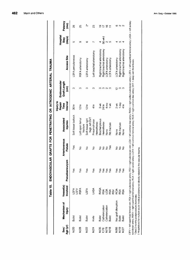
Penetrating and latrogenic Arterial TraumaEleven patients sustained vascular trauma resulting in
nine isolated pseudoaneurysms and two arteriovenousfistulas (Table 10, Fig. 1 1). Seven of these injuries were
the result ofgunshot wounds and two were secondary toiatrogenic needle injuries. Eight patients had sustainedother injuries in conjunction with their vascular trauma.All penetrating and iatrogenic traumatic vascular inju-ries were repaired from 4 hours to 4 months after the
Figure 4. Transfemoral, endoluminal repair of an abdominal aortic aneu-rysm with the EVT Endograft. (a) Preoperative arteriogram demonstratesa suitable proximal and distal segment of normal aorta above and belowthe aneurysm. (b) After transfemoral insertion of the Endograft, the aneu-rysm lumen is excluded from the circulation.
Vol. 222 - No. 4

456 Marin and Others
Figure 5. (a,b) Transfemoral, en-doluminal repair of a complex ab-dominal aortic aneurysm (arrow)and bilateral common iliac arteryaneurysms (open arrows). (c,d) Af-ter insertion of an endovasculargraft (G), the aortic aneurysm andthe bilateral common iliac artery an-eurysms have thrombosed. (e)(next page) This transbrachial arte-riogram demonstrates the com-pleted reconstruction. Note thepresence of embolization coilswithin the origin of the left hypogas-tric artery (arrow), which preventbackflow from this vessel into theleft common iliac artery aneurysm.Ligation of the proximal left com-mon femoral artery at point X fol-lowed by the creation of a femoro-femoral bypass (FF) restores circu-lation to the lower extremities. s:proximal stent fixation site.
primary injury. No endovascular grafts inserted for pen-etrating trauma have thrombosed during the follow-upperiod, which ranges from 2 to 28 months (mean, 15months). Complications encountered with the use ofen-dovascular grafts for traumatic vascular injuries in-
cluded one immediate insertion site stenosis that wassuccessfully treated with a vein patch angioplasty (Table5). A single stent graft stenosis, which occurred at 8months' follow-up, responded to percutaneous balloonangioplasty and remained patent for 23 months.
Table 4. ENDOVASCULAR GRAFTS FOR THE TREATMENTOF ABDOMINAL AORTIC ANEURYSMS
Type of Anesthesia[no. (%)J Perigraft
No. of Successful Graft Hospital Stayt Channel§ Follow-upEndovascular Device Patients General Epidural Local Insertion [no. (%)J [days (mean)] [no. (%)] [mo (mean)]
EVTBalloon expandable device*Balloon expandable devicet
4 4(100)1 1 1 (9)3 0
EVT = Endovascular Technologies' Endograft.* Includes both aorto-aortic (8 patients) and aortoiliac with femorofemoral bypasses (3 patients).t Endovascular graft for contained aortic rupture. All had aortoiliac grafts, contralateral iliac occlusion, and femorofemoral bypasses.t Mean hospital length of stay is calculated exclusive of early postoperative deaths.§ Perigraft channel describes any contrast flow seen outside the lumen of the graft and into the aneurysmal sac on the initial follow-up computed tomograph with contrast.|| One procedure was converted to a standard repair, and one procedure was aborted without grafting.
08 (73)3 (100)
02 (18)
0
4 (100)9 (82)113 (100)
2-7 (3.5)6-21 (12)4-15 (9.5)
2 (50)4 (36)
0
4-22 (13.7)4-25 (13.3)6-12 (9)
Ann. Surg. .-October 1995

Endovascular Grafts for Complex Lesions 457
Figure 5. continued
DISCUSSIONTransluminally placed endovascular grafting proce-
dures are part of a growing trend to provide improvedpatient care in association with reduced cost by means ofcarefully planned, less invasive therapies. Such conceptshave resulted in the successful development ofimportanttechniques, such as transurethral prostatectomy, laparo-scopic cholecystectomy, and a variety of other endo-scopic procedures.
Less invasive endovascular therapies were conceptual-ized by Dotter, who first described catheter-based angio-plasty and conceived of devices for intravascular stent-ing.43.44 The use of balloon angioplasty procedures forthe treatment of short-segment arterial occlusive diseasehas become relatively well established in a variety of vas-cular systems.45-47 The supplemental use of arterialstents for more complex lesions has also proved benefi-cial.48 The logical extension oftransluminally placed en-
dovascular grafting using covered stents and stent-fixedprosthetic conduits to treat more complex arterial lesionshas followed a relatively ordered course with the devel-opment of new devices and animal models in which totest these devices.4955The earliest clinical experience with endovascular
grafts is attributed to Volodos in Russia, who describedhis experience with self-expanding endovascular graftsfor the treatment of a thoracic aortic aneurysm and aor-toiliac occlusive disease.'7' 9 However, the true potentialof this technology was realized only after Parodi et al.successfully treated a patient with an abdominal aorticaneurysm in Argentina in 1990.18 This and subsequentefforts by Parodi have sparked worldwide efforts to findnew applications and improved devices for endovasculargraft treatment of various vascular lesions.20-38,56-58
In the current report, we described our institution'sinitial experience with this new technology for the treat-ment ofabdominal and peripheral artery aneurysms, ar-terial occlusions, and traumatic vascular injuries. Al-though the pathologic lesions for each of these entitiesare markedly different, the technical concepts for endo-vascular graft treatment are surprisingly constant. Theyinclude remote arterial access, limited operative inci-sions, and a dependence on catheter-based instrumentsthat are guided by indirect imaging systems, whichchiefly use digital fluoroscopy.The decision regarding which patients should initially
be treated by this new technology has provoked contro-versy among vascular specialists. 18.25,28.59 Should low-risk patients without coexisting medical or surgical prob-lems be used in the early trials of this new technology, soas to permit rapid and safe conversion to standard tech-niques if the endovascular graft procedure is unsuccess-ful? Until such devices are proven effective, this philoso-phy would deny all patients with life-threatening aorticaneurysms, limb-threatening ischemia, or central vascu-lar injuries in the face of prohibitive cardiac, pulmonary,and other medical comorbidities possible benefit fromreceiving this treatment.We have chosen the alternative approach oflargely us-
ing these endovascular grafts in patients for whom stan-dard surgical operations would be difficult or impossibleto perform because of major medical or surgical comor-bidities that precluded general or other major anesthesiaand/or easy direct surgical access to the lesion. Althoughthis approach offers the hope of great benefit to the pa-tients if the new devices are effective, it largely deniesthem the possibility of a surgical "rescue" procedureshould the device fail or cause a complication. Such asurgical rescue would be associated with a very high risk.These concerns have prompted us to treat with these en-dovascular graft devices those patients who were facingimmediate loss of life or limb. Moreover, in our initial
Vol. 222 - No. 4

458 Marin and Others
Figure 6. Transfemoral repair of a
contained rupture of the distal aorta.(a) A spiral CT scan demonstratesextravasation of contrast materialfrom the aorta (arrow) into a large,partially clot-filled pseudoaneurysm(P). (b) A transfemoral arteriogramconfirms the presence of the largeaortic pseudoaneurysm (arrow). (c)A spiral CT scan performed after
transfemoral insertion of an endovas-cular graft demonstrates that thepseudoaneurysm is thrombosed,and vascular continuity within the lu-men of the aorta (arrow) is pre-
served. (d) A postoperative trans-femoral arteriogram at 1 week dem-onstrates vascular continuitybetween the aorta (open arrow) andthe common femoral arteries (ar-rows).
Table 5. COMPLICATIONS AND DEATHS FROM ENDOVASCULAR GRAFT PROCEDURES
Acute Graft 30 DayNo. of Majort Minort Thrombosis§ Mortality
Procedure Patients [no. (%)J [no. (%)J [no. (%)] ([no. (%)J
EVT aneurysm repair 4 0 1 (25) 0 0Balloon expandable aneurysm repair 1 1 4 (36) 2 (18) 0 4 (36)Balloon expandable aneurysm repair 3 1(33) 0 0 1(33)
(for contained rupture)Peripheral artery aneurysms* 15 2 (13) 2 (13) 1(17) 0Femoropopliteal occlusive disease 6 0 1 (17) 1 (17) 0Aortoiliac occlusive disease 42 3 (7) 7 (17) || 0Arterial trauma 11 0 0 0 0
EVT = Endovascular Technologies' Endograft.* Includes iliac, popliteal, and subclavian aneurysms.t Includes pulmonary failure, renal insufficiency, intestinal ischemia, multiorgan failure, myocardial infarction, congestive heart failure, and stroke (excluding death).t Includes wound complications, urinary tract infection, and uncomplicated pneumonia.§ Thrombosis within 30 days of graft insertion.|| Refer to Tables 7 and 8 regarding early and late patency data.
Ann. Surg. * October 1995

Endovascular Grafts for Complex Lesions 459Vol. 222. No. 4
Figure 7. Transfemoral repair of aright internal iliac artery aneurysm 5years after a bifurcated graft repair ofaortic and common iliac artery aneu-rysms. (a) CT scan documents thepresence of the right internal iliac ar-tery aneurysm (arrow). (b) The twolimbs of the bifurcated graft can beidentified (arrow). (c) Transfemoralarteriography documents filling of theaneurysm before coil embolization ofthe anterior and posterior divisions ofthe vessel. (Note aneurysmal wallcalcification [small arrows].) (d) Afterendovascular graft repair, the aneu-rysm has thrombosed. Contrast canbe identified within the endovasculargraft (arrow). (e) This CT scan docu-ments fixation of the proximal Palmazstent (arrow) within the old bifurcatedgraft. (f) Postoperative digital arte-riogram shows the anchoring stents(arrows) and coils in the posteriorand anterior divisions of the right in-ternal iliac artery for prevention of ret-rograde flow (open arrow). (Inset) Di-agram of a completed repair.
t t

460 Marin and Others
Table 6. ENDOVASCULAR GRAFTS FOR THE TREATMENT OF OCCLUSIVE DISEASE
Ankle/BrachialDistal Graft Graft Associated Indices (mean)
Disease No. of Indication for Proximal Graft Insertion Length ProceduresLocation Grafts Surgery [no. (%)] Origin [no. (%)] [no. (%)] [m (mean)] [No. (%)] Pre Post
47 Gangrene = 39 (83)Rest pain = 8 (17)
6 Gangrene = 6 (100)
Aorta or CIA = 34 (72)EIA= 13(28)
CFA = 3 (50)SFA = 3 (50)
CFA = 29 (62)SFA = 4 (8.5)DFA = 11 (23)AKP = 3 (6)AKP = 6 (100)
15-46(20)
24-37(29)
35 (74)* 0.32 0.78
3 (50)t 0.30 0.71
CIA = common iliac artery; EIA = external iliac artery; CFA = common femoral artery; SFA = superficial femoral artery; DFA = deep femoral artery; AKP = above-knee poplitealartery.* Associated procedures include standard femoropopliteal, femorocrural, femorofemoral, and endoluminal femoropopliteal bypasses.t Associated inflow procedure (axillofemoral or femorofemoral bypass or endoluminal aortoiliofemoral bypass).
experience, these high medical risks in patients with ad-vanced vascular disease contributed significantly to therelatively high mortality and complication rates that oc-
curred in our aortic aneurysm patients (Table 4). Con-sidering that our patients with occlusions were similar inregard to risk status and the presence of complex, ad-vanced arterial disease and ischemia, it is encouragingthat mortality and morbidity were so low and patencyand limb salvage rates were acceptable (Tables 7-9, Fig.10).The advanced nature of the arterial disease in most of
our patients with aneurysms and occlusions often madethe endovascular grafting procedures lengthy and diffi-cult and required that the endovascular graft be com-
bined with a relatively simple, open, standard arterialsurgical operation (e.g., a distal arterial reconstruc-tion).38 Although these combined procedures were usu-
ally successful, they made it difficult to demonstrate thatthe endovascular approach reduced operating room us-
Figure 8. Transluminal repair of a popliteal artery aneurysm. (a) A trans-femoral digital arteriogram demonstrates a large popliteal artery aneurysm(A). (b) After insertion of an endovascular graft (arrow) affixed to the below-knee popliteal artery with a 1-cm Palmaz stent, the popliteal artery aneu-
rysm is excluded.
age or length of hospitalization. Such a demonstrationwas further hampered by the presence of advanced footgangrene in many of our patients with occlusive disease.We believe, however, that the use of endovascular graft-ing techniques, when applied to lower-risk patients withless complex and less advanced arterial disease, will leadto a reduced requirement for hospitalization and lowercosts. Evidence of this has been documented with trau-matic lesions and anatomically simple aortic aneurysmstreated with endovascular grafts.26"28
This raises the question ofwhether it is justified to use
these new grafts to replace standard treatment in patientswho have the usual indications for surgery and who do
Figure 9. Transfemoral iliofemoral bypass for limb-threatening ischemia.(a) Before insertion of an endovascular graft, severe aortoiliac disease isdemonstrated by this preoperative transfemoral arteriogram. Disease inthe right common iliac artery system would preclude effective standardfemorofemoral bypass to reestablish circulation to the left lower extremityin this 86-year-old woman. The left common and external iliac arteries are
completely occluded. (b) After long-segment balloon dilatation of the leftcommon and external iliac arteries and insertion of an endovascular graft(G), vascular continuity is established to the left lower extremity. A percu-
taneous balloon expandable stent has also been inserted into the rightcommon iliac artery to treat the symptomatic high-grade common iliacartery stenosis and to protect the origin of the right common iliac arteryduring endovascular graft insertion.
Aortoiliac
Femoropopliteal
Ann. Surg. . October 1995

Endovascular Grafts for Complex Lesions 461
Table 7. PRIMARY PATENCY OF ENDOVASCULAR GRAFT PROCEDURES TO TREATAORTOILIAC OCCLUSIVE DISEASE
No. ofLimbs at Interval Cumulative Standard
Month Risk Closed Dead Duration Patency (%) Patency (%) Error
0-1 42 0 0 0 100 100 01-3 42 3 0 4 93 93 3.93-6 35 1 0 5 97 90 4.86-9 29 0 0 10 100 90 5.39-12 19 1 0 3 94 85 7.612-15 15 0 0 2 100 85 8.515-18 13 1 0 2 92 77 10.0
Table8. SECONDARY PATENCY OF ENDOVASCULAR GRAFT PROCEDURES TO TREATAORTOILIAC OCCLUSIVE DISEASE
No. ofLimbs at Interval Cumulative Standard
Month Risk Closed Dead Duration Patency (%) Patency (%) Error
0-1 42 0 0 0 100 100 01-3 42 2 0 4 95 95 3.273-6 36 0 0 5 100 95 3.546-9 31 0 0 10 100 95 3.819-12 21 0 0 3 100 95 4.6312-15 18 0 0 2 100 95 5.0015-18 16 0 0 2 100 95 5.31
Table9. LIMB SALVAGE FOR PATIENTS WITH ENDOVASCULAR GRAFTS FOR THETREATMENT OF AORTOILIAC OCCLUSIVE DISEASE
No. ofLimbs at Interval Cumulative Standard
Month Risk Closed Dead Duration Patency (%) Patency (%) Error
0-1 42 0 0 0 100 100 01-3 42 1 0 4 98 98 2.33-6 37 0 0 5 100 98 2.56-9 32 0 0 10 100 98 2.79-12 22 0 0 3 100 98 3.212-15 19 0 0 2 100 98 3.515-18 17 0 0 2 100 98 3.7
60-
31 21 18 161
19 15 ~
is
0 3 6 9 12Tlme aftr opeaton In months
15 18
Prmary Paec Sondar Patn-U
Figure 10. Cumulative 18-month primary and secondary patency ratesfor endovascular grafts. The numbers next to each point indicate the num-ber of grafts observed to be patent for that length of time.
not have major systemic or local factors that increase riskand contraindicate standard therapy. The less invasivenature of endovascular grafts makes them intrinsicallyattractive to patients and physicians. One would thinkthat endovascular grafting procedures should lower mor-bidity and cost. However, their safety, efficacy, andeffectiveness in treating and curing various pathologic le-sions must be proven by appropriately controlled scien-tific studies showing that these endovascular grafts are as
good or better than standard grafts or other treatments.Only then can they be recommended for widespread use
and their exact role in treating vascular disease be de-fined. Reasonable guidelines have been created to direct
% Patu
so -
Vol. 222 - No. 4
W-Y
462 Marin and Others Ann. Surg. * October 1995
co r C\j cY o)r- , .OcsJC' xC\J C\J C\J co
aU
LO 0) (cs rl "i co c\ LO It LOss >
LO~~~~~~~~~~Cco~
-0
E E E E E EE 0 0 oo 40 _O- ---- ..
0 ~~~ ~~~000 0
E ~ E E _o E o co0.2 -
0 0 0ccc -0Um E1 ECZ
as, -4- -4- CZ co.0s
0-. 0 0 0 .000fi a) a))CD)00a) 0 o coCZ C -0
a CcZ c co " Lccc II~< < < .0
C C < < < -- <*U-L LLI u. -0-0LLLL U-00
>UI) U) ci Da, .).2'CDC jr -i -i EJC I-I
,Q)
L >
>.
CZ
CYCY CY CY CY CY) C\ CY) C\ CY 7
-0-
cc
E
0) '- ('1 O z a)1
0-
E E oUc
(.0 C\c C\*0 ~ CY)c "I'"t C\
ccz
0 Er
>O OccE
a,0 n cn c0no ) 0 0 2o0 9 o(
0)CZ ~ ~ -
=3 0 ~~~~~~~~~~~~i0
-0 Eco LL L6LQ6o E
C/() U<) co6 z CC z Erc. CE -
o U) 0 U C S0
U)~~~ ~ ~~~~~cnO
a) 0 0 0 000a 0C00_ II c6>- Z Z ZZ> - zZ < E
cj-i
~co co7c-c >
>00 0 0 0 0000 000~~~~~~>0
>0 >
-0 C
Lu- -u U- U) U) < <>.<< < 0aC/) 4<()CU)UU<C)C)QcU)U) cE
cr.~~ ~ ~ 0 cciE 0
0 -
---- 0~~~~~~~~-o 0 ccoo0 0co~~~~~ m~~oom Cmm a c-
NN E~~Occ co (Ni 4 U)c~~~coco c~o L-Q.
(D a CY / -5 :.4 cc E
4
4
I~--j4
'I=
w
E
Uz
w
Lo0c-4cc
0
zo
4
c-wzw
CLcc
0LLC)U-4ccO
-j
U)4
0azO
uJ
.0I -
CNOC
Wt E0.
> 0
. >0co -W
C
co
W
0 (
S. U.
0
0E~
0a.
0
00
o0
0
0
Co C
0.
00
w000
3C0
C)~
4

Vol. 222 No. 4
'X i | a ..........0.--.f.......
..da~~~~~~~~~~~~~~~~~~~~~~~~~~~~~~~~~~~~~~~~~~~~~~~~~~......
1'~~~~~~~~~~~~~~~~~~~~~~~~~~~~~~~~~~~~~~~~~~~~~~~~~~~~ .... ...
L -i
Figure 11. Transluminal treatment of a traumatic arteriovenous fistula. A20-year-old man sustained a gunshot wound to the right upper chest. (a)A transfemoral arteriogram demonstrated a fistula between the distal sub-clavian artery and vein. (b) Schematic illustration of the fistula. (c) With thepatient under local anesthesia, an endovascular graft was inserted througha brachial artery cut-down. After deployment of the device (arrow), a pro-grade arteriogram demonstrated occlusion of the arteriovenous fistula withpreservation of the distal circulation. (d) Endovascular graft occlusion of afistula. f: fistula; v: vein; a: artery.
the development of this field and to help prevent earlyuncontrolled and unjustified overuse."9
Before endovascular grafts can be accepted for wide-spread use to treat aneurysms, they must be shown toeffectively and permanently exclude the aneurysm fromthe arterial circulation and to prevent aneurysm expan-sion and rupture. The high rate ofperigraft channels intothe aneurysm sac and the associated reports ofaneurysmexpansion and rupture after endovascular graft treat-ment mandate caution before these techniques are usedwidely or without appropriately controlled studies (Ta-ble 4).6o Sealing ofthese channels by thrombosis may notprevent transmission ofarterial pressure to the aneurysmwall. We are encouraged, however, by our observation ofaneurysm shrinkage in some ofour cases and in those ofothers.32The devices and techniques for inserting endovascular
grafts are primitive. We believe that they will be im-proved and that these improvements will lead to highersuccess rates and fewer complications with wider appli-cability. However, such extensions of use must befounded on carefully controlled clinical trials and proveneffectiveness.
Endovascular stented grafts have a combined originin the disciplines of vascular surgery and interventionalradiology. This and the fact that vascular surgeons andinterventional radiologists have been involved in devel-oping and pioneering clinical use of these devices raise
Endovascular Grafts for Complex Lesions 463
the question of who should use them and who shouldcontrol them. Both specialties have legitimate claims.The procedures described in this report have often beencomplex and difficult and have challenged the surgicaland catheter-guide wire skills of a combined group ofsurgeons and radiologists. Surgical and endovascular res-cue techniques have often been required. We thereforebelieve that these devices should be used initially by ahealth care team that combines the highest levels of skillin vascular surgery and interventional radiology.61'62This has been effective in the developmental phase ofthese devices at our institution and at others as well. Ul-timate development of a single, combined specialty or agroup of specialists with dual skills working together willlikely lead to the most rapid, effective advancement notonly in the evolution of transluminally placed endovas-cular grafts, but also in the treatment of patients withvascular disease in general.
References1. Szilagyi DE, Smith RF, DeRusso FJ, et al. Contribution ofabdom-
inal aortic aneurysmectomy to prolongation of life. Ann Surg1966; 164:678-679.
2. Crawford ES, Saleh SA, Babb JW III, et al. Infrarenal abdominalaortic aneurysm: factors influencing survival after operation over a25-year period. Ann Surg 1981; 193:699-709.
3. DeBakey ME, Crawford ES, Cooley DA, et al. Aneurysms of theabdominal aorta: analysis of results of graft replacement therapyone to eleven years after operation. Ann Surg 1964; 160:622-639.
4. AbuRahma AF, Robinson PA, Boland JP, et al. Elective resectionof 332 abdominal aortic aneurysms in a southern West Virginiacommunity during a recent five-year period. Surgery 1991; 109:244-251.
5. Johnson KW, Scobie TK. Multicenter prospective study of non-ruptured abdominal aortic aneurysms. 1. Population and operativemanagement. J Vasc Surg 1988; 7:69-81.
6. Veith FJ, Gupta SK. Wengerter KR, et al. Changing arterioscle-rotic disease patterns and management strategies in lower-limb-threatening ischemia. Ann Surg 1990; 212:402-414.
7. Mattox KL, Feliciano DV, Burch J, et al. Five thousand seven hun-dred sixty cardiovascular injuries in 4459 patients: epidemiologicevaluation 1958 to 1987. Ann Surg 1989; 209:698-707.
8. Lim RC Jr., Trunkey DD, Blaisdell FW. Acute abdominal aorticinjury: an analysis of operative and postoperative management.Arch Surg 1974; 109:706-711.
9. Snyder WH III, Thal ER, Perry MO. Peripheral and abdominalvascular injuries. In Rutherford RB, ed. Vascular Surgery. 2nd ed.Philadelphia: WB Saunders; 1984:460-500.
10. McCombs PR, Roberts B. Acute renal failure following resectionof abdominal aortic aneurysm. Surg Gyn Obstet 1979; 148:175-178.
11. Gardner RJ, Gardner NL, Tarnay TJ, et al. The surgical experienceand a one to sixteen year follow-up of 277 abdominal aortic aneu-rysms. Am J Surg 1978; 135:226-230.
12. Thompson JE, Hollier LH, Patman RD, et al. Surgical manage-ment ofabdominal aortic aneurysms: factors influencing mortalityand morbidity-a 20 year experience. Ann Surg 1975; 181:654-661.
13. Hollier LH, Reigel MM, Kazmier FJ, et al. Conventional repairof abdominal aortic aneurysm in the high-risk patient: a plea for

464 Marin and Others
abandonment of nonresective treatment. J Vasc Surg 1986; 3:712-717.
14. Sher MH. Principles in the management of arterial injuries associ-ated with fracture/dislocations. Ann Surg 1975; 182:630-634.
15. Flint LM, Richardson JD. Arterial injuries with lower extremityfracture. Surgery 1983; 93:5-8.
16. Mattox KL. Whisennand HH, Espada R, Beall AC Jr. Manage-ment of acute combined injuries to the aorta and inferior venacava. Am J Surg 1975; 130:720-724.
17. Volodos NL, Shekhanin VE, Karpovich IP, et al. Self-fixing syn-thetic prosthesis for endoprosthetics of the vessels. Vestn Khir(Russia) 1986; 137:123-125.
18. Parodi JC. Palmaz JC. Barone HD. Transfemoral intraluminalgraft implantation for abdominal aortic aneurysms. Ann Vasc Surg1991; 5:491-499.
19. Volodos NL, Karpovich IP, Troyan VI, et al. Clinical experienceof the use of self-fixing synthetic prostheses for remote endopros-thetics of the thoracic and the abdominal aorta and iliac arteriesthrough the femoral artery and as intraoperative endoprosthesis foraorta reconstruction. Vasa Suppl 1991; 33:93-95.
20. Marin ML, Veith FJ, Panetta TF, et al. Percutaneous transfemoralinsertion of a stented graft to repair a traumatic femoral arteriove-nous fistula. J Vasc Surg 1993; 18:299-302.
21. Cragg AH, Dake MD. Percutaneous femoropopliteal graft place-ment. J Vasc Interv Radiol 1993; 4:455-463.
22. May J, White G. Waugh R, et al. Transluminal placement of aprosthetic graft-stent device for treatment of subclavian artery an-eurysm. J Vasc Surg 1993; 18:1056-1059.
23. Parodi JC, Barone HD. Transfemoral placement of aortic grafts inaortic aneurysms: clinical experience in patients. In Yao JST,Pearce WH, eds. Aneurysms: New Findings and Treatments. Nor-walk, CT: Appleton and Lange; 1994:341-349.
24. Marin ML, Veith FJ, Panetta TF, et al. Transfemoral endoluminalstented graft repair of a popliteal artery aneurysm. J Vasc Surg1994; 19:754-757.
25. Marin ML, Veith FJ, Cynamon J, et al. Transfemoral endovascu-lar stented graft treatment of aorto-iliac and femoropopliteal oc-clusive disease for limb salvage. Am J Surg 1994; 168:156-162.
26. Marin ML, Veith FJ, Panetta TF, et al. Transluminally placed en-dovascular stented graft repair for arterial trauma. J Vasc Surg1994; 20:466-473.
27. Marin ML, Veith FJ. Transfemoral repair ofabdominal aortic an-eurysm. New EngI J Med 1994; 331:1751.
28. Moore WS, Vescera CL. Repair ofabdominal aortic aneurysm bytransfemoral endovascular graft placement. Ann Surg 1994; 220:331-341.
29. Dake MD, Miller DC, Semba CP, et al. Transluminal placement ofendovascular stent-grafts for the treatment of descending thoracicaortic aneurysms. New Engl J Med 1994; 331:1729-1734.
30. May J, White G, Waugh R, et al. Treatment ofcomplex abdominalaortic aneurysms by a combination ofendoluminal and extralum-inal aortofemoral grafts. J Vasc Surg 1994; 19:924-933.
31. May J, White GH, Yu W, et al. Endoluminal grafting ofabdominalaortic aneurysms: causes of failure and their prevention. J Endo-vasc Surg 1994; 1:44-52.
32. Parodi JC. Endovascular repair of abdominal aortic aneurysmsand other arterial lesions. J Vasc Surg 1995; 21:549-557.
33. Scott RAP, Chuter TAM. Clinical endovascular placement of bi-furcated graft in abdominal aortic aneurysm without laparotomy.Lancet 1994; 343:413.
34. Yusuf SW, Baker DM, Chuter TAM, Whitaker SC. Transfemoralendoluminal repair ofabdominal aortic aneurysm with bifurcatedgraft. Lancet 1994; 344:350-35 1.
35. Mialhe C. Clinical experience with the Stentor bifurcated devicefor treatment of abdominal aortic aneurysmal disease. Interna-
Ann. Surg. * October 1995
tional Congress VIII on Endovascular Interventions, Scottsdale,Arizona, February 12-16, 1995.
36. Marin ML, Veith FJ, Lyon RT, et al. Transfemoral endovascularrepair of iliac artery aneurysms. Am J Surg 1995; 170:179-182.
37. Sanchez LA, Marin ML, Veith FJ, et al. Placement ofendovascularstented grafts via remote access sites: A new approach to the treat-ment of failed aortoiliofemoral reconstructions. Ann Vasc Surg1995; 9:1-8.
38. Marin ML, Veith FJ, Sanchez LA, et al. Endovascular aortoiliacgrafts in combination with standard infrainguinal arterial bypassesin the management of limb-threatening ischemia. J Vasc Surg1995; 22:316-322.
39. Parodi JC. Transfemoral intraluminal graft implantation for ab-dominal aortic aneurysm. In Greenhalgh RM, ed. Vascular andEndovascular Surgical Techniques. 3rd ed. London: WB Saunders;1994:71-77.
40. Moore WS. Transfemoral endovascular repair ofabdominal aorticaneurysm using the endovascular graft system device. InGreenhalgh RM, ed. Vascular and Endovascular Surgical Tech-niques. 3rd ed. London: WB Saunders; 1994:78-91.
41. Marin ML, Veith FJ. Endoluminal stented graft aorto-bifemoralreconstruction. In Greenhalgh RM, ed. Vascular and Endovascu-lar Surgical Techniques. 3rd ed. London: WB Saunders; 1994: 100-104.
42. Ad Hoc Committee on Reporting Standards, Society for VascularSurgery/North American Chapter, International Society for Car-diovascular Surgery. Suggested standards for reports dealing withlower extremity ischemia. J Vasc Surg 1986; 4:80-94.
43. Dotter CT, Judkins MP. Transluminal treatment of arterioscle-rotic obstruction: description of a new technic and a preliminaryreport of its application. Circulation 1964; 30:654-670.
44. Dotter CT. Transluminally-placed coilspring endarterial tubegrafts: long-term patency in canine popliteal artery. Invest Radiol1969; 4:329-332.
45. Johnston KW, Rae M, Hogg-Johnston SA, et al. Five-year resultsof a prospective study of percutaneous transluminal angioplasty.Ann Surg 1987; 206:403-413.
46. van Andel GJ, van Erp WFM, Krepel VM, Breslau PJ. Percutane-ous transluminal dilatation of the iliac artery: long term results.Radiology 1985; 156:321-323.
47. King SB III, SchlumpfM. Ten-year completed follow-up of percu-taneous transluminal coronary angioplasty: the early Zurich expe-rience. J Am Coll Cardiol 1993; 22:353-360.
48. Palmaz JC, Laborde JC, Rivera FJ, et al. Stenting ofthe iliac arter-ies with the Palmaz stent: experience from a multicenter trial.Cardiovasc Intervent Radiol 1992; 15:291-297.
49. Dotter CT, Buschmann RW, McKinney MK. Transluminal ex-pandable nitinol coil stent grafting: preliminary report. Radiology1983; 147:259-260.
50. Cragg A, Lund G, Rysavy J, et al. Nonsurgical placement ofarterialendoprostheses: a new technique using nitinol wire. Radiology1983; 147:261-263.
51. Maass D, Kropf L, Egloff L, et al. Transluminal implantation ofintravascular "double-helix" spiral prostheses: technical and bio-logical considerations. Eur Soc ArtifOrgans Proc 1982; 9:252-256.
52. Balko A, Piasecki GJ, Shah DM, et al. Transfemoral placement ofintraluminal polyurethane prosthesis for abdominal aortic aneu-rysm. J Surg Res 1986; 40:305-309.
53. Mirich D, Wright KC, Wallace S, et al. Percutaneously placed en-dovascular grafts for aortic aneurysms: feasibility study. Radiology1989; 170:1033-1037.
54. Chuter TAM, Green RM, Ouriel K, et al. Transfemoral endovas-cular aortic graft placement. J Vasc Surg 1993; 18:185-197.
55. Laborde JC, Parodi JC, Clem MF, et al. Intraluminal bypass of

Vol. 222 . No. 4
abdominal aortic aneurysm: feasibility study. Radiology 1992;184:185-190.
56. Chuter TAM, Wendt G, Hopkinson BR, et al. Early clinical expe-rience with bifurcated endovascular grafts for abdominal aortic an-eurysm repair. International Congress VIII on Endovascular Inter-ventions, Scottsdale, Arizona, February 12-16, 1995.
57. May J. Comparison of the Sydney and EVT prostheses for treat-ment ofaneurysmal disease. International Congress VIII on Endo-vascular Interventions, Scottsdale, Arizona, February 12-16,1995.
58. Henry M, Amor M, Ethevenot G. Initial experience with the CraggEndopro System I for intraluminal treatment of peripheral vascu-lar disease: report of the first 64 cases. International Congress VIIIon Endovascular Interventions, Scottsdale, Arizona, February 12-16, 1995.
59. Veith FJ, Abbott WM, Yao JST, et al. Endovascular Graft Com-mittee. Guidelines for development and use of transluminallyplaced endovascular prosthetic grafts in the arterial system. J VascSurg 1995; 21:670-685; and J Vasc Interv Radiol 1995; 6:477-492.
60. Lumsden AB, Allen RC, Chaikof EL, et al. Delayed rupture ofaortic aneurysm following endovascular stent graftings. Am J Surg1995; 170:174-178.
61. Veith FJ. Presidential address. Transluminally placed endovascu-lar stented grafts and their impact on vascular surgery. J Vasc Surg1994; 20:855-860.
62. Veith FJ, Marin ML. Editorial. Endovascular surgery and its effecton the relationship between vascular surgery and radiology. J En-dovasc Surg 1995; 2:1-7.
DiscussionDR. CLYDE F. BARKER (Philadelphia, Pennsylvania): This is
an important paper. As those who attend vascular meetings arewell aware, endovascular techniques appear to be sweeping thecountry. This report by a pioneering group describes one ofthelargest experiences with these methods.The appeal of this approach for treatment of aortic aneu-
rysms, particularly in high-risk patients, is obvious. The manu-script details the technical challenges and the immediate andearly complications and the remarkable success which is some-times achieved. It will be equally important to define the possi-ble late complications.
Will the neck ofthe aneurysm continue to dilate, resulting inaneurysm formation proximal to the endovascular device ordistal migration ofthe prosthesis? How important are perigraftchannels? Will they all thrombose in time? Or will they allowcontinued enlargement of the aneurysm? The authors' manu-script contains the first systemic attempt I have seen at serialobservations of the size of endovascular-treated aneurysms inwhich a perigraft channel remains open.The place of this endovascular approach for arterial occlu-
sive disease is even less clear than it is for aneurysms. The au-thors are the real pioneers in endovascular procedures for arte-rial occlusion, a setting in which the appeal and possible advan-tage ofthis method over conventional reconstructive surgery isless obvious. In high-risk patients with occlusive disease, extra-anatomic bypasses would be another method to avoid the riskofconventional surgery, such as aortofemoral bypass. Compar-ison ofendovascular with standard bypass procedures will haveto await determination of long-term patency of the grafts aswell as immediate outcome.
Endovascular Grafts for Complex Lesions 465
I am convinced by the results presented here and in themanuscript that endovascular techniques will sometimes resultin long-term patency, but definitive comparisons to conven-tional bypass surgery must still await randomized trials. I won-der if the authors feel that randomized comparisons of endo-vascular treatment versus conventional surgery for occlusivedisease are warranted at this stage.
Is there a role for this technique in more distal arterial occlu-sion? It seems as though it might be especially suitable for pop-liteal aneurysms because for these, only a short stent would berequired. For distal bypasses in the leg. It might be more diffi-cult to see an advantage. Does Dr. Veith, whose interest andsuccess with distal bypasses performed with prostheses are wellknown, have optimism for an endovascular approach for fem-oral-popliteal or femoral-tibial bypasses? Or will surgical place-ment with autogenous veins remain the optimal procedure fordistal occlusions?
There are several important nonmedical issues on which Iwould like to have the authors' views. First, will the scrutiny ofthe Food and Drug Administration and other accrediting bod-ies seriously delay the broad application of endovascular pro-cedures? In view ofthe current Medicare audits that many uni-versity hospitals are undergoing with regard to the use ofexper-imental devices, can either the surgeons or the hospitals billpatients for such treatment?
Finally, there is a turf issue here. The authors seem to haveachieved an admirable collaborative arrangement with their ra-diology colleagues. But this has not been true everywhere.What is their advice on this? Endovascular techniques willclearly have a place in treatment of vascular disease and theauthors are among those who are defining it. There will be atendency for these procedures to proliferate because of the ap-peal of minimally invasive procedures to patients. This is per-fectly understandable and proper, but there is a danger thatthis, along with turf and marketing issues, may prevail, ratherthan sound judgment, based on clinical experience and appro-priately constructed comparative studies.
Surgeons need to remain involved and in appropriate controlof the patient care, decision making, and in reporting of theoutcome and complications of these procedures. The authorsare to be congratulated on a good beginning in all three oftheseareas.
DR. LAZAR J. GREENFIELD (Ann Arbor, Michigan): Mycompliments to Dr. Marin for an excellent presentation. I alsoappreciate the opportunity to review the manuscript and com-ment on this impressive initial experience with endovasculargrafts. The concept is certainly an attractive one, but has beenlimited by current technology, which was developed to deliverstents rather than the combination of graft and stent.
Dr. Veith has also pioneered in extending the applicationfrom the high-risk aneurysm patient to the patient with limb-threatening occlusive disease. This latter group raises somequestions in my mind.
First, were pressure gradients measured before and after ves-sel dilation and graft placement? It seems that adequate dila-tion with or without stent placement has a respectable recordand adding a graft could compromise the lumen for only a mar-ginal improvement in the surface of the vessel.