The effect of injectable biocompatible elastomer (PDMS) on the strength of the proximal fixation of...
7
The effect of injectable biocompatible elastomer (PDMS) on the strength of the proximal fixation of endovascular aneurysm repair grafts: An in vitro study Willem-Maarten P.F. Bosman, MD, a Tim J. van der Steenhoven, MD, a,b Daniel R. Suárez, MSc, c,d Edward R. Valstar, MSc, PhD, c,e Alexander C. de Vries, MD, PhD, f Hans L.F. Brom, MD, PhD, g Michael J. Jacobs, MD, PhD, h and Jaap F. Hamming, MD, PhD, a Leiden, Tilburg, Delft, The Hague, Haarlem, and Maastricht, The Netherlands Purpose: One of the major concerns in the long-term success of endovascular aneurysm repair (EVAR) is stent graft migration, which can cause type I endoleak and even aneurysm rupture. Fixation depends on the mechanical forces between the graft and both the aortic neck and the blood flow. Therefore, there are anatomical restrictions for EVAR, such as short and angulated necks. To improve the fixation of EVAR grafts, elastomer (PDMS) can be injected in the aneurysm sac. The support given by the elastomer might prevent dislocation and migration of the graft. The aim of this study was to measure the influence of an injectable biocompatible elastomer on the fixation strength of different EVAR grafts in an in vitro model. Methods: The proximal part of three different stent grafts was inserted in a bovine artery with an attached latex aneurysm. The graft was connected to a tensile testing machine, applying force to the proximal fixation, while the artery with the aneurysm was fixated to the setup. The force to obtain graft dislodgement (DF) from the aorta was recorded in Newtons (N). Three different proximal seal lengths (5, 10, and 15 mm) were evaluated. The experiments were repeated after the space between the graft and the latex aneurysm was filled with the elastomer. Independent sample ttests were used for the comparison between the DF before and after elastomer treatment for each seal length. Results: The mean DF (mean SD) of all grafts without elastomer sac filling for a proximal seal length of 5, 10, and 15 mm were respectively, 4.4 3.1 N, 12.2 10.6 N, and 15.1 6.9 N. After elastomer sac filling, the dislodgement forces increased significantly (P < .001) to 20.9 3.8 N, 31.8 9.8 N, and 36.0 14.1 N, respectively. Conclusions: The present study shows that aneurysm sac filling may have a role as an adjuvant procedure to the present EVAR technique. The strength of the proximal fixation of three different stent grafts increases significantly in this in vitro setting. Further in vivo research must be done to see if this could facilitate the treatment of aneurysms with short infrarenal necks. ( J Vasc Surg 2010;52:152-8.) Clinical Relevance: Stent graft migration and endoleak due to suboptimal fixation are major drawbacks of currently available stent grafts. Optimizing the proximal fixation by peri-graft elastomer aneurysm sac filling may lead to lower incidence of graft migration and endoleak. It might make endovascular aneurysm repair available to larger group of patients with an abdominal aortic aneurysm. A major complication affecting the long-term success of endovascular aneurysm repair (EVAR) is stent graft migration, which can cause type I endoleak and even aneurysm rupture. 1-8 Fixation is dependent on the me- chanical forces between the graft and the aortic neck, as well as the blood flow. 9 Therefore, EVAR has anatomical restrictions. Despite the manufacturers instructions for use, EVAR grafts are inserted in necks shorter than 15 mm, and sometimes significant angulation is accept- ed. 10,11 Zarins et al showed, in a prospective multicenter trial, that insufficient length of proximal seal will lead to stent migration. 8 Earlier studies have shown that, in up to 45% of abdominal aortic aneurysms (AAA), the anat- omy of the proximal neck makes the AAA unsuitable for EVAR because of insufficient length, large diameter, or severe angulation. 12-14 To overcome the disadvantages of current EVAR ther- apies, Aortic Customize was devised: a method of exclud- ing the aortic aneurysm using endovascular techniques to inject a biocompatible elastomer into the aneurysm sac (Fig 1). 15 Filling the aneurysm sac with an injectable biocom- patible elastomer reduces the wall stress and thereby the From the Department of Surgery, Leiden University Medical Center, a the Department of Surgery, St. Elisabeth Hospital, b the Biomechanics and Imaging Group, Department of Orthopaedics, Leiden University Medical Center, c the Department of Precision and Microsystems Engineering, d Faculty of Mechanical, Maritime, and Material Engineering, Delft Uni- versity of Technology, e the Department of Surgery, Medisch Centrum Haaglanden, f the Department of Surgery, Kennemer Gasthuis, g and the Department of Surgery, Maastricht University Medical Center. h Supported by the Leiden University Funds-Gratama Fund and the Ketel 1 Studiefonds. Competition of interest: Drs de Vries, Brom, and Jacobs have developed and patented the elastomer used in the experiment. The other authors have no financial or other competing interests in the experiments. Reprint requests: W.M.P.F. Bosman, MD, Department of Surgery, K6-R, Leiden University Medical Center, Postbox 9600, 2300 RC Leiden, The Netherlands (e-mail: [email protected]). The editors and reviewers of this article have no relevant financial relationships to disclose per the JVS policy that requires reviewers to decline review of any manuscript for which they may have a competition of interest. 0741-5214/$36.00 Copyright © 2010 by the Society for Vascular Surgery. doi:10.1016/j.jvs.2010.01.026 152
-
Upload
independent -
Category
Documents
-
view
2 -
download
0
Transcript of The effect of injectable biocompatible elastomer (PDMS) on the strength of the proximal fixation of...

The effect of injectable biocompatible elastomer(PDMS) on the strength of the proximal fixation ofendovascular aneurysm repair grafts: An in vitro studyWillem-Maarten P.F. Bosman, MD,a Tim J. van der Steenhoven, MD,a,b Daniel R. Suárez, MSc,c,d
Edward R. Valstar, MSc, PhD,c,e Alexander C. de Vries, MD, PhD,f Hans L.F. Brom, MD, PhD,g
Michael J. Jacobs, MD, PhD,h and Jaap F. Hamming, MD, PhD,a Leiden, Tilburg, Delft, The Hague,Haarlem, and Maastricht, The Netherlands
Purpose: One of the major concerns in the long-term success of endovascular aneurysm repair (EVAR) is stent graft migration,which can cause type I endoleak and even aneurysm rupture. Fixation depends on the mechanical forces between the graft andboth the aortic neck and the blood flow. Therefore, there are anatomical restrictions for EVAR, such as short and angulatednecks. To improve the fixation of EVAR grafts, elastomer (PDMS) can be injected in the aneurysm sac. The support given bythe elastomer might prevent dislocation and migration of the graft. The aim of this study was to measure the influence of aninjectable biocompatible elastomer on the fixation strength of different EVAR grafts in an in vitro model.Methods: The proximal part of three different stent grafts was inserted in a bovine artery with an attached latex aneurysm.The graft was connected to a tensile testing machine, applying force to the proximal fixation, while the artery with theaneurysm was fixated to the setup. The force to obtain graft dislodgement (DF) from the aorta was recorded in Newtons(N). Three different proximal seal lengths (5, 10, and 15 mm) were evaluated. The experiments were repeated after thespace between the graft and the latex aneurysm was filled with the elastomer. Independent sample ttests were used for thecomparison between the DF before and after elastomer treatment for each seal length.Results: The mean DF (mean � SD) of all grafts without elastomer sac filling for a proximal seal length of 5, 10, and 15mm were respectively, 4.4 � 3.1 N, 12.2 � 10.6 N, and 15.1 � 6.9 N. After elastomer sac filling, the dislodgement forcesincreased significantly (P < .001) to 20.9 � 3.8 N, 31.8 � 9.8 N, and 36.0 � 14.1 N, respectively.Conclusions: The present study shows that aneurysm sac filling may have a role as an adjuvant procedure to the present EVARtechnique. The strength of the proximal fixation of three different stent grafts increases significantly in this in vitro setting.Further in vivo research must be done to see if this could facilitate the treatment of aneurysms with short infrarenal necks.(J Vasc Surg 2010;52:152-8.)
Clinical Relevance: Stent graft migration and endoleak due to suboptimal fixation are major drawbacks of currentlyavailable stent grafts. Optimizing the proximal fixation by peri-graft elastomer aneurysm sac filling may lead to lowerincidence of graft migration and endoleak. It might make endovascular aneurysm repair available to larger group of
patients with an abdominal aortic aneurysm.A major complication affecting the long-term successof endovascular aneurysm repair (EVAR) is stent graft
From the Department of Surgery, Leiden University Medical Center,a theDepartment of Surgery, St. Elisabeth Hospital,b the Biomechanics andImaging Group, Department of Orthopaedics, Leiden University MedicalCenter,c the Department of Precision and Microsystems Engineering,d
Faculty of Mechanical, Maritime, and Material Engineering, Delft Uni-versity of Technology,e the Department of Surgery, Medisch CentrumHaaglanden,f the Department of Surgery, Kennemer Gasthuis,g and theDepartment of Surgery, Maastricht University Medical Center.h
Supported by the Leiden University Funds-Gratama Fund and the Ketel 1Studiefonds.
Competition of interest: Drs de Vries, Brom, and Jacobs have developed andpatented the elastomer used in the experiment. The other authors have nofinancial or other competing interests in the experiments.
Reprint requests: W.M.P.F. Bosman, MD, Department of Surgery, K6-R,Leiden University Medical Center, Postbox 9600, 2300 RC Leiden, TheNetherlands (e-mail: [email protected]).
The editors and reviewers of this article have no relevant financial relationshipsto disclose per the JVS policy that requires reviewers to decline review of anymanuscript for which they may have a competition of interest.
0741-5214/$36.00Copyright © 2010 by the Society for Vascular Surgery.
doi:10.1016/j.jvs.2010.01.026152
migration, which can cause type I endoleak and evenaneurysm rupture.1-8 Fixation is dependent on the me-chanical forces between the graft and the aortic neck, aswell as the blood flow.9 Therefore, EVAR has anatomicalrestrictions. Despite the manufacturers instructions foruse, EVAR grafts are inserted in necks shorter than 15mm, and sometimes significant angulation is accept-ed.10,11 Zarins et al showed, in a prospective multicentertrial, that insufficient length of proximal seal will lead tostent migration.8 Earlier studies have shown that, in upto 45% of abdominal aortic aneurysms (AAA), the anat-omy of the proximal neck makes the AAA unsuitable forEVAR because of insufficient length, large diameter, orsevere angulation.12-14
To overcome the disadvantages of current EVAR ther-apies, Aortic Customize was devised: a method of exclud-ing the aortic aneurysm using endovascular techniques toinject a biocompatible elastomer into the aneurysm sac (Fig1).15 Filling the aneurysm sac with an injectable biocom-
patible elastomer reduces the wall stress and thereby the
JOURNAL OF VASCULAR SURGERYVolume 52, Number 1 Bosman et al 153
rupture risk,15 since aneurysm rupture occurs when thelocal wall stress exceeds the local wall strength.16,17
This treatment concept can function as a standalonetreatment, but might also play a role as an adjuvant proce-dure to the current EVAR treatment (Fig 1). The elastomercan be injected next to the inserted EVAR graft and willcure around the graft, taking on its form. The support ofthe elastomer could prevent dislocation and migration ofthe graft. With this technology, aortic aneurysms with lessfavorable proximal neck anatomy could be made suitablefor EVAR.
Our hypothesis is that filling the spaces between thegraft and the aneurysm sac will increase the proximal fixa-tion as it increases the supporting areas of the graft. The aimof this study is to measure the influence of aneurysm sacfilling with an injectable biocompatible elastomer on thestrength of the proximal fixation and the necessary aneu-rysm neck length in an in vitro model.
MATERIALS AND METHODS
Setup. Fresh bovine aortas were obtained from anabattoir. The abdominal portion of the aorta was retainedand all non-vascular tissue was removed. Three to foursamples with a length of 45 mm were selected from eachaorta with a mean diameter of 19.5 mm (�1.0 mm). Thesamples of bovine artery were fixated in an experimentalsetup (Fig 2). The distal part of the artery was stitched with4.0 prolene stitches (Ethicon, Somerville, Mass) to a latexsphere, resembling an AAA. Side branches of the aorta were
Fig. 1. Aortic Customize, the treatment concept. Left side depictsthe stand-alone therapy (A-F), while the right side depicts thetherapy as adjuvance to current EVAR-therapy (G-L). Guide wiresare inserted to the aneurysm (B), over which endovascular balloonsare inserted to exclude the aneurysm sac from the circulation (C).A fill catheter was inserted next to the balloon, through which theliquid elastomer is injected (D). The elastomer fills every cavity inthe aneurysm sac and obliterates lumbar arteries, thereby prevent-ing potential endoleaks (E). When the elastomer has cured (�5minutes), the endovascular balloon is deflated, leaving the aneu-rysm excluded with a new lumen (F). As adjuvant therapy (G-I),the process is very similar, whereby the cavities between the graft inan aneurysm sac are filled with the elastomer (J, K), fixating thegraft in an elastomer mould of the sac.
ligated with the same 4.0 prolene stitches.
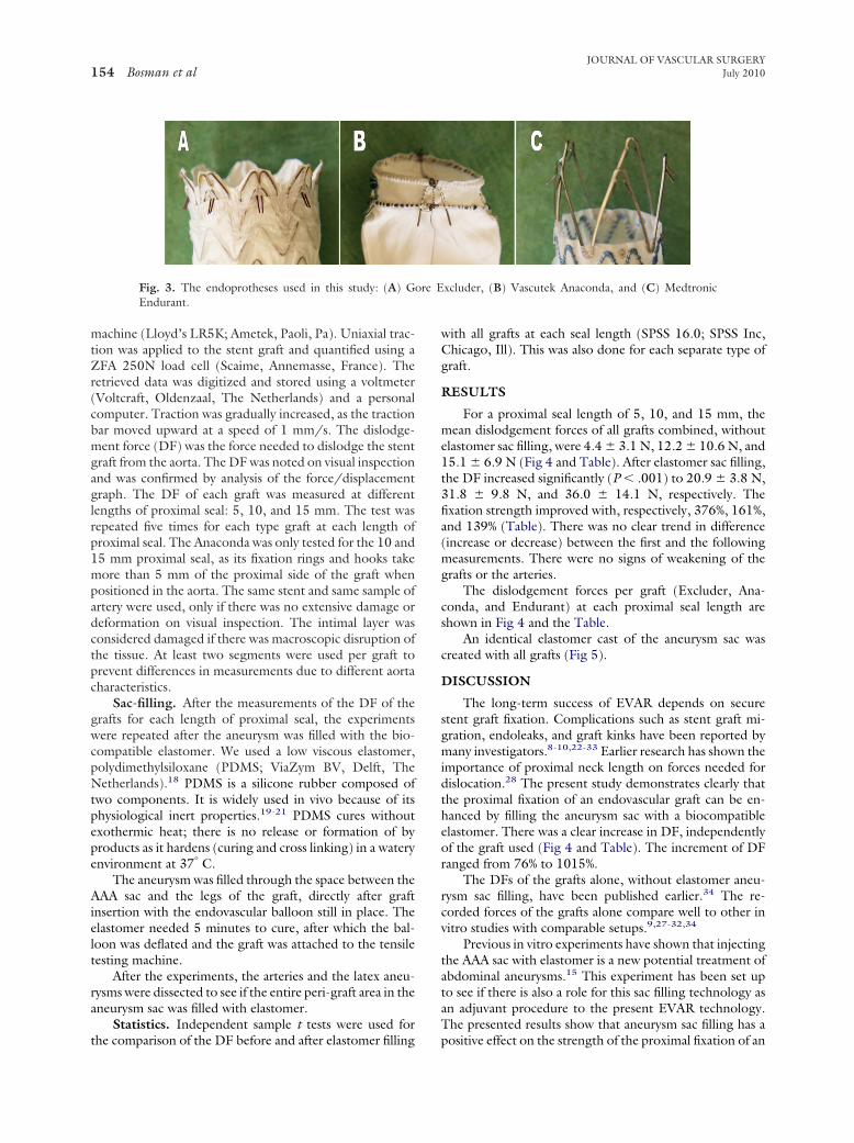
Stent grafts. Three types of commercial endografts ofdifferent manufacturers were used and compared in thisexperimental study (Fig 3): An Excluder AAA Endoproth-esis (WL Gore and Associates, Inc, Flagstaff, Ariz), anAnaconda AAA Endovascular Graft (Vascutek, Inchinnan,Scotland), and an Endurant AAA Stent Graft (Medtronic,Minneapolis, Minn). The diameter of all grafts was 23 mm.
Method of excluding the aneurysm. The proximalpart of the stent graft was inserted in the bovine artery andfixated by inflating a Reliant endovascular balloon(Medtronic) with a diameter of 30 mm. Using 23 mmstents in arteries of 19.5 mm, a mean 13% oversizing wasapplied. The neck length was measured from the start of theaortic segment to the lowest covered part of the proximalsite of the stent graft. With the Endurant, this meant thatthe suprarenal fixation stent was inserted further down inthe aortic neck.
Measurements. After insertion in the artery, a rigid
Fig. 2. Schematic drawing of experimental set-up: (A) Sample ofbovine artery (45 mm long). (B) Distal fixation plug on which thebovine artery is attached. (C) Artificial aneurysm made of latex(neck 19 mm; widest part 40 mm), which is sutured to the bovineartery (D) A stent-graft inserted in the bovine neck of the aneu-rysm. (E) Rigid rod with anchor through the endograft, connect-ing it to the tensile testing machine. (F) Plugs for fixation of theset-up on tensile testing machine.
hook was used to connect the graft to a tensile testing
JOURNAL OF VASCULAR SURGERYJuly 2010154 Bosman et al
machine (Lloyd’s LR5K; Ametek, Paoli, Pa). Uniaxial trac-tion was applied to the stent graft and quantified using aZFA 250N load cell (Scaime, Annemasse, France). Theretrieved data was digitized and stored using a voltmeter(Voltcraft, Oldenzaal, The Netherlands) and a personalcomputer. Traction was gradually increased, as the tractionbar moved upward at a speed of 1 mm/s. The dislodge-ment force (DF) was the force needed to dislodge the stentgraft from the aorta. The DF was noted on visual inspectionand was confirmed by analysis of the force/displacementgraph. The DF of each graft was measured at differentlengths of proximal seal: 5, 10, and 15 mm. The test wasrepeated five times for each type graft at each length ofproximal seal. The Anaconda was only tested for the 10 and15 mm proximal seal, as its fixation rings and hooks takemore than 5 mm of the proximal side of the graft whenpositioned in the aorta. The same stent and same sample ofartery were used, only if there was no extensive damage ordeformation on visual inspection. The intimal layer wasconsidered damaged if there was macroscopic disruption ofthe tissue. At least two segments were used per graft toprevent differences in measurements due to different aortacharacteristics.
Sac-filling. After the measurements of the DF of thegrafts for each length of proximal seal, the experimentswere repeated after the aneurysm was filled with the bio-compatible elastomer. We used a low viscous elastomer,polydimethylsiloxane (PDMS; ViaZym BV, Delft, TheNetherlands).18 PDMS is a silicone rubber composed oftwo components. It is widely used in vivo because of itsphysiological inert properties.19-21 PDMS cures withoutexothermic heat; there is no release or formation of byproducts as it hardens (curing and cross linking) in a wateryenvironment at 37° C.
The aneurysm was filled through the space between theAAA sac and the legs of the graft, directly after graftinsertion with the endovascular balloon still in place. Theelastomer needed 5 minutes to cure, after which the bal-loon was deflated and the graft was attached to the tensiletesting machine.
After the experiments, the arteries and the latex aneu-rysms were dissected to see if the entire peri-graft area in theaneurysm sac was filled with elastomer.
Statistics. Independent sample t tests were used for
Fig. 3. The endoprotheses used in this study: (A) GEndurant.
the comparison of the DF before and after elastomer filling
with all grafts at each seal length (SPSS 16.0; SPSS Inc,Chicago, Ill). This was also done for each separate type ofgraft.
RESULTS
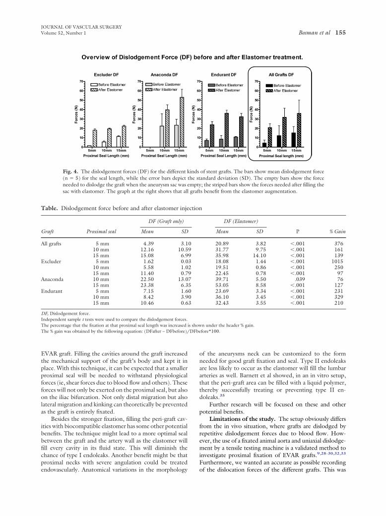
For a proximal seal length of 5, 10, and 15 mm, themean dislodgement forces of all grafts combined, withoutelastomer sac filling, were 4.4 � 3.1 N, 12.2 � 10.6 N, and15.1 � 6.9 N (Fig 4 and Table). After elastomer sac filling,the DF increased significantly (P � .001) to 20.9 � 3.8 N,31.8 � 9.8 N, and 36.0 � 14.1 N, respectively. Thefixation strength improved with, respectively, 376%, 161%,and 139% (Table). There was no clear trend in difference(increase or decrease) between the first and the followingmeasurements. There were no signs of weakening of thegrafts or the arteries.
The dislodgement forces per graft (Excluder, Ana-conda, and Endurant) at each proximal seal length areshown in Fig 4 and the Table.
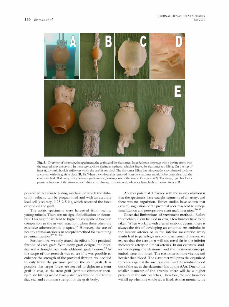
An identical elastomer cast of the aneurysm sac wascreated with all grafts (Fig 5).
DISCUSSION
The long-term success of EVAR depends on securestent graft fixation. Complications such as stent graft mi-gration, endoleaks, and graft kinks have been reported bymany investigators.8-10,22-33 Earlier research has shown theimportance of proximal neck length on forces needed fordislocation.28 The present study demonstrates clearly thatthe proximal fixation of an endovascular graft can be en-hanced by filling the aneurysm sac with a biocompatibleelastomer. There was a clear increase in DF, independentlyof the graft used (Fig 4 and Table). The increment of DFranged from 76% to 1015%.
The DFs of the grafts alone, without elastomer aneu-rysm sac filling, have been published earlier.34 The re-corded forces of the grafts alone compare well to other invitro studies with comparable setups.9,27-32,34
Previous in vitro experiments have shown that injectingthe AAA sac with elastomer is a new potential treatment ofabdominal aneurysms.15 This experiment has been set upto see if there is also a role for this sac filling technology asan adjuvant procedure to the present EVAR technology.The presented results show that aneurysm sac filling has a
xcluder, (B) Vascutek Anaconda, and (C) Medtronic
ore Epositive effect on the strength of the proximal fixation of an
graft
DFbe
JOURNAL OF VASCULAR SURGERYVolume 52, Number 1 Bosman et al 155
EVAR graft. Filling the cavities around the graft increasedthe mechanical support of the graft’s body and kept it inplace. With this technique, it can be expected that a smallerproximal seal will be needed to withstand physiologicalforces (ie, shear forces due to blood flow and others). Theseforces will not only be exerted on the proximal seal, but alsoon the iliac bifurcation. Not only distal migration but alsolateral migration and kinking can theoretically be preventedas the graft is entirely fixated.
Besides the stronger fixation, filling the peri-graft cav-ities with biocompatible elastomer has some other potentialbenefits. The technique might lead to a more optimal sealbetween the graft and the artery wall as the elastomer willfill every cavity in its fluid state. This will diminish thechance of type I endoleaks. Another benefit might be thatproximal necks with severe angulation could be treated
Fig. 4. The dislodgement forces (DF) for the different k(n � 5) for the seal length, while the error bars depictneeded to dislodge the graft when the aneurysm sac was esac with elastomer. The graph at the right shows that all
Table. Dislodgement force before and after elastomer inje
Graft Proximal seal
DF (Graft only)
Mean SD
All grafts 5 mm 4.39 3.110 mm 12.16 10.515 mm 15.08 6.9
Excluder 5 mm 1.62 0.010 mm 5.58 1.015 mm 11.40 0.7
Anaconda 10 mm 22.50 13.015 mm 23.38 6.3
Endurant 5 mm 7.15 1.610 mm 8.42 3.915 mm 10.46 0.6
DF, Dislodgement force.Independent sample t tests were used to compare the dislodgement forces.The percentage that the fixation at that proximal seal length was increasedThe % gain was obtained by the following equation: (DFafter – DFbefore)/
endovascularly. Anatomical variations in the morphology
of the aneurysms neck can be customized to the formneeded for good graft fixation and seal. Type II endoleaksare less likely to occur as the elastomer will fill the lumbararteries as well. Barnett et al showed, in an in vitro setup,that the peri-graft area can be filled with a liquid polymer,thereby successfully treating or preventing type II en-doleaks.35
Further research will be focused on these and otherpotential benefits.
Limitations of the study. The setup obviously differsfrom the in vivo situation, where grafts are dislodged byrepetitive dislodgement forces due to blood flow. How-ever, the use of a fixated animal aorta and uniaxial dislodge-ment by a tensile testing machine is a validated method toinvestigate proximal fixation of EVAR grafts.9,28-30,32,33
Furthermore, we wanted an accurate as possible recording
of stent grafts. The bars show mean dislodgement forceandard deviation (SD). The empty bars show the force; the striped bars show the forces needed after filling thes benefit from the elastomer augmentation.
DF (Elastomer)
P % GainMean SD
20.89 3.82 �.001 37631.77 9.75 �.001 16135.98 14.10 �.001 13918.08 1.44 �.001 101519.51 0.86 �.001 25022.45 0.78 �.001 9739.71 5.50 .039 7653.05 8.58 �.001 12723.69 3.34 �.001 23136.10 3.45 �.001 32932.43 3.55 �.001 210
n under the header % gain.fore*100.
indsthe stmpty
ction
09932975003
is show
of the dislocation forces of the different grafts. This was
o aor
JOURNAL OF VASCULAR SURGERYJuly 2010156 Bosman et al
possible with a tensile testing machine, in which the dislo-cation velocity can be programmed and with an accurateload cell (accuracy, 0.25-2.5 N), which recorded the forceexerted on the graft.
The aortic specimens were harvested from healthyyoung animals. There was no sign of calcification or throm-bus. This might have lead to higher dislodgement forces incomparison to the in vivo situation, when there often areextensive atherosclerotic plaques.23 However, the use ofhealthy animal arteries is an accepted method for examiningproximal fixation.27,31-33
Furthermore, we only tested the effect of the proximalfixation of each graft. With many graft designs, the distaliliac seal is thought to provide additional graft fixation.33 Asthe scope of our research was to see if it was possible toenhance the strength of the proximal fixation, we decidedto only fixate the proximal part of the stent graft. It ispossible that larger forces are needed to dislocate a stentgraft in vivo, as the stent-graft (without elastomer aneu-rysm sac filling) would have a stronger fixation due to the
Fig. 5. Overview of the setup, the specimens, the grafts, andthe sutured latex aneurysm. In the artery, a Gore Excluderinset A, the rigid hook is visible on which the graft is attachaneurysm with the graft in place (B, E). When the endografelastomer had filled every cavity between graft and sac, leavproximal fixation of the Anaconda left distinctive damage t
iliac seal and columnar strength of the graft body.
Another potential difference with the in vivo situation isthat the specimens were straight segments of an artery, andthere was no angulation. Earlier studies have shown that(severe) angulation of the proximal neck may lead to subop-timal fixation and postoperative stent graft migration.36,37
Potential limitations of treatment method. Beforethis technique can be used in vivo, a few hurdles have to betaken. When working with arterial embolic agents, there isalways the risk of developing an embolus. An embolus inthe lumbar arteries or in the inferior mesenteric arterymight lead to paraplegia or colonic ischemia. However, weexpect that the elastomer will not travel far in the inferiormesenteric artery or lumbar arteries. In our extensive stud-ies developing the elastomer and the treatment concept,emboli were not noted. The elastomer is more viscous andheavier then blood. The elastomer will press the organizedthrombus against the aneurysm wall and the residual bloodout of the sac as the elastomer fills up the AAA. Due to thesmaller diameter of the arteries, there will be a higherpressure in the side branches. Therefore, the side branches
lastomer. Inset A shows the setup with a bovine artery withed, which is fixated by elastomer sac-filling. On the top ofe elastomer filling has taken on the exact form of the latexoved from the elastomer mould, it becomes clear that the
sts of the struts of the graft (C). The sharp, rigid hooks fortic wall, when applying high extraction forces (D).
the eis placed. Tht is reming ca
will fill up when the whole sac is filled. At that moment, the

JOURNAL OF VASCULAR SURGERYVolume 52, Number 1 Bosman et al 157
curing process of the elastomer is in full progress, thesubstance becomes even more viscous, and it will be verydifficult for it to travel far in a pressurized small diametervessel. Furthermore, it should be noted that with thecurrent EVAR treatment of infrarenal AAAs, by which theinferior mesenteric artery is excluded as well, complicationssuch as colon ischemia are seen seldom.
CONCLUSIONS
Earlier research has shown that the concept of filling ananeurysm with a biocompatible elastomer may be a poten-tial new treatment option of abdominal aortic aneurysms.15
The present study shows that elastomer aneurysm sac fillingmay have a role as a complementary procedure to thepresent EVAR technique. The strength of the proximalfixation of different stent grafts increases significantly afterelastomer aneurysm sac filling. In vivo research must bedone to see if this could facilitate the treatment of aneu-rysms with short infrarenal necks.
We would like to acknowledge the help of M.Boonekamp and his colleagues of the Department of FineMechanics, Leiden University Medical Center, for theirassistance in designing and building the models, and G.J.Hultzer and his colleagues of the Skillslab, Leiden Univer-sity Medical Center, for their assistance in the preparationand the preservation of the bovine aortic tissue.
AUTHOR CONTRIBUTIONS
Conception and design: WB, TS, DS, EV, AV, HB, MJ, JHAnalysis and interpretation: WB, TS, DS, EVData collection: WB, TS, DSWriting the article: WBCritical revision of the article: TS, DS, EV, AV, HB, MJ, JHFinal approval of the article: TS, DS, EV, AV, HB, MJ, JHStatistical analysis: WB, DSObtained funding: WB, JHOverall responsibility: WB, JH
REFERENCES
1. Greenhalgh RM, Brown LC, Kwong GP, Powell JT, Thompson SG.Comparison of endovascular aneurysm repair with open repair in pa-tients with abdominal aortic aneurysm (EVAR trial 1), 30-day operativemortality results: randomised controlled trial. Lancet 2004;364:843-8.
2. Prinssen M, Verhoeven EL, Buth J, Cuypers PW, van Sambeek MR,Balm R, et al. A randomized trial comparing conventional and endovas-cular repair of abdominal aortic aneurysms. N Engl J Med 2004;351:1607-18.
3. Torsello GB, Klenk E, Kasprzak B, Umscheid T. Rupture of abdominalaortic aneurysm previously treated by endovascular stentgraft. J VascSurg 1998;28:184-7.
4. Alimi YS, Chakfe N, Rivoal E, Slimane KK, Valerio N, Riepe G, et al.Rupture of an abdominal aortic aneurysm after endovascular graftplacement and aneurysm size reduction. J Vasc Surg 1998;28:178-83.
5. Zarins CK, White RA, Fogarty TJ. Aneurysm rupture after endovascularrepair using the AneuRx stent graft. J Vasc Surg 2000;31:960-70.
6. Harris PL, Vallabhaneni SR, Desgranges P, Becquemin JP, van MC,Laheij RJ. Incidence and risk factors of late rupture, conversion, anddeath after endovascular repair of infrarenal aortic aneurysms: theEUROSTAR experience. European Collaborators on Stent/graft tech-
niques for aortic aneurysm repair. J Vasc Surg 2000;32:739-49.7. Fransen GA, Vallabhaneni SR Sr, Van Marrewijk CJ, Laheij RJ, HarrisPL, Buth J. Rupture of infra-renal aortic aneurysm after endovascularrepair: a series from EUROSTAR registry. Eur J Vasc Endovasc Surg2003;26:487-93.
8. Zarins CK, Bloch DA, Crabtree T, Matsumoto AH, White RA, FogartyTJ. Stent graft migration after endovascular aneurysm repair: impor-tance of proximal fixation. J Vasc Surg 2003;38:1264-72.
9. Resch T, Malina M, Lindblad B, Malina J, Brunkwall J, Ivancev K. Theimpact of stent design on proximal stent-graft fixation in the abdominalaorta: an experimental study. Eur J Vasc Endovasc Surg 2000;20:190-5.
10. Choke E, Munneke G, Morgan R, Belli AM, Loftus I, McFarland R, etal. Outcomes of endovascular abdominal aortic aneurysm repair inpatients with hostile neck anatomy. Cardiovasc Intervent Radiol 2006;29:975-80.
11. Greenberg R, Fairman R, Srivastava S, Criado F, Green R. Endovasculargrafting in patients with short proximal necks: an analysis of short-termresults. Cardiovasc Surg 2000;8:350-4.
12. Schumacher H, Eckstein HH, Kallinowski F, Allenberg JR. Morphom-etry and classification in abdominal aortic aneurysms: patient selectionfor endovascular and open surgery. J Endovasc Surg 1997;4:39-44.
13. Armon MP, Yusuf SW, Latief K, Whitaker SC, Gregson RH, WenhamPW, et al. Anatomical suitability of abdominal aortic aneurysms forendovascular repair. Br J Surg 1997;84:178-80.
14. Cotroneo AR, Iezzi R, Giancristofaro D, Santoro M, Quinto F, Spigo-nardo F, et al. Endovascular abdominal aortic aneurysm repair: howmany patients are eligible for endovascular repair? Radiol Med Torino.2006;111:597-606.
15. Bosman WM, van der Steenhoven TJ, Hinnen JW, Kaptein BL, de VriesAC, Brom HL, et al. Aortic Customize: a new alternative endovascularapproach to aortic aneurysm repair using injectable biocompatibleelastomer. An in-vitro study. J Vasc Surg 2010;51:1230-7.
16. Vorp DA, Raghavan ML, Webster MW. Mechanical wall stress inabdominal aortic aneurysm: influence of diameter and asymmetry. JVasc Surg 1998;27:632-9.
17. Vorp DA, Raghavan ML, Muluk SC, Makaroun MS, Steed DL, ShapiroR, et al. Wall strength and stiffness of aneurysmal and nonaneurysmalabdominal aorta. Ann N Y Acad Sci 1996;800:274-6.
18. de Vries AC, inventor; Vesalius NV, assignee. Composition for in vivovessel repair. Willemstad, NL patent 20040209998. 2004 Oct 21.
19. Kheir JN, Leslie LF, Fulmer NL, Edlich RF, Gampper TJ. Polydimeth-ylsiloxane for augmentation of the chin, malar, and nasal bones. J LongTerm Eff Med Implants 1998;8:55-67.
20. Arkles B. Look what you can make out of silicones. Chemtech 1983;13:542-55.
21. van der Steenhoven TJ, Schaasberg W, de Vries AC, Valstar ER,Nelissen RG. Augmentation with silicone stabilizes proximal femurfractures: an in vitro biomechanical study. Clin Biomech (Bristol, Avon)2009;24:286-90.
22. May J, White GH, Harris JP. The complications and downside ofendovascular therapies. Adv Surg 2001;35:153-72.
23. Mohan IV, Harris PL, Van Marrewijk CJ, Laheij RJ, How TV. Factorsand forces influencing stent-graft migration after endovascular aorticaneurysm repair. J Endovasc Ther 2002;9:748-55.
24. Waasdorp EJ, de Vries JP, Hobo R, Leurs LJ, Buth J, Moll FL. Aneurysmdiameter and proximal aortic neck diameter influence clinical outcome ofendovascular abdominal aortic repair: a 4-year EUROSTAR experience.Ann Vasc Surg 2005;19:755-61.
25. Cotroneo AR, Iezzi R, Giancristofaro D, Santoro M, Pierro A, Spigo-nardo F, et al. Endovascular abdominal aortic aneurysm repair and renalcomplications: a comparison between suprarenal and infrarenal fixationof stent grafts. Radiol Med Torino 2007;112:252-63.
26. Li Z, Kleinstreuer C. Analysis of biomechanical factors affecting stent-graft migration in an abdominal aortic aneurysm model. J Biomech2006;39:2264-73.
27. Andrews SM, Anson AW, Greenhalgh RM, Nott DM. In vitro evalua-tion of endovascular stents to assess suitability for endovascular graftfixation. Eur J Vasc Endovasc Surg 1995;9:403-7.
28. Lambert AW, Williams DJ, Budd JS, Horrocks M. Experimental assess-ment of proximal stent-graft InterVascular. fixation in human cadaveric
infrarenal aortas. Eur J Vasc Endovasc Surg 1999;17:60-5.
JOURNAL OF VASCULAR SURGERYJuly 2010158 Bosman et al
29. Malina M, Lindblad B, Ivancev K, Lindh M, Malina J, Brunkwall J.Endovascular AAA exclusion: will stents with hooks and barbs preventstent-graft migration? J Endovasc Surg 1998;5:310-7.
30. Veerapen R, Dorandeu A, Serre I, Berthet JP, Marty-Ane CH, Mary H,et al. Improvement in proximal aortic endograft fixation: an experimen-tal study using different stent-grafts in human cadaveric aortas. J Endo-vasc Ther 2003;10:1101-9.
31. Zhou SS, How TV, Rao VS, Gilling-Smith GL, Brennan JA, Harris PL,et al. Comparison of the fixation strength of standard and fenestratedstent-grafts for endovascular abdominal aortic aneurysm repair. J En-dovasc Ther 2007;14:168-75.
32. Kratzberg JA, Golzarian J, Raghavan ML. Role of graft oversizing in thefixation strength of barbed endovascular grafts. J Vasc Surg 2009;49:1543-53.
33. Murphy EH, Johnson ED, Arko FR. Device-specific resistance to invivo displacement of stent-grafts implanted with maximum iliac fixa-tion. J Endovasc Ther 2007;14:585-92.
34. Bosman WM, van der Steenhoven TJ, Suárez DR, Hinnen JW,
Valstar ER, Hamming JF. The proximal fixation strength of modernEVAR grafts in a short aneurysm neck. An in vitro study. Eur J VascEndovasc Surg 2010;39:187-92.
35. Barnett BP, Hughes AH, Lin S, Arepally A, Gailloud PH. In vitroassessment of EmboGel and UltraGel radiopaque hydrogels for theendovascular treatment of aneurysms. J Vasc Interv Radiol 2009;20:507-12.
36. Hobo R, Kievit J, Leurs LJ, Buth J. Influence of severe infrarenal aorticneck angulation on complications at the proximal neck following endo-vascular AAA repair: a EUROSTAR study. J Endovasc Ther 2007;14:1-11.
37. Albertini J, Kalliafas S, Travis S, Yusuf SW, Macierewicz JA, WhitakerSC, et al. Anatomical risk factors for proximal perigraft endoleak andgraft migration following endovascular repair of abdominal aortic an-eurysms. Eur J Vasc Endovasc Surg 2000;19:308-12.
Submitted Nov 5, 2009; accepted Jan 3, 2010.