Endovascular therapy for critical limb ischemia
16
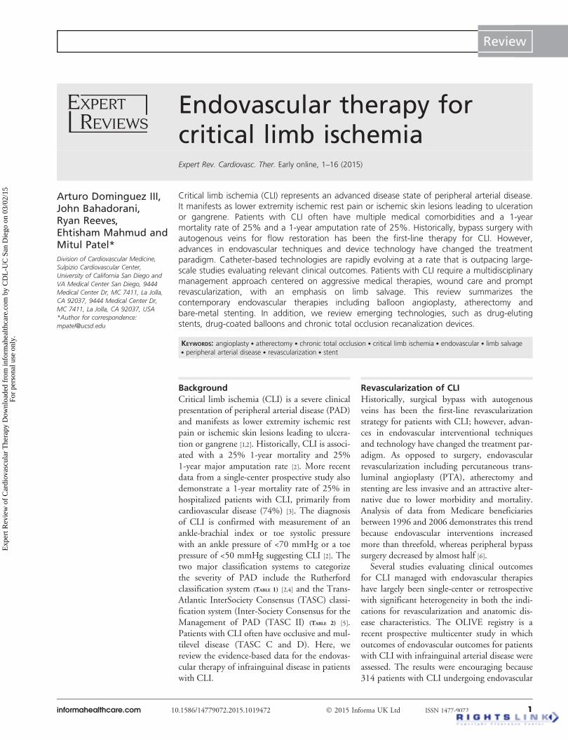
Endovascular therapy for critical limb ischemia Expert Rev. Cardiovasc. Ther. Early online, 1–16 (2015) Arturo Dominguez III, John Bahadorani, Ryan Reeves, Ehtisham Mahmud and Mitul Patel* Division of Cardiovascular Medicine, Sulpizio Cardiovascular Center, University of California San Diego and VA Medical Center San Diego, 9444 Medical Center Dr, MC 7411, La Jolla, CA 92037, 9444 Medical Center Dr, MC 7411, La Jolla, CA 92037, USA *Author for correspondence: [email protected] Critical limb ischemia (CLI) represents an advanced disease state of peripheral arterial disease. It manifests as lower extremity ischemic rest pain or ischemic skin lesions leading to ulceration or gangrene. Patients with CLI often have multiple medical comorbidities and a 1-year mortality rate of 25% and a 1-year amputation rate of 25%. Historically, bypass surgery with autogenous veins for flow restoration has been the first-line therapy for CLI. However, advances in endovascular techniques and device technology have changed the treatment paradigm. Catheter-based technologies are rapidly evolving at a rate that is outpacing large- scale studies evaluating relevant clinical outcomes. Patients with CLI require a multidisciplinary management approach centered on aggressive medical therapies, wound care and prompt revascularization, with an emphasis on limb salvage. This review summarizes the contemporary endovascular therapies including balloon angioplasty, atherectomy and bare-metal stenting. In addition, we review emerging technologies, such as drug-eluting stents, drug-coated balloons and chronic total occlusion recanalization devices. KEYWORDS: angioplasty . atherectomy . chronic total occlusion . critical limb ischemia . endovascular . limb salvage . peripheral arterial disease . revascularization . stent Background Critical limb ischemia (CLI) is a severe clinical presentation of peripheral arterial disease (PAD) and manifests as lower extremity ischemic rest pain or ischemic skin lesions leading to ulcera- tion or gangrene [1,2]. Historically, CLI is associ- ated with a 25% 1-year mortality and 25% 1-year major amputation rate [2]. More recent data from a single-center prospective study also demonstrate a 1-year mortality rate of 25% in hospitalized patients with CLI, primarily from cardiovascular disease (74%) [3]. The diagnosis of CLI is confirmed with measurement of an ankle-brachial index or toe systolic pressure with an ankle pressure of <70 mmHg or a toe pressure of <50 mmHg suggesting CLI [2]. The two major classification systems to categorize the severity of PAD include the Rutherford classification system (TABLE 1) [2,4] and the Trans- Atlantic InterSociety Consensus (TASC) classi- fication system (Inter-Society Consensus for the Management of PAD (TASC II) (TABLE 2) [5]. Patients with CLI often have occlusive and mul- tilevel disease (TASC C and D). Here, we review the evidence-based data for the endovas- cular therapy of infrainguinal disease in patients with CLI. Revascularization of CLI Historically, surgical bypass with autogenous veins has been the first-line revascularization strategy for patients with CLI; however, advan- ces in endovascular interventional techniques and technology have changed the treatment par- adigm. As opposed to surgery, endovascular revascularization including percutaneous trans- luminal angioplasty (PTA), atherectomy and stenting are less invasive and an attractive alter- native due to lower morbidity and mortality. Analysis of data from Medicare beneficiaries between 1996 and 2006 demonstrates this trend because endovascular interventions increased more than threefold, whereas peripheral bypass surgery decreased by almost half [6]. Several studies evaluating clinical outcomes for CLI managed with endovascular therapies have largely been single-center or retrospective with significant heterogeneity in both the indi- cations for revascularization and anatomic dis- ease characteristics. The OLIVE registry is a recent prospective multicenter study in which outcomes of endovascular outcomes for patients with CLI with infrainguinal arterial disease were assessed. The results were encouraging because 314 patients with CLI undergoing endovascular informahealthcare.com 10.1586/14779072.2015.1019472 Ó 2015 Informa UK Ltd ISSN 1477-9072 1 Review Expert Review of Cardiovascular Therapy Downloaded from informahealthcare.com by CDL-UC San Diego on 03/02/15 For personal use only.
-
Upload
independent -
Category
Documents
-
view
2 -
download
0
Transcript of Endovascular therapy for critical limb ischemia
Endovascular therapy forcritical limb ischemiaExpert Rev. Cardiovasc. Ther. Early online, 1–16 (2015)
Arturo Dominguez III,John Bahadorani,Ryan Reeves,Ehtisham Mahmud andMitul Patel*Division of Cardiovascular Medicine,
Sulpizio Cardiovascular Center,
University of California San Diego and
VA Medical Center San Diego, 9444
Medical Center Dr, MC 7411, La Jolla,
CA 92037, 9444 Medical Center Dr,
MC 7411, La Jolla, CA 92037, USA
*Author for correspondence:
Critical limb ischemia (CLI) represents an advanced disease state of peripheral arterial disease.It manifests as lower extremity ischemic rest pain or ischemic skin lesions leading to ulcerationor gangrene. Patients with CLI often have multiple medical comorbidities and a 1-yearmortality rate of 25% and a 1-year amputation rate of 25%. Historically, bypass surgery withautogenous veins for flow restoration has been the first-line therapy for CLI. However,advances in endovascular techniques and device technology have changed the treatmentparadigm. Catheter-based technologies are rapidly evolving at a rate that is outpacing large-scale studies evaluating relevant clinical outcomes. Patients with CLI require a multidisciplinarymanagement approach centered on aggressive medical therapies, wound care and promptrevascularization, with an emphasis on limb salvage. This review summarizes thecontemporary endovascular therapies including balloon angioplasty, atherectomy andbare-metal stenting. In addition, we review emerging technologies, such as drug-elutingstents, drug-coated balloons and chronic total occlusion recanalization devices.
KEYWORDS: angioplasty . atherectomy . chronic total occlusion . critical limb ischemia . endovascular . limb salvage. peripheral arterial disease . revascularization . stent
BackgroundCritical limb ischemia (CLI) is a severe clinicalpresentation of peripheral arterial disease (PAD)and manifests as lower extremity ischemic restpain or ischemic skin lesions leading to ulcera-tion or gangrene [1,2]. Historically, CLI is associ-ated with a 25% 1-year mortality and 25%1-year major amputation rate [2]. More recentdata from a single-center prospective study alsodemonstrate a 1-year mortality rate of 25% inhospitalized patients with CLI, primarily fromcardiovascular disease (74%) [3]. The diagnosisof CLI is confirmed with measurement of anankle-brachial index or toe systolic pressurewith an ankle pressure of <70 mmHg or a toepressure of <50 mmHg suggesting CLI [2]. Thetwo major classification systems to categorizethe severity of PAD include the Rutherfordclassification system (TABLE 1) [2,4] and the Trans-Atlantic InterSociety Consensus (TASC) classi-fication system (Inter-Society Consensus for theManagement of PAD (TASC II) (TABLE 2) [5].Patients with CLI often have occlusive and mul-tilevel disease (TASC C and D). Here, wereview the evidence-based data for the endovas-cular therapy of infrainguinal disease in patientswith CLI.
Revascularization of CLIHistorically, surgical bypass with autogenousveins has been the first-line revascularizationstrategy for patients with CLI; however, advan-ces in endovascular interventional techniquesand technology have changed the treatment par-adigm. As opposed to surgery, endovascularrevascularization including percutaneous trans-luminal angioplasty (PTA), atherectomy andstenting are less invasive and an attractive alter-native due to lower morbidity and mortality.Analysis of data from Medicare beneficiariesbetween 1996 and 2006 demonstrates this trendbecause endovascular interventions increasedmore than threefold, whereas peripheral bypasssurgery decreased by almost half [6].
Several studies evaluating clinical outcomesfor CLI managed with endovascular therapieshave largely been single-center or retrospectivewith significant heterogeneity in both the indi-cations for revascularization and anatomic dis-ease characteristics. The OLIVE registry is arecent prospective multicenter study in whichoutcomes of endovascular outcomes for patientswith CLI with infrainguinal arterial disease wereassessed. The results were encouraging because314 patients with CLI undergoing endovascular
informahealthcare.com 10.1586/14779072.2015.1019472 � 2015 Informa UK Ltd ISSN 1477-9072 1
Review
Exp
ert R
evie
w o
f C
ardi
ovas
cula
r T
hera
py D
ownl
oade
d fr
om in
form
ahea
lthca
re.c
om b
y C
DL
-UC
San
Die
go o
n 03
/02/
15Fo
r pe
rson
al u
se o
nly.

therapy had 12-month amputation-free survival of 74%, whereas34% of patients required repeat revascularization (endovascular31.7%; bypass surgery 2.6%) [7]. These data are promising inthat even in a very high-risk CLI population (52% hemodialysis;71% diabetes mellitus) with severe anatomic disease (42% belowthe knee lesions and >50% TASC type C and D), endovascularrevascularization results in >70% amputation-free survival [7].
Revascularization options: endovascular therapy versussurgical bypassThere is a paucity of randomized clinical trials that directlycompare surgical bypass to angioplasty in the management ofCLI. The BASIL trial included 452 patients with severe limbischemia and showed no difference in amputation-free survivalat 6 months (48 vs 60 patients; unadjusted HR = 1.07; 95%CI: 0.72–1.6; adjusted HR = 0.73; 0.49–1.07) [8]. It should benoted, however, that the surgery-first strategy was associatedwith greater morbidity, longer hospital stay and increased useof an intensive-therapy unit stay compared with theangioplasty-first group. This led to a 33% increase in cost inthe surgical arm. Conversely, the angioplasty-first group was
found to have a higher rate of immediate procedural failureand re-intervention at 12 months [8]. The 2.5-year follow-upstudy of the BASIL trial revealed no difference in bothamputation-free survival and overall survival between the tworevascularization strategies [9]. Notably, for patients who sur-vived 2 years after randomization, the surgery-first strategy wasassociated with a reduced hazard ratio for overall survival of0.61 (95% CI: 12–13.4 months; p = 0.02) [9].
Following the publication of the BASIL trial, new recommen-dations were released in a 2011 American College of CardiologyFoundation/American Heart Association (ACCF/AHA) updatefor the management of patients with PAD guideline focusing onlife expectancy to help guide a revascularization approach.Patients with lower extremity ischemia and an estimated lifeexpectancy of 2 years or less without autogenous vein conduits,an endovascular approach was felt to be a reasonable initial treat-ment strategy [10]. However, for patients with a life expectancygreater than 2 years, bypass surgery remains a reasonable initialtreatment option when autogenous vein conduits are avail-able [10]. Compared to patients with claudication, patients withCLI often have multilevel PAD. The current, 2011 ACCF/AHAguidelines remain unchanged from 2005 in that patients withCLI with combined inflow and outflow disease are to have theirinflow disease addressed first. However, for patients whose signsor symptoms of CLI persist after inflow revascularization, out-flow revascularization procedure is then recommended [10].
A recently published meta-analysis, which reviewed the liter-ature from 1995 to 2012, found no difference in overall death(OR = 1.07; 95% CI: 0.73–1.56), or 3-year amputation-freesurvival (OR = 1.22; 95% CI: 0.84–1.77) in patients with CLIwho were treated with either endovascular or surgical revascu-larization [11]. Furthermore, elderly patients tend to have multi-ple comorbidities and comprise a large proportion of patientswith CLI. These patients often lack adequate venous conduitsand tend to have high surgical risk, making them poor candi-dates for surgical bypass. Endovascular therapy may representthe only reasonable revascularization option for limb salvage inthis subset of patients. As catheter-based technologies evolve,endovascular therapy is becoming the first-line treatment strategyfor revascularization in patients with CLI, challenging the role ofinfrainguinal bypass surgery as the gold standard and optimaltreatment approach for CLI revascularization in complex ana-tomic disease [12]. The BEST-CLI trial, which is currently enroll-ing patients, is randomizing this patient population to comparethe effectiveness of best available surgical treatment with bestavailable endovascular treatment in patients with CLI who are eli-gible for both treatment options [13].
AngioplastyRevascularization of CLI with PTA can achieve a 5-year limbsalvage rate of 89%, similar to earlier reported values with sur-gical revascularization, which ranged from 70 to 80% [14]. Aspreviously mentioned, patients with CLI tend to have multi-level disease, which often includes infrapopliteal arterial disease.A meta-analysis evaluating the outcomes of patients with CLI
Table 1. Rutherford classification.
Grade Category Clinical description
0 0 Asymptomatic
I 1 Mild claudication
I 2 Moderate claudication
I 3 Severe claudication
II 4 Ischemic rest pain
III 5 Minor tissue loss – nonhealing ulcer, focal
gangrene with diffuse pedal ischemia
III 6 Major tissue loss – extending above
transmetatarsal level, frank gangrene
Table 2. TASC-II classification
Femoropopliteal lesions
Type-A lesions Single stenosis of £10 cm in length or a single
occlusion £5 cm in length
Type-B lesions Multiple lesions, each £5 cm or single lesion
£15 cm not involving the popliteal artery.
Heavily calcified occlusion £5 cm.
Single popliteal stenosis
Type-C lesions Multiple lesions totaling ‡15 cm regardless
of calcification or recurrent lesions that require
treatment after two endovascular interventions
Type-D lesions CFA or SFA CTOs >20 cm involving the
popliteal or CTO of the popliteal and
proximal trifurcation
Lesions include stenosis or occlusions.CFA: Common femoral artery; CTO: Chronic total occlusion; SFA: Superficialfemoral artery.
Review Dominguez, Bahadorani, Reeves, Mahmud & Patel
doi: 10.1586/14779072.2015.1019472 Expert Rev. Cardiovasc. Ther.
Exp
ert R
evie
w o
f C
ardi
ovas
cula
r T
hera
py D
ownl
oade
d fr
om in
form
ahea
lthca
re.c
om b
y C
DL
-UC
San
Die
go o
n 03
/02/
15Fo
r pe
rson
al u
se o
nly.
treated with infrapopliteal balloon angioplasty from 1990 to2006 revealed a 3-year limb salvage rate of over 80% [15].Although the primary patency after PTA for CLI may be low,the limb salvage rate remains quite high [14,15]. Furthermore, aDepartment of Veterans Affairs study examined major lowerextremity amputations and found the survival after amputationwas 78% at 1 year but only 55% at 3 years; approximately50% of patients were nonambulatory and 17% required care ata nursing facility [16]. A recent study analyzing amputations pat-terns in US Medicare data from 2000 to 2008 found a signifi-cant decline in overall lower extremity amputation rates from7258 to 5790 per 100,000 Medicare beneficiaries with PADduring the study period, p < 0.001 [17]. This study also demon-strated that despite adjusting for clinical variables and temporaltrends, a significant geographic variation persists as lowerextremity amputations are performed more often in the East,South and Central regions of the United States with anadjusted OR of 1.152; 95% CI: 1.131–1.174, p < 0.001 [17].The overall decline in amputation rates in the past decade islikely due to a combination of earlier detection, prompt referraland advances in endovascular interventions. Preventing majoramputation remains the principal treatment goal in any CLIrevascularization procedure, and standard PTA remains animportant endovascular technique.
Impact of anatomy on endovascular options &outcomesIn patients with CLI, the more proximal the disease, the betterthe primary patency. In general, endovascular therapies for iliaclesions result in a higher primary patency compared with femo-ropopliteal disease, and likewise femoropopliteal disease has ahigher primary patency compared with infrapopliteal disease [14].Thus, when considering revascularization strategy for multileveldisease, common practice involves treating proximal or in-flowlesions followed by close observation for improvement prior toapproaching more complex, distal targets.
The ACCF/AHA guidelines currently recommend either pro-visional stent placement for suboptimal or failed balloon dilata-tion result or primary stenting for treatment of common iliacand external iliac disease [1]. However, for femoropopliteal andinfrapopliteal disease, current management guidelines do not rec-ommend primary stenting and suggest that stenting should onlybe reserved for bailout purposes in cases of suboptimal or failedballoon angioplasty results [1].
The extent of disease and lesion characteristics also play amajor role in choosing between endovascular and surgical revas-cularization. Current guidelines have the following Class I rec-ommendations for revascularization: endovascular therapy foraortoiliac and femoropopliteal lesions which are TASC type Alesions and bypass surgery for TASC type D lesions [2]. Despitethese recommendations, many vascular interventionalists arenow managing complex disease (TASC types B, C and D)with an endovascular approach. A study of infrapopliteal angio-plasty for CLI by TASC classification revealed that even forpatients with TASC A, B or C lesions, infrapopliteal
angioplasty is a reasonable primary treatment approach despitehigh restenosis and reintervention rates given that limb salvagerates of 84% were achieved at 1, 2 and 3 years [18].
Angiosome-guided revascularizationThe angiosome concept emphasizes distinct three-dimensionalblocks of tissue fed by source arteries (FIGURE 1) [19]. The angio-somes of the foot have been used to help guide vascular recon-struction for foot wounds [19]. Applying the same concept oftargeted revascularization with endovascular techniques appearsto have promising results for improved wound healing andclinical outcomes [20]. A retrospective analysis of 52 patientswith CLI and nonhealing lower extremity wounds who under-went surgical bypass revealed that a bypass to the artery directlyfeeding the ischemic angiosome was associated with a 91%healing rate and only a 9% amputation rate. This was in con-trast to a bypass feeding an unrelated ischemic angiosome,which demonstrated only a 62% wound healing rate alongwith a 38% amputation rate (p = 0.03) [21]. In a prospectivestudy of 64 patients with CLI, 60.9% were revascularized withopen surgery and 39.1% with an endovascular interventionusing either an angiosome-directed or non angiosome-directedapproach. The study found that ulcer healing was improvedwith an angiosome-directed approach. At 3 months, 57.6% ofpatients in the angiosome-directed group had wound healing,compared with 12.5% in the non angiosome-directed group;similarly, 6-month healing rates were 96.4 and 83.3%, respec-tively (p = 0.06) [22]. A retrospective single-center review byAlexandrescu et al. examined 208 diabetic CLI patients withwounds treated with below-the-knee angioplasty. They alsofound improved wound healing and higher amputation-freerates in the angiosome-directed group at 1 year (90% com-pared with 84% in the standard angioplasty group,p = 0.035) [23]. Another retrospective study analyzed117 patients with CLI affecting 203 limbs and showedimproved sustained limb salvage rates with angiosome-targetedrevascularization compared with non angiosome-directed revas-cularization (86 vs 69% at 1 year; p = 0.03) [24]. Conversely, withsurgical revascularization, Azuma et al. studied 228 patients withCLI and tissue loss involving 249 limbs that underwent revascu-larization with a distal bypass for crural occlusive disease. Theyconcluded that an angiosome-directed bypass resulted in similarlimb salvage rates with 97.8% in the angiosome-directed groupand 92.3% in the non angiosome-directed group at 2 years,p = 0.855 [25].
Although guiding revascularization to the wound site byrestoring flow to the corresponding ischemic angiosome makesintuitive sense and appears to be a promising revascularizationstrategy to promote wound healing, most of the available evi-dence for this strategy has consisted of small, retrospective stud-ies. In clinical practice, an angiosome-directed revascularizationis not always technically feasible due to anatomic limitationsincluding long chronic total occlusions, or the presence of dif-fuse distal disease that may result in suboptimal run-off in thetargeted angiosome.
Endovascular therapy for CLI Review
informahealthcare.com doi: 10.1586/14779072.2015.1019472
Exp
ert R
evie
w o
f C
ardi
ovas
cula
r T
hera
py D
ownl
oade
d fr
om in
form
ahea
lthca
re.c
om b
y C
DL
-UC
San
Die
go o
n 03
/02/
15Fo
r pe
rson
al u
se o
nly.

Cutting balloon angioplastyThe cutting balloon was originally developed for use in the cor-onary arteries and initially approved in 1995 [26]. Cutting bal-loon angioplasty (CBA) uses low-pressure balloon catheters,which are mounted with microsurgical blades or microtomesthat directly cut into the luminal vessel wall during inflation [27].This creates a controlled fault line during dilation, leading to acontrolled crack propagation which may minimize the degreeof balloon-induced injury and vessel stretching [26,27]. Examina-tion of lesions by intravascular ultrasound before and afterCBA and conventional balloon angioplasty reveals that CBAreduces overall vessel expansion. This suggests that the predom-inant mechanism is plaque compression or shift rather thanvessel expansion, as is seen in conventional angioplasty [28].
CBA has been used in endovascular revascularization forCLI. A retrospective analysis and other small case series ofpatients with symptomatic lower extremity ischemia treatedwith CBA revealed limb salvage rates of 89.5–100% during amean 1-year follow-up. These studies have demonstrated thatCBA is safe and effective for the treatment of popliteal andinfrapopliteal disease [29,30]. Another prospective, nonrandom-ized study involving 128 patients with symptomatic infraingui-nal lower extremity PAD (86% of limbs with CLI) treatedwith CBA reported primary patency rates in the CLI group of64.4% at 12 months and 52% at 24 months, whereas the limbsalvage rates were 84.2 and 76.9% at 12 and 24 months,respectively. Of note, a very low bail-out stent implantationrate of <3% was seen in the CBA group [31]. Approximatelyhalf the lesions treated were infrapopliteal and the majority ofthe lesions treated were TASC type A and B [31]. Conversely,
in a prospective randomized controlled trial, de novo superficialfemoral artery (SFA) lesions in 43 patients (19% of patientshad CLI) treated with either CBA versus conventional angio-plasty found a 32% restenosis rate in the standard PTA groupand 62% in the CBA group (p = 0.048) by Duplex ultrasoundat 6 months [32]. This study suggests that in patients with clau-dication and SFA disease, the use of CBA does not result insuperior outcomes. Due to the lack of superiority over conven-tional PTA and higher cost, CBA is not routinely used. There-fore, CBA use is predominantly reserved for the treatment ofin-stent restenosis, and diseased arterial segments in flexionpoints such as the common femoral artery or the poplitealartery where stent implantation is limited by fracture risk.
StentsFemoropopliteal stents
A meta-analysis reviewing the literature from 1993 to2000 showed a 3-year patency rate for femoropopliteal CLIafter PTA of 43% for stenotic lesions and 30% for occlusions.As expected, there was a higher patency rate for patients withclaudication, which was 61% for stenotic lesions (standarderror, 2.2%) and 48% for occlusions (standard error,4.1%) [33]. Although PTA has a high immediate proceduralsuccess rate, the long-term patency and freedom from reinter-vention remain major obstacles. Stents pose a viable solution toaddress the problem of long-term patency and reintervention,but currently they are only recommended to be used as a bail-out option for failed or suboptimal angioplasty results in femo-ropopliteal and infrapopliteal vessels [1]. There is an ongoingdebate as to whether primary stent implantation for infraingui-nal disease is a reasonable treatment strategy because it mayreduce the rates of restenosis and reintervention.
Initial experience with primary stenting using the Palmazvascular stent was disappointing (Cordis or Johnson & Johnsoninterventional systems) when it was compared with PTA in amulticenter prospective randomized trial of 227 patients withSFA lesions (lesion length <7 cm) and disabling claudication orCLI. This showed no difference in angiographic restenosis at1 year (defined as >50% angiographic stenosis). The 1-yearrestenosis rates were 32.3% in the PTA with bailout stentinggroup and 34.7% in the primary stent group (p = 0.85) [34].
With advances in stent technology, nitinol self-expandingstents ultimately led to improved results for primary stenting inSFA lesions. The use of newer generation nitinol stents in SFAdisease demonstrated reduced restenosis compared with stan-dard PTA [35,36]. Schillinger et al. studied 104 patients withsymptomatic PAD (only 12.5% of patients with CLI) and SFAdisease. They were randomly assigned to either primary stent-ing or angioplasty with bailout stenting for suboptimal angio-plasty results. At 6 months, the angiographic restenosis was24% in the stent group and 43% in the angioplasty group(p = 0.05). This trend became significant at 12 months, atwhich point restenosis by duplex ultrasound was 37% in thestent group and 63% in the angioplasty group (p = 0.01) [35].Additional evidence from the RESILIENT trial, consisting of
Anterior tibialangiosome
Posterior tibialangiosome
Peronealangiosome
Anterior tibial
Medial plantar
Lateral plantar
Calcaneal
Peroneal
Figure 1. The Angiosome concept. Illustration of the lowerextremity angiosomes represented as a topographic map dividedinto five territories, supplied by the three main arteries andtheir branches.
Review Dominguez, Bahadorani, Reeves, Mahmud & Patel
doi: 10.1586/14779072.2015.1019472 Expert Rev. Cardiovasc. Ther.
Exp
ert R
evie
w o
f C
ardi
ovas
cula
r T
hera
py D
ownl
oade
d fr
om in
form
ahea
lthca
re.c
om b
y C
DL
-UC
San
Die
go o
n 03
/02/
15Fo
r pe
rson
al u
se o
nly.

206 patients with SFA and proximal popliteal artery diseasewith claudication who were randomized to either primary niti-nol self-expanding stent implantation or standard PTA, alsofound superior Duplex ultrasound-determined primary patencyat 1 year: 81.3% in the primary stent group and 36.7% in thePTA group (p < 0.0001) [37]. Furthermore, the DURABILITY Inonrandomized study of 151 patients also found promisingresults with nitinol stent implantation for SFA lesions in symp-tomatic PAD with freedom from restenosis at 1 year in 72.2%(95% CI: 63.8–79.6%) [38]. These data reflect improvement in1-year outcomes using nitinol stents primarily in SFA lesionsand primarily in non-CLI patients. However, specifically for theCLI population, the primary patency rates can be as low as31.4 ± 10.4% at 5 years [14]. Another limitation of stenting isstent fracture, which can be associated with significant complica-tions. A study of systematic x-ray screening detected a stent frac-ture rate of 37.2% in a series of 121 treated lower extremities.Fracture rates are increased for long lesions, and fracture inci-dence is known to contribute to in-stent restenosis [39]. Anotherstudy of 286 patients identified stent fracture rates rangingbetween 19 and 28% [40]. Currently, stents remain widely usedin endovascular therapy for PAD and depending on the targetedvascular bed they are used as a primary treatment device or as abailout device.
Drug-eluting stents
Implantation of metallic stents remains limited by the develop-ment of neointimal hyperplasia resulting in late luminal lossand in-stent restenosis. In an effort to improve the long-termpatency of primary stenting, several trials have explored theeffect of drug-coated stent implantation. Sirolimus-elutingstents (SES) were compared with bare metal stents in SFA dis-ease in the SIROCCO I and SIROCCO II trials, both ofwhich found no difference when considering in-stent restenosisat 6 months and at 2 years [41,42]. However, later studies withthe paclitaxel-coated Zilver PTX stent (Cook Medical, Bloo-mington, IN) proved not only safety and efficacy of the ZilverDES, but also superiority when compared with PTA with pro-visional stent placement. This study randomized 474 patientsto primary DES implantation or PTA with provisional stentplacement and had a subsequent secondary randomization toeither BMS or DES for the subset of patients requiring provi-sional stenting. At 1 year, the event-free survival was 90.4% inthe primary DES implantation group versus 82.6% in the con-trol arm, p = 0.004. The primary patency at 1 year was83.1% in DES versus 32.8% for PTA alone (this groupincluded the prespecified criteria that acute PTA failure wasloss of patency), (p < 0.001). The patency rate in the primaryDES group was also superior to the optimal PTA group(excluding acute PTA failure) at 83.1% in the DES group ver-sus 65.3% in the PTA group (p < 0.001). Extrapolating thesuccess of DES use in the patient with CLI is problematicgiven that less than 9% of patients had CLI. At 2 years, thedata remained supportive of the safety and efficacy of thepaclitaxel DES [43–45].
Covered stents
Covered stents or stent grafts have been studied as a potentialsolution to the predominant mode of failure of bare nitinolstents (BNS): in-stent restenosis (ISR). This is achieved by add-ing a lining to the stent frame to mitigate the in-growth ofintimal hyperplasia. The majority of data in covered stents arelimited to SFA lesions, and there is a paucity of data specificfor patients with CLI. The Viabhan covered stent (W.L. Goreand Associates, Flagstaff, AZ) was used in a randomized studyof 148 patients undergoing revascularization for SFA lesionsand compared Viabhan with BNS for symptomatic PAD. At3 years, this study found no significant difference in primarypatency rates between the Viabhan group and the BNSgroup [46]. Early experience with Dacron-covered stent graftsfor femoropopliteal disease found high rates of early and laterestenosis, early stent graft thrombosis and a postimplantationsyndrome of noninfectious fever and pain [47]. The evolution ofcovered stents led to the development of the heparin-bondedexpanded polytetrafluoroethylene (ePTFE)–covered Viabhanstent (WL Gore, Flagstaff, AZ), which was used in the VIA-STAR study of 141 patients (only 15% of patients with CLI)undergoing PTA for SFA lesions. Subjects were randomized toimplantation of heparin-coated covered stent or BNS andfound that at 1 year the primary patency rates in the Viabahngroup and the BNS group were not statistically different [48].Yet this study yielded promising results for lesions ‡20 cm(TASC class D), showing a 1-year patency rate of 71.3% inthe Viabhan group and 36.8% in the BNS group(p = 0.01) [48]. A persistent major disadvantage of the coveredstent is graft thrombosis, and one third of patients with recur-rent claudication in the Viabhan group had total thromboticocclusions [48]. The VIPER study similarly evaluated the perfor-mance of the heparin-bonded, ePTFE Viabhan stent graft inlong segment lesions (mean length of 19 cm) with severe femo-ropopliteal disease (56% of lesion were occlusions) in113 patients (13% of patients with CLI) and found a 1-yearpatency of 73% [49]. Currently, covered stents are not widelyused as an alternative to nitinol stents in SFA revascularizationbecause the issue of thrombosis remains a major concern.
Infrapopliteal stentingRetrospective studies of infrapopliteal stent implantation trialshave used a variety of stents, including coronary drug-elutingstents (DES) given that tibial vessels often have similar diame-ters to coronary arteries. Bare nitinol self-expanding stentsare currently used to treat suboptimal angioplasty results ininfrapopliteal disease. A nonrandomized retrospective studyof 53 patients with CLI and infrapopliteal disease(Rutherford ‡4) undergoing infrapopliteal stent placement withself-expanding nitinol stents for suboptimal angioplastyreported a high technical success (98%) and primary patencyrate of 75.5% with a freedom from amputation rate of 88.7%at 2 years [50]. This study supports the feasibility and efficacyof infrapopliteal stenting, specifically in the CLI patient popu-lation. Additional data from a nonrandomized, single-center
Endovascular therapy for CLI Review
informahealthcare.com doi: 10.1586/14779072.2015.1019472
Exp
ert R
evie
w o
f C
ardi
ovas
cula
r T
hera
py D
ownl
oade
d fr
om in
form
ahea
lthca
re.c
om b
y C
DL
-UC
San
Die
go o
n 03
/02/
15Fo
r pe
rson
al u
se o
nly.

study of 82 patients (92 limbs) undergoing primary infrapopli-teal stenting where 68% of the study population had CLIreported a high technical success rate (94%) with relief of restpain and wound healing in 95% of patients at 1 year [51].A small, single-center, randomized trial compared infrapoplitealprimary stenting with angioplasty in 38 limbs of 35 patients.There was no difference in survival, limb salvage or primarypatency at 1 year [52]. However, this study was limited by its smallsample size and the use of a wide variety of stents includingcovered stents.
Several studies have compared drug-eluting stents with baremetal stents for infrapopliteal disease. A single-center non-randomized prospective registry of 103 patients with CLI andinfrapopliteal disease undergoing infrapopliteal PTA withbailout stent implantation were treated with either a sirolimus-eluting stent (SES) or a bare metal stent. The long-term out-comes of this study are available at 3 years. The SES-treatedlesions were associated with similar primary patency andreintervention-free survival, which was 77.6% in the SES groupand 70.3% in the BMS group (p = 0.49) [53]. The PARADISEtrial was a prospective, nonrandomized single-center study of106 patients (118 limbs) who underwent primary below-the-knee coronary DES implantation with 83% Cypher (Cordis;Johnson & Johnson, Warren, NJ) and 17% Taxus (Boston Sci-entific, Maple Grove, MN) stents, revealed a 3-year cumulativeamputation-free survival of 68 ± 5% with 94% limb salvage,which exceeding historical expectations for PTA alone or surgi-cal revascularization [54]. A meta-analysis of 18 studies com-prised 640 patients who underwent infrapopliteal bailout stentimplantation after failed or suboptimal PTA results for CLIshowed a primary patency of 78.9% (95% CI: 71.8–98.1%), atarget vessel revascularization of 10.1% (95% CI: 6.2–13.9%),and a limb salvage rate of 96.4% (95% CI: 94.7–98.1%) at12 months [55]. Of note, sirolimus-eluting stents appeared supe-rior to both BMS and paclitaxel-eluting stents with regard toprimary patency and need for repeat revascularization [55].
Recently, there have been more rigorous randomized clinicaltrials evaluating implantation of DES in infrapopliteal vesselsto help determine whether DES leads to improved clinical out-comes including decreased restenosis and freedom from reinter-vention over traditional PTA. The ACHILLES study was aprospective, multicenter, randomized controlled trial inwhich 200 patients with symptomatic PAD (mean baselineRutherford category of 4.1 ± 0.9) and infrapopliteal diseasewere randomized to revascularization with SES implantation orPTA with bailout SES. At 1 year, angiographic restenosis rateswere 22.4% in the SES group and 41.9% in the PTA group(p = 0.019). Although the primary patency rates were greaterin the SES group, there were no significant difference in therates of death, repeat revascularization and index-limb amputa-tion [56]. The Yukon-BTK trial was a randomized, blinded,multicenter trial comparing SES with BMS in 161 patientswith symptomatic infrapopliteal PAD (46.6% of patients withCLI) that demonstrated a primary patency at 1 year of 80.6%in the SES group and 55.6% in the BMS group,
p = 0.004 [57]. Long-term results of the Yukon-BTK trial wererecently published and showed a clinical benefit because theevent-free survival was 65.8% in the SES group and 44.6% inthe BMS group during an overall mean follow-up period of1016 ± 132 days (p = 0.02) [58]. Of note, endovascular inter-vention to treat infrapopliteal disease for claudication, whichrepresented 53.4% of the Yukon-BTX study population, is notcurrently a guideline recommended treatment option. TheDESTINY trial compared everolimus-eluting stents (EES) withBMS in 140 patients with CLI and the primary patency at1 year was 85% in the EES group and 54% in the BMSgroup. In addition, this study revealed that freedom from targetlesion revascularization was 91% in the EES group comparedwith 66% in the BMS group (p = 0.001) [59]. These studiesshow promising data in favor of DES compared with BMS ininfrapopliteal disease; however, it is still currently debatedwhether even primary stenting over angioplasty in this ana-tomic segment is appropriate.
Another concern is the much higher cost associated with pri-mary stenting with DES for infrapopliteal disease in CLIbecause it typically requires multiple coronary drug-elutingstents. In addition, there is a large discrepancy between primarypatency and clinical benefit in terms of limb salvage for thosepatients with CLI [60]. A high (90%) limb salvage rate is seenin standard PTA, despite the associated limitations of highrestenosis rate (60–80%) at 6 months based on data from theBASIL trial and cohort studies [61]. Collectively, these data showthat primary infrapopliteal DES use has an associated primarypatency rate 70–90% and ‡80% based on the more recent ran-domized controlled trials. Additional studies are needed to con-firm whether these positive angiographic findings associated withinfrapopliteal DES use – specifically in the CLI population – willtranslate to improved clinical outcomes, namely reintervention,limb salvage and amputation rates compared with standard PTAwith bailout BMS use.
Data supporting the use of DES for infrapopliteal revascular-ization in the CLI population should be interpreted with cau-tion as the literature’s emphasis has been on the endpoints ofvessel patency or ISR rates. These should not be considered theprimary clinical endpoints, as the fundamental aim in thesepatients is wound healing and amputation prevention. Theseclinical objectives may be achieved by endovascular or surgicalrevascularization despite development of re-occlusion, graft fail-ure or elevated ISR rates. Additional research is needed todetermine whether infrapopliteal DES is superior to currentendovascular strategies specifically for the CLI population withregards to these clinical endpoints.
Drug-coated balloonsTraditional balloon angioplasty as a stand-alone treatment forCLI remains limited by elastic recoil and restenosis due to cel-lular proliferation. Stent deployment can certainly mitigate ves-sel recoil; however, stent thrombosis, the need for long-termdual antiplatelet therapy and stent fracture, which contributesto restenosis and thrombosis, remain major limitations. In
Review Dominguez, Bahadorani, Reeves, Mahmud & Patel
doi: 10.1586/14779072.2015.1019472 Expert Rev. Cardiovasc. Ther.
Exp
ert R
evie
w o
f C
ardi
ovas
cula
r T
hera
py D
ownl
oade
d fr
om in
form
ahea
lthca
re.c
om b
y C
DL
-UC
San
Die
go o
n 03
/02/
15Fo
r pe
rson
al u
se o
nly.
addition, PAD in lower extremity vessels poses a challenge withstent implantation given the high risk of fracture, erosion orexternal trauma due to the proximity to the skin. These limita-tions have led to the development of drug-coated balloons(DCB). Current DCBs use paclitaxel as the antiproliferativedrug as it is highly lipophilic and has favorable tissue kinetics.To improve efficacy, DCBs are prepared with a coated mixtureof paclitaxel and an excipient which is a hydrophilic spacer thatfacilitates local uptake into the vessel wall resulting in greaterinhibition of neointimal growth [62].
The bulk of clinical trial data thus far with DCB has pre-dominantly included patients with claudication and lowerRutherford classifications as opposed to CLI patients. TheTHUNDER, FemPac, PACIFIER and LEVANT-1 trials allassessed femoropopliteal lesions comparing DCB with standardPTA. More than 90% of the patients in these trials were clau-dicants with Rutherford classes of 3 or less. Late lumen loss,TLR and angiographic restenosis significantly favored DCB inthese trials [63–66]. The IN.PACT SFA is a recent, large pro-spective, international, multicenter, study of 331 patients withsymptomatic PAD (Rutherford 2–4) who were randomized toendovascular treatment with DCB using the IN.PACT Admiralpaclitaxel-coated balloon (Medtronic, Santa Rosa, CA) or stan-dard PTA. In this study, >90% of lesions were de novo with amean lesion length of ‡8 cm in both groups. The 1-year pri-mary patency was 82.2% in the DCB group and 52.4% in thePTA group, p < 0.001 [67]. An impressively low rate of clini-cally driven TLR was reported at 2.4% in the DCB groupcompared with 20.6% in the PTA group at 1 year (p < 0.001);the lowest reported TLR for an SFA device trial [67]. TheLEVANT-2 trial, currently ongoing [68], is another large, inter-national, randomized trial comparing paclitaxel-coated balloonangioplasty (Lutonix, Bard, Tempe, AZ) to standard PTA forfemoropopliteal disease. Currently, the data for patients withCLI and infrapopliteal disease are sparse compared with theaforementioned data involving claudicants with femoropoplitealdisease. Schmidt et al. published the first series of patientstreated with the DCB (In.Pact Amphirion; Medtronic, SantaRosa, CA) for long infrapopliteal disease (mean lesion lengthof 176 ± 88 mm) in 104 patients (82.6% with CLI). Theirseries revealed a 3-month angiographic restenosis rate of 27%compared with 69% in historical controls with standardPTA [69]. At 1 year, complete wound healing occurred in74.2% and a limb salvage rate of 95.6% of patients withCLI [69]. The DEBATE-BTK randomized trial compared theIN.PACT Amphirion DCB (Medtronic, Santa Rosa, CA) withstandard PTA in diabetic patients with CLI and showed signifi-cant reductions in both restenosis and TLR at 1 year. Binaryrestenosis in the DCB arm was 27% compared with 74% inthe standard PTA arm (p < 0.001). TLR at 1 year was 18%with the DCB compared with 43% with standard PTA(p = 0.002) [70]. The IN.PACT DEEP study was halted prema-turely as it showed, in contrast to the aforementioned trials,there was no difference between the In. Pact Amphirion DCBtreatment group and the standard balloon angioplasty control
group in any of the study’s three primary outcome measures.The study also identified a potential safety signal with a trendtoward an increased rate of major amputations in the DCBstudy arm [71]. Similar to the studies addressing infrapoplitealDES use, the studies involving infrapopliteal DCB failed to uselimb salvage or amputation-free survival as primary endpoints.
AtherectomyAtherectomy involves atherosclerotic plaque removal or debulk-ing. The currently available technologies for this therapyinclude directional, rotational, orbital and laser atherectomy.Rotational atherectomy in the coronary vasculature has notbeen shown to improve ISR rates over traditional PTCA or toresult in reduced late lumen loss in the DES era [72,73]. Atherec-tomy for treatment of PAD is currently used as adjunctive ther-apy or as an alternative therapy to traditional PTA or stenting.Plaque debulking leads to an immediate increase in lumen sizewhich mechanistically should result in reduced stretch injuryon the arterial walls [61,74]. Atherectomy has been evaluated as apotential solution to the problem of long-term primary patencyof PTA in femoropopliteal disease given the 3-year patencyrates of approximately 50% for standard PTA [75].
Directional atherectomy with the Silverhawk (Covidien, Ply-mouth, MN) catheter shaves off plaque as the atherectomyblade rotates, whereas debris is collected inside a nose conelocated at the tip of the catheter. The Silverhawk device is cur-rently used in endovascular interventions for the treatment ofsymptomatic PAD and appears to be a viable tool [76–82]. Forthe CLI population specifically, the Silverhawk device can besafely used either as an adjunctive tool or as stand-alone ther-apy [83]. This was supported by a small series evaluatingTASC-type C femoropopliteal lesions in CLI showing thatatherectomy achieved acceptable early results [78]. A potentialcomplication of directional atherectomy is embolizationalthough the TALON registry reported a very low rate ofembolization of 0.1% [81]. A small consecutive series of patientsundergoing directional atherectomy with the Silverhawk devicefor femoropopliteal lesions used a distal embolic protectiondevice and found debris in the filter of each case [84]. A studyof 60 patients undergoing SFA PTA quantitatively measuredembolic signals (ES) with a continuous 4-MHz Doppler in theipsilateral popliteal artery found the average number of ES was12 for PTA, 28 for stent deployment, 49 for Silverhawk athe-rectomy and 51 for laser atherectomy. Despite the frequencyof ES, only 1 patient had a clinically significant embolicevent [85]. The PROTECT study was a single-center prospec-tive registry of 40 patients undergoing infrainguinal interven-tion to 56 lesions using distal embolic protection both duringstandard PTA or stenting and also during directional atherec-tomy using the Silverhawk device. Investigators found thatmacroembolization occurred in 37.9% of the PTA/stentinggroup and 100% in the Silverhawk group with debris ‡2 mmin 90% of the atherectomy group [86]. The DEFINITIVECa++ study involved 133 patients with 168 moderate toseverely calcified femoropopliteal lesions treated with
Endovascular therapy for CLI Review
informahealthcare.com doi: 10.1586/14779072.2015.1019472
Exp
ert R
evie
w o
f C
ardi
ovas
cula
r T
hera
py D
ownl
oade
d fr
om in
form
ahea
lthca
re.c
om b
y C
DL
-UC
San
Die
go o
n 03
/02/
15Fo
r pe
rson
al u
se o
nly.
directional atherectomy using the Silverhawk or Turbohawk(Covidien, Plymouth, MN) and distal embolic protection. Thestudy reported 93% freedom from major adverse events, whichincluded clinically significant embolization [87]. Definitive clinicalbenefit of atherectomy over PTA is not clear. A retrospectivestudy of 418 tibial interventions for CLI found no benefit overPTA, and this study used variety of atherectomy devices includ-ing directional, orbital and laser [88].
Orbital atherectomy using the Diamondback 360 device(Cardiovascular Systems Inc. [CSI], St. Paul, MN) performsplaque ablation using a flexible coil-wound drive shaft thatrotates an eccentric diamond-coated crown. This increases therotational speed and the centrifugal force, leading to anincreased radius of ablation [89,90]. The Diamondback 360 deviceappears to be a safe tool for atherectomy of infrainguinal dis-ease with promising results compared with PTA. Safian et al.reported procedural success (final diameter stenosis of £30%)in 90.1% of 124 patients undergoing infrapopliteal orbitalatherectomy with a major adverse event rate of 10.4% at6 months. The Calcium 360 study randomized 50 patientswith CLI to infrapopliteal orbital atherectomy with PTA versusPTA alone. It reported a procedural success rate of 93.1% inthe orbital atherectomy with PTA group compared with 82.4%in the PTA-alone group, p = 0.27. In addition, the orbitalatherectomy with PTA group had a lower bailout stenting rateof 6.9% compared with 14.3% in the PTA-alone group,p = 0.44. The Compliance 360 trial was a randomized studycomparing orbital atherectomy to PTA for femoropopliteal dis-ease in 50 patients and found freedom from TLR (includedadjunctive stenting) of 77.1% in the orbital atherectomy groupcompared with 11.5% in the PTA group, p < 0.001. However,at 12 months excluding adjunctive stenting, there was no dif-ference 81.2 versus 78.3%, p > 0.99. This study demonstratedthat orbital atherectomy compared with PTA alone yields bet-ter luminal gain and decreased need for adjunctive stenting incalcified femoropopliteal lesions [91–93].
Another debulking technology is rotational aspiration athe-rectomy. The Jetstream device (Boston Scientific, Maple Grove,MN) is a rotating, aspirating, expandable catheter that hasbeen used successfully in infrainguinal PAD with early experi-ence having moderate adverse event rates [94]. The subsequentMulticenter Pathway PVD trial had promising results with amajor adverse event rate of 1% at 30 days [95,96]. This devicehas also been shown to achieve a lumen area greater than aburr-sized lumen by intravascular ultrasound [97] in ahuman study.
The LACI study used laser atherectomy with a 308-nmexcimer laser, which delivers intense bursts of ultravioletenergy in short pulses, ablating plaque and thrombus on con-tact. This study treated 145 patients with CLI in 155 limbs(92% of lesions were occlusions) and reported a proceduralsuccess (<50% residual stenosis) of 86% and a 6-month limbsalvage rate of 93% [98]. Another small study fromBoccalandro et al. also supports the use of laser atherectomyas a feasible tool for treatment of femoropopliteal chronic total
occlusions in CLI patients who have failed conventional endo-vascular revascularization attempts, they reported a proceduralsuccess rate of 84% and a limb salvage rate of 88% [99].
However, the problem of embolization remains a concernfor all debulking technologies, especially when compared withconventional PTA. A large registry of 1029 patients evaluateddistal embolization rate for a total of 2137 lesions, and it dem-onstrated that overall the embolization rate was low at 1.6%.It also appeared that the Jetsream and Diamondback atherec-tomy devices had higher rates of embolic events (both at 22%)compared with Silverhawak (1.9%) and laser atherectomy(3.6%); PTA alone had the lowest rate of <1% [100]. In addi-tion, in-stent restenosis, chronic total occlusions and TASCtype C and D lesions were all associated with higher emboliza-tion rates [90].
Additional research is required with large randomized con-trolled trials to definitively establish whether atherectomy willprovide sustained clinical benefit over standard PTA/stentingand whether use of distal embolic protection is needed toprevent clinically significant events, which may be prudent toconsider in complex lesions [101]. A recent Cochrane meta-analysis included four randomized controlled trials comparingatherectomy to PTA ± stenting found no benefit in primarypatency or target vessel revascularization in atherectomy overPTA although atherectomy did appear to lead to a reductionin bailout stenting [102].
BrachytherapyBrachytherapy is an another strategy used to treat the problemof restenosis following PTA. It uses ionizing radiation to inhibitcellular proliferation with the aim of preventing the componentof restenosis mediated by the uncontrolled proliferation ofsmooth muscle cells. The Paris study demonstrated the safetyand feasibility of gamma radiation after balloon angioplasty [103].The use of brachytherapy for PAD was supported by a small,randomized trial of 113 patients, which compared PTA andbrachytherapy with PTA-alone for both de novo and recurrentfemoropopliteal lesions. Investigators found restenosis rates of28.3% in the PTA-brachytherapy group compared with 53.7%in the PTA-alone group (p < 0.05) [104]. MRI has been used toevaluate the vascular effects of PTA combined with brachyther-apy compared with PTA alone; luminal loss after PTA alonewas partially attributed to inward remodeling, whereas brachy-therapy prevented inward remodeling [105]. A recent review of8 trials with 1090 participants revealed that TLR was reducedin patients treated with brachytherapy over PTA alone at6 months; however, there was no statistically significant differ-ence in reintervention at 12–24 months [106]. Additionalresearch is needed in this area to determine how brachytherapycompares with PTA with adjunctive stenting, drug-eluting bal-loons and drug-eluting stents.
CryoplastyCryoplasty is a combination of a low-pressure balloon angio-plasty combined with cryotherapy, briefly cooling the vessel to
Review Dominguez, Bahadorani, Reeves, Mahmud & Patel
doi: 10.1586/14779072.2015.1019472 Expert Rev. Cardiovasc. Ther.
Exp
ert R
evie
w o
f C
ardi
ovas
cula
r T
hera
py D
ownl
oade
d fr
om in
form
ahea
lthca
re.c
om b
y C
DL
-UC
San
Die
go o
n 03
/02/
15Fo
r pe
rson
al u
se o
nly.

�10�C. The theoretical goal is to createa uniform rigidity in the vessel wall andinduce smooth cell apoptosis [27]. Cryo-plasty can be safely used for lowerextremity arterial revascularization [107–109].Cryoplasty has also been studied specifi-cally in patients with CLI, and whenapplied to treat long infrapopliteal lesionsit showed promising results, achieving ahigh technical success and high rate oflimb salvage [110,111]. However, the datafor cryoplasty are conflicting, and somestudies have revealed no benefit or even alack of efficacy compared to PTA. TheCOLD study was associated with loweranatomic success compared with conven-tional PTA and a nonstatistically signifi-cant difference in primary patency at9 months [112]. Long-term results from asingle-center controlled trial of 50 patientswith diabetes and femoropopliteal diseasecomparing cryoplasty to PTA also foundlower primary patency and higher clinically driven reinterventionin the cryoplasty group [113]. Further evidence supports the lackof efficacy specifically in the diabetic population – a small trialof 48 insulin-dependent diabetic patients randomized to cryo-plasty or PTA demonstrated that conventional PTA had asuperior technical success and decreased restenosis at 6 monthscompared with cryoplasty [114].
It remains unclear whether cryoplasty is truly an effectiveand acceptable alternative to conventional PTA, as the data aremixed. A new application of cryoplastyshowing promising results comes fromthe COBRA trial. Cryoplasty was com-pared with conventional balloon postdila-tion of nitinol self-expanding stents inSFA lesion in diabetic patients, andinvestigators found a statistically signifi-cant decrease in binary restenosis with29.3% in the cryoplasty group comparedwith 55.8% in the standard balloongroup at 12 months (p = 0.01) [115].
Chronic total occlusionsChronic total occlusions remain amajor challenge for endovascular inter-vention because they are associated withlower rates of procedural success andhigher risk for complications while rep-resenting up to 20–40% of patientsundergoing treatment for symptomaticPAD [116]. Failure of successful CTOintervention may be due to failure tocross the CTO proximal cap or theinability to reenter the true lumen after
subintimal tracking. Subintimal angioplasty and stenting is asafe and effective therapy for chronic total occlusion revascu-larization with favorable patency at mid-term and a long-term patency that is comparable with secondary patency ofautologous vein bypass [117–119]. In patients with CLI, retro-grade pedal access after a failed antegrade recanalizationattempt appears to be a safe, feasible and promisingtechnique for limb salvage in high-risk patients who are notsurgical candidates [120,121].
True lumen withChromaFlo® feature
Intima
PioneerTM Plus catheter insubintimal space
Adventitia
A B
C
D
Figure 2. Subintimal re-entry technique. Digital subtraction angiography of a48-year-old female with critical limb ischemia (A) reveals a long-segment distal superficialfemoral artery occlusion with reconstitution beyond the adductor canal via collaterals. Anillustration (B) and fluoroscopy still-frame (C) of the Pioneer� Plus re-entry catheter(Volcano, San Diego, CA) is shown. The catheter is equipped with a nitinol reentry needleand phased-array intravascular ultrasound to assist with re-entry (D).
AdventitiaDisease EEL
Disease Media
A B D E
C
Figure 3. The ocelot chronic total occlusion crossing system. Digital subtractionangiography of a 59-year-old male with critical limb ischemia (A) reveals a long-segmentocclusion of the superficial femoral artery from the proximal to distal portion. There isreconstitution distally via collaterals. A photograph (B) and fluoroscopy still-frame (D) ofthe Ocelot catheter (Avinger, Redwood City, CA) is shown. The catheter is equippedwith optical coherence tomography that provides real-time intravascular imaging tooptimize true lumen crossing (C). Final digital subtraction angiography of the lowerextremity after crossing the CTO segment and stenting is shown (E).
Endovascular therapy for CLI Review
informahealthcare.com doi: 10.1586/14779072.2015.1019472
Exp
ert R
evie
w o
f C
ardi
ovas
cula
r T
hera
py D
ownl
oade
d fr
om in
form
ahea
lthca
re.c
om b
y C
DL
-UC
San
Die
go o
n 03
/02/
15Fo
r pe
rson
al u
se o
nly.

There are constantly emerging catheter-based technologies aim-ing to address these two common problems of CTO recanaliza-tion. Reentering the true lumen can be facilitated by specificreentry devices, which use either intravascular ultrasound or fluo-roscopic guidance to guide the reentry site. They are effective ingaining guidewire access in the true lumen that may otherwisehave not been accessible with a traditional approach [122]. TheOutback LTD Re-entry catheter (Cordis Corporation, Bridge-water, NJ) consists of a coaxial catheter with a needle that is intro-duced across the intima into the true lumen, and it is an effectiveoption for true lumen reentry, improving technical success andreducing fluoroscopy time [123,124]. The Pioneer Plus catheter(Volcano, San Diego, CA) is an another device that enables reen-try and uses a nitinol reentry needle with an integrated phased-array intravascular ultrasound and is likely safer (FIGURE 2) [125].
Traversing the CTO cap can be facilitated by the use ofdevices like the Crosser CTO system (Bard, Tempe, AZ),which uses high-frequency mechanical vibration propagatedthough a nitinol core to a stainless steel tip using AC currentapplied to piezoelectric crystals in the transducer that transmitsthe energy to the catheter tip. It is effective for coronary CTOs
that are refractory to guidewires [116,126]. This device has beenapplied to peripheral CTO in a single-center study, whichshowed a procedural success rate of 41% [127]. Additionally,another study reported a similarly low success rate at 32% withthis device tracking subintimally in 60% of cases [128]. TheFrontrunner device (Cordis Corporation, Bridgewater, NJ) usesa controlled blunt microdissection with a pair of miniaturehinged jaws and appears to be an effective tool for recanalizingperipheral arterial CTOs in some subsets [129,130]. Finally, theOcelot catheter (Avinger, Redwood City, CA) incorporatesAvinger’s Wildcat catheter technology with optical coherencetomography to navigate revascularization with real-time intra-vascular imaging to optimize true lumen crossing (FIGURE 3). Thistechnology might improve procedural success rates while signif-icantly reducing fluoroscopy time [131]. These catheter-basedtechnologies are rapidly evolving and have been incorporatedinto the armamentarium of interventionalists at a pace that isoutpacing significant large-scale studies evaluating their clinicalbenefit compared with conventional therapies. However, inmany series they facilitate procedural success in a failed conven-tional guidewire approach.
Profundafemoris artery
Chronic totalocclusion
Subintimal re-entry
Diffuse plaque, longsegment occlusion
Atherosclerotic disease
Ocelot
Frontrunner
Crosser CTOsystem
Pioneer
Outback
External iliacartery
Common femoralartery
Superficialfemoral artery
Poplitealartery
Anterior tibialartery
Directionalatherectomy
Bare metal anddrug-coatedstents
Covered stent
Drug-elutingballoon
Orbitalatherectomy
Jetstream rotationalatherectomy
Laser atherectomy
Peroneal artery
Posterior tibialartery
Tibioperonealtrunk
Figure 4. Overview of lower extremity endovascular technologies.
Review Dominguez, Bahadorani, Reeves, Mahmud & Patel
doi: 10.1586/14779072.2015.1019472 Expert Rev. Cardiovasc. Ther.
Exp
ert R
evie
w o
f C
ardi
ovas
cula
r T
hera
py D
ownl
oade
d fr
om in
form
ahea
lthca
re.c
om b
y C
DL
-UC
San
Die
go o
n 03
/02/
15Fo
r pe
rson
al u
se o
nly.
ConclusionsEndovascular therapies for PAD are rapidly advancing withdevelopment of novel technologies outpacing clinical trialdata (FIGURE 4 & TABLE 3). The adjunctive devices mentioned in thisreview have led to improved procedural success and represent amajor move forward in the field of endovascular therapy. Giventhat patients with CLI frequently have extensive comorbiditiesmaking them higher risk surgical candidates, the futuremanagement of patients with CLI will likely involve an endo-vascular approach. The more promising components of periph-eral intervention are in the femoropopliteal segment,specifically with the use of DCB, DES and CTO adjunctivedevices. DCB and DES may ultimately replace traditional bal-loon angioplasty in this anatomic segment. Advances in endo-vascular therapy must be tempered with a cost-consciousapproach. Device cost, feasibility of multiple devices used inindividual patients and lack of cost–effectiveness data must allbe carefully considered as the field evolves. The CLI patient pop-ulation represents a challenging group of patients not only withhigher risk features clinically but also with anatomic complexityand frequent concurrent infrapopliteal disease which pushes thelimits of endovascular therapies. Finally, the CLI populationremains a poorly represented group in many current studies.Randomized controlled trials of endovascular therapy for thisspecific population are required. This high-risk patient popula-tion should be treated with a multidisciplinary team approach, a
CLI team. The team should be comprised of a cardiovascular/vascular specialist, podiatrist and wound care specialist, all ofwhom make significant contributions to providing optimal medi-cal therapy for concurrent cardiovascular risk factors, topicalwound care for ulcers and necrotic lesions, and aggressiverevascularization with the goal of improving limb outcomes andpreserving functional status of individual patients.
Expert commentaryEndovascular therapy for the management of CLI is currentlyflourishing amidst a series of technologic advancements. Intravas-cular imaging assisted recanalization tools, a wide variety of athe-rectomy catheters, drug-eluting stents, and DCBs have allcontributed to improved technical success and patency rates.Unfortunately, large-scale, randomized, clinical trials comparingmany of these tools using hard endpoints such as limb salvagerates or mortality are lacking. Much of the available data arefraught with heterogeneity in the underlying patient populationtested, whereas other trials are limited by comparisons with his-torical controls. The prohibitive cost of large-scale clinical trialsin a primarily industry-funded trial environment will likely pre-vent the availability of strong, convincing data in the foreseeablefuture. Thus, we are left with a practice environment in whichexpertise with and access to the necessary tools and technologythat can improve technical success while assuring safety is of para-mount importance. The cost-effective use of these advanced tools
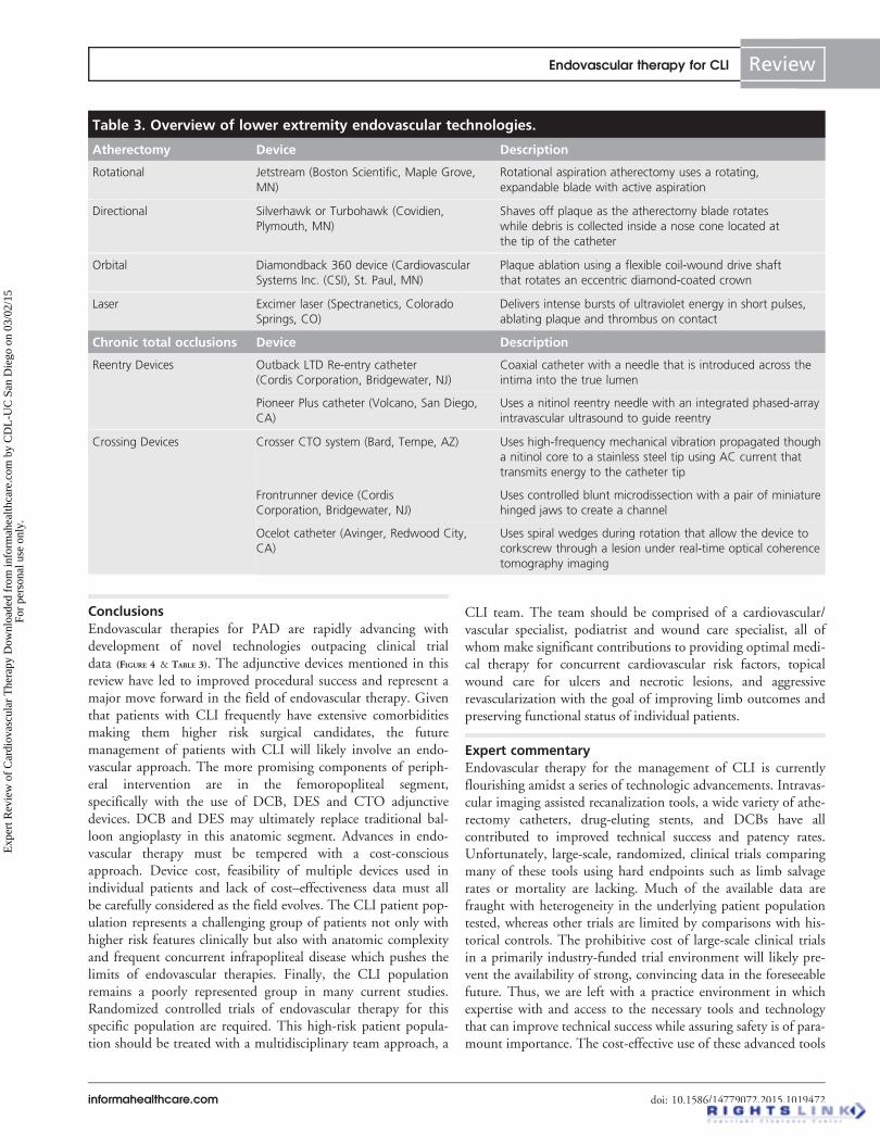
Table 3. Overview of lower extremity endovascular technologies.
Atherectomy Device Description
Rotational Jetstream (Boston Scientific, Maple Grove,
MN)
Rotational aspiration atherectomy uses a rotating,
expandable blade with active aspiration
Directional Silverhawk or Turbohawk (Covidien,
Plymouth, MN)
Shaves off plaque as the atherectomy blade rotates
while debris is collected inside a nose cone located at
the tip of the catheter
Orbital Diamondback 360 device (Cardiovascular
Systems Inc. (CSI), St. Paul, MN)
Plaque ablation using a flexible coil-wound drive shaft
that rotates an eccentric diamond-coated crown
Laser Excimer laser (Spectranetics, Colorado
Springs, CO)
Delivers intense bursts of ultraviolet energy in short pulses,
ablating plaque and thrombus on contact
Chronic total occlusions Device Description
Reentry Devices Outback LTD Re-entry catheter
(Cordis Corporation, Bridgewater, NJ)
Coaxial catheter with a needle that is introduced across the
intima into the true lumen
Pioneer Plus catheter (Volcano, San Diego,
CA)
Uses a nitinol reentry needle with an integrated phased-array
intravascular ultrasound to guide reentry
Crossing Devices Crosser CTO system (Bard, Tempe, AZ) Uses high-frequency mechanical vibration propagated though
a nitinol core to a stainless steel tip using AC current that
transmits energy to the catheter tip
Frontrunner device (Cordis
Corporation, Bridgewater, NJ)
Uses controlled blunt microdissection with a pair of miniature
hinged jaws to create a channel
Ocelot catheter (Avinger, Redwood City,
CA)
Uses spiral wedges during rotation that allow the device to
corkscrew through a lesion under real-time optical coherence
tomography imaging
Endovascular therapy for CLI Review
informahealthcare.com doi: 10.1586/14779072.2015.1019472
Exp
ert R
evie
w o
f C
ardi
ovas
cula
r T
hera
py D
ownl
oade
d fr
om in
form
ahea
lthca
re.c
om b
y C
DL
-UC
San
Die
go o
n 03
/02/
15Fo
r pe
rson
al u
se o
nly.

in the current cost-conscious medical economy is another limita-tion but should be weighed against the cost savings of avoiding amajor limb amputation, the final common goal in managing thepatient with CLI.
Five-year viewThe management of patients with CLI during the next 5 yearswill continue to evolve into a predominantly percutaneousapproach. The patient population with CLI commonly carriesmultiple comorbid conditions, including advanced age, chronickidney disease, uncontrolled diabetes mellitus and advancedcoronary artery disease. These comorbidities significantlyincrease surgical risk. Thus, without definitive data supportingsurgical superiority along with concomitant advances andimprovements in endovascular tools and techniques, the para-digm will shift further to an endovascular-first approach. Theresults of the aforementioned BEST-CLI trial may further elu-cidate the optimal management in this high-risk patient
population. Major limb amputation rates have experienced asignificant decline during the past decade and this trend willcontinue with early identification of CLI and advances inwound care management and catheter-based technology alongwith dissemination of the skills and techniques required to treatthese patients.
Financial & competing interests disclosure
Dr. Mahmud reports receiving research grant support from Corindus,
serves as a consultant for Abbott Vascular and serves on the speakers
bureau for Medtronic. Dr. Patel serves on the speakers bureau for Astra
Zeneca. The remaining authors have no relevant affiliations or financial
involvement with any organization or entity with a financial interest in
or financial conflict with the subject matter or materials discussed in the
manuscript. This includes employment, consultancies, honoraria, stock
ownership or options, expert testimony, grants or patents received or pend-
ing, or royalties.
No writing assistance was utilized in the production of this manuscript.
Key issues
. Critical limb ischemia represents an advanced presentation of peripheral arterial disease and manifests as lower extremity ischemic rest
pain or ischemic skin lesions leading to ulceration or gangrene.
. 1-year mortality rates reach 25% for patients with CLI and 1-year major amputation rates are also 25%.
. Advances in percutaneous interventional revascularization techniques and technology have transitioned the treatment paradigm from a
predominantly surgical approach to a percutaneous approach.
. There has been an overall decline in amputation rates in the past decade, likely due to a combination of earlier detection, prompt
referral, improved medical and wound care management, and advances in endovascular interventions.
. Preventing major amputation remains the principal treatment goal in any CLI revascularization procedure.
. Current clinical trials and registry data encompassing the various revascularization techniques and multitude of adjunctive tools and
technology are limited by low numbers of patients with CLI in comparison with claudicants.
. A comprehensive team of providers including the primary physician, podiatrist, wound care specialist and vascular specialist best
provides the optimal management of the patient with CLI.
References
Papers of special note have been highlighted as:. of interest.. of considerable interest
1. Hirsch AT, Haskal ZJ, Hertzer NR, et al.
ACC/AHA 2005 Practice guidelines for the
management of patients with peripheral arterial
disease (lower extremity, renal, mesenteric, and
abdominal aortic) a collaborative report from
the American association for vascular surgery/
society for vascular surgery,* society for
cardiovascular angiography and interventions,
society for vascular medicine and biology,
society of interventional radiology, and the acc/
aha task force on practice guidelines (writing
committee to develop guidelines for the
management of patients with peripheral arterial
disease): endorsed by the american association
of cardiovascular and pulmonary rehabilitation;
national heart, lung, and blood institute; society
for vascular nursing; transatlantic inter-society
consensus; and vascular disease foundation.
Circulation 2006;113(11):e463-654
.. This reference represents the US and
European guidelines that form the basis
for lower extremity revascularization.
2. Norgren L, Hiatt WR, Dormandy JA, et al.
Inter-society consensus for the management
of peripheral arterial disease (TASC II). Eur
J Vasc Endovasc Surg 2007;33(1):S1-S75
.. This reference represents the US and
European guidelines that form the basis
for lower extremity revascularization.
3. Barani J, Nilsson J-A, Mattiasson I, et al.
Inflammatory mediators are associated with
1-year mortality in critical limb ischemia. J
Vasc Surg 2005;42(1):75-80
4. Rutherford RB, Baker JD, Ernst C, et al.
Recommended standards for reports dealing
with lower extremity ischemia: revised
version. J Vasc Surg 1997;26(3):517-38
5. Norgren L, Hiatt WR, Dormandy JA, et al.
Inter-society consensus for the management
of peripheral arterial disease (TASC II). J
Vasc Surg 2007;45(Suppl S):S5-67
6. Goodney PP, Beck AW, Nagle J, et al.
National trends in lower extremity bypass
surgery, endovascular interventions, and major
amputations. J Vasc Surg 2009;50(1):54-60
7. Iida O, Nakamura M, Yamauchi Y, et al.
Endovascular treatment for infrainguinal
vessels in patients with critical limb
ischemia: olive registry, a prospective,
multicenter study in japan with 12-month
follow-up. Circ Cardiovasc Interv 2013;
6(1):68-76
8. Bradbury AW, Ruckley CV, Fowkes FGR,
et al. Bypass versus angioplasty in severe
ischaemia of the leg (BASIL): multicentre,
Review Dominguez, Bahadorani, Reeves, Mahmud & Patel
doi: 10.1586/14779072.2015.1019472 Expert Rev. Cardiovasc. Ther.
Exp
ert R
evie
w o
f C
ardi
ovas
cula
r T
hera
py D
ownl
oade
d fr
om in
form
ahea
lthca
re.c
om b
y C
DL
-UC
San
Die
go o
n 03
/02/
15Fo
r pe
rson
al u
se o
nly.

randomised controlled trial. Lancet 2005;
366(9501):1925-34
. This references the largest trial to date
comparing surgical versus endovascular
revascularization for critical limb
ischemia.
9. Bradbury AW, Adam DJ, Bell J, et al.
Bypass versus Angioplasty in Severe
Ischaemia of the Leg (BASIL) trial:
an intention-to-treat analysis of
amputation-free and overall survival in
patients randomized to a bypass surgery-first
or a balloon angioplasty-first
revascularization strategy. J Vasc Surg 2010;
51(5 Suppl):5S-17S
10. Rooke TW, Hirsch AT, Misra S, et al.
2011 ACCF/AHA focused update of the
guideline for the management of patients
with peripheral artery disease (updating the
2005 guideline): a report of the American
College of Cardiology Foundation/American
Heart Association task force on practice
guidelines: developed in collaboration with
the society for cardiovascular angiography
and interventions, society of interventional
radiology, society for vascular medicine, and
society for vascular surgery. J Vasc Surg
2011;54(5):e32-58
11. Jones WS, Dolor RJ, Hasselblad V, et al.
Comparative effectiveness of endovascular
and surgical revascularization for patients
with peripheral artery disease and critical
limb ischemia. Am Heart J 2014;167(4):
489-98.e7
12. Allie DE, Hebert CJ, Ingraldi A, et al.
24-Carat gold, 14-carat gold, or platinum
standards in the treatment of critical limb
ischemia: bypass surgery or endovascular
intervention? J Endovasc Ther 2009;
16(Suppl I):I-134
13. Best endovascular vs. Best surgical therapy
in patients with critical limb ischemia
(BEST-CLI). Available from: https://
clinicaltrials.gov/ct2/show/NCT02060630
14. Kudo T, Chandra FA, Ahn SS. The
effectiveness of percutaneous transluminal
angioplasty for the treatment of critical limb
ischemia: a 10-year experience. J Vasc Surg
2005;41(3):423-35.discussion 435
15. Romiti M, Albers M, Brochado-Neto FC,
et al. Meta-analysis of infrapopliteal
angioplasty for chronic critical limb
ischemia. J Vasc Surg 2008;47(5):975-81.e1
16. Nehler MR, Coll JR, Hiatt WR, et al.
Functional outcome in a contemporary
series of major lower extremity amputations.
J Vasc Surg 2003;38(1):7-14
17. Jones WS, Patel MR, Dai D, et al.
Temporal trends and geographic variation of
lower-extremity amputation in patients with
peripheral artery disease: results from U.S.
Medicare 2000-2008. J Am Coll Cardiol
2012;60(21):2230-6
18. Giles KA, Pomposelli FB, Hamdan AD,
et al. Infrapopliteal angioplasty for critical
limb ischemia: relation of transatlantic
intersociety consensus class to outcome in
176 limbs. J Vasc Surg 2008;48(1):128-36
19. Attinger CE, Evans KK, Bulan E, et al.
Angiosomes of the foot and ankle and
clinical implications for limb salvage:
reconstruction, incisions, and
revascularization. Plast Reconstr Surg 2006;
117(7 Suppl):261S-93S
20. Alexandrescu V-A, Hubermont G, Philips Y,
et al. Selective primary angioplasty following
an angiosome model of reperfusion in the
treatment of wagner 1–4 diabetic foot
lesions: practice in a multidisciplinary
diabetic limb service. J Endovasc Ther 2008;
15(5):580-93
. This reference justifies the use of the
angiosome model for endovascular
intervention as it pertains to critical limb
ischemia.
21. Neville RF, Attinger CE, Bulan EJ, et al.
Revascularization of a specific angiosome for
limb salvage: does the target artery matter?
Ann Vasc Surg 2009;23(3):367-73
22. Kabra A, Suresh KR, Vivekanand V, et al.
Outcomes of angiosome and non-angiosome
targeted revascularization in critical lower
limb ischemia. J Vasc Surg 2013;57(1):44-9
23. Alexandrescu V, Vincent G, Azdad K, et al.
A reliable approach to diabetic
neuroischemic foot wounds: below-the-knee
angiosome-oriented angioplasty. J Endovasc
Ther 2011;18(3):376-87
24. Iida O, Nanto S, Uematsu M, et al.
Importance of the angiosome concept for
endovascular therapy in patients with critical
limb ischemia. Catheter Cardiovasc Interv
2010;75(6):830-6
25. Azuma N, Uchida H, Kokubo T, et al.
Factors influencing wound healing of critical
ischaemic foot after bypass surgery: is the
angiosome important in selecting bypass
target artery? Eur J Vasc Endovasc Surg
2012;43(3):322-8
26. Cejna M. Cutting balloon: review on
principles and background of use in
peripheral arteries. Cardiovasc Intervent
Radiol 2005;28(4):400-8
27. Davies MG, Anaya-Ayala JE. Endovascular
techniques in limb salvage: cutting, cryo,
brachy, and drug-eluting balloons.
Methodist Debakey Cardiovasc J 2013;9(2):
69
28. Hara H, Nakamura M, Asahara T, et al.
Intravascular ultrasonic comparisons of
mechanisms of vasodilatation of cutting
balloon angioplasty versus conventional
balloon angioplasty. Am J Cardiol 2002;
89(11):1253-6
29. Ansel GM, Sample NS, Botti CF, et al.
Cutting balloon angioplasty of the popliteal
and infrapopliteal vessels for symptomatic
limb ischemia. Catheter Cardiovasc Interv
2004;61(1):1-4
30. Rabbi JF, Kiran RP, Gersten G, et al. Early
results with infrainguinal cutting balloon
angioplasty limits distal dissection. Ann
Vasc Surg 2004;18(6):640-3
31. Canaud L, Alric P, Berthet J-P, et al.
Infrainguinal cutting balloon angioplasty in
de novo arterial lesions. J Vasc Surg 2008;
48(5):1182-8
32. Amighi J, Schillinger M, Dick P, et al. De
novo superficial femoropopliteal artery
lesions: peripheral cutting balloon
angioplasty and restenosis rates –
randomized controlled trial 1. Radiology
2008;247(1):267-72
33. Muradin GSR, Bosch JL, Stijnen T,
Hunink MGM. Balloon dilation and stent
implantation for treatment of
femoropopliteal arterial disease:
meta-analysis1. Radiology 2001;221(1):
137-45
34. Becquemin J-P, Favre J-P, Marzelle J, et al.
Systematic versus selective stent placement
after superficial femoral artery balloon
angioplasty: a multicenter prospective
randomized study. J Vasc Surg 2003;37(3):
487-94
35. Schillinger M, Sabeti S, Loewe C, et al.
Balloon angioplasty versus implantation of
nitinol stents in the superficial femoral
artery. N Engl J Med 2006;354(18):
1879-88
36. Schillinger M, Sabeti S, Dick P, et al.
Sustained benefit at 2 years of primary
femoropopliteal stenting compared with
balloon angioplasty with optional stenting.
Circulation 2007;115(21):2745-9
37. Laird JR, Katzen BT, Scheinert D, et al.
Nitinol stent implantation versus balloon
angioplasty for lesions in the superficial
femoral artery and proximal popliteal artery:
twelve-month results from the resilient
randomized trial. Circ Cardiovasc Interv
2010;3(3):267-76
. This reference established the use of
nitinol based self-expanding stents for
superficial femoral artery intervention
over balloon angioplasty alone.
Endovascular therapy for CLI Review
informahealthcare.com doi: 10.1586/14779072.2015.1019472
Exp
ert R
evie
w o
f C
ardi
ovas
cula
r T
hera
py D
ownl
oade
d fr
om in
form
ahea
lthca
re.c
om b
y C
DL
-UC
San
Die
go o
n 03
/02/
15Fo
r pe
rson
al u
se o
nly.
38. Bosiers M, Torsello G, Gissler H-M, et al.
Nitinol stent implantation in long
superficial femoral artery lesions: 12-month
results of the DURABILITY I study. J
Endovasc Ther 2009;16(3):261-9
39. Scheinert D, Scheinert S, Sax J, et al.
Prevalence and clinical impact of stent
fractures after femoropopliteal stenting. J
Am Coll Cardiol 2005;45(2):312-15
40. Schlager O, Dick P, Sabeti S, et al.
Long-segment SFA stenting–the dark sides:
in-stent restenosis, clinical deterioration, and
stent fractures. J Endovasc Ther 2005;12(6):
676-84
41. Duda SH, Bosiers M, Lammer J, et al.
Sirolimus-eluting versus bare nitinol stent
for obstructive superficial femoral artery
disease: the SIROCCO II trial. J Vasc
Interv Radiol 2005;16(3):331-8
42. Duda SH, Bosiers M, Lammer J, et al.
Drug-eluting and bare nitinol stents for the
treatment of atherosclerotic lesions in the
superficial femoral artery: long-term results
from the SIROCCO trial. J Endovasc Ther
2006;13(6):701-10
43. Dake MD, Ansel GM, Jaff MR, et al.
Paclitaxel-eluting stents show superiority to
balloon angioplasty and bare metal stents in
femoropopliteal disease: twelve-month zilver
ptx randomized study results. Circ
Cardiovasc Interv 2011;4(5):495-504
44. Dake MD, Ansel GM, Jaff MR, et al.
Sustained safety and effectiveness of
paclitaxel-eluting stents for femoropopliteal
lesions. J Am Coll Cardiol 2013;61(24):
2417-27
45. Dake MD, Scheinert D, Tepe G, et al.
Nitinol stents with polymer-free paclitaxel
coating for lesions in the superficial femoral
and popliteal arteries above the knee:
twelve-month safety and effectiveness results
from the zilver ptx single-arm clinical study.
J Endovasc Ther 2011;18(5):613-23
46. Geraghty PJ, Mewissen MW, Jaff MR,
Ansel GM; VIBRANT Investigators.
Three-year results of the VIBRANT trial of
VIABAHN endoprosthesis versus bare
nitinol stent implantation for complex
superficial femoral artery occlusive disease. J
Vasc Surg 2013;58(2):386-95.e4
47. Ahmadi R, Schillinger M, Maca T, Minar E.
Femoropopliteal Arteries: immediate and
Long-term Results with a Dacron-covered
Stent-Graft1. Radiology 2002;223(2):345-50
48. Lammer J, Zeller T, Hausegger KA, et al.
Heparin-bonded covered stents versus
bare-metal stents for complex
femoropopliteal artery lesions: the
randomized VIASTAR trial (Viabahn
endoprosthesis with PROPATEN bioactive
surface [VIA] versus bare nitinol stent in the
treatment of long lesions in superficial
femoral artery occlusive disease). J Am Coll
Cardiol 2013;62(15):1320-7
49. Saxon RR, Chervu A, Jones PA, et al.
Heparin-bonded, expanded
polytetrafluoroethylene-lined stent graft in
the treatment of femoropopliteal artery
disease: 1-year results of the VIPER
(viabahn endoprosthesis with heparin
bioactive surface in the treatment of
superficial femoral artery obstructive disease)
trial. J Vasc Interv Radiol 2013;24(2):
165-73.quiz 174
50. Donas KP, Torsello G, Schwindt A, et al.
Below knee bare nitinol stent placement in
high-risk patients with critical limb ischemia
is still durable after 24 months of follow-up.
J Vasc Surg 2010;52(2):356-61
51. Feiring AJ, Wesolowski AA, Lade S.
Primary stent-supported angioplasty for
treatment of below-knee critical limb
ischemia and severe claudication. J Am Coll
Cardiol 2004;44(12):2307-14
52. Randon C, Jacobs B, Ryck F, Vermassen F.
Angioplasty or primary stenting for
infrapopliteal lesions: results of a prospective
randomized trial. Cardiovasc Intervent
Radiol 2010;33(2):260-9
53. Siablis D, Karnabatidis D, Katsanos K,
et al. Infrapopliteal application of
sirolimus-eluting versus bare metal stents for
critical limb ischemia: analysis of long-term
angiographic and clinical outcome. J Vasc
Interv Radiol 2009;20(9):1141-50
54. Feiring AJ, Krahn M, Nelson L, et al.
Preventing leg amputations in critical limb
ischemia with below-the-knee drug-eluting
stents. J Am Coll Cardiol 2010;55(15):
1580-9
55. Biondi-Zoccai GGL, Sangiorgi G,
Lotrionte M, et al. Infragenicular stent
implantation for below-the-knee
atherosclerotic disease: clinical evidence from
an international collaborative meta-analysis
on 640 patients. J Endovasc Ther 2009;
16(3):251-60
56. Scheinert D, Katsanos K, Zeller T, et al.
A prospective randomized multicenter
comparison of balloon angioplasty and
infrapopliteal stenting with the
sirolimus-eluting stent in patients with
ischemic peripheral arterial disease. J Am
Coll Cardiol 2012;60(22):2290-5
57. Rastan A, Tepe G, Krankenberg H, et al.
Sirolimus-eluting stents vs. bare-metal stents
for treatment of focal lesions in
infrapopliteal arteries: a double-blind,
multi-centre, randomized clinical trial. Eur
Heart J 2011;32(18):2274-81
58. Rastan A, Brechtel K, Krankenberg H, et al.
Sirolimus-eluting stents for treatment of
infrapopliteal arteries reduce clinical event
rate compared to bare-metal stents:
long-term results from a randomized trial. J
Am Coll Cardiol 2012;60(7):587-91
59. Bosiers M, Scheinert D, Peeters P, et al.
Randomized comparison of
everolimus-eluting versus bare-metal stents
in patients with critical limb ischemia and
infrapopliteal arterial occlusive disease. J
Vasc Surg 2012;55(2):390-8
60. Graziani L, Piaggesi A. Indications and
clinical outcomes for below knee
endovascular therapy: Review article.
Catheter Cardiovasc Interv 2010;75(3):
433-43
61. Schillinger M, Minar E. Percutaneous
treatment of peripheral artery disease: novel
techniques. Circulation 2012;126(20):
2433-40
62. Byrne RA, Joner M, Alfonso F, Kastrati A.
Drug-coated balloon therapy in coronary
and peripheral artery disease. Nat Rev
Cardiol 2014;11(1):13-23
63. Werk M, Langner S, Reinkensmeier B,
et al. Inhibition of restenosis in
femoropopliteal arteries paclitaxel-coated
versus uncoated balloon: femoral paclitaxel
randomized pilot trial. Circulation 2008;
118(13):1358-65
64. Tepe G, Zeller T, Albrecht T, et al. Local
delivery of paclitaxel to inhibit restenosis
during angioplasty of the leg. N Engl J Med
2008;358(7):689-99
65. Werk M, Albrecht T, Meyer D-R, et al.
Paclitaxel-coated balloons reduce restenosis
after femoro-popliteal angioplasty evidence
from the randomized pacifier trial. Circ
Cardiovasc Interv 2012;5(6):831-40
66. Scheinert D, Duda S, Zeller T, et al. The
LEVANT I (Lutonix paclitaxel-coated
balloon for the prevention of
femoropopliteal restenosis) trial for
femoropopliteal revascularization:
first-in-human randomized trial of low-dose
drug-coated balloon versus uncoated balloon
angioplasty. JACC Cardiovasc Interv 2014;
7(1):10-19
67. Tepe G, Laird J, Schneider P, et al.
Drug-coated balloon versus standard
percutaneous transluminal angioplasty for
the treatment of superficial femoral and/or
popliteal peripheral artery disease: 12-month
results from the in.pact sfa randomized trial.
Circulation 2015;131(5):495-502
Review Dominguez, Bahadorani, Reeves, Mahmud & Patel
doi: 10.1586/14779072.2015.1019472 Expert Rev. Cardiovasc. Ther.
Exp
ert R
evie
w o
f C
ardi
ovas
cula
r T
hera
py D
ownl
oade
d fr
om in
form
ahea
lthca
re.c
om b
y C
DL
-UC
San
Die
go o
n 03
/02/
15Fo
r pe
rson
al u
se o
nly.

68. Moxy drug coated balloon vs. Standard
balloon angioplasty for the treatment of
femoropopliteal arteries (LEVANT 2).
Available from: https://clinicaltrials.gov/ct2/
show/NCT01412541
69. Schmidt A, Piorkowski M, Werner M, et al.
First experience with drug-eluting balloons
in infrapopliteal arteries. J Am Coll Cardiol
2011;58(11):1105-9
70. Liistro F, Porto I, Angioli P, et al.
Drug-eluting balloon in peripheral
intervention for below the knee angioplasty
evaluation (debate-btk): a randomized trial
in diabetic patients with critical limb
ischemia. Circulation 2013;128(6):615-21
71. Zeller T, Baumgartner I, Scheinert D, et al.
Drug-eluting balloon versus standard
balloon angioplasty for infrapopliteal arterial
revascularization in critical limb ischemia:
12-month results from the IN.PACT DEEP
randomized trial. J Am Coll Cardiol 2014;
64(15):1568-76
72. Vom Dahl J, Dietz U, Haager PK, et al.
Rotational atherectomy does not reduce
recurrent in-stent restenosis: results of the
angioplasty versus rotational atherectomy for
treatment of diffuse in-stent restenosis trial
(ARTIST). Circulation 2002;105(5):583-8
73. Abdel-Wahab M, Richardt G,
Joachim Buttner H, et al. High-speed
rotational atherectomy before
paclitaxel-eluting stent implantation in
complex calcified coronary lesions: the
randomized ROTAXUS (rotational
atherectomy prior to taxus stent treatment
for complex native coronary artery disease)
trial. JACC Cardiovasc Interv 2013;6(1):
10-19
74. Garcia LA, Lyden SP. Atherectomy for
infrainguinal peripheral artery disease. J
Endovasc Ther 2009;16(Suppl II):II105-15
75. Laird JR. Limitations of percutaneous
transluminal angioplasty and stenting for
the treatment of disease of the superficial
femoral and popliteal arteries. J Endovasc
Ther 2006;13(Suppl 2):II30-40
76. Zeller T, Frank U, Burgelin K, et al. Initial
clinical experience with percutaneous
atherectomy in the infragenicular arteries. J
Endovasc Ther 2003;10(5):987-93
77. Zeller T, Rastan A, Sixt S, et al. Long-term
results after directional atherectomy of
femoro-popliteal lesions. J Am Coll Cardiol
2006;48(8):1573-8
78. Yancey AE, Minion DJ, Rodriguez C, et al.
Peripheral atherectomy in transatlantic
intersociety consensus type c
femoropopliteal lesions for limb salvage. J
Vasc Surg 2006;44(3):503-9
79. Keeling WB, Shames ML, Stone PA, et al.
Plaque excision with the Silverhawk
catheter: early results in patients with
claudication or critical limb ischemia. J Vasc
Surg 2007;45(1):25-31
80. McKinsey JF, Zeller T, Rocha-Singh KJ,
DEFINITIVE LE Investigators. Lower
extremity revascularization using directional
atherectomy: 12-month prospective results
of the DEFINITIVE LE Study. JACC
Cardiovasc Interv 2014;7(8):923-33
81. Ramaiah V, Gammon R, Kiesz S, et al.
Midterm outcomes from the TALON
Registry: treating peripherals with
SilverHawk: outcomes collection. J
Endovasc Ther 2006;13(5):592-602
82. McKinsey JF, Goldstein L, Khan HU, et al.
Novel treatment of patients with lower
extremity ischemia: use of percutaneous
atherectomy in 579 Lesions: Transactions of
the. Meeting of the American Surgical
Association 2008;126:160-9
83. Kandzari DE, Kiesz RS, Allie D, et al.
Procedural and clinical outcomes with
catheter-based plaque excision in critical
limb ischemia. J Endovasc Ther 2006;13(1):
12-22
84. Suri R, Wholey MH, Postoak D, et al.
Distal embolic protection during
femoropopliteal atherectomy. Catheter
Cardiovasc Interv 2006;67(3):417-22
85. Lam RC, Shah S, Faries PL, et al. Incidence
and clinical significance of distal
embolization during percutaneous
interventions involving the superficial
femoral artery. J Vasc Surg 2007;46(6):
1155-9
86. Shammas NW, Dippel EJ, Coiner D, et al.
Preventing lower extremity distal
embolization using embolic filter protection:
results of the PROTECT registry. J
Endovasc Ther 2008;15(3):270-6
87. Roberts D, Niazi K, Miller W, et al.
Effective endovascular treatment of calcified
femoropopliteal disease with directional
atherectomy and distal embolic protection:
final results of the DEFINITIVE Ca++ trial.
Catheter Cardiovasc Interv 2014;84(2):
236-44
88. Todd KE, Ahanchi SS, Maurer CA, et al.
Atherectomy offers no benefits over balloon
angioplasty in tibial interventions for critical
limb ischemia. J Vasc Surg 2013;58(4):
941-8
89. Heuser RR. Treatment of lower extremity
vascular disease: the Diamondback
360 degrees orbital atherectomy system.
Expert Rev Med Devices 2008;5(3):279-86
90. Chambers JW, Diage T. Evaluation of the
diamondback 360 coronary orbital
atherectomy system for treating de novo,
severely calcified lesions. Expert Rev Med
Devices 2014;11(5):457-66
91. Safian RD, Niazi K, Runyon JP, et al.
Orbital atherectomy for infrapopliteal
disease: Device concept and outcome data
for the oasis trial. Catheter Cardiovasc
Interv 2009;73(3):406-12
92. Shammas NW, Lam R, Mustapha J, et al.
Comparison of orbital atherectomy plus
balloon angioplasty vs. balloon angioplasty
alone in patients with critical limb ischemia:
results of the CALCIUM 360 randomized
pilot trial. J Endovasc Ther 2012;19(4):
480-8
93. Dattilo R, Himmelstein SI, Cuff RF. The
COMPLIANCE 360� trial: a randomized,
prospective, multicenter, pilot study
comparing acute and long-term results of
orbital atherectomy to balloon angioplasty
for calcified femoropopliteal disease. J
Invasive Cardiol 2014;26(8):355-60
94. Zeller T, Krankenberg H, Rastan A, et al.
Percutaneous rotational and aspiration
atherectomy in infrainguinal peripheral
arterial occlusive disease: a multicenter pilot
study. J Endovasc Ther 2007;14(3):357-64
95. Zeller T, Krankenberg H, Steinkamp H,
et al. One-year outcome of percutaneous
rotational atherectomy with aspiration in
infrainguinal peripheral arterial occlusive
disease: the multicenter pathway PVD trial.
J Endovasc Ther 2009;16(6):653-62
96. Sixt S, Rastan A, Scheinert D, et al. The
1-year clinical impact of rotational
aspiration atherectomy of infrainguinal
lesions. Angiology 2011;62(8):645-56
97. Hassan AHM, Ako J, Waseda K, et al.
Mechanism of lumen gain with a novel
rotational aspiration atherectomy system for
peripheral arterial disease: examination by
intravascular ultrasound. Cardiovasc Revasc
Med 2010;11(3):155-8
98. Laird JR, Zeller T, Gray BH, et al. Limb
salvage following laser-assisted angioplasty
for critical limb ischemia: results of the
LACI multicenter trial. J Endovasc Ther
2006;13(1):1-11
99. Boccalandro F, Muench A, Sdringola S,
Rosales OR. Wireless laser-assisted
angioplasty of the superficial femoral artery
in patients with critical limb ischemia who
have failed conventional percutaneous
revascularization. Catheter Cardiovasc Interv
2004;63(1):7-12
100. Shrikhande GV, Khan SZ, Hussain HG,
et al. Lesion types and device characteristics
Endovascular therapy for CLI Review
informahealthcare.com doi: 10.1586/14779072.2015.1019472
Exp
ert R
evie
w o
f C
ardi
ovas
cula
r T
hera
py D
ownl
oade
d fr
om in
form
ahea
lthca
re.c
om b
y C
DL
-UC
San
Die
go o
n 03
/02/
15Fo
r pe
rson
al u
se o
nly.

that predict distal embolization during
percutaneous lower extremity interventions.
J Vasc Surg 2011;53(2):347-52
101. Reeves R, Imsais JK, Prasad A. Successful
management of lower extremity distal
embolization following percutaneous
atherectomy with the JetStream G3 device. J
Invasive Cardiol 2012;24(6):E124-8
102. Ambler GK, Radwan R, Hayes PD, et al.
Atherectomy for peripheral arterial disease.
Cochrane Database Syst Rev 2014;3:
CD006680
103. Waksman R, Laird JR, Jurkovitz CT, et al.
Intravascular radiation therapy after balloon
angioplasty of narrowed femoropopliteal
arteries to prevent restenosis: results of the
PARIS feasibility clinical trial. J Vasc Interv
Radiol 2001;12(8):915-21
104. Minar E, Pokrajac B, Maca T, et al.
Endovascular brachytherapy for prophylaxis
of restenosis after femoropopliteal
angioplasty : results of a prospective
randomized study. Circulation 2000;
102(22):2694-9
105. Wyttenbach R. Effects of Percutaneous
Transluminal Angioplasty and Endovascular
Brachytherapy on Vascular Remodeling of
Human Femoropopliteal Artery by
Noninvasive Magnetic Resonance Imaging.
Circulation 2004;110(9):1156-61
106. Andras A, Hansrani M, Stewart M, et al.
Intravascular brachytherapy for peripheral
vascular disease. Cochrane Database Syst
Rev 2014;1:CD003504
107. Laird J, Jaff MR, Biamino G, et al.
Cryoplasty for the treatment of
femoropopliteal arterial disease: results of a
prospective, multicenter registry. J Vasc
Interv Radiol 2005;16(8):1067-73
108. Laird JR, Biamino G, McNamara T, et al.
Cryoplasty for the treatment of
femoropopliteal arterial disease: extended
follow-up results. J Endovasc Ther 2006;
13(Suppl 2):II52-9
109. Das TS, McNamara T, Gray B, et al.
Primary cryoplasty therapy provides durable
support for limb salvage in critical limb
ischemia patients with infrapopliteal lesions:
12-month follow-up results from the BTK
Chill Trial. J Endovas Ther 2009;
16(Suppl II):II19-30
110. Bosiers M, Deloose K, Vermassen F, et al.
The use of the cryoplasty technique in the
treatment of infrapopliteal lesions for critical
limb ischemia patients in a routine hospital
setting: one-year outcome of the Cryoplasty
CLIMB Registry. J Cardiovasc Surg
(Torino) 2010;51(2):193-202
111. Das T, McNamara T, Gray B, et al.
Cryoplasty therapy for limb salvage in
patients with critical limb ischemia. J
Endovasc Ther 2007;14(6):753-62
112. Jahnke T, Mueller-Huelsbeck S,
Charalambous N, et al. Prospective,
randomized single-center trial to compare
cryoplasty versus conventional angioplasty in
the popliteal artery: midterm results of the
COLD study. J Vasc Interv Radiol 2010;
21(2):186-94
113. Spiliopoulos S, Katsanos K, Karnabatidis D,
et al. Cryoplasty versus conventional balloon
angioplasty of the femoropopliteal artery in
diabetic patients: long-term results from a
prospective randomized single-center
controlled trial. Cardiovasc Intervent Radiol
2010;33(5):929-38
114. Fossaceca R, Guzzardi G, Di Terlizzi M,
et al. Comparison of cryoplasty and
conventional angioplasty for treating
stenotic-occlusive lesions of the
femoropopliteal arteries in diabetic patients:
immediate, mid-term and long-term results.
Radiol Med (Torino) 2012;117(7):1176-89
115. Banerjee S, Das TS, Abu-Fadel MS, et al.
Pilot trial of cryoplasty or conventional
balloon post-dilation of nitinol stents for
revascularization of peripheral arterial
segments. J Am Coll Cardiol 2012;60(15):
1352-9
116. Rogers JH, Laird JR. Overview of new
technologies for lower extremity
revascularization. Circulation 2007;116(18):
2072-85
117. Tatli E, Buturak A, Kayapınar O, et al.
Subintimal angioplasty and stenting for
chronic total femoropopliteal artery
occlusions: Early and mid-term outcomes.
Cardiol J Internet 2013;Available from:
http://czasopisma.viamedica.pl/cj/article/
view/37463
118. Scott EC, Biuckians A, Light RE, et al.
Subintimal angioplasty for the treatment of
claudication and critical limb ischemia:
3-year results. J Vasc Surg 2007;46(5):
959-64
119. Hong S-J, Ko Y-G, Kim J-S, et al. Midterm
outcomes of subintimal angioplasty
supported by primary proximal stenting for
chronic total occlusion of the superficial
femoral artery. J Endovasc Ther 2013;20(6):
782-91
120. Bazan HA, Le L, Donovan M, et al.
Retrograde pedal access for patients with
critical limb ischemia. J Vasc Surg 2014;
60(2):375-82
121. Ruzsa Z, Nemes B, Bansaghi Z, et al.
Transpedal access after failed anterograde
recanalization of complex below-the-knee
and femoropoliteal occlusions in critical
limb ischemia. Catheter Cardiovasc Interv
2014;83(6):997-1007
122. Jacobs DL, Motaganahalli RL, Cox DE,
et al. True lumen re-entry devices facilitate
subintimal angioplasty and stenting of total
chronic occlusions: initial report. J Vasc
Surg 2006;43(6):1291-6
123. Gandini R, Fabiano S, Spano S, et al.
Randomized control study of the outback
LTD reentry catheter versus manual reentry
for the treatment of chronic total occlusions
in the superficial femoral artery. Catheter
Cardiovasc Interv 2013;82(3):485-92
124. Aslam MS, Allaqaband S, Haddadian B,
et al. Subintimal angioplasty with a true
reentry device for treatment of chronic total
occlusion of the arteries of the lower
extremity. Catheter Cardiovasc Interv 2013;
82(5):701-6
125. Saket RR, Razavi MK, Padidar A, et al.
Novel intravascular ultrasound-guided
method to create transintimal arterial
communications: initial experience in
peripheral occlusive disease and aortic
dissection. J Endovasc Ther 2004;11(3):
274-80
126. Melzi G, Cosgrave J, Biondi-Zoccai GL,
et al. A novel approach to chronic total
occlusions: the crosser system. Catheter
Cardiovasc Interv 2006;68(1):29-35
127. Khalid MR, Khalid FR, Farooqui FA, et al.
A novel catheter in patients with peripheral
chronic total occlusions: a single center
experience. Catheter Cardiovasc Interv
2010;76(5):735-9
128. Beschorner U, Rastan A, Zeller T.
Recanalization of femoropopliteal occlusions
using the crosser system. J Endovasc Ther
2009;16(4):526-7.author reply 527–529
129. Shetty R, Vivek G, Thakkar A, et al. Safety
and efficacy of the frontrunner XP catheter
for recanalization of chronic total occlusion
of the femoropopliteal arteries. J Invasive
Cardiol 2013;25(7):344-7
130. Mossop PJ, Amukotuwa SA, Whitbourn RJ.
Controlled blunt microdissection for
percutaneous recanalization of lower limb
arterial chronic total occlusions: a single
center experience. Catheter Cardiovasc
Interv 2006;68(2):304-10
131. Selmon MR, Schwindt AG, Cawich IM,
et al. Final results of the chronic total
occlusion crossing with the ocelot system II
(CONNECT II) study. J Endovasc Ther
2013;20(6):770-81
Review Dominguez, Bahadorani, Reeves, Mahmud & Patel
doi: 10.1586/14779072.2015.1019472 Expert Rev. Cardiovasc. Ther.
Exp
ert R
evie
w o
f C
ardi
ovas
cula
r T
hera
py D
ownl
oade
d fr
om in
form
ahea
lthca
re.c
om b
y C
DL
-UC
San
Die
go o
n 03
/02/
15Fo
r pe
rson
al u
se o
nly.