Inhalation of carbon monoxide reduces skeletal muscle injury after hind limb ischemia-reperfusion...
18
Inhalation of Carbon Monoxide Reduces Skeletal Muscle Injury Following Hind Limb Ischemia Reperfusion Injury in Mice Rajendra Patel, MD 1 , Hassan Albadawi, MD 1 , Wolfgang Steudel, MD 2 , Faraz F. Hashmi 1 , Jeanwan Kang, MD 1 , Hyung-Jin Yoo, MA 1 , and Michael T. Watkins, MD 1,3 1 Division of Vascular and Endovascular Surgery-Department of Surgery, Massachusetts General Hospital, Harvard Medical School, Boston, MA 2 Department of Anesthesia and Critical Care, Massachusetts General Hospital, Harvard Medical School, Boston, MA Abstract Introduction—The purpose of this study was to determine if inhaled carbon monoxide (CO) can ameliorate skeletal muscle injury, modulate endogenous heme oxygenase-1 (HO) expression, improve indices of tissue integrity and inflammation following hind limb ischemia reperfusion(IR). Methods—C57BL6 mice inhaling CO (250ppm) or room air were subjected to 1.5 hrs of ischemia followed by limb reperfusion for either 3 or 6 hours (total treatment time of 4.5 or 7.5 hrs). After the initial period of reperfusion, all mice breathed only room air until 24 hours after the onset of ischemia. Mice were sacrificed at either the end of CO treatment or at 24 hours reperfusion. Skeletal muscle was subjected to histologic and biochemical analysis. Results—CO treatment for 7.5 hours protected skeletal muscle from histologic and structural evidence of skeletal muscle injury. Serum and tissue cytokines were significantly reduced (p<0.05) in mice treated with CO for 7.5 hours. Tubulin, Heme Oxygenase, and ATP levels were higher in CO treated mice. Conclusions—Inhaled CO protected muscle from structural injury and energy depletion following IR. Keywords Carbon Monoxide; Reperfusion Injury; Heme Oxygenase; Skeletal Muscle Introduction Acute limb ischemia is a complication of advanced peripheral vascular disease. Therapeutic interventions are geared toward restoration of blood flow to the affected extremity and can result in the development of ischemia reperfusion injury (I/R). The local manifestations of extremity I/R injury often results in limb loss or dysfunction. Systemic manifestations of I/R © 2011 Excerpta Medica, Inc. All rights reserved. 3 To Whom Correspondence is addressed: Michael T. Watkins, MD, FACS, FAHA Division of Vascular and Endovascular Surgery 15 Parkman Street, Suite 440 Boston, MA 02114 PH: 617-726-0908 FAX: 617-726-2560 [email protected]. Publisher's Disclaimer: This is a PDF file of an unedited manuscript that has been accepted for publication. As a service to our customers we are providing this early version of the manuscript. The manuscript will undergo copyediting, typesetting, and review of the resulting proof before it is published in its final citable form. Please note that during the production process errors may be discovered which could affect the content, and all legal disclaimers that apply to the journal pertain. NIH Public Access Author Manuscript Am J Surg. Author manuscript; available in PMC 2013 April 1. Published in final edited form as: Am J Surg. 2012 April ; 203(4): 488–495. doi:10.1016/j.amjsurg.2011.05.005. NIH-PA Author Manuscript NIH-PA Author Manuscript NIH-PA Author Manuscript
-
Upload
independent -
Category
Documents
-
view
1 -
download
0
Transcript of Inhalation of carbon monoxide reduces skeletal muscle injury after hind limb ischemia-reperfusion...

Inhalation of Carbon Monoxide Reduces Skeletal Muscle InjuryFollowing Hind Limb Ischemia Reperfusion Injury in Mice
Rajendra Patel, MD 1, Hassan Albadawi, MD 1, Wolfgang Steudel, MD 2, Faraz F. Hashmi 1,Jeanwan Kang, MD 1, Hyung-Jin Yoo, MA 1, and Michael T. Watkins, MD 1,3
1Division of Vascular and Endovascular Surgery-Department of Surgery, Massachusetts GeneralHospital, Harvard Medical School, Boston, MA
2Department of Anesthesia and Critical Care, Massachusetts General Hospital, Harvard MedicalSchool, Boston, MA
Abstract
Introduction— The purpose of this study was to determine if inhaled carbon monoxide (CO) canameliorate skeletal muscle injury, modulate endogenous heme oxygenase-1 (HO) expression,improve indices of tissue integrity and inflammation following hind limb ischemiareperfusion(IR).
Methods— C57BL6 mice inhaling CO (250ppm) or room air were subjected to 1.5 hrs ofischemia followed by limb reperfusion for either 3 or 6 hours (total treatment time of 4.5 or 7.5hrs). After the initial period of reperfusion, all mice breathed only room air until 24 hours after theonset of ischemia. Mice were sacrificed at either the end of CO treatment or at 24 hoursreperfusion. Skeletal muscle was subjected to histologic and biochemical analysis.
Results— CO treatment for 7.5 hours protected skeletal muscle from histologic and structuralevidence of skeletal muscle injury. Serum and tissue cytokines were significantly reduced(p<0.05) in mice treated with CO for 7.5 hours. Tubulin, Heme Oxygenase, and ATP levels werehigher in CO treated mice.
Conclusions— Inhaled CO protected muscle from structural injury and energy depletionfollowing IR.
Keywords
Carbon Monoxide; Reperfusion Injury; Heme Oxygenase; Skeletal Muscle
Introduction
Acute limb ischemia is a complication of advanced peripheral vascular disease. Therapeuticinterventions are geared toward restoration of blood flow to the affected extremity and canresult in the development of ischemia reperfusion injury (I/R). The local manifestations ofextremity I/R injury often results in limb loss or dysfunction. Systemic manifestations of I/R
© 2011 Excerpta Medica, Inc. All rights reserved.3To Whom Correspondence is addressed: Michael T. Watkins, MD, FACS, FAHA Division of Vascular and Endovascular Surgery 15Parkman Street, Suite 440 Boston, MA 02114 PH: 617-726-0908 FAX: 617-726-2560 [email protected].
Publisher's Disclaimer: This is a PDF file of an unedited manuscript that has been accepted for publication. As a service to ourcustomers we are providing this early version of the manuscript. The manuscript will undergo copyediting, typesetting, and review ofthe resulting proof before it is published in its final citable form. Please note that during the production process errors may bediscovered which could affect the content, and all legal disclaimers that apply to the journal pertain.
NIH Public AccessAuthor ManuscriptAm J Surg. Author manuscript; available in PMC 2013 April 1.
Published in final edited form as:Am J Surg. 2012 April ; 203(4): 488–495. doi:10.1016/j.amjsurg.2011.05.005.
NIH
-PA
Author M
anuscriptN
IH-P
A A
uthor Manuscript
NIH
-PA
Author M
anuscript

injury include cardiopulmonary dysfunction and shock associated with release of metabolicbyproducts and proinflammatory mediators from skeletal muscle. I/R injury is a complexphysiologic scenario which is initially triggered by stasis, depletion of energy substrates andacidosis in skeletal muscle during ischemia. Upon reperfusion, there is a paradoxicalincrease in muscle injury which results in ongoing metabolic dysfunction, local thrombosisand a severe inflammatory response. Despite major improvements in the diagnosis of limbischemia and interventions to successfully reperfuse ischemic extremities, the incidence oflimb loss and mortality following acute limb ischemia have remained relatively unchangedover past decades. Pharmacologic interventions to ameliorate the local and systemic injuryhave been confined to thrombolytic, anticoagulant and anti-platelet agents.
Recent data suggest that non-toxic concentrations of inhaled carbon monoxide (CO) canreduce IR mediated injury in the heart (1), liver (2), and the kidney (3, 4). It has beenproposed that CO activates cytoprotective pathways including expression ofhemeoxygenase, which can decrease inflammation, thrombosis and atherosclerosis.
These experiments were designed to test the hypothesis that inhaled CO may reduce tissueinjury in a murine model of hind limb ischemia reperfusion (8). Since there is substantialevidence to indicate that the basis for successful CO therapy for reperfusion injury is basedon its pleiotropic effects, these experiments utilized biochemical analysis of inflammation,metabolism and morphologic evidence of muscle injury. Biochemical analysis ofmetabolism was confined to skeletal muscle ATP levels. To assess local skeletal muscleinflammation, skeletal muscle extracts were assayed for Keratinocyte chemoattractantprotein (KC, a murine chemokine analogue of human IL-8)(9–11) and Interleukin-6 (IL-6) acytokine known to mediate tissue injury in humans(12–14). Serum levels of KC and tailblood pressure monitoring were used to detect evidence of systemic stress and hypotensionduring skeletal muscle reperfusion. Tissue myeloperoxidase (MPO) levels were alsoassessed as an index of neutrophil infiltration and activation. Muscle levels ofhemeoxygenase -1 protein were evaluated to determine whether exogenous administrationof CO could increase expression of this cytoprotective protein during reperfusion. α-Tubulinexpression in skeletal muscle was assessed to determine whether CO treatment preservedskeletal muscle levels of this important structural protein during reperfusion.
Methods
Hind Limb Ischemia Reperfusion
All experimental procedures were approved by the Massachusetts General Hospital’sInstitutional Review Board and were in accordance with Principles of Laboratory AnimalCare. Male C57BL6 mice (23–28 g) (Jackson, Bar Harbor, ME) were anesthetized viaintraperitoneal administration of pentobarbital solution (60 mg/kg in 0.5 ml normal saline).Mice were warmed to 36±1 ºC on a warming blanket. An Orthodontic rubber band (ORB)was applied to the left hind limb to induce ischemia as previously described using aMcGivney applicator (8). After induction of ischemia, the anesthetized mice were then keptin an airtight chamber breathing 20% oxygen and 80% nitrogen with or without 250 ppmCO. CO levels in the chamber were continuously monitored using a CO detector (T40Rattler; Industrial Scientific Corporation, PA). Limbs were reperfused after 90 minutes bycutting the ORB. During reperfusion, mice recovered from anesthesia while they were keptin their respective chambers for either an additional 3 or 6 hours of either CO or room airtherapy resulting in a cumulative CO treatment time of either 4.5 or 7.5 hours. After CO orroom air therapy in the airtight chamber, one set of mice were sacrificed at 4.5 or 7.5 hours;another set of identically treated mice were then returned to their cages and allowed freeaccess to water and food. The second set of mice were euthanized at 24 hours of reperfusion.Mice were euthanized with a lethal dose of pentobarbital. Serum was collected from whole
Patel et al. Page 2
Am J Surg. Author manuscript; available in PMC 2013 April 1.
NIH
-PA
Author M
anuscriptN
IH-P
A A
uthor Manuscript
NIH
-PA
Author M
anuscript

blood for ELISA analysis. Skeletal muscle was harvested from the posterior calfcompartment of injured and control hind limbs; the tissue was either fixed for histologicevaluation or snap frozen in liquid nitrogen, then stored at −80°C for biochemical analysis.Frozen muscle samples were homogenized in test tubes containing 1 mL RadioimmunoassayPrecipitation Assay Buffer and 10-μL of protease inhibitor cocktail (Sigma, St. Louis, MO).Each homogenate was sonicated for 20 seconds and then centrifuged at 13,000 × g for 10minutes. The supernatant (skeletal muscle protein extract) was aspirated, dispensed into testtubes and frozen at 80°C until ELISA analysis for cytokines and myeloperoxidase.
Blood gas measurement
To determine whether inhalation of 250 ppm CO resulted in increased bloodcarboxyhemoglobin levels, a group of non-ischemic animals was subjected to 20% oxygen,balance nitrogen with and without 250 ppm CO for a total of either 4.5 or 7.5 hours. Thesetime intervals were chosen to reflect CO treatment during hind limb ischemia of 1.5 hoursand either 3 or 6 hours CO treatment during reperfusion. Whole blood samples werecollected and subjected to blood gas analysis using OSM3 blood gas analyzer (Radiometer;Copenhagen, Denmark).
Histology
Limbs from mice exposed to 90 minutes of ischemia reperfusion in the presence of air orCO ( a total of 4.5 or 7.5hrs treatment) were harvested at 24 hours of reperfusion, then fixedin 4% paraformaldehyde for at least 4 hours. The gastrocnemius muscle was dissected out,rinsed in Dulbecco’s phosphate buffered saline (PBS) for 1 hour and dehydrated. Thesamples were embedded in Jb-4 glycomethylmethacrylate, cut in cross-section at 2 μmthickness and stained with Masson’s trichrome stain. Slides were examined undermicroscopy at x200 magnification (NikonE600 upright microscope). Images were acquiredfrom the entire muscle section and assigned a serial number using SPOT Insight microscopecamera (Diagnostic Instruments, Sterling Heights, MI). A random numbers generator wasused to obtain unique set of field numbers. Images were then processed with imagingsoftware (Diagnostic Instruments, Sterling Heights, MI). A minimum of 1600 total fiberswere counted per tissue section by an observer blinded to the treatment regimen. Musclefibers were scored as uninjured or injured based on the morphology of the individual fibersand reported as percent injured muscle fibers(15).
ATP assay
200mg of frozen skeletal muscle samples were homogenized with a polytron homogenizerin 10% trichloroacetic acid. Samples were centrifuged for 10 minutes at 10,000 x g andsupernatants were diluted in PBS. ATP levels were measured using ATPlite LuminescenceAssay according to the manufacturer’s protocol (PerkinElmer Life, Boston, MA). Topcounts were read using 1450 MicroBeta plate reader (PerkinElmer Life, Boston, MA).Concentrations of the unknowns were extrapolated off the standard curve and expressed asnmole per mg tissue weight. ATP levels of the ischemic limbs were then divided by ATPlevels of the respective contralateral limbs and expressed as percent of contralateral limbATP level.
IL-6, KC, Myeloperoxidase and Hemeoxygenase -1 Assays
KC, IL-6, Hemeoxygenase-1 (Quantikine mouse, R&D Systems, Minneapolis, MN), MPO(mouse MPO, Cell Sciences, Canton, MA) ELISA, employing quantitative sandwichenzyme immunoassay techniques were used to determine levels of KC, IL-6 and MPOlevels. The ELISA plates were read with Spectromax-250 plate reader (Molecular Devices,Sunnyvale, CA). Values were extrapolated from the standard curve and normalized to the
Patel et al. Page 3
Am J Surg. Author manuscript; available in PMC 2013 April 1.
NIH
-PA
Author M
anuscriptN
IH-P
A A
uthor Manuscript
NIH
-PA
Author M
anuscript

total protein concentration, which was determined with another assay using the BCA ProteinAssay Reagent Kit (Pierce Biotechnology, Rockford, IL).
Total p38MAPK expression and activity (Thr180/Tyr 182 phosphorylation). Need methods,extraction buffer
Frozen skeletal muscle tissues were homogenized on ice with lysis buffer containing 20 nMTris, pH 7.4, 100mM NaCl, 1mM EDTA, 1mM EGTA, 1mM NaF, 20mM sodiumpyrophosphate, 2mM sodium orthovanadate, 1% Triton X-100, 10% glycerol, 0.1% SDSand 0.5% deoxycholate and protease inhibitor cocktail (Sigma-Aldrich, St Louise MO).tissue lysates were centrifuged at 10000xg at 4°C for 10 min. The total p38 MAPK and thephosphorylated 180 Threonine and 182 Tyrosine p38 MAPK residues were quantified usingELISA (Invitrogen Corporation, Camarillo, CA) data was normalized to total protein levelsin each sample.
Western Blot for α-Tubulin
40 μg of total protein was solubilized with equal volume of Laemmli sample buffer, boiledfor 5 min, loaded onto lanes in a 12% density Tris-HCl polyacrylamide/sodium dodecylsulfate gel (BioRad, Hercules CA). Samples were subjected to electrophoresis followed byelectro blotting transfer to a 0.45μm nitrocellulose membrane (BioRad, Hercules, CA). Theblots were incubated with 1:1000 rabbit anti-mouse tubulin IgG peroxidase-conjugated IgG(Abcam, Cambridge, MA) in blocking buffer for one hour at room temperature. Themembranes were then washed four times and incubated with peroxidase-conjugated goatanti-Rabbit IgG (Amersham) at 1:4000 dilutions in blocking for 1 hour at room temperature.Membranes were washed with PBS-T and developed with the advanced western blottingdetection reagents enhanced chemiluminescence detection system (GE, Healthcare,Piscataway, NJ) at 1/20 dilution. Chemiluminescence light was visualized by exposing theblots into a BioMAX x-ray film. The generated specific proteins were quantified using andIntegrated Data Viewer (IDV) gel-imaging system (Alpha Innotech Corporation, SanLeandro, CA).
Tissue Edema
Immediately after harvest, posterior calf muscle samples were blotted, weighed and placedin a drying oven at 55°C until a constant weight was obtained. Muscle edema wasdetermined by calculating the wet to dry weight ratio.
Analysis of Blood Pressure and Heart Rate
On three separate 20 minute intervals 48 hours prior to the IR experiments, mice wereacclimatized to measurement of tail blood pressure without anesthesia by placing theminside the heated holder (37°C) of the Coda2 blood pressure system (Kent Scientific,Torrington, CN). Using this system, murine blood pressure was recorded non-invasivelyutilizing a tail blood pressure cuff at 6 and 24 hours reperfusion.
Statistical Analysis
All data was expressed as mean ± SEM and all analyses were performed with the Instatsoftware (version 3, Graph Pad, San Diego, CA). Comparisons between groups wereperformed with parametric and non-parametric (Welch) unpaired, two-tailed Student’s t test.A value of p< 0.05 was considered significant. One way ANOVA with Tukey’s post testwas used to analyze the carboxyhemoglobin levels.
Patel et al. Page 4
Am J Surg. Author manuscript; available in PMC 2013 April 1.
NIH
-PA
Author M
anuscriptN
IH-P
A A
uthor Manuscript
NIH
-PA
Author M
anuscript

Results
Blood Gas Analysis
Mice subjected to inhaled CO had significantly higher carboxyhemoglobin levels comparedto mice breathing room air at 4.5 (4.5±0.7 vs 24.6±3.0 percent, p<0.001, n=4) and 7.5 (29.5± 1.8 percent, p<0.001, n=5) hours.
Skeletal Muscle Injury
Histological evaluation of skeletal muscle in mice subjected to hind limb ischemiareperfusion with inhalation of CO during 1.5 hours of ischemia and 3 hours reperfusion(total 4.5 hours of CO treatment) showed no significant difference in injured fibers by 24hours reperfusion( 39±7 vs 45±9 % injured fibers, p=0.61, n=6). In contrast, skeletal muscleof mice subjected to CO treatment during 1.5 hours of ischemia and 6 hours of reperfusion(i.e. total inhaled CO treatment interval of 7.5 hours)showed a marked reduction of injuredfibers at 24 hours reperfusion (16.8%±3 vs. 29.2%±2.7, p=0.01, n = 6). Based on thesefindings, all further biochemical analysis of skeletal muscle response to CO treatment weremade in mice subjected to CO for 1.5 hours ischemia and 6 hours reperfusion.
Skeletal Muscle ATP Content
Skeletal muscle of mice subjected to CO treatment during 1.5 hours of ischemia and 6 hoursof reperfusion showed a marked preservation of ATP at 24 hours reperfusion (21.8±2.67 vs.8.5%±2.7 percent contralateral, p=0.002, n=10–13).
Serum and Skeletal Muscle Cytokine Levels
The levels of pro-inflammatory cytokine KC was markedly reduced in serum and skeletalmuscle tissue from CO treated group when compared to Control: KC muscle at 24 hoursreperfusion- 11.6±1.5 vs. 26.3±3.7 pg/mg total protein,, p=0.01, n=6 Fig.1A.; KC serum125.2±19.8 vs. 382.5±70.9 pg/ml, p=0.02, n=6, Figure 1B); Skeletal muscle levels of IL-6muscle at 24 hours reperfusion were markedly decreased in CO vs. room air treated mice:32±4 vs. 90±11 pg/mg total protein, p=0.0032, n=6 , Fig. 2.
Expression of Cytoskeletal Muscle Proteins
Ischemia reperfusion significant altered α-Tubulin expression in skeletal muscle in thepresence of CO (p=0.008 ANOVA) and inhaled room air (p=0.014 ANOVA). Under shamconditions, 7.5 hours of inhaled CO increased α-Tubulin expression (CO: 60,077±5156 vsAir: 42511±7469 IDV, *p=0.04) in skeletal muscle compared to mice exposed only to roomair after 24 hours. However, at 7.5hrs IR (CO:55,262±5118 vs Air:36,331±7,065 IDV,**p=0.03, n=10, Figure 3) and 24hrs reperfusion (CO:32,741±6392 vs Air: 17,768±3.294IDV, +p=0.04, n=10, Figure 3), α-Tubulin levels were relatively preserved in mice treatedwith CO inhalation compared to mice exposed to room air.
Expression of Hemeoxygenase-1 Protein
After 7.5 hours of IR in the presence of inhaled CO or room air followed by 24 hoursreperfusion in room air, HO-1 protein expression was markedly increased by 24 hoursreperfusion compared to sham (CO: 1.4±0.2 vs CO sham: 0.19±0.01 ng/mg, **p<0.001,n=10; Air: 0.83±0.13 vs air sham: 0.17±0.006 ng/mg protein, +p<0.001, n=10, Figure 4). Atthis 24 hour reperfusion interval, there was greater HO-1 protein expression in mice thatinhaled CO during IR compared to mice that inhaled room air (CO: 1.4±0.2 vs. Air:0.83±0.13, *p=0.03). Under 7.5 hour IR conditions, there was no difference in the level ofHO-1 expression in mice treated with CO vs room air (CO: 0.33±0.02 vs. Air: 0.33±0.02 ng/mg protein, p=0.96, n=10). 7.5 hours of inhaled CO treatment under sham conditions did not
Patel et al. Page 5
Am J Surg. Author manuscript; available in PMC 2013 April 1.
NIH
-PA
Author M
anuscriptN
IH-P
A A
uthor Manuscript
NIH
-PA
Author M
anuscript

alter levels of HO-1 expression when compared to levels measured in mice treated withroom air alone at 24 hours (CO: 0.19±0.011 vs. Air: 0.17±0.006 ng/mg protein, p=0.23,n=10).
Total p38MAPK expression and activity (Thr180/Tyr 182 phosphorylation)
There was no significant difference in total p38 MAPK expression in mice treated with COor room air for 7.5 hours sham conditions or 7.5 hours I/R (Figure 5A). By 24 hoursreperfusion, there was significant preservation of total p38MAPK expression in the CO vsroom air treated mice (159.4 + 19.9 vs 51.6 + 8.8 pg/mg protein, p<0.05, n=7). Similarly, by24 hours reperfusion, the pattern of Thr180/Tyr phosphorylation was significantly preservedin the CO vs room air treated mice (Figure 5B, 41.4 + 5.7 vs 21.4 + 2.7pg/mg protein,p<0.05 ,n=7).
Skeletal Muscle Myeloperoxidase Levels
Myeloperoxidase levels were markedly increased in CO treated mice as compared to miceexposed to IR during ischemia reperfusion (48.8±6.3 vs 25.8±3.3 ng/mg total protein,p=0.007, n=10, Figure 6).
Tissue Edema
CO treatment did not alter the amount of edema found in reperfused skeletal muscle 24hours following limb ischemia (6.3±0.11 vs. 6.4 ± 0.3, p =0.75, n= 10).
Blood Pressure and Heart Rate
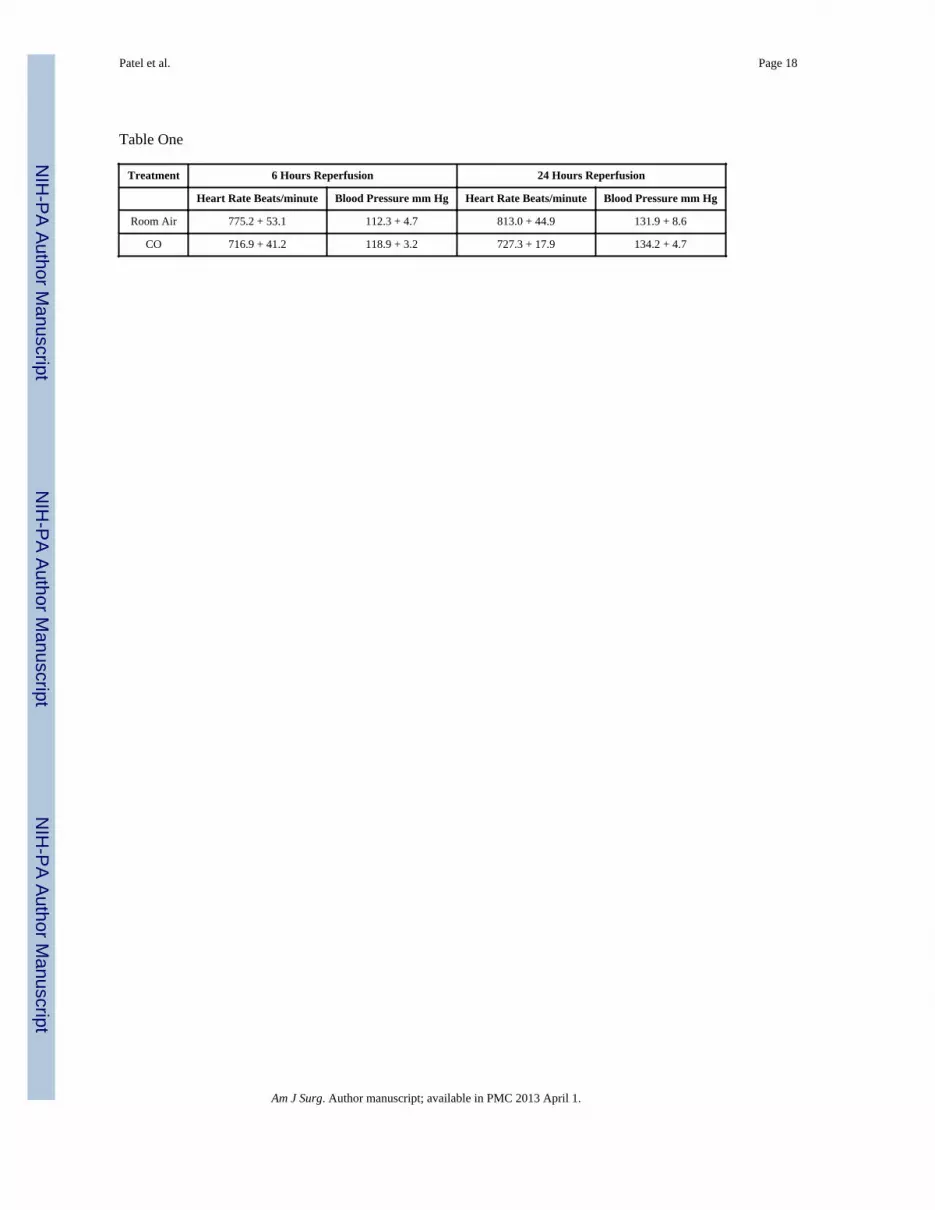
There was no difference in blood pressure or heart rate (Table One) in the CO vs. controltreated mice at 6 and 24 hours reperfusion.
Discussion
The central hypothesis of this study was that CO inhalation of 4.5 and 7.5 hours can improvemuscle damage caused by ischemia reperfusion in a hind limb model in the mouse. Wereport that CO inhalation decreased markers of tissue injury and components of the local andsystemic inflammatory response. It is important to note that the CO protocol used for theseexperiments was substantially different from the organ transplant IR models. In thetransplant models of liver and renal tissue IR, CO treatment has been usually initiated onehour prior to the onset of tissue ischemia and is continued for up to 24 hours reperfusion(16) . Such preemptive therapy is probably not relevant for patients at risk for acute hindlimb ischemia reperfusion since these individuals cannot be identified prior to the onset ofischemia, whereas transplantation is an urgent life saving planned event. However, it seemsclinically feasible to begin CO inhalation therapy after the diagnosis of limb ischemia isestablished, in a manner similar to beginning intravenous heparin therapy.
The level of carboxyhemoglobin in the blood of mice subjected to CO treatment in thisexperimental protocol was comparable to levels reported in studies of rodent liver andmyocardial ischemia reperfusion where administration of CO proved to providecytoprotection (17, 18). There is a time dependent relationship between CO treatment andprotection against fiber injury since the 4.5 hour treatment protocol did not provideprotection, whereas a 7.5 hour treatment protocol did provide protection against fiber injury.Since the 7.5 hour treatment protocol provided protection against I/R induced muscle fiberinjury, all subsequent biochemical analysis was performed using this duration of COtreatment. Subsequent biochemical analysis in mice treated for 7.5 hours and confirmed thatCO treatment preserved tissue levels of ATP essential for maintaining cellular membraneintegrity and contractility. This finding may suggest that CO therapy provides some degree
Patel et al. Page 6
Am J Surg. Author manuscript; available in PMC 2013 April 1.
NIH
-PA
Author M
anuscriptN
IH-P
A A
uthor Manuscript
NIH
-PA
Author M
anuscript

of metabolic rescue. ATP levels have been previously shown to be proportional to muscleviability(19, 20). Work studying myocardial IR reported a marked improvement inmyocardial energetics associated with inhaled CO (1). Similarly, in a rodent model ofhemorrhagic shock, ATP levels in liver were preserved by treatment with inhaled CO (21).Thus our findings showing preserved ATP levels in skeletal muscle following IR and COtreatment are consistent with previous reports from other tissues.
Additional analysis of local and systemic cytokine release, which has been associated withmortality and morbidity during vascular reconstruction in humans(22–24), revealed amarked decrease in serum and skeletal muscle KC at 24 hours reperfusion(Figure 1AB).There was also a significant decrease in muscle IL-6 (Figure 2). An important component ofthis observation is the fact that the levels of cytokines were reduced in both serum andskeletal muscle 18 hours after cessation of therapy, indicating the ability of CO to modulateinflammation in tissue after therapy is stopped. The finding of decreased local and systemiccytokines associated with inhaled CO treatment is also consistent with successfulexperimental therapeutic interventions from our and other laboratories (25, 26).
α-Tubulin levels were preserved in the skeletal muscle of mice that inhaled CO duringischemia reperfusion when compared to mice exposed to room air under the same conditions(Figure 3). α-Tubulin is a major component of microtubules which are present in alleukaryotic cells where they facilitate several biologic processes including mitosis,intracellular transport and mechanical response to the environment. Decreases in thecytoskeletal tubulin have been observed in atrophic skeletal muscle in rats (27). A specifichighly conserved isoform of α-Tubulin has been cloned and sequenced from a human adultskeletal muscle cDNA library. Sequence comparison of these isoforms show that there iscross species preservation of these genes, consistent with the functional importance of thismolecule (28). It has been proposed that Tubulin, a major component of muscle cytoskeletoncontributes to maintenance of cell structure during degenerative processes by providing acytostructural framework around which the regenerative processes may be initiated (29).Preservation of α-Tubulin in skeletal muscle of mice treated with CO during ischemiareperfusion suggests that the reparative characteristics and the functional capability of themuscle to respond to mechanical stimuli, such as contraction may also be preserved.
After 7.5 hours of IR, there was no difference in the level of heme oxygenase-1 expressionin skeletal muscle of CO treated and untreated mice (Figure 4). By 24 hours reperfusion,heme oxygenase-1 protein expression was markedly stimulated in CO treated and untreatedmice (Figure 4). In the CO treated mice, heme oxygenase-1 protein expression wassignificantly greater than levels detected in mice breathing room air during 7.5 hrs IR. Thisfinding suggests that CO inhalation only during the 7.5 IR period stimulated ongoingexpression of heme oxygenase-1 during the next 17.5 hours of reperfusion after CO therapyhad ceased. Since CO is a product of HO-1 activity, it was possible that exogenousadministration of CO might have decreased the expression of HO-1 protein through negativefeed back. The fact that exogenous CO did not down regulate HO-1 protein expression is animportant observation since HO is known to suppress microvascular thrombus formation(5). Thrombosis is a major component of reperfusion injury as it contributes to the no reflowperiod (30). Increased HO expression during hind limb ischemia reperfusion may enhancetissue healing since HO is also know to promote progenitor cell mobilization,neovascularization and functional recovery after critical hind limb ischemia in mice (31). Inconcert with increased expression of HO-1 at 24 hours reperfusion following inhaled COtherapy, there was preservation of total p38 MAPKinase and phosphorylated p38MAPKinase (Figure 5A and 5B). These observations provide additional evidence that COtreatment influenced downstream signaling pathways know to be cytoprotective andmodulated by heme Oxygenase (17).
Patel et al. Page 7
Am J Surg. Author manuscript; available in PMC 2013 April 1.
NIH
-PA
Author M
anuscriptN
IH-P
A A
uthor Manuscript
NIH
-PA
Author M
anuscript

Despite the decrease in muscle fiber injury, preserved cytoskeletal muscle protein, decreasedskeletal muscle and serum cytokines in mice exposed to IR in the presence of CO, skeletalmuscle MPO levels were actually higher in the CO treated mice than in the control mice(Figure 6). Inadvertent CO exposure has been associated with activation of neutrophils inlung tissue (16). Based on histologic assessment of skeletal muscle fiber injury, muscle ATPand cytokine analysis, we were not able to identify solid macroscopic or biochemical signsof skeletal muscle fiber cytotoxicity in the hind limbs of mice subjected to IR whilebreathing CO. It appears that the increase in muscle MPO did not reflect an increase indetectable tissue damage.
CO inhalation therapy in our model did not lower skeletal muscle edema. However,decreased skeletal muscle edema is not always associated with decreased skeletal muscleinjury (25, 32). It is possible that the edema associated with CO treatment observed in thesestudies is related in part to the activation of neutrophils(33) as suggested by the paradoxicalincrease in MPO in CO treated mice (figure 6). However, the amount of edema detected inCO treated mice was no different than untreated mice, even though the amount of MPO inthe muscle of reperfused mice treated with CO was twice that of untreated mice. Thisfinding seems to indicate that as a single variable, edema does may not contribute to musclefiber injury in the murine model.
Similar to the results of the analysis of skeletal muscle edema, there was no significantdifference in blood pressure in the CO treated vs. control treated mice at 6 and 24 hours(Table One). It is possible that the blood pressure at earlier time points might have beendifferent, however the intervals of 6 and 24 hours were selected to be sure the data was notcomplicated by the hemodynamic effects of anesthesia administered during ischemia andearly reperfusion. While there is evidence that administration of CO can reverse the effectsof shock and hemorrhage (21), the cytoprotective effects of CO on skeletal muscle ischemiareperfusion do not appear to be related to a long lasting effect on the blood pressure andheart rate associated with unilateral hind limb ischemia followed by reperfusion.
The precise mechanisms contributing to CO mediated cytoprotection of skeletal muscleexposed to IR have not been defined at this point. The breakdown of heme viahemeoxygenase naturally produces CO. CO has a variety of postulated effects includingvasorelaxation, anti-apoptotic, anti-proliferative, and anti-inflammatory effects (34–36). Theresult of the experiments in this report indicates that exogenous CO preserved skeletalmuscle ATP, and induced a predominantly anti-inflammatory/cytoprotective responsefollowing I/R injury. These findings parallel results reported in experimental transplantationand shock models (21).
In summary, we conclude that CO inhalation may have therapeutic potential to protectskeletal muscle from I/R injury in a model of acute hind limb ischemia reperfusion injury.However, examination into the precise mechanisms on how CO protects skeletal musclefrom IR injury requires further investigation.
AcknowledgmentsThe authors acknowledge funding from the Pacific Vascular Research Foundation, The Geneen Fund of theMassachusetts General Hospital, the Division of Vascular and Endovascular Surgery, and the National Institutes ofHealth (1R01AR055843). Dr. Watkins is the Isenberg Fellow in Academic Surgery at the Massachusetts GeneralHospital. Faraz Hashmi received a student research award from the AVA Foundation. Wolfgang Steudel has beensupported by the Department of Anesthesia and Critical Care at Massachusetts General Hospital.
Patel et al. Page 8
Am J Surg. Author manuscript; available in PMC 2013 April 1.
NIH
-PA
Author M
anuscriptN
IH-P
A A
uthor Manuscript
NIH
-PA
Author M
anuscript

References
1. Lavitrano M, Smolenski RT, Musumeci A, Maccherini M, Slominska E, Di Florio E, Bracco A,Mancini A, Stassi G, Patti M, Giovannoni R, Froio A, Simeone F, Forni M, Bacci ML, D'Alise G,Cozzi E, Otterbein LE, Yacoub MH, Bach FH, Calise F. Carbon monoxide improves cardiacenergetics and safeguards the heart during reperfusion after cardiopulmonary bypass in pigs.FASEB J. 2004; 18 :1093–1095. [PubMed: 15132974]
2. Ott MC, Scott JR, Bihari A, Badhwar A, Otterbein LE, Gray DK, Harris KA, Potter RF. Inhalationof carbon monoxide prevents liver injury and inflammation following hind limb ischemia/reperfusion. FASEB J. 2005; 19 :106–108. [PubMed: 15514102]
3. Martins PN, Reuzel-Selke A, Jurisch A, Atrott K, Pascher A, Pratschke J, Buelow R, Neuhaus P,Volk HD, Tullius SG. Induction of carbon monoxide in the donor reduces graft immunogenicity andchronic graft deterioration. Transplant Proc. 2005; 37 :379–381. [PubMed: 15808651]
4. Nakao A, Neto JS, Kanno S, Stolz DB, Kimizuka K, Liu F, Bach FH, Billiar TR, Choi AM,Otterbein LE, Murase N. Protection against ischemia/reperfusion injury in cardiac and renaltransplantation with carbon monoxide, biliverdin and both. Am J Transplant. 2005; 5 :282–291.[PubMed: 15643987]
5. Lindenblatt N, Bordel R, Schareck W, Menger MD, Vollmar B. Vascular heme oxygenase-1induction suppresses microvascular thrombus formation in vivo. Arterioscler Thromb Vasc Biol.2004; 24 :601–606. [PubMed: 14739126]
6. Orozco LD, Kapturczak MH, Barajas B, Wang X, Weinstein MM, Wong J, Deshane J, Bolisetty S,Shaposhnik Z, Shih DM, Agarwal A, Lusis AJ, Araujo JA. Heme oxygenase-1 expression inmacrophages plays a beneficial role in atherosclerosis. Circ Res. 2007; 100:1703–1711. [PubMed:17495224]
7. True AL, Olive M, Boehm M, San H, Westrick RJ, Raghavachari N, Xu X, Lynn EG, Sack MN,Munson PJ, Gladwin MT, Nabel EG. Heme oxygenase-1 deficiency accelerates formation ofarterial thrombosis through oxidative damage to the endothelium, which is rescued by inhaledcarbon monoxide. Circ Res. 2007; 101:893–901. [PubMed: 17885218]
8. Crawford RS, Hashmi FF, Jones JE, Albadawi H, McCormack M, Eberlin K, Entabi F, Atkins MD,Conrad MF, Austen WG Jr, Watkins MT. A novel model of acute murine hindlimb ischemia. Am JPhysiol Heart Circ Physiol. 2007; 292 :H830–837. [PubMed: 17012358]
9. Feng Y, Zhao H, Xu X, Buys ES, Raher MJ, Bopassa JC, Thibault H, Scherrer-Crosbie M, SchmidtU, Chao W. Innate immune adaptor MyD88 mediates neutrophil recruitment and myocardial injuryafter ischemia-reperfusion in mice. Am J Physiol Heart Circ Physiol. 2008; 295 :H1311–H1318.[PubMed: 18660455]
10. Lentsch AB, Yoshidome H, Cheadle WG, Miller FN, Edwards MJ. Chemokine involvement inhepatic ischemia/reperfusion injury in mice: roles for macrophage inflammatory protein-2 andKupffer cells. Hepatology. 1998; 27 :507–512. [PubMed: 9462650]
11. Liu M, Liang Y, Chigurupati S, Lathia JD, Pletnikov M, Sun Z, Crow M, Ross CA, Mattson MP,Rabb H. Acute kidney injury leads to inflammation and functional changes in the brain. J Am SocNephrol. 2008; 19 :1360–1370. [PubMed: 18385426]
12. Andersen K, Pedersen BK. The role of inflammation in vascular insulin resistance with focus onIL-6. Horm Metab Res. 2008; 40 :635–639. [PubMed: 18792875]
13. Szekanecz Z. Pro-inflammatory cytokines in atherosclerosis. Isr Med Assoc J. 2008; 10 :529–530.[PubMed: 18751634]
14. Franke A, Lante W, Kupser S, Becker HP, Weinhold C, Markewitz A. Procalcitonin levels afterdifferent types of conventional thoracic surgery. Thorac Cardiovasc Surg. 2008; 56:46–50.[PubMed: 18200468]
15. McCormack MC, Kwon E, Eberlin KR, Randolph M, Friend DS, Thomas AC, Watkins MT,Austen WG Jr. Development of reproducible histologic injury severity scores: skeletal musclereperfusion injury. Surgery. 2008; 143 :126–133. [PubMed: 18154940]
16. Song R, Kubo M, Morse D, Zhou Z, Zhang X, Dauber JH, Fabisiak J, Alber SM, Watkins SC,Zuckerbraun BS, Otterbein LE, Ning W, Oury TD, Lee PJ, McCurry KR, Choi AM. Carbonmonoxide induces cytoprotection in rat orthotopic lung transplantation via anti-inflammatory andanti-apoptotic effects. Am J Pathol. 2003; 163:231–242. [PubMed: 12819027]
Patel et al. Page 9
Am J Surg. Author manuscript; available in PMC 2013 April 1.
NIH
-PA
Author M
anuscriptN
IH-P
A A
uthor Manuscript
NIH
-PA
Author M
anuscript

17. Fujimoto H, Ohno M, Ayabe S, Kobayashi H, Ishizaka N, Kimura H, Yoshida K, Nagai R. Carbonmonoxide protects against cardiac ischemia--reperfusion injury in vivo via MAPK and Akt--eNOSpathways. Arterioscler Thromb Vasc Biol. 2004; 24 :1848–1853. [PubMed: 15308554]
18. Kaizu T, Ikeda A, Nakao A, Tsung A, Toyokawa H, Ueki S, Geller DA, Murase N. Protection oftransplant-induced hepatic ischemia/reperfusion injury with carbon monoxide via MEK/ERK1/2pathway downregulation. Am J Physiol Gastrointest Liver Physiol. 2008; 294:G236–244.[PubMed: 18006605]
19. Lindgard A, Lundberg J, Rakotonirainy O, Elander A, Soussi B. Preservation of rat skeletal muscleenergy metabolism by illumination. Life Sci. 2003; 72:2649–2658. [PubMed: 12672510]
20. Tsuchida T, Kato T, Yamaga M, Ikebe K, Oniki Y, Irie H, Takagi K. Effect of perfusion duringischemia on skeletal muscle. J Surg Res. 2001; 101:238–241. [PubMed: 11735281]
21. Zuckerbraun BS, McCloskey CA, Gallo D, Liu F, Ifedigbo E, Otterbein LE, Billiar TR. Carbonmonoxide prevents multiple organ injury in a model of hemorrhagic shock and resuscitation.Shock. 2005; 23 :527–532. [PubMed: 15897805]
22. Dawson J, Cockerill G, Choke E, Loftus I, Thompson MM. Circulating cytokines in patients withabdominal aortic aneurysms. Ann N Y Acad Sci. 2006; 1085:324–326. [PubMed: 17182951]
23. Gol MK, Nisanoglu V, Iscan Z, Balci M, Kandemir O, Tasdemir O. Inhibition of systemicinflammatory response with sodium nitroprusside in open heart surgery. J Cardiovasc Surg(Torino). 2002; 43 :803–809.
24. van Besouw NM, Baan CC, Holweg CT, de Groot-Kruseman HA, Peeters AM, Balk AH, WeimarW. Cytokine profiles as marker for graft vascular disease after clinical heart transplantation. AnnTransplant. 2000; 5 :61–67. [PubMed: 11217209]
25. Hua HT, Albadawi H, Entabi F, Conrad M, Stoner MC, Meriam BT, Sroufe R, Houser S,Lamuraglia GM, Watkins MT. Polyadenosine diphosphate-ribose polymerase inhibition modulatesskeletal muscle injury following ischemia reperfusion. Arch Surg. 2005; 140:344–351. discussion351–342. [PubMed: 15837884]
26. Wakai M, Winter D, Street J, O'Sullivan R, Wang J, Redmond H. Inosine Attenuates TourniquetInduced Skeletal Muscle Reperfusion Injury. J Surg Res. 2001; 99:311–315. [PubMed: 11469903]
27. Sakurai T, Fujita Y, Ohto E, Oguro A, Atomi Y. The decrease of the cytoskeleton tubulin followsthe decrease of the associating molecular chaperone alphaB-crystallin in unloaded soleus muscleatrophy without stretch. FASEB J. 2005; 19 :1199–1201. [PubMed: 15894563]
28. Stanchi F, Corso V, Scannapieco P, Ievolella C, Negrisolo E, Tiso N, Lanfranchi G, Valle G.TUBA8: A new tissue-specific isoform of alpha-tubulin that is highly conserved in human andmouse. Biochem Biophys Res Commun. 2000; 270 :1111–1118. [PubMed: 10772959]
29. Boudriau S, Cote CH, Vincent M, Houle P, Tremblay RR, Rogers PA. Remodeling of thecytoskeletal lattice in denervated skeletal muscle. Muscle Nerve. 1996; 19 :1383–1390. [PubMed:8874395]
30. Conrad MF, Stone DH, Albadawi H, Hua HT, Entabi F, Stoner MC, Watkins MT. Localinflammatory and thrombotic responses differ in a murine model of partial and complete hindlimbischemia/reperfusion. Surgery. 2005; 138 :375–381. [PubMed: 16153450]
31. Tongers J, Knapp JM, Korf M, Kempf T, Limbourg A, Limbourg FP, Li Z, Fraccarollo D,Bauersachs J, Han X, Drexler H, Fiedler B, Wollert KC. Haeme oxygenase promotes progenitorcell mobilization, neovascularization, and functional recovery after critical hindlimb ischaemia inmice. Cardiovasc Res. 2008; 78:294–300. [PubMed: 18093990]
32. Kauvar DS, Baer DG, Dubick MA, Walters TJ. Effect of fluid resuscitation on acute skeletalmuscle ischemia-reperfusion injury after hemorrhagic shock in rats. J Am Coll Surg. 2006;202:888–896. [PubMed: 16735202]
33. Crinnion JN, Homer-Vanniasinkam S, Parkin SM, Gough MJ. Role of neutrophil-endothelialadhesion in skeletal muscle reperfusion injury. Br J Surg. 1996; 83 :251–254. [PubMed: 8689180]
34. Lee S, Suk K. Heme oxygenase-1 mediates cytoprotective effects of immunostimulation inmicroglia. Biochem Pharmacol. 2007; 74 :723–729. [PubMed: 17632083]
35. Liu SH, Xu XR, Ma K, Xu B. Protection of carbon monoxide inhalation on lipopolysaccharide-induced multiple organ injury in rats. Chin Med Sci J. 2007; 22:169–176. [PubMed: 17966165]
Patel et al. Page 10
Am J Surg. Author manuscript; available in PMC 2013 April 1.
NIH
-PA
Author M
anuscriptN
IH-P
A A
uthor Manuscript
NIH
-PA
Author M
anuscript

36. Hasan RN, Schafer AI. Hemin upregulates Egr-1 expression in vascular smooth muscle cells viareactive oxygen species ERK-1/2-Elk-1 and NF-kappaB. Circ Res. 2008; 102:42–50. [PubMed:17967787]
Patel et al. Page 11
Am J Surg. Author manuscript; available in PMC 2013 April 1.
NIH
-PA
Author M
anuscriptN
IH-P
A A
uthor Manuscript
NIH
-PA
Author M
anuscript

Figure 1. Muscle and Serum KC levelsCO breathing markedly decreased KC levels in skeletal muscle (A,*p=0.01) and serum (B,**p=0.02) as compared to mice breathing room air alone.
Patel et al. Page 12
Am J Surg. Author manuscript; available in PMC 2013 April 1.
NIH
-PA
Author M
anuscriptN
IH-P
A A
uthor Manuscript
NIH
-PA
Author M
anuscript
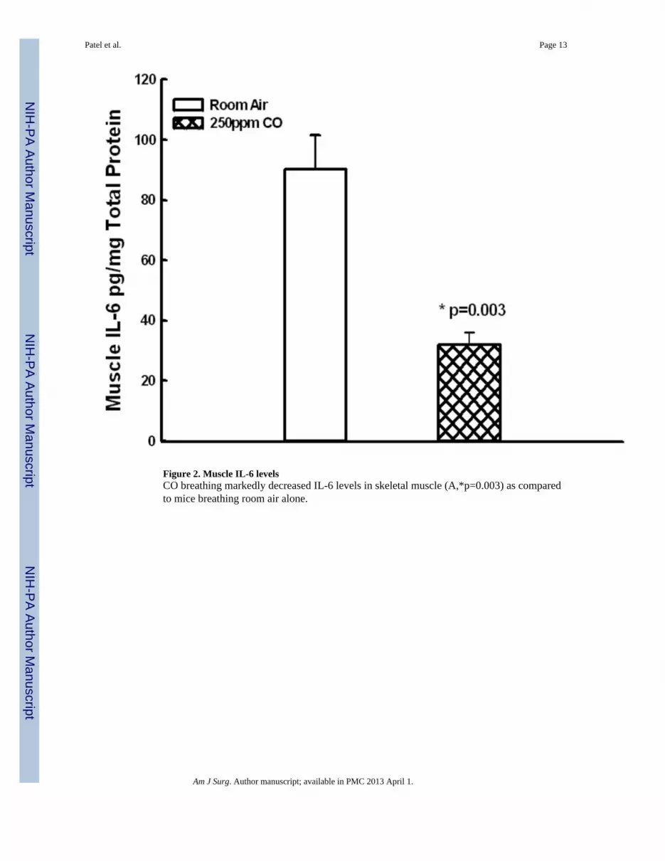
Figure 2. Muscle IL-6 levelsCO breathing markedly decreased IL-6 levels in skeletal muscle (A,*p=0.003) as comparedto mice breathing room air alone.
Patel et al. Page 13
Am J Surg. Author manuscript; available in PMC 2013 April 1.
NIH
-PA
Author M
anuscriptN
IH-P
A A
uthor Manuscript
NIH
-PA
Author M
anuscript

Figure 3. α-Tubulin in Skeletal MuscleCO breathing for 7.5 hours of sham ischemia reperfusion increased skeletal muscle α-tubulin level (*p=0.03). CO breathing preserved IR induced decreases in α-Tubulin at 7.5hours ischemia reperfusion (**p=0.03) and 24 hours reperfusion (+p=0.04).
Patel et al. Page 14
Am J Surg. Author manuscript; available in PMC 2013 April 1.
NIH
-PA
Author M
anuscriptN
IH-P
A A
uthor Manuscript
NIH
-PA
Author M
anuscript

Figure 4. Heme Oxygenase Expression during IRIR stimulated HO expression in skeletal muscle by 24 hours reperfusion during IR (*,+p<0.01). CO breathing during IR resulted in significantly greater HO expression by 24hours reperfusion as compared to mice who breathed room air during IR(*p=0.03).
Patel et al. Page 15
Am J Surg. Author manuscript; available in PMC 2013 April 1.
NIH
-PA
Author M
anuscriptN
IH-P
A A
uthor Manuscript
NIH
-PA
Author M
anuscript

Figure 5. Total p38MAPK expression and activity (Thr180/Tyr 182 phosphorylation)CO treatment preserved total p38MAPK expression (5A, p<0.05) and phosphorylatedp38MAPK (5B, p<0.05) by 24 hours reperfusion.
Patel et al. Page 16
Am J Surg. Author manuscript; available in PMC 2013 April 1.
NIH
-PA
Author M
anuscriptN
IH-P
A A
uthor Manuscript
NIH
-PA
Author M
anuscript

Figure 6. Skeletal Muscle MyeloperoxidaseCO breathing increased levels of skeletal muscle MPO as compared to mice breathing roomair alone(*p=0.007).
Patel et al. Page 17
Am J Surg. Author manuscript; available in PMC 2013 April 1.
NIH
-PA
Author M
anuscriptN
IH-P
A A
uthor Manuscript
NIH
-PA
Author M
anuscript
NIH
-PA
Author M
anuscriptN
IH-P
A A
uthor Manuscript
NIH
-PA
Author M
anuscript
Patel et al. Page 18
Table One
Treatment 6 Hours Reperfusion 24 Hours Reperfusion
Heart Rate Beats/minute Blood Pressure mm Hg Heart Rate Beats/minute Blood Pressure mm Hg
Room Air 775.2 + 53.1 112.3 + 4.7 813.0 + 44.9 131.9 + 8.6
CO 716.9 + 41.2 118.9 + 3.2 727.3 + 17.9 134.2 + 4.7
Am J Surg. Author manuscript; available in PMC 2013 April 1.