Carbon Monoxide Generated by Heme Oxygenase 1 Suppresses Endothelial Cell Apoptosis
11
J. Exp. Med. The Rockefeller University Press • 0022-1007/2000/10/1015/11 $5.00 Volume 192, Number 7, October 2, 2000 1015–1025 http://www.jem.org/cgi/content/full/192/7/1015 1015 Carbon Monoxide Generated by Heme Oxygenase 1 Suppresses Endothelial Cell Apoptosis By Sophie Brouard,* Leo E. Otterbein, ‡ Josef Anrather, § Edda Tobiasch,* Fritz H. Bach,* Augustine M.K. Choi, ‡ and Miguel P. Soares* From the *Immunobiology Research Center, Beth Israel Deaconess Medical Center, Harvard Medical School, Boston, Massachusetts 02215; the ‡ Department of Internal Medicine, Pulmonary and Critical Care Section,Yale University, School of Medicine, New Haven, Connecticut 06520; and the § Department of Neurobiology,Weill Medical College of Cornell University, New York, New York 10021 Abstract Heme oxygenase 1 (HO-1) inhibits apoptosis by regulating cellular prooxidant iron. We now show that there is an additional mechanism by which HO-1 inhibits apoptosis, namely by gen- erating the gaseous molecule carbon monoxide (CO). Overexpression of HO-1, or induction of HO-1 expression by heme, protects endothelial cells (ECs) from apoptosis. When HO-1 en- zymatic activity is blocked by tin protoporphyrin (SnPPIX) or the action of CO is inhibited by hemoglobin (Hb), HO-1 no longer prevents EC apoptosis while these reagents do not affect the antiapoptotic action of bcl-2. Exposure of ECs to exogenous CO, under inhibition of HO-1 activity by SnPPIX, substitutes HO-1 in preventing EC apoptosis. The mechanism of action of HO-1/CO is dependent on the activation of the p38 mitogen-activated protein kinase (MAPK) signaling transduction pathway. Expression of HO-1 or exposure of ECs to exoge- nous CO enhanced p38 MAPK activation by TNF-a. Specific inhibition of p38 MAPK acti- vation by the pyridinyl imidazol SB203580 or through overexpression of a p38 MAPK domi- nant negative mutant abrogated the antiapoptotic effect of HO-1. Taken together, these data demonstrate that the antiapoptotic effect of HO-1 in ECs is mediated by CO and more specif- ically via the activation of p38 MAPK by CO. Key words: apoptosis • endothelial cells • heme oxygenase 1 • carbon monoxide • p38 mitogen-activated protein kinase Introduction In their normally quiescent state, endothelial cells (ECs) 1 maintain blood flow, allowing the continuous traffic of plasma and cellular constituents between blood and paren- chymal tissues. To accomplish this function, ECs must pro- mote a certain level of vasorelaxation and inhibit leukocyte adhesion as well as coagulation and thrombosis (for a re- view, see reference 1). However, when ECs are exposed to proinflammatory stimuli, they become “activated” and promote vasoconstriction, leukocyte adhesion, and activa- tion, as well as coagulation and thrombosis (for reviews, see references 1–3). These functional changes are due to the expression by activated ECs of a series of proinflammatory genes encoding adhesion molecules, cytokines/chemo- kines, and costimulatory and procoagulant molecules (for reviews, see references 1–4). To prevent unfettered EC ac- tivation that could lead to EC injury and apoptosis, the ex- pression of these proinflammatory genes must be tightly regulated (5). One of the mechanisms by which this occurs relies on the expression by activated ECs of “protective genes” (6–10). One such gene is the stress responsive gene heme oxygenase 1 (HO-1). Heme oxygenases are the rate-limiting enzymes in the catabolism of heme into biliverdin, free iron, and carbon monoxide (CO), with biliverdin being subsequently catabo- A.M.K. Choi and M.P. Soares contributed equally to this work. Address correspondence to Miguel P. Soares, Immunobiology Re- search Center, Beth Israel Deaconess Medical Center, Harvard Medical School, 99 Brookline Ave., Boston, MA 02215. Phone: 617-632-0885; Fax: 617-632-0880; E-mail: [email protected] 1 Abbreviations used in this paper: Act.D, actinomycin D; BAEC, bovine aortic endothelial cell; CoPPIX, cobalt protoporphyrin; DFO, deferox- amine mesylate; EC, endothelial cell; ERK, extracellular signal–regulated kinase; FePP, iron protoporphyrin; GFP, green fluorescent protein; Hb, hemoglobin; HO-1, heme oxygenase 1; JNK, c-Jun NH 2 -terminal ki- nase; MAPK, mitogen-activated protein kinase; Mf, monocyte/mac- rophage(s); ppm, parts per million; SnPPIX, tin protoporphyrin; VASP, vasodilatator-stimulated phosphoprotein.
Transcript of Carbon Monoxide Generated by Heme Oxygenase 1 Suppresses Endothelial Cell Apoptosis

J. Exp. Med.
The Rockefeller University Press • 0022-1007/2000/10/1015/11 $5.00Volume 192, Number 7, October 2, 2000 1015–1025http://www.jem.org/cgi/content/full/192/7/1015
1015
Carbon Monoxide Generated by Heme Oxygenase 1 Suppresses Endothelial Cell Apoptosis
By Sophie Brouard,
*
Leo E. Otterbein,
‡
Josef Anrather,
§
Edda Tobiasch,
*
Fritz H. Bach,
*
Augustine M.K. Choi,
‡
and Miguel P. Soares
*
From the
*
Immunobiology Research Center, Beth Israel Deaconess Medical Center, Harvard Medical
School, Boston, Massachusetts 02215; the
‡
Department of Internal Medicine, Pulmonary and Critical Care Section, Yale University, School of Medicine, New Haven, Connecticut 06520; and the
§
Department of Neurobiology, Weill Medical College of Cornell University, New York, New York 10021
Abstract
Heme oxygenase 1 (HO-1) inhibits apoptosis by regulating cellular prooxidant iron. We nowshow that there is an additional mechanism by which HO-1 inhibits apoptosis, namely by gen-erating the gaseous molecule carbon monoxide (CO). Overexpression of HO-1, or inductionof HO-1 expression by heme, protects endothelial cells (ECs) from apoptosis. When HO-1 en-zymatic activity is blocked by tin protoporphyrin (SnPPIX) or the action of CO is inhibited byhemoglobin (Hb), HO-1 no longer prevents EC apoptosis while these reagents do not affectthe antiapoptotic action of bcl-2. Exposure of ECs to exogenous CO, under inhibition of HO-1activity by SnPPIX, substitutes HO-1 in preventing EC apoptosis. The mechanism of action ofHO-1/CO is dependent on the activation of the p38 mitogen-activated protein kinase(MAPK) signaling transduction pathway. Expression of HO-1 or exposure of ECs to exoge-nous CO enhanced p38 MAPK activation by TNF-
a
. Specific inhibition of p38 MAPK acti-vation by the pyridinyl imidazol SB203580 or through overexpression of a p38 MAPK domi-nant negative mutant abrogated the antiapoptotic effect of HO-1. Taken together, these datademonstrate that the antiapoptotic effect of HO-1 in ECs is mediated by CO and more specif-ically via the activation of p38 MAPK by CO.
Key words: apoptosis • endothelial cells • heme oxygenase 1 • carbon monoxide • p38 mitogen-activated protein kinase
Introduction
In their normally quiescent state, endothelial cells (ECs)
1
maintain blood flow, allowing the continuous traffic ofplasma and cellular constituents between blood and paren-chymal tissues. To accomplish this function, ECs must pro-mote a certain level of vasorelaxation and inhibit leukocyteadhesion as well as coagulation and thrombosis (for a re-
view, see reference 1). However, when ECs are exposed toproinflammatory stimuli, they become “activated” andpromote vasoconstriction, leukocyte adhesion, and activa-tion, as well as coagulation and thrombosis (for reviews, seereferences 1–3). These functional changes are due to theexpression by activated ECs of a series of proinflammatorygenes encoding adhesion molecules, cytokines/chemo-kines, and costimulatory and procoagulant molecules (forreviews, see references 1–4). To prevent unfettered EC ac-tivation that could lead to EC injury and apoptosis, the ex-pression of these proinflammatory genes must be tightlyregulated (5). One of the mechanisms by which this occursrelies on the expression by activated ECs of “protectivegenes” (6–10). One such gene is the stress responsive geneheme oxygenase 1 (HO-1).
Heme oxygenases are the rate-limiting enzymes in thecatabolism of heme into biliverdin, free iron, and carbonmonoxide (CO), with biliverdin being subsequently catabo-
A.M.K. Choi and M.P. Soares contributed equally to this work.Address correspondence to Miguel P. Soares, Immunobiology Re-
search Center, Beth Israel Deaconess Medical Center, Harvard MedicalSchool, 99 Brookline Ave., Boston, MA 02215. Phone: 617-632-0885;Fax: 617-632-0880; E-mail: [email protected]
1
Abbreviations used in this paper:
Act.D, actinomycin D; BAEC, bovineaortic endothelial cell; CoPPIX, cobalt protoporphyrin; DFO, deferox-amine mesylate; EC, endothelial cell; ERK, extracellular signal–regulatedkinase; FePP, iron protoporphyrin; GFP, green fluorescent protein; Hb,hemoglobin; HO-1, heme oxygenase 1; JNK, c-Jun NH
2
-terminal ki-nase; MAPK, mitogen-activated protein kinase; M
f
, monocyte/mac-
rophage(s);
ppm, parts per million; SnPPIX, tin protoporphyrin; VASP,vasodilatator-stimulated phosphoprotein.

1016
Carbon Monoxide Suppresses Endothelial Cell Apoptosis
lyzed into bilirubin (for reviews, see references 11–13). Sev-eral proinflammatory stimuli that lead to EC activation alsoupregulate the expression of HO-1 (for reviews, see refer-ences 12 and 13). That HO-1 acts as a cytoprotective geneis suggested by the observation that expression of HO-1 invitro prevents EC injury mediated by activated polymor-phonuclear cells (14), hydrogen peroxide (14, 15), or heme(15–17).
In addition, expression of HO-1 in vivo suppressesa variety of inflammatory responses, including endotoxicshock (17–19), hyperoxia (20), acute pleurisy (21), ischemiareperfusion injury (22), and graft rejection (23, 24), furthersupporting the cytoprotective role of this gene.
We have shown previously that expression of HO-1prevents ECs from undergoing apoptosis (23). This may bean important mechanism by which HO-1 exerts its cyto-protective function, since EC apoptosis, such as it occursduring acute (25) and chronic inflammation (26), is highlydeleterious. We now demonstrate that the antiapoptotic ef-fect of HO-1 is mediated through the ability of HO-1 togenerate the gaseous molecule CO. In addition, we dem-onstrate that CO suppresses EC apoptosis through a mech-anism that is dependent on the activation of the p38 mito-gen-activated protein kinase (MAPK) signal transductionpathway.
Materials and Methods
Cell Culture.
The murine 2F-2B EC line (American TypeCulture Collection) and primary bovine aortic ECs (BAECs)were cultured as described previously (23, 27).
Expression Plasmids.
The
b
-galactosidase was cloned into thepcDNA3 vector (Invitrogen) as described previously (28). Twovectors encoding rat HO-1 cDNA were used. The original vec-tor encoding the full-length rat HO-1 cDNA under the controlof a the
b
-actin enhancer/promoter (
b
-actin/HO-1) has beendescribed elsewhere (29). A 1.0-kbp XhoI-HindIII fragment en-coding the full-length rat HO-1 cDNA was cut from the prHO-1vector (30) and subcloned into the pcDNA3 vector to achieveexpression of the HO-1 cDNA under the control of the CMVenhancer/promoter (pcDNA3/HO-1). The mouse Bcl-2 cDNAwas cloned in the pac vector, as described elsewhere (10). p38/CSBP1 MAPK was amplified from HeLa cDNA by PCR andcloned into the pcDNA3/HA vector derived from pcDNA3 (In-vitrogen) by inserting a DNA fragment coding for an epitope de-rived from the hemagglutinin protein of the human influenza vi-rus hemagglutinin (HA; MYPYDVPDYASL). A dominantnegative mutant of p38/CSBP1, harboring a T180A and a Y182Fsubstitution, was generated by overlap extension mutagenesis.Green fluorescent protein (GFP) cDNA (CLONTECH Labora-tories, Inc.) was cloned into the pcDNA3-expressing vector.
Transient Transfections.
BAECs and 2F-2B ECs were tran-siently transfected as described elsewhere (23, 28). All experi-ments were carried out 24–48 h after transfection.
b
-Galactosi-dase–transfected cells were detected as described elsewhere (23,28). The percentage of viable cells was assessed by evaluating thenumber of
b
-galactosidase–expressing cells that retained normalmorphology, as described elsewhere (23, 28). The number of ran-dom fields counted was determined to have a minimum of 200viable transfected cells per control well. The percentage of viablecells was normalized for each DNA preparation to the number of
Figure 1. HO-1 suppresses EC apoptosis. (A) 2F-2B ECs were trans-fected with a GFP-expressing plasmid and monitored for GFP expressionby flow cytometry. The percentage of transfected ECs was assessed bymeasuring fluorescence intensity in ECs transfected with control(pcDNA3; filled histogram) versus GFP (open histogram) expression plas-mids. (B) ECs were cotransfected with b-galactosidase plus control(pcDNA3) or HO-1 (b-actin/HO-1) expression vectors. EC apoptosiswas induced by TNF-a plus Act.D and apoptosis of b-galactosidase–transfected ECs was quantified. Gray bars represent ECs treated withAct.D and black bars represent ECs treated with TNF-a plus Act.D. Re-sults shown are the mean 6 SD from duplicate wells taken from 1 repre-sentative experiment out of 10. (C) HO-1 expression was detected inBAECs by Western blot. No Tr, nontransfected. NT, nontreated. (D)2F-2B ECs were cotransfected with b-galactosidase plus control(pcDNA3) or HO-1 (b-actin/HO-1) expression vectors. Gray bars rep-resent untreated ECs and black bars represent ECs treated with etoposide(200 mM, 8 h) or subjected to serum deprivation (0.1% FCS for 24 h).Results shown are the mean 6 SD from duplicate wells taken from onerepresentative experiment out of three independent experiments. Similarresults were obtained using BAECs.

1017
Brouard et al.
image scanner (Arcus II; Agfa) equipped with FotoLook andPhotoshop
®
software. The amount of phosphorylated ERK,JNK, and p38 MAPK was quantified using ImageQuant
®
soft-ware (Molecular Dynamics). When indicated, membranes werestripped (62.5 mM Tris-HCl, pH 6.8, 2% SDS, and 100 mM
b
-mercaptoethanol, 30 min, 50
8
C). Phosphorylated ERK, JNK,and p38 MAPK were normalized to the total amount of totalERK, JNK, and p38 MAPK detected in the same membrane.
Flow Cytometry.
2F-2B ECs were transfected with the GFPexpression plasmid and harvested 24 h after transfection bytrypsin digestion (0.05% in PBS). ECs were washed in PBS, pH7.2, 5% FCS, and fluorescent labeling was evaluated using aFACSort™ equipped with CELLQuest™ software (BectonDickinson).
Cell Treatment and Reagents.
Actinomycin D (Act.D; Sigma-Aldrich) was dissolved in PBS and added to the culture medium24 h after transfection.
The Act.D concentration
used corre-sponded to the optimal concentration necessary to sensitize ECsto TNF-
a
–mediated apoptosis, e.g., 10
m
g/ml for 2F-2B ECsand 0.1
m
g/ml for BAECs. When indicated, EC apoptosis wasinduced by etoposide (200
m
M, 8 h; Calbiochem-Novabiochem)or by serum deprivation (0.1% FCS for 24 h). The iron chelatordeferoxamine mesylate (DFO; Sigma-Aldrich) was dissolved ex-temporarily in water and added to culture medium (1–100
m
M)1 h before the induction of apoptosis. Hemoglobin (Hb; Sigma-Aldrich) was dissolved (1 mM) extemporarily in PBS, 10 mMNa
2
S
2
O
4
, dialyzed against PBS (2 h, 4
8
C, 1:800 dilution),
andadded to the culture medium (1–100
m
M) 6 h after transfection.The guanylcyclase inhibitor 1H(1,2,4)oxadiazolo(4,3-
a
)quin-oxalin-1 (ODQ; Calbiochem-Novabiochem) was dissolved inDMSO (Sigma-Aldrich) and added to the culture medium (10–100
m
M) 6 h after transfection. Iron protoporphyrin ([FePP]/
Figure 2. The antiapoptotic effect of HO-1 isdose dependent. (A) 2F-2B ECs were cotrans-fected with increasing doses of HO-1 expressionvector (b-actin/HO-1). EC apoptosis was in-duced by TNF-a plus Act.D. Results shown arethe mean 6 SD from duplicate wells taken fromone representative experiment out of three. Simi-lar results were obtained using BAECs. (B) Theexpression of HO-1 was detected in BAECs byWestern blot. Values indicate the amount ofHO-1 vector (pcDNA3/HO-1) used in eachtransfection (ng of DNA per 3 3 105 cells). NoTr., nontransfected.
Figure 3. The antiapoptotic effect ofHO-1 is dependent on HO enzymatic ac-tivity. (A) 2F-2B ECs were cotransfectedwith b-galactosidase plus pcDNA3, HO-1(b-actin/HO-1), or bcl-2 expression vec-tors. Cells were either left untreated (Con-trol) or treated with the inhibitor of HOenzymatic activity SnPPIX. CoPPIX, a pro-toporphyrin that does not inhibit HO enzy-matic activity, was used as a control treat-ment. Gray bars represent ECs treated withAct.D and black bars represent ECs treatedwith TNF-a plus Act.D. Results shown arethe mean 6 SD from duplicate wells takenfrom one representative experiment out ofthree. (B) 2F-2B ECs were transfected with
b-galactosidase plus pcDNA3 expression vectors. ECs were either left untreated (Control) or were treated with SnPPIX and CoPPIX as in A. The resultsshown are the mean 6 SD from duplicate wells taken from one representative experiment out of three.
transfected cells counted in the absence of the apoptosis-inducingagent (100% viability). All experiments were performed at leastthree times in duplicate.
Adenovirus.
The recombinant HO-1 adenovirus has been de-scribed previously (31). The recombinant
b
-galactosidase ade-novirus was a gift of Dr. Robert Gerard (University of TexasSouthwestern Medical Center, Dallas, Texas). Adenoviruses wereproduced, extracted, purified, and titrated, as described previ-ously (28). Confluent BAECs were infected with a multiplicity ofinfection of 200 PFU/cell, as described elsewhere (28).
Cell Extracts and Western Blot Analysis.
Cell extracts wereprepared, electrophoresed under denaturing conditions (10–12.5% polyacrylamide gels), and transferred into polyvinyldifluo-ridine membranes (Immobilon P; Millipore), as described else-where (28). HO-1 was detected using a rabbit anti–human HO-1polyclonal antibody (StressGen Biotechnologies). Vasodilatator-stimulated phosphoprotein (VASP) was detected using a rabbitanti–human VASP polyclonal antibody (Calbiochem-Novabio-chem). Total and activated/phosphorylated forms of extracellularsignal–regulated kinases (ERK-1 and -2), c-Jun NH
2
-terminal ki-nases (JNK-1, -2, and -3), and p38 MAPK were detected usingrabbit polyclonal antibodies directed against the total or phos-phorylated forms of these MAPKs, according to the manufac-turer’s suggestions (New England Biolabs, Inc.).
b
-Tubulin wasdetected using anti–human
b
-tubulin monoclonal antibody(Boehringer).
Primary antibodies were detected using horseradishperoxidase–conjugated donkey anti–rabbit or goat anti–mouseIgG secondary antibodies (Pierce Chemical Co.). Peroxidase wasvisualized using the enhanced chemiluminescence assay (Amer-sham Pharmacia Biotech) according to the manufacturer’s in-structions, and stored in the form of photoradiographs (Biomax™MS; Eastman Kodak Co.). Digital images were obtained using an

1018
Carbon Monoxide Suppresses Endothelial Cell Apoptosis
Figure 4. Scavenging of CO by Hb sup-presses the antiapoptotic effect of HO-1.2F-2B ECs were cotransfected withb-galactosidase plus control (pcDNA3),HO-1 (b-actin/HO-1), or bcl-2 expres-sion vectors. ECs were either left untreated(0) or were treated with increasing concen-trations of Hb. Gray bars represent ECstreated with Act.D and black bars representECs treated with TNF-a plus Act.D. Theresults shown are the mean 6 SD from du-plicate wells taken from one representativeexperiment out of four.
Figure 5. Exogenous CO suppresses EC apoptosis in the absence ofHO-1. (A) 2F-2B ECs were transfected with a b-galactosidase expressionvector and exposed to exogenous CO. Gray bars represent ECs treatedwith Act.D alone and black bars represent ECs treated with TNF-a plusAct.D. (B) 2F-2B ECs were transfected with a b-galactosidase expressionvector and exposed to exogenous CO (10,000 ppm) with or without Hb.Gray bars represent ECs treated with Act.D and black bars represent ECstreated with TNF-a plus Act.D. (C) 2F-2B ECs were cotransfected withb-galactosidase and HO-1 (b-actin/HO-1) expression vectors. Whereindicated (1), HO-1 enzymatic activity was inhibited by SnPPIX and/orexposed to exogenous CO. Gray bars represent ECs treated with Act.Dand black bars represent ECs treated with TNF-a plus Act.D. Resultsshown (A, B, and C) are the mean 6 SD from duplicate wells taken fromone representative experiment out of three.
heme), cobalt protoporphyrin (CoPPIX),
and tin
protoporphyrin(SnPPIX; all from Porphyrin Products, Inc.) were dissolved (10mM) in 100 mM NaOH and conserved at
2
20
8
C until use.Metalloporphyrins were added to the culture medium (50
m
M)6 h after transfection. The cGMP analogue 8-bromo-cGMP so-dium salt (8-Br-cGMP; Sigma-Aldrich) was dissolved in waterand added to the culture medium (10–100
m
M) 30 min beforethe induction of apoptosis. Human recombinant TNF-
a
(R&DSystems) was dissolved in PBS and 1% BSA, and added to the cul-ture medium (10–100 ng/ml) 24 h after transfection. The p38MAPK inhibitor pyridinyl imidazol SB203580 (32) was dissolvedin DMSO and added to the culture medium (5–20
m
M) 6 h aftertransfection.
CO Exposure.
Cells were exposed to compressed air or vary-ing concentration of CO (250 and 10,000 parts per million[ppm]), as described elsewhere (33).
Results
HO-1 Protects ECs from Apoptosis.
TNF-
a
induces ap-optosis of cultured ECs when transcriptional activity is in-hibited by Act.D (28). We have used this experimental sys-tem to ask whether transient overexpression of HO-1could prevent ECs from undergoing apoptosis. We firstevaluated the ability of ECs to be transiently transfected.To do so, ECs were transfected with a GFP-expressingplasmid and the percentage of GFP-expressing ECs wasevaluated by flow cytometry. As illustrated in Fig. 1, 45–50% of ECs expressed the GFP protein 24 h after transfec-tion. We then tested whether transfection of HO-1 wouldprevent ECs from undergoing TNF-
a
–mediated apoptosis.To do so, ECs were cotransfected with HO-1 and
b
-galac-tosidase and apoptosis was evaluated by counting the num-ber of viable
b
-galactosidase–expressing ECs. TNF-a plusAct.D induced apoptosis of control ECs transfected withthe pcDNA3 (60–70% apoptotic ECs). Overexpression ofHO-1 prevented EC apoptosis (5–10% apoptotic ECs; Fig.1). The expression of HO-1 was confirmed by Westernblot (Fig. 1). Overexpression of HO-1 also prevented ECapoptosis induced by other proapoptotic stimuli such asetoposide or serum deprivation, which is in keeping withsimilar observations in 293 cells (34). The antiapoptotic ef-fect of HO-1 was dose dependent in that increasing levelsof HO-1 expression resulted in increased protection fromTNF-a plus Act.D–mediated apoptosis (Fig. 2). The maxi-mal antiapoptotic effect of HO-1 (90–100% protection)was reached using 500–1,000 ng of the b-actin/HO-1 ex-

1019 Brouard et al.
4). The ability of Hb to block the antiapoptotic effect ofHO-1 was dose dependent, in that increasing concentra-tions of Hb (3–50 mM) decreased the ability of HO-1 toprevent EC apoptosis (Fig. 4). Hb did not impair the anti-apoptotic effect of bcl-2 (Fig. 4), nor did it sensitize controlECs (pcDNA3) to apoptosis (Fig. 4).
Exogenous CO Can Substitute for HO-1 in Preventing ECApoptosis. If CO mediates the antiapoptotic action ofHO-1, then exogenous CO should prevent EC apoptosis.The data illustrated in Fig. 5 show that this is the case.When control ECs (transfected with pcDNA3) were ex-posed to exogenous CO (10,000 ppm), TNF-a–mediatedapoptosis was suppressed (Fig. 5). Exogenous CO alsosuppressed EC apoptosis when HO-1 activity was inhib-ited by SnPPIX, suggesting that CO can prevent EC ap-optosis in the absence of other biological functions ofHO-1 (Fig. 5). We then tested whether the level of exog-enous CO used (10,000 ppm) was comparable to that pro-duced when HO-1 is expressed in ECs. Given that Hb (50mM) blocks the protective effect of endogenously pro-duced CO (Fig. 4), we reasoned that if this was the casefor exogenous CO then the effects of exogenous CO maymimic those of endogenous CO. The antiapoptotic effectof exogenous CO was suppressed by Hb (50 mM), sug-gesting that the concentration of exogenous CO (10,000ppm) used in these experiments is not supraphysiologic(Fig. 5).
ECs That Express HO-1 Can Suppress Apoptosis of ECsThat Do Not Express HO-1. Given that CO can act as anintercellular signaling molecule, we hypothesized that ECsthat express HO-1 may generate sufficient levels of CO toprotect neighboring ECs that do not express HO-1 fromundergoing apoptosis. To test this hypothesis, ECs weretransfected with control (pcDNA3) or HO-1 expressionvectors and cocultured with b-galactosidase–transfectedECs. When cocultured with control ECs (pcDNA3), TNF-aplus Act.D induced apoptosis of b-galactosidase–transfectedECs (that do not express HO-1). However, when cocul-tured with ECs expressing HO-1, b-galactosidase–trans-fected ECs were protected from TNF-a plus Act.D–medi-ated apoptosis (Fig. 6).
Expression of Endogenous HO-1 Inhibits EC Apoptosisvia CO. We questioned whether upregulation of endog-enous HO-1 by heme would suppress EC apoptosis. Thedata illustrated in Fig. 7 suggest that this is the case. Expo-
pression vector per 3 3 105 cells (Fig. 2). All subsequentexperiments were carried out using these experimentalconditions.
The Antiapoptotic Function of HO-1 Requires Its EnzymaticActivity. To test whether the antiapoptotic action of HO-1was dependent on its enzymatic action, HO-1 activity wasblocked using SnPPIX. When HO-1 activity was blockedby SnPPIX, HO-1 was no longer able to prevent EC apop-tosis (Fig. 3), and the antiapoptotic effect of bcl-2 was notimpaired by SnPPIX (Fig. 3). CoPPIX, which has a similarstructure to SnPPIX but does not inhibit HO activity, didnot suppress the antiapoptotic effect of HO-1 or that ofbcl-2 (Fig. 3). These protoporphyrins had no detectable ef-fect per se on EC viability (Fig. 3).
Endogenous CO Mediates the Antiapoptotic Effect ofHO-1. Since HO-1 enzymatic activity is needed for itsantiapoptotic effect, this suggests that this antiapoptotic ef-fect is mediated through one or more end products ofheme catabolism by HO-1, i.e., bilirubin, iron, and/orCO. We tested if CO would account for the antiapoptoticeffect of HO-1. ECs were transiently transfected with HO-1and treated with Hb to scavenge CO. Under these condi-tions, the antiapoptotic effect of HO-1 was suppressed (Fig.
Figure 6. ECs that express HO-1 suppress apoptosis of ECs that do notexpress HO-1. 2F-2B ECs were transfected with control (pcDNA3; I andII) or HO-1 (III) expression vectors. 16 h after transfection, ECs wereharvested, washed, and cocultured at a ratio of 1:1 with ECs transfectedwith b-galactosidase (I and II) or with b-galactosidase plus HO-1 (III).Cocultures were maintained for an additional 24 h before induction ofapoptosis by TNF-a plus Act.D. The percentage of survival was evalu-ated by counting the number of b-galactosidase–positive cells that re-tained normal morphology. Gray bars represent ECs treated with Act.Dand black bars represent ECs treated with TNF-a plus Act.D. Resultsshown are the mean 6 SD from duplicates taken from one representativeexperiment out of three.
Figure 7. Upregulation of endogenous HO-1 expres-sion inhibits EC apoptosis via CO. (A) 2F-2B ECs werecotransfected with a b-galactosidase expression vector andexposed to FePP. Apoptosis was induced by TNF-a andAct.D. Gray bars represent ECs treated with Act.D and blackbars represent ECs treated with TNF-a plus Act.D. Theresults shown are the mean 6 SD from duplicate wellstaken from one representative experiment out of three. (B)2F-2B ECs were cotransfected with a b-galactosidase ex-pression vector and exposed to FePP (6.25 mM). Whereindicated, ECs were treated with Hb (50 mM). The resultsshown are the mean 6 SD from duplicate wells taken fromone representative experiment out of three.

1020 Carbon Monoxide Suppresses Endothelial Cell Apoptosis
sure to heme protected ECs from TNF-a plus Act.D–mediated apoptosis (Fig. 7). This protective effect was ob-served only at heme concentrations ranging from 5 to 7mM and was lost at higher concentrations, suggesting that
heme becomes cytotoxic at concentrations higher than 10mM (35; Fig. 7). The antiapoptotic effect of heme was de-pendent on the generation of CO, since heme was nolonger able to suppress EC apoptosis when CO was scav-enged by Hb (Fig. 7).
Iron Chelation Protects ECs from Apoptosis. The observa-tion that CO can prevent EC apoptosis (Figs. 4 and 5) con-trasts with the notion that the antiapoptotic effect of HO-1relies exclusively on its ability to prevent intracellular ironaccumulation (34). Given that overexpression of HO-1 inECs resulted in significant upregulation of ferritin expres-sion (data not shown), we questioned whether eliminationof reactive intracellular iron such as it occurs when ferritinis expressed would contribute to prevent TNF-a–mediatedapoptosis of ECs. To mimic the iron chelator effect of fer-ritin, we used the iron chelator DFO and tested whetherDFO would suppress EC apoptosis. The data illustrated inFig. 8 suggest that this is the case. Induction of EC apopto-sis by TNF-a plus Act.D was suppressed by DFO (Fig. 8).When HO-1 activity was inhibited by SnPPIX or the ac-tion of CO was suppressed by Hb, DFO was still able toprevent EC apoptosis (Fig. 8).
CO and Iron Chelation Have Additive Effects in ProtectingECs from Apoptosis. Given the ability of both CO andiron chelation to suppress EC apoptosis, we asked whetherthese two biological functions, engendered by HO-1,would act together to suppress EC apoptosis. Under inhibi-tion of HO activity by SnPPIX, exposure to low levels ofCO (250 ppm) did not suppress EC apoptosis significantly.When used alone, DFO (100 mM) suppressed EC apopto-sis, but to a lesser extent than HO-1 (Fig. 9). However,when ECs were exposed to both CO (250 ppm) and DFO(100 mM), inhibition of EC apoptosis was comparable tothat achieved with the expression of HO-1.
Figure 8. Iron chelation by DFO suppresses EC apoptosis. (A) 2F-2BECs were transfected with a b-galactosidase expression vector and ex-posed to DFO. Gray bars represent ECs treated with Act.D alone andblack bars represent ECs treated with TNF-a plus Act.D. (B) 2F-2B ECswere cotransfected as described above in A. Where indicated (1), HO-1enzymatic activity was inhibited by SnPPIX and iron was chelated byDFO, as described above in A. Gray bars represent ECs treated withAct.D and black bars represent ECs treated with TNF-a plus Act.D. (C)2F-2B ECs were cotransfected as described above in A. Where indicated(1), CO was removed from the culture medium by Hb and/or iron waschelated by DFO as described above in A and B. Gray bars represent ECstreated with Act.D and black bars represent ECs treated with TNF-a plusAct.D. Results shown (A, B, and C) are the mean 6 SD from duplicatewells taken from one representative experiment out of three.
Figure 9. Iron chelation and CO have additive effects in suppressingEC apoptosis. 2F-2B ECs were cotransfected with b-galactosidase andHO-1 (b-actin/HO-1) expression vectors. Where indicated (1), cellswere treated with the inhibitor of HO enzymatic activity SnPPIX. ECswere exposed to CO (250 ppm) and to the iron chelator DFO. Gray barsrepresent ECs treated with Act.D and black bars represent ECs treatedwith TNF-a plus Act.D. The results shown are the mean 6 SD from du-plicate wells taken from one representative experiment out of three.
1021 Brouard et al.
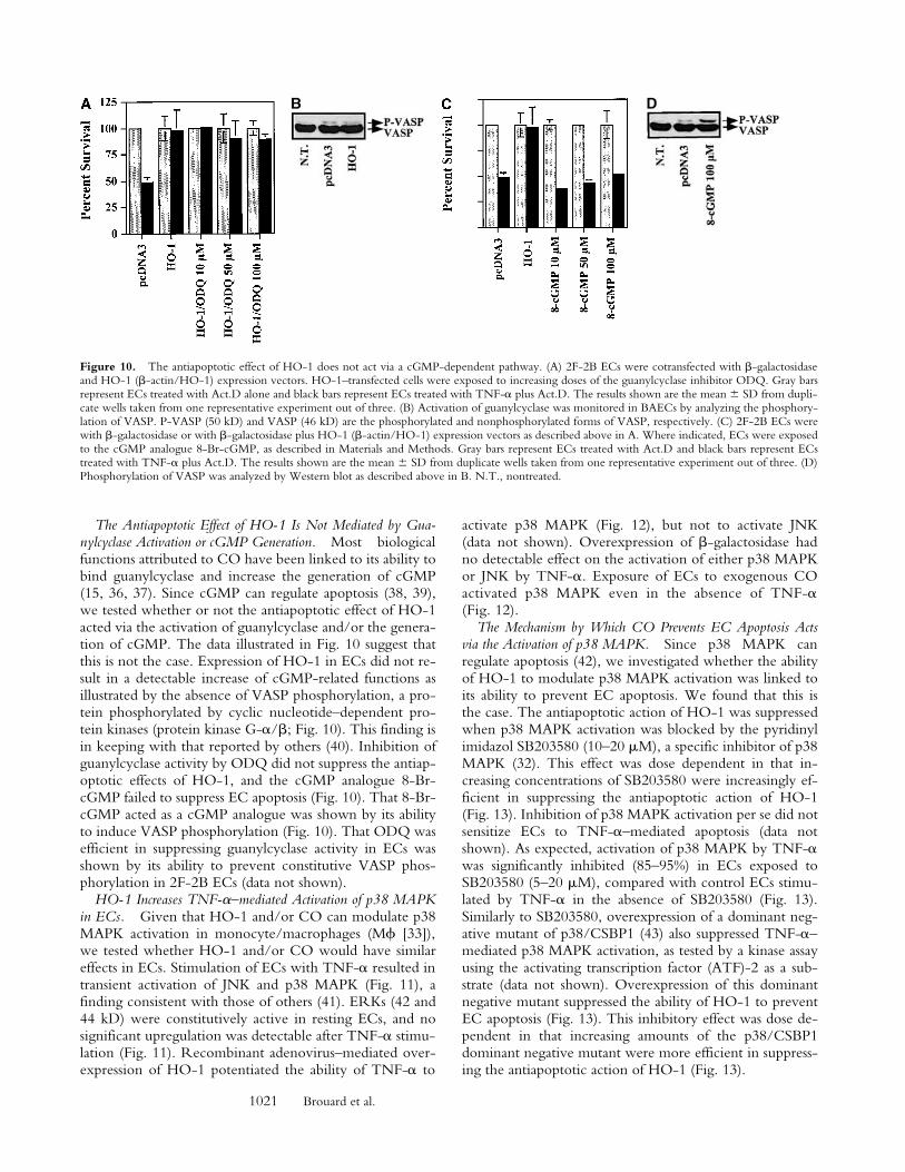
The Antiapoptotic Effect of HO-1 Is Not Mediated by Gua-nylcyclase Activation or cGMP Generation. Most biologicalfunctions attributed to CO have been linked to its ability tobind guanylcyclase and increase the generation of cGMP(15, 36, 37). Since cGMP can regulate apoptosis (38, 39),we tested whether or not the antiapoptotic effect of HO-1acted via the activation of guanylcyclase and/or the genera-tion of cGMP. The data illustrated in Fig. 10 suggest thatthis is not the case. Expression of HO-1 in ECs did not re-sult in a detectable increase of cGMP-related functions asillustrated by the absence of VASP phosphorylation, a pro-tein phosphorylated by cyclic nucleotide–dependent pro-tein kinases (protein kinase G-a/b; Fig. 10). This finding isin keeping with that reported by others (40). Inhibition ofguanylcyclase activity by ODQ did not suppress the antiap-optotic effects of HO-1, and the cGMP analogue 8-Br-cGMP failed to suppress EC apoptosis (Fig. 10). That 8-Br-cGMP acted as a cGMP analogue was shown by its abilityto induce VASP phosphorylation (Fig. 10). That ODQ wasefficient in suppressing guanylcyclase activity in ECs wasshown by its ability to prevent constitutive VASP phos-phorylation in 2F-2B ECs (data not shown).
HO-1 Increases TNF-a–mediated Activation of p38 MAPKin ECs. Given that HO-1 and/or CO can modulate p38MAPK activation in monocyte/macrophages (Mf [33]),we tested whether HO-1 and/or CO would have similareffects in ECs. Stimulation of ECs with TNF-a resulted intransient activation of JNK and p38 MAPK (Fig. 11), afinding consistent with those of others (41). ERKs (42 and44 kD) were constitutively active in resting ECs, and nosignificant upregulation was detectable after TNF-a stimu-lation (Fig. 11). Recombinant adenovirus–mediated over-expression of HO-1 potentiated the ability of TNF-a to
activate p38 MAPK (Fig. 12), but not to activate JNK(data not shown). Overexpression of b-galactosidase hadno detectable effect on the activation of either p38 MAPKor JNK by TNF-a. Exposure of ECs to exogenous COactivated p38 MAPK even in the absence of TNF-a(Fig. 12).
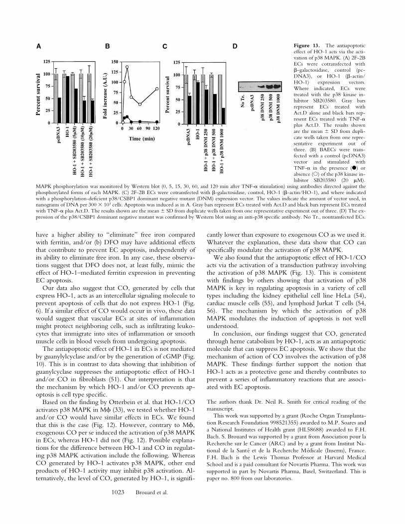
The Mechanism by Which CO Prevents EC Apoptosis Actsvia the Activation of p38 MAPK. Since p38 MAPK canregulate apoptosis (42), we investigated whether the abilityof HO-1 to modulate p38 MAPK activation was linked toits ability to prevent EC apoptosis. We found that this isthe case. The antiapoptotic action of HO-1 was suppressedwhen p38 MAPK activation was blocked by the pyridinylimidazol SB203580 (10–20 mM), a specific inhibitor of p38MAPK (32). This effect was dose dependent in that in-creasing concentrations of SB203580 were increasingly ef-ficient in suppressing the antiapoptotic action of HO-1(Fig. 13). Inhibition of p38 MAPK activation per se did notsensitize ECs to TNF-a–mediated apoptosis (data notshown). As expected, activation of p38 MAPK by TNF-awas significantly inhibited (85–95%) in ECs exposed toSB203580 (5–20 mM), compared with control ECs stimu-lated by TNF-a in the absence of SB203580 (Fig. 13).Similarly to SB203580, overexpression of a dominant neg-ative mutant of p38/CSBP1 (43) also suppressed TNF-a–mediated p38 MAPK activation, as tested by a kinase assayusing the activating transcription factor (ATF)-2 as a sub-strate (data not shown). Overexpression of this dominantnegative mutant suppressed the ability of HO-1 to preventEC apoptosis (Fig. 13). This inhibitory effect was dose de-pendent in that increasing amounts of the p38/CSBP1dominant negative mutant were more efficient in suppress-ing the antiapoptotic action of HO-1 (Fig. 13).
Figure 10. The antiapoptotic effect of HO-1 does not act via a cGMP-dependent pathway. (A) 2F-2B ECs were cotransfected with b-galactosidaseand HO-1 (b-actin/HO-1) expression vectors. HO-1–transfected cells were exposed to increasing doses of the guanylcyclase inhibitor ODQ. Gray barsrepresent ECs treated with Act.D alone and black bars represent ECs treated with TNF-a plus Act.D. The results shown are the mean 6 SD from dupli-cate wells taken from one representative experiment out of three. (B) Activation of guanylcyclase was monitored in BAECs by analyzing the phosphory-lation of VASP. P-VASP (50 kD) and VASP (46 kD) are the phosphorylated and nonphosphorylated forms of VASP, respectively. (C) 2F-2B ECs werewith b-galactosidase or with b-galactosidase plus HO-1 (b-actin/HO-1) expression vectors as described above in A. Where indicated, ECs were exposedto the cGMP analogue 8-Br-cGMP, as described in Materials and Methods. Gray bars represent ECs treated with Act.D and black bars represent ECstreated with TNF-a plus Act.D. The results shown are the mean 6 SD from duplicate wells taken from one representative experiment out of three. (D)Phosphorylation of VASP was analyzed by Western blot as described above in B. N.T., nontreated.

1022 Carbon Monoxide Suppresses Endothelial Cell Apoptosis
sustaining inflammation and promoting vascular thrombosis(25, 45). The mechanism by which apoptotic ECs promotevascular thrombosis is thought to involve the expression ofprocoagulant phospholipids by apoptotic bodies (49) andpresumably the exposure of the procoagulant subendothe-lial matrix that is associated with EC apoptosis. In addition,apoptotic cells can activate the complement cascade di-rectly through the binding of C1q to apoptotic bodies (50),and promote platelet adhesion (49) that will sustain inflam-mation and thrombosis as well.
The observation that HO-1 prevents apoptosis inducedby different proapoptotic stimuli (23, 24; Fig. 1) suggeststhat HO-1 suppresses one or several signaling pathwaysthat are common to a broad spectrum of proapoptoticstimuli. The antiapoptotic effect of HO-1 has recently beenassociated with increased cellular iron efflux through theupregulation of an iron pump that remains to be fully char-acterized (34). According to this study (34), HO-1 inhibitsapoptosis by limiting the availability of prooxidant-freeiron to participate in the generation of reactive oxygen spe-cies through the Fenton reaction, a well-established com-ponent in the signaling cascades leading to apoptosis (51).We hypothesized that HO-1 may have additional effectsthat could contribute to suppress EC apoptosis, such as bygenerating CO. Data from L.E. Otterbein, A.M.K. Choi,and colleagues have suggested that this may be the case infibroblasts (52). However, data from other laboratorieshave suggested that in 293 cells CO is not antiapoptotic(34). Moreover, CO has been suggested to be proapoptoticin ECs (53). This data suggests that HO-1 can suppress ECapoptosis and that the antiapoptotic effect of HO-1 is me-diated through the generation of CO (Figs. 4, 5, and 7).
The observation that the elimination of endogenous COby Hb abrogates the cytoprotective effect of HO-1 (Figs. 4and 5) supports the notion that, in the absence of CO,other biological functions engendered by HO-1, i.e., up-regulation of ferritin expression and subsequent iron chela-tion, are not sufficient per se to prevent EC apoptosis (Figs.4 and 7). Given the above, it is difficult to understand whyiron chelation by DFO can protect ECs from apoptosis,even under conditions in which the action of CO is pre-vented (i.e., inhibition of HO-1 activity by SnPPIX orelimination of CO by Hb; Fig. 8). At least two possible in-terpretations may explain these observations: (a) DFO may
Figure 11. Activation of MAPK by TNF-a. (A) BAECs were stimu-lated with TNF-a (10 ng/ml; time 0) and MAPK phosphorylation wasmonitored by Western blot (0, 5, 15, 30, 60, and 120 min after TNF-astimulation) using antibodies directed against the phosphorylated forms ofeach MAPK. One single membrane was used for all the stainings shown.Experiments were repeated three times with virtually identical results.n.s., nonspecific band. (B) Phosphorylation of different MAPKs wasquantified. The results were presented as fold induction in arbitrary units(A.U.), compared with time 0, before TNF-a stimulation. The results inB correspond to the membranes shown in A.
DiscussionEC apoptosis is a prominent feature associated with acute
and/or chronic inflammation such as it occurs during hy-peroxia (44), endotoxic shock (25), arteriosclerosis (26), isch-emia reperfusion injury (45, 46), and acute or chronic graftrejection (23, 47, 48). Presumably, EC apoptosis contrib-utes to the development of these inflammatory reactions by
Figure 12. HO-1 and CO modulate p38 MAPK activation in ECs. (A)BAECs were either nontransduced (NT), transduced with a b-galactosidase(bgal.), or HO-1 recombinant adenovirus, and were left untreated (2) or treated(1) with TNF-a (10 ng/ml for 15 min). p38 MAPK phosphorylation was mon-itored by Western blot using antibodies directed against the phosphorylatedforms of each MAPK. Results are presented as fold induction of MAPK activa-tion by TNF-a in arbitrary units (A.U.), compared with time 0, before TNF-astimulation. (B) BAECs were stimulated (1) or not (2) by TNF-a (10 ng/ml,30 min) in the presence or absence of CO (10,000 ppm). Phosphorylation of p38MAPK was quantified as in A. The results are presented as fold induction ofMAPK activation by TNF-a in arbitrary units (A.U.).
1023 Brouard et al.
have a higher ability to “eliminate” free iron comparedwith ferritin, and/or (b) DFO may have additional effectsthat contribute to prevent EC apoptosis, independently ofits ability to eliminate free iron. In any case, these observa-tions suggest that DFO does not, at least fully, mimic theeffect of HO-1–mediated ferritin expression in preventingEC apoptosis.
Our data also suggest that CO, generated by cells thatexpress HO-1, acts as an intercellular signaling molecule toprevent apoptosis of cells that do not express HO-1 (Fig.6). If a similar effect of CO would occur in vivo, these datawould suggest that vascular ECs at sites of inflammationmight protect neighboring cells, such as infiltrating leuko-cytes that immigrate into sites of inflammation or smoothmuscle cells in blood vessels from undergoing apoptosis.
The antiapoptotic effect of HO-1 in ECs is not mediatedby guanylylcyclase and/or by the generation of cGMP (Fig.10). This is in contrast to data showing that inhibition ofguanylcyclase suppresses the antiapoptotic effect of HO-1and/or CO in fibroblasts (51). Our interpretation is thatthe mechanism by which HO-1 and/or CO prevents ap-optosis is cell type specific.
Based on the finding by Otterbein et al. that HO-1/COactivates p38 MAPK in Mf (33), we tested whether HO-1and/or CO would have similar effects in ECs. We foundthat this is the case (Fig. 12). However, contrary to Mf,exogenous CO per se induced the activation of p38 MAPKin ECs, whereas HO-1 did not (Fig. 12). Possible explana-tions for the difference between HO-1 and CO in regulat-ing p38 MAPK activation include the following. WhereasCO generated by HO-1 activates p38 MAPK, other endproducts of HO-1 activity may inhibit p38 activation. Al-ternatively, the level of CO, generated by HO-1, is signifi-
cantly lower than exposure to exogenous CO as we used it.Whatever the explanation, these data show that CO canspecifically modulate the activation of p38 MAPK.
We also found that the antiapoptotic effect of HO-1/COacts via the activation of a transduction pathway involvingthe activation of p38 MAPK (Fig. 13). This is consistentwith findings by others showing that activation of p38MAPK is key in regulating apoptosis in a variety of celltypes including the kidney epithelial cell line HeLa (54),cardiac muscle cells (55), and lymphoid Jurkat T cells (54,56). The mechanism by which the activation of p38MAPK modulates the induction of apoptosis is not wellunderstood.
In conclusion, our findings suggest that CO, generatedthrough heme catabolism by HO-1, acts as an antiapoptoticmolecule that can suppress EC apoptosis. We show that themechanism of action of CO involves the activation of p38MAPK. These findings further support the notion thatHO-1 acts as a protective gene and thereby contributes toprevent a series of inflammatory reactions that are associ-ated with EC apoptosis.
The authors thank Dr. Neil R. Smith for critical reading of themanuscript.
This work was supported by a grant (Roche Organ Transplanta-tion Research Foundation 998521355) awarded to M.P. Soares anda National Institutes of Health grant (HL58688) awarded to F.H.Bach. S. Brouard was supported by a grant from Association pour laRecherche sur le Cancer (ARC) and by a grant from Institut Na-tional de la Santé et de la Recherche Médicale (Inserm), France.F.H. Bach is the Lewis Thomas Professor at Harvard MedicalSchool and is a paid consultant for Novartis Pharma. This work wassupported in part by Novartis Pharma, Basel, Switzerland. This ispaper no. 800 from our laboratories.
Figure 13. The antiapoptoticeffect of HO-1 acts via the acti-vation of p38 MAPK. (A) 2F-2BECs were cotransfected withb-galactosidase, control (pc-DNA3), or HO-1 (b-actin/HO-1) expression vectors.Where indicated, ECs weretreated with the p38 kinase in-hibitor SB203580. Gray barsrepresent ECs treated withAct.D alone and black bars rep-resent ECs treated with TNF-aplus Act.D. The results shownare the mean 6 SD from dupli-cate wells taken from one repre-sentative experiment out ofthree. (B) BAECs were trans-fected with a control (pcDNA3)vector and stimulated withTNF-a in the presence (d) orabsence (s) of the p38 kinase in-hibitor SB203580 (20 mM).
MAPK phosphorylation was monitored by Western blot (0, 5, 15, 30, 60, and 120 min after TNF-a stimulation) using antibodies directed against thephosphorylated forms of each MAPK. (C) 2F-2B ECs were cotransfected with b-galactosidase, control, HO-1 (b-actin/HO-1), and where indicatedwith a phosphorylation-deficient p38/CSBP1 dominant negative mutant (DNM) expression vector. The values indicate the amount of vector used, innanograms of DNA per 300 3 103 cells. Apoptosis was induced as in A. Gray bars represent ECs treated with Act.D and black bars represent ECs treatedwith TNF-a plus Act.D. The results shown are the mean 6 SD from duplicate wells taken from one representative experiment out of three. (D) The ex-pression of the p38/CSBP1 dominant negative mutant was confirmed by Western blot using an anti-p38 specific antibody. No Tr., nontransfected ECs.

1024 Carbon Monoxide Suppresses Endothelial Cell Apoptosis
Submitted: 16 March 2000Revised: 23 June 2000Accepted: 14 July 2000
References1. Cines, D.B., E.S. Pollak, C.A. Buck, J. Loscalzo, G.A. Zim-
merman, R.P. McEver, J.S. Pober, T.M. Wick, B.A. Kon-kle, B.S. Schwartz, et al. 1998. Endothelial cells in physiologyand in the pathophysiology of vascular disorders. Blood. 91:3527–3561.
2. Hughes, C.C., C.O. Savage, and J.S. Pober. 1990. The en-dothelial cell as a regulator of T-cell function. Immunol. Rev.117:85–102.
3. Mantovani, A., F. Bussolino, and M. Introna. 1997. Cytokineregulation of endothelial cell function: from molecular levelto the bedside. Immunol. Today. 18:231–240.
4. Springer, T.A. 1990. Adhesion receptors of the immune sys-tem. Nature. 346:425–434.
5. Soares, M.P., C. Ferran, K. Sato, K. Takigami, J. Anrather,Y. Lin, and F.H. Bach. 2000. Protective responses of endo-thelial cells. In Genes and Resistance to Disease. WorldHealth Organization and Foundation IPSEN. V. Boulyjen-kov, K. Berg, and Y. Christen, editors. Springer-Verlag,Heidelberg. 91–104.
6. Bach, F.H., W.W. Hancock, and C. Ferran. 1997. Protectivegenes expressed in endothelial cells: a regulatory response toinjury. Immunol. Today. 18:483–486.
7. Stroka, D.M., A.Z. Badrichani, F.H. Bach, and C. Ferran.1999. Overexpression of A1, an NF-kB-inducible anti-apop-totic bcl gene, inhibits endothelial cell activation. Blood. 93:3803–3810.
8. Ferran, C., D.M. Stroka, A.Z. Badrichani, J.T. Cooper, C.J.Wrighton, M. Soares, S.T. Grey, and F.H. Bach. 1998. A20inhibits NF-kB activation in endothelial cells without sensi-tizing to tumor necrosis factor-mediated apoptosis. Blood. 91:2249–2258.
9. Cooper, J.T., D.M. Stroka, C. Brostjan, A. Palmetshofer,F.H. Bach, and C. Ferran. 1996. A20 blocks endothelial cellactivation through a NF-kB-dependent mechanism. J. Biol.Chem. 271:18068–18073.
10. Badrichani, A.Z., D.M. Stroka, G. Bilbao, D.T. Curiel, F.H.Bach, and C. Ferran. 1999. Bcl-2 and Bcl-XL serve an anti-inflammatory function in endothelial cells through inhibitionof NF-kB. J. Clin. Invest. 103:543–553.
11. Maines, M.D. 1997. The heme oxygenase system: a regulatorof second messenger gases. Annu. Rev. Pharmacol. Toxicol. 37:517–554.
12. Choi, A.M., and J. Alam. 1996. Heme oxygenase-1: func-tion, regulation, and implication of a novel stress-inducibleprotein in oxidant-induced lung injury. Am. J. Respir. CellMol. Biol. 15:9–19.
13. Willis, D. 1999. Overview of HO-1 in inflammatory pathol-ogies. In Inducible Enzymes in the Inflammatory Response.D.A. Willoughby and A. Tomlinson, editors. Birkhauser,Basel. 55–96.
14. Balla, G., H.S. Jacob, J. Balla, M. Rosenberg, K. Nath, F.Apple, J.W. Eaton, and G.M. Vercellotti. 1992. Ferritin: acytoprotective antioxidant strategem of endothelium. J. Biol.Chem. 267:18148–18153.
15. Yang, L., S. Quan, and N.G. Abraham. 1999. Retrovirus-mediated HO gene transfer into endothelial cells protectsagainst oxidant-induced injury. Am. J. Physiol. 277:L127–
L133.16. Abraham, N.G., Y. Lavrovsky, M.L. Schwartzman, R.A.
Stoltz, R.D. Levere, M.E. Gerritsen, S. Shibahara, and A.Kappas. 1995. Transfection of the human heme oxygenasegene into rabbit coronary microvessel endothelial cells: pro-tective effect against heme and hemoglobin toxicity. Proc.Natl. Acad. Sci. USA. 92:6798–6802.
17. Poss, K.D., and S. Tonegawa. 1997. Reduced stress defensein heme oxygenase 1-deficient cells. Proc. Natl. Acad. Sci.USA. 94:10925–10930.
18. Otterbein, L., S.L. Sylvester, and A.M. Choi. 1995. Hemo-globin provides protection against lethal endotoxemia in rats:the role of heme oxygenase-1. Am. J. Respir. Cell Mol. Biol.13:595–601.
19. Otterbein, L., B.Y. Chin, S.L. Otterbein, V.C. Lowe, H.E.Fessler, and A.M. Choi. 1997. Mechanism of hemoglobin-induced protection against endotoxemia in rats: a ferritin-independent pathway. Am. J. Physiol. 272:L268–L275.
20. Otterbein, L.E., J.K. Kolls, L.L. Mantell, J.L. Cook, J. Alam,and A.M. Choi. 1999. Exogenous administration of hemeoxygenase-1 by gene transfer provides protection against hy-peroxia-induced lung injury. J. Clin. Invest. 103:1047–1054.
21. Willis, D., A.R. Moore, R. Frederick, and D.A. Willoughby.1996. Heme oxygenase: a novel target for the modulation ofthe inflammatory response. Nat. Med. 2:87–90.
22. Amersi, F., R. Buelow, H. Kato, B. Ke, A. Coito, X. Shen,D. Zhao, J. Zaky, J. Melinek, C. Lassman, et al. 1999. Up-regulation of heme oxygenase-1 protects genetically fatZucker rat livers from ischemia/reperfusion injury. J. Clin.Invest. 104:1631–1639.
23. Soares, M.P., Y. Lin, J. Anrather, E. Csizmadia, K. Takigami,K. Sato, S.T. Grey, R.B. Colvin, A.M. Choi, K.D. Poss, andF.H. Bach. 1998. Expression of heme oxygenase-1 (HO-1)can determine cardiac xenograft survival. Nat. Med. 4:1073–1077.
24. Hancock, W.W., R. Buelow, M.H. Sayegh, and L.A. Turka.1998. Antibody-induced transplant arteriosclerosis is pre-vented by graft expression of anti-oxidant and anti-apoptoticgenes. Nat. Med. 4:1392–1396.
25. Haimovitz, F.A., C. Cordon-Cardo, S. Bayoumy, M. Gar-zotto, M. McLoughlin, M. Gallily, C. Edwards III, E.H.Schuchman, Z. Fuks, and R. Kolesnick. 1997. Lipopolysac-charide induces disseminated endothelial cell apoptosis re-quiring ceramide. J. Exp. Med. 186:1831–1841.
26. Dimmeler, S., C. Hermann, and A.M. Zeiher. 1998. Apop-tosis of endothelial cells. Contribution to the pathophysiol-ogy of atherosclerosis? Eur. Cytokine Netw. 9:697–698.
27. Anrather, J., V. Csizmadia, C. Brostjan, M.P. Soares, F.H.Bach, and H. Winkler. 1997. Inhibition of bovine endothe-lial cell activation in vitro by regulated expression of a trans-dominant inhibitor of NF-kB. J. Clin. Invest. 99:763–772.
28. Soares, M.P., A. Muniappan, E. Kaczmarek, K. Koziak, C.J.Wrighton, F. Steinhauslin, C. Ferran, H. Winkler, F.H.Bach, and J. Anrather. 1998. Adenovirus mediated expressionof a dominant negative mutant of p65/RelA inhibits proin-flammatory gene expression in endothelial cells without sen-sitizing to apoptosis. J. Immunol. 161:4572–4582.
29. Lee, P.J., J. Alam, G.W. Wiegand, and A.M. Choi. 1996.Overexpression of heme oxygenase-1 in human pulmonaryepithelial cells results in cell growth arrest and increased resis-tance to hyperoxia. Proc. Natl. Acad. Sci. USA. 93:10393–10398.
30. Shibahara, S., M. Yoshizawa, H. Suzuki, K. Takeda, K. Me-

1025 Brouard et al.
guro, and K. Endo. 1993. Functional analysis of cDNAs fortwo types of human heme oxygenase and evidence for theirseparate regulation. J. Biol. Chem. 113:214–218.
31. Otterbein, L.E., P.J. Lee, B.Y. Chin, I. Petrache, S.L.Camhi, J. Alam, and A.M. Choi. 1999. Protective effects ofheme oxygenase-1 in acute lung injury. Chest. 116:61S–63S.
32. Lee, J.C., J.T. Laydon, P.C. McDonnell, T.F. Gallagher, S.Kumar, D. Green, D. McNulty, M.J. Blumenthal, J.R. Heys,S.W. Landvatter, et al. 1994. A protein kinase involved in theregulation of inflammatory cytokine biosynthesis. Nature.372:739–746.
33. Otterbein, L.E., F.H. Bach, J. Alam, M.P. Soares, H.L. Tao,M. Wysk, R. Davis, R. Flavell, and A.M.K. Choi. 2000.Carbon monoxide mediates anti-inflammatory effects via themitogen activated protein kinase pathway. Nat. Med. 6:422–428.
34. Ferris, C., S. Jaffrey, A. Sawa, M. Takahashi, S. Brady, R.Barrow, S. Tysoc, H. Wolosker, D. Baranano, S. Dore, et al.1999. Haem oxygenase-1 prevents cell death by regulatingcellular iron. Nat. Cell Biol. 1:152–157.
35. Balla, J., H.S. Jacob, G. Balla, K. Nath, J.W. Eaton, andG.M. Vercellotti. 1993. Endothelial-cell heme uptake fromheme proteins: induction of sensitization and desensitizationto oxidant damage. Proc. Natl. Acad. Sci. USA. 90:9285–9289.
36. Morita, T., M.A. Perrella, M.E. Lee, and S. Kourembanas.1995. Smooth muscle cell-derived carbon monoxide is a reg-ulator of vascular cGMP. Proc. Natl. Acad. Sci. USA. 92:1475–1479.
37. Verma, A., D.J. Hirsch, C.E. Glatt, G.V. Ronnett, and S.H.Snyder. 1993. Carbon monoxide: a putative neural messen-ger. Science. 259:381–384.
38. Kim, Y.M., R.V. Talanian, and T.R. Billiar. 1997. Nitricoxide inhibits apoptosis by preventing increases in caspase-3-like activity via two distinct mechanisms. J. Biol. Chem. 272:31138–31148.
39. Chiche, J.D., S.M. Schlutsmeyer, D.B. Bloch, S.M. de laMonte, J.D. Roberts, Jr., G. Filippov, S.P. Janssens, A.Rosenzweig, and K.D. Bloch. 1998. Adenovirus-mediatedgene transfer of cGMP-dependent protein kinase increasesthe sensitivity of cultured vascular smooth muscle cells to theantiproliferative and pro-apoptotic effects of nitric oxide/cGMP. J. Biol. Chem. 273:34263–34271.
40. Suttner, D.M., and P.A. Dennery. 1999. Reversal of HO-1related cytoprotection with increased expression is due to re-active iron. FASEB (Fed. Am. Soc. Exp. Biol.) J. 13:1800–1809.
41. Roulston, A., C. Reinhard, P. Amiri, and L.T. Williams.1998. Early activation of c-Jun N-terminal kinase and p38kinase regulate cell survival in response to tumor necrosis fac-tor alpha. J. Biol. Chem. 273:10232–10239.
42. Kyriakis, J.M., and J. Avruch. 1996. Sounding the alarm:protein kinase cascades activated by stress and inflammation.J. Biol. Chem. 271:24313–24316.
43. Wang, Y., S. Huang, V.P. Sah, J. Ross, Jr., J.H. Brown, J.Han, and K.R. Chien. 1998. Cardiac muscle cell hypertro-
phy and apoptosis induced by distinct members of the p38mitogen-activated protein kinase family. J. Biol. Chem. 273:2161–2168.
44. Otterbein, L.E., L.L. Mantell, and A.M. Choi. 1999. Carbonmonoxide provides protection against hyperoxic lung injury.Am. J. Physiol. 276:L688–L694.
45. Cursio, R., J. Gugenheim, J.E. Ricci, D. Crenesse, P. Ros-tagno, L. Maulon, M.C. Saint-Paul, B. Ferrua, and A.P.Auberger. 1999. A caspase inhibitor fully protects rats againstlethal normothermic liver ischemia by inhibition of liver ap-optosis. FASEB (Fed. Am. Soc. Exp. Biol.) J. 13:253–261.
46. Yaoita, H., K. Ogawa, K. Maehara, and Y. Maruyama. 1998.Attenuation of ischemia/reperfusion injury in rats by acaspase inhibitor. Circulation. 97:276–281.
47. Bach, F.H., C. Ferran, M. Soares, C.J. Wrighton, J.Anrather, H. Winkler, S.C. Robson, and W.W. Hancock.1997. Modification of vascular responses in xenotransplanta-tion: inflammation and apoptosis. Nat. Med. 3:944–948.
48. Brouard, S., M.C. Cuturi, P. Pignon, R. Buelow, P. Loth,A. Moreau, and J.P. Soulillou. 1999. Prolongation of heartxenograft survival in a hamster-to-rat model after therapywith a rationally designed immunosuppressive peptide. Trans-plantation. 67:1614–1618.
49. Bombeli, T., B.R. Schwartz, and J.M. Harlan. 1999. Endo-thelial cells undergoing apoptosis become proadhesive fornonactivated platelets. Blood. 93:3831–3838.
50. Korb, L.C., and J.M. Ahearn. 1997. C1q binds directly andspecifically to surface blebs of apoptotic human keratinocytes:complement deficiency and systemic lupus erythematosus re-visited. J. Immunol. 158:4525–4528.
51. Kroemer, G., P. Petit, N. Zamzami, J.L. Vayssiere, and B.Mignotte. 1995. The biochemistry of programmed celldeath. FASEB (Fed. Am. Soc. Exp. Biol.) J. 9:1277–1287.
52. Petrache, I., L.E. Otterbein, J. Alam, G.W. Wiegand, andA.M. Choi. 2000. Heme oxygenase-1 inhibits TNF-a-induced apoptosis in cultured fibroblast. Am. J. Physiol. LungCell Mol. Physiol. 278:312–319.
53. Thom, S.R., D. Fisher, Y.A. Xu, K. Notarfrancesco, and H.Ishiropoulos. 2000. Adaptive responses and apoptosis in en-dothelial cells exposed to carbon monoxide. Proc. Natl. Acad.Sci. USA. 97:1305–1310.
54. Nemoto, S., J. Xiang, S. Huang, and A. Lin. 1998. Inductionof apoptosis by SB202190 through inhibition of p38beta mi-togen-activated protein kinase. J. Biol. Chem. 273:16415–16420.
55. Wang, Y., B. Su, V.P. Sah, J.H. Brown, J. Han, and K.R.Chien. 1998. Cardiac hypertrophy induced by mitogen-acti-vated protein kinase kinase 7, a specific activator for c-JunNH2-terminal kinase in ventricular muscle cells. J. Biol.Chem. 273:5423–5426.
56. Huang, S., Y. Jiang, Z. Li, E. Nishida, P. Mathias, S. Lin,R.J. Ulevitch, G.R. Nemerow, and J. Han. 1997. Apoptosissignaling pathway in T cells is composed of ICE/Ced-3 fam-ily proteases and MAP kinase kinase 6b. Immunity. 6:739–749.