The antimicrobial activity of a carbon monoxide releasing ...
24
This is a repository copy of The antimicrobial activity of a carbon monoxide releasing molecule (EBOR-CORM-1) is shaped by intraspecific variation within3 Pseudomonas aeruginosa populations. White Rose Research Online URL for this paper: https://eprints.whiterose.ac.uk/127017/ Version: Accepted Version Article: Flanagan, Lindsey Anne, Steen, Rachel Rosemary, Saxby, Karinna Isobel et al. (7 more authors) (2018) The antimicrobial activity of a carbon monoxide releasing molecule (EBOR-CORM-1) is shaped by intraspecific variation within3 Pseudomonas aeruginosa populations. Frontiers in Microbiology. 195. https://doi.org/10.3389/fmicb.2018.00195 [email protected] https://eprints.whiterose.ac.uk/ Reuse Items deposited in White Rose Research Online are protected by copyright, with all rights reserved unless indicated otherwise. They may be downloaded and/or printed for private study, or other acts as permitted by national copyright laws. The publisher or other rights holders may allow further reproduction and re-use of the full text version. This is indicated by the licence information on the White Rose Research Online record for the item. Takedown If you consider content in White Rose Research Online to be in breach of UK law, please notify us by emailing [email protected] including the URL of the record and the reason for the withdrawal request.
-
Upload
khangminh22 -
Category
Documents
-
view
2 -
download
0
Transcript of The antimicrobial activity of a carbon monoxide releasing ...

This is a repository copy of The antimicrobial activity of a carbon monoxide releasing molecule (EBOR-CORM-1) is shaped by intraspecific variation within3 Pseudomonas aeruginosa populations.
White Rose Research Online URL for this paper:https://eprints.whiterose.ac.uk/127017/
Version: Accepted Version
Article:
Flanagan, Lindsey Anne, Steen, Rachel Rosemary, Saxby, Karinna Isobel et al. (7 more authors) (2018) The antimicrobial activity of a carbon monoxide releasing molecule (EBOR-CORM-1) is shaped by intraspecific variation within3 Pseudomonas aeruginosa populations. Frontiers in Microbiology. 195.
https://doi.org/10.3389/fmicb.2018.00195
[email protected]://eprints.whiterose.ac.uk/
Reuse
Items deposited in White Rose Research Online are protected by copyright, with all rights reserved unless indicated otherwise. They may be downloaded and/or printed for private study, or other acts as permitted by national copyright laws. The publisher or other rights holders may allow further reproduction and re-use of the full text version. This is indicated by the licence information on the White Rose Research Online record for the item.
Takedown
If you consider content in White Rose Research Online to be in breach of UK law, please notify us by emailing [email protected] including the URL of the record and the reason for the withdrawal request.

1
The antimicrobial activity of a carbon monoxide releasing molecule 1
(EBOR-CORM-1) is shaped by intraspecific variation within 2
Pseudomonas aeruginosa populations 3
Lindsey Flanagan1,2, Rachel Steen2, Karinna Saxby1,2, Mirre Klatter1, Benjamin J. 4
Aucott2, Craig Winstanley3, Ian J. S. Fairlamb2, Jason M. Lynam2, Alison Parkin2 and 5
Ville-Petri Friman1 6 1 The University of York, Department of Biology, Wentworth Way, York, YO10 5DD, UK 7
2 Department of Chemistry, University of York, Heslington, York, YO10 5DD, UK 8
3 Department of Clinical Infection, Microbiology and Immunology, Institute of Infection and Global Health, Ronald Ross Building, 9
University of Liverpool, 8 West Derby Street, Liverpool, L69 7BE, UK 10
Correspondence: Ville-Petri Friman, The University of York, Department of Biology, Wentworth Way, York, YO10 5DD, UK, tel: 11
01904 328675, e-mail: [email protected] 12
ABSTRACT 13
Carbon monoxide releasing molecules (CORMs) have been suggested as a new synthetic 14
class of antimicrobials to treat bacterial infections. Here we utilised a novel EBOR-CORM-1 15
([NEt4][MnBr2(CO)4]) capable of water-triggered CO-release, and tested its efficacy against a 16
collection of clinical Pseudomonas aeruginosa strains that differ in infection-related 17
virulence traits. We found that while EBOR-CORM-1was effective in clearing planktonic 18
and biofilm cells of P. aeruginosa strain PAO1 in a concentration dependent manner, this 19
effect was less clear and varied considerably between different P. aeruginosa cystic fibrosis 20
(CF) lung isolates. While a reduction in cell growth was observed after 8 hours of CORM 21
application, either no effect or even a slight increase in cell densities and the amount of 22
biofilm was observed after 24 hours. This variation could be partly explained by differences 23
in bacterial virulence traits: while CF isolates showed attenuated in vivo virulence and growth 24
compared to strain PAO1, they formed much more biofilm, which could have potentially 25
protected them from the CORM. Even though no clear therapeutic benefits against a subset of 26
isolates was observed in an in vivo wax moth acute infection model, EBOR-CORM-1was 27
more efficient at reducing the growth of CF isolate co-culture populations harbouring 28
intraspecific variation, in comparison with efficacy against more uniform single isolate 29
culture populations. Together these results suggest that CORMs could be effective at 30
controlling genetically diverse P. aeruginosa populations typical for natural chronic CF 31
infections and that the potential benefits of some antibiotics might not be observed if tested 32
only against clonal bacterial populations. 33
34

2
Keywords: Biofilms, Carbon monoxide releasing molecules, CORM, Cystic fibrosis, 35
Polymicrobial infections, Pseudomonas aeruginosa, Synthetic chemistry, Virulence 36
1. INTRODUCTION 37
The rapid emergence of multidrug-resistant bacteria is a global problem that is predicted to 38
cause ten million deaths per year by 2050 (O’Neill, 2014). Antibiotic resistance often evolves 39
very quickly via de novo mutations and horizontal gene transfer (Normark and Normark, 40
2002), and as a result, antibiotic discovery has not been able to replace all of the antibiotics 41
that have now become ineffective (Brown and Wright, 2016). New methods and approaches 42
for treating bacterial infections are thus urgently required. 43
In recent years, carbon monoxide has emerged as a new potential therapeutic due to its 44
properties as a homeostatic and cytoprotective molecule with important signalling 45
capabilities (Motterlini and Otterbein, 2010). Carbon monoxide can be delivered via carbon 46
monoxide releasing molecules (CORMs), which are small molecules that release carbon 47
monoxide in response to certain environmental triggers such as enzymes (Stamellou et al., 48
2014) or light (Jimenez et al., 2016). Nobre et al. first investigated the effect of CORMs on 49
bacteria (Nobre et al., 2007) and found that CORM-2 and CORM-3 reduced the number of 50
colony-forming units of Escherichia coli in minimal salts media and Staphylococcus aureus 51
in Luria Broth (LB) media (Nobre et al., 2007). The CORM effects were stronger in near-52
anaerobic conditions and the activation of CORM required direct contact between the 53
molecule and its cellular targets (Nobre et al., 2007). Moreover, the effect of CORM-3 on 54
Pseudomonas aeruginosa wild type strain PAO1 was investigated by Desmard et al. 55
(Desmard et al., 2009), who found that treatment with the CORM reduced bacterial densities 56
and increased the survival of immunocompromised mice during an infection. It has also 57
previously been found that CORM-2 effectively reduces the densities of P. aeruginosa 58
planktonic and biofilm cultures with wild type and clinical strains (Murray et al., 2012). 59
Another study found that manganese-based Trypto-CORM is able to inhibit the growth of E. 60
coli when exposed to photochemical stimulus (Ward et al., 2014), while in the dark it is 61
active against Neisseria gonorrhoeae (Ward et al., 2017). In both cases, control experiments 62
indicate that the CO liberated from the metal is responsible for the observed behaviour. 63
However, most of the studies thus far have concentrated on exploring CORM effects on 64
relatively short time span (less than 24 hours). Furthermore, although it has been established 65
that many infections are polymicrobial, and that clinical bacterial pathogens can respond 66
differently to CORMs than laboratory strains, no studies have explored CORM effects on 67
bacterial co-cultures. 68

3
Cystic fibrosis (CF) is a genetically inherited disease which affects 1 in 2000 to 3000 69
newborn infants in the EU (who.int, 2010). Patients with CF often develop a thick mucus in 70
the lungs which they are unable to clear (Flume et al., 2010). This mucus makes patients 71
susceptible to frequent and recurring bacterial chest infections and the presence of P. 72
aeruginosa is often associated with increasing morbidity and loss of lung function (Pritt et 73
al., 2007). One of the key features of P. aeruginosa is its capability to rapidly adapt to the 74
lung environment and to become highly resistant to the antibiotics that are used to treat 75
infections (Smith et al., 2006;Poole, 2011;Folkesson et al., 2012;Winstanley et al., 2016). As 76
a result, P. aeruginosa populations show high levels of genetic variation within and between 77
CF patients (Marvig et al., 2013;Williams et al., 2015;O'Brien et al., 2017). This includes 78
phenotypic and genomic heterogeneity within genetically-related populations of P. 79
aeruginosa derived from the same clonal lineage (Mowat et al., 2011;Workentine et al., 80
2013;Williams et al., 2015). This variation might also affect the applicability of potential 81
alternative therapies if it is linked with bacterial life-history traits that relate to potential 82
resistance mechanisms. 83
Here we synthesised and characterised a water-soluble CORM (EBOR-CORM-1), 84
[NEt4][MnBr2(CO)4], and tested its effectiveness against P. aeruginosa strain PAO1 and a 85
selection of P. aeruginosa CF isolates originating from a single sputum sample from the 86
lungs of a CF patient, namely patient CF03 from previously published studies (Mowat et al., 87
2011;Williams et al., 2015). Based on genome sequence data presented in a previous study, 88
these CF isolates were classified into two genetically distinct Liverpool Epidemic Strain 89
(LES) lineages, A and B (Williams et al., 2015;Williams et al., 2016), that differ regarding 90
their virulence traits (O'Brien et al., 2017). These genetically diverged lineages have been 91
shown to commonly coexist within individual patients and to share mutations via 92
homologous recombination that potentially help strains to adapt to the airway during chronic 93
infection (Williams et al., 2015). However, the implications of within-patient genetic 94
variation have been seldom considered in the context of antimicrobial therapies. We 95
hypothesised that effects of EBOR-CORM-1 could vary between different clinical isolates 96
and lineages, and that the susceptibility of isolates could be linked to expression of some 97
other bacterial virulence factors. We found that the CORM was effective in reducing both 98
planktonic and biofilm cells of strain PAO1 in a density-dependent manner. However, 99
CORM effects were more varied and generally weaker against clinical CF isolates. 100
Regardless, CORM efficiently reduced the growth of CF strain lineage co-cultures, which 101
suggest that CORMs could be effective at controlling genetically diverse P. aeruginosa 102
infections. 103

4
2. MATERIALS AND METHODS 104
Synthesis and properties of [NEt4][MnBr2(CO)4], EBOR-CORM-1 105
EBOR-CORM-1 was synthesised as described previously (Angelici, 1964): Mn(CO)5Br (466 106
mg, 1.69 mmol) and 330 mg (1.57 mmol) of [(C2H6)4N]Br were heated in 18 mL of absolute 107
methanol under a nitrogen atmosphere at 50 °C for 1 hour. The methanol was then 108
evaporated from the orange solution at the above temperature. The remaining yellow solid 109
was dissolved in 40 mL of chloroform, and the solution was filtered under nitrogen. After 110
adding 200 mL of hexane to the filtrate, the cloudy solution was allowed to stand under 111
nitrogen for 2 hours. The air-stable yellow crystals were separated by filtration, washed with 112
hexane, and dried under vacuum giving a yield of 88 % (636 mg). The compound was 113
characterised via solid state IR spectroscopy recorded using a KBr disk. Four main bands 114
were seen at 2090, 2001, 1984 and 1942 cm-1
and a small shoulder was seen at 1897 cm-1
. 115
This is consistent with the literature values (Angelici, 1964). In a chloroform solution of 116
CORM four distinct bands were observed at 2092, 2015, 1987 and 1943 cm-1
, again this is 117
similar to previously reported literature values (Angelici, 1964). The change in the number of 118
carbonyl bands between the solid and solution phase measurements typically reflects that 119
different orientations are present in the solid state. The stability of the CORM in the solid 120
state was tested by heating a sample to 50 °C and running ATR IR spectra at 1 hour intervals. 121
Infrared detection of CO release from EBOR-CORM-1following dissolution in different 122
solvents was conducted by dissolving 12 mg of CORM in 4 mL of solvent in a 25 mL round 123
bottomed flask attached to vacuum evacuated gas IR cell via a closed tap. After 1 h of stirring 124
the flask, the tap was opened to enable gas from the headspace of the flask to enter the IR 125
cell. Carbon monoxide could then be identified via the distinctive gaseous IR signature of a 126
double band, with fine rotational splitting, centred at 2150 cm-1
(Klein et al., 2014). The 127
impact of different solvents can be quantified by comparison of the intensity of the CO bands 128
to those from CO2, which is assumed to act as an effective internal standard. 129
The release of CO from EBOR-CORM-1following dissolution in water was also followed via 130
solution phase monitoring of the metal complex’s IR bands. In contrast to chloroform, when 131
EBOR-CORM-1was first dissolved in water only two main IR bands were observed at 2050 132
and 1943 cm-1
. In order to investigate activity of EBOR-CORM-1 in liquid culture media, we 133
compared the effects of active and ‘inactivated’ CORM on the growth of PAO1 strain in LB 134
media as described previously (Murray et al., 2012). Briefly, CORM was inactivated by 135
storing a 2 mM CORM stock LB solution (10% v/v of standard LB concentration, i.e., the 136
same that was used in all the experiments; see below) at room temperature for 24 hours. To 137

5
estimate the effect of CORM inactivation on PAO1 growth, we added 50 µL of freshly 138
prepared 2 mM CORM, 50 µL of inactivated 2 mM CORM or 50 µL 10% v/v LB (control) to 139
150 µL of PAO1 starter culture on 96-well microplate. All treatments were replicated five 140
times and PAO1 growth monitored for 8 hours at 37 ˚C with spectrophotometer (OD 600 nm; 141
Tecan Infinite). 142
Bacterial strains and culture media 143
In this study we used P. aeruginosa strain PAO1 (ATCC 15692), the earliest archived isolate 144
of the Liverpool Epidemic strain, LESB58 (Winstanley et al., 2009), and 19 clinical P. 145
aeruginosa LES isolates from the same sputum sample of a chronically infected CF patient 146
(Williams et al., 2015). The CF lung LES isolates originate from the sputum sample of one 147
patient, identified as patient CF03 in previous studies, and consist of two genetically separate 148
lineages A and B (Williams et al., 2015). Lineage A was represented by six isolates, namely 149
isolates: 2, 5, 10, 19, 23 and 25. Lineage B was represented by 13 isolates, namely isolates: 1, 150
6, 8, 17, 24, 26, 28, 32, 33, 34, 35, 36 and 37. Clinical isolates were collected with the 151
consent of the patient and under institutional human investigation approval. All strains and 152
isolates of P. aeruginosa were routinely cultured in liquid or solid LB media containing 10.0 153
g tryptone, 5.0 g yeast extract and 10.0 g NaCl in 1 L of ultra-pure water (final pH adjusted to 154
7.0 and 15 g of agar was used for solid media). For all experiments, starter cultures were 155
prepared from cryofrozen stocks by streaking frozen stock culture onto LB plates. After 24 156
hours growth, a single colony was selected and inoculated into 5 mL of liquid LB and grown 157
overnight in a shaking incubator at 37 ˚C in 50 mL centrifuge tubes. Overnight cultures were 158
centrifuged at 4000 rpm (11.5 g) for 15 min (Eppendorf), the resultant pellets were suspended 159
in 10% LB and bacterial densities adjusted to optical density at 600 nm of 0.066 before use 160
(OD 600nm), equalling ~1 x 10⁸ cells mL
-1. 161
Measuring the effects of EBOR-CORM-1 concentration on P. aeruginosa PAO1 162
strain 163
We measured the effect of CORM concentration on P. aeruginosa PAO1 in four different 164
ways. First, we examined how EBOR-CORM-1 affects PAO1 growth after both 8 and 24 165
hours of inoculation in 10% LB media (bacteria and CORM inoculated at the same time). 166
Additionally, we measured how effective EBOR-CORM-1 is at clearing both established 167
planktonic and biofilm PAO1 cultures (bacteria pre-grown before adding EBOR-CORM-1). 168
All measurements were conducted on 96-well microplates and each treatment was replicated 169
5 times. A variety of EBOR-CORM-1 concentrations were tested by first preparing a 4 mM 170
CORM stock solution (dissolving EBOR-CORM-1 in 10% LB media by vortexing for 30 s 171

6
and sonicating for 1.5 min). The stock solution was then sterilised with syringe filtration and 172
serially diluted to result in 1 mM, 0.5 mM, 0.25 mM, 0.125 mM and 0 mM (control) EBOR-173
CORM-1 concentrations and 1 x 10⁸
PAO1 cells mL-1
with final volume of 200 µl of media. 174
The microplate was then incubated at 37 ˚C for 24 hours. 175
All replicate populations were sampled at 8 and 24 hours after the start of the experiment (20 176
µl of samples) and serially diluted in sterile PBS on microplates to quantify the number of 177
living versus dead cells by flow cytometry. Briefly, DAPI (4',6-diamidino-2-phenylindole for 178
dead and living cells) and PI (Propidium iodide for dead cells) fluorescent stains (both from 179
Sigma-Aldrich) were added to microplate wells with diluted bacterial samples at 180
concentrations of 1 µg/mL and 50 µM, respectively. Plates were then incubated at room 181
temperature for 1 hour before measuring cell densities with a Cytoflex flow cytometer and 182
the CytExpert program. Every well was sampled for 60 s at fast speed setting. Gating of live 183
and dead cells was performed by monitoring DAPI staining on the PB450 channel with the 184
405 nm laser, and PI staining on the ECD channel of the 488 nm laser. Number of living cells 185
was determined as total cells (DAPI) – dead cells (PI). 186
To quantify the effects of EBOR-CORM-1 on established planktonic and biofilm cultures, 187
PAO1 was first grown in the absence of CORM at 37 ˚C for 48 hours. Cell cultures were then 188
inoculated with stock CORM solution to reach the same final concentrations as above: 1 189
mM, 0.5 mM, 0.25 mM, 0.125 mM and 0 mM (control) of CORM. The plate was incubated 190
for four more hours at 37 ˚C before sampling (20 µL), serial dilution and flow cytometry as 191
described above. To quantify effects of EBOR-CORM-1 on biofilm, crystal violet was added 192
to the remaining cell cultures at 10% v/v. After 15 min of incubation, the plate was rinsed 193
with deionised water and solubilised with 228 µL ethanol per well. The biofilm was 194
quantified by measuring absorbance at 600 nm. 195
Measuring the effects of EBOR-CORM-1 on clinical P. aeruginosa isolates in 196
mono- and co-cultures 197
Similar to the PAO1 strain experiments, we measured the effect of EBOR-CORM-1on 198
clinical P. aeruginosa isolates after 8 and 24 hours of inoculation in 10% LB media. We also 199
measured the impact of growing the isolates in the absence of EBOR-CORM-1 for 48 hours 200
and then applying EBOR-CORM-1 for 4 hours using both flow cytometry and crystal violet 201
staining. We used only one EBOR-CORM-1 concentration, 0.5 mM, which resulted in clear 202
reduction of PAO1 cultures (see results) alongside control treatment (no CORM). 203
7
In addition to measuring the effects of EBOR-CORM-1 in monocultures of each clinical 204
isolate, we also quantified the effect of the CORM on mixtures of the CF clinical isolates 205
from patient CF03. First, we prepared the clinical isolate starter cultures as described above, 206
then we mixed the standardised monocultures together in three different ways: as a whole 207
mix (all isolates mixed together in equal proportions), lineage A mix (all isolates classified as 208
lineage A mixed together in equal proportions) and lineage B mix (all isolates classified as 209
lineage B mixed together in equal proportions). All final mixes contained approximately 1 x 210
108 cells mL
-1 before the application of 0.5 mM of EBOR-CORM-1. Each experiment was 211
replicated 5 times. After 24 hours growth at 37 °C, bacterial densities were measured by 212
using a Tecan infinite spectrophotometer: optical density measurements correlate well with 213
the proportion of living cells measured with flow cytometer (Supplementary figure 1). 214
Characterising bacterial virulence and growth 215
To characterise production of the virulence factors pyocyanin and pyoverdine, all clinical 216
isolates were grown in 200 µL of 10% LB media in round-bottomed 96-well microplates for 217
48 hours at 37 °C (no shaking). After incubation, we measured the bacterial densities (OD 218
600 nm) and centrifuged the microplate for 10 min. at 4000 rpm (11.5 g) in a swing rotor 219
Eppendorf centrifuge. To measure pyocyanin and pyoverdine production, 150 µL of the 220
supernatant of each well was transferred to flat-bottomed 96-well microplates and the 221
absorbance spectrum measured with a spectrophotometer (Tecan infinite). Per capita 222
pyocyanin production was measured for each isolate by measuring the absorbance of 223
supernatant at 691 nm, and then standardizing by bacterial OD (Reszka et al., 2004). Per 224
capita production of the iron-chelating siderophore, pyoverdine, was measured by using 225
excitation-emission assay (O'Brien et al., 2017) where the fluorescence of each supernatant 226
well was measured at 470 nm following excitation at 380 nm, using a Tecan infinite M200 227
pro spectrophotometer. Also, OD was measured at 600 nm to quantify the ratio 228
fluorescence/OD as a quantitative measure of per capita pyoverdine production (O'Brien et 229
al., 2017). The isolate biofilm production was measured as described previously and growth 230
as maximum density and growth rate h-1
during 24-hour growth period. Lastly, we also 231
measured the in vivo virulence of each isolate by using wax moth model as described 232
previously (O'Brien et al., 2017). 233
234
Testing EBOR-CORM-1 antimicrobial activity in wax moth model in vivo 235

8
To test the efficacy of EBOR-CORM-1 to constrain bacterial infections in vivo, we used a 236
wax moth larvae model (Galleria mellonella [Lepidoptera: Pyralidae], Livefood UK Ltd) and 237
followed the infection methodology described previously (O'Brien et al., 2017). We chose 238
three strains for infection experiments: PAO1, LESB58 and isolate 36 (Lineage B) from the 239
clinical sample collection. Before infection, we first grew the selected P. aeruginosa isolates 240
for 24 hours at 37 °C and subsequently diluted all cultures to approximately similar densities 241
(equalling approximately 1 x 10⁶ cells mL
-1 in 0.8% w:v NaCl). The virulence of every 242
isolate was then tested in 16 independent wax moth larvae. We also infected 16 larvae with 243
0.8% w/v NaCl salt solution to control for the damage caused by the injection itself. The 244
larvae were injected with either 20 µL of one bacterial solution or NaCl buffer (“non-245
infected”) between the abdominal segments six and seven with a 1 mL Terumo syringe. After 246
2 hours, 8 larvae from each bacterial infection or non-infection group were treated with 20 247
µL injection of 500 µM EBOR-CORM-1, and the other 8 were injected with 0.8% w:v NaCl 248
salt solution (control placebo) in the same location where the bacteria were originally 249
injected. After infection, larvae were placed on individual wells of 24-well cell culture plates 250
and the survival was monitored at 2-hour intervals for 3 days at 37 °C. Larvae were scored as 251
dead when they did not respond to touch with forceps. Larvae that were still alive after 7 days 252
from the infection were given a time of death of 168 hours. Every bacterial isolate was tested 253
for three times. It was concluded that the EBOR-CORM-1 injection alone did not affect 254
larval survival in the absence of bacteria (mortality similar between non-infected CORM-255
injected larvae and non-infected CORM-free larvae: 5-10%). 256
Statistical analysis 257
All data were analysed with Generalized Mixed Models (factorial ANOVA) or regression 258
analysis where bacterial densities (Figs. 2, 3 and 4b) or trait values (Fig. 4a; Supplementary 259
figure 4) were explained with the presence and/or concentration of EBOR-CORM-1, CF 260
isolate identity (isolate number) or CF lineage (A or B). All proportional data (%) were 261
arcsine transformed before the analysis to meet the assumptions of parametric models. 262
3. RESULTS 263
Chemistry of EBOR-CORM-1 264
The stability of EBOR-CORM-1 in the solid state was demonstrated by heating a sample of 265
solid to 50 °C in air, and showing that there is very little difference in the carbonyl bands 266
observed in ATR IR spectra measured at 1 hour intervals over a 3-hour period (Fig. 1A). In 267
contrast, gas phase infrared analysis proved that CO release from EBOR-CORM-1can be 268

9
triggered by dissolution in water, phosphate buffer or LB media, or addition of water to a 269
solution of the compound in an organic solvent (Fig. 1B). 270
Solution phase monitoring of the CO stretches of the compound showed that there was no 271
reaction with water over short periods of time, since dissolving EBOR-CORM-1 in water, 272
immediately re-drying it on a vacuum line and then re-dissolving the resultant solid in 273
chloroform yielded an IR spectra which matched that of the as-purified compound in 274
chloroform (Fig. 1C). The only two observed IR bands in the CORM spectrum in water 275
(2050 and 1943 cm-1
) were therefore attributed to the molecular symmetry of the hydrated 276
complex, rather than an immediate loss of CO upon contact with water. However, after 90 277
min in water, a loss of these carbonyl bands was observed, and this was attributed to the 278
release of all the CO from the complex (Fig. 1D). 279
EBOR-CORM-1 activity against planktonic and biofilm cells of P. aeruginosa 280
PAO1 281
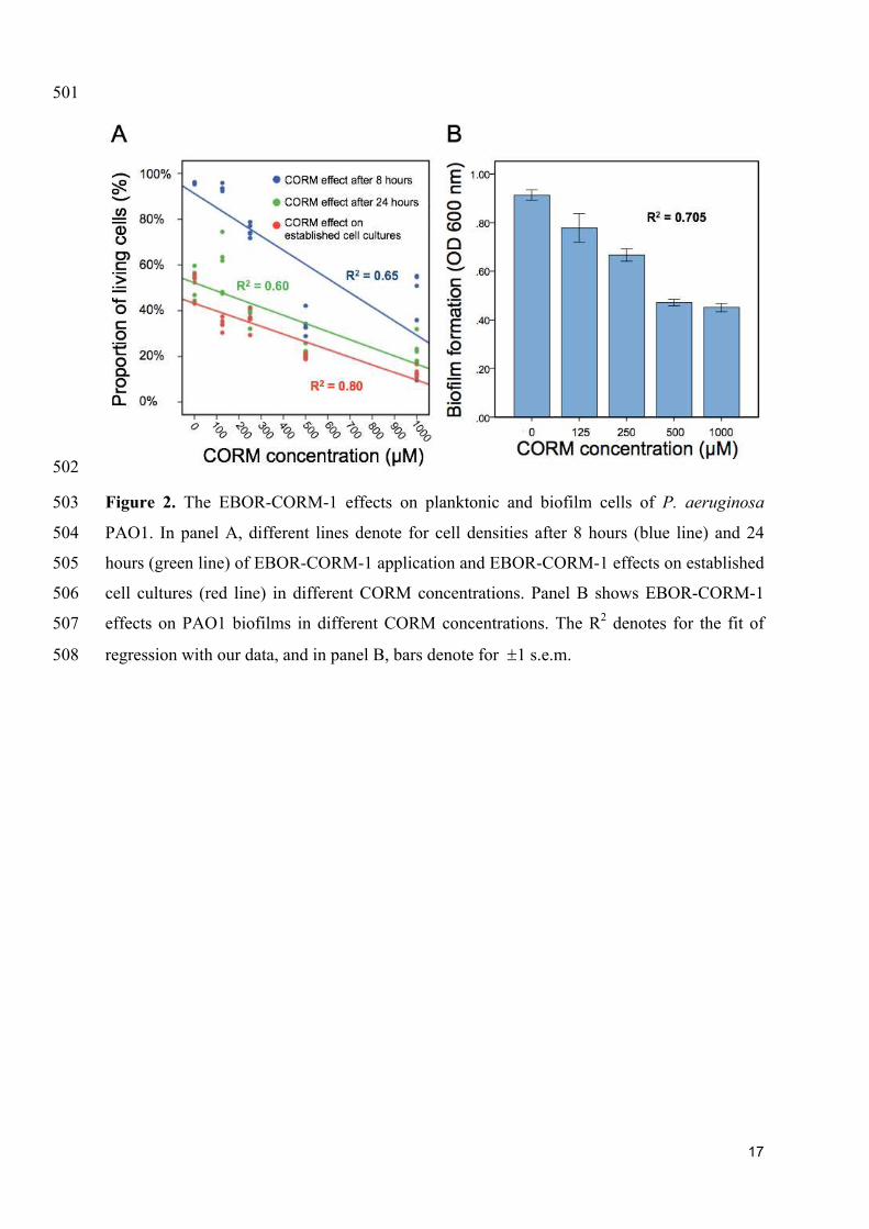
We found that applying EBOR-CORM-1 had generally negative effects on P. aeruginosa 282
PAO1 growth both after 8 and 24 hours of application (F4, 25 = 50.9, p < 0.001 and F4, 25 = 283
31.8, p < 0.001 for proportion of living cells after 8 and 24 hours, respectively, Fig. 2A) and 284
that these negative effects increased along with the increasing concentration of applied 285
EBOR-CORM-1 (regression analysis: F1, 24 = 43, p < 0.001 and F1, 24 = 35, p < 0.001 for 286
proportion of living cells after 8 and 24 hours, respectively, Fig. 2A). Similarly, EBOR-287
CORM-1 was highly effective against both established planktonic and biofilm P. aeruginosa 288
PAO1 cultures (F4, 25 = 77.5, p < 0.001 and F4, 25 = 39.5, p < 0.001, respectively, Fig. 2A-B) 289
and the antimicrobial activity of CORM increased in a density-dependent manner (regression 290
analysis: F1, 24 = 92, p < 0.001 and F1, 24 = 54, p < 0.001, respectively, Fig. 2A-B). 291
EBOR-CORM-1 activity against planktonic and biofilm cells of clinical P. 292
aeruginosa cystic fibrosis isolates 293
Similar to strain PAO1, we found that EBOR-CORM-1 had inhibitory effects on all tested 294
clinical P. aeruginosa isolates after 8 hours of application of CORM (F1, 152 = 11969, p < 295
0.001, Fig. 3A). While this effect did not depend on the lineage (CORM ´ lineage: F1, 152 = 296
1.4, p < 0.001), it varied between different clinical isolates (CORM ´ isolate: F18, 152 = 11969, 297
p < 0.001, Fig. 3A). In contrast, EBOR-CORM-1 had slightly positive effects on P. 298
aeruginosa growth after 24 hours of application (F1, 152 = 256, p < 0.001, Fig. 3B) and this 299
effect varied between different isolates (CORM ´ strain: F18, 152 = 2.8, p = 0.001) being 300
slightly stronger (i.e. positive) with isolates belonging to a lineage B (CORM ´ lineage: F1, 301

10
152 = 24.9, p < 0.001, Fig. 3B). EBOR-CORM-1 also had negative effects when applied to 302
established P. aeruginosa cell cultures (F1, 152 = 222, p < 0.001, Fig. 3C). However, these 303
effects depended on the isolate (CORM ´ isolate: F18, 152 = 2.8, p = 0.001) and the lineage 304
(F1, 152 = 65.2, p = 0.001), reduction being relatively larger with isolates belonging to lineage 305
A (Fig. 3C). In the case of established biofilms, EBOR-CORM-1 had a slightly positive 306
effect (F1, 152 = 9.6, p = 0.002, Fig. 3D) and while this effect varied between different isolates 307
(F18, 152 = 2.0, p = 0.01) it did not differ between the lineages (F1, 152 = 1.2, p = 0.265, 308
respectively, Fig. 3D). Together these results suggest that compared to strain PAO1, EBOR-309
CORM-1 effects varied more with the clinical P. aeruginosa isolates having negative, neutral 310
or positive effects on bacterial growth depending on the isolate identity, lineage and the 311
timing of CORM application. 312
Linking EBOR-CORM-1 antimicrobial activity with clinical P. aeruginosa isolate 313
virulence and growth 314
We found that all the isolates belonging to a lineage A formed non-mucoid colonies (6 out of 315
6), while most of the isolates belonging to a lineage B formed mucoid (i.e., mucus-like) 316
colonies (11 out of 13) on LB plates (typical mucoid and non-mucoid colonies shown in 317
supplementary figure 3). All clinical isolates differed from the non-mucoid PAO1 strain 318
respective of their virulence and growth (Fig. 4A). More specifically, clinical isolates 319
produced less pyoverdine (F1, 23 = 286, p < 0.001) and pyocyanin (F1, 23 = 170, p < 0.001) and 320
grew slower (F1, 23 = 91, p < 0.00) and reached lower maximum densities in LB medium (F1, 321
23 = 15.5, p = 0.001, Fig. 4A). However, clinical isolates produced a considerably larger 322
amount of biofilm (F1, 23 = 21.7, p < 0.001) and showed very low virulence (high time to 323
death) in wax moth larvae in vivo (F1, 23 = 1296, p < 0.001, Fig. 4A). 324
When comparing the two CF lineages, we found that isolates belonging to a lineage B 325
consistently outperformed the isolates belonging to a lineage A by producing more 326
pyoverdine (F1, 18 = 6.06, p = 0.025), biofilm (F1, 18 = 15.08, p = 0.001) and by growing faster 327
(F1, 18 = 22.35, p < 0.001) and to higher maximum densities (F1, 18 = 6.27, p = 0.023) in LB 328
medium (Fig. 4A; Supplementary figure 4). However, lineages did not differ in pyocyanin 329
production (F1, 18 = 1.99, p = 0.176) or virulence (F1, 18 = 1.03, p = 0.324; Fig. 4A; 330
Supplementary figure 4). Across all clinical isolates, density reduction by CORM correlated 331
negatively with biofilm formation (F1, 18 = 4.8, p = 0.042). Together these results suggest that 332
clinical isolates differed from PAO1 and from each other respective to various life-history 333
traits important for establishing an infection. 334
EBOR-CORM-1 activity against clinical P. aeruginosa CF isolate co-cultures 335

11
Despite the observed isolate-specific variation in P. aeruginosa monocultures, EBOR-336
CORM-1 was effective in reducing the growth of P. aeruginosa co-cultures after 24 hours of 337
application (CORM: F1, 24 = 132, p < 0.001, Fig. 4B). Moreover, this reduction was the same 338
regardless of whether the mix contained only one lineage or both lineages (CORM ´ co-339
culture: F2, 24 = 0.5, p = 0.612). These results suggest that intraspecific P. aeruginosa 340
population heterogeneity makes the bacteria more susceptible to EBOR-CORM-1 treatment. 341
EBOR-CORM-1 activity against P. aeruginosa strains in wax moth model 342
We found that P. aeruginosa isolates differed in their virulence (time to death) from each 343
other (F2, 24 = 12.2, p < 0.001): PAO1 and LESB58 strains were equally virulent, and both 344
exhibited higher virulence than the clinical isolate 36 (killing larvae approximately in 17 345
hours [PAO1], 36 hours [LESB58] and 92 hours [clinical isolate 36]; values averaged over 346
both non-CORM and CORM treatments, Fig. 5). In contrast to in vitro results, application of 347
EBOR-CORM-1 did not increase the survival of infected larvae (F1, 24 = 1.3, p = 0.257) with 348
any of the infected strains (CORM ´ strain: F2, 24 = 1.4, p = 0.273, Fig. 5). All larvae became 349
highly pigmented (black throughout) during the infection regardless of the P. aeruginosa 350
isolate. 351
4. DISCUSSION 352
Here we set out to study the antimicrobial activity of [NEt4][MnBr2(CO)4], EBOR-CORM-1, 353
against clinical P. aeruginosa isolates in vitro. This CORM was chosen as a suitable 354
representative of this class of molecule based on the aqueous solubility, facile synthesis 355
(Angelici, 1964), content of a non-toxic metal core, and simple architecture which makes it 356
akin to a “parent compound” for CORMs that have been engineered to possess sophisticated 357
CO release mechanisms. In contrast to more complex CORMs, the molecule was shown to 358
have a water activated mechanism of CO release, as seen in previous studies of [MX(CO)5]- 359
species, where X is a halide (Zhang et al., 2009). Such water induced degradations are 360
believed to proceed via a two-step pathway whereby water causes loss of the halide followed 361
by formation of a dimer species; from which the CO is released. This may explain the 362
changes in the IR spectra recorded in water when compared to chloroform, although the data 363
do not directly match those for [Mn2Br2(CO)8] (El-Sayed and Kaesz, 1963), the product 364
expect on loss of Br- from EBOR-CORM-1. We found that while EBOR-CORM-1showed 365
density-dependent antimicrobial activity against both planktonic and biofilm cells of the 366
widely studied laboratory-adapted strain PAO1, these effects were more varied and weaker 367
against clinical CF lung isolates. Regardless, EBOR-CORM-1 was efficient at reducing the 368

12
growth of CF isolate lineage mixes, which suggests that it could have therapeutic potential in 369
controlling heterogeneous P. aeruginosa infections. Solutions of inactivated EBOR-CORM-1 370
were essentially inactive against P. aeruginosa strain PAO1 (Supplementary figure 2) 371
implying that, at least in this case, the observed activity was due to CO released from the 372
complex rather than the residual metal salts (or indeed [NEt4]+). 373
Similar to a study published by Murray et al. (2012), we found considerable variation in 374
CORM antimicrobial activity between different clinical CF isolates, which depended whether 375
we explored EBOR-CORM-1 effects on relatively short (8 hours) or long timescales (24 376
hours) and if we compared CORM antimicrobial activity on actively growing and established 377
cell cultures (after 48 hours of bacterial growth). Our results after 8 hours of EBOR-CORM-1 378
application are very similar to a previous study (Murray et al., 2012) showing clear reduction 379
in bacterial densities. However, this effect vanished by the 24 hour time point, and 380
surprisingly, some bacterial isolate cultures reached higher optical densities in the presence 381
compared to absence of CORM, which could have been due to increase in number of cells or 382
expression of exoproducts that were picked up by OD600 nm (e.g. pyocyanin or alginate). 383
The most likely explanation for this is that CORM effects were short-lived (Fig. 1), which 384
allowed bacteria to recover and grow to high densities during 24 hours after application of 385
CORM. However, when CORM effects were measured after 4 hours of application to 386
established cell cultures, we could still observe clear reduction in mean bacterial densities. 387
Together these results suggest that CORM effects could be seen up to 4 hours post 388
application and that CORM could eradicate bacterial cells whether they are at exponential or 389
stationary phase of their growth. Interestingly, CORM effects varied between clinical isolates 390
and were clearer with the isolates belonging to lineage A. While Murray et al. (2012) did not 391
observe clear variation in CORM effects against planktonic cell cultures, they found 392
differences in CORM efficiency in eradicating bacterial biofilms. This is also consistent with 393
our data and reinforces the hypothesis that P. aeruginosa clinical isolates are likely to 394
respond differently to CORM therapies. 395
To explore clinical isolate variation in more detail, we compared differences in bacterial 396
virulence and growth traits between the PAO1 and clinical CF lung isolates. We found that 397
relative to strain PAO1, clinical CF isolates grew slower, had lowered virulence and 398
produced lower amounts of pyoverdine and pyocyanin, which are important virulence factors 399
(O'Brien et al., 2017). This is consistent with previous research and typical for P. aeruginosa 400
isolates retrieved from chronic lung infections (Smith et al., 2006;Folkesson et al., 401
2012;Marvig et al., 2013;Williams et al., 2015). The clinical isolates produced much more 402
13
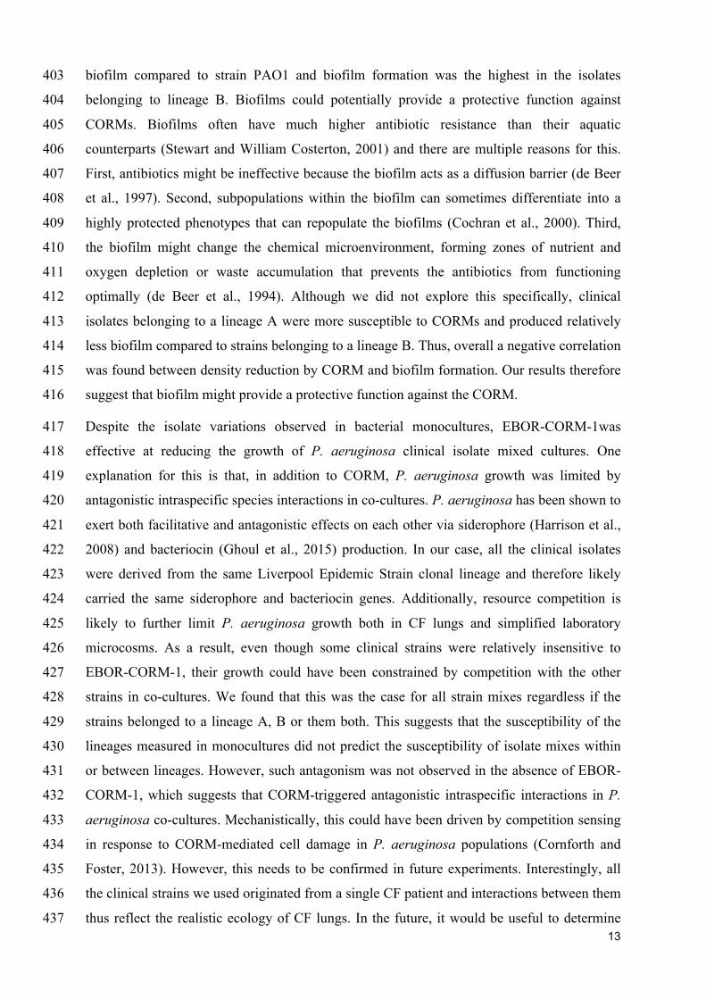
biofilm compared to strain PAO1 and biofilm formation was the highest in the isolates 403
belonging to lineage B. Biofilms could potentially provide a protective function against 404
CORMs. Biofilms often have much higher antibiotic resistance than their aquatic 405
counterparts (Stewart and William Costerton, 2001) and there are multiple reasons for this. 406
First, antibiotics might be ineffective because the biofilm acts as a diffusion barrier (de Beer 407
et al., 1997). Second, subpopulations within the biofilm can sometimes differentiate into a 408
highly protected phenotypes that can repopulate the biofilms (Cochran et al., 2000). Third, 409
the biofilm might change the chemical microenvironment, forming zones of nutrient and 410
oxygen depletion or waste accumulation that prevents the antibiotics from functioning 411
optimally (de Beer et al., 1994). Although we did not explore this specifically, clinical 412
isolates belonging to a lineage A were more susceptible to CORMs and produced relatively 413
less biofilm compared to strains belonging to a lineage B. Thus, overall a negative correlation 414
was found between density reduction by CORM and biofilm formation. Our results therefore 415
suggest that biofilm might provide a protective function against the CORM. 416
Despite the isolate variations observed in bacterial monocultures, EBOR-CORM-1was 417
effective at reducing the growth of P. aeruginosa clinical isolate mixed cultures. One 418
explanation for this is that, in addition to CORM, P. aeruginosa growth was limited by 419
antagonistic intraspecific species interactions in co-cultures. P. aeruginosa has been shown to 420
exert both facilitative and antagonistic effects on each other via siderophore (Harrison et al., 421
2008) and bacteriocin (Ghoul et al., 2015) production. In our case, all the clinical isolates 422
were derived from the same Liverpool Epidemic Strain clonal lineage and therefore likely 423
carried the same siderophore and bacteriocin genes. Additionally, resource competition is 424
likely to further limit P. aeruginosa growth both in CF lungs and simplified laboratory 425
microcosms. As a result, even though some clinical strains were relatively insensitive to 426
EBOR-CORM-1, their growth could have been constrained by competition with the other 427
strains in co-cultures. We found that this was the case for all strain mixes regardless if the 428
strains belonged to a lineage A, B or them both. This suggests that the susceptibility of the 429
lineages measured in monocultures did not predict the susceptibility of isolate mixes within 430
or between lineages. However, such antagonism was not observed in the absence of EBOR-431
CORM-1, which suggests that CORM-triggered antagonistic intraspecific interactions in P. 432
aeruginosa co-cultures. Mechanistically, this could have been driven by competition sensing 433
in response to CORM-mediated cell damage in P. aeruginosa populations (Cornforth and 434
Foster, 2013). However, this needs to be confirmed in future experiments. Interestingly, all 435
the clinical strains we used originated from a single CF patient and interactions between them 436
thus reflect the realistic ecology of CF lungs. In the future, it would be useful to determine 437

14
pairwise interactions between these CF strains and look at CORM effects on other coexisting 438
bacterial species observed in CF infections (Folkesson et al., 2012). 439
We found that EBOR-CORM-1 had no clear therapeutic benefits in the wax moth infection 440
model. There are several potential explanations for this. First, EBOR-CORM-1 had limited 441
long-term activity when in contact with water. As a result, the bactericidal effect may only 442
have elicited lag in the initial phase of bacterial growth and proliferation within the wax 443
moths. Second, insect tissue is not homogeneous and it is possible that we failed to deliver 444
the CORM to the specific area of infection, or that bacteria were able to colonise new areas 445
that were not exposed to the CORM. Third, insects differ from laboratory media (such as LB) 446
as a bacterial growth environment, which could also affect pathogen virulence. For example, 447
it has been recently demonstrated that plant versus animal based growth media can have 448
physiological effects on bacterial virulence (Ketola et al., 2016) and that LB media does not 449
adequately reflect P. aeruginosa growth on lung tissue (Harrison et al., 2014;Harrison and 450
Diggle, 2016). Hence, the wax moth injection model might not reliably reflect the virulence 451
of CF isolates derived from chronic infections. However, it is also the case that many of the 452
affordable and available CF infection animal models do not truly reflect the real CF lung 453
disease environment. It remains to be established whether CORM therapy could be applied in 454
the context of CF lung infections. It is possible, for example, that it might be more suitable 455
for treating topical infections such as burn wounds, for which better animal models are 456
available (Rumbaugh et al., 2012). 457
Further work is also needed to understand the mode of action of EBOR-CORM-1. While 458
respiratory oxidases and globins at heme targets are generally considered the prime targets of 459
CO and CORMs (Wareham et al., 2015), it has been demonstrated that CORMs can have 460
multiple different other targets (Wilson et al., 2015). For example, CO also binds to the di-461
iron site in bacterial NO reductases and to iron, copper, and nickel sites in certain microbial 462
proteins such as CO dehydrogenase (Lu et al., 2004;Wasser et al., 2005). In some cases, 463
CORMs might have intracellular targets but their accumulation within the cells can be very 464
weak (Tinajero-Trejo et al., 2016). Moreover, in the future it would be important to test if 465
EBOR-CORM-1 is cytotoxic to eukaryotic cells. The concentration we used are in line with 466
previously published work where no, or very mild, cytotoxic effects were observed (Murray 467
et al., 2012). We are currently conducting experiments to validate this independently and to 468
understand how EBOR-CORM-1interacts with bacterial cells. While, our wax moth assays 469
show that the concentrations we used had no negative effects on short-term insect viability, 470
more detailed cytotoxicity assays are needed in the future. Lastly, the low solubility of 471

15
EBOR-CORM-1 in water, and its activation in this medium, is problematic for delivery and 472
activation at specific sites within patients. In addition to chemically increasing the molecule 473
stability, CORMs could be enclosed in microvesicles (van Dommelen et al., 2012) to ensure 474
more efficient antimicrobial activity and drug delivery. 475
In conclusion, our results show that EBOR-CORM-1 shows antimicrobial activity against 476
both planktonic and biofilm cells of P. aeruginosa strain PAO1 but that these effects are 477
more varied and less pronounced against clinical CF lung isolates in monocultures. In 478
contrast, more heterogeneous P. aeruginosa populations comprising intraspecific phenotypic 479
variants were more susceptible to CORM treatment. This potentially has wider implications 480
in the testing of novel therapeutics. At present, this is done almost exclusively using clonal P. 481
aeruginosa populations. Our observations suggest that testing carried out on more 482
heterogeneous populations of P. aeruginosa, more closely resembling those found in the CF 483
lung, may give different and sometimes more promising results. 484
485
ACKNOWLEDGEMENTS 486
We thank James Pitt for carrying out an initial EBOR-CORM-1 synthesis and TARGeTED 487
Antimicrobial Resistance (AMR) Project and EPSRC council for funding (EP/M027538/1). 488
Ville-Petri Friman is also supported by the Wellcome Trust [reference no. 105624] through 489
the Centre for Chronic Diseases and Disorders (C2D2) at the University of York. 490
491
FIGURES AND FIGURE LEGENDS 492

16
493
Figure 1. The stability of1 EBOR-CORM-1. Panel A shows the IR spectra of EBOR-CORM-494
1 upon heating at 50° C for 0 hours (black line), for 1 hours (red line), for 2 hours (blue) and 495
for 3 hours (pink). The structure of the EBOR-CORM-1 is shown in inset on the left. Panel B 496
shows the gas phase IR spectra of EBOR-CORM-1 in chloroform with added water where 497
the * indicates a band from chloroform. Panel C shows the IR spectra of EBOR-CORM-1 in 498
chloroform (top) and water (bottom) and panel D the IR spectra of CO in water after 1 min 499
(top) and 90 min (bottom) dissolution. All frequencies given are in cm-1
. 500
17
501
502
Figure 2. The EBOR-CORM-1 effects on planktonic and biofilm cells of P. aeruginosa 503
PAO1. In panel A, different lines denote for cell densities after 8 hours (blue line) and 24 504
hours (green line) of EBOR-CORM-1 application and EBOR-CORM-1 effects on established 505
cell cultures (red line) in different CORM concentrations. Panel B shows EBOR-CORM-1 506
effects on PAO1 biofilms in different CORM concentrations. The R2 denotes for the fit of 507
regression with our data, and in panel B, bars denote for ±1 s.e.m. 508

18
509
510
Figure 3. The EBOR-CORM-1 effects on planktonic and biofilm cells of clinical P. 511
aeruginosa CF isolates. Panels A and B show the proportion of living cells after 8 hours and 512
24 hours of EBOR-CORM-1 application, respectively. Panel C and D show the EBOR-513
CORM-1 effects on established cell cultures and biofilms, respectively. In all panels, bars 514
denote for ±1 s.e.m. 515
516

19
517 518
Figure 4. Differences in P. aeruginosa growth and virulence trait variation between PAO1 519
and clinical CF isolates (panel A) and EBOR-CORM-1 effects on clinical CF isolate lineage 520
mixes (panel B). In panel A, different colours denote for pyocyanin (blue) and pyoverdine 521
(green) production, time to death (black), growth rate (purple), maximum density (yellow) 522
and biofilm production for clinical isolates belonging to lineages A and B. The dashed line 523
shows the mean performance of PAO1 strain. Panel B shows EBOR-CORM-1 effect on 524
clinical CF isolate mixes after 24 hours of CORM application. In panel A, bars denote for ±1 525
s.e.m., and in panel B, extreme values around lower and upper quartile (black line shows the 526
median). 527

20
528
Figure 5. The EBOR-CORM-1 activity against three P. aeruginosa strains in wax moth 529
model. Boxplots show larval survival in the absence (light grey) and presence (blue) of 530
EBOR-CORM-1 for PAO1, LESB58 and clinical isolate #36 (lineage B). Bars show extreme 531
values around lower and upper quartile and black lines show the median. 532

21
REFERENCES 533
Angelici, R.J. (1964). Preparation, Characterization, and Reactions of the cis-534
Dihalotetracarbonylmanganate (I) Anions. Inorganic Chemistry 3, 1099-1102. 535
Brown, E.D., and Wright, G.D. (2016). Antibacterial drug discovery in the resistance era. 536
Nature 529, 336-343. 537
Cochran, W., Mcfeters, G., and Stewart, P. (2000). Reduced susceptibility of thin 538
Pseudomonas aeruginosa biofilms to hydrogen peroxide and monochloramine. 539
Journal of applied microbiology 88, 22-30. 540
Cornforth, D.M., and Foster, K.R. (2013). Competition sensing: the social side of bacterial 541
stress responses. Nature Reviews Microbiology 11, 285-293. 542
De Beer, D., Stoodley, P., and Lewandowski, Z. (1997). Measurement of local diffusion 543
coefficients in biofilms by microinjection and confocal microscopy. Biotechnology 544
and Bioengineering 53, 151-158. 545
De Beer, D., Stoodley, P., Roe, F., and Lewandowski, Z. (1994). Effects of biofilm structures 546
on oxygen distribution and mass transport. Biotechnology and bioengineering 43, 547
1131-1138. 548
Desmard, M., Davidge, K.S., Bouvet, O., Morin, D., Roux, D., Foresti, R., Ricard, J.D., 549
Denamur, E., Poole, R.K., Montravers, P., Motterlini, R., and Boczkowski, J. (2009). 550
A carbon monoxide-releasing molecule (CORM-3) exerts bactericidal activity against 551
Pseudomonas aeruginosa and improves survival in an animal model of bacteraemia. 552
Faseb j 23, 1023-1031. 553
El-Sayed, M., and Kaesz, H. (1963). Infrared Spectra and Structure of the Tetracarbonyl 554
Halide Dimers of Manganese, Technetium, and Rhenium. Inorganic Chemistry 2, 555
158-162. 556
Flume, P.A., Mogayzel, P.J., Robinson, K.A., Rosenblatt, R.L., Quittell, L., and Marshall, 557
B.C. (2010). Cystic Fibrosis Pulmonary Guidelines. American Journal of Respiratory 558
and Critical Care Medicine 182, 298-306. 559
Folkesson, A., Jelsbak, L., Yang, L., Johansen, H.K., Ciofu, O., Hoiby, N., and Molin, S. 560
(2012). Adaptation of Pseudomonas aeruginosa to the cystic fibrosis airway: an 561
evolutionary perspective. Nature Reviews Microbiology 10, 841-851. 562
Ghoul, M., West, S.A., Johansen, H.K., Molin, S., Harrison, O.B., Maiden, M.C., Jelsbak, L., 563
Bruce, J.B., and Griffin, A.S. (2015). Bacteriocin-mediated competition in cystic 564
fibrosis lung infections. Proc Biol Sci 282. 565
Harrison, F., and Diggle, S.P. (2016). An ex vivo lung model to study bronchioles infected 566
with Pseudomonas aeruginosa biofilms. Microbiology 162, 1755-1760. 567
Harrison, F., Muruli, A., Higgins, S., and Diggle, S.P. (2014). Development of an ex vivo 568
porcine lung model for studying growth, virulence, and signaling of Pseudomonas 569
aeruginosa. Infection and immunity 82, 3312-3323. 570
Harrison, F., Paul, J., Massey, R.C., and Buckling, A. (2008). Interspecific competition and 571
siderophore-mediated cooperation in Pseudomonas aeruginosa. ISME J 2, 49-55. 572
Jimenez, J., Chakraborty, I., Carrington, S.J., and Mascharak, P.K. (2016). Light-triggered 573
CO delivery by a water-soluble and biocompatible manganese photoCORM. Dalton 574
Trans 45, 13204-13213. 575
Ketola, T., Mikonranta, L., Laakso, J., and Mappes, J. (2016). Different food sources elicit 576
fast changes to bacterial virulence. Biol Lett 12, 20150660. 577
Klein, M., Neugebauer, U., Gheisari, A., Malassa, A., Jazzazi, T.M., Froehlich, F., 578
Westerhausen, M., Schmitt, M., and Popp, J.R. (2014). IR spectroscopic methods for 579
the investigation of the CO release from CORMs. The Journal of Physical Chemistry 580
A 118, 5381-5390. 581

22
Lu, S., Suharti, De Vries, S., and Moënne-Loccoz, P. (2004). Two CO Molecules Can Bind 582
Concomitantly at the Diiron Site of NO Reductase from Bacillus a zotoformans. 583
Journal of the American Chemical Society 126, 15332-15333. 584
Marvig, R.L., Johansen, H.K., Molin, S., and Jelsbak, L. (2013). Genome analysis of a 585
transmissible lineage of pseudomonas aeruginosa reveals pathoadaptive mutations 586
and distinct evolutionary paths of hypermutators. PLoS Genet 9, e1003741. 587
Motterlini, R., and Otterbein, L.E. (2010). The therapeutic potential of carbon monoxide. Nat 588
Rev Drug Discov 9, 728-743. 589
Mowat, E., Paterson, S., Fothergill, J.L., Wright, E.A., Ledson, M.J., Walshaw, M.J., 590
Brockhurst, M.A., and Winstanley, C. (2011). Pseudomonas aeruginosa population 591
diversity and turnover in cystic fibrosis chronic infections. Am J Respir Crit Care 592
Med 183, 1674-1679. 593
Murray, T.S., Okegbe, C., Gao, Y., Kazmierczak, B.I., Motterlini, R., Dietrich, L.E., and 594
Bruscia, E.M. (2012). The carbon monoxide releasing molecule CORM-2 attenuates 595
Pseudomonas aeruginosa biofilm formation. PLoS One 7, e35499. 596
Nobre, L.S., Seixas, J.D., Romao, C.C., and Saraiva, L.M. (2007). Antimicrobial action of 597
carbon monoxide-releasing compounds. Antimicrob Agents Chemother 51, 4303-598
4307. 599
Normark, B.H., and Normark, S. (2002). Evolution and spread of antibiotic resistance. 600
Journal of internal medicine 252, 91-106. 601
O'brien, S., Williams, D., Fothergill, J.L., Paterson, S., Winstanley, C., and Brockhurst, M.A. 602
(2017). High virulence sub-populations in Pseudomonas aeruginosa long-term cystic 603
fibrosis airway infections. BMC Microbiol 17, 30. 604
O’neill, J. (2014). "Antimicrobial Resistance: Tackling a Crisis for the Health and Wealth of 605
Nations", in: Review on Antimicrobial Resistance. HM Government & Wellcome 606
Trust ). 607
Poole, K. (2011). Pseudomonas aeruginosa: resistance to the max. Front Microbiol 2, 65. 608
Pritt, B., O'brien, L., and Winn, W. (2007). Mucoid Pseudomonas in cystic fibrosis. Am J 609
Clin Pathol 128, 32-34. 610
Reszka, K.J., O'malley, Y., Mccormick, M.L., Denning, G.M., and Britigan, B.E. (2004). 611
Oxidation of pyocyanin, a cytotoxic product from Pseudomonas aeruginosa, by 612
microperoxidase 11 and hydrogen peroxide. Free Radic Biol Med 36, 1448-1459. 613
Rumbaugh, K.P., Trivedi, U., Watters, C., Burton-Chellew, M.N., Diggle, S.P., and West, 614
S.A. (2012). Kin selection, quorum sensing and virulence in pathogenic bacteria. Proc 615
Biol Sci 279, 3584-3588. 616
Smith, E.E., Buckley, D.G., Wu, Z., Saenphimmachak, C., Hoffman, L.R., D'argenio, D.A., 617
Miller, S.I., Ramsey, B.W., Speert, D.P., Moskowitz, S.M., Burns, J.L., Kaul, R., and 618
Olson, M.V. (2006). Genetic adaptation by Pseudomonas aeruginosa to the airways of 619
cystic fibrosis patients. Proceedings of the National Academy of Sciences of the 620
United States of America 103, 8487-8492. 621
Stamellou, E., Storz, D., Botov, S., Ntasis, E., Wedel, J., Sollazzo, S., Kramer, B.K., Van 622
Son, W., Seelen, M., Schmalz, H.G., Schmidt, A., Hafner, M., and Yard, B.A. (2014). 623
Different design of enzyme-triggered CO-releasing molecules (ET-CORMs) reveals 624
quantitative differences in biological activities in terms of toxicity and inflammation. 625
Redox Biol 2, 739-748. 626
Stewart, P.S., and William Costerton, J. (2001). Antibiotic resistance of bacteria in biofilms. 627
The Lancet 358, 135-138. 628
Tinajero-Trejo, M., Rana, N., Nagel, C., Jesse, H.E., Smith, T.W., Wareham, L.K., Hippler, 629
M., Schatzschneider, U., and Poole, R.K. (2016). Antimicrobial Activity of the 630
Manganese Photoactivated Carbon Monoxide-Releasing Molecule [Mn (CO) 3 (tpa-631
κ3 N)]+ Against a Pathogenic Escherichia coli that Causes Urinary Infections. 632
Antioxidants & redox signaling 24, 765-780. 633
23
Van Dommelen, S.M., Vader, P., Lakhal, S., Kooijmans, S., Van Solinge, W.W., Wood, 634
M.J., and Schiffelers, R.M. (2012). Microvesicles and exosomes: opportunities for 635
cell-derived membrane vesicles in drug delivery. Journal of Controlled Release 161, 636
635-644. 637
Ward, J.S., Lynam, J.M., Moir, J., and Fairlamb, I.J. (2014). Visible‐Light‐Induced CO 638
Release from a Therapeutically Viable Tryptophan‐Derived Manganese (I) 639
Carbonyl (TryptoCORM) Exhibiting Potent Inhibition against E. coli. Chemistry-A 640
European Journal 20, 15061-15068. 641
Ward, J.S., Morgan, R., Lynam, J.M., Fairlamb, I.J., and Moir, J.W. (2017). Toxicity of 642
tryptophan manganese (I) carbonyl (Trypto-CORM), against Neisseria gonorrhoeae. 643
MedChemComm 8, 346-352. 644
Wareham, L.K., Poole, R.K., and Tinajero-Trejo, M. (2015). CO-releasing Metal Carbonyl 645
Compounds as Antimicrobial Agents in the Post-antibiotic Era. J Biol Chem 290, 646
18999-19007. 647
Wasser, I.M., Huang, H.-W., Moënne-Loccoz, P., and Karlin, K.D. (2005). Heme/non-heme 648
diiron (II) complexes and O2, CO, and NO adducts as reduced and substrate-bound 649
models for the active site of bacterial nitric oxide reductase. Journal of the American 650
Chemical Society 127, 3310-3320. 651
Who.Int (2010). WHO | Genes and human disease [Online]. Available: 652
http://www.who.int/genomics/public/geneticdiseases/en/index2.html [Accessed 16-02 653
2017]. 654
Williams, D., Evans, B., Haldenby, S., Walshaw, M.J., Brockhurst, M.A., Winstanley, C., 655
and Paterson, S. (2015). Divergent, coexisting Pseudomonas aeruginosa lineages in 656
chronic cystic fibrosis lung infections. Am J Respir Crit Care Med 191, 775-785. 657
Williams, D., Paterson, S., Brockhurst, M.A., and Winstanley, C. (2016). Refined analyses 658
suggest that recombination is a minor source of genomic diversity in Pseudomonas 659
aeruginosa chronic cystic fibrosis infections. Microbial Genomics 2. 660
Wilson, J.L., Wareham, L.K., Mclean, S., Begg, R., Greaves, S., Mann, B.E., Sanguinetti, G., 661
and Poole, R.K. (2015). CO-Releasing Molecules Have Nonheme Targets in Bacteria: 662
Transcriptomic, Mathematical Modeling and Biochemical Analyses of CORM-3 663
[Ru(CO)3Cl(glycinate)] Actions on a Heme-Deficient Mutant of Escherichia coli. 664
Antioxid Redox Signal 23, 148-162. 665
Winstanley, C., Langille, M.G., Fothergill, J.L., Kukavica-Ibrulj, I., Paradis-Bleau, C., 666
Sanschagrin, F., Thomson, N.R., Winsor, G.L., Quail, M.A., Lennard, N., Bignell, A., 667
Clarke, L., Seeger, K., Saunders, D., Harris, D., Parkhill, J., Hancock, R.E., 668
Brinkman, F.S., and Levesque, R.C. (2009). Newly introduced genomic prophage 669
islands are critical determinants of in vivo competitiveness in the Liverpool Epidemic 670
Strain of Pseudomonas aeruginosa. Genome Res 19, 12-23. 671
Winstanley, C., O'brien, S., and Brockhurst, M.A. (2016). Pseudomonas aeruginosa 672
Evolutionary Adaptation and Diversification in Cystic Fibrosis Chronic Lung 673
Infections. Trends Microbiol 24, 327-337. 674
Workentine, M.L., Sibley, C.D., Glezerson, B., Purighalla, S., Norgaard-Gron, J.C., Parkins, 675
M.D., Rabin, H.R., and Surette, M.G. (2013). Phenotypic heterogeneity of 676
Pseudomonas aeruginosa populations in a cystic fibrosis patient. PLoS One 8, e60225. 677
Zhang, W.-Q., Atkin, A.J., Thatcher, R.J., Whitwood, A.C., Fairlamb, I.J., and Lynam, J.M. 678
(2009). Diversity and design of metal-based carbon monoxide-releasing molecules 679
(CO-RMs) in aqueous systems: revealing the essential trends. Dalton Transactions, 680
4351-4358. 681
682