Sex differences in corticotropin-releasing factor signaling and trafficking
16
Sex Differences in Corticotropin-Releasing Factor Receptor Signaling and Trafficking: Potential Role in Female Vulnerability to Stress-Related Psychopathology Debra A. Bangasser, PhD 1 , Andre Curtis, PhD 1 , Beverly A.S. Reyes, PhD 3 , Thelma T. Bethea 1 , Ioannis Parastatidis, MD 2 , Harry Ischiropoulos, PhD 2 , Elisabeth J. Van Bockstaele, PhD 3 , and Rita J. Valentino, PhD 1 1 Department of Anesthesiology, The Children's Hospital of Philadelphia, Philadelphia, PA 19104 2 Department of Pediatrics, The Children's Hospital of Philadelphia, Philadelphia, PA 19104 3 Department of Neurosurgery, Thomas Jefferson University, Farber Institute for Neurosciences, Philadelphia, PA 19107 Abstract Although the higher incidence of stress-related psychiatric disorders in females is well documented, its basis is unknown. Here we demonstrate that the receptor for corticotropin- releasing factor (CRF), the neuropeptide that orchestrates the stress response, signals and is trafficked differently in female rats in a manner that could result in a greater response and decreased adaptation to stressors. Most cellular responses to CRF in the brain are mediated by CRF receptor (CRFr) association with the GTP-binding protein, G s . Receptor immunoprecipitation studies revealed enhanced CRFr-G s coupling in cortical tissue of unstressed female rats. Previous stressor exposure abolished this sex difference by increasing CRFr-G s coupling selectively in males. These molecular results mirrored the effects of sex and stress on sensitivity of locus ceruleus (LC)-norepinephrine neurons to CRF. Differences in CRFr trafficking were also identified that could compromise stress adaptation in females. Specifically, stress- induced CRFr association with β-arrestin2, an integral step in receptor internalization, occurred only in male rats. Immunoelectron microscopy confirmed that stress elicited CRFr internalization in LC neurons of male rats exclusively, consistent with reported electrophysiological evidence for stress-induced desensitization to CRF in males. Together, these studies identified two aspects of CRFr function, increased cellular signaling and compromised internalization, which render CRF- receptive neurons of females more sensitive to low levels of CRF and less adaptable to high levels of CRF. CRFr dysfunction in females may underlie their increased vulnerability to develop stress- related pathology, particularly that related to increased activity of the LC-norepinephrine system, such as depression or post-traumatic stress disorder. Users may view, print, copy, download and text and data- mine the content in such documents, for the purposes of academic research, subject always to the full Conditions of use: http://www.nature.com/authors/editorial_policies/license.html#terms Corresponding author information Debbie Bangasser, PhD The Children's Hospital of Philadelphia 3615 Civic Center Blvd. ARC 402E Philadelphia, PA 19104 Phone: 215-590-0654; Fax: 215-590-3364 [email protected]. Supplementary Information is available at Molecular Psychiatry at http://www.nature.com/mp/index.html. CONFLICT OF INTEREST: We have no competing financial interests in relation to the work described. NIH Public Access Author Manuscript Mol Psychiatry. Author manuscript; available in PMC 2011 March 1. Published in final edited form as: Mol Psychiatry. 2010 September ; 15(9): 877–904. doi:10.1038/mp.2010.66. NIH-PA Author Manuscript NIH-PA Author Manuscript NIH-PA Author Manuscript
Transcript of Sex differences in corticotropin-releasing factor signaling and trafficking

Sex Differences in Corticotropin-Releasing Factor ReceptorSignaling and Trafficking: Potential Role in Female Vulnerabilityto Stress-Related Psychopathology
Debra A. Bangasser, PhD1, Andre Curtis, PhD1, Beverly A.S. Reyes, PhD3, Thelma T.Bethea1, Ioannis Parastatidis, MD2, Harry Ischiropoulos, PhD2, Elisabeth J. VanBockstaele, PhD3, and Rita J. Valentino, PhD1
1Department of Anesthesiology, The Children's Hospital of Philadelphia, Philadelphia, PA 191042 Department of Pediatrics, The Children's Hospital of Philadelphia, Philadelphia, PA 191043Department of Neurosurgery, Thomas Jefferson University, Farber Institute for Neurosciences,Philadelphia, PA 19107
AbstractAlthough the higher incidence of stress-related psychiatric disorders in females is welldocumented, its basis is unknown. Here we demonstrate that the receptor for corticotropin-releasing factor (CRF), the neuropeptide that orchestrates the stress response, signals and istrafficked differently in female rats in a manner that could result in a greater response anddecreased adaptation to stressors. Most cellular responses to CRF in the brain are mediated byCRF receptor (CRFr) association with the GTP-binding protein, Gs. Receptorimmunoprecipitation studies revealed enhanced CRFr-Gs coupling in cortical tissue of unstressedfemale rats. Previous stressor exposure abolished this sex difference by increasing CRFr-Gscoupling selectively in males. These molecular results mirrored the effects of sex and stress onsensitivity of locus ceruleus (LC)-norepinephrine neurons to CRF. Differences in CRFr traffickingwere also identified that could compromise stress adaptation in females. Specifically, stress-induced CRFr association with β-arrestin2, an integral step in receptor internalization, occurredonly in male rats. Immunoelectron microscopy confirmed that stress elicited CRFr internalizationin LC neurons of male rats exclusively, consistent with reported electrophysiological evidence forstress-induced desensitization to CRF in males. Together, these studies identified two aspects ofCRFr function, increased cellular signaling and compromised internalization, which render CRF-receptive neurons of females more sensitive to low levels of CRF and less adaptable to high levelsof CRF. CRFr dysfunction in females may underlie their increased vulnerability to develop stress-related pathology, particularly that related to increased activity of the LC-norepinephrine system,such as depression or post-traumatic stress disorder.
Users may view, print, copy, download and text and data- mine the content in such documents, for the purposes of academic research,subject always to the full Conditions of use: http://www.nature.com/authors/editorial_policies/license.html#terms
Corresponding author information Debbie Bangasser, PhD The Children's Hospital of Philadelphia 3615 Civic Center Blvd. ARC402E Philadelphia, PA 19104 Phone: 215-590-0654; Fax: 215-590-3364 [email protected].
Supplementary Information is available at Molecular Psychiatry at http://www.nature.com/mp/index.html.
CONFLICT OF INTEREST:We have no competing financial interests in relation to the work described.
NIH Public AccessAuthor ManuscriptMol Psychiatry. Author manuscript; available in PMC 2011 March 1.
Published in final edited form as:Mol Psychiatry. 2010 September ; 15(9): 877–904. doi:10.1038/mp.2010.66.
NIH
-PA Author Manuscript
NIH
-PA Author Manuscript
NIH
-PA Author Manuscript

Keywordsstress; locus ceruleus; norepinephrine; corticotropin-releasing hormone; depression; post-traumaticstress disorder
Stress-related psychiatric disorders (e.g., depression, post-traumatic stress disorder) aretwice as prevalent in women compared to men (1-3). Although the neurobiological basis forthis is unknown, differences in stress reactivity have been implicated in this disparity (4-7).Because corticotropin-releasing factor (CRF), a primary mediator of the stress response, isdysregulated in stress-related psychiatric disorders, it is a likely substrate for sex differencesin stress vulnerability (8-11). Indeed, evidence for direct estrogenic regulation of CRF geneexpression provides a compelling mechanism for sexual dimorphism of stress reactivity andprevalence of stress-related psychopathology in women (12,13).
CRF acts as a neurohormone to initiate the hypothalamic-pituitary-adrenal response to stressand as a neurotransmitter to initiate autonomic, behavioral, and cognitive components of thestress response (14-16). One target of CRF neurotransmission is the locus ceruleus (LC), thesource of the major brain norepinephrine system that regulates emotional arousal (17-20).CRF activates LC neurons during stress and this is associated with heightened arousal(19,21,22). Although these effects are adaptive in response to an acute stressor, persistent orinappropriate LC-norepinephrine activation has pathological consequences. Indeed,excessive activity of CRF and LC-norepinephrine systems is thought to underlie the corefeature of hyperarousal in melancholic depression (8,10,23). Similarly, CRF hypersecretionand increased LC sensitivity have been implicated in post-traumatic stress disorder (24,25).Thus, sex differences in these systems or their interaction could contribute to femalevulnerability to these stress-related illnesses.
Our previous electrophysiological studies demonstrated sex differences in LC sensitivity toCRF and its regulation by prior stress that could be expressed as excessive activation of theLC-norepinephrine system in females (26). LC neurons of unstressed female rats were moresensitive to CRF compared to males, as indicated by a leftward shift in the CRF dose-response curve. Additionally, prior swim stress sensitized LC neurons of male rats only tolow doses of CRF and desensitized them to high doses, such that the CRF dose-responsecurve shifted to the left to match that seen in unstressed females but plateaued at a lowerlevel. Together, the findings suggested that the CRF receptor (CRFr), which mediates LCactivation by CRF, signals and/or is trafficked differently in males and females.
This study was designed to identify the molecular basis for sex differences in neuronalsensitivity to CRF. Because CRFr signaling occurs primarily through its coupling to theGTP binding protein, Gs, receptor immunoprecipitation was used to determine whetherCRFr-Gs coupling differed in male and female rats (27). To examine potential sexdifferences in CRFr trafficking, CRFr phosphorylation and association with β-arrestin2 werecompared, as these are important steps in the CRFr internalization process (28,29). Toconfirm that the molecular events had cellular consequences, immunoelectron microscopywas used to visualize cellular compartmentalization of the receptor and stress-inducedinternalization. The results converged to reveal sexual dimorphism in CRFr function atmolecular and cellular levels that could contribute to the higher incidence of certain stress-related psychiatric disorders in females.
Bangasser et al. Page 2
Mol Psychiatry. Author manuscript; available in PMC 2011 March 1.
NIH
-PA Author Manuscript
NIH
-PA Author Manuscript
NIH
-PA Author Manuscript

METHODS AND MATERIALSSubjects
The subjects were male and female Sprague-Dawley rats (Charles River, Wilmington, MA).Females were intact or ovariectomized by the vendor at 42 days. Rats were approximately47 days of age when shipped and used approximately 2 weeks after arrival. Shipments ofmale and female rats were age matched. See Supplementary Information (SI) Methods fordetails on subjects, housing conditions, and tracking the estrous cycle. Care and use ofanimals was approved by the Children's Hospital of Philadelphia Institutional Animal Careand Use Committee and was in accordance with the NIH Guide for the Care and Use ofLaboratory Animals.
ElectrophysiologyExtracellular single unit LC activity was recorded in the halothane-anesthetized state 24 hafter a 15 min swim stress or brief handling as described (26). The 24 h timepoint waschosen to correspond to the time that sex differences in stress regulation of LC sensitivity toCRF were observed (26). Surgery and procedures for LC recording coupled with drugmicroinfusion were as previously described with modifications detailed in SI Methods (26).Artificial cerebrospinal fluid (ACSF) or the cyclic adenosine monophosphate (cAMP)/protein kinase A antagonist, Rp-cAMP-S (adenosine-3′, 5′-cyclic monophosphorothioate,Rp-isomer, triethylammonium salt), was microinfused into the LC (600 ng in 120 nl ACSF)20-40 min before CRF (1-30 ng in 30 nl ACSF). LC activity was recorded at least 6 minbefore and 10 min after CRF. Only one dose of CRF was tested on a single cell and only onecell tested in an individual rat. Recording sites were histologically identified as previouslydescribed (26).
CRFr immunoprecipitationTissue was usually collected 24 h after stress or handling, to match electrophysiologicaltimepoints. For receptor phosphorylation and β-arrestin2 studies tissue also was collectedimmediately after and one hour after stressor exposure. Unanesthetized rats were placed in aflexible plastic restrainer (Decapicone) and rapidly decapitated. Frontal cortex (anterior tothe +3.20 AP coordinate, relative to Bregma) was dissected and frozen (−80°C). Initialstudies revealed no sex- or stress-induced differences in cortical CRFr expression indicatingthat cortical samples prepared for the immunoprecipitation procedure contained comparableamounts of CRFr protein in each group (see SI Results). CRFr was immunoprecipitatedfrom 3 pooled samples for a single determination as detailed in SI Methods.
Western blottingImmunoprecipitated samples (5 μg per condition) were subjected to SDS-PAGE gelelectrophoresis and proteins transferred to polyvinylidene fluoride membranes (Immobilon-FL) as described (26). Membranes were probed for specific proteins as previously describedwith modifications detailed in SI Methods (30). Odyssey Infrared Imaging softwarequantified the integrated intensity of each band and determined molecular weights based onBiorad Precision Plus Protein Standards. The ratio of target protein (Gs, Go, Gq/11,phosphothreonine, or β-arrestin2) to CRFr was calculated and the mean ratios werecompared between groups using ANOVAs. For the Figures, each individual fluorescentchannel of the image was adjusted for brightness and contrast using the Odyssey InfraredImaging Software.
Bangasser et al. Page 3
Mol Psychiatry. Author manuscript; available in PMC 2011 March 1.
NIH
-PA Author Manuscript
NIH
-PA Author Manuscript
NIH
-PA Author Manuscript
Immunoelectron microscopyTissue preparation, immunolabeling, and quantification for the immunoelectron microscopystudies were as previously described (31,32). Immunogold-silver and immunoperoxidaselabeling were used to detect CRFr- and tyrosine hydroxylase (TH)-immunoreactivity,respectively. Further details on immunoelectron microscopy methods and quantification arein SI (methods and Fig. S3).
Antibody controlsEvidence that the antibodies used are detecting only CRF1 in cortical and LC tissue in thesestudies is described in detail in SI Methods. Thus, CRFr refers to CRF1 in cortical and LCtissue in this study.
RESULTSCRFr signaling is increased in females and differentially regulated by stress compared tomales
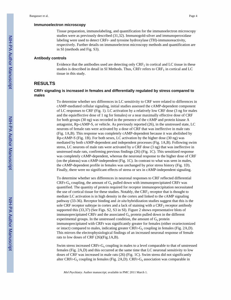
To determine whether sex differences in LC sensitivity to CRF were related to differences incAMP-mediated cellular signaling, initial studies assessed the cAMP-dependent componentof LC responses to CRF (Fig. 1). LC activation by a relatively low CRF dose (3 ng for malesand the equieffective dose of 1 ng for females) or a near maximally effective dose of CRFfor both groups (30 ng) was recorded in the presence of the cAMP and protein kinase Aantagonist, Rp-cAMP-S, or vehicle. As previously reported (26), in the unstressed state, LCneurons of female rats were activated by a dose of CRF that was ineffective in male rats(Fig. 1A,B). This response was completely cAMP-dependent because it was abolished byRp-cAMP-S (Fig. 1B). For both sexes, LC activation by the higher dose (30 ng) wasmediated by both cAMP-dependent and independent processes (Fig. 1A,B). Following swimstress, LC neurons of male rats were activated by a CRF dose (3 ng) that was ineffective inunstressed male rats, confirming previous findings (26) (Fig. 1C). This sensitized responsewas completely cAMP-dependent, whereas the neuronal response to the higher dose of CRF(on the plateau) was cAMP-independent (Fig. 1C). In contrast to what was seen in males,the cAMP-dependent profile in females was unchanged by prior stress history (Fig. 1D).Finally, there were no significant effects of stress or sex in cAMP-independent signaling.
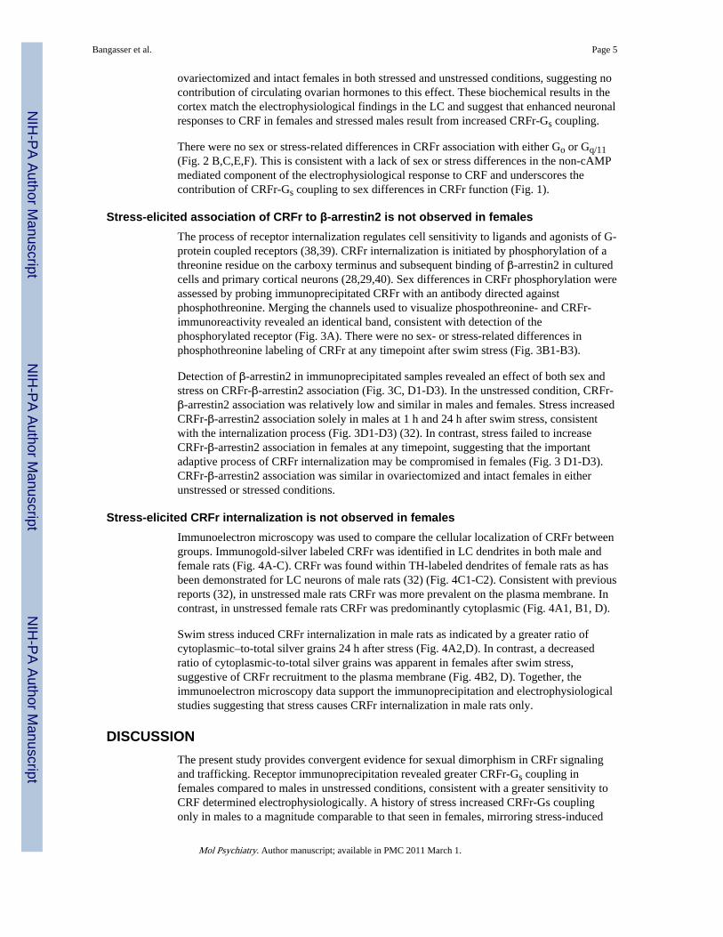
To determine whether sex differences in neuronal responses to CRF reflected differentialCRFr-Gs coupling, the amount of Gs pulled down with immunoprecipitated CRFr wasquantified. The quantity of protein required for receptor immunoprecipitation necessitatedthe use of cortical tissue for these studies. Notably, the CRF1 receptor that is thought tomediate LC activation is in high density in the cortex and linked to the cAMP signalingpathway (33-36). Receptor binding and in situ hybridization studies suggest that this is thesole CRF receptor subtype in cortex and a lack of staining with a CRF2 receptor antibodysupported this (33,37) (See Figs. S2, S3 in SI). Figure 2 shows representative blots ofimmunoprecipitated CRFr and the associated Gs protein pulled down in the differentexperimental groups. In the unstressed condition, the amount of Gs proteinimmunoprecipitated with CRFr was significantly greater for females (either ovariectomizedor intact) compared to males, indicating greater CRFr-Gs coupling in females (Fig. 2A,D).This mirrors the electrophysiological findings of an increased neuronal response of femalerats to low doses of CRF (26)(Fig.1A,B).
Swim stress increased CRFr-Gs coupling in males to a level comparable to that of unstressedfemales (Fig. 2A,D) and this occurred at the same time that LC neuronal sensitivity to lowdoses of CRF was increased in male rats (26) (Fig. 1C). Swim stress did not significantlyalter CRFr-Gs coupling in females (Fig. 2A,D). CRFr-Gs association was comparable in
Bangasser et al. Page 4
Mol Psychiatry. Author manuscript; available in PMC 2011 March 1.
NIH
-PA Author Manuscript
NIH
-PA Author Manuscript
NIH
-PA Author Manuscript
ovariectomized and intact females in both stressed and unstressed conditions, suggesting nocontribution of circulating ovarian hormones to this effect. These biochemical results in thecortex match the electrophysiological findings in the LC and suggest that enhanced neuronalresponses to CRF in females and stressed males result from increased CRFr-Gs coupling.
There were no sex or stress-related differences in CRFr association with either Go or Gq/11(Fig. 2 B,C,E,F). This is consistent with a lack of sex or stress differences in the non-cAMPmediated component of the electrophysiological response to CRF and underscores thecontribution of CRFr-Gs coupling to sex differences in CRFr function (Fig. 1).
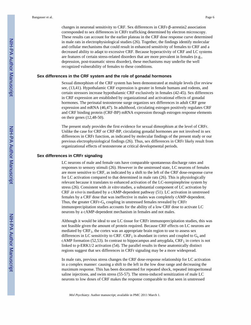
Stress-elicited association of CRFr to β-arrestin2 is not observed in femalesThe process of receptor internalization regulates cell sensitivity to ligands and agonists of G-protein coupled receptors (38,39). CRFr internalization is initiated by phosphorylation of athreonine residue on the carboxy terminus and subsequent binding of β-arrestin2 in culturedcells and primary cortical neurons (28,29,40). Sex differences in CRFr phosphorylation wereassessed by probing immunoprecipitated CRFr with an antibody directed againstphosphothreonine. Merging the channels used to visualize phospothreonine- and CRFr-immunoreactivity revealed an identical band, consistent with detection of thephosphorylated receptor (Fig. 3A). There were no sex- or stress-related differences inphosphothreonine labeling of CRFr at any timepoint after swim stress (Fig. 3B1-B3).
Detection of β-arrestin2 in immunoprecipitated samples revealed an effect of both sex andstress on CRFr-β-arrestin2 association (Fig. 3C, D1-D3). In the unstressed condition, CRFr-β-arrestin2 association was relatively low and similar in males and females. Stress increasedCRFr-β-arrestin2 association solely in males at 1 h and 24 h after swim stress, consistentwith the internalization process (Fig. 3D1-D3) (32). In contrast, stress failed to increaseCRFr-β-arrestin2 association in females at any timepoint, suggesting that the importantadaptive process of CRFr internalization may be compromised in females (Fig. 3 D1-D3).CRFr-β-arrestin2 association was similar in ovariectomized and intact females in eitherunstressed or stressed conditions.
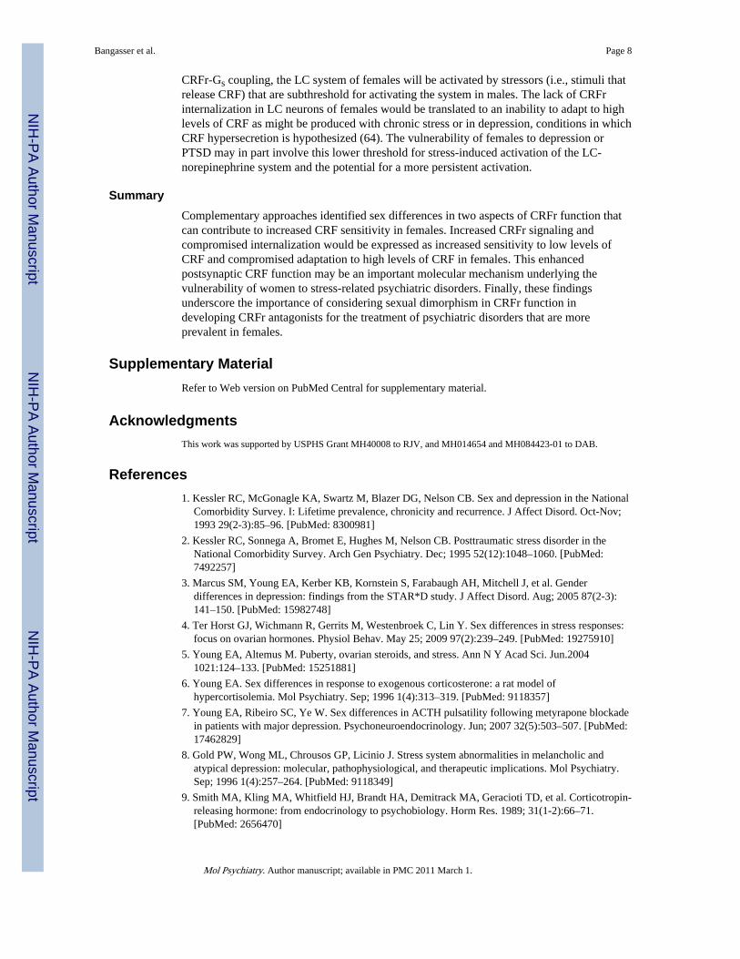
Stress-elicited CRFr internalization is not observed in femalesImmunoelectron microscopy was used to compare the cellular localization of CRFr betweengroups. Immunogold-silver labeled CRFr was identified in LC dendrites in both male andfemale rats (Fig. 4A-C). CRFr was found within TH-labeled dendrites of female rats as hasbeen demonstrated for LC neurons of male rats (32) (Fig. 4C1-C2). Consistent with previousreports (32), in unstressed male rats CRFr was more prevalent on the plasma membrane. Incontrast, in unstressed female rats CRFr was predominantly cytoplasmic (Fig. 4A1, B1, D).
Swim stress induced CRFr internalization in male rats as indicated by a greater ratio ofcytoplasmic–to-total silver grains 24 h after stress (Fig. 4A2,D). In contrast, a decreasedratio of cytoplasmic-to-total silver grains was apparent in females after swim stress,suggestive of CRFr recruitment to the plasma membrane (Fig. 4B2, D). Together, theimmunoelectron microscopy data support the immunoprecipitation and electrophysiologicalstudies suggesting that stress causes CRFr internalization in male rats only.
DISCUSSIONThe present study provides convergent evidence for sexual dimorphism in CRFr signalingand trafficking. Receptor immunoprecipitation revealed greater CRFr-Gs coupling infemales compared to males in unstressed conditions, consistent with a greater sensitivity toCRF determined electrophysiologically. A history of stress increased CRFr-Gs couplingonly in males to a magnitude comparable to that seen in females, mirroring stress-induced
Bangasser et al. Page 5
Mol Psychiatry. Author manuscript; available in PMC 2011 March 1.
NIH
-PA Author Manuscript
NIH
-PA Author Manuscript
NIH
-PA Author Manuscript
changes in neuronal sensitivity to CRF. Sex differences in CRFr-β-arrestin2 associationcorresponded to sex differences in CRFr trafficking determined by electron microscopy.These results can account for the earlier plateau in the CRF dose response curve determinedin male rats in electrophysiological studies (26). Together, the findings identify molecularand cellular mechanisms that could result in enhanced sensitivity of females to CRF and adecreased ability to adapt to excessive CRF. Because hyperactivity of CRF and LC systemsare features of certain stress-related disorders that are more prevalent in females (e.g.,depression, post-traumatic stress disorder), these mechanisms may underlie the wellrecognized vulnerability of females to these conditions.
Sex differences in the CRF system and the role of gonadal hormonesSexual dimorphism of the CRF system has been demonstrated at multiple levels (for reviewsee, (13,41). Hypothalamic CRF expression is greater in female humans and rodents, andcertain stressors increase hypothalamic CRF exclusively in females (42-45). Sex differencesin CRF expression are established by organizational and activational effects of gonadalhormones. The perinatal testosterone surge organizes sex differences in adult CRF geneexpression and mRNA (46,47). In adulthood, circulating estrogen positively regulates CRFand CRF binding protein (CRF-BP) mRNA expression through estrogen response elementson their genes (12,48-50).
The present study provides the first evidence for sexual dimorphism at the level of CRFr.Unlike the case for CRF or CRF-BP, circulating gonadal hormones are not involved in sexdifferences in CRFr function, as indicated by molecular findings of the present study or ourprevious electrophysiological findings (26). Thus, sex differences in CRFr likely result fromorganizational effects of testosterone at critical developmental periods.
Sex differences in CRFr signalingLC neurons of male and female rats have comparable spontaneous discharge rates andresponses to sensory stimuli (26). However in the unstressed state, LC neurons of femalesare more sensitive to CRF, as indicated by a shift to the left of the CRF dose-response curvefor LC activation compared to that determined in male rats (26). This is physiologicallyrelevant because it translates to enhanced activation of the LC-norepinephrine system bystress (26). Consistent with in vitro studies, a substantial component of LC activation byCRF in vivo is mediated by a cAMP-dependent pathway (51). LC activation in unstressedfemales by a CRF dose that was ineffective in males was completely cAMP-dependent.Thus, the greater CRFr-Gs coupling in unstressed females revealed by CRFrimmunoprecipitation studies accounts for the ability of a low CRF dose to activate LCneurons by a cAMP-dependent mechanism in females and not males.
Although it would be ideal to use LC tissue for CRFr immunoprecipitation studies, this wasnot feasible given the amount of protein required. Because CRF effects on LC neurons aremediated by CRF1, the cortex was an appropriate brain region to use to assess sexdifferences in LC sensitivity to CRF. CRF1 is abundant in cortex and coupled to Gs andcAMP formation (52,53). In contrast to hippocampus and amygdala, CRF1 in cortex is notlinked to p-ERK1/2 activation (54). The parallel results in these anatomically distinctregions suggest that sex differences in CRFr signaling may be a more widespread.
In male rats, previous stress changes the CRF dose-response relationship for LC activationin a complex manner: causing a shift to the left in the low dose range and decreasing themaximum response. This has been documented for repeated shock, repeated intraperitonealsaline injections, and swim stress (55-57). The stress-induced sensitization of male LCneurons to low doses of CRF makes the response comparable to that seen in unstressed
Bangasser et al. Page 6
Mol Psychiatry. Author manuscript; available in PMC 2011 March 1.
NIH
-PA Author Manuscript
NIH
-PA Author Manuscript
NIH
-PA Author Manuscript

females and, as in females, this response is completely cAMP dependent. Sex and stressdifferences in CRFr-Gs coupling mirrored the electrophysiological differences, supportingthis as an underlying mechanism for stress-induced neuronal sensitization in males.
Sex differences in CRFr internalizationInternalization of G-protein coupled receptors regulates cell sensitivity to agonists (38,39).Both agonist- and swim stress-induced CRFr internalization in LC neurons of male rats havebeen previously documented (31,32). As time increases from 1 to 24 h after swim stress,CRFr incorporation into multivesicular bodies increases, consistent with receptordownregulation (32). The functional consequence of this is an earlier plateau of the CRFdose-response curve for LC activation in male rats exposed to swim stress (26). The cellularprocesses involved in CRFr internalization in cultured cells indicate a requirement forphosphorylation of a threonine residue Thr399 on the carboxy tail and recruitment of β-arrestin2 (28). The finding that stress did not alter CRFr-threonine phosphorylation may bean indication that stress-induced CRFr internalization in brain requires phosphorylation atother sites on the receptor. Alternatively, the technique used in this study may not besufficiently sensitive to detect differences in phosphorylation sites that are required forinternalization.
Stress increased CRFr-β-arrestin2 association in males at 1 and 24 h after stress, times atwhich CRFr internalization was apparent in LC neurons (32). The inability of swim stress toalter CRFr-β-arrestin2 association in females predicted that the internalization processwould be impaired and this was confirmed in electron microscopy studies. The finding thatCRFr was prominent in the cytoplasm in unstressed females and on the plasma membrane instressed females was unexpected. This could reflect stress-induced recruitment of CRFr tothe plasma membrane. Alternatively, it is possible that unstressed females produce morereceptors that remain cytoplasmic and this is attenuated by stress, effectively increasing theproportion of receptors on the plasma membrane by decreasing the amount in the cytoplasm.Evidence for simultaneous receptor internalization and increased coupling in previouslystressed male rats completely accounts for the complex shift in the CRF dose-response curvein male rats with a history of stress (i.e., a shift to the left with a decreased maximumresponse). This suggests that CRFr remaining on plasma membrane after stress is morehighly coupled.
Relevance of enhanced CRFr signaling in femalesSex differences in CRFr in rat studies should translate to increased sensitivity in rodentmodels of stress-related psychopathology. However, most rodent models of these disordersuse males exclusively and studies using both sexes are equivocal with regard to whetherfemales are more sensitive (for review see, (58-60). An important issue is that many of thesemodels (e.g. the forced swim test, open field) use an inhibition of motor activity as anendpoint indicative of depressive- or anxiety-like behavior. This is problematic becausethere are baseline sex differences in activity in many rodent species (60). Moreover, it isunclear that a decrease in activity appropriately models the hyperarousal that characterizesmelancholic depression and PTSD, and which is thought to involve LC hyperactivity and/orsensitivity.
CRF and stressors shift the mode of LC discharge towards a high tonic-low phasic state thatis associated with heightened arousal and a shift from a focused to labile attention thatfacilitates scanning the environment (61-63). This is an adaptive behavioral response toacute stress. However, if this response is engaged inappropriately or if it persists, this isexpressed as pathology similar to that described in depression or PTSD (e.g., hyperarousal,sleep disturbance, inability to concentrate, anxiogenic behaviors). As a result of increased
Bangasser et al. Page 7
Mol Psychiatry. Author manuscript; available in PMC 2011 March 1.
NIH
-PA Author Manuscript
NIH
-PA Author Manuscript
NIH
-PA Author Manuscript
CRFr-Gs coupling, the LC system of females will be activated by stressors (i.e., stimuli thatrelease CRF) that are subthreshold for activating the system in males. The lack of CRFrinternalization in LC neurons of females would be translated to an inability to adapt to highlevels of CRF as might be produced with chronic stress or in depression, conditions in whichCRF hypersecretion is hypothesized (64). The vulnerability of females to depression orPTSD may in part involve this lower threshold for stress-induced activation of the LC-norepinephrine system and the potential for a more persistent activation.
SummaryComplementary approaches identified sex differences in two aspects of CRFr function thatcan contribute to increased CRF sensitivity in females. Increased CRFr signaling andcompromised internalization would be expressed as increased sensitivity to low levels ofCRF and compromised adaptation to high levels of CRF in females. This enhancedpostsynaptic CRF function may be an important molecular mechanism underlying thevulnerability of women to stress-related psychiatric disorders. Finally, these findingsunderscore the importance of considering sexual dimorphism in CRFr function indeveloping CRFr antagonists for the treatment of psychiatric disorders that are moreprevalent in females.
Supplementary MaterialRefer to Web version on PubMed Central for supplementary material.
AcknowledgmentsThis work was supported by USPHS Grant MH40008 to RJV, and MH014654 and MH084423-01 to DAB.
References1. Kessler RC, McGonagle KA, Swartz M, Blazer DG, Nelson CB. Sex and depression in the National
Comorbidity Survey. I: Lifetime prevalence, chronicity and recurrence. J Affect Disord. Oct-Nov;1993 29(2-3):85–96. [PubMed: 8300981]
2. Kessler RC, Sonnega A, Bromet E, Hughes M, Nelson CB. Posttraumatic stress disorder in theNational Comorbidity Survey. Arch Gen Psychiatry. Dec; 1995 52(12):1048–1060. [PubMed:7492257]
3. Marcus SM, Young EA, Kerber KB, Kornstein S, Farabaugh AH, Mitchell J, et al. Genderdifferences in depression: findings from the STAR*D study. J Affect Disord. Aug; 2005 87(2-3):141–150. [PubMed: 15982748]
4. Ter Horst GJ, Wichmann R, Gerrits M, Westenbroek C, Lin Y. Sex differences in stress responses:focus on ovarian hormones. Physiol Behav. May 25; 2009 97(2):239–249. [PubMed: 19275910]
5. Young EA, Altemus M. Puberty, ovarian steroids, and stress. Ann N Y Acad Sci. Jun.20041021:124–133. [PubMed: 15251881]
6. Young EA. Sex differences in response to exogenous corticosterone: a rat model ofhypercortisolemia. Mol Psychiatry. Sep; 1996 1(4):313–319. [PubMed: 9118357]
7. Young EA, Ribeiro SC, Ye W. Sex differences in ACTH pulsatility following metyrapone blockadein patients with major depression. Psychoneuroendocrinology. Jun; 2007 32(5):503–507. [PubMed:17462829]
8. Gold PW, Wong ML, Chrousos GP, Licinio J. Stress system abnormalities in melancholic andatypical depression: molecular, pathophysiological, and therapeutic implications. Mol Psychiatry.Sep; 1996 1(4):257–264. [PubMed: 9118349]
9. Smith MA, Kling MA, Whitfield HJ, Brandt HA, Demitrack MA, Geracioti TD, et al. Corticotropin-releasing hormone: from endocrinology to psychobiology. Horm Res. 1989; 31(1-2):66–71.[PubMed: 2656470]
Bangasser et al. Page 8
Mol Psychiatry. Author manuscript; available in PMC 2011 March 1.
NIH
-PA Author Manuscript
NIH
-PA Author Manuscript
NIH
-PA Author Manuscript
10. Gold PW, Chrousos GP. Organization of the stress system and its dysregulation in melancholic andatypical depression: high vs low CRH/NE states. Mol Psychiatry. 2002; 7(3):254–275. [PubMed:11920153]
11. Strohle A, Holsboer F. Stress responsive neurohormones in depression and anxiety.Pharmacopsychiatry. Nov; 2003 36(Suppl 3):S207–214. [PubMed: 14677081]
12. Vamvakopoulos NC, Chrousos GP. Evidence of direct estrogenic regulation of humancorticotropin-releasing hormone gene expression. Potential implications for the sexual dimophismof the stress response and immune/inflammatory reaction. J Clin Invest. Oct; 1993 92(4):1896–1902. [PubMed: 8408641]
13. Vamvakopoulos NV. Sexual dimorphism of stress response and immune/inflammatory reaction:the corticotropin releasing hormone perspective. Mediators Inflamm. 1995; 4(3):163–174.[PubMed: 18475634]
14. Vale W, Spiess J, Rivier C, Rivier J. Characterization of a 41-residue ovine hypothalamic peptidethat stimulates secretion of corticotropin and beta-endorphin. Science. Sep 18; 1981 213(4514):1394–1397. [PubMed: 6267699]
15. Bale TL, Vale WW. CRF and CRF receptors: role in stress responsivity and other behaviors. AnnuRev Pharmacol Toxicol. 2004; 44:525–557. [PubMed: 14744257]
16. Owens MJ, Nemeroff CB. Physiology and pharmacology of corticotropin-releasing factor.Pharmacol Rev. Dec; 1991 43(4):425–473. [PubMed: 1775506]
17. Van Bockstaele EJ, Colago EE, Valentino RJ. Corticotropin-releasing factor-containing axonterminals synapse onto catecholamine dendrites and may presynaptically modulate other afferentsin the rostral pole of the nucleus locus coeruleus in the rat brain. J Comp Neurol. Jan 15; 1996364(3):523–534. [PubMed: 8820881]
18. Van Bockstaele EJ, Colago EE, Valentino RJ. Amygdaloid corticotropin-releasing factor targetslocus coeruleus dendrites: substrate for the co-ordination of emotional and cognitive limbs of thestress response. J Neuroendocrinol. Oct; 1998 10(10):743–757. [PubMed: 9792326]
19. Valentino RJ, Van Bockstaele E. Convergent regulation of locus coeruleus activity as an adaptiveresponse to stress. Eur J Pharmacol. Apr 7; 2008 583(2-3):194–203. [PubMed: 18255055]
20. Aston-Jones G, Rajkowski J, Kubiak P, Valentino RJ, Shipley MT. Role of the locus coeruleus inemotional activation. Prog Brain Res. 1996; 107:380–402.
21. Page ME, Berridge CW, Foote SL, Valentino RJ. Corticotropin-releasing factor in the locuscoeruleus mediates EEG activation associated with hypotensive stress. Neurosci Lett. 1993;164:81–84. [PubMed: 8152620]
22. Lechner SM, Curtis AL, Brons R, Valentino RJ. Locus coeruleus activation by colon distention:role of corticotropin-releasing factor and excitatory amino acids. Brain Res. May 9; 1997756(1-2):114–124. [PubMed: 9187321]
23. Wong ML, Kling MA, Munson PJ, Listwak S, Licinio J, Prolo P, et al. Pronounced and sustainedcentral hypernoradrenergic function in major depression with melancholic features: relation tohypercortisolism and corticotropin-releasing hormone. Proc Natl Acad Sci U S A. Jan 4; 200097(1):325–330. [PubMed: 10618417]
24. O'Donnell T, Hegadoren KM, Coupland NC. Noradrenergic mechanisms in the pathophysiology ofpost-traumatic stress disorder. Neuropsychobiology. 2004; 50(4):273–283. [PubMed: 15539856]
25. Southwick SM, Bremner JD, Rasmusson A, Morgan CA 3rd, Arnsten A, Charney DS. Role ofnorepinephrine in the pathophysiology and treatment of posttraumatic stress disorder. BiolPsychiatry. Nov 1; 1999 46(9):1192–1204. [PubMed: 10560025]
26. Curtis AL, Bethea T, Valentino RJ. Sexually dimorphic responses of the brain norepinephrinesystem to stress and corticotropin-releasing factor. Neuropsychopharmacology. Mar; 2006 31(3):544–554. [PubMed: 16123744]
27. Hillhouse EW, Grammatopoulos DK. The molecular mechanisms underlying the regulation of thebiological activity of corticotropin-releasing hormone receptors: implications for physiology andpathophysiology. Endocr Rev. May; 2006 27(3):260–286. [PubMed: 16484629]
28. Teli T, Markovic D, Levine MA, Hillhouse EW, Grammatopoulos DK. Regulation ofcorticotropin-releasing hormone receptor type 1alpha signaling: structural determinants for G
Bangasser et al. Page 9
Mol Psychiatry. Author manuscript; available in PMC 2011 March 1.
NIH
-PA Author Manuscript
NIH
-PA Author Manuscript
NIH
-PA Author Manuscript

protein-coupled receptor kinase-mediated phosphorylation and agonist-mediated desensitization.Mol Endocrinol. Feb; 2005 19(2):474–490. [PubMed: 15498832]
29. Oakley RH, Olivares-Reyes JA, Hudson CC, Flores-Vega F, Dautzenberg FM, Hauger RL.Carboxyl-terminal and intracellular loop sites for CRF1 receptor phosphorylation and beta-arrestin-2 recruitment: a mechanism regulating stress and anxiety responses. Am J Physiol RegulIntegr Comp Physiol. Jul; 2007 293(1):R209–222. [PubMed: 17363685]
30. Carr GV, Bangasser DA, Bethea T, Young M, Valentino RJ, Lucki I. Antidepressant-Like Effectsof kappa-Opioid Receptor Antagonists in Wistar Kyoto Rats. Neuropsychopharmacology. Nov18.2009
31. Reyes BA, Fox K, Valentino RJ, Van Bockstaele EJ. Agonist-induced internalization ofcorticotropin-releasing factor receptors in noradrenergic neurons of the rat locus coeruleus. Eur JNeurosci. Jun; 2006 23(11):2991–2998. [PubMed: 16819988]
32. Reyes BA, Valentino RJ, Van Bockstaele EJ. Stress-induced intracellular trafficking ofcorticotropin-releasing factor receptors in rat locus coeruleus neurons. Endocrinology. Jan; 2008149(1):122–130. [PubMed: 17947354]
33. Rominger DH, Rominger CM, Fitzgerald LW, Grzanna R, Largent BL, Zaczek R. Characterizationof [125I]sauvagine binding to CRH2 receptors: membrane homogenate and autoradiographicstudies. J Pharmacol Exp Ther. Jul; 1998 286(1):459–468. [PubMed: 9655891]
34. Chen FM, Bilezikjian LM, Perrin MH, Rivier J, Vale W. Corticotropin releasing factor receptor-mediated stimulation of adenylate cyclase activity in the rat brain. Brain Res. Aug 27; 1986381(1):49–57. [PubMed: 3019476]
35. Battaglia G, Webster EL, De Souza EB. Characterization of corticotropin-releasing factor receptor-mediated adenylate cyclase activity in the rat central nervous system. Synapse. 1987; 1(6):572–581. [PubMed: 2843998]
36. De Souza EB. Corticotropin-releasing factor receptors: physiology, pharmacology, biochemistryand role in central nervous system and immune disorders. Psychoneuroendocrinology. 1995;20(8):789–819. [PubMed: 8834089]
37. Van Pett K, Viau V, Bittencourt JC, Chan RK, Li HY, Arias C, et al. Distribution of mRNAsencoding CRF receptors in brain and pituitary of rat and mouse. J Comp Neurol. Dec 11; 2000428(2):191–212. [PubMed: 11064361]
38. Premont RT. Once and future signaling: G protein-coupled receptor kinase control of neuronalsensitivity. Neuromolecular Med. 2005; 7(1-2):129–147. [PubMed: 16052042]
39. Krupnick JG, Benovic JL. The role of receptor kinases and arrestins in G protein-coupled receptorregulation. Annu Rev Pharmacol Toxicol. 1998; 38:289–319. [PubMed: 9597157]
40. Holmes KD, Babwah AV, Dale LB, Poulter MO, Ferguson SS. Differential regulation ofcorticotropin releasing factor 1alpha receptor endocytosis and trafficking by beta-arrestins and RabGTPases. J Neurochem. Feb; 2006 96(4):934–949. [PubMed: 16412099]
41. Swaab DF, Bao AM, Lucassen PJ. The stress system in the human brain in depression andneurodegeneration. Ageing Res Rev. May; 2005 4(2):141–194. [PubMed: 15996533]
42. Frederiksen SO, Ekman R, Gottfries CG, Widerlov E, Jonsson S. Reduced concentrations ofgalanin, arginine vasopressin, neuropeptide Y and peptide YY in the temporal cortex but not in thehypothalamus of brains from schizophrenics. Acta Psychiatr Scand. Apr; 1991 83(4):273–277.[PubMed: 1709331]
43. Viau V, Bingham B, Davis J, Lee P, Wong M. Gender and puberty interact on the stress-inducedactivation of parvocellular neurosecretory neurons and corticotropin-releasing hormone messengerribonucleic acid expression in the rat. Endocrinology. Jan; 2005 146(1):137–146. [PubMed:15375029]
44. Iwasaki-Sekino A, Mano-Otagiri A, Ohata H, Yamauchi N, Shibasaki T. Gender differences incorticotropin and corticosterone secretion and corticotropin-releasing factor mRNA expression inthe paraventricular nucleus of the hypothalamus and the central nucleus of the amygdala inresponse to footshock stress or psychological stress in rats. Psychoneuroendocrinology. Feb; 200934(2):226–237. [PubMed: 18849120]
Bangasser et al. Page 10
Mol Psychiatry. Author manuscript; available in PMC 2011 March 1.
NIH
-PA Author Manuscript
NIH
-PA Author Manuscript
NIH
-PA Author Manuscript

45. Desbonnet L, Garrett L, Daly E, McDermott KW, Dinan TG. Sexually dimorphic effects ofmaternal separation stress on corticotrophin-releasing factor and vasopressin systems in the adultrat brain. Int J Dev Neurosci. May-Jun; 2008 26(3-4):259–268. [PubMed: 18367364]
46. Patchev VK, Hayashi S, Orikasa C, Almeida OF. Ontogeny of gender-specific responsiveness tostress and glucocorticoids in the rat and its determination by the neonatal gonadal steroidenvironment. Stress. Aug; 1999 3(1):41–54. [PubMed: 19016192]
47. Seale JV, Wood SA, Atkinson HC, Harbuz MS, Lightman SL. Postnatal masculinization alters theHPA axis phenotype in the adult female rat. J Physiol. Feb 15; 2005 563(Pt 1):265–274. [PubMed:15611026]
48. van de Stolpe A, Slycke AJ, Reinders MO, Zomer AW, Goodenough S, Behl C, et al. Estrogenreceptor (ER)-mediated transcriptional regulation of the human corticotropin-releasing hormone-binding protein promoter: differential effects of ERalpha and ERbeta. Mol Endocrinol. Dec; 200418(12):2908–2923. [PubMed: 15345745]
49. Bohler HC Jr. Zoeller RT, King JC, Rubin BS, Weber R, Merriam GR. Corticotropin releasinghormone mRNA is elevated on the afternoon of proestrus in the parvocellular paraventricularnuclei of the female rat. Brain Res Mol Brain Res. Aug; 1990 8(3):259–262. [PubMed: 2170804]
50. Speert DB, SJ MC, Seasholtz AF. Sexually dimorphic expression of corticotropin-releasinghormone-binding protein in the mouse pituitary. Endocrinology. Dec; 2002 143(12):4730–4741.[PubMed: 12446601]
51. Jedema HP, Grace AA. Corticotropin-releasing hormone directly activates noradrenergic neuronsof the locus ceruleus recorded in vitro. J Neurosci. Oct 27; 2004 24(43):9703–9713. [PubMed:15509759]
52. Potter E, Sutton S, Donaldson C, Chen R, Perrin M, Lewis K, et al. Distribution of corticotropin-releasing factor receptor mRNA expression in the rat brain and pituitary. Proc Natl Acad Sci U SA. Sep 13; 1994 91(19):8777–8781. [PubMed: 8090722]
53. Grammatopoulos DK, Randeva HS, Levine MA, Kanellopoulou KA, Hillhouse EW. Rat cerebralcortex corticotropin-releasing hormone receptors: evidence for receptor coupling to multiple G-proteins. J Neurochem. Jan; 2001 76(2):509–519. [PubMed: 11208914]
54. Refojo D, Echenique C, Muller MB, Reul JM, Deussing JM, Wurst W, et al. Corticotropin-releasing hormone activates ERK1/2 MAPK in specific brain areas. Proc Natl Acad Sci U S A.Apr 26; 2005 102(17):6183–6188. [PubMed: 15833812]
55. Curtis AL, Pavcovich LA, Grigoriadis DE, Valentino RJ. Previous stress alters corticotropin-releasing factor neurotransmission in the locus coeruleus. Neuroscience. Mar; 1995 65(2):541–550. [PubMed: 7777167]
56. Valentino, RJ.; Van Bockstaele, EJ. Functional interactions between stress neuromediator and thelocus coeruleur-noradrenaline system. In: Steckler, T.; K, N., editors. Handbook of Stress and theBrain. The Netherlands; Elsevier: 2005. p. 465-486.
57. Curtis AL, Pavcovich LA, Valentino RJ. Long-term regulation of locus ceruleus sensitivity tocorticotropin-releasing factor by swim stress. J Pharmacol Exp Ther. Jun; 1999 289(3):1211–1219.[PubMed: 10336508]
58. Dalla C, Pitychoutis PM, Kokras N, Papadopoulou-Daifoti Z. Sex Differences in Animal Modelsof Depression and Antidepressant Response. Basic Clin Pharmacol Toxicol. Dec 30.2009
59. Palanza P. Animal models of anxiety and depression: how are females different? NeurosciBiobehav Rev. May; 2001 25(3):219–233. [PubMed: 11378178]
60. Cryan JF, Mombereau C. In search of a depressed mouse: utility of models for studyingdepression-related behavior in genetically modified mice. Mol Psychiatry. Apr; 2004 9(4):326–357. [PubMed: 14743184]
61. Aston-Jones G, Rajkowski J, Cohen J. Locus coeruleus and regulation of behavioral flexibility andattention. Prog Brain Res. 2000; 126:165–182. [PubMed: 11105646]
62. Aston-Jones G, Cohen JD. An integrative theory of locus coeruleus-norepinephrine function:adaptive gain and optimal performance. Annu Rev Neurosci. 2005; 28:403–450. [PubMed:16022602]
Bangasser et al. Page 11
Mol Psychiatry. Author manuscript; available in PMC 2011 March 1.
NIH
-PA Author Manuscript
NIH
-PA Author Manuscript
NIH
-PA Author Manuscript
63. Berridge CW, Waterhouse BD. The locus coeruleus-noradrenergic system: modulation ofbehavioral state and state-dependent cognitive processes. Brain Res Brain Res Rev. Apr; 200342(1):33–84. [PubMed: 12668290]
64. Nemeroff CB. The corticotropin-releasing factor (CRF) hypothesis of depression: new findings andnew directions. Mol Psychiatry. Sep; 1996 1(4):336–342. [PubMed: 9118360]
Bangasser et al. Page 12
Mol Psychiatry. Author manuscript; available in PMC 2011 March 1.
NIH
-PA Author Manuscript
NIH
-PA Author Manuscript
NIH
-PA Author Manuscript

Figure 1.The role of cAMP signaling in the sex- and stress-related differences in LC neuronalactivation by CRF. (A-D) LC activation by local CRF after pretreatment with Rp-cAMP-S(600 ng in 120 nl, intra-LC) or ACSF (120 nl) is shown for unstressed male (n=5-8) andfemale rats (n=4-6) and male (n=6-8) and female rats 24 h after swim stress (n=4-6). Barsdepict the average response to CRF following ACSF (black) or Rp-cAMP-S (dark gray).Light gray bars represent the cAMP-mediated component (calculated by taking thedifference between the vehicle and Rp-cAMP-S treated groups). In the unstressed state, arelatively low CRF dose activated LC neurons in females only [t(6)=3.56, p<0.05], and thisresponse was completely cAMP-dependent. For both males and females, neuronal responsesto the higher dose (30 ng) were mediated by cAMP-dependent and independent processes[F(1,26)=12.88, p<0.05] . Swim stress changed the cAMP signaling profile in males{stress×dose×drug interaction [F(1,46)=4.45, p<0.05]}, but not in females [F(1,39)=1.87,p>0.05].
Bangasser et al. Page 13
Mol Psychiatry. Author manuscript; available in PMC 2011 March 1.
NIH
-PA Author Manuscript
NIH
-PA Author Manuscript
NIH
-PA Author Manuscript
Figure 2.Sex- and stress-related differences in CRFr association with different G proteins. (A-C)Representative blots of immunoprecipitated CRFr (green, MW=52kD) from different groupsand (A) the Gs protein (red, MW=48kDa), (B) the Go protein (red, MW=40kDa) and (C) theGq/11 protein (red, MW=40kDa). So that the presentation in A, matched that in B and C, alane containing the molecular weight marker that was between female and male samples onthe same gel was deleted and the image of male samples that were to the right of this lanewere moved to the left of female samples. (D-F) Graphs show the mean ratio of theintegrated intensity of each band of G proteins to the corresponding band of CRFr from thesame samples (n=4-6 determinations, pooled 3 rats per determination). CRFr-Gs couplingwas greater in unstressed ovariectomized and intact females compared to unstressed males[F(5,26)=2.56, p<0.05, post-hocs p<0.05]. Stress increased coupling in males (p<0.05) to alevel comparable to that of unstressed females (p>0.05), but had no further effect on females(regardless of hormonal status; p>0.05). There were no significant differences in coupling ofthe CRFr to Go [F(3,20)=0.55, p>0.05] or Gq/11 [F(3,12)=0.55, p>0.05] (top bandquantified). Data are represented as the mean (±SEM). Number sign indicates sex differenceunder basal (unstressed) conditions (i.e., greater coupling in unstressed females vs.unstressed males; p<0.05). Asterisks indicate a significant stress-induced increase comparedto the unstressed same sex control (p<0.05).
Bangasser et al. Page 14
Mol Psychiatry. Author manuscript; available in PMC 2011 March 1.
NIH
-PA Author Manuscript
NIH
-PA Author Manuscript
NIH
-PA Author Manuscript
Figure 3.Sex differences in proteins involved in CRFr internalization processes. (A) Blots representthe phosphothreonine band (red), CRFr band (green) and the merged image (yellow)indicating that both label the same protein (i.e., phosphorylated CRFr , MW=52 kDa).Protein for this blot was collected 24 h after stressor exposure. (B1-3) Bar graphs show themean ratio of phosphothreonine:CRFr for each condition from tissue collected immediately[F(3,12)=0.39, p>0.05], 1 h [F(3,20)=0.58, p>0.05]or 24 h [F(3,20)=0.66, p>0.05] post-stress (n=4-6 determinations, pooled 3 rats per determination). (C) The Western blot showsthe β-arrestin2 band (MW = 54 kDa) and the CRFr band 24 h after stressor exposure orhandling. (D1-3) Graphs illustrate the ratio of β–arrestin2:CRFr for rats sacrificedimmediately [F(3,12)=0.52, p>0.05], 1 h [F(3,16)=4.74, p<0.05] or 24 h post-stress[F(5,30)=5.77, p<0.05] (n=4-6 determinations, pooled 3 rats per determination). At both 1and 24 h after stress, β–arrestin2 association with the CRFr was significantly increased inmales (p<0.05) but not in females (p>0.05). Cycling females were included for an additionalcomparison at the 24 h timepoint, and there was no statistically significant difference inCRFr-β-arrestin2 association between ovariectomized and cycling females in either theunstressed or stressed condition (p>0.05). Data are represented as the mean (±SEM).Asterisks indicate a significant effect of stress compared to unstressed same sex control(p<0.05).
Bangasser et al. Page 15
Mol Psychiatry. Author manuscript; available in PMC 2011 March 1.
NIH
-PA Author Manuscript
NIH
-PA Author Manuscript
NIH
-PA Author Manuscript

Figure 4.Electron microscopic visualization of CRFr compartmentalization and stress-inducedtrafficking in LC dendrites. A-C are electron photomicrographs of sections through the LC.(A1) LC dendritic profile (d) in an unstressed male rat with immunogold-silver labeling forthe CRFr along the plasma membrane (arrowheads). The dendrite receives synaptic contactsfrom axon terminals (t). (A2) Dendrite from a male rat 24 h following swim stress. CRFrlabeling shifts from the plasma membrane to the cytoplasm. (B1) Dendrite from anunstressed female rat shows that CRFr is prominent in the cytoplasm. (B2) Dendrite from afemale rat 24 h following swim stress shows that CRFr labeling shifts from the cytoplasm tothe plasma membrane. (C1-2) TH-immunoperoxidase-labeled dendrites containingimmunogold-silver labeling for CRFr (CRFr+TH) in a female control (C1) and a stressed rat(C2). Arrowheads point to CRFr on the plasma membrane in C2. Arrows point toimmunoperoxidase reaction product. (D) Bar graph indicating the percentage of internalizedreceptors for each condition (n=3, mean per rat generated from at least 125 dendriticprofiles). Unstressed females had a significantly greater percentage of cytoplasmic receptorsthan unstressed males [F(1,8)=45.3, p<0.05, post-hoc, p<0.05]. Swim stress increased thepercentage of CRFr in cytoplasm in males rats (p<0.05). In contrast, swim stress decreasedthe percentage of cytoplasmic CRFr in females (p<0.05). Data are represented as the mean(± SEM). Number sign indicates sex difference under unstressed conditions (p<0.05).Asterisks indicate a significant effect of stress compared to the unstressed same sex control(p<0.05). Scale bars=500 nm (A-C).
Bangasser et al. Page 16
Mol Psychiatry. Author manuscript; available in PMC 2011 March 1.
NIH
-PA Author Manuscript
NIH
-PA Author Manuscript
NIH
-PA Author Manuscript