Effect of targeted deletion of the heme oxygenase-2 gene on hemoglobin toxicity in the striatum
10
Effect of targeted deletion of the heme oxygenase-2 gene on hemoglobin toxicity in the striatum Yan Qu, Jing Chen, Luna Benvenisti-Zarom, Xin Ma and Raymond F Regan Department of Emergency Medicine, Thomas Jefferson University, Philadelphia, Pennsylvania, USA The heme oxygenase (HO) enzymes catalyze the rate-limiting step in the breakdown of heme to iron, carbon monoxide, and biliverdin. A prior cell culture study demonstrated that deletion of HO-2, the isoform constitutively expressed in neurons, attenuated hemoglobin (Hb) neurotoxicity. The present study tested the hypothesis that HO-2 gene deletion is cytoprotective in a model of Hb toxicity in vivo. Stereotactic injection of 6 lL stroma-free Hb (SFHb) into the striatum significantly increased protein oxidation in wild-type mice at 24 to 72 h, as detected by an assay for carbonyl groups. At 72 h, carbonylation was increased 2.5-fold compared with that in the contralateral striatum. In HO-2 knockout mice, protein oxidation was not increased at 24 h, and was increased by only 1.7-fold at 72 h. Similarly, striatal lipid peroxidation, as detected by the malondialdehyde assay, was significantly greater in the SFHb-injected striata of wild-type mice than in knockout mice. Striatal cell viability, determined by the 3-(4,5-dimethylthiazol-2-yl)-2,5-diphenyltetrazolium bromide assay, was 45.0%76.3% of that in contralateral striata in wild-type mice at 72 h; it was increased to 85%7 8% in knockouts. Heme oxygenase-2 gene deletion did not alter weight loss or mortality after SFHb injection. Baseline striatal HO-1 expression was similar in knockout and wild-type mice; induction after SFHb injection occurred more rapidly in the latter. These results suggest that HO-2 gene deletion protects striatal cells from the oxidative toxicity of Hb in vivo. Pharmacologic or genetic strategies that target HO-2 may be beneficial after central nervous system hemorrhage, and warrant further investigation. Journal of Cerebral Blood Flow & Metabolism (2005) 25, 1466–1475. doi:10.1038/sj.jcbfm.9600143; published online 18 May 2005 Keywords: brain; CNS; hemorrhage; ischemia; oxidative stress; trauma Introduction Central nervous system (CNS) hemorrhage is the primary event in approximately 15% of strokes, and is associated with most traumatic injuries requiring hospitalization. Although its deleterious effect has often been attributed to compressive ischemia produced by a hematoma (Bullock et al, 1988; Nath et al, 1986), a growing body of evidence sug- gests that blood components may directly injure surrounding tissue and thereby worsen outcome (Qureshi et al, 2001). Early edema formation appears to be mediated in part by the proteolytic activity of thrombin (Xi et al, 1998); hemoglobin (Hb) and its metabolites have a later cytotoxic effect, at least in rodents (Huang et al, 2002). In prior cell culture studies, Hb has been shown to be an oxidative neurotoxin (Regan and Panter, 1993; Wang et al, 2002). This effect is at least partly due to iron release from heme, because it was prevented with very high efficacy by iron chelators (Regan and Rogers, 2003). Heme degradation leading to iron release is catalyzed by the heme oxygenase (HO) enzymes (Abraham et al, 1996), and also occurs by reaction with peroxides and other reactive oxygen species (ROS) (Gutteridge, 1986; Nagababu and Rifkind, 2004). The former process appears to predominate in heme-mediated injuries to cultured neurons. Targeted deletion of the HO-2 gene, the isoform that is constitutively expressed in neurons (Ewing and Maines, 1997), attenuated ROS forma- tion, protein oxidation, and cell death due to Hb or hemin (Regan et al, 2004; Rogers et al, 2003). These observations are consistent with reports that HO inhibitors attenuated edema and neuronal death after injection of whole blood or Hb into the rat Received 23 November 2004; revised 30 March 2005; accepted 31 March 2005; published online 18 May 2005 Correspondence: Dr RF Regan, Department of Emergency Medi- cine, Thomas Jefferson University, 1020 Sansom Street, Thompson Building Room 239, Philadelphia, PA 19107, USA. E-mail: [email protected] Funding for this study was provided by the National Institutes of Health (NS42273) and by a grant-in-aid from the Pennsylvania/ Delaware Affiliate of the American Heart Association. Journal of Cerebral Blood Flow & Metabolism (2005) 25, 1466–1475 & 2005 ISCBFM All rights reserved 0271-678X/05 $30.00 www.jcbfm.com
-
Upload
independent -
Category
Documents
-
view
1 -
download
0
Transcript of Effect of targeted deletion of the heme oxygenase-2 gene on hemoglobin toxicity in the striatum
Effect of targeted deletion of the heme oxygenase-2gene on hemoglobin toxicity in the striatum
Yan Qu, Jing Chen, Luna Benvenisti-Zarom, Xin Ma and Raymond F Regan
Department of Emergency Medicine, Thomas Jefferson University, Philadelphia, Pennsylvania, USA
The heme oxygenase (HO) enzymes catalyze the rate-limiting step in the breakdown of heme to iron,carbon monoxide, and biliverdin. A prior cell culture study demonstrated that deletion of HO-2, theisoform constitutively expressed in neurons, attenuated hemoglobin (Hb) neurotoxicity. The presentstudy tested the hypothesis that HO-2 gene deletion is cytoprotective in a model of Hb toxicity invivo. Stereotactic injection of 6 lL stroma-free Hb (SFHb) into the striatum significantly increasedprotein oxidation in wild-type mice at 24 to 72 h, as detected by an assay for carbonyl groups. At72 h, carbonylation was increased 2.5-fold compared with that in the contralateral striatum. In HO-2knockout mice, protein oxidation was not increased at 24 h, and was increased by only 1.7-fold at72 h. Similarly, striatal lipid peroxidation, as detected by the malondialdehyde assay, wassignificantly greater in the SFHb-injected striata of wild-type mice than in knockout mice. Striatalcell viability, determined by the 3-(4,5-dimethylthiazol-2-yl)-2,5-diphenyltetrazolium bromide assay,was 45.0%76.3% of that in contralateral striata in wild-type mice at 72 h; it was increased to 85%78% in knockouts. Heme oxygenase-2 gene deletion did not alter weight loss or mortality after SFHbinjection. Baseline striatal HO-1 expression was similar in knockout and wild-type mice; inductionafter SFHb injection occurred more rapidly in the latter. These results suggest that HO-2 genedeletion protects striatal cells from the oxidative toxicity of Hb in vivo. Pharmacologic or geneticstrategies that target HO-2 may be beneficial after central nervous system hemorrhage, and warrantfurther investigation.Journal of Cerebral Blood Flow & Metabolism (2005) 25, 1466–1475. doi:10.1038/sj.jcbfm.9600143; published online18 May 2005
Keywords: brain; CNS; hemorrhage; ischemia; oxidative stress; trauma
Introduction
Central nervous system (CNS) hemorrhage is theprimary event in approximately 15% of strokes, andis associated with most traumatic injuries requiringhospitalization. Although its deleterious effect hasoften been attributed to compressive ischemiaproduced by a hematoma (Bullock et al, 1988;Nath et al, 1986), a growing body of evidence sug-gests that blood components may directly injuresurrounding tissue and thereby worsen outcome(Qureshi et al, 2001). Early edema formation appearsto be mediated in part by the proteolytic activity of
thrombin (Xi et al, 1998); hemoglobin (Hb) and itsmetabolites have a later cytotoxic effect, at least inrodents (Huang et al, 2002).
In prior cell culture studies, Hb has been shown tobe an oxidative neurotoxin (Regan and Panter, 1993;Wang et al, 2002). This effect is at least partly due toiron release from heme, because it was preventedwith very high efficacy by iron chelators (Regan andRogers, 2003). Heme degradation leading to ironrelease is catalyzed by the heme oxygenase (HO)enzymes (Abraham et al, 1996), and also occurs byreaction with peroxides and other reactive oxygenspecies (ROS) (Gutteridge, 1986; Nagababu andRifkind, 2004). The former process appears topredominate in heme-mediated injuries to culturedneurons. Targeted deletion of the HO-2 gene, theisoform that is constitutively expressed in neurons(Ewing and Maines, 1997), attenuated ROS forma-tion, protein oxidation, and cell death due to Hb orhemin (Regan et al, 2004; Rogers et al, 2003). Theseobservations are consistent with reports that HOinhibitors attenuated edema and neuronal deathafter injection of whole blood or Hb into the rat
Received 23 November 2004; revised 30 March 2005; accepted 31March 2005; published online 18 May 2005
Correspondence: Dr RF Regan, Department of Emergency Medi-cine, Thomas Jefferson University, 1020 Sansom Street, ThompsonBuilding Room 239, Philadelphia, PA 19107, USA.E-mail: [email protected]
Funding for this study was provided by the National Institutes of
Health (NS42273) and by a grant-in-aid from the Pennsylvania/
Delaware Affiliate of the American Heart Association.
Journal of Cerebral Blood Flow & Metabolism (2005) 25, 1466–1475& 2005 ISCBFM All rights reserved 0271-678X/05 $30.00
www.jcbfm.com

striatum (Huang et al, 2002), rabbit thalamus(Koeppen et al, 2004), or pig frontal lobe (Wagneret al, 2000).
Although pharmacologic inhibition of HO hasprovided key information about its proxidant andantioxidant effects, currently available inhibitorshave nonspecific effects that complicate data inter-pretation. Protoporphyrin derivatives are highlyreactive compounds that alter the activity of avariety of cell enzymes and membrane channels(Grundemar and Ny, 1997; Linden et al, 1993; Luoand Vincent, 1994; Meffert et al, 1994). Some mayalso have an antioxidant effect that is unrelated toHO inhibition (Wagner and Dwyer, 2004). To furtherdelineate the role of HO in hemorrhagic injury,we have developed an in vivo model that facilitatesthe quantitative investigation of Hb toxicity in themouse striatum. In the present study, we testedthe hypothesis that mice lacking the HO-2 gene areless vulnerable to Hb.
Materials and methods
Animal breeding and housing
All mice were obtained from our breeding colony andwere maintained under standard conditions with a 12 hlight/dark cycle; they were provided with unlimitedaccess to food and water. Heme oxygenase-2 deficientmice with a B6/129 background were descended fromthose generated by Poss et al (1995), as previouslydescribed. Both the HO-2 knockouts and the wild-typecontrols used in all experiments were the first- or second-generation offspring of heterozygous breeders. Animalcare and treatments were in accordance with guidelines asdescribed in ‘Principles of Laboratory Animal Care’ (NIHpublication No. 80–23, revised 1996) and approved by theInstitutional Animal Care and Use Committee. Genotypewas determined by polymerase chain reaction (PCR) usinggenomic DNA isolated from tail clippings and previouslypublished primers (Rogers et al, 2003).
Preparation of Murine Stroma-Free Hb
After mice were euthanized with isoflurane, blood waswithdrawn by cardiac puncture using aseptic techniques.After centrifugation (2500 r.p.m.) for 5 mins at 41C, thesupernatant was removed and the cell pellet was washedthree times with sterile saline. Cells were then collected,suspended in sterile saline, and lysed by two freeze–thawcycles. The sample was then centrifuged, the supernatantwas removed, and Hb concentration was determinedspectrophotometrically using the method of Winterbourn(1990). Stroma-free Hb (SFHb) was then diluted withsterile saline to 2 mmol/L (expressed as the concentrationof the Hb tetramer), which approximates its concentrationin whole blood. It was aliquoted and stored at �701C untilused.
Hemoglobin Injection
Mice were anesthetized with 2% isoflurane in a mixture of100% oxygen delivered by face mask. They were thenplaced into a stereotactic frame (David Kopf Instruments).A 30-gauge stainless-steel needle was introduced througha burr hole into the right striatum at the followingcoordinates relative to bregma: 2 mm lateral, 1 mm ante-rior, 3.5 mm below the surface of the skull. Each mousereceived 6 mL of 2 mmol/L SFHb injected over a period of30 mins with a microinfusion apparatus (1mL per 5 mins).The injection needle was slowly withdrawn 15 mins later,and the wound was sutured. After recovery, food andwater were provided ad libitum. Control mice were sub-jected to the same surgical procedure, but were injectedwith an equal volume of sterile saline instead of SFHb.
Assessment of Cell Injury
In initial experiments, brains were removed 72 h afterSFHb injection, sectioned, and stained with 1% 2,3,5-triphenyl tetrazolium chloride (TTC), which is reduced byviable cells to a red formazan product (Lundy et al, 1986).However, in this model an area of nonstaining tissue wasoften surrounded by an irregular area of tissue with visiblebut reduced staining; lesion boundaries could not beprecisely defined. To quantify tissue injury more accu-rately, we modified the MTT (3-(4,5-dimethylthiazol-2-yl)-2,5-diphenyltetrazolium bromide) assay, which has beenused for many years to assess the viability and prolifera-tion of cultured cells (Carmichael et al, 1987). We madeuse of the fact that CNS tissue can be dissociated intoindividual cells or small groups of cells by gentletrituration, without loss of cell viability as determinedby trypan blue exclusion or the ability to reducetetrazolium salts to formazan (Rose et al, 1993). Dissocia-tion allows a uniform concentration of tetrazolium to bedelivered to all cells.
At 24 or 72 h after SFHb injection, mice were againanesthetized with isoflurane and were then killed bycervical dislocation. Brains were rapidly removed andplaced into a 60 mm culture dish containing 3 mL Hank’sbalanced salt solution (HBSS). Injected and contralateralstriata were excised under a dissecting microscope,minced, and placed into separate centrifuge tubes contain-ing 1 mL HBSS. Tissue was then dissociated by gentletrituration through a Pasteur pipette, followed by passagethrough another pipette with narrowed tip. 1 mL of0.25 mg/mL MTT in Dulbecco’s mimimal essential med-ium (DMEM) was added to cells, resulting in a finalconcentration of 0.125 mg/mL MTT. The cell suspensionwas then placed in a 371C water bath for 4 mins. Cellswere then collected by low-speed centrifugation for2 mins, and the supernatant was removed and discarded.Formazan was then extracted from cells by adding 2 mLisopropanol containing 0.04 N HCl, and vortexing. Aftercentrifugation to remove cellular debris, absorbance of thesupernatant was determined at 562 nm with a referencewavelength of 650 nm. The formazan signal in the injectedstriatum was expressed as a percentage of that in thecontralateral striatum.
Effect of targeted deletion of the heme oxygenase-2Y Qu et al
1467
Journal of Cerebral Blood Flow & Metabolism (2005) 25, 1466–1475

Protein Oxidation Assay
At the end of the SFHb exposure interval, injected andcontralateral striata were rapidly removed and werehomogenized in ice-cold cell lysis buffer containing210 mmol/L mannitol, 70 mmol/L sucrose, 5 mmol/LHEPES, 1 mmol/L EDTA, 0.1% sodium dodecyl sulfate(SDS). After sonication and centrifugation, the proteinconcentration was determined (Pierce BCA protein assaykit). Proteins were denatured in 12% SDS, and carbonylgroups were derivatized to 2,4-dinitrophenylhydrazone(DNP-hydrazone) by reaction with 2,4-dinitrophenyl-hydrazine, using the Oxyblott kit (Chemicon, Inc.,Temecula, CA, USA) and following the manufacturer’sinstructions. Proteins were then separated on a 12%polyacrylamide gel and were transferred to a polyvinyli-dene difluoride Imobilon-P transfer membrane filter(Millipore, Billerica, MA, USA) using a semidry transferapparatus (Bio-Rad, Hercules, CA, USA). Carbonylatedproteins were detected with rabbit anti-DNP (1:150,Chemicon) followed by goat anti-rabbit IgG (1:300).Immunoreactive proteins were visualized using SuperSignal West Femto Reagent (Pierce, Rockford, IL, USA)and Kodak ImageStation 400.
Malondialdehyde Assay
Three days after SFHb injection, injected and contralateralstriata were removed, dissociated in 5% trichloroaceticacid, homogenized, and sonicated on ice. After centrifuga-tion, the supernatant was collected, and a thiobarbituricacid/acetate solution was added to give a final concentra-tion of 0.3% (pH 3.5). Tubes were placed into a boilingwater bath for 15 mins, and then were cooled to roomtemperature. Fluorescence was determined with a fluor-escence spectrometer (Perkin-Elmer) with an excitationwavelength of 515 nm, an emission wavelength of 553 nm,and a slit width of 5. Fluorescence intensity wascompared with that of a malondialdehyde standard curve.
Cell Counts
Animals were deeply anesthetized with pentobarbital(75 mg/kg), and then were perfused with 4% paraformal-dehyde in 0.1 mol/L PBS (pH 7.4). After postfixation in thesame solution at 41C for 48 h, brains were embedded inparaffin. They were then cut coronally through the needleentry site (identifiable on the brain surface), as well as2 mm anterior and 2 mm posterior to that plane. Sections(5mm) were deparaffinized with xylene and gradedalcohol and then stained with hematoxylin and eosin.Sections were examined by light microscopy (BX 51,Olympus); the needle track and injection site werevisualized, and 200� images were captured. Injuredneurons were identified by their condensed and irregularnuclei and/or eosinophilic cytoplasm. Morphologicallynormal neurons were quantified in a reactive zone thatwas 360 to 600mm from the injection site; neurons in thecorresponding area of the contralateral striatum were alsocounted.
HO-1 and HO-2 Immunoblotting
Striata were dissected free of other tissue, dissociated, andsonicated in ice-cold lysis buffer as described above.Proteins (15mg/sample) were separated on a 15% poly-acrylamide gel and were transferred onto a polyvinylidenedifluoride Immobilon-P transfer membrane filter (Milli-pore) using a semidry transfer apparatus (Bio-Rad).Nonspecific sites were blocked by incubating with 5%nonfat dry milk in buffer containing 20 mmol/L Tris,500 mmol/L NaCl, and 0.1% Tween 20 (pH 7.5) for 1 h at371C. Membranes were incubated at 41C overnight with a1:1,000 dilution of rabbit anti-HO-1 or anti-HO-2 (Stress-gen Biotechnologies, Victoria, BC, Canada), followed byhorseradish peroxidase-conjugated goat anti-rabbit IgG at1:2,000 dilution at 371C for 1 h. Immunoreactive proteinswere visualized as described above.
Statistical Analysis
Data were analyzed with one-way analysis of variance.Differences between groups were determined with theBonferroni multiple comparisons test. Significance wasassigned to a P-value o0.05.
Results
Mice used for all experiments were 3 to 6 monthsold. Genotype was determined by PCR at 3 to 5weeks; the knockout PCR product is 90 base pairslarger than the wild type, and migrated separatelyon a 2% agarose gel (Figure 1).
Preinjection mean weights were not significantlydifferent between groups (Table 1). Striatal injectionof 6 mL SFHb resulted in significant weight loss inboth wild-type and knockout animals by 3 days. Nosignificant weight loss was observed in controlanimals injected with 6mL sterile saline. Four of47 (8.5%) wild-type animals and 3 of 47 (6.4%)knockout animals died within 3 days of SFHbinjection (P¼ 0.70). Large parenchymal hemor-rhages, which are easily distinguished from injectedSFHb by clot formation and bright red color, wereseen at autopsy in each. No deaths occurred incontrol animals injected with saline.
Figure 1 Representative 2% agarose gel, showing separatemigration of wild-type (þ ) and HO-2 knockout (�) PCRproducts.
Effect of targeted deletion of the heme oxygenase-2Y Qu et al
1468
Journal of Cerebral Blood Flow & Metabolism (2005) 25, 1466–1475

HO-2 Gene Deletion Reduces Cell Death After SFHbInjection
In initial experiments, brains were removed 72 hafter SFHb injection, sectioned, and stained withTTC (Figure 2), which is reduced by viable cells to ared formazan product. This produced areas com-pletely devoid of staining and areas with clearlyvisible but attenuated staining. Because lesionsboundaries could not be delineated, injury wasquantified by cell dissociation and the MTT assay.In mice injected with 6mL saline, the mean ratio offormazan absorbance in the injected striatum com-pared with the contralateral side 72 h later was0.9870.06 (Figure 3). This was reduced to 0.4570.06
in wild-type mice injected with 6mL SFHb. Incontrast, the ratio in HO-2 knockout mice injectedwith SFHb was 0.8570.08.
Two wild-type and two knockout mice injectedwith SFHb for this cell viability experiment diedbefore 72 h, and were not included in the above dataanalysis. If each is assumed to have a ratio of 0 (i.e.,100% striatal cell death in the injected side), thenthe mean ratios would be reduced to 0.3870.07 and0.7170.11 for the wild-type and knockout groups,respectively (Po0.05, Bonferroni multiple compar-isons test).
HO-2 Gene Deletion Reduces Striatal Protein andLipid Oxidation After SFHb Injection
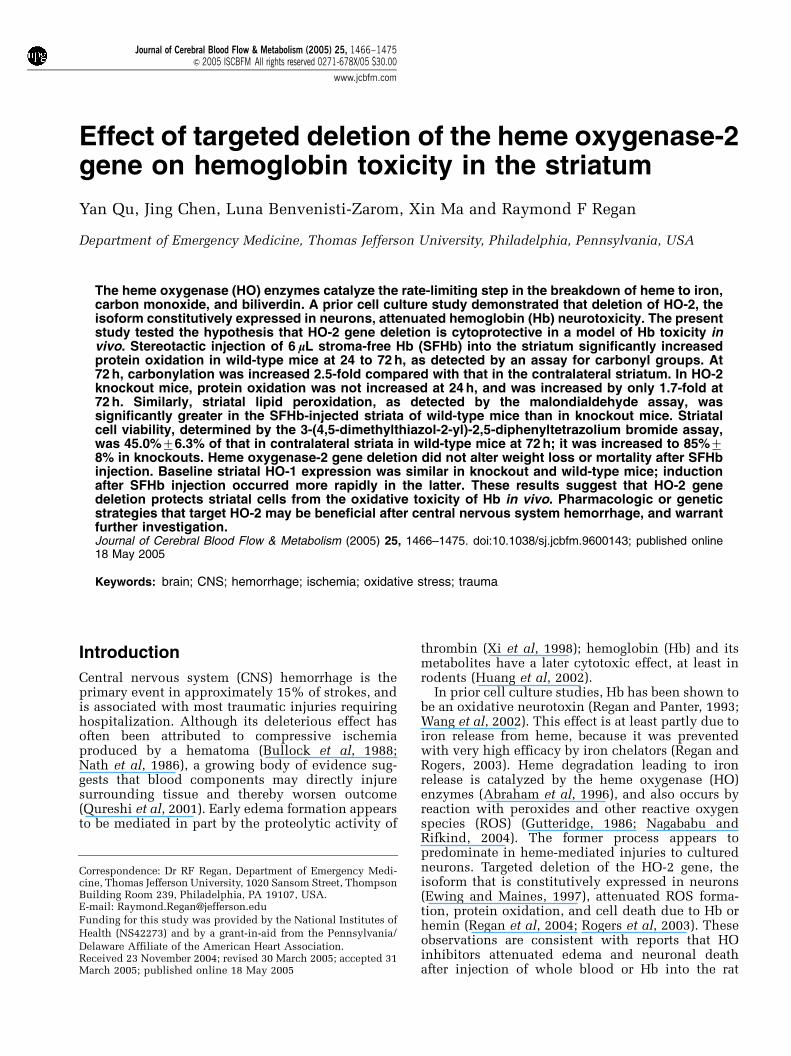
To determine if the protective effect of HO-2 genedeletion was associated with evidence of reducedoxidative stress, striatal malondialdehyde and pro-tein carbonyl content were determined. In wild-typemice, SFHb injection was associated with a 1.6-foldincrease in protein carbonylation at 24 h and 2.5-fold increase at 72 h, compared with the contra-lateral striatum (Figure 4). In HO-2 knockout mice,no increase was observed 24 h after SFHb injection,and a 1.7-fold increase was observed at 72 h.Consistent with this observation, striatal malondi-aldehyde was increased 3.5-fold in wild-type mice72 h after SFHb injection, but was increased onlytwo-fold in knockouts (Figure 5).
Effect of HO-2 Gene Deletion on Neuronal MorphologyAfter SFHb Injection
At 3 days after SFHb injection, most neurons thatwere immediately adjacent to the injection site had
Table 1 Mean weight (7S.E.M., n¼12 to 37/condition) ofmice immediately before striatal injection of 6 mL stroma-freeHb solution or saline, and 3 days later
Preinjection weight (g) Postinjection weight (g)
WT-Hb 28.1970.46 24.1870.45KO-Hb 27.6070.47 23.7770.46WT-saline 27.1570.48 25.9170.49KO-saline 27.0170.87 25.9370.64
Po0.001 for difference between pre- and post-injection weights in Hb-injected animals only, Bonferroni multiple comparisons test.
Figure 2 Heme oxygenase (HO)-2 gene deletion attenuatesstroma-free hemoglobin toxicity in striatum. Sections of brainfrom wild-type (A) and HO-2 knockout mouse (B), 72 h afterstriatal stroma-free hemoglobin injection, stained with 2,3,5-triphenyl tetrazolium chloride (TTC). Area of nonstainingnecrosis in wild-type section is apparent, with smaller area inknockout section.
Saline Hb, WT0.0
0.2
0.4
0.6
0.8
1.0
1.2
***
MT
T R
edu
ctio
n
##
Hb, KO
Figure 3 Heme oxygenase (HO)-2 gene deletion reduces thecytotoxic effect of stroma-free hemoglobin in vivo. Barsrepresent striatal cell viability 72 h after Hb or saline injection,as assessed by MTT reduction to formazan. MTT reduction(mean7S.E.M., n¼10/condition) is expressed as a fraction ofthat in the hemisphere contralateral to the injection site. Thesaline group contained five knockout and five wild-type mice.***Po0.001 versus ratio in mice injected with saline,##Po0.01 versus that in wild-type (WT) mice.
Effect of targeted deletion of the heme oxygenase-2Y Qu et al
1469
Journal of Cerebral Blood Flow & Metabolism (2005) 25, 1466–1475
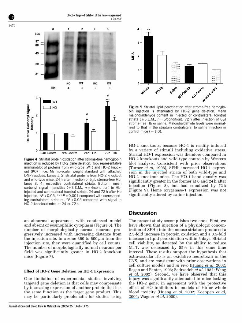
an abnormal appearance, with condensed nucleiand absent or eosinophilic cytoplasm (Figure 6). Thenumber of morphologically normal neurons pro-gressively increased with increasing distance fromthe injection site. In a zone 360 to 600 mm from theinjection site, they were quantified by cell counts.The number of morphologically normal neurons perfield was significantly greater in HO-2 knockoutmice (Figure 7).
Effect of HO-2 Gene Deletion on HO-1 Expression
One limitation of experimental studies involvingtargeted gene deletion is that cells may compensateby increasing expression of another protein that hasthe same function as the target gene product. Thismay be particularly problematic for studies using
HO-2 knockouts, because HO-1 is readily inducedby a variety of stimuli including oxidative stress.Striatal HO-1 expression was therefore compared inHO-2 knockouts and wild-type controls by Westernblot analysis. Consistent with prior observations(Turner et al, 1998), SFHb increased HO-1 expres-sion in the injected striata of both wild-type andHO-2 knockout mice. The HO-1 band density wassignificantly greater in the former at 6 and 24 h afterinjection (Figure 8), but had equalized by 72 h(Figure 9). Heme oxygenase-1 expression was notsignificantly altered by saline injection.
Discussion
The present study accomplishes two ends. First, wehave shown that injection of a physiologic concen-tration of SFHb into the mouse striatum produced a2.5-fold increase in protein oxidation and a 3.5-foldincrease in lipid peroxidation within 3 days. Striatalcell viability, as detected by the ability to reduceMTT, was decreased by 55% in this same timeinterval. These results support the hypothesis thatextravascular Hb is an oxidative neurotoxin in theCNS, and are consistent with prior observations incell culture models and in vivo (Huang et al, 2002;Regan and Panter, 1993; Sadrzadeh et al, 1987; Wanget al, 2002). Second, we have observed that thisinjury was significantly attenuated in mice lackingthe HO-2 gene, in agreement with the protectiveeffect of HO inhibitors in models of Hb or wholeblood toxicity (Huang et al, 2002; Koeppen et al,2004; Wagner et al, 2000).
24h Contra 72h Contra 24h0
1
2
3WT
KO
*
***#
#*
Rel
ativ
e C
arb
ony
l Sig
nal
In
ten
sity
Hb 72h Hb
Figure 4 Striatal protein oxidation after stroma-free hemoglobininjection is reduced by HO-2 gene deletion. Top: representativeimmunoblot of proteins from wild-type (WT) and HO-2 knock-out (KO) mice. M: molecular weight standard with attachedDNP residues. Lanes 1, 2: striatal proteins from HO-2 knockoutand wild-type mice, 24 h after injection of 6 mL stroma-free Hb;lanes 3, 4: respective contralateral striata. Bottom: meancarbonyl signal intensities (7S.E.M., n¼4/condition) in Hb-injected and contralateral (contra) striata, 24 and 72 h after Hbinjection. *Po0.05, ***Po0.001 compared with correspond-ing contralateral striatum, #Po0.05 compared with signal inHO-2 knockout mice at 24 or 72 h.
Figure 5 Striatal lipid peroxidation after stroma-free hemoglo-bin injection is attenuated by HO-2 gene deletion. Meanmalondialdehyde content in injected or contralateral (contra)striata (7S.E.M., n¼6/condition), 72 h after injection of 6 mlstroma-free Hb or saline. Malondialdehyde levels were normal-ized to that in the striatum contralateral to saline injection incontrol mice (¼1.0).
Effect of targeted deletion of the heme oxygenase-2Y Qu et al
1470
Journal of Cerebral Blood Flow & Metabolism (2005) 25, 1466–1475

The effect of the HO enzymes on oxidative injuryhas been controversial in the CNS and elsewhere, aswidely disparate results have been reported. It isbecoming increasingly apparent that HO may haveboth proxidant and antioxidant effects (Ryter andTyrrell, 2000), and that its net effect depends onboth the cell type and the nature of the oxidativeinsult. Its antioxidant properties are likely mediatedby its catalysis of heme breakdown to biliverdin,which is an antioxidant per se and is converted toanother antioxidant, bilirubin, by biliverdin reduc-
tase (Stocker et al, 1987). This protective effectappears to predominate in models relevant to CNSischemia and trauma (Chang et al, 2003; Dore et al,
Figure 6 Morphologic appearance of striatal cells, 72 h after stroma-free hemoglobin injection, in wild-type (A, C) and HO-2knockout mice (B, D). (A, B): Images captured 120 mm from injection site; (C, D) images captured 360 mm from injection site.Damaged neurons have a condensed, irregular-shaped and darkly stained nucleus. Scale bar¼25mm.
Contra HB, KO HB, WT0
10
20
30
40
50
60
70
80
90
***
Neu
ron
s/F
ield
Figure 7 Bars represent mean number of morphologicallynormal neurons per 320�240 mm field, captured at an innerboundary that was 360 mm from the injection site, 72 h afterstroma-free hemoglobin injection. ***Po0.001 versus meannumber in wild-type mice, n¼3 mice/condition.
6h
Hbsaline
WT WT WT WTKO KO- - -
-++
+ - - - --
24h
6h Hb 24h Hb0
1
2
3 KO
WT *** ***
HO
-1 b
and
den
city
Figure 8 Greater HO-1 induction in wild-type mice than inHO-2 knockout mice at 6 and 24 h after stroma-freehemoglobin injection. Immunoblots of lysates from wild-typeand HO-2 knockout striata were stained with polyclonalantibodies to HO-1. The mean band densities (7S.E.M.,n¼3/conditon) were normalized to that of contralateral striataof WT mice injected with Hb (¼1.0). ***Po0.001 versusmean value in KO mice at same time point.
Effect of targeted deletion of the heme oxygenase-2Y Qu et al
1471
Journal of Cerebral Blood Flow & Metabolism (2005) 25, 1466–1475

1999a; Dore et al, 1999b; Panahian et al, 1999).However, in the presence of excess substrate, whichoccurs after hemorrhage, HO activity may enhanceoxidation of cellular proteins and lipids (Lamb et al,1999; Levere et al, 1989). This phenomenon isattenuated by deferoxamine (Lamb et al, 1999;Sadrzadeh et al, 1987), and is therefore likelymediated by release of iron from heme (Koeppenand Dickson, 2002).
Consistent with prior observations (Matz et al,1997), HO-1 expression was increased by SFHbinjection in both wild-type and HO-2 knockoutmice; however, its upregulation was more rapid inthe former. Heme oxygenase-1 is induced by avariety of oxidative cytotoxins in the striatum, andit appears to be a sensitive marker of early cell injury(Munoz et al, 2005). This phenomenon is likely dueto the presence of the antioxidant response elementin the 50 untranslated region of the HO-1 gene,which increases transcription in response to oxida-tive stress by binding the Nrf-2 transcription factor(Elbirt and Bonkovsky, 1999; Ishii et al, 2000).The attenuated HO-1 expression in HO-2 knockoutmice at 6 and 24 h may merely reflect a slowerprogression of injury. In this regard, it is note-worthy that striatal protein oxidation was alreadydetectable in wild-type mice by 24 h, but not inknockouts.
In addition to being a marker of oxidative cellinjury, heme breakdown by induced HO-1 may alsocontribute to cytotoxicity (Lu and Ong, 2001;Schipper et al, 1999). In a prior cell culture study,HO-2 gene deletion attenuated but did not comple-tely prevent the toxic effect of Hb on corticalneurons (Rogers et al, 2003). Heme oxygenase-1was induced by Hb treatment in both wild-type andknockout cultures. Most of the neuronal death inknockout cultures was prevented by concomitanttreatment with the HO inhibitor tin protoporphyrinIX, suggesting that heme breakdown by HO-1 is alsodeleterious. The delayed expression of HO-1 in HO-2knockout mice in the present study may thereforeaccount for part of the protection provided by HO-2gene deletion.
In contrast to its effect on neuronal injury, priorstudies suggest that HO decreases the vulnerabilityof astrocytes to Hb and hemin (Chen and Regan,2004; Regan et al, 2000; Teng et al, 2004). Both wild-type and HO-2 knockout astrocytes are highlyresistant to Hb in culture. This resistance ismediated at least in part by HO activity, because itis markedly diminished by HO inhibitors (Reganet al, 2000). Astrocytes are quite vulnerable to therapid injury produced by hemin, the oxidized formof heme that accumulates to high micromolarconcentrations in hematomas (Letarte et al, 1993).Increasing HO-1 by adenoviral gene transfer beforehemin exposure is protective (Teng et al, 2004),while HO-2 gene deletion is deleterious (Chen andRegan, 2004). The disparate effect of HO on heme-mediated injury to neurons and astrocytes may limitthe utility of HO inhibitor therapy for CNS hemor-rhage. In animal models, its net effect is protective(Huang et al, 2002; Koeppen et al, 2004). It shouldbe noted, however, that the cellular composition ofthe human and rodent striatum is quite different.Approximately two-thirds of cells in the mouse stri-atum are neurons (Sturrock, 1980). In contrast, 70%of cells in the striata of middle-aged humans dying ofnon-neurologic disease are glia (Heinsen et al, 1994).Because HO apparently protects astrocytes fromheme-mediated injury but is deleterious to neurons,reducing its activity may have a different net effecton hemorrhagic striatal injury in humans androdents.
The concentration and injection volume of SFHbwere chosen to produce a consistent injury withminimal mortality. Nevertheless, a low mortalityrate was observed that was similar in both knockoutand wild-type mice. It was accompanied by a largeintracranial hemorrhage in all animals. While thesehemorrhages may have been produced by mechan-ical disruption of vessels during needle insertion orinjection, examination of striata of control miceinjected with saline suggests that this is unlikely. Of35 mice in this latter group, only one punctatehemorrhage at the needle insertion site was seenwhen striata were inspected under a dissectingmicroscope; no deaths occurred. It therefore seems
KO-Hb
WT-H
b
Saline
KO-Contra
WT-C
ontra
Saline-C
ontra0
1
2
3
HO
-1 B
and
den
sity
Figure 9 Heme oxygenase (HO)-1 expression is similar in wild-type and HO-2 knockout mice 72 h after stroma-free hemo-globin injection. Immunoblots of lysates from wild-type andHO-2 knockout striata 72 h after Hb or saline injection, stainedwith polyclonal antibodies to either HO-1 or HO-2. The meanHO-1 protein band densities (7S.E.M., n¼3 each) in the barchart were normalized to that of contralateral striata of wild-type mice injected with Hb (¼1.0).
Effect of targeted deletion of the heme oxygenase-2Y Qu et al
1472
Journal of Cerebral Blood Flow & Metabolism (2005) 25, 1466–1475

likely that hemorrhage may be a consequence ofextravascular SFHb per se. Hemoglobin has beenshown to be toxic to endothelial and vascularsmooth muscle cells in culture (Comair et al, 1993;Motterlini et al, 1995), and to intact vessels in vivo(Mayberg et al, 1990a). Its toxicity to vascular cellshas primarily been associated with arterial spasmand subsequent ischemia in models of subarachnoidhemorrhage (Mayberg et al, 1990b), and is attenu-ated by HO (Abraham et al, 1995; Ono et al, 2001).Further investigation of the effect of Hb on vesselintegrity and rebleeding in intracerebral hemorrhagemodels seems warranted.
This study was designed to quantify oxidative cellinjury in the striatum after SFHb injection, ratherthan assess metabolic end points. However, animalweight was recorded before injection and 3 dayslater, and was significantly decreased by Hb in bothwild-type and knockout mice. Weight loss hascommonly been observed after hemorrhagic andtraumatic brain injury, in both animal models andhumans (Roe and Rothwell, 1997; Touho et al,1990). Its etiology has not been precisely defined,but it is likely due to both an increased metabolicrate and hypophagia. The results of this studysuggest that this effect is not altered by expressionof HO-2.
Intracerebral hemorrhage is a complex injury.Experimental evidence suggests that clot retraction,thrombin proteolysis, transferrin-bound iron toxi-city, and a mass effect contribute to cell injury,in addition to the deleterious effect of extracellularHb (Bullock et al, 1988; Nakamura et al, 2005;Xi et al, 2002; Xi et al, 2000). The model used inthis study addresses only the latter mechanism,and therefore does not represent many key elementsof clinical CNS hemorrhage. However, by facili-tating the investigation of the toxicity of Hb inthe intact CNS, without the confounding effects ofother injury cascades, it will hopefully comp-lement studies assessing the toxicity of wholeblood.
In summary, the present results suggest thatHO-2 increases local oxidative stress after injectionof SFHb into the neuron-rich mouse striatum.Therapeutic strategies that decrease its expressionor activity may protect neurons from heme-mediatedinjury after CNS hemorrhage. However, the conse-quence of such approaches on glial and vascularinjury should be considered, in light of the protec-tive effect of HO on heme-mediated injury to thesecell populations.
Acknowledgements
The authors thank Dr Frank Sharp for providing theHO-2 knockout mice used to establish our colony,and Mr James O’Dowd for his technical assistance inthis project.
References
Abraham NG, Drummond GS, Lutton JD, Kappas A (1996)The biological significance and physiological role ofheme oxygenase. Cell Physiol Biochem 6:129–68
Abraham NG, Lavrosky Y, Schwartzman ML, Stoltz RA,Levere RD, Gerristen ME, Shibahara S, Kappas A (1995)Transfection of the human heme oxygenase gene intorabbit microvessel endothelial cells: protective effectagainst heme and hemoglobin toxicity. Proc Natl AcadSci USA 92:6798–802
Bullock R, Brock-Utne J, van Dellen J, Blake G (1988)Intracerebral hemorrhage in a primate model: effect onregional cerebral blood flow. Surg Neurol 29:101–7
Carmichael J, DeGraff WG, Gazdar AF, Minna JD, MitchellJB (1987) Evaluation of a tetrazolium-based semiauto-mated colorimetric assay: assessment of chemosensi-tivity testing. Cancer Res 47:936–42
Chang EF, Wong RJ, Vreman HJ, Igarashi T, Galo E, SharpFR, Stevenson DK, Noble-Haeusslein LJ (2003) Hemeoxygenase-2 protects against lipid peroxidation-mediated cell loss and impaired motor recovery aftertraumatic brain injury. J Neurosci 23:3689–96
Chen J, Regan RF (2004) Heme oxygenase-2 gene deletionincreases astrocyte vulnerability to hemin. BiochemBiophys Res Commun 318:88–94
Comair YG, Schipper HM, Brem S (1993) The preventionof oxyhemoglobin-induced endothelial and smoothmuscle cytoskeletal injury by deferoxamine. Neurosur-gery 32:58–65
Dore S, Sampei K, Goto S, Alkayed NJ, Guastella D,Blackshaw S, Gallagher M, Traystman RJ, Hurn PD,Koehler RC, Snyder SH (1999a) Heme oxygenase-2 isneuroprotective in cerebral ischemia. Mol Med 5:656–63
Dore S, Takahashi M, Ferris CD, Hester LD, Guastella D,Snyder SH (1999b) Bilirubin, formed by activation ofheme oxygenase-2, protects neurons against oxidativestress injury. Proc Natl Acad Sci USA 96:2445–50
Elbirt KK, Bonkovsky HL (1999) Heme oxygenase: recentadvances in understanding its regulation and role. ProcAssoc Am Physicians 111:438–47
Ewing JF, Maines MD (1997) Histochemical localization ofheme oxygenase-2 protein and mRNA expression in ratbrain. Brain Res Brain Res Protoc 1:165–74
Grundemar L, Ny L (1997) Pitfalls using metalloporphyr-ins in carbon monoxide research. Trends PharmacolSci 18:193–5
Gutteridge JMC (1986) Iron promoters of the Fentonreaction and lipid peroxidation can be released fromhaemoglobin by peroxides. FEBS Lett 201:291–5
Heinsen H, Strik M, Bauer M, Luther K, Ulmar G, GangnusD, Jungkunz G, Eisenmenger W, Gotz M (1994) Corticaland striatal neurone number in Huntington’s disease.Acta Neuropathol (Berlin) 88:320–33
Huang FP, Xi G, Keep RF, Hua Y, Nemoianu A, Hoff JT(2002) Brain edema after experimental intracerebralhemorrhage: role of hemoglobin degradation products.J Neurosurg 96:287–93
Ishii T, Itoh K, Takahashi S, Sato H, Yanagawa T, Katoh Y,Bannai S, Yamamoto M (2000) Transcription factorNrf2 coordinately regulates a group of oxidative stress-inducible genes in macrophages. J Biol Chem275:16023–9
Koeppen AH, Dickson AC (2002) Tin-protoporphyrinprevents experimental superficial siderosis in rabbits.J Neuropathol Exp Neurol 61:689–701
Effect of targeted deletion of the heme oxygenase-2Y Qu et al
1473
Journal of Cerebral Blood Flow & Metabolism (2005) 25, 1466–1475

Koeppen AH, Dickson AC, Smith J (2004) Heme oxygenasein experimental intracerebral hemorrhage: the benefitof tin-mesoporphyrin. J Neuropathol Exp Neurol63:587–97
Lamb NJ, Quinlan GJ, Mumby S, Evans TW, GutteridgeJMC (1999) Haem oxygenase shows pro-oxidant activityin microsomal and cellular systems: implications forthe release of low-molecular-mass iron. Biochem J344:153–8
Letarte PB, Lieberman K, Nagatani K, Haworth RA, OdellGB, Duff TA (1993) Hemin: levels in experimentalsubarachnoid hematoma and effects on dissociatedvascular smooth muscle cells. J Neurosurg 79:252–5
Levere RD, Escalante B, Schwartzman ML, Abraham NG(1989) Role of heme oxygenase in heme-mediatedinhibition of rat brain Na+-K+-ATPase: protection bytin protoporphyrin. Neurochem Res 14:861–4
Linden DJ, Narisimhan K, Gurfel D (1993) Protoporphyr-ins modulate voltage-gated calcium current in AtT-20pituitary cells. J Neurophysiol 70:2673–7
Lu XR, Ong WY (2001) Heme oxgenase-1 is expressed inviable astrocytes and microglia but in degeneratingpyramidal neurons in the kainate-lesioned rat hippo-campus. Exp Brain Res 137:424–31
Lundy EF, Solik BS, Frank RS, Lacy PS, Combs DJ,Zelenock GB, D’Alecy LG (1986) Morphometric evalua-tion of brain infarcts in rats and gerbils. J PharmacolMethods 16:201–14
Luo D, Vincent SR (1994) Metalloporphyrins inhibit nitricoxide-dependent cGMP formation in vivo. Eur JPharmacol 267:263–7
Matz PG, Weinstein PR, Sharp FR (1997) Heme oxygenase-1 and heat shock protein-70 induction in glia andneurons throughout rat brain after experimental in-tracerebral hemorrhage. Neurosurgery 40:152–60
Mayberg MR, Okada T, Bark DH (1990a) The role ofhemoglobin in arterial narrowing after subarachnoidhemorrhage. J Neurosurg 72:634–40
Mayberg MR, Okada T, Bark DH (1990b) The significanceof morphological changes in cerebral arteries aftersubarachnoid hemorrhage. J Neurosurg 72:626–33
Meffert MK, Haley JE, Schuman EM, Schulman H,Madison DV (1994) Inhibition of hippocampalheme oxygenase, nitric oxide synthase, and long-termpotentiation by metalloporphyrins. Neuron 13:1225–33
Motterlini R, Foresti R, Vandegriff K, Intaglietta M,Winslow RM (1995) Oxidative-stress response invascular endothelial cells exposed to acellular hemo-globin solutions. Am J Physiol 269:H648–55
Munoz AM, Rey P, Parga J, Guerra MJ, Labandeira-GarciaJL (2005) Glial overexpression of heme oxygenase-1:a histochemical marker for early stages of striataldamage. J Chem Neuroanat 29:113–26
Nagababu E, Rifkind JM (2004) Heme degradation byreactive oxygen species. Antioxid Redox Signal 6:967–978
Nakamura T, Xi G, Park JW, Hua Y, Hoff JT, Keep RF (2005)Holo-transferrin and thrombin can interact to causebrain damage. Stroke 36:348–52
Nath FP, Jenkins A, Mendelow AD, Graham DI, TeasdaleGM (1986) Early hemodynamic changes in experimen-tal intracerebral hemorrhage. J Neurosurg 65:697–703
Ono S, Komuro T, Macdonald RL (2001) Adenovirus-mediated heme oxygenase-1 gene transfection preventshemoglobin-induced contraction of rat basilar artery.Acta Neurochir Suppl 77:93–6
Panahian N, Yoshiura M, Maines MD (1999) Overexpres-sion of heme oxygenase-1 is neuroprotective in a modelof permanent middle cerebral artery occlusion intransgenic mice. J Neurochem 72:1187–203
Poss KD, Thomas MJ, Ebralidze AK, TJ OD, Tonegawa S(1995) Hippocampal long-term potentiation is normalin heme oxygenase-2 mutant mice. Neuron 15:867–73
Qureshi AI, Tuhrim S, Broderick JP, Batjer HH, Hondo H,Hanley DF (2001) Spontaneous intracerebral hemor-rhage. N Engl J Med 344:1450–60
Regan RF, Chen J, Benvenisti-Zarom L (2004) Hemeoxygenase-2 gene deletion attenuates oxidative stressin neurons exposed to extracellular hemin. BMCNeurosci 5:34
Regan RF, Guo YP, Kumar N (2000) Heme oxygenase-1induction protects murine cortical astrocytes fromhemoglobin toxicity. Neurosci Lett 282:1–4
Regan RF, Panter SS (1993) Neurotoxicity of hemoglobinin cortical cell culture. Neurosci Lett 153:219–22
Regan RF, Rogers B (2003) Delayed treatment of hemo-globin neurotoxicity. J Neurotrauma 20:111–20
Roe SY, Rothwell NJ (1997) Whole body metabolicresponses to brain trauma in the rat. J Neurotrauma14:399–408
Rogers B, Yakopson V, Teng ZP, Guo Y, Regan RF (2003)Heme oxygenase-2 knockout neurons are less vulner-able to hemoglobin toxicity. Free Rad Biol Med 35:872–81
Rose K, Goldberg MP, Choi DW (1993) Cytotoxicityin murine neocortical cell culture. In: Methods intoxicology; Part A: in vitro biological systems (TysonCA, Frazier JM, eds), San Diego: Academic Press,46–60
Ryter SW, Tyrrell RM (2000) The heme synthesis anddegradation pathways: role of oxidant sensitivity—heme oxygenase has both pro- and antioxidant proper-ties. Free Rad Biol Med 28:289–309
Sadrzadeh SMH, Anderson DK, Panter SS, Hallaway PE,Eaton JW (1987) Hemoglobin potentiates central ner-vous system damage. J Clin Invest 79:662–4
Schipper HM, Bernier L, Mehindate K, Frankel D (1999)Mitochondrial iron sequestration in dopamine-chal-lenged astroglia: role of heme oxygenase-1 and thepermeability transition pore. J Neurochem 72:1802–11
Stocker R, Yamamoto Y, McDonagh AF, Glazer AN, AmesBN (1987) Bilirubin is an antioxidant of possiblephysiologic significance. Science 235:1043–6
Sturrock RR (1980) A comparative quantitative andmorphological study of ageing in the mouse neostria-tum, indusium griseum and anterior commissure.Neuropathol Appl Neurobiol 6:51–68
Teng ZP, Chen J, Chau LY, Galunic N, Regan RF (2004)Adenoviral transfer of the heme oxygenase-1 geneprotects cortical astrocytes from heme-mediated oxida-tive injury. Neurobiol Dis 17:179–87
Touho H, Karasawa J, Shishido H, Morisako T, Yamada K,Shibamoto K (1990) Hypermetabolism in the acutestage of hemorrhagic cerebrovascular disease. J Neuro-surg 72:710–4
Turner CP, Bergeron M, Matz P, Zegna A, Noble LJ,Panter SS, Sharp FR (1998) Heme oxygenase-1is induced in glia throughout brain by subarach-noid hemoglobin. J Cereb Blood Flow Metab 18:257–73
Wagner KR, Dwyer BE (2004) Hematoma removal, heme,and heme oxygenase following hemorrhagic stroke.Ann NY Acad Sci 1012:237–51
Effect of targeted deletion of the heme oxygenase-2Y Qu et al
1474
Journal of Cerebral Blood Flow & Metabolism (2005) 25, 1466–1475

Wagner KR, Hua Y, de Courten-Myers GM, BroderickJP, Nishimura RN, Lu SY, Dwyer BE (2000) Tin-mesoporphyrin, a potent heme oxygenase inhibitor,for treatment of intracerebral hemorrhage: in vivo andin vitro studies. Cell Mol Biol 46:597–608
Wang X, Mori T, Sumii T, Lo EH (2002) Hemoglobin-induced cytotoxicity in rat cerebral cortical neurons:caspase activation and oxidative stress. Stroke33:1882–8
Winterbourn CC (1990) Oxidative reactions of hemo-globin. Methods Enzymol 186:265–72
Xi G, Keep RF, Hoff JT (2002) Pathophysiology ofbrain edema formation. Neurosurg Clin North Am 13:371–83
Xi G, Wagner KR, Keep RF, Hua Y, deCourten-Myers GM,Broderick JP, Brott TG, Hoff JT, Muizelaar JP (2000)Role of blood clot formation on early edema developmentafter experimental intracerebral hemorrhage. Stroke29:2580–6
Xi GH, Keep RF, Hoff JT (1998) Erythrocytes and delayedbrain edema formation following intracerebral hemor-rhage in rats. J Neurosurg 89:991–6
Effect of targeted deletion of the heme oxygenase-2Y Qu et al
1475
Journal of Cerebral Blood Flow & Metabolism (2005) 25, 1466–1475