Optimization schemes for endovascular repair with parallel ...
Upload
khangminh22Category
view
0download
0
Citation: Meisenbacher, K.;
Hagedorn, M.; Skrypnik, D.; Kilian,
S.; Böckler, D.; Bischoff, M.S.; Peters,
A.S. Thoracic Endovascular Aortic
Repair (TEVAR) First in Patients with
Lower Limb Ischemia in Complicated
Type B Aortic Dissection: Clinical
Outcome and Morphology. J. Clin.
Med. 2022, 11, 4154. https://doi.org/
10.3390/jcm11144154
Academic Editor: Olivier Morel
Received: 18 June 2022
Accepted: 15 July 2022
Published: 17 July 2022
Publisher’s Note: MDPI stays neutral
with regard to jurisdictional claims in
published maps and institutional affil-
iations.
Copyright: © 2022 by the authors.
Licensee MDPI, Basel, Switzerland.
This article is an open access article
distributed under the terms and
conditions of the Creative Commons
Attribution (CC BY) license (https://
creativecommons.org/licenses/by/
4.0/).
Journal of
Clinical Medicine
Article
Thoracic Endovascular Aortic Repair (TEVAR) First in Patientswith Lower Limb Ischemia in Complicated Type B AorticDissection: Clinical Outcome and MorphologyKatrin Meisenbacher 1,*, Matthias Hagedorn 1, Denis Skrypnik 1 , Samuel Kilian 2, Dittmar Böckler 1,Moritz S. Bischoff 1 and Andreas S. Peters 1
1 Department of Vascular and Endovascular Surgery, University Hospital Heidelberg,69120 Heidelberg, Germany; [email protected] (M.H.);[email protected] (D.S.); [email protected] (D.B.);[email protected] (M.S.B.); [email protected] (A.S.P.)
2 Institute of Medical Biometry, University of Heidelberg, 69120 Heidelberg, Germany;[email protected]
* Correspondence: [email protected]
Abstract: Acute Type B aortic dissection (TBAD) can cause organ malperfusion, e.g., lower limbischemia (LLI). Thoracic endovascular aortic repair (TEVAR) represents the standard treatmentfor complicated TBAD; however, with respect to LLI, data is scant. The aim of this study was toinvestigate clinical and morphological outcomes in patients with complicated TBAD and LLI managedwith a “TEVAR-first” policy. Between March 1997 and December 2021, 731 TEVAR-procedures wereperformed, including 106 TBAD-cases. Cases with TBAD + LLI were included in this retrospectiveanalysis. Study endpoints were morphological/clinical success of TEVAR, regarding aortic andextremity-related outcome, including extremity-related adjunct procedures (erAP) during a medianFU of 28.68 months. A total of 20/106 TBAD-cases (18.8%, 32–82 years, 7 women) presented withacute LLI (12/20 Rutherford class IIb/III). In 15/20 cases, true lumen-collapse (TLC) was presentbelow the aortic bifurcation. In 16/20 cases, TEVAR alone resolved LLI. In the remaining four cases,erAP was necessary. A morphological analysis showed a relation between lower starting point andlesser extent of TLC and TEVAR success. No extremity-related reinterventions and only one majoramputation was needed. The data strongly suggest that aTEVAR-first-strategy for treating TBADwith LLI is reasonable. Morphological parameters might be of importance to anticipate the failure ofTEVAR alone.
Keywords: aortic dissection; thoracic endovascular repair; TEVAR; lower limb ischemia; extremitymalperfusion; complicated type B dissection; malperfusion
1. Introduction
The standard treatment strategy for Stanford Type B aortic dissection (TBAD) consistsof the best possible drug therapy, with the aim of adequate blood pressure control [1,2].However, the disease can take a complicated course: false lumen rupture or organ malper-fusion may occur, both requiring emergency treatment, to avoid a fatal outcome. Theadvances in endovascular therapy in recent decades have led to thoracic endovascularaortic repair (TEVAR) becoming the treatment of choice for complicated TBAD (cTBAD),with lower rates of morbidity and mortality than open repair [1–4]. With respect to malper-fusion, the visceral segment of the aorta can be involved, leading to ischemia of the liver, thesmall intestine, and the kidneys. Lower limb ischemia (LLI) may appear as an additionalor isolated clinical sign. Data from the International Registry of Aortic Dissection (IRAD)show that LLI occurs in 7.1% of patients and represents a risk factor for mortality [5]. Froma pathophysiological point of view, malperfusion is caused either by a proximally located
J. Clin. Med. 2022, 11, 4154. https://doi.org/10.3390/jcm11144154 https://www.mdpi.com/journal/jcm

J. Clin. Med. 2022, 11, 4154 2 of 14
aortic true lumen collapse (TLC) or by a dissection of the respective organ-supplying vesselitself. The basic principle of the TEVAR procedure for cTBAD is coverage of the proximalentry tear, to redirect the blood flow into the true lumen, hence overcoming the collapse.Data on the treatment and outcome of cTBAD, in terms of LLI, are sparse. Whether primaryTEVAR represents the treatment of choice for LLI in cTBAD is an unresolved question. Thestudy presented here comprises a clinical and morphological analysis of the outcome inpatients with cTBAD and LLI managed with a “TEVAR-first” policy.
2. Materials and Methods2.1. Study Design
An observational retrospective single-center study was designed to evaluate the impactof TEVAR on LLI in patients with acute cTBAD. This approach includes characterization ofboth morphological alterations and clinical outcomes. The patients were identified from adepartmental prospective database of cases involving any TEVAR procedure, which wasapproved by the local ethics committee (protocol no. S-158/2015).
2.2. Study Cohort
The inclusion criteria comprised cTBAD with clinical and/or radiological signs of LLIand treatment with TEVAR at the authors’ institution between March 1997 and December2021. Other pathologies, cTBAD with no clinical and/or radiological signs of LLI, andTBAD treated conservatively were excluded, as were patients with missing preoperativeand/or postoperative imaging results.
2.3. Procedural Data
Up to September 2010, an Axiom-U imaging system (Siemens, Healthineers, Erlangen,Germany) was used; from October 2010 onward, TEVAR took place in a hybrid operatingroom (Artis zeego multiaxis imaging system; Siemens Healthineers, Erlangen, Germany).From October 2020, the operating theatre was equipped with an Artis pheno angiographysystem (Siemens Healthineers, Erlangen, Germany). The implantation protocol has beenpublished previously [6,7]. Left subclavian artery (LSA) revascularization was undertakenselectively [8]. The local institutional protocol for the treatment of cTBAD stipulatesa TEVAR-first strategy with coverage of the primary entry, irrespective of the clinicalpresentation and/or associated malperfusion. This comprises the implantation of a singleTEVAR device covering the main entry. Hereafter, any further distal extension of TEVAR(usually down to the level of the celiac trunk) or adjunct procedure is based on the findingsof subsequent downstream angiography. In general, all TEVAR procedures are performedby senior physicians with extensive TEVAR experience.
2.4. Study Endpoints and Definitions
The underlying hypothesis of this study was that TEVAR alone is able to re-establisharterial inflow in patients affected by LLI, without any additional procedures. Therefore,the study endpoints were the morphological and clinical success of TEVAR, with respectto aortic and extremity-related outcome. This approach includes the detailed evaluationof all extremity-related adjunct procedures (erAP) and/or reinterventions, particularlyfocusing on a detailed morphological characterization, with regard to any association withthe technical approach and procedural success.
erAP were defined as any endovascular, open, or hybrid vascular procedure adjunctiveto TEVAR with the aim of restoration of blood flow into the affected limb during the indexprocedure, meeting the definition of the Society for Vascular Surgery and Society of ThoracicSurgeons reporting standards for type B aortic dissections [9].
Extremity-related reinterventions (erRI) were defined as any secondary revascularization-procedure performed for the affected limb after the index procedure (e.g., secondary bypass).
Extremity-related outcomes were defined as freedom from lower limb amputationand freedom from functional limb impairment.

J. Clin. Med. 2022, 11, 4154 3 of 14
Based on the criteria of White et al., lower extremity malperfusion was defined as miss-ing femoral artery pulses, together with one or more of the following findings: radiographicmalperfusion of the affected limb, loss of sensorimotor function, paleness/discolorationof the affected limb, or pain in the affected limb [10]. The Rutherford acute limb ischemiaclassification system (class I–III) was used for clinical categorization of lower extremitymalperfusion [11]. For morphological analysis, the classification of aortic zones introducedby Fillinger et al. [12] was extended to provide a more detailed description of zones 4, 5,and 9 (Figure 1).
J. Clin. Med. 2022, 11, x FOR PEER REVIEW 3 of 15
Extremity-related reinterventions (erRI) were defined as any secondary revasculari-zation-procedure performed for the affected limb after the index procedure (e.g., second-ary bypass).
Extremity-related outcomes were defined as freedom from lower limb amputation and freedom from functional limb impairment.
Based on the criteria of White et al., lower extremity malperfusion was defined as missing femoral artery pulses, together with one or more of the following findings: radi-ographic malperfusion of the affected limb, loss of sensorimotor function, paleness/dis-coloration of the affected limb, or pain in the affected limb [10]. The Rutherford acute limb ischemia classification system (class I‒III) was used for clinical categorization of lower extremity malperfusion [11]. For morphological analysis, the classification of aortic zones introduced by Fillinger et al. [12] was extended to provide a more detailed description of zones 4, 5, and 9 (Figure 1).
Figure 1. Extended aortic zones; based on Fillinger et al. [12]. CT: celiac trunk; SMA: superior mes-enteric artery; RA: renal artery; CIA: common iliac artery; EIA: external iliac artery; IIA: internal iliac artery.
Figure 1. Extended aortic zones; based on Fillinger et al. [12]. CT: celiac trunk; SMA: superiormesenteric artery; RA: renal artery; CIA: common iliac artery; EIA: external iliac artery; IIA: internaliliac artery.
2.5. Imaging Data and Follow-Up
The institutional protocol includes multidetector electrocardiography-gated computedtomography angiography (CTA) of the entire aorta (supra-aortic branches to femoralarteries) with a 1-mm slice thickness acquired at 60% of the R–R interval, corresponding tolate diastole. CTA was carried out before TEVAR; before discharge; at 3 months, 6 months,and 12 months after the index procedure; and annually thereafter [13]. For the datapresented here, preoperative and postoperative CTA images were analyzed using certifiedthree-dimensional reconstruction software and centerline measurements (OsiriX PRO;aycan Medical Systems, Rochester, NY, USA).
Morphological assessment was performed by visualization in a case-by-case fashion,displaying the extent of the dissected segments, the localization of the main entry and there-entries, and the extent of the TLC, in terms of the aortic zones before the index procedure,after TEVAR (based on the intraoperative angiography), and after treatment (includinga potential erAP). In order to visualize any potential distribution pattern in morphology,

J. Clin. Med. 2022, 11, 4154 4 of 14
the cases were secondarily regrouped, comparing the cases treated by TEVAR alone withthe TEVAR + erAP cases. Assessment was performed by at least two experienced readers,blinded to all clinical information. In the event of discrepancies, the investigators reached aconsensus. In addition, the intraoperative angiography findings following primary TEVARwere evaluated.
The CTA before TEVAR and at least one postoperative CTA were available in all cases(imaging follow-up [FU] 100%). FU was completed up to January 2022 for all 20 cases(100%). No patient was lost to FU.
2.6. Statistical Analysis
Patient and disease characteristics are described as absolute and relative frequenciesfor categorical variables and median (range) for continuous data [14]. FU was given by themedian, including the 25% and the 75% quantiles (Q1–Q3). Morphological presentationwas visualized at patient level and aggregated using further visualization methods.
3. Results3.1. Demographics
Between March 1997 and December 2021, a total of 731 TEVAR procedures wereperformed at the authors’ institution, in patients with various aortic pathologies, including106 cases of cTBAD. Of these 106 patients, 23 (21.7%) presented with LLI. Three caseswithout available preoperative and/or postoperative imaging results were excluded, leav-ing 20 patients (20/106; 18.8%; 7 female, 13 male) with a median age of 53 years (range32–82 years) for analysis (Figure 2). Concomitant visceral/renal malperfusion was found in80% of cases (16/20), with renal involvement alone in n = 7, visceral malperfusion in n = 2,and both renal and visceral malperfusion in n = 7. The most common comorbidity wasarterial hypertension (in 80% of cases, 16/20), while none of the patients had a documentedhistory of peripheral artery disease. Four patients had a history of kidney failure (20%),with a pre-existing need for hemodialysis in n = 1 patient. The patient demographics aredisplayed in Table 1.
J. Clin. Med. 2022, 11, x FOR PEER REVIEW 5 of 15
Figure 2. Flowchart of patient selection.
Table 1. Demographics.
Total (n = 20) Age, median (median/range; years) 53 (32–82)
Gender (male/female) 13/7 ASA classification (median/range) 3 (1–5)
Heart failure 1 (5%) Arterial Hypertension 16 (80%)
History of myocardial infarction 2 (10%) Coronary artery disease 3 (15%) Carotid artery stenosis 0 (0%)
Peripheral artery occlusive disease 0 (0%) History of stroke 0 (0%)
COPD 2 (10%) Diabetes mellitus 1 (5%)
BMI >30 kg/m2 6 (30%) Renal insufficiency * 4 (20%)
Need for hemodialysis 1 (5%) History of smoking 5 (25%)
Previous aortic surgery/intervention 2 (10%) Abdominal aorta 1 (5%)
Thoracic aorta 1 (5%) Categorical data are n (number)/%. * (creatinine >1.2 mg/dL). BMI: body mass index; COPD: chronic obstructive pulmonary disease.
3.2. Clinical Presentation of LLI Sixty percent (12/20) of the patients presented with immediately threatened extrem-
ities (Rutherford class IIb: n = 7, class III: n = 5). Eight patients (20%) presented mild symp-toms of LLI (Rutherford class IIa). Unilateral LLI was present in 70% (n = 14) and bilateral LLI in 30% of cases (n = 6). Unilateral malperfusion was seen more frequently in the right (9/14; 64.3%) than in the left limb (5/14; 35.7%). The median creatine kinase (CK) level on
Overall TEVAR 03/1997 and 12/2021
n = 731
Acute aortic dissection n = 106
Complicated acute aortic dissection with extremity malperfusion
n = 23
Cases included in analysis n = 20
Other pathologies n = 625
Uncomplicated acute aortic dissection n = 16
Complicated acute aortic dissection
without extremity malperfusion n = 67
No pre-/postoperative imaging n = 3
Figure 2. Flowchart of patient selection.

J. Clin. Med. 2022, 11, 4154 5 of 14
Table 1. Demographics.
Total (n = 20)
Age, median (median/range; years) 53 (32–82)Gender (male/female) 13/7
ASA classification (median/range) 3 (1–5)Heart failure 1 (5%)
Arterial Hypertension 16 (80%)History of myocardial infarction 2 (10%)
Coronary artery disease 3 (15%)Carotid artery stenosis 0 (0%)
Peripheral artery occlusive disease 0 (0%)History of stroke 0 (0%)
COPD 2 (10%)Diabetes mellitus 1 (5%)BMI > 30 kg/m2 6 (30%)
Renal insufficiency * 4 (20%)Need for hemodialysis 1 (5%)
History of smoking 5 (25%)Previous aortic surgery/intervention 2 (10%)
Abdominal aorta 1 (5%)Thoracic aorta 1 (5%)
Categorical data are n (number)/%. * (creatinine > 1.2 mg/dL). BMI: body mass index; COPD: chronic obstructivepulmonary disease.
3.2. Clinical Presentation of LLI
Sixty percent (12/20) of the patients presented with immediately threatened extrem-ities (Rutherford class IIb: n = 7, class III: n = 5). Eight patients (20%) presented mildsymptoms of LLI (Rutherford class IIa). Unilateral LLI was present in 70% (n = 14) andbilateral LLI in 30% of cases (n = 6). Unilateral malperfusion was seen more frequently inthe right (9/14; 64.3%) than in the left limb (5/14; 35.7%). The median creatine kinase (CK)level on admission was 149 U/L (range 52–1231; reference < 190 U/L), and the medianserum creatinine (Crea) level was 0.99 mg/dL (range 0.56–5; reference 0.6–1.3 mg/dL).
3.3. Procedural Data with Respect to TEVAR and Extremity-Related Adjunct Procedures
All patients except one were treated by TEVAR first, including main entry coverageas an emergency procedure. The proximal landing zone (PLZ) was aortic zone 2 in 16/20(80%) of the cases; in six cases revascularization of the LSA was performed simultaneously.In one patient, iliac stenting of the affected limb was performed 11 days before TEVAR.Resolution of LLI was achieved immediately in 16/20 (80%) patients after TEVAR, alonewith a median of one (range 1–3) implanted device and a median treatment length of200 mm (range 145–300 mm).
In four cases (20%), TEVAR failed to restore the blood flow into the affected limb(s). Inthese cases, additional erAP were performed during the index procedure. A femorofemoralcrossover bypass was performed in n = 2, iliac stenting in n = 1, and infrarenal aortic barestent implantation in n = 1. All procedural details are shown in Table 2. All erAP aredisplayed case by case in Table 3.
Table 2. Procedural data.
Total (n = 20) TEVAR Alone (n = 16) TEVAR + erAP (n = 4)
Duration of procedure (min; median/range) 162 (83–435) 159 (83–384) 296 (125–435)Radiation time (min; median/range) 13 (7–38) 13 (7–38) 19 (12–32)
Contrast agent volume (mL; median/range) 220 (90–480) 215 (90–480) 220 (220–250)Dose area product (uGy/m2; median/range) 38,206 (2215–257,955) 40,500 (2215–257,955) 23,685 (8102–80,428)
CSFD 8 (40%) 6 (37.5%) 2 (50%)
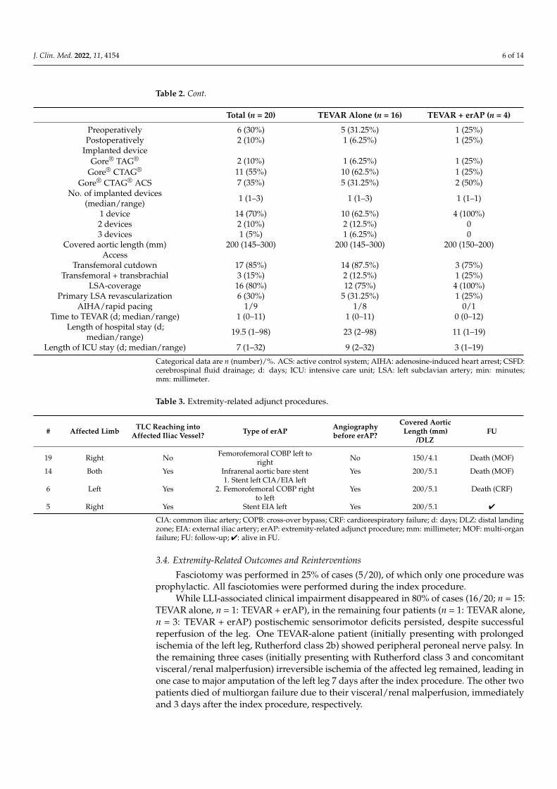
J. Clin. Med. 2022, 11, 4154 6 of 14
Table 2. Cont.
Total (n = 20) TEVAR Alone (n = 16) TEVAR + erAP (n = 4)
Preoperatively 6 (30%) 5 (31.25%) 1 (25%)Postoperatively 2 (10%) 1 (6.25%) 1 (25%)
Implanted deviceGore® TAG® 2 (10%) 1 (6.25%) 1 (25%)
Gore® CTAG® 11 (55%) 10 (62.5%) 1 (25%)Gore® CTAG® ACS 7 (35%) 5 (31.25%) 2 (50%)
No. of implanted devices(median/range) 1 (1–3) 1 (1–3) 1 (1–1)
1 device 14 (70%) 10 (62.5%) 4 (100%)2 devices 2 (10%) 2 (12.5%) 03 devices 1 (5%) 1 (6.25%) 0
Covered aortic length (mm) 200 (145–300) 200 (145–300) 200 (150–200)Access
Transfemoral cutdown 17 (85%) 14 (87.5%) 3 (75%)Transfemoral + transbrachial 3 (15%) 2 (12.5%) 1 (25%)
LSA-coverage 16 (80%) 12 (75%) 4 (100%)Primary LSA revascularization 6 (30%) 5 (31.25%) 1 (25%)
AIHA/rapid pacing 1/9 1/8 0/1Time to TEVAR (d; median/range) 1 (0–11) 1 (0–11) 0 (0–12)
Length of hospital stay (d;median/range) 19.5 (1–98) 23 (2–98) 11 (1–19)
Length of ICU stay (d; median/range) 7 (1–32) 9 (2–32) 3 (1–19)
Categorical data are n (number)/%. ACS: active control system; AIHA: adenosine-induced heart arrest; CSFD:cerebrospinal fluid drainage; d: days; ICU: intensive care unit; LSA: left subclavian artery; min: minutes;mm: millimeter.
Table 3. Extremity-related adjunct procedures.
# Affected Limb TLC Reaching intoAffected Iliac Vessel? Type of erAP Angiography
before erAP?
Covered AorticLength (mm)
/DLZFU
19 Right No Femorofemoral COBP left toright No 150/4.1 Death (MOF)
14 Both Yes Infrarenal aortic bare stent Yes 200/5.1 Death (MOF)
6 Left Yes1. Stent left CIA/EIA left
2. Femorofemoral COBP rightto left
Yes 200/5.1 Death (CRF)
5 Right Yes Stent EIA left Yes 200/5.1 4
CIA: common iliac artery; COPB: cross-over bypass; CRF: cardiorespiratory failure; d: days; DLZ: distal landingzone; EIA: external iliac artery; erAP: extremity-related adjunct procedure; mm: millimeter; MOF: multi-organfailure; FU: follow-up; 4: alive in FU.
3.4. Extremity-Related Outcomes and Reinterventions
Fasciotomy was performed in 25% of cases (5/20), of which only one procedure wasprophylactic. All fasciotomies were performed during the index procedure.
While LLI-associated clinical impairment disappeared in 80% of cases (16/20; n = 15:TEVAR alone, n = 1: TEVAR + erAP), in the remaining four patients (n = 1: TEVAR alone,n = 3: TEVAR + erAP) postischemic sensorimotor deficits persisted, despite successfulreperfusion of the leg. One TEVAR-alone patient (initially presenting with prolongedischemia of the left leg, Rutherford class 2b) showed peripheral peroneal nerve palsy. Inthe remaining three cases (initially presenting with Rutherford class 3 and concomitantvisceral/renal malperfusion) irreversible ischemia of the affected leg remained, leading inone case to major amputation of the left leg 7 days after the index procedure. The other twopatients died of multiorgan failure due to their visceral/renal malperfusion, immediatelyand 3 days after the index procedure, respectively.

J. Clin. Med. 2022, 11, 4154 7 of 14
There was no need for any further extremity-related reintervention during the early orlate phase of FU in any case.
3.5. Mortality
During a median FU of 28.68 months (Q1 0.6 months, Q3 59.3 months, IQR 58.7 months),six of the 20 patients died (overall mortality 30%). All of them died during the primaryhospital stay (in-hospital mortality 30%), an average of 4.5 days after TEVAR (range0–168 days). Of these six patients, n = 3 were treated by TEVAR alone, yielding a mortalityrate of 18.75% (3/16) in this group, while the other three were treated with TEVAR anderAP (3/4, 75%). The reasons for death were multiorgan failure in n = 4, cardiorespiratoryfailure in n = 1, and liver failure in n = 1. None of the patients died due to LLI alone, butrather due to a complicated course of cTBAD with multiorgan involvement.
3.6. Morphological Analysis
The primary entry was located in aortic zone 2 or 3 in 80% of cases (16/20), while onlyfour cases showed a primary entry in zone 4 or 5. The median length of the primary entrywas 15 mm (range 6–100 mm). In all cases, multiple re-entries were present (median 3,range 1–9). In n = 15/20 (75%) TLC was present below the aortic bifurcation, extending intothe affected leg. In 5/20 (15%), the TLC did not reach further than the aortic bifurcation.Post-TEVAR control angiography during the index procedure showed resolution of theTLC in all but four cases (16/20, 80%). In these four cases, LLI persisted in the affectedlimb/s, so erAP was performed.
The morphological presentation is detailed in Table 4 and visualized in case-by-casefashion in Figure 3, which shows the extent of the dissected segments, the localizationof the main entry and the re-entries, and the extent of the TLC, in terms of aortic zonesbefore the index procedure (Figure 3A), after TEVAR (based on the intraoperative an-giography) (Figure 3B), and after treatment (including a potential erAP) (Figure 3C). Theexplanatory caption for Figure 3A–C is displayed in Figure 4. Figure 5 shows the initialmorphological pattern, comparing the cases subsequently treated by TEVAR alone withthe TEVAR + erAP cases.
Table 4. Morphological data before TEVAR.
Total (n = 20) TEVAR Alone (n = 16) TEVAR + erAP (n = 4)
Proximal ETZone 2 10 (50%) 8 (50%) 2 (50%)Zone 3 6 (30%) 5 (31.25%) 1 (25%)Zone 4 3 (15%) 2 (12.5%) 1 (25%)Zone 5 1 (5%) 1 (6.25%) 0
Extent of ET (mm; median, range) 15 (6–100) 15 (6–36) 14.2 (12–100)No. of Re-Entries (median, range) 3 (1–9) 3 (1–9) 3.5 (1–6)Start TLC/Zone (median, range) 6 (4.2–11) 7 (4.2–11) 5.2 (5.2–8)
No. of TLC affected segment (median, range 4 (1–9) 3 (1–9) 5 (4–6)One segment TLC 6 (30%) 6 (37.5%) 02 + segments TLC 14 (70%) 10 (62.5%) 4 (100%)
Concomitant visceral organ malperfusion 16 (80%) 12 (75%) 4 (100%)TLC in affected leg 15 (75%) 12 (75%) 3 (75%)
Categorical data are n (number)/%. ET: entry tear; mm: millimeter; TLC: true lumen collapse.

J. Clin. Med. 2022, 11, 4154 8 of 14
J. Clin. Med. 2022, 11, x FOR PEER REVIEW 8 of 15
the main entry and the re-entries, and the extent of the TLC, in terms of aortic zones before the index procedure (Figure 3A), after TEVAR (based on the intraoperative angiography) (Figure 3B), and after treatment (including a potential erAP) (Figure 3C). The explanatory caption for Figure 3A–C is displayed in Figure 4. Figure 5 shows the initial morphological pattern, comparing the cases subsequently treated by TEVAR alone with the TEVAR + erAP cases.
Table 4. Morphological data before TEVAR.
Total (n = 20) TEVAR Alone (n = 16) TEVAR + erAP (n = 4) Proximal ET
Zone 2 10 (50%) 8 (50%) 2 (50%) Zone 3 6 (30%) 5 (31.25%) 1 (25%) Zone 4 3 (15%) 2 (12.5%) 1 (25%) Zone 5 1 (5%) 1 (6.25%) 0
Extent of ET (mm; median, range) 15 (6–100) 15 (6–36) 14.2 (12–100) No. of Re-Entries (median, range) 3 (1–9) 3 (1–9) 3.5 (1–6) Start TLC/Zone (median, range) 6 (4.2–11) 7 (4.2–11) 5.2 (5.2–8)
No. of TLC affected segment (median, range
4 (1–9) 3 (1–9) 5 (4–6)
One segment TLC 6 (30%) 6 (37.5%) 0 2 + segments TLC 14 (70%) 10 (62.5%) 4 (100%)
Concomitant visceral organ malperfu-sion 16 (80%) 12 (75%) 4 (100%)
TLC in affected leg 15 (75%) 12 (75%) 3 (75%) Categorical data are n (number)/%. ET: entry tear; mm: millimeter; TLC: true lumen collapse.
J. Clin. Med. 2022, 11, x FOR PEER REVIEW 9 of 15
Figure 3. Morphological presentation before TEVAR (A), after TEVAR with respect to intraoperative angiography (B) and after treatment, including a potential extremity related adjunct procedure (C).
Figure 4. Explanatory caption of Figure 3A–C as well as Figure 5.
Figure 5. Secondary rearrangement according to morphological distribution patterns, comparing TEVAR alone with TEVAR + extremity related adjunct procedure (erAP) cases.
Graphical analysis of the evaluated parameters showed a relation between the start-ing point and the extent of the TLC regarding TEVAR success. (Figures 6 and 7A,B).
In addition, the first re-entry was located slightly further downstream in cases treated with TEVAR and erAP. The number of re-entries did not differ between TEVAR alone and TEVAR + erAP. Furthermore, neither the extent of the primary entry tear nor the
Figure 3. Morphological presentation before TEVAR (A), after TEVAR with respect to intraoperativeangiography (B) and after treatment, including a potential extremity related adjunct procedure (C).
J. Clin. Med. 2022, 11, x FOR PEER REVIEW 9 of 15
Figure 3. Morphological presentation before TEVAR (A), after TEVAR with respect to intraoperative angiography (B) and after treatment, including a potential extremity related adjunct procedure (C).
Figure 4. Explanatory caption of Figure 3A–C as well as Figure 5.
Figure 5. Secondary rearrangement according to morphological distribution patterns, comparing TEVAR alone with TEVAR + extremity related adjunct procedure (erAP) cases.
Graphical analysis of the evaluated parameters showed a relation between the start-ing point and the extent of the TLC regarding TEVAR success. (Figures 6 and 7A,B).
In addition, the first re-entry was located slightly further downstream in cases treated with TEVAR and erAP. The number of re-entries did not differ between TEVAR alone and TEVAR + erAP. Furthermore, neither the extent of the primary entry tear nor the
Figure 4. Explanatory caption of Figure 3A–C as well as Figure 5.

J. Clin. Med. 2022, 11, 4154 9 of 14
J. Clin. Med. 2022, 11, x FOR PEER REVIEW 9 of 15
Figure 3. Morphological presentation before TEVAR (A), after TEVAR with respect to intraoperative angiography (B) and after treatment, including a potential extremity related adjunct procedure (C).
Figure 4. Explanatory caption of Figure 3A–C as well as Figure 5.
Figure 5. Secondary rearrangement according to morphological distribution patterns, comparing TEVAR alone with TEVAR + extremity related adjunct procedure (erAP) cases.
Graphical analysis of the evaluated parameters showed a relation between the start-ing point and the extent of the TLC regarding TEVAR success. (Figures 6 and 7A,B).
In addition, the first re-entry was located slightly further downstream in cases treated with TEVAR and erAP. The number of re-entries did not differ between TEVAR alone and TEVAR + erAP. Furthermore, neither the extent of the primary entry tear nor the
Figure 5. Secondary rearrangement according to morphological distribution patterns, comparingTEVAR alone with TEVAR + extremity related adjunct procedure (erAP) cases.
Graphical analysis of the evaluated parameters showed a relation between the startingpoint and the extent of the TLC regarding TEVAR success. (Figures 6 and 7A,B).
In addition, the first re-entry was located slightly further downstream in cases treatedwith TEVAR and erAP. The number of re-entries did not differ between TEVAR alone andTEVAR + erAP. Furthermore, neither the extent of the primary entry tear nor the distributionpattern of the first entry, in terms of aortic zones, showed any apparent difference.
J. Clin. Med. 2022, 11, x FOR PEER REVIEW 10 of 15
distribution pattern of the first entry, in terms of aortic zones, showed any apparent dif-ference.
Figure 6. Cumulative frequencies of true lumen collapse, comparing cases treated with TEVAR alone with TEVAR + erAP cases.
(A) (B)
Figure 7. Boxplots representing the starting point (A) and the extent of true lumen collapse (TLC) (B) in cases treated with TEVAR alone versus TEVAR + erAP cases.
4. Discussion This analysis shows that LLI in cTBAD can be favorably managed with TEVAR alone.
In the majority of cases, proximal aortic repair led to a restoration of blood flow into the affected limb, independently of the initial morphological extent of the cTBAD. In 20%
Figure 6. Cumulative frequencies of true lumen collapse, comparing cases treated with TEVAR alonewith TEVAR + erAP cases.

J. Clin. Med. 2022, 11, 4154 10 of 14
J. Clin. Med. 2022, 11, x FOR PEER REVIEW 10 of 15
distribution pattern of the first entry, in terms of aortic zones, showed any apparent dif-ference.
Figure 6. Cumulative frequencies of true lumen collapse, comparing cases treated with TEVAR alone with TEVAR + erAP cases.
(A) (B)
Figure 7. Boxplots representing the starting point (A) and the extent of true lumen collapse (TLC) (B) in cases treated with TEVAR alone versus TEVAR + erAP cases.
4. Discussion This analysis shows that LLI in cTBAD can be favorably managed with TEVAR alone.
In the majority of cases, proximal aortic repair led to a restoration of blood flow into the affected limb, independently of the initial morphological extent of the cTBAD. In 20%
Figure 7. Boxplots representing the starting point (A) and the extent of true lumen collapse (TLC)(B) in cases treated with TEVAR alone versus TEVAR + erAP cases.
4. Discussion
This analysis shows that LLI in cTBAD can be favorably managed with TEVAR alone.In the majority of cases, proximal aortic repair led to a restoration of blood flow into theaffected limb, independently of the initial morphological extent of the cTBAD. In 20% (4/20)of the cases presented here, however, LLI persisted, leading to erAP. Nevertheless, theamputation rate was low (n = 1) and there was no need for extremity-related reintervention.
Isolated lower extremity malperfusion accounts for 5.7–30% of cTBAD cases [15–17],with bilateral clinical ischemia in more than 50% [15]. Our findings are within thisrange, with an 18.8% incidence of extremity malperfusion, predominantly in the formof unilateral ischemia.
The incidence of cTBAD presenting with lower extremity malperfusion has to beinterpreted with caution, as in most studies downstream malperfusion syndromes arelumped together, with no figures for extremity malperfusion only. This contributes tothe fact that malperfusion of the limbs in aortic dissection is frequently associated withvisceral/renal malperfusion [15,18–20], as was the case in 80% of our cohort.
By implication, limb ischemia can be considered as a clinical predictor for the severityof the disease, as it indicates a greater extent of dissection [16,18].
Our data show an in-hospital mortality of 30%. However, mortality is related to theconsequences of visceral/renal malperfusion, rather than the limb ischemia in itself. Inline with our findings, data from the IRAD registry show threefold mortality in patientspresenting acute limb ischemia secondary to TBAD, with an association with mesentericinfarction (OR = 6.9; 95% CI 2.5–20; p < 0.001), emphasizing the severity of cTBAD withLLI [5,16].
Nevertheless, acute limb ischemia per se represents a serious condition, with hospitalmortality rates ranging from 10% to 30%, even in recent years, and with an unchanged1-year mortality of around 40% [21,22]. Not least, patients presenting with LLI haveamputation rates of up to 30%, including a high proportion of amputations above theknee [22,23].
Hence, cTBAD patients presenting with lower extremity malperfusion face bothdissection-related and extremity-related sequelae.
While TEVAR is clearly established as the first-line treatment for cTBAD to solve theaortic problem, guidelines are less explicit regarding TEVAR for the treatment of LLI inaortic dissection [24].
Data from type A aortic dissection cohorts indicate that in most cases extremityischemia will resolve after proximal aortic repair [20,25–27]. In a study of 335 cases of type

J. Clin. Med. 2022, 11, 4154 11 of 14
A dissection, including 18.2% with limb ischemia, only 21.6% of the patients presentedwith unrelieved ischemia after proximal dissection repair [20].
In general, the evidence on the treatment of lower extremity malperfusion in cTBADis scant. The available data derive mostly from small cohorts, mainly focusing on anyperipheral vascular malperfusion.
Nevertheless, some authors do support a TEVAR-first approach. Ryan and colleaguesreported their experience of TEVAR in a cohort of 68 cTBAD cases, showing malperfusionin various vascular beds, 62% of them with LLI. Concomitant iliac artery stenting wasperformed in 13 patients (21.3%), matching our 20% rate of erAP [28]. Similarly, Liu et al.compared the treatment outcomes of TEVAR alone versus TEVAR with adjunctive proce-dures in a collective of 86 type B dissections, including 24 patients with lower extremitymalperfusion, in order to identify potential risk factors for the necessity of adjunctiveprocedures. They were able to show an association between the occurrence of LLI andadjunctive procedures (OR 5.2, 95% CI 1.8–17.4, p = 0.003), thereby strengthening the hy-pothesis that limb ischemia is a marker for more extensive dissection. In their analysis,adjunctive procedures were ultimately necessary in about 20% of cases, whereas in 80%of cases, TEVAR alone was sufficient [18]. Recently, Plotkin et al. reported on 42 patientspresenting lower extremity malperfusion in aortic dissection, of whom 26 had type Baortic dissection. The authors compared a “limb-first” to a “dissection-first” approach,with respect to resolution of the malperfusion. They found that 28% of the dissection-firstand 50% of the limb-first cases required reintervention to resolve their lower extremitymalperfusion. They concluded that proximal aortic repair might be more effective in thetreatment of lower extremity malperfusion [17]. This agrees with the findings of our study.
By contrast, some authors support a limb-first approach. Norton and colleaguespropose that aortic fenestration and branch vessel stenting alone may suffice to treatdissection-caused malperfusion, even for LLI, and supporting their argument by referenceto the risks involved with TEVAR, such as retrograde aortic dissection and paraplegia [29].A case series by Corfield et al. suggests femorofemoral crossover grafting as a fast andreliable treatment strategy for acute limb ischemia due to type B aortic dissection [30].Nevertheless, aortic reintervention in the form of TEVAR was necessary in 56% of theircases during FU. Moreover, femorofemoral crossover grafting is not an option in thepresence of bilateral LLI.
Obviously, TEVAR does not guarantee resolution of lower extremity malperfusion.However, a limb-first approach is not supported by previous publications or, althoughsomewhat implicitly, by our own data. The literature clearly shows that lower extremitymalperfusion due to aortic dissection is suggestive of associated malperfusion defects inother peripheral vascular beds [20,31]. Thus, a limb-first approach is time consuming, withthe potential for fatal delays. Therefore, the therapeutic approach to cTBAD with LLI mustbe holistic. This goal is achieved by proximal aortic repair, treating the site from which thedisease arises.
While most studies concentrate on clinical endpoints, our objective was to distinguishcertain morphological constellations arguing for or against a pure TEVAR approach. Theo-retically, branch vessel malperfusions can be divided on pathophysiological grounds, intodynamic obstructions and static obstructions. While dynamic obstruction seems to be moreprone to depressurization of the false lumen, dissected branch orifices, characterizing staticobstruction, might entail further adjunctive procedures [18,28,29,31]. Precise differentiationis often complicated by the fact that imaging methods, e.g., CTA, are mostly static.
In evaluating dissection-relevant parameters, such as the primary entry tear, the re-entries, and TLC, we were not able to distinguish any clear difference between cases inwhich TEVAR alone failed and those where it was successful. Perhaps counterintuitively,TLC appears to start at a more proximal level in cases treated with TEVAR alone, yet incases requiring erAP, TLC affected a greater number of segments in terms of cumulativefrequencies (Figure 6). More predictably, a location of the first re-entry further downstreamseemed to be associated with the need for erAP following TEVAR. One may hypothesize
J. Clin. Med. 2022, 11, 4154 12 of 14
that the more re-entries are covered by the endograft, the greater the extent to which theincrease in true lumen perfusion is induced, thereby alleviating branch vessel malperfusion.
For now, there is no apparent morphological pattern enabling advance identificationof risk factors for the necessity of erAP. However, while the analysis showed that TEVARis sufficient in the majority of cases, there are several therapeutic options in the eventof clinical and/or morphological TEVAR failure, in terms of persisting malperfusion.While the appropriate treatment for complicated aortic dissection continues to dependon individual patient-related and surgeon-related factors, the morphological approach toseveral parameters presented here may serve as a blueprint for ongoing research.
Limitations
In addition to the retrospective monocentric character and the inherent restrictions ofretrospective data, this study shows several limitations.
Because data retrieved from a TEVAR database including only patients who haveundergone placement of a thoracic stent graft, our cohort included no patients with limbrevascularization procedures only; a potential control group. However, due to the long-standing institutional TEVAR-first approach in the treatment of cTBAD, missed cases areunlikely. The indications for and timing of both TEVAR and erAP were at the discretion ofthe treating surgeons and cannot be reconstructed for individual cases. Due to the longinclusion period, the CTA data may differ in terms of imaging quality. Given the small num-ber of cases, a distinctive statistical analysis was not performed. Despite extensive effortsand detailed assessment, we were not able to identify a consistent morphological pattern.
Nevertheless, considering the limited nature of the literature regarding detailed mor-phological assessment to date, this article represents an important addition to the existingdata, strengthening the role of TEVAR in cTBAD with LLI.
5. Conclusions
The data presented here strongly suggest that TEVAR-first-and-see is an adequatestrategy for the treatment of cTBAD presenting with LLI. Morphological parameters couldbe of importance in terms of risk factors, predicting failure of TEVAR alone. In order torecognize morphological patterns, potentially allowing for prospective individualization oftreatment, future large-scale multicenter collection of imaging data is imperative.
Author Contributions: Conceptualization, K.M., M.S.B. and A.S.P.; methodology, K.M., M.S.B., M.H.and A.S.P.; validation, K.M., M.S.B., D.B. and A.S.P.; formal analysis, K.M., M.S.B., A.S.P. and S.K.;data curation, K.M., A.S.P., M.H., D.S. and M.S.B.; writing—original draft preparation, K.M., M.S.B.and A.S.P.; writing—review and editing, K.M., M.S.B., A.S.P., M.H., S.K., D.S. and D.B.; visualization,K.M., A.S.P. and S.K.; supervision, D.B.; project administration, K.M., M.S.B., D.B. and A.S.P. Allauthors have read and agreed to the published version of the manuscript.
Funding: This research received no external funding.
Institutional Review Board Statement: The study was conducted in accordance with the Declarationof Helsinki, and approved by the Ethics Committee of the University of Heidelberg (protocol no.S-158/2015).
Informed Consent Statement: Informed consent was obtained from all subjects involved in the study.
Data Availability Statement: Data are available in the manuscript and on personal request to thecorresponding author.
Conflicts of Interest: D.B. is a consultant for W.L. Gore & Associates, Arsenal Medical, Brainlab AG,Cook Medical, Endologix LLC, Medtronic GmbH, and a member of the advisory board of BrainlabAG, Cook Medical, Endologix LLC, Medtronic GmbH, W.L. Gore & Associates, and Siemens AG andhas received speaker honoraria and educational and research grants from W.L. Gore & Associates,Cook Medical, Endologix LLC, Medtronic GmbH, Siemens Ag, and Getinge GmbH. M.S.B. hasreceived speaker honoraria from W.L. Gore & Associates. A.S.P. has received speaker honoraria from

J. Clin. Med. 2022, 11, 4154 13 of 14
Medtronic GmbH and Johnson & Johnson Medical GmbH and a travel grant from the 41st CharingCross Symposium 2019, 15–18 April 2019. All other authors declare no conflict of interest.
References1. Riambau, V.; Böckler, D.; Brunkwall, J.; Cao, P.; Chiesa, R.; Coppi, G.; Czerny, M.; Fraedrich, G.; Haulon, S.; Jacobs, M.J.; et al.
Editor’s Choice-Management of Descending Thoracic Aorta Diseases: Clinical Practice Guidelines of the European Society forVascular Surgery (ESVS). Eur. J. Vasc. Endovasc. Surg. 2017, 53, 4–52. [CrossRef] [PubMed]
2. MacGillivray, T.E.; Gleason, T.G.; Patel, H.J.; Aldea, G.S.; Bavaria, J.E.; Beaver, T.M.; Chen, E.P.; Czerny, M.; Estrera, A.L.;Firestone, S.; et al. The Society of Thoracic Surgeons/American Association for Thoracic Surgery Clinical Practice Guidelines onthe Management of Type B Aortic Dissection. Ann. Thorac. Surg. 2022, 113, 1073–1092. [CrossRef] [PubMed]
3. Fattori, R.; Tsai, T.T.; Myrmel, T.; Evangelista, A.; Cooper, J.V.; Trimarchi, S.; Li, J.; Lovato, L.; Kische, S.; Eagle, K.A.; et al.Complicated acute type B dissection: Is surgery still the best option?: A report from the International Registry of Acute AorticDissection. JACC Cardiovasc. Interv. 2008, 1, 395–402. [CrossRef] [PubMed]
4. Zeeshan, A.; Woo, E.Y.; Bavaria, J.E.; Fairman, R.M.; Desai, N.D.; Pochettino, A.; Szeto, W.Y. Thoracic endovascular aortic repairfor acute complicated type B aortic dissection: Superiority relative to conventional open surgical and medical therapy. J. Thorac.Cardiovasc. Surg. 2010, 140 (Suppl. S6), S109–S115; discussion S42–S46. [CrossRef]
5. Suzuki, T.; Mehta, R.H.; Ince, H.; Nagai, R.; Sakomura, Y.; Weber, F.; Sumiyoshi, T.; Bossone, E.; Trimarchi, S.; Cooper, J.V.; et al.Clinical profiles and outcomes of acute type B aortic dissection in the current era: Lessons from the International Registry ofAortic Dissection (IRAD). Circulation 2003, 108 (Suppl. S1), II312–II317. [CrossRef]
6. Geisbüsch, P.; Hoffmann, S.; Kotelis, D.; Able, T.; Hyhlik-Dürr, A.; Böckler, D. Reinterventions during midterm follow-up afterendovascular treatment of thoracic aortic disease. J. Vasc. Surg. 2011, 53, 1528–1533. [CrossRef]
7. Böckler, D.; Bischoff, M.S.; Kronsteiner, D.; Skrypnik, D.; Meisenbacher, K. Outcome analysis of the Gore Conformable ThoracicStent Graft with active control system for the treatment of arch and descending thoracic aortic disease. Eur. J. Cardiothorac. Surg.2021, 60, 1455–1463. [CrossRef]
8. Kotelis, D.; Geisbüsch, P.; Hinz, U.; Hyhlik-Dürr, A.; von Tengg-Kobligk, H.; Allenberg, J.R.; Böckler, D. Short and midtermresults after left subclavian artery coverage during endovascular repair of the thoracic aorta. J. Vasc. Surg. 2009, 50, 1285–1292.[CrossRef]
9. Lombardi, J.V.; Hughes, G.C.; Appoo, J.J.; Bavaria, J.E.; Beck, A.W.; Cambria, R.P.; Charlton-Ouw, K.; Eslami, M.H.; Kim, K.M.;Leshnower, B.G.; et al. Society for Vascular Surgery (SVS) and Society of Thoracic Surgeons (STS) reporting standards for type Baortic dissections. J. Vasc. Surg. 2020, 71, 723–747. [CrossRef]
10. White, R.A.; Miller, D.C.; Criado, F.J.; Dake, M.D.; Diethrich, E.B.; Greenberg, R.K.; Piccolo, R.S.; Siami, F.S. Report on the results ofthoracic endovascular aortic repair for acute, complicated, type B aortic dissection at 30 days and 1 year from a multidisciplinarysubcommittee of the Society for Vascular Surgery Outcomes Committee. J. Vasc. Surg. 2011, 53, 1082–1090. [CrossRef]
11. Rutherford, R.B.; Baker, J.; Ernst, C.; Johnston, K.; Porter, J.M.; Ahn, S.; Jones, D.N. Recommended standards for reports dealingwith lower extremity ischemia: Revised version. J. Vasc. Surg. 1997, 26, 517–538. [CrossRef]
12. Fillinger, M.F.; Greenberg, R.K.; McKinsey, J.F.; Chaikof, E.L. Society for Vascular Surgery Ad Hoc Committee on TRS. Reportingstandards for thoracic endovascular aortic repair (TEVAR). J. Vasc. Surg. 2010, 52, 1022–1033.e15. [CrossRef] [PubMed]
13. Meisenbacher, K.; Böckler, D.; Geisbüsch, P.; Hank, T.; Bischoff, M.S. Preliminary results of spot-stent grafting in Stanford type Baortic dissection and intramural haematoma. Eur. J. Cardiothorac. Surg. 2020, 58, 932–939. [CrossRef] [PubMed]
14. Hickey, G.L.; Dunning, J.; Seifert, B.; Sodeck, G.; Carr, M.J.; Burger, H.U.; Beyersdorf, F. Statistical and data reporting guidelinesfor the European Journal of Cardio-Thoracic Surgery and the Interactive CardioVascular and Thoracic Surgery. Eur. J. Cardiothorac.Surg. 2015, 48, 180–193. [CrossRef]
15. Gargiulo, M.; Massoni, C.B.; Gallitto, E.; Freyrie, A.; Trimarchi, S.; Faggioli, G.; Stella, A. Lower limb malperfusion in type B aorticdissection: A systematic review. Ann. Cardiothorac. Surg. 2014, 3, 351–367.
16. Henke, P.K.; Williams, D.M.; Upchurch, G.R., Jr.; Proctor, M.; Cooper, J.V.; Fang, J.; Nienaber, C.A.; Isselbacher, E.M.; Fattori, R.;Dasika, N.; et al. Acute limb ischemia associated with type B aortic dissection: Clinical relevance and therapy. Surgery 2006, 140,532–539, discussion 9–40. [CrossRef]
17. Plotkin, A.; Vares-Lum, D.; Magee, G.A.; Han, S.M.; Fleischman, F.; Rowe, V.L. Management strategy for lower extremitymalperfusion due to acute aortic dissection. J. Vasc. Surg. 2021, 74, 1143–1151. [CrossRef]
18. Liu, Y.; Jiang, X.; Chen, B.; Jiang, J.; Ma, T.; Dong, Z.; Fu, W. Risk factors and treatment outcomes for type B aortic dissectionwith malperfusion requiring adjunctive procedures after thoracic endovascular aortic repair. J. Vasc. Surg. 2022, 75, 1192–1200.e2.[CrossRef]
19. Lu, W.; Fu, W.; Wang, L.; Guo, D.; Xu, X.; Chen, B.; Jiang, J. Morphologic characteristics and endovascular management of acutetype B dissection patients with superior mesenteric artery involvement. J. Vasc. Surg. 2021, 74, 528–536.e2. [CrossRef]
20. Charlton-Ouw, K.M.; Sritharan, K.; Leake, S.S.; Sandhu, H.K.; Miller, C.C., 3rd; Azizzadeh, A.; Safi, H.J.; Estrera, A.L. Managementof limb ischemia in acute proximal aortic dissection. J. Vasc. Surg. 2013, 57, 1023–1029. [CrossRef]
21. Bjorck, M.; Earnshaw, J.J.; Acosta, S.; Goncalves, F.B.; Cochennec, F.; Debus, E.S.; Hinchliffe, R.; Jongkind, V.; Koelemay, M.J.W.;Menyhei, G.; et al. Editor’s Choice-European Society for Vascular Surgery (ESVS) 2020 Clinical Practice Guidelines on theManagement of Acute Limb Ischaemia. Eur. J. Cardiothorac. Surg. 2020, 59, 173–218. [CrossRef] [PubMed]

J. Clin. Med. 2022, 11, 4154 14 of 14
22. Duran, M.; Oberhuber, A.; Schelzig, H.; Simon, F. Aktueller Forschungsstand zur akuten Extremitätenischämie. Gefässchirurgie2016, 21, 83–90. [CrossRef]
23. Grip, O.; Kuoppala, M.; Acosta, S.; Wanhainen, A.; Akeson, J.; Bjorck, M. Outcome and complications after intra-arterialthrombolysis for lower limb ischaemia with or without continuous heparin infusion. Br. J. Surg. 2014, 101, 1105–1112. [CrossRef][PubMed]
24. Erbel, R.; Aboyans, V.; Boileau, C.; Bossone, E.; Bartolomeo, R.D.; Eggebrecht, H.; Evangelista, A.; Falk, V.; Frank, H.;Gaemperli, O.; et al. 2014 ESC Guidelines on the diagnosis and treatment of aortic diseases: Document covering acute andchronic aortic diseases of the thoracic and abdominal aorta of the adult. The Task Force for the Diagnosis and Treatment of AorticDiseases of the European Society of Cardiology (ESC). Eur. Heart J. 2014, 35, 2873–2926. [PubMed]
25. Fann, J.I.; Sarris, G.E.; Mitchell, R.S.; Shumway, N.E.; Stinson, E.B.; Oyer, P.E.; Miller, D.C. Treatment of patients with aorticdissection presenting with peripheral vascular complications. Ann. Surg. 1990, 212, 705–713. [CrossRef]
26. Girardi, L.N.; Krieger, K.H.; Lee, L.Y.; Mack, C.A.; Tortolani, A.J.; Isom, O.W. Management strategies for type A dissectioncomplicated by peripheral vascular malperfusion. Ann. Thorac. Surg. 2004, 77, 1309–1314, discussion 14. [CrossRef]
27. Lauterbach, S.R.; Cambria, R.P.; Brewster, D.C.; Gertler, J.P.; LaMuraglia, G.M.; Isselbacher, E.M.; Hilgenberg, A.D.; Moncure, A.C.Contemporary management of aortic branch compromise resulting from acute aortic dissection. J. Vasc. Surg. 2001, 33, 1185–1192.[CrossRef]
28. Ryan, C.; Vargas, L.; Mastracci, T.; Srivastava, S.; Eagleton, M.; Kelso, R.; Clair, D.; Sarac, T.P. Progress in management ofmalperfusion syndrome from type B dissections. J. Vasc. Surg. 2013, 57, 1283–1290, discussion 90. [CrossRef]
29. Norton, E.L.; Williams, D.M.; Kim, K.M.; Khaja, M.S.; Wu, X.; Patel, H.J.; Deeb, G.M.; Yang, B. Management of acute type Baortic dissection with malperfusion via endovascular fenestration/stenting. J. Thorac. Cardiovasc. Surg. 2020, 160, 1151–1161.e1.[CrossRef]
30. Corfield, L.; McCormack, D.J.; Bell, R.; Taylor, P.; Reidy, J. Role of the femorofemoral crossover graft in acute lower limb ischemiadue to acute type B aortic dissection. Vascular 2014, 22, 121–126. [CrossRef]
31. Wang, G.J.; Jackson, B.M.; Damrauer, S.M.; Kalapatapu, V.; Glaser, J.; Golden, M.A.; Schneider, D. Unique characteristics of thetype B aortic dissection patients with malperfusion in the Vascular Quality Initiative. J. Vasc. Surg. 2021, 74, 53–62. [CrossRef][PubMed]
Copyright © 2022 FDOKUMEN