Reelin and mDab1 regulate the development of hippocampal connections
Upload
khangminh22Category
view
1download
0
11700–11710 Nucleic Acids Research, 2017, Vol. 45, No. 20 Published online 30 August 2017doi: 10.1093/nar/gkx775
PP32 and SET/TAF-I� proteins regulate theacetylation of newly synthesized histone H4Francisco Saavedra1,†, Carlos Rivera1,†, Elizabeth Rivas1, Paola Merino1, Daniel Garrido1,Sergio Hernandez1, Ignasi Forne2, Isabelle Vassias3,4, Zachary A. Gurard-Levin3,4, IvanE. Alfaro1,5, Axel Imhof2, Genevieve Almouzni3,4 and Alejandra Loyola1,*
1Fundacion Ciencia & Vida, Santiago 7780272, Chile, 2Munich Center of Integrated Protein Science and BiomedicalCenter, Ludwig-Maximilians University of Munich, Planegg-Martinsried 80336, Germany, 3Institut Curie, PSLResearch University, CNRS, UMR3664, Equipe Labellisee Ligue contre le Cancer, Paris F-75248, France, 4SorbonneUniversites, UPMC Univ Paris 06, CNRS, UMR3664, Paris F-75248, France and 5Departamento de Biologıa.Facultad de Ciencias Naturales y Exactas. Universidad de Playa Ancha, Valparaıso, Chile
Received April 12, 2017; Revised August 21, 2017; Editorial Decision August 23, 2017; Accepted August 24, 2017
ABSTRACT
Newly synthesized histones H3 and H4 undergo acascade of maturation steps to achieve proper fold-ing and to establish post-translational modificationsprior to chromatin deposition. Acetylation of H4 onlysines 5 and 12 by the HAT1 acetyltransferase is ob-served late in the histone maturation cascade. A keyquestion is to understand how to establish and reg-ulate the distinct timing of sequential modificationsand their biological significance. Here, we performproteomic analysis of the newly synthesized histoneH4 complex at the earliest time point in the cas-cade. In addition to known binding partners Hsp90and Hsp70, we also identify for the first time twosubunits of the histone acetyltransferase inhibitorcomplex (INHAT): PP32 and SET/TAF-I�. We showthat both proteins function to prevent HAT1-mediatedH4 acetylation in vitro. When PP32 and SET/TAF-I� protein levels are down-regulated in vivo, we de-tect hyperacetylation on lysines 5 and 12 and otherH4 lysine residues. Notably, aberrantly acetylated H4is less stable and this reduces the interaction withHsp90. As a consequence, PP32 and SET/TAF-I� de-pleted cells show an S-phase arrest. Our data demon-strate a novel function of PP32 and SET/TAF-I� andprovide new insight into the mechanisms regulatingacetylation of newly synthesized histone H4.
INTRODUCTION
Histones, the major proteins associated with DNA in eu-karyotic cells, are produced in a tightly regulated manner
(1,2). The bulk of histone synthesis occurs during S phaseof the cell cycle to provide the necessary supply to packagethe newly replicated DNA into chromatin. After their syn-thesis, and before their translocation to the nucleus, newlysynthesized histones H3 and H4 are processed in a cascadeof maturation steps that allows them to acquire their cor-rect folding and to establish distinct post-translational mod-ifications (3,4). This cascade is comprised of at least fivebiochemically-defined complexes, each one characterizedby a different set of histone chaperones and other bindingpartners (3–5). The earliest detected modification, methyla-tion of H3K9 by the enzyme SetDB1 (SET domain, Bifur-cated 1), occurs during translation while H3 is still associ-ated to the ribosome (5). Then, histones H3 and H4 individ-ually associate with the heat shock proteins Hsc70 (Com-plex Ia) and Hsp90/70 (Complex Ib), respectively. Subse-quently, histones H3 and H4 form a heterodimer, whichis mediated by the histone chaperone tNASP (testicularnuclear autoantigenic sperm protein) and Hsp90 (Com-plex II). The acetylation of lysines 14 and 18 of histoneH3, a newly synthesized mark described in human cells(6,7), is imposed in this complex by an uncharacterizedacetyltransferase. The H3–H4 dimer next associates withthe histone chaperones ASF1 (anti-silencing function 1)and sNASP (somatic nuclear autoantigenic sperm protein),and the acetyltransferase HAT1 (histone acetyltransferase1) (Complex III). It is in this complex that HAT1 acetylatesH4 on lysines 5 and 12. Importantly, this reaction requiresthat lysines 8 and 16 of the histone H4 are unacetylated(8–10), consistent with the observation that these modifica-tions are largely absent on newly synthesized histones (7).This diacetylated pattern (H4K5K12ac) is an evolutionaryconserved mark of newly synthesized histones (10,11). Fi-nally, histones H3–H4 interact with Importin4 and ASF1
*To whom correspondence should be addressed. Tel: +562 23672048; Fax: +562 22372259; Email: [email protected]†These authors contributed equally to this work as first authors.Present address: Zachary A. Gurard-Levin, SAMDI Tech, Inc. Chicago, IL 60616, USA.
C© The Author(s) 2017. Published by Oxford University Press on behalf of Nucleic Acids Research.This is an Open Access article distributed under the terms of the Creative Commons Attribution License (http://creativecommons.org/licenses/by-nc/4.0/), whichpermits non-commercial re-use, distribution, and reproduction in any medium, provided the original work is properly cited. For commercial re-use, please [email protected]
Dow
nloaded from https://academ
ic.oup.com/nar/article/45/20/11700/4097618 by guest on 23 July 2022
Nucleic Acids Research, 2017, Vol. 45, No. 20 11701
to translocate to the nucleus (Complex IV) prior to chro-matin assembly (3,4).
Newly synthesized histones H3–H4 display distinct mod-ifications (7) that are imposed in a sequential manner whilepassing through each one of the chaperone complexes (3,4).Previous studies indicated that the H4K5K12ac is the lastmodification imposed before histones translocate to the nu-cleus (3,4). Here, we explore the mechanism that regulatesthis temporal modification. We find that in the first de-tected cytosolic histone H4 complex, Complex Ib, histoneH4 associates with two subunits of the INHAT complex (in-hibitor of acetyltransferases): the myeloid leukemia associ-ated oncoprotein SET/TAF-I� and a member of a nuclearphosphoprotein family PP32 protein. The INHAT com-plex inhibits the p300 and PCAF acetyltransferase activ-ity on histone and non-histone substrates by binding andoccluding accessibility of the lysine residues for acetyla-tion (12–15). Consequently, HAT-dependent transcriptionis repressed (14–16). In vitro studies showed that the sub-units SET/TAF-I� and PP32 bind preferentially to un-acetylated histones H3 and H4 (16,17). In line with this, wefind that both SET/TAF-I� and PP32 block the establish-ment of HAT1 mediated H4K12ac. Importantly, PP32 andSET/TAF-I� not only inhibit the formation of H4K12ac,but also block acetylation of other lysine residues, such asH4K8 and H4K16. We show that hyperacetylated newlysynthesized histone H4 is less stable at higher temperatures,likely as the result of an impaired interaction between acety-lated histone H4 with Hsp90, supported by the lack of inter-action between Hsp90 and an acetylated H4 (aa1–20) pep-tide. This aberrant acetylation also impacts cell cycle pro-gression. Together, our data support a model where the IN-HAT subunits PP32 and SET/TAF-I� cooperate to ensurea proper spatio-temporal control over acetylation of newlysynthesized histone H4.
MATERIALS AND METHODS
Antibodies
HA (Sigma clone 12CA5), HAT1 (Abcam ab12164), His-tone H3 (Abcam ab7834), Histone H4 (Abcam ab10158),H4ac (Millipore 06-866), H4K5ac (Millipore 07-327),H4K8ac (Millipore 06-760), H4K12ac (Millipore 07-595),H4K16ac (Millipore 07-329), Hsp70 (Cell Signalling 4876),Hsp90 (Santa Cruz sc-7947), NASP (Dr Almouzni), PP32(Abcam ab5991), SET/TAF-I� (Abcam ab1183).
Purification of Complex Ib
Seven grams of cytosolic HeLa extracts (donated by D.Reinberg) were precipitated with 20% ammonium sulfateand the supernatant precipitated with 40% ammoniumsulfate. Complex Ib was purified from the supernatantfraction, by loading it sequentially twice onto a TosohBioscience TSK-DEAE-5PW column equilibrated with abuffer containing 50 mM KCl. The column was washedwith the same buffer and eluted with a linear gradient ofsalt from 50 mM KCl to 500 mM KCl and then 200 mMKCl to 500 mM KCl. Each purification step was analyzedby Western blot with antibodies against H4 and Hsp90. The
Complex Ib peak fractions from the last DEAE-5PW col-umn (250–280 mM KCl) were pooled and analyzed by massspectrometry after TCA precipitation.
Mass spectrometry
Peptides derived from the trypsin digestion of the complexeswere extracted, reverse phase HPLC-separated and then an-alyzed by tandem mass spectrometry (MS/MS) in Orbi-trap LTQ spectrometer as previously described (18). Spec-tra were acquired and analyzed by Scaffold software, as de-scribed previously (7,19).
Labeling and isolation of newly synthesized histone H4
Seventy percent confluent HeLa cells were transfected witha Flag-H4 expressing plasmid. Twenty four hours later, cellswere incubated for 1 h with fresh methionine and cysteinefree DMEM. Cells were then pulsed for 10 min with 25 �Mof AHA (L-azidohomoalanine). Cytosolic extraction wasperformed as described (20). To conjugate AHA-labeledproteins to Biotin, cytosolic extracts were conjugated tobiotin-alkyne by using the ‘Click-iT™ Protein ReactionBuffer Kit’ following the manufacturer’s instructions (Invit-rogen™, USA). To perform the Flag-immunoprecipitationand Streptavidin pulldown, 600 �g of cytosolic extractsconjugated to biotin-alkyne were incubated with 30 �l ofanti-Flag agarose beads 1 h at 4◦C, in the presence of 0.05%NP40. Then, beads were washed three times with WashBuffer (10 mM Tris pH 7.9, 50 mM KCl and 0.05% NP40).One third of the total beads were stored for western blotanalyses and the remaining two-thirds were eluted by in-cubating 1 h, at room temperature and rotation, with 0.4�g/�l of Flag peptide in Wash Buffer. Then, the eluted ma-terial was mixed with 20 �l of Streptavidin-agarose beads,incubated 1 h at 4◦C and washed 3 times with Wash Buffer.Samples were analyzed by Western blot against HSP90,PP32, SET, H4 and streptavidin–HRP to visualize AHA-labeled proteins conjugated to biotin.
Protein purification
Bacterially expressed His-tagged PP32, His-tagged Hsp90and His-tagged SET/TAF-I� were purified by standardnickel affinity purification. Bacterially expressed untaggedhistone H4 was purified as described (21).
siRNA treatment
Either 10 nM of human I2PP2A (SET/TAF-I�) siRNAduplex (Santa Cruz, sc-43856), human ANP32A (PP32)siRNA duplex (Santa Cruz, sc-40696), or negative controlsiRNA (Silencer Negative Control #1 siRNA, Ambion)were transfected with Lipofectamine 2000 (Invitrogen) for72 h, according to the manufacturer’s instructions. For cellcycle distribution analysis, 72 h post-transfection, cells werefixed with 70% ethanol ON at –20◦C, and then incubatedON at 4◦C with 40 �g/ml RNase A. The DNA content wasanalyzed by flow cytometry, staining the cells with 40 �g/mlpropidium iodide (Life Technologies). Histograms obtainedwere analyzed by FlowJo software (Vx 0.7, Tree Star).
Dow
nloaded from https://academ
ic.oup.com/nar/article/45/20/11700/4097618 by guest on 23 July 2022
11702 Nucleic Acids Research, 2017, Vol. 45, No. 20
Histone acetyltransferase assay
The assay was performed using 0.11 �M of purified HAT1and 3.2 �M of recombinant histone H4 in the presenceof the acetylation buffer containing 50 mM Tris–HCl, pH8.0, 50 mM KCl, 0.1 mM acetylCoA, 10% glycerol, 0.1mM EDTA, 10 mM sodium butyrate, 1 mM DTT, 0.2 mMPMSF. When using PP32 or SET/TAF-I� proteins on theassay, proteins were pre-incubated with histone H4 15 minat 4◦C before adding HAT1. The reaction was incubated at30◦C for 30 min and stopped by adding 10% SDS. The reac-tion products were loaded onto nitrocellulose membranes,dried and analyzed by Dot blot against H4K12ac.
Thermal stability assay
20 �g of cytosolic extract derived from either untreated orsiPP32 treated HeLa cells were incubated at increasing tem-peratures, as indicated, for 20 min. After this, samples werecentrifuged at 10 000 rpm for 10 min at 4◦C. Supernatantand pellet were separated and the pellet resuspended in 1×SDS-PAGE sample buffer.
Fluorescence spectroscopy
Fluorescence spectra were acquired at room temperatureon a Fluorolog-3 FL3C22 spectrofluorimeter (Horiba JovinYvon) (excitation: 280 nm, emission: 300–450 nm) in 25 mMNa-phosphate, pH 7.2, 300 mM NaCl and 1 mM DTT,using quartz cuvettes with 20 mm light path (Hellma An-alytics). To evaluate interaction between Hsp90 and fulllength H4, 11.1 �M of HSP90 and a range between 0.5 to8.34 �M of H4 were used. To evaluate interaction betweenHsp90 and either unmodified (Millipore 12-347) or acety-lated (Millipore 12-353) peptides containing amino acids 1–20 of histone H4, 55.5 �M of Hsp90 and a range between1.0 and 26.5 �M of H4 peptides were used. In either case, in-teractions were evaluated by quenching of intrinsic fluores-cence emission of Hsp90. To obtain the maximum fluores-cence intensity peak and the integrated fluorescence inten-sity of each acquired spectra, each one were subjected to anon-linear least square fitting to an Asymmetric double sig-moidal function in the OriginLab 8.0 software. Curve fittingfor Kd calculations was accomplished using the GraphPadPrism 6.0 software assuming one site-binding by the equa-tion (�F = F0 – FQ = �Fmax × [Q]/(Kd + [Q]) + NS[Q]),where �F is the difference between initial fluorescence (F0)and the fluorescence after ligand addition (FQ) observed flu-orescence intensity, �Fmax, the fluorescence intensity at thestart of the titration (F0) and at saturating concentrations ofthe ligand (Q), NS, the nonspecific fluorescence signal andKd the dissociation constant of the ligand with Hsp90.
RESULTS
Purification of endogenous Complex Ib
To gain insight into the role of Complex Ib in the process-ing of newly synthesized histone H4, we sought to iden-tify novel components of this complex. For this, we pu-rified Complex Ib from HeLa S100 cytosolic extracts. Wepreviously showed that H3–H4 histones derived from S100
extracts mainly correspond to newly synthesized histones(7). We purified Complex Ib using four chromatographicsteps including two ammonium sulfate cuts and two an-ion exchange columns following the elution of histone H4(Figure 1A). We performed Western blot analyses of frac-tions derived from the last purification step, the anion ex-change DEAE-5PW column, for the known components ofComplex Ib, Hsp90 and H4 (Figure 1B). The elution pro-file shows two H4 peaks, a main peak eluting in fractions40–42 corresponding to Complex II, and a minor peak elut-ing in fraction 22 corresponding to Complex Ib. Consistentwith previous results, Hsp90 co-elutes with both histone H4peaks (3,4). Importantly, histone H3 co-elutes with Com-plex II (fractions 40–42), and not with Complex Ib (frac-tions 21–22).
The INHAT subunits PP32 and SET/TAF-I� are novel com-ponents of Complex Ib
To identify novel components, we analyzed the peak ofComplex Ib by mass spectrometry (Figure 1C, left). Wefind several peptides corresponding to Hsp90, Hsp70 andHsc70 (Figure 1C, right). We do not find peptides fromother H4 interacting proteins, such as NASP or chromatin-binding factors, validating the purification procedure. In-terestingly, we further detect several peptides correspondingto the PP32 and SET/TAF-I� proteins, components of theINHAT complex (15). We confirmed our mass spectrome-try results by Western blot analysis with antibodies specificfor the proteins Hsp90, Hsp70, H4 and PP32 in ComplexIb (Figure 1D). Consistent with the mass spectrometry re-sults, we do not detect histone H4 interacting proteins ofother cytosolic complexes, including tNASP, sNASP andHAT1 (Figure 1D). We confirmed by Western blot the co-elution of INHAT and Complex Ib in the last purificationstep (Figure 1B, fractions 19–23). In addition, as previouslyshown (16,17), pull-down assays using recombinant His-tagged PP32 and recombinant histone H4 demonstrate adirect interaction between PP32 and histone H4 (Supple-mentary Figure S1A–F). We then investigated whether theinteraction of PP32 and SET/TAF-I� proteins with newlysynthesized histone H4 could occur in the cell. For this,we transiently expressed Flag-tagged histone H4 and pulse-labelled all newly synthesized proteins utilizing the methio-nine analogue AHA 24 h after transfection. After a 10 minpulse we performed the CLICK-IT procedure to crosslinkAHA to alkyne-biotin (Figure 2A). We then immunopuri-fied H4 from S100 extracts using Flag-beads. From this poolof soluble H4 we isolated newly synthesized proteins withStreptavidin–agarose beads. Western blot analyses of the in-put material show that we started from the same amount ofmaterial (Figure 2B, left). When using Flag-beads, we couldspecifically purify the tagged H4 from transfected cells (Fig-ure 2B, middle). After a short pulse labeling using AHA andalkyne–biotin, we could purify the newly synthesized H4from the soluble H4 pool and found that we could co-purifyHSP90, PP32, SET and Flag-H4 (Figure 2B, right). As acontrol for non-specific protein absorption to the beads, wealso performed the Flag-IP and Streptavidin–agarose pull-down with extracts in which we performed the CLICK-ITprocedure with DMSO instead of AHA (Figure 2C). Here,
Dow
nloaded from https://academ
ic.oup.com/nar/article/45/20/11700/4097618 by guest on 23 July 2022

Nucleic Acids Research, 2017, Vol. 45, No. 20 11703
A
S100
CIb
Hsp90Hsp70
PP32H4
Western blot
DC
20% (NH4)2SO4
SN P
HeLa S100 extract
40% (NH4)2SO4
SN PDEAE-5PW
50500
mM KCl420490
DEAE-5PW
200500
mM KCl280320
Complex Ib
Hsp90 FT 10 12 14 16 18 20 22 24 26 28 30 32 34 36 38 40 42 44 46 48 50 52 54 56 58
H3
H4
KCl (mM)250 300 350 400
PP32
SET
Complex Ib Complex II
B
FT FT 10 12 14 16 18 20 22 24 26 28 30 32 34 36 38 40 42 44 46 48 50 52 54 56 58
FT FT 13 15 17 19 21 23 25 27 29 31 33 IP 35 37 39 41 43 45 47 49 51 53 55 57 59
1301007055
25
4035
17
10
MW (kDa)
Silver staining
HAT1
tNASPsNASP
Uniquepeptides
Coverage(%)
PP32APP32BSetHsp70Hsc70Hsp90NASP
176
137
1590
534650162313
0
MS data CIb complex
350390
Complex II
SETPP32
Hsp90Hsp70Hsc70
H4
Inpu
t
Inpu
tIn
put
Figure 1. PP32 and SET/TAF-I� are components of the Complex Ib. (A) Scheme illustrating the purification procedure for obtaining Complex Ib fromHeLa S100 extracts. (B) Western blots of fractions derived from the last DEAE-5PW column, as indicated. The corresponding salt concentration of thefractions and the elution of Complex Ib and Complex II are specified at the bottom. Complex Ib elutes in fractions 21–22 and Complex II in fractions 40–42.An additional uncharacterized histone H3 and H4 peak is observed from fraction 48. (C) Left, silver staining of the enriched Complex Ib, correspondingto the pool of fractions 19–23 derived from the last DEAE-5PW column. Right, mass spectrometry (MS) data of the enriched Complex Ib, indicating thenumber of peptides identified for each protein and the coverage. (D) Western blot of the enriched Complex Ib, corresponding to the pool of fractions 19–23derived from the last DEAE-5PW column.
we specifically purified the tagged H4 and co-purified SETfrom transfected cells, but we could not pull-down the pro-teins with Streptavidin–agarose beads, confirming the speci-ficity of the pull-down and CLICK-IT reaction. Taken to-gether, we conclude that PP32 and SET/TAF-I� proteinsindeed associate with newly synthesized H4 in the cytosolicComplex Ib.
PP32 and SET/TAF-I� regulates the levels of acetylation onnewly synthesized histone H4 in vivo
We next examined whether PP32 and SET/TAF-I� proteinscould block acetylation of histone H4 in vivo. Using siRNA(siPP32 and siSET) we reduced the cellular levels of PP32and SET/TAF-I� proteins. Western blot analyses confirmthe depletion of siPP32 and siSET relative to siControl (Fig-ure 3A and B, respectively). Importantly, while an antibodythat recognizes any acetylated lysine on the H4 tail suggestshyperacetylated histone H4 in each case (Figure 3A andB, H4ac), further examination of H4K5ac and H4K12acshows a specific increase in the siPP32/siSET extracts (Fig-ure 3A and B). We confirmed that this effect is not merelydue to changes in the HAT-1 protein level (Figure 3A andB). Intriguingly, we also observe hyperacetylation of H4K8
and H4K16 (Figure 3A and B) in the siPP32/siSET ex-tracts, marks that are associated with nucleosomal histoneH4 rather than newly synthesized histones (7). However, wedo not observe changes in histone H3 acetylation (Supple-mentary Figure S2). These findings suggest that, by bind-ing to histone H4, PP32 and SET/TAF-I� proteins havea broad effect occluding acetylation of all H4 tail lysineresidues. These data suggest that an aberrant acetylationpattern will arise if PP32 and SET/TAF-I� proteins are per-turbed. Taken together, our results indicate that PP32 andSET/TAF-I� are critical players to regulate the acetylationstate of newly synthesized histone H4 in vivo.
PP32 and SET/TAF-I� proteins block HAT1-mediated H4acetylation in vitro
We next explore whether PP32 and SET/TAF-I� proteinsblock HAT1-mediated H4 acetylation, given that HAT1 isthe enzyme implicated in establishing this mark (22). Wefirst performed in vitro acetylation assays by incubating re-combinant histone H4 with purified native HAT1 (Supple-mentary Figure S1D and E) and revealed histone H4 ly-sine 12 acetylation (H4K12ac). In the absence of HAT1,H4 or acetyl-CoA, we observe minimal background lev-
Dow
nloaded from https://academ
ic.oup.com/nar/article/45/20/11700/4097618 by guest on 23 July 2022

11704 Nucleic Acids Research, 2017, Vol. 45, No. 20
1% Input IP α-FlagPulldown
strep
Strep-HRP Strep-HRP Strep-HRP
HSP90
PP32SETFlag-H4H4
Flag-H4
Flag-H4
HSP90
PP32
SET
Untrea
tedFla
g-H4/A
HAFla
g-H4/A
HA+Clic
k-It
Untrea
tedFla
g-H4/A
HAFla
g-H4/A
HA+Clic
k-It
Flag-
H4/AHA
Flag-
H4/AHA+C
lick-I
t
A
B
Flag-H4 AHAS100
Extract10 min24 h Click-It IP
α-FlagElution Pulldown
Strep-Agarose
Flag-H4 AHAS100
Extract10 min24 h IP
α-FlagElution Pulldown
Strep-Agarose
S100Extract
10 min24 h IPα-Flag
mock DMSO
SET
Flag-H4
Strep-HRP1%
Inpu
tIP
α−F
lagPull
down s
trep
C
Flag-H4 DMSOS100
Extract10 min24 h Click-It IP
α-FlagElution Pulldown
Strep-Agarose
Flag-H4DMSOCLICK-IT
Figure 2. PP32 and SET/TAF-I� associates with newly synthesized histone H4. (A) Scheme illustrating the procedure utilized to label and isolate newlysynthesized proteins. (B) Western blot analysis of 1% of the input material (left), purified Flag-immunoprecipitated proteins (middle), and Streptavidin–agarose pulled-down proteins derived from the Flag-immunoprecipitated material (right), as indicated. Top panels were developed with Streptavidin-HRPto visualize AHA-labeled proteins conjugated to biotin AHA–biotin. Bottom panels were developed as indicated. (C) Western blot analysis of 1% ofthe input material, purified Flag-immunoprecipitated proteins and Streptavidin–agarose pulled-down proteins derived from the Flag-immunoprecipitatedmaterial, with extracts in which the CLICK-IT procedure was performed with DMSO instead of AHA. Top panel was developed with Streptavidin–HRPto visualize AHA-labeled proteins conjugated to biotin AHA–biotin. Bottom panels were developed as indicated.
els, validating the assay (Supplementary Figure S1G). Ac-cordingly, H4K12 acetylation increases in a HAT1 dose-dependent manner (Supplementary Figure S1G), consis-tent with previous reports (10). We then titrated PP32 orSET/TAF-I� proteins (Supplementary Figure S1A, B andE) and incubated them with a constant H4 concentrationfor 15 min before initiating the reaction by adding HAT1.We observe that both proteins block HAT1 activity in adose-dependent manner (Figure 4A and B). Although PP32appears to be more active than SET/TAF-I�, we cannotrule out the possibility that the recombinant SET/TAF-I� is less active due to other issues. This blocking activityis PP32 and SET/TAF-I� specific, as similar amount ofBSA do not affect the acetylation (Figure 4A and B). Tocorroborate their specificity, we further tested the follow-ing known histone binding proteins: ASF1a, ASF1b, Hsp90
and Hsc70, on their ability to inhibit acetylation and findthat only PP32 and SET/TAF-I� impact H4 acetylation(Figure 4C). Finally, PP32 and SET/TAF-I� have an ad-ditive effect on inhibiting HAT1 mediated H4K12 acetyla-tion (Figure 4D). Taken together, our results indicate thatPP32 and SET/TAF-I� proteins inhibit HAT1 mediatedH4K12ac mark in vitro.
PP32 knock down affects the interaction between histone H4and Hsp90
Newly synthesized histones are always escorted by his-tone chaperones and other proteins to prevent spuriousinteractions (23,24). Therefore, we speculated that prema-ture acetylation of histone H4 blocks the association withkey interacting proteins, leading to changes in the com-
Dow
nloaded from https://academ
ic.oup.com/nar/article/45/20/11700/4097618 by guest on 23 July 2022

Nucleic Acids Research, 2017, Vol. 45, No. 20 11705
Set
A
H4ac
H4K12ac
1x 3xsiControl siSet
1x 3x
H4K8ac
H4K16ac
PP32
1x 3xsiControl siPP32
HAT1
β-Actin
1x 3xB
H4K5ac
β-Actin
H4K5ac
H4K8ac
H4K12ac
H4K16ac
H4ac
H4
HAT1
H4
Figure 3. PP32 and SET/TAF-I� proteins regulate newly synthesized H4acetylation levels in vivo. Western blots of 10 and 30 �g of S100 extractsderived from either siControl and siPP32 (A) or siSET/TAF-I� (B) treatedHeLa cells.
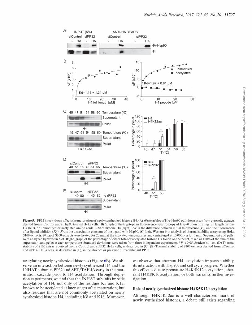
position of H4 protein-complexes. We focus on the Hsp90protein because it interacts with histone H4 in ComplexIb, prior to H4 acetylation (3). We ectopically expresseda hemagglutinin (HA)-tagged version of Hsp90 and per-formed pull-down experiments from cytosolic extracts de-rived from siControl and siPP32 treated HeLa cells. HA-Hsp90 is pulled-down efficiently from both conditions (Fig-ure 5A). Interestingly, histone H4 co-immunoprecipitatesonly from siControl, but not from siPP32 derived extracts(Figure 5A). This suggests that acetylation of histone H4possibly interferes with Hsp90 interaction. However, Hsp90function itself is also regulated by acetylation in a way thatacetylation weakens its affinity for most of its client proteins(25). Therefore, we investigated the impact that histone H4acetylation has on its interaction with Hsp90. We first mon-itored intrinsic protein fluorescence of recombinant Hsp90expressed in bacteria to avoid acetylation (Figure 5B andSupplementary Figure S3). Titration of Hsp90 with histoneH4 (tryptophan free protein) reveals a binding affinity typ-ical for chaperone-substrate interactions (KD = 1.13 ± 1.31�M; Figure 5B, left). We then evaluate the interaction be-tween Hsp90 and either an unmodified or acetylated pep-tide containing amino acids 1–20 of histone H4. Titrationof Hsp90 with the unmodified histone H4 peptide reveals asimilar binding affinity as the full-length protein (KD = 1.97± 0.81 �M; Figure 5B, right). In contrast, we do not detectan interaction between Hsp90 and the acetylated H4 pep-tide (Figure 5B, right). Taken together, our results suggestthat the interaction between histone H4 and Hsp90 may benegatively affected due to aberrant histone H4 acetylationwhen the levels of PP32 are reduced.
PP32 affects the stability of newly synthesized histone H4
We then investigated the consequences of reducing the H4–Hsp90 interaction in cells that have lowered PP32 levels.Given that Hsp90 is a heat shock chaperone, we focusedon histone H4 stability. It is well documented that histoneacetylation increases the solubility of chromatin, but its ef-
fect on soluble histones remains an open question (26–28).Our results suggest that in the absence of PP32, newly syn-thesized histone H4 is not only acetylated at an inappro-priate time, but it is aberrantly acetylated on K8 and K16,which contribute to preventing the interaction with Hsp90.Therefore, we hypothesized that newly synthesized histoneH4 presenting aberrant acetylation could influence its sta-bility due to a defective Hsp90 interaction. To investigatethis, we set up a thermal stability assay, in which cytosolicextracts are incubated for 20 min at increasing temperatures,after which soluble and insoluble proteins are separated bycentrifugation. We find that, under basal conditions, his-tone H4 remains soluble at 45◦C, 12% of the protein pre-cipitates at 51◦C, and almost all the histone H4 precipitatesat 58◦C (Figure 5C). However, when we analyzed H4K12acunder the same conditions, 40% of the acetylated proteinprecipitates at 51◦C (Figure 5C), indicating that acetylatednewly synthesized histone H4 is less stable than hypoacety-lated H4. We then compared the thermal stability of cytoso-lic histone H4 derived from siControl and siPP32 treatedcells. We find that cytosolic histone H4 from siPP32 treatedcells is less stable at 51◦C compared to siControl cells, as 6%compared to 27% of histone H4 precipitated from siCon-trol and siPP32 cells, respectively (Figure 5D). To confirmthat this effect was not due to a direct role that PP32 itselfcould confer thermal stability, we performed the assay at51◦C while adding back recombinant PP32 (Figure 5E). Weobserve that histones derived from siPP32 are less stable re-gardless of the presence of recombinant PP32. Therefore, weconclude that cytosolic histone H4 stability decreases whenPP32 protein levels are reduced, likely due to histone H4hyperacetylation and a decreased interaction with Hsp90.
PP32 knock down affects cell cycle progression
Our results suggest that PP32 is involved in the proper mat-uration of newly synthesized histone H4 by preventing pre-mature and aberrant acetylation. Therefore, we hypothe-sized that histone H3–H4 deposition may be altered uponsiPP32. We monitored newly synthesized histone deposi-tion in vivo utilizing HeLa cell lines stably expressing H3.1-and H3.3-SNAP tagged proteins and quench-chase-pulseexperiments (29). The SNAP tag enables covalent labelingwith a cell-permeable small molecule in vivo. To distinguishparental from newly synthesized histones, parental histoneH3 are quenched with a non-fluorescent blocking molecule.A chase time permits the synthesis of new SNAP-H3 vari-ant proteins, which are subsequently labeled with a fluores-cent probe (TMR) (29). We tested the incorporation of thenewly synthesized histone H3 variants H3.1 and H3.3 (Sup-plementary Figure S4). As a control, we confirmed that de-pleting the H3.3 chaperone HIRA (histone regulation A)(30) using siRNA leads to a decrease in H3.3, but not H3.1,deposition (Supplementary Figure S4) (29). We then inves-tigated the effect of siPP32 (Supplementary Figure S4) andfind a small, but statistically significant, defect on the in-corporation of both H3.1 and H3.3 variants when com-pared to siControl (Supplementary Figure S4). These datasuggest that perturbation of PP32 may impact histone H3–H4 deposition. Further work will address whether this im-pact is more pronounced at defined windows of the cell cy-
Dow
nloaded from https://academ
ic.oup.com/nar/article/45/20/11700/4097618 by guest on 23 July 2022

11706 Nucleic Acids Research, 2017, Vol. 45, No. 20
rPP32 (pmol)
-H4
-HAT1
BSA0 0.9
44.7
29.4
518
.8rSET (pmol)
-H4
BSAFT 3.0
015
.030
.060
.00-H
AT1
PP32 SET
(+) BSA FT3.0
015
.0
- HAT1
- H4
0
20
40
60
80
100
120
Rem
aini
ng A
ctiv
ity (%
)
rSET (pmol)
A B
D
Contro
lPP32 SET
Mix 1X
Mix 0.5
X0
20
40
60
80
100
Rem
aini
ng A
ctiv
ity (%
)
Mix 1x0 Mix 0.5x
Rem
aini
ng a
ctiv
ity (%
)
30.0
60.0
0
20
40
60
80
100
(+) BSA0.9
44.7
29.4
518
.8.-H
AT1 -H4
rPP32 (pmol)
molar ratioprotein:H4
74.032.021.020.051.070.0400.0700.0molar ratioprotein:H4
MW
Coomasie Blue
180 kDa130 kDa
95 kDa72 kDa55 kDa43 kDa34 kDa26 kDa17 kDa10 kDa
PP32
BSA
Asf1
aAs
f1b
Hsp90
Hsc70
H4
BSA, Hsc70GST-Asf1aGST-Asf1b
Hsp90
PP32
H4
PP32
BSA
Asf1
aAs
f1b
Hsp90
Hsc70
-H4
C
Figure 4. PP32 and SET/TAF-I� proteins block HAT1 mediated H4 acetylation in vitro. Acetylation assay performed using 0.128 nmol of recombinanthistone H4 and purified HAT1 in the presence or absence of increasing amounts of recombinant PP32 (A) and recombinant SET/TAF-I� (B), followed byH4K12ac Dot-blot detection. Top: graph of the remaining HAT1 activity. 100% activity correspond to the HAT1 mediated H4 acetylation in the absenceof PP32 and SET/TAF-I�. Bottom: a representative H4K12ac dot blot HAT1 assay. FT corresponds to the flow through material of the recombinantSET/TAF-I� Ni+2-beads purification. (C) Top: Coomassie blue stained gel of the different recombinant proteins used in the acetylation assay. Bottom:acetylation assay performed as in (A), pre-incubating H4 as indicated. (D) Acetylation assay performed using recombinant histone H4 and purified HAT1in the presence of 8 pmol of recombinant PP32, 12 pmol of recombinant SET/TAF-I�, and a mix of 8 pmol of recombinant PP32 and 12 pmol ofrecombinant SET/TAF-I� (Mix 1×), and 4 pmol of recombinant PP32 and 6 pmol of recombinant SET/TAF-I� (Mix 0.5×).
cle and if it plays a particular role during critical phases inan organism’s life cycle, such as during development and inparticular pathologies. In line with this, we examined theconsequences of knocking down PP32 and SET/TAF-I�on cell cycle distribution. Flow cytometry analysis of DNAcontent shows that cells accumulated in S-phase 72 h aftersiPP32 (31% of cells) and siSET/TAF-I� (29% of cells), ascompared to control cells (22% of cells) (Figure 6A). Thisresembles the S-phase arrest observed upon perturbationof CAF1 and ASF1 (31–33). Together, our data supporta model that PP32 and SET/TAF-I�, by regulating newlysynthesized histone H4 metabolism, may contribute to reg-ulate histone deposition and cell cycle progression.
DISCUSSION
Acetylation of lysine is a cellular-wide dynamic post-translational modification that mediates interactions with
other proteins and can impact genome stability and func-tion (34). In one example, acetylation of tubulin is impli-cated in regulating microtubule assembly (35), while acety-lation of Hsp90 weakens its affinity for most of its clientproteins (25). Histone acetylation plays critical roles bothin the context of a nucleosome but also on soluble his-tones. Following biosynthesis, histones H3 and H4 pro-ceed through a maturation cascade of complexes that en-sure their proper folding and post-translational modifica-tion state prior to chromatin assembly or storage. Notably,the H4K5K12 acetylation mark, established by the HAT1acetyltransferase, is observed late in the histone maturationcascade, suggesting that there are regulatory mechanismsthat prevent premature acetylation. In addition, acetylationof other lysine residues is not observed on newly synthe-sized histone H4. Here, we reveal that the inhibitor of HAT(INHAT) complex subunits PP32 and SET/TAF-I� playkey roles in regulating the timing and residue specificity of
Dow
nloaded from https://academ
ic.oup.com/nar/article/45/20/11700/4097618 by guest on 23 July 2022
Nucleic Acids Research, 2017, Vol. 45, No. 20 11707
B
C
A ANTI-HA BEADSsiControl siPP32
HA-Hsp90
H4
- HA - HA
INPUT (5%)siControl siPP32
- HA - HA
Supernatant
Pellet
H4K12ac
H4
60 45 47 51 54 58
60 45 47 51 54 58 Supernatant
Pellet
Temperature (0C)
Temperature (0C)
siControl48 51 55
D
*
120100
80604020
045 47 51 54 58 60
T (0C)
H4H4K12ac
Per
cent
age
in p
elle
t
100
80
60
40
20
0Per
cent
age
in p
elle
t
48 51 55T (0C)
*
siCsiPP32
0 10 20 30 40H4 full length [μM]
Kd=1.13 + 1.31 μM
ΔF
(x10
4 )
0
1
2
3
45
6
ΔF
(x10
4 )
-5
0
5
10
15
0 10 20 30H4 peptide [μM]
Kd=1.97 + 0.81 μM
unmodifiedacetylated
H4
Supernatant
Pellet
Temperature (0C)siPP32
48 51 55
Supernatant
PelletH4
siControl- 40 80
siPP32- 40 80 ng rPP32
E
Figure 5. PP32 knock-down affects the maturation of newly synthesized histone H4. (A) Western blot of HA-Hsp90 pull-down assay from cytosolic extractsderived from siControl and siHsp90 treated HeLa cells. (B) Graph of the tryptophan fluorescence spectroscopy of Hsp90 upon titrating full length histoneH4 (left), or unmodified or acetylated amino acids 1–20 of histone H4 (right). �F is the difference between initial fluorescence (F0) and the fluorescenceafter ligand addition (FQ). KD is the dissociation constant of the ligand with Hsp90. (C) Left, Western blot analysis of thermal stability assay using HeLaS100 extracts. 20 �g of S100 extracts were heated for 20 min at the indicated temperatures and centrifuged at 10 000 × g for 5 min. Supernatant and pelletwere analyzed by western blot. Right, graph of the percentage of either total or acetylated histone H4 found on the pellet, taken as 100% of the sum of thesupernatant and pellet at each temperature. Standard deviations were taken from three independent experiments. *P < 0.05, Student´s t-test. (D) Thermalstability of S100 extracts derived from siControl and siPP32 HeLa cells, as described in (C). (E) Thermal stability of S100 extracts derived from siControland siPP32 HeLa cells, as described in (C), in the absence or presence of recombinant PP32.
acetylating newly synthesized histones (Figure 6B). We ob-serve an interaction between newly synthesized H4 and theINHAT subunits PP32 and SET/TAF-I� early in the mat-uration cascade prior to H4 acetylation. Through deple-tion experiments, we find that the INHAT subunits impedeacetylation of H4, not only of the residues K5 and K12,known to be acetylated at later stages of its maturation, butalso residues that are not commonly acetylated on newlysynthesized histone H4, including K8 and K16. Moreover,
we observe that aberrant H4 acetylation impacts stability,its interaction with Hsp90, and cell cycle progress. Whetherthis effect is due to premature H4K5K12 acetylation, aber-rant H4K8K16 acetylation, or both warrants further inves-tigation.
Role of newly synthesized histone H4K5K12 acetylation
Although H4K5K12ac is a well characterized mark ofnewly synthesized histones, a debate still exists regarding
Dow
nloaded from https://academ
ic.oup.com/nar/article/45/20/11700/4097618 by guest on 23 July 2022

11708 Nucleic Acids Research, 2017, Vol. 45, No. 20
B
Hsp90
H4Hsp70
Complex Ib
HAT1SETPP32
sNASP
Complex III
HAT1
Maturation cascadeH4H4
H3
% c
ell
60
40
20
0
G1 S G2/M
*p< 0.05n=4
**
siCsiP
P32siS
etsiCsiP
P32siS
etsiCsiP
P32siS
et
A
Other HATs?
Chromatinassembly
Storagepool
Asf1a
Figure 6. PP32 knock-down affects cell cycle progression. (A) Cell cycleprofiles of HeLa cells treated with siControl, siPP32, and siSET/TAF-I�.(B) Proposed model for the regulation of newly synthesized histone H4acetylation. After synthesis, unacetylated histone H4 interacts with theheat shock proteins Hsp90 and Hsp70 and two members of the INHATcomplex: PP32 and SET/TAF-I�. The binding of PP32 and SET/TAF-I�protects histone H4 from premature acetylation by HAT1, as well as fornon-specific acetylation mediated by other HATs. When PP32 protein lev-els are diminished, histone H4 is acetylated at early steps of the maturationcascade, and it no longer can interact with Hsp90 affecting its stability andmaturation.
the role of this mark on histone function (10,36,37). Giventhat all newly synthesized H4 histones carry this pattern,and that this acetylation pattern is removed shortly afterdeposition onto chromatin (38–40), it has been suggestedthat H4K5K12ac mediates chromatin assembly. However,genetic studies in yeast suggest that this modification isnot essential (41–44). This may be species specific, or per-haps there is functional redundancy of H4K5K12 acety-lation with other acetylation sites. In support of the latteridea, combining the mutation H4K5RK12R (resemblingunacetylated lysines) with H4K91Q (an acetylation mimic)leads to defects in chromatin reassembly following double-strand DNA breaks (45). Alternatively, H4 acetylation pat-terns may play a role in the cytoplasmic/nuclear trans-port of histones given that K5 and K12 are located withinthe nuclear localization signal of H4. Indeed, in the moldPhysarum model the H4K5RK12R mutation blocks H3–H4 nuclear translocation, while H4K5QK12Q favors im-port (46). Similarly, in HeLa cells, the mutant H4K5QK12Qfavors the nuclear transport in an in vitro translocation as-say by favoring the importin/histone interaction (3). Inter-estingly, our data suggest a new role for H4 acetylation inwhich it regulates a protein-protein interaction by block-ing the association with heat shock proteins, thus helpingto regulate the timing of the establishment of this mark.
Mechanisms that regulate post-translational modifications
The establishment and maintenance of histone acetylationinvolves several mechanisms, including the opposing ac-tivities of acetyltransferases and deacetylases and histone
turnover. Additionally, protein-protein interactions maycontribute to the maintenance of modifications. In one ex-ample, bromodomains, which recognize and bind acety-lated lysines, may limit access to the lysine, preventing en-zymatic deacetylation. Indeed, BRD4 (bromodomain con-taining protein 4) binds acetylated histones at transcrip-tionally active genes throughout the cell cycle (47) to main-tain the transcriptionally active mark (38). In response toDNA damage, BRD4 dissociates, which induces deacety-lation of H4K5ac and H4K8ac (48,49) by HDACs. An-other example is the regulation of acetylation on non-histone proteins. SET/TAF-I� interacts in the nucleus withthe Ku70/80 heterodimer, required for the NHEJ (non-homologous end-joining) DNA DSB (double strand break)repair pathway, and inhibits Ku70 acetylation. Upon DNAdamage, SET/TAF-I� dissociates and releases the Ku com-plex, which is recruited to DNA DSB sites (13). Solu-ble histones offer a different context for associating withbinding proteins, suggesting that other mechanisms maybe involved to regulate the timing of establishing modifi-cations. Here, we reveal a novel mechanism where PP32and SET/TAF-I� associate with a histone complex and oc-clude HAT1, and potentially other acetyltransferases, frombinding and acting on its histone substrate. In this way,PP32 and SET/TAF-I� proteins ensure that histone H4is acetylated at the right time and on the proper lysineresidues. When early acetylation occurs, as in the case ofPP32 knock down cells depletion, histone H4 cannot in-teract with Hsp90, affecting its stability and maturation.Although we did not investigate the transit of histone H4beyond Complex Ib, we speculate that following its associ-ation with heat shock proteins and INHAT subunits, his-tone H4 passes through the other complexes to continue itsmaturation. On Complex III, histone H4 does no longer in-teracts with the INHAT subunits, instead it interacts withthe enzyme HAT1 to acetylate histone H4 on K5 and K12(Figure 6B). How PP32 and SET/TAF-I� dissociation fromhistone H4 is regulated will be explored in future work. In-vestigating whether INHAT and histone H4 binding is reg-ulated in a cell cycle dependent manner will be useful to un-derstand how histone maturation is linked to the cellularrequirements of histones. Future work should also addresswhether this kind of mechanism applies more broadly toother modifications and proteins.
SUPPLEMENTARY DATA
Supplementary Data are available at NAR Online.
ACKNOWLEDGEMENTS
We thank Danny Reinberg for providing S100 extracts,Zhiguo Zhang for providing Flag-H4 expressing plasmid.
FUNDING
Comision Nacional de Ciencia y Tecnologıa [FONDE-CYT 1160480 and Basal Project PFB16 to A.L.,PCHA/Doctorado Nacional/2016-21161044 to C.R.,PCHA/Doctorado Nacional/2014–21140346 to F.S.,PCHA/Doctorado Nacional/2014-21140956 to S.H.];
Dow
nloaded from https://academ
ic.oup.com/nar/article/45/20/11700/4097618 by guest on 23 July 2022
Nucleic Acids Research, 2017, Vol. 45, No. 20 11709
Ligue Nationale contre le Cancer (Equipe labellisee Ligue)(to G.A.); French National Research Agency [ANR-11-LABX-0044 DEEP, ANR-10-IDEX-0001-02 PSL,ANR-12-BSV5–0022-02 ‘CHAPINHIB’, ANR-14-CE16-0009 ‘Epicure’, ANR-14-CE10-0013 ‘CELLECTCHIP’,ANR-16-CE15-0018 ‘CHRODYT’, ANR-16-CE12–0024‘CHIFT’ to G.A.]; European Commission [N 678563‘EPOCH28’, ERC-2015-ADG- 694694 ‘ChromADICT’ toG.A.]; Deutsche Forschungsgemeinschaft [CRC1064/Z03to A.I.]. Funding for open access charge: ComisionNacional de Ciencia y Tecnologıa [FONDECYT 1160480].Conflict of interest statement. None declared.
REFERENCES1. MacAlpine,D.M. and Almouzni,G. (2013) Chromatin and DNA
replication. Cold Spring Harb. Perspect. Biol., 5, a010207.2. Marzluff,W.F. and Duronio,R.J. (2002) Histone mRNA expression:
multiple levels of cell cycle regulation and important developmentalconsequences. Curr. Opin. Cell Biol., 14, 692–699.
3. Alvarez,F., Munoz,F., Schilcher,P., Imhof,A., Almouzni,G. andLoyola,A. (2011) Sequential establishment of marks on solublehistones H3 and H4. J. Biol. Chem., 286, 17714–17721.
4. Campos,E.I., Fillingham,J., Li,G., Zheng,H., Voigt,P., Kuo,W.H.,Seepany,H., Gao,Z., Day,L.A., Greenblatt,J.F. et al. (2010) Theprogram for processing newly synthesized histones H3.1 and H4. Nat.Struct. Mol. Biol., 17, 1343–1351.
5. Rivera,C., Saavedra,F., Alvarez,F., Diaz-Celis,C., Ugalde,V., Li,J.,Forne,I., Gurard-Levin,Z.A., Almouzni,G., Imhof,A. et al. (2015)Methylation of histone H3 lysine 9 occurs during translation. NucleicAcids Res., 43, 9097–9106.
6. Jasencakova,Z., Scharf,A.N., Ask,K., Corpet,A., Imhof,A.,Almouzni,G. and Groth,A. (2010) Replication stress interferes withhistone recycling and predeposition marking of new histones. Mol.Cell, 37, 736–743.
7. Loyola,A., Bonaldi,T., Roche,D., Imhof,A. and Almouzni,G. (2006)PTMs on H3 variants before chromatin assembly potentiate theirfinal epigenetic state. Mol. Cell, 24, 309–316.
8. Benson,L.J., Phillips,J.A., Gu,Y., Parthun,M.R., Hoffman,C.S. andAnnunziato,A.T. (2007) Properties of the type B histoneacetyltransferase Hat1: H4 tail interaction, site preference, andinvolvement in DNA repair. J. Biol. Chem., 282, 836–842.
9. Makowski,A.M., Dutnall,R.N. and Annunziato,A.T. (2001) Effectsof acetylation of histone H4 at lysines 8 and 16 on activity of the Hat1histone acetyltransferase. J. Biol. Chem., 276, 43499–43502.
10. Parthun,M.R. (2012) Histone acetyltransferase 1: more than just anenzyme? Biochim. Biophys. Acta, 1819, 256–263.
11. Sobel,R.E., Cook,R.G., Perry,C.A., Annunziato,A.T. and Allis,C.D.(1995) Conservation of deposition-related acetylation sites in newlysynthesized histones H3 and H4. Proc. Natl. Acad. Sci. U.S.A., 92,1237–1241.
12. Kim,J.Y., Lee,K.S., Seol,J.E., Yu,K., Chakravarti,D. and Seo,S.B.(2012) Inhibition of p53 acetylation by INHAT subunitSET/TAF-Ibeta represses p53 activity. Nucleic Acids Res., 40, 75–87.
13. Kim,K.B., Kim,D.W., Park,J.W., Jeon,Y.J., Kim,D., Rhee,S., Chae,J.I.and Seo,S.B. (2014) Inhibition of Ku70 acetylation by INHATsubunit SET/TAF-Ibeta regulates Ku70-mediated DNA damageresponse. Cell. Mol. Life Sci.: CMLS, 71, 2731–2745.
14. Seo,S.B., Macfarlan,T., McNamara,P., Hong,R., Mukai,Y., Heo,S.and Chakravarti,D. (2002) Regulation of histone acetylation andtranscription by nuclear protein pp32, a subunit of the INHATcomplex. J. Biol. Chem., 277, 14005–14010.
15. Seo,S.B., McNamara,P., Heo,S., Turner,A., Lane,W.S. andChakravarti,D. (2001) Regulation of histone acetylation andtranscription by INHAT, a human cellular complex containing the setoncoprotein. Cell, 104, 119–130.
16. Kutney,S.N., Hong,R., Macfarlan,T. and Chakravarti,D. (2004) Asignaling role of histone-binding proteins and INHAT subunits pp32and Set/TAF-Ibeta in integrating chromatin hypoacetylation andtranscriptional repression. J. Biol. Chem., 279, 30850–30855.
17. Schneider,R., Bannister,A.J., Weise,C. and Kouzarides,T. (2004)Direct binding of INHAT to H3 tails disrupted by modifications. J.Biol. Chem., 279, 23859–23862.
18. Barth,T.K., Schade,G.O., Schmidt,A., Vetter,I., Wirth,M., Heun,P.,Imhof,A. and Thomae,A.W. (2015) Identification of Drosophilacentromere associated proteins by quantitative affinitypurification-mass spectrometry. Data Brief., 4, 544–550.
19. Bonaldi,T., Regula,J.T. and Imhof,A. (2004) The use of massspectrometry for the analysis of histone modifications. MethodsEnzymol., 377, 111–130.
20. Dignam,J.D., Lebovitz,R.M. and Roeder,R.G. (1983) Accuratetranscription initiation by RNA polymerase II in a soluble extractfrom isolated mammalian nuclei. Nucleic Acids Res., 11, 1475–1489.
21. Luger,K., Rechsteiner,T.J., Flaus,A.J., Waye,M.M. andRichmond,T.J. (1997) Characterization of nucleosome core particlescontaining histone proteins made in bacteria. J. Mol. Biol., 272,301–311.
22. Verreault,A., Kaufman,P.D., Kobayashi,R. and Stillman,B. (1998)Nucleosomal DNA regulates the core-histone-binding subunit of thehuman Hat1 acetyltransferase. Curr. Biol.: CB, 8, 96–108.
23. Gurard-Levin,Z.A., Quivy,J.P. and Almouzni,G. (2014) Histonechaperones: assisting histone traffic and nucleosome dynamics. Annu.Rev. Biochem., 83, 487–517.
24. Loyola,A. and Almouzni,G. (2004) Histone chaperones, a supportingrole in the limelight. Biochim. Biophys. Acta, 1677, 3–11.
25. Mollapour,M. and Neckers,L. (2012) Post-translationalmodifications of Hsp90 and their contributions to chaperoneregulation. Biochim. Biophys. Acta, 1823, 648–655.
26. Wallace,R.B., Sargent,T.D., Murphy,R.F. and Bonner,J. (1977)Physical properties of chemically acetylated rat liver chromatin. Proc.Natl. Acad. Sci. U.S.A., 74, 3244–3248.
27. Perry,M. and Chalkley,R. (1982) Histone acetylation increases thesolubility of chromatin and occurs sequentially over most of thechromatin. A novel model for the biological role of histoneacetylation. J. Biol. Chem., 257, 7336–7347.
28. Wang,X., He,C., Moore,S.C. and Ausio,J. (2001) Effects of histoneacetylation on the solubility and folding of the chromatin fiber. J.Biol. Chem., 276, 12764–12768.
29. Ray-Gallet,D., Woolfe,A., Vassias,I., Pellentz,C., Lacoste,N.,Puri,A., Schultz,D.C., Pchelintsev,N.A., Adams,P.D., Jansen,L.E.et al. (2011) Dynamics of histone H3 deposition in vivo reveal anucleosome gap-filling mechanism for H3.3 to maintain chromatinintegrity. Mol. Cell, 44, 928–941.
30. Tagami,H., Ray-Gallet,D., Almouzni,G. and Nakatani,Y. (2004)Histone H3.1 and H3.3 complexes mediate nucleosome assemblypathways dependent or independent of DNA synthesis. Cell, 116,51–61.
31. Groth,A., Ray-Gallet,D., Quivy,J.P., Lukas,J., Bartek,J. andAlmouzni,G. (2005) Human Asf1 regulates the flow of S phasehistones during replicational stress. Mol. Cell, 17, 301–311.
32. Hoek,M. and Stillman,B. (2003) Chromatin assembly factor 1 isessential and couples chromatin assembly to DNA replication in vivo.Proc. Natl. Acad. Sci. U.S.A., 100, 12183–12188.
33. Ye,X., Franco,A.A., Santos,H., Nelson,D.M., Kaufman,P.D. andAdams,P.D. (2003) Defective S phase chromatin assembly causesDNA damage, activation of the S phase checkpoint, and S phasearrest. Mol. Cell, 11, 341–351.
34. Choudhary,C., Kumar,C., Gnad,F., Nielsen,M.L., Rehman,M.,Walther,T.C., Olsen,J.V. and Mann,M. (2009) Lysine acetylationtargets protein complexes and co-regulates major cellular functions.Science, 325, 834–840.
35. Janke,C. (2014) The tubulin code: molecular components, readoutmechanisms, and functions. J. Cell Biol., 206, 461–472.
36. Keck,K.M. and Pemberton,L.F. (2013) Histone chaperones linkhistone nuclear import and chromatin assembly. Biochim. Biophys.Acta, 1819, 277–289.
37. Li,Q., Burgess,R. and Zhang,Z. (2012) All roads lead to chromatin:Multiple pathways for histone deposition. Biochim. Biophys. Acta,1819, 238–246.
38. Annunziato,A.T. and Seale,R.L. (1983) Histone deacetylation isrequired for the maturation of newly replicated chromatin. J. Biol.Chem., 258, 12675–12684.
39. Jackson,V., Granner,D. and Chalkley,R. (1976) Deposition ofhistone onto the replicating chromosome: newly synthesized histone
Dow
nloaded from https://academ
ic.oup.com/nar/article/45/20/11700/4097618 by guest on 23 July 2022
11710 Nucleic Acids Research, 2017, Vol. 45, No. 20
is not found near the replication fork. Proc. Natl. Acad. Sci. U.S.A.,73, 2266–2269.
40. Nagarajan,P., Ge,Z., Sirbu,B., Doughty,C., Agudelo Garcia,P.A.,Schlederer,M., Annunziato,A.T., Cortez,D., Kenner,L. andParthun,M.R. (2013) Histone acetyl transferase 1 is essential formammalian development, genome stability, and the processing ofnewly synthesized histones H3 and H4. PLoS Genet., 9, e1003518.
41. Ma,X.J., Wu,J., Altheim,B.A., Schultz,M.C. and Grunstein,M. (1998)Deposition-related sites K5/K12 in histone H4 are not required fornucleosome deposition in yeast. Proc. Natl. Acad. Sci. U.S.A., 95,6693–6698.
42. Megee,P.C., Morgan,B.A., Mittman,B.A. and Smith,M.M. (1990)Genetic analysis of histone H4: essential role of lysines subject toreversible acetylation. Science, 247, 841–845.
43. Park,E.C. and Szostak,J.W. (1990) Point mutations in the yeasthistone H4 gene prevent silencing of the silent mating type locusHML. Mol. Cell. Biol., 10, 4932–4934.
44. Zhang,W., Bone,J.R., Edmondson,D.G., Turner,B.M. and Roth,S.Y.(1998) Essential and redundant functions of histone acetylationrevealed by mutation of target lysines and loss of the Gcn5pacetyltransferase. EMBO J., 17, 3155–3167.
45. Ge,Z., Nair,D., Guan,X., Rastogi,N., Freitas,M.A. andParthun,M.R. (2013) Sites of acetylation on newly synthesizedhistone H4 are required for chromatin assembly and DNA damageresponse signaling. Mol. Cell. Biol., 33, 3286–3298.
46. Ejlassi-Lassallette,A., Mocquard,E., Arnaud,M.C. and Thiriet,C.(2011) H4 replication-dependent diacetylation and Hat1 promoteS-phase chromatin assembly in vivo. Mol. Biol. Cell, 22, 245–255.
47. Devaiah,B.N. and Singer,D.S. (2013) Two faces of brd4: mitoticbookmark and transcriptional lynchpin. Transcription, 4, 13–17.
48. Ai,N., Hu,X., Ding,F., Yu,B., Wang,H., Lu,X., Zhang,K., Li,Y.,Han,A., Lin,W. et al. (2011) Signal-induced Brd4 release fromchromatin is essential for its role transition from chromatin targetingto transcriptional regulation. Nucleic Acids Res., 39, 9592–9604.
49. Hu,X., Lu,X., Liu,R., Ai,N., Cao,Z., Li,Y., Liu,J., Yu,B., Liu,K.,Wang,H. et al. (2014) Histone cross-talk connects proteinphosphatase 1alpha (PP1alpha) and histone deacetylase (HDAC)pathways to regulate the functional transition ofbromodomain-containing 4 (BRD4) for inducible gene expression. J.Biol. Chem., 289, 23154–23167.
Dow
nloaded from https://academ
ic.oup.com/nar/article/45/20/11700/4097618 by guest on 23 July 2022
Copyright © 2022 FDOKUMEN