MiR-221 Influences Effector Functions and Actin Cytoskeleton ...

Metalloproteases regulate T-cell proliferationand effector function via LAG-3
Nianyu Li1,9, Yao Wang1, Karen Forbes1,Kate M Vignali1, Bret S Heale2,10, PaulSaftig3, Dieter Hartmann4, Roy A Black5,John J Rossi2, Carl P Blobel6,Peter J Dempsey7,8,11, Creg J Workman1
and Dario AA Vignali1,*1Department of Immunology, St Jude Children’s Research Hospital,Memphis, TN, USA, 2Graduate School of Biological Sciences, BeckmanResearch Institute of the City of Hope, Duarte, CA, USA, 3TheBiochemical Institute, Christian-Albrechts University, Kiel, Germany,4Department for Human Genetics, KU Leuven and FlandersInteruniversity Institute for Biotechnology (VIB4), Leuven, Belgium,5Department of Inflammation, Amgen Inc., Seattle, WA, USA, 6Arthritisand Tissue Degeneration Program, Hospital for Special Surgery at WeillMedical College of Cornell University, New York, NY, USA, 7PacificNorthwest Research Institute, Seattle, WA, USA and 8Department ofMedicine, University of Washington, Seattle, WA, USA
Tight control of T-cell proliferation and effector function is
essential to ensure an effective but appropriate immune
response. Here, we reveal that this is controlled by the
metalloprotease-mediated cleavage of LAG-3, a negative
regulatory protein expressed by all activated T cells. We
show that LAG-3 cleavage is mediated by two transmem-
brane metalloproteases, ADAM10 and ADAM17, with the
activity of both modulated by two distinct T-cell receptor
(TCR) signaling-dependent mechanisms. ADAM10 med-
iates constitutive LAG-3 cleavage but increases B12-fold
following T-cell activation, whereas LAG-3 shedding by
ADAM17 is induced by TCR signaling in a PKCh-dependent
manner. LAG-3 must be cleaved from the cell surface to
allow for normal T-cell activation as noncleavable LAG-3
mutants prevented proliferation and cytokine production.
Lastly, ADAM10 knockdown reduced wild-type but not
LAG-3�/� T-cell proliferation. These data demonstrate
that LAG-3 must be cleaved to allow efficient T-cell pro-
liferation and cytokine production and establish a novel
paradigm in which T-cell expansion and function are
regulated by metalloprotease cleavage with LAG-3 as its
sole molecular target.
The EMBO Journal (2007) 26, 494–504.
doi:10.1038/sj.emboj.7601520
Subject Categories: immunology; proteins
Keywords: ADAM; LAG-3; metalloproteases; shedding; T-cell
function
Introduction
Metalloproteases have long been considered viable therapeu-
tic targets for a variety of important human diseases such as
cancer, cardiovascular disease, arthritis and multiple sclerosis
(Baker et al, 2002; Overall and Kleifeld, 2006). However,
many of the clinical trials using broad-range metalloprotease
inhibitors have produced disappointing results, in part owing
to unexpected side effects. This is complicated by the broad
range of molecules targeted by these metalloproteases. Matrix
metalloproteases (MMP), membrane-tethered MMPs and
the zinc-dependent a disintegrin and metalloproteinases
(ADAM), have all been shown to shed proteins from the
cell surface (Black and White, 1998; Becherer and Blobel,
2003; Seals and Courtneidge, 2003; Parks et al, 2004; Blobel,
2005). Among these, two members of the ADAM family
of metalloproteases, ADAM10 (Kuzbanian) and ADAM17
(TACE), are known to be important cell surface sheddases
for a diverse array of transmembrane proteins of immuno-
logical importance, such as Notch, EGFR ligands, TNF-a,
TNF-a receptor, CD44, CD62L (L-selectin) and CD23 (Black
and White, 1998; Becherer and Blobel, 2003; Blobel, 2005;
Maretzky et al, 2005; Reiss et al, 2005; Weskamp et al, 2006).
For some time, metalloprotease inhibitors have been known
to inhibit T-cell proliferation but the target molecule and
mechanism that is inhibited remain unknown.
T-cell proliferation and function following antigenic
stimulation is a tightly regulated process. Inappropriate or
uncontrolled expansion of activated T cells is regulated by
activation-induced cell death, downregulation of stimulatory
molecules and/or upregulation of inhibitory molecules.
Lymphocyte activation gene-3 (LAG-3; CD223) has recently
been shown to be a novel inhibitory molecule that is required
for maximal regulatory T-cell function, and controls effector
T-cell expansion and homeostasis (Huang et al, 2004; Workman
et al, 2004; Workman and Vignali, 2005). Importantly, these
studies clearly show that LAG-3 has cell-intrinsic regulatory
activity, but the physiological importance of this is unclear.
LAG-3 is related to CD4 in chromosomal location, exon
organization and structure (Triebel et al, 1990; Bruniquel
et al, 1997). They also share the same ligand, MHC class II,
although LAG-3 binds with a much higher affinity (Triebel
et al, 1990; Bruniquel et al, 1998; Workman et al, 2002a, b).
We have shown that binding to MHC class II molecules and
a conserved KIEELE motif in the LAG-3 cytoplasmic domain
are essential for its function. LAG-3 clearly possesses both
cell-intrinsic and cell-extrinsic regulatory activity (Huang
et al, 2004; Workman and Vignali, 2005). Ectopic expression
of LAG-3 on effector T cells controls their proliferation andReceived: 10 October 2006; accepted: 30 November 2006
*Corresponding author. Department of Immunology, St Jude Children’sResearch Hospital, 332 North Lauderdale, Memphis, TN 38105, USA.Tel.: þ 1 901 495 2332; Fax: þ 1 901 495 3107;E-mail: [email protected] Address: Department of Investigative Toxicology, Amgen Inc.,1201 Amgen Court West, Seattle, WA 98119, USA10Present Address: MRC Human Genetics Unit, Western GeneralHospital, Edinburgh EH4 2XU, Scotland11Present Address: Departments of Pediatrics and Molecular andIntegrative Physiology, University of Michigan, 1150 W. Medical CtrDrive, Ann Arbor, MI 48109-0656, USA
The EMBO Journal (2007) 26, 494–504 | & 2007 European Molecular Biology Organization | All Rights Reserved 0261-4189/07
www.embojournal.org
The EMBO Journal VOL 26 | NO 2 | 2007 &2007 European Molecular Biology Organization
EMBO
THE
EMBOJOURNAL
THE
EMBOJOURNAL
494
cytokine production. It is noteworthy that all activated Tcells
and NK cells express LAG-3. It is unclear what effect the
presence of this negative regulatory pressure might have on
their ability to proliferate and function, and if there are any
mechanisms present that modulate LAG-3 activity.
We recently observed that LAG-3 is cleaved within the
short connecting peptide (CP) between the membrane-
proximal D4 domain and the transmembrane domain, result-
ing in the release of a soluble form of LAG-3 (sLAG-3) (Li
et al, 2004). Indeed, significant amounts of sLAG-3 are found
in murine sera (B200 ng/ml), which increases following
T-cell stimulation in vivo. LAG-3 is known to have inhibitory
activity, yet is expressed by all activated T cells. In this study,
we tested our hypothesis that there was a direct link between
the ability of metalloproteases to regulate T-cell proliferation
and effector function and the possibility that LAG-3 may be
the target of metalloprotease activity.
Results
Metalloprotease inhibition reduced wild-type but not
LAG-3�/� T-cell proliferation
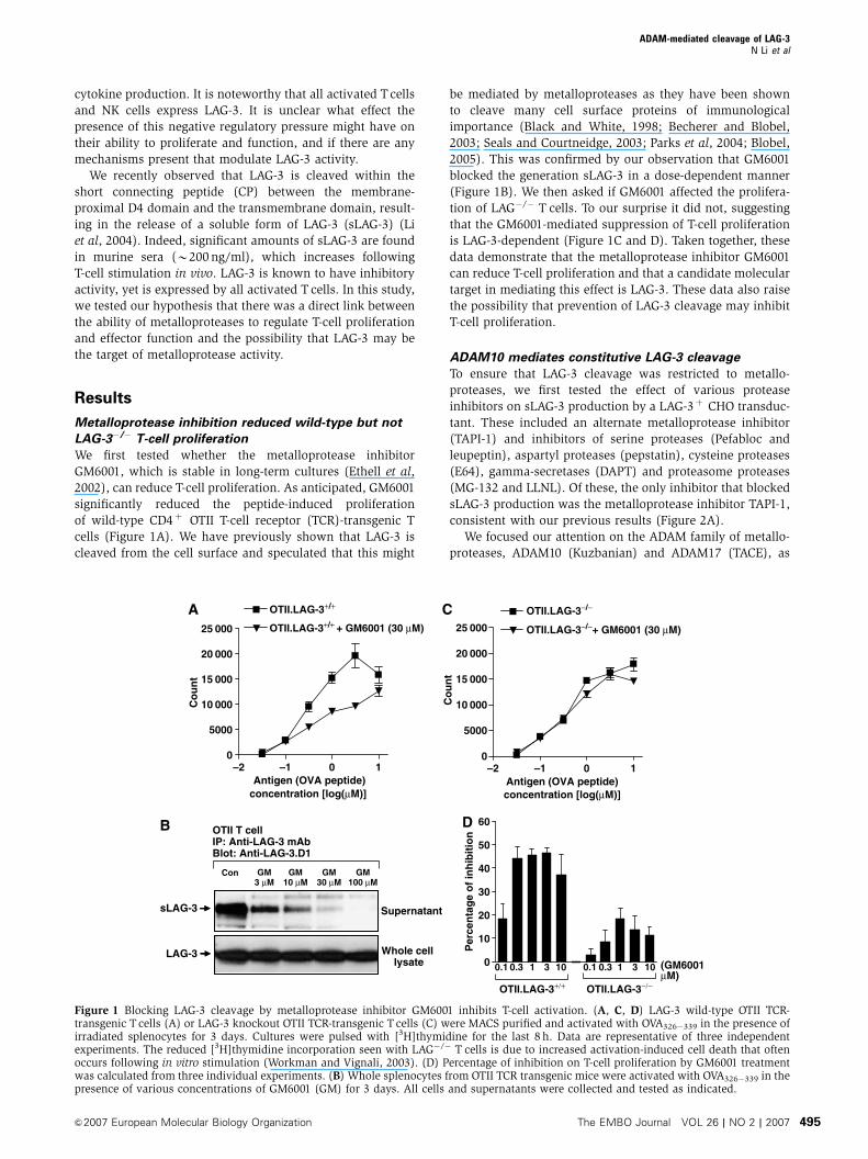
We first tested whether the metalloprotease inhibitor
GM6001, which is stable in long-term cultures (Ethell et al,
2002), can reduce T-cell proliferation. As anticipated, GM6001
significantly reduced the peptide-induced proliferation
of wild-type CD4þ OTII T-cell receptor (TCR)-transgenic T
cells (Figure 1A). We have previously shown that LAG-3 is
cleaved from the cell surface and speculated that this might
be mediated by metalloproteases as they have been shown
to cleave many cell surface proteins of immunological
importance (Black and White, 1998; Becherer and Blobel,
2003; Seals and Courtneidge, 2003; Parks et al, 2004; Blobel,
2005). This was confirmed by our observation that GM6001
blocked the generation sLAG-3 in a dose-dependent manner
(Figure 1B). We then asked if GM6001 affected the prolifera-
tion of LAG�/� T cells. To our surprise it did not, suggesting
that the GM6001-mediated suppression of T-cell proliferation
is LAG-3-dependent (Figure 1C and D). Taken together, these
data demonstrate that the metalloprotease inhibitor GM6001
can reduce T-cell proliferation and that a candidate molecular
target in mediating this effect is LAG-3. These data also raise
the possibility that prevention of LAG-3 cleavage may inhibit
T-cell proliferation.
ADAM10 mediates constitutive LAG-3 cleavage
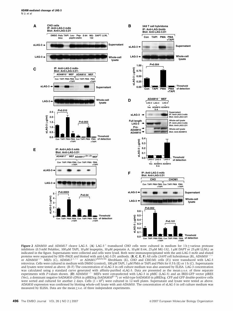
To ensure that LAG-3 cleavage was restricted to metallo-
proteases, we first tested the effect of various protease
inhibitors on sLAG-3 production by a LAG-3þ CHO transduc-
tant. These included an alternate metalloprotease inhibitor
(TAPI-1) and inhibitors of serine proteases (Pefabloc and
leupeptin), aspartyl proteases (pepstatin), cysteine proteases
(E64), gamma-secretases (DAPT) and proteasome proteases
(MG-132 and LLNL). Of these, the only inhibitor that blocked
sLAG-3 production was the metalloprotease inhibitor TAPI-1,
consistent with our previous results (Figure 2A).
We focused our attention on the ADAM family of metallo-
proteases, ADAM10 (Kuzbanian) and ADAM17 (TACE), as
0.1
0
5000
10 000
15 000
20 000
25 000
0
5000
10 000
15 000
20 000
25 000
–2 –1 0 1Antigen (OVA peptide)
concentration [log(µM)]Antigen (OVA peptide)concentration [log(µM)]
OTII.LAG-3+/+
OTII.LAG-3+/+ + GM6001 (30 µM)
OTII.LAG-3−/−
OTII.LAG-3−/−+ GM6001 (30 µM)
0.10
10
20
30
40
50
60
3 101 30.3 1010.3 (GM6001µM)
OTII.LAG-3+/+ OTII.LAG-3−/−
Per
cen
tag
e o
f in
hib
itio
n
Co
un
t
Co
un
t
GM100 µM
Con
OTII T cellIP: Anti-LAG-3 mAbBlot: Anti-LAG-3.D1
GM10 µM
GM30 µM
GM3 µM
sLAG-3
LAG-3
Supernatant
Whole cell lysate
–2 –1 0 1
A C
B D
Figure 1 Blocking LAG-3 cleavage by metalloprotease inhibitor GM6001 inhibits T-cell activation. (A, C, D) LAG-3 wild-type OTII TCR-transgenic T cells (A) or LAG-3 knockout OTII TCR-transgenic T cells (C) were MACS purified and activated with OVA326�339 in the presence ofirradiated splenocytes for 3 days. Cultures were pulsed with [3H]thymidine for the last 8 h. Data are representative of three independentexperiments. The reduced [3H]thymidine incorporation seen with LAG�/� T cells is due to increased activation-induced cell death that oftenoccurs following in vitro stimulation (Workman and Vignali, 2003). (D) Percentage of inhibition on T-cell proliferation by GM6001 treatmentwas calculated from three individual experiments. (B) Whole splenocytes from OTII TCR transgenic mice were activated with OVA326�339 in thepresence of various concentrations of GM6001 (GM) for 3 days. All cells and supernatants were collected and tested as indicated.
ADAM-mediated cleavage of LAG-3N Li et al
&2007 European Molecular Biology Organization The EMBO Journal VOL 26 | NO 2 | 2007 495
Full length
0.4
0.6
0.5
0.4
0.3
0.2
0.1
0.0
0.3
0.2
0.1
0.0
ADAM10ADAM10
LAG-3+
E-A
LAG-3+
Vec
LAG-3+
ADAM10 ADAM10
LAG-3+
E-A
LAG-3+
Vec
LAG-3+
ADAM10 ADAM10
Pro-
ADAM10−/− MEF
DMSO
CHO cellsIP: Anti-LAG-3 mAbBlot: Anti-LAG-3.D1 IP: Anti-LAG-3mAb
Blot: Anti-LAG-3.D1
IP: Anti-LAG-3 mAbBlot: Anti-LAG-3.D1
IP: Anti-LAG-3 mAbBlot: Anti-LAG-3.D1
Blot: Anti-ADAM10
IP: Anti-LAG-3 mAbBlot: Anti-LAG-3.D1
Pefabloc
TAPI Leu-peptin
Pep-statin
E-64 MG-132
DAPT LLNL
LAG-3
sLAG-3
LAG-3
sLAG-3
sLAG-3
LAG-3
LAG-3
sLAG-3
LAG-3
sLAG-3
LAG-3
sLAG-3
sLA
G-3
(µ
g/m
l)
sLA
G-3
(µ
g/m
l)
sLA
G-3
(µ
g/m
l)
sLA
G-3
(µ
g/m
l)
sLA
G-3
(µ
g/m
l)
Con TAPI PMA PMA+TAPI
ADAM10+/−MEF ADAM10−/−MEF
IP: Anti-LAG-3 mAb-Blot: Anti-LAG-3.D1-
IP: Anti-LAG-3 mAbBlot: Anti-LAG-3.D1
Con TAPI PMA PMA+TAPI
Con TAPI PMA PMA+TAPI
Con TAPI PMA PMA+TAPI
Con TAPI PMA PMA+TAPI
Con TAPI PMA PMA+TAPI
Con TAPI PMA PMA+TAPI
Con TAPI PMA PMA+TAPI
Con TAPI PMA PMA+TAPI
Con TAPI PMA PMA+TAPI
Con TAPI PMA PMA+TAPI
Con TAPI PMA PMA+TAPI
Con TAPI PMA PMA+TAPI
Con TAPI PMA PMA+TAPI
0.0
0.5
1.0
1.5
2.0
2.5
Supernatant
Whole-celllysate
Supernatant
Whole-celllysate
Supernatant
Supernatant
Whole-celllysate
Supernatant
Whole-celllysate
Whole-cell lysate
Whole-cell lysate
P=0.010
P=0.004
P=0.003
CHO CHOM1
0.00
0.25
0.50
0.75
1.00
3A9 T cell hybridoma
ADAM17+/+ MEF ADAM17∆Zn/∆Zn MEF
0
1
2
3
4
5 P=0.0003
P=0.002
P=0.101
P=0.04
Thresholdof detection
Thresholdof detection
Thresholdof detection
Thresholdof detection
Thresholdof detection
A B
C
D
E
F
Figure 2 ADAM10 and ADAM17 cleave LAG-3. (A) LAG-3þ -transduced CHO cells were cultured in medium for 1 h7various proteaseinhibitors (0.5 mM Pefabloc, 100mM TAPI, 10mM leupeptin, 10mM pepstatin A, 10mM E-64, 25mM MG-132, 1mM DAPT or 25 mM LLNL) asindicated in the figure. Supernatants were collected and cells were lysed. Both were immunoprecipitated with the anti-LAG-3 mAb and elutedproteins were separated by SDS–PAGE and blotted with anti-LAG-3.D1 antibody. (B, C, E, F) All cells (3A9T cell hybridomas (B), ADAM10�/þ
or ADAM10�/� MEFs (C), ADAM17þ /þ or ADAM17DZnD/Zn fibroblasts (E), CHO and CHO.M1 cells (F)) were transduced with LAG-3retrovirus. Cells were cultured in medium with DMSO (control), 100 mM TAPI, 1 mM PMA or TAPI and PMA for 0.5 h (E) or 1 h (C). Supernatantsand lysates were tested as above. (B–F) The concentration of sLAG-3 in cell culture medium was also assessed by ELISA. LAG-3 concentrationwas calculated using a standard curve generated with affinity-purified sLAG-3. Data are presented as the mean7s.e. of three separateexperiments with P-values shown. (D) ADAM10�/� MEFs were cotransfected with LAG-3 in pMIC (LAG-3) and an IRES-GFP vector pIRES(Vec), a dominant negative bADAM10 cDNA in pIREScg (bADAM10E�A) or wild-type bADAM10 in pIREScg. CFP and GFP double-positive cellswere sorted and cultured for another 2 days. Cells (1�106) were cultured in 12-well plates. Supernatant and lysate were tested as above.ADAM10 expression was confirmed by blotting whole-cell lysate with anti-ADAM10. The concentration of sLAG-3 in cell culture medium wasmeasured by ELISA. Data are the mean7s.e. of three independent experiments.
ADAM-mediated cleavage of LAG-3N Li et al
The EMBO Journal VOL 26 | NO 2 | 2007 &2007 European Molecular Biology Organization496

they are known to cleave many transmembrane proteins
of immunological importance (Black and White, 1998;
Becherer and Blobel, 2003; Blobel, 2005). Although the
proteolytic activity of ADAM10 is generally constitutive,
cleavage by ADAM17 can be induced by PMA (Sahin et al,
2004). Therefore, we first tested whether sLAG-3 production
by the LAG-3þ 3A9T cell hybridoma was altered by
PMA treatment. Production of sLAG-3 was significantly
increased by 1 h PMA treatment (Figure 2B), implicating
a role for ADAMs in LAG-3 shedding. This increase was
not due to enhanced LAG-3 synthesis, as the total protein in
whole-cell lysates was unchanged. It should be noted that
sLAG-3 can be generated by multiple transduced or trans-
fected cell types including T cells, CHO and 3T3 cells
(Li et al, 2004), suggesting that the sheddase is ubiquitously
expressed. Furthermore, shedding does not require LAG-3
ligation, cellular activation or the presence of serum-derived
proteases or cofactors (Li et al, 2004) (Supplementary Figure
S1A and B).
Initial analysis of serum sLAG-3 concentration in mice
lacking ADAM9, 12, 15 and/or 17 suggested that these
were not responsible for constitutive LAG-3 cleavage
(Supplementary Figure S1C and D). As ADAM10 deficiency
results in embryonic lethality (Hartmann et al, 2002), we
assessed its role in LAG-3 cleavage using ADAM10�/� and
ADAM10þ /� MEFs transduced with LAG-3 encoding retro-
virus. Strikingly, there was a 90% reduction in sLAG-3
production by ADAM10�/� MEFs compared with hetero-
zygous control cells (Figure 2C). However, when the LAG-3þ
ADAM10�/� MEFs were treated with PMA, sLAG-3 was
still generated, suggesting that a different protease was
responsible for PMA-induced LAG-3 cleavage. It is note-
worthy that this increase was comparable to that seen
following PMA induction of the ADAM10þ /� control MEFs
(increase in sLAG-3 production in the presence of PMA
over control untreated cells: ADAM10þ /�¼ 0.81mg/ml,
ADAM10�/�¼ 0.80 mg/ml) (Figure 2C).
To confirm that ADAM10 was responsible for consti-
tutive LAG-3 cleavage, LAG-3.pMIC was cotransfected into
ADAM10�/� MEFs with either bovine ADAM10 (bADAM10)
in the GFP containing plasmid pIRES, an enzymatically
inactive mutant (bADAM10E�A) or the empty vector control
(Vec). Analysis of sLAG-3 production by CFPþ/GFPþ MEFs
demonstrated that LAG-3 cleavage was restored in the pre-
sence of active but not inactive bADAM10 (Figure 2D). In
addition, surface LAG-3 expression was drastically reduced
in bADAM10-expressing MEFs (Supplementary Figure S1E).
Taken together, our data clearly show that ADAM10 is
responsible for constitutive LAG-3 cleavage.
ADAM17 mediates PMA-induced LAG-3 cleavage
We established that ADAM17 was the PMA-inducible LAG-3
sheddase with two experiments. First, ADAM17DZn/DZn and
wild-type Ras/Myc-transformed fibroblasts (Reddy et al,
2000) were transduced with retrovirus encoding LAG-3.
sLAG-3 production by transduced ADAM17DZn/DZn fibroblasts
was not increased after PMA treatment compared with the
wild-type fibroblasts (Figure 2E). Second, we expressed LAG-
3 on the ADAM17-deficient CHO-M1 cell line (Li and Fan,
2004; Villanueva de la et al, 2004) and assessed sLAG-3
production following PMA treatment. An B2.5-fold increase
in sLAG-3 production by ELISA was seen with the wild-type
CHO LAG-3 transfectant, whereas PMA treatment had no
effect on the sLAG-3 production by the LAG-3þ CHO-M1
cell line (Figure 2F). In both experiments, constitutive LAG-3
cleavage was unaffected. Taken together, these data demon-
strate that ADAM17 is responsible for the PMA-induced
cleavage of LAG-3. Furthermore, our data show that there
are two distinct metalloproteases (ADAM10 and ADAM17)
that independently cleave LAG-3.
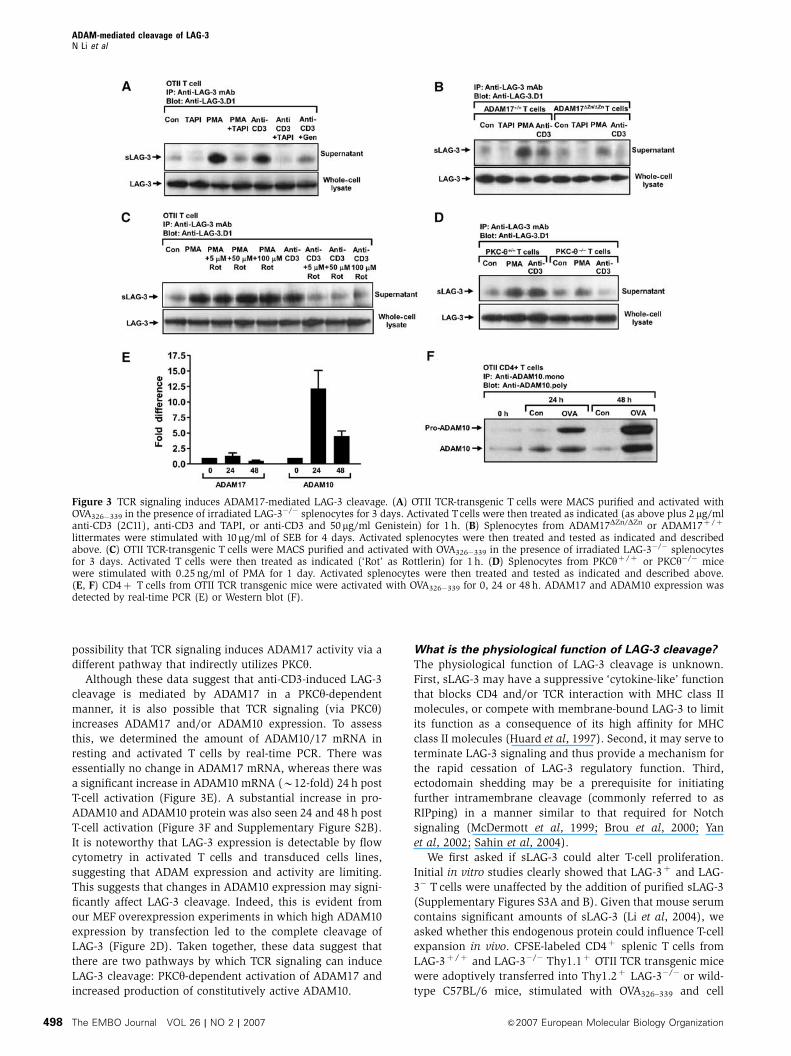
TCR signaling increases LAG-3 cleavage via two distinct
pathways
Our observation that PMA treatment induced LAG-3 cleavage
by ADAM17 suggested that this process might be controlled
by a protein kinase C (PKC)-dependent signaling pathway.
As PKC had been shown to play important roles in T-cell
activation, we questioned whether ADAM17-mediated LAG-3
cleavage was regulated by the TCR signaling pathway. MACS-
purified CD4þ OTII Tcells were activated with OVA326�339 for
2 days. LAG-3 expression was confirmed by flow cytometry
and sLAG-3 production was verified by Western blot.
As shown above, constitutive sLAG-3 shedding was again
increased by PMA treatment and inhibited by TAPI-1
(Figure 3A). Interestingly, TCR crosslinking by anti-CD3eantibody (2C11) also stimulated T cells to produce more
sLAG-3, which could be inhibited by TAPI-1 and the tyrosine
kinase inhibitor genistein. This increase was not due to
induction of LAG-3 synthesis, as the total protein in whole-
cell lysates was unchanged.
We then asked if TCR-induced LAG-3 cleavage was absent
in ADAM17-deficient T cells. As expected, low-level constitu-
tive sLAG-3 shedding was seen with unstimulated wild-type
and mutant T cells, whereas sLAG-3 production was
increased following PMA and anti-CD3 stimulation of wild-
type T cells (Figure 3B). However, CD3 crosslinking induced
minimal sLAG-3 production by ADAM17DZn/DZn T cells.
The same observation was also made with T cells from
Rag-1�/� mice reconstituted with bone marrow from
ADAM17DZn/DZn mice, eliminating the possibility that this
phenotype was due to the development of T cells in an
ADAM17-deficient environment (Supplementary Figure
S2A). Although we cannot rule out the possibility that the
absence of ADAM17 has affected T-cell responsiveness
in general and/or the function of ADAM10, these data do
suggest that ADAM17 is responsible, at least in part, for
the TCR-induced cleavage of LAG-3.
To determine if PKC was required for CD3-induced sLAG-3
production, we stimulated LAG-3þ T cells with anti-CD3 in
the presence of rottlerin, a broad-spectrum PKC inhibitor.
LAG-3 cleavage was effectively blocked by rottlerin, even at
5 mM (Figure 3C). PKCy and PKCd are particularly sensitive to
rottlerin, having an ID50 of 5–30mM (Gschwendt et al, 1993;
Villalba et al, 1999). PKCy is known to be activated by p56lck
and recruited to the immunological synapse following TCR
ligation (Arendt et al, 2002; Isakov and Altman, 2002).
Furthermore, PKCy�/� T cells are poorly responsive to TCR
ligation but respond normally to PMA (Pfeifhofer et al, 2003).
Thus, we asked if PKCy was required for CD3-induced LAG-3
cleavage. While PMA-induced LAG-3 cleavage was intact in
PKCy�/� T cells, CD3 ligation failed to increase LAG-3 shed-
ding (Figure 3D). The simplest explanation for these data is
that PKCy participates directly by phosphorylating ADAM17
and inducing its activation. However, we cannot exclude the
ADAM-mediated cleavage of LAG-3N Li et al
&2007 European Molecular Biology Organization The EMBO Journal VOL 26 | NO 2 | 2007 497
possibility that TCR signaling induces ADAM17 activity via a
different pathway that indirectly utilizes PKCy.
Although these data suggest that anti-CD3-induced LAG-3
cleavage is mediated by ADAM17 in a PKCy-dependent
manner, it is also possible that TCR signaling (via PKCy)
increases ADAM17 and/or ADAM10 expression. To assess
this, we determined the amount of ADAM10/17 mRNA in
resting and activated T cells by real-time PCR. There was
essentially no change in ADAM17 mRNA, whereas there was
a significant increase in ADAM10 mRNA (B12-fold) 24 h post
T-cell activation (Figure 3E). A substantial increase in pro-
ADAM10 and ADAM10 protein was also seen 24 and 48 h post
T-cell activation (Figure 3F and Supplementary Figure S2B).
It is noteworthy that LAG-3 expression is detectable by flow
cytometry in activated T cells and transduced cells lines,
suggesting that ADAM expression and activity are limiting.
This suggests that changes in ADAM10 expression may signi-
ficantly affect LAG-3 cleavage. Indeed, this is evident from
our MEF overexpression experiments in which high ADAM10
expression by transfection led to the complete cleavage of
LAG-3 (Figure 2D). Taken together, these data suggest that
there are two pathways by which TCR signaling can induce
LAG-3 cleavage: PKCy-dependent activation of ADAM17 and
increased production of constitutively active ADAM10.
What is the physiological function of LAG-3 cleavage?
The physiological function of LAG-3 cleavage is unknown.
First, sLAG-3 may have a suppressive ‘cytokine-like’ function
that blocks CD4 and/or TCR interaction with MHC class II
molecules, or compete with membrane-bound LAG-3 to limit
its function as a consequence of its high affinity for MHC
class II molecules (Huard et al, 1997). Second, it may serve to
terminate LAG-3 signaling and thus provide a mechanism for
the rapid cessation of LAG-3 regulatory function. Third,
ectodomain shedding may be a prerequisite for initiating
further intramembrane cleavage (commonly referred to as
RIPping) in a manner similar to that required for Notch
signaling (McDermott et al, 1999; Brou et al, 2000; Yan
et al, 2002; Sahin et al, 2004).
We first asked if sLAG-3 could alter T-cell proliferation.
Initial in vitro studies clearly showed that LAG-3þ and LAG-
3� T cells were unaffected by the addition of purified sLAG-3
(Supplementary Figures S3A and B). Given that mouse serum
contains significant amounts of sLAG-3 (Li et al, 2004), we
asked whether this endogenous protein could influence T-cell
expansion in vivo. CFSE-labeled CD4þ splenic T cells from
LAG-3þ /þ and LAG-3�/� Thy1.1þ OTII TCR transgenic mice
were adoptively transferred into Thy1.2þ LAG-3�/� or wild-
type C57BL/6 mice, stimulated with OVA326–339 and cell
Figure 3 TCR signaling induces ADAM17-mediated LAG-3 cleavage. (A) OTII TCR-transgenic T cells were MACS purified and activated withOVA326�339 in the presence of irradiated LAG-3�/� splenocytes for 3 days. Activated Tcells were then treated as indicated (as above plus 2mg/mlanti-CD3 (2C11), anti-CD3 and TAPI, or anti-CD3 and 50mg/ml Genistein) for 1 h. (B) Splenocytes from ADAM17DZn/DZn or ADAM17þ /þ
littermates were stimulated with 10mg/ml of SEB for 4 days. Activated splenocytes were then treated and tested as indicated and describedabove. (C) OTII TCR-transgenic T cells were MACS purified and activated with OVA326�339 in the presence of irradiated LAG-3�/� splenocytesfor 3 days. Activated T cells were then treated as indicated (‘Rot’ as Rottlerin) for 1 h. (D) Splenocytes from PKCyþ /þ or PKCy�/� micewere stimulated with 0.25 ng/ml of PMA for 1 day. Activated splenocytes were then treated and tested as indicated and described above.(E, F) CD4þ T cells from OTII TCR transgenic mice were activated with OVA326�339 for 0, 24 or 48 h. ADAM17 and ADAM10 expression wasdetected by real-time PCR (E) or Western blot (F).
ADAM-mediated cleavage of LAG-3N Li et al
The EMBO Journal VOL 26 | NO 2 | 2007 &2007 European Molecular Biology Organization498
division was analyzed 6 days later. While a clear difference in
the extent of cell division was seen between the LAG-3�/�
and LAG-3þ /þ OTII T cells, whereas the presence of sLAG-3
in the serum of recipient mice had no effect on the antigen-
induced division of either T-cell population (Supplementary
Figure S3C and D). It is possible that the local sLAG-3
concentration in the microenvironment of T-cell activation
and/or in the presence of LAG-3þ T cells might be much
higher than the serum sLAG-3 concentration. To address this
possibility, we generated mice expressing B1000-fold higher
levels of sLAG-3 than normal serum concentrations by retro-
viral-mediated stem cell gene transfer (Supplementary Figure
S3E) (Workman and Vignali, 2003). These mice then served
as recipients for purified, CFSE-labeled, LAGþ /þ or LAG�/�
Thy1.2þ CD4þ OTII T cells and were treated as above.
Despite the presence of substantial quantities of sLAG-3,
T-cell proliferation was surprisingly unaffected, and the ability
of membrane-associated LAG-3 to control this expansion was
also unperturbed (Supplementary Figure S3F and G). Taken
together, these results suggest that sLAG-3 has no effect on
antigen-driven T-cell activation and proliferation in vitro or
in vivo, and does not serve to limit or control LAG-3 function.
Prevention of LAG-3 cleavage blocks T-cell proliferation
and cytokine production
We reasoned that if LAG-3 cleavage was required to attenuate
its negative regulatory function, a noncleavable version of
LAG-3 would be predicted to have enhanced regulatory
activity. In contrast, reduced regulatory activity would be
observed if cleavage was required to release a ‘functional’
sLAG-3 molecule or if LAG-3 RIPping initiated signaling. We
had previously shown that LAG-3 cleavage occurs within
membrane-proximal CP (Li et al, 2004). To generate nonclea-
vable LAG-3 mutants for functional analysis, we first ana-
lyzed the influence of CP length and amino-acid composition
on LAG-3 shedding. A series of LAG-3 CP mutants were
expressed in a LAG-3/CD4 double loss variant 3A9 T-cell
hybridoma by retroviral transduction (Supplementary Figure
S4). Constitutive shedding by unstimulated cells was as-
sessed by detection of sLAG-3 using Western blot and
ELISA. Analysis of these mutants demonstrated that cleavage
requires a long CP (48 amino acids) and that the protease(s)
that mediates this shedding are relatively promiscuous, as
indicated by some tolerance for amino-acid substitutions
within the CP (Supplementary Figure S4).
Two noncleavable LAG-3 mutants were chosen for func-
tional analysis: LAG-3mCD4CP in which the 20–amino-acid
LAG-3 CP has been replaced with the eight-amino-acid CD4
CP, and LAG-3ESCP which has a 12-amino-acid deletion of the
LAG-3 CP (Supplementary Figure S4A) (Li et al, 2004).
Splenic LAG-3�/� CD4þ OTII T cells were transduced with
vector alone (pMIC), wild-type LAG-3 or LAG-3mCD4CP or
LAG-3ESCP-encoding retrovirus. Physiological levels of LAG-3
expression, that were comparable to that seen on activated T
cells, were ensured by FACS. Uniform CFP and LAG-3 expres-
sion was confirmed by Western blot and flow cytometric
analysis (Figures 4A and B). No sLAG-3 was detected by
Western blot and ELISA in supernatants collected from T
cells transduced with either LAG-3mCD4-CP or LAG-3ESCP
(Figure 4A and Supplementary Figure S4A) (Li et al, 2004).
We first asked if expression of non-cleavable LAG-3
affected T-cell proliferation and cytokine production in vitro.
Ectopic expression of LAG-3 significantly reduced T-cell
proliferation, but only at lower antigen doses (Figure 4C).
In contrast, T cells expressing either of the noncleavable
LAG-3 mutants barely proliferated except at the highest
antigenic peptide concentration. We also measured interfer-
on-g (IFN-g) and interleukin-2 (IL-2) production following
peptide stimulation of these transduced T cells. Again, wild-
type LAG-3 expression clearly reduced production of both
cytokines (Figures 4D and E). However, T cells expressing
either version of noncleavable LAG-3 produced substantially
less IFN-g and essentially no IL-2, even at the highest
concentration of antigenic peptide.
We then investigated the effect of LAG-3 cleavage on T-cell
proliferation in vivo. Retrovirally transduced LAG-3�/�
Thy1.1þ OTII T cells were labeled with CFSE, adoptively
transferred into Thy1.2þ C57BL/6 mice and then activated
with OVA326�339 peptide in vivo. Some background prolifera-
tion was seen with the controls (no peptide added) in these
experiments owing to the previous in vitro antigen exposure
required for retroviral transduction (horizontal hashed bar;
Figure 4F). However, peptide treatment in vivo clearly
induced substantial T-cell proliferation of the CFP vector
control-transduced OTII T cells. Consistent with our in vitro
observations, T cells transduced with wild-type LAG-3
proliferated less than the vector control T cells. However,
remarkably, very few T cells expressing noncleavable LAG-3
proliferated (Figure 4F and G). In summary, these data show
that noncleavable LAG-3 has a potent inhibitory effect on
T-cell proliferation and cytokine production that is greater
than wild-type LAG-3, suggesting that cell surface cleavage
serves as an important negative feedback mechanism to
moderate its function.
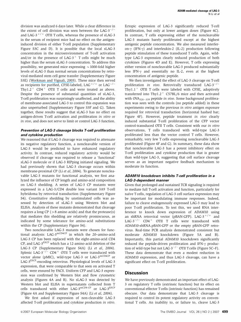
ADAM10 knockdown inhibits T-cell proliferation in a
LAG-3-dependent manner
Given that prolonged and sustained TCR signaling is required
to mediate full T-cell activation and function, particularly for
naı̈ve Tcells, regulation of LAG-3 cell surface expression may
be important for modulating immune responses. Indeed,
failure to cleave endogenously expressed LAG-3 may lead to
poor T-cell proliferation. To test this, we used RNA inter-
ference to knock down expression of ADAM10 using
an shRNA retroviral vector (pBAN-GFP). LAG-3þ /þ and
LAG-3�/� CD4þ OTII T cells were transduced with
ADAM10-shRNA.pBAN-GFP or the empty pBAN-GFP retro-
virus. Real-time PCR analysis demonstrated consistent but
moderate ADAM10 knockdown (Figure 5A and B).
Importantly, this partial ADAM10 knockdown significantly
reduced the peptide-driven proliferation and IFN-g produc-
tion of wild-type but not LAG-3�/� OTII Tcells (Figure 5C–F).
These data demonstrate that even a modest reduction in
ADAM10 expression, and thus LAG-3 cleavage, can have a
significant effect on T-cell proliferation.
Discussion
We have previously demonstrated an important effect of LAG-
3 on regulatory T cells (extrinsic function) but its effect on
conventional effector Tcells (intrinsic function) has remained
obscure. Our data demonstrate that LAG-3 cleavage is
required to control its potent regulatory activity on conven-
tional T cells. An inability to, or failure to, cleave LAG-3
ADAM-mediated cleavage of LAG-3N Li et al
&2007 European Molecular Biology Organization The EMBO Journal VOL 26 | NO 2 | 2007 499

Figure 4 Noncleavable LAG-3 has a more potent inhibitory effect on T-cell activation than wild-type LAG-3. LAG-3�/� Thy1.1þ OTII TCR-transgenic T cells were purified by negative MACS, activated and transduced with empty vector pMIC (Vec), LAG-3 in pMIC (LAG-3),LAG3mCD4CP in pMIC (LAG3mCD4CP) or LAG3ESCP in pMIC (LAG3ESCP). Transduced CD4þ OTII T cells were FACS purified by gating on CFPþ
cells. (A) Cells were cultured in medium for 1 h. Both supernatant and whole-cell lysate were immunoprecipitated with anti-LAG-3 mAb. Elutedproteins were separated by SDS–PAGE and blotted with rabbit anti-LAG-3.D1 antisera. (B) Flow cytometric analysis of transductantsdemonstrating equivalence of CFP and LAG-3 expression. (C–E) Transduced T cells were stimulated with OVA326�339 at the concentrationsindicated. Cultures were either pulsed with [3H]thymidine during the last 8 h of a 48 h assay (C) or culture medium collected 24 h afteractivation for determination of IFN-g (D) and IL-2 (E) production by ELISA. (F, G) Cells were labeled with CFSE, adoptively transferred intoLAG-3�/� mice and cells stimulated in vivo 24 h later with (no peptide control) or without OVA 326–339 peptide. Spleens were removed 6 dayslater and the percentage of dividing cells was determined by measuring CFSEnegative�low/Thy1.1þ/CD4þ Tcells. (F) Data are the mean7s.e. ofthree independent experiments with a total of 8/9 mice per group. Hatched horizontal bar represents the mean7s.e. of the no peptide controls.(G) Representative histograms are also presented with individual divisions displayed using FlowJo.
ADAM-mediated cleavage of LAG-3N Li et al
The EMBO Journal VOL 26 | NO 2 | 2007 &2007 European Molecular Biology Organization500

prevents T-cell proliferation but does not appear to result in
cell death. Why might this be important? All activated T cells
express high levels of LAG-3, yet efficiently proliferate in vivo.
The modulation of LAG-3 cleavage, and thus its signaling/
function, may be important in facilitating unencumbered
expansion. The ability of TCR signaling to modulate both
PKCy-dependent, ADAM17-mediated activation and upregu-
lation of ADAM10 expression provides two direct mechan-
isms for potentiating LAG-3 cleavage and thus negating its
regulatory activity. As activated T cells express high levels of
LAG-3, modulation of its negative cell-intrinsic regulatory
function may help ‘shape’ the contraction phase and perhaps
influence the establishment of T-cell memory.
Despite extensive in vitro and in vivo analysis, we found no
evidence to support a suppressive ‘cytokine-like’ activity for
sLAG-3. Indeed, T-cell proliferation and LAG-3 function were
unaffected even in the presence of an B1000-fold increased
concentration of serum sLAG-3 in vivo. Although we cannot
completely exclude the possibility that sLAG-3 performs a
unique and specific function that has yet to be identified, our
data do suggest that sLAG-3 has no ‘global’ function and
is likely a ‘waste product’ of LAG-3 cleavage. Two additional
observations are consistent with this notion. First, sLAG-3
appears to be rapidly excreted and/or degraded in vivo, as the
half-life of passively transferred, purified sLAG-3 is less than
4 h (Supplementary Figure S5A). Second, whereas dimeric
LAG-3:Ig fusion proteins have a high affinity for MHC class II
molecules, naturally cleaved sLAG-3 does not specifically
bind with MHC class II molecules and no detectable bind-
ing of endogenous sLAG-3 on splenic B cells is seen
(Supplementary Figure S5B). We and others have shown
that LAG-3 is expressed on cells as a weak dimer (Huard
et al, 1997; Li et al, 2004). However, gel filtration analysis
suggests that purified sLAG-3 is a monomer (data not
shown). Thus, based on these data, we propose that only
the cell surface LAG-3 dimer or the dimeric LAG-3:Ig fusion
protein possesses high affinity for MHC class II molecules. As
this is a weak dimer, membrane tethering would be required
to maintain this high-affinity form, as this appears to be lost
upon cleavage. It is also conceivable that MHC class II
binding further stabilizes dimerization but this would still
be dependent on membrane tethering. This system provides
a unique mechanism for retaining the high-affinity MHC
class II interaction required for LAG-3 function while safely
rendering sLAG-3 in sera innocuous.
Previous studies have proposed two functions for cell
surface metalloprotease-mediated shedding. First, cleavage
can serve to activate target proteins. For instance, ADAM10-
and ADAM17-mediated cleavage of EGFR ligands is in many
cases critical for activation of the EGFR (Peschon et al, 1998;
Jackson et al, 2003; Blobel, 2005). Moreover, cleavage of
Notch is required for effective signaling (Hartmann et al,
2002). Second, cleavage can serve to inhibit target protein
function. Shedding can generate soluble receptors that can
0.0
0.5
1.0
1.5
2.0
0.0
0.5
1.0
1.5
2.0
Rel
ativ
e A
DA
M10
-R
NA
leve
l (fo
ld)
Rel
ativ
e A
DA
M10
-R
NA
leve
l (fo
ld)
pBan-GFP
ADAM10-shRNA.pBan-GFP
pBan-GFP
ADAM10-shRNA.pBan-GFP
00
10 000
20 000
30 000
pBan-GFPADAM10-shRNA.pBan-GFP
Co
un
tC
ou
nt
104103102101 10410310210100
1000
2000
3000
4000
pBan-GFPADAM10-shRNA.pBan-GFP
INF
-γ (
pg
/ml)
10 000
20 000
30 000
40 000
OTII.LAG-3+/+
0
1000
2000
3000
4000
INF
-γ (
pg
/ml)
0
pBan-GFPADAM10-shRNA.pBan-GFP
pBan-GFPADAM10-shRNA.pBan-GFP
OTII.LAG-3−/−
OVA (nM)
0 104103102101
OVA (nM)
0 104103102101
OVA (nM)
OVA (nM)
A C E
B D F
Figure 5 ADAM10 knockdown reduces T-cell proliferation in wild-type but not LAG-3�/� T cells. Thy1.1þ OTII TCR-transgenic T cells fromeither LAG-3�/� or LAG-3þ /þ were purified by negative MACS, activated and transduced with empty vector pBan-GFP or the ADAM10 shRNAcassette in pBan-GFP. Transduced CD4þ OTII T cells were FACS purified by gating on top 50% of GFPþ cells. (A, B) RNAs from retroviral-transduced OTII.LAG-3þ /þ T cells (A) or OTII.LAG-3�/� T cells (B) were purified using TRIzol RNA isolation reagent (Invitrogen LifeTechnologies, Carlsbad, CA) according to the manufacturer’s protocol. The relative quantities of ADAM10 mRNA were calculated from standardcurves and normalized to 18S mRNA. (C–F) Transduced T cells from either LAG-3�/� (C, E) or LAG-3þ /þ (D, F) were stimulated withOVA326�339 at the concentrations indicated. Cultures were either pulsed with [3H]thymidine during the last 8 h of a 72 h assay (C, D) or culturemedium collected 48 h after activation for determination of IFN-g (E, F) production. All figures are representative of three individualexperiments.
ADAM-mediated cleavage of LAG-3N Li et al
&2007 European Molecular Biology Organization The EMBO Journal VOL 26 | NO 2 | 2007 501
act either as scavengers to soak up soluble ligands or compe-
titors to block interaction with membrane-associated ligands,
effectively reducing signaling through the intact receptors.
One proposed example of this is with the TNF-a receptor
(McDermott et al, 1999; Galon et al, 2000). Cleavage defects
result in TNF-a receptor accumulation on the cell surface and
reduction of the soluble competitor in serum, leading to
intensified TNF-a receptor-mediated signaling. Our results
may provide an additional twist to the importance of
metalloprotease-mediated shedding in immune modulation.
Although the shedding of cytokine receptors may generate
soluble ligand scavengers, LAG-3 cleavage does not produce
a soluble molecule that interferes with LAG-3 function. Thus,
the substantially enhanced regulatory activity observed
can be attributed purely to increased LAG-3 signaling.
Furthermore, this represents the first example of a protein
required for dampening T-cell function being controlled by
cell surface cleavage.
One intriguing aspect of our data is that the regulatory
activity of ectopically expressed noncleavable LAG-3 was
enhanced even though its cell surface expression was com-
parable to wild-type LAG-3. This demonstrates that reduced
cleavage enhances the proficiency of signaling rather than
simply resulting in increased expression, which may be the
case for other receptors. How this might be mediated is
unclear, but could be due to prolonged MHC class II ligation.
Given that normal LAG-3 expression is highly regulated, it
is conceivable that the consequence of preventing cleavage
of the endogenous protein could be even greater. Another
related issue is the fate of the LAG-3TM-CY fragment (Li et al,
2004) that remains following ADAM10/17 shedding. It is
conceivable that it is subjected to a RIPping-like activity by
either the g-secretase complex or a related intramembrane
protease system (Urban and Freeman, 2002). A major func-
tion of g-secretase is the clearance of membrane anchors of
shed type I membrane proteins (Schenk, 2000; Kopan and
Ilagan, 2004). Alternatively, this could serve to ‘activate’ the
LAG-3 cytoplasmic tail in a Notch-like manner. Given that
large ectodomains of 200 amino acids or more appear
to prevent g-secretase cleavage (Struhl and Adachi, 2000),
we would argue that RIPping of the LAG-3TM-CY remnant to
generate an active signaling fragment is an unlikely scenario
as one would predict that the generation of a noncleavable
LAG-3 mutant would prevent this and thus lead to a reduction
in LAG-3 regulatory activity, which is the antithesis of our
observations. Last, the recent observation that ADAM10 can
mediate cleavage in trans suggests that antigen-presenting
cells may also facilitate T-cell expansion by cleavage of LAG-3
in trans (Janes et al, 2005).
Thus, we would speculate that in normal T cells ADAM10/
17 are limiting, and thus very subtle changes in their quantity
and/or enzymic activity could have a significant effect on
LAG-3 shedding. Given that ADAM10 and ADAM17 mediate
the cleavage of many diverse cell surface molecules, consti-
tutive and TCR-induced modulation of their activity could
represent a new paradigm for the control of T-cell expansion
and function.
Recently, a number of metalloprotease inhibitors have
been used in animal models and clinical trials as potential
therapies for cancer, multiple sclerosis, arthritis and cardio-
vascular diseases (Gijbels et al, 1994; Bigg and Rowan, 2001;
Hidalgo and Eckhardt, 2001). Interestingly, some studies
suggest that administration of metalloprotease inhibitors
could prevent inflammation-induced tissue damage (Gijbels
et al, 1994; Ramesh and Reeves, 2002). Currently, the pro-
tective effects of metalloprotease inhibitors are thought to be
due to a reduction of TNF-a function (Gijbels et al, 1994;
Ramesh and Reeves, 2002). Our observation that a metallo-
protease inhibitor can reduce T-cell proliferation in a LAG-3-
dependent manner provides a novel mode of action for
these anti-inflammatory agents.
Materials and methods
LAG-3 constructs and cell linesLAG-3 constructs were produced using recombinant PCR as des-cribed previously (Vignali and Vignali, 1999). All LAG-3 constructswere cloned into murine stem cell virus-based retroviral vectors,MSCV-IRES-GFP/CFP (pMIG/pMIC) (Workman et al, 2002a).Details of primers and strategy will be provided on request([email protected]). bADAM10 in pIREScg vector was kindlyprovided by Drs Postina and Fahrenholz. bADAM10E�A was madeby substituting amino acid Glu to Ala at position 384 as describedbefore (Lammich et al, 1999). ADAM10�/� and ADAM10þ /� MEFs(Hartmann et al, 2002), and ADAM17DZn/DZn (TaceDZn/DZn-EC-2)and ADAM17þ /þ (EC-4) Ras/Myc-transformed fibroblast cloneswere described elsewhere (Reddy et al, 2000). T-cell hybridomasand fibroblasts were transduced essentially as described (Blacket al, 1997). CHO cells and ADAM17-deficient CHO cells (M1) werestably transfected with the LAG-3.pMIG plasmid and pHbAPRII-puro for selection at a DNA ratio 10:1 using Fugene (Roche,Indianapolis, IN) (Arribas and Massague, 1995). Transfectants wereselected in 8 mg/ml of puromycin (Clontech, Mountain View, CA)for 7 days and the expression of LAG-3 was confirmed by flowcytometry. All transductants/transfectants were sorted on aMoFlow for uniform GFP expression.
Antibodies and protease inhibitorsThe following antibodies were used for immunoprecipitation andor Western blotting: rat anti-LAG-3 mAb (Workman et al, 2002b)(C9B7W specific for the D2 domain; BD-PharMingen, San Diego,CA), rabbit anti-LAG-3.D1 (Li et al, 2004) and rabbit anti-murineADAM10 (Chemicon International, Temecula, CA). Leupeptin,pepstatin, aprotinin and Pefabloc were obtained from Roche(Indianapolis, IN). E64, TAPI-1, MG132, DAPT and LLNL wereobtained from Calbiochem (San Diego, CA).
Immunoprecipitation and immunoblottingImmunoprecipitation and immunoblotting were performed asdescribed previously (Li et al, 2004). In brief, whole-cell lysateswere generated using 1% NP-40 lysis buffer (50 mM Tris, 150 mMNaCl, 1% NP-40, 10 mg/ml leupeptin, 10 mg/ml pepstatin, 10 mg/mlaprotinin, 2 mM Pefabloc, pH 7.4) on ice for 30 min, followed bycentrifugation at 15 000 g for 10 min. For immunoprecipitation,whole-cell lysates or culture media were incubated with 5mg anti-LAG-3 mAb and 30ml protein G-Sepharose (Amersham Biosciences,Piscataway, NJ) for 3 h at 41C. Lysates or eluted proteins fromimmunoprecipitates were resolved by SDS–PAGE (Invitrogen,Carlsbad, CA) and blots probed as detailed. Blots were developedusing ECL (Amersham, Piscataway, NJ) and autoradiography.
sLAG-3 ELISAFor sLAG-3 quantification by ELISA, C9B7W (5mg/ml) mAb wascoated on 96-well flat-bottomed-microtiter plates (Dynatech Labs,Franklin, MA) in carbonate buffer (50 mM Na2CO3, pH 10.4) at 371Cfor 1 h. The plates were washed three times with PBS–Tween 20(0.05%) and then blocked with 0.5% FBS in carbonate buffer at 41Covernight. The plates were washed and the serum or cell culturemedium was added. Following 1 h incubation at 371C, the plateswere washed and then probed with rabbit anti-LAG-3.D1 antisera(1:200 dilution, 371C, 1 h). This was followed by three washes and a1 h incubation with an HRP-conjugated, anti-rabbit Ig secondary Ab(1:2000 dilution, Amersham, Piscataway, NJ). Plates were devel-oped with TMB substrate solution (Pierce, Rockford, IL) and thereaction was stopped by adding 50ml of 1 N H2SO4 in each well.Absorbance was measured by a spectrophotometer (Molecular
ADAM-mediated cleavage of LAG-3N Li et al
The EMBO Journal VOL 26 | NO 2 | 2007 &2007 European Molecular Biology Organization502

Devices, Sunnyvale, CA). LAG-3 concentration was calculated usinga purified sLAG-3 standard curve.
Retroviral transduction of CD4þ T cells and T-cell activationin vitro and in vivoSplenocytes were stained with biotin-labeled anti-B220, anti-Gr1,anti-Mac1, anti-TER119, anti-CD49b and anti-CD8 antibody (Phar-Mingen, San Diego, CA), incubated with magnetic beads coupledwith streptavidin and then negatively sorted on an AutoMACS.Purity of CD4þ T cells was over 80% based on FACS analysis.Purified T cells were activated with 5mg/ml OVA326�339 peptide inthe presence of irradiated whole splenocytes from B6.LAG-3�/�
mice. After 2 days activation, T cells were spin transduced (90 min3000 r.p.m., two times on 2 consecutive days) with supernatantfrom vector alone (pMIC), LAG-3/CFP, LAG-3.CD4CP/CFP, LAG-3.ESCP/CFP GPEþ 86 retroviral producer cell lines. Cells were thencultured in the presence of 1 ng/ml of IL-2 for 5 days, sorted on CFPand then allowed to rest for another 3 days.
For the in vitro activation assays, the purified CFPþ T cells(2.5�104) were cultured with 5�105 irradiated (3000 rad) spleno-cytes in a 96-well flat-bottomed plate. Antigen (OVA326�339) wasadded into each well with a 3.3-fold dilution. Cells were cultured for24 h and pulsed with 1mCi/well [3 H]thymidine (Du Pont, Wilming-ton, DE) in the last 7–8 h of culture.
For the in vivo activation assay, purified T cells from eitherThy1.1þ OTII TCR or Thy1.1þ OTII TCR LAG-3�/� mice werelabeled with 5 mM CFSE for 10 min at 371C in PBS plus 0.1% BSA at1�107 cells/ml and washed twice. Mice were injected with 5�106
cells i.v. and 24 h later with 100 mm of OVA326–339 peptide in 500 mlof PBS i.p. Six days later, splenocytes were counted by Trypan blueexclusion, stained with anti-Thy1.1.PE and anti-CD4.allophycocya-nin and analyzed for CFSE levels by flow cytometry. Cell divisionwas calculated by FlowJo (Treestar Inc., Ashland, OR).
Supplementary dataSupplementary data are available at The EMBO Journal Online(http://www.embojournal.org).
Acknowledgements
We thank Doug Green for his advice and critical review of the paper.We are grateful to Steve Shaw, Yin Liu and Gottfried Baier for thePKCy�/� spleens, David Wiest for the pBAN-GFP vector, RolfPostina and Falk Fahrenholz for the bADAM10 cDNA and JoanMassague and Joaquin Arribas for the CHO-M1 cells. We also wishto thank Richard Cross, Jennifer Hoffrage and Jennifer Smith forFACS, Sue Rowe for cytokine analysis, Mike Nash for AutoMACS,staff in the St Jude Hartwell Center for peptide synthesis, oligosynthesis and DNA sequencing, and Sarah Fitzgerald (Dempsey lab)for the preparation of samples from the ADAM17DZn/DZn mice. Thiswork was supported by the National Institutes of Health (R01AI-39480), a Cancer Center Support CORE grant (CA-21765) andthe American Lebanese Syrian Associated Charities (ALSAC) (toDAAV). PS is supported by the Deutsche ForschungsgemeinschaftSFB45/TPB9. CPB is supported by NIH RO1 GM64750 and EY01571.PJD is supported by NIH DK59778 and DK63363, and a CCFA grant.
References
Arendt CW, Albrecht B, Soos TJ, Littman DR (2002) Protein kinaseC-theta: signaling from the center of the T-cell synapse. Curr OpinImmunol 14: 323–330
Arribas J, Massague J (1995) Transforming growth factor-alphaand beta-amyloid precursor protein share a secretory mechanism.J Cell Biol 128: 433–441
Baker AH, Edwards DR, Murphy G (2002) Metalloproteinaseinhibitors: biological actions and therapeutic opportunities.J Cell Sci 115: 3719–3727
Becherer JD, Blobel CP (2003) Biochemical properties and functionsof membrane-anchored metalloprotease-disintegrin proteins(ADAMs). Curr Top Dev Biol 54: 101–123
Bigg HF, Rowan AD (2001) The inhibition of metalloproteinases asa therapeutic target in rheumatoid arthritis and osteoarthritis.Curr Opin Pharmacol 1: 314–320
Black RA, Rauch CT, Kozlosky CJ, Peschon JJ, Slack JL, WolfsonMF, Castner BJ, Stocking KL, Reddy P, Srinivasan S, Nelson N,Boiani N, Schooley KA, Gerhart M, Davis R, Fitzner JN, JohnsonRS, Paxton RJ, March CJ, Cerretti DP (1997) A metalloproteinasedisintegrin that releases tumour-necrosis factor-alpha from cells.Nature 385: 729–733
Black RA, White JM (1998) ADAMs: focus on the protease domain.Curr Opin Cell Biol 10: 654–659
Blobel CP (2005) ADAMs: key components in EGFR signalling anddevelopment. Nat Rev Mol Cell Biol 6: 32–43
Brou C, Logeat F, Gupta N, Bessia C, LeBail O, Doedens JR, CumanoA, Roux P, Black RA, Israel A (2000) A novel proteolytic cleavageinvolved in Notch signaling: the role of the disintegrin-metallo-protease TACE. Mol Cell 5: 207–216
Bruniquel D, Borie N, Hannier S, Triebel F (1998) Regulationof expression of the human lymphocyte activation gene-3(LAG-3) molecule, a ligand for MHC class II. Immunogenetics48: 116–124
Bruniquel D, Borie N, Triebel F (1997) Genomic organization of thehuman LAG-3/CD4 locus. Immunogenetics 47: 96–98
Ethell DW, Kinloch R, Green DR (2002) Metalloproteinase sheddingof Fas ligand regulates beta-amyloid neurotoxicity. Curr Biol 12:1595–1600
Galon J, Aksentijevich I, McDermott MF, O’Shea JJ, Kastner DL(2000) TNFRSF1A mutations and autoinflammatory syndromes.Curr Opin Immunol 12: 479–486
Gijbels K, Galardy RE, Steinman L (1994) Reversal of experimentalautoimmune encephalomyelitis with a hydroxamate inhibitor ofmatrix metalloproteases. J Clin Invest 94: 2177–2182
Gschwendt M, Kittstein W, Marks F (1993) Protein kinase C forms acomplex with and phosphorylates the GTPase activating proteinGAP: phosphorylation by PKC is dependent on tyrosine phos-phorylation of GAP and/or a GAP-associated protein. BiochemBiophys Res Commun 194: 571–576
Hartmann D, de SB, Serneels L, Craessaerts K, Herreman A, AnnaertW, Umans L, Lubke T, Lena IA, von FK, Saftig P (2002) Thedisintegrin/metalloprotease ADAM 10 is essential for Notch sig-nalling but not for alpha-secretase activity in fibroblasts. HumMol Genet 11: 2615–2624
Hidalgo M, Eckhardt SG (2001) Development of matrix metallo-proteinase inhibitors in cancer therapy. J Natl Cancer Inst 93:178–193
Huang CT, Workman CJ, Flies D, Pan X, Marson AL, Zhou G,Hipkiss EL, Ravi S, Kowalski J, Levitsky HI, Powell JD, PardollDM, Drake CG, Vignali DA (2004) Role of LAG-3 in regulatory Tcells. Immunity 21: 503–513
Huard B, Mastrangeli R, Prigent P, Bruniquel D, Donini S, El-TayarN, Maigret B, Dreano M, Triebel F (1997) Characterization of themajor histocompatibility complex class II binding site on LAG-3protein. Proc Natl Acad Sci USA 94: 5744–5749
Isakov N, Altman A (2002) Protein kinase C(theta) in T cellactivation. Annu Rev Immunol 20: 761–794
Jackson LF, Qiu TH, Sunnarborg SW, Chang A, Zhang C, PattersonC, Lee DC (2003) Defective valvulogenesis in HB-EGF and TACE-null mice is associated with aberrant BMP signaling. EMBO J 22:2704–2716
Janes PW, Saha N, Barton WA, Kolev MV, Wimmer-Kleikamp SH,Nievergall E, Blobel CP, Himanen JP, Lackmann M, Nikolov DB(2005) Adam meets Eph: an ADAM substrate recognition moduleacts as a molecular switch for ephrin cleavage in trans. Cell 123:291–304
Kopan R, Ilagan MX (2004) Gamma-secretase: proteasome of themembrane? Nat Rev Mol Cell Biol 5: 499–504
Lammich S, Kojro E, Postina R, Gilbert S, Pfeiffer R, Jasionowski M,Haass C, Fahrenholz F (1999) Constitutive and regulated alpha-secretase cleavage of Alzheimer’s amyloid precursor protein bya disintegrin metalloprotease. Proc Natl Acad Sci USA 96:3922–3927
Li N, Workman CJ, Martin SM, Vignali DA (2004) Biochemicalanalysis of the regulatory T cell protein lymphocyte activationgene-3 (LAG-3; CD223). J Immunol 173: 6806–6812
Li X, Fan H (2004) Loss of ectodomain shedding due to mutations inthe metalloprotease and cysteine-rich/disintegrin domains of the
ADAM-mediated cleavage of LAG-3N Li et al
&2007 European Molecular Biology Organization The EMBO Journal VOL 26 | NO 2 | 2007 503
tumor necrosis factor-alpha converting enzyme (TACE). J BiolChem 279: 27365–27375
Maretzky T, Reiss K, Ludwig A, Buchholz J, Scholz F, Proksch E,de SB, Hartmann D, Saftig P (2005) ADAM10 mediates E-cadherinshedding and regulates epithelial cell-cell adhesion, migration,and beta-catenin translocation. Proc Natl Acad Sci USA 102:9182–9187
McDermott MF, Aksentijevich I, Galon J, McDermott EM,Ogunkolade BW, Centola M, Mansfield E, Gadina M, Karenko L,Pettersson T, McCarthy J, Frucht DM, Aringer M, Torosyan Y,Teppo AM, Wilson M, Karaarslan HM, Wan Y, Todd I, Wood G,Schlimgen R, Kumarajeewa TR, Cooper SM, Vella JP, Amos CI,Mulley J, Quane KA, Molloy MG, Ranki A, Powell RJ, Hitman GA,O’Shea JJ, Kastner DL (1999) Germline mutations in the extra-cellular domains of the 55 kDa TNF receptor, TNFR1, definea family of dominantly inherited autoinflammatory syndromes.Cell 97: 133–144
Overall CM, Kleifeld O (2006) Tumour microenvironment—opinion:validating matrix metalloproteinases as drug targets and anti-targets for cancer therapy. Nat Rev Cancer 6: 227–239
Parks WC, Wilson CL, Lopez-Boado YS (2004) Matrix metallo-proteinases as modulators of inflammation and innate immunity.Nat Rev Immunol 4: 617–629
Peschon JJ, Slack JL, Reddy P, Stocking KL, Sunnarborg SW, Lee DC,Russell WE, Castner BJ, Johnson RS, Fitzner JN, Boyce RW,Nelson N, Kozlosky CJ, Wolfson MF, Rauch CT, Cerretti DP,Paxton RJ, March CJ, Black RA (1998) An essential role forectodomain shedding in mammalian development. Science 282:1281–1284
Pfeifhofer C, Kofler K, Gruber T, Tabrizi NG, Lutz C, Maly K, LeitgesM, Baier G (2003) Protein kinase C theta affects Ca2+ mobiliza-tion and NFATcell activation in primary mouse Tcells. J Exp Med197: 1525–1535
Ramesh G, Reeves WB (2002) TNF-alpha mediates chemokine andcytokine expression and renal injury in cisplatin nephrotoxicity.J Clin Invest 110: 835–842
Reddy P, Slack JL, Davis R, Cerretti DP, Kozlosky CJ, Blanton RA,Shows D, Peschon JJ, Black RA (2000) Functional analysis ofthe domain structure of tumor necrosis factor-alpha convertingenzyme. J Biol Chem 275: 14608–14614
Reiss K, Maretzky T, Ludwig A, Tousseyn T, de SB, Hartmann D,Saftig P (2005) ADAM10 cleavage of N-cadherin and regulation ofcell-cell adhesion and beta-catenin nuclear signalling. EMBO J 24:742–752
Sahin U, Weskamp G, Kelly K, Zhou HM, Higashiyama S, Peschon J,Hartmann D, Saftig P, Blobel CP (2004) Distinct roles for ADAM10and ADAM17 in ectodomain shedding of six EGFR ligands. J CellBiol 164: 769–779
Schenk D (2000) Alzheimer’s disease. a partner for presenilin.Nature 407: 34–35
Seals DF, Courtneidge SA (2003) The ADAMs family of metallopro-teases: multidomain proteins with multiple functions. Genes Dev17: 7–30
Struhl G, Adachi A (2000) Requirements for presenilin-dependent cleav-age of notch and other transmembrane proteins. Mol Cell 6: 625–636
Triebel F, Jitsukawa S, Baixeras E, Roman-Roman S, Genevee C,Viegas-Pequignot E, Hercend T (1990) LAG-3, a novel lymphocyteactivation gene closely related to CD4. J Exp Med 171: 1393–1405
Urban S, Freeman M (2002) Intramembrane proteolysis controlsdiverse signalling pathways throughout evolution. Curr OpinGenet Dev 12: 512–518
Vignali DA, Vignali KM (1999) Profound enhancement of T cellactivation mediated by the interaction between the TCR and theD3 domain of CD4. J Immunol 162: 1431–1439
Villalba M, Kasibhatla S, Genestier L, Mahboubi A, Green DR,Altman A (1999) Protein kinase c theta cooperates with calci-neurin to induce Fas ligand expression during activation-inducedT cell death. J Immunol 163: 5813–5819
Villanueva de la TT, Bech-Serra JJ, Ruiz-Paz S, Baselga J, Arribas J(2004) Inactivating mutations block the tumor necrosis factor-alpha-converting enzyme in the early secretory pathway. BiochemBiophys Res Commun 314: 1028–1035
Weskamp G, Ford JW, Sturgill J, Martin S, Docherty AJ, SwendemanS, Broadway N, Hartmann D, Saftig P, Umland S, Sehara-FujisawaA, Black RA, Ludwig A, Becherer JD, Conrad DH, Blobel CP(2006) ADAM10 is a principal ‘sheddase’ of the low-affinityimmunoglobulin E receptor CD23. Nat Immunol 7: 1293–1298
Workman CJ, Cauley LS, Kim IJ, Blackman MA, Woodland DL,Vignali DA (2004) Lymphocyte activation gene-3 (CD223) regu-lates the size of the expanding Tcell population following antigenactivation in vivo. J Immunol 172: 5450–5455
Workman CJ, Dugger KJ, Vignali DA (2002a) Cutting edge: mole-cular analysis of the negative regulatory function of lymphocyteactivation gene-3. J Immunol 169: 5392–5395
Workman CJ, Rice DS, Dugger KJ, Kurschner C, Vignali DA (2002b)Phenotypic analysis of the murine CD4-related glycoprotein,CD223 (LAG-3). Eur J Immunol 32: 2255–2263
Workman CJ, Vignali DA (2003) The CD4-related molecule, LAG-3(CD223), regulates the expansion of activated T cells. Eur JImmunol 33: 970–979
Workman CJ, Vignali DA (2005) Negative regulation of T cellhomeostasis by lymphocyte activation gene-3 (CD223).J Immunol 174: 688–695
Yan Y, Shirakabe K, Werb Z (2002) The metalloprotease Kuzbanian(ADAM10) mediates the transactivation of EGF receptor by Gprotein-coupled receptors. J Cell Biol 158: 221–226
ADAM-mediated cleavage of LAG-3N Li et al
The EMBO Journal VOL 26 | NO 2 | 2007 &2007 European Molecular Biology Organization504
Copyright © 2022 FDOKUMEN