Multiple Tumoricidal Effector Mechanisms Induced by Adriamycin1
Upload
khangminh22Category
view
0download
0
Cloning and Characterization of RIP1, an Effector of Ral
by
Sang-Ho ParkB. S., Yonsei University
1985
Submitted to the Department of Biologyin partial fulfillment of the requirements
for the degree of
DOCTOR OF PHILOSOPHY
at theMassachusetts Institute of Technology
May 1996
0 1996 by Sang-Ho Park. All rights reserved.The author hereby grants to MIT permission to reproduce and to distribute publicly
copies of this thesis document in whole or in part.
I1 I' ISignature of Author
Department of BiologyMay 29, 1996
Certified by N- 6Robert A. Weinberg
Thesis Supervisor
Accepted by
77.- .... .. ,,,ASAC- -~ETTS INS;: , Ui'L
OF TECHNOLOGY
JUL 0 81996 Science
F-rank SolomonChairman
Graduate Committee
LIBRARIES

To Winnie,
You started me off on this path of research,
questioning what is, and why.
I will never forget you,
for every day I am reminded of the things you taught me,
what you meant to me.
I only regret not having realized it sooner.
Enduring pain, agony and hopelessness,
showing only perseverance, hope and love throughout.
An inspiration always.....

ACKNOWLEDGEMENTS
First and foremost, I would like to thank Mina and Chan-Jun for their
love, support, and understanding. I also give thanks to my parents and
parents-in-law for their unwavering support.
I would also like to thank all the members of the Weinberg lab, past
and present, not only for their help in scientific matters, but also for their
companionship and camaraderie, without which my stay in the Weinberg
lab would not have been as fun and enjoyable. I give special thanks to
Snezna Rogelj, who was always there for me when I needed help or advice;
to Dennis Templeton, who taught me sequencing, as well as many other
fine points in experimentation; to Steve Friend, who took me under his
wing when I first joined the lab; to Bart Giddings and other members of
the now defunct Ras group, who helped me start afresh when I returned
from the Army; to past baymates Roman Herrera and Qiang Yu, who were
there to commiserate and encourage me when things weren't going just
right; to the sheik, the monk, the wire, and, of course, the chairman, may
you never be called before a congressional hearing.
I thank Bob for letting me join his lab, twice; for his support,
encouragement and endurance for tolerating me for the eight years I have
been in his lab; and especially for his support during the period I was off
being all that I could be. I also thank Phil Sharp, David Housman for their
helpful discussions and scientific supervision, as well as for past WIBR vs.
CCR softball games, Peter Kim for his interest and encouragement and
Andre Bernards for his insight, willingness and especially for his generous

gift, without which this work would have been much delayed. I also thank
Suzanne Brill for reading this manuscript and for her many helpful
comments.
I have very fond memories of the Whitehead and M.I.T. The Pit (yes,
good things happen in the Pit, too), Tang Hall, project lab (yes, I enjoyed
that too), softball games, Eastgate, Summer barbecues, the Retreats. But
my fondest memories are of the third floor of the Whitehead, from back in
the days when it was cram-packed with absolutely no extra space to now
where we are practically the only lab left on the floor. There was always
an excitement about doing Science and, again, I thank Bob for his part in
creating and maintaining such an atmosphere. If I leave this place with
nothing else, let me take that excitement with me.

TABLE OF CONTENTS
DEDICATION i
ACKNOWLEDGEMENT ii
TABLE OF CONTENTS iv
FIGURES vii
ABSTRACT 1
CHAPTER 1. RAS-RELATED GTPases AND THEIR EFFECTORS 3
GTPases 5
Ras, the oncogene 7
GTPase Activating Proteins 8
Guanine Nucleotide Exchange Factors 10
Down-stream signaling from Ras 12
Ras-related proteins 17
The Rho subfamily 18
Effectors and regulators of Rho-related GTPases 21
FIGURES 27
CHAPTER 2 CLONING AND MOLECULAR ANALYSIS
INTRODUCTION 32
RESULTS
Isolation of the RIP1 clone 34
Sequence analysis 36
Northern Analysis 36
Nucleotide specificity 37
Interacting domain mapping 38

DISCUSSION 38
MATERIALS AND METHODS 42
ACKNOWLEDGEMENT 44
FIGURES 45
CHAPTER 3 ANALYSIS OF RIP1 PROTEIN
INTRODUCTION 58
RESULTS
Expression of RIP1 60
Covalent Modification of RIP1 62
Ral-RIP1 interaction 64
GAP assays 65
Immunofluorescence 67
DISCUSSION 69
MATERIALS AND METHODS 74
ACKNOWLEDGEMENT 81
FIGURES 82
CHAPTER 4. CONCLUSIONS AND PROSPECTS
Effector proteins 101
RIP1 as an effector 104
Covalent modification of RIP1 105
GTPase cascade 106
FIGURES 109
CHAPTER 5. REFERENCES 112

FIGURES
Figure
Figure
Figure
1-1.
1-2.
2-1.
Figure 2-2.
Figure
Figure
Figure
Figure
Figure
Figure
Figure
Figure
Figure
2-3.
2-4.
2-5.
2-6.
3-1.
3-2.
3-3.
3-4.
3-5.
Figure 3-6.
Figure 3-7.
Figure
Figure
Figure
3-8.
3-9.
4-1.
The basic GTPase cycle.
Family tree of Ras-related proteins.
Nucleotide and deduced amino acid
sequence of RIP1.
Amino acid sequence comparison of
proteins containing Rho/Rac GAP activity.
Tissue distribution of RIP1 transcript.
Preferential binding of 19-6 to KH-ral
preloaded with GTP vs. GDP.
Mapping of the Ral-binding region.
Amino acid sequence comparison of
RIP1 homologs.
Western analysis of RIP1.
Effect of cycloheximide treatment on RIP1.
Effect of phosphatase on RIP1.
Effect of serum on RIP1 modification.
Ral-RIP1 complexes in Cos-1 transient
transfections.
The effect of RIP1 on the GTPase activity
of several GTPases.
The effect of RIP1 on guanine nucleotides
bound to GTPases.
Immunofluorescence of RIP1.
Co-localization of RIP1 and vinculin.
Models of Ral-RIP1 interaction.
27
29
45
47
49
51
53
55
82
84
86
88
90
92
95
96
98
109

The Cloning and Characterization of RIP1, an Effector of Ral
by
Sang-Ho Park
Submitted to the Department of Biology on May 29, 1996
in partial fulfillment of the requirements for the Degree of
Doctor of Philosophy
ABSTRACT
I report here the cloning of a gene encoding a novel Ral-
interacting protein (RIP1) from a cDNA expression library using
radiolabeled Ral as probe. RIP1 binds to Ral in a GTP-dependent
manner. The 4.1 kb transcript of the RIP] gene is present in all
tissues analyzed and encodes for a protein product of 648 residues.
RIP1 shares sequence similarity with GTPase activating
proteins that are capable of activating the GTPase activity of
members of the Rho/Rac family of GTPases. When tested, RIP1 could
activate the GTPase activity of CDC42 and, to a lesser extent, Racl but
not RhoA, Ras, or Ral. Activated Ral had no direct effect on the
GTPase-activating ability of RIP1, in vitro.
RIP1 can be covalently modified by phosphorylation as a result
of serum stimulation in serum starved Balb/3T3 cells. RIP1 is found
in a complex with Ral within five minutes after serum stimulation of

serum starved cells. RIP1 co-localizes with vinculin in focal adhesion
complexes.
Thesis Advisor: Robert A. Weinberg, Ph.D.
Professor of Biology, M.I.T.
Member, Whitehead Institute for Biomedical Research

Chapter 1. Ras-related GTPases and Their Effectors
Ral is a member of a large family of small molecular weight
GTPases, the most well-known of which is Ras. Like other members
of this family of proteins, Ras functions as a molecular switch,
oscillating between a GTP bound 'on' state and a GDP-bound 'off'
state.
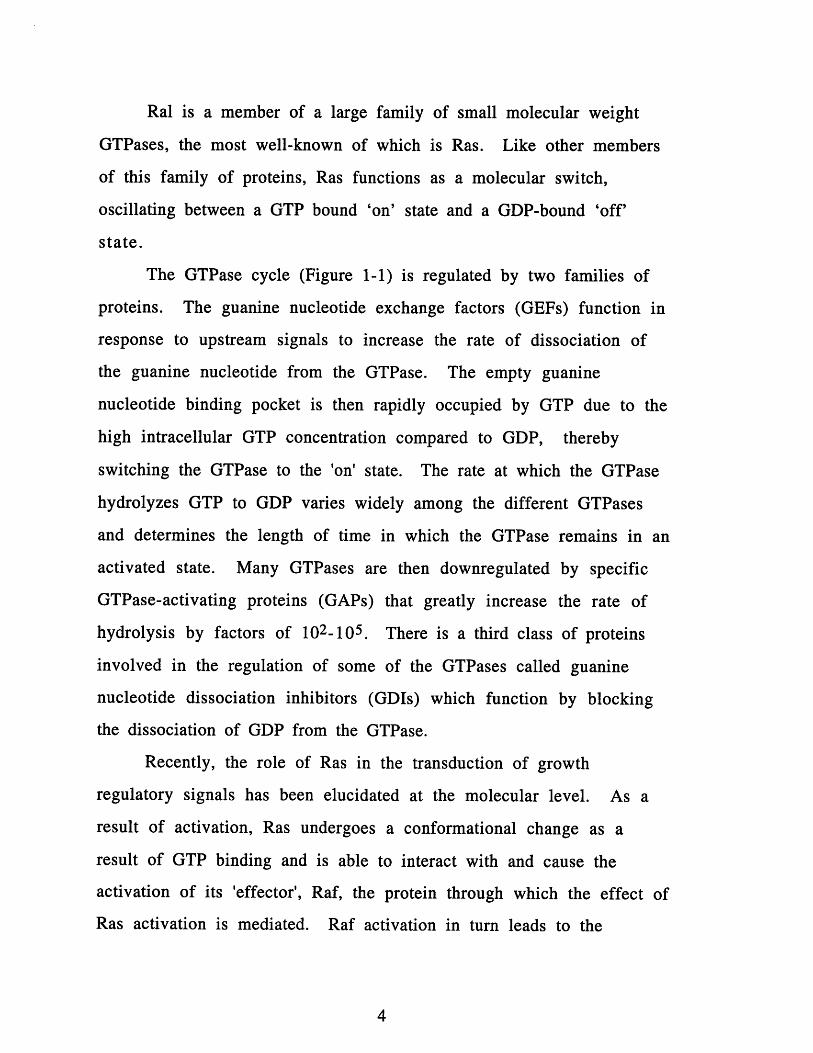
The GTPase cycle (Figure 1-1) is regulated by two families of
proteins. The guanine nucleotide exchange factors (GEFs) function in
response to upstream signals to increase the rate of dissociation of
the guanine nucleotide from the GTPase. The empty guanine
nucleotide binding pocket is then rapidly occupied by GTP due to the
high intracellular GTP concentration compared to GDP, thereby
switching the GTPase to the 'on' state. The rate at which the GTPase
hydrolyzes GTP to GDP varies widely among the different GTPases
and determines the length of time in which the GTPase remains in an
activated state. Many GTPases are then downregulated by specific
GTPase-activating proteins (GAPs) that greatly increase the rate of
hydrolysis by factors of 102-105. There is a third class of proteins
involved in the regulation of some of the GTPases called guanine
nucleotide dissociation inhibitors (GDIs) which function by blocking
the dissociation of GDP from the GTPase.
Recently, the role of Ras in the transduction of growth
regulatory signals has been elucidated at the molecular level. As a
result of activation, Ras undergoes a conformational change as a
result of GTP binding and is able to interact with and cause the
activation of its 'effector', Raf, the protein through which the effect of
Ras activation is mediated. Raf activation in turn leads to the

activation of a kinase cascade that has been shown to be important in
signaling growth.
Ever since Ras was identified as a gene that was capable of
transforming cells (Reddy et al., 1982; Tabin et al., 1982; Taparowsky
et al., 1982), much effort has been exerted into identifying the role
that Ras plays in normal cells and the mutations that allow Ras to
become transforming, as well as identifying novel genes that might
mimic Ras in structure or function.
To date, in excess of 60 Ras-related proteins have been isolated
from a wide variety of species, ranging from yeast and slugs to frogs,
fruit flies and mammals, with some estimates predicting the
existence of at least 60 Ras-related proteins in mammals, at least 30
of which are expected to be Rab proteins (Chardin, 1993).
Recent work in this laboratory has identified a protein that is
involved in the regulation of Ral, a protein related to Ras. In this
thesis, I will present work done in an attempt to elucidate the role of
Ral in signaling by identifying a candidate effector protein for it. As
an introduction to the world of GTPases, I will start off with a brief
summary of GTPase in general and the conserved structure of Ras-
related proteins. I will then introduce Ras and its regulatory
proteins, and the role of Ras in signal transduction. Finally, I will
introduce other Ras-related proteins with emphasis on the Rho-
related protein for reasons that will become apparent later.
GTPases.
GTPases play a role in a wide variety of cellular functions,
ranging from protein synthesis and vision to hormone action and

cancer. These GTPases function primarily as switches oscillating
between an activated, GTP-bound form to an inactive GDP-bound
form. There are three families of GTPases that share a common
design, in that all members of these families contain five regions of
homology whose function is to either participate in guanine
nucleotide binding or undergo conformational change in response to
the nucleotide bound (Bourne et al., 1991). These families are the
alpha subunits of heterotrimeric G proteins, the Ras-related small
molecular weight GTPases, and EF-Tu, an elongation factor involved
in bacterial protein synthesis.
The heterotrimeric G-proteins are involved in hormonal or
sensory signaling as a response to stimulation of heptahelical
receptors. Representative of heterotrimeric G-proteins are GS, the
stimulatory regulator of adenylyl cyclase, and GT (transducin), which
mediates phototransduction. EF-Tu performs a proofreading function
that ensures that the mRNA codon matches the anticodon of the
aminoacyl-tRNA. The Ras-related GTPases are involved in a wide
variety of functions ranging from signaling proliferation and
differentiation to regulation of the actin cytoskeleton and vesicular
trafficking. Although the amino acid sequence of Ras and EF-Tu
guanine nucleotide binding domains are only 16% identical, their
three dimensional structures are almost superimposable (Valencia et
al., 1991).
All members of the Ras family possess three conserved regions,
all of which are critical for the proper function of Ras. The first
region (residues 10-18, 57-63, 116-119, 143-146) is involved in
GTP-binding. Mutations in this region, such as those frequently

found in residues 12 or 61, result in a slowing down of the intrinsic
GTP hydrolysis activity. The second region (residues 32-40) is
commonly referred to as the 'effector' domain. Mutations in this
region render oncogenic mutants of Ras no longer able to transform,
without having any effect on the localization, GTPase activity or
stability of the protein. It is thought that this is the region that binds
to the cellular effector of Ras. Interestingly, this also overlaps nicely
with one of the two domains (residues 30-37 and 60-76) shown to
undergo a conformational switch when GTP replaces GDP in the
protein (Kim et al., 1988). The third region is the C-terminal end
which contains sites for isoprenylation, either a CAAX motif, or a CXC
or CC motif where C is cysteine, A is an aliphatic amino acid, and X in
any amino acid. The isoprenylation helps anchor the C-terminal end
to the membrane.
Ras, the oncogene.
In 1964, Jennifer Harvey first reported a virus that was
capable of inducing sarcomas and splenomegaly in infected rats
(Harvey, 1964). Several years later, Kirsten and Mayer reported a
mouse lymphoma virus that was capable of inducing sarcomas after
passage through rats (Kirsten et al., 1967). It was later shown that
both viruses carried closely related genes that encoded a 21 kD
protein, namely, Ha-ras and Ki-ras (Shih et al., 1979). Further, it was
soon demonstrated that these Ras proteins were capable of not only
binding guanine nucleotides (Shih et al., 1980), but also of
hydrolyzing GTP to GDP (McGrath et al., 1984; Sweet et al., 1984).
Sequence analysis of the viral oncogene and the proto-oncogene

revealed point mutations in residues 12 and 59. It was later shown
that a single mutation at the position 12 residue was sufficient to
cause normal Ras to become transforming (Reddy et al., 1982; Tabin
et al., 1982; Taparowsky et al., 1982). This was due to the fact that
although the residue 12 mutated form of Ras was capable of binding
either GTP or GDP, its ability to hydrolyze GTP to GDP was greatly
reduced compared to the wild type Ras (Gibbs et al., 1984). This was
the first indication that the GTP-bound form of Ras was able to
transduce a signal to a downstream target that allowed the cell to
become transformed, and that the ability to regulate the guanine
nucleotide binding and GTPase activity properly was crucial for
normal cell function.
GTPase Activating Proteins.
Additional mutations in residues 13, 61 and 63, discovered in
other human tumors, were shown to possess a reduced intrinsic rate
of GTP hydrolysis (Colby et al., 1986; Gibbs et al., 1985; McGrath et
al., 1984; Sweet et al., 1984). These activating mutations in Ras were
long thought to endow Ras with a more aggressive growth potential
as a direct consequence of reduced GTPase activity.
But upon further investigation, a direct correlation between
reduction in intrinsic GTPase activity and transforming potency did
not hold up (Der et al., 1986; Lacal et al., 1986; Trahey et al., 1987).
For example, although the T59 mutant form of Ras, where residue 59
was mutated to threonine, possesses GTPase activity comparable to
that of wild type, it has a transforming potency comparable to the
L12 mutant, indicating that activation of the transforming property

of Ras can occur by mechanisms not involving just a reduction in in
vitro GTPase activity (Lacal et al., 1986). In another study, two
different mutants in position 12 that possessed a 4-fold difference in
GTPase activity were shown to be equally potent in their
transforming ability, suggesting that position 12 mutations might
affect some other aspect of Ras function (Trahey et al., 1987).
In experiments in Xenopus oocytes using the induction of
maturation as an assay for Ras activity two activated forms of Ras,
D12 and V12, could induce maturation while the wild type Ras, G12,
was found to be relatively inactive. While both mutant forms were
found to be GTP-bound, the wild type Ras was found to be
predominately GDP-bound, indicating the presence of an activity
capable of inducing the GTPase activity of Ras and, thereby,
rendering it inactive. A cytoplasmic protein was subsequently
purified that was capable of activating the GTPase activity of wild
type Ras by more than 200-fold while having no effect on the
mutant forms. A similar activity was detected in mammalian cells
(Trahey and McCormick, 1987) and later purified as pl20GAP (Gibbs
et al., 1988; Vogel et al., 1988). These data provide strong support
for the model that the role of the position 12 mutants is to render
these molecules unresponsive to GAP, allowing them to remain in
their active state for a protracted period of time. Overexpression of
pl20GAP can inhibit the function of wild type Ras by blocking the
accumulation of its GTP-bound form (Zhang et al., 1990). pl20GAP
was later shown to interact with Ras through its effector domain,
which provided initial support for its role not only as a

downregulator but also as a potential effector for Ras (Adari et al.,
1988).
Other GAPs for Ras have since been identified. Neurofibromin-
1 (NF1) is another mammalian protein that possesses GAP activity.
Originally identified as a gene disrupted in neurofibromatosis
patients (Cawthon et al., 1990; Viskochil et al., 1990; Wallace et al.,
1990), when cloned, NF1 was found to have 26% identity to the C-
terminal third of human pl20GAP (Xu et al., 1990). Although cell
lines derived from these tumors have normal levels of p120GAP, Ras
is predominately in the GTP-bound form, supporting the role of NF1
as a down regulator of Ras (Basu et al., 1992; DeClue et al., 1992).
Two yeast proteins, IRA1 and IRA2, have also been identified as Ras
GAPs (Tanaka et al., 1990). NF1 expressed in yeast is capable of
replacing IRA (Ballester et al., 1990; Martin et al., 1990; Tanaka et al.,
1990; Xu et al., 1990).
Guanine Nucleotide Exchange Factors.
Much effort has been invested in identifying the factor
responsible for the activation of Ras. Although several groups had
identified exchange activity in mammalian cell extracts (Downward
et al., 1990; West et al., 1990; Wolfman and Macara, 1990), the
protein responsible remained elusive.
S. cerevisiae have two Ras homologs, RAS1 and RAS2, that
function in the activation of adenylyl cyclase. Through genetic
methods, CDC25 had been identified as functioning upstream of RAS
(Broek et al., 1987; Robinson et al., 1987). Disruption of the CDC25
gene is lethal but its function can be replaced by an activated RAS2
10

gene. Wild type RAS2 cannot replace CDC25 even at very high levels,
indicating that the CDC25 gene product is required for the activation
of RAS2. The CDC25 gene product was subsequently shown to be
able to promote guanine nucleotide exchange on mammalian Ras.
A second related protein, SDC25, was also shown to function as
an exchange factor when activated by truncating its C-terminal third
(Crechet et al., 1990). In fission yeast, the ste6 gene product acts as
an exchange factor for Ras and was found to share substantial
homology with CDC25 (Hughes et al., 1990). A mammalian
homologue of CDC25, CDC25Mm, was cloned from a mouse brain cDNA
library by functional complementation in yeast (Martegani et al.,
1992).
In Drosophila, the sev gene, a gene found to be required for the
proper development of the R7 photoreceptor cell, was shown to be
structurally similar to mammalian receptor tyrosine kinases (Hafen
et al., 1987; Simon et al., 1989). One of the genes identified in a
screen looking for downstream elements was the Drosophila ras]
gene, indicating that Drasl was involved the signaling from Sev.
Indeed, activated mammalian Ras was able to rescue the formation
of the R7 cell in sev null mutants (Fortini et al., 1992). Another gene,
sos, that was identified in that screen was capable of suppressing a
weak loss of function mutant of sev and, interestingly, contained
homology to the yeast CDC25 gene (Bonfini et al., 1992; Simon et al.,
1991). Not so surprisingly, Sos turned out to be the exchange factor
for Drasl. A widely expressed human homologue of the Drosophila
sos gene was soon isolated and found to able to stimulated guanine
nucleotide exchange specifically on mammalian Ras proteins in vitro

(Chardin et al., 1993). Also, mammalian cells overexpressing the full
length human Sos were found to have increased guanine nucleotide
exchange activity on Ras, indicating that the human Sos was indeed
the exchange factor for Ras.
Downstream signaling from Ras.
Analysis of mutations in Ras identified regions that were
indispensable for function, such as those required for guanine
nucleotide binding and the C-terminal residues (Willumsen et al.,
1986). Some of these mutations, such as those on positions 35, 36,
38, and 40, did not have an obvious function in that they did not
have any effect on the biochemical properties of the protein.
Mutations at these positions greatly reduced the ability of Ras to
transform cells, and it had been postulated that these mutations
defined the effector domain of Ras, a region that was important for
transmitting the downstream signal (Sigal et al., 1986).
Efforts to identify cellular responses to mitogen stimulation of
mammalian cells led to the discovery that following an initial burst
of receptor-mediated phosphorylation on tyrosines came a more
widespread phosphorylation on serine/threonine residues, and that
the prominent targets that were phosphorylated in response to a
variety of different growth factors seemed to overlap (Avruch,
1985). One of the targets of phosphorylation turned out to be the S6
ribosomal subunit (Blenis and Erikson, 1986; Maller et al., 1986;
Pelech et al., 1986; Stefanovic et al., 1986). The kinase responsible
for the phosphorylation, Rsk, was purified (Blenis et al., 1987;
Erikson and Maller, 1986) and its gene was cloned (Alcorta et al.,
12
1989; Jones et al., 1988). Soon afterward, a second type of mitogen-
activated protein kinase, the p42/p44 MAPKs, was identified that
was shown to be able to phosphorylate and activate the Xenopus S6
kinase (Sturgill et al., 1988). While the crucial targets of Rsk remain
uncertain, the p42/p44 MAPKs was found to be able to regulate a
number of other protein kinases as well as a variety of proto-
oncogenic transcription factors, such as c-Jun and c-Myc (Davis,
1993). MAPKs were then shown to be regulated by growth factor-
stimulated tyrosine phosphorylation (Anderson et al., 1990),
although the regulation turned out not to be a direct regulation by
the receptor tyrosine kinases, but through yet another kinase, the
MAPK kinase, that was able to activate the p42/p44 MAPs by
phosphorylation on tyrosine as well as serine/threonine residues
(Ahn et al., 1992). Thus, the regulation of proteins involved in the
response to mitogens relied on a cascade of protein phosphorylation
events (Ahn and Krebs, 1990; Ahn et al., 1990).
MEK, a MAPK kinase, was found to be directly phosphorylated,
and thus activated, by c-Raf, a cytoplasmic serine/threonine kinase
whose oncogenic counterpart, v-Raf, was identified in mouse sarcoma
virus 3611 (Rapp et al., 1988). Although other kinases capable of
activating MEK in vitro have been identified (Lange-Carter et al.,
1993), genetic evidence suggests that Raf is the primary element
through which receptor tyrosine kinases exert their regulation of
MEK (Tsuda et al., 1993).
The initial indication that Ras and Raf might be on the same
signaling pathway came from experiments in which the Y13-259
13

antibody was microinjected into cells. Y13-259, a rat monoclonal
antibody against Ras (Furth et al., 1982), was critical in the functional
studies of Ras in that it was able to neutralize Ras in the context of
inhibiting morphological transformation induced by mutated Ras as
well as being able to block the effect of serum induced mitogenic
responses in certain cell lines (Kung et al., 1986; Mulcahy et al.,
1985). Interestingly, this antibody could not inhibit the mitogenic
effects of the v-raf gene product, an activated serine/threonine
kinase, or an activated mutant Ras that was not recognized by the
antibody. Later, it was found that a dominant negative Raf could
block the mitogenic signal of Ras (Kolch et al., 1991). Conversely,
while the activation of several serine/threonine kinases implicated in
mitogenesis, including Raf, has been shown to require Ras, the
stimulation of these kinases as a response to receptor tyrosine kinase
activation can be inhibited by the N17 dominant negative Ras, while
oncogenic Ras can activate their kinase activities (Wood et al., 1992).
Soon after Raf had been identified as the activator of MEK and
the primary element through which receptor tyrosine kinases
regulate MEK, examination of the possible regulation of Raf by Ras
led to a number of reports showing direct interaction between the
two proteins. This interaction was shown to be dependent on Ras
activation, in that Raf could interact only with the GTP-bound form
of Ras. Also, these interactions were mediated through domains of
the two proteins known to be crucial for their functions, the effector
domain in the case of Ras and the N-terminal, presumably
regulatory, domain of Raf (Koide et al., 1993; Moodie et al., 1993; Van
Aelst et al., 1993; Vojtek et al., 1993; Warne et al., 1993; Zhang et al.,
14

1993). The emerging model was that activation of receptor tyrosine
kinases led to the activation of Ras through the
translocation/activation of Sos, which in turn led to the activation of
Ras and the subsequent recruitment of Raf to the plasma membrane
where it is activated. Studies in which the CAAX box of Ras is fused
to Raf further validated this model by demonstrating that by
allowing Raf to be localized to the membrane independent of Ras
through its own membrane localization domain, Ras was no longer
necessary for, nor was the dominant negative N17 Ras able to inhibit,
the activation of Raf (Leevers et al., 1994; Stokoe et al., 1994).
Although the Raf-CAAX possessed an eight- to ten- fold higher
specific activity compared to that of wild type Raf in the absence of
Ras activation, treatment by EGF further activates Raf-CAAX by ten
fold indicating that membrane localization is not sufficient for full
activation of Raf and that other events are necessary.
Activation of Raf is most likely not the only consequence of Ras
activation. Ras activation is able to activate multiple signaling events
that regulate cell growth and differentiation. In quiescent
fibroblasts, the expression of ectopic H-Ras activated by the V12
mutation can induce membrane ruffling, activation of the MAP
kinase and stimulation of DNA synthesis. Mutations in the effector
domain of Ras in conjunction with the V12 activating mutation can
further dissect the effector domain. The V12C40 mutant (where
residue 40 is mutated to cysteine in the presence of the V12
mutation) can stimulate membrane ruffling but was defective in
MAP kinase activation and stimulation of DNA synthesis. The
V12S35 mutation, on the other hand, could activate MAP kinase but
15

was defective in stimulation of both DNA synthesis and membrane
ruffling. The expression of both V12C40 and V12S35 could restore
the stimulation of DNA synthesis to a level comparable to V12 Ras.
These data suggest that Ras has multiple effectors, and that
membrane ruffling and MAP kinase activation require the activation
of distinct effector pathways while input from both are required for
the stimulation of DNA synthesis (Joneson et al., 1996).
A possible alternative effector for Ras is the
phosphatidylinositol-3-OH kinase (PI3K) (Rodriguez-Viciana et al.,
1994). Ras can interact directly with the catalytic subunit of PI3K in
a GTP-dependent manner through its effector domain. The
expression of the N17 dominant negative mutant of Ras can inhibit
the production of 3' phosphorylated phosphoinositides in PC12 cells,
while in COS cells the transfection of Ras, but not Raf, leads to a large
elevation in the level of these lipids, supporting the existence of an
effector pathway distinct from the Raf pathway.
Also, by yeast two hybrid screens, RalGDS, a guanine nucleotide
exchange factor specific for Ral was shown to be able to interact with
the effector domain of Ras and with Rap (Hofer et al., 1994; Kikuchi
et al., 1994; Spaargaren and Bischoff, 1994). Indeed, these screens
were able to identify a family of related proteins that have RalGDS
homology and that can interact with Ras (Ikeda et al., 1995),
supporting not only the existence of multiple Ras signaling pathways,
but also the existence of other Ral-like proteins.
16

Ras-related proteins.
Soon after the H-ras and K-ras genes were discovered, other
genes that shared homology with these genes were identified; the
ypt gene in yeast (Papageorge et al., 1984) and the rho gene in
Aplysia (Madaule and Axel, 1985), each of which share about 30%
sequence similarity with the ras gene, indicating that Ras belonged to
a family of related proteins. Since then, through the use of
degenerate oligonucleotide probes against conserved sequences,
through low stringency hybridization, or by biochemical purification
of proteins based on their ability to bind guanine nucleotides, more
than 60 Ras-related proteins have been identified (Chardin, 1993).
The Ras family of proteins can be divided into four subfamilies
according to their sequences: Ras, Rho/Rac, Rab, and Ran. (Figure 1-2)
This division delineates function as well as structural conservation.
Members of the Rho/Rac subfamily are involved in the regulation of
the cytoskeleton, and members of the Rab family are involved in
vesicular transport, while Ran proteins are involved in nuclear
transport.
Rap and Ral are the other members of the Ras subfamily.
These GTPases share approximately 50% homology with Ras
(Kitayama et al., 1989; Pizon et al., 1988). The Rap proteins are
enigmatic, in that one of the ways in which it was isolated was by its
ability to cause reversion of the transformed phenotype in DT
fibroblasts transformed by v-K-ras. The effector domain of Rapl is
identical to Ras, leading to a model in which Rapl competes for
downstream targets with Ras (Frech et al., 1990; Hata et al., 1990).
Interestingly, although Rapl has a much higher affinity for pl20GAP
17

than does Ras, Rap1 GTPase hydrolysis is not stimulated as a result
of interaction (Quilliam et al., 1990). To complicate matters, Rap2,
along with R-Ras, also has an identical effector domain with Ras,
although it is incapable of suppressing the ability of Ras to transform
cells (Schweighoffer et al., 1990), indicating that structural
components outside of the effector domain are important for the
ability of Rapl to antagonize Ras.
A GAP protein for Rapl has been purified (Polakis et al., 1991),
and its gene subsequently cloned (Rubinfeld et al., 1991).
Interestingly, the Rapl GAP shows no homology to the pl20GAP or
NF1. A guanine nucleotide exchange factor for Rapl has also been
identified (Kaibuchi et al., 1991), and was shown to have limited
homology to CDC25 and SDC25 proteins. Despite its high degree of
homology with Ras, little is known about its exact function. Rapla
has been shown to associate with the NADPH oxidase cytochrome b
component in human neutrophils (Bokoch et al., 1991), but in light of
its ubiquitous expression, a regulatory role for a phagocyte specific
enzyme is unlikely to be its only function.
The other member of the Ras subfamily is the Ral GTPase
which was identified through its sequence homology with Ras
(Chardin and Tavitian, 1986). As it is the focus of this thesis, a
detailed introduction will be given in Chapter 2.
The Rho subfamily.
The Rho subfamily is comprised of RhoA, RhoB, and RhoC; Racl
and Rac2; G25K/Cdc42Hs; TC10O; RhoG. Members of this subfamily are
involved in rearrangement of the actin cytoskeleton, such as the
18
formation of stress fibers, filopodia and lamellipodia. The signals
that cause these different events are the result of the stimulation of
a wide variety of different receptors, but it seems that these
divergent signals converge on one or more the Rho-related GTPases
(Zigmond, 1996).
Microinjection of constitutively active Rho into serum-starved
Swiss 3T3 cells will cause the formation of stress fibers and focal
adhesion plaques (Ridley and Hall, 1992). Constitutively active Rac
will induce the formation of membrane ruffles and lamellipodia
(Ridley et al., 1992), and Cdc42 will induce filopodia (Kozma et al.,
1995; Nobes and Hall, 1995). Although Rac and Cdc42 are not able to
induce the formation of focal adhesion plaques, they are capable of
inducing the formation of vinculin-containing focal complexes. The
factor that will induce only stress fibers, and cause the activation of
only Rho, has been shown to be lysophosphatidic acid (LPA). PDGF or
bombesin can induce the activation of Rac and will result in the
formation of membrane ruffling and subsequently the formation of
stress fibers through the activation of Rho. Bradykinin-induced
activation of Cdc42 will also result in the seemingly stepwise
activation of Rac followed by Rho. The activation of these Rho-
related GTPases seem both necessary and sufficient for the induction
of actin rearrangement, in that dominant negative forms of these
GTPases or specific inhibitors can block the corresponding
rearrangement from occurring. For example, in the presence of C3
transferase, a specific inhibitor of Rho, PDGF treatment will result in
the formation of lamellipodia but not of stress fibers.
19

A wide range of cellular functions in response to the interaction
of a cell with its surrounding extracellular matrix is mediated
through integrin adhesion receptors. Contact with extracellular
matrix induces integrins to cluster and form complexes with
cytoplasmic proteins and the actin cytoskeleton. It is thought that
the signaling by integrins is mediated by the focal adhesion kinase
(FAK) and that after recruitment to the focal adhesion complex FAK
is then able to activate the ERK1/2 MAP kinase pathway. Rho-
related GTPases are important in the formation of focal adhesion
complexes in that neither the interaction of extracellular matrix and
integrins alone nor the activation of Rho or Rac by extracellular
growth factors in the absence of extracellular matrix is capable of
inducing focal complex formation. Also, the interaction of integrins
with the matrix in the absence of activated Rho or Rac does not lead
to the activation of ERK1/2 kinases in Swiss 3T3 fibroblasts,
indicating that both extracellular matrix and activation of Rho-
related GTPases are necessary for integrin-dependent signal
transduction (Hotchin and Hall, 1995).
In lymphocytes, the leukocyte integrin LFA-1 is involved in
aggregation. While the actin polymerization inhibitor cytochalasin B
has no effect on integrin-mediated aggregation, C3 transferase, an
inhibitor of Rho, completely blocked phorbol ester induced
aggregation (Tominaga et al., 1993). This strongly suggests that,
while actin polymerization is not required for aggregation, Rho is
involved in the activation of integrin complexes, and that the changes
in actin polymerization observed in fibroblasts might be a secondary
consequence of Rho activation.
20

A number of other cellular events have also been shown to
result from, or require, the activation Rho-related proteins. The
expression of a constitutively activated Racl has be shown to be
sufficient for causing malignant transformation (Qiu et al., 1995).
Also, although Rho itself cannot transform cells, the expression of the
constitutively activated V14-RhoA strongly cooperates with Raf-
CAAX in focus-formation assays in NIH 3T3 cells (Qiu et al., 1995).
Furthermore, dominant-negative N19 RhoA inhibits focus formation
by V12-H-Ras and Raf-CAAX in NIH 3T3 cells.
The Rho-related GTPases have also been shown to stimulate
cell cycle progression through G1 and subsequent DNA synthesis
when microinjected into quiescent fibroblasts (Olson et al., 1995),
while the microinjection of dominant negative forms of Rac and
Cdc42 or of the Rho inhibitor C3 transferase blocked serum-induced
DNA synthesis. None of the Rho GTPases activated the mitogen-
activated protein kinase (MAPK) cascade. Instead, Rac and Cdc42,
but not Rho, could stimulated the c-Jun kinase JNK/SAPK (Jun NH2-
terminal kinase or stress-activated protein kinase), a distinct MAP
kinase (Coso et al., 1995; Minden et al., 1995; Olson et al., 1995;
Vojtek and Cooper, 1995).
Effectors and regulators of Rho-related GTPases.
The first demonstration of a direct role for a Rho-related
GTPase came in phagocytic cells where Rac was found to be involved
in NADPH oxidase through its interaction with its effector, p67phOx
(Abo et al., 1991). Later, in a screen for proteins capable of
interacting with the Rho-related GTPases, Cdc42 and Racl were
21

found to directly interact with and cause the activation of p65PAK
(Manser et al., 1994) as well as other members of the PAK65 and
STE20 family serine/threonine kinases (Martin et al., 1995).
However, activation of p65PAK does not seem to be required for the
formation of membrane ruffles (Knaus et al., 1995).
In budding yeast, the actin cytoskeleton plays a role in bud
construction by polarizing the machinery necessary in providing the
building materials for cell wall assembly to the bud tip. Rholp has
been co-localized with cortical actin at the inner surface of the
plasma membrane at the bud tip and at the mother-daughter neck
(Yamochi et al., 1994). Recently, Rholp has been shown to interact
with and activate the regulatory subunit of 1,3-0-glucon synthase, an
enzyme involved in cell wall biogenesis (Drgonova et al., 1996;
Qadota et al., 1996), thus directing glucon synthesis to sites of cell
wall growth. Rholp has also been shown to interact with and
activate Pkclp, the yeast homolog of protein kinase C (Kamada et al.,
1996; Nonaka et al., 1995). Mutants in pkcl displayed no defect in
glucon synthesis while overexpression of Pkcl was not able to rescue
glucon synthesis in rhol mutants indicating that Rholp functions in
two distinct regulatory roles.
Recently, protein kinase N and the protein kinase N-related
protein rhophilin have been identified as targets of mammalian Rho.
Activated Rho has been shown to directly interact and activate
protein kinase N. LPA treatment of Swiss 3T3 cells can cause the
phosphorylation of protein kinase N, while treating these cells with
C3 transferase can block this phosphorylation (Amano et al., 1996;
Watanabe et al., 1996). The p70S6Kinase has also been shown to
22

interact directly with Rac and can be activated by Rac and Cdc42 but
not Rho in Cos transient transfection assays (Chou et al., 1996).
Although a number of kinase cascades can be activated by the
stimulation of Rho-related GTPases, none of these yet provide a
direct link to actin rearrangement.
By probing immobilized protein with radiolabeled Racl, Racl
has been shown to bind tubulin (Best et al., 1996). This binding
occurs only in the GTP-bound state. While the dominant negative
mutants did not significantly affect the ability of Racl to interact
with tubulin, the effector domain mutant D38A prevented
interaction. These results suggest that the Racl-tubulin interaction
may play a role in Racl function.
Sequence analysis of Myr 5, a novel type of widely expressed
myosin from rat tissue, also exhibited sequence similarity to the
GTPase activating domain for Rho-related proteins. A fusion protein
containing the GAP domain of Myr 5 was shown to stimulate the
GTPase activity of RhoA and Cdc42, but only mildly that of Racl.
Myr 5 provides not only a link between Rho-related proteins and the
actin cytoskeleton, but also implies the importance of myosins and
the actin cytoskeleton in signal transduction (Reinhard et al., 1995).
Another link between a Rho family member and the actin
cytoskeleton can be found in Cdc42. Cdc42 has been recently shown
to bind WASP, a protein that is defective in Wiskott-Aldrich
syndrome, in a GTP-dependent manner. While overexpression of
WASP induces clusters of WASP that are highly rich in polymerized
actin, co-expression of dominant negative Cdc42 prevents this
clustering (Symons et al., 1996).
23

A number of guanine nucleotide exchange factors have been
identified for the Rho family members. Most of these contain the Dbl
homology domain, which has the exchange activity, along with a
pleckstrin homology domain, which can bind phosphatidylinositol
4,5-bisphosphate. The Dbl oncoprotein was initially identified in
transfection experiments from B-cells, in which it was found to be
activated by truncation. Later it was found to be homologous to the
CDC24 of S. cerevisiae, which was genetically the upstream regulator
of CDC42 (Ron et al., 1991). CDC24 was shown to be an exchange
factor for CDC42 as was Dbl (Hart et al., 1991). Dbl showed no
homology to the Ras-specific guanine nucleotide exchange factor
CDC25, although sequence similarity was identified between Dbl, Bcr
and Vav.
Since then a number of oncoproteins have been identified that
show homology to Dbl. Ost, identified from osteosarcoma cDNA
library, shares sequence similarity with both Dbl and pleckstrin. Ost
was shown to be able to activate the guanine nucleotide exchange
activity of RhoA and Cdc42 but not Racl (Horii et al., 1994).
Tiam-1 is a gene that affects the invasiveness of T lymphomas.
Sequence analysis indicated that the protein product shared
sequence similarity with Dbl and pleckstrin (Habets et al., 1994). In
fibroblasts, Tiam-1 was able to induce the same phenotypes as
activated Rac (Michiels et al., 1995) suggesting guanine exchange
activity for Rac. This exchange activity was confirmed in vitro.
Ectopic expression of activated Tiam-1 was able to transform
fibroblasts and was dependent on the presence of the Dbl-homology
24

domain. The transformed phenotype was similar to cell transformed
with activated Rac, suggesting that the oncogenic transformation of
Tiam-l was mediated through Rac (van Leeuwen et al., 1995).
Lbc, another oncoprotein that shares homology with Dbl, has
been shown to associate specifically with Rho (Zheng et al., 1995).
The recombinant, affinity-purified Lbc specifically catalyze guanine-
nucleotide exchange on Rho in vitro. Also, microinjected onco-Lbc
potently induces actin stress fiber formation in quiescent Swiss 3T3
fibroblasts indistinguishable from that induced by Rho. Lbc-induced
NIH 3T3 focus formation is inhibited by the co-transfection with the
dominant negative Rho mutant, indicating that Lbc is a specific
guanine nucleotide exchange factor for Rho and causes cellular
transformation through activation of the Rho signaling pathway.
Recently a protein that interacts with the phosphotyrosine
phosphatase LAR has been shown to contain two separate Dbl-like
domains. Each domain was shown to activate the GTPase activity of a
specific Rho-related GTPase, one for Rho and the other for Rac
(Debant et al., 1996).
A growing number of proteins that can act as GAPs for Rho-
related proteins have also been identified. During the purification of
a protein that was able to activate the GTPase activity of Rho but not
Ras, a 30 amino acid peptide was found to have homology with Bcr
(Diekmann et al., 1991). The C-terminal domain of Bcr and of a
related protein, n-chimerin, has been shown to act as GAPs on Rac.
Later, a homolog of Bcr, Abr, was also shown to share homology with
Dbl, and have GAP activity towards Rac (Heisterkamp et al., 1993).
25

Intriguingly, p190, a protein shown to interact with p120GAP
of Ras, has been shown to function as a GAP for not only Rac, but also
for Cdc42 and Rho as well (Settleman et al., 1992). This was the first
direct indication of cross-regulation between members of the Ras
subfamily and Rho subfamily members. While RhoGAP can also act
as a GAP for Rac, Cdc42 and Rho in vitro (Lancaster et al., 1994), in
vivo experiment show that p190 and, Bcr and RhoGAP have distinct
specificities for GTPases (Ridley et al., 1993).
p122-RhoGAP, a protein involved in the activation of
phospholipase C-51, has also been found to share sequence similarity
with Bcr, and has been shown to activate the GTPase activity of
RhoA, but not RacI (Homma et al., 1995). A protein that binds to the
C-terminal domain of focal adhesion kinase (FAK) has recently been
shown to function as a GAP for Rho and Cdc42, but not Rac.
Overexpression of this protein, Graf, greatly reduces the number of
stress fibers, consistent with in vivo Rho GAP activity (Hildebrand et
al., 1996).
A search of proteins sharing homology with these proteins
shown to have GAP activity to Rho-related proteins shows that the
p85a regulatory subunit of PI3-kinase also shares homology with the
domain implicated in GAP function, although GAP activity has not yet
been demonstrated.
26

Figure 1-1. The basic GTPase cycle.
Dissociation of GDP from the GTPase-GDP complex is accelerated by
guanine nucleotide exchange factors (GEFs). GTP binds to the
transient empty state of the GTPase. The intrinsic GTPase activity of
the GTPase hydrolyses GTP to GDP, and GTPase activating proteins
(GAP) greatly enhances this activity.
27

(4EF
GDP
GTP
GAP

Figure 1-2. Family tree of Ras-related proteins.
Mammalian proteins are on the outer part, Drosophila, Aplysia, or
plant proteins underneath, while yeast proteins, indicated in capital
letters, and Dictyostelium proteins are closest to the center. From
Chardin (1993).
29

.3
.0~
N,
93b
m
-
-Y
O 0
g
r "
0 cr
4
S>
-4'
U,
), ar
000 P ) ar

Cloning and Molecular Analysis
31
Chapter 2

INTRODUCTION
The gene encoding Ral was first isolated from a simian B cell
library using a synthetic oligonucleotide mix corresponding to amino
acids 57 to 63 in Ras, a region implicated nucleotide binding (Chardin
and Tavitian, 1986). It codes for a 206 amino acid protein that
shares greater than 50% sequence identity with Ras. Like Ras, Ral
can bind guanine nucleotides and possesses an intrinsic GTPase
activity. Nuclear magnetic resonance studies have shown that Ras
and Ral possess a similar three dimensional environment
surrounding the bound nucleotide (Frech et al., 1990), indicating a
similar structure. Moreover, mutant forms of Ral that contain
mutations comparable to those activating Ras exhibit the identical,
albeit less pronounced changes in function. Thus, Gly1 2 to Val and
Gln 6 1 to Leu mutations decrease the GTPase activity by factors of 10
and 2, respectively. A factor of 10 is usually observed in the
corresponding mutants of Ras.
Like Ras, the carboxyl terminal of Ral is isoprenylated.
(Kinsella et al., 1991), but unlike Ras which is farnesylated, Ral is
modified by 20-carbon isoprenyl groups, most likely a
geranylgeranyl moiety. Ral and Ras differ in their subcellular
distribution as well. While Ras is found exclusively associated with
the inner surface of the plasma membrane, Ral has been found in
association with endo- and exocytic vesicles along with the plasma
membrane (Bielinski et al., 1993; Urano et al., 1996; Volknandt et al.,
1993). Also, Ral contains an extra 11 amino acids on the N-terminus
that do not have corresponding residues in Ras.
32

Ral transcripts are expressed in a wide variety of tissues, with
the highest levels detected in testis and ovaries. Somewhat less was
found in brain, adrenal and pituitary glands, kidney and ovary and
the lowest levels in muscle tissue (Wildey et al., 1993) (Olofsson et
al., 1988). The Ral proteins have been identified as being a major
GTP binding protein in human platelets (Bhullar et al., 1990; Polakis
et al., 1989) as well as being a major protein in the supernatant
fraction of rabbit and bovine brains (Bhullar, 1992).
A GAP activity for Ral has been demonstrated in cytosolic
fractions of brain and testis (Emkey et al., 1991). The activity
sediments between 150 and 443 kDa and fails to activate the GTPase
activity in mutant forms of Ral that contain amino acid substitutions
corresponding to those rendering Ras insensitive to interaction with
its corresponding GAP. The Ral-GAP protein has yet to be identified.
RalGDS, the guanine nucleotide exchange factor specific for only
RalA and RalB, has been identified in this lab (Albright et al., 1993),
and has been implicated as a possible effector for Ras (Hofer et al.,
1994; Kikuchi et al., 1994; Spaargaren and Bischoff, 1994) through its
association with activated ras in yeast two hybrid systems.
Furthermore, a recent report suggests that activated H-Ras, but not
R-Ras or Rapl, can induce RalGDS to stimulate Ral activation and that
although RalA activation cannot induce oncogenic transformation
independently it can enhance the transforming abilities of both H-
Ras and Raf (Urano et al., 1996).
Ral has recently been implicated in the activation of
phospholipase D (PLD) (Jiang et al., 1996). Although it is unable to
33

activate PLD alone, Ral has been reported to constitutively associate
with PLD through its N-terminal tail.
In an attempt to elucidate the role of Ral in signaling, I set out
to identify proteins that would interact with Ral in a GTP-specific
manner. To this end, I set up a screen to search for proteins that
were capable of physically interacting with Ral. In this chapter, I
describe the cloning of a gene encoding such a protein and the
preliminary characterization of this protein.
RESULTS
Isolation of the RIP1 clone.
In order to detect proteins that were capable of physical
interaction with Ral, a radiolabeled Ral protein was used as probe to
screen a cDNA expression library. This method was chosen instead of
the commonly used yeast two-hybrid system because the use of a
protein probe made it possible to preload the probe with either GDP
or GTP; this in turn made it possible to screen for interacting proteins
that bound specifically to the GTP form of the Ral protein.
The probe used in this screen was a modified version of human
RalB. In order to facilitate radiolabelling of this protein in vitro, an
arg-arg-asp-ser-val (K-domain) sequence, a substrate for cAMP-
dependent protein kinase, was fused to the N-terminus of Ral
(Hateboer et al., 1995). In addition, a poly-histidine tag (Janknecht
et al., 1991) was added to the C-terminus for purification purposes.
The resulting chimeric protein, termed KH-Ral, was expressed in
bacteria and purified over a nickel column (Hochuli et al., 1987).
34

The guanine nucleotide-binding property of the chimeric
KH-Ral protein was found to be similar to that of previously purified,
bacterially produced, human RalB (data not shown; Albright et al.,
1993). I wished to confirm that the effector domain of KH-Ral also
remained unaffected by the modifications undertaken to construct
this probe. The effector domain was originally defined in the related
Ras protein as the region in which mutations rendered oncogenic
mutants of Ras no longer able to transform; at the same time, such
mutations had no effect on the localization, GTPase activity or
stability of the protein. Previously, we have found (B. Giddings and
R. A Weinberg, unpublished) that the intactness of the corresponding
region of the Ral protein, which we presume serves as the effector
domain of Ral, was required in order for RalGDS to stimulate guanine
nucleotide exchange on Ral.
As a test of whether the effector region of KH-Ral remained
functionally intact, nucleotide exchange reactions using purified
RalGDS were conducted on this protein. Neither the presence of the
extra N- and C-terminal residues sequence nor the phosphorylation
of the K-domain caused a substantial difference in the ability of
KH-Ral to be recognized by RalGDS, indicating that the presence of
the K-domain and the His-tag do not alter the structure of the
effector domain of Ral (data not shown).
KH-Ral was used to screen a cDNA expression library generated
from day 10 mouse embryo cells. 2 x 10⁵ plaques were screened
with the radiolabeled, GTPyS-loaded KH-Ral. Three positive plaques
were identified. One of the positive plaques, termed 19-6, was
purified and its DNA insert of 1331 bases pairs was sequenced on
35

both strands. This clone was named RIP1 (for Ral interacting
protein).
A 342 base pair EcoRI-PstI fragment subcloned from this
cDNA clone was used as a DNA probe to screen the library for full
length clones. Fourteen plaques that reacted with this probe were
identified and divided into six groups by restriction mapping.
Although none of the detected clones contained the entire sequence,
overlapping sequences between them made it possible to deduce the
full length sequence (Figure 2-1), which encompassed 3661 base
pairs in which I found an open reading frame of 1944 bases encoding
a protein of 648 amino acids. Approximately 3 kilobases including
the open reading frame was sequenced on both strands, while the
majority of the 3' untranslated region was sequenced only on one
strand.
Sequence analysis.
Analysis of the imputed reading frame revealed a striking
sequence similarity shared with other proteins known to possess
GTPase activating (GAP) activity toward members of the Rho/Rac
sub-family of GTPases. (Figure 2-2) The homology extended over
147 amino acids. No significant homologies were identified in the
domains flanking the rho GAP homology domain. The C-terminal
domain was found to be rich in glutamic acid residues while the N-
terminal domain was rich in both glutamic acid and lysine residues.
36

Northern Analysis.
I wished to determine the range of expression of the RIP1 gene
in various tissues. To this end, I used a 342 base pair EcoRI-PstI
fragment of RIP1 to probe two panels of Northern blots containing
RNA from a total of 14 different human tissues. (Figure 2-3)
Although the levels of expression varied substantially, a 4.1 kilobase
transcript was evident in all of the tissues examined. The highest
level of expression was observed in ovaries and skeletal muscle,
whereas the lowest was found in spleen, liver and peripheral blood
leukocytes. Interestingly, these expression patterns did not
resemble those of the Ral mRNA previously reported for these
various tissues (Olofsson et al., 1988; Wildey et al., 1993).
Nucleotide specificity.
To test whether the binding of Ral to the RIP1 protein was
dependent on the specific guanine nucleotide bound by Ral, we
tested the relative affinities of the GDP- and GTP-loaded forms of the
KH-Ral probe for RIP1. Using the initially isolated phage, filters
were prepared for the protein screen that contained induced 19-6
protein (19-6 protein refers to the fusion protein product expressed
in phage 19-6). Each filter was then cut in half and incubated with
radiolabeled KH-Ral that was preloaded with either GTPyS or GDP. As
shown in Figure 2-4, 19-6 bound KH-Ral strongly when it was
preloaded with GTPyS, while only background level binding was
detected when the KH-Ral probe was preloaded with GDP. This
indicated that the interaction between RIP1 and Ral was GTP-
37

dependent, and thus provided strong support for the notion that
RIP1 functions as an effector of Ral.
Interacting domain mapping.
I wished to localize the region in RIP1 involved in its
interaction with Ral. To do so, derivatives of the pEXloxl9-6 plasmid
encoding fragments of RIP1 were generated. Two series of nested
deletions were constructed, one beginning from the C-terminus, the
other from an internal Eco47 III site. The truncated RIPI proteins
encoded by these deleted forms of pEXloxl9-6 were then
synthesized by in vitro translation and incubated with HA
(hemagglutinin antigen)-tagged human RalB protein that had been
preloaded with GTPyS. Anti-HA antibody was used to
immunoprecipitate any Ral-19-6 complexes that formed. (Figure 2-5)
It was clear that ability of RIP1 to bind Ral was critically
dependent upon a domain that lay between 390 and 445. This
segment is 34 amino acids C-terminal of the domain containing
homology to Rho GAPs. I concluded that the observed binding was
attributable to the interaction of Ral with a specific, well-defined
domain of RIP1.
DISCUSSION
Almost a decade has past since the isolation of the gene
encoding Ral was first described (Chardin et al., 1986). Although
much work has been done in characterizing the biochemistry of the
Ral protein and in cataloguing the various tissues in which it is
38

expressed, the physiologic function of Ral has yet to be revealed. We
describe here an attempt to elucidate the function of Ral through the
identification of a putative effector protein.
A protein probe was used to screen directly a cDNA expression
library for proteins that associated physically with Ral. A clone was
identified encoding a protein, termed RIP1, that would interact with
GTPyS-loaded Ral, which we presume to be the activated form of this
protein. Importantly, the RIP1 protein clone would not interact with
Ral when Ral was preloaded with GDP, underscoring the specificity of
this interaction.
Of interest is the fact that RIP1 shares substantial sequence
similarity with proteins known to exhibit GAP activity toward
proteins in the Rho/Rac subfamily. Members of the Rho/Rac family
have been implicated in cytoskeletal regulation. Thus, a strong
induction of stress fibers was demonstrated when activated Rho
protein was microinjected into serum-starved Swiss 3T3 fibroblasts
(Ridley et al., 1992). Rac activation, in contrast, leads to the
formation of lamellipodia, while CDC42 leads to the formation of
filopodia in the same system (Nobes and Hall, 1995; Ridley et al.,
1992).
RIP1 was found to be ubiquitously expressed and is highly
conserved between species. Since the identification and cloning of
RIP1 in mouse, two other groups reported the cloning of a protein
that was capable of interacting with Ral from a Rat brain cDNA
library (Cantor et al., 1995) and from a Jurkat cDNA library (Jullien-
Flores et al., 1995). (Figure 2-6) Both of these group used the yeast
two hybrid system to identify RalBP-1 and RLIP, respectively. They
39
were shown to bind activated Ral, but incapable of interacting with
effector domain mutants of Ral. The amino acid sequence shows a
95.2 % identity between the mouse and rat proteins, with a single
residue deletion and 23 of the 31 substitutions being conservative.
Between the mouse and human proteins 92.3 % of the amino acid
sequence is identical with 25 of the 51 substitutions being conserved.
There is also a 7 amino acid insertion near the C-terminus. The Ral
binding domain is absolutely conserved between mouse and human,
and there is a single leucine to phenylalanine substitution between
mouse and rat.
Interestingly, in the 4 out of 128 acidic residues that are
substituted, the substitution is to the other acidic residue. (99 out of
100 glutamic acid residues are identical.) Similarly, in the 6 out 110
basic amino acids that are substituted, 5 are conserved while the
remaining arginine to tryptophan substitution observed between
mouse and rat might be due to a single base pair error in sequencing.
(The human protein also has an arginine at that position.) The triple
KEEKH repeat, along with numerous other multi-K stretches, is
absolutely conserved among the three species, suggesting an
important structural or functional role for these repeats.
The question remains as to whether the region in RIP1 that
shares sequence similarity with other Rho GAPs is, indeed, capable of
activating the GTPase activity of one or more of the Rho-related
GTPases, and if so how RIP1 activity might be regulated. In the next
chapter, I will describe GAP assays using purified RIP1 protein
designed to answer this question. I also look into possible regulation
of RIP1 through post-translational modification, as well as
40

intracellular localization of RIPI.
41

MATERIALS AND METHODS
Preparation and labeling of KH-Ral.
The human Ralb coding region was cloned into the KpnI-SacI
site of pKH-Ela (Hateboer et al., 1995), replacing Ela with Ral
resulting in the expression of Ral upstream of six histidine codons
and downstream of arginine-arginine-aspartate-serine-valine, a
cAMP-dependent protein kinase phosphorylation site.
The plasmid was transformed into the BL21(DE3) strain of E.
coli (Novagen). Following a 1 hour induction with 1 mM IPTG (Sigma)
the bacteria were pelleted and resuspended in lysis buffer (50 mM
TrisHCl pH 8.0, 20% sucrose, 0.1 mM EDTA, 0.1 mM EGTA, 10 mM
beta-mercaptoethanol, 0.2 mM sodium-metabisulfite) plus protease
inhibitors (1 mM PMSF, 5 gg/ml aprotinin, 5 ptg/ml leupeptin) and
sonicated. An equal volume of high salt buffer (50 mM TrisHC1 pH
8.0, 600 mM KC1, 10 mM beta-mercaptoethanol, 0.2 mM sodium-
metabisulfite) was added to the sonicate and centrifuged. Ni-NTA
resin (Qiagen) was added to the supernatant and washed with a 1:1
mix of lysis buffer and high salt buffer plus 1 mM imidazole (Sigma),
then with PBS plus 16 mM imidazole. The protein was eluted in PBS
plus 100 mM imidazole and dialyzed against PBS to remove the
imidazole.
The purified protein was labeled in vitro by combining 100 gCi
[y- 3 2P]ATP (ICN), 30 units cAMP-dependent protein kinase (Sigma),
and 1-2 p g protein to a final volume of 30 gl in protein kinase buffer
(20 mM Tris pH 7.5, 100 mM NaC1, 12 mM MgC12, 1 mM DTT).
Following incubation at 40 C for 30 min the probe was purified over a
42

Sephadex G25 column prewashed with Tris/NaC1 (100 mM Tris pH
8.0, 120 mM NaCi) by eluting with Tris/NaCi. KH-Ral was then
incubated in exchange buffer (50 mM Tris 7.5, 10 mM EDTA, 5 mM
MgC12, 1 mg/ml BSA) with GTPyS (Sigma) for 20 min at 370C. The
reaction was stopped by adding MgC12 to a final concentration of 10
mM.
Isolation of RIP] cDNA.
A 10-day whole mouse embryo cDNA expression library
(,EXlox, Novagen) was plated on BL21(DE3)pLysE (Novagen) at 104
plaques/ plate. A total of 2x10 5 plaques were plated. The plates
were then incubated at 370 C until plaques are about 0.5 - 1 mm in
size and then overlaid with nitrocellulose filters (Schleicher &
Schuell) previously saturated in 10 mM IPTG and incubated for an
additional 3-5 hours at 370 C. The filters were blocked at 40 C in HBB
(25 mM HEPES-KOH pH 7.7, 25 mM NaC1, 50 mM MgC12, 5% milk) 4-
10 hours and hybridized in Hyb 75 (20 mM HEPES pH 7.7, 75 mM
KC1, 0.1 mM EDTA, 2.5 mM MgCl2, 0.05% NP-40) plus 1% dried non-
fat milk with 2 - 7x10 5 cpm probe per ml of hybridization solution,
overnight with rocking at 40C. The filters were then washed in Hyb
75 and exposed at -800 C for 1 - 3 hrs.
Phage from positive plaques were used to infect the BM25.8
strain of E. coli to allow the cre-lox mediated excision of the insert
containing plasmid, pEXlox, from the ,EXlox vector. DNA sequencing
was done using a Sequenase kit (Amersham). [3 5S]dATP for
sequencing was from Amersham Oligonucleotides were synthesized
on a Applied Biosystem model 391 DNA synthesizer. A 342 base pair
43

EcoRI-PstI fragment was used as a DNA probe to rescreen the library
for full length cDNAs.
Mapping of the interacting domain.
Nested deletions were created using Erase-a-base (Promega)
and verified by sequencing. 1 jig of plasmid was used per 25 tl in
vitro translation reaction (TNT coupled Reticulocyte Lysate System,
Promega; [3 5S]-methionine, NEN DuPont). 5 pl were diluted into 1 ml
of ELB (150 mM NaC1, 0.1% NP-40, 50 mM Hepes, pH 7.4, 5 mM EDTA,
0.5 mM DTT, 1 mM PMSF) and incubated with HA(hemagglutinin
antigen)-tagged human Ralb preloaded with GTPyS. The complexes
were then immunoprecipitated with anti-HA antibody, washed with
ELB, electophoresed on an SDS/10% polyacrylamide gel and exposed
overnight at -70'C.
Northern analysis.
A 342 base pair EcoRI-PstI fragment was used to probe
(Oligolabelling Kit, Pharmacia; [a- 32P]dCTP, NEN DuPont) Multiple
Tissue Northern Blots (Clontech).
ACKNOWLEDGEMENTS
I would like to gratefully acknowledge Dr. Ren6 Bernard for the
pKH-Ela plasmid and many helpful comments, and Dr. Andr6
Bernards for an aliquot of the XEXlox library used in this study.
44

Figure 2-1. Nucleotide and deduced amino acid sequence of RIP1.
The Rho/Rac GAP homology domain is underlined. The box indicates
the Ral-binding region. The stop codon is indicated by an asterisk.
45

ft P p,00p0ftPJo 0 H 0 o >(0Of-t (q 0 ftrt ftO r QC) H -3seeo~~onoooe cao o
rt QcOt P a0prO Pir t 0 C 0 IQp) o Q p) 4 (0 0 , f0 t t o > > 0
ft0 ftP)p@fp Q 0 0 0rt Ct o( 0 0q~oj~' t~lH H
) > H7) 0 /0p,Ofr t 1)0) rO OLcrt( P W D0 C) 0 r)fr(- tOPJPJ(0OQ Pc-t'ftOO0 C) K ) C) > - -
0 Q rt 00H0 t 9 ) o0 f () OOt 0 Q t 0f 0 0( Pi) 0r,0fr portt C 0 QO C C o0 Q Q (000f00ft 0 O Q t ) O 00)0 ( )00P o rtOPr o 0)rt C) C) ) 0) a
CS O(D)O9 O PSOQPS c ) H t C) >)
pltasarcarctpSr E"ympgppW 0(0(00 W) rt(0 ft 0 t PW PH0
ra aj~C a0 0 ( 0 0 0500 O 0 1 0 H
WR G OW O R 0 4 0 >t (Qr tp0 18 - - > rrt 0 0p O t P Q00 P 0Q t0 C C4 Q 0LQ Qct 0) oLQ o i rr(0 00(00P0) LQ 0 0) 0 0 0 W 3crf() 0) f O) PJ)ftt 0t C) >CQp () CQQP 00Q 000 0 0 w 0 C C) C C)rt (Q (Q LQ rt LO 0 2) t t rr rt 0 >a·1~ft 4 tOO0)OOOiO00) r(0 0Q mjP > P X 1 t ý -s K) ' ,QPs 0 rt rt o g(Q0)f(Q trt ) C) H C C)
ft 0ftOf OPO 0 0 0 C H 0 Lgggaaaoogggg a agag0) po 0000t ( 0 0 (0 ( ( 0 0 m 8H(PQftDtt 0 Q t0 o t ft o (t Ct C) C C C-3Pt 0 St ft 0 O O (t (0 rt to Di 0 0 ( -)
00 (0) 0) 0 oP o(0 0 C ( 0 SS )pif t t Q LSP OPS 0) ftO 0Q Q S H n C)PO(OfOftO0 ( p o o o> H
f 0 0 S P f 0 0 0 > z- > r)P) Q Q rt 0 o rQ )t a LQ ) a 0 mrt PSJ0) (r Q0 W PQ (Q f 0 rP CQ 0 0 C) Crt ft 0t Ot 0P 0 0 S(0 ft o 0 O C Grt rr 0 W 0 P) r t P) r (Q ri- 0 rG Z > ti C>) prt p) P) Q tQ Q O rr LQ r (Q 0(7)ft rt rr 0 (Q 0t C) Qt H
0 8 r) HHQrt 0 n LQ 0 0 0 0 >t L " 0 0 10rn rt 0 0 0 rt r L- 0 ti 8 8 I
0 rt Pr rrt 0 rt. ( 0 ( C) 0 C) r) Ht wtf t sft(p so 4 a ft I ) C C - )o f 0P rt Lfo(OPOft (0 C)C 0 0 0
Pftfto oPSa oP A Poo p C) C )(0ftQ ftf0P tftQPSP1OOOPJ0 ft, H
0 0H 8
rt0ftfOPP P0 0 r 0 QC3 C C0(0f-r tOP(f t( 0ftP(1- J ft C) Ct( r10n t ) Hr ) C) ý3HO Pft S w oO 4 f o Q 0 0
rt r ft 0ft W 00 0 n p o r P 0 H C) 0 0 C
rt rt 0 rt 0 rt W o it w t o 1 0() td i c ( C >tO Q 00 OftO t 0 w S 0 C H -ct 0t 0 Q Q PL ftftt ft(0 p i -0 0 0 t:O H0 HQrPt r( ) C) C) oP o L Q G n
rt Q (0( 4 0 Pi ft(oftP Q C) C 0 C) HPS0t 0f0ftO 0 0 0 C 0 w o Qft St O (0((Q ( tfQ w 4 c ft C (7) H -3 P 0
D( fttPPr t(QL )o 0) )o rt. (t 0 0- C) 0- C) HW PS(0tP c (00r (P S tt (t ft H C) 0 H >PwP Q (0ft ftPSP ftQSrr rt (0 0 C Q P :C qH H0
(t cr0f rft0( 0 o H C) 0 0(0fr rt t(0 w( P t P ft ft (0 C) C) C) 00 r Pt r ft OL pt(0ptP ft(t ft (t 0 LI rI O rjP ýi >
0) fQ(0 (0)O P f( (0 Q0i( Q r- )L 0 C) C) Cp0r-t P r ( S o 0Pt (-r (Q C) r t 0 0p)fQ r Pr 0 0o rt rt 0 O 0 H 0 H H P
0)ft Q fT(0(0Qt0(0tft0 ri C) r C 0 C)3D)p)(0tft0 0 (0QfO ftft ( 0 <H HH G) r) ) 1-ftf tP)ift(0 Q o ft(tpio 0 0 0 C H C)
r t 0 ct w 4 t to (-t Q 0 > n n r)f r tftftftP( o0ftO(fP 0 w C) Q 0 C)e0) 0LQ 0 Qt(0)S0(fP( (Q tp Qo 0 0 C) Q C)>P) rr ff0 f rr tftQ Q (0ftP (00 C 0 C 0
0 D) 0) rt Q )(Q( rr rr 0 > Q (7) 0 c(0 (0f t Ot tft rt (q 0 p-PD 0 -i tzi H <HD)ft0 ftP (0fD)tOL O ft C) 0 ~ rQ0 C) 0 Hw rPrtft O0) Q tp 0OOPS (0 C) 2)0 C) C))ftPSPQ ftPW(P ftoftw 0PS ftQ C) N S 0 0 0lF3 l0) (00 (0OQ (0 0 2)0)Q0 C) C) H 0 Q
2) 0tftftpft 0fQ Q(0OPS0 (t 0 n)H S0P)O(00(0ftPs(0ft(0 Q C) C) C t PP rft(0t((00rt (0(0O SP 0 C) 0 CK) C)ZC
(PQ Q rt 0 0t(P4(0(Q ftft 0 r) C) C) > 0rt rf rt rt 0 Q 0f Q Pt0 i rt ft C) - C) C C
0) Qtftftfto ft fPS 0 o0 o LQ~t 7r-S o 0 rPS OPQ(0Qf oofptf( ft Q > C) -
S fftfp (0 PS (0 f(Q0 PS (0Q 0 0 C) rprt 0) P ) f QtP 0(c 0 PS r ( ft < 3 L, Hp(t(0(0(0o rr SQ tftPW 0 0 Q C) C) r C
PS rfQOPSOO)0(0Q0Ort ( CQ ) 0 0 C0
PSP PSP)Jr ftft0(Q ft(Q ( 0 Qr (0 C) C) 7) C) H(0(0t 0 St pSo(o S(0 (0 0q Q 0 0 > C) 0 F-(T0(0(0SQfP) (0OfrLQto (0 P 0) 0 >H 0H HH q0)tf t (p0(0) Q QfQt> n r 0 3 >
PS0 f t 0 ft 0) O(Q0ftQ 0) r C) n S0 C)
PSS0 Q wft(O ft ft 4 0w 0 r)(0 rr s 0 ft ftW 0 0 p, f rt 0 > Mft fr Qt ftft0f S SP rr C Q 0 C) 0 C)ft Prftft(f(000t PS Hr 0r C) 09 C)c P Oft tPS(0 ft ft a(0 t(0 ctP PS C) P 0 PSctp ftofr tp((p 00, ft 0 0t C) H9 >s Irrp,ofts p rOpprtp94 ft 0 pC) G
ftPaft(0ft pS(00(OPS 09 60 C)(S yc r H
0 H 0 S C) X > H DH1P - t-'H ý PS p PS H
0 -3 (- pi C qH x
PS () aC C0 C) (7 H> > M o t >1 o 3 > 1-q S H C) C) C) C) 0 H 10 Hf> 0 C) 0 0 1- C ) 0 C)
C a ) Hn > F3 n C) C(~1- C) LO)C H
0 0 0 0 C C C) H H C) H (-)
H 0 0 H - q 0 C (Q
0 0 0 H H 0 C)0 00(0 t > 0 0 ( 0 - 0 >-H n C) C)0
H 0 0 0 0 ~0 0S C) Q C 0C) 0 n(C) 0 HtC) ýd C ) ) C) C) Hq >0Qt'r 'i) n Q C)
ý3 C 7) G) nP nOftn n O g 0 n n n C) LQC)ýg a G) a C)3 C) g (H C)-3)C n C ) 0 ~(QH C ) 0 H C) 0 C 0 >Hp,H C) C H C) 0 ) 0 (00x~0 C) C)3 H ) 0 1- 0 H (0>ag a g o g (7) n n > ()0 C) C)3 H p CH 0 0tH (C) C) 3 C) ) a rJ >0 <to
C 0H 0 > 0 C) 0 0(0
Be" <g<ag > psg13 a a ý- 1- (D n 0@0 (7) ý n n 0 0 PS> - 0 C) C C) Ot C) 2 ) > 0 00C) F-3 C ) 0 0 0 D> C q 0 C) C) C) C C)
C) 4 4 O S 0C)HO C P H0 o(
S n t P 0 C) G) 0 P(Q> > Q n q o Q0 q 0 n a n rt Qe) He C) > C ýG) H 0 0(0H s > < O H HO (Q
H 0 C ) C) 0 QH0
a) ýd > 0 8 q o o Qz ý &1 0g a a n 6 t r
> > < C- m m C)> PO0
n > 0 Q q 0 q 0 Qp0 H > H C 0 0 C a
to 0 H C ti C 10 C C) H(nH r~t ) C) ) q C)pC CP(
H C 0 C 0 w C) C) 0 C) 0q t.4 C3 ) m0- oý qS P "r C) ~(QC) C) C)3 C) C) HC(QO S
r ) 0 H n 0 0 >n a CH 0 0 0 H 0 LC0- >C Cg C >) (00>r) pq C) C) H E
C) C) 0 C H >HCq C ) 0 0 C)0 a aOP oC)00 C r t2*) 7PS > 0 > 0C H r Cp(0
H > ) 0 C 0 ; 0 C (
3 n 0> ' C) 0 H C) n 0 t oH > > > 0 > C H aH
C) ý aC C H 0 HI HQ qH (0(0C)n 0 C) C) C) n r 0(0> ) C) C) 0
0 -3 r~ C)q u n rT(Qro) oC) a~< C r):P n 5DC HO 0
C) C ) 0 C 0 0 nP C) 000
0 0 0 C) ) ýPS C) H C (qH3 r i) > Q nC CC <Hl H
0 iC >j (QC)0o
C) C) 13 m 0 C) 0 C) C) 0 En 0 0w
C) C)3 H C) 0 )L'H C) (
H r ) <) ~ ~ C 3 C 3 > - ) (0(0 0 H n q ~ q a -3 t
?0 > 1-H t-H co- a <H0 coP co<OF> C) H ) a
C) C) a - )a f
C) 7) ý3 0 0 n 0 ;1 > 0(
w wwwwww t' tJ w w H H l F H H H
0)mlýt)000m &t)00 P. & ; LtJ 00 'i (a) co & 00 mSoo w OS) co & S 0 0 ON 00 O w 1 Dlý )00H Nw ti )t~it N ) )t) Otl) O t) 0 t i w 0t)O' Ot0 MN ON O i 0t) )N) ON ON ) O.J ON ON) ON ON) ON) t) ONN N)

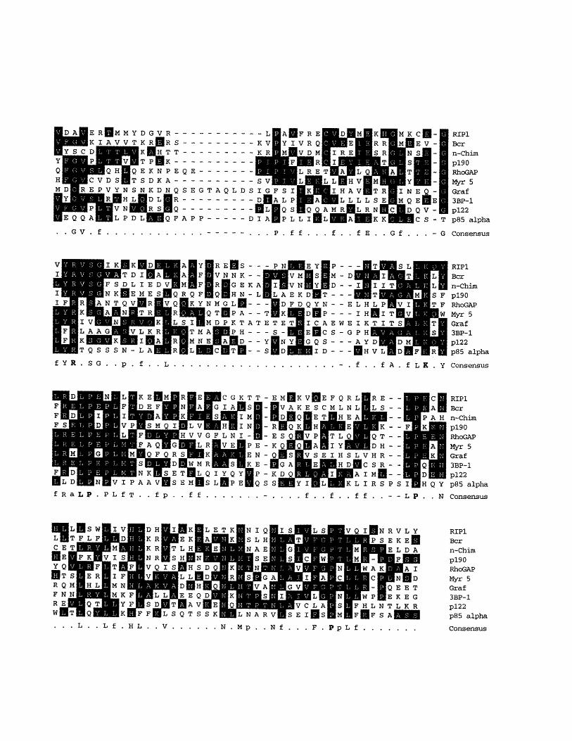
Figure 2-2. Amino acid sequence comparison of proteins containing
Rho/Rac GAP activity.
The sequence of RIP1 is compared to the sequence of n-Chimerin
(Hall et al., 1990), Bcr (Heisterkamp et al., 1985), p190 (Settleman et
al., 1992), Myr 5 (Reinhard et al., 1995), Graf (Hildebrand et al.,
1996), RhoGAP (Lancaster et al., 1994), 3BP-1 (Cicchetti et al., 1995),
p122-RhoGAP (Homma et al. 1995), and p85 alpha subunit of PI3-K
(Otsu et al., 1991). Identical residues are indicated with black boxes.
Amino acids present in 7/9 sequences are indicated in the consensus
sequence (amino acids present in all sequences are indicated in bold),
while conservative substitutions present in 8/9 sequences are
indicated in the consensus sequence as a (acidic residues: D, E), b
(basic residues: K, R, H), f (hydrophobic residues: A, F, L, M, P, V, W),
and p (polar residues: C, G, N, Q, S, T, Y).
47
DA ER MMYDGVR------ -----SR R S ---------S TT - - - - - - - - -P :K - - - - - - - - - -KNPEQE--------DKA- ----------KDNQSEGTAQLDS
RS QFAPP ---------Q F A PP -- -- ----D
.. GV . f .. .. . . . . - - - - - - P . ff . . . f . . fE . . G f . . . -G Consensus
RE S --- PN E Y P - -F V N NK V M S V M -D
F R GEKA I VN Y D - -FEQ HN-L LAEKD T ---YNMGL --- DFDQYN--E
QTNPA--T K D A E---MDPKTATETET ICAEWE
MA PH - - -Sc- E S - GNEC D -- Y NY GQS ---
T -S D ID-f YR . S G . P . L. ..... . . . . . . . . . . . . . - . f . . fA . fL K . Y Consensus
C GKTT -IA S -
IN -IN
LNI- -I
VE PE -EN -N
S K E-Y Q Y P-iPE Q S
fRaLP. PLfT. . fp .. f f ....... - L P . . N Consensus
MQ WV C L AIASIF H L N T L KMLNARVMSEIESEMEFEFSAM
RIP1Bcrn-Chimpl90RhoGAPMyr 5Graf3BP-1p122p85 alpha
. . V .. .... N .Mp . . N f . . . F . PpL f ......
S V L P gH VL MggIG F SI AI HA L L L
AO M M Q Q A M L R NINAgPL LI L A K K
cM-E V -
QV-CS-TQv- ICS- T
RIP1Bcrn-Chim
p190RhoGAPMyr 5Graf3BP-1p122
p85 alpha
RIP1Bcrn-Chimp190RhoGAPMyr SGraf3BP-1p122p85 alpha
RIP1Bcrn-Chimp19 0
RhoGAPMyr 5Graf3BP-1
p122p85 alpha
-
-
-
-
-
-
-
S
S Consensus. . . L . . L f . H L

Figure 2-3. Tissue distribution of RIP1 transcript.
A 342 base pair EcoRI-PstI fragment was used to probe multiple
tissue Northern blots. 2 micrograms of poly-(A) selected RNA from
the indicated tissue was run in each lane. The number indicates size
in kilobases.
49

6 0
eb;
2 $ !5&; e %•O4.1 --
4.1 -
CPSr

Figure 2-4. Preferential binding of 19-6 to KH-Ral preloaded with
GTP vs. GDP.
Filters were prepared as described in Material and Methods and cut
in half. The left half was hybridized to KH-Ral probe preloaded with
GDP, the right half was hybridized to KH-Ral probe preloaded with
GTPyS. 17 is a negative control.
51

19-6
GDP GTPGDP : GTP

Figure 2-5. Mapping of the Ral-binding region.
Two series of deletions were constructed and translated in vitro. The
translates were incubated with HA-tagged human RalB preloaded
GTPyS and immunoprecipitated with anti-HA antibody. The numbers
indicate the position of the deletion. The hashed bar indicates the T7
gene 10 fusion protein of pEXlox.
53

w
w
ND
co
4
Sco
+
+
I I
+ +
I C1
3
•
U
..J
0
.o
0 0D
I-
0
C0
u U
0CA
D
.o
* I
I +
+rl
l

Figure 2-6. Amino acid sequence comparison of RIP1 homologs.
The sequence of RIP1 is compared to RalBP-1 (Cantor et al., 1995), a
rat homolog, and RLIP (Jullien-Flores et al., 1995), a human homolog.
Conserved residues are indicated with black boxes. The GAP
homology domain and the Ral-interacting domain (RID) are indicated.
55

ripiraflhpnip
nipiralbplnlip
nipiralbplnlip
Fripirallhpl1nlip
ripi
rJlip
i-
nipiraflbp1nlip
GAP domainripiralbplnlip
F N10 [
C)6 I _ID _E1 WRGu R 1AVQ L
LKl i :
1 E. L E
PKAGKEP
~-I
A
R
nipiralbhrpl-nlip
nipiralbplnlip
ripiralbplnlip
ripi
flip
nipiraftplnlip,
ripirailpinlip
nipiralbplnlip
I RID domain
I _. a
m .................
I
m l II i i i I II I I II I I I I •.. .
|
a -
a~aer~
F
A V l," 1 El A 1 11 I'l I"? QE L I I L E D R M I ý7 E 1, E' T K 1ý M H 0 Am H 17 E R L A I I E! L R V 0 L R 1, L 0 M R A Ký

Analysis of RIP1 Protein
57
Chapter 3.

INTRODUCTION
In the previous chapter, I described the cloning of the RIP1
gene along with initial analysis of its expression in different tissues
and the guanine nucleotide specificity of the Ral-RIP1 interaction.
Analysis of the amino acid sequence predicted a region in RIP1 that
likely possesses GAP activity toward members of the Rho subfamily
of GTPases. An activity that was capable of activating GTPase
activity of Rho but not of Ras was attributed to a 29 kD protein
(Garrett et al., 1991), that was subsequently purified and sequenced.
Analysis of the sequence indicated that it contained a domain that
was 50% identical to one in Bcr and n-chimerin (Diekmann et al.,
1991), both of which were shown to be GTPase activating proteins
(GAPs) for Rac but not Rho. Since then, other proteins have been
identified that also share homology with this region. They include
the p190 (Settleman et al., 1992) that was identified as a protein
capable of binding to the pl20GAP; Abr (Heisterkamp et al., 1993), a
homolog of Bcr, that, like Bcr, also contains a Dbl-like domain; 3BP-1
(Cicchetti et al., 1995), an SH3 domain containing protein capable of
binding the Abl protein; Myr 5, a novel type of myosin (Reinhard et
al., 1995); p122-RhoGAP, a regulatory protein of phospholipase C-81
(Homma et al., 1995); Graf, a protein that associates with p125FAK
(Hildebrand et al., 1996); p85a, the regulatory subunit of PI3K (Otsu
et al., 1991). All but p85a has been shown to possess GAP activity
toward one or more of the Rho subfamily members. While Rho-GAP
and p190 exhibit activity toward Rho, Rac and Cdc42, Bcr and Abr
58

are only active toward Rac and Cdc42 as is N-chimerin, while 3BP-1
is active only toward Rac.
The key function for Ras-related GTPases is to transduce
upstream signals through their respective downstream effectors to
elicit a cellular response. A number of proteins have been identified
whose enzymatic activity has been shown to be modified as a result
of their interaction with their respective GTPase. In most cases, this
modification is due to covalent modification of the effector protein,
usually phosphorylation. For example, following translocation to the
membrane as a result of Ras activation, Raf-1 becomes
phosphorylated and it becomes catalytically activated as a kinase.
The same is true for p65PAK, a serine/threonine kinase that gets
activated through phosphorylation as a result of its interaction with
Rac or Cdc42 (Manser et al., 1994). More recently, protein kinase N
(PKN) has been shown to autophosphorylate and thereby activate
itself as a result of Rho interaction (Watanabe et al., 1996).
In this chapter, I will describe studies looking at possible
covalent modification of RIP1 as a result of Ral activation, analogous
to those found in other GTPase-effector interactions. I will also
describe Ral-RIP1 interaction in a transient transfection system. I
will then describe results from GAP assays using RIP1 to ascertain
whether the Rho GAP homology domain found within RIP1 is
catalytically active. Lastly, I will describe immunofluorescence
studies examining intracellular localization of RIP1 protein.
59

RESULTS
Expression of RIP1.
To examine the protein expression of RIP1, the C-terminal
portion (see Material and Methods) of RIP1 was expressed in an E.
coli expression system and then purified; the resulting protein was
used to generate anti-RIP1 antibodies in 2rabbits. The resulting
unfractionated polyclonal sera failed to detect specific protein species
in both immunoprecipitation and Western blot experiments.
However, affinity purification against full length RIP1 protein
yielded an antibody which, when used as a probe in Western blot
analysis, detected a 110 kilodalton species in lysates of Balb/3T3
cells. (Figure 3-1)
We compared this size to that seen following direct expression
of the cloned RIP1 gene. Introduction of the RIP1 coding segment
into the pGEX-4T plasmid resulted in expression of a protein
composed of RIP1 fused to glutathione S-transferase; this fusion
protein could subsequently be cleaved with thrombin, releasing full
length RIP1 protein. As an alternative, a segment containing the RIP
reading frame was introduced into the pVL1393 baculovirus vector
which allows expression in transfected insect cells.
While the amino acid sequence predicted a 75 kilodalton
protein, a protein of approximately 110 kilodaltons was observed
when bacterially produced protein was cleaved with thrombin. A
protein of similar size was observed in the insect cells transfected
with pVL1393. These sizes were virtually identical to those seen
upon Western blotting of Balb/3T3 lysates, indicating that the cloned
60

cDNA contains the entire reading frame of RIP1. The slight size
discrepancy between the bacterially produced protein and the
protein detected in Balb/3T3 lysates might be due to cleavage of
RIP1 by thrombin at a potential cleavage site near the C-terminus or
to the lack of proper post-translational modification. (Figure 3-1)
Subsequently, the full length protein produced in insect cells
was also used to generate antiserum. The polyclonal antiserum
generated from a rabbit injected with the full length protein was
able to detect endogenous RIP1 protein by Western blot and
ectopically RIP1 by immunoprecipitation.
Because the endogenous RIP1 in Balb/3T3 cells could not be
detected by immunoprecipitation, the metabolic stability of RIP1
could not be determined by pulse labeling. For that reason, cells
were treated with cycloheximide to inhibit protein synthesis and the
subsequent degradation of RIP1 was followed by Western blotting.
(Figure 3-2) The metabolic stability of proteins following
cycloheximide treatment has been shown to mimic the results
obtained from pulse labeling in a number of cases (Hasegawa et al.,
1995; Seely et al., 1982). RIP1 was found to be relative stable since
no decrease in protein levels were detected within 10 hours of
cycloheximide treatment.
Covalent Modification of RIP1.
When endogenous RIP1 was examined, multiple bands could be
detected by Western blot, indicating that RIP1 might undergo post-
translational modification or alternative forms of RIP1 might be
synthesized within the cell. To examine this further, RIP1 was
61

immunoprecipitated from COS-1 cells transiently transfected with an
expression vector designed to overexpress wild type mouse RIP1.
The protein was then treated with alkaline phosphatase. While the
immunoprecipitated RIP1 not treated with alkaline phosphatase
retains the modified forms, treatment with this enzyme resulted in
the multiple bands being reduced to a single band, indicating that
the multiple bands were due to post-translational phosphorylation of
RIP1. (Figure 3-3)
To examine further the nature of the phosphorylation, the RIP1
immunoprecipitated from COS-1 cells was resolved on a SDS-PAGE
gel, transferred to nitrocellulose, and probed with an anti-
phosphotyrosine antibody. Although an EGF-treated control lysate
demonstrated that the antibody could indeed recognize
phosphotyrosine residues present in EGF-receptor as a result of EGF
treatment, this antibody did not recognize RIP1. This is a strong
indication that the phosphorylation of RIP1 in likely due to a
serine/threonine kinase, although definitive proof can only be
obtained through phosphoamino acid analysis.
Previous work on RalGDS, an exchange factor specific for the
RalA and RalB GTPases, demonstrated that the protein levels of the
exchange factor changed dramatically in response to serum
starvation/stimulation. To examine whether the modification of
RIP1 could also respond to serum starvation/stimulation, Balb/3T3
cells were serum-starved for 60 hours and then stimulated with 10%
calf serum. (Figure 3-4) Under conditions of serum starvation, the
modifications of RIP1 gradually disappeared, resulting in a single
62

RIP1 band in Western blots. Following serum stimulation, although
the first indication of protein phosphorylation became evident within
1 hour, the full range of phosphorylation was not achieved until 24
hours of serum stimulation.
63

Ral-RIP1 interaction.
One of the difficulties in studying signal transduction through
Ral is the inability to gauge whether Ral is activated hence identify
signals that can activate Ral. In the case of Ras, the activation of Ras
can be examined directly by looking at the guanine nucleotide bound
to Ras following immunoprecipitation thereby allowing the factors
capable of activating Ras. The unavailability of anti-Ral antibodies
capable of immunoprecipitating Ral made it difficult to ascertain
conditions in which Ral is activated. In light of the fact that the
interaction of RIP1 and Ral is dependent on the activation of Ral
(Fig 2-4) , this provides an indirect method of gauging Ral
activation. The COS-1 transient transfections applied previously
were used to this end.
Cos-1 cells were co-transfected with the vector that expressed
the full length wild type RIP1 along with a vector that expressed the
wild type RalB protein with the Myc epitope fused to its N-terminus.
Following transfection, the cells were starved in 0.2% calf serum for
24 hours and then stimulated with serum for 5, 15, and 30 minutes.
The cells were lysed, the Ral protein immunoprecipitated using the
9E10 anti-Myc monoclonal antibody, and the immunoprecipitates
were resolved on an SDS-PAGE gel. This gel was then transferred to
a nitrocellulose filter and probed with anti-RIP1 antiserum. Under
conditions in which Ral is activated and in its GTP-bound state, I
expected to see RIP1 co-immunoprecipitate with Ral. This was
indeed the case, as shown in Figure 3-5a. As early as five minutes
following serum stimulation, RIP1 could be seen co-
immunoprecipitating with Ral while none could be detected in
64
serum-starved cells. Interestingly, the level of co-
immunoprecipitating RIP1 seems to decline with time suggesting that
Ral activation is transient and subject to downregulation and/or RIP1
is modified such that it is no longer able to interact with Ral.
To test whether the inability of RIP1 in serum-starved cells to
bind to Ral was due to a modification of RIP1 in those cells rather
than a change in Ral, the Ral-binding ability of RIP1 present in
serum-starved lysate to that of RIP1 from serum-stimulated lysate
were compared. As shown in Figure 3-5b, GTPyS-bound Ral was able
to interact with RIP1 from either lysate equally well, indicating that
the interaction between Ral and RIP1 observed in serum-stimulated
cells was most likely due to a change in Ral rather than RIP1,
specifically the activation of Ral through guanine nucleotide
exchange.
GAP assays.
To ascertain whether the Rho/Rac GAP homology domain found
in RIP1 was indeed capable of activating the GTPase activity of the
Rho, Rac, or related proteins, full length, recombinant RIP1 protein
was used in GAP assays with either Racl, CDC42Hs, RhoA, c-H-Ras, or
RalB. The assay consists of loading the GTPase with [y32P]GTP and
incubation in the presence of the protein being studied. The reaction
is then stopped by the addition of stop buffer, filtered through a
nitrocellulose filter to retain the GTPase and any associated
nucleotide, and then washed to remove non-specific radioactivity.
An activation of the GTPase activity will result in the loss of the y-
phosphate hence a decrease in the amount of radioactivity retained.
65

Initially 20 ng of each GTPase were incubated in the presence
of 0.5 gxg of purified RIP1 protein (a molar ratio of approximately 1
to 10) for 10 minutes at 25 0 C. RIP1 succeeded in stimulating the
GTPase activity of Racl and CDC42. Under these conditions, 22%
more of the GTP was hydrolyzed to GDP by Racl in the presence of
RIP1, while as much as 35% was hydrolyzed by CDC42 in the
presence of RIP1. However, no increase in GTPase activity could be
detected when Ras or Ral were used in this assay, and very little, if
any, could be detected when RhoA was used. (Figure 3-6) While Ral
interacts with RIP1 in a GTP-dependent manner, as shown above,
this interaction had no effect on the intrinsic GTPase activity of Ral.
This indicates that although RIP1 can interact with Ral, the
interaction is such that it does not affect on the GTPase activity of
Ral, rather RIP1 is able to activate the GTPase activity of Cdc42 and
Racl. This correlates nicely with the mapping of the Ral-binding
domain of RIP1 since the GAP homology region and Ral-binding
region does not overlap.
To test whether the effects observed were indeed an activation
of specific GTPase activity and not due to the simple dissociation of
the nucleotide from the GTPase or due to protease activity, these
assays were repeated using [a- 32P]GTP. Since the a-phosphate is not
lost as a result of GTPase activation, a loss of radioactivity would
indicate that the nucleotide was not retained on the filter due to
either dissociation of the nucleotide from the protein or its
degradation. As illustrated in Figure 3-7, no radioactivity was lost
when the GTPases were loaded with [a- 32P]GTP. To exaggerate any
effect RIP1 might have on the GTPases, the amount of purified RIP1
66

used in these assays were increased to 5 tg while the GTPase was
decreased to 10 ng per reaction giving a Ral to RIP1 molar ratio of
approximately 1 to 200.
I also tested whether the binding of GTP-activated Ral to RIP1
affected the GAP activity of RIP1 toward Racl, Cdc42Hs or RhoA. To
do this, RalB preloaded with either GTPyS or GDP was added to these
GAP assays. Neither of these forms of Ral had an observable effect
on the GAP activity of RIP1 toward these various GTPases (data not
shown). Therefore, although Ral has the ability to bind RIP1 in a
GTP-dependent manner, that interaction itself does not have a
regulatory role for the GAP domain.
Immunofluorescence.
To study the intracellular localization and possible translocation
of RIP1 as a result of Ral activation immunofluorescence microscopy
was performed using anti-RIP antibody. Initially, polyclonal rabbit
antiserum generated against the full length RIP1 protein was used.
Several different fixation methods were tested, including the organic
solvents methanol or acetone and formaldehyde or glutaraldehyde
fixation followed by permeablization by detergent or organic solvent.
The most striking pattern was a punctate pattern seen most
convincingly following fixation by organic solvents although still
visible when cells were fixed with formaldehyde. (Figure 3-8a)
Glutaraldehyde fixation was problematic since the fixation process
itself generated a fluorescence that was difficult to eliminate
consistently.
67

Interestingly, following formaldehyde fixation, another pattern
is also discernible. Aside from the punctate pattern observed when
organic solvents were used to fix cells, a pattern reminiscent of
vinculin or focal adhesion plaques was observed. I recognized that
these patterns of staining might be due to non-specific cross-
reactivity of the polyclonal antiserum used in these experiments.
Therefore, in an attempt to determine which, if any, of the observed
patterns were indeed due to RIPl-specific staining, a blocking
strategy was attempted. Thus, the polyclonal serum was
preincubated with purified RIP1 protein prior to staining in an
attempt to block the antiserum from recognizing RIP1-specific
epitopes while allowing it to still recognize nonspecific proteins. As a
result, the pattern reminiscent of vinculin staining disappeared while
the punctate pattern remained. (Figure 3-8b) This suggested that
the punctate pattern observed was likely due to non-specific
staining.
To ascertain whether the focal adhesion plaque suggestive
staining pattern was indeed RIPl-specific, purified RIP1 protein was
coupled to Affigel-15 (Bio-Rad) and used to affinity purify RIP1-
specific antibodies. The results of the immunofluorescence using the
affinity purified antibody corroborated the blocking results in that,
although the punctate pattern was no longer observed, the pattern
reminiscent of vinculin staining remained. (Figure 3-8c)
To test whether the pattern observed actually coincided with
vinculin staining, a mouse monoclonal antibody against the human
vinculin protein was used to do a double staining with the RIP1
specific antibody. As shown in Figure 3-9, the major staining
68

observed using the affinity purified did indeed co-localize with
vinculin staining, indicating that RIP1 was present in focal adhesion
plaques.
To test whether RIP1 undergoes a translocation as a result of
Ral activation, the staining patterns of serum-starved and
restimulated cells were compared. The bulk of the staining pattern
remained constant, indicating that RIP1 did not undergo a drastic
translocalization as a result of Ral activation.
DISCUSSION
When antibodies generated against the full length RIP1 was
used for Western analysis, RIP was found to be a protein running at
about 110 kD on an SDS-PAGE gel. Using lower percent acrylamide
gels (6% to 7.5%), the RIP1 band could be resolved as multiple bands,
indicating that either the RIP1 protein might undergo covalent
modification resulting in proteins with altered mobility or that the
antibody generated against the full length RIP1 protein might be
recognizing possible RIPl-related proteins. Upon further analysis,
the multiple bands were found to be attributable to phosphorylation,
as RIP1 immunoprecipitated and treated with calf alkaline
phosphatase resulted in the multiple bands collapsing into a single
lower molecular weight band. Interestingly, the single lower
molecular weight band resulting from phosphatase treatment of RIP1
is slightly faster in migration than the RIP1 immunoprecipitated
from cells, indicating that all RIP1 probably undergoes a basal
69

phosphorylation and the multiple bands observed in Western blots
were the result of additional phosphorylation events. This would
nicely explain the discrepancy observed between the bacterially
produced protein and endogenous RIP1 found in mammalian cells, in
that the basal phosphorylation seen in the latter might be dependent
on kinases not present in E. coli.
In Balb/3T3 cells, I could resolve three distinct forms of RIP1
upon acrylamide gel electrophoresis of lysate prepared from
exponentially growing cells. Following serum starvation in 0.2% calf
serum, the two upper bands disappeared while the lowest band
remained. Within 1 hour of serum stimulation, an extra RIP1 band
appeared, indicating a phosphorylation of RIP1 in response to serum
stimulation. A second phosphorylated band became apparent after
24 hours. In the transient transfections where Cos-1 cells received
expression vectors for RIP1 and myc-tagged Ral, a Ral-RIP co-
immunoprecipitating complex was detectable within five minutes of
serum stimulation following 24 hours of serum starvation, indicating
that a component within calf serum was capable of activating Ral,
allowing it to form a complex with RIP.
An attractive mechanistic model of RIP1 action would be one in
which, in analogy to the behavior of Ras, Ral serves to recruit RIP1 to
the membrane where RIP1 gets activated. As a result of its
interaction with Ral, RIP1 gets phosphorylated, possibly activating
the catalytic activity of RIP1. While in Figure 3-3 very little, if any,
phosphorylated RIP1 is detectable by Western after five minutes of
serum stimulation, it may be enough to function properly. Obviously,
70

a more detailed study of RIP1 phosphorylation within one hour after
serum stimulation is required.
Of course, it is also possible that phosphorylation of RIP1 has
nothing to do with its interaction with Ral and is the result of an
independent regulatory mechanism, and Ral serves to recruit RIP1 to
a compartment in which it can function, or alternatively, to sequester
RIP1, removing it from a compartment in which it serves as a GAP
toward proteins like CDC42 or Rac.
When recombinant RIP1 protein was purified and used in GAP
assays, it was shown that this protein could activate the GTPase
activity of CDC42 and, to a lesser extent, Racl but not that of RhoA,
Ral, or Ras. While RIP1 interacts specifically with the GTP-bound
form of Ral, this binding had no effect on the GTPase activity of Ral.
Moreover, when the Ral-binding domain of RIP1 was mapped, it was
found to be in a region C-terminal to, and not overlapping with, the
GTPase activating domain of RIP1. This suggests that RIP1 does not
serve as a regulator of Ral. Rather, this indicates that RIP1 is the
target of activated Ral and thus serves as an effector of Ral.
The degree of GAP activity of RIP1 toward CDC42 and Racl is
much lower than has been associated with other previously
identified Rho/Rac GAP-containing proteins. This might be explained
by the fact that the RIP1 protein may be labile, resulting in its
substantial inactivation in vitro. Although several independent
bacterial and insect cell preparations exhibited the same low specific
GAP activity, it is possible nevertheless that one of the steps used in
all of the purifications caused the inactivation of much of the protein.
71

Alternatively, it is possible that the RIP1 protein requires functional
activation, either through covalent modification or the presence of an
associated, distinct regulatory protein. Finally, it is possible that the
bona fide target of RIP1 GAP activity has not yet been identified.
I considered the possibility that Ral might function as an
allosteric regulator of RIP1, such that the binding of activated Ral to
RIP1 would modulate the GAP activity of RIP1 toward its target
GTPases. To test this possibility, GAP assays were done in the
presence of excess GTPyS or GDP-loaded Ral. No modulation of RIP1
GAP activity toward RhoA, Racl or CDC42 was observed. This failure
to observe a modulation of RIP1 GAP activity in vitro is not
definitive.
Immunofluorescence studies indicate that a substantial fraction
of RIP1 is present in focal adhesion complexes. This was
demonstrated by using antibodies affinity-purified against RIP1
which showed that the observed staining pattern overlapped with
that of the anti-vinculin antibody. Vinculin is a component of, and is
found almost exclusively in, focal adhesion complexes. This is
provocative in that one of the effects of Rac or Cdc42 activation is the
formation of such complexes. Focal adhesions are sites through
which fibroblasts adhere to the extracellular matrix and are the sites
of integrin-mediated signaling. Thus, RIP1 might function in the
regulation of signals through these complexes by regulating Rac or
Cdc42.
Interestingly, other regulatory proteins for Rho-related
GTPases have been found in focal adhesion plaques. Graf is a
72

recently identified member of the Rho GAP family that has GTPase
activating activity toward Rho and Cdc42. Graf been shown to bind
the C-terminal domain of FAK, a protein tyrosine kinase associated
with focal adhesions (Hildebrand et al., 1996). Lar is a
transmembrane phosphotyrosine phosphatase that co-localizes at the
ends of focal adhesion (Serra-Pages et al., 1995). Trio, a recently
identified protein that associates with LAR, has been shown to
contain two Dbl-like domains, one of which has been shown to have
guanine nucleotide exchange activity specific for Rac, the other for
Rho (Debant et al., 1996). This indicates that Rho-related GTPases
play a major role in integrin-mediated signaling and that RIP1
probably functions in the regulation such signaling.
73

MATERIALS AND METHODS
Expression of recombinant RIP1.
The coding region of RIP1 was cloned into pGEX-4T
(Pharmacia). Transformed bacteria were induced with 0.1 mM IPTG
for 4 hours, centrifuged and washed. The pellet was resuspended in
STE (10 mM Tris, pH 8.0, 150 mM NaC1, 1 mM EDTA) containing 100
tg/ml of lysozyme and incubated on ice. DTT was added to final
concentration of 5 mM. The bacteria were sonicated and centrifuged.
Triton X-100 was added to the supernatant to a final concentration of
2%. Glutathione Sepharose 4B (Pharmacia) beads were added and
incubated at 40 C for 30 minutes. The beads were washed in PBS.
RIP1 was cleaved off by adding thrombin in PBS, incubating
overnight.
Alternatively, the coding region of RIP1 was also cloned into
pVL1393 (Pharmingen). A high titer baculoviral stock was prepared
by using the BaculoGold System (Pharmingen) in Sf9 insect cells and
used to infect High Five insect cells (Invitrogen). The cells were
collected, washed with PBS and resuspended in buffer A (10 mM
Tris, pH 7.5, 1.5 mM MgC12, 10 mM KC1, 5 mM P-mercaptoethanol, 1
mM EDTA) + protease inhibitors. The cells were lysed in a Dounce
homogenizer and centrifuged to pellet nuclei. 0.11 volumes of
buffer B (100 mM Tris, pH 7.5, 1,500 mM NaC1, 15 mM MgCl2, 100
mM KC1, 50 mM P-mercaptoethanol, 10 mM EDTA, 1% Triton X-100)
was added to the supernatant. Following a second centrifugation to
pellet cellular debris, Ni-NTA resin (Qiagen) was added to the
supernatant and incubated for 1 hour. The beads were then washed
74

with 0.1x buffer B + 1 mM imidazole, loaded onto a column and
washed with buffer B-(0.1 x buffer B minus Triton X-100) plus 16
mM imidazole and 10% glycerol. The protein was eluted in a 10 ml
gradient of 16 - 150 mM imidazole in buffer B-. Positive fractions
were dialyzed against buffer B- to remove the imidazole.
Antibody Preparation.
The EcoRI-HindIII insert from pEXloxl9-6 was cloned into pET-
29a (Novagen). Protein was expressed, purified (S-Tag Purification
System, Novagen) and used to immunize rabbits. The resulting
antiserum was affinity purified against full length GST-RIP1 protein
by binding the protein to Glutathione Sepharose 4B (Pharmacia) and
washing with 0.1 M borate buffer (pH 8.0), 0.2 M triethanolamine
(pH 8.2). The protein was cross-linked to the column by incubation
in 40 mM dimethylpimelimidate (DMP) in 0.2 M triethanolamine.
The column was then washed in 40 mM ethanolamine (pH 8.2), 0.1 M
borate (pH 8.0). The antiserum was passed over the column.
Following washes in PBS and PBS + 2 M KCl the antibody was eluted
in 5 M Nal and 1 mM sodium thiosulfate in PBS.
For affinity purification of antibody using Affi-gel 15 beads
(Bio-Rad), 10 micrograms of RIP1 protein purified from insect cells
infected with the RIP1 expressing virus described above was cross-
linked to 2 ml of Affi-Gel 15 beads according to the protocol supplied
by the manufacturer. Five milliliters of anti-RIP1 antisera was
passed over the beads. Following washes in PBS the antibody was
eluted in 0.2 M glycine, pH 2.5 and neutralized with 1/10 volumes of
75

1 M Tris, pH 8.0. The affinity purified antibody was then dialyzed
against PBS and concentrated.
Western analysis.
Lysates from Balb/3T3 cells and purified RIP1 protein were
run on a SDS/7.5% acrylamide gel and transferred to nitrocellulose.
The filter was blocked in PBS plus 10% dried non-fat milk for 1 hour,
incubated with affinity purified anti-RIP1 antibody in PBST (PBS
plus 0.1% Tween-20) for 1 hour, washed with PBST and incubated
with horseradish peroxidase conjugated donkey anti-rabbit IgG
antibody (Jackson ImmunoResearch) in PBST. The filter was washed
in PBST and visualized used ECL (Amersham).
For cycloheximide treatment, cells were grown in the presence
of 45 gg/ml of cycloheximide.
GTPases.
H-ras and Ral were prepared as previously described (Albright
et al., 1993). HA-Ral was expressed by cloning the coding region of
human RalB downstream of the HA epitope and upstream of 6
histidine codons into pET-15 and purified as described above for KH-
Ral. Racl, CDC42, and RhoA were kindly provided by Dr. Marc
Symons (Malcolm et al., 1994).
GAP Assay.
1 gg GTPase was combined with 10 gCi [y- 32P]GTP (NEN DuPont)
in 100 p•l exchange buffer (50 mM Tris, pH 7.5, 50 mM NaC1, 5 mM
EDTA, 1 mg/ml BSA, 1 mM DTT), 10 min, 25'C. The exchange
76

reaction was stopped by adding 100 p.l stop exchange buffer (50 mM
Tris, pH 7.5, 50 mM NaC1, 20 mM MgC12, 5 mM EDTA, 1 mg/ml BSA, 1
mM DTT), diluted into reaction buffer (50 mM Tris, pH 7.5, 10 mM
MgC12, 1 mg/ml BSA, 1 mM DTT), and aliquoted into 100 p.l reactions
(10 ng of GTPase per reaction). 0.5 micrograms of RIP1 was added
per reaction and incubated 10 min, 25°C. The reaction was diluted in
500 pl of wash buffer (50 mM Tris, pH 7.5, 10 mM MgC12, 1 mM DTT),
filtered through nitrocellulose and washed with additional 10 ml of
wash buffer.
For testing the RIP1 protein for guanine nucleotide dissociation
activity or protease activity, the assays were done identically as
describe above except that [O- 32P]GTP (NEN DuPont) was used instead
of [y- 32P]GTP and 10 ng of labeled GTPase was incubated in the
presence of 5 micrograms of RIP1 for 15 minutes at 25°C instead.
For GAP assays in the presence of Ral, 5 micrograms of GTPyS
or GDP preloaded Ral was incubated with 0.5 micrograms of RIP1 on
ice for 30 min prior to addition to the GAP assay.
Construction of expression vectors for transfection.
pCIN-RIP1 and pCIN-mRal are based on pCI-Neo (Promega).
ACGCGTTCTAGAATGGCTGAGTGCTTCCTGCC was ligated onto the 5' Ava
I site of RIP1 and GACGCCCATCTCCGGATGAGCGGCCGC ligated onto the
3' BsaHI site. This was then cut and ligated into the XbaI-NotI sites
of pCI-Neo. pCIN-mRal was constructed by PCR cloning the Myc-
epitope (GGCGAACAAAAGCTGATTTCTGAAGAAGACTTG) directly 5' of
the starting ATG of human RalB and cloning into the XbaI-NotI sites
of pCI-Neo. The sequence was confirmed by sequencing.
77

Transient transfections.
Cos-1 cells were split 1:4 the day before transfection to
approximately 2x10⁶ cells per 10 cm plate and grown in 10% calf
serum in DME. The plates were washed with PBS (137 mM NaC1, 2.7
mM KC1, 4.3 mM Na2HPO4-7H 20, 1.4 mM KH2PO 4 , pH 7.2) and the
growth media was changed to 4 mls 10% Nu serum in DME 1 hr prior
to transfection. 8 gg of DNA per plate was brought up to 40 pl in TE
and mixed with 80 pl of 10 mg/ml DEAE dextran in water. This
mixture is added to the plate, followed by 16 p1 of 5 mM chloroquine
diphosphate to a final concentration of 20 gM. The cells were
incubated at 370 C for 4 hours and then shocked with 5 mls 10%
glycerol in PBS for 60 seconds, washed twice with PBS and fed with
10 ml 10% calf serum in DME.
Immunoprecipitations.
For phosphatase treatment, cells transfected with pCIN-RIP1
were harvested 36 to 48 hours after transfection and lysed in 1 ml
PLB (PBS + 0.1% NP-40, 10 mM sodium fluoride, 1 mM sodium
vanadate,10 mM 03-glycerophosphate) plus protease inhibitors.
Cellular debris was removed by centrifugation. One microliter of
anti-RIP1 antiserum was added per ml of lysate and incubated on ice
for 1 hour. Ten microliters of Protein A sepharose (Bio-Rad) was
added and incubated for 1 hour at 4VC on a rotator. The beads was
then washed two times with PBS, once with a 1:1 mixture of PBS and
alkaline phosphatase reaction buffer, and two times in alkaline
phosphatase reaction buffer (50 mM Tris-C1, pH 7.5, 1 mM MgCl2).
78
The beads were then resuspended in 50 microliters of alkaline
phosphatase reaction buffer and incubated at 300 C for 10 minutes.
20 units of calf intestinal phosphatase (New England Biolabs) was
added per sample and each sample was incubated an additional 30
minutes. The reaction was stopped by addition of ten microliters of
6x protein sample buffer (0.35 M Tris-C1, pH 6.8, 30% glycerol, 1.28%
SDS, 6mM DTT, 0.012% bromophenol blue), the sample was then
boiled for 10 minutes and resolved on a 7.5% SDS-PAGE gel. RIP1
was visualized by Western blot analysis as described above.
For co-immunoprecipitation of RIP1 and Ral, Cos-1 cells were
transfected with three micrograms of pCIN-RIP1 and five
micrograms of pCIN-mRal per 10 cm plate. 36 to 48 hours after
transfection, the cells were serum starved for 24 hours and lysed in
1 ml of PLB+ (PLB + 15 mM MgCl2) per plate. Cellular debris was
removed by centrifugation and one microliter of anti-Myc ascites
fluid was added per ml of lysate. Following one hour of incubation on
ice ten microliters of Protein G agarose (Sigma) was added to each
sample and incubated for 1 hour at 40 C on a rotator. The beads was
then washed four times with PLB+ and boiled in 50 gl lx protein
sample buffer. Protein was resolved on a 10% SDS-PAGE gel and
RIP1 was visualized by Western analysis as described above.
For KH-Ral co-precipitation, the lysates described above were
incubated with one microgram KH-Ral preloaded with GTPyS as
described previously, and then incubated for 1 hour on ice. Ten
microliters of Ni-NTA resin (Qiagen) were added and the samples
were rotated at 4°C for one hour. The samples were then processed
identically as described for co-immunoprecipitation of RIP1 and Ral.
79

Immunofluorescence.
Balb/3T3 cells were grown on 12 mm coverslips (Fisher) in 24
well tissue culture plates. For organic solvent fixation, the coverslips
were submerged in either methanol or acetone for two minutes at
room temperature. For formaldehyde, coverslips were submerged in
a 1:5 dilution of 16% EM-grade formaldehyde (Ted Pella) in PBS for
15 minutes at room temperature, rinsed once in PBS and
permeabilized by submerging in acetone for 2 minutes at room
temperature. The coverslips were rinsed twice in PBS and placed
cell-side up on parafilm on top of wet Whatman paper in a culture
plate. The coverslips were blocked with 50 microliters of 3% BSA in
PBS for 30 minutes. The block solution was removed by aspiration
and replaced with 50 microliters of primary antibody diluted 1:100
in 3% BSA in PBS for 1 hour. The coverslips were washed three
times fifteen minutes and then replaced in the incubation chamber
with 50 microliters of secondary antibody diluted 1:100 to 1:250 in
3% BSA in PBS for 1 hour. The primary antibodies used were a
mouse anti-human vinculin monoclonal antibody (Sigma) and affinity
purified and neat rabbit anti-mouse RIP1 antiserum described
above. The secondary antibodies were rhodamine- or fluorescein-
conjugated donkey anti-mouse or rabbit IgG antibodies from Jackson
ImmunoResearch.
80

ACKNOWLEDGEMENT
I would like to gratefully acknowledge Dr. Marc Symons for the
GTPases.

Figure 3-1. Western analysis of
Purified RIP1 cleaved from a GS
prepared from Balb/3T3 cells.
purified anti-RIP1 rabbit serum.
weight in kilodaltons.
RIP1.
T fusion was run parallel to lysate
RIP1 was detected using affinity
The numbers indicate molecular
82

-112
53.2
34.9

Figure 3-2. Effect of cycloheximide treatment on RIP1.
Balb/3T3 cells were treated with 45 tg of cycloheximide per ml of
media for the indicated period of time. Cells were lysed and proteins
separated on a 7.5% polyacrylamide gel, transferred to nitrocellulose.
RIP1 was visualized with anti-RIP1 antibodies and the multiple
bands are indicated by the arrows. The length of cycloheximide
treatment is indicated in hours.
84

Cycloheximidetreatment(hrs)
1 1 3 3 6 6 12 12 24 24
I 1RIP1
I Iim a

Figure 3-3. Effect of phosphatase on RIPi.
RIP1 from Cos-1 cells transiently transfected with RIP1 was
immunoprecipitated, incubated with or without calf intestinal
phosphatase, resolved on a 7.5% polyacrylamide gel, transferred to
nitrocellulose and detected with anti-RIP1 antibody.
86

AlkalinePhosphatase
- RIP1

Figure 3-4. Effect of serum on RIP1 modification.
Balb/3T3 cells were grown in 10% calf serum, transferred to 0.2%
serum for 60 hours, and stimulated with 10% serum. RIP1 was
visualized by Western blot after separating whole cell lysate on a
7.5% polyacrylamide gel. Length of serum starvation or stimulation
is indicated in hours, except for the five minute time point (5').
88

Serum starved 0 8 13 24 45 60
Serum stimulated 0(hr)
5' 1 4 12 18.5 24
RIP1
% f
-I

Figure 3-5. Ral-RIP1 complexes in Cos-1 transient transfections.
A. Expression vector for RIP1 and Myc-tagged Ral were transiently
transfected into Cos-1 cells. The cells were serum starved in 0.2%
calf serum for 24 hours and stimulated with 10% calf serum for the
indicated time. Ral was immunoprecipitated using anti-Myc
antibody and co-immunoprecipitating RIP1 was visualized by
Western blot of a 10% polyacrylamide gel.
B. The lysate described above was incubated for 1 hour in the
presence of GTP-loaded KHral. Ral-RIP1 complexes were precipitated
with Ni-NTA resin. RIP1 was visualized by Western blot of a 7.5%
polyacrylamide gel.
90

serumstimulation : 0 5(min)
15 30
-- RIP1
IP: anti-myc
Western blot: anti-RIP1
Transfection: pCIN-RIP1 + pCIN-mycRal
10% polyacrylamide gel
B KH-RalGDPGTPyS
serum stimulation
+ +
++
ppt: Ni-agarose
Western Blot: anti-RIP1
7.5% polyacrylamide gel
w-- RIP1
D mWSW

Figure 3-6. The effect of RIP1 on the GTPase activity of several
GTPases.
20 ng of each GTPase was preloaded with [y- 32P]GTP and incubated
with or without 500 ng RIP1 for 10 minutes at 25°C. The products
were separated by filtration, the filter-bound radioactivity was
determined and is indicated as percent radioactivity retained. The
sample without RIP1 addition is depicted as 100 percent.
92

M - RIP1
El + RIP1
I
75-
Z
A=0
IC 50 -0-- 50-
25 -
01Cdc42 RhoA
I T~I T
I
H-Ras
-
I .
RalB Racl

Figure 3-7. The effect of RIP1 on guanine nucleotides bound to
GTPases.
10 ng of each GTPase was preloaded with [iX- 32P]GTP or [y- 32 P]GTP
and incubated with or without 5 gg RIP1 for 10 minutes at 25oC. The
products were separated by filtration, the filter-bound radioactivity
was determined and is indicated as percent radioactivity retained.
The sample without RIP1 addition is depicted as 100 percent.
94

CDC42
S[a-32p] GTP- RIP1
[0Ia-32P] GTP+ RIP1
S[y-32 P] GTP- RIP1
E] [y.32 P] GTP+ RIP1
Racl RhoA

Figure 3-8. Immunofluorescence of RIP1.
Balb/3T3 cells were seeded on glass cover slips, fixed with
formaldehyde, permeabilized with acetone, and incubated with A.
anti-RIP1 antiserum neat, B. anti-RIP1 antiserum preincubated with
purified RIP1 protein, C. affinity purified anti-RIP1 antibody. The
pattern reminiscent of vinculin localization is indicated by the
arrows. Rhodamine-conjugated secondary antibody was used in A
and B, while fluorescein-conjugated secondary was used in C.
96


Figure 3-9. Co-localization of RIP1 and vinculin.
Balb/3T3 cells were seeded on glass cover slips, fixed with
formaldehyde, permeabilized with acetone, and incubated with
affinity purified anti-RIP1 antibody and a mouse monoclonal
antibody against human vinculin, and visualized with A. fluorescein-
conjugated anti-rabbit antibody or B. rhodamine-conjugated anti-
mouse antibody, or C. doubly exposed.
98


Conclusions and Prospects
100
Chapter 4.

In the previous two chapters, I described the cloning of the
RIP1 gene and the characterization of this putative effector of Ral.
The RIP1 gene was cloned by virtue of the ability of its encoded
protein to bind GTP-bound Ral, and was shown to be incapable of
binding GDP-bound Ral. One domain in RIP1 shares homology with
that of a family of known proteins in the domain capable of
activating the GTPase activity of members of the Rho-related
GTPases. RIP1 was shown to have limited but nonetheless significant
ability to activate the GTPase activity of Cdc42 and, to a lesser
extent, that of Racl, but not that of RhoA.
RIP1 is phosphorylated, presumably on serine/threonine
residues, in response to serum stimulation of serum-starved Cos-1
cells transiently transfected with a RIP expression vector. In Cos-1
cells transiently transfected with RIP and Myc-tagged Ral expression
vectors, starved and restimulated with serum, RIP1 can co-
immunoprecipitate with Ral within five minutes after stimulation.
This indicates that as a response to activation by an, as yet,
unidentified factor present in serum Ral is activated and binds to
RIP1. By immunofluorescence, RIP1 has been shown to co-localize
with vinculin in focal adhesion plaques.
Effector proteins.
For a protein to be a bona fide effector of a GTPase, two
criteria must be met. The first of these is the ability to bind
specifically to the GTP-bound form, but not the GDP-bound form, of
the GTPase. I have shown in Chapter 2 that this is indeed the case.
A corollary of this would be that the effector protein be able to bind
101

activating mutants of the GTPase but not effector mutants. This was
shown nicely by two other groups using yeast two hybrid system
(Cantor et al., 1995; Jullien-Flores et al., 1995). Both groups show
that the rat and human homolog of RIP1 can interact with the
activated V23 Ral but not with the N49 or A46 effector domain
mutants of Ral.
The second criterion of an effector is a change in its activity as
a result of the interaction. There are several models by which one
could envision such an activation to take place (Marshall, 1996). The
first is one in which the effector is directly altered through its
interaction with the GTPase. For example, the binding of the GTPase
to the effector may induce a conformational change in the effector
that increases its activity. In this model, one wouldn't expect that
membrane localization of the GTPase is required, but it is formally
possible that the plasma membrane contributes by constraining the
interaction between the effector and GTPase to one favorable for
activation of the effector. A second model is one in which
recruitment to a membrane is required for activation, either to allow
activation by a component present only at the plasma membrane or
to allow association with a substrate present only at the plasma
membrane.
An example of the first model can be found in PAK activation
by Cdc42 (Manser et al., 1994). Another example is B-Raf activation
by Ras (Yamamori et al., 1995). The addition of activated Ras to
complexes containing B-Raf and 14-3-3 can lead to the activation of
B-Raf. Although this activation can occur in the absence of plasma
membrane, it requires the isoprenylation of Ras, indicating that
102

although Ras might be able to activate B-Raf through a mechanism
involving conformational change, an additional role is played by the
isoprenylation, possibly through the stabilization of the activating
configuration. It is also possible that the requirement for 14-3-3 for
the activation can only be fulfilled at the membrane. Although 14-3-
3 is distributed throughout the cell, the isoform required for
activation might be present only at the membrane, or possible
modification on 14-3-3 required to allow its participation in the
activation might occur only at the membrane.
Raf-1 activation is an example of the second model in which
interaction with the activated GTPase induces translocation to the
membrane where subsequent activation of the Raf-1 effector can
occur. Many groups have reported the direct interaction between
Raf-1 and Ras as a result of Ras activation (Moodie et al., 1993;
Morrison et al., 1993; Warne et al., 1993; Zhang et al., 1993). Indeed,
the requirement for Ras in Raf-1 activation can be bypassed by
directly targeting Raf-1 to the membrane by the addition of the
membrane-targeting CAAX-box of K-Ras to the C-terminus of Raf-1,
allowing Raf-1 to then be activated in the absence of activated Ras.
(Leevers et al., 1994; Stokoe et al., 1994)
The interaction between RalGDS and activated Ras (Hofer et al.,
1994; Kikuchi et al., 1994; Spaargaren and Bischoff, 1994), might be
an example of membrane localization that allows association to a
substrate present only at the membrane, since translocation of
RalGDS to the membrane as a result of Ras activation would increase
the probability of RalGDS to interact with its substrate Ral. Whether
103

the guanine exchange activity of RalGDS undergoes activation as a
result of Ras interaction is not known.
In the case of phosphatidylinositol-3-OH kinase (PI3K), it is not
yet known whether the interaction between the GTPase and its
effector leads to a direct activation of the kinase activity or whether
the translocation of PI3K to the plasma membrane as a result of Ras
activation merely increases the proximity of the kinase to its
substrate (Rodriguez-Viciana et al., 1994).
RIP1 as an effector protein.
Experiments in which GTPyS-bound Ral was incubated with
RIP1 prior to GAP assays showed no elevation of GAP activity as a
result of Ral-RIP1 interaction, suggesting that the role of Ral in RIP1
activity is not one in which it can induce a direct activation of the
GAP activity of RIP1. This would lead to the speculation that
recruitment of RIP1 to the membrane by Ral is important in some
aspect of RIP1 regulation, perhaps by allowing activation of RIP1 by
other factors present at the membrane and/or by recruiting RIP1 to
the membrane where it is now in close proximity to its substrate.
(Figure 4-la) In Chapter Three, I have presented data indicating
that some component present in calf serum is capable of activating
Ral and that, within five minutes of serum stimulation of serum
starved cells, a complexing of RIP1 and Ral can be observed,
indicating an immediate activation of Ral and, presumably, an
immediate role for RIP1 following serum stimulation. There were
also repeated observations of the level of Ral-RIP1 interaction
104

decreasing over time, suggesting that Ral activation is transient, and
subject to negative regulation by a Ral-specific GAP.
Covalent modification of RIP1.
Covalent modification of RIP1 can occur as a result of serum
stimulation. I have demonstrated that RIP1 can be phosphorylated
within one hour of stimulation. An attractive model for RIP1 action
is one in which, as a result of Ral activation, RIP1 is recruited to the
membrane where it gets phosphorylated as a result of the Ral-RIP1
interaction. Experiments to determine the role of Ral in the
phosphorylation of RIP1 are currently underway using activated and
dominant-negative mutants of Ral. If Ral-RIP1 interaction is a direct
cause of RIP1 phosphorylation, cells transfected with activated
mutants would be expected to have a higher proportion of
phosphorylated RIP1, while cells transfected with dominant negative
Ral would be expected to be inhibited in their ability to
phosphorylate RIP1. This experiment does have the caveat that the
kinase responsible for the phosphorylation of RIP1 might itself be
regulated.
It is also possible that the phosphorylation of RIP1 is
independent of Ral-RIP1 interaction and is the result of a separate
mechanism that regulates the activity of RIP1. (Figure 4-1b) The
function of Ral in this model would be one in which Ral mediates the
translocation of RIP1, allowing it to function at a new site and the
duration of the signal is determined by the length of Ral activation or
by the regulation of RIP1 through phosphorylation. Attempts are
being made to directly address the role of RIP1 phosphorylation by
105

immunoprecipitating RIP1 from cells at different growth states to
correlate phosphorylation with either activation or inactivation of its
GAP activity.
It is possible that Ral-RIP1 interaction allows the activity of
RIP1 by bringing it to close proximity to its target, while the
subsequent phosphorylation of RIP1 inactivates its GAP function
rendering any RIP1 still complexed with Ral inactive. (Figure 4-la)
Alternatively, it is possible that the RIP1 is activated through
phosphorylation and that the amount phosphorylated is not detected
at five minutes after serum stimulation. The secondary
phosphorylation observed on RIP1 would support a model in which
RIP1 is activated by the initial phosphorylation and that it remains
active until the secondary inactivating phosphorylation occurring
around 24 hours after serum stimulation.
GTPase cascade.
In the majority of cases where a protein has been shown to
interact with a GTPase in a GTP-dependent manner, the interacting
proteins have turned out to be kinases, able to regulate downstream
elements directly through covalent modification. Examples of these
are Raf-1, PI3K, PAK, and PKN. However, emerging data from a
variety of different organisms suggest that some GTPases might be
involved in a quite different mode of signaling, through a GTPase
cascade. For example, in S. cerevisiae, Cdc24 has previously been
shown to act as a guanine nucleotide exchange factor for Cdc42.
(Zheng et al., 1994) More recently, Cdc24 has been implied to be a
putative effector for Rsrl, a Ras-related GTPase, through its ability to
106

inhibit GAP-stimulated GTPase activation of Rsrl and its preferential
binding to the activated form of Rsrl (Zheng et al., 1995). Data
suggest that Rsrl does not directly control the guanine nucleotide
exchange activity of Cdc24, but instead exerts its control on Cdc42 by
positioning Cdc24 such that Cdc24 is then able to regulate Cdc42. A
similar situation is observed in S. pombe between Cdc42sp, Rasl1 and
Scd1, an S. pombe homolog of Cdc24 (Chang et al., 1994).
The interaction between RalGDS and activated Ras suggests yet
another GTPase cascade in which activated Ras regulates the activity
of members of the Rho subfamily through Ral activation, which
would then lead to the inactivation of Cdc42 or Racl. This simple-
minded GTPase regulatory cascade is raised to a higher level of
complexity when Ras regulatory proteins and p190 is included since
p 190, a Rho subfamily regulatory protein shown to have GAP activity
on Rho, Rac and Cdc42, can bind directly to pl20GAP, a Ras
regulatory protein shown to have Ras GAP activity.
A recent report indeed indicates that an increase in GTP-bound
Ral is observed in response to H-Ras activation (Urano et al., 1996).
Although R-Ras and Rapl were also shown to be capable of
interacting with RalGDS, it seems that the interaction of RalGDS with
H-Ras is the only productive one. These results, however, are in
contrast to results obtained previously from in vitro experiments in
which GTPyS-bound H-Ras was incubated with RalGDS prior to
guanine nucleotide exchange assays on Ral. Exchange reactions
conducted in the presence of activated Ras were found to be
indistinguishable from those done in the presence of GDP-bound Ras.
Since these assays were utilizing purified proteins, it is possible that
107

another component necessary for promoting the dissociation of the
guanine nucleotide from Ral was missing from the reaction. It is also
possible that the mere mixing of proteins is not sufficient to activate
RalGDS, and that certain membrane constraints that allow the proper
juxtaposition of Ras, RalGDS and Ral must be met in order to observe
an increase in exchange activity.
Using transient transfections, I have shown that a component
present in serum had the ability to activate Ral. Using this as an
assay, it should be possible to identify factors that are capable of
activating Ral and subsequently identify the consequences of Ral
activation. The proof that RIP1 is indeed a true effector for Ral
would be to demonstrate that these downstream events of Ral
signaling are mediated through a function of RIP1 and that a Ral-
independent activation of RIP1 is capable of bypassing the need for
Ral activation.
In this thesis, I have described the cloning and characterization
of RIP1. Although the experiments described in this thesis do not
yet answer the question of what kind of signaling Ral is involved in,
it does bring us one step closer in elucidating the Ral-dependent
signal transduction pathway. The consequence of Ral signaling is not
yet clear, but it is likely that Ral activation leads to the regulation of
other GTPases through its effector RIP1. It is also becoming clear
that the signaling through Ras-related GTPases is not a simple
unidirectional event but rather a complex network of signaling
events that involve cross-talking and multiple regulatory
mechanisms.
108

Figure 4-1. Models of Ral-RIP1 interaction.
a. Ral-dependent activation of RIP1. In this model, RIP1 activation
is dependent on the activation of Ral. The Ral activation leads to the
binding of RIP1 to Ral which allows the activation RIP1 by other
proteins. Once activated, RIP1 may or may not be required to
maintain its interaction with Ral for full activity. Subsequently, RIP1
may be inactivated by a Ral-independent phosphorylation of RIP1
either free or still complexed with Ral.
b. Ral-independent activation of RIP1. In this model, the activation
of the catalytic activity of RIP1 is independent of Ral activation, and
activated RIP1 can function properly only when interacting with
activated Ral. An independent signal leads to the activation of the
catalytic activity of RIP1 through phosphorylation, thus priming RIP1
for action. Upon Ral activation, activated RIP1 can bind to Ral and
form an active complex. The activity of RIP1 in this complex is
dependent on the maintenance of activated Ral. Subsequently RIP1
can then be inactivated by an additional phosphorylation on RIP1.
The interaction of inactive RIP1 (either the unphosphorylated or
doubly phosphorylated RIPI) with Ral would not lead to the
formation of an active complex.
109

RIP1(inactive)
(active) RIP1 p
(active)
(inactive)
(inactive)P
(inactive)
1
1

(inactive)
Ral activation
RIP1 activation
(catalytically active) \RIP inactivation
Active complexRIP1 inactivation
RIP1
(inactive)
Ral inactivation
(inactive)
(inactive)
"*V

Chapter 5.
112
References

Abo, A., Pick, E., Hall, A., Totty, N., Teahan, C. G., and Segal, A. W.
(1991). Activation of the NADPH oxidase involves the small GTP-
binding protein p21racl. Nature 353, 668-70.
Adari, H., Lowy, D. R., Willumsen, B. M., Der, C. J., and McCormick, F.
(1988). Guanosine triphosphatase activating protein (GAP) interacts
with the p21 ras effector binding domain. Science 240, 518-21.
Ahn, N. G., and Krebs, E. G. (1990). Evidence for an epidermal growth
factor-stimulated protein kinase cascade in Swiss 3T3 cells.
Activation of serine peptide kinase activity by myelin basic protein
kinases in vitro. J Biol Chem 265, 11495-501.
Ahn, N. G., Seger, R., and Krebs, E. G. (1992). The mitogen-activated
protein kinase activator. Curr Opin Cell Biol 4, 992-9.
Ahn, N. G., Weiel, J. E., Chan, C. P., and Krebs, E. G. (1990).
Identification of multiple epidermal growth factor-stimulated protein
serine/threonine kinases from Swiss 3T3 cells. J Biol Chem 265,
11487-94.
Albright, C. F., Giddings, B. W., Liu, J., Vito, M., and Weinberg, R. A.
(1993). Characterization of a guanine nucleotide dissociation
stimulator for a ras-related GTPase. Embo J 12, 339-47.
Alcorta, D. A., Crews, C. M., Sweet, L. J., Bankston, L., Jones, S. W., and
Erikson, R. L. (1989). Sequence and expression of chicken and mouse
113

rsk: homologs of Xenopus laevis ribosomal S6 kinase. Mol Cell Biol 9,
3850-9.
Amano, M., Mukai, H., Ono, Y., Chihara, K., Matsui, T., Hamajima, Y.,
Okawa, K., Iwamatsu, A., and Kaibuchi, K. (1996). Identification of a
putative target for Rho as the serine-threonine kinase protein kinase
N. Science 271, 648-50.
Anderson, N. G., Mailer, J. L., Tonks, N. K., and Sturgill, T. W. (1990).
Requirement for integration of signals from two distinct
phosphorylation pathways for activation of MAP kinase. Nature 343,
651-3.
Avruch, J. e. a. (1985). . In Molecular Basis of Insulin Action, M.
Czech, ed.: Plenum), pp. 263-296.
Ballester, R., Marchuk, D., Boguski, M., Saulino, A., Letcher, R., Wigler,
M., and Collins, F. (1990). The NF1 locus encodes a protein
functionally related to mammalian GAP and yeast IRA proteins. Cell
63, 851-9.
Basu, T. N., Gutmann, D. H., Fletcher, J. A., Glover, T. W., Collins, F. S.,
and Downward, J. (1992). Aberrant regulation of ras proteins in
malignant tumour cells from type 1 neurofibromatosis patients [see
comments]. Nature 356, 713-5.
114

Best, A., Ahmed, S., Kozma, R., and Lim, L. (1996). The Ras-related
GTPase Racl binds tubulin. J Biol Chem 271, 3756-3762.
Bhullar, R. P. (1992). Identification of some of the brain Gn27 as the
ral gene product. Comparison between the brain and platelet Gn-
proteins. Febs Lett 298, 61-4.
Bhullar, R. P., Chardin, P., and Haslam, R. J. (1990). Identification of
multiple ral gene products in human platelets that account for some
but not all of the platelet Gn-proteins. Febs Lett 260, 48-52.
Bielinski, D. F., Pyun, H. Y., Linko-Stentz, K., Macara, I. G., and Fine, R.
E. (1993). Ral and Rab3a are major GTP-binding proteins of axonal
rapid transport and synaptic vesicles and do not redistribute
following depolarization stimulated synaptosomal exocytosis. Biochim
Biophys Acta 1151, 246-56.
Blenis, J., and Erikson, R. L. (1986). Stimulation of ribosomal protein
S6 kinase activity by pp60v-src or by serum: dissociation from
phorbol ester-stimulated activity. Proc Natl Acad Sci U S A 83, 1733-
7.
Blenis, J., Kuo, C. J., and Erikson, R. L. (1987). Identification of a
ribosomal protein S6 kinase regulated by transformation and
growth-promoting stimuli. J Biol Chem 262, 14373-6.
115

Bokoch, G. M., Quilliam, L. A., Bohl, B. P., Jesaitis, A. J., and Quinn, M. T.
(1991). Inhibition of RaplA binding to cytochrome b558 of NADPH
oxidase by phosphorylation of RaplA. Science 254, 1794-6.
Bonfini, L., Karlovich, C. A., Dasgupta, C., and Banerjee, U. (1992). The
Son of sevenless gene product: a putative activator of Ras. Science
255, 603-6.
Bourne, H. R., Sanders, D. A., and McCormick, F. (1991). The GTPase
superfamily: conserved structure and molecular mechanism. Nature
349, 117-27.
Broek, D., Toda, T., Michaeli, T., Levin, L., Birchmeier, C., Zoller, M.,
Powers, S., and Wigler, M. (1987). The S. cerevisiae CDC25 gene
product regulates the RAS/adenylate cyclase pathway. Cell 48, 789-
99.
Cantor, S. B., Urano, T., and Feig, L. A. (1995). Identification and
characterization of Ral-binding Protein 1, a potential downstream
target of Ral GTPases. Molecular and Cellular Biology 15, 4578-4584.
Cawthon, R. M., Weiss, R., Xu, G. F., Viskochil, D., Culver, M., Stevens, J.,
Robertson, M., Dunn, D., Gesteland, R., O'Connell, P., and et, a. (1990).
A major segment of the neurofibromatosis type 1 gene: cDNA
sequence, genomic structure, and point mutations [published erratum
appears in Cell 1990 Aug 10; 62(3):following 608]. Cell 62, 193-201.
116

Chang, E. C., Barr, M., Wang, Y., Jung, V., Xu, H. P., and Wigler, M. H.
(1994). Cooperative interaction of S. pombe proteins required for
mating and morphogenesis. Cell 79, 131-41.
Chardin, P. (1993). Structural conservation of Ras-related proteins
and its functional implications. In GTPases in Biology, B. Dickey and L.
Birnbaumer, eds.: Springer-Verlag), pp. 159-176.
Chardin, P., Camonis, J. H., Gale, N. W., van Aelst, L., Schlessinger, J.,
Wigler, M. H., and Bar-Sagi, D. (1993). Human Sosl: a guanine
nucleotide exchange factor for Ras that binds to GRB2. Science 260,
1338-43.
Chardin, P., and Tavitian, A. (1986). The ral gene: a new ras related
gene isolated by the use of a synthetic probe. Embo J 5, 2203-8.
Chou, M., Blenis, J. (1996). The 70 kDa S6 kinase complexes with and
is activated by the Rho family of G proteins Cdc42 and Racl. Cell 85,
753-584.
Cicchetti, P., Ridley, A. J., Zheng, Y., Cerione, R. A., and Baltimore, D.
(1995). 3BP-1, an SH3 domain binding protein, has GAP activity for
Rac and inhibits growth factor-induced membrane ruffling in
fibroblasts. Embo J 14, 3127-35.
Colby, W., Hayflick, J., Clark, S., and Levinson, A. (1986). Mol Cell Biol
11, 202-212.
117

Coso, O. A., Chiariello, M., Yu, J. C., Teramoto, H., Crespo, P., Xu, N., Miki,
T., and Gutkind, J. S. (1995). The small GTP-binding proteins Racl and
Cdc42 regulate the activity of the JNK/SAPK signaling pathway. Cell
81, 1137-46.
Crechet, J. B., Poullet, P., Mistou, M. Y., Parmeggiani, A., Camonis, J.,
Boy, M. E., Damak, F., and Jacquet, M. (1990). Enhancement of the
GDP-GTP exchange of RAS proteins by the carboxyl-terminal domain
of SCD25 [see comments]. Science 248, 866-8.
Davis, R. J. (1993). The mitogen-activated protein kinase signal
transduction pathway. J Biol Chem 268, 14553-6.
Debant, A., Serra-Pages, C., Seipel, K., O'Brian, S., Tang, M., Park, S.-H.,
and Streuli, M. (1996). The multidomain protein Trio binds the LAR
transmembrane tyrosine phosphatase, contains a protein kinase
domain, and has separate rac-specific and rho-specific guanine
nucleotide exchange factor domains. Proc Natl Acad Sci USA in press.
DeClue, J., Papageorge, A. G., Fletcher, J. A., Diehl, S. R., Ratner, N.,
Vass, W. C., and Lowy, D. R. (1992). Abnormal regulation of
mammalian p21ras contributes to malignant tumor growth in von
Recklinghausen (type 1) neurofibromatosis. Cell 69, 265-73.
118

Der, C. J., Finkel, T., and Cooper, G. M. (1986). Biological and
biochemical properties of human rasH genes mutated at codon 61.
Cell 44, 167-76.
Diekmann, D., Brill, S., Garrett, M. D., Totty, N., Hsuan, J., Monfries, C.,
Hall, C., Lim, L., and Hall, A. (1991). Bcr encodes a GTPase-activating
protein for p21rac. Nature 351, 400-2.
Downward, J., Riehl, R., Wu, L., and Weinberg, R. A. (1990).
Identification of a nucleotide exchange-promoting activity for
p21ras. Proc Natl Acad Sci U S A 87, 5998-6002.
Drgonova, J., Drgon, T., Tanaka, K., Kollar, R., Chen, G.-C., Ford, R. A.,
Chan, C. S. M., Takai, Y., and Cabib, E. (1996). Rholp, a yeast protein at
the interface between cell polarization and morphogenesis. Science
272, 277-279.
Emkey, R., Freedman, S., and Feig, L. A. (1991). Characterization of a
GTPase-activating protein for the Ras-related Ral protein. J Biol Chem
266, 9703-6.
Erikson, E., and Maller, J. L. (1986). Purification and characterization
of a protein kinase from Xenopus eggs highly specific for ribosomal
protein S6. J Biol Chem 261, 350-5.
119

Fortini, M. E., Simon, M. A., and Rubin, G. M. (1992). Signalling by the
sevenless protein tyrosine kinase is mimicked by Rasl activation [see
comments]. Nature 355, 559-61.
Frech, M., Schlichting, I., Wittinghofer, A., and Chardin, P. (1990).
Guanine nucleotide binding properties of the mammalian RalA
protein produced in Escherichia coli. J Biol Chem 265, 6353-9.
Furth, M. E., Davis, L. J., Fleurdelys, B., and Scolnick, E. M. (1982).
Monoclonal antibodies to the p21 products of the transforming gene
of Harvey murine sarcoma virus and of the cellular ras gene family. J
Virol 43, 294-304.
Garrett, M. D., Major, G. N., Totty, N., and Hall, A. (1991). Purification
and N-terminal sequence of the p21rho GTPase-activating protein,
rho GAP. Biochem J 276, 833-6.
Gibbs, J., Sigal, IS, Poe, M, and Scolnick, EM (1984). Intrinsic GTPase
activity distinguishes normal and oncogenic ras p21 molecules. Proc
Natl Acad Sci USA 81, 5704-5708.
Gibbs, J., Sigal, I., and Scolnick, E. (1985). TIBS 10, 350-353.
Gibbs, J. B., Schaber, M. D., Allard, W. J., Sigal, I. S., and Scolnick, E. M.
(1988). Purification of ras GTPase activating protein from bovine
brain. Proc Natl Acad Sci U S A 85, 5026-30.
120
Habets, G. G., Scholtes, E. H., Zuydgeest, D., van der Kammen, R. A.,
Stam, J. C., Berns, A., and Collard, J. G. (1994). Identification of an
invasion-inducing gene, Tiam-1, that encodes a protein with
homology to GDP-GTP exchangers for Rho-like proteins. Cell 77, 537-
49.
Hafen, E., Basler, K., Edstroem, J. E., and Rubin, G. M. (1987).
Sevenless, a cell-specific homeotic gene of Drosophila, encodes a
putative transmembrane receptor with a tyrosine kinase domain.
Science 236, 55-63.
Hall, C., Monfries, C., Smith, P., Lim, H. H., Kozma, R., Ahmed, S.,
Vanniasingham, V., Leung, T., Lim, L. (1990). Novel human brain
cDNA encoding a 34,000 Mr protein n-chimerin, related to both the
regulatory domain of protein kinase C and BCR, the product of the
breakpoint cluster region gene. J. Mol. Biol. 221, 11-16
Hall, A. (1992). Signal transduction through small GTPases--a tale of
two GAPs. Cell 69, 389-91.
Hart, M. J., Eva, A., Evans, T., Aaronson, S. A., and Cerione, R. A.
(1991). Catalysis of guanine nucleotide exchange on the CDC42Hs
protein by the dbl oncogene product. Nature 354, 311-4.
Harvey, J. J. (1964). An unidentified virus which causes the rapid
production of tumors in mice. Nature 204, 1104-1105.
121

Hasegawa, H., Kojima, M., Oguro, K., and Nakanishi, N. (1995). Rapid
turnover of tryptophan hydroxylase in serotonin producing cells:
demonstration of ATP-dependent proteolytic degradation. Febs Lett
368, 151-4.
Hata, Y., Kikuchi, A., Sasaki, T., Schaber, M. D., Gibbs, J. B., and Takai,
Y. (1990). Inhibition of the ras p21 GTPase-activating protein-
stimulated GTPase activity of c-Ha-ras p21 by smg p21 having the
same putative effector domain as ras p21s. J Biol Chem 265, 7104-7.
Hateboer, G., Gennissen, A., Ramos, Y. F. M., Kerkhoven, R. M.,
Sonntag-Buck, V., Stunnenberg, H. G., and Bernards, R. (1995). BS69, a
novel adenovirus E1A-associated protein that inhibits E1A
transactivation. EMBO J., 3159-3169.
Heisterkamp, N., Stam, K., Groffen, J., De Klein, A., Grosveld, G. (1985).
Structural organization of the bcr gene and its role in Ph'
translocation. Nature 315, 758-761.
Heisterkamp, N., Kaartinen, V., van Soest, S., Bokoch, G. M., and
Groffen, J. (1993). Human ABR encodes a protein with GAP r ac activity
and homology to the DBL nucleotide exchange factor domain. J Biol
Chem 268, 16903-6.
Hildebrand, J. H., Taylor, J. M., and Parsons, J.T. (1996). An SH3
domain-containing GTPase-activating protein for Rho and Cdc42
associates with focal adhesion kinase. Mol. Cell Biol. 16, 3169-3178.
122

Hochuli, E., D6beli, H., and Schacher, A. (1987). New metal chelate
adsorbents selective for proteins and peptide containing
neighbouring histidine residues. J. Chromatography 411, 177-184.
Hofer, F., Fields, S., Schneider, C., and Martin, G. S. (1994). Activated
Ras interacts with the Ral guanine nucleotide dissociation stimulator.
Proc Natl Acad Sci U S A 91, 11089-93.
Homma, Y., and Emori, Y. (1995). A dual functional signal mediator
showing RhoGAP and phospholipase C-8 stimulating activities. Embo
J. 14, 286-291.
Horii, Y., Beeler, J. F., Sakaguchi, K., Tachibana, M., and Miki, T. (1994).
A novel oncogene, ost, encodes a guanine nucleotide exchange factor
that potentially links Rho and Rac signaling pathways. Embo J 13,
4776-86.
Hotchin, N. A., and Hall, A. (1995). The assembly of integrin adhesion
complexes requires both extracellular matrix and intracellular
rho/rac GTPases. J Cell Biol 131, 1857-65.
Hughes, D. A., Fukui, Y., and Yamamoto, M. (1990). Homologous
activators of ras in fission and budding yeast. Nature 344, 355-7.
123

Ikeda, M., Koyama, S., Okazaki, M., Dohi, K., and Kikuchi, A. (1995).
rapl p21 regulates the interaction of ras p21 with RGL, a new
effector protein of ras p21. Febs Lett 375, 37-40.
Janknecht, R., Martynoff, G. d., Lou, J., Hipskind, R. A., Nordheim, A.,
and Stunnenberg, H. G. (1991). Rapid and efficient purification of
native histidine-tagged protein expressed by recombinant vaccinia
virus. PNAS 88, 8972-8976.
Jiang, H., Luo, J.-Q., Urano, T., Frankel, P., Lu, Z., Foster, D. A., and Feig,
L. A. (1996). Involvement of Ral GTPase in v-Src-induced
phospholipase D activation. Nature, 409-412.
Jones, S. W., Erikson, E., Blenis, J., Maller, J. L., and Erikson, R. L.
(1988). A Xenopus ribosomal protein S6 kinase has two apparent
kinase domains that are each similar to distinct protein kinases. Proc
Natl Acad Sci U S A 85, 3377-81.
Joneson, T., White, M. A., Wigler, M. H., and Bar-Sagi, D. (1996).
Stimulation of membrane ruffling and MAP kinase activation by
distint effectors of RAS. Science 271, 810-812.
Jullien-Flores, V., Dorseuil, O., Romero, F., Letourneur, F., Saragosti, S.,
Berger, R., Tavitian, A., Gacon, G., and Camonis, J. H. (1995). Bridging
Ral GTPase to Rho pathways. RLIP76, a Ral effector with CDC42/Rac
GTPase-activating protein activity. J Biol Chem 270, 22473-7.
124

Kaibuchi, K., Mizuno, T., Fujioka, H., Yamamoto, T., Kishi, K., Fukumoto,
Y., Hori, Y., and Takai, Y. (1991). Molecular cloning of the cDNA for
stimulatory GDP/GTP exchange protein for smg p21s (ras p21-like
small GTP-binding proteins) and characterization of stimulatory
GDP/GTP exchange protein. Mol Cell Biol 11, 2873-80.
Kamada, Y., Qadota, H., Python, C. P., Anraku, Y., Ohya, Y., and Levin,
D. E. (1996). Activation of yeast protein kinase C by Rhol GTPase. J.
Biol. Chem. 271, 9193-9196.
Kikuchi, A., Demo, S. D., Ye, Z. H., Chen, Y. W., and Williams, L. T.
(1994). ralGDS family members interact with the effector loop of ras
p21. Mol Cell Biol 14, 7483-91.
Kim, S. H., de Vos, A. M., Tong, L., Milburn, M. V., Matias, P. M.,
Jancarik, J., Ohtsuka, E., and Nishimura, S. (1988). ras oncogene
proteins: three-dimensional structures, functional implications, and a
model for signal transducer. Cold Spring Harb Symp Quant Biol 1,
273-81.
Kinsella, B. T., Erdman, R. A., and Maltese, W. A. (1991). Carboxyl-
terminal isoprenylation of ras-related GTP-binding proteins encoded
by racl, rac2, and ralA. J Biol Chem 266, 9786-94.
Kirsten, W., and Mayer, LA (1967). Morphological responses to a
murine erythroblastosis virus. J. Natl Cancer Inst 39.
125

Kitayama, H., Sugimoto, Y., Matsuzaki, T., Ikawa, Y., and Noda, M.
(1989). A ras-related gene with transformation suppressor activity.
Cell 56, 77-84.
Knaus, U. G., Morris, S., Dong, H. J., Chernoff, J., and Bokoch, G. M.
(1995). Regulation of human leukocyte p21-activated kinases
through G protein-coupled receptors. Science 269, 221-3.
Koide, H., Satoh, T., Nakafuku, M., and Kaziro, Y. (1993). GTP-
dependent association of Raf-1 with Ha-Ras: identification of Raf as a
target downstream of Ras in mammalian cells. Proc Natl Acad Sci U S
A 90, 8683-6.
Kolch, W., Heidecker, G., Lloyd, P., and Rapp, U. R. (1991). Raf-1
protein kinase is required for growth of induced NIH/3T3 cells.
Nature 349, 426-8.
Kozma, R., Ahmed, S., Best, A., and Lim, L. (1995). The Ras-related
protein Cdc42Hs and bradykinin promote formation of peripheral
actin microspikes and filopodia in Swiss 3T3 fibroblasts. Mol Cell Biol
15, 1942-52.
Kung, H. F., Smith, M. R., Bekesi, E., Manne, V., and Stacey, D. W.
(1986). Reversal of transformed phenotype by monoclonal antibodies
against Ha-ras p21 proteins. Exp Cell Res 162, 363-71.
126

Lacal, J. C., Srivastava, S. K., Anderson, P. S., and Aaronson, S. A.
(1986). Ras p21 proteins with high or low GTPase activity can
efficiently transform NIH/3T3 cells. Cell 44, 609-17.
Lancaster, C. A., Taylor-Harris, P. M., Self, A. J., Brill, S., van Erp, H. E.,
and Hall, A. (1994). Characterization of rhoGAP. A GTPase-activating
protein for rho-related small GTPases. J Biol Chem 269, 1137-42.
Lange-Carter, C. A., Pleiman, C. M., Gardner, A. M., Blumer, K. J., and
Johnson, G. L. (1993). A divergence in the MAP kinase regulatory
network defined by MEK kinase and Raf. Science 260, 315-9.
Leevers, S. J., Paterson, H. F., and Marshall, C. J. (1994). Requirement
for Ras in Raf activation is overcome by targeting Raf to the plasma
membrane. Nature 369, 411-4.
Madaule, P., and Axel, R. (1985). A novel ras-related gene family. Cell
41, 31-40.
Malcolm, K. C., Ross, A. H., Qiu, R. G., Symons, M., and Exton, J. H.
(1994). Activation of rat liver phospholipase D by the small GTP-
binding protein RhoA. J Biol Chem 269, 25951-4.
Maller, J. L., Pike, L. J., Freidenberg, G. R., Cordera, R., Stith, B. J.,
Olefsky, J. M., and Krebs, E. G. (1986). Increased phosphorylation of
ribosomal protein S6 following microinjection of insulin receptor-
kinase into Xenopus oocytes. Nature 320, 459-61.
127

Manser, E., Leung, T., Salihuddin, H., Zhao, Z. S., and Lim, L. (1994). A
brain serine/threonine protein kinase activated by Cdc42 and Racl.
Nature 367, 40-6.
Marshall, C. J. (1996). Ras Effectors. Curr. Opin. Cell Biol. 8, 197-204.
Martegani, E., Vanoni, M., Zippel, R., Coccetti, P., Brambilla, R., Ferrari,
C., Sturani, E., and Alberghina, L. (1992). Cloning by functional
complementation of a mouse cDNA encoding a homologue of CDC25, a
Saccharomyces cerevisiae RAS activator. Embo J 11, 2151-7.
Martin, G. A., Bollag, G., McCormick, F., and Abo, A. (1995). A novel
serine kinase activated by racl /CDC42Hs-dependent
autophosphorylation is related to PAK65 and STE20. Embo J 14,
1970-8.
Martin, G. A., Viskochil, D., Bollag, G., McCabe, P. C., Crosier, W. J.,
Haubruck, H., Conroy, L., Clark, R., O'Connell, P., Cawthon, R. M., and et,
a. (1990). The GAP-related domain of the neurofibromatosis type 1
gene product interacts with ras p21. Cell 63, 843-9.
McGrath, J. P., Capon, D. J., Goeddel, D. V., and Levinson, A. D. (1984).
Comparative biochemical properties of normal and activated human
ras p21 protein. Nature 310, 644-9.
128

Michiels, F., Habets, G. G., Stam, J. C., van der Kammen, R. A., and
Collard, J. G. (1995). A role for Rac in Tiaml-induced membrane
ruffling and invasion. Nature 375, 338-40.
Minden, A., Lin, A., Claret, F. X., Abo, A., and Karin, M. (1995).
Selective activation of the JNK signaling cascade and c-Jun
transcriptional activity by the small GTPases Rac and Cdc42Hs. Cell
81, 1147-57.
Moodie, S. A., Willumsen, B. M., Weber, M. J., and Wolfman, A. (1993).
Complexes of Ras.GTP with Raf-1 and mitogen-activated protein
kinase kinase [see comments]. Science 260, 1658-61.
Morrison, D. K., Heidecker, G., Rapp, U. R., and Copeland, T. D. (1993).
Identification of the major phosphorylation sites of the Raf-1 kinase.
J Biol Chem 268, 17309-16.
Mulcahy, L. S., Smith, M. R., and Stacey, D. W. (1985). Requirement for
ras proto-oncogene function during serum-stimulated growth of NIH
3T3 cells. Nature 313, 241-3.
Nobes, C. D., and Hall, A. (1995). Rho, Rac, and Cdc42 GTPase regulate
the assemble of multimolecular focal complexes associated with actin
stress fibers, lamellipodia, and filopodia. Cell 81, 53-62.
Nonaka, H., Tanaka, K., Hirano, H., Fujiwara, T., Kohno, H., Umikawa,
M., Mino, A., and Takai, Y. (1995). A downstream target of RHO1
129

small GTP-binding protein is PKC1, a homolog of protein kinase C,
which leads to activation of the MAP kinase cascade in
Saccharomyces cerevisiae. EMBO J. 14, 5931-5938.
Olofsson, B., Chardin, P., Touchot, N., Zahraoui, A., and Tavitian, A.
(1988). Expression of the ras-related ralA, rhol2 and rab genes in
adult mouse tissues. Oncogene 3, 231-4.
Olson, M. F., Ashworth, A., and Hall, A. (1995). An essential role for
Rho, Rac, and Cdc42 GTPases in cell cycle progression through Gl.
Science 269, 1270-2.
Otsu, M., Hiles, I., Gout, I., Fry, M. J., Ruiz-Larrea, F., Panayotou, G.,
Thompson, A., Dhand, R., Hsuan, J., Totty, N., Smith, A. D., Morgan, S. J.,
Courtneidge, S. A., Parker, P., and Waterfield, M. D. (1991).
Characterization of two 85kD proteins that associate with receptor
tyrosine kinases, middle T/pp60c -src complexes, and PI3-kinase. Cell
65, 91-104.
Papageorge, A. G., Defeo-Jones, D., Robinson, P., Temeles, G., and
Scolnick, E. M. (1984). Saccharomyces cerevisiae synthesizes proteins
related to the p21 gene product of ras genes found in mammals. Mol
Cell Biol 4, 23-9.
Parsons, J. T. (1996). Integrin mediated signallng: regulation by
protein tyrosine kinases and small GTP-binding proteins. Curr. Opin.
Cell Biol. 8, 146-152.
130

Pelech, S. L., Olwin, B. B., and Krebs, E. G. (1986). Fibroblast growth
factor treatment of Swiss 3T3 cells activates a subunit S6 kinase that
phosphorylates a synthetic peptide substrate. Proc Natl Acad Sci U S
A 83, 5968-72.
Pizon, V., Lerosey, I., Chardin, P., and Tavitian, A. (1988). Nucleotide
sequence of a human cDNA encoding a ras-related protein (raplB).
Nucleic Acids Res 16, 7719.
Polakis, P. G., Rubinfeld, B., Evans, T., and McCormick, F. (1991).
Purification of a plasma membrane-associated GTPase-activating
protein specific for rapl/Krev-1 from HL60 cells. Proc Natl Acad Sci
U S A 88, 239-43.
Polakis, P. G., Weber, R. F., Nevins, B., Didsbury, J. R., Evans, T., and
Snyderman, R. (1989). Identification of the ral and racl gene
products, low molecular mass GTP-binding proteins from human
platelets. J Biol Chem 264, 16383-9.
Qadota, H., Python, C. P., Inoue, S. B., Arisawa, M., Anraku, Y., Zheng,
Y., Watanabe, T., Levin, D. E., and Ohya, Y. (1996). Identification of
yeast Rholp GTPase as a regulatory subunit of 1,3-beta-glucan
synthase. Science 272, 279-281.
Qiu, R. G., Chen, J., Kirn, D., McCormick, F., and Symons, M. (1995). An
essential role for Rac in Ras transformation. Nature 374, 457-9.
131

Qiu, R. G., Chen, J., McCormick, F., and Symons, M. (1995). A role for
Rho in Ras transformation. Proc Natl Acad Sci U S A 92, 11781-5.
Quilliam, L. A., Der, C. J., Clark, R., O'Rourke, E. C., Zhang, K., McCormick,
F., and Bokoch, G. M. (1990). Biochemical characterization of
baculovirus-expressed raplA/Krev-1 and its regulation by GTPase-
activating proteins. Mol Cell Biol 10, 2901-8.
Rapp, U. R., Heidecker, G., Huleihel, M., Cleveland, J. L., Choi, W. C.,
Pawson, T., Ihle, J. N., and Anderson, W. B. (1988). raf family
serine/threonine protein kinases in mitogen signal transduction. Cold
Spring Harb Symp Quant Biol 1, 173-84.
Reddy, E., Reynolds, RK, Santos, E, and Barbacid, M (1982). A point
mutation is responsible for the acquistition of transforming
properties by the T24 human bladder carcinoma oncogene. Nature
300, 149-152.
Reinhard, J., Scheel, A. A., Diekmann, D., Hall, A., Ruppert, C., and
Bahler, M. (1995). A novel type of myosin implicated in signalling by
rho family GTPases. Embo J 14, 697-704.
Ridley, A. J., and Hall, A. (1992a). The small GTP-binding protein rho
regulates the assemble of focal adhesions and actin stress fibers in
response to growth factors. Cell 70, 389-399.
132

Ridley, A. J., Paterson, H. F., Johnston, C. L., Diekmann, D., and Hall, A.
(1992b). The small GTP-binding protein rac regulates growth factor-
induced membrane ruffling. Cell 70, 401-10.
Ridley, A. J., Self, A. J., Kasmi, F., Paterson, H. F., Hall, A., Marshall, C. J.,
and Ellis, C. (1993). rho family GTPase activating proteins p190, ber
and rhoGAP show distinct specificities in vitro and in vivo. Embo J
12, 5151-60.
Robinson, L. C., Gibbs, J. B., Marshall, M. S., Sigal, I. S., and Tatchell, K.
(1987). CDC25: a component of the RAS-adenylate cyclase pathway in
Saccharomyces cerevisiae. Science 235, 1218-21.
Rodriguez-Viciana, P., Warne, P. H., Dhand, R., Vanhaesebroeck, B.,
Gout, I., Fry, M. J., Waterfield, M. D., and Downward, J. (1994).
Phosphatidylinositol-3-OH kinase as a direct target of Ras [see
comments]. Nature 370, 527-32.
Ron, D., Zannini, M., Lewis, M., Wickner, R. B., Hunt, L. T., Graziani, G.,
Tronick, S. R., Aaronson, S. A., and Eva, A. (1991). A region of proto-
dbl essential for its transforming activity shows sequence similarity
to a yeast cell cycle gene, CDC24, and the human breakpoint cluster
gene, bcr. New Biol 3, 372-9.
Rubinfeld, B., Munemitsu, S., Clark, R., Conroy, L., Watt, K., Crosier, W.
J., McCormick, F., and Polakis, P. (1991). Molecular cloning of a
133

GTPase activating protein specific for the Krev-1 protein p21rap1.
Cell 65, 1033-42.
Schweighoffer, F., Rey, I., Barlot, I., Soubigou, P., Mayaux, J. F., and
Tocque, B. (1990). Rap gene products mobilize a different metabolic
pathway than p21 ras proteins. In The biology and medicine of signal
transduction, Y. e. a. Nishizuka, ed. (New York: Raven), pp. 329-334.
Seely, J. E., Poso, H., and Pegg, A. E. (1982). Effect of androgens on
turnover of ornithine decarboxylase in mouse kidney. Studies using
labeling of the enzyme by reaction with [14C] alpha-
difluoromethylornithine. J Biol Chem 257, 7549-53.
Serra-Pages, C., Kedersha, N. K., Fazikas, L., Medley, Q., Debant, A., and
Streuli, M. (1995). The LAR transmembrane protein tyrosine
phosphatase and a coiled-coil LAR-interacting protein co-localize at
focal adhesions. EMBO J 14, 2827-2838.
Settleman, J., Albright, C. F., Foster, L. C., and Weinberg, R. A. (1992).
Association between GTPase activators for Rho and Ras families.
Nature 359, 153-4.
Shih, T., Papageorge, AG, Stokes, PE, Weeks, MO, and Scolnick, EM
(1980). Guanine nucleotide-binding and autophosphorylating
activities associated with the p21src protein of Harvey murine
sarcoma virus. Nature 287, 686-691.
134
Shih, T., Weeks, MO, Young, HA, and Scolnick, EM (1979).
Identification of a sarcoma virus-coded phosphoprotein in
nonproducer cells transformed by Kirsten or Harvey murine sarcoma
virus. Virology 96, 64-79.
Sigal, I. S., Gibbs, J. B., D'Alonzo, J. S., and Scolnick, E. M. (1986).
Identification of effector residues and a neutralizing epitope of Ha-
ras-encoded p21. Proc Natl Acad Sci U S A 83, 4725-9.
Simon, M. A., Bowtell, D. D., Dodson, G. S., Laverty, T. R., and Rubin, G.
M. (1991). Rasl and a putative guanine nucleotide exchange factor
perform crucial steps in signaling by the sevenless protein tyrosine
kinase. Cell 67, 701-16.
Simon, M. A., Bowtell, D. D., and Rubin, G. M. (1989). Structure and
activity of the sevenless protein: a protein tyrosine kinase receptor
required for photoreceptor development in Drosophila. Proc Natl
Acad Sci U S A 86, 8333-7.
Spaargaren, M., and Bischoff, J. R. (1994). Identification of the
guanine nucleotide dissociation stimulator for Ral as a putative
effector molecule of R-ras, H-ras, K-ras, and Rap. Proc Natl Acad Sci U
S A 91, 12609-13.
Stefanovic, D., Erikson, E., Pike, L. J., and Maller, J. L. (1986).
Activation of a ribosomal protein S6 protein kinase in Xenopus
oocytes by insulin and insulin-receptor kinase. Embo J 5, 157-60.
135

Stokoe, D., Macdonald, S. G., Cadwallader, K., Symons, M., and Hancock,
J. F. (1994). Activation of Raf as a result of recruitment to the plasma
membrane [see comments]. Science 264, 1463-7.
Sturgill, T. W., Ray, L. B., Erikson, E., and Maller, J. L. (1988). Insulin-
stimulated MAP-2 kinase phosphorylates and activates ribosomal
protein S6 kinase II. Nature 334, 715-8.
Sweet, R. W., Yokoyama, S., Kamata, T., Feramisco, J. R., Rosenberg, M.,
and Gross, M. (1984). The product of ras is a GTPase and the T24
oncogenic mutant is deficient in this activity. Nature 311, 273-5.
Symons, M., Derry, J. M. J., Karkak, B., Jiang, S., Lemanhieu, V.,
McCormick, F., Franke, U., and Abo, A. (1996). Wiskott-Aldrich
syndrome protein, a novel effector for the GTPase CDC42Hs, is
implicated in actin polymerization. Cell 84, 723-734.
Tabin, C., Bradley, SM, Bargmann, CI, Weinberg, RA, Papageorge, AG,
Scolnick, EM, Dhar, R, Lowy, DR, and Chang, EH (1982). Mechanism of
activation of a human oncogene. Nature 300, 142-149.
Tanaka, K., Nakafuku, M., Tamanoi, F., Kaziro, Y., Matsumoto, K., and
Toh-e, A. (1990). IRA2, a second gene of Saccharomyces cerevisiae
that encodes a protein with a domain homologous to mammalian ras
GTPase-activating protein. Mol Cell Biol 10, 4303-13.
136

Taparowsky, E., Suard, Y, Fasano, O, Shimizu, K, Goldfarb, M, and
Wigler, M (1982). Activation of the T24 bladder carcinoma
transforming gene is linked to a single amino acid change. Nature
300, 762-765.
Tominaga, T., Sugie, K., Hirata, M., Morii, N., Fukata, J., Uchida, A.,
Imura, H., and Narumiya, S. (1993). Inhibition of PMA-induced, LFA-
1-dependent lymphocyte aggregation by ADP ribosylation of the
small molecular weight GTP binding protein, rho. J Cell Biol 120,
1529-37.
Trahey, M., and McCormick, F. (1987). A cytoplasmic protein
stimulates normal N-ras p21 GTPase, but does not affect oncogenic
mutants. Science 238, 542-5.
Trahey, M., Milley, R. J., Cole, G. E., Innis, M., Paterson, H., Marshall, C.
J., Hall, A., and McCormick, F. (1987). Biochemical and biological
properties of the human N-ras p21 protein. Mol Cell Biol 7, 541-4.
Tsuda, L., Inoue, Y. H., Yoo, M. A., Mizuno, M., Hata, M., Lim, Y. M.,
Adachi-Yamada, T., Ryo, H., Masamune, Y., and Nishida, Y. (1993). A
protein kinase similar to MAP kinase activator acts downstream of
the raf kinase in Drosophila. Cell 72, 407-14.
Urano, T., Emkey, R., and Feig, L. A. (1996). Ral-GTPases mediate a
distinct downstream signaling pathway from Ras that facilitates
cellular tranformation. EMBO Journal 15, 810-816.
137

Valencia, A., Kjeldgaard, M., Pai, E. F., and Sander, C. (1991). GTPase
domains of ras p21 oncogene protein and elongation factor Tu:
analysis of three-dimensional structures, sequence families, and
functional sites. Proc Natl Acad Sci U S A 88, 5443-7.
Van Aelst, L., Barr, M., Marcus, S., Polverino, A., and Wigler, M.
(1993). Complex formation between RAS and RAF and other protein
kinases. Proc Natl Acad Sci U S A 90, 6213-7.
van Leeuwen, F. N., van der Kammen, R. A., Habets, G. G., and Collard,
J. G. (1995). Oncogenic activity of Tiaml and Racl in NIH3T3 cells.
Oncogene 11, 2215-21.
Viskochil, D., Buchberg, A. M., Xu, G., Cawthon, R. M., Stevens, J., Wolff,
R. K., Culver, M., Carey, J. C., Copeland, N. G., Jenkins, N. A., and et, a.
(1990). Deletions and a translocation interrupt a cloned gene at the
neurofibromatosis type 1 locus. Cell 62, 187-92.
Vogel, U. S., Dixon, R. A., Schaber, M. D., Diehl, R. E., Marshall, M. S.,
Scolnick, E. M., Sigal, I. S., and Gibbs, J. B. (1988). Cloning of bovine
GAP and its interaction with oncogenic ras p21. Nature 335, 90-3.
Vojtek, A. B., and Cooper, J. A. (1995). Rho family members:
activators of MAP kinase cascades. Cell 82, 527-9.
138

Vojtek, A. B., Hollenberg, S. M., and Cooper, J. A. (1993). Mammalian
Ras interacts directly with the serine/threonine kinase Raf. Cell 74,
205-14.
Volknandt, W., Pevsner, J., Elferink, L. A., and Scheller, R. H. (1993).
Association of three small GTP-binding proteins with cholinergic
synaptic vesicles. Febs Lett 317, 53-6.
Wallace, M. R., Marchuk, D. A., Andersen, L. B., Letcher, R., Odeh, H.
M., Saulino, A. M., Fountain, J. W., Brereton, A., Nicholson, J., Mitchell,
A. L., and et, a. (1990). Type 1 neurofibromatosis gene: identification
of a large transcript disrupted in three NF1 patients [published
erratum appears in Science 1990 Dec 21; 250(4988):1749]. Science
249, 181-6.
Warne, P. H., Viciana, P. R., and Downward, J. (1993). Direct
interaction of Ras and the amino-terminal region of Raf-1 in vitro.
Nature 364, 352-5.
Watanabe, G., Saito, Y., Madaule, P., Ishizaki, T., Fujisawa, K., Morii, N.,
Mukai, H., Ono, Y., Kakizuka, A., and Narumiya, S. (1996). Protein
kinase N (PKN) and PKN-related protein rhophilin as targets of small
GTPase Rho. Science 271, 645-8.
West, M., Kung, H. F., and Kamata, T. (1990). A novel membrane
factor stimulates guanine nucleotide exchange reaction of ras
proteins. Febs Lett 259, 245-8.
139

Wildey, G. M., Viggeswarapu, M., Rim, S., and Denker, J. K. (1993).
Isolation of cDNA clones and tissue expression of rat ral A and ral B
GTP-binding proteins. Biochem Biophys Res Commun 194, 552-9.
Willumsen, B. M., Papageorge, A. G., Kung, H. F., Bekesi, E., Robins, T.,
Johnsen, M., Vass, W. C., and Lowy, D. R. (1986). Mutational analysis
of a ras catalytic domain. Mol Cell Biol 6, 2646-54.
Wolfman, A., and Macara, I. G. (1990). A cytosolic protein catalyzes
the release of GDP from p21ras. Science 248, 67-9.
Wood, K. W., Sarnecki, C., Roberts, T. M., and Blenis, J. (1992). ras
mediates nerve growth factor receptor modulation of three signal-
transducing protein kinases: MAP kinase, Raf-1, and RSK. Cell 68,
1041-50.
Xu, G. F., Lin, B., Tanaka, K., Dunn, D., Wood, D., Gesteland, R., White, R.,
Weiss, R., and Tamanoi, F. (1990). The catalytic domain of the
neurofibromatosis type 1 gene product stimulates ras GTPase and
complements ira mutants of S. cerevisiae. Cell 63, 835-41.
Xu, G. F., O'Connell, P., Viskochil, D., Cawthon, R., Robertson, M., Culver,
M., Dunn, D., Stevens, J., Gesteland, R., White, R., and et, a. (1990). The
neurofibromatosis type 1 gene encodes a protein related to GAP. Cell
62, 599-608.
140

Yamamori, B., Kuroda, S., Shimizu, K., Fukui, K., Ohtsuka, T., and Takai,
Y. (1995). Purification of a Ras-dependent mitogen-activated protein
kinase kinase kinase from bovine brain cytosol and its identification
as a complex of B-Raf and 14-3-3 proteins. J. Biol. Chem. 270, 11723-
11726.
Yamochi, W., Tanaka, K., Nonaka, H., Maeda, A., Musha, T., and Takai,
Y. (1994). Growth site localization of Rhol small GTP-binding protein
and its involvement in bud formation in Saccharomyces cerevisiae. J
Cell Biol 125, 1077-93.
Zhang, K., De Clue, J. E., Vass, W. C., Papageorge, A. G., McCormick, F.,
and Lowy, D. R. (1990). Suppression of c-ras transformation by
GTPase-activating protein [see comments]. Nature 346, 754-6.
Zhang, X. F., Settleman, J., Kyriakis, J. M., Takeuchi-Suzuki, E., Elledge,
S. J., Marshall, M. S., Bruder, J. T., Rapp, U. R., and Avruch, J. (1993).
Normal and oncogenic p21ras proteins bind to the amino-terminal
regulatory domain of c-Raf-1. Nature 364, 308-13.
Zheng, Y., Bender, A., and Cerione, R. A. (1995). Interactions among
proteins involved in bud-site selection and bud-site assembly in
Saccharomyces cerevisiae. J Biol Chem 270, 626-30.
Zheng, Y., Cerione, R., and Bender, A. (1994). Control of the yeast bud-
site assembly GTPase Cdc42. Catalysis of guanine nucleotide exchange
141

by Cdc24 and stimulation of GTPase activity by Bem3. J Biol Chem
269, 2369-72.
Zheng, Y., Olson, M. F., Hall, A., Cerione, R. A., and Toksoz, D. (1995).
Direct involvement of the small GTP-binding protein Rho in lbc
oncogene function. J Biol Chem 270, 9031-4.
Zigmond, H. S. (1996). Signal transduction and actin filament
organization. Curr Opin Cell Biol 8, 66-73.
142
Copyright © 2022 FDOKUMEN