The Powdery Mildew Effector CSEP0027 Interacts With Barley ...
Upload
independentCategory
view
0download
0
High Efficiency Ex Vivo Cloning of Antigen-SpecificHuman Effector T CellsMichelle A. Neller1,3, Michael H.-L. Lai1, Catherine M. Lanagan1, Linda E. O9Connor1, Antonia L. Pritchard2,
Nathan R. Martinez1¤, Christopher W. Schmidt1*
1 Cancer Immunotherapy Laboratory, QIMR Berghofer Medical Research Institute, Brisbane, Queensland, Australia, 2 Oncogenomics Laboratory, QIMR Berghofer Medical
Research Institute, Brisbane, Queensland, Australia, 3 School of Medicine, The University of Queensland Mayne Medical School, Brisbane, Queensland, Australia
Abstract
While cloned T cells are valuable tools for the exploration of immune responses against viruses and tumours, currentcloning methods do not allow inferences to be made about the function and phenotype of a clone’s in vivo precursor, norcan precise cloning efficiencies be calculated. Additionally, there is currently no general method for cloning antigen-specificeffector T cells directly from peripheral blood mononuclear cells, without the need for prior expansion in vitro. Here wedescribe an efficient method for cloning effector T cells ex vivo. Functional T cells are detected using optimised interferongamma capture following stimulation with viral or tumour cell-derived antigen. In combination with multiple phenotypicmarkers, single effector T cells are sorted using a flow cytometer directly into multi-well plates, and cloned using standard,non antigen-specific expansion methods. We provide examples of this novel technology to generate antigen-reactiveclones from healthy donors using Epstein-Barr virus and cytomegalovirus as representative viral antigen sources, and fromtwo melanoma patients using autologous melanoma cells. Cloning efficiency, clonality, and retention/loss of function aredescribed. Ex vivo effector cell cloning provides a rapid and effective method of deriving antigen-specific T cells clones withtraceable in vivo precursor function and phenotype.
Citation: Neller MA, Lai MH-L, Lanagan CM, O9Connor LE, Pritchard AL, et al. (2014) High Efficiency Ex Vivo Cloning of Antigen-Specific Human Effector TCells. PLoS ONE 9(11): e110741. doi:10.1371/journal.pone.0110741
Editor: Derya Unutmaz, New York University, United States of America
Received July 17, 2014; Accepted September 14, 2014; Published November 4, 2014
Copyright: � 2014 Neller et al. This is an open-access article distributed under the terms of the Creative Commons Attribution License, which permitsunrestricted use, distribution, and reproduction in any medium, provided the original author and source are credited.
Funding: This work was supported by the National Health and Medical Research Council of Australia (www.nhmrc.gov.au) (Grant 290358 to CWS); Rio Tinto Rideto Conquer Cancer (www.conquercancer.org.au) QIMR Berghofer Medical Research Institute Flagship grant to ALP and CWS; Cure Cancer Australia Foundation(www.cure.org.au) fellowship to ALP; and Australian Postgraduate Award to MAN (www.uq.edu.au/grad-school/apa). The funders had no role in study design,data collection and analysis, decision to publish, or preparation of the manuscript.
Competing Interests: The authors have declared that no competing interests exist.
* Email: [email protected]
¤ Current address: Novartis Australia Pty Ltd, North Ryde, New South Wales, Australia
Introduction
In addition to their frequent use for in vitro studies of immune
function, antigen-specific T cell clones are important tools for
identifying viral and tumour antigens. They have also been
expanded to large numbers for use in adoptive immunotherapy
trials [1,2]. The majority of T cell cloning methods involve
stimulating unselected precursors for one or more rounds prior to
limiting dilution cloning, to expand small populations of antigen-
specific T cells [3,4]. Whilst this procedure facilitates the isolation
of rare T cells of interest, prior in vitro culture can have a number
of undesirable effects. For example, the choice of cytokine
combination, source of antigen and antigen dose can promote
selective out-growth of particular T cell subpopulations [5–8] and
affect the phenotype and function of the subsequently expanded
cells. The effects of extended in vitro culture on T cell phenotype
and function therefore preclude the correlation of many T cell
clonal attributes with typical in vivo characteristics. Alternatively,
T cells may be cloned directly ex vivo, by sorting individual T cells
based on peptide-major histocompatibility complex multimer
labelling [9,10]; however, this requires knowledge of the epitope
target, which prevents the use of such methods in antigen
discovery or for generating clones against diverse virus or tumour
antigens. A specialised technology for ex vivo cloning of HIV-Gag
peptide-reactive CD8+ T cells, from arrays of sub-nanolitre wells
that capture secreted cytokines, has also been described [11].
While this is at odds with the notion that effector cells have a
limited potential for expansion in culture, as they are likely to be
highly differentiated and possess short telomeres [12,13], it
suggests that ex vivo function could provide a basis for prior
selection of T cells for efficient cloning.
We here describe a novel method for cloning effector T cells
based on single-cell, fluorescence activated sorting of cytokine-
secreting cells ex vivo. Direct cloning of antigen-specific effector T
cells following a brief period of antigen stimulation, enables the
acquisition of information on the characteristics of individual T
cell clone precursors, prior to the influences of long-term culture,
and repeated rounds of cell division. The method generates
effectors with diverse specificities and HLA-restrictions by
stimulating with complex antigen sources, such as whole tumour
cells and whole protein, and enables the selection of T cells with
known precursor ex vivo function and phenotype. By allowing the
correlation of ex vivo T cell characteristics with more stable
attributes (such as T cell receptor usage) identified for clones invitro, this method adds a new dimension to the study of T cell
responses to tumours and infections.
PLOS ONE | www.plosone.org 1 November 2014 | Volume 9 | Issue 11 | e110741
Data Availability: The authors confirm that all data underlying the findings are fully available without restriction. All relevant data are within the paper and theSupporting Information files.

Material and Methods
Ethics StatementThe Queensland Institute of Medical Research - Human
Research Ethics Committee approved this research under
protocols P962 (approval H0609-044, cancer patients) and P598
(H0306-044, healthy donors), and all patients and donors gave
written, informed consent prior to enrolment.
Mononuclear cell isolation and cryopreservationPeripheral blood from two Stage IV melanoma patients (both
clinical trial participants, coded A02 and D14), and from healthy
donors, was collected in tubes containing sodium heparin (BD
Diagnostics, Franklin Lakes, NJ, USA). Peripheral blood mono-
nuclear cells (PBMC) were isolated by density gradient centrifu-
gation over Ficoll-Hypaque (GE Healthcare, Little Chalfont, UK)
and cryopreserved in autologous plasma or Albumex 4 (Australian
Red Cross Blood Service, Brisbane, Queensland) containing 10%
dimethyl sulfoxide (Sigma-Aldrich Pty Ltd, St Louis, MO, USA).
Cells were frozen in cryotubes (Thermo Fisher Scientific, Roskilde,
Denmark), using Cryo 1uC Freezing Containers (Thermo Fisher),
and cryopreservation was completed within 4 h of blood
collection. Cells were thawed by incubating cryotubes in a 37uCwaterbath, then cells were washed once in either RPMI 1640 (Life
Technologies, Grand Island, NY, USA) containing 10 mg/ml
DNase I (Sigma-Aldrich), for PBMC, or RPMI 1640 alone, for all
other cell types.
Cell culture mediaEstablished cell lines were grown in complete medium: RPMI
1640 containing L-glutamine (Life Technologies) and supplement-
ed with 10% heat-inactivated foetal bovine serum (Sigma-Aldrich)
and 40 mg/ml gentamicin sulfate (Pfizer, Bentley, WA, Australia).
T cells were cloned and cultured in clone medium: RPMI 1640
containing L-glutamine, supplemented with 40 mg/ml gentamicin
sulfate, 1 mM HEPES (4-(2-hydroxyethyl)-1-piperazineethanesul-
fonic acid), 100 IU/ml IL-2 (Roche Diagnostics GmbH, Mann-
heim, Germany) and 10% heat-inactivated pooled human serum
(Australian Red Cross Blood Service). For initial sorting and
restimulation, clone medium contained 1 mg/ml Phytohaemag-
glutinin-L (Sigma-Aldrich) and feeder cells as detailed below.
For functional assays, T cell clones and stimulator/target cells
were suspended in assay medium – RPMI 1640 containing L-
glutamine, 40 mg/ml gentamicin sulfate and 5% heat-inactivated
pooled human serum or foetal bovine serum. All 37uC incubations
were undertaken in 5% CO2.
Cell linesMelanoma cell lines (denoted A02-M and D14-M) were
established from metastases from trial participants A02 and D14
[14,15]. In brief, cells from mechanically disaggregated tumours
were cultured in complete medium to establish cell lines, which
were confirmed to be of melanoma origin by expression profiling
[15], and authenticated via short tandem repeat profiling
according to the manufacturer’s instructions (AmpF‘STR Profiler
Plus ID kit; Applied Biosystems, Foster City, CA, USA). B-
lymphoblastoid cell lines (LCL) were established by exogenous
transformation of peripheral B cells with Epstein Barr virus (EBV),
derived from the supernatant of the B95.8 cell line, and were
maintained in complete medium. The K562 cell line was obtained
from the American Type Culture Collection (USA). All cell lines
were tested negative for mycoplasma contamination using the
Venor GeM Mycoplasma Detection Kit (Minerva Biolabs GmbH,
Berlin, Germany) or the MycoAlert Detection Kit (Lonza Group
Ltd, Basel, Switzerland), prior to use in experiments.
Interferon (IFN)-c capture assay and antibody labellingThe human IFN-c secretion assay (phycoerythrin (PE) label;
Miltenyi Biotec, Bergisch Gladbach, Germany) was used to detect
antigen-specific T cells from patient PBMC samples. In each
assay, 4-56106 PBMC were tested and a modification of standard
protocols [16] was used. Donor/patient PBMC were thawed
rapidly, washed, and resuspended in assay medium at 106 cells per
200 ml well in U-bottom 96-well plates with 56105 irradiated (30
Gy) autologous LCL or melanoma cells. Alternatively, PBMC
were stimulated with 1/100 (vol/vol) recombinant human
cytomegalovirus (CMV) phosphoprotein 65 (pp65; Miltenyi
Biotec). Cells were incubated at 37uC for 14 h (or as indicated
in preliminary experiments), replicates were pooled, then washed
with 0.5% bovine serum albumin/phosphate buffered saline (PBS)
(FACS buffer), resuspended, transferred to capped 10 ml tubes
and labelled with IFN-c catch reagent, a CD45-specific monoclo-
nal antibody (mAb) conjugated to an anti-IFN-c mAb. The IFN-ccatch reagent was incubated with cells at a 1/10 dilution in a 50 ml
total volume, for 15 min at 4uC. Cells were then resuspended at 1–
26104 PBMC/ml in complete medium and incubated at 37uC for
1 h under slow rotation. For each stimulus, the optimal cell
concentration for this step was determined empirically from the
expected number of IFN-c-secreting cells. Cells were subsequently
washed twice with FACS buffer, and then labelled for 30 min at
4uC with pre-titred volumes of IFN-c PE detection mAb (Miltenyi
Biotec), CD8 allophycocyanin (APC; clone RPA-T8), CD4 Alexa
Fluor 700 (RPA-T4), CD16 fluorescein (FITC; NKP15), CD19
FITC (HIB19), and CD14 FITC (MøP9) (BD Biosciences,
Franklin Lakes, NJ, USA). Following a single wash with FACS
buffer, cells were resuspended in 1 ml PBS containing 1 mg/ml
propidium iodide (PI; Sigma-Aldrich).
Sorting and cloningCell suspensions were filtered through sterile 37 mm nylon mesh
immediately prior to purification sorting of CD4+ IFN-c+ and
CD8+ IFN-c+ populations using a MoFlo cell sorter running
Summit software (Beckman Coulter, Fullerton, CA, USA). Sorting
gates were determined by the bimodal expression of phenotypic
markers (CD4, CD8, CD14, CD16, CD19) and IFN-c, and in
most cases were confirmed using negative controls. Subsequently a
FACSVantage SE cell sorter running CellQuest and ClonCyt
software (BD Biosciences) and equipped with a single cell
deposition unit was used to sort single CD4+ or CD8+, IFN-c+,
CD14- CD16- CD19- cells into wells of U-bottom 96-well plates
containing clone medium and feeder cells consisting of 26104
irradiated allogeneic LCL (a mixture of three different lines) and
16105 irradiated allogeneic PBMC per well. The overall cloning
procedure is summarised in Fig. 1.
Every seven days, half the volume of medium in T cell clone
plates (100 ml/well) was replaced with fresh clone medium.
Proliferating clones could be visualised by microscopy after 10
days. Clones were re-plated in U-bottom 96-well plates at 56104
cells per well every four weeks and re-stimulated with clone
medium containing phytohaemagglutinin-L and feeder cells.
Flow cytometric T-cell receptor b variable (TRBV) chainanalysis
To determine TRBV usage of T cell clones, aliquots of selected
clones were labelled with a panel of 23 TRBV-specific mAb
(currently available from Beckman Coulter). The T cell clones
Ex Vivo T Cell Cloning
PLOS ONE | www.plosone.org 2 November 2014 | Volume 9 | Issue 11 | e110741
were incubated for 30 min at 4uC with CD8 APC, CD3 PE or
FITC and one of the FITC- or PE-labelled TRBV-specific mAb.
Cells were washed and analysed on a FACSCanto flow cytometer
using FACSDiva software (BD Biosciences). Data were analysed
and presented using FlowJo Software (Tree Star Inc., San Carlos,
CA, USA).
Cytotoxicity assayThe cytolytic activity of T cell clones was assessed in 4 h 51Cr-
release assays. Autologous or allogeneic melanoma cells or LCL
received fresh medium two days prior to use as targets. Target cells
suspended in 50 ml assay medium were loaded with 50 ml Na251CrO4 (PerkinElmer Inc., Waltham, MA, USA) for 1 h. 51Cr-
labeled targets were then washed three times with RPMI 1640 and
combined with effector cells at ratios ranging between 1:10 and 1:3
(103 targets per well) in duplicate or triplicate in a total volume of
150 ml assay medium containing a 506 target excess of unlabelled
K562 cells. Maximum 51Cr release was defined by incubation of
targets with assay medium containing 0.1% sodium dodecyl sulfate
(Sigma-Aldrich), and spontaneous 51Cr release was determined by
incubation of targets with assay medium alone. Plates were
centrifuged at 50 g for 2 min, and then incubated for 4 h at 37uC.
After incubation, 25 ml of supernatant was removed from each
well, transferred to LumaPlates (Packard Bioscience, Lenexa, KS,
USA) and allowed to dry overnight, prior to measurement as
counts per minute (cpm) 51Cr release using a TopCount
Microplate Scintillation Counter (Packard). Percent specific lysis
was calculated using the standard equation:
% specific lysis~
100|mean test cpm - mean spontaneous cpm
mean maximum cpm - mean spontaneous cpm
Clones were considered to be specific if they lysed autologous
targets at.15% of maximum lysis, and lysed allogeneic targets ,
5%.
IFN-c ELISAAntigenic stimulation was provided by autologous or allogeneic
LCL or melanoma cells, or irradiated autologous LCL that had
been pre-incubated with recombinant CMV pp65 (1 ml protein/
100 ml assay medium/106 cells) for 1–2 h. T cell clones were
combined with stimulator cells at ratios of 50:1 or 10:1 (as detailed
in figure legends). After 20 h, 100 ml supernatant was removed
from each well to measure IFN-c secretion by ELISA, using
standard methods (Mabtech AB, Stockholm, Sweden) in PBS.
Standard curves and cytokine concentrations were calculated
using SOFTmax PRO software (Molecular Devices, Sunnyvale,
CA). Clones were considered to be specific if they produced.
100 pg/ml IFN-c above controls (allogeneic LCL, allogeneic
melanoma cells, or autologous LCL without CMV pp65).
Proliferation assayCD4+ clones derived from EBV antigen stimulation were
combined in assay medium with gamma-irradiated (150 Gy)
autologous LCL at a stimulator: responder ratio of 1:20 in
duplicate wells of a U-bottom 96 well plate. Proliferation was
assessed by addition of 1mCi/well 3H-thymidine (Amersham
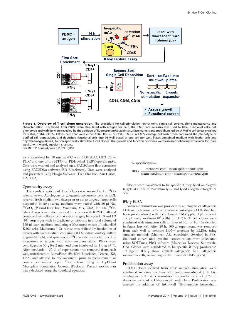
Figure 1. Overview of T cell clone generation. The procedure for cell stimulation, enrichment, single cell sorting, clone maintenance andcharacterisation is outlined. After PBMC were stimulated with antigen for 14 h, the IFN-c capture assay was used to label functional cells. Cellphenotype and viability were revealed by the addition of fluorescent mAb against surface markers and propidium iodide. A MoFlo cell sorter enrichedfor viable, CD14– CD16– CD19– cells that were either CD4+ IFN-c+ or CD8+ IFN-c+. A FACS Vantage cell sorter then confirmed the phenotype ofpurified cell populations, and deposited functional cells into 96 well plates at one cell per well. Plates contained medium with feeder cells andphytohaemagglutinin-L, to non-specifically stimulate T cell clones. The growth and function of clones were assessed following expansion for threeweeks, with weekly medium changes.doi:10.1371/journal.pone.0110741.g001
Ex Vivo T Cell Cloning
PLOS ONE | www.plosone.org 3 November 2014 | Volume 9 | Issue 11 | e110741

Pharmacia Biotech Pty Ltd, Australia) for the last 6 h of the 3 day
culture period. Cells were harvested onto glass fibre filter mats and3H incorporation measured as cpm on a MicroBeta scintillation
counter (Wallac, Finland).
One-step TRBV gene sequence analysisTRBV sequence analysis of T cell clones was undertaken using
an adaptation of a previously described method [17]. RNA was
extracted from 105 cells, using PureZOL RNA isolation reagent
(Bio-Rad Laboratories, Hercules, CA, USA). RNA pellets were
suspended in 20 ml of diethyl pyrocarbonate-treated deionised
water (MP Biomedicals Inc., Solon, OH, USA). RNA was reverse
transcribed and amplified in a one-step reverse transcription-PCR
(RT-PCR) system (Life Technologies). Primers used in one-step
reactions and subsequent sequencing reactions are as follows:
Degenerate forward primers VP1 (59-GCIITKTIYTGGTAYM-
GACA-39) and VP2 (59-CTITKTWTTGGTAYCIKCAG-39),
specific forward primers VP3 (59-ATCCTTTATTGGTATC-
GACGT-39) and VP4 (59-ATGTTTACTGGTATCATAAG-39),
specific reverse primer CP1 (59-GCACCTTCCTTCCCATT-
CAC-39). Each initial 25 ml reaction contained 12.5 ml of 26reaction mix, 0.5 ml RT/Platinum Taq mix, 1 ml RNA, 200 nM
CP1 and 2 mM VP1. If no product was detected in the PCR
product by agarose gel electrophoresis, the one-step reaction was
repeated using CP1 (200 nM), VP2 (2 mM), VP3 and VP4
(200 nM). RT-PCR cycling was performed at 50uC for 30 min
and 94uC for 2 min. PCR cycling was then performed at 94uC for
20 s, 50uC for 40 s then 72uC for 40s for 40 cycles, with a final
10 min extension at 72uC. International ImMunoGeneTics
information system (IMGT) nomenclature is used throughout this
report [18].
Sequencing of PCR productsPCR products were sequenced with BigDye Terminator v3.1
Cycle Sequencing Kit (Applied Biosystems, Foster City, CA,
USA), according to the manufacturer’s protocol, using a VP1 or
VP2 primer. Sequences were then analysed on an ABI Prism 310
Genetic Analyzer (Applied Biosystems Inc., Foster City, CA,
USA). Product sequences were assessed using Chromas software
(Technelysium Pty Ltd, Tewantin, Qld, Australia). TRBV(D)Jusage was determined using the IMGT V-QUEST online analysis
tool [19].
StatisticsStandard statistical tests (99% confidence intervals, standard
errors of the mean, Kolmogorov-Smirnov test for differences in
data distribution) were performed and graphs created in Prism
v6.02 (GraphPad Software, San Diego, CA, USA).
Results
Optimisation of T cell stimulationThe human IFN-c secretion assay (Miltenyi Biotec) uses
cytokine capture to identify viable, functional T cells responding
to antigenic stimulation. This kit has been used to purify IFN-c-
secreting cells prior to cloning by limiting dilution [20], but we
wanted to determine if it could be combined with single-cell
sorting to clone effector T cells ex vivo. Non-specific staining of
bystander T cells in close proximity to cytokine-secreting effectors
(‘‘cross-feeding’’) is a well recognised problem with the cytokine
capture technique. In preliminary tests of the IFN-c detection kit,
a shift in the IFN-c negative population was occasionally observed,
despite following the manufacturer’s recommendations (Fig. 2A).
By decreasing the concentration of cells during the one hour IFN-
c secretion period to 1–26104 PBMC/ml, the negative population
consistently remained in the left quadrants defined by stained,
unstimulated PBMC (Fig. 2B).
The period of stimulation prior to IFN-c capture was also
varied, to identify a timepoint at which IFN-c-secreting T cells
could be detected prior to significant antigen-specific proliferation.
Although responses were detected as early as 45 min in some
analyses, extensive longitudinal experiments consistently showed
that 10-23 h maximised responses (Fig. 2C). In subsequent
experiments, PBMC were stimulated for 14 h, a convenient
period minimising the possibility of in vitro division prior to
sorting, and maximising the number of cells detected.
Identification and sorting of antigen-specific effector Tcells
Current T cell cloning methods frequently require prior,
antigen-driven cell expansion, to overcome low precursor
Figure 2. Optimisation of IFN-c capture assay. (A, B) PBMC fromhealthy donors were stimulated for 14 h with recombinant CMV pp65,or (C) for increasing periods of time with the autologous lymphoblas-toid cell line (LCL), then assessed for reactivity by IFN-c capture assay.Cells were maintained at (A) too high a concentration during IFN-csecretion, or (B) an optimal concentration. Cells are gated on viable,CD14– CD16– CD19– subsets. (C) The percentage of CD8+ and CD4+ Tcells secreting IFN-c, gated on viable CD3+ cells. Results arerepresentative of (A, B) two, or (C) three independent experiments.doi:10.1371/journal.pone.0110741.g002
Ex Vivo T Cell Cloning
PLOS ONE | www.plosone.org 4 November 2014 | Volume 9 | Issue 11 | e110741

frequencies. However, in chronic infections, antigen-specific
effector cell frequencies are elevated. Initial experiments indicated
that IFN-c production by T cells stimulated with autologous cell
lines presenting cancer or viral antigens could be detected using
flow cytometry, suggesting that ex vivo sorting based on function
could enrich for antigen-specific cloning. In four separate
experiments, different antigen sources were used to stimulate
antigen-specific T cells: autologous melanoma cells (for two
different patients), autologous LCL, or recombinant CMV pp65.
PBMC from each donor were thawed, stimulated for 14 h with a
single antigen source, and then IFN-c-secreting T cells were
identified using the cell surface capture IFN-c detection kit. As
expected, the magnitude of the responses to each antigen source
varied, with IFN-c-secreting T cells ranging from 0.1% of all T
cells from melanoma patient A02 to 2.8% in the EBV model
(Fig. 3). Although ex vivo T cell responses were very low for
melanoma cell-stimulated PBMC, they were at least 2 fold higher
than backgrounds detected in unstimulated PBMC from each
donor. In summary, in each case precursor frequencies were
sufficient to detect and sort antigen-reactive effector T cells based
on phenotype and IFN-c secretion.
Cloning virus- and tumour-specific CD8+ and CD4+ Tcells
Using the method outlined in Fig. 1, T cells specific for viral and
tumour antigens were identified and sorted in two steps: following
a preliminary enrichment for CD4+ IFN-c+ and CD8+ IFN-c+ T
cells using a MoFlo cell sorter, single IFN-c+ cells were then
deposited into individual wells of a 96 well plate using a FACS
Vantage SE cell sorter. This two step, tandem procedure
eliminated the need to re-configure the primary sorter for single
cell deposition. In four separate experiments, 26-79% of single-
sorted T cells proliferated as clones after three weeks in culture
(Table 1). CD4+ and CD8+ T cells from the healthy donors
(stimulated with CMV pp65 or autologous LCL) and melanoma
patient A02 (stimulated with the autologous melanoma cell line)
cloned at comparable efficiencies (26-47% of all wells grew).
However, CD8+ T cells from melanoma patient D14 had a
substantially higher cloning efficiency, as all wells proliferated
initially, and 79% of the sorted T cells generated long term clones.
No relationship was found between cloning efficiency and
precursor frequency, as PBMC from patient D14 had one of the
lowest ex vivo responses to antigenic stimulation. The subsequent
growth of a subset of wells seeded with CD8+ T cells producing
IFN-c following CMV pp65 stimulation was assessed, using the
‘‘index sorting’’ function available in ClonCyte software. A
significantly different distribution of IFN-c production was
observed between wells that grew vs. those that died (P,0.0001;
Kolmogorov-Smirnov test), consistent with a lower cloning
efficiency for cells producing low amounts of IFN-c ex vivo (Fig.
S1). This suggests that, in this instance, the fitness of CD8 T cells
for subsequent expansion was related to their functional response
to cognate antigen.
Clones were re-stimulated with mitogen every four weeks, and
using this procedure, it was possible to expand clones to obtain up
to 108 cells per clone, after repeated stimulation.
Functional assessment of T cell clonesThe use of T cell clones is generally dependent on their
expression of some antigen-specific function. We therefore
analysed clones for functional characteristics in response to
antigenic stimulation, given that effector T cells can alter their
expression of cytokines following expansion [21].
CD4+ and CD8+ clones were stimulated with autologous or
allogeneic melanoma cells, LCL or LCL loaded (or untreated) with
CMV pp65 protein, as appropriate, and then culture supernatants
were analysed for IFN-a by ELISA (Fig. 4 A–D). The proportion
of clones that retained IFN-c production varied from 8% (CD8+clones derived from LCL stimulation) to 77% (CD8+ clones from
melanoma patient D14; Table 1). As with cloning efficiency, the
ability of clones to secrete IFN-c was not related to higher
precursor frequency – indeed, the opposite may be true, as the
high proportion of cells producing IFN-c ex vivo in response to
CMV and EBV translated into lower proportions of functional
CD8+ T cells (Table 1).
To determine whether T cells sorted on the basis of IFN-cproduction were capable of killing, selected LCL- and melanoma-
stimulated CD8+ clones were tested for cytolytic activity against
autologous and allogeneic cell lines. Strong activity was demon-
strated for all tested CD8+ clones (Fig 5 A, B, C), with minimal
reactivity against the allogeneic control. To obtain representative
data across a broader range of clones, we screened all patient D14
CD8+ T cell clones against autologous and allogeneic melanoma
lines using 51Cr-release assays (Fig 5D). Cytolytic activity was
demonstrated for 75% of clones, of which 16% were scored
negative for IFN-c production in the parallel assay (Fig 5D).
Likewise, a proportion of clones secreted IFN-c but lacked
cytolytic activity against the autologous melanoma line. Overall,
only 7% of sorted, proliferating D14 CD8+ T cell clones failed to
exhibit significant functional activity in either of these assays.
Selected CD4+ clones derived by LCL stimulation of healthy
donor PBMC were also tested for proliferation in response to
autologous antigen using a tritiated thymidine assay; a total of 14/
63 clones proliferated significantly above background defined by
non-stimulated controls (Figure S2).
Overall, between 12% and 93% of clones expressed some
functional response to antigen stimulation. This is likely to be an
underestimate of the true total for some stimuli, as (except for
patient D14) only selected clones were tested for cytolytic activity
or proliferation (Table 1).
Assessment of clonality by TRBV analysisClonality is required for many applications, for example,
antigen discovery and studies of T cell receptor usage. The most
common method of T cell cloning, limiting dilution, relies on the
statistical probability that no more than one proliferating cell will
be placed in each well during plate setup. This makes the method
inefficient, as many wells will not contain T cells, and re-cloning is
often required due to outgrowth of mixed populations. Cloning
using single cell sorting avoids this problem, by providing a
definitive method of obtaining clonal T cells, whilst employing
comparatively few plates. The only confounding factor would be if
cross-contamination occurred during sorting, or during extended
culture, which could compromise the outcome.
To confirm that T cell cultures generated by single-cell sorting
were truly clonal, we undertook TRBV expression analysis of
eleven clones from the LCL sort, using a panel of 23 mAb specific
for different TRBV (representative clones shown in Fig. 6A). Six
clones stained uniformly with a single mAb: TRBV20, TRBV22,
and TRBV3 (2 clones) and TRBV2 (2 clones). These six clones
were not stained by the other 22 TRBV mAb. The clones for
which a TRBV was not identified (i.e. they were not stained by
any of the 23 mAb) likely express TRBV that were not part of the
mAb set. These data definitively support the clonality of these
cultures, and show that feeder cells do not interfere with the
analysis. Flow cytometric data was confirmed by sequencing the
TRBV regions of these clones (Fig. 6C), and both forms of analysis
Ex Vivo T Cell Cloning
PLOS ONE | www.plosone.org 5 November 2014 | Volume 9 | Issue 11 | e110741

were repeated on tumour antigen-specific T cell clones (Fig. 6B,
D). All T cell clones stained positively with a maximum of one
TRBV mAb and only one TRBV region was detected in T cell
clone RNA, therefore we conclude that the generated T cells were
clonal.
Discussion
Here we describe a rapid and highly efficient method for
cloning effector CD4+ and CD8+ T cells ex vivo, which can use a
range of antigen sources as stimuli, and generates T cell clones
with diverse specificities. The method enables the selection of T
cell clones with known function ex vivo, without the need for prior,
multiple rounds of in vitro stimulation and cell division.
Figure 3. Detection of antigen-specific effector T cells ex vivo. IFN-c production measured by the capture assay in (A) unstimulated PBMCsamples and (B) PBMC from healthy donors stimulated with the autologous lymphoblastoid cell line (LCL) or CMV pp65 (top two panels), or PBMCfrom melanoma patients A02 or D14 stimulated with autologous melanoma cells (bottom two panels), as indicated. Cells were gated on the singlecell, low scatter, viable, CD14–CD16–CD19– subpopulation. Percentages are shown in each quadrant; ND = not done.doi:10.1371/journal.pone.0110741.g003
Ex Vivo T Cell Cloning
PLOS ONE | www.plosone.org 6 November 2014 | Volume 9 | Issue 11 | e110741
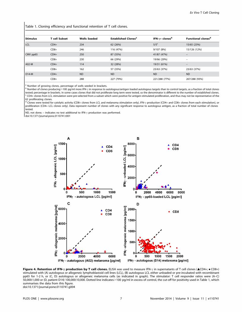
Table 1. Cloning efficiency and functional retention of T cell clones.
Stimulus T cell Subset Wells Seeded Established Clonesa IFN-c+ clonesb Functional clonesd
LCL CD4+ 234 62 (26%) 5/5c 15/65 (23%)
CD8+ 246 116 (47%) 9/107 (8%) 15/126 (12%)
CMV pp65 CD4+ 250 87 (35%) 41/87 (47%) –
CD8+ 230 66 (29%) 19/66 (29%) –
A02-M CD4+ 114 32 (28%) 19/31 (61%) –
CD8+ 162 57 (35%) 23/63 (37%) 23/63 (37%)
D14-M CD4+ ND ND ND ND
CD8+ 288 227 (79%) 221/288 (77%) 267/288 (93%)
a Number of growing clones, percentage of wells seeded in brackets.b Number of clones producing.100 pg/ml more IFN-c in response to autologous/antigen loaded autologous targets than to control targets, as a fraction of total clonestested; percentage in brackets. In some cases clones that did not proliferate long term were tested, so the denominator is different to the number of established clones.c CD4+ clones from LCL stimulation were pre-selected from a subset which were positive for antigen-stimulated proliferation, and thus may not be representative of the62 proliferating clones.d Clones were tested for cytolytic activity (CD8+ clones from LCL and melanoma stimulation only), IFN-c production (CD4+ and CD8+ clones from each stimulation), orproliferation (CD4+ LCL clones only). Data represent number of clones with any significant response to autologous antigen, as a fraction of total number of clonestested.ND, not done; – indicates no test additional to IFN-c production was performed.doi:10.1371/journal.pone.0110741.t001
Figure 4. Retention of IFN-c production by T cell clones. ELISA was used to measure IFN-c in supernatants of T cell clones (mCD4+; N CD8+)stimulated with (A) autologous or allogeneic lymphoblastoid cell lines (LCL), (B) autologous LCL either unloaded or pre-incubated with recombinantpp65 for 1-2 h, or (C, D) autologous or allogeneic melanoma cells (as indicated in graph). The stimulator: T cell responder ratios were (A–C)50,000:1,000 or (D, patient D14) 100,000:10,000. Dotted line indicates.100 pg/ml in excess of control, the cut-off for positivity used in Table 1, whichsummarises the data from this figure.doi:10.1371/journal.pone.0110741.g004
Ex Vivo T Cell Cloning
PLOS ONE | www.plosone.org 7 November 2014 | Volume 9 | Issue 11 | e110741

Subpopulations of T cells ,0.1% above background (Fig. 1) were
cloned and expanded to large numbers, and the capability of
recording ex vivo phenotypic characteristics enables the linkage of
these attributes with data subsequently generated in vitro.
Current cloning methods do not allow precise cloning
efficiencies to be calculated, since limiting dilution analysis
frequently underestimates the precursor frequency [22]. In the
case of known epitopes, multimer technology does allow precursor
frequencies to be estimated. Dunbar et al. sorted single HLA-A2-
restricted, melanoma-epitope tetramer+ CD8+ T cells and cloned
them at an average efficiency of 6.5% [9]. In contrast, the method
described in this paper generates CD4+ and CD8+ clones without
prior knowledge of epitope specificity or limitation of particular
HLA types, and is thus suitable for goals such as antigen discovery
and the analysis of broad responses to antigen. Since flow
cytometric and sequence analysis of TRBV regions of the clones
indicated that each clone had indeed originated from a single
precursor, the method provides a direct estimate of cloning
efficiency. Clonal expansion was successful for an average of 30%
(CD4+) and 48% (CD8+) of effector T cells that were sorted from
PBMC in response to autologous melanoma cells, LCL or a CMV
protein. The proportion of clones that retained antigen-specific
functionality varied widely among donors, and between CD4+ and
CD8+ subsets. Although this could reflect the sorting and clonal
expansion of IFN-c false-positive (e.g., cross feeding) contami-
nants, the high functional capacity of CD8+ T cells cloned from
melanoma patient D14 (despite a low precursor frequency)
indicates that instability of functional characteristics likely
contributes to this phenomenon. Importantly, the method
described herein could be used to investigate this further. For
example, index sorting data reveal that effector CD8+ T cells
producing low amounts of INF-c in response to CMV pp65 have a
significantly lower cloning efficiency, conceivably due to a lessened
fitness of T cells to replicate or to survive. It would be of interest to
investigate whether this characteristic has clinical implications.
T cells are often cloned after cultures of PBMC are stimulated
and allowed to expand in vitro [10,23,24]. This prevents the
analysis of some precusor characteristics, based on cell surface
marker or cytokine expression, due to the differentiation of cells
within the in vitro cultures. The method described in this paper
enables highly efficient selection of subpopulations of interest, by
avoiding this extended in vitro culture step. Short-term stimulation
with antigen limits the opportunity for T cell replication, and
phenotypic and functional modulation, and also enables direct
quantitation of the T cells of interest. Index sorting allows the
‘‘history’’ of each T cell clone to be examined retrospectively,
allowing connections to be made between ex vivo and in vitro T
cell characteristics. This becomes important when assessing
parameters used to define T cell characteristics in vivo [25,26],
which may not be stable in vitro, e.g. CD27 and CD45RA.
By removing the need for extended stimulation with antigen
and antigen-presenting cells, T cell clones can be generated
rapidly. Clones are available for use within three weeks of sorting,
and can be expanded to large numbers (107–108) after a further
three weeks of culture. There is no need for re-cloning, which is
often required following limiting dilution cloning. In addition, by
avoiding the selective outgrowth of more rapidly dividing cells,
direct cloning likely enhances the repertoire diversity of the
resulting collection of clones.
Figure 5. Cytotoxicity of CD8+ T cell clones. Cytotoxicity of selected CD8+ T cell clones from (A) healthy donor, (B) melanoma patient A02, and(C, D) melanoma patient D14, was assessed against the autologous and allogeneic lymphoblastoid cell line (LCL) or melanoma cells by chromiumrelease assay at an effector: target ratio of 10:1. Data indicate the mean specific lysis from (A) one; (B) three; and (C) two independent experiments,and error bars represent the standard error of the mean. (D) T cell clones from patient D14 were simultaneously assessed for IFN-c production inresponse to autologous and allogeneic melanoma cells (as described in Fig 4). The specific lysis of autologous melanoma cells (vertical axis) is plottedagainst IFN-c production (data corrected by subtracting allogeneic release). Dotted lines indicate cut-offs for positivity (see methods); inset indicatespercentages positive in each quadrant.doi:10.1371/journal.pone.0110741.g005
Ex Vivo T Cell Cloning
PLOS ONE | www.plosone.org 8 November 2014 | Volume 9 | Issue 11 | e110741

The addition of exogenous IL-2 or a co-stimulatory antibody
against CD28 did not noticeably improve the ex vivo response to
melanoma cell stimulation in preliminary studies, but the
proportion of T cells activated might, however, be enhanced
through the use of these additional signals when using other
antigen sources, or by using other growth factors e.g. GM-CSF
[27,28].
Although our cloning technique as described here is based on
IFN-c secretion, the method could be adapted to select T cells
based on production of other cytokines for which secretion assays
are available, such as TNF, IL-2 or IL-17, alone or in
combination. We sorted directly from stained PBMC, but
commercially available cytokine capture assays allow magnetic
enrichment of extremely low precursor numbers, so the extension
of our method to very rare subsets would be straight forward.
Direct cloning might also be applied to other non-lethal methods
of identifying antigen reactive T cells, such as membrane-bound
TNF [29] and the expression of a wide variety of activation
markers (reviewed in [30]). However, the reliance of direct cloning
on effector function makes it unsuitable for expanding naıve
precursors, which is achievable by limit dilution cloning of
amplified T cells [31].
T cell clones generated using direct cloning have advantages for
some specific applications that are of potential clinical importance.
The method preferentially selects effector-memory T cells, with
immediate functional ability, which presumably reflects the active,
circulating anti-pathogen or anti-tumour population. Since the
cloning efficiency is high, the application of clones to defining the
antigenic repertoire or discovery of novel antigens [32] would
enable establishment of an immunodominance hierarchy for
identified epitopes. Furthermore, by sequencing T-cell receptors of
clones recognising an epitope of interest, the level of diversity
within the effector response can be determined.
In conclusion, the ex vivo effector cell cloning method described
here provides a rapid, powerful and effective method of deriving
antigen-specific T cells clones with traceable in vivo precursor
Figure 6. TRBV analysis of LCL- and tumour-reactive CD8+ T cell clones. CD8+ T cell clones specific for (A, C) LCL or (B, D) tumour antigenswere assessed for clonality by flow cytometric and molecular analysis. (A, B) Clones were stained with a panel of fluorescently-labelled TRBV mAb,then analysed on a flow cytometer. Positive and negative TRBV staining is shown for representative T cell clones. (C, D) To confirm the flowcytometric findings and determine unidentified TRBV regions, RNA was extracted from T cell clones and TRBV regions reverse transcribed, amplified,then sequenced. The designations TRBV and TRBJ follow the TCR gene nomenclature specified by IMGT [18].doi:10.1371/journal.pone.0110741.g006
Ex Vivo T Cell Cloning
PLOS ONE | www.plosone.org 9 November 2014 | Volume 9 | Issue 11 | e110741

function and phenotype, thus improving on functionality and
applicability compared to traditional T cell cloning techniques.
Supporting Information
Figure S1 (A) Index sorting was used to assign ex vivoIFN-c secretion levels (arbitrary fluorescence units), inresponse to CMV pp65, to individual CD8+ T cellsseeded into wells, which were scored for subsequentgrowth into long-term clones (‘‘Grew’’ vs. ‘‘Died’’). (B)
Percentile plot showing cumulative percentage of cells that
established clones (– –) or died (––) according to IFN-c production.
The distribution of IFN-c production differed significantly
between the two groups of cells (Kolmogorov-Smirnov test; P,
0.0001).
(TIF)
Figure S2 CD4+ clones derived from EBV stimulationwere tested for proliferation in response to the autolo-gous LCL by tritiated thymidine incorporation; the
background proliferation (Nil stimulation) of a randomselection of clones was also assessed. Data indicate means
of duplicate measurements in a single experiment. The geometric
mean (––) and upper 99% confidence limit of the geometric mean
(---) of proliferation of unstimulated clones are indicated.
(TIF)
Acknowledgments
The authors wish to acknowledge the Grace Chojnowski and Paula Hall
from the Flow Cytometry facility for their technical support, and the
Australian Red Cross Blood Service, for provision of blood products.
Author Contributions
Conceived and designed the experiments: MAN CWS NRM MHLL.
Performed the experiments: MAN MHLL CML LEO NRM. Analyzed the
data: MAN CWS NRM. Contributed reagents/materials/analysis tools:
CWS NRM. Wrote the paper: MAN CWS NRM ALP. Data
interpretation: MAN NRM ALP CWS.
References
1. Hunder NN, Wallen H, Cao J, Hendricks DW, Reilly JZ, et al. (2008)
Treatment of metastatic melanoma with autologous CD4+ T cells against NY-
ESO-1. N Engl J Med 358: 2698–2703.
2. Riddell SR, Watanabe KS, Goodrich JM, Li CR, Agha ME, et al. (1992)
Restoration of viral immunity in immunodeficient humans by the adoptive
transfer of T cell clones. Science 257: 238–241.
3. Fonteneau JF, Larsson M, Somersan S, Sanders C, Munz C, et al. (2001)
Generation of high quantities of viral and tumor-specific human CD4+ and
CD8+ T-cell clones using peptide pulsed mature dendritic cells. J Immunol
Methods 258: 111–126.
4. Gervois N, Labarriere N, Le Guiner S, Pandolfino MC, Fonteneau JF, et al.
(2000) High avidity melanoma-reactive cytotoxic T lymphocytes are efficiently
induced from peripheral blood lymphocytes on stimulation by peptide-pulsed
melanoma cells. Clin Cancer Res 6: 1459–1467.
5. Alexander-Miller MA, Leggatt GR, Sarin A, Berzofsky JA (1996) Role of
antigen, CD8, and cytotoxic T lymphocyte (CTL) avidity in high dose antigen
induction of apoptosis of effector CTL. J Exp Med 184: 485–492.
6. Geginat J, Sallusto F, Lanzavecchia A (2001) Cytokine-driven proliferation and
differentiation of human naive, central memory, and effector memory CD4(+) T
cells. J Exp Med 194: 1711–1719.
7. Kim M, Moon HB, Kim K, Lee KY (2006) Antigen dose governs the shaping of
CTL repertoires in vitro and in vivo. Int Immunol 18: 435–444.
8. Leggatt GR, Narayan S, Fernando GJ, Frazer IH (2004) Changes to peptide
structure, not concentration, contribute to expansion of the lowest avidity
cytotoxic T lymphocytes. J Leukoc Biol 76: 787–795.
9. Dunbar PR, Chen JL, Chao D, Rust N, Teisserenc H, et al. (1999) Cutting edge:
rapid cloning of tumor-specific CTL suitable for adoptive immunotherapy of
melanoma. J Immunol 162: 6959–6962.
10. Yee C, Savage PA, Lee PP, Davis MM, Greenberg PD (1999) Isolation of high
avidity melanoma-reactive CTL from heterogeneous populations using peptide-
MHC tetramers. J Immunol 162: 2227–2234.
11. Varadarajan N, Kwon DS, Law KM, Ogunniyi AO, Anahtar MN, et al. (2012)
Rapid, efficient functional characterization and recovery of HIV-specific human
CD8+ T cells using microengraving. Proc Natl Acad Sci U S A 109: 3885–
3890.
12. Fletcher JM, Vukmanovic-Stejic M, Dunne PJ, Birch KE, Cook JE, et al. (2005)
Cytomegalovirus-specific CD4+ T cells in healthy carriers are continuously
driven to replicative exhaustion. J Immunol 175: 8218–8225.
13. Plunkett FJ, Franzese O, Belaramani LL, Fletcher JM, Gilmour KC, et al. (2005)
The impact of telomere erosion on memory CD8+ T cells in patients with X-
linked lymphoproliferative syndrome. Mech Ageing Dev 126: 855–865.
14. O’Rourke MG, Johnson M, Lanagan C, See J, Yang J, et al. (2003) Durable
complete clinical responses in a phase I/II trial using an autologous melanoma
cell/dendritic cell vaccine. Cancer Immunol Immunother 52: 387–395.
15. Pavey S, Johansson P, Packer L, Taylor J, Stark M, et al. (2004) Microarray
expression profiling in melanoma reveals a BRAF mutation signature. Oncogene
23: 4060–4067.
16. Campbell JD (2003) Detection and enrichment of antigen-specific CD4+ and
CD8+ T cells based on cytokine secretion. Methods 31: 150–159.
17. Zhou D, Srivastava R, Grummel V, Cepok S, Hartung HP, et al. (2006) Highthroughput analysis of TCR-beta rearrangement and gene expression in single T
cells. Lab Invest 86: 314–321.18. Giudicelli V, Duroux P, Ginestoux C, Folch G, Jabado-Michaloud J, et al.
(2006) IMGT/LIGM-DB, the IMGT comprehensive database of immunoglob-
ulin and T cell receptor nucleotide sequences. Nucleic Acids Res 34: D781–784.19. Giudicelli V, Brochet X, Lefranc MP (2011) IMGT/V-QUEST: IMGT
standardized analysis of the immunoglobulin (IG) and T cell receptor (TR)nucleotide sequences. Cold Spring Harb Protoc 2011: 695–715.
20. Manley TJ, Luy L, Jones T, Boeckh M, Mutimer H, et al. (2004) Immuneevasion proteins of human cytomegalovirus do not prevent a diverse CD8+cytotoxic T-cell response in natural infection. Blood 104: 1075–1082.
21. Doyle AG, Buttigieg K, Groves P, Johnson BJ, Kelso A (1999) The activatedtype 1-polarized CD8(+) T cell population isolated from an effector site contains
cells with flexible cytokine profiles. J Exp Med 190: 1081–1092.22. Ogg GS, McMichael AJ (1998) HLA-peptide tetrameric complexes. Curr Opin
Immunol 10: 393–396.
23. Ho WY, Nguyen HN, Wolfl M, Kuball J, Greenberg PD (2006) In vitro methodsfor generating CD8+ T-cell clones for immunotherapy from the naive repertoire.
J Immunol Methods 310: 40–52.24. Riddell SR, Greenberg PD (1990) The use of anti-CD3 and anti-CD28
monoclonal antibodies to clone and expand human antigen-specific T cells.
J Immunol Methods 128: 189–201.25. Hamann D, Baars PA, Rep MH, Hooibrink B, Kerkhof-Garde SR, et al. (1997)
Phenotypic and functional separation of memory and effector human CD8+ Tcells. J Exp Med 186: 1407–1418.
26. Sallusto F, Lenig D, Forster R, Lipp M, Lanzavecchia A (1999) Two subsets ofmemory T lymphocytes with distinct homing potentials and effector functions.
Nature 401: 708–712.
27. Morrissey PJ, Bressler L, Park LS, Alpert A, Gillis S (1987) Granulocyte-macrophage colony-stimulating factor augments the primary antibody response
by enhancing the function of antigen-presenting cells. J Immunol 139: 1113–1119.
28. Martinuzzi E, Afonso G, Gagnerault MC, Naselli G, Mittag D, et al. (2011)
acDCs enhance human antigen-specific T-cell responses. Blood 118: 2128–2137.29. Haney D, Quigley MF, Asher TE, Ambrozak DR, Gostick E, et al. (2011)
Isolation of viable antigen-specific CD8+ T cells based on membrane-boundtumor necrosis factor (TNF)-alpha expression. J Immunol Methods 369: 33–41.
30. Bacher P, Scheffold A (2013) Flow-cytometric analysis of rare antigen-specific Tcells. Cytometry A 83: 692–701.
31. Geiger R, Duhen T, Lanzavecchia A, Sallusto F (2009) Human naive and
memory CD4+ T cell repertoires specific for naturally processed antigensanalyzed using libraries of amplified T cells. J Exp Med 206: 1525–1534.
32. Lennerz V, Fatho M, Gentilini C, Frye RA, Lifke A, et al. (2005) The responseof autologous T cells to a human melanoma is dominated by mutated
neoantigens. Proc Natl Acad Sci U S A 102: 16013–16018.
Ex Vivo T Cell Cloning
PLOS ONE | www.plosone.org 10 November 2014 | Volume 9 | Issue 11 | e110741
Copyright © 2022 FDOKUMEN