Role of VANGL1 as an effector of R-RAS - UCL Discovery
295
1 Role of VANGL1 as an effector of R-RAS Nicole Hartig A thesis submitted towards the degree of Doctor of Philosophy April 2013 Cell Signalling Laboratory UCL Cancer Institute 72 Huntley Street, London Department of Biochemistry and Molecular Biology University College London Gower Street, London
-
Upload
khangminh22 -
Category
Documents
-
view
0 -
download
0
Transcript of Role of VANGL1 as an effector of R-RAS - UCL Discovery
1
Role of VANGL1 as an effector
of R-RAS
Nicole Hartig
A thesis submitted towards the degree of
Doctor of Philosophy
April 2013
Cell Signalling Laboratory
UCL Cancer Institute
72 Huntley Street, London
Department of Biochemistry and Molecular Biology
University College London
Gower Street, London

2
Declaration
I, Nicole Hartig, confirm that the work presented in this thesis is my own. Where
information has been derived from other sources, I confirm that this has been
indicated in the thesis.
London, April 2013

3
In loving memory of my Mama Steffi

4
Abstract
The WNT pathway plays a key role in development and disease. In addition to
the better studied ß-catenin dependent pathway, WNT ligands can also activate
the separate ‘non-canonical’ or Planar Cell Polarity (PCP) pathway. Perturbations
in the PCP pathway contribute to the pathogenesis of a variety of diseases
including cardiac and neural tube defects, and to the invasiveness of cancer
cells.
R-RAS subgroup GTPases share many of the properties of classical RAS GTPases
including the ability to behave as oncogenes. However, they also have distinct
functions of their own and how signalling and biological specificity is achieved is
not fully understood. Using a proteomic approach to identify novel R-RAS
subgroup effectors led to the identification of VANGL1, a WNT/PCP protein,
demonstrated to be a novel R-RAS interacting protein.
In this thesis, I have shown that VANGL1 functions as an effector of R-RAS and
TC21. Using proteomic approaches, multiple VANGL1 interacting proteins have
been identified and R-RAS, as well as selected Frizzled (FZD) receptors and the
tyrosine kinase ROR2 can modulate at least some of these VANGL1 interactions.
Furthermore, VANGL1 leads to the protein degradation of PRICKLE by a
mechanism that remains to be determined, and R-RAS GTPases are able to
inhibit this effect.
Using RBD pulldown assays, I was able to show that WNT ligands and ROR2 can
lead to the activation of R-RAS, and that R-RAS and TC21 are key mediators of
RHO activation by WNT5a. Finally, I demonstrated that R-RAS/TC21 and VANGL1
are critically required for directed migration.
The identification of R-RAS activation by WNT ligands and its interaction with
VANGL1 provides an exciting new link between the R-RAS subgroup of the RAS
family and the WNT/PCP pathway.

5
Acknowledgments
First and foremost I would like to thank my supervisor Pablo Rodriguez-Viciana
for giving me the opportunity to work in his laboratory, the supervision and
guidance throughout my PhD studies and the final push during writing this
thesis. I also want to thank my thesis examiners for their interest and time, and
I am looking forward to our discussion.
I truly enjoyed my PhD time, thanks to an amazing group of work colleagues and
students. In particular, I would like to thank Kristina for helpful discussions,
interest and encouragement throughout my project. Work would have not been
the same without Lucy, my partner in crime, who is a great team mate and
friend. Thanks to Berna, even long days went by fast and never got dull due to
creation of a fun work environment and her entertaining personality.
I also want to thank Marta, Ariadna, Giammy and Konstantinos for helpful
experimental discussions and conversations about (PhD) life in general and
guidance through the last 4 years.
A very warm thank you goes to Michiru Nishita and Prof Yasuhiro Minami and the
rest of the very hardworking laboratory members at Kobe University for the
wonderful and unique time in Japan, and especially for the experimental
assistance during my stay.
A specific thank you goes to my family, who supported me all along during my
studies and their interest in my work. Especially my beloved mum, who
unfortunately is not able to experience this day – I miss you very much.
And last but not least, without my second family, the Jhalas, a big, fun and
loving part of my life in London would be missing – I love you all!
The biggest gratitude I want to express is for Shiv, who supported me through
the last three years, is always there for me, and is simply a wonderful person.
Thank you for being at my side.
Thank you all for being part of my long and interesting PhD journey.
6
Table of Contents
Abstract……………………………………………………………………………………………………………………4
Acknowledgements………………………………………………………………….…………………..………..5
Table of Contents…………………………………………………………………………………..………………6
List of Figures ………………………………………………………………………………………………..……..13
List of Tables………………………………………………………………………………………………………….17
Abbreviations…………………………………………………………………………………………………………18
CHAPTER 1 ............................................................................................ 24
1 Introduction ................................................................................... 25
1.1 RAS superfamily ...........................................................................25
1.2 RAS subfamily ..............................................................................26
1.2.1 RAS regulatory proteins: GEFs and GAPs ...................................28
1.2.2 RAS protein effectors ..............................................................32
1.2.3 Structural features of RAS proteins ...........................................37
1.2.4 Post translational modification of RAS proteins ...........................39
1.3 The R-RAS subgroup .....................................................................43
1.3.1 R-RAS ...................................................................................43
1.3.2 TC21 ....................................................................................47
1.3.3 M-RAS ..................................................................................48
1.4 RHO GTPases ...............................................................................49
1.4.1 Rho regulatory proteins: GEFs, GAPs and GDIs ..........................50
1.4.2 RHO proteins .........................................................................51
1.4.3 RAC and CDC42 .....................................................................51
1.5 WNT signalling branches................................................................52
1.5.1 β-catenin (canonical) WNT-signalling pathway ...........................52
1.5.2 Planar Cell Polarity (PCP) pathway ............................................55

7
1.5.3 The WNT/Ca2+ pathway ...........................................................60
1.6 WNT signalling components ...........................................................62
1.6.1 WNT ligands ..........................................................................62
1.6.2 Non-WNT ligand modulators of WNT signalling ...........................63
1.6.2.1 WNT inhibitors .................................................................63
1.6.2.2 WNT activators ................................................................64
1.6.3 Frizzled receptors ...................................................................66
1.6.4 WNT cell surface receptors ......................................................67
1.6.4.1 LRP5/6 ............................................................................68
1.6.4.2 ROR2 ..............................................................................69
1.6.4.3 PTK7...............................................................................72
1.6.4.4 RYK ................................................................................73
1.6.4.5 HSPGs and Syndecans ......................................................73
1.6.4.6 Tetraspanins ....................................................................74
1.6.5 Dishevelled proteins ...............................................................74
1.7 VANGL proteins ............................................................................78
1.7.1 VANGL1 ................................................................................79
1.7.2 VANGL2 ................................................................................81
1.8 Cell migration and invasion processes .............................................83
1.8.1 Actin structures and actin-binding protein Filamin A ....................84
1.8.2 Invadopodia structures and matrix metalloproteases ..................85
1.9 Autophagy ...................................................................................86
1.10 Aim of my study and outline of subsequent chapters ......................88
CHAPTER 2 ............................................................................................ 89
2 Materials and Methods .................................................................... 90
2.1 MATERIALS ..................................................................................90
2.1.1 Chemical compounds and reagents ...........................................90
2.1.2 Media and growth plates .........................................................91
2.1.3 Antibodies .............................................................................92
8
2.1.4 Plasmids ...............................................................................93
2.1.5 Primers .................................................................................95
2.1.6 RNAi sequences .....................................................................95
2.1.7 Buffers ..................................................................................90
2.2 DNA TECHNIQUES ........................................................................97
2.2.1 Basic DNA manipulations .........................................................97
2.2.1.1 Cloning approach of VANGL1 truncation constructs ...............97
2.2.1.2 Cloning approach of ARHGEF17 truncation constructs ...........98
2.2.2 Plasmid mutagenesis ..............................................................99
2.2.3 DNA gel electrophoresis ........................................................ 101
2.2.4 Transformation of bacteria ..................................................... 101
2.2.5 Preparation and purification of plasmid DNA ............................ 102
2.2.6 DNA sequencing ................................................................... 102
2.3 RNA TECHNIQUES ...................................................................... 103
2.3.1 RNA isolation ....................................................................... 103
2.3.2 Semiquantitative PCR ........................................................... 103
2.3.3 Visualisation of RT-PCR products ............................................ 104
2.4 MAMMLIAN CELL CULTURE .......................................................... 105
2.4.1 Cell lines and culture conditions ............................................. 105
2.4.2 Thawing and freezing/long term storage of mammalian cells ..... 105
2.4.3 Manual cell counting and assessment of viability ...................... 105
2.4.4 IncuCyte based proliferation or phenotype analysis .................. 106
2.4.5 DNA transfection .................................................................. 106
2.4.5.1 Lipofectamine2000 transfection of plasmid DNA ................. 106
2.4.5.2 Polyethylenimine transfection of plasmid DNA .................... 106
2.4.6 RNAiMAX siRNA transfection .................................................. 106
2.4.7 Virus generation ................................................................... 107
2.4.7.1 Retrovirus ..................................................................... 107
2.4.7.2 Lentivirus ...................................................................... 108
2.4.8 Generation of stable cell lines ................................................ 109
9
2.4.8.1 Generation of stably WNT ligand expressing cell lines and
collection of WNT conditioned media ................................................ 109
2.4.8.2 Generation of stable cells expressing YFP-fusion proteins for
localisation studies ........................................................................ 111
2.5 PROTEIN TECHNIQUES ................................................................ 112
2.5.1 Preparation of cell extracts and immunoblot analysis ................ 112
2.5.2 Determination of protein concentractions ................................ 112
2.5.3 Densitometric analysis .......................................................... 112
2.5.4 GST-pull down and immunoprecipitation experiments ............... 113
2.5.5 Expression and purification of recombinant proteins .................. 113
2.5.6 Generation of GST-RBD beads for RHO/RAS activation assays .... 113
2.5.7 RAS and RHO binding domain assays ...................................... 114
2.5.8 In vitro interaction assays ..................................................... 114
2.5.9 TAP-tagged protein transfection and purification for mass
spectrometry analysis ....................................................................... 115
2.5.10 Mass spectrometry analysis ................................................... 116
2.6 CELL BIOLOGICAL TECHNIQUES ................................................... 117
2.6.1 Scratch wound migration assays ............................................ 117
2.6.2 Transwell/Boyden chamber assays ......................................... 117
2.6.3 Cell polarisation assays: GM130 and MTOC assays ................... 117
2.6.4 Invadopodia assay ................................................................ 118
2.6.5 Luciferase reporter assays for β-catenin level detection ............. 119
2.6.6 Autophagy inducing treatments .............................................. 120
2.6.7 Statistical analysis ................................................................ 120
2.7 MICROSCOPY ............................................................................. 121
2.7.1 Preparation and staining procesdure of cells for fluorescent
microscopy purposes ......................................................................... 121
2.7.1.1 PFA fixation and permeabilisation for GM130 stainings ........ 121
2.7.1.2 Methanol based fixation for γ-tubulin stainings ................... 122
2.7.2 Localisation studies using confocal flourescence microscopy ....... 122
10
CHAPTER 3 .......................................................................................... 123
3 Characterisation of VANGL1 as an effector of R-RAS .................... 124
3.1 Identification of potential novel effector of R-RAS and TC21 ............. 124
3.2 VANGL1 behaves as an effector of R-RAS subfamily GTPases ........... 128
3.2.1 R-RAS interacts with VANGL1 in an activation and effector domain
dependent manner ........................................................................... 128
3.2.2 VANGL1 and VANGL2 interact specifically with R-RAS subgroup
members ......................................................................................... 129
3.2.3 Characterisation of VANGL1-R-RAS interaction using R-RAS effector
loop mutants and the ∆CAAX truncation .............................................. 132
3.2.4 R-RAS interacts with the C-terminus of VANGL1 ....................... 134
3.2.5 R-RAS interaction with VANGL1 is disrupted by the D259E mutant ..
.......................................................................................... 136
3.2.6 R-RAS and VANGL1 interact directly in vitro ............................. 137
3.3 ARHGEF17/p164RhoGEF behaves as a TC21 and R-RAS effector....... 139
3.3.1 ARHGEF17/p164RhoGEF interacts with R-RAS and TC21 in an
activation and effector domain dependent manner ................................ 139
3.3.2 Mapping of p164RhoGEF domains that interact with TC21 ......... 140
3.3.3 ARHGEF17/p164RhoGEF and TC21 interact in vitro ................... 143
3.3.4 ARHGEF17/p164RhoGEF acts as a GEF for RHOA ...................... 144
3.4 Discussion ................................................................................. 146
3.4.1 VANGL1 functions as an effector of R-RAS subgroup GTPases .... 146
3.4.2 ARHGEF17/p164RhoGEF functions as an effector of R-RAS and TC21
.......................................................................................... 149
CHAPTER 4 .......................................................................................... 151
4 Identification of VANGL1 interaction partners .............................. 152
4.1 Initial VANGL1 TAP Screen ........................................................... 152
4.2 Validation of VANGL1-TAP hits identified by mass spectrometry ....... 154
4.2.1 Effect of VANGL1 point mutations found in NTC patients on
interaction with SCRIB and DVL3 ........................................................ 160

11
4.3 Modulation of VANGL1 interactions by R-RAS ................................. 162
4.3.1 VANGL proteins or R-RAS GTPases do not modulate canonical WNT
signalling ......................................................................................... 162
4.3.2 Active R-RAS modulates a subset of VANGL1 binding partners ... 163
4.4 WNT ligand and Frizzled effects on VANGL1 interactions .................. 165
4.4.1 WNT ligand generation and assessment .................................. 166
4.4.2 Assessment of WNT ligand responsiveness in a panel of cell lines ....
.......................................................................................... 167
4.4.3 Frizzled expression alters sensitivity towards WNT ligands ......... 170
4.4.4 FZDs and ROR2 specifically stimulate VANGL1 interaction with DVL2
.......................................................................................... 172
4.4.5 TC21 can co-immunoprecipitate with ROR2 or VANGL1 in the
prsence of specific FZD receptors ....................................................... 174
4.4.6 WNT5a modulates VANGL1 interaction with DVL but not SCRIB,
CASK or DLG1 .................................................................................. 176
4.5 Proteomic analysis of VANGL1 interaction and their modulation by R-
RAS, mFZD5 and ROR2 ........................................................................ 179
4.6 Discussion ................................................................................. 181
4.6.1 Identification and characterisation of VANGL1 interacting proteins ...
.......................................................................................... 181
4.6.2 Frizzled expression leads to enhanced VANGL1-DVL2 interaction 185
CHAPTER 5 .......................................................................................... 191
5 R-RAS GTPases inhibit VANGL1 induced PRICKLE degradation ..... 192
5.1 VANGL1 interacts with PRICKLE1 and stimulates its destabilisation ... 192
5.2 R-RAS GTPases inhibit VANGL1 induced PRICKLE1/2 protein
destabilisation ..................................................................................... 195
5.3 VANGL1 localises to vesicle like structures upon autophagy induction 197
5.4 VANGL1 subcellular localisation studies ......................................... 202
5.5 Discussion ................................................................................. 207

12
CHAPTER 6 .......................................................................................... 210
6 Effect of WNT ligands and Frizzled receptors on R-RAS and RHO
activation ............................................................................................ 211
6.1 Establishment of RAS binding domain assays ................................. 211
6.2 WNT ligands activate R-RAS ........................................................ 216
6.3 R-RAS and TC21 is required for WNT5a dependent activation of RHOA
and RHOB ........................................................................................... 220
6.4 Discussion ................................................................................. 225
CHAPTER 7 .......................................................................................... 228
7 Role of VANGL1 and R-RAS in polarised migration ........................ 229
7.1 VANGL1 and R-RAS are required for migration ............................... 230
7.2 VANGL1 and R-RAS are required for scratch induced polarisation ..... 237
7.3 VANGL1 depletion leads to increased invadosome formation ............ 241
7.4 Discussion ................................................................................. 244
CHAPTER 8 .......................................................................................... 248
8 Final Discussion and Summary ..................................................... 249
8.1 Conclusions and and proposed model ............................................ 249
CHAPTER 9 .......................................................................................... 256
9 References .................................................................................... 257
13
List of Figures
Figure 1.1: RAS proteins cycle between a GTP-bound active and a GDP-bound
inactive state. ..........................................................................................26
Figure 1.2: Overview of RAS subfamily proteins. .........................................27
Figure 1.3: RAS regulatory proteins and effectors. ......................................28
Figure 1.4: Domain overview of RAS regulatory proteins…….. .......................30
Figure 1.5: Overview of RASGEF and RASGAP GTPase substrate specificity .....31
Figure 1.6: RAS effector pathways. Upon stimulation, RAS proteins interact with
a multitude of effector proteins involved in various cellular processes. .............32
Figure 1.7: The RAS-RAF-ERK signalling pathway ........................................33
Figure 1.8: Phosphoinositide 3-kinases are RAS effectors. ............................34
Figure 1.9: Overview of RAL effectors and associated signalling pathways ......36
Figure 1.10: Structure of RAS proteins ......................................................38
Figure 1.11: C-terminal RAS membrane targeting signals ............................40
Figure 1.12: Post translational modifications of RAS proteins ........................41
Figure 1.13: RAS post translational modifications lead to various subcellular
localisations .............................................................................................42
Figure 1.14: Oncogenic classical Ras proteins are structurally highly similar to
R-RAS proteins .........................................................................................43
Figure 1.15: Overview of RHO GTPase family..............................................49
Figure 1.16: β-catenin (canonical) WNT signalling .......................................54
Figure 1.17: Asymmetrical distribution of planar cell polarity proteins in
Drosophila ...............................................................................................56
Figure 1.18: PCP phenotype examples in Drosophila and mammals ...............57
Figure 1.19: WNT/Planar Cell Polarity signalling ..........................................58
Figure 1.20: Convergent extension and planar polarity ................................59
Figure 1.21: WNT/Ca2+ signalling ..............................................................61
Figure 1.22: WNT ligand structure and post-translational modifications of
WNT3a and WNT5a proteins .......................................................................62
Figure 1.23: Non-WNT ligands modulators .................................................65
Figure 1.24: Schematic of Frizzled receptor key features and binding proteins
and modulators ........................................................................................67
Figure 1.25: Overview of WNT modulating receptors and their effect on
downstream WNT signalling .......................................................................68
Figure 1.26: Schematic representation of the LRP5/6 domain structure .........69
Figure 1.27: Structure of ROR-family receptor tyrosine kinase ROR2 .............70
Figure 1.28: ROR2/WNT5a signalling in regulation of cell polarity, migration and
invasion...................................................................................................71

14
Figure 1.29: DVL proteins form the branching point of WNT pathways ...........75
Figure 1.30: VANGL1 and VANGL2 proteins display a high degree of sequence
similarity .................................................................................................78
Figure 1.31: Topological model and structural features of VANGL1 ................80
Figure 1.32: Polarity of migrating cells .......................................................83
Figure 1.33: Schematic model of the autophagy process ..............................87
Figure 2.1: Examples of Gateway cloning vectors ........................................94
Figure 2.2: Expression analysis of Flag-VANGL1 point mutants and truncation
constructs in HEK293T cells ..................................................................... 101
Figure 2.3: Analysis of WNT3a conditioned media biological activity and optimal
culture and storage conditions .................................................................. 110
Figure 3.1: Tandem affinity purified R-RAS V38 interactors, identified by mass
sprectrometry ........................................................................................ 125
Figure 3.2: Tandem affinity purified TC21 V12 interactors, identified by mass
spectroscopy .......................................................................................... 126
Figure 3.3: Proposed model of how R-RAS GTPases could act downstream of
WNT ligands in the non-canonical WNT signalling pathway ........................... 127
Figure 3.4: VANGL1 interacts with R-RAS activation and effector domain
dependent ............................................................................................. 129
Figure 3.5: R-RAS GTPases interact in an activation-dependent manner with
VANGL1 and VANGL2 .............................................................................. 130
Figure 3.6: Interaction of endogenous VANGL1 and other effectors with RAS
family GTPases. ...................................................................................... 131
Figure 3.7: R-RAS effector domain mutations do not affect VANGL1 interaction,
whereas deletion of the CAAX box completely inhibits VANGL1 binding .......... 133
Figure 3.8: VANGL1 interacts with R-RAS through its C-terminus in a PDZ-motif
independent fashion ................................................................................ 135
Figure 3.9: VANGL1 point mutations D259E identified in neural tube defect
(NTD) patients disrupts binding to R-RAS .................................................. 137
Figure 3.10: R-RAS interacts directly with the cytoplasmic C-terminus of
VANGL1 ................................................................................................. 138
Figure 3.11: ARHGEF17/p164RhoGEF binds specifically to TC21 and R-RAS .. 140
Figure 3.12: Mapping of the p164RhoGEF binding region to TC21................ 142
Figure 3.13: In vitro interaction between p164RhoGEF and TC21 ................ 143
Figure 3.14: p164RhoGEF is an activator of RHOA in HEK293T cells ............ 144
Figure 3.15: Alignment of RAS, RAP2 and R-RAS subgroup members .......... 147
Figure 3.16: R-Ras interaction chapter summary ...................................... 148
Figure 3.17: TC21 interaction summary ................................................... 150
15
Figure 4.1: VANGL1-TAP approach lead to identification of novel interaction
partners ................................................................................................ 153
Figure 4.2: Validation of VANGL1 interactions identified by TAP ................... 156
Figure 4.3: Validation of TAP-identifed VANGL1 interacting proteins............. 158
Figure 4.4: VANGL1 D259E mutant disrupts binding to DVL3 but not SCRIB . 160
Figure 4.5: Expression of VANGL proteins or activated R-RAS/TC21 do not alter
β-catenin reporter activity ....................................................................... 163
Figure 4.6: R-RAS modulates a subset of endogenous VANGL1 binding partners
involved in cell polarity and WNT signalling ................................................ 164
Figure 4.7: Recombinant WNT3a, but not WNT5a or WNT7a is able to stimulate
canonical β-catenin signalling ................................................................... 167
Figure 4.8: WNT ligands differentially stimulate DVL2 phosphorylation in tumour
derived cell lines but have no effect on non-transformed breast cell lines ....... 169
Figure 4.9: HMECs expressing mFzd5 gain responsiveness to WNT3a and
WNT5a ligands ....................................................................................... 171
Figure 4.10: FZDs and ROR2 cooperate to stimulate VANGL1-DVL2 binding .. 173
Figure 4.11: ROR2 co-expressed with FZD2 is able to immunoprecipitate
endogenous TC21 in HEK293T cells ........................................................... 175
Figure 4.12: VANGL1 is able to immunoprecipitate endogenous TC21 but not R-
RAS in two breast cell lines stably expressing mFzd5 .................................. 176
Figure 4.13: WNT5a stimulation of HMEC mFzd5 cells leads to modulation of
VANGL1 and VANGL2 interaction with DVL proteins, but not CASK or DLG1 ... 177
Figure 4.14: WNT5a treatment enhances binding of DVL2 and DVL3 to VANGL1
after 60 min, but SCRIB or CASK interactors are not modulated ................... 178
Figure 4.15: Summary of proteins identified by mass spectrometry in VANGL1
affinity purifications ................................................................................ 180
Figure 4.16: Overview of R-RAS VANGL1 interaction and binding partners ... 190
Figure 5.1: VANGL1 and VANGL2 interact with and destabilise PRICKLE1 ..... 193
Figure 5.2: R-RAS subgroup GTPases are able to rescue VANGL1 induced
destabilisation of PRICKLE1 and PRICKLE2 ................................................. 196
Figure 5.3: VANGL1 and PRICKLE1 localise to ring-like structures suggestive of
autophagosomes .................................................................................... 198
Figure 5.4: YFP-PRICKLE1 and YFP-VANGL1 exhibit similar subcellular
localisation............................................................................................. 199
Figure 5.5: Treatment with Chloroquine (CQ) causes acumulation of VANGL1
and VANGL2 but not PRICKLE1 or DVL2 in autophagosome-like structures ..... 201
Figure 5.6: VANGL1 localises to various subcellular compartments, whereas R-
RAS or TC21 are mostly found membrane associated .................................. 203
16
Figure 5.7: Localisation of YFP-VANGL1 and YFP-VANGL2 ........................... 205
Figure 5.8: Chapter summary ................................................................. 209
Figure 6.1: RAF1 or RALGDS RAS binding domain (RBD) constructs are able to
precipitate activated RAS and R-RAS ......................................................... 213
Figure 6.2: EGF activates H-RAS not R-RAS or TC21 in HEK293T or HeLa cells.
............................................................................................................ 214
Figure 6.3: Characterisation of RAS family GEF activity towards RAS and R-RAS
GTPases ................................................................................................ 216
Figure 6.4: WNT5a stimulation leads to sharp and short lived R-RAS activation
peak around 3 min after treatment ........................................................... 217
Figure 6.5: WNT5a stimulation leads to transient activation of flag-R-RAS in
U2OS cells ............................................................................................. 218
Figure 6.6: ROR2 expression leads to sustained R-RAS activation in U2OS ... 219
Figure 6.7: WNT5a and WNT7a are able to activate R-RAS in different cellular
contexts ................................................................................................ 220
Figure 6.8: ROR2 and Frizzled receptors stimulate RHOA activation ............. 222
Figure 6.9: Expression of ROR2 in combination with FZD2 stimulates RHOA
activity .................................................................................................. 223
Figure 6.10: R-RAS and TC21 depletion inhibits RHOA and RHOB activation by
WNT5a but not FBS ................................................................................. 224
Figure 6.11: Chapter summary ............................................................... 227
Figure 7.1: Establishment of wound scratch assays ................................... 230
Figure 7.2: VANGL1 and R-RAS/TC21 are required for wound closure ......... 232
Figure 7.3: VANGL1 knock down leads to a more severe migration defect than
VANGL2 ................................................................................................. 233
Figure 7.4: Loss of VANGL1 or R-RAS/TC21 inhibits membrane protrusion and
migration ............................................................................................... 234
Figure 7.5: VANGL1 or R-RAS/TC21 are required for transwell migration ...... 236
Figure 7.6: VANGL1, SCRIB or R-RAS delays Golgi wound orientation .......... 238
Figure 7.7: Loss of VANGL1 or R-RAS/TC21 disrupts MTOC polarisation towards
a wound ................................................................................................ 239
Figure 7.8: Downregulation of SCRIB levels leads to VANGL1 gel migration shift.
............................................................................................................ 240
Figure 7.9: VANGL1 or R-RAS/TC21 knock down reduces invadosome
formation. ............................................................................................. 242
Figure 7.10: MMP-13 expression is regulated through VANGL1 but not R-RAS or
TC21 ..................................................................................................... 243
Figure 8.1: R-RAS-VANGL1 signalling model ............................................. 251
17
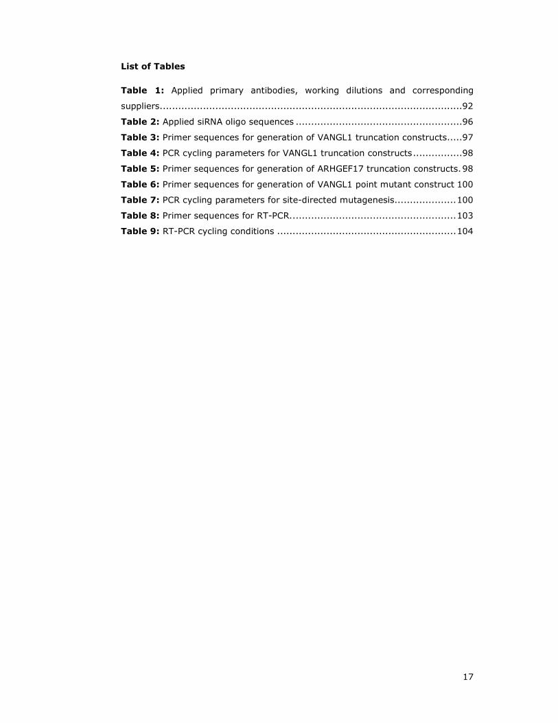
List of Tables
Table 1: Applied primary antibodies, working dilutions and corresponding
suppliers..................................................................................................92
Table 2: Applied siRNA oligo sequences ......................................................96
Table 3: Primer sequences for generation of VANGL1 truncation constructs.....97
Table 4: PCR cycling parameters for VANGL1 truncation constructs ................98
Table 5: Primer sequences for generation of ARHGEF17 truncation constructs. 98
Table 6: Primer sequences for generation of VANGL1 point mutant construct 100
Table 7: PCR cycling parameters for site-directed mutagenesis.................... 100
Table 8: Primer sequences for RT-PCR. ..................................................... 103
Table 9: RT-PCR cycling conditions .......................................................... 104

18
Abbreviations
aa amino acid
AF6 Acute lymphoblastic leukaemia-1 Fused gene on chr. 6
AMBRA Activating Molecule Beclin-Regulated Autophagy
AP-1 Activator Protein-1
APC Adenomatous Polyposis Coli
aPKC atypical Protein Kinase C
ATGs autophagy regulators
BBS Bardet-Biedl Syndrome
BCR B-Cell antigen Receptor
BGH Bovine Growth Hormone
BSA Bovine Serum Albumin
C17ORF62 Chromosome 17 Open Reading Frame 62
CamKII Calcium/calmodulin-dependent Kinase II
CASK CAlcium/calmodulin-dependent Serine protein Kinase
CBP Calmodulin Binding Peptide
CE Convergent Extension
CELSR Cadherin, EGF LAG Seven-pass G-type receptor 1
CK Casein Kinase
CM Conditioned Media
CMV Cytomegalovirus
CQ Chloroquine
CRD Cysteine Rich Domain
C-terminal Carboxy-terminal
CTGF Connective Tissue Growth Factors
CTHRC1 Collagen Triple Helix Repeat-Containing 1
CXCR2 CXC-motif chemokine Receptor 2
DAAM DVL Associated Activator of Morphogenesis 1
DAB2IP Doc-2/DAB2 Interactive Protein
DAG Diacylglycerol
DAPI 4',6-diamidino-2-phenylindole

19
DEPC Diethylpyrocarbonate
Dgo Diego
DH Dbl Homology
Dkk Dickkopf
DLG1 Disks Large homolog 1
DMSO Dimethyl Dulfoxide
DPI Dots Per Inch
DTT Dithiothreitol
DVL/Dsh Dishevelled
ECM Extracellular Matrix
E.coli Escherichia coli
EGF Epidermal Growth Factor
ERK Extracellular signal Regulated Kinase
ETS E26-Transcription factor proteins
FAK Focal Adhesion Kinase
FBS Foetal Bovine Serum
FL Full Length
FLNa Filamin A
FTase Farnesyl Transferase
FZD Frizzled
GAP GTPase Activating Protein
GEF Guanine nucleotide Exchange Factor
GMP-PNP 5'-Guanylyl imidodiphosphate
GPC3 Glypican 3
GPCR G Protein Coupled Receptor
GSK3 Glycogen Synthase Kinase 3
GST Glutathione S-Transferase
GTP Guanosintriphosphat
GTPases Guanosine Triphosphatases
HEK293T Human Embryonic Kidney 293 T cells
HSPGs Heparansulfate Proteoglycans
HVR Hypervariable Region

20
ICMT1 Isoprenylcystein Carboxymethyltransferase 1
JNK c-Jun NH2-terminal Kinase
KAI1/CD82 Kangai1/Cluster of Differentiation 82
LB Luria Bertani
LiCl Lithium Chloride
LIM LIN-11, Isl1 and MEC-3
LC3 microtubule-associated protein 1 Light Chain 3
LDLR Low-Density Lipoprotein Receptor
Lp loop tail
LPHN2 Latrophilin 2
LRP Low density lipoprotein-Related Protein
Ltap Loop-Tail-Associated Protein
MAP Mitogen-Activated Protein
MINK1 Misshapen-like Kinase 1
MMP Matrix Metalloprotease
MOPS 3-(N-morpholino)propanesulfonic acid
MS Mass Spectrometry
MST Mammalian Sterile-20-like protein kinase-1
MTOC Microtubule-Organizing Center
Myr myristolated
NDFIP1 Nedd4 Family Interacting Protein 1
NF-AT Nuclear Factor of Activated T cells
NMDAR N-methyl-D-aspartate glutamate ligand-gated ion channel receptors
NMT N-Myristoyltransferase
NTD Neural Tube Defect
O/N Overnight
PAK p21-activated kinases
PBS Phosphate Buffered Saline
PCP Planar Cell Polarity
PCR Polymerase Chain Reaction
PDZ PSD-95-Discs-large-ZO-1;

21
PE Phosphatidylethanolamine
PET Polyethylene terephthalate
PI3K Phosphatidylinositide 3-Kinases
PFA Paraformaldehyde
PH Pleckstrin Homology
PIP2 Phosphatidylinositol 4,5-bisphosphate
PIP3 Phosphatidylinositol (3,4,5)-triphosphate
PK PRICKLE
PKA cAMP-dependent Protein Kinase A
PKC Protein Kinase C
PLC Phospholipase C
PTM Post Translational Modification
PP1 Protein Phosphatase 1
PP2A Protein Phosphatase 2A
PRD Proline Rich Domain
PTK7 tyrosine-Protein Kinase-like 7
PTKR Protein Tyrosine Kinase Receptor
PTase Palmitoyltransferase
PVDF Polyvinylidene Fluoride
R1A Protein Kinase A type 1 regulatory subunit alpha
RA RAS Associating
RAB3GAP1 Rab3 GTPase-activating protein catalytic subunit
RAL RAS-like
RALBP1 RAL-Binding Protein-1
RALGDS RAL Guanine nucleotide Dissociation Stimulator
RASAL RASGAP-Activating-Like
RASGRF RAS-specific Guanine-nucleotide-Releasing Factor
RASGRP RAS-specific Guanine-nucleotide-Releasing Protein
RASIP1 RAS Interacting Protein 1
RASSF RAS Association domain-containing Family
RBD RAS/RHO Binding Domain
RCE1 RAS Converting Enzyme-1

22
REM RAS Exchange Motif
RHO RAS Homologous
RHOGDI RHO GDP Dissociation Inhibitor
RIN RAS interaction/interference protein
RLIP76 76 kDa RAL-Interacting Protein
RFG RAS Family GTPases
ROCK Rho Associated Kinase
ROR2 Receptor tyrosine kinase-like Orphan Receptor
R-RAS Related RAS
RSPO R-Spondins
RT Room Temperature
RT-PCR Reverse Transcript PCR
RTK Receptor Tyrosine Kinase
RYK Receptor related to tyrosine Kinase
SBP Streptavidine Binding Peptide
Scr Scramble
SCRIB SCRIBBLE
SDC Syndecan
SEM Standard Errors of the Means
SF9 Spodoptera frugiperda
sFRP secreted Frizzled-Related Proteins
SH SRC homology
SHOC2 soc-2 suppressor of clear homolog
SOS Son of Sevenless
SOST Sclerostin
Stbm Strabismus
SYNJ2BP Synaptojanin 2 Binding Protein
TAP Tandem Affinity Purification
TAE Tris-Acetate-EDTA buffer
TE Tris-EDTA buffer
TCA Trichloroacetic Acid
TCF T-Cell Factor

23
TCR T-Cell Receptor
TF8 Transcription Factor 8
TFF Trefoil Family Factors
TIAM T-cell lymphoma Invasion And Metastasis 1
TK Tyrosine Kinase
TM4SF Tetra Membrane Spanning Family
TM Transmembrane domain
tri trilobite
Ub Ubiquitin
UFD1L Ubiquitin Fusion Degradation 1-Like
UTR Untranslated Region
VANGL1 Vang-like
WD40 WD40 repeat protein
Wg Wingless
WGEF Weak similarity GEF
WIF WNT Inhibitory Factor
WNT Wingless/Int-1
wt Wild Type
YIF Yip1 Interacting Factor

24
CHAPTER 1
INTRODUCTION
25
1 Introduction
Cancer is a diverse disease affecting one out of four people and is characterised
among others by uncontrolled cell growth, evasion of apoptosis and invasion
(Hanahan and Weinberg 2000; Hanahan and Weinberg 2011).
RAS proteins play critical roles in multiple biological processes including
proliferation, differentiation and survival, and are one of the most frequently
mutated oncogenes detected in human cancer with an incidence of about 30%
(Barbacid 1987; Bos 1989; Campbell and Der 2004; Karnoub and Weinberg
2008).
Consequently, considerable efforts have been made to understand how RAS
proteins work and how their transforming activity can be blocked therapeutically
(Kloog and Cox 2000; Rodriguez-Viciana et al. 2005; Baines et al. 2011). In
addition, a better understanding of the full spectrum of effector pathways
regulated by RAS proteins is critical for comprehension of their many biological
functions and their contribution to human disease in order to develop more
specific treatment options (Rodriguez-Viciana et al. 2004).
1.1 RAS superfamily
The RAS superfamily of small guanosine triphosphatases (GTPases) consists of
over 150 human members and is subdivided into at least five major branches
based on structural homology, sequence and function (RAS, RHO, RAB, ARF, and
RAN subfamilies) (Figure 1.2A) (Wennerberg et al. 2005; Karnoub and Weinberg
2008).
GTPases act as nucleotide driven switches, cycling between an inactive GDP-
bound and an active GTP-bound state (Figure 1.1). The transition from an
inactive to active conformation is achieved by guanine nucleotide exchange
factors (GEFs), which stimulate the dissociation of GDP and subsequent
exchange to GTP (Schmidt and Hall 2002). This in turn induces a conformational
change, enabling the respective GTPase to recognise downstream targets,
termed effector proteins. The active state is terminated by the antagonising
effect of GTPase activating proteins (GAPs), which accelerate the intrinsic
GTPase activity of RAS proteins by several orders of magnitude, rendering the
GTPase GDP-bound, and consequently inactive (Takai et al. 2001; Bernards and
Settleman 2004).

26
Figure 1.1: RAS proteins cycle between a GTP-bound active and a GDP-bound
inactive state. Receptor tyrosine kinases (RTK), G-protein coupled receptors (GPCR)
and other types of membrane receptors (not shown) are able to activate guanine
exchange factors (GEFs), which in turn stimulate GDP release and GTP exchange. Binding
of GTP induces a conformational change within the switch 1 and switch 2 region of RAS
proteins, enabling effector interaction. This state is terminated by GTPase activating
proteins, enhancing RAS poor intrinsic GTPase activity, rendering the protein GDP-bound
and thus inactive.
1.2 RAS subfamily
RAS proteins are highly conserved and are found in all organisms from yeast to
human (DeFeo et al. 1981; Chang et al. 1982; Takai et al. 2001; Wennerberg et
al. 2005). Within the RAS superfamily, RAS lends its name to a subgroup
consisting of at least 39 members encoded by 36 genes in the human genome
(Figure 1.2). The best-studied genes of this family are the classical RAS proteins
encoded by H-RAS, K-RAS, and N-RAS genes (Figure 1.2B, blue square).
RAS proteins serve as signalling hubs, activated in response to a wide range of
extracellular stimuli, regulate cytoplasmic signalling networks that control gene
expression and regulation of cell proliferation, differentation and survival
(Wennerberg et al. 2005; Karnoub and Weinberg 2008). Because of their critical
27
roles in human oncogenesis they are the focus of intense research (Downward
2003; Repasky et al. 2004; Roberts and Der 2007).
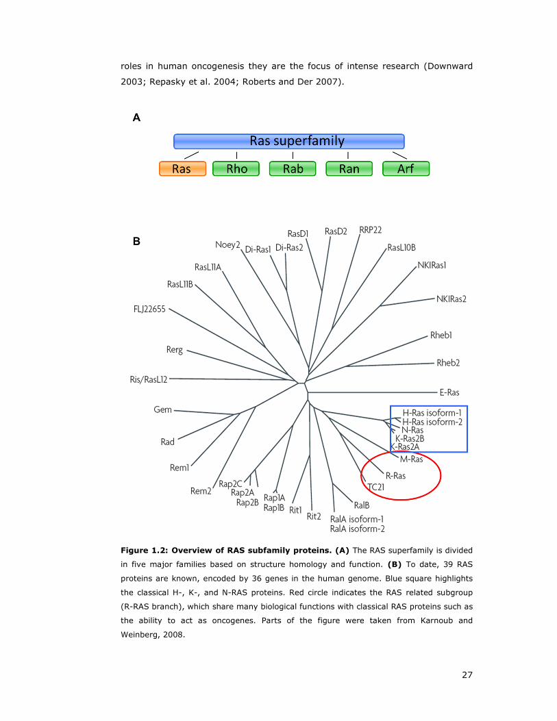
Figure 1.2: Overview of RAS subfamily proteins. (A) The RAS superfamily is divided
in five major families based on structure homology and function. (B) To date, 39 RAS
proteins are known, encoded by 36 genes in the human genome. Blue square highlights
the classical H-, K-, and N-RAS proteins. Red circle indicates the RAS related subgroup
(R-RAS branch), which share many biological functions with classical RAS proteins such as
the ability to act as oncogenes. Parts of the figure were taken from Karnoub and
Weinberg, 2008.
28
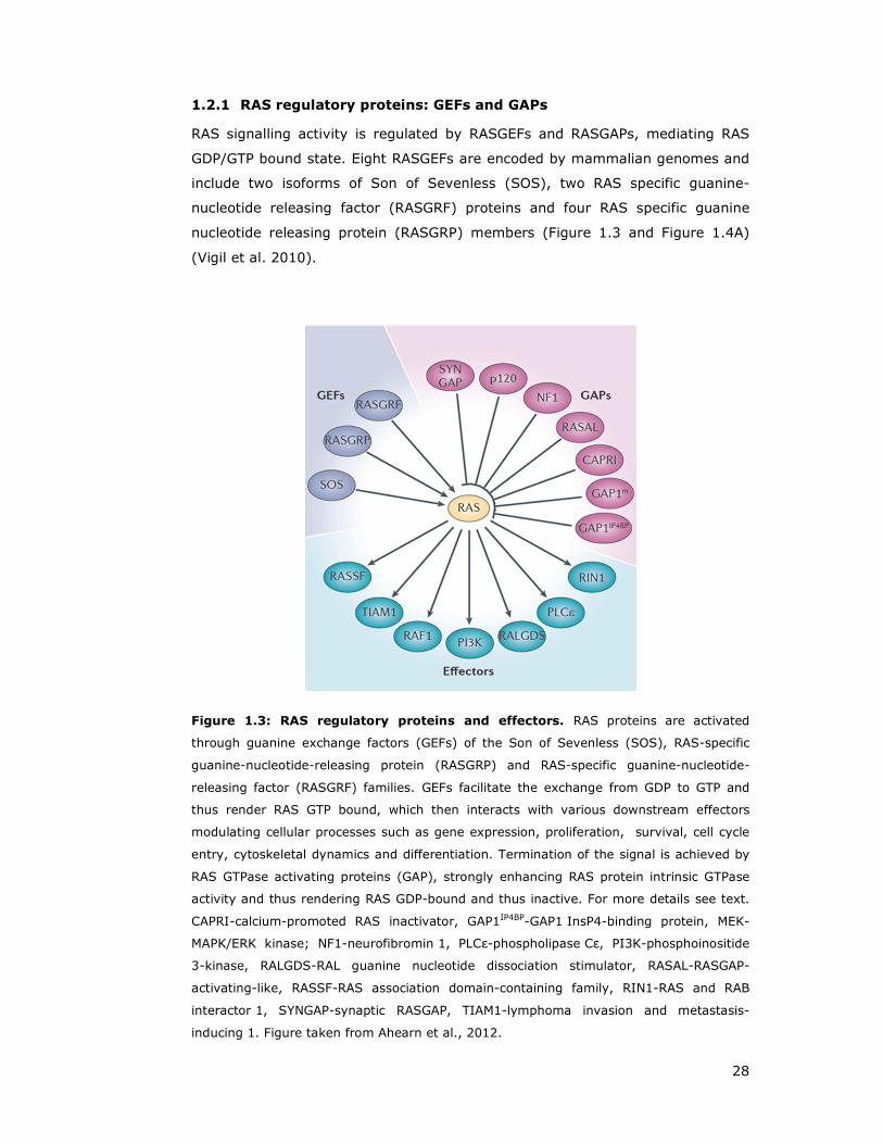
1.2.1 RAS regulatory proteins: GEFs and GAPs
RAS signalling activity is regulated by RASGEFs and RASGAPs, mediating RAS
GDP/GTP bound state. Eight RASGEFs are encoded by mammalian genomes and
include two isoforms of Son of Sevenless (SOS), two RAS specific guanine-
nucleotide releasing factor (RASGRF) proteins and four RAS specific guanine
nucleotide releasing protein (RASGRP) members (Figure 1.3 and Figure 1.4A)
(Vigil et al. 2010).
Figure 1.3: RAS regulatory proteins and effectors. RAS proteins are activated
through guanine exchange factors (GEFs) of the Son of Sevenless (SOS), RAS-specific
guanine-nucleotide-releasing protein (RASGRP) and RAS-specific guanine-nucleotide-
releasing factor (RASGRF) families. GEFs facilitate the exchange from GDP to GTP and
thus render RAS GTP bound, which then interacts with various downstream effectors
modulating cellular processes such as gene expression, proliferation, survival, cell cycle
entry, cytoskeletal dynamics and differentiation. Termination of the signal is achieved by
RAS GTPase activating proteins (GAP), strongly enhancing RAS protein intrinsic GTPase
activity and thus rendering RAS GDP-bound and thus inactive. For more details see text.
CAPRI-calcium-promoted RAS inactivator, GAP1IP4BP-GAP1 InsP4-binding protein, MEK-
MAPK/ERK kinase; NF1-neurofibromin 1, PLCε-phospholipase Cε, PI3K-phosphoinositide
3-kinase, RALGDS-RAL guanine nucleotide dissociation stimulator, RASAL-RASGAP-
activating-like, RASSF-RAS association domain-containing family, RIN1-RAS and RAB
interactor 1, SYNGAP-synaptic RASGAP, TIAM1-lymphoma invasion and metastasis-
inducing 1. Figure taken from Ahearn et al., 2012.
29
The best characterised GEF is SOS due to its role downstream of receptor
tyrosine kinases (RTKs) (Figure 1.7) (Yang et al. 2013). Each GEF contains a
CDC25 homology catalytic domain, which stimulates the release of GDP, and is
flanked by at least one adjacent RAS exchange motif (REM) (Figure 1.4A). The
Dbl homology domain (DH) mediates GDP/GTP exchange on RHO GTPases
(Rossman et al. 2005), and is located adjacent to the pleckstrin homology (PH)
domain, which facilitates membrane association. SOS specific histone homology
domains interact with negatively charged phospholipids (Figure 1.4A) (Yadav and
Bar-Sagi 2010).
Similar to SOS, RASGRFs also contain tandem REM-CDC25 and DH-PH domains,
in addition to calmodulin-binding motif containing conserved I and Q residues
(Figure 1.4A). RASGRF promotes RAS activation downstream of N-methyl-D-
aspartate glutamate ligand-gated ion channel receptors (NMDARs) through
receptor stimulated Ca2+ influx (Krapivinsky et al. 2003; Tian et al. 2004).
Both SOS and RASGRF exhibit a dual specificity for RAS and RAC GTPases,
providing a link between RAS activation and the function of RAS and RHO
proteins (Fan et al. 1998; Kiyono et al. 1999; Kiyono et al. 2000; Wennerberg
and Der 2004).
RASGRPs, also termed CalDAG-GEFs, form a group which is activated by
diacylglycerol (DAG) and phorbol ester. They share a common domain
architecture of a REM-CDC25 tandem domain, followed by a pair of Ca2+ binding
atypical EF hands and C1-protein kinase C conserved region 1, able to bind DAG
and phorbol esters (Figure 1.4A) (Mitin et al. 2006). Among the four members,
variations in GTPase specificity, expression, subcellular localisation and signal
regulation have been observed. RASGRP3 is reported to exhibit the broadest
GTPase substrate specificity, including RAP1 and RAP2 (Figure 1.5) (Mitin et al.
2006).
RAS activity is negatively regulated through GAPs, stimulating RAS poor intrinsic
GTPase activity by several orders of magnitude through stabilising the transition
state of the nucleophilic attack of water, mediated by insertion of an ‘Arg finger’,
thus terminating signalling (Bernards 2003; Bernards and Settleman 2004).
RASGAPs share a common ~250 amino acid RASGAP catalytic domain, but
otherwise do not shows sequence similarity or consistent domain structures in
the adjacent RASGAP flanking regions (Figure 1.4B) (Bernards and Settleman
2004).

30
Figure 1.4: Domain overview of RAS regulatory proteins. (A) RAS specific GEFs. In
addition to the CDC25 domain, SOS and RASGRF contain Dbl homology (DH) and
pleckstrin (PH) domains. Other domains found in RASGEFs include: H2A-histone 2A
homology, IQ-short calmodulin-binding motif containing conserved I and Q residues, EF-
Ca2+-binding EF hand, and C1-protein kinase C conserved region 1, which binds DAG and
phorbol esters. (B) RAS specific GAPs. RASGAPs have a common ~250 amino acid α-
helical catalytic domain (RasGAP). Other RASGAPs domains include: SH2-Src homology 2,
SH3-Src homology 3, PH, C2-protein kinase C conserved region 2, serving as a Ca2+-
dependent lipid-binding motif, Sec14-Sec14p-like lipid-binding domain and BTK-Bruton’s
tyrosine kinase cysteine-rich zinc-binding motif. Figure from Mitin et al, 2005.
31
There are eight RAS specific GAPs known, of which p120 RASGAP/RASA1 and the
tumour suppressor neurofibromin are the best studied (Dasgupta and Gutmann
2003; Bernards and Settleman 2004). Others are the synaptic RASGAP/SYNGAP,
GAP1m/RASA2, GAP1IB4BP/RASA3, and the two calcium activated GAPs
CAPRI/RASA4 and RASGAP-activating-like (RASAL) (Figure 1.4B) (Mitin et al.
2006; Rojas and Santos 2006).
Despite the similar domain structure of GAP1m, GAP1IB4BP, CAPRI and RASAL,
these RASGAP members exhibit different modes of regulation and plasma
membrane association. GAP1m exhibits a distinct perinuclear and cytosolic
localisation, which translocates to the plasma membrane upon EGF stimulation.
In contrast, GAP1IB4BP displays a constitutive plasma membrane association
(Lockyer et al. 1997; Cozier et al. 2000), and its RASGAP activity might be
regulated through inositol 1,3,4,5 tetrakisphosphate (Cullen et al. 1995).
Both CAPRI and RASAL undergo Ca2+ stimulated C2-dependent plasma
membrane association (Lockyer et al. 2001; Walker et al. 2004). In summary,
Ca2+ regulation is able to control activities of RASGAPs and RASGEFs.
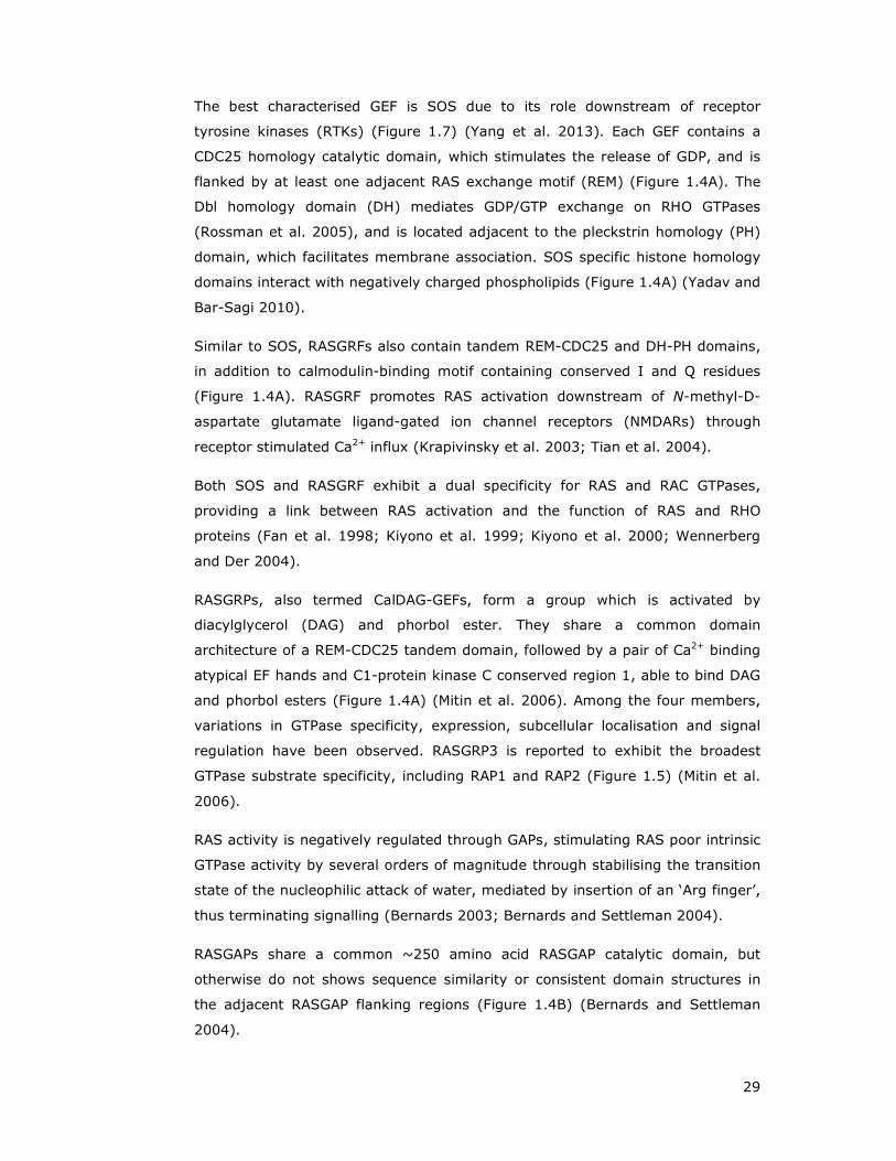
Figure 1.5: Overview of RASGEF and RASGAP GTPase substrate specificity.
1Conflicting reports for substrate specificity have been made. Figure from Mitin et al,
2006.

32
SYNGAP is expressed primarily in the brain and localises to glutamatergic
synapses and is positively regulated through phosphorylation of
calcium/calmodulin-dependent kinase II (Oh et al. 2004).
Doc-2/DAB2 interactive protein (DAB2IP) has been demonstrated to exhibit
RASGAP activity in vitro, and its gene expression has been reported to be
reduced in certain cancers (Wang et al. 2002).
1.2.2 RAS protein effectors
Figure 1.6: RAS effector pathways. Upon stimulation, RAS proteins interact with a
multitude of effector proteins involved in various cellular processes. See section 1.2.1 and
1.2.2 for details. Abbreviations are: AF6-acute lymphoblastic leukaemia-1 fused gene on
chromosome 6, CD1-cadherin domain-1, CDC42-cell division cycle-42, ELK-ETS-like
protein, ERK-extracellular signal regulated kinase, ETS-E26-transcription factor proteins,
Ins(1,4,5)P3-inositol-1,4,5-trisphosphate, JNK-Jun N-terminal kinase, MEK-mitogen-
activated protein kinase/ERK kinase, NF-κB-nuclear factor-κB, PI3K-phosphoinositide 3-
kinase, PKB/C-protein kinase B/C, PLA/Cε/D- phospholipase A/Cε/D, RalBP1-Ral-binding
protein-1, RASSF-RAS association domain-containing family, Rin1-RAS
interaction/interference protein-1, SAPK-stress-activated protein kinase, SHP2-Src-
homology 2 domain-containing protein Tyr phosphatase-2, TIAM1-T-cell lymphoma
invasion and metastasis-1. Figure from Karnoub and Weinberg 2008.

33
The first identified and also best characterised RAS effectors were RAF
serine/theonine kinases (Moodie et al. 1993; Vojtek et al. 1993; Warne et al.
1993; Zhang et al. 1993; Wellbrock et al. 2004), through which RAS activates
the growth-regulatory mitogen-activated protein (MAP) kinase cascade and the
E26-transcription factor proteins (ETS) (Figure 1.7) (Ahearn et al. 2012). This
pathway has been demonstrated to be necessary and sufficient for RAS induced
transformation of murine cells (Leevers et al. 1994; Stokoe et al. 1994;
Khosravi-Far et al. 1995; White et al. 1995). The identification of BRAF
mutations in cancers, generally in a mutually exclusive manner with RAS
mutations, underscores the importance of a this pathway downstream of RAS
function and in oncogenesis (Rajagopalan et al. 2002).
Figure 1.7: The RAS-RAF-ERK signalling pathway. Upon stimulation of a protein
tyrosine kinase receptor (PTKR) with growth factors, the cytoplasmic GRB2-SOS complex
is recruited to the plasma membrane through association of the GRB2-SH2 domain to
phosphorylated residues of the activated receptor. Interaction of the PH domain of SOS
with phospholipids induces a conformational change which allows binding of membrane
localised RAS and leads to its activation through GTP loading. RAS then initiates
downstream signalling through recruitment of RAF1 to the membrane and stimulation of
its kinase activity. RAF kinases then phosphorylate and activate MEK, which then
activates ERK, followed by phosphorylation and activation of multiple transcription factors
including ELK. Figure from Ahearn et al, 2012.
The second best characterised RAS effectors are the p110 (α, β, γ and δ)
catalytic subunit of class I Phosphoinositide 3-kinases (PI3K) (Rodriguez-Viciana
et al. 1994; Castellano and Downward 2010). PI3K signalling mediates cell

34
growth, proliferation, differentiation, motility, survival and intracellular
trafficking, which if deregulated, results in cancer (Cain and Ridley 2009;
Vanhaesebroeck et al. 2012).
Figure 1.8: Phosphoinositide 3-kinases are RAS effectors. (A) PI3K are activated
through binding of the regulatory p85 subunit the SRC homology (SH2) domain to the
phosphorylated receptor YxxM motif or through RAS binding of the catalytic subunits in a
GTP dependent manner. Class IA PI3Ks are activated through receptor tyrosine kinases
(RTKs), whereas Class IB PI3K are either G-protein couple receptor (GPRC) or RAS
activated. (B) The catalytic subunit of PI3K comprises a lipid kinase activity and
catalyses phosphorylation of Phosphatidylinositol 4,5-bisphosphate (PIP2) to
Phosphatidylinositol (3,4,5)-triphosphate (PIP3). PTEN- Phosphatase and tensin
homologue.

35
PI3Ks generate lipid second messengers by phosphorylation of phosphoinositides
at the 3’ position (Class I: phosphatidylinositol (3,4,5)-triphosphate) (Figure
1.8), which then recruit and bind downstream effectors such as AKT or lead to
stimulation of RAC and RHO through respective GEF activation (Welch et al.
2003; Costa et al. 2007; Cain and Ridley 2009).
Both described pathways, MAPK and PI3K, display a critical role in RAS mediated
oncogenesis, supported by the frequent mutational activation of BRAF (13%,
COSMIC database, www.sanger.ac.uk/genetics/CGP/cosmic) and PIK3CA (19%).
RAL guanine nucleotide dissociation stimulator (RALGDS) was the first of RAL
(RAS-like) exchange factors (RALGEFs) identified to be RAS effector proteins
(Kikuchi et al. 1994; Spaargaren and Bischoff 1994). RALGDS possesses
sequence homology with the REM and CDC25 domains found in RASGEFs.
However, studies revealed that RALGDS does not display an exchange activity
for RAS and rather is selective for RALA and RALB (Neel et al. 2011).
RAL interacts with multiple, functionally divergent downstream effector proteins
involved in endocytosis, exocytosis, actin organisation, phosholipid metabolism
and second messenger generation (Figure 1.9) (Neel et al. 2011). Effectors are
RALBP1 (Ral binding protein 1)/RLIP76 (76 kDa Ral-interacting protein) (Cantor
et al. 1995; Jullien-Flores et al. 1995), the octameric exocyst complex including
Sec5 and Exo84 (Moskalenko et al. 2002; Moskalenko et al. 2003),
Phospholipase C delta 1 (PLCδ1) (Sidhu et al. 2005), or the actin filament
crosslinking protein FilaminA required for RALA induced filopodium formation
(Ohta et al. 1999; Ohta et al. 2006).
In addition, RAS GTPases interact with a wide array of other effectors that are
less well characterised. RAS interaction/interference protein 1 (RIN1) has been
described as a GEF for RAB5-like proteins, implicated in endocytosis downstream
of RAS stimulating growth factors such as EGF (Han and Colicelli 1995; Tall et al.
2001).
T-cell lymphoma invasion and metastasis 1 (TIAM1) has been described as a
RAS effector and acts as a RAC specific GEF, that links RAS signalling to RAC,
which regulates the actin cytoskeleton and stimulates p21-activated kinases
(PAK) and c-JUN activated kinase (JNK) (Lambert et al. 2002). TIAM deficient

36
mice displayed a resistance to RAS induced skin carcinogenesis, suggesting that
TIAM activity is required for RAS transformation (Malliri et al. 2002).
Figure 1.9: Overview of RAL effectors and associated signalling pathways. RAL
downstream effectors mediate endocytosis, exocytosis, actin re-organisation,
phospholipid metabolism and generation of second messengers. Also, RAL has been
shown to activate various transcription factors and to regulate gene expression. PC-
phosphadidylcholine, PA-phosphatidic acid. Figure from Neel et al., 2011.
ALL (acute lymphoblastic leukaemia) 1 fused gene on chromosome 6 (AF6) or
AFADIN was identified by RAS-GTP affinity chromatography (Kuriyama et al.
1996), and has been characterised to contain both microtubule and actin binding
motifs, suggesting that AF6 might participate in cytoskeletal processes
downstream of RAS (Ponting and Benjamin 1996). Indeed, studies suggest that
AF6 associates with proteins that are involved in regulation of polarity, and to
localise to adherens junctions (Mandai et al. 1997).
Activation of the RAS effector Phospholipase C-ε (PLCε) leads to generation of
diacylglycerol through the cleavage of Phosphatidylinositol 1,4,5-bisphosphate
into Phosphatidylinositol (4,5)-triphosphate. This in turn promotes the release of
Ca2+ and the activation of PKC (Kelley et al. 2001; Song et al. 2001).

37
RAS association domain-containing family 5 (RASSF5) also known as NORE1 has
been identified as a RAS effector protein (Vavvas et al. 1998), which possesses
pro-apoptotic functions (Khokhlatchev et al. 2002; Vos et al. 2003) and a
potential involvement in cell migration (Dallol et al. 2005). Although the
functional mechanism is not fully elucidated yet, preliminary evidence links
RASSF proteins to the mammalian sterile-20-like protein kinase-1 (MST1) and
MST2, and to the Hippo signalling pathway (Tommasi et al. 2005; Cho et al.
2006; Varelas et al. 2010; Heallen et al. 2011).
In mitogenically stimulated cells, a complex formation of a RASGAP and the
cellular protein p190 has been observed (Moran et al. 1991). p190 contains a
carboxy-terminal domain that functions as a GAP for the RHO family GTPases,
and thus the RASGAP-p190 complex may serve to couple RAS- and RHO-
mediated signalling pathways.
More recent and less well characterised RAS effectors are the RAS interacting
protein 1 (RASIP1), also known as RAIN, which is required for endothelial cell
motility, angiogenesis and vessel formation (Xu et al. 2009; Xu et al. 2011). The
regulator of G-protein signalling 14 (RGS14) has been demonstrated to
facilitates the formation of a selective RAS-GTP/RAF/MEK/ERK multi-protein
complex to promote sustained ERK activation, which regulates H-RAS dependent
neuritogenesis (Willard et al. 2007; Willard et al. 2009). The protein impedes
mitogenic signal propagation (IMP1A) E3 ligase member acts as a steady-state
resistor within the RAF/MEK/ERK kinase module, and is directly regulated by RAS
(Matheny et al. 2004; Matheny and White 2009).
1.2.3 Structural features of RAS proteins
RAS proteins share a highly conserved G domain, involved in Mg2+ and
nucleotide binding and nucleotide hydrolysis (residues 1-164). The exchange of
GDP to GTP leads to conformational changes in two stretches of the protein, the
switch I (Asp30 - Asp38) and switch II (Gly59 – Glu67) regions, located close to
the γ-phosphate group of the activating GTP. The third structurally important
element of RAS GTPases is the P-loop (Gly10 – Ser17), which winds around the
β- and γ-phosphates and contributes to most of the required energy for GTP
binding (Figure 1.10) (Milburn et al. 1990).

38
Overlapping with switch I is the core RAS effector domain (Tyr32 - Tyr40), also
known as effector loop, which is important for downstream interactions with
effectors or inactivating proteins like GAPs (Figure 1.10) (Wennerberg et al.
2005).
Single amino acid activating mutations in RAS genes are frequently detected in
human cancers (Cox and Der 2010). For example, replacement of P-loop located
Gly12 residue in H-RAS with any other amino acid except proline results in an
increased tumour transforming potential due to its importance for GTP binding
(Clark et al. 1985).
Figure 1.10: Structure of RAS proteins. (A) RAS proteins consist of a GTP binding
domain (residues 1-164) and a C-terminal hypervariable region (amino acids 166-189)
that contains isoform-specific membrane binding and trafficking determinants. Further
strutural motifs include the phosphate binding motif P-loop, switch I and II regions as well
as a C-terminal targeting sequence (CAAX motif). (B) Nucleotide-dependent structural
rearrangements. The differences between the inactive GDP bound and the active GTP-
bound RAS are confined to mainly in two regions, termed switch I and switch II, both of
which are required for the interactions of RAS with its regulators and effectors. The γ-
phosphate induces significant changes in the orientation of the switch II region through
the interactions that it establishes with Thr35 and Gly60. Notably, these two residues are
among the most conserved residues in the GTPase family, suggesting that the
mechanisms of GTP binding (and hydrolysis) are essentially the same among various
members. Figure adapted from Karnoub and Weinberg, 2008.

39
The oncogenic mutation Gln61Leu positioned in switch II, impairs GTP hydrolysis
as a result of its catalytically essential residue involved in GAP interaction (Resat
et al. 2001; Karnoub and Weinberg 2008; Cox and Der 2010).
1.2.4 Post translational modification of RAS proteins
RAS proteins are post-translational modified (PTM) through hydrophobic groups,
dictating specific subcellular localisations, that are essential for biological activity
(Hancock 2003; Hancock and Parton 2005; Wright and Philips 2006; Ahearn et
al. 2012).
RAS proteins terminate with a CAAX tetrapeptide, were C represents a cysteine
residue, A - any aliphatic amino acid and X symbolises any residue, which in
conjunction with immediate upstream cysteine residues form the membrane
targeting sequence, dictate interaction with distinct membrane compartments
and subcellular localisations (Figure 1.11A) (Wright and Philips 2006; Eisenberg
et al. 2013).
Notably, all CAAX proteins still require a second signal for trafficking to the
plasma membrane (Hancock et al. 1990). In case of H-RAS, N-RAS and K-
RAS4A, the second signal is comprised of Cys residues that are palmitoylated, a
process also known as acylation (Resh 1999). For K-RAS4B, the second signal
consists of a polybasic Lys-rich region with a net positive charge that is believed
to form an eloctrostatic interaction with the negatively charged phospholipids at
the plasma membrane (Figure 1.11A).
The CAAX motif recognition sequence is modified by the enzyme farnesyl
transferase (FTase), which attaches a farnesyl group to the cysteine residue via
a thioester bond. Next, the last three amino acids (AAX) are proteolytic cleaved,
catalysed by the RAS converting enzyme-1 (RCE1) and the now C-terminal
cysteine residue undergoes a methylesterification mediated by isoprenylcystein
carboxymethyltransferase 1 (ICMT1). In addition, RAS proteins are further
modified with palmitoyl groups at different cysteine residues in the vicinity of the
C-terminus performed through palmitoyltransferase (PTase), which is required
for membrane association (Figure 1.11B) (Wright and Philips 2006).
Besides attachment of polyisoprenoid lipids, RAS GTPases are found to be further
post translationally modified through phosphorylation, nitrosylation,
40
ubiquitinylation and peptidyl-prolyl isomerisation, which are conditional upon cell
activation, redox state or microbial pathogenesis (Figure 1.12) (Ahearn et al.
2012).
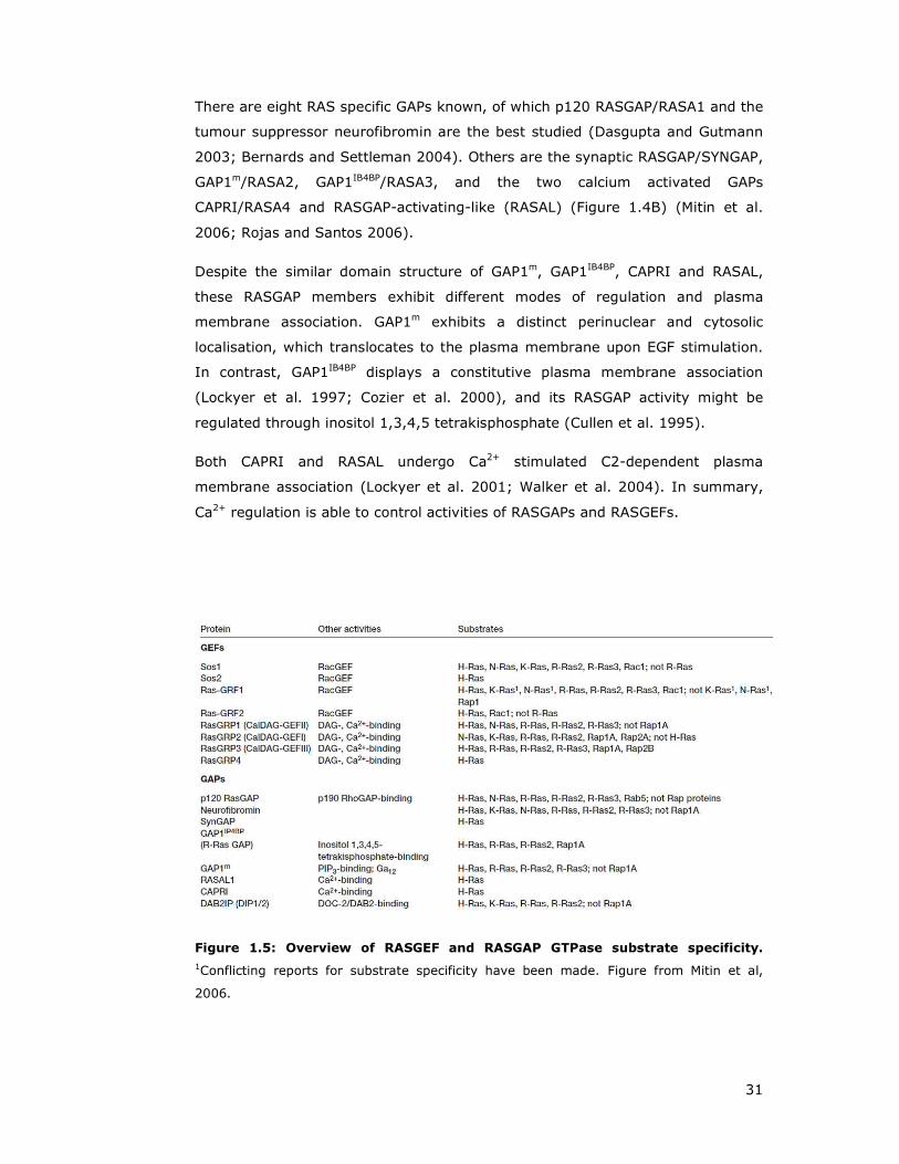
Figure 1.11: C-terminal RAS membrane targeting signals. (A) This figure illustrates
the hypervariable region (HVR) of H-, K- and N-RAS proteins. Highlighted in yellow is the
first post translational modification (PMT) motif, the cysteine residue forming the
membrane targeting CAAX motif, which is farnesylated. An additional upstream ‘second’
signal is required for plama membrane targeting, shaded in green. For H-RAS, N-RAS and
K-RAS4A, the second signal consists of Cys residues that are palmitoylated. For K-RAS4B,
the second signal consists of a polybasic region with a net positive charge of eight (pink)
and Ser181 residue, which is the principal site of phosphorylation is highlighted in blue.
Figure adapted from Ahearn et al, 2012. (B) C-terminal processing of RAS proteins.
Membrane association is absolutely required for RAS GTPase function and takes place
through C-terminal modification of the CAAX motif. Farnesyltransferase (FTase) covalently
attaches a farnesyl group (F, orange) to the final cysteine residue, followed by protealytic
cleavage of the last three amino acids (AAX) by RAS converting enzyme-1 (RCEI). The
now terminal farnesylated cysteine is carboxymethylated by isoprenylcystein
carboxymethyltransferase 1 (ICMT1). RAS proteins are further modified (with the
exception of K-RAS-4B) through attachment of palmitoyl groups (red lines) catalysed
through palmitoyltransferase (PTase). Figure taken from Karnoub and Weinberg, 2008.

41
A recent study showed that the cis–trans prolyl isomerase 12 kDa FK506-binding
protein (FKBP12) functions in the regulation of RAS depalmitoylation (Ahearn et
al. 2011) through isomerization of the Gly–Pro peptidyl-prolyl bond at position
178–179. This constitutes a molecular timer for acylation and therefore this
modification gained more appreciation for signalling purposes (Lu et al. 2007).
Figure 1.12: Post translational modifications of RAS proteins. Overview of reported
post-translational modifications (PTMs) for H-RAS (top) and K-RAS4B (bottom). Sites of
mono-ubiquitylation and di-ubiquitylation are indicated with blue spheres (Ub-ubiquitin).
Note that glucosylation and ADP-ribosylation modifications have been only observed in
cells intoxicated with bacterial virulence factors. All other PTMs are intrinsic to all
eukaryotic cells, which have been implicated in RAS trafficking and signalling. Figure from
Ahearn 2012.
H-RAS, N-RAS and K-RAS4B were recently shown to be substrates for mono-
ubiquitination and di- ubiquitination modifications (Jura et al. 2006; Sasaki et al.
2011). The responsible E3 ligase was identified as rabaptin 5-associated
exchange factor for RAB5 (RABEX5), which requires RIN for RABEX5-dependent
RAS ubiquitination (Xu et al. 2010). RIN1, through its function as a GEF for
RAB5, is believed to promote the RAB5-dependent recruitment of RABEX5 to
endosomal sites, where H-RAS ubiquitination takes place. Thus, ubiquitination
regulates trafficking of H-RAS to and from endosomes, indicating that this
modification poses an additional way to regulate RAS compartmentalisation, and
the spatial control of its signalling output.
Similarly, phosphorylation of K-RAS at Ser181 by PKC reduces the net charge of
the polybasic region, thus leading K-RAS4B to lose affinity for plasma membrane
association and to accumulate on endomembranes (Ballester et al. 1987; Bivona
et al. 2006).
42
The RAS Cys118 residue is highly conserved among RAS isoforms, and it is the
most surface-exposed Cys on these GTPases. Studies revealed that Cys118 could
be nitrosylated when exposed to nitric oxide (NO) (Lander et al. 1995b; Lander
et al. 1996). Even though S-nitrosylation does not alter the structure of RAS,
this modification leads to enhanced guanine nucleotide exchange (Lander et al.
1995a; Williams et al. 2003), which promotes more efficient RAS activation.
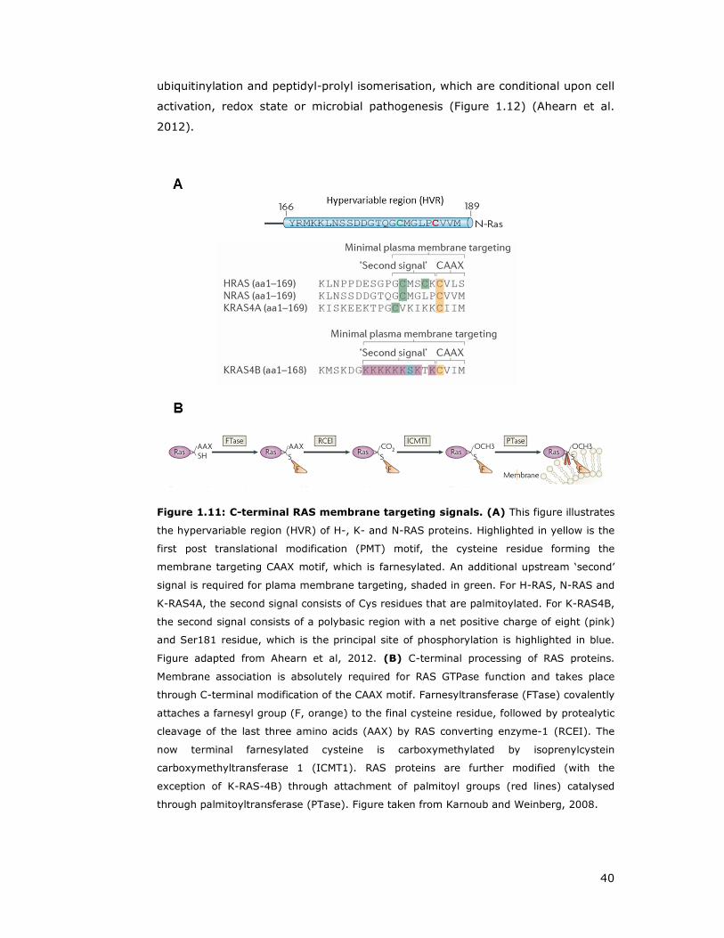
Once the ER localised CAAX processing is complete, RAS isoforms diverge into
their subsequent trafficking routes (Wright and Philips 2006). Palmitoylated RAS
proteins (H-RAS, N-RAS and K-RAS4A) travel to the Golgi, where they get
acylated. This step traps them into the Golgi, and leads to incorporation into
transport vesicles and to enter the secretory pathway.
In contrast, K-RAS4B does not shuttle to the Golgi but instead associates directly
with the plasma membrane. Whether this is achieved through diffusion or by an
as yet unknown mechanism remains elusive (Figure 1.13) (Choy et al. 1999;
Apolloni et al. 2000).
On the membrane, RAS proteins are able to become enriched with the
cholesterol-binding protein caveolin and localise to specific plasma membrane
micro-domains (lipid raft or non-raft domains) (Simons and Ikonen 1997; Prior
et al. 2001) (Figure 1.12). It seems that functional activation of RAS depends on
its release from lipid-raft micro-domains in the plasma membrane and its
redistribution to nearby sites thus enabling interaction with various downstream
effectors (Prior et al. 2001).
Figure 1.13: RAS post translational modifications lead to various subcellular
localisations. See text for details. Figure from Karnoub 2008.

43
1.3 The R-RAS subgroup
Within the Ras subfamiliy, the related Ras (R-RAS) subgroup shares the highest
similarity with classical Ras proteins (H-, K- and N-RAS ) and exhibits significant
oncogenic potential (Figure 1.2B, red circle) (Saez et al. 1994; Graham et al.
1999; Movilla et al. 1999; Erdogan et al. 2007; Erdogan et al. 2008). Three R-
RAS family members have been characterised, R-RAS (Lowe et al. 1987), TC21
(R-RAS2)(Drivas et al. 1990) and M-RAS (R-RAS3) (Matsumoto et al. 1997).
Although, this subgroup shares many biochemical and biological properties with
classical Ras proteins such as an identical effector domain, overlapping GEFs and
GAPs and the ability to act as oncogenes, evidence suggest that R-RAS GTPases
have distinct properties and functions to classical RAS proteins (Zhang et al.
1996; Huff et al. 1997; Self et al. 2001) (Figure 1.14).
1.3.1 R-RAS
The founding member R-RAS shares a 55% aa identity with H-RAS and was
discovered using a low stringency hybridisation probe of H-RAS, underscoring
the high similarity to the classical H-, K- and N-RAS proteins (Lowe et al. 1987)
(Figure 1.14). Accordingly, R-RAS and RAS share at least two GAPs,
p120RASGAP and neurofibromin, the exchange factor RASGRF and effectors RAF,
PI3K, RalGDS, RIN and AF6 (Rey et al. 1994; Spaargaren and Bischoff 1994;
Spaargaren et al. 1994; Gotoh et al. 1997; Marte et al. 1997; Ebinu et al. 1998).
Figure 1.14: Oncogenic classical Ras proteins are structurally highly similar to R-
RAS proteins. R-RAS displays a 55% amino acid homology to H-RAS, and structural
conserved features such as the effector loop mediating downstream effector binding, are
entirely identical between classical RAS proteins and the members of the R-RAS branch.
Figure adapted from Karnoub and Weinberg, 2008.

44
Further, oncogenic activating mutants in human cancers analogous to those of
found in the H-RAS oncogenes, have been identified for R-RAS GTPases
(COSMIC database).
Despite the fact that R-RAS proteins share many regulatory and effector proteins
with RAS proteins (Ehrhardt et al. 2002), R-RAS causes a transformed
phenotype which is distinct from that induced by RAS (Self et al. 2001).
Therefore, it is likely that R-RAS controls signalling and cellular processes which
are distinct from classical RAS proteins. Indeed, R-RAS does not activate MAP
kinase pathways and is only weakly oncogenic (Self et al. 2001). Furthermore,
R-RAS activates α5β1 integrin independently of PI3K, through a different
mechanism to H-RAS (Zhang et al. 1996; Sethi et al. 1999; Kinashi et al. 2000).
RALBP1 also termed RLIP76, is the only effector known to date exclusive to R-
RAS that is not shared with other classial RAS family members, and mediates
adhesion-dependent RAC activation and cell migration (Goldfinger et al. 2006).
R-RAS differs from classical RAS proteins structurally through e.g. an N-terminal
26 amino acid extension, and differs at nine amino acids within the extended
effector region across amino acids 23-46 (Lowe et al. 1987; Self et al. 1993;
Holly et al. 2005). N-terminal truncated R-RAS failed to activate RAC and
resulted in reduced cell spreading compared to full length R-RAS protein in 32D
mouse myeloid cells, whereas adhesion or subcellular localisation was not
affected (Holly et al. 2005).
Active R-RAS was demonstrated to increase ligand binding activity of integrins
and to stimulate the adhesive properties of suspension cells (Zhang et al. 1996).
In this context it has been further shown that R-RAS is able to antagonise H-RAS
mediated integrin activation suppression (Sethi et al. 1999). R-RAS integrin
regulation seems to be mediated through the effector loop and prenylation site
of R-RAS (Oertli et al. 2000) possibly through a distinct plasma membrane
micro-domain distribution (Hansen et al. 2003). Further, it has been
demonstrated that R-RAS effects on integrin activation are negatively regulated
through phosphorylation of the effector domain tyrosine residue Tyr66 by Eph
receptor kinase EphB2 (Zou et al. 1999) or c-SRC, measured by cell adhesion
reducing effects (Zou et al. 2002). Notably, it has been reported that R-RAS
activation of integrin signalling could be a bi-directional system, since R-RAS has
been observed to be activated downstream of integrin-mediated adhesion
(Wozniak et al. 2005).

45
Studies focussing on R-RAS C-terminal sequences employing R-RAS/RAS
chimeras, showed that the last 53 amino acids are necessary and sufficient to
specify the contrasting biological activities beween R-RAS and RAS. Identified
properties included H-RAS induced integrin suppression, reactive oxygen species
production and morphological effects of transformed cells (Hansen et al. 2002).
R-RAS acts as a regulator of cell adhesion through effects on integrin affinity
(Keely et al. 1999; Hansen et al. 2003), and focal adhesion formation (Berrier et
al. 2000; Kwong et al. 2003). For example, in T47D breast cells active R-RAS
strongly stimulates focal adhesion kinase (FAK) and p130Cas phosphorylation by
an unknown mechanism (Kwong et al. 2003). Using a H-RAS/R-RAS chimera
approach mapped the focal adhesion targeting signal to the C-terminal end of R-
RAS (Furuhjelm and Peranen 2003). It is further known that R-RAS promotes
adhesion-induced activation of RAC through its effector PI3K (Marte et al. 1997;
Berrier et al. 2000; Holly et al. 2005).
R-RAS has been found to interact with Oxysterol-binding protein (OSBP)-related
protein 3 (ORP3) (Goldfinger et al. 2007; Lehto et al. 2008). ORP3 is highly
expressed in epithelial, neuronal and hematopoietic cells and has been described
to be involved in integrin signalling and organisation of the actin cytoskeleton
(Lehto et al. 2001). Therefore it has been proposed that R-RAS – ORP3
interaction is able to regulate cell-cell and cell-matrix adhesion (Lehto et al.
2008).
As noted above, the exclusively R-RAS binding protein RLIP76 (does not bind H-
RAS or RAP1) mediates the R-RAS induced effect on cell spreading and migration
through ARF6 (Goldfinger et al. 2006). Activated ARF6 stimulates adhesion-
induced RAC1 activation, leading to lamellipodium formation and cell migration
(Radhakrishna et al. 1999; Santy and Casanova 2001; Goldfinger et al. 2006).
Additional studies confirm a role of R-RAS in cell migration through the spatial
regulation of RAC and RHO (Wozniak et al. 2005; Shang et al. 2011). Consistent
with this, activated R-RAS has been shown to promote cell migration of breast
(Keely et al. 1999; Jeong et al. 2005) and cervical epithelial cells (Rincon-Arano
et al. 2003; Mora et al. 2007) and has been identified as a cancer linked gene
(Nishigaki et al. 2005). In the latter study using gastric cancer samples, it was
discovered that gene demethylation of specific CpG sites within the first intron of
R-RAS causes activation in more than half of the tested samples (Nishigaki et al.
2005).

46
Further evidence for a role of R-RAS in actin cytoskeleton re-modelling and cell
migration stems from interaction with the actin-binding scaffolding protein
FilaminA (FLNa), which also binds to integrin β1, β2, β7 tails. FLNa is necessary
for tumour cell metastasis and vascular remodelling (Ott et al. 1998; de Curtis
2001; Yamazaki et al. 2005; Feng et al. 2006). Studies in human melanoma
cells M2 showed that R-RAS functionally associates with FLNa and thereby
regulates integrin dependent migration (Gawecka et al. 2010) or maintenance of
endothelial barrier function in human coronary artery endothelial cells (Griffiths
et al. 2011).
R-RAS was found enriched on both early and recycling endosomes, and displayed
a higher activity if endosome located compared to the plasma membrane,
resulting in RGL2 effector recruitment (a GEF for RALA) (Takaya et al. 2007).
Active R-RAS lengthened the duration of initial membrane protrusion and
promoted the formation of ruffling lamellipodia and the lack of filopodia
structures, which might be mediated through activity of PLCε, (Ada-Nguema et
al. 2006). Consistently, studies in COS-7 cells showed that R-RAS localises to the
leading edge of migrating cells, where it regulates membrane protrusion and
ruffle formation (Conklin et al. 2010). Furthermore, it has been demonstrated
that endogenous R-RAS co-localises with β-integrin in ruffles and endocytic
vesicles and R-RAS is required for integrin accumulation into ruffles (Conklin et
al. 2010). In addition, it has been described that active R-RAS is able to convert
RIN2 from a GEF to an adaptor protein which preferentially binds RAB5. Active
RAB5 then triggers the endocytosis of ECM bound/active integrins and active R-
RAS to early endosomes, which then mediates the TIAM-mediated activation of
RAC1 (Sandri et al. 2012).
Semaphorins are large secreted and transmembrane molecules involved in axon
guidance, which are modulated by plexins and signal to the cytoskeleton in a
RHO and RAC dependent manner (Kolodkin et al. 1993; Tamagnone et al. 1999).
Plexins have been proposed to behave as GAPs specific for R-RAS and M-RAS,
suggesting a selective role for R-RAS proteins in plexin-regulated signalling
pathways, involved in cell migration and axon guidance (Oinuma et al. 2004;
Kruger et al. 2005; Pasterkamp 2005; Saito et al. 2009; Oinuma et al. 2010).
However, a more recent paper suggested that Plexins are GAPs for RAP1 rather
than R-RAS (Bos and Pannekoek 2012).

47
R-RAS null mice are viable and fertile with no obvious abnormalities and normal
tissues upon histologic examination (Komatsu and Ruoslahti 2005). However, R-
RAS deficient mice exhibited exaggerated neointimal thickening in response to
arterial injury and increased angiogenesis in implanted tumours (Komatsu and
Ruoslahti 2005). Expression of R-RAS is mainly restricted to endothelial cells and
vascular smooth muscle cells, where is has been found to regulate blood vessel
remodelling, through to integrin-mediated cell adhesion to ECM components
(Zhang et al. 1996; Kinbara et al. 2003; Komatsu and Ruoslahti 2005).
Moreover, a recent study confirmed this observation and established R-RAS as a
critical regulator of vessel integrity and function during tumour vascularisation
(Sawada et al. 2012).
1.3.2 TC21
TC21 was initially cloned from a human teratocarcinoma cDNA library using
oligonucleotides corresponding to the conserved region of RAS genes (Drivas et
al. 1990). TC21 shows similar transformation ability than H-RAS (Graham et al.
1999) and mutated TC21 has been found in human tumours (Chan et al. 1994;
Huang et al. 1995; Barker and Crompton 1998) and COSMIC database.
Moreover, overexpression of TC21 has been associated with oral, breast,
hepatocellular and esophagous carcinoma (Clark et al. 1996; Arora et al. 2005;
Sharma et al. 2005; Luo et al. 2010; Macha et al. 2010).
TC21 has been found to be essential for survival and maintenance of B- and T
lymphocytes, through linking respective antigen receptors to PI3K-mediated
survival pathways (Delgado et al. 2009). Moreover, TC21 and RHOG mediate
T cell receptor internalisation from the immunological synapse through
phagocytosis (Alarcon and Martinez-Martin 2011; Martinez-Martin et al. 2011).
TC21 deficient mice do not display major alterations in viability, growth rates,
cardiovascular parameters, or fertility but exhibit a delay in mammary gland
development (Larive et al. 2012). TC21 is expressed in the same cell types as R-
RAS (Kozian and Augustin 1997; Komatsu and Ruoslahti 2005; Larive et al.
2012) and they share overlapping regulatory proteins, and thus might mediate
similar cellular functions (Ohba et al. 2000; Self et al. 2001; Ehrhardt et al.
2002).

48
1.3.3 M-RAS
The last identified R-RAS member is M-RAS (208aa) or R-RAS3, which was first
cloned from mouse skeletal muscle myoblasts and rat brain (Matsumoto et al.
1997). M-RAS is predominantly expressed in the central nervous system, and is
implicated in re-organisation of the actin cytoskeleton and promotes neurite
outgrowth in PC12 cells (Matsumoto et al. 1997; Kimmelman et al. 2002; Nunez
Rodriguez et al. 2006; Sun et al. 2006). Plexins also function as GAPs for M-RAS
and thereby suppress dentritic outgrowth and branching (Saito et al. 2009).
Further, the MRAS gene has been identified by genome wide associated studies
to be a new susceptibilty locus for coronary heart disease (Erdmann et al. 2009).
Despite the high homology of M-RAS and R-RAS, these GTPases perform
different functions due to differential effector coupling (Marte et al. 1997; Nunez
Rodriguez et al. 2006). M-RAS activates MAPK signalling through BRaf,
stimulating neurite outgrowth, even though activation by EGF seems to be more
sustained compared to prototypic RAS. Furthermore, phosphatase holoenzyme
comprised of Shoc2/Sur8 and the catalytic subunit of PP1 functions as a highly
specific M-RAS effector to modulate RAF activity (Rodriguez-Viciana et al. 2006).
M-Ras null mice appeared phenotypically normal, including lack of detectable
morphological and neurological defects, or alteration of MAPK/PI3K signalling
(Nunez Rodriguez et al. 2006).

49
1.4 RHO GTPases
Ras HOmologous (RHO) GTPases form another subgroup of the RAS superfamily
and regulate cytoskeletal architectures associated with cell migration and
polarity, adhesion, proliferation and differentiation (Figure 1.2A and Figure 1.15)
(Etienne-Manneville and Hall 2002; Raftopoulou and Hall 2004; Ridley 2006;
Heasman and Ridley 2008; Vega and Ridley 2008).
RHO family small GTPases act as molecular switches and are activated by GEFs
and inhibited by GAPs as described earlier for RAS proteins (Figure 1.1). A third
level of regulation is provided by the stoichiometric binding of RHO GDP
dissociation inhibitor (RHOGDI), masking prenyl modification and promoting
cytosolic sequestration (DerMardirossian and Bokoch 2005; Cherfils and Zeghouf
2013).
Figure 1.15: Overview of RHO GTPase family. The RHO GTPase group consists of 20
members subdivided into eight branches. High sequence similarity is found between
proteins within the RAC and RHO subfamilies. Figure taken from Heasman and Ridley,
2008.
50
The RHO GTPase familiy is comprised of more than 20 distinct proteins with the
best characterised members RHOA, RAC1 and CDC42, acting as regulators of
actin cytoskeleton re-arrangements (Heasman 2008 #482). In addition to the
wide range of cellular processes from cell polarity and movement, cell shape,
cell-cell and cell-matrix interactions (Etienne-Manneville and Hall 2002;
Raftopoulou and Hall 2004; Ridley 2006), RHO familiy proteins play additional
roles in a variety of cellular processes, including secretion, smooth muscle
contraction, migration, neurite retraction and gene transcription (Etienne-
Manneville and Hall 2002; Jaffe and Hall 2002; Jaffe and Hall 2005).
1.4.1 RHO regulatory proteins: GEFs, GAPs and GDIs
About 71 different GEFs for RHO family members (RhoGEFs) belonging to the Dbl
protein family, which share the typical tandem motif consisting of a Dbl
homology (DH) domain, responsible for exchange activity, and a pleckstrin
homology (PH) domain, involved in subcellular localisation of the GEFs
(Blomberg et al. 1999; Zheng 2001; Schmidt and Hall 2002; Hall 2012). In
addition to this tandem motif, RhoGEFs often contain one or more additional
signal transduction domains, like SH2, SH3, PDZ and additional PH domains.
Therefore, they often function as molecular bridges between different signal
transducion pathways (Zheng 2001; Schmidt and Hall 2002).
In addition, characterisation of a protein DOCK and its orthologues led to the
identification of a second sequence-unrelated family of GEFs, which consist of 11
members in the mammalian genome (Meller et al. 2005). The substrates of
DOCK family proteins appear to be RAC and CDC42 GTPases only (Hall 2012).
So far, 67 RHO family GAPs within the mammalin genome have been reported.
RHOGAPs harbour a bcr homology (BH) domain (Diekmann et al. 1991; Hall
2012). In addition, three RHO family GDIs have been identified, which inhibit the
dissociation of GDP from RHO (Matsui et al. 1990).
Identification of RHO family GTPase effectors is hard due to the lack of CRIB
domains. Nonetheless, more than 100 targets have been reported to interact
with the RHO family (Bishop and Hall 2000). About one third are kinases and a
large number are scaffold or adaptor proteins. Analysis of RHO GTPases
signalling pathways is a challenging task due to the fact that RHO, RAC and
CDC42 are able to interact with 20 or 30 different proteins in a GTP dependent

51
manner. This led to the hypothesis that each of the mentioned small GTPases is
able to regulate numerous distinct signalling transduction pathways.
1.4.2 RHO proteins
RAS homologue gene family member A (RHOA), RHOB and RHOC are highly
homologous, but appear to have divergent biological functions (Wheeler and
Ridley 2004). Carboxy-terminal modifications and differences in subcellular
localisation allow these three proteins to respond to and act on distinct signalling
molecules (Wennerberg and Der 2004; Wheeler and Ridley 2004).
RHOA, the best studied member of the three has been demonstrated to function
in regulation of actomyosin contractility, cytokinesis, focal adhesion assembly
and cell polarity (Barry and Critchley 1994; Kimura et al. 2000; Bi et al. 2005;
Van Keymeulen et al. 2006).
RHOB is involved in regulation of cell shape, migration and adhesion, but also
has been discovered to play a role in protein trafficking and in CXC-motif
chemokine receptor 2 (CXCR2)-mediated chemotaxis (Neel et al. 2007;
Sandilands et al. 2007; Wheeler and Ridley 2007). Recent studies showed that
RHOB is able to differentially control Akt function in endothelial cells during
breast tumourigenesis, and that RHOB regulates cell migration through altered
focal adhesion dynamics (Witze et al. 2008; Vega et al. 2012; Kazerounian et al.
2013).
1.4.3 RAC and CDC42
RAC and CDC42 play key signalling roles in cytoskeletal re-organisation,
membrane trafficking, transcriptional regulation, cell growth and development
(Ridley 2001c; Wennerberg and Der 2004; Heasman and Ridley 2008).
RAC1 promotes lamellipodium formation and membrane ruffling, whereas CDC42
stimulates filopodia and actin microspike formation (Ridley 1999; Ridley 2001a).
52
1.5 WNT signalling branches
The WNT pathway regulates vital developmental processes during
embryogenesis, including cell-fate specification, tissue patterning, cell polarity,
and cell movements during morphogenesis. In addition, this pathway plays key
roles in adult tissue homeostasis, stem cell renewal and has been implicated in
tumourigenesis (Veeman et al. 2003a; Logan and Nusse 2004; Reya and Clevers
2005; Zallen 2007).
Notably, the WNT signalling pathway comprises a complex network of ligands,
cognate receptors and co-receptors and agonistic/antagonistic modulators acting
to activate a vast intracellular downstream signalling network (Niehrs 2012),
which will be discussed in more detail in the following section 1.6.
The best studied branch of the signaling network is commonly known as
canonical signalling pathway and results in ß-catenin dependent gene
transcription and regulation of cell fate decisions (Logan and Nusse 2004;
Kikuchi et al. 2009; MacDonald et al. 2009). Less well-defined cascades, known
as ß-catenin independent or non-canonical pathways, include the WNT/Ca2+
pathway and the planar cell polarity branch, involved in cytoskeletal
rearrangements and cell movements (Veeman et al. 2003a; Zallen 2007).
Overall, these pathways collectively regulate cell survival, cell proliferation, cell
fate determination, cell polarity, tissue patterning, and tissue homeostasis
(Veeman et al. 2003a; Reya and Clevers 2005; Zallen 2007; MacDonald et al.
2009). These three major signalling pathways associated with WNT-receptor
interaction, are outlined in more detail below.
1.5.1 β-catenin (canonical) WNT-signalling pathway
Canonical β-catenin WNT signalling is highly conserved across species, and
carries out essential roles in cell proliferation, survival and cell fate specification.
In addition, this signalling cascade is associated with tumourigenesis, in
particular with colorectal cancer (Giles et al. 2003; Logan and Nusse 2004; Reya
and Clevers 2005; MacDonald et al. 2009; Clevers and Nusse 2012; Niehrs
2012).
In the unstimulated state (in the absence of WNT ligands), β-catenin - the
fundamental player of the pathway (Willert and Nusse 1998), is phosphorylated
and targeted for proteasomal degradation. Cytosolic β-catenin is bound to a
53
destruction complex formed by several proteins including the scaffolding protein
AXIN, adenomatous polyposis coli (APC), casein kinase 1 alpha (CK1α) and
glycogen synthase kinase 3 beta (GSK3β) (Aberle et al. 1997; Ikeda et al. 1998;
Kishida et al. 1998; Kitagawa et al. 1999; Liu et al. 2002; Hino et al. 2003).
CK1α phosphorylates β-catenin at Ser45 (Amit et al. 2002). This phosphorylation
event primes β-catenin for subsequent phosphorylation by GSK3β at residues
Ser33, Ser37, and Thr42 (Yost et al. 1996; Liu et al. 2002; Yanagawa et al.
2002). This N-terminal phosphorylation pattern is recognised by the E3 Ubiquitin
ligase β-TRCP, which performs ubiquitination of β-catenin, leading to subsequent
proteasomal degradation (Marikawa and Elinson 1998; Stamos and Weis 2013).
Activation of canonical signalling is triggered by WNT proteins (section 1.6.1)
and is further modulated through agonists/antagonists of WNT ligands or
cognate receptors (section 1.6.2). Binding of WNT ligands (such as WNT1,
WNT3a or WNT8) to Frizzled (FZD) receptors lead to plasma membrane
recruitment and association of the cytoplasmic destruction complex with
phosphorylatyed LRP5/6, which loses its ability to target β-catenin for
proteasomal degradation (Figure 1.16). This in turns results in an increase of
cytoplasmic β-catenin levels and subsequent translocation to the nucleus, where
it associates with transcriptional co-activators like CREB-binding protein
(CBP/p300), BRG1 and CDC73. Binding of β-catenin to T-cell factor (TCF)
transcription factor displaces Groucho, and the β-catenin complex can then
stimulate WNT target gene transcription (Figure 1.16B). WNT responsive genes
include among others c-Myc, cyclinD1 and Axin2 (Hurlstone and Clevers 2002;
Clevers and Nusse 2012).
Notably, mutations at phosphorylation sites targeted by the destruction complex
lead to the stabilisation of β-catenin protein levels and have been found in many
tumour cell lines (Morin et al. 1997).

54
Figure 1.16: β-catenin (canonical) WNT signalling. (A) In the unstimulated (WNT
off) state, a destuction complex is formed of DVL, AXIN, CK1, GSK3β, APC and β-catenin,
which leads to phosphorylation and subsequent ubiquitination of β-catenin through the E3
Ubiquitin ligase β-TRCP. β-catenin is then degraded by the proteasome. WNT signalling is
initiated (WNT on) through WNT ligand binding to Frizzled receptors and co-receptors
LRP5/6. This pathway stimulation leads to association of the intact degradation complex
with phosphorylated LRP, which still is able to capture and phosphorylate β-catenin, but
the ubiquitination by β-TRCP is blocked. This leads to β-catenin accumulation in the
cytoplasm, and eventually nuclear translocation. (B) In the absence of WNT ligands, TCF
occupies and represses its target genes, assisted by transcriptional co-repressors such as
Groucho. Upon WNT activation, nuclear translocated β-catenin replaces Groucho from TCF
and recruits transcriptional co-activators and histone modifiers such as BRG1, CBP,
CDC73, BCL9, and Pygopus to drive target gene expression. APC - adenomatous polyposis
coli, CK1-Caseine kinase 1, DVL – Dishevelled, GSK3β – glycogen synthase kinase 3β,
LRP5/6 - LDL receptor-related proteins 5&6, TCF - T-cell factor. Figure taken from Clevers
and Nusse, 2012.
55
1.5.2 Planar Cell Polarity (PCP) pathway
Planar Cell Polarity (PCP) was originally identified in insects and then extensively
studied in Drosophila (Lawrence and Shelton 1975; Gubb and Garcia-Bellido
1982). This pathway regulates the polarity of epithelial cells within a plane
orthogonal to their apical – basal axis, thus resulting in uniform coordinated
polarised cells and cellular behaviour across a field (Tree et al. 2002; Jenny
2010). In multicellular organisms, PCP is required for establishment and co-
ordination of cell polarities and regulation of directional morphogenesis during
embryonic development and tissue morphogenesis. Studies in mammalian model
systems elucidated that PCP is involved in neural tube closure through
convergent extension movements, asymmetric cell division, orientation of cilia
and sensory hair cells, axon guidance and cell motility (Tissir and Goffinet 2005;
Seifert and Mlodzik 2007; Zallen 2007; Rida and Chen 2009; Jenny 2010;
Goodrich and Strutt 2011; Gray et al. 2011).
Genetic studies in Drosophila identified a group of core PCP genes, in which
mutations caused misorientation of bristles of the Drosphila wing and
disarrangement of the ommatidia in the Drosophila eye (Figure 1.18B and F)
(Adler 2002; Tree et al. 2002; Seifert and Mlodzik 2007; McNeill 2010). These
include the membrane-spanning Frizzled proteins (Vinson and Adler 1987; Adler
et al. 1997), Strabismus (VANGL) (Taylor et al. 1998; Wolff and Rubin 1998),
Flamingo (CELSR) (Chae et al. 1999; Usui et al. 1999) and the cytoplasmic
proteins Dishevelled (Theisen et al. 1994), Prickle (Gubb et al. 1999) and Diego
(Inversin) (Feiguin et al. 2001).
The hallmark of the PCP activation in Drosophila is the asymmetric distribution of
the aforementioned PCP proteins (Figure 1.17). Frizzled, Dishevelled and Diego
preferentially localise to the distal side and Strabismus and Prickle to the
proximal side of the cell. Thus, Frizzled and Strabismus complexes are
asymmetrically localized in a mutually exclusive manner, whereas the atypical
cadherin Flamingo localises both distally and proximally (Usui et al. 1999;
Axelrod 2001; Strutt 2001; Tree et al. 2002; Lawrence et al. 2004; Chen et al.
2008). This asymmetric membrane localisation is presumed to lead to polarised
localisation and/or activation of associated components and downstream
effectors and thereby elicit tissue-specific responses (Adler 2002; Tree et al.
2002; Seifert and Mlodzik 2007; Vladar et al. 2009; McNeill 2010).

56
Figure 1.17: Asymmetrical distribution of planar cell polarity proteins in
Drosophila. The hallmark of PCP activity is asymmetric distribution of PCP core proteins
along the proximal-distal axis. Vangl proteins binds to Prickle (Pk) and form a complex at
the proximal end of the cell. Whereas the Wnt receptor Frizzled (Fzd) binds to Diego
(Dgo) and Dishevelled (Dsh) to form a complex at the distal site of the cell. CELSR1
localises both distally and proximally and recruits Vangl or Fzd to the membrane.
Abbreviations: aPKC - atypical protein kinase C, CELSR1 - Cadherin, EGF LAG seven-pass
G-type receptor 1, PATJ - Pals1-associated tight junction protein, Scrib – Scribble. Figure
taken from Muthuswamy and Xue, 2012.
The PCP pathway is well conserved across species (Figure 1.18), but differences
have been observed between Drosophila and vertebrate model systems. For
example, additional proteins have been identified in vertebrates, which are
proposed to function in PCP signalling (Wang and Nathans 2007). SCRIB and
PTK7 mutants exhibit classic PCP phenotypes and they show genetic interaction
with VANGL2 (Murdoch et al. 2003; Lu et al. 2004b). Furthermore, to date no
WNT ligand has been identified to initate PCP signalling in Drosophila (Wu and
Mlodzik 2009), whereas in vertebrates WNT ligands WNT11 (Zhou et al. 2007),
WNT5a (Qian et al. 2007), and possibly WNT9b (Karner et al. 2009) have been
shown to be required for PCP activation (Mlodzik 2002; Fanto and McNeill 2004;
Simons and Mlodzik 2008; Wallingford 2012).

57
Figure 1.18: PCP phenotype examples in Drosophila and mammals. The left hand
side column displays wild type appearance, compared to the right hand column side,
demonstrating a PCP defect in the respective tissue. (a,b) Drosophila wing hair patterns,
(c,d) mouse skin hairs (in dorsal view of neck), (e,f) Drosophila eye neuroepithelium,
(g,h) mouse inner ear neuroepithelium. Orientation of inner ear sensory cells is
representated as arrows in the schematic below. Figure from Klein and Mlodzik, 2005.
In contrast to β-catenin signalling (section 1.5.1), β-catenin independent/PCP
signalling is much less understood. This is further hampered by the fact that
Frizzled receptors and Dishevelled proteins participate in all known WNT
signalling cascades, complicating PCP signalling analysis (Figure 1.19). In
addition, PCP studies mostly rely on in vivo phenotypic analysis, due to the lack
of convenient biochemical reporter assays such as the TCF/LEF based luciferase
assay (TOPFLASH).
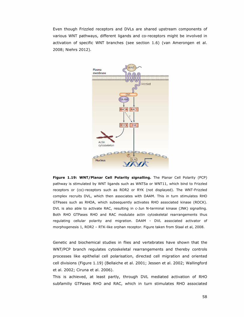
58
Even though Frizzled receptors and DVLs are shared upstream components of
various WNT pathways, different ligands and co-receptors might be involved in
activation of specific WNT branches (see section 1.6) (van Amerongen et al.
2008; Niehrs 2012).
Figure 1.19: WNT/Planar Cell Polarity signalling. The Planar Cell Polarity (PCP)
pathway is stimulated by WNT ligands such as WNT5a or WNT11, which bind to Frizzled
receptors or (co)-receptors such as ROR2 or RYK (not displayed). The WNT-Frizzled
complex recruits DVL, which then associates with DAAM. This in turn stimulates RHO
GTPases such as RHOA, which subsequently activates RHO associated kinase (ROCK).
DVL is also able to activate RAC, resulting in c-Jun N-terminal kinase (JNK) signalling.
Both RHO GTPases RHO and RAC modulate actin cytoskeletal rearrangements thus
regulating cellular polarity and migration. DAAM - DVL associated activator of
morphogenesis 1, ROR2 – RTK-like orphan receptor. Figure taken from Staal et al, 2008.
Genetic and biochemical studies in flies and vertebrates have shown that the
WNT/PCP branch regulates cytoskeletal rearrangements and thereby controls
processes like epithelial cell polarisation, directed cell migration and oriented
cell divisions (Figure 1.19) (Bellaiche et al. 2001; Jessen et al. 2002; Wallingford
et al. 2002; Ciruna et al. 2006).
This is achieved, at least partly, through DVL mediated activation of RHO
subfamiliy GTPases RHO and RAC, which in turn stimulates RHO associated

59
kinase (ROCK) activity and activation of JNK (Figure 1.19) (Winter et al. 2001;
Marlow et al. 2002; Habas et al. 2003; Jaffe and Hall 2005). Furthermore,
PRICKLE or VANGL proteins have been reported to be instrumental in this
context (Park and Moon 2002; Takeuchi et al. 2003; Veeman et al. 2003b;
Phillips et al. 2005; Gao et al. 2011).
Perturbations in the PCP pathway contribute to the pathogenesis of a variety of
diseases including cardiac and neural tube defects, the two most common types
of birth defects (Ciruna et al. 2006; Wen et al. 2010; Bartsch et al. 2012; Tada
and Kai 2012).
Figure 1.20: Convergent extension and planar polarity. (A) Vertebrate embryos
convert from spherical eggs into elongated animals by a process called convergent
extension (CE). (B) In the frog, convergent extension is achieved through cell
intercalation. This process requires co-ordinated migration towards each other
(convergence), intercalating along a single axis and a mediolateral extension, yielding a
cell population which shaped a short and wide arrangement into along and narrow one.
(C) This process involves stabilisation of lamellipodia specifically on the mediolateral
faces, and it is thought that crosstalk between neighbouring cells is critical to the
positioning of lamellipodia. The control of this process is complicated by the fact that cells
are constantly changing neighbours. Figure from Wallingford 2004. (Wallingford 2004)
Another observed PCP-linked disorder is the genetic disorder Bardet-Biedl
syndrome (BBS) (Fliegauf et al. 2007), which is a ciliopathic disorder,
characterised principally by obesity, retinitis, polydactyly, hypogonadism and
pigmentosa (Beales et al. 1999). Affected gene products are the BBS proteins,

60
which are found in the basal body and cilia of the cell (Ansley et al. 2003). BBS
proteins are involved in intraflagellar transport, a bi-directional transportation
activity along the long axis of the ciliary shaft that is essential
for ciliogenesis and the maintenance of cilia (Blacque et al. 2004). PCP has been
linked to BBS since it was demonstrated that PCP signalling is essential for
directional beating of motile cilia, which play a crucial role in the movement of
materials and cells (Vladar et al. 2009; Wallingford 2010; Wallingford and
Mitchell 2011). Further, PCP controls tissue level of multi-ciliated cells through
VANGL2, CELSR2, and CELSR3, which are required for establishment of rotation
(Mitchell et al. 2009; Guirao et al. 2010; Tissir et al. 2010; Wallingford and
Mitchell 2011)
Aberrant regulation of planar cell polarity has further been linked to polycystic
kidney disease (Simons and Mlodzik 2008; Luyten et al. 2010) and
Nephronophthisis type 2, a renal cystic disease cause by a mutation in the PCP
component Inversin (Otto et al. 2003; Simons et al. 2005).
Overall, the β-catenin (canonical) pathway has been linked to carcinogenesis,
while the planar cell polarity (PCP) pathway is implicated in cell motility and
metastasis (Clevers and Nusse 2012; Muthuswamy and Xue 2012).
1.5.3 The WNT/Ca2+ pathway
The non-canonical WNT/Ca2+ pathway is stimulated by a distinct group of WNT
ligands like WNT5a or WNT11 (Slusarski et al. 1997a; Slusarski et al. 1997b;
Westfall et al. 2003; Wang et al. 2010) and Frizzled receptors such as FZD2
(Slusarski et al. 1997a; Niu et al. 2012), leading to an intracellular increase of
the second messenger Ca2+ (Figure 1.21). This in turn leads to activation of
calcium/calmodulin-dependent kinase II (CamKII) (Kuhl et al. 2000a) and
protein kinase C (PKC) (Sheldahl et al. 1999). PKC then translocates to the
plasma membrane where it interacts with target proteins (Sheldahl et al. 1999).
The calcium responsive factor nuclear factor of activated T-cells (NF-AT) acts
downstream of CamKII and is involved in the development of cardiac, skeletal
muscle, and nervous systems (Kuhl et al. 2000b; Kohn and Moon 2005).
Furthermore, the WNT/Ca2+ pathway mediates cell migration and has been

61
implicated in the promotion of malignant progression and cell invasiveness of
cancer cells (Weeraratna et al. 2002; Kuhl 2004).
WNT5a induced PKC signalling has been shown to increase melanoma cell
motility through elevated Snail and Vimentin and decreased E-cadherin
expression (Dissanayake et al. 2007).
Thus, WNT/Ca2+ signalling modulates cell movement and behaviour and induces
structural changes similar to PCP (Torres et al. 1996; Kuhl et al. 2001; Okamoto
et al. 2007; Witze et al. 2008); but the extent, to which this pathway overlaps
with PCP still remains uncertain (Bryja et al. 2009; Andersson et al. 2010; Gao
et al. 2011).
Notably, besides Frizzled proteins, WNT5a ligand binding to ROR2 has been
demonstrated to activate Ca2+/CamKII pathway and is required for axonal
pathfinding in mammalian brain development (Logan and Nusse 2004; Li et al.
2009a; Nishita et al. 2010a).
Figure 1.21: WNT/Ca2+ signalling. A subset of WNT ligands and Frizzled (FZ) receptors
is able to increase intracellular Ca2+ levels, leading to subsequent activation of
calcium/calmodulin-dependent kinase II (CamKII) and protein kinase C (PKC)
downstream of Dishevelled (DVL). The calcium responsive factor NF-AT acts as
transcription factor involved in cell fate specifications and cellular movement. NF-AT -
nuclear factor of activated T-cells. Figure taken from Ciani and Salinas, 2005. (Ciani and
Salinas 2005) 005 #638}

62
1.6 WNT signalling components
1.6.1 WNT ligands
The human genome harbours 19 WNT genes, which encode functionally distinct
WNT proteins whose expression is spatially and temporally regulated during
development (Moon et al. 1997; Logan and Nusse 2004; Mikels and Nusse
2006b). WNT ligands are secreted hydrophobic glycoproteins, which are similar
in size (350 - 400 amino acids) and possesses 22 to 24 cysteine residues (Figure
1.22) (Miller 2002; Mikels and Nusse 2006b). WNT proteins bind to the Frizzled
family of seven-pass transmembrane proteins and activate intracellular WNT
signalling pathways depending on receptor context (Kohn and Moon 2005;
Cadigan and Liu 2006).
Figure 1.22: WNT ligand structure and post-translational modifications of
WNT3a and WNT5a proteins. WNTs are produced from precursor proteins and post
translational modified (glycosylation and lipid modification), before being secreted form
the cell surface in a matured and active form. WNT proteins are similar in size, rangeing
from 320 to 400 amino acids in length, and containing between 22-24 cysteine residues.
Figure taken from Kikuchi et al, 2011.
Historically, WNT ligands were subdivided into two functional groups: canonical
WNT ligands and non-canonical WNT ligands. Canonical WNTs such as WNT1,
WNT3, WNT3a or WNT7a are classified by the ability to induce duplication of the
embryonic axis in Xenopus laevis embryos or to induce transformation of
mammary epithelial cells (Wong et al. 1994; Shimizu et al. 1997), whereas non-
canonical WNT ligands have been reported to alter morphogenetic movements or
be involved in tumourigenesis through pro-migratory effects as reported for
WNT2, WNT4, WNT5a, WNT5b, WNT6, WNT7b or WNT11 (Wong et al. 1994; Du
et al. 1995; Shimizu et al. 1997).
63
WNT3 has been demonstrated to activate canonical and non-canonical WNT
signalling and to regulate cell motility through activation of RHO GTPases
(Shibamoto et al. 1998; Endo et al. 2005). WNT5 (Slusarski et al. 1997b;
Weeraratna et al. 2002; Qian et al. 2007) and WNT11 (Zhou et al. 2007)
activate β-catenin independent pathways, modulating embryonic cell movement
and polarity (Veeman et al. 2003a).
However, WNT5a has also been observed to signal through the canonical WNT
pathway depending on the receptor context (Mikels and Nusse 2006a). In
addition, WNT5a is found upregulated in many human tumours and stimulates
cell motility and invasiveness of tumour cells (Iozzo et al. 1995; Weeraratna et
al. 2002; Kurayoshi et al. 2006; Pukrop et al. 2006; Dissanayake et al. 2007).
1.6.2 Non-WNT ligand modulators of WNT signalling
WNT/β-catenin signaling is modulated at many levels by a number of
evolutionarily conserved secreted or transmembrane inhibitors or activators of
WNT signalling. With few exceptions, these antagonists and agonists are not
pure WNT modulators, but also affect additional signaling pathways, such as
TGF-β and FGF signalling (Cruciat and Niehrs 2012).
1.6.2.1 WNT inhibitors
Secreted Frizzled-related proteins (sFRP) were the first WNT antagonists to be
identified. These proteins are approximately 300 amino acids in size and contain
a signal sequence, a Frizzled-like cysteine-rich domain (CRD), and a small
conserved hydrophilic C-terminus harbouring a netrin-related motif (Rattner et
al. 1997). sFRPs are able to bind WNT proteins and FZD receptors in the
extracellular compartment, which prevents WNT/FZD binding (Wawrzak et al.
2007), similarily to the WNT inhibitory protein (WIF), which can bind WNTs,
thereby inhibiting interactions between WNT and WNT receptors (Figure 1.23)
(Bovolenta et al. 2008).
Other WNT inhibitors include proteins of the Dickkopf (DKK) (Bafico et al. 2001;
Semenov et al. 2001) and the WISE/Sclerostin (SOST) (Li et al. 2005; Semenov
et al. 2005) families, which antagonise signalling by binding to LRP5/6 and thus
preventing FZD/LRP complex formation.

64
Mouse Cerberus protein (Cerl-1), in contrast to Xenopus, does not bind WNT
ligands and thus does not inhibit WNT signalling (Piccolo et al. 1999; Belo et al.
2000). Rather, Cerberus proteins form key regulators of Nodal signalling and
play an important role in the establishment of left-right asymmetry in the
vertebrate embryo (Zhu et al. 1999; Tavares et al. 2007; Vonica and Brivanlou
2007).
Insulin-like growth factor binding protein 4 (IGFBP-4) has been shown to bind
directly to LRP6 and FZD8 through the the C-terminal thyroglobulin domain and
thus blocks WNT3a binding (Zhu et al. 2008).
The transmembrane WNT inhibitors Shisa (Yamamoto et al. 2005; Furushima et
al. 2007; Pei and Grishin 2012), Waif1 (Kagermeier-Schenk et al. 2011),
Adenomatosis polyposis coli down regulated 1 (APCDD1) (Takahashi et al. 2002;
O'Shaughnessy et al. 2004), and Tiki1 (Zhang et al. 2012) antagonise WNT
signalling by preventing ligand–receptor interactions or WNT receptor
maturation.
1.6.2.2 WNT activators
Two WNT ligand unrelated tyes of proteins – Norrin and R-spondins (RSPO),
promote WNT signalling by binding to WNT receptors or releasing a WNT-
inhibitory step (Kazanskaya et al. 2004; Xu et al. 2004; Kim et al. 2005; de Lau
et al. 2012). Norrin binds to FZD4 (but only to that member of the Frizzled
family), while RSPO2 interacts with FZD8 and LRP6 (Nam et al. 2006; Wei et al.
2007).
Connective tissue growth factors (CTGF) associate with the extracellular matrix
(ECM) and mediate cell adhesion, cell migration and chemotaxis (Hall-Glenn and
Lyons 2011; Jun and Lau 2011; Kubota and Takigawa 2011). They have been
further implicated to regulate WNT signalling through its ability to bind the LRP6
co-receptor (Mercurio et al. 2004).
The secreted glycoprotein collagen triple helix repeat-containing 1 (CTHRC1) has
been demonstrated to promote cell migration (Pyagay et al. 2005) and
melanoma cell adhesion and survival (Ip et al. 2011). Recent studies, however,
found that CTHRC1 selectively activates the WNT/PCP pathway by stabilising
WNT/FZD/ROR2 complex formation (Yamamoto et al. 2008).

65
Figure 1.23: Non-WNT ligands modulators. Domain structure of WNT antagonists and
agonists. Signal peptides are indicated in red. For more information refer to text.
Abbreviations: NTR-Netrin related motif, WIF-WNT-inhibitory factor 1 domain, EGF-
epidermal growth factor-like domain, CT-cystine knot-like domain, IB-insulin growth
factor binding protein domain, TY-thyroglobulin type-1 domain, TM-transmembrane
domain, L-leucine-rich repeats, TSP-thrombospondin type-1 domain. Figure from Cruciat
and Niehrs, 2012. (Cruciat and Niehrs 2012)

66
1.6.3 Frizzled receptors
Frizzled (FZD) receptors belong to the seven transmembrane-spanning G-
protein-coupled receptor (GPCR) superfamily, and are characterised by a large
extracellular N-terminal region containing a cysteine-rich domain, involved in
WNT ligand binding (Bhanot et al. 1996; Hsieh et al. 1999; Schulte and Bryja
2007). The intracellular C-terminus interacts with the PDZ domain of DVL
proteins through the highly conserved internal KTxxxW motif (Wong et al. 2003).
Therefore, Frizzled proteins act as WNT receptors and provide the link between
the extracellular WNT ligand and the intracellular components of WNT signalling
cascades (Figure 1.24) (Bhanot et al. 1996).
The mammalian FZD subfamily is comprised of 10 members (FZD1 to FZD10),
which mediate and modulate different pathway activation and signalling outcome
upon binding to various co-receptors and/or other secreted proteins like Norrin,
RSPO or CTHRC1 (see section 1.6.2) (Xu et al. 2004; Cadigan and Liu 2006;
Nam et al. 2006; Hendrickx and Leyns 2008; Yamamoto et al. 2008; Cruciat and
Niehrs 2012).
Like WNT proteins, Frizzled receptors have been grouped historically into two
classes: one class is thought to stimulate the β-catenin pathway, whereas the
other group is able to stimulate the Ca2+ pathway (Kuhl et al. 2000b). However,
this understanding has been challenged by observations that Frizzled receptors,
for example FZD4 can signal both β-catenin dependently (Ye et al. 2009) and
independently (Robitaille et al. 2002; Descamps et al. 2012), based on cell,
receptor and ligand context. FZD3 and FZD6 on the other hand have been
strongly implicated in PCP signalling, due to observed neural tube closure defects
in Fzd3/Fzd6 double deficient mice and defects in hair orientation of inner ear
sensory cells (Wang et al. 2006; Stuebner et al. 2010; Wen et al. 2010).
FZD2 has been indicated in β-catenin signalling outcomes and non-canonical
context (Ishitani et al. 2003; Li et al. 2008; Verkaar et al. 2009) and has been
linked to heart disease (Blankesteijn et al. 1996; Blankesteijn et al. 1997).
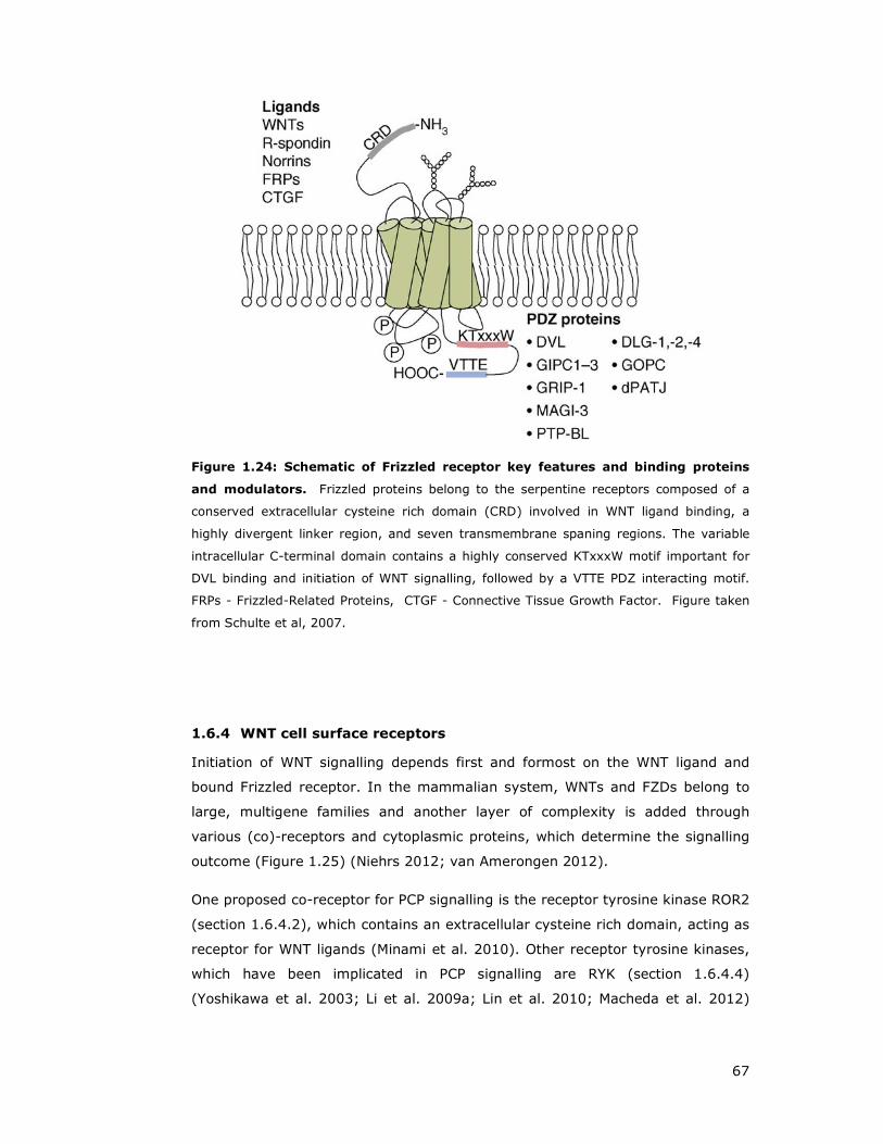
67
Figure 1.24: Schematic of Frizzled receptor key features and binding proteins
and modulators. Frizzled proteins belong to the serpentine receptors composed of a
conserved extracellular cysteine rich domain (CRD) involved in WNT ligand binding, a
highly divergent linker region, and seven transmembrane spaning regions. The variable
intracellular C-terminal domain contains a highly conserved KTxxxW motif important for
DVL binding and initiation of WNT signalling, followed by a VTTE PDZ interacting motif.
FRPs - Frizzled-Related Proteins, CTGF - Connective Tissue Growth Factor. Figure taken
from Schulte et al, 2007.
1.6.4 WNT cell surface receptors
Initiation of WNT signalling depends first and formost on the WNT ligand and
bound Frizzled receptor. In the mammalian system, WNTs and FZDs belong to
large, multigene families and another layer of complexity is added through
various (co)-receptors and cytoplasmic proteins, which determine the signalling
outcome (Figure 1.25) (Niehrs 2012; van Amerongen 2012).
One proposed co-receptor for PCP signalling is the receptor tyrosine kinase ROR2
(section 1.6.4.2), which contains an extracellular cysteine rich domain, acting as
receptor for WNT ligands (Minami et al. 2010). Other receptor tyrosine kinases,
which have been implicated in PCP signalling are RYK (section 1.6.4.4)
(Yoshikawa et al. 2003; Li et al. 2009a; Lin et al. 2010; Macheda et al. 2012)

68
and PTK7 (section 1.6.4.3) (Lu et al. 2004b; Shnitsar and Borchers 2008;
Peradziryi et al. 2011).
Figure 1.25: Overview of WNT modulating receptors and their effect on
downstream WNT signalling. The main WNT ligand receptors are Frizzled (Fz) proteins.
However, an expanding number of co-receptors can modulate WNT signalling outcome.
For example, PTK7 and ROR transmembrane receptors in conjunction with Frizzled are
able to bind to WNT ligands and to modulate β-catenin dependent and β-catenin
independent (PCP) pathways. LGR can bind R-spondin, which enhances response to low
dose WNT ligands, and has been demonstrated to interact and co-internalise with LDL-
receptor related proteins (LRP)/Fz complexes, which are established β-catenin pathway
activators. LGR - leucine-rich repeat containing G protein-coupled receptor PTK – Protein
Tyrosine Kinase 7, ROR – Receptor tyrosine kinase-like Orphan Receptor, PCP – Planar
Cell Polarity. Figure from Vincent and Beckett, 2011. (Vincent and Beckett 2011).
1.6.4.1 LRP5/6
LDL receptor-related proteins 5&6 (LRP5/6) function as co-receptors for WNT
ligand interaction and are required for β-catenin signalling pathway activation
(Pinson et al. 2000; Tamai et al. 2000).
They are single-pass transmembrane proteins belonging to the low-density
lipoprotein receptor (LDLR)-related protein family. LRP5 and LRP6 are highly
homologous and display redundant roles during development (He et al. 2004;
Kelly et al. 2004). Structurally, they contain four EGF and three LDLR repeats
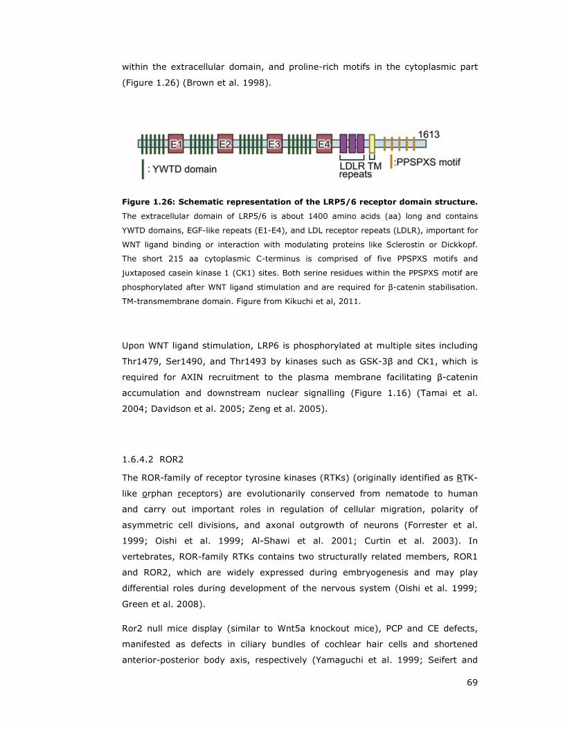
69
within the extracellular domain, and proline-rich motifs in the cytoplasmic part
(Figure 1.26) (Brown et al. 1998).
Figure 1.26: Schematic representation of the LRP5/6 receptor domain structure.
The extracellular domain of LRP5/6 is about 1400 amino acids (aa) long and contains
YWTD domains, EGF-like repeats (E1-E4), and LDL receptor repeats (LDLR), important for
WNT ligand binding or interaction with modulating proteins like Sclerostin or Dickkopf.
The short 215 aa cytoplasmic C-terminus is comprised of five PPSPXS motifs and
juxtaposed casein kinase 1 (CK1) sites. Both serine residues within the PPSPXS motif are
phosphorylated after WNT ligand stimulation and are required for β-catenin stabilisation.
TM-transmembrane domain. Figure from Kikuchi et al, 2011.
Upon WNT ligand stimulation, LRP6 is phosphorylated at multiple sites including
Thr1479, Ser1490, and Thr1493 by kinases such as GSK-3β and CK1, which is
required for AXIN recruitment to the plasma membrane facilitating β-catenin
accumulation and downstream nuclear signalling (Figure 1.16) (Tamai et al.
2004; Davidson et al. 2005; Zeng et al. 2005).
1.6.4.2 ROR2
The ROR-family of receptor tyrosine kinases (RTKs) (originally identified as RTK-
like orphan receptors) are evolutionarily conserved from nematode to human
and carry out important roles in regulation of cellular migration, polarity of
asymmetric cell divisions, and axonal outgrowth of neurons (Forrester et al.
1999; Oishi et al. 1999; Al-Shawi et al. 2001; Curtin et al. 2003). In
vertebrates, ROR-family RTKs contains two structurally related members, ROR1
and ROR2, which are widely expressed during embryogenesis and may play
differential roles during development of the nervous system (Oishi et al. 1999;
Green et al. 2008).
Ror2 null mice display (similar to Wnt5a knockout mice), PCP and CE defects,
manifested as defects in ciliary bundles of cochlear hair cells and shortened
anterior-posterior body axis, respectively (Yamaguchi et al. 1999; Seifert and
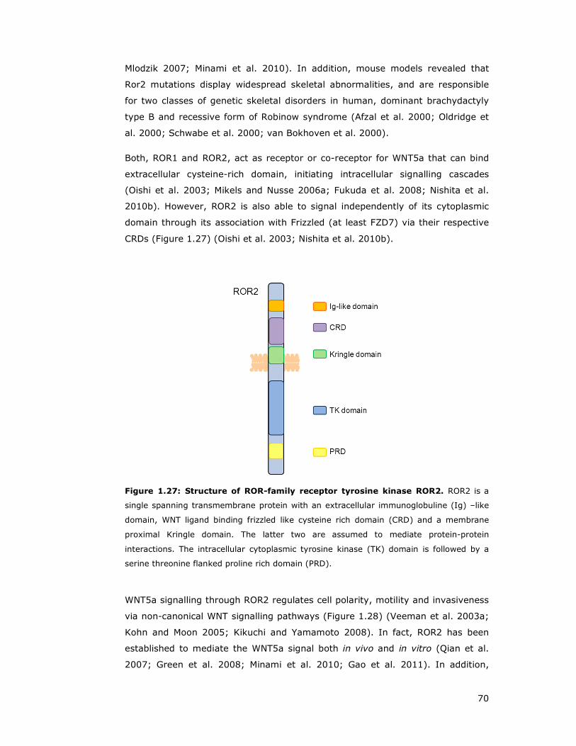
70
Mlodzik 2007; Minami et al. 2010). In addition, mouse models revealed that
Ror2 mutations display widespread skeletal abnormalities, and are responsible
for two classes of genetic skeletal disorders in human, dominant brachydactyly
type B and recessive form of Robinow syndrome (Afzal et al. 2000; Oldridge et
al. 2000; Schwabe et al. 2000; van Bokhoven et al. 2000).
Both, ROR1 and ROR2, act as receptor or co-receptor for WNT5a that can bind
extracellular cysteine-rich domain, initiating intracellular signalling cascades
(Oishi et al. 2003; Mikels and Nusse 2006a; Fukuda et al. 2008; Nishita et al.
2010b). However, ROR2 is also able to signal independently of its cytoplasmic
domain through its association with Frizzled (at least FZD7) via their respective
CRDs (Figure 1.27) (Oishi et al. 2003; Nishita et al. 2010b).
Figure 1.27: Structure of ROR-family receptor tyrosine kinase ROR2. ROR2 is a
single spanning transmembrane protein with an extracellular immunoglobuline (Ig) –like
domain, WNT ligand binding frizzled like cysteine rich domain (CRD) and a membrane
proximal Kringle domain. The latter two are assumed to mediate protein-protein
interactions. The intracellular cytoplasmic tyrosine kinase (TK) domain is followed by a
serine threonine flanked proline rich domain (PRD).
WNT5a signalling through ROR2 regulates cell polarity, motility and invasiveness
via non-canonical WNT signalling pathways (Figure 1.28) (Veeman et al. 2003a;
Kohn and Moon 2005; Kikuchi and Yamamoto 2008). In fact, ROR2 has been
established to mediate the WNT5a signal both in vivo and in vitro (Qian et al.
2007; Green et al. 2008; Minami et al. 2010; Gao et al. 2011). In addition,
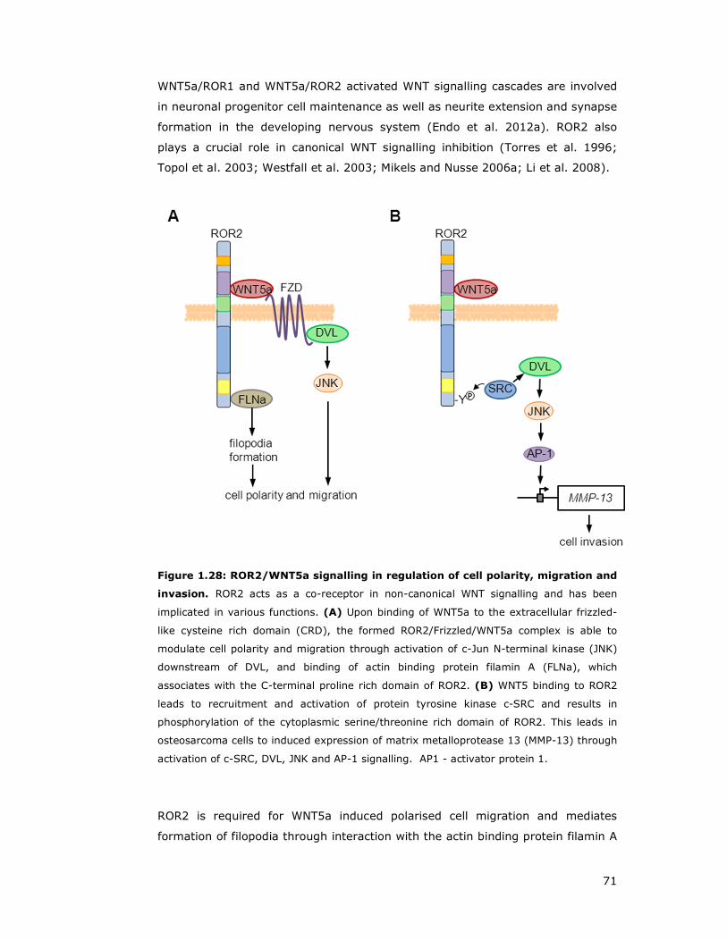
71
WNT5a/ROR1 and WNT5a/ROR2 activated WNT signalling cascades are involved
in neuronal progenitor cell maintenance as well as neurite extension and synapse
formation in the developing nervous system (Endo et al. 2012a). ROR2 also
plays a crucial role in canonical WNT signalling inhibition (Torres et al. 1996;
Topol et al. 2003; Westfall et al. 2003; Mikels and Nusse 2006a; Li et al. 2008).
Figure 1.28: ROR2/WNT5a signalling in regulation of cell polarity, migration and
invasion. ROR2 acts as a co-receptor in non-canonical WNT signalling and has been
implicated in various functions. (A) Upon binding of WNT5a to the extracellular frizzled-
like cysteine rich domain (CRD), the formed ROR2/Frizzled/WNT5a complex is able to
modulate cell polarity and migration through activation of c-Jun N-terminal kinase (JNK)
downstream of DVL, and binding of actin binding protein filamin A (FLNa), which
associates with the C-terminal proline rich domain of ROR2. (B) WNT5 binding to ROR2
leads to recruitment and activation of protein tyrosine kinase c-SRC and results in
phosphorylation of the cytoplasmic serine/threonine rich domain of ROR2. This leads in
osteosarcoma cells to induced expression of matrix metalloprotease 13 (MMP-13) through
activation of c-SRC, DVL, JNK and AP-1 signalling. AP1 - activator protein 1.
ROR2 is required for WNT5a induced polarised cell migration and mediates
formation of filopodia through interaction with the actin binding protein filamin A
72
(FLNa) (Figure 1.28A) (Nishita et al. 2006). Furthermore, polarised cell migration
experiments showed that WNT5a stimulated lamellipodium formation and
microtubule organizing centre (MTOC) reorientation is mediated through
ROR2/FLNa/JNK signalling (Nomachi et al. 2008).
It has been shown that atypical Protein Kinase C (aPKC) associates with DVL,
functioning upstream of JNK, suggesting a possible functional link between
WNT5a/ROR2/FLNa/JNK and CDC42/PAR/aPKC pathways in polarised cell
migration (Schlessinger et al. 2007).
Although the roles of WNT5a in cancers remain controversial, ROR1 and ROR2
have been demonstrated to promote tumour progression (Enomoto et al. 2009;
Wright et al. 2009; Ford et al. 2012; Daneshmanesh et al. 2013) (Figure 1.28).
1.6.4.3 PTK7
The transmembrane protein kinase 7 (PTK7) displays extracellular
immunoglobulin domains and an intracellular tyrosine kinase homology domain,
which is evolutionary conserved but lacks catalytic activity (Mossie et al. 1995;
Kroiher et al. 2001). PTK7 functions in cell adhesions, cell migration, cell
polarity, proliferation, actin cytoskeleton reorganisation and embryogenesis
related apoptosis in order to regulate processes such as epithelial tissue
organisation, neural tube closure and neural crest formation as well as axon
guidance (Lu et al. 2004b; Toyofuku et al. 2004; Shnitsar and Borchers 2008;
Meng et al. 2010).
It has been demonstrated that PTK7 participates in WNT signalling through
acting as a co-receptor in both β-catenin dependent and independent WNT
signalling cascades, and is involved in the choice of signalling outcome (Shnitsar
and Borchers 2008; Wehner et al. 2011; Peradziryi et al. 2012). Despite the lack
of any known WNT protein binding domain, PTK7 has been reported to co-
immunoprecipitate with WNT3a and WNT8 but not with WNT5a or WNT11
(Peradziryi et al. 2011).
In addition to WNT signalling, PTK7 has been shown to regulate other signalling
pathways by functioning as co-receptor with membrane receptors like Plexin A1
and VEGFR1 (Shin et al. 2008; Wagner et al. 2010; Lee et al. 2011).

73
Furthermore, recent studies showed that PTK7 is a target of matrix
metalloproteases (MMP) 14 and ADAM17, leading to proteolytic degradation and
extracellular domain shedding, thus resulting in enhanced cell proliferation,
migration, and facilitated cancer cell invasion (Golubkov et al. 2010; Na et al.
2012). Originally identified in colon carcinoma cells (Mossie et al. 1995), PTK7
has been further demonstrated to be frequently deregulated in other types of
cancer (Easty et al. 1997; Muller-Tidow et al. 2004; Prebet et al. 2010; Gobble
et al. 2011).
1.6.4.4 RYK
The transmembrane atypical receptor related tyrosine kinase RYK is required for
normal axon guidance and neuronal differentiation during development and
involved in WNT signalling (Hovens et al. 1992; Stacker et al. 1993; Fradkin et
al. 2010).
RYK functions as a WNT receptor through its WNT protein binding WIF domain
and has been demonstrated to regulate convergent extension movements in
Xenopus embryos through FZD7 and DVL endocytosis (Patthy 2000; Kim et al.
2008). In addition, RYK plays a role in mammalian planar cell polarity signalling,
forming a complex with VANGL2 and modulating its stability (Andre et al. 2012;
Macheda et al. 2012).
Overall, mammalian RYK is able to mediate both β-catenin dependent and
independent signalling pathways downstream of WNT3a, WNT5a and WNT11 (Lu
et al. 2004a; Li et al. 2009a; Lin et al. 2010). RYK deficient mice exhibit
craniofacial defects associated with perturbed Eph receptor crosstalk (Halford et
al. 2000).
1.6.4.5 HSPGs and Syndecans
Heparansulphate proteoglycans (HSPGs) are important for concentrating and
stabilising of WNT proteins on the cell surface (Fuerer et al. 2010). Moreover,
HSPGs modulate β-catenin independent signalling. For example Glypican 3
(GPC3) binds to WNT5a and enhances WNT5a dependent JNK activity and is
required for gastrulation movements in zebrafish (De Cat et al. 2003; Song et al.

74
2005). GPC4 interacts with WNT11 and enhances WNT stimulated cell migration
(Ohkawara et al. 2003). However, recent evidence suggests that GPCs might be
also involved in β-catenin dependent signalling (Capurro et al. 2005). It has
been suggested that the localisation of GPCs (lipid raft or non-lipid raft micro
domains) might be important for selective WNT cascade activation (Sakane et al.
2012).
The syndecan (SDC) protein family also regulates WNT signalling and they have
been implicated in β-catenin dependent (Alexander et al. 2000) and independent
pathway modulation (Munoz et al. 2006; Ohkawara et al. 2011).
1.6.4.6 Tetraspanins
Tetraspanins are cell surface glycoproteins with four transmembrane domains
and are able to form multimeric complexes with other cell surface proteins
(Yanez-Mo et al. 2009). Tetraspanin proteins function in many cellular processes
including differentiation, adhesion, signal transduction, and play critical roles in
the suppression of cancer cell motility and metastasis (Richardson et al. 2011;
Rubinstein 2011; Sala-Valdes et al. 2012). The tetraspanin family member
TSPAN12 binds to FZD4 and promotes complex formation of FZD4 co-operatively
with Norrin, thereby activating the β-catenin signalling cascade (Junge et al.
2009).
1.6.5 Dishevelled proteins
DVL proteins are key components of WNT signalling and interact with more than
50 proteins (Wallingford and Habas 2005; Gao and Chen 2010). They form
multi-component signalling complexes and constitute the branching point of WNT
signalling cascades (Figure 1.29A) (Gao and Chen 2010). Mammals encode three
members of the Dishevelled family, DVL1, DVL2 and DVL3, which all contain the
conserved structural features required for downstream WNT signalling (Figure
1.29B).
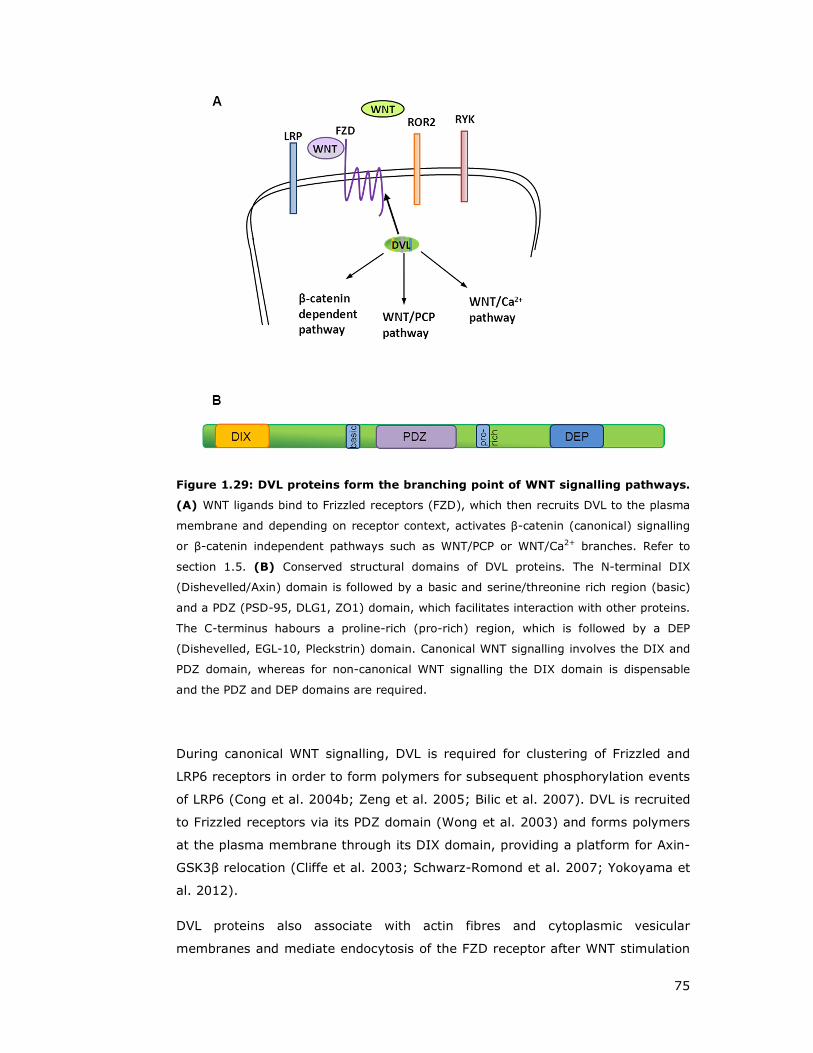
75
Figure 1.29: DVL proteins form the branching point of WNT signalling pathways.
(A) WNT ligands bind to Frizzled receptors (FZD), which then recruits DVL to the plasma
membrane and depending on receptor context, activates β-catenin (canonical) signalling
or β-catenin independent pathways such as WNT/PCP or WNT/Ca2+ branches. Refer to
section 1.5. (B) Conserved structural domains of DVL proteins. The N-terminal DIX
(Dishevelled/Axin) domain is followed by a basic and serine/threonine rich region (basic)
and a PDZ (PSD-95, DLG1, ZO1) domain, which facilitates interaction with other proteins.
The C-terminus habours a proline-rich (pro-rich) region, which is followed by a DEP
(Dishevelled, EGL-10, Pleckstrin) domain. Canonical WNT signalling involves the DIX and
PDZ domain, whereas for non-canonical WNT signalling the DIX domain is dispensable
and the PDZ and DEP domains are required.
During canonical WNT signalling, DVL is required for clustering of Frizzled and
LRP6 receptors in order to form polymers for subsequent phosphorylation events
of LRP6 (Cong et al. 2004b; Zeng et al. 2005; Bilic et al. 2007). DVL is recruited
to Frizzled receptors via its PDZ domain (Wong et al. 2003) and forms polymers
at the plasma membrane through its DIX domain, providing a platform for Axin-
GSK3β relocation (Cliffe et al. 2003; Schwarz-Romond et al. 2007; Yokoyama et
al. 2012).
DVL proteins also associate with actin fibres and cytoplasmic vesicular
membranes and mediate endocytosis of the FZD receptor after WNT stimulation
76
(Capelluto et al. 2002; Chen et al. 2003). Further, DVL has been observed to
shuttle from the cytoplasm to the nucleus and to interact with c-JUN and β-
catenin (Itoh et al. 2005; Gan et al. 2008).
Upon initiation of non canonical WNT/PCP signalling, DVL associates with DVL
associated activator of morphogenesis 1 (DAAM1) through its PDZ domain
(Habas et al. 2001; Liu et al. 2008a). In conjunction with WGEF (weak similarity
GEF) this complex then stimulates RHO activity and subsequently leads to RHO
associated kinase (ROCK) activation, resulting in cytoskeleton remodelling
(Figure 1.19) (Habas et al. 2001). In addition, DVL is able to stimulate RAC,
which in turn can activate the downstream effector JNK, which regulates polarity,
embryonic cell movements (Habas et al. 2003), and dendrite growth (Rosso et
al. 2005).
The DIX domain is dispensable for PCP and RHO/RAC activation but is critically
required for the β-catenin pathway (Veeman et al. 2003a; Wharton 2003). In
contrast, the DEP domain is obligatory for RHO activation and stimulation of JNK
activity (Boutros et al. 1998; Li et al. 1999; Habas et al. 2003).
In addition, DVL has been implicated in the modulation of WNT/Ca2+ signalling
(Sheldahl et al. 2003), aPKC activity in hippocampal neurons (Dollar et al. 2005;
Zhang et al. 2007) and is required for mTorc activation by WNT (Mak et al.
2005; Inoki et al. 2006).
WNT stimulation leads to hyper-phosphorylation of DVL, but the mechanism and
function remains incompletely understood. Kinases which have been
demonstrated to phosphorylate DVLs are Casein kinase 1є (CK1є) (Klimowski et
al. 2006), CK2 (Wharton 2003) and PAR1 (Elbert et al. 2006).
DVL protein stability is controlled through proteasomal or lysosomal degradation.
DVL proteins are targeted for proteasomal degradation through HECT-type
ubiquitin ligase NEDL1 (Miyazaki et al. 2004) or KLHL12 (Angers et al. 2006) or
mediated through a degradation complex involving PRICKLE1 (Carreira-Barbosa
et al. 2003). Lysosomal destabilisation of DVL is mediated by Dapper (Cheyette
et al. 2002; Zhang et al. 2006) or through a negative WNT signalling feedback
loop (Jung et al. 2009). DVL2 has been reported to be degraded by the
autophagy pathway as part of a negative feedback loop of WNT regulation (Gao
et al. 2010).
DVL1 null mice are viable and fertile but display social interaction abnormalities
(Lijam et al. 1997; Long et al. 2004). In contrast, DVL2 null mice displayed

77
somite segmentation defects and cardiac outflow tract abnormalities, which is
also responsible for perinatal lethality of half of the offspring (Hamblet et al.
2002). DVL3 knockout mice show similar cardiac outflow tract abnormalities as
observed for DVL2, but display a mild PCP defect of misoriented stereocilia in the
organ of Corti (Etheridge et al. 2008; Wynshaw-Boris 2012).
Individual DVL proteins can rescue knock outs of other DVL genes, suggesting
functional redundancy, which is also consistent with overlapping expression
patterns (Sussman et al. 1994; Klingensmith et al. 1996; Tsang et al. 1996;
Wang et al. 2005; Etheridge et al. 2008). Notably, overexpression of DVL has
been reported in certain cancers (Okino et al. 2003; Uematsu et al. 2003).

78
1.7 VANGL proteins
VANGL proteins are tetra-membrane-spanning proteins, which are highly
conserved from fly to human. In flies and worms only one Vangl gene can be
found, whereas zebrafish, mouse and human contain two VANGL homologues
with the exception of frogs, where only a VANGL2 homologue was identified
(Torban et al. 2004).
The mammalian VANGL1 and VANGL2 proteins share a 73% total-amino-acid
identity if compared to each other, and a 44% (VANGL1) or 50% (VANGL2),
similarity to the Drosophila homologue strabismus (stbm), suggesting that the
vertebrate genes evolved by gene duplication from a common ancestor present
in flies (Figure 1.30) (Torban et al. 2004).
Figure 1.30: VANGL1 and VANGL2 proteins display a high degree of sequence
similarity. Alignment of mouse VANGL1 and VANGL2 protein sequences. Identical amino
acid (aa) residues of both sequences are highlighted in grey and transmembrane portions
are indicated through a black line runnig above the respective amino acids. The C-
terminal PDZ-binding motif (ETSV) is indicated by a box and the D255E and S464N
mutations causing neural tube defects in looptail (Lp) mice are indicated by arrowheads.
Figure taken from Torban et al, 2004.
Further, both proteins present two solvent accessible extracellular loops and an
intracellular amino and carboxy terminus, and share the same C-terminal ESTV
79
motif, promoting protein interactions with PDZ domain containing proteins
(Figure 1.31) (Nourry et al. 2003; Torban et al. 2004).
The presence of a N-terminal cytoplasmic serine/threonine cluster motif is
evolutionarily conserved among vertebrate VANGL1 and VANGL2 orthologs and
phosphorylation events in this cluster are required for at least VANGL2 activity
(Daub et al. 2008; Gao et al. 2011).
Vangl homologues were initially discovered in a study of polarity establishment
in the Drosophila eye and termed strabismus (Wolff and Rubin 1998). X-
irradiation-induced mutations of the stbm locus showed disruption of polarity of
multiple structures like the orientation of bristles (leg) and hairs (wing) (Figure
1.18b) or affect correct ommatidial rotation (eye) (Figure 1.18f). Studies of
Vangl function in vertebrates using Zebrafish and Xenopus as model organisms
demonstrated a role in mediation of cell movements during gastrulation (Park
and Moon 2002).
Van gogh-like (VANGL) proteins play a role in planar cell polarity and convergent
extension movements (see section 1.5.2) (Torban et al. 2004; Torban et al.
2008; Torban et al. 2012). Studies in vertebrate systems further identified
VANGL proteins to function in unexpected developmental processes such as
asymmetric cell division, left-right asymmetry and morphogenesis of different
tissues (Torban et al. 2012).
1.7.1 VANGL1
Human VANGL1, also termed Strabismus Drosophila homologue 2 (STB2),
encodes a 524-amino-acid protein, distributed over eight exons. Northern blot
analysis detected 4.8- and 6.8-kb transcripts in nearly all human tissues
examined (Katoh 2002; Yagyu et al. 2002; Kalabis et al. 2006). VANGL1
proteins localise to various subcellular departments such as cytoplasmic vesicular
structures, co-localisation with E-cadherin or at the plasma membrane (see
section 5.4) (Kalabis et al. 2006).
Mouse model studies using an inactivating mutation in the Vangl1 gene showed
that heterozygous mutants mice were viable and fertile, whereas homozygous
mutants displayed subtle alterations in polarity, displayed by inner hair cells

80
orientation of the cochlea (Torban et al. 2008). Crosses between Vangl1 and
Vangl2 heterozygous mice showed profound developmental defects, including
severe craniorachischisis, inner ear defects, and cardiac abnormality, suggesting
a genetic interaction between Vangl1 and Vangl2 (Torban et al. 2008).
In addition, mutational analysis of neural tube defects (NTDs) patients revealed
a set of VANGL1 point mutations across highly conserved amino acids, including
the corresponding amino acids affected in the VANGL2 looptail mouse D255E and
S464N (Figure 1.30 and Figure 1.31) (Kibar et al. 2007; Kibar et al. 2009).
These findings suggests that VANGL proteins play an important role in
developmental processes such as co-ordinated cellular migration in convergent
extension movements during neurulation.
Figure 1.31: Topological model and structural features of VANGL1. This figure
illustrates the four transmembrane domains, intracellular and extracellular segments and
their respective position in the primary amino acid sequence of VANGL1. Amino acid
residues in VANGL1 and VANGL2 associated with neural tube defects in mice and humans
are depicted in yellow, whereas loss of function mutations have been demonstrated, are
shaded in pink. The PDZ interacting tetrapeptide ETSV is coloured in green. Amino acids
highlighted in blue represent consensus tyrosine based and di-leucine motifs associated
with protein sorting and membrane targeting. Figure from Iliescu et al, 2011.

81
Whereas the link of VANGL1 to developmental tube defects is relatively weak
(homozygous mice for VANGL1 are normal and fertile), an increasing body of
evidence suggests a strong connection between high levels of VANGL1
expression and cancer progression/promotion. Several studies using colorectal
cancer cells observed VANGL1 involvement in promotion of migration and
invasion processes, while knock down of VANGL1 in various carcinoma cells
showed inhibited growth and reduced migration (Yagyu et al. 2002; Lee et al.
2004; Kalabis et al. 2006; Kho et al. 2009; Lee et al. 2009; Ryu et al. 2010).
Expression analysis of gastric tumour samples showed higher VANGL1 levels
compared to adjacent normal tissue (Lee et al. 2005). This ties in with the
observation that VANGL1 expression in breast cancer cancer patients leads to a
poor outcome prognosis, based on the demonstarted relationship between high
VANGL1 expression and breast cancer relapse (Anastas et al. 2011).
Taken together, mammalian VANGL1 is involved in neural tube closure during
gastrulation and neurolation processes, and modulates cell migration and
invasion processes in adult tissues, which can contribute to malignant
phenotypes such as abnormal tissue polarity, invasion and metastasis as found
in human cancers.
1.7.2 VANGL2
VANGL2 (521 aa) is a well established PCP core protein and interacts with a
multitude of WNT and PCP signalling components. Ranging from WNT (co)-
receptors, ROR2 (Gao et al. 2011), RYK (Andre et al. 2012; Macheda et al.
2012) and PTK7 (Lu et al. 2004a) (see section 1.6.4), Frizzled receptors
(Montcouquiol et al. 2006), DVL2 (Torban et al. 2004; Shafer et al. 2011),
SCRIB (Montcouquiol et al. 2003; Montcouquiol et al. 2006), to RHO GTPase
RAC1 (Lindqvist et al. 2010). Further, VANGL2 regulates the endocytosis and cell
surface level of membrane type-1 matrix metalloproteinase (MMP14 or MT1-
MMP) modulation of MMP endocytosis (Williams et al. 2012a; Williams et al.
2012b).
A mouse model termed looptail (Lp) exhibited independent mutations in the
VANGL2 locus, thus giving VANGL2 the alternative name Loop-tail associated
protein (Ltap), displayed typical PCP defects (Kibar et al. 2001a; Kibar et al.
2001b; Torban et al. 2007). Loop-tail (Lp) mouse model homozygotes die in

82
utero of severe craniorachischisis, display defects in the inner ear cochlea hair
orientation, heart outflow tract defects and defects of the musculoskeletal
system characterised by fused ribs (Montcouquiol et al. 2003; Phillips et al.
2005; Torban et al. 2008).
VANGL2 point mutations have been identified to contribute to NTDs (Kibar et al.
2011) and several identified mutations are homologous to those found in
VANGL1, such as Lp specific VANGL2 loss of function mutation S464N and
mutant D255E (Kibar et al. 2001a; Kibar et al. 2001b; Guyot et al. 2011). These
mutations have been demonstrated to affect plasma membrane targeting and
result in ER retention, where protein half-life is reduced and stability affected by
increased proteasome dependent degradation (Gravel et al. 2010).
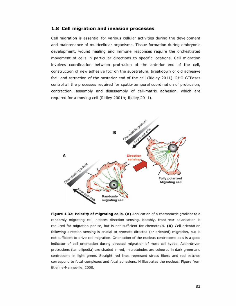
83
1.8 Cell migration and invasion processes
Cell migration is essential for various cellular activities during the development
and maintenance of multicellular organisms. Tissue formation during embryonic
development, wound healing and immune responses require the orchestrated
movement of cells in particular directions to specific locations. Cell migration
involves coordination between protrusion at the anterior end of the cell,
construction of new adhesive foci on the substratum, breakdown of old adhesive
foci, and retraction of the posterior end of the cell (Ridley 2011). RHO GTPases
control all the processes required for spatio-temporal coordination of protrusion,
contraction, assembly and disassembly of cell-matrix adhesion, which are
required for a moving cell (Ridley 2001b; Ridley 2011).
Figure 1.32: Polarity of migrating cells. (A) Application of a chemotactic gradient to a
randomly migrating cell initiates direction sensing. Notably, front-rear polarisation is
required for migration per se, but is not sufficient for chemotaxis. (B) Cell orientation
following direction sensing is crucial to promote directed (or oriented) migration, but is
not sufficient to drive cell migration. Orientation of the nucleus-centrosome axis is a good
indicator of cell orientation during directed migration of most cell types. Actin-driven
protrusions (lamellipodia) are shaded in red, microtubules are coloured in dark green and
centrosome in light green. Straight red lines represent stress fibers and red patches
correspond to focal complexes and focal adhesions. N illustrates the nucleus. Figure from
Etienne-Manneville, 2008. (Etienne-Manneville 2008).

84
1.8.1 Actin structures and actin-binding protein Filamin A
Actin and myosin II play major roles in cell migration. Actin, a ubiquitous
eukaryotic protein, is the major component of the cytoskeleton, controlling cell
structure and motility (Herman 1993; Ridley 2001b). Actin exists mainly as a
fibrous polymer (F-actin), but in response to cytoskeletal re-organising signals
during processes such as cytokinesis, endocytosis, or stress. Cofilin promotes
fragmentation and depolymerisation of F-actin, resulting in an increase in the
monomeric globular form (G-actin) (Condeelis 2001). The Arp2/3 complex
stabilises F-actin fragments and promotes formation of new actin filaments
(Condeelis 2001).
Lamellipodia are actin projections on the mobile edge at the front of the cell and
form a two-dimensional actin mesh, which pulls the cell forward across a
substrate during the process of cell migration (Ridley 2011). Actin microspikes
spreading beyond the lamellipodium frontier are termed filopodia (Small et al.
2002). RAC stimulates lamellipodium formation, while Cdc42 promotes
filopodium construction (Hall 1998).
Filamin A (FLNa) is a 280-kD actin-crosslinking protein, which regulates re-
organisation of the actin cytoskeleton by interacting with integrins,
transmembrane receptor complexes, and second messengers (Ohta et al. 2006;
Kim and McCulloch 2011; Nakamura et al. 2011). Filamins crosslink actin
filaments into orthogonal networks in the cytoplasm and participate in the
anchoring of membrane proteins to the actin cytoskeleton (Maestrini et al. 1993;
Fox et al. 1998). In addition, cell motility-independent functions of FLNa at cell-
cell contacts and adherens junctions affect the development of organs (Feng et
al. 2006).
Interestingly, R-RAS (Gawecka et al. 2010; Griffiths et al. 2011) and ROR2
(Nishita et al. 2006; Nomachi et al. 2008) have been demonstrated
independently to be interactors of FLNa, and are required for migration
processes or endothelial barrier maintenance.

85
1.8.2 Invadopodial structures and matrix metalloproteases
Invadopodia are cell membrane localised, actin rich protrusions, extending into
the extracellular matrix (ECM) where they are able to degrade ECM components
(Linder et al. 2011; Murphy and Courtneidge 2011; Ridley 2011; Sibony-
Benyamini and Gil-Henn 2012). Invadopodia are therefore thought to be key
structures of cell invasion (Linder et al. 2011; Sibony-Benyamini and Gil-Henn
2012).
β1A integrin is a master regulator of invadosome organisation and function,
mediated by matrix metalloproteinase MMPs (Destaing et al. 2010). MMPs belong
to a family of proteases that target extracellular proteins including other
proteases, growth factors, cell surface receptors and adhesion molecules
(McCawley and Matrisian 2001). Among MMPs, MMP-2, MMP-3, MMP-7, MMP-9
and MMP-13 have been characterised as important factors for normal tissue
remodelling during embryonic development, wound healing, tumour invasion,
angiogenesis, carcinogenesis and apoptosis (Nagase et al. 1990; Vu et al. 1998;
Sternlicht et al. 1999; Coussens et al. 2002; Kudo et al. 2012). MMPs are
synthesised as pro-enzyme and are activated by releasing the inhibitory
propeptide domain in order to yield an active, full length protease (Nagase et al.
1990).
Notably, WNT5a-ROR2 signalling confers MMP-13 linked invasive properties
assessed by invadopodium formation (Figure 1.28B) (Enomoto et al. 2009). In
addition, VANGL2 has been reported to regulate endocytosis and cell-surface
availability of MMP14, resulting in proteolytic degradation or remodelling of the
extracellular matrix (Cantrell and Jessen 2010; Williams et al. 2012a; Williams et
al. 2012b).

86
1.9 Autophagy
Protein turnover takes place through the ubiquitin mediated proteasome
pathway or is lysosome mediated (Ciechanover 2005). Autophagy or
autophagocytosis is a key mechanism that clears unnecessary or dysfunctional
cellular components through the lysosomal machinery or recycles nutrients
during starvation or stress periods, thus maintaining cellular energy levels
(Mizushima and Klionsky 2007).
Macroautophagy, the main pathway, generally eradicates damaged cell
organelles or unused proteins. This process is tightly regulated through highly
conserved genes termed autophagy regulators (ATGs), which are required at
several steps along the process and orchestrate for instance the induction, cargo
recognition and packaging or vesicle formation (Mizushima et al. 2011).
Autophagy is stimulated in response to unfavourable conditions such as nutrient
deprivation and is initiated through phagophore formation by class III PI3K, Atg6
(also known as Beclin-1) and ubiquitin-like conjugation reactions (Figure 1.33).
During phagophore nucleation, cytosolic proteins, protein aggregates and
organelles are enclosed, and recruitment of ATG proteins such as ATG12, ATG5,
ATG16 and covalent lipidated ATG8 together with transmembrane ATG9 facilitate
phagophore extension. Upon completion of the double membrane organelle
known as an autophagosome vesicle, ATG proteins start to dissociate, and fusion
of the autophagosome with a lysosome results in a autolysosome and
subsequent degradation of the organells content through acidic lysosomal
hydrolases (Figure 1.33) (Yorimitsu and Klionsky 2005; He and Klionsky 2009).
To date, the processed form of microtubule-associated protein 1 light chain 3
(LC3) is the only reliable protein marker associated with completed
autophagosomes and phagophores (Mizushima and Yoshimori 2007; Klionsky et
al. 2008). LC3 also known as ATG8, is a ubiquitin like protein involved in
autophagosome formation (Figure 1.33) (Kabeya et al. 2000). LC3 is initially
synthesised as proLC3 (unprocessed form), which is converted into a
proteolytical processed form lacking C-terminal amino-acids (LC3-I). Mature LC3
(LC3-II) is achived through modification of LC3-I by phosphatidylethanolamine
(PE) conjugation (Kabeya et al. 2000; Klionsky et al. 2007).
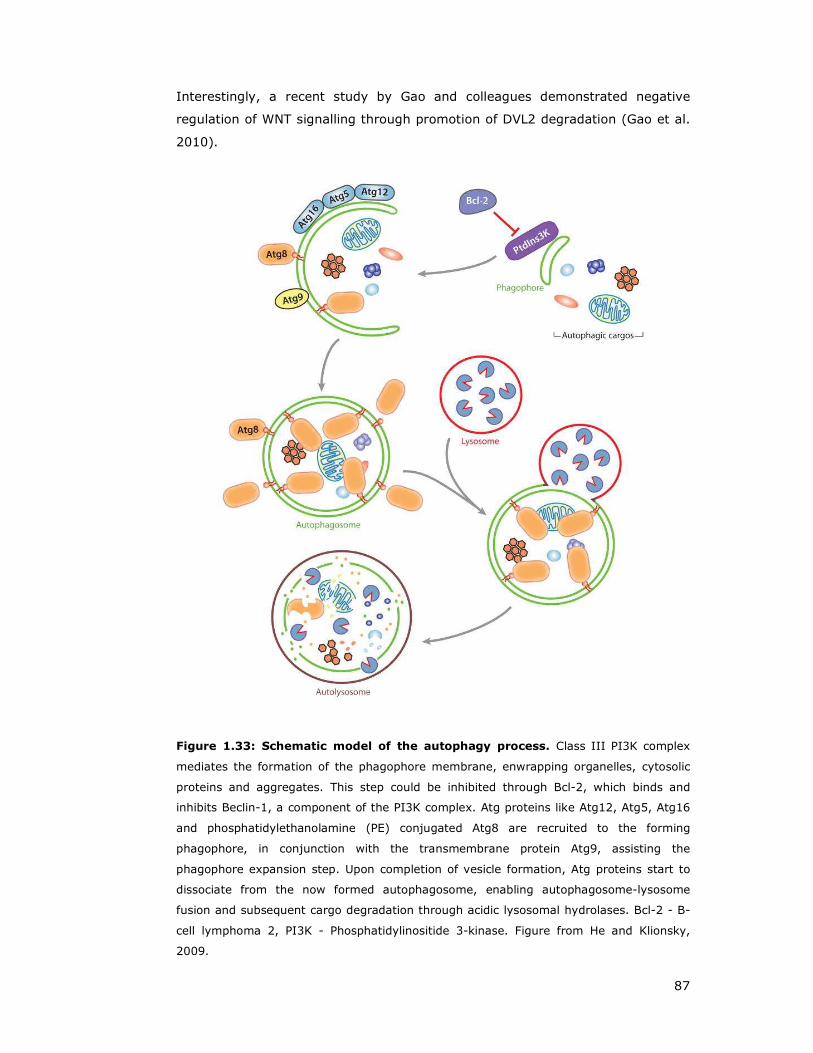
87
Interestingly, a recent study by Gao and colleagues demonstrated negative
regulation of WNT signalling through promotion of DVL2 degradation (Gao et al.
2010).
Figure 1.33: Schematic model of the autophagy process. Class III PI3K complex
mediates the formation of the phagophore membrane, enwrapping organelles, cytosolic
proteins and aggregates. This step could be inhibited through Bcl-2, which binds and
inhibits Beclin-1, a component of the PI3K complex. Atg proteins like Atg12, Atg5, Atg16
and phosphatidylethanolamine (PE) conjugated Atg8 are recruited to the forming
phagophore, in conjunction with the transmembrane protein Atg9, assisting the
phagophore expansion step. Upon completion of vesicle formation, Atg proteins start to
dissociate from the now formed autophagosome, enabling autophagosome-lysosome
fusion and subsequent cargo degradation through acidic lysosomal hydrolases. Bcl-2 - B-
cell lymphoma 2, PI3K - Phosphatidylinositide 3-kinase. Figure from He and Klionsky,
2009.
88
1.10 Aim of my study and outline of subsequent chapters
RAS proteins mediate important cellular functions such as gene expression,
proliferation, differentiation and survival through a wide variety of effector
proteins. Despite overlapping regulatory and effector proteins, and similar
biochemical and biological features between oncogenic RAS and the closely
related R-RAS members, they can also exhibit divergent regulation of signalling
and cellular processes. Therefore, a better understanding of the full spectrum of
effector pathways regulated by RAS family GTPases is critical to comprehend
their many biological functions and their contribution to human disease.
In order to identify novel effector proteins specific to R-RAS members, Dr.
Rodriguez-Viciana carried out a non-biased proteomic Tandem Affinity
Purification (TAP) approach employing active R-RAS subgroup members as baits.
This led to discovery of VANGL1 protein, a component of the WNT/Planar Cell
Polarity pathway, as a potential novel effector of R-RAS proteins.
The aim of this thesis project was to validate and characterise the novel R-RAS-
interactions, identified by TAP to study how R-RAS members regulate VANGL1
function and how VANGL1 may contribute to the signalling properties of R-RAS.
In addition, the possibility was tested that R-RAS GTPases function downstream
of WNT ligands in the PCP pathway, co-ordinating the regulation of VANGL
proteins in order to control polarity and cell motility.
Cell motility and migration play a paramount role in both normal embryogenesis
and the development of diseases such as cancer. Therefore, experiments were
carried out to analysis and assess a role of R-RAS and VANGL1 in regulation of
cell movement and invasion.

89
CHAPTER 2
Materials and Methods
90
2 Materials and Methods
2.1 MATERIALS
2.1.1 Chemical compounds and reagents
Human WNT3a (5036-WN), WNT5a (645-WN) and WNT7a (3008-WN) ligands
were purchased from R&D. Murine Wnt3a (315-20) ligand was obtained from
Peprotech. The proteasomal inhibitor MG132 (Santa Cruz Biotechnology) was
prepared as 10 mM stock solution in Dimethyl sulfoxide (DMSO).
Polyethyleneimine (Polysciences Inc) transfection stocks were prepared as 1
mg/mL solution in water and sterile filtered. Polybrene (hexadimethrine
bromide), a cationic polymer used to increase the efficiency of retrovirus or
lentivirus cell culture infections, was prepared as 5 mg/mL sterile filtered
aqueous solution and applied as 5 µg/ml final concentration. Crystal violet dye
(Sigma) was prepared as a 0.5% solution in 10% methanol/water mix.
Paraformaldehyde (PFA) 4% w/v was dissolved in PBS at 60°C while stirring and
subsequently aliquoted and frozen for long term storage. Aliquots were thawed
when required and used immediately for fixation purposes. Autophagy reagents
3-methyladenine, Bafilomycin A, and Chloroquine were obtained from Sigma.
Hanks balanced salt solution (HBSS) for amino acid starvation was obtained from
Invitrogen. For immunoprecipitation experiments, Streptavidin sepharose or
Protein A / Protein G sepharose were obtained from GE Healthcare. Non-
targeting control siRNA conjugated with Alexa Fluor Red (46-5318, Invitrogen)
was used to assess siRNA transfection efficiency.
If not stated otherwise, chemical reagents were obtained from Sigma.
2.1.2 Buffers
Coomassie stain: 0.2% (w/v) Brilliant Blue R (Sigma), 10% acetic acid (BDH),
50% methanol (BDH).
Coomassie destain: 10% acetic acid, 50% methanol.
DNA loading buffer (10x): 0.00125 g/mL Bromophenol Blue, 40% TE, 60 %
Glycerol.
Lysis buffer (LB-E): pH 7.4; PBS supplemented with 1% w/v Triton X-100, 1 mM
EDTA, 1mM Dithiothreitol (DTT), Protease inhibitor cocktail (Roche) and

91
Phosphatase inhibitor solution (10mM NaF, 2mM Na3VO4, 2 mM Na4P2O7 , 2 mM
β-glycerophosphate).
Lysis buffer (LB-M): pH 7.4; PBS supplemented with 1% w/v Triton X-100, 5 mM
MgCl2, 1mM DTT, Protease inhibitor cocktail (Roche) and Phosphatase inhibitor
solution (10mM NaF, 2mM Na3VO4, 2 mM Na4P2O7 , 2 mM β-glycerophosphate).
3-(N-morpholino)propanesulfonic acid (MOPS) running buffer: pH 7.7, 50 mM
MOPS, 50 mM Tris Base, 0.1% SDS, 1 mM EDTA.
Phosphate buffered saline (PBS) buffer: pH 7.4; 137 mM NaCl, 2.7 mM KCl, 10.1
mM Na2HPO4, 1.76 mM KH2PO4).
Protein loading buffer (4xLDS): 40% glycerol, 4% lithium dodecyl sulfate (LDS),
4% Ficoll-400, 0.8 M triethanolamine-Cl, pH 7.6, 0.025% phenol red, 0.025%
coomassie G250, 2mM EDTA disodium. This buffer was purchased from
Invitrogen and supplemented with 0.25 M DTT to achieve reducing capability.
Tween supplemented PBS (PBST): 0.1% Tween in PBS.
Transfer buffer: 10 % (w/v) Methanol, 25 mM Bicine, 25 mM Bis-Tris, 1 mM
EDTA.
Tris-Acetate-EDTA buffer (TAE): 40 mM Tris, 20 mM acetic acid, 2.5 mM EDTA.
Tris-EDTA buffer (TE): pH 8, 10 mM Tris, 1 mM EDTA.
2.1.3 Media and growth plates
Luria Bertani (LB) was prepared using 10 g/L Bacto™-Tryptone (BD), 5 g/L
Bacto™-yeast extract (BD), 10 g/L NaCl (Sigma), 1 g/L Glucose as a ready to
dissolve mix (Sigma). LB selection media contained either 100 µg/mL Ampicillin
(Sigma) or 50 µg/mL Kanamycin (Gibco). LB growth plates were prepared as LB
media above and supplemented with 15 g/L agar (Sigma) and selection plates
were supplemented with appropriate antibiotic post autoclaving.
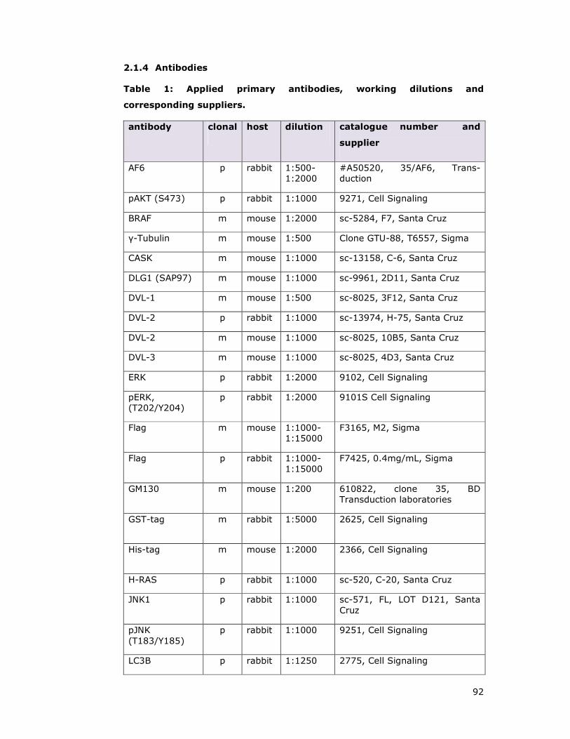
92
2.1.4 Antibodies
Table 1: Applied primary antibodies, working dilutions and
corresponding suppliers.
antibody clonal host dilution catalogue number and
supplier
AF6 p rabbit 1:500-1:2000
#A50520, 35/AF6, Trans-duction
pAKT (S473) p rabbit 1:1000 9271, Cell Signaling
BRAF m mouse 1:2000 sc-5284, F7, Santa Cruz
γ-Tubulin m mouse 1:500 Clone GTU-88, T6557, Sigma
CASK m mouse 1:1000 sc-13158, C-6, Santa Cruz
DLG1 (SAP97) m mouse 1:1000 sc-9961, 2D11, Santa Cruz
DVL-1 m mouse 1:500 sc-8025, 3F12, Santa Cruz
DVL-2 p rabbit 1:1000 sc-13974, H-75, Santa Cruz
DVL-2 m mouse 1:1000 sc-8025, 10B5, Santa Cruz
DVL-3 m mouse 1:1000 sc-8025, 4D3, Santa Cruz
ERK p rabbit 1:2000 9102, Cell Signaling
pERK, (T202/Y204)
p rabbit 1:2000 9101S Cell Signaling
Flag m mouse 1:1000-1:15000
F3165, M2, Sigma
Flag p rabbit 1:1000-1:15000
F7425, 0.4mg/mL, Sigma
GM130 m mouse 1:200 610822, clone 35, BD Transduction laboratories
GST-tag m rabbit 1:5000 2625, Cell Signaling
His-tag m mouse 1:2000 2366, Cell Signaling
H-RAS p rabbit 1:1000 sc-520, C-20, Santa Cruz
JNK1 p rabbit 1:1000 sc-571, FL, LOT D121, Santa Cruz
pJNK (T183/Y185)
p rabbit 1:1000 9251, Cell Signaling
LC3B p rabbit 1:1250 2775, Cell Signaling

93
LRP6 m rabbit 1:1000 2560, Cell Signaling
pLRP6 (Ser1490)
p rabbit 1:1000 2568, Cell Signaling
Myc p rabbit 1:2000 sc-789, A-14, Santa Cruz
PP1 m mouse 1:1000 sc-7482, E-9, Santa Cruz
RHOA m mouse 1:1000 sc-418, 26C4, Santa Cruz
RHOB p rabbit 1:1000 sc-180, C-119, Santa Cruz
R-RAS m mouse 1:500 sc-81930, YC-16, Santa Cruz
R-RAS p rabbit 1:500 sc-523, C-19, Santa Cruz
SCRIB m mouse 1:1000 sc-55543, C-6, Santa Cruz
Syntenin-1 m mouse 1:1000 sc-100336, S-31, Santa Cruz
Tubulin-HRP p rabbit 1:5000 PM054-7, MPL
TC21 m mouse 1:500 Sc-8193, 37-Y, Santa Cruz
TC21 p rabbit 1:1000 In house generation
VANGL1 m mouse 1:1000 MAB5476, Clone 497707, R&D systems
VANGL1 p rabbit 1:15000 In house generation, serum1 and pre-sera
Note: Antibodies are indicated as monoclonal (m) or polyclonal (p) and were prepared in 3% BSA/PBST supplemented with 0.02% NaN3.
2.1.5 Plasmids
For all cloning and subcloning vector construction, the Invitrogen Gateway®
Cloning Technology was employed (www.invitrogen.com/gateway).
This system relies on cloning the gene of interest into an Entry vector (pENTR,
see Figure 2.1A), which then can be amplified in bacteria and forms the donation
vector for any subsequent subcloning step into various destination vectors by a
simple and fast recombination reaction. Destination vectors (for an example
refer to mammalian GST-tagged destination vector pDEST27, Figure 2.1B) are
available for a wide range of applications and systems (E. coli, yeast, insect, or
mammalian cell) and thus provide a fast, reliable and convenient method of
construct generation.
For interaction studies in mammalian cells, pcDNA™ destination vectors were
used, which are designed for constitutive expression in mammalian cell lines
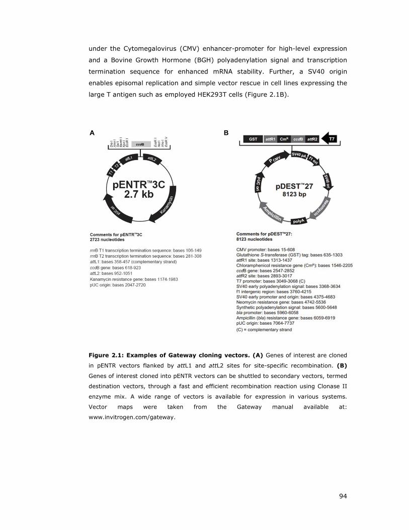
94
under the Cytomegalovirus (CMV) enhancer-promoter for high-level expression
and a Bovine Growth Hormone (BGH) polyadenylation signal and transcription
termination sequence for enhanced mRNA stability. Further, a SV40 origin
enables episomal replication and simple vector rescue in cell lines expressing the
large T antigen such as employed HEK293T cells (Figure 2.1B).
Figure 2.1: Examples of Gateway cloning vectors. (A) Genes of interest are cloned
in pENTR vectors flanked by attL1 and attL2 sites for site-specific recombination. (B)
Genes of interest cloned into pENTR vectors can be shuttled to secondary vectors, termed
destination vectors, through a fast and efficient recombination reaction using Clonase II
enzyme mix. A wide range of vectors is available for expression in various systems.
Vector maps were taken from the Gateway manual available at:
www.invitrogen.com/gateway.

95
All pENTR clones were generated by Lucy C. Young, Marta Mũnoz-Alegre or Pablo
Rodriguez-Viciana using Image clones or human placental pLIB cDNA library
(PT3230.1) (Clontech) as templates for PCR. After cloning, pENTR vectors were
sequenced and used for destination vector generation by means of
recombination reactions.
Recombination reactions were performed by combining 0.5 µL Entry clone
miniprep DNA, 0.2 µL desired Destination vector (100 ng), 1.3 µL 0.2x TE buffer
and 0.5 µL 5x Gateway® LR Clonase II Plus enzyme mix (Invitrogen). The
reaction volume was well mixed and microcentrifuged to collect at the bottom of
the tube. The reaction was incubated at room temperature for at least 60
minutes up to O/N before transforming into bacteria and subsequent DNA
isolation as outlined in 2.2.4 and 2.2.5.
2.1.6 Primers
Cloning primers for mutagenesis reactions were generated and obtained from
Invitrogen and were delivered lyophilised. Oligos were resuspended with 0.2x TE
to yield a stock solution of 100 pmol/µL (100 µM), incubated for 5 min and then
vortex thoroughly. Working stocks for Polymerase chain reactions (PCR) were
prepared at 10 pmol/µL (10 µM) with 0.2x TE.
2.1.7 RNAi sequences
RNAi duplex oligonucleotides (Stealth RNAi™) were designed and ordered from
Invitrogen. Supplied lyophilised primers were reconstituted with
Diethylpyrocarbonate (DEPC) treated water to yield a master stock concentration
of 100 µM. Working stock concentrations were generated from the master stock
at concentrations of 20 µM and 4 µM stock using DEPC-treated water. All
employed RNAi duplexes were initially tested for respective protein knock down
efficiency. In order to prevent off target effects, dose dependent experiments
were carried out to determine minimal siRNA concentrations for efficient knock
down, which were observed to be in the range between 2nM to 10nM (data not
shown). Furthermore, time course experiments were carried out in order to
estimate the longevity of the knock down effect, which lasted up to 7 days in a
fast proliferating cell line such as HEK293T (data not shown).

96
Table 2: Applied siRNA oligo sequences
siRNA Target sequence
R-RAS-1 5'-GAG AGC AGU ACA UGC GUG CUG GCC A-3'
R-RAS-2 5'-UCG CCA UUA ACG ACC GGC AGA GUU U-3'
TC21-1 5'-CAG UAU AUG AGG ACU GGC GAA GGC U-3'
TC21-2 5'-GGA UCG UGA UGA GUU CCC AAU GAU U-3'
VANGL1-1 5'-AGG CCG AAC AUG AAC GGC GAG UAA A-3'
VANGL1-2 5'-CAA UAU GAC AAG GAC CGC UGG CUC U-3'
VANGL2-1 5'-CGU CUD UGU GCU GCG UGC CCU GCU U-3'
VANGL2-2 5'-UCG UGG UCU CCU ACU GGC UCU UCU A-3'
Scribble-1 5'-GCG UCA GCG UCA UCC AGU UCC UGG A-3'
Scribble-2 5'-GGC CUA CGG GAA CUG UGC AUC CAG A-3'
Shoc2-2 5'-UAC CUU CGC UUU AAU CGU AUA-3'
scramble 5'-UUC UCC GAA CGU GUC ACG UUU-3'
The number post the dash indicates individual siRNA oligos, targeting different regions of the same gene of interest.
In order to assess transfection efficiency of siRNA molecules, an Alexa Fluor®
555-labeled, double-stranded RNA (Invitrogen, BLOCK-iT™ Alexa Fluor® Red
Fluorescent Oligo Cat. No. 14750-100) was transfected alongside as control and
assessed by fluorescence microscopy for successful delivery 24-48 h post
tranfection. Alexa 555 is a duplex with the same length, charge, and
configuration as standard siRNA and its sequence is not homologous to any
known gene.

97
2.2 DNA TECHNIQUES
2.2.1 Basic DNA manipulations
2.2.1.1 Cloning approach of VANGL1 truncation constructs
Subcloning tasks were carried out utilising the Invitrogen Gateway® technology
with ClonaseTM II (Invitrogen) approach (see section 2.1.5.). DNA digestions
were performed using New England Biolabs restriction enzymes and the supplied
buffers according to the instructions of the manufacturer. Routinely, 0.5 µg of
DNA was utilised for diagnostic restrictions, and 1-5 µg of DNA for preparative
digests for subsequent use in ligation reactions. DNA restriction digests were
usually carried out at 37°C for 2 h according to instructions of the manufacturer.
For primer sequences and template plasmids used in this study, refer to Table 3.
Chosen bands of DNA were cut out of the gel and purified using QIAquick Gel-
extraction kit (Qiagen) according to the instructions of the manufacturer. For
ligation reactions 1 µL of T4 ligase (Invitrogen), T4 ligase reaction buffer
(Invitrogen), vector and insert DNA were added together to a final reaction
volume of 10 µL. The ratio of vector to insert was maintained at 1:3 and ligation
was carried out for 1 h to overnight at room temperature. Subsequently, 5 µL of
ligation reaction was transformed into chemically competent bacteria
(Invitrogen) as described in section 2.2.4.
Table 3: Primer sequences for generation of VANGL1 truncation
constructs.
Con-
struct
Size [bp]
Forward primer (5’ -> 3’)
Reverse primer (3’ -> 5’)
Tm
[°C]
VANGL1-N 343 caccggatccatggataccgaatccacttattc cacgaattcttaggtgaggcccaggtagcgtttgc
59 63
VANGL1-C1 852 caccggatccgagctcaggcagctgcagccca cgcgaattcttaaacggatgtctcagactgta
67 57
VANGL1-C2 426 caccggatccgagctcaggcagctgcagccca cacgaattcttaggctttctgctgctcctcagcc
67 62
VANGL1-C3 426 caccggatccccaggggaggtgatggaccctag cgcgaattcttaaacggatgtctcagactgta
67 57
VANGL1-∆PDZ-BM
1560 caccggatccatggataccgaatccacttattc cacgaattcttaagactgtaagcgaaggacaaatttg
59 57
Note: For a detailed features and overview about truncated regions refer to Figure 3.8. Annealing temperatures were calculated using Promega online calculation tool, referring to salt adjusted Tm.

98
Table 4: PCR cycling parameters for VANGL1 truncation constructs
Segment Cycles Temperature Time
1 1 94°C 3 minutes
2 25 94°C (denaturation)
30 seconds
55°C (annealing) 30 seconds
72°C (extension) 2 minutes
3 1 72°C 2 minutes
4 1 16°C ∞
Note: The extension time is determined by the length of the expected longest fragment of 1.6 kb (1 kb per minute for extension).
2.2.1.2 Cloning approach of ARHGEF17 truncation constructs
Subcloning and DNA handling techniques were carried out as described above for
generation of VANGL1 truncations and respective PCR reactions were carried out
using the cycle parameters as outlined in Table 4.
Table 5: Primer sequences for generation of ARHGEF17 truncation
constructs.
Con-
struct
Size
[bp]
Forward primer (5’ -> 3’)
Reverse primer (3’ -> 5’)
Tm
[°C]
ARHGEF17-N 1530 caccgaattcatggcccgccgcctgccccgca cacgcggccgctcagtgcttccgcatgtccacctgtg
68 69
ARHGEF17-∆N
3040 caccgaattcatggccagcaagtgctgcagcaagc cacgcggccgctcacaccctccacaggaggagg
64 69
ARHGEF17-C1
2440 caccgaattcatgggtgtgcggagtgccgaggaggc cacgcggccgctcacaccctccacaggaggagg
67 69
ARHGEF17-∆C
2120 caccgaattcatggcccgccgcctgccccgca cacgcggccgctcacacacgggcatggcgctccgc
68 72
ARHGEF17-C2
1454 caccgaattcagccacacgcctcacggctcctccg cacgcggccgctcactggtaggcctcagggtcgacact
69 70
ARHGEF17-C3
487 caccgaattcagctccgtgtggctgggcactgag cacgcggccgctcagacgtctacttctgccagctgct
65 68
ARHGEF17- 722 caccgaattcagctccgtgtggctgggcactgag 65

99
C4 cacgcggccgctcagactgcagccaagaagcggac 69
ARHGEF17-C5
977 caccgaattcagctccgtgtggctgggcactgag agtgtgggaggtgtcctcctccaccaaaac
65 67
Note: annealing temperatures were calculated using Promega online calculation tool, referring to at salt adjusted Tm. (http://www.promega.com/techserv/tools /biomath/calc11.htm)
2.2.2 Plasmid mutagenesis
Generation of VANGL1 point mutations by site directed mutagenesis
A PCR approach was used to introduce VANGL1 point mutations at reported
residues associated with neural tube defects and impairment of Dishevelled
binding (Kibar et al. 2007) by means of site directed mutagenesis using the Quik
change strategy (Stratagene). Extensions of the oligonucleotide primers,
containing desired point mutations (Table 6), were generated with the PfuUltraTM
high-fidelity polymerase, producing a mutated plasmid (Hemsley et al. 1989).
The amplified product was subsequently DpnI treated, which specifically cleaves
fully methylated GMe6ATC sequences. As a result, the bacterially generated DNA
used as template for amplification was digested and while the unmethylated
product of the PCR reaction is left intact. The product of this digestion reaction
was used to transform chemically competent Escherichia coli (E.coli) bacteria
and colonies formed on selection plates were sequenced to confirm successful
mutations.
PCR reactions were carried out in 50 µL volumes using provided reagents of the
Quik Change® II Site-Directed Mutagenesis Kit (Stratagene), according to the
instructions of the manufacturer. Conditions for amplification are shown in Table
7, utilised primers are summarised in Table 6 below.

100
Table 6: Primer sequences for generation of VANGL1 point mutant
constructs
Con-
struct
Size
[bp]
Forward primer (5’ -> 3’)
Reverse primer (3’ -> 5’)
VANGL1-V239I
1572 ctcttcatccattacctggccatcatcctgctggagctcaggcagctgc
gcagctgcctgagctccagcaggatgatggccaggtaatggatgaagag
VANGL1-D259E
1572 gctgcaggtggtccgctccaccgagggcgagtcccgcttctacagcctg
caggctgtagaagcgggactcgccctcggtggagcggaccacctgcagc
VANGL1-R274Q
1572 gcctgggacacctgagtatccagcaagcagcattggtggtcctagaaaat
attttctaggaccaccaatgctgcttgctggatactcaggtgtcccaggc
VANGL1-M328T
1572 gccactggccagtcccgggccacgattgctgcagctgctcggcgcag
ctgcgccgagcagctgcagcaatcgtggcccgggactggccagtggc
VANGL1-S467N
1572 tctctacacagtggaggcttgtcaatgatgaggctgtgactaatggatt
aatccattagtcacagcctcatcattgacaagcctccactgtgtagaga
Table 7: PCR cycling parameters for site-directed mutagenesis
Segment Cycles Temperature Time
1 1 95°C 30 seconds
2 18 95°C (denaturation)
30 seconds
55°C (annealing) 1 minute
68°C (extension) 11 minutes and 35 sec
3 1 4°C ∞
Note: The extension time was determined by the length of the amplified plasmid
(1 kb per minute for extension).
In order to test expression of the generated point and truncation mutants of
VANGL1, plasmids were transfected into HEK293T cells and subjected to
immunoprecipitation (as described in section 2.5.4). Coomassie staining of a size

101
separated protein gel displayed similar expression levels of VANGL1 point
mutants, except mutant D259E, which was expressed at a significantly lower
level. VANGL1-N or C-terminal truncation proteins appeared to be equally
expressed whereas VANGL1-C2 or C3 fragments showed severely reduced
protein levels (Figure 2.2).
Figure 2.2: Expression analysis of Flag-VANGL1 point mutants and truncation
constructs in HEK293T cells. 2ug of Flag-VANGL1 constructs were transfected into HEK293T cells, lysed after 48 hours and subjected to immunoprecipitation using Flag affinity beads. Immunocomplexes were size spearated on SDS-PAGE and Coomassie stained. Asteriks depict respective VANGL1 proteins.
2.2.3 DNA gel electrophoresis
In order to visualise DNA fragments from diagnostic digests or for cloning
purposes, 1% or 1.2% agarose gels (Invitrogen) were prepared using 1x TAE
buffer and TrackIt™ 1 Kb Plus DNA Ladder (1 µl) (Invitrogen, 0.1 µg/µL,) was
run alongside for size estimation.
2.2.4 Transformation of bacteria
Chemically competent E.coli DH5α cells (Invitrogen) were gently thawed on ice,
5 µL of prechilled ligation reaction or gateway reaction mix was added to 50 µl
aliquots of bacterial cell suspension and then kept on ice for 10 min.
Subsequently, bacteria were heat-shocked at 42°C for 45 sec, recovered on ice
for another 2 min, combined with 250 µL LB or S.O.C. solution (0.5 % Bacto
tryptone, 2 % Bacto yeast extract, 10 mM NaCl, 2.5 mM KCl, 10 mM MgCl2, 10
mM MgSO4, 20 mM glucose) and incubated for one hour at 37°C at 200 rpm.
102
Bacteria suspensions were spread on LB culture plates supplemented with either
100 µg/mL Ampicillin or 50 µg/mL Kanamycin and incubated overnight at 37°C.
Single colonies formed on the plates were picked and grown for extraction of
plasmid DNA.
2.2.5 Preparation and purification of plasmid DNA
Bacterial DNA for analytical purposes was isolated from 1.5 mL minipreparation
cultures utilizing a QIAprep® mini kit (Qiagen), and for generation of plasmid
stocks, 50 mL E.coli cultures were extracted using QIAprep® midi kit (Qiagen)
according to the instructions of the manufacturer.
2.2.6 DNA sequencing
DNA sequencing was performed by an inhouse service using AB sequencing
technology. Supplied ab1 format files were visualised using Finch TV software.

103
2.3 RNA TECHNIQUES
2.3.1 RNA isolation
Total RNA from SaOS2 cells of equivalents of six well plates were prepared using
ISOGEN reagent (Life Sciences). 500 µL ISOGEN was added to a PBS washed
well of cells and incubated for 1 min at RT. Next, the ISOGEN solution was
collected in an eppendorf tube and after 5 min incubation at RT, 100 µL
chloroform added, vigorously shaken for 15 sec and incubated at RT for 2 min.
Aqueous phase was separated from organic phase by spinning at 20 000 x g for
15 min at 4°C, following transfer of the aqueous phase to a fresh tube.
Subsequently, 250 µL isopropanol was added, briefly vortexted and incubated for
10 min at RT. Precipitated RNA was collected as pellet by spinning at 20 000 x g
for 10 min at 4°C, and the pellet washed with 70% ethanol/water solution,
before spinning again at 20 000 x g for 5 min at 4°C and subsequent aspiration
of ethanol solution. Yielded pellets were air dried at 37°C and dissolved in RNAse
free water. Concentration of yielded RNA was determined using a
spectrophotometer (Hitachi U-1900).
2.3.2 Semiquantitative PCR
For semi-quantitative reverse-transcription polymerase chain reaction, 0.5 µg of
total RNA was used and the polymerase chain reaction was performed using the
SuperScript One-Step RT-PCR kit (Invitrogen) according to the instructions of
the manufacturer using the primer sequences listed in Table 8. MMP-13 specfic
PCR reactions were carried out using following conditions for 40 cycles (Table 9)
The amount of 18S rRNA was amplified using the same conditions as outlined for
MMP-13 reactions (Table 9) for 25 cycles. The relative amount of MMP-13 was
normalised using 18S RNA.
Table 8: Primer sequences for RT-PCR.
Gene Primer sequences
(forward/reverse)
Prod.
size
MMP-13 5'-AAC ATC CAA AAA CGC CAG AC-3'
5'-GGA AGT TCT GGC CAA AAT GA -3' 166 bp
18S rRNA 5'-ATG GCC GTT CTT AGT TGG TG -3'
5'-CGC TGA GCC AGT CAG TGT AG -3' 217 bp
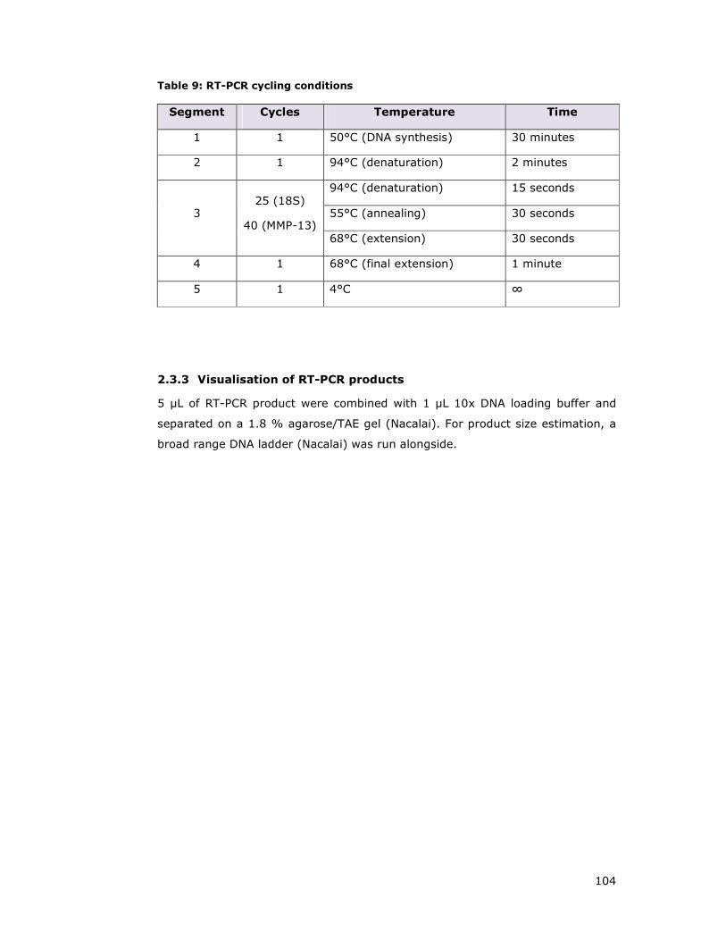
104
Table 9: RT-PCR cycling conditions
Segment Cycles Temperature Time
1 1 50°C (DNA synthesis) 30 minutes
2 1 94°C (denaturation) 2 minutes
3 25 (18S)
40 (MMP-13)
94°C (denaturation) 15 seconds
55°C (annealing) 30 seconds
68°C (extension) 30 seconds
4 1 68°C (final extension) 1 minute
5 1 4°C ∞
2.3.3 Visualisation of RT-PCR products
5 µL of RT-PCR product were combined with 1 µL 10x DNA loading buffer and
separated on a 1.8 % agarose/TAE gel (Nacalai). For product size estimation, a
broad range DNA ladder (Nacalai) was run alongside.
105
2.4 MAMMLIAN CELL CULTURE
2.4.1 Cell lines and culture conditions
Immortalised human mammary epithelial cells HMEC, HMLE, 184A1 and MCF10A
were cultured in DMEM F12 media (Invitrogen) maintained as described in
(Debnath et al. 2003) supplemented with 2mM L-Glutamine (Gibco) in the
absence of Penicillin/Streptomycin. Human breast adenocarcinoma MDA-MB-231
and MCF-7, alveolar basal epithelia adenocarcinoma A549, colorectal carcinoma
HCT116, embryonic kidney cells 293 (HEK293T), osteosarcoma SaOS2 and
U2OS, cervical carcinoma HeLa and mouse parental L cell lines were maintained
in DMEM supplemented with 10% fetal bovine serum (FBS) (HyClone). Cells
were grown in 150 mm dishes (Falcon) and passaged by washing with PBS and
trypsinised by applying 0.05% Trypsin/EDTA (Gibco) solution for 5 up to 20 min.
Cell lines were long term stored in FBS supplemented with 10% DMSO and
placed in the vapour phase of liquid nitrogen.
2.4.2 Thawing and freezing/long term storage of mammalian cells
Cells were retrieved from liquid nitrogen and thawed in 37°C in a water bath,
warm medium added and spun at 400 x g for 5 min to pellet cells. The pellet was
resuspended in fresh medium and transferred to the appropriate cell culture
dish. Medium was replaced the following day.
For long term storage of cell lines, cells were trypsinised and pelleted at 400 x g
for 4min. Supernatant were aspirated and the pellet resuspended in freezing
medium, containing 10% DMSO and 90% FBS. Aliquots of 1ml cell suspension
were transferred into cryogenic vials (Corning) and placed in a styrofoam box
and kept at -80°C for 24h, before transferred to liquid nitrogen.
2.4.3 Manual cell counting and assessment of viability
For seeding purposes and cell number determination, 10 µL of single cell
suspension was applied to a Neubauer haemocytometer (VWR). Cells were
counted in four independent areas of the haemocytometer and mean values
were determined as cell number per mL. Cell viability was analysed by the
Trypan blue dye (Sigma) exclusion method. Equal amounts of cell suspension
are mixed with 0.4% trypan blue solution (Sigma), both populations either
106
positive or negative for dye staining were counted and viability was expressed as
percentage of viable (dye negative) cells.
2.4.4 IncuCyte based proliferation or phenotype analysis
Cells were counted manually and seeded in 24 well or 6 well plates at various
densities, depending on the cell line and rate of proliferation. They were then
placed in the IncuCyte (Essen Instruments) and well density was measured
every 2h until cells reached complete confluence. Medium was changed regularly
(every 48h) between readings. Readings were analysed using MS Office Excel.
2.4.5 DNA transfection
2.4.5.1 Lipofectamine2000 transfection of plasmid DNA
For overexpression experiments, 1.5x10⁶ HEK293T cells were seeded in six-well
dishes 2-4 h prior transfection. For each transfection reaction, 4 µL
Lipofectamine2000 (Invitrogen) pre-incubated with 96 µL Opti-MEM for 5 min
were mixed with 2.0 µg of plasmid DNA pre-diluted into 100 µL Opti-MEM
(Gibco). After 20 min incubation at RT, the transfection solution was then added
drop-wise to cells. After 16-24 h, additional 2 mL of full media was added to
each well in order to boost growth and cells were harvested the following day.
2.4.5.2 Polyethylenimine transfection of plasmid DNA
For overexpression experiments, 2x10⁶ HEK293T cells were seeded in six-well
dishes 2-4 h prior transfection. For each transfection reaction, 2.0 µg of plasmid
DNA prediluted into 200 µL Opti-MEM (Gibco) were mixed with 8 µg
polyethylenimine (PEI) (as aqueous solution of 1mg/mL). After 30 min
incubation at RT the transfection solution was then added drop-wise to cells.
After 16-24 h, additional 2 mL of full media was added to each well in order to
boost growth and cells were harvested the following day. When performing
transfections in 24 wells plates, 0.5 µg DNA constructs and 2ug polyethylenimine
were combined in 100 µL Opti-MEM per reaction.
2.4.6 RNAiMAX siRNA transfection
Forward transfection protocols
U2OS cells were plated the day before, at a density of 0.75x10⁶ cells per 24 well
in a total volume of 500 µL regular growth media. The next day, 6 pmol RNAi

107
duplex was diluted in 50 µL serum free OptiMEM®I medium (Gibco), while 1µL of
RNAiMAX reagent was diluted with 50 µL serum free OptiMEM®I medium and
mixed gently. RNAi duplex and RNAiMAX mixtures were combined and incubated
at room temperature for 15 min, before adding the incubated mixture dropwise
to wells and mix gently by rocking the plate. The final volume per 24 well came
up to 600 µL, which yielded in a final of 10nM RNAi duplex concentration. 16h
post transfection, the complete medium was changed and cells were either
wound scratched or lysed for knock down efficiency verification 72h post
transfection.
Oligonucleotide delivery efficiency into mammalian cells was assessed by
transfection of a red-labelled dsRNA oligomer (BLOCK-iT-Alexa), which does not
display any homology to any known gene sequences.
Reverse transfection protocols
Reverse transfection protocols were applied for Saos2 invadopodia experiments.
Transfection mixes were prepared as above by combining 100uL OPTIMEM/RNAi
oligonucleotide solution with a 98 µL OPTIMEM / 2µL RNAiMAX mix, followed by a
15 min of incubation at RT. Transfection mix, media and 2x10⁵ Saos2 cells were
combined to yield a 1mL volume prior adding to 6 wells. Media was changed 16h
later and cells were manipulated 72h post transfection.
2.4.7 Virus generation
2.4.7.1 Retrovirus
For generation of retrovirus containing supernatants in 60 mm dishes, 5 x 10⁶
cells of the retrovirus producer cell line Phoenix Eco 293 were seeded 4 h before
transfection. Cells were lipid-complex transfected using 5µg FBneo plasmids
diluted in 250 µL OPTIMEM, which was combined with 10 µL Lipofectamine2000
per 250 µL OPTIMEM, followed by a 20 min incubation period at room
temperature. The transfection mix was added drop wise onto cells while rocking
gently, leading to final volume of 2.5mL. The next morning, culture media was
exchanged with 3 mL regular growth medium and ecotrophic retrovirus
supernatants were harvested at 24, 48, 72 hours after this by replacing collected
supernatant with 3mL regular growth media. Harvested virus was either frozen

108
in 1 mL aliquots and stored at -80°C or utilised for immediate subsequent
infection of EcoR target cells.
2.4.7.2 Lentivirus
Lentiviral constructs were employed due to its convenient ability to infect most
cell types including primary or non-dividing cells, and as an alternative to
retroviral transduction approaches in case of lack of required stable EcoR cell
lines.
In order to generate lentivirus supernatants for transduction purposes and
generation of stable cell lines, 2 x 10⁶ HEK293T cells were seeded in 35 mm 6
well plates in a final volume of 1.5 mL. After allowing cells to settle for 4 h, a
PEI transfection approach (refer to 2.4.5.2) was carried out in order to deliver all
required packaging vectors for virus particle production. Cells were triple
transfected with 0.73 µg of the gag-pol expressor plasmid p8.91, 0.43 µg of the
VSV-G expressor vector pMDG, and 0.84 µg of either pLEOC (Blasticidin
resistance) or pLEX (Puromycin resistance) lentiviral destination vector. Medium
was replaced 16 hours later with 1 mL growth medium, and virus harvest was
commenced 48h after transfection. Aliquots of 1 mL viral supernatants were
supplemented with a final concentration of 5 µg/mL Polybrene in order to
increase transduction efficiency and aliquots were either frozen or used fresh for
transduction of target cell lines. Virus collection was performed on three
consecutive days 48h, 72h and 96h post initial transfection.
In order to transduce mammalian cells with lentiviral particles, cells were seeded
into 6-well dishes and allowed to grow overnight to 50% to 70% confluency.
Next day, frozen virus stocks in 1 ml aliquots including 5µg/ml Polybrene were
thawed at 37°C and then spun for 1 min at 15000 x g rpm at 4°C in order to
pellet residual cells in the supernatant. Then, cell medium was replaced by 1ml
of the virus/Polybrene solution and plates were gently rocked every 30 min to
increase viral transduction efficiency. After 4 hour of incubation, remaining
supernatant was removed and replaced with 2ml of growth medium for 24h
before adding the appropriate selective agent. The appropriate selection is vector
dependent and media was either supplemented with 2.5 µg/mL Puromycin
(Sigma) or 5-10 µg/mL Blasticidin (Sigma), until sensitive control populations
died under selection pressure. Remaining transduced cell populations were either
expanded and used for experiments or frozen for long term storage.

109
2.4.8 Generation of stable cell lines
2.4.8.1 Generation of stably WNT ligand expressing cell lines and collection of
WNT conditioned media
Respective WNT ligand genes were cloned into retroviral FB destination vectors
and used for Retrovirus generation. Mouse L cells were subsequently transduced,
Geneticin (G418, 500 µg/mL) selected and grown to confluency. WNT ligand
conditioned media was collected from 15 cm dishes of control (eGFP) or stable
WNT ligand expressing L cells after reaching confluency. Conditioned media was
collected every 24 h over the course of three days, centrifuged to remove cell
debris and stored at 4°C for up to two months or snap frozen and stored at -
80°C for long term storage.
Generated WNT ligand conditioned media was tested for biological activity using
ß-catenin dependent transcription Luciferase reporter assay or Dvl2
phosphorylation shift assessed by western blotting.
Dilution of WNT3a CM with regular growth media at 1:2 (50%) or 1:4 (25%)
ratios revealed that the biological activity is not compromised compared to
undiluted CM (100%) (Figure 2.3A). In fact, the 1:2 (50%) dilution seemed to
enhance the biological activity by about 35%, suggesting that at least WNT3a
CM might contain other antagonistic factors besides WNT3a, which are able to
modulate activation of canonical signalling. Further, a single freeze/thaw (F/T)
cycle of WNT3a CM does not significantly affect its biological activity, allowing
long term storage of CM at -80°C (Figure 2.3A).
Since I planned to use WNT CM media for various stimulation experiments,
which might be sensitive to high FBS concentrations (above 0.25%), I assessed
WNT3a ligand activity with conditioned medium containing various FBS
concentrations using the β-catenin luciferase readout. Conditions ranged from
using serum free, BSA supplemented serum free, 0.25% FBS or regular growth
media levels, which demonstrated that WNT3a activity is 3-4 fold less in WNT
conditioned media containing low or no FBS. Therefore, we generally produced
WNT ligand CM in the presence of 10% FBS (Figure 2.3B).

110
Figure 2.3: Analysis of WNT3a conditioned media biological activity and optimal
culture and storage conditions. HEK293T cells were co-transfected with TOPFLASH
(firefly luciferase) and renilla luciferase reporter constructs. 24h post transfection, cells
were stimulated for 16h with WNT3a conditioned media prior cell harvest. Cell extracts
were assayed for firefly and renilla luciferase activities and data obtained were normalised
for renilla activity. β-catenin levels were expressed as fold increase across analysed
samples using control CM stimulated cells as reference. (A) WNT3a conditioned media
(CM) either undiluted (100%) or diluted 1:2 (50%) or 1:4 (25%) as indicated were
compared to a frozen and thawed (F/T) sample and assessed for biological activity.
Treatment with Lithium Chloride (LiCl, 30 mM, 16h) served as positive control. (B) In
order to examine if FBS concentrations during WNT3a CM generation affect its signalling
effectiveness, conditioned media with varied FBS amounts were compared for canonical
pathway activation. Expression of β-catenin or DVL3 served as positive canonical
stimulation control. eGFP transfection was used as transfection and background control.
111
2.4.8.2 Generation of stable cells expressing YFP-fusion proteins for localisation
studies
In order to analyse subcellular localisation of VANGL or other proteins of interest,
YFP-VANGL1, YFP-VANGL2, YFP-Dvl2 and others as outlined on respective figures
were subcloned into lentivirus pLEX-YFP (Puromycin selection) destination
vectors and viral supernatants generated as described above (refer to 2.4.7.2).
Cells were infected and selected as outlined in 2.4.7.2 and seeded on coverslips
for immuno-fluorescent analysis (2.7.1.1) or biochemical analysis.

112
2.5 PROTEIN TECHNIQUES
2.5.1 Preparation of cell extracts and immunoblot analysis
Cells extracts were prepared by washing cells once with ice-cold PBS and adding
lysis buffer LB-E or LB-M, while rocking dishes for 5 – 10 min at 4°C. Extracts
were collected and centrifuged at 15,000 x g for 15 min at 4 °C and normalised
according to the Bradford protocol including BSA-standard curve see below
(Bradford 1976). 10 µg of protein lysates were generally loaded on 4-20%
gradient NuPage SDS–polyacrylamide gels (Invitrogen) and blotted on a
polyvinylidene fluoride (PVDF) membrane (Amersham). Membranes were
coomassie stained for visualisation of protein content, blocked for 1 h in blocking
buffer (5% non-fat dried milk (Sigma)/PBST) and incubated with primary
antibody according to Table 1 in 3% BSA/PBST supplemented with 0.02% NaN3
at 4 °C overnight. Membranes were washed three times with PBST and
incubated for 1 h either with donkey anti-rabbit-HRP 1:5000 (Amersham) or
sheep anti-mouse-HRP 1:5000 (Amersham), followed by washing three times
with PBST and immunodetection was performed by application of a
chemiluminescence detection system (Insight Biotechnology) and exposed to
medical x-ray film (Fujifilm).
2.5.2 Determination of protein concentractions
Protein contents were determined based on the Bradford protocol (Bradford
1976) employing a BSA standard curve from 10 to 120 µg/mL protein content
using a protein assay dye reagent concentrate (Bio-Rad, 500-0006) according to
instructions of the manufacturer and measuring absorbance at 595 nm.
Unknown protein samples were calculated from BSA readings using a linear
regression approach.
2.5.3 Densitometric analysis
Western blot films were scanned at a resolution of 600 dots per inch (dpi) and
saved in jpg format at maximum quality. Bands of interest were selected and
intensity measured of each individual band as outlined in
http://lukemiller.org/index.php/2010/11/analyzing-gels-and-western-blots-with-
image-j. Background values were subtracted and densities displayed as fold
increase.
113
2.5.4 GST-pull down and immunoprecipitation experiments
Cell lysates for interaction studies (either IP or pull down experiments) were
harvested and centrifuged as described above. GST-proteins were purified using
glutathione beads (Amersham), Flag-tagged proteins were immunoprecipitated
with Flag-beads (Sigma) using an equivalent of 10 µL packed beads. Lysates
were incubated with respective agarose beads for 2 h while rotating at 4°C and
subsequently washed 3 times with lysis buffer without protease and phosphatase
inhibitors. After completion of washes, beads were drained with a needle (25G,
Terumo) and proteins eluded with LDS sample buffer. Interaction were analysed
by Westen botting as described above.
2.5.5 Expression and purification of recombinant proteins
Competent E.coli strain BL21(DE3) (Invitrogen) were transformed with desired
expression constructs. A two ml overnight culture from an individual bacterial
colony was used to inoculate 900 mL LB/ampicillin (100 µg/mL) media per
construct and incubated at 37°C at 200 rpm until reaching an optical density
(OD) value of OD600=0.6. Cultures were induced with 1 mM IPTG (Isopropyl-β-D-
Thiogalactopyranoside) and kept shaking for four additional hours. Bacteria were
then pelleted at 4500 rpm (JLA-10.500 rotor, 3847 x g) at 4 °C for 20 min, and
the supernatant was discarded. Pellets were resuspended in 20 mL PBS-E lysis
buffer containing 0.5 mg/mL Lysozyme (Sigma), protease inhibitors (tablet,
Roche), 5 µg/mL DNase (Sigma) and 5 mM β-Mercaptoethanol on ice. Next,
resuspended bacteria were lysed using a sonication approach of 4 rounds at 45
sec each on ice and debris was pelleted by centrifugation at 20000 rpm (JA-
25.50 rotor, 32816 x g). Fusion proteins were then purified from the yielded
lysate with either His, Glutathione, Streptavidin or Flag beads during incubating
for 2-4h at 4°C and gentle rotation. After three to four wash steps, proteins were
either eluted using the respective elution buffer or sample buffer (LDS).
2.5.6 Generation of GST-RBD beads for RHO/RAS activation assays
Respective GST- Raf, RalGDS or Rhodekin binding domain (RBD) fusion proteins
were expressed in E.coli and lysed as described above. Lysates were purified
using glutathione beads for 2h while gently rotating at 4°C, before collecting
beads by centrifugation at 2000 x g for 4 min. Supernatant was either discarded
or used for a second round of GST-purification. GST-beads were washed for four
114
times with PBS-0.1% Trition X-100 and once with LB-M/50% Glycerol before
storing beads as 1:4 in PBS-M/glycerol (50% Glycerol). An equivalent of 10 uL
packed beads were run on a SDS-PAGE gel in order to assess amount of bound
protein and purity, and appropriate stimulated lysates were used to test if GST-
RBD beads are able to enrich respective GTPases.
2.5.7 RAS and RHO binding domain assays
RAS binding domain (RBD) assays can estimate various endogenous RAS-family
protein activities and have been employed to analyse R-RAS and RHO levels in
mammalian cells upon various stimulations. In order to monitor levels of
activated R-RAS GTPases in cell lysates, I used GST-fusions of the Ras-binding
domain (RBD) of the RAS effector RAF1 (residues 2–140) or RALGDS (C-terminal
97 residues), exploiting the fact that RAS family proteins only bind effectors in
the GTP-bound state (Brtva et al. 1995; de Rooij and Bos 1997; Franke et al.
1997). Cells were lysed in LB-M lysis buffer, and cleared lysates incubated for 45
min at 4°C with 10 µL packed GST-RBD glutathione sepharose beads (see
2.5.6). Beads were washed three times, and bound proteins were eluted with
LDS sample buffer and resolved by SDS-PAGE. Activated H-RAS, R-RAS or TC21
was detected by immunoblotting using respective antibodies (Table 1).
The cellular level of GTP-loaded RHOA and RHOB was determined using a GST
fusion protein containing the RHO binding domain of Rhotekin (GST-RBD),
corresponding to residues 7-89 of Rhotekin, which specifically precipitates GTP-
RHO from cell lysates (Ren and Schwartz 2000; Mezzacappa et al. 2012). Cells
were lysed in LB-M lysis buffer, and cleared lysates incubated for 45 min at 4°C
with 10 µL packed GST-RBD glutathione sepharose beads (see 2.5.6). Beads
were washed three times, and bound proteins were eluted with LDS sample
buffer and resolved by SDS-PAGE. Activated RHOA or RHOB was then detected
by immunoblotting using respective antibodies (Table 1).
2.5.8 In vitro interaction assays
Spodoptera frugiperda (Sf9) cells were infected with baculoviruses expressing
GST-VANGL1 truncations (N and C-terminal) or various ARHGEF17/p164RhoGEF
C-terminal truncations (C1-C5). His-R-RAS, His-TC21 and effector proteins were
expressed and purified from bacteria as described above (section 2.5.5). In vitro

115
interactions were carried out by loading purified His-R-RAS or His-TC21 with
either GDP or the non-hydrolysable analogue of GTP (GMP-PNP) by incubation
for 5 min at 37°C in loading buffer (20 mM Hepes (pH 7.5), 50 mM NaCl, 5 mM
EDTA, 5 mg/mL BSA) in a 20-fold molar excess of GDP or GMP-PNP. Reactions
were stopped by placing the tubes on ice and adding MgCl2 to a final
concentration of 20 mM. 0.5 µg GDP/GMP-PNP loaded R-RAS or TC21 was
combined with 15 µL of packed glutathione beads bound to GST-VANGL1 or
GST- ARHGEF17/p164RhoGEF (or other effector proteins) in a tube containing
200 µL of PBS, 5 mM MgCl2, 0.1% Triton X-100. After 1-3 h of incubation at 4°C
while rotating, GST-beads were washed three times with PBS, 5 mM MgCl2, 0.1%
Triton X-100, needle drained and LDS sample buffer added. After separation on
4-12% SDS-PAGE gels, western blots were performed using His antibodies.
2.5.9 TAP-tagged protein transfection and purification for mass
spectrometry analysis
For the transient expression approach, eight confluent 15cm dishes (Greiner) of
HEK293T cells per condition were seeded at 50% confluence in a volume of
15mL, and transfected 3h post seeding using the PEI approach (2.4.5.2). Per 15
cm dish 10 µg TAP6-VANGL1 plasmid were combined with GST-R-RAS expression
plasmids (10 µg), mFzd5 (5ug), ROR2 (5 µg) or empty vector (5-10 µg) to yield
a total of 20 µg plasmid DNA, which was combined with 80 µg PEI reagent. The
media was exchanged 5 h post transfection with 20 mL growth media and cells
were additional boosted with 10 mL media 16 h later. HEK293T cells were
harvested 48h after transfection and cells extracts were prepared by washing
cells twice with ice-cold PBS and collected in PBS-M lysis buffer (1.6 mL per
plate) on ice (2.1.2). Extracts were centrifuged at 20,000 x g for 20 min at 4 °C
to pellet cellular debris. The soluble fraction was incubated with 250µl packed
Streptavidin high performance beads (GE Healthcare, 17-5113-01) per condition
for 2 hours at 4°C while rotating gently. Beads were pelleted through
centrifugation at 2000 x g for 5 min and column washed four times (VWR,
gravity flow columns) to remove unbound proteins. Bound proteins were eluted
by supplementing 2.5mM biotin in PBS-M wash buffer and shaking beads gently
for 10 min prior draining the beads and collecting the eluate. In order to increase
eluate protein concentration, proteins were precipitated using a Trichloroacetic
acid (TCA) approach. Eluates were supplemented with 2% deoxycholate/PBS
(1/100 of total volume) and TCA (1/20 of total volume), vortexed and incubated
on ice for 30 min. Precipitated proteins were pelleted by centrifugation at 15000

116
x g at 4°C for 30 min, and supernatants were carefully aspirated before pellets
were washed with 500 µl acetone. Pellets were collected again through spinning
at 15000 x g at 4°C for 5 min and the acetone removed in order to air dry the
remaining pellet. Dried pellets were dissolved in 30µl of 2x LDS sample buffer,
and left O/N at RT.
2.5.10 Mass spectrometry analysis
LDS samples were resolved on a 4-12% NuPAGE gels (Invitrogen) and stained
with SimplyBlueTM SafeStain (Invitrogen) according to the instructions of the
manufacturer. Visualised bands were compared to control/background TAP-
samples and differential bands were excised with a clean scalpel and proteins
were digested in-gel with trypsin as described previously (Rosenfeld et al. 1992)
and under http://msf.ucsf.edu/ingel.html.
Mass spectrometry analysis was carried out by Juan A. Oses-Prieto at the
Department of Pharmaceutical Chemistry (University of California, San Francisco,
California 94143, USA), as described previously (Rodriguez-Viciana et al. 2006).

117
2.6 CELL BIOLOGICAL TECHNIQUES
2.6.1 Scratch wound migration assays
Cells were seeded in 24 well plates (Imagelock plates distributed by Essen
Instruments), grown to confluency and scratch wounded using a woundmaker
(Essen Instruments) using 10 µL sterile tips. Wells were washed up to three
times with PBS in order to remove debris, and 500 µL media added. Plates were
placed in the Essen Instruments IncuCyte device and the wound area including a
small area of the bordering monolayer of cells monitored over time. Images were
automatically taken over a course of two days with intervals of one to two hours.
Scratch induced migration was analysed by tracking the cell repopulation of the
wound in percent over a period of time and plotting a graph over time using
Essen Instruments provided software IncuCyte version 2010A.
2.6.2 Transwell/Boyden chamber assays
Transwell migration of U2OS was examined by means of 8 µm pore PET
membranes of 24-well transwell inserts (BD Biosciences). 72h post
siRNA/RNAiMax transfection, U2OS cells were trypsinised and resuspended in
serum-containing DMEM at a density of 250,000 cells/mL. 200 µl cell suspension
was plated into each insert (upper chamber), which were placed into lower
chambers containing 800 µl DMEM/10% FBS. Cells were left to settle on the PET
membrane, which required serum presence and after 90 min, media in the top
chamber was exchanged with 200 µL serum free media, thus generating a
chemoattractant gradient towards the lower chamber. The assay was
terminated after 14 h, and non-migrated cells were removed from the upper
chamber with a cotton bud and migrated cells were fixed and stained with 0.5%
crystal violet/10% Methanol for 15 min. Residual staining solution was rinsed off
with water and images taken using a 10x magnification. The number of migrated
cells was determined using pictures of five representative fields and normalised
as percent of migrated cells using initial seeding densities for calculation.
2.6.3 Cell polarisation assays: GM130 and MTOC assays
To assess polarisation in response to wounding, cells were plated on coverslips
and transfected with siRNAs using RNAiMAX lipid complexes as described in
section 2.4.6. 72 h post transfection, confluent cell monolayers were wounded

118
with a sterile pipette tip, wells washed with PBS to remove debris and cells were
allowed to migrate for 4h (GM130 experiments) or 60-90 min (γ-tubulin
experiments). Cells were fixed, permeabilised and stained as described in
section 2.7.1.
2.6.4 Invadopodia assay
Preparation of Alexa Fluor568-gelatin coated coverslips
Prewarmed Alexa Fluor568-gelatin (type A, 300 bloom, Sigma G-1890, prepared
by M. Nishita) was mixed with regular gelatin (25 mg/mL) at equal volumes at
37°C until completely melted. For 12 coverslips (18 mm, Matsunami), 75 µL of
Alexa Fluor568 was mixed with 75 µL of regular gelatin on a parafilm piece on a
heating block until completely dissolved. Coverslips were placed onto the gelatin
drop for one sec, excess gelatin removed with an aspirator and the coverslip
submerged into ice cold 0.5% Glutaraldehyde/PBS (Nacalai tesque, freshly
prepared) solution for 15 min. Next, Glutaraldehyde/PBS was removed and
coverslips were washed three times with PBS and subsequently 1mL of 5 mg/mL
NaBH4 /PBS (Sigma, S9125-100, freshly prepared) added, following three min
incubation at RT. Subsequently, NaBH4 /PBS solution was removed and
coverslips washed three times with PBS, before adding 70% ethanol/ddH2O in
order to sterilise coverslips at RT for 15min. After aspiration of the
ethanol/ddH2O mixture, coverslips were washed three times with sterile PBS,
and either kept for storage purposes wrapped in aluminium foil at 4°C to avoid
bleaching or were used immediately, which required at least 1h quenching with 2
mL culture medium at 37°C. Before cells were seeded onto Alexa Fluor568
treated coverslips, cell culture medium was removed and coverslips were washed
two more times with culture medium before seeding of cells.
Experimental process
Saos2 cells were reverse transfected as outlined in section 2.4.6 and 72h later
trypsinised and counted in order to plate 3.2 x 10⁵ cells per coverslip. Cells were
seeded in regular growth media and allowed to attach for 9h. Experiments were
terminated at this time point through a single wash with PBS and immediate
fixation using 4% PFA (Nacalai) for 10 min. Formation of invadosomes was
assesses after actin counterstaining with phalloidin. Thus, fixed coverslips were
washed twice with PBS and cells permeabilised for 5 min with 0.2% Triton

119
X100/PBS. Next, coverslips were washed twice with PBS and blocked for 60min
with 2% FBS/PBS at RT before staining with 2% FBS/PBS supplemented with
1:500 Phalloidin-488 (Cambrex, PA-3010) and 1:1000 DAPI (4',6-diamidino-2-
phenylindole) (Sigma, 1mg/mL) for 1h. Coverslips were washed three times for
10 min each, before mounting on glass slides (Matsunami) using ProLong Gold
antifade mounting media (Invitrogen, P36930). Invadosome formation was
quantified by microscope imaging of 30 representative fields per condition at
similar cell densities per coverslip at 40x magnification and counting of Alexa
Fluor 568 depleted “black spots” indicating ECM degradation.
2.6.5 Luciferase reporter assays for β-catenin level detection
The TCF/LEF reporter assay was employed to monitor the biological activity of
WNT ligands through activation of the β-catenin dependent pathway in in
cultured cells. WNT ligand binding leads to stabilisation and nuclear translocation
of β-catenin, which complexes with TCF/LEF transcription factors, leading to the
activation of WNT-responsive genes. The TCF/LEF-responsive luciferase construct
encodes the firefly luciferase reporter gene under the control of a minimal CMV
promoter and tandem repeats of the TCF/LEF transcriptional response element
(TRE) (Korinek et al. 1998; Ishitani et al. 1999). Lithium is a selective inhibitor
of the serine/threonine protein kinase GSK-3β at a relatively high concentration
(IC50 = 2 mM) and has been employed as a positive assay control in all
experiments (Klein and Melton 1996; Stambolic et al. 1996).
HEK293T cells were seeded at a density of 3.2 x 10⁵ per 24 well, and 4 h later
Lipofectamine2000 co-transfected using 75ng of the TOPFLASH reporter
construct LEF-Luc (TOPT, firefly luciferase), 2.5 ng control reporter construct
Renilla-Luciferase, alongside with 100 ng pcDNA3 expression constructs such as
the positive controls β-catenin, DVL2 or empty vector in case of ligand
treatment. The next morning, cells were boosted with 500 µl of growth medium
and 24h post transfection, media was exchanged for WNT ligand conditioned
media, recombinant WNT ligands or treated with LiCl acting as an activation
control for 16h prior harvest. For luciferase detection, a Dual Dual-Luciferase®
Reporter Assay System (Promega) was employed, which is based on a two-step
detection method. Cells were passively lysed according to instructions of the
manufacturer followed by sequential measurements of firefly (Photinus pyralis)
and Renilla (Renilla reniformis) luciferase activities from a single sample. The
firefly luciferase reporter is measured first by adding Luciferase Assay Reagent II
120
(LAR II) and after quantifying the firefly luminescence, this reaction is quenched,
and the Renilla luciferase reaction is initiated by simultaneously adding Stop &
Glo® Reagent to the same reaction tube. Light emissions were detected using a
luminometer (Varioscan platereader) and the obtained luciferase values were
normalised for variations in transfection efficiency by using the renilla internal
control (Renilla detection), and are expressed as fold stimulation of firefly
luciferase activity.
2.6.6 Autophagy inducing treatments
For starvation purposes and autophagy induction, cells were washed three times
with PBS and incubated with HBSS for 6h. Autophagy stimulation and activity
was assessed by LC3B-II cleavage. In order to inhibit starvation induced
autophagy, cells were treated with HBSS containing 10 mM 3-methyladenine,
25µM Chloroquine or 30nM Bafilomycin A for indicated time points.
2.6.7 Statistical analysis
Data is reported as ±standard errors of the means (SEM) and were examined by
one way analysis of variance (ANOVA). Statistical significance was determined at
p<0.05 for all experiments.

121
2.7 MICROSCOPY
2.7.1 Preparation and staining procesdure of cells for fluorescent
microscopy purposes
2.7.1.1 PFA fixation and permeabilisation for GM130 stainings
Cells were grown and manipulated on coverslips (VWR, 13 mm for 24 wells if not
stated otherwise) and washed with PBS once prior to 4% PFA/PBS fixation for 15
min at RT. Subsequently, the PFA/PBS solution was aspirated and washed three
times with PBS for 5 min each in order to remove all PFA traces. Cells were
permeabilised with 0.1% Triton X-100/PBS for 10 min at RT, washed once with
PBS and blocked by incubation with 4% BSA/PBS for 45 – 60 min. Primary
antibodies were applied as a dilution in 2% BSA/PBS solution O/N (16h) at 4°C,
and coverslips were placed in a hydrated chamber. For polarisation experiments
assessing Golgi reorientation, GM130 antibodies were used at 1:200 dilution
(Transduction, 610822, clone 35). After completion of the primary antibody
incubation period, coverslips were washed three times for 5 min each with 2%
BSA/PBS solution, followed by application of the appropriate secondary antibody.
For GM130 experiments Goat anti-mouse, Alexa-488 conjugated antibodies
(Pierce) diluted 1:200 in 2% BSA/PBS were used and coverslips incubated for 45
min at RT protected from light. In order to counter stain the cellular nucleus,
coverslips were immediately treated with Hoechst stain (Sigma H6024) at a
concentration of 0.5 µg/mL in 2% BSA/PBS for 10 min and subsequently washed
twice for 5 min each with 2% BSA/PBS, followed by one PBS wash for 5 min.
Coverslips were desalted by dipping each slide into distilled water, briefly air
dried and placed on a glass microscopy slide with a drop of mounting media
(Dako S3023). Slides were imaged using a Zeiss AxioImager A1 and images
taken of confluent areas or the wound edge.
PFA fixation was usually employed for visualisation of exogenously expressed
YFP- or GFP-fusion proteins and associated localisation studies. In several
experiments, actin was counterstained using Phalloidin/methanol solution
(Sigma, P1951) at a concentration of 0.25 µg/mL carried out simultaneously with
the Hoechst nucleus staining step.

122
2.7.1.2 Methanol based fixation for γ-tubulin staining
Cells were grown and manipulated on coverslips (VWR, 13 mm for 24 wells if not
stated otherwise) and washed with PBS once prior to 100% ice cold methanol
fixation for 10 min at -20°C, and PBS washed three times, followed by 1 h BSA
blocking in 4% BSA/PBS at RT. Immediately after, coverslips were incubated in a
hydrated chamber with anti-γ-tubulin primary antibody (Sigma, GTU-88) at a
1:500 dilution in 2%BSA/PBS O/N (16h) at 4°C. After completion of the primary
antibody incubation period, coverslips were washed three times for 5 min each
with 2% BSA/PBS solution, followed by application of the appropriate secondary
antibody. For γ-tubulin MTOC experiments Goat anti-mouse, Alexa-488
conjugated antibodies (Pierce) diluted 1:200 in 2% BSA/PBS were used and
coverslips incubated for 45 min at RT protected from light. In order to counter
stain the cellular nucleus, coverslips were immediately treated with Hoechst stain
(Sigma H6024) at a concentration of 0.5 µg/mL in 2% BSA/PBS for 10 min and
subsequently washed twice for 5 min each with 2% BSA/PBS, followed by one
PBS wash for 5 min. Coverslips were desalted by dipping each slide into distilled
water, briefly air dried and placed on a glass microscopy slide with a drop of
mounting media (Dako S3023). Slides were imaged using a Zeiss AxioImager A1
and images taken of confluent areas or the wound edge.
2.7.2 Localisation studies using confocal flourescence microscopy
Fixation protocols were carried out at described above (section), samples were
desalted as outlined before and images acquired at 40x, 63x or 100x using
either air or oil objectives on a Perkin Elmer spinning disc confocal microscope.
Images were processed using Velocity software version 5.4.

123
CHAPTER 3
Characterisation of
VANGL1 as a R-RAS
effector
and
ARHGEF17/p164RhoGEF
as a TC21 effector
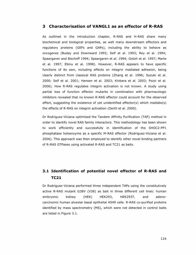
124
3 Characterisation of VANGL1 as an effector of R-RAS
As outlined in the introduction chapter, R-RAS and H-RAS share many
biochemical and biological properties, as well many downstream effectors and
regulatory proteins (GEFs and GAPs), including the ability to behave as
oncogenes (Buday and Downward 1993; Self et al. 1993; Rey et al. 1994;
Spaargaren and Bischoff 1994; Spaargaren et al. 1994; Gotoh et al. 1997; Marte
et al. 1997; Ebinu et al. 1998). However, R-RAS appears to have specific
functions of its own, including effects on integrin mediated adhesion, being
clearly distinct from classical RAS proteins (Zhang et al. 1996; Suzuki et al.
2000; Self et al. 2001; Hansen et al. 2003; Kinbara et al. 2003; Pozzi et al.
2006). How R-RAS regulates integrin activation is not known. A study using
partial loss of function effector mutants in combination with pharmacologic
inhibitors revealed that no known R-RAS effector could account for the observed
effect, suggesting the existence of yet unidentified effector(s) which mediate(s)
the effects of R-RAS on integrin activation (Oertli et al. 2000).
Dr Rodriguez-Viciana optimised the Tandem Affinity Purification (TAP) method in
order to identify novel RAS family interactors. This methodology has been shown
to work efficiently and successfully in identification of the SHOC2-PP1
phosphatase holoenzyme as a specific M-RAS effector (Rodriguez-Viciana et al.
2006). This approach was then employed to identify other novel binding partners
of R-RAS GTPases using activated R-RAS and TC21 as baits.
3.1 Identification of potential novel effector of R-RAS and
TC21
Dr Rodriguez-Viciana performed three independent TAPs using the constitutively
active R-RAS mutant G38V (V38) as bait in three different cell lines: human
embryonic kidney (HEK) HEK293, HEK293T, and adeno-
carcinomic human alveolar basal epithelial A549 cells. R-RAS co-purified proteins
identified by mass spectrometry (MS), which were not detected in control baits
are listed in Figure 3.1.
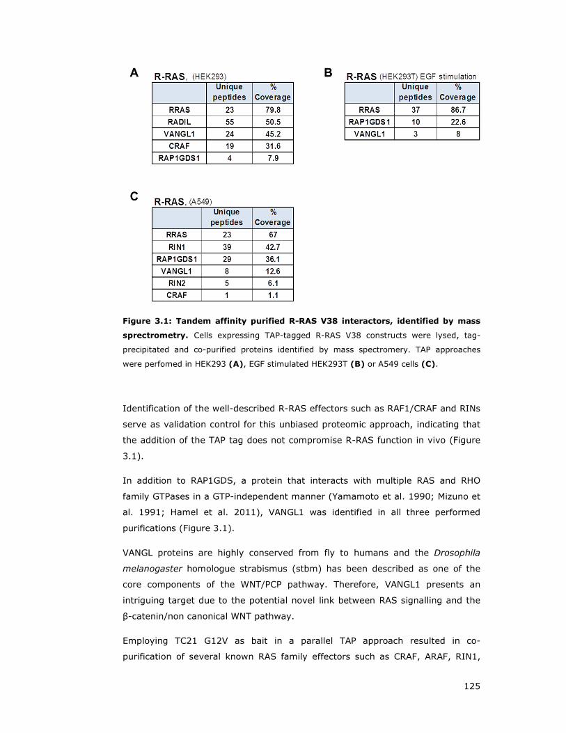
125
Figure 3.1: Tandem affinity purified R-RAS V38 interactors, identified by mass
sprectrometry. Cells expressing TAP-tagged R-RAS V38 constructs were lysed, tag-
precipitated and co-purified proteins identified by mass spectromery. TAP approaches
were perfomed in HEK293 (A), EGF stimulated HEK293T (B) or A549 cells (C).
Identification of the well-described R-RAS effectors such as RAF1/CRAF and RINs
serve as validation control for this unbiased proteomic approach, indicating that
the addition of the TAP tag does not compromise R-RAS function in vivo (Figure
3.1).
In addition to RAP1GDS, a protein that interacts with multiple RAS and RHO
family GTPases in a GTP-independent manner (Yamamoto et al. 1990; Mizuno et
al. 1991; Hamel et al. 2011), VANGL1 was identified in all three performed
purifications (Figure 3.1).
VANGL proteins are highly conserved from fly to humans and the Drosophila
melanogaster homologue strabismus (stbm) has been described as one of the
core components of the WNT/PCP pathway. Therefore, VANGL1 presents an
intriguing target due to the potential novel link between RAS signalling and the
β-catenin/non canonical WNT pathway.
Employing TC21 G12V as bait in a parallel TAP approach resulted in co-
purification of several known RAS family effectors such as CRAF, ARAF, RIN1,

126
MLLT4/AFADIN/AF6 and the RALGEF RGL2 (Rodriguez-Viciana et al. 2004), again
validating the sensitivity of the approach (Figure 3.2). In addition,
ARHGEF17/p164RhoGEF, a RHOA activating nucleotide exchange factor
(Rumenapp et al. 2002) was identified in A549 cells (Figure 3.2C).
This discovery led to the intriguing hypothesis that R-RAS could act downstream
of WNT ligands and modulate VANGL1 function in the WNT/PCP pathway, while
TC21 is also able to modulate RHO GTPase activation through
ARHGEF17/p164RhoGEF. Recruitment of other factors such as PI3Ks or RALGEFs
could then enable the regulation of cell polarity and motility (Figure 3.3).
Figure 3.2: Tandem affinity purified TC21 V12 interactors, identified by mass
spectroscopy. Cells expressing TAP-tagged R-RAS V38 constructs were lysed, tag-
precipitated and co-purified proteins identified by mass spectromery. TAP approaches
were perfomed in HEK293 (A), EGF stimulated HEK293T (B) or A549 cells (C).
To validate VANGL1 and ARHGEF17/p164RhoGEF as novel R-RAS GTPase
effectors, characterisation experiments are described in two separate sequential
sections. The initial part of this chapter focuses on the VANGL1 and R-RAS
interaction (section 3.2), and this is followed by the interaction validation of
ARHGEF17/p164RhoGEF as a novel effector of TC21 (section 3.3).

127
Figure 3.3: Proposed model of how R-RAS GTPases could act downstream of
WNT ligands in the non-canonical WNT signalling pathway. R-RAS subgroup
members are activated upon binding of WNT ligands (WNT) to Frizzled receptors (FZD) by
an unknown mechanism. R-RAS GTPases are able to recruit VANGL1,
ARHGEF17/p164RhoGEF as well as other factors such as Phosphatidylinositide 3-kinases
(PI3K) or RAL guanine exchange factors (GEFs) in order to modulate cell adhesion,
polarity and motility through the activation of RAL and RHO GTPases. R-RAS GTPases
could be also activated by Receptor Tyrosine Kinases (RTK), Notch ligands or Trefoil
family factors (TFFs). DVL - Dishevelled.

128
3.2 VANGL1 behaves as an effector of R-RAS subfamily
GTPases
3.2.1 R-RAS interacts with VANGL1 in an activation and effector domain
dependent manner
TAP experiments identified VANGL1 as a potential R-RAS interacting protein. In
order to validate and to characterise this interaction, co-transfection approaches
utilising HEK293T were performed, due to the lack of a commercially available
endogenous antibody for VANGL1. This system is based on co-transfection of a
glutathione S-transferase (GST) tagged bait construct and respective flag-tagged
potential interactors. After pull down of GST-proteins with gluthathione beads,
binding to the GST-bait is verified by immunoblotting using flag-antibodies.
Notably, avoiding antibody based beads for immunoprecipitation has been
demonstrated to decrease background and increase signal to noise ratio in co-
transfection experiments (Rodriguez-Viciana and McCormick 2006).
To validate VANGL1 as a true R-RAS effector, VANGL1 is required to bind to R-
RAS in a GTP-dependent manner, therefore constitutively active mutants of R-
RAS (G38V and Q87L, analogous to H-RAS oncogenic mutants G12V and Q61L)
were used for binding studies.
Further, RAS effector proteins interact with RAS through a defined small stretch
termed the effector domain, only when the RAS is GTP bound and thus active. A
point mutation in the effector domain of R-RAS (D64A, equivalent to D38A in H-
RAS) leads to disruption of effector protein binding in the activated background
(Oertli et al. 2000; Repasky et al. 2004). Therefore, this point mutation was
employed to assess RAS effector domain dependent binding of VANGL1. In
addition to the initial biochemical interaction studies, further biological assays
were carried out in order to address the R-RAS – VANGL1 functional relevance
(see section 4.3 and chapter 7).
Co-expression experiments in HEK293T cells showed a strong preferential
binding of VANGL1 to R-RAS activating mutants V38 or L87 over R-RAS wild type
(wt), whereas no interaction with the effector domain mutants V38 A64 or L87
A64 could be detected, confirming an activation dependent interaction with R-
RAS as well effector domain mediated binding (Figure 3.4). This fulfils the
requirements of a RAS family effector protein (Repasky et al. 2004). The closely
related GTPase RAP2 fails to precipitate VANGL1 and therefore was used as
background control (Figure 3.4).
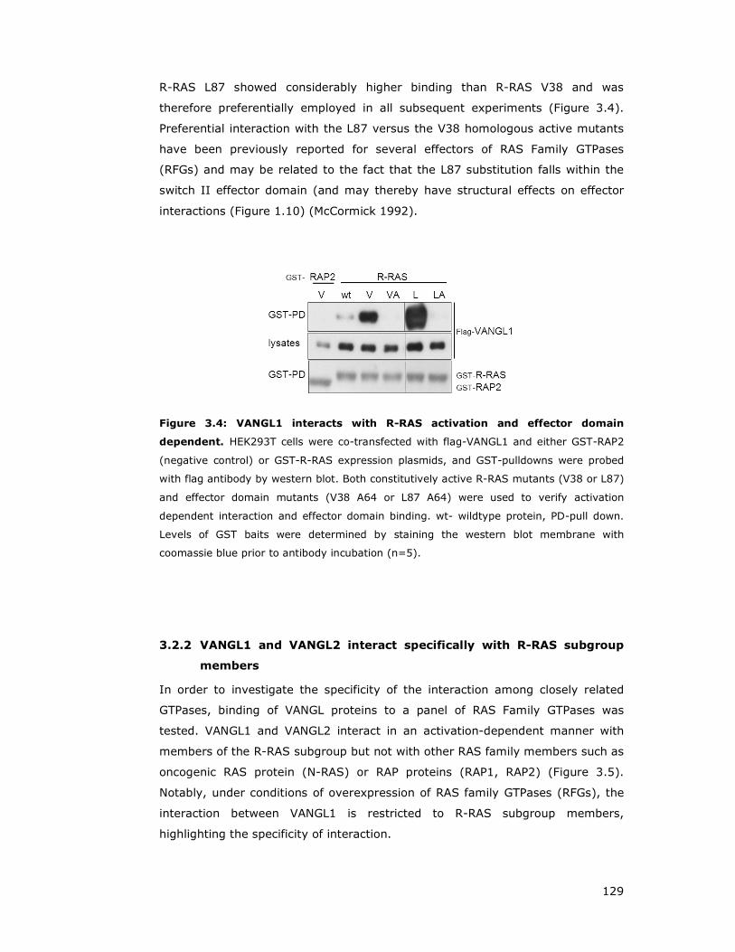
129
R-RAS L87 showed considerably higher binding than R-RAS V38 and was
therefore preferentially employed in all subsequent experiments (Figure 3.4).
Preferential interaction with the L87 versus the V38 homologous active mutants
have been previously reported for several effectors of RAS Family GTPases
(RFGs) and may be related to the fact that the L87 substitution falls within the
switch II effector domain (and may thereby have structural effects on effector
interactions (Figure 1.10) (McCormick 1992).
Figure 3.4: VANGL1 interacts with R-RAS activation and effector domain
dependent. HEK293T cells were co-transfected with flag-VANGL1 and either GST-RAP2
(negative control) or GST-R-RAS expression plasmids, and GST-pulldowns were probed
with flag antibody by western blot. Both constitutively active R-RAS mutants (V38 or L87)
and effector domain mutants (V38 A64 or L87 A64) were used to verify activation
dependent interaction and effector domain binding. wt- wildtype protein, PD-pull down.
Levels of GST baits were determined by staining the western blot membrane with
coomassie blue prior to antibody incubation (n=5).
3.2.2 VANGL1 and VANGL2 interact specifically with R-RAS subgroup
members
In order to investigate the specificity of the interaction among closely related
GTPases, binding of VANGL proteins to a panel of RAS Family GTPases was
tested. VANGL1 and VANGL2 interact in an activation-dependent manner with
members of the R-RAS subgroup but not with other RAS family members such as
oncogenic RAS protein (N-RAS) or RAP proteins (RAP1, RAP2) (Figure 3.5).
Notably, under conditions of overexpression of RAS family GTPases (RFGs), the
interaction between VANGL1 is restricted to R-RAS subgroup members,
highlighting the specificity of interaction.

130
The levels of expression of TC21 (and RAP1) achieved with our expression
plasmids are always consistently lower compared to other RFGs, and therefore
the relative amounts of co-immunoprecipitated proteins have to be considered
with respect to the levels of baits. With this consideration in mind, VANGL1 can
be seen to interact preferentially with R-RAS and TC21 compared to M-RAS
(Figure 3.5).
Figure 3.5: R-RAS GTPases interact in an activation-dependent manner with
VANGL1 and VANGL2. GST-tagged constructs of wild-type (wt) and activated mutants
(V12, L87, L71, L81) of RAS family GTPases (RFG) were expressed in HEK293T cells
together with flag-tagged VANGL1 or VANGL2. GST-RFGs were purified with glutathione
beads and interacting VANGL proteins detected by western blotting using anti-flag
antibodies. PD – pull down. GST-bait levels was visualised by Coomassie blue staining of
the western blot membrane prior to antibody incubation (n=3).
Flag-VANGL2 can also be detected to associate with R-RAS subgroup GTPases
but with significant lower affinity; much longer exposures had to be carried out
during chemoluminescent detection and signal above background was not as
high as observed for VANGL1. VANGL2 is known to hetero-dimerise with VANGL1
(Belotti et al. 2012) and therefore at least a certain fraction of the VANGL2
detected level could be indirectly associated through endogenous VANGL1.
Furthermore, VANGL1 but not VANGL2 was repeatedly found by mass
spectrometry to co-purify with R-RAS (Figure 3.1). Therefore, we decided to
focus our studies on VANGL1.
After testing and verification of multiple commercially available VANGL1
immunoglobulins, one antibody was identified to detect immunoprecipitated
VANGL1 protein (data not shown), enabling to study the interaction of R-RAS

131
GTPases with endogenous VANGL1. Due to unavailability of good
immunoprecipitating antibodies against R-RAS GTPases coupled to the fact that
R-RAS GTPase interaction is strictly GTP dependent, and no potent R-RAS
stimulators were known, exogenously expressed activated R-RAS proteins were
required to be expressed for effector interaction experiments (Figure 3.6).
Figure 3.6: Interaction of endogenous VANGL1 and other effectors with RAS
family GTPases. Flag-tagged constructs of wild-type (wt), activated mutants (V or L) or
respective effector domain mutants (VA or LA) of Ras family GTPases (RFG) were
expressed in HEK293T cells. Flag-GTPases were purified 48h after transfection with flag
affinity beads and endogenous proteins detected by western blotting using indicated
antibodies. IP – Immunoprecipitation, PP1C – protein phosphatase 1C. Applied mutants
were the following: RAP2 V12 or V12 A38, R-RAS L87 or L87 A64, TC21 L71 or L71 A48,
M-RAS L81 or L81 A48, and N-RAS V12 or V12 A38.
Endogenous VANGL1 immunoprecipitates with activated R-RAS and TC21, and
only very faint interaction could be detected with M-RAS (Figure 3.6), consistent
with a preferential interaction with these two R-RAS subgroup members
observed in previous co-expression experiments (Figure 3.5). It could be
confirmed that interaction of endogenous VANGL1 with R-RAS takes place in a
strict activation and effector domain dependent manner and no interaction with
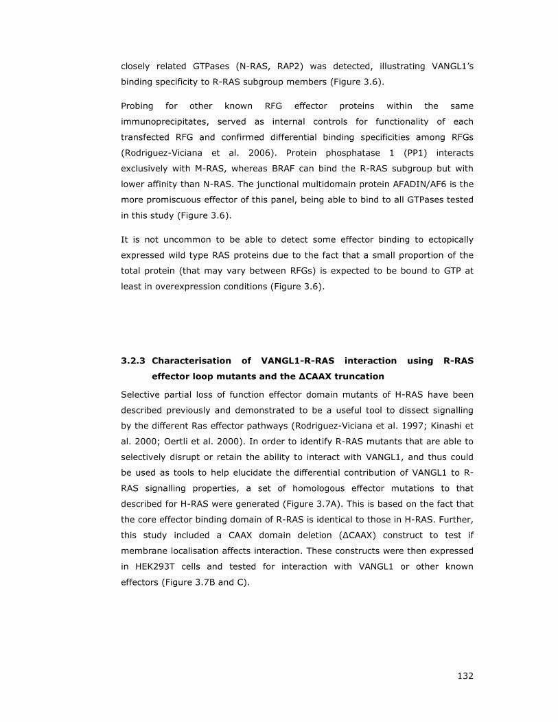
132
closely related GTPases (N-RAS, RAP2) was detected, illustrating VANGL1’s
binding specificity to R-RAS subgroup members (Figure 3.6).
Probing for other known RFG effector proteins within the same
immunoprecipitates, served as internal controls for functionality of each
transfected RFG and confirmed differential binding specificities among RFGs
(Rodriguez-Viciana et al. 2006). Protein phosphatase 1 (PP1) interacts
exclusively with M-RAS, whereas BRAF can bind the R-RAS subgroup but with
lower affinity than N-RAS. The junctional multidomain protein AFADIN/AF6 is the
more promiscuous effector of this panel, being able to bind to all GTPases tested
in this study (Figure 3.6).
It is not uncommon to be able to detect some effector binding to ectopically
expressed wild type RAS proteins due to the fact that a small proportion of the
total protein (that may vary between RFGs) is expected to be bound to GTP at
least in overexpression conditions (Figure 3.6).
3.2.3 Characterisation of VANGL1-R-RAS interaction using R-RAS
effector loop mutants and the ∆CAAX truncation
Selective partial loss of function effector domain mutants of H-RAS have been
described previously and demonstrated to be a useful tool to dissect signalling
by the different Ras effector pathways (Rodriguez-Viciana et al. 1997; Kinashi et
al. 2000; Oertli et al. 2000). In order to identify R-RAS mutants that are able to
selectively disrupt or retain the ability to interact with VANGL1, and thus could
be used as tools to help elucidate the differential contribution of VANGL1 to R-
RAS signalling properties, a set of homologous effector mutations to that
described for H-RAS were generated (Figure 3.7A). This is based on the fact that
the core effector binding domain of R-RAS is identical to those in H-RAS. Further,
this study included a CAAX domain deletion (∆CAAX) construct to test if
membrane localisation affects interaction. These constructs were then expressed
in HEK293T cells and tested for interaction with VANGL1 or other known
effectors (Figure 3.7B and C).

133
Figure 3.7: R-RAS effector domain mutations do not affect VANGL1 interaction,
whereas deletion of the CAAX box completely inhibits VANGL1 binding. (A) Partial
protein alignment of R-RAS and H-RAS including the effector domain (orange box, R-RAS
residues V58-Y66, H-RAS residues V32-Y40). Identical amino acids are marked as
asteriks (*) and partial loss-of-function effector mutants are highlighted in red, and
repective activated effectors are indicated below. This figure was adapted from Oertli et
al. 2000. (B) GST-tagged constructs of wild-type (wt) and activated mutants (V12, L87)
of RFGs were expressed in HEK293T cells together with flag-tagged VANGL1. GST-
GTPases were purified with glutathione beads and interacting proteins detected by
western blotting using anti-flag, AF6 or BRAF antibodies. As a negative control for binding
served active RAP2 (V12 mutant). (C) Deletion of the R-RAS CAAX motif completely
disrupts interaction with VANGL1. HEK293T cells were transfected with R-RAS effector
domain mutant (A64) or C-terminal CAAX motif (∆) deletion constructs in both activating
backgrounds (V38 and L87). RAP2 V12 served as background binding control. Lysate
harvest, purification and detection was carried out as decribed in B.
VANGL1 binds strongly to constitutively active R-RAS L87, but introduction of
any of the effector domain point mutation tested led to a significant reduction of
interaction. The less well described effector domain T61S, exhibits a slightly
milder disruption effect on the VANGL1 binding to R-RAS, whereas the most
severe inhibiting effect is observed with the effector domain mutant D64A, which
completely abolishes effector interaction and thus is commonly termed defective

134
mutant (Figure 3.7B). Probing for binding of endogenous R-RAS effectors BRAF
and AFADIN/AF6 showed that all employed effector domain mutants completely
disrupted binding to R-RAS (Figure 3.7B). Experiments carried out using
respective mutants in R-RAS V38 background showed similar results (data not
shown).
In summary, all employed effector loop mutants decreased binding to VANGL1
as well as other effectors and therefore, unfortunately, I was not able to identify
partial loss of function R-RAS mutants.
Interestingly, deletion of the R-RAS CAAX motif in both activating backgrounds
completely disrupts VANGL1 interaction comparable to the level of the defective
mutant R-RAS D64A (Figure 3.7C). The CAAX motif is essential for appropriate
post translational processing and targeting of RFGs to the plasma membrane
(Ahearn et al. 2012), suggesting that membrane localisation of R-RAS is critically
required for interaction with VANGL1, which itself is a transmembrane protein.
R-RAS ∆CAAX was still able to interact efficiently with the membrane associated
AF6/AFADIN, or with BRAF (Figure 3.7C). In clear contrast to VANGL1, deletion
of the CAAX box increased the affinity of the interaction with AF6/AFADIN, while
having little effect on BRAF binding (Figure 3.7C).
3.2.4 R-RAS interacts with the C-terminus of VANGL1
RAS effectors interact through a distinct small region termed RAS Binding
Domain (RBD). Studies on known RAS effectors such as RAF, PI3K and RALGEFs
showed that their respective RBDs share little sequence homology but certain
structural similarity. Hence, there is the assumption that a wider array of
unidentified RBDs exists, based on the fact that there are also RFG effectors,
which do not show any RBD similarity to any of the aforementioned three
effector proteins (Matheny et al. 2004; Rodriguez-Viciana et al. 2006).
VANGL1 protein structure prediction revealed four transmembrane domains and
a C-terminal tetra-peptide PDZ binding motif. No other domain or motif similarity
has been identified. In order to map which part of VANGL1 is binding to R-RAS,
one N-terminal (excluding TM region) and various C-terminal VANGL1 truncation
mutants were generated (Figure 2.2) and tested in co-expression experiments
(Figure 3.8).
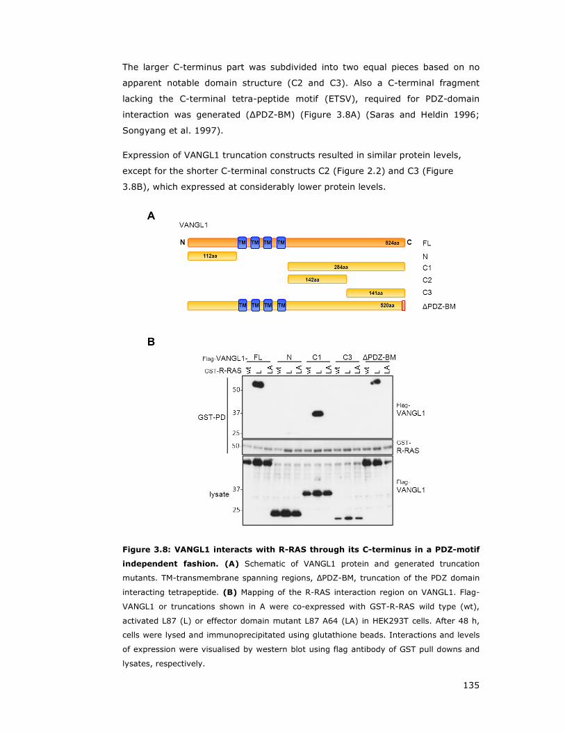
135
The larger C-terminus part was subdivided into two equal pieces based on no
apparent notable domain structure (C2 and C3). Also a C-terminal fragment
lacking the C-terminal tetra-peptide motif (ETSV), required for PDZ-domain
interaction was generated (∆PDZ-BM) (Figure 3.8A) (Saras and Heldin 1996;
Songyang et al. 1997).
Expression of VANGL1 truncation constructs resulted in similar protein levels,
except for the shorter C-terminal constructs C2 (Figure 2.2) and C3 (Figure
3.8B), which expressed at considerably lower protein levels.
Figure 3.8: VANGL1 interacts with R-RAS through its C-terminus in a PDZ-motif
independent fashion. (A) Schematic of VANGL1 protein and generated truncation
mutants. TM-transmembrane spanning regions, ∆PDZ-BM, truncation of the PDZ domain
interacting tetrapeptide. (B) Mapping of the R-RAS interaction region on VANGL1. Flag-
VANGL1 or truncations shown in A were co-expressed with GST-R-RAS wild type (wt),
activated L87 (L) or effector domain mutant L87 A64 (LA) in HEK293T cells. After 48 h,
cells were lysed and immunoprecipitated using glutathione beads. Interactions and levels
of expression were visualised by western blot using flag antibody of GST pull downs and
lysates, respectively.

136
Under comparable expression levels of VANGL1 constructs (except C3
truncation), GTP-dependent R-RAS interaction is observed with the full length
protein, complete C-terminus (C1) and the ∆PDZ-BM truncation with similar
affinity. No interaction of R-RAS L87 could be observed with the VANGL1 N-
terminal fragment (Figure 3.8B). This leads to the conclusion that R-RAS
interaction with VANGL1 maps to the C-terminus in a PDZ motif independent
manner.
Even employing very long exposures, no interaction could be detected with
VANGL1-C2 (data not shown) or VANGL1-C3. Therefore, VANGL1-C2 or VANGL1-
C3 regions appear insufficient for binding, and the interaction with R-RAS can
only be mapped to the C-terminus without further refinement (Figure 3.8B).
Similar results were achieved using the activated R-RAS V38 mutant in VANGL1
mapping experiments (data not shown).
3.2.5 R-RAS interaction with VANGL1 is disrupted by the D259E mutant
Point mutants of VANGL1 have been identified in a cohort of neural tube defect
patients (Figure 1.31A) (Kibar et al. 2007; Kibar et al. 2009). In order to test
whether reported VANGL1 mutations disrupt the interaction with R-RAS, five C-
terminal point mutations were generated and included in VANGL1 co-expression
interaction studies (Figure 2.2 and Figure 3.9A).
VANGL1 interaction with R-RAS takes place in an activation and effector domain
dependent manner with VANGL1 V2391I, R274Q and M328T point mutants at a
similar level compared to wild type (wt) or the ∆PDZ-BM truncation. Expression
of the D259E mutation construct leads to similar protein levels as the wild type
or point mutant M328T, but no interaction with R-RAS could be detected under
these conditions. Although longer exposures showed residual interaction, this
binding is no longer effector domain dependent and is therefore unspecific.
Interestingly, this mutant is homologous to the looptail mouse VANGL2 D255E
mutant, known to be involved in neural tube defects (Figure 3.9B).
Another analogous point mutation initially identified in the looptail mouse,
S467N, appears to be defective on short exposures, but expression levels of this
protein are considerably lower than other point mutants or wild type VANGL1.
On longer exposures, specific binding of VANGL1 S467N to R-RAS L87 could be
detected at similar levels than wild type (Figure 3.9B).

137
Detection of endogenous AFADIN/AF6 served as binding control for transfected
RAP2 and R-RAS constructs (Figure 3.9B).
Figure 3.9: VANGL1 point mutations D259E identified in neural tube defect
(NTD) patients disrupts binding to R-RAS. (A) Overview of VANGL1 structure and
location of employed NTD point mutations. TM-transmembrane domain. (B) HEK293T
cells were co-transfected with flag-tagged VANGL1 wt or respective point mutants as
indicated above with either GST-tagged activated R-RAS (L87) or effector domain mutant
(L87 A64). After 48 h, cells were lysed and immunoprecipitated using glutathione beads.
Interactions were detected by western blot using flag-antibodies.
3.2.6 R-RAS and VANGL1 interact directly in vitro
Identification of VANGL1 as a novel R-RAS interacting protein by TAP or co-
immunoprecipitation does not elucidate if this interaction is direct or mediated
through other proteins. In order to establish if VANGL1 binds to R-RAS in a direct
fashion, an in-vitro assay was employed which tests interaction of purified, GDP
or GTP loaded R-RAS with purified GST-VANGL1 fusion proteins (Figure 3.10).
Recombinant VANGL1-N and VANGL1-C1 truncations (Figure 3.8A) were
expressed as GST-fusion proteins and purified from an insect cell line
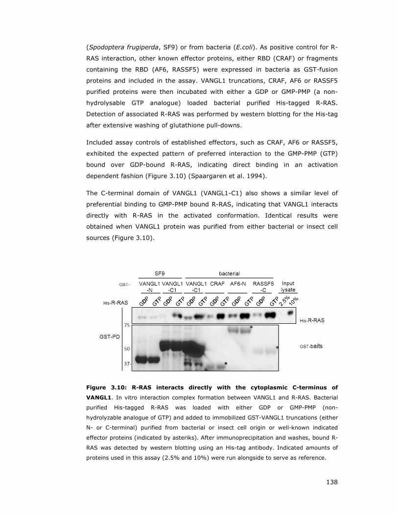
138
(Spodoptera frugiperda, SF9) or from bacteria (E.coli). As positive control for R-
RAS interaction, other known effector proteins, either RBD (CRAF) or fragments
containing the RBD (AF6, RASSF5) were expressed in bacteria as GST-fusion
proteins and included in the assay. VANGL1 truncations, CRAF, AF6 or RASSF5
purified proteins were then incubated with either a GDP or GMP-PMP (a non-
hydrolysable GTP analogue) loaded bacterial purified His-tagged R-RAS.
Detection of associated R-RAS was performed by western blotting for the His-tag
after extensive washing of glutathione pull-downs.
Included assay controls of established effectors, such as CRAF, AF6 or RASSF5,
exhibited the expected pattern of preferred interaction to the GMP-PMP (GTP)
bound over GDP-bound R-RAS, indicating direct binding in an activation
dependent fashion (Figure 3.10) (Spaargaren et al. 1994).
The C-terminal domain of VANGL1 (VANGL1-C1) also shows a similar level of
preferential binding to GMP-PMP bound R-RAS, indicating that VANGL1 interacts
directly with R-RAS in the activated conformation. Identical results were
obtained when VANGL1 protein was purified from either bacterial or insect cell
sources (Figure 3.10).
Figure 3.10: R-RAS interacts directly with the cytoplasmic C-terminus of
VANGL1. In vitro interaction complex formation between VANGL1 and R-RAS. Bacterial
purified His-tagged R-RAS was loaded with either GDP or GMP-PMP (non-
hydrolyzable analogue of GTP) and added to immobilized GST-VANGL1 truncations (either
N- or C-terminal) purified from bacterial or insect cell origin or well-known indicated
effector proteins (indicated by asteriks). After immunoprecipitation and washes, bound R-
RAS was detected by western blotting using an His-tag antibody. Indicated amounts of
proteins used in this assay (2.5% and 10%) were run alongside to serve as reference.

139
Very low residual binding of VANGL1 N-terminus (VANGL1-N) at similar levels to
the GDP and GTP form of R-RAS was observed, suggesting that this interaction is
non-specific, and further confirming previous results that the VANGL1 N-
terminus is not involved in R-RAS binding (Figure 3.10).
3.3 ARHGEF17/p164RhoGEF behaves as a TC21 and R-RAS
effector
This section will focus on characterisation of ARHGEF17/p164RhoGEF interaction
with the R-RAS subgroup members and its ability to act as a RHOA GEF.
3.3.1 ARHGEF17/p164RhoGEF interacts with R-RAS and TC21 in an
activation and effector domain dependent manner
Initial binding studies were designed to verify that ARHGEF17/p164RhoGEF
interaction with TC21 takes place in an activation dependent and effector domain
mediated manner. Binding affinity between two available constitutive activating
mutants of TC21 (V13 and L71) and their respective effector domain mutants
were carried out. Due to the lack of any commercially available antibodies for
ARHGEF17/p164RhoGEF, and failure to successfully generate an antibody
against a N-terminal ARHGEF17/p164RhoGEF fragment purified from bacteria,
the following binding studies were carried out in a co-expression study as
described earlier for VANGL1 (refer to 3.2.1).
ARHGEF17/p164RhoGEF interaction with TC21 was observed with both activating
mutants (V13, L71), but no binding could be detected with the effector domain
mutation or the wild type protein. Also, despite a lower expression of TC21 L71
and its respective effector domain mutant, it appears that
ARHGEF17/p164RhoGEF demonstrates a higher affinity to TC21 L71 over TC21
V13 (Figure 3.11A).
Testing the binding specificity of other R-RAS subgroup members (R-RAS, M-
RAS) or closely related RAS family GTPases (N-RAS, RAP1/2) showed that
ARHGEF17/p164RhoGEF binds to R-RAS L87 and both TC21 activating mutants
but not to any other GTPase tested in this panel. Comparing R-RAS V38 and R-
RAS L87 activating GTPases at similar expression levels, p164RhoGEF seems to
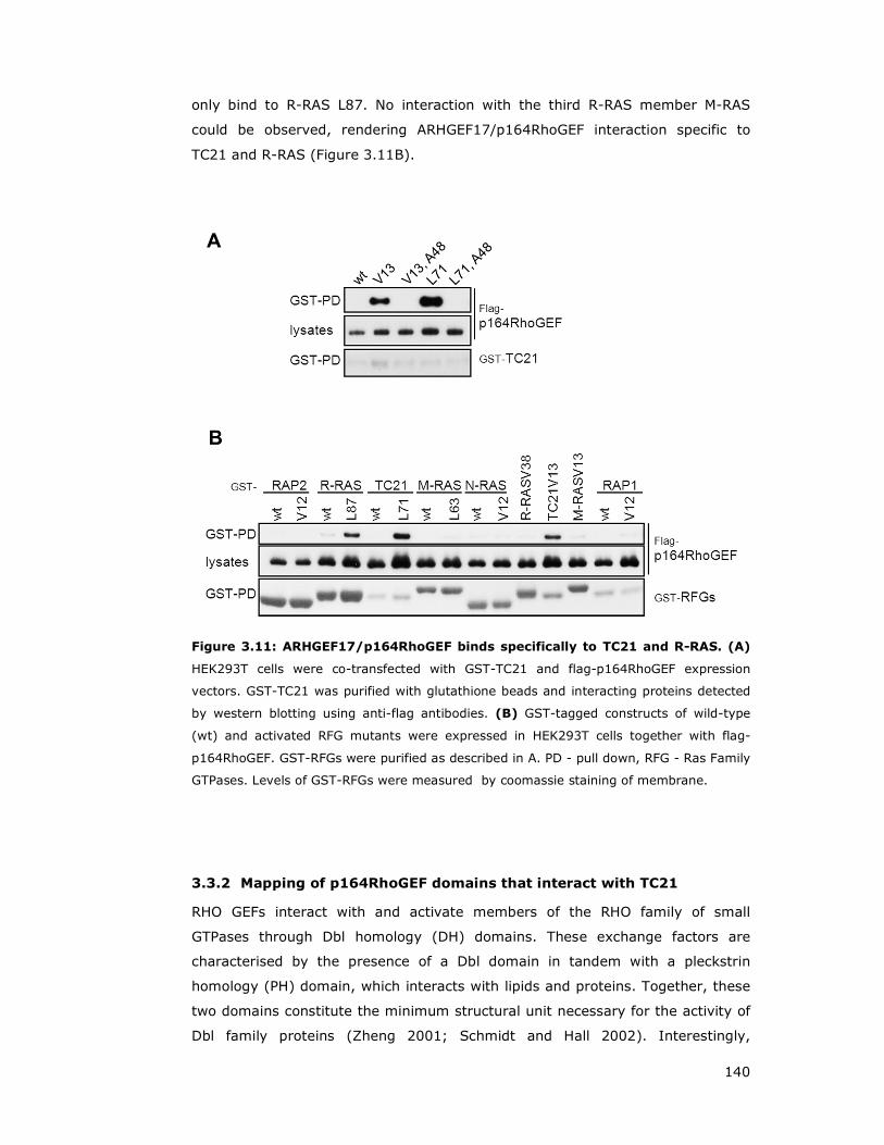
140
only bind to R-RAS L87. No interaction with the third R-RAS member M-RAS
could be observed, rendering ARHGEF17/p164RhoGEF interaction specific to
TC21 and R-RAS (Figure 3.11B).
Figure 3.11: ARHGEF17/p164RhoGEF binds specifically to TC21 and R-RAS. (A)
HEK293T cells were co-transfected with GST-TC21 and flag-p164RhoGEF expression
vectors. GST-TC21 was purified with glutathione beads and interacting proteins detected
by western blotting using anti-flag antibodies. (B) GST-tagged constructs of wild-type
(wt) and activated RFG mutants were expressed in HEK293T cells together with flag-
p164RhoGEF. GST-RFGs were purified as described in A. PD - pull down, RFG - Ras Family
GTPases. Levels of GST-RFGs were measured by coomassie staining of membrane.
3.3.2 Mapping of p164RhoGEF domains that interact with TC21
RHO GEFs interact with and activate members of the RHO family of small
GTPases through Dbl homology (DH) domains. These exchange factors are
characterised by the presence of a Dbl domain in tandem with a pleckstrin
homology (PH) domain, which interacts with lipids and proteins. Together, these
two domains constitute the minimum structural unit necessary for the activity of
Dbl family proteins (Zheng 2001; Schmidt and Hall 2002). Interestingly,
141
p164RhoGEF does not contain any detectable PH motif by conventional domain
prediction, but has a WD40 domain, which has been reported for two additional
GEFs: GrinchGEF and GEF10 (Rumenapp et al. 2002; Winkler et al. 2005; Mohl
et al. 2006).
Therefore, truncation constructs of p164RhoGEF were generated in order to map
the TC21 binding region and to test if the WD40 domain is required for
interaction. Truncation constructs were designed to cover the N-terminus or C-
terminus including or excluding the DH domain. Further, the C-terminal part was
shortened to be either WD40 domain excluding, or to contain the WD40 domain
only or a truncation thereof (Figure 3.12A).
Binding studies employing all generated truncations showed varied levels of
expression, p164RhoGEF-C1 and -C2 being the highest expressed, whereas the
shorter C-terminal truncations (p164RhoGEF-C3, -C4 and ∆N) expressed at very
low levels (Figure 3.12B).
Full length p164RhoGEF (FL) binds to active TC21, whereas no interaction with
the N-terminal fragments p164RhoGEF-N or DH domain containing construct
p164RhoGEF-∆C could be detected. In contrast, a C-terminal fragment lacking
the N-terminus (p164RhoGEF-∆N) exhibited an even higher binding affinity to
TC21 than the full length protein, even though it was expressed at greatly
reduced levels (Figure 3.12B).
Examining C-terminal truncations excluding the WC40 domain (p164RhoGEF-C2)
compared to the DH lacking complete C-terminus (p164RhoGEF-C1) at similar
expression levels, the WD40 excluding truncation displayed a significantly
reduced interaction with TC21 L71. This indicates that the WD40 domain might
not be critically required for binding but has a supporting role (Figure 3.12B).
Taken together these results suggest that TC21 likely interacts predominantly
with the region between the DH and WD40 domains although the integrity of the
WD domain is likely contributing for a higher affinity interaction.
Notably, flag-p164RhoGEF protein levels in lysates were consistently
considerably higher when TC21 L71 was co-expressed suggesting that active
TC21 may have an effect on the protein stability of p164RhoGEF (Figure 3.12B).
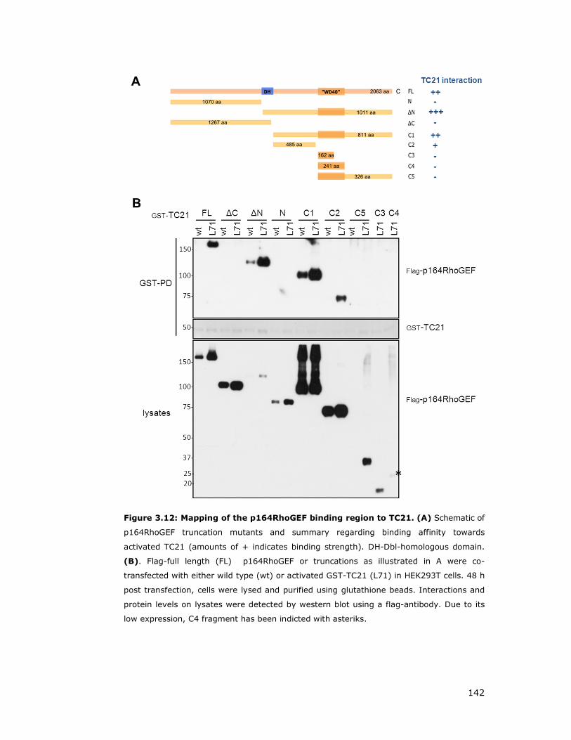
142
Figure 3.12: Mapping of the p164RhoGEF binding region to TC21. (A) Schematic of
p164RhoGEF truncation mutants and summary regarding binding affinity towards
activated TC21 (amounts of + indicates binding strength). DH-Dbl-homologous domain.
(B). Flag-full length (FL) p164RhoGEF or truncations as illustrated in A were co-
transfected with either wild type (wt) or activated GST-TC21 (L71) in HEK293T cells. 48 h
post transfection, cells were lysed and purified using glutathione beads. Interactions and
protein levels on lysates were detected by western blot using a flag-antibody. Due to its
low expression, C4 fragment has been indicted with asteriks.
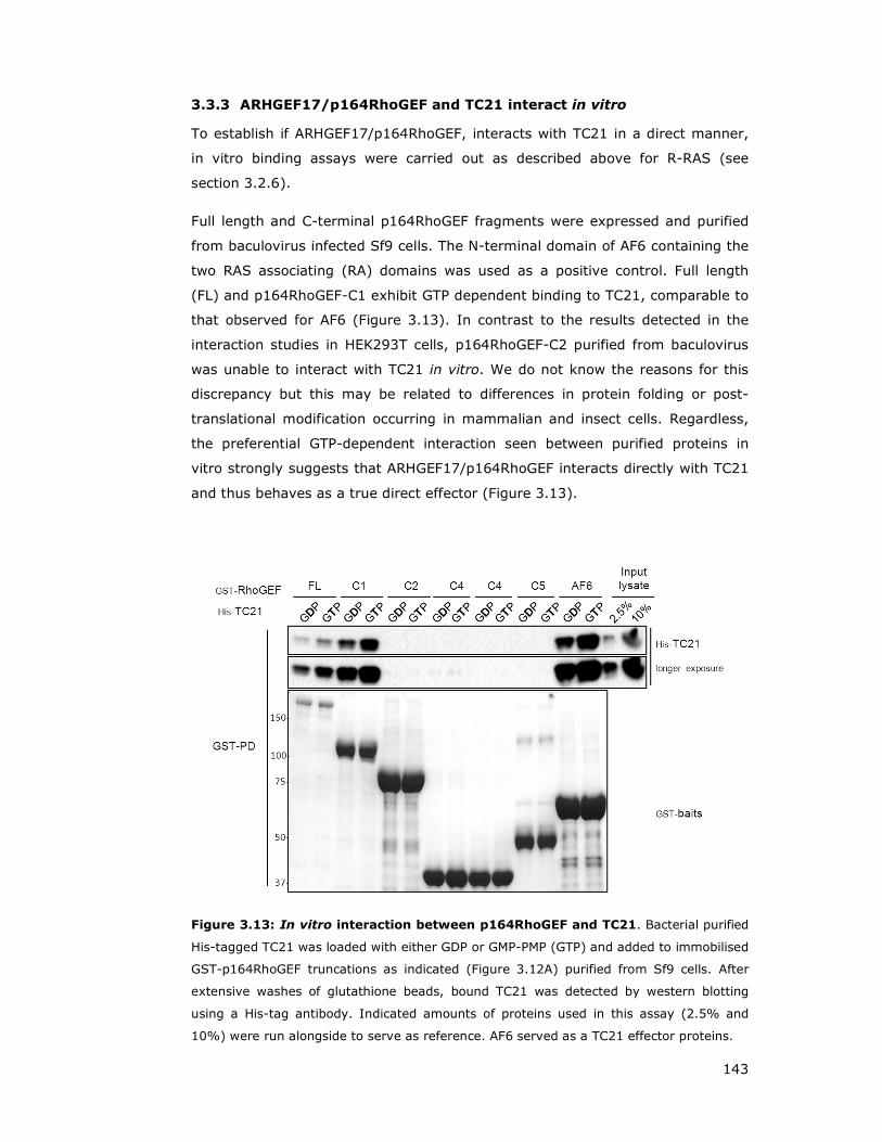
143
3.3.3 ARHGEF17/p164RhoGEF and TC21 interact in vitro
To establish if ARHGEF17/p164RhoGEF, interacts with TC21 in a direct manner,
in vitro binding assays were carried out as described above for R-RAS (see
section 3.2.6).
Full length and C-terminal p164RhoGEF fragments were expressed and purified
from baculovirus infected Sf9 cells. The N-terminal domain of AF6 containing the
two RAS associating (RA) domains was used as a positive control. Full length
(FL) and p164RhoGEF-C1 exhibit GTP dependent binding to TC21, comparable to
that observed for AF6 (Figure 3.13). In contrast to the results detected in the
interaction studies in HEK293T cells, p164RhoGEF-C2 purified from baculovirus
was unable to interact with TC21 in vitro. We do not know the reasons for this
discrepancy but this may be related to differences in protein folding or post-
translational modification occurring in mammalian and insect cells. Regardless,
the preferential GTP-dependent interaction seen between purified proteins in
vitro strongly suggests that ARHGEF17/p164RhoGEF interacts directly with TC21
and thus behaves as a true direct effector (Figure 3.13).
Figure 3.13: In vitro interaction between p164RhoGEF and TC21. Bacterial purified
His-tagged TC21 was loaded with either GDP or GMP-PMP (GTP) and added to immobilised
GST-p164RhoGEF truncations as indicated (Figure 3.12A) purified from Sf9 cells. After
extensive washes of glutathione beads, bound TC21 was detected by western blotting
using a His-tag antibody. Indicated amounts of proteins used in this assay (2.5% and
10%) were run alongside to serve as reference. AF6 served as a TC21 effector proteins.

144
3.3.4 ARHGEF17/p164RhoGEF acts as a GEF for RHOA
ARHGEF17/p164RhoGEF has been demonstrated to be a RHOA specific GEF
(Rumenapp et al. 2002). In order to confirm that ARHGEF17/p164RhoGEF is able
to activate the RHOA, and to establish RHO activation assays to be used in
future experiments (see later section 6.3), various GEFs were co-transfected
with myc-RHOA and RHOA activation measured using Rhotekin-RBD pull-down
assays (Habas and He 2006). ARHGEF19/WGEF, which has been reported to be
involved in WNT regulated convergent extension and to act as GEF for RHOA
(Tanegashima et al. 2008) was also cloned and used in parallel, as well as the
ARHGEF17/p164RhoGEF related proteins ARHGEF10L/GrinchGEF and
ARHGEF10/GEF10).
Cells were also stimulated as positive control with serum which is known to
trigger activation of RHO GTPases, as well as with EGF, which has been shown to
be a much weaker activator (Ridley and Hall 1992). mSOS was included as
negative control, since it acts as nucleotide exchange factor for RAS
GTPases and RAC (Figure 3.14) (Chardin et al. 1993; Nimnual and Bar-Sagi
2002).
Figure 3.14: p164RhoGEF is an activator of RHOA in HEK293T cells. Empty vector
or GEFs (mSOS, ARHGEF19, ARHGEF17/p164RhoGEF, ARHGEF10 and ARHGEF10L) were
co-transfected with myc-RHOA. 32h post transfection, cells were serum deprived for 16h
and either stimulated with 100 ng/mL EGF or 10% final serum for indicated times, or left
untreated (-). The GTP form of RHOA was pulled down (PD) from HEK293T cell lysates
using GST-RBD (RHO Binding Domain from Rhotekin). Activated RHOA fraction was
detected using myc antibodies.

145
HEK293T cells stimulated with serum or EGF show a small increase in
precipitated activated RHOA over basal level (-). Expression of the RASGEF
mSOS elevated activated RHOA levels only slightly over basal levels, whereas
the ARHGEF19 exhibited a significant increase. Expression of
ARHGEF17/p164RhoGEF on the other hand strongly increased the amount of
activated myc-RHOA and therefore represents a very potent RHOA guanine
nucleotide exchange factor (Figure 3.14).
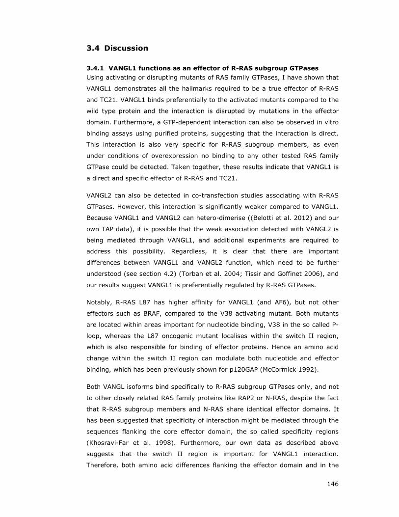
146
3.4 Discussion
3.4.1 VANGL1 functions as an effector of R-RAS subgroup GTPases
Using activating or disrupting mutants of RAS family GTPases, I have shown that
VANGL1 demonstrates all the hallmarks required to be a true effector of R-RAS
and TC21. VANGL1 binds preferentially to the activated mutants compared to the
wild type protein and the interaction is disrupted by mutations in the effector
domain. Furthermore, a GTP-dependent interaction can also be observed in vitro
binding assays using purified proteins, suggesting that the interaction is direct.
This interaction is also very specific for R-RAS subgroup members, as even
under conditions of overexpression no binding to any other tested RAS family
GTPase could be detected. Taken together, these results indicate that VANGL1 is
a direct and specific effector of R-RAS and TC21.
VANGL2 can also be detected in co-transfection studies associating with R-RAS
GTPases. However, this interaction is significantly weaker compared to VANGL1.
Because VANGL1 and VANGL2 can hetero-dimerise ((Belotti et al. 2012) and our
own TAP data), it is possible that the weak association detected with VANGL2 is
being mediated through VANGL1, and additional experiments are required to
address this possibility. Regardless, it is clear that there are important
differences between VANGL1 and VANGL2 function, which need to be further
understood (see section 4.2) (Torban et al. 2004; Tissir and Goffinet 2006), and
our results suggest VANGL1 is preferentially regulated by R-RAS GTPases.
Notably, R-RAS L87 has higher affinity for VANGL1 (and AF6), but not other
effectors such as BRAF, compared to the V38 activating mutant. Both mutants
are located within areas important for nucleotide binding, V38 in the so called P-
loop, whereas the L87 oncogenic mutant localises within the switch II region,
which is also responsible for binding of effector proteins. Hence an amino acid
change within the switch II region can modulate both nucleotide and effector
binding, which has been previously shown for p120GAP (McCormick 1992).
Both VANGL isoforms bind specifically to R-RAS subgroup GTPases only, and not
to other closely related RAS family proteins like RAP2 or N-RAS, despite the fact
that R-RAS subgroup members and N-RAS share identical effector domains. It
has been suggested that specificity of interaction might be mediated through the
sequences flanking the core effector domain, the so called specificity regions
(Khosravi-Far et al. 1998). Furthermore, our own data as described above
suggests that the switch II region is important for VANGL1 interaction.
Therefore, both amino acid differences flanking the effector domain and in the

147
switch II region may account for this differential interaction with R-RAS GTPases.
Consistent with this idea, the switch II loop of R-RAS and TC21 exhibits an
identical amino acid pattern, which differs in two amino acids to classical RAS
proteins, and shows one residue alteration compared to M-RAS (Figure 3.15).
Figure 3.15: Alignment of RAS, RAP2 and R-RAS subgroup members. Indicated
GTPases were aligned across switch I and switch II, including flanking regions. Identical
amino acids are depicted as asteriks and subscript number specify residue number of
respective amino acid position.
VANGL1 has four transmembrane domains and is therefore predicted to be a
membrane protein (Iliescu et al. 2011). Localisation studies reported various
subcellular distributions including plasma membrane in MDCK cells (Gravel et al.
2010; Iliescu et al. 2011) and ER in COS7 cells (Merte et al. 2011) (see also
section 5.4). Notably, VANGL1 interaction with R-RAS requires the membrane
targeting CAAX motif, in contrast to other effectors such as the membrane
associated AF6 or BRAF, the latter one being only partially affected (Stokoe et al.
1994). Therefore, unlike for other effectors, R-RAS needs to be membrane
bound in order to interact with VANGL1.
The domain structure of VANGL1 does not suggest any enzymatic activity;
therefore it is likely that R-RAS/TC21 regulates VANGL1 function at least partly
by regulating its localisation in some contexts: upon activation, RRAS/TC21
would recruit VANGL1 to specialised signalling platforms at the membrane micro-
domains where they reside. Furthermore, R-RAS/TC21 binding could also
regulate (stimulate or inhibit) the association of VANGL1 with its interacting
partners and thus modulate its signalling properties.

148
VANGL1 point mutations have been found in sporadic and familial cases of neural
tube defects (Kibar et al. 2007; Kibar et al. 2009). These mutants also
demonstrated defects in convergent extension in a zebrafish model,
strengthening the point of VANGL protein involvement in neural tube closure and
migration, and a high degree of functional conservation across vertebrates
(Reynolds et al. 2010).
Interaction studies analysing binding of VANGL1 point mutant D259E, identified
in human NTDs, demonstrated it to be defective for DVL protein interaction
(Kibar et al. 2007). VANGL1 D259E also disrupts binding to R-RAS. This suggests
that R-RAS and DVL proteins bind to the same site of VANGL1 and therefore may
compete for interaction. In addition, this mutant is located within the first half
the C-terminus, which also has been shown to be required for R-RAS-VANGL1
interaction. The VANGL2 homologous point mutant is the conserved Looptail
mouse D255E substitution, which has been reported to have a reduced protein
half-life, and fails to be shuttled to the plasma membrane (Gravel et al. 2010).
Instead, VANGL2 D255E proteins are retained in the ER lumen due to
impairment of binding to the transport protein SEC24b (Merte et al. 2011;
Wansleeben et al. 2011).
Figure 3.16: R-RAS interaction summary. (A) VANGL1 is a direct and specific
effector of the R-RAS subgroup and does not bind to closely related GTPases.
(B) This interaction is mediated through VANGL1’s C-terminus, and R-RAS
requires the membrane-targeting CAAX motif for VANGL1 binding. Furthermore,
R-RAS-VANGL1 binding is also disrupted through the VANGL1 point mutant
D259E, implicated in neural tube defects.

149
3.4.2 ARHGEF17/p164RhoGEF functions as an effector of R-RAS and
TC21
Using constitutively active TC21 in a TAP, we identified various interactors,
several of which have been previously published. One hit, identified in A549
cells, was ARHGEF17. This gene encodes for a 164kDa large RhoGEF, thus also
known as p164RhoGEF.
Initial validation experiments demonstrated that ARHGEF17/p164RhoGEF binds
to both TC21 activated mutants and R-RAS L87 in an activation and effector
domain dependent. No interaction with M-RAS, RAP1, RAP2 or N-RAS could be
observed.
Interestingly, ARHGEF17/p164RhoGEF does not contain any detectable PH motif,
but encompasses a WD40 domain, which in addition has been reported for
further two GEFs: GrinchGEF and GEF10 (Rumenapp et al. 2002; Winkler et al.
2005; Mohl et al. 2006). However, a recent publication challenged this finding
and descibed that ARHGEF17/p164RhoGEF contains a functional PH domain,
which was not detected previously using convential domain prediction software
due to an ~70 residue α-helical domain insertion (Mitin et al. 2012).
Furthermore, the original paper describing ARHGEF17/p164RhoGEF (Rumenapp
et al. 2002) predicted a size of 164 kDa, but annotations by Croix and colleagues
descibed ARHGEF17 with an extensive N-terminal domain, resulting in a
predicted 222 kDa protein in size (St Croix et al. 2000).
As observed from our experiments, and consistent with literature reports (De
Toledo et al. 2000; Rumenapp et al. 2002), ARHGEF17/p164RhoGEF acts as a
RHOAGEF. A more detailed study further illuminated that
ARHGEF17/p164RhoGEF is able to activate RHOB and RHOC isoforms, but fails to
activate RAC or CDC42 (Mitin et al. 2012).
The ARHGEF17/p164RhoGEF N-terminus has been reported to contain a novel
actin binding domain, and has been found to be actin stress fibre associated,
independently of DH/PH domains (Mitin et al. 2012). Notably, TC21 has been
mapped to interact with ARHGEF17/p164RhoGEF C-terminus downstream of the
DH domain.
The original paper describing ARHGEF17/p164RhoGEF (Rumenapp et al. 2002)
showed a restricted tissue expression in the mouse with predominate expression
in the heart. Furthermore, our own TAP data identified ARHGEF17/p164RhoGEF

150
interacting with TC21 in A549 cells but not HEK293T, consistent with its
differential expression in different cell types.
Due to unavailability of commercial antibodies, considerable time and effort was
devoted to express and purify from bacteria recombinant fragments of
ARHGEF17/p164RhoGEF (and VANGL1) to be used for the generation of
polyclonal antibodies. Disappointingly, when the outsourced rabbit serum was
received, it could not efficiently recognise or immunoprecipitate even the
overexpressed tagged protein for reasons that are not clear. Because of the
likely cell type specific expression of ARHGEF17/p164RhoGEF and the
unavailability of antibodies to detect its expression, further studies would have
been significantly complicated and thus it was decided with my supervisor to
focus my PhD studies on VANGL1 and its role as an effector of R-RAS GTPases.
Figure 3.17: TC21 interaction summary. The RhoGEF protein p164RhoGEF binds to R-
RAS and TC21 through its C-terminus in an activation and effector domain dependent
manner. This leads to the speculation that R-RAS subgroup GTPases could modulate the
WNT-PCP member VANGL1 and RHO GTPase activating factors in order to modulate
cellular motility, adhesion and polarity.

151
CHAPTER 4
Identification of VANGL1
interaction partners

152
4 Identification of VANGL1 interaction partners
4.1 Initial VANGL1 TAP Screen
Initial studies in fruit flies identified the VANGL1 homologue strabismus as a core
PCP protein (Wolff and Rubin 1998; Jenny et al. 2003). Despite great advances
in the WNT signalling field, very little is known about VANGL1 and its role in PCP
in higher vertebrates.
In order to identify in a non-biased approach VANGL1 interacting proteins that
may shed light on its function in mammalian cells, we used a proteomic
approach by transient expression of TAP-tagged VANGL1 in HEK293T cells.
Three independent TAP purifications were performed by Dr Rodriguez-Viciana
and in one case active R-RAS G38V was co-expressed. Proteins identified by
mass spectrometry are listed in Figure 4.1. Increased sensitivity was achieved
with each purification and the last VANGL1-TAP was performed only with co-
expressed R-RAS V38. Therefore no comparison between purifications or the
effects of R-RAS on purified interaction could be made. Consequently, further
biochemical experiments were employed to investigate if R-RAS has a
modulating effect on VANGL1 interacting proteins (see section 4.3).
Several TAP-identified VANGL1 binding proteins confirmed previously reported
interactions, known to be involved in polarity such as DVL2, DVL3, SCRIB and
DLG1 (Lee et al. 2003; Torban et al. 2004; Torban et al. 2008; Courbard et al.
2009), validating our approach.
In addition to these known interactors, multiple novel interactors were identified,
including additional proteins involved in polarity (LIN7C, CASK, CELSR2, LPHN2),
several that suggest an role for VANGL1 in vesicle trafficking (YIF1B, Sec24B,
RAB3GAP2) (Figure 4.1).
153
Figure 4.1: VANGL1-TAP approach lead to identification of novel interaction
partners. TAP-VANGL1 construct was expressed in HEK293T cells, and co-purified
proteins identified by mass spectrometry. List of novel VANGL1 interactors categorised by
performed TAP experiment are ranked by detected unique peptides, from highest to
lowest. (A) Initial VANG1 TAP approach. (B) Second VANGL1-TAP, which has been EGF
stimulated prior to cell lysis. (C) Final VANGL1-TAP in which active R-RAS V38 has been
co-expressed.

154
4.2 Validation of VANGL1-TAP hits identified by mass
spectrometry
Initial validation of VANGL1 binding proteins focused on polarity proteins for
which commercially antibodies were available and hence would allow detection of
endogenous proteins. This approach included Disks large homolog 1 (DLG1),
Calcium/calmodulin-dependent serine protein kinase 3 (CASK), SCRIB, as well as
the WNT signalling components DVL2 and DVL3 (Figure 4.2).
For all interaction studies, flag-VANGL constructs were transfected in HEK293T
cells and immunoprecipitations were probed with antibodies against endogenous
proteins (Figure 4.2B). It is worth noting that all chosen polarity proteins contain
at least one PDZ domain. In order to assess whether VANGL1 PDZ binding motif
was mediating the interaction, a VANGL1 mutant lacking the four last amino
acids (∆PDZ-BM) was also employed. In addition, to test for the specificity of
PDZ domain interactions, the PDZ domain containing protein AFADIN/AF6, which
was not identified by mass spectrometry, was included in my interaction studies.
DLG1, CASK and SCRIB interact with both VANGL1 and VANGL2 and the
interaction is dependent on the PDZ-binding motif of VANGL1. Of note, DLG1 and
CASK have been reported to interact (Sabio et al. 2005) and therefore could be
part of a complex with one mediating the interaction with the other. SCRIB and
DLG1 have also been reported to interact in Drosophila and mammalian context
(Humbert et al. 2008; Li et al. 2009b; Massimi et al. 2012). However, SCRIB
was detected in all three TAP purifications, whereas other proteins including
DLG1 were only detected in the more sensitive last TAP (Figure 4.1C).
Furthermore, a performed TAP with SCRIB as bait detected VANGL but not DLG1
(data not shown). These results suggest that SCRIB and DLG1 are binding
independently to VANGL1.
In contrast to DLG1, CASK and SCRIB, DVL proteins interact preferentially with
VANGL1 compared to VANGL2. Furthermore, the interaction with DVL proteins
appears not to be mediated by their PDZ domains as VANGL1 lacking the PDZ-
binding domain can still efficiently interact with DVL3 whereas it is only partially
disrupted in the case of DVL2.
The control AF-6 did not show binding to VANGL1 or VANGL2 above background
levels, illustrating specificity of VANGL protein interaction with PDZ domain
containing proteins (Figure 4.2B).

155

156
Figure 4.2: Validation of VANGL1 interactions identified by TAP. (A) Overview of
protein structure and respective protein domains. VANGL1 (∆PDZ-BM) lacks the PDZ
domain interacting tetra-peptide motif ETSV. Protein sizes are indicated as amino acid
(aa) counts. This figure was constructed using the SMART database (http://smart.embl-
heidelberg.de). Note that all indicated endogenous proteins possess at least one PDZ
domain. Depicted protein domains are abbreviated as follows: TM - Transmembrane, LRR
– Leucine Rich Repeat, PDZ - PSD-95-Discs-large-ZO-1, PK - Protein kinase, L27 - Lin-2
and Lin-7, SH3 - SRC Homology 3, GuKc- Guanylate kinase , DIX – Dishevelled and Axin,
DEP - Dishevelled, Egl-10 and Pleckstrin, RA - Ras-association, FHA - forkhead-
associated. (B) VANGL1 interaction with CASK, DLG1 and SCRIB takes place in an strict
PDZ-dependent manner, whereas DVL proteins are only partially (DVL2) or mildly affected
(DVL3) by the VANGL1 PDZ motif truncation. Flag-VANGL1 (V1), VANGL2 (V2) or control
constructs were transfected into HEK293T cells, and flag-proteins were purified using flag
affinity beads and interaction with endogenous proteins were detected by western blotting
probing for indicated antibodies. The type I regulatory subunit of protein kinase A (R1A)
was used as background control.
In summary, relative to the amount of bait expression, which is always lower for
VANGL2, the polarity proteins DLG1 and CASK showed higher affinity for
VANGL2, whereas SCRIB in turn seems to bind with similar affinity to both
VANGL proteins (Figure 4.2B). Overall, it appears that proteins implicated in
epithelial cell polarity display a strong dependency on the VANGL1 PDZ-binding
motif for interaction.
Several novel VANGL1 binding proteins identified by TAP could not be assessed
on endogenous level due to the lack of reliable commercially available
antibodies. Therefore, a subset of potential interactors were cloned by PCR and
expressed as flag-tagged constructs, in co-transfection studies with GST-VANGL
baits as performed before (section 3.3.1).
It is worth noting that many of the proteins identified by TAP contain at least one
transmembrane domain, such as CELSR proteins (Figure 4.3A). However, due to
their long extracellular domain and resulting cloning issues it was decided to only
use the cytoplasmic part for interaction studies. In addition, KAI/CD82 was not
identified by TAP but was reported as a VANGL1 interacting protein (Lee et al.
2004). Because of its role in cancer it was considered to be of interest and was
also cloned and included in the study (Figure 4.3A) (Lee et al. 2005; Rowe and
Jackson 2006; Kho et al. 2009; Lee et al. 2009; Ryu et al. 2010).
157
Both VANGL proteins demonstrate binding to YIF1B (Figure 4.3B and Figure
4.6B), but it seems that YIF1B has a higher affinity for VANGL2, whereas no
interaction could be detected with DVL2, which was used as background control
(Figure 4.3B).
Another link for a potential role of VANGL1 in protein trafficking is provided by
Synaptojanin 2 binding protein (SYNJ2BP) a widely expressed small 145 aa
protein containing a single PDZ domain and a C-terminal transmembrane helix.
Initial interaction studies with VANGL1 confirmed a strong specific binding
(Figure 4.3B), which is critically PDZ motif dependent and could not be observed
for wild type VANGL2, DVL2 or DVL3 (Figure 4.6B).
The uncharacterised chromosome 17 open reading frame 62 (C17ORF62) gene
encodes for a 187 amino acid protein, consisting of a single pass N-terminal
transmembrane helix. Interaction studies revealed that C17ORF62 exhibits a
strong interaction with VANGL1 but not DVL2 or other control proteins (Figure
4.3B).
Nedd4 family proteins are involved in protein trafficking and protein degradation,
binding to their targets directly or through the adaptor protein Nedd4 family
interacting protein 1 (NDFIP1) (Putz et al. 2008). Interaction studies between
VANGL1 and NDFIP1 demonstrated a strong interaction, which is not observed
for DVL2 or a control protein (Figure 4.3B).
RAB3GAP1 encodes the catalytic subunit of a RAB GTPase activating protein
forming a heterodimer with a non-catalytic subunit (RAB3GAP2), which
specifically regulates the activity of the RAB3 subfamily of small G proteins
(Diekmann et al. 2011). RAB3 proteins are involved in regulated exocytosis of
neurotransmitters and hormones and are required for normal eye and brain
development (Lledo et al. 1994; Corbeel and Freson 2008; Fukuda 2008;
Stenmark 2009).
Binding studies using both RAB3GAP subunits individually revealed that only the
catalytic protein (RAB3GAP1), but not RAB3GAP2 binds to VANGL1 (Figure 4.3B
and data not shown). Interestingly, RAB3GAP2 and not RAB3GAP1 has been
observed to bind to DVL2 (data not shown).
Due to its large extracellular protein structure, only the complete cytoplasmic
regions starting immediately after the 7TM region of CELSR1 and CELSR2
proteins were cloned and used for interaction studies. Interestingly, binding of
VANGL1 to CELSR cytoplasmic truncations was only observed with the

158
cytoplasmic fraction of CELSR2, whereas CELSR1 failed to interact with VANGL1
(Figure 4.3B), despite the fact that expression of the CELSR1 construct yielded
much higher protein levels than CELSR2 (data not shown).
Figure 4.3: Validation of TAP-identifed VANGL1 interacting proteins. (A)
Schematic of validated proteins depicting structural features such as transmembrane
regions (TM) or PDZ domains. Protein sizes are indicated in amino acids (aa). (B) GST-
tagged constructs of wild-type VANGL1, DVL2, or R1A serving as control, were co-
expressed with potential flag-tagged VANGL1 interactors in HEK293T cells. Cells were
harvested 48h post transfection and subjected to purification using glutathione beads.
Interactions were detected using flag-antibodies. Protein appreviations and functions are
detailed in the text. Note that CELSR1 and CELSR2 are membane proteins with a long
extracellular domain, but for technical reasons, this study only employed the respective
cytoplasmic C-termini. KAI1/CD82, a heavily glycosylated protein, has been reported to
migrate at different molecular sizes at around 27kDa and 45 kDa depicted by asterisks
(Wang et al. 2012).
The membrane glycoprotein Kangai1 (KAI1) also termed Cluster of
Differentiation 82 (CD82) is a member of the tetraspanin transmembrane
superfamily (TM4SF) (Yanez-Mo et al. 2009; Wang et al. 2011). VANGL1 was
identified to interact with the C-terminus of KAI1/CD82, and thus enables to

159
modulate VANGL1 function through this binding and therefore might act as a
negative regulator of cell motility (Zhang et al. 2003; Lee et al. 2004; Rowe and
Jackson 2006).
Carried out interaction studies in HEK293T cells exogenously expressing VANGL1
and KAI1/CD82 confirmed reported binding, whereas no interaction to the
control protein or DVL2 was detected (Figure 4.3B). KAI1/CD82 is a heavily
glycosylated protein and post translational modification studies confirmed
various KAI1 moieties, which could be distinguished on SDS-PAGE (Wang et al.
2012). Notably, VANGL1 binds to both KAI1/CD82 forms, but it seems that
there is a preference for the faster migrating form of KAI1/CD82 (lower band
Figure 4.3B), taking overall expression levels in consideration.
Interaction studies showed that VANGL2 and VANGL1 bind each other due to the
ability of these proteins to form heterodimers and therefore were not included in
this interaction study (Belotti et al. 2012).
Validation of LIN7C as VANGL1 binding partner was tested in overexpression
approaches or by endogenous binding assays. However, I was unable to observe
convincing binding of LIN7C to either VANGL1 or VANGL2, therefore detection of
LIN7C in VANGL1-TAP screens could be a case of indirect co-purification due to
binding as a complex member. For example, CASK and Lin7C form a complex to
target the epidermal growth factor receptor basolaterally in C. elegans (Kaech et
al. 1998; Doerks et al. 2000). Also, DLG1 forms a complex with LIN7 members
(A or C) and MPP-7, modulating stability and localisation of DLG1 to cell-cell
junctions (Bohl et al. 2007).
The VANGL1-TAP hit ubiquitin fusion degradation 1-like (UFD1L) protein is an
essential component of the ubiquitin-dependent proteolytic pathway and is able
to delay cell cycle progression in response to ER stress (Chen et al. 2011).
Mutations in this gene have been associated with cardiac and craniofacial defects
(Yamagishi et al. 1999). However, interaction studies were not able to confirm
binding of UFD1L to VANGL1 (data not shown).
In conclusion, VANGL1-TAP approaches led to the identification of multiple novel
VANGL1 interacting proteins, displaying a wide range of structural and functional
properties, but with a enrichment in PDZ domain or transmembrane domain
containing proteins. Therefore, VANGL1 seems to be involved in various cellular
functions ranging form cellular polarity establishment to vesicular trafficking.

160
4.2.1 Effect of VANGL1 point mutations found in NTC patients on
interaction with SCRIB and DVL3
Screening of neural tube defect patients revealed a set of VANGL1 point
mutations across highly conserved amino acids, of which mutant D259E lost the
ability to interact with R-RAS (Figure 3.9). Therefore it was intriguing to test if
this particular mutation influences VANGL1 binding to other interaction partners
(Kibar et al. 2007; Kibar et al. 2009; Bartsch et al. 2012). Thus, a panel of flag-
tagged VANGL1 point mutants including the ∆PDZ binding motif truncation were
expressed in HEK293T cells and tested for endogenous SCRIB or DVL3 binding
(Figure 4.4).
Figure 4.4: VANGL1 D259E mutant disrupts binding to DVL3 but not SCRIB.
VANGL1 interacts with SCRIB via its C-terminus in an PDZ-dependent manner, but none
of the employed NTD mutants show disruption of binding. On the other hand, DVL3 binds
to VANGL1 C-terminus in a PDZ independent manner and is disrupted by the D259E
mutant. Flag-tagged constructs of wild-type VANGL1 (V1) or VANGL2 (V2), VANGL1- N or
C-terminal fragments or point mutants as indicated were expressed in HEK293T cells.
Flag-constructs were purified with flag affinity beads 48 hours later. Detection of
endogenous SCRIB or DVL3 was performed by western blotting using respective
endogenous antibodies. Employed extracellular soluable binding protein (EB4.1a) served
as control protein. NTD - neural tube defects.

161
None of the tested point mutants of VANGL1 (Figure 3.9) interferes with SCRIB
binding, consistent with the possibility that the interaction is exclusively
mediated by the PDZ-binding motif of VANGL1 to a PDZ of SCRIB (Figure 4.4).
Among the reported mutations identified in neural tube defect patients, only
VANGL1-D259E severely impairs binding to DVL3 but not SCRIB, suggesting that
this mutation disrupts selective protein interactions (Figure 4.4). Notably, this
point mutation is identical to the one observed to disrupt R-RAS binding
specificity (Figure 3.9).

162
4.3 Modulation of VANGL1 interactions by R-RAS
4.3.1 VANGL proteins or R-RAS GTPases do not modulate canonical
WNT signalling
Initial experiments in zebrafish demonstrated that the VANGL homologue
trilobite (tri) is able to antagonise canonical WNT signalling, while promoting c-
JUN phosphorylation (Park and Moon 2002). In conjunction with the observation
that VANGL2/tri interacts with DVL/dsh, these findings suggest that VANGL1
might be able to modulate the PCP pathway downstream of WNT ligands and
Frizzled activation through its interaction with DVL (Park and Moon 2002).
Particularly, since it has been observed that activation of either the canonical or
PCP pathway seems to be exclusive within a given cell (Vincent and Beckett
2011). Therefore, it was intriguing to test whether R-RAS or VANGL proteins
have a modulating effect on WNT canonical signalling, analysing β-catenin
activity using a β-catenin/TCF-mediated transactivation luciferase assay.
To assess a potential effect of VANGL1 or R-RAS on WNT signal modulation in a
mammalian context, exogenous expression of VANGL or R-RAS GTPases in
combination with β-catenin firefly luciferase and renilla control reporter
constructs were transfected into HEK293T cells.
Overexpression of VANGL1 or VANGL2 did not alter β-catenin reporter activity
compared to the GFP transfection control or unstimulated cells. Similarly, no
effect of activated R-RAS or TC21 was observed (Figure 4.5). Thus, under these
conditions tested, VANGL or R-RAS GTPases do not affect basal β-catenin levels.
However, stimulation of the canonical pathway could be detected by transfection
of DVL2 or DVL3 constructs, which were able to increase reporter activity by
more than 15-fold (Figure 4.5). As positive control in this assay served
expression of a non-degradable N-terminal β-catenin truncation (∆N-β-cat),
which prevents its phosphorylation and thus impairs proteasome targeting,
leading to stimulation of reporter activity by approximately 350-fold (Yost et al.
1996; Amit et al. 2002).

163
Figure 4.5: Expression of VANGL proteins or activated R-RAS/TC21 do not alter
β-catenin reporter activity. HEK293T cells were co-transfected with the TCF-4
luciferase (firefly) (TOPFLASH) reporter construct and a Renilla vector, together with
indicated plasmids. Cell extracts were harvested and analysed 40h post transfection for
firefly and renilla luciferase activities. Data obtained were normalised for Renilla activity.
RLU – relative luciferase units. GFP transfection sample served as transfection control.
Expression of transfected plasmids were confirmed by western blotting.
4.3.2 Active R-RAS modulates a subset of VANGL1 binding partners
As noted before, VANGL1 does not display catalytic activity and therefore it is
likely that R-RAS governs VANGL1 function by regulating either localisation to
specialised signalling platforms and/or modulation of its interactions within these
signalling platforms.
In order to test the possibility of an R-RAS modulation effect on VANGL1
interacting proteins, co-transfection studies were performed in which the ability
of flag-VANGL1 to interact with endogenous proteins in the presence of activated
R-RAS L87 or the defective mutant L87-A64 was assessed.
Interestingly co-expression of active R-RAS decreased the interaction with
SCRIB, CASK, DLG1 and DVL2, whereas it has no effect on the interaction of
VANGL1 with SYNTENIN-1 (Figure 4.6A), a VANGL1 interacting protein identified
in a subsequent TAP experiment (section 4.5).

164
Figure 4.6: R-RAS modulates a subset of endogenous VANGL1 binding partners
involved in cell polarity and WNT signalling. (A) In order to test R-RAS specific
modulation of VANGL1 binding partners, flag-VANGL1 was co-tranfected with either GST-
R-RAS L87 or GST-R-RAS L87 A64, and lysates were purified 48h post transfection using
flag beads. Binding of endogenous VANGL1 interactors were detected using the respective
antibodies as indicated above. As background binding control served flag-GFP. (B)
Activated R-RAS modulates VANGL1 interaction with the cytoplasmic portion of CELSR2-
C2, SYNJ2BP and YIF1B. Due to the lack of endogenous antibodies for both proteins, flag-
R-RAS L87 or L87 A64 was co-expressed with flag-YIF1B, flag-SYNJ2BP or flag-CELSR-C2
in addition with GST-tagged DVL3, VANGL2 (V2), VANGL1 (V1) or respective PDZ binding
motif mutant (∆PDZ). As background binding control served R1A. Cells were harvested 48
h later and subjected to gluthathion purification and were probed for binding proteins
using a flag antibody.
165
Due to the unavailability of antibodies to detect the endogenous proteins, the
effect of R-RAS on the interaction with ER and Golgi localised YIF1B and
SYNJ2BP, as well the cytoplasmic domain of CELSR2 (CELSR2-C2) was tested in
co-transfections assays with tagged constructs. In contrast to the inhibitory
effect of R-RAS on the interaction of VANGL1 with polarity proteins shown in
Figure 4.6A, expression of active R-RAS had a modest stimulatory effect on the
interaction of VANGL1 with CELSR2-C2, YIF1B and SYNJ2BP (Figure 4.6B).
4.4 WNT ligand and Frizzled effects on VANGL1 interactions
The ability of activated R-RAS to differentially modulate distinct subsets of
VANGL1 binding partners suggested a model that R-RAS upon activation by WNT
ligands could regulate VANGL1 function in polarity/PCP.
The next sections will describe the characterisation of ligands and cellular
systems that was performed in order to be able to assess the role of R-RAS and
VANGL1 in polarity downstream of WNT ligands in mammalian cells.
To investigate VANGL1’s physiological role(s) and a potential modulation of these
by R-RAS, a cellular based system was required in order to investigate VANGL1
biological function downstream of WNT signalling, focussing on PCP activation.
Initial studies of the PCP pathway were carried out in fruit flies and pathway
activation read-outs assessed included alignment of wing hairs, hexagonal
packing of wing epithelial cells, or the orientation of photoreceptors in the eye
(Mlodzik 2002; Zallen 2007). In zebrafish or Xenopus models, DVL membrane
recruitment or developmental defects such as convergent extension is used to
assess PCP activation or disruption (Tada and Kai 2009). In higher vertebrates,
cochlea stereocilia orientation of the neurosensory hair cells is a commonly used
in-vivo assessment (Lewis and Davies 2002; Wallingford 2012).
Because our lab does not have direct access to any of these developmental
systems, my studies focused on identifying biochemical and cell based assays of
WNT ligand stimulation in a panel of mammalian cell lines.
A commonly applied read out of WNT ligand stimulation in mammalian cells is to
observe an upward mobility shift of phosphorylated DVL when analysed by SDS-
PAGE (Gonzalez-Sancho et al. 2004; Bryja et al. 2007b). However, since this
technique is not able to distinguish between canonical versus PCP activation, this
is a straight forward but limited approach (Gonzalez-Sancho et al. 2004; Angers

166
and Moon 2009). Other biochemical assays are assessment of JNK
phosphorylation at residues T183/Y185 or activation of the heterodimeric
transcription factor activator protein-1 (AP-1), which might be cell type specific
(Boutros et al. 1998; Cheyette et al. 2002; Yamanaka et al. 2002; Pukrop et al.
2006; Nomachi et al. 2008; Nishita et al. 2010b).
In order to test above mentioned methods, the next step was to obtain or
generate WNT ligands and to find a readout that demonstrated biological activity
in the particular cell type.
4.4.1 WNT ligand generation and assessment
For a long time, research studies using WNT ligands were mostly hampered by
the fact that these lipid-modified, hydrophobic secreted proteins are hard to
isolate and purify. At the time of these studies, WNT3a, WNT5a, WNT7a ligands
had just become commercially available. In order to address the possibility that
other WNT ligands might be preferentially involved in the regulation of R-RAS
and/or VANGL1, it was decided to generate our own panel of WNT ligands
independently. This approach required stable transduction of WNT ligand
constructs into mouse L cells and collection of resulting conditioned media, which
then can be used for subsequent cell based assays (2.4.8.1) (Shibamoto et al.
1998).
Initial experiments were performed with commercial WNT ligands that were
subsequently compared with our own generated ligands. Commercially available
WNT3a, WNT5a and WNT7a ligands were tested using a β-catenin based
luciferase reporter assay, and employing lithium chloride (LiCl) as positive
control due to inhibition of GSK3β function and thus leading β-catenin
accumulation (Klein and Melton 1996).
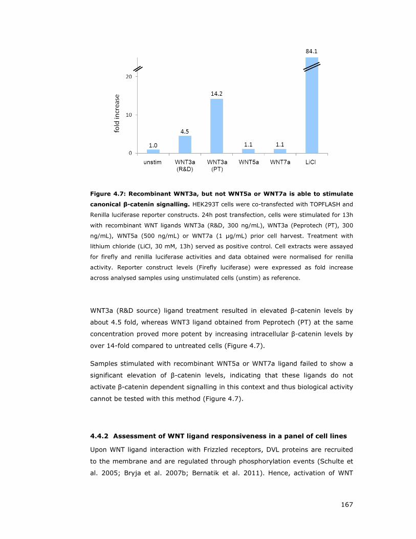
167
Figure 4.7: Recombinant WNT3a, but not WNT5a or WNT7a is able to stimulate
canonical β-catenin signalling. HEK293T cells were co-transfected with TOPFLASH and
Renilla luciferase reporter constructs. 24h post transfection, cells were stimulated for 13h
with recombinant WNT ligands WNT3a (R&D, 300 ng/mL), WNT3a (Peprotech (PT), 300
ng/mL), WNT5a (500 ng/mL) or WNT7a (1 µg/mL) prior cell harvest. Treatment with
lithium chloride (LiCl, 30 mM, 13h) served as positive control. Cell extracts were assayed
for firefly and renilla luciferase activities and data obtained were normalised for renilla
activity. Reporter construct levels (Firefly luciferase) were expressed as fold increase
across analysed samples using unstimulated cells (unstim) as reference.
WNT3a (R&D source) ligand treatment resulted in elevated β-catenin levels by
about 4.5 fold, whereas WNT3 ligand obtained from Peprotech (PT) at the same
concentration proved more potent by increasing intracellular β-catenin levels by
over 14-fold compared to untreated cells (Figure 4.7).
Samples stimulated with recombinant WNT5a or WNT7a ligand failed to show a
significant elevation of β-catenin levels, indicating that these ligands do not
activate β-catenin dependent signalling in this context and thus biological activity
cannot be tested with this method (Figure 4.7).
4.4.2 Assessment of WNT ligand responsiveness in a panel of cell lines
Upon WNT ligand interaction with Frizzled receptors, DVL proteins are recruited
to the membrane and are regulated through phosphorylation events (Schulte et
al. 2005; Bryja et al. 2007b; Bernatik et al. 2011). Hence, activation of WNT

168
signalling could be assessed by analysis of DVL hyper-phosphorylation in gel
shift assays (Figure 4.8 and Figure 4.9) (Bryja et al. 2007b).
The ability of cells to activate WNT signalling cascades depends on the available
WNT ligands, expression of cognate receptors and co-receptors (Gordon and
Nusse 2006; Green et al. 2008; Kikuchi et al. 2009; Sato et al. 2010; Niehrs
2012). In order to identify a suitable cellular system for subsequent experiments
in this study, a panel of tumour cell lines were chosen from breast (MCF-7, MDA-
MB-231), lung (A549), colon (HCT-116), bone (SaOS2) or neuroblast origin
(IMR32, SY5YSH), as well non-transformed immortalised cell lines of mammary
epithelial origin and investigated for response to WNT ligands 3a, 5a and 7a
(Figure 4.8).
Notably, all investigated cancerous cell lines employed in the panel showed a
response to WNT5a conditioned media after two hours of treatment,
demonstrated by a slower migrating form of DVL2 (Figure 4.8A upper part).
Stimulation of the same cell line panel with WNT3a conditioned media showed
responsiveness, with the exception of MDA-MB-231 cells, displayed by a
phosphorylation induced shift of DVL2. However, WNT7a conditioned media did
not seem to stimulate any of the tumour derived cell lines (Figure 4.8A upper
section).
However, even though all tested tumour cell lines respond to WNT3a and WNT5a
CM, there are cell line dependent preferences. For example HCT-116 and SaOS2
cells show a similar DVL2 phosphorylation shift in response to WNT3a and
WNT5a CM, A549 cells react more strongly to WNT3a, whereas MDA-MB-231
cells respond very strongly to WNT5a as judged by DVL2 phosphorylation shift
(Figure 4.8).
In contrast, treatment of four different immortalised non-tumorigenic mammary
epithelia cell lines with WNT3a, WNT5a or WNT7a conditioned media failed in all
four tested cell lines to induce a DVL2 phosphorylation shift (Figure 4.8A lower
part).
In order to assess if generated WNT conditioned media performs similar to
recombinant commercially available purified WNT ligands, SaOS2, HCT116 and
MDA-MB-231 were treated either with WNT3a, WNT5a, WNT7a CM or
corresponding recombinant ligands under similar conditions and lysates were
analysed for DVL2 phosphorylation shift (Figure 4.8A upper part and B). Overall,
CM and recombinant ligands yielded the same DVL2 migration pattern, except

169
for MDA-MB-231 cells, which showed a strong response to recombinant WNT3a
ligand, which was not observed for WNT3a CM. This could be due to lower ligand
concentration in WNT3a CM.
Figure 4.8: WNT ligands differentially stimulate DVL2 phosphorylation in tumour
derived cell lines but have no effect on non-transformed breast cell lines.
Assessment of WNT responsiveness in a panel of cell lines. Cancerous and non-
tumourigenic cell lines were chosen in order to test responsiveness to selected WNT
ligands and analysed by DVL2 migration on SDS-polyacrylamide gels. (A) A549 (lung
adenocarcinoma), SaOS2 (osteosarcoma), HCT-116 (colorectal carcinoma), MCF7 and
MDA-MB-231 (breast adenocarcinoma), or non-tumorigenic mammary epithelia cell lines
MCF10A, 184A1, HMEC and HMLE cell lines (normal) were grown to confluency, regular
growth media aspirated and replaced with either control (cCM), indicated WNT ligand
conditioned media or left untreated (-). Cells were lysed after 2 hours and analysed by
Western blot for endogenous DVL2 protein shift. (B) U2OS (osteosarcoma), HEK293T
(embryonic kidney), SY5YSH and IMR32 (human neuroblastoma) cell lines were grown to
confluency and treated with recombinant WNT3a (225 ng/mL), WNT5a (225 ng/mL) or
WNT7a (550 ng/mL) for 2h or left untreated (-). Cells were harvested and probed for
endogenous DVL2. Recombinant and conditioned media induced a similar DVL2
phosphorylation shift when compared to SaOS2, HCT-116 or MDA-MB-231 cells under
both stimulation conditions (A versus B).
170
Assessment of WNT ligand responsiveness of neuroblastoma derived SY5YSH or
IMR32 cells displayed a strong DVL2 phosphorylation shift if stimulated with
recombinant WNT3a or WNT5a, but not if treated with WNT7a ligand. Notably,
HEK293T cells have a high basal level of phosphorylated DVL2 (Figure 4.8B).
4.4.3 Frizzled expression alters sensitivity towards WNT ligands
WNT ligands bind to Frizzled receptors in order to activate downstream signalling
cascades. Since there are 19 different WNT ligands and 10 Frizzled receptors
known in mice and humans, it is difficult to find appropriate ligand-receptor
combinations for in vitro studies. Therefore, we first tested if overexpression of
Frizzled receptors could stimulate WNT signalling or enable cells to selectively
respond to specific WNT ligands. Based on reports in the literature of their
involvement in non-canonical WNT signalling (Wang et al. 2006; De Marco et al.
2012), FZD5 and FZD6 were cloned and stably expressed by lentiviral
transduction in HMEC and MDA-MB-231 cells. For technical reasons, a human
FZD5 construct could not be cloned. Alignment of human FZD5 and mouse
Frizzled5 (mFzd5) indicted a 97% amino acid identity and was therefore used as
alternative in all corresponding experiments.
The effect of mFzd5/FZD6 expression in HMEC cells, which were unresponsive to
various WNT ligand conditioned media (Figure 4.8A lower part and Figure 4.9A)
was assessed by DVL2 phosphorylation induced shift by immunoblotting.
Expression of either mFzd5 or FZD6 in HMEC cells resulted in a slight increase of
endogenous DVL2 phosphorylation, compared to vector control cells (Figure
4.9A, lanes 5, 9 and 1, respectively). Interestingly, stimulation of mFzd5 cells
with WNT3A or WNT5A CM resulted in a clear phospho-shift of DVL2. In contrast,
expression of FZD6 did not lead to increased responsiveness towards WNT3a or
WNT5a CM, indicating that FZD6 might not bind to WNT3a or WNT5a ligands in
this cellular context (Figure 4.9A).
In contrast, expression of mFzd5 or FZD6 in MDA-MB-231 cells led to a higher
constitutive level of hyper-phosphorylated DVL2, which could not be further
stimulated by WNT3A or WNT5A ligands (Figure 4.9B).
Therefore, overexpression of the mFzd5 receptor provides a great tool to
stimulate formerly unresponsive HMEC cells with WNT ligands and this system is
utilised in a later section.

171
Because R-RAS GTPases could possibly function downstream of only selected
Frizzled receptors that could be expressed or function together with co-receptors
in a cell-type specific manner, we decided to employ and expand the
aforementioned overexpression approach of Frizzled receptors. Out of the eight
remaining Frizzled members available in a mammalian system, we achieved to
clone further FZD2, FZD3, FZD4, and FZD7 by PCR from cDNA clones ordered
from Open Biosystems.
Figure 4.9: HMECs expressing mFzd5 gain responsiveness to WNT3a and WNT5a
ligands. Human mammary epithelial cells (HMECs) or breast adenocarcinoma cells (MDA-
MB-231) were transduced using lentiviral vectors encoding mFrizzled5 (mFzd5-mouse
homologue), Frizzled6 (FZD6) or empty vector, serving as control. (A) Responsiveness of
HMECs towards WNT ligand 3a and 5a was assessed by DVL2 gel shift post treatment with
either control conditioned media (cCM), indicated WNT ligand conditioned media or cells
that were left untreated in regular growth conditions (-). Treatment of indicated CMs were
performed for 2h. Activation by WNT ligands was assessed by immunoblotting for
phosphorylation shift of endogenous DVL2 protein. (B) Assessment of MDA-MB-231 cells
response to WNT5a or WNT3a conditioned media in stably expressing mFzd5 or FZD6
cells. Treatments were carried out as described for A.
Expression of mFzd5 but not FZD6 in HMEC resulted in a slight increase of basal
DVL2 phosphorylation, which was further stimulated by WNT3a and WNT5a,
indicating that mFzd5 exogenous expression renders these cells responsive to
these ligands. On the other hand, expression of both mFzd5 and FZD6 in MDA-
MB-231 cells, resulted in a maximal DVL2 shift that cannot be further stimulated

172
(Figure 4.9). This suggests that expression of at least a subset of Frizzled
receptors is able to induce a DVL2 phosphorylation-shift, suggesting that
expression of Frizzled receptors alone can stimulate downstream signalling
without WNT ligand interaction. Therefore, taking advantage of this tool, we
used the cloned panel of Frizzled receptors including FZD2 and mFzd5, which
have been suggested to carry a role in non-canonical signalling (Sato et al.
2010; Yu et al. 2012). Further, this also enabled us to test which Frizzled and/or
ROR2 combination could be useful for further experiments.
4.4.4 FZDs and ROR2 specifically stimulate VANGL1 interaction with
DVL2
Expression of ROR2 in HEK293T cells strongly increases the amount of DVL2 co-
immunoprecipitating with VANGL1 compared to the empty vector sample (Figure
4.10A). Similarly, expression of mFZD5, FZD2, FZD4 and FZD7, but not FZD3 or
FZD6, stimulated VANGL1 binding to DVL2, suggesting that the ability to
modulate the VANGL1-DVL interaction is specific to a subset of FZD receptors
(Figure 4.10A). Furthermore, under conditions where exogenous expression of
ROR2 or Frizzled proteins stimulates the interaction of VANGL1 with DVL2, no
effect was seen on the interaction of VANGL1 with SCRIB, CASK or DLG1
suggesting that a subset of Frizzled receptors or ROR2 specifically modulate
VANGL1 interaction with DVL proteins (Figure 4.10A).
It has been reported that ROR2 can act as co-receptor for Frizzled proteins
prompting the question if ROR2 and Frizzled could further stimulate the VANGL1-
DVL2 interaction in a cooperative manner (Nishita et al. 2010b) (Figure 4.10B).
Hence, the same panel of Frizzled proteins were co-expressed in the presence or
absence of ROR2 and flag-VANGL1 constructs, and lysates were flag-VANGL1
immunoprecipitated and probed for endogenous DVL2 interaction (Figure 4.10C).
Co-expression of ROR2 and FZD2 is able to further increase bound DVL2 to
VANGL1 compared to ROR2 expression alone. This cooperative effect was also
observed with a FZD4, mFzd5 or FZD7 - ROR2 combination, but not in case of
Frizzled receptor FZD3 or FZD6 (Figure 4.10C).
Furthermore, two mutations of mFzd5 were included in this study, the point
mutation W530G and a deletion of the C-terminal PDZ binding motif (Figure
4.10C).

173
Figure 4.10: FZDs and ROR2 cooperate to stimulate VANGL1-DVL2 interaction.
(A) HEK293T cells were transfected with indicated Frizzled (FZD) and ROR2 constructs
together with flag-VANGL1. Two days after transfection, cells were harvested,
immunoprecipitated using flag-sepharose beads and probed for endogenous DVL2, CASK,
DLG1 and SCRIB. (B) Schematic of WNT signalling pathway (incomplete illustration),
focused on the PCP branch and our observations of ROR2 and FZD overexpression. (C)
Co-expression of Frizzled and ROR2 cooperate to increase VANGL1-interacton with DVL2
but not other proteins. Flag-VANGL1 was co-transfected with a panel of FZD receptors in
the absence or presence (+) of ROR2. Cell harvest, immunoprecipitation and detection
was carried out as in A. Flag-tagged Protein Kinase A regulatory subunit type 1 (R1A) and
GFP constructs were expressed for assessment of background binding. Note that part C of
this figure represents one gel, where the vertical line indicates omitted lanes.
The highly conserved cytosolic KTxxxW motif of Frizzled receptors interacts with
the PDZ domain of DVL proteins and is required for Frizzled-induced
translocation of DVL to the plasma membrane and DVL phosphorylation
(Umbhauer et al. 2000). The mFzd5 W530G point mutation localises to this motif
and has been demonstrated to disrupt the receptors’ association with DVL
(Umbhauer et al. 2000; Cong et al. 2004b). Deletion of the PDZ binding motif in
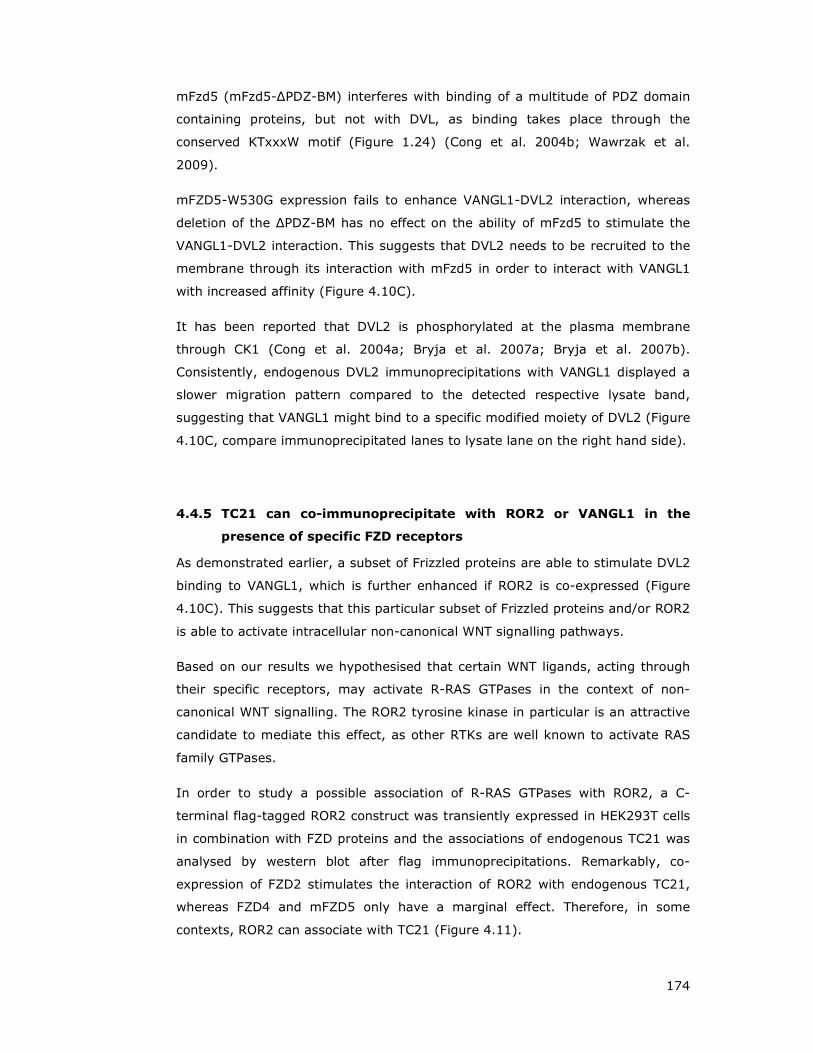
174
mFzd5 (mFzd5-∆PDZ-BM) interferes with binding of a multitude of PDZ domain
containing proteins, but not with DVL, as binding takes place through the
conserved KTxxxW motif (Figure 1.24) (Cong et al. 2004b; Wawrzak et al.
2009).
mFZD5-W530G expression fails to enhance VANGL1-DVL2 interaction, whereas
deletion of the ∆PDZ-BM has no effect on the ability of mFzd5 to stimulate the
VANGL1-DVL2 interaction. This suggests that DVL2 needs to be recruited to the
membrane through its interaction with mFzd5 in order to interact with VANGL1
with increased affinity (Figure 4.10C).
It has been reported that DVL2 is phosphorylated at the plasma membrane
through CK1 (Cong et al. 2004a; Bryja et al. 2007a; Bryja et al. 2007b).
Consistently, endogenous DVL2 immunoprecipitations with VANGL1 displayed a
slower migration pattern compared to the detected respective lysate band,
suggesting that VANGL1 might bind to a specific modified moiety of DVL2 (Figure
4.10C, compare immunoprecipitated lanes to lysate lane on the right hand side).
4.4.5 TC21 can co-immunoprecipitate with ROR2 or VANGL1 in the
presence of specific FZD receptors
As demonstrated earlier, a subset of Frizzled proteins are able to stimulate DVL2
binding to VANGL1, which is further enhanced if ROR2 is co-expressed (Figure
4.10C). This suggests that this particular subset of Frizzled proteins and/or ROR2
is able to activate intracellular non-canonical WNT signalling pathways.
Based on our results we hypothesised that certain WNT ligands, acting through
their specific receptors, may activate R-RAS GTPases in the context of non-
canonical WNT signalling. The ROR2 tyrosine kinase in particular is an attractive
candidate to mediate this effect, as other RTKs are well known to activate RAS
family GTPases.
In order to study a possible association of R-RAS GTPases with ROR2, a C-
terminal flag-tagged ROR2 construct was transiently expressed in HEK293T cells
in combination with FZD proteins and the associations of endogenous TC21 was
analysed by western blot after flag immunoprecipitations. Remarkably, co-
expression of FZD2 stimulates the interaction of ROR2 with endogenous TC21,
whereas FZD4 and mFZD5 only have a marginal effect. Therefore, in some
contexts, ROR2 can associate with TC21 (Figure 4.11).

175
For technical reasons it was difficult to perform similar experiments in other cell
types such as HMEC and MDA-MB-231 cells as transient transfection efficiency is
much lower than HEK293T cells. Unfortunately, the cell lines stably expressing
ROR2 were generated using an untagged ROR2 construct therefore precluding
analogous HEK293T cell experiments.
Figure 4.11: ROR2 co-expressed with FZD2 is able to immunoprecipitate
endogenous TC21 in HEK293T cells. C-terminal flag-tagged ROR2 was co-transfected
with FZD2, FZD4, mFzd5 and FZD7, and cell lysates were harvested 48 h post
transfection and purified using flag-sepharose beads. Immunoprecipitates were probed for
endogenous TC21 protein using a specific antibody.
However, in MDA-MB-231 and HMEC cells stably expressing flag-tagged VANGL1
in the context of mFZD5 stable exogenous expression, VANGL1 is able to co-
purify endogenous TC21 but not R-RAS (Figure 4.12). Since VANGL1 is a strict
GTP dependent interactor of R-RAS GTPases, this suggests that mFzd5
expression in this cellular context may be able to selectively activate TC21.

176
Figure 4.12: VANGL1 is able to immunoprecipitate endogenous TC21 but not R-
RAS in two breast cell lines stably expressing mFzd5. MDA-MB-231 or HMEC cells
were transduced using a lentiviral mFzd5 construct and selected, before a second round
of lentiviral infection with either flag-VANGL1 (V1) or Flag-NF2 was performed. Cells were
lysed and flag immunoprecipitates probed for endogenous R-RAS or TC21.
4.4.6 WNT5a modulates VANGL1 interaction with DVL but not SCRIB,
CASK or DLG1
HMEC cells stably expressing mFzd5 become responsive to WNT5a as shown in
Figure 4.9A, and provided an attractive cellular system to study the effects of
WNT5a stimulation. Therefore, tagged VANGL1 or VANGL2 were stably
introduced into HMEC mFzd5 cells and the resulting cell lines were subsequently
used to study the effect of WNT5a on VANGL interactions with endogenous
proteins (Figure 4.13).
Treatment of HMEC mFzd5 cells with WNT5a conditioned media for two hours
stimulated binding of VANGL1 to DVL1 and DVL3 compared to control media.
Similar results could be observed for VANGL2, albeit levels are much lower
(Figure 4.13). Probing for additional endogenous VANGL1/2 interactors, using
the same immunoprecipitated samples, did not display WNT5a mediated
modulation effects for CASK or DLG1 proteins (Figure 4.13).
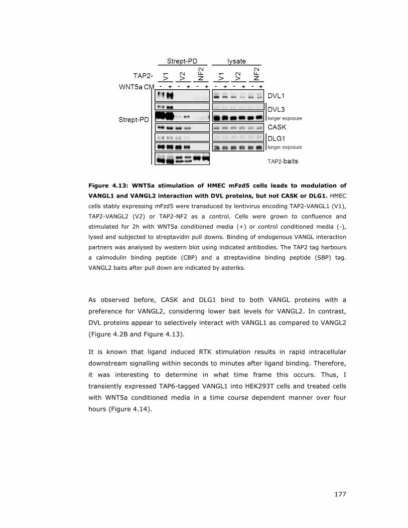
177
Figure 4.13: WNT5a stimulation of HMEC mFzd5 cells leads to modulation of
VANGL1 and VANGL2 interaction with DVL proteins, but not CASK or DLG1. HMEC
cells stably expressing mFzd5 were transduced by lentivirus encoding TAP2-VANGL1 (V1),
TAP2-VANGL2 (V2) or TAP2-NF2 as a control. Cells were grown to confluence and
stimulated for 2h with WNT5a conditioned media (+) or control conditioned media (-),
lysed and subjected to streptavidin pull downs. Binding of endogenous VANGL interaction
partners was analysed by western blot using indicated antibodies. The TAP2 tag harbours
a calmodulin binding peptide (CBP) and a streptavidine binding peptide (SBP) tag.
VANGL2 baits after pull down are indicated by asteriks.
As observed before, CASK and DLG1 bind to both VANGL proteins with a
preference for VANGL2, considering lower bait levels for VANGL2. In contrast,
DVL proteins appear to selectively interact with VANGL1 as compared to VANGL2
(Figure 4.2B and Figure 4.13).
It is known that ligand induced RTK stimulation results in rapid intracellular
downstream signalling within seconds to minutes after ligand binding. Therefore,
it was interesting to determine in what time frame this occurs. Thus, I
transiently expressed TAP6-tagged VANGL1 into HEK293T cells and treated cells
with WNT5a conditioned media in a time course dependent manner over four
hours (Figure 4.14).
178
Figure 4.14: WNT5a treatment enhances binding of DVL2 and DVL3 to VANGL1
after 60 min, but SCRIB or CASK interactors are not modulated. HEK293T cells
were transfected with TAP6-VANGL1 or TAP6-control constructs. 44h after transfection,
cells were stimulated either with conditioned WNT5a media (WNT5a CM) at time points
indicated above or control media (Control CM) for either 10 or 120 min. Cells were
harvested and immunoprecipitated using flag-sepharose beads and probed with the
indicated endogenous antibodies. TAP6 contains a large tag cassette, harbouring an EE-
tag, streptavidine binding peptide, histidine-tag and flag-tags.
Stimulation of HEK293T cells with WNT5a conditioned media resulted in
enhanced DVL2 and DVL1 binding to VANGL1, which could be observed from 60
min after WNT5a application and was sustained for four hours (Figure 4.14). In
contrast, no effect on the interaction with SCRIB or CASK is seen at any time
point. Therefore, WNT5a selectively stimulates the association of VANGL1 with
DVL2, which takes place in a delayed manner.
Taken together, these experiments showed that either WNT5a or Frizzled
proteins in cooperation with ROR2 are able to stimulate VANGL1-DVL2
interaction. This binding is specific for DVL proteins, preferentially interacting
with VANGL1 over VANGL2.
WNT5a mediated enhanced VANGL1-DVL2 stimulation displays a response delay
of approximately 60 min that may correlate with potential phosphorylation of
DVL2, possibly by CK1. Furthermore, TC21 could be immunoprecipitated with
ROR2 if FZD2 was co-expressed. Likewise, TC21 could be detected to precipitate
with VANGL1 if mFzd5 was stably expressed, indicating that TC21 could be
activated but in a Frizzled and ROR2 context dependent manner.

179
4.5 Proteomic analysis of VANGL1 interaction and their
modulation by R-RAS, mFZD5 and ROR2
ROR2 and mFzd5 both selectively stimulate VANGL1 interactions with DVL
proteins and when expressed together cooperate to enhance this interaction
(Figure 4.10). On the other hand, active R-RAS appears to negatively regulate
several VANGL1 interactions (Figure 4.6).
In order to shed more light on the VANGL1 interactome and study in an unbiased
approach how these interactions may be modulated in vivo, an additional
proteomic study was performed. TAP-tagged VANGL1 was affinity purified from
cells co-expressing either mFzd5, ROR2, a combination thereof, activated (L87)
or dominant negative R-RAS (N43) (Figure 4.15).
This purification achieved high sensitivity and in addition to proteins already
detected in previous TAPs, multiple potential novel VANGL1 interactors were
identified. Furthermore, many of the proteins appear to be modulated in one or
more of the conditions tested, as suggested by this semi-quantitative approach.
A summary of the mass spectrometry results is shown in Figure 4.15.
Co-expression of R-RAS L87 led to the identification by mass spectrometry of
fewer peptides for DVL proteins, SCRIB and DLG1, suggesting that active R-RAS
decreases these interactions with VANGL1. This is consistent with the results
obtained previously (Figure 4.6) and independently suggests that R-RAS, upon
activation, is likely to modulate the VANGL1 interactome.
The mass spectrometry results for this experiment were received towards the
end of my PhD study and therefore I was unable to perform further validation
and follow up experiments.

180
Figure 4.15: Summary of proteins identified by mass spectrometry in VANGL1
affinity purifications. (A) VANGL1 protein complexes were isolated from HEK293T cells
expressing a flag-tagged VANGL1 construct alone or in combination with mFzd5, ROR2, a
combination of mFzd5 and ROR2, activated R-RAS L87 (R-RasL) or the dominant negative
analogue R-RAS N43 (R-RasN) as indicated in the illustration. (B) Cell lysates were
harvested 48h after transfection and subjected to flag-sepharose purification. Purified
proteins were precipitated and separated by SDS-PAGE, gel bands cut, trypsinised and
analysed by tandem mass spectrometry. Proteins are ranked by percent coverage and are
colored blue or coral if the interaction with VANGL1 was decreased or increased
respectively, as judged by the number of peptides identified.

181
4.6 Discussion
4.6.1 Identification and characterisation of VANGL1 interacting proteins
Using TAP as an unbiased proteomic approach, multiple VANGL1 interacting
proteins have been identified. Some of them such as DVL/Dsh, Scrib and Dlg1
had been previously identified as interacting with the VANGL homologue in
Drosophila, and confirm a role in cell polarity (Park and Moon 2002; Lee et al.
2003; Bellaiche et al. 2004; Courbard et al. 2009).
VANGL1 contains a PDZ domain binding motif at its C-terminus, allowing
interaction with PDZ domain containing proteins. PDZ proteins are found from
bacteria to vertebrates and are involved in various cellular functions including
cell polarity (Ponting 1997; Subbaiah et al. 2011). Notably, a subset of PDZ
containing VANGL1 interactors, such as SCRIB, DLG1 and CASK, which are
involved in polarity and migration have been demonstrated to be strictly
dependent on the VANGL1-PDZ binding motif for interaction.
SCRIB, one of the identified VANGL1 interactors, has been reported in
mammalian cells to associate with the PIX/GIT complex that regulates
RAC/CDC42 and ARF GTPases (Audebert et al. 2004; Frank and Hansen 2008)
and be involved in cell polarity during epithelial migration through regulation of
RHO GTPase recruitment to the leading edge (Dow et al. 2007), and to mediate
neural migration and CE (Qin et al. 2005a; Wada et al. 2005). SCRIB’s role in
PCP is further strengthened by the observation that SCRIB mouse mutants have
neural tube closure defects (Murdoch et al. 2003) and display hair cell polarity
defects in the inner ear (Montcouquiol et al. 2003).
SCRIB is a cytosolic protein but can localise to the plasma membrane, e.g. sites
of cell-cell contact in epithelial cells and leading edge in migrating cells,
nonetheless how SCRIB translocation to the membrane is regulated is not clear.
A recent report has confirmed that SCRIB forms a complex with VANGL1 and
further showed that VANGL1 co-localises with SCRIB at the leading edge in
migrating cells (Anastas et al. 2011). Therefore, SCRIB interaction with
VANGL1, a membrane protein, may provide a mechanism to recruit SCRIB to the
membrane. Based on our results, we propose that activation of R-RAS GTPases
by extracellular signals would recruit the VANGL1-SCRIB complex to sites of
activation at specialised membrane micro-domains and thus be a mechanism to
spatially coordinate polarity with other signalling pathways regulated by R-RAS
GTPases.
182
DLG1 has been shown to interact with VANGL1 in a PDZ binding motif dependent
manner. Consistent with earlier findings in Drosohila, Dlg and Stbm form a
complex which is important in plasma membrane formation (Lee et al. 2003). It
has been suggested that this complex is required to recruit membrane-
associated proteins and lipids from internal membranes to sites of new plasma
membrane formation (Lee et al. 2003).
Notably, DLG1 null mice suffer from skeletal defects throughout the embryo and
characteristic PCP defects such as open neural tube and misorientation of
cochlear hair cell stereociliary bundles, consistent with the possibility that at
least in mice, DLG1 functions with SCRIB in planar cell polarity (Rivera et al.
2013).
CASK, has been originally described to be localised at neuronal synapses (Zheng
et al. 2011) and to act as a scaffold for proteins involved in synaptic junctions,
vesicle exocytosis and ion channel trafficking (Butz et al. 1998; Setou et al.
2000; Leonoudakis et al. 2004). In addition, CASK has been shown to interact
with DLG1 (Sabio et al. 2005) and it remains to be determined if both bind
independently to VANGL1 or as a complex with each other.
Unlike other PDZ domain containing proteins like SCRIB, DLG1 and CASK,
VANGL1 interaction with DVL2 is not exclusively mediated through the VANGL1
PDZ-binding motif, which is in line with observations of the corresponding
Drosophila homologue (Park and Moon 2002). Remarkably, R-RAS interaction is
specific to VANGL1 and so is the DVL interaction. Furthermore, R-RAS and DVL3
binding to VANGL1 is disrupted by the point mutant D259E, suggesting that both
proteins could be competing for the same binding site. This is further confirmed
by observations that active R-RAS modulates VANGL1-DVL binding, suggesting a
common ‘axis’ of VANGL1 function.
DVL proteins are major signalling hubs, which interact with a multitude of
proteins, and mediate canonical and non-canonical WNT signalling (Boutros and
Mlodzik 1999; Gao and Chen 2010; Wynshaw-Boris 2012) for example WNT5a
induced migration through activation of RHOA or RAC in breast cancer cells (Zhu
et al. 2012; Zhu et al. 2013). Therefore, VANGL1 interaction with all three DVL
proteins highlights its potential role in downstream WNT signal mediation, which
will be discussed later in this section.
The observation that VANGL1 is able to bind to the intracellular domain of
CELSR2 but not CELSR1, which was also the only CELSR isoform detected in
183
VANGL1-TAP experiments, indicates C-terminus mediated functional differences
across CELSR proteins. Indeed, CELSR1 is a major player in PCP and is directly
linked to cochlear stereocilia bundle misorientation and neural tube closure
defects (Curtin et al. 2003; Devenport and Fuchs 2008; Robinson et al. 2011).
CELSR2 and CELSR3 regulate less direct PCP processes such as ciliogenesis,
maintenance of dendrites, neuronal migration and axon guidance (Shima et al.
2002; Tissir et al. 2005; Wada et al. 2006; Shima et al. 2007; Tissir et al.
2010), implicating that VANGL1 might not be directly involved in neurulation and
related CE (Torban et al. 2008; Antic et al. 2010; Song et al. 2010), but rather
in PCP directed processes associated with ciliogenesis and neuronal development
and maintenance.
Due to technical reasons, interaction studies were carried out with the
intracellular C-terminus of CELSR1 and CELSR2, but it cannot be ruled out that
the large ectodomain could be also involved in VANGL1 binding in order to
establish PCP across a plane of cells. It has been reported for the Drosophila
homologue of VANGL proteins, that stbm interacts with Frizzled proteins through
their respective extracellular domains (Wu and Mlodzik 2008) and thus VANGL1-
CELSR protein interaction could also take place through extracellular domain
association.
The adhesion-GPCR Latrophilin 2 (LPHN2) was detected in the VANGL1-TAP co-
expressing activated R-RAS (Figure 4.1C). This is particular interesting since
LPHN2 is structurally related to CELSR proteins and plays an essential role in the
establishment of tissue polarity and thus could constitute a novel PCP pathway
mediator (Langenhan et al. 2009). Unfortunately, several cloning attempts of the
full length protein or truncation constructs led to very low or undetectable
expression levels, rendering interaction approaches unfruitful.
Multiple other novel VANGL1 - interacting proteins identified by TAP are involved
in vesicle trafficking and imply additional roles for VANGL1 function. For example
YIF1B interacts with the transport GTPases Ypt1p, Ypt31p and Sec4p, and loss of
YIF1 function results in a block of ER-to-Golgi protein transport and vesicle
accumulation, suggesting a vital role in vesicle docking and fusion (Matern et al.
2000). YIF1B localisation to ER or Golgi membranes suggests that VANGL1 might
interact with YIF1B on its way to the plasma membrane as shown for Sec24B
and VANGL2 transport (Wendeler et al. 2007; Merte et al. 2011; Wansleeben et
al. 2011). Another possibility is that VANGL1 could also localise to ER or Golgi
membranes, consistent with what is observed for its subcellular localisation
pattern (see section 5.4.
184
NDFIP1 is found associated with the exosome, Golgi or vesicles. It forms an
adaptor for Nedd4 family proteins in order to bind respective target proteins for
subsequent ubiquitination. Interestingly, NEDDL family proteins have been
demonstrated to negatively regulate WNT signalling through DVL2 ubiquitination
(Wei et al. 2012; Ding et al. 2013). Thus, it would be interesting to determine if
VANGL1 could act as a modulator for DVL2 directed ubiquitination or might be a
target of NEDD4 proteins itself.
Endocytosis of WNT ligand-receptor complexes is required for WNT signalling
regulation, which is achieved through clathrin or caveolin mediated endocytosis
(Sorkin and von Zastrow 2009; Traub 2009; Lajoie and Nabi 2010). Clathrin
mediated internalisation has been shown to regulate β-catenin dependent (Chen
et al. 2001; Blitzer and Nusse 2006) and independent pathways (Sato et al.
2010; Ohkawara et al. 2011; Sakane et al. 2012). SYNJ2BP a putative growth-
promoting factor implicated in breast tumourigenesis and tumour development
(Li et al. 2009c), has been shown to be involved in clathrin-mediated
endocytosis and recycling (Matsuzaki et al. 2002; Verstreken et al. 2003).
VANGL1 interaction with SYNJ2BP, which takes place in a PDZ-dependent
manner, suggests a regulatory function for VANGL1 in vesicle formation and
trafficking.
RAB proteins control intracellular trafficking (Chua et al. 2010; Mizuno-Yamasaki
et al. 2012) and have been shown to regulate WNT signalling (Lee et al. 2007;
Zhu et al. 2013). VANGL1 interacts specifically with the catalytic subunit of a
RAB3GAP (RAB3GAP1), whereas DVL2 binds to the non-catalytic subunit
(RAB3GAP2). Therefore it could be speculated that RAB3GAP2 could be
associated with DVL2 and upon binding of VANGL1 to DVL2, the catalytic subunit
is recruited to this complex and is now able to modulate RAB protein activity in
vesicle trafficking processes.
C17ORF62 still remains mostly uncharacterised besides the MS detected C-
terminal serine/tyrosine phosphorylation cluster (at position S159, S168, S170,
S173, S175, S176, S178, S187 and Y124) (http://www.phosphosite.org). Initial
co-localisation studies found C17ORF62 associated with the Golgi complex and it
has been proposed that this protein could be involved in cell death induction
(Deng et al. 2011). However, more studies will be required to shed light into
C17ORF62 function and its relation to VANGL1.
Sec24b has been recently demonstrated to be part of the endoplasmic reticulum
to Golgi complex transport machinery, important for VANGL2 transport and thus

185
has not been the focus of our efforts (Merte et al. 2011; Wansleeben et al.
2011).
Although some of the proteins tested in this study bind to both VANGL1 and
VANGL2 isoforms (SCRIB, DLG1, CASK, YIF1B, CELSR2-C2, RAP3GAP1,
SYNTENIN-1), others show differential interaction affinities and suggest different
functions for the two VANGL proteins (DVL proteins, SYNJ2BP).
4.6.2 Frizzled expression leads to enhanced VANGL1-DVL2 interaction
Immunoprecipitation experiments demonstrated that TC21 associates with ROR2
in HEK293T cells co-expressing FZD2. Furthermore, employing HMEC and MDA-
MB-231 cells expressing stably mFzd5, endogenous TC21 but not R-RAS co-
immunoprecipitates with VANGL1. This is despite the fact that R-RAS protein
levels could be readily detected in lysates of these cells. This suggests that
depending on the receptor/co-receptor combination and cellular context different
R-RAS GTPases might be differentially regulated.
R-RAS and TC21 seem to be regulated by the same set of GEFs and GAPs,
whereas M-RAS showed a regulation pattern more similar to classical RAS
proteins (Ohba et al. 2000), suggesting that R-RAS and TC21 are likely to be
co-activated by extracellular signals. On the other hand, TC21 has been shown
to constitutively interact with the T-cell and B-cell antigen receptors (TCR and
BCR, respectively) through a direct interaction with a signalling sequence known
as the Immunoreceptor Tyrosine-based Activation Motif or ITAM, which is
present in the CD3 signalling subunits of the TCR and the Igα and β subunits of
the BCR (Alarcon and Martinez-Martin 2011). This fact highlights the possibility
that there might be additional mechanisms to achieve differential regulation of
R-RAS GTPases. It remains to be determined if in some contexts TC21 may
interact directly with ROR2 by a mechanism similar to that shown for the ITAMs
of the TCR and BCR.
However, in section 6.2 I will provide evidence that R-RAS can also be activated
by ROR2 expression or WNT5a stimulation, and thus both TC21 and R-RAS
GTPases seem to play a role in non-canonical WNT signalling.
DVL proteins interact with a multitude of proteins and serve as a key signalling
components downstream of Frizzled receptors, regulating WNT/β-catenin and
non-canonical WNT pathways (Wharton 2003; Wallingford and Habas 2005;
Malbon and Wang 2006; Gao and Chen 2010). WNT-induced activation of DVL is

186
visible as a electrophoretic mobility shift of all three DVL isoforms (Gonzalez-
Sancho et al. 2004; Bryja et al. 2007a; Bryja et al. 2007b), and leads to
activation of DVL downstream of WNT ligands (Peters et al. 1999; Sakanaka et
al. 1999; Kishida et al. 2001; Cong et al. 2004a; Foldynova-Trantirkova et al.
2010).
VANGL1 interacts with all three DVL proteins. Deletion of the PDZ-binding motif
in VANGL1 displays only a modest effect on interaction reduction, whereas the
point mutation D259E, which is found in patients with NTDs, disrupts the
interaction. This suggests that a membrane proximal region, which remains to
be further mapped, interacts with a region of DVL other than the PDZ.
Interestingly the interaction of VANGL1 with DVLs, but not with other proteins
such as SCRIB, DLG1 and CASK (whose interactions are mediated via PDZ
domains) is stimulated by a subset of FZD proteins as well as by ROR2. The
underlying mechanism by which FZD and/or ROR2 achieve this stimulatory effect
remains to be identified but may be related to DVL hyper-phosphorylation (see
below).
Based on the observation that the DVL bound to VANGL1 appears to migrate
more slowly and that WNT stimulation of the DVL-VANGL1 interaction follows a
delayed time course that correlates with DVL phosphorylation, it is tempting to
speculate that the interaction of DVL with VANGL1 is stimulated by DVL
phosphorylation. Therefore, DVL phosphorylation (by casein kinase or other
kinases) may be at least part of the mechanism regulating this interaction.
Future experiments should address this possibility.
ROR2 contains WNT-binding domains in its extracellular region and could
therefore bind WNTs directly (Green et al. 2008; Angers and Moon 2009).
Further, ROR2 could also function as a co-receptor operating in combination with
FZDs. It has been proposed that one mechanism by which WNTs can trigger
canonical versus non-canonical signalling is through a differential use of co-
receptors in combination with FZD receptors (Sato et al. 2010; Niehrs 2012).
Genetic interactions between ROR2 and WNT5a were suggested by observations
that mice with inactivation of the gene encoding Ror2 showed striking similarities
to Wnt5a−/− mice, including perinatal lethality, dwarfism, facial abnormalities,
and limb shortening (Green et al. 2008). Of note, Ror2−/− embryos also exhibited
defects in the orientation of the sensory hair cells in the inner ear, a hallmark of
WNT planar polarity pathway aberrations observed in Wnt5a−/−, Fzd3−/−, and

187
Fzd6−/− mice (Wang et al. 2006; Qian et al. 2007; Yamamoto et al. 2008). In
addition, ROR2 was shown to be involved in the effects of WNT5a in some
cellular and developmental processes, such as polarised cell migration and
convergent extension movements (He et al. 2008; Nomachi et al. 2008;
Enomoto et al. 2009; Laird et al. 2011).
FZDs recruit DVL to the plasma membrane by a still poorly understood
mechanism. FZD proteins contain a C-terminal PDZ binding motif required for
recruitment of PDZ proteins (Wawrzak et al. 2009), as well as an additional
KTxxxW motif which is required for DVL translocation to the membrane and
subsequent phosphorylation (Rothbacher et al. 2000; Umbhauer et al. 2000;
Axelrod 2001; Cong et al. 2004b). Our studies demonstrated that deletion of the
PDZ binding motif of mFzd5 has no effect on the stimulated VANGL1-DVL2
binding, whereas a mutation in its KTxxxW motif (W530G) abolishes the
enhanced interaction. Therefore, the ability of mFzd5 to stimulate the VANGL1-
DVL2 interaction correlates with its ability to translocate to the membrane and
phosphorylate DVL2 and further suggests that DVL translocation and
phosphorylation could be one mechanism of recruitment of VANGL1 (and its
associated interactome) to sites of activation by WNT ligands.
Of the tested panel of Frizzled receptors, FZD2, FZD4, mFzd5 and FZD7
stimulate the VANGL1-DVL2 interaction, whereas FZD3 and FZD6 did not display
a detectable interaction enhancement. Interestingly, FZD3 and FZD6 form a
distinct phylogenetic branch of Frizzled proteins (Huang and Klein 2004), and
have been demonstrated to be required with VANGL2, CELSR1 and PRICKLE2 for
inner ear stereociliary bundle orientation (Montcouquiol et al. 2006;
Montcouquiol et al. 2008).
As mentioned earlier, DVL interacts preferentially with VANGL1 compared to
VANGL2, and VANGL1 interacts with CELSR2 but not CELSR1. Taken together,
these observations suggest the possibility that a subset of FZD receptors,
possibly in combination with different CELSR co-receptors, may use different
VANGL isoforms to regulate various aspects of non-canonical signalling.
Notably, VANGL2 has been reported to form a WNT5a dependent complex with
ROR2, which leads to VANGL2 phosphorylation and PCP induced limb elongation
(Gao et al. 2011). It remains to be established if VANGL1 can also associate with
ROR2, although in a preliminary experiment I was able to detect endogenous
VANGL1 co-immunoprecipitating with ROR2 only in the presence of mFzd5 (data
not shown).
188
Several observations strongly suggest that DVL phosphorylation correlates with
the ability to interact with VANGL1. First, DVL2 that co-immunoprecipitates with
VANGL1 appears to migrate more slowly on SDS-PAGE compared to lysates.
Second, WNT stimulation of the DVL2-VANGL1 interaction follows a delayed time
course that correlates with DVL phosphorylation. Third, co-expression of certain
Frizzled receptors with ROR2 cooperates to enhance the VANGL1-DVL interaction
and this correlates with enhanced DVL phosphorylation shift. Therefore, DVL
phosphorylation may be at least part of the mechanism regulating the interaction
with VANGL1. Future experiments should further address this possibility.
DVL proteins display a variety of post translational modification sites (Wu et al.
2012) and are phosphorylated by at least three kinases CK1 (Klimowski et al.
2006; Bryja et al. 2007b; Corbit et al. 2008), CK2 (Wharton 2003) and PAR1
(Sun et al. 2001; Elbert et al. 2006), whereas protein phosphatase 2A (PP2A)
has been shown to negatively regulate DVL activity (Yokoyama and Malbon
2007).
CK1δ/ε have been identified as the relevant kinases hyper-phosphorylating DVL
in response to WNT5a, whereas WNT3a stimulation leads to β-catenin
stabilisation which was not reliant on hyper-phosphorylated DVL2 (Bryja et al.
2007a; Bryja et al. 2007b). However, activation of DVL mediated β-catenin
related downstream signalling appears in less than 10–15 min, whereas the
earliest formation of hyper-phosphorylated DVL is detected after 30 min
(Gonzalez-Sancho et al. 2004; Bryja et al. 2007b; Witte et al. 2010) and seems
to be stable for at least 12 h (Bryja et al. 2007a). Additionaly, CK1 induced
hyper-phosphorylation of DVL2 can result in the inactivation of β-catenin
signalling (Bernatik et al. 2011).
The role of DVL phosphorylation in WNT signalling remains obscure. Based on
these and our observations, a model emerges where DVL phosphorylation may
be a mechanism for the separate temporal control of β-catenin dependent and
independent WNT signalling. According to this model, the phosphorylation of DVL
that occurs at later time points of WNT stimulation and that correlates with
increased interaction with VANGL1, may shift WNT signalling from the β-catenin
pathway to the PCP pathway.
Expression of active R-RAS leads to a modest decrease in the ability of VANGL1
to interact with DVL2, CASK, DLG1, and SCRIB. This has been seen in co-
transfection-western blot experiments and was independently confirmed by mass
spectrometry. The decreased binding of VANGL1 with DVL in the presence of R-
189
RAS is consistent with the observation that mutation D259E in VANGL1 disrupts
the interaction with both R-RAS and DVL3. This suggests that both R-RAS and
DVL bind to the same region of VANGL1 and may therefore compete for its
interaction.
Furthermore, the proteomic study identified other proteins both negatively and
positively modulated by R-RAS. While these additional results remain to be
validated, our observations clearly suggest that R-RAS has the ability to regulate
VANGL1 interaction with a subset of its partners.
We propose a model where upon activation by extracellular signals, R-RAS
GTPases will recruit VANGL1 (and its associated proteins) to sites of activation.
This will not only lead to the localisation of VANGL1 complexes at spatially
distinct micro-domains at the plasma membrane but also to a selective
‘rearrangement’ of the interacting partners within the complex to give rise to
VANGL1 complexes with specialised signalling properties.
The observation that WNT stimulation can lead to both the activation of R-RAS
GTPases and its association with VANGL1 on one hand and to the association of
VANGL1 with DVL on the other hand, with both interactions potentially being
mutually exclusive may seem counter-intuitive. However, the spatial-temporal
dynamics have to be considered. R-RAS proteins are activated within minutes of
WNT stimulation; whereas the WNT induced increase in the interaction of
VANGL1 with DVL is first detected after 60 minutes and therefore clearly displays
a delayed response that may correlate with DVL phosphorylation. This will be
more clearly outlined and discussed in a subsequent chapter (see section 6.2)
and the final discussion (see section 8.1).

190
Figure 4.16: Overview of VANGL1 binding partners and R-RAS dependent
modulation of respective interactions. (A) VANGL1 interacts through its PDZ motif
with polarity proteins like DLG, CASK, SCRIB or the WNT core proteins DVL or CELSR2. In
addition, further interaction studies showed that VANGL1 binds to the Tetraspanin KAI1 or
the Syndecan-binding protein SYNTENIN. (B) Other novel VANGL1 interactors are
implicated in vesicle trafficking such as YIF1 (ER-GOLGI transport), RAB3GAP
(intracellular trafficking), SYNJ2BP (clathrin-mediated endocytosis) or endosome
associated (NDFIP1). (C) Overview of R-RAS modulating effect on identified VANGL1
binding partners. (D) Biochemical studies presented that R-RAS subgroup members (at
least TC21) are found associated with ROR2, suggesting that R-RAS GTPases act
downstream of WNT inducing stimuli, leading to enhanced DVL-VANGL1 interaction and
further to RHO GTPase modulation and PCP pathway activation.

191
CHAPTER 5
R-RAS GTPases inhibit
VANGL1 mediated
PRICKLE degradation
192
5 R-RAS GTPases inhibit VANGL1 induced PRICKLE degradation
5.1 VANGL1 interacts with PRICKLE1 and stimulates its destabilisation
Strabismus and Prickle have been shown to form a functional complex in
Drosophila that is proximal to the distally localised Frizzled-Dishevelled complex,
and this asymmetric distribution is required for tissue polarity patterning
(Bastock et al. 2003; Jenny et al. 2003; Rawls and Wolff 2003).
Due to unavailability of commercial antibodies against human PRICKLE proteins
at the time, human PRICKLE1 and PRICKLE2 were cloned by PCR. Vertebrate
PRICKLE1 consists of an N-terminal Prickle, Espinas and Testin (PET) domain and
three zinc binding LIN-11, Isl1 and MEC-3 (LIM) motifs. To test if these domains
are required for VANGL1 binding and to map the interaction, two truncation
constructs were further generated; PRICKLE1-N contains the PET and LIM
domains, whereas the larger PRICKLE-C truncation construct contains the
remaining part of the protein without any known domains (Figure 5.1A).
Initial interaction studies in HEK293T cells using co-transfection showed that
human VANGL1 binding maps to the C-terminal region of PRICKLE1 (Figure
5.1B).
Both VANGL1 and VANGL2 are able to interact with PRICKLE1 (Figure 5.1C). In
the course of these studies it became apparent that co-expression of VANGL
proteins leads to a strong reduction of transfected PRICKLE1 protein levels on
lysates. This effect is more prominent with VANGL1 but differences in the levels
of expression achieved between the two VANGL proteins have to be taken into
consideration (Figure 5.1C). Under conditions were PRICKLE1 is destabilised by
VANGL proteins, simultaneous binding experiments for VANGL1 interacting
proteins such as the PCP core protein DVL1 displayed the expected binding, but
did not exhibit any modulation effect on PRICKLE1 protein levels, indicating that
the decreased PRICKLE1 protein level is specific for VANGL co-expression (Figure
5.1C).
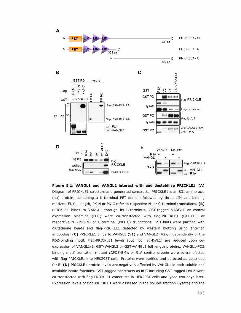
193
Figure 5.1: VANGL1 and VANGL2 interact with and destabilise PRICKLE1. (A)
Diagram of PRICKLE1 structure and generated constructs. PRICKLE1 is an 831 amino acid
(aa) protein, containing a N-terminal PET domain followed by three LIM zinc binding
motives. FL-full length, PK-N or PK-C refer to respective N- or C-terminal truncations. (B)
PRICKLE1 binds to VANGL1 through its C-terminus. GST-tagged VANGL1 or control
expression plasmids (FLII) were co-transfected with flag-PRICKLE1 (PK1-FL), or
respective N- (PK1-N) or C-terminal (PK1-C) truncations. GST-baits were purified with
glutathione beads and flag-PRICKLE1 detected by western blotting using anti-flag
antibodies. (C) PRICKLE1 binds to VANGL1 (V1) and VANGL2 (V2), independently of the
PDZ-binding motif. Flag-PRICKLE1 levels (but not flag-DVL1) are reduced upon co-
expression of VANGL1/2. GST-VANGL2 or GST-VANGL1 full length proteins, VANGL1-PDZ
binding motif truncation mutant (∆PDZ-BM), or R1A control protein were co-transfected
with flag-PRICKLE1 into HEK293T cells. Proteins were purified and detected as described
for B. (D) PRICKLE1 protein levels are negatively affected by VANGL1 in both soluble and
insoluble lysate fractions. GST-tagged constructs as in C including GST-tagged DVL2 were
co-transfected with flag-PRICKLE1 constructs in HEK293T cells and lysed two days later.
Expression levels of flag-PRICKLE1 were assessed in the soluble fraction (lysate) and the
194
guanidinium chloride resolubilised pellet (pellet fraction). Total levels of ectopic PRICKLE1
were detected with flag-antibodies. (E) Treatment with proteasome inhibitor MG132 does
not counteract VANGL1-induced PRICKLE1 degradation. HEK293T cells were double
transfected with GST-VANGL1 or control protein (R1A) and flag-PRICKLE1 as indicated.
42h after transfection, cells were either treated with the proteasomal inhibitor MG132 or
vehicle control (DMSO) at a concentration of 10 µM for 4.5 h. Cells were subsequently
lysed and analysed by western blot, probing with either flag- or GST-antibodies.
In order to confirm that PRICKLE1 protein levels are reduced and not associated
with insoluble structures during the generation of lysates, insoluble lysate
fractions were subjected to guanidinium chloride solubilisation and analysed by
western blot for PRICKLE1 levels. Indeed, the pellet fraction showed the same
relative reduced amounts of flag-PRICKLE1 in VANGL1 expressing cells, as
observed for the soluble fractions (Figure 5.1D, compare lysates versus
reconstituted lysates).
These results suggest that VANGL proteins interact strongly with PRICKLE1,
since even under conditions where expression levels were hardly detectable in
whole cell lysates, PRICKLE1 could be readily detected in flag-VANGL co-
immunoprecipitates. Furthermore, these observations also imply that VANGL
proteins may lead to the degradation of PRICKLE1 proteins.
To test this possibility, similar experiments were performed in the presence of
the proteasomal inhibitor MG132 which is known to interfere with the 26S
proteasome function and consequently would allow stabilisation of proteins
otherwise targeted for proteasomal degradation (Tawa et al. 1997). Treatment
of cells with 10µM MG132 did not prevent reduced levels of PRICKLE1 induced
upon VANGL1 expression (Figure 5.1E). Additional experiments employing higher
concentrations of MG132 or longer treatment periods also failed to yield
increased PRICKLE1 levels (data not shown). This suggests that VANGL1 may
decrease PRICKLE1 levels by a mechanism other than ubiquitin mediated
proteasome degradation.
195
5.2 R-RAS GTPases inhibit VANGL1 induced PRICKLE1/2
protein destabilisation
I next addressed if R-RAS interaction with VANGL1 is able to regulate VANGL1
binding to PRICKLE1 and/or regulate PRICKLE1 protein stability. Activated R-RAS
L87 does not alter interaction between VANGL1 and PRICKLE1 taking VANGL1
bait levels in consideration (Figure 5.2A), but significantly prevents the
destabilisation effect of PRICKLE1 protein exerted by VANGL1 (Figure 5.2B).
I also compared the effect of other R-RAS subgroup members on the
degradation of both PRICKLE1 and PRICKLE2. Expression of all three R-RAS
members can partially rescue the VANGL1 induced PRICKLE1/2 destabilisation in
a manner dependent on an intact effector domain (Figure 5.2C). This effect is
specific to R-RAS subgroup GTPases as the RAP2 member of the RAS family did
not display a stabilisation effect. Surprisingly, the ability to prevent PRICKLE1/2
destabilisation does not correlate with the binding affinity to VANGL1; TC21 and
M-RAS do not show as strong an interaction with VANGL1 as observed for R-RAS
(Figure 3.5) but can however prevent the VANGL1 induced PRICKLE1/2
destabilisation more efficiently than R-RAS (Figure 5.2C).

196
Figure 5.2: R-RAS subgroup GTPases are able to rescue VANGL1 induced
destabilisation of PRICKLE1 and PRICKLE2. (A) Activated R-RAS L87 does not
modulate VANGL1 and PRICKLE1 interaction. HEK293T cells were triple transfected with
GST-VANGL1 or control contstruct (R1A), GST-R-RAS active mutant L87 (R-RASL) or
respective defective mutant L87 64A (R-RASLA) and flag-PRICKLE1, as indicated. 48h
after transfection, cells were lysed and GST proteins precipitated using glutathione
sepharose beads. Interactions were analysed by western blot, probing with flag- or GST-
antibodies. (B) R-RAS L87 partially rescues VANGL1-induced PRICKLE1 decay. HEK293T
cells were triple transfected with GST-VANGL1 or control (R1A) contstructs, GST-R-RAS
active mutant L87 (R-RASL) and flag-PRICKLE1, as outlined. 48h after transfection, cells
were lysed and analysed by western blot, probing with flag- or GST-antibodies. (C) R-
RAS subgroup members TC21 and M-Ras (but not the closely related GTPase RAP2)
rescue VANGL1-induced PRICKLE1/2 destabilisation more efficiently than R-RAS.
HEK293T cells were triple transfected with GST-VANGL1 or R1A, flag-PRICKLE1 or flag-
PRICKLE2, and flag-RAP2 or flag-R-RAS subgroup GTPases using respective consitutive
active (V or L), effector domain mutant (VA or LA) or empty vector (E). 48h post
transfection, cells were subjected to lysis and analysed by western blot by probing with a
flag antibody. Blotting of endogenous SCRIB protein served as loading control. Employed
GTPase mutations are as followed: Rap2 V (V12), Rap2 VA (V12 A38), R-RAS L (L87), R-
RAS LA (L87 A64), TC21 L (L71), TC21 LA (L71 A48), M-Ras (L81), M-Ras LA (L81
A48). RFG – Ras Family GTPases.

197
5.3 VANGL1 localises to vesicle like structures upon
autophagy induction
Inhibition of proteasome-mediated degradation utilising the MG132 inhibitor
could not prevent the decrease in PRICKLE1/2 levels, observed upon VANGL1 co-
expression, suggesting alternative mechanisms of degradation.
Another mechanism for the degradation and recycling of cellular components is
lysosome-mediated autophagy. Autophagy is a highly conserved self-digestion
process to promote cell survival in response to nutrient starvation and other
metabolic stresses (He and Klionsky 2009). During the autophagic process,
portions of cytoplasm are first surrounded by a single-membrane structure and
these autophagic vacuoles or autophagosomes then fuse with pre-existing
lysosomes to form autolysosomes. Autophagic dysfunction is associated with
cancer, neurodegeneration, microbial infection and ageing (Kundu and
Thompson 2008).
Consistent with a role of autophagy in the PCP pathway, DVL2 has been reported
to be regulated through autophagy mediated degradation (Gao et al. 2010).
Therefore, experiments were conducted in order to test if VANGL1 expression
leads to PRICKLE1 protein destabilisation through the lysosome mediated
pathway.
Expression of YFP-VANGL1 or YFP-PRICKLE1 fusion proteins in HMEC cells allows
monitoring their respective subcellular localisation in presence or absence of
autophagy stimulants. In untreated cells, YFP-VANGL1 displays diverse
localisation patterns with staining of various subcellular compartments including
plasma membrane, vesicle-like structures and perinuclear rings (Figure 5.3A and
B, Figure 5.4D).
Notably, YFP-PRICKLE1 displays similar membrane (Figure 5.4A) and vesicle-like
structures (Figure 5.3C and D, Figure 5.4A-C), suggesting that VANGL1 and
PRICKLE1 localise to overlapping compartments such as vesicle-like structures,
indicative of potential autophagosomes.
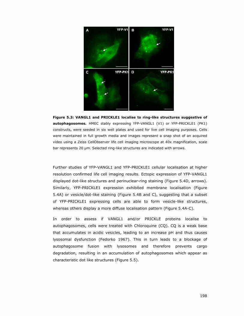
198
Figure 5.3: VANGL1 and PRICKLE1 localise to ring-like structures suggestive of
autophagosomes. HMEC stably expressing YFP-VANGL1 (V1) or YFP-PRICKLE1 (PK1)
constructs, were seeded in six well plates and used for live cell imaging purposes. Cells
were maintained in full growth media and images represent a snap shot of an acquired
video using a Zeiss CellObserver life cell imaging microscope at 40x magnification, scale
bar represents 20 µm. Selected ring-like structures are indicated with arrows.
Further studies of YFP-VANGL1 and YFP-PRICKLE1 cellular localisation at higher
resolution confirmed life cell imaging results. Ectopic expression of YFP-VANGL1
displayed dot-like structures and perinuclear-ring staining (Figure 5.4D, arrows).
Similarly, YFP-PRICKLE1 expression exhibited membrane localisation (Figure
5.4A) or vesicle/dot-like staining (Figure 5.4B and C), suggesting that a subset
of YFP-PRICKLE1 expressing cells are able to form vesicle-like structures,
whereas others display a more diffuse localisation pattern (Figure 5.4A-C).
In order to assess if VANGL1 and/or PRICKLE proteins localise to
autophagosomes, cells were treated with Chloroquine (CQ). CQ is a weak base
that accumulates in acidic vesicles, leading to an increase pH and thus causes
lysosomal dysfunction (Fedorko 1967). This in turn leads to a blockage of
autophagosome fusion with lysosomes and therefore prevents cargo
degradation, resulting in an accumulation of autophagosomes which appear as
characteristic dot like structures (Figure 5.5).
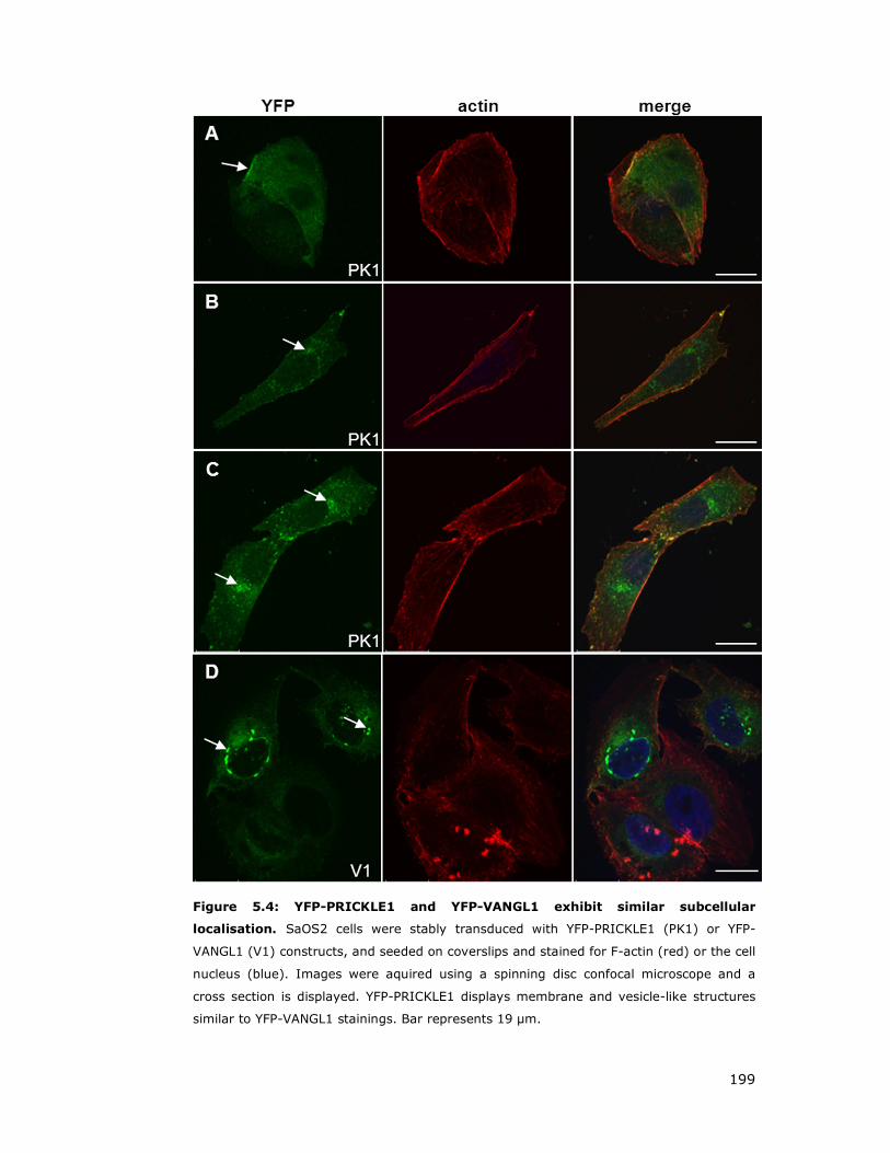
199
Figure 5.4: YFP-PRICKLE1 and YFP-VANGL1 exhibit similar subcellular
localisation. SaOS2 cells were stably transduced with YFP-PRICKLE1 (PK1) or YFP-
VANGL1 (V1) constructs, and seeded on coverslips and stained for F-actin (red) or the cell
nucleus (blue). Images were aquired using a spinning disc confocal microscope and a
cross section is displayed. YFP-PRICKLE1 displays membrane and vesicle-like structures
similar to YFP-VANGL1 stainings. Bar represents 19 µm.

200
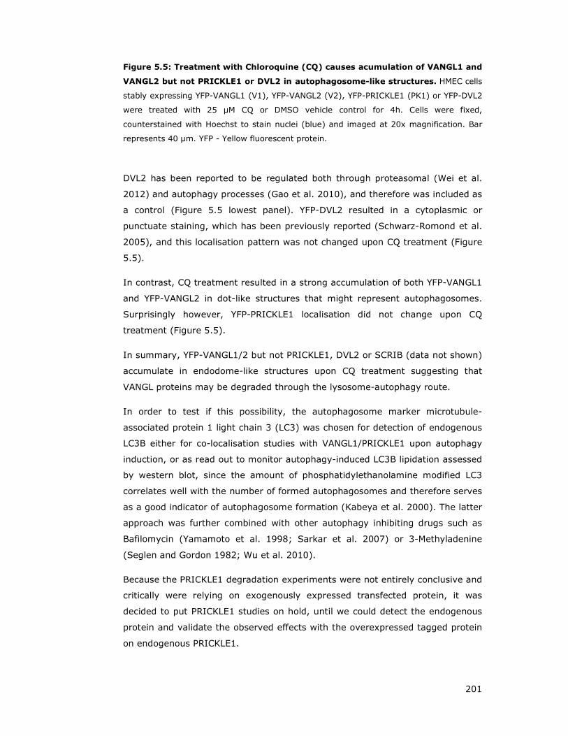
201
Figure 5.5: Treatment with Chloroquine (CQ) causes acumulation of VANGL1 and
VANGL2 but not PRICKLE1 or DVL2 in autophagosome-like structures. HMEC cells
stably expressing YFP-VANGL1 (V1), YFP-VANGL2 (V2), YFP-PRICKLE1 (PK1) or YFP-DVL2
were treated with 25 µM CQ or DMSO vehicle control for 4h. Cells were fixed,
counterstained with Hoechst to stain nuclei (blue) and imaged at 20x magnification. Bar
represents 40 µm. YFP - Yellow fluorescent protein.
DVL2 has been reported to be regulated both through proteasomal (Wei et al.
2012) and autophagy processes (Gao et al. 2010), and therefore was included as
a control (Figure 5.5 lowest panel). YFP-DVL2 resulted in a cytoplasmic or
punctuate staining, which has been previously reported (Schwarz-Romond et al.
2005), and this localisation pattern was not changed upon CQ treatment (Figure
5.5).
In contrast, CQ treatment resulted in a strong accumulation of both YFP-VANGL1
and YFP-VANGL2 in dot-like structures that might represent autophagosomes.
Surprisingly however, YFP-PRICKLE1 localisation did not change upon CQ
treatment (Figure 5.5).
In summary, YFP-VANGL1/2 but not PRICKLE1, DVL2 or SCRIB (data not shown)
accumulate in endodome-like structures upon CQ treatment suggesting that
VANGL proteins may be degraded through the lysosome-autophagy route.
In order to test if this possibility, the autophagosome marker microtubule-
associated protein 1 light chain 3 (LC3) was chosen for detection of endogenous
LC3B either for co-localisation studies with VANGL1/PRICKLE1 upon autophagy
induction, or as read out to monitor autophagy-induced LC3B lipidation assessed
by western blot, since the amount of phosphatidylethanolamine modified LC3
correlates well with the number of formed autophagosomes and therefore serves
as a good indicator of autophagosome formation (Kabeya et al. 2000). The latter
approach was further combined with other autophagy inhibiting drugs such as
Bafilomycin (Yamamoto et al. 1998; Sarkar et al. 2007) or 3-Methyladenine
(Seglen and Gordon 1982; Wu et al. 2010).
Because the PRICKLE1 degradation experiments were not entirely conclusive and
critically were relying on exogenously expressed transfected protein, it was
decided to put PRICKLE1 studies on hold, until we could detect the endogenous
protein and validate the observed effects with the overexpressed tagged protein
on endogenous PRICKLE1.

202
In order to generate an antibody against PRICKLE1, fragments of different
lengths of PRICKLE1 N- and C-terminal regions were cloned into bacterial
expression vectors. An N-terminal fragment, resulting in highest expression of
recombinant protein, was purified and shipped to Thermo Scientific for rabbit
immunisation. To our great disappointment, after arrival of the generated
antibody and several company-caused delays, the provided anti-sera failed to
recognise any specific protein bands on lysates and failed to immunoprecipitate
exogenous flag-PRICKLE1 (data not shown). This setback, in combination with
my focus on other areas of the project and time constrain issues meant that no
further work was carried out in this area. Future studies should help elucidate
the mechanism of VANGL-mediated degradation of PRICKLE1 as well as the
potential role of VANGL proteins in autophagy.
5.4 VANGL1 subcellular localisation studies
R-RAS GTPases have been reported to be membrane associated and to localise
to various subcellular localisations such as plasma membrane, Golgi, ER and
endosomes (Ohba et al. 2000; Takaya et al. 2007). VANGL1, being a tetraspanin
protein has been reported to be mostly plasma membrane associated (Kalabis et
al. 2006; Gravel et al. 2010; Anastas et al. 2011; Iliescu et al. 2011).
In order to study the intracellular localisation of VANGL1 and assess the
possibility that its localisation might be regulated by interaction with R-RAS
GTPases, ROR2 or WNT ligands, YFP fusion proteins of R-RAS GTPases and
VANGL proteins were ectopically expressed in multiple cell types and its
localisation studied in both live and fixed cells.
During the course of localisation studies using stably expression of YFP-fusion
proteins it became apparent that VANGL1 displays very diverse localisation in
different cell types (Figure 5.4D, Figure 5.6A-D and Figure 5.6).
In HMLE mammary epithelial cells, YFP-VANGL1 localises at the plasma
membrane, at areas of cell-cell contact in some cells (Figure 5.6A), whereas in
other cells it localised in vesicle/ring like structures as well as in an area
surrounding the nucleus giving a characteristic perinuclear staining (see arrows
in Figure 5.6 B,C and Figure 5.7A, F).

203
Figure 5.6: VANGL1 localises to various subcellular compartments, whereas R-
RAS or TC21 are mostly found membrane associated. HMLE cells were stably
transduced with lentivirus encoding YFP-VANGL1, YFP-VANGL2, YFP-R-RAS wt or YFP-
TC21 wt fusion proteins as indicated. YFP - yellow fluorescent protein, wt - wild type.
Pictures were aquired with a Zeiss fluorescent micoscope at 20x magnification amd the
scale bar represents 50 µm.
VANGL1 observed subcellular localisations varied depending by cell type, cell
density and also likely during phases of cell cycle as suggested by characteristic
mitotic localisations observed in live cell imaging studies (data not shown). Even
within a field of cells of the same cell type, different localisation patterns could
often be detected. These observations strongly suggest that VANGL1 can localise
to multiple membrane sub-compartments within the cell, and this localisation
seems to be dynamically regulated.
In contrast, expression of YFP-VANGL2 in various cell lines exhibited a staining
pattern (Figure 5.6 and Figure 5.7), which is clearly different from VANGL1,
highlighting potential functional differences.
YFP-VANGL2 expression resulted in ER-type staining with little plasma
membrane localisation in osteosarcoma (SaOS2) and thyroid-carcinoma cells
(8505C) (Figure 5.7). Notably, breast cancer cells MDA-MB-231 displayed
additionally vesicle-like structures, which seem to be similar for YFP-VANGL1 and
YFP-VANGL2 expression (Figure 5.7H and I, respectively).

204
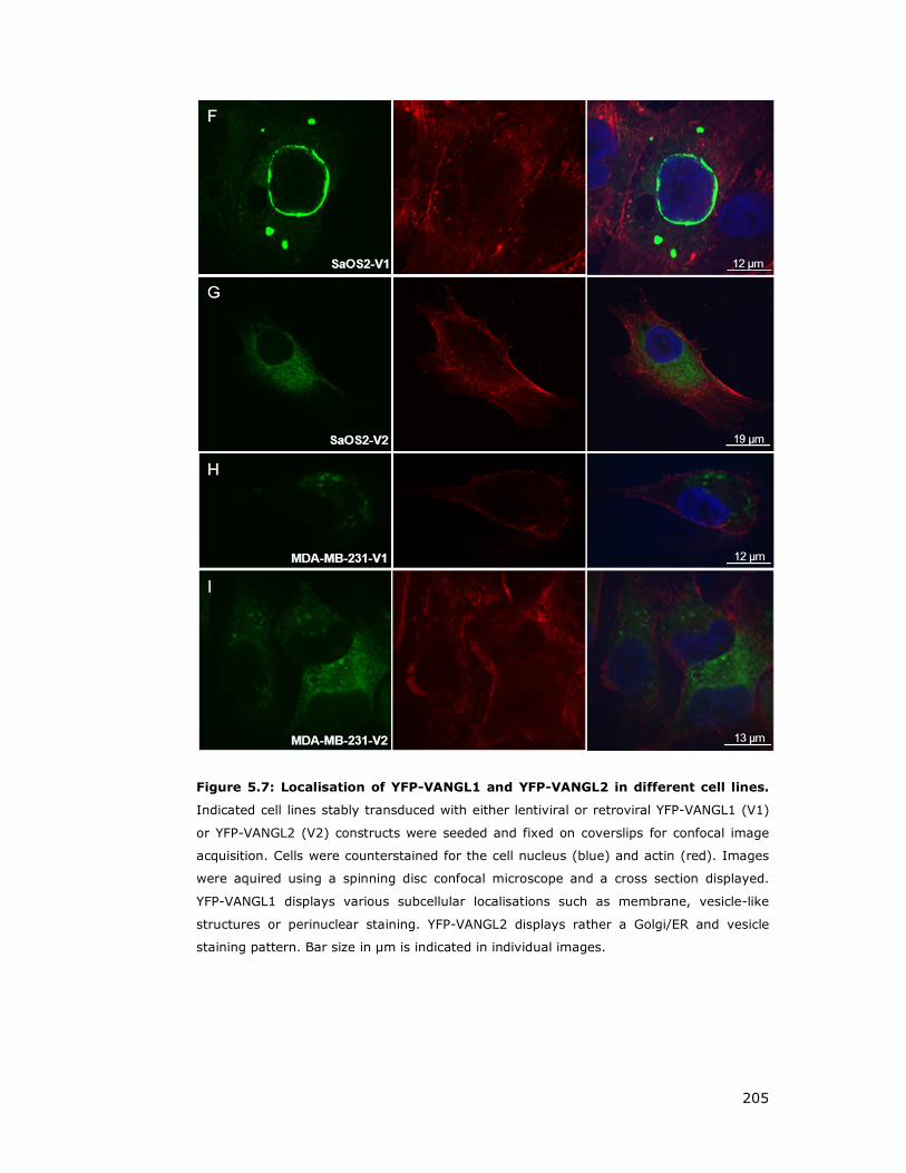
205
Figure 5.7: Localisation of YFP-VANGL1 and YFP-VANGL2 in different cell lines.
Indicated cell lines stably transduced with either lentiviral or retroviral YFP-VANGL1 (V1)
or YFP-VANGL2 (V2) constructs were seeded and fixed on coverslips for confocal image
acquisition. Cells were counterstained for the cell nucleus (blue) and actin (red). Images
were aquired using a spinning disc confocal microscope and a cross section displayed.
YFP-VANGL1 displays various subcellular localisations such as membrane, vesicle-like
structures or perinuclear staining. YFP-VANGL2 displays rather a Golgi/ER and vesicle
staining pattern. Bar size in µm is indicated in individual images.

206
Initial localisation studies showed that YFP-R-RAS and TC21 are predominantly
localised at the plasma membrane (Figure 5.6E-G). Therefore, CFP-R-RAS or
CFP-TC21 constructs were generated in order to co-express them with YFP-
VANGL1-R-RAS for co-localisation studies. However, because of technical issues,
I was not able to get these experiments to work.
I also performed live cell imaging studies on YFP-VANGL1 cells to assess the
dynamic localisation of the protein in different contexts (e.g. growing cells, cells
stimulated with WNT ligands and cells induced to polarise in scratch/ wound
closure assays). Mainly due to technical issues associated with lack of sensitivity
I was unable to observe any changes on VANGL1 localisation in these studies. In
order to increase sensitivity, time course treatments of WNT5a stimulation were
performed in YFP-VANGL1 cells and fixed cells analysed by confocal microscopy.
However, no effects were observed in preliminary experiments and studies on
the regulation of VANGL1 localisation were stopped.
207
5.5 Discussion:
PRICKLE1 is involved in PCP signalling and has been demonstrated to be a key
intermediate for gastrulation cell movements in Drosophila (Gubb et al. 1999)
Xenopus (Takeuchi et al. 2003) and zebrafish (Carreira-Barbosa et al. 2003;
Takeuchi et al. 2003; Veeman et al. 2003b). Furthermore, mouse knock out
models of Prickle1 resulted in embryonic lethality (E5.5-6.5), whereas down
regulation causes severe morphogenetic defects in CE. Prickle2 phenotypes
appear less severe, while a combination of Prickle1 and Prickle2 loss
demonstrates a synergetic effect on CE defects (Veeman et al. 2003b).
In this chapter, I presented evidence that VANGL proteins strongly interact with
PRICKLE1/2. Surprisingly, overexpression of VANGL1/2 leads to a strong
reduction of PRICKLE1/2 protein levels, which can be partially rescued by active
R-RAS subgroup GTPases without affecting VANGL1-PRICKLE1 interaction.
It has been reported that that PRICKLE1 protein levels can be targeted for
proteasomal degradation by SMURF1/2 ubiquitin ligases (Narimatsu et al. 2009).
SMURF ubiquitin ligases regulate signalling, cell polarity and motility; and mice
mutants for Smurf1 and Smurf2 display PCP defects in the cochlea and CE
defects that include a failure to close the neural tube (Narimatsu et al. 2009).
According to the proposed model, WNT5a stimulation leads to activation of DVL2
by phosphorylation and subsequent recruitment of SMURF1/2, which then
targets PRICKLE1 for ubiquitin-mediated degradation (Narimatsu et al. 2009).
Besides proteasome mediated regulation of WNT signalling, autophagy regulation
has been implicated to negatively impact WNT signalling through DVL2
degradation (Gao et al. 2010). Another link between PCP and autophagy stems
from the protein activating molecule Beclin-regulated autophagy (AMBRA), which
upon functional inactivation, displays impaired autophagy (Fimia et al. 2007). In
addition, homozygosity of Ambra1 mutation causes embryonic lethality and
embryos exhibit PCP typical neural tube defects or spina bifida characteristics
(Cecconi et al. 2007; Fimia et al. 2007).
In endothelial cells, PCP signalling is required for cell growth, polarity, and
migration (Masckauchan et al. 2006; Cheng et al. 2008). Silencing of either
DVL2 or PRICKLE1 suppressed endothelial cell proliferation, highlighting the
function of a DVL2-PRICKLE complex (Cirone et al. 2008). However, it also has
been reported that PRICKLE1 acts as negative regulator of DVL/Dsh (Carreira-
Barbosa et al. 2003; Veeman et al. 2003b; Fujimura et al. 2009) and facilitates a

208
proteasome mediated degradation of DVL3 through its two destruction box
motifs, essential for E3 ligase activity (Chan et al. 2006).
Knockdown of PRICKLE1 causes reduced axonal and dendritic extension in
hippocampal neurons, thus implicating a function for PRICKLE in axonal-dendritic
development (Liu et al. 2013). Notably, studies in C.elegans, which contains only
one Vangl isoform, demonstrated that Vangl co-operates with Prickle1 to
negatively regulate neurite formation (Sanchez-Alvarez et al. 2011).
Misshapen-like kinase (MINK1), a Ste20 group kinase, has been described to be
important for clustering of VANGL1 within PRICKLE1 plasma membrane puncta.
Further, MINK induced phosphorylation of PRICKLE1 at a conserved threonine
residue (T370) regulates RAB5 mediated endosomal trafficking of PRICKLE1,
which is important for localised plasma membrane accumulation and function
(Daulat et al. 2012). Further, PRICKLE1 has been reported to be a farnesylation
target, implying that this post translational modification can target cytoplasmic
PRICKLE1 to (plasma) membrane subcellular localisations (Maurer-Stroh et al.
2007).
A role for VANGL in regulating PRICKLE levels is in agreement with localisation
studies of VANGL/stbm and Prickle in Drosophila pupal wings, illustrating that in
the absence of VANGL/stbm, Prickle protein levels were found to be increased
within the cytosol and at the apicolateral cell cortex (Bastock et al. 2003; Jenny
et al. 2003).
PET/LIM domains have been reported to be important for PRICKLE function in
zebrafish gastrulation (Takeuchi et al. 2003). Interestingly, VANGL1 binds to the
C-terminal part of PRICKLE1, lacking both PET and LIM domains, but still harbour
both destruction box motifs implicated in DVL degradation (Chan et al. 2006).
Asymmetrical localisation of VANGL and PRICKLE1 is a hallmark of PCP in
Drosophila and this interaction seems to be conserved from fly to humans (Jenny
et al. 2003; Sanchez-Alvarez et al. 2011; Daulat et al. 2012). VANGL induced
PRICKLE degradation could therefore be a mechanism to ensure the proteins are
asymmetrically localised. Interestingly, PRICKLE1 has been reported to stimulate
proteasomal degradation of DVL, another VANGL1 interacting protein (Chan et
al. 2006). Therefore a complex regulatory interplay emerges between VANGL1,
PRICKLE and DVL at the level of protein degradation where VANGL1, by
regulating PRICKLE stability, may indirectly control DVL levels. Critically, R-RAS
GTPases are able to modulate this regulatory interplay, potentially by recruiting

209
VANGL1 to specialised membrane domains and away from the pool of PRICKLE
proteins.
The mechanism of VANGL1/2 induced PRICKLE1/2 degradation remains to be
determined as preliminary experiments suggested that this degradation is not
proteasome mediated. Furthermore, localisation studies suggested a potential
role for VANGL proteins in autophagy. Treatment with chloroquine resulted in a
dramatic accumulation of YFP-VANGL1 and YFP-VANGL2 in autophagosome-like
structures. However, due to technical and time constraining issues, I was not
able expand on these studies. Future experiments need to be performed in order
to elucidate the mechanism of VANGL-mediated degradation of PRICKLE1/2 as
well as the potential role of VANGL proteins in autophagy.
Figure 5.8: Chapter summary. VANGL1 interacts with PRICKLE1 and PRICKLE2 through
its C-terminus. However, exogenous expression of VANGL1 results in PRICKLE protein
destabilisation through a not fully understood mechanism, whereas other PCP core
components such as DVL are not affected. Expression of activated R-RAS subgroup
members like R-RAS, TC21 and M-RAS are able, at least partially, to rescue this effect.
210
CHAPTER 6
Effect of WNT ligands and
Frizzled proteins on R-
RAS and RHO activation

211
6 Effect of WNT ligands and Frizzled receptors on R-
RAS and RHO activation
Our working model aims to test if R-RAS GTPases act downstream of WNT
ligands and Frizzled receptors, resulting in modulation of VANGL1 function. In
order to assess R-RAS activity, RAS binding domain (RBD) assays were
optimised and performed with the intention to assess R-RAS activity upon WNT
stimulation. The receptor tyrosine kinase ROR2 was an attractive candidate to
mediate R-RAS activation due to its WNT5a binding and signalling capabilities
(Oishi et al. 2003; Mikels and Nusse 2006a; Nishita et al. 2010b), and the
potential link through the common interactor filaminA (Nishita et al. 2006;
Nomachi et al. 2008; Gawecka et al. 2010; Griffiths et al. 2011). Furthermore,
VANGL2 has been demonstrated to be phosphorylated by ROR2 in a WNT5a
dependent manner (Gao et al. 2011). And lastly, due to our observations, which
detected the R-RAS member TC21 in ROR2 immunoprecipitations (Figure 4.11),
indicating that ROR2, VANGL2 and R-RAS GTPase TC21 could form a complex.
Therefore, WNT ligands will be tested in ROR2 contexts for R-RAS GTPase
member activation.
6.1 Establishment of RAS binding domain assays
In order to monitor and assess R-RAS activation levels upon WNT stimulation in
cell lysates, glutathione S-transferase (GST)-fused RAS binding domains (RBDs)
from downstream effectors such as RAF1 and RALGDS were employed (Brtva et
al. 1995; de Rooij and Bos 1997; Franke et al. 1997; Castro et al. 2005;
Rodriguez-Viciana and McCormick 2006). These effector domains preferentially
interact with GTP-bound RAS and act as activation specific probes, since affinity
for GDP is three orders of magnitude lower (Herrmann et al. 1995). Thus,
glutathione beads enable the selective purification from lysates and subsequent
quantification of active RAS proteins by immunoblotting.
Several reports suggest that R-RAS subgroup GTPases have considerably lower
affinity in their ability to interact with RAF1 compared to classical RAS proteins,
whereas they interact as strongly as classical RAS proteins with RALGEFs (Figure
3.6) (Rodriguez-Viciana et al. 1994; Huff et al. 1997; Marte et al. 1997).
However, these experiments assessed the interaction with the full length protein
and it is known that the interaction with the isolated RBD may not accurately

212
reflect the affinity of the interaction with the full length protein (Rodriguez-
Viciana and McCormick 2006).
In order to determine which RBD was more effective in capturing activated R-
RAS GTPases, GST fusion proteins immobilised on glutathione beads of either the
RAF1 RBD (residues 2–140) or RALGDS RBD (C-terminal 97 residues) were
incubated with equal amounts of the same lysate of cells transfected with tagged
GTPases. In order to evaluate the specificity of the interaction, activating and
effector domain mutants were compared to the respective wild type protein
(Figure 6.1A). After incubation, precipitated amounts of flag-tagged GTPases
were detected by immunoblotting using flag antibodies.
Both GST-RBD beads exhibited a strong increase in precipitated protein if the
constitutive active mutant was expressed, compared to wild type protein.
Moreover, GTPase effector domain mutants (A38 or equivalent) did not show any
significant interaction, demonstrating that the interaction is specific and
activation and effector domain dependent. Detection of lower levels of wild type
RAS GTPases can be explained by the fact that under conditions of
overexpression a significant amount of wild type protein may be GTP-bound
(Figure 6.1A).
Overall, both GST-RBDs performed similarly in their ability to selectively
precipitate active tagged N-RAS, and the three R-RAS family members R-RAS,
TC21 and M-RAS, although the RAF1 RBD beads displayed more sensitivity and
were therefore used in all subsequent studies (Figure 6.1A).
In order to measure the ability of our RAF1 beads to detect activation of
endogenous GTPases, HEK293T cells were stimulated with EGF, a strong
stimulator of classical RAS proteins and the MAPK pathway (Figure 6.1B) (Buday
and Downward 1993; Zheng et al. 1994).
Over a time course ranging from 0 to 20 min, samples were taken, incubated
with GST-RAF1 RBD protein and precipitated using glutathione sepharose beads.
Immunoblotting for H-RAS illustrated a characteristic activation pattern, which
peaked around 2 min by over 20 fold compared to basal levels and started to
decline within 5 min after stimulation and dropped to 8-fold over basal level
within 20 min after stimulation (Figure 6.1B and C). This illustrates that
detection of endogenous levels of RAS activation with our RBD beads is possible
and specific.
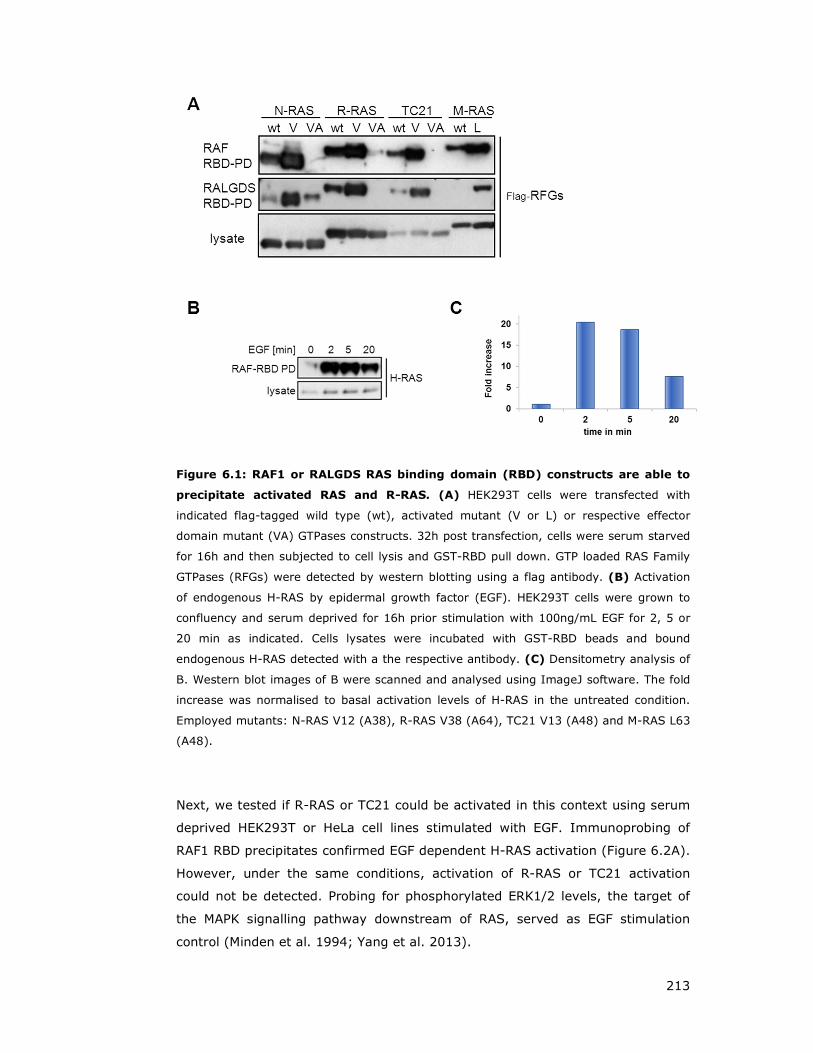
213
Figure 6.1: RAF1 or RALGDS RAS binding domain (RBD) constructs are able to
precipitate activated RAS and R-RAS. (A) HEK293T cells were transfected with
indicated flag-tagged wild type (wt), activated mutant (V or L) or respective effector
domain mutant (VA) GTPases constructs. 32h post transfection, cells were serum starved
for 16h and then subjected to cell lysis and GST-RBD pull down. GTP loaded RAS Family
GTPases (RFGs) were detected by western blotting using a flag antibody. (B) Activation
of endogenous H-RAS by epidermal growth factor (EGF). HEK293T cells were grown to
confluency and serum deprived for 16h prior stimulation with 100ng/mL EGF for 2, 5 or
20 min as indicated. Cells lysates were incubated with GST-RBD beads and bound
endogenous H-RAS detected with a the respective antibody. (C) Densitometry analysis of
B. Western blot images of B were scanned and analysed using ImageJ software. The fold
increase was normalised to basal activation levels of H-RAS in the untreated condition.
Employed mutants: N-RAS V12 (A38), R-RAS V38 (A64), TC21 V13 (A48) and M-RAS L63
(A48).
Next, we tested if R-RAS or TC21 could be activated in this context using serum
deprived HEK293T or HeLa cell lines stimulated with EGF. Immunoprobing of
RAF1 RBD precipitates confirmed EGF dependent H-RAS activation (Figure 6.2A).
However, under the same conditions, activation of R-RAS or TC21 activation
could not be detected. Probing for phosphorylated ERK1/2 levels, the target of
the MAPK signalling pathway downstream of RAS, served as EGF stimulation
control (Minden et al. 1994; Yang et al. 2013).
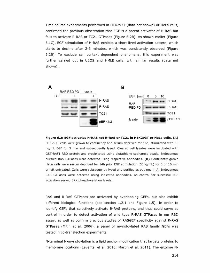
214
Time course experiments performed in HEK293T (data not shown) or HeLa cells,
confirmed the previous observation that EGF is a potent activator of H-RAS but
fails to activate R-RAS or TC21 GTPases (Figure 6.2B). As shown earlier (Figure
6.1C), EGF stimulation of H-RAS exhibits a short lived activation pattern, which
starts to decline after 2-3 minutes, which was consistently observed (Figure
6.2B). To exclude cell context dependent phenomena, this experiment was
further carried out in U2OS and HMLE cells, with similar results (data not
shown).
Figure 6.2: EGF activates H-RAS not R-RAS or TC21 in HEK293T or HeLa cells. (A)
HEK293T cells were grown to confluency and serum deprived for 16h, stimulated with 50
ng/mL EGF for 5 min and subsequently lysed. Cleared cell lysates were incubated with
GST-RAF1 RBD protein and precipitated using glutathione sepharose beads. Endogenous
purified RAS GTPases were detected using respective antibodies. (B) Confluently grown
HeLa cells were serum deprived for 14h prior EGF stimulation (50ng/mL) for 3 or 10 min
or left untreated. Cells were subsequently lysed and purified as outlined in A. Endogenous
RAS GTPases were detected using indicated antibodies. As control for sucessful EGF
activation served ERK phosphorylation levels.
RAS and R-RAS GTPases are activated by overlapping GEFs, but also exhibit
different biological functions (see section 1.2.1 and Figure 1.5). In order to
identify GEFs that selectively activate R-RAS proteins, and thus could serve as
control in order to detect activation of wild type R-RAS GTPases in our RBD
assay, as well as confirm previous studies of RASGEF specificity against R-RAS
GTPases (Mitin et al. 2006), a panel of myristoylated RAS family GEFs was
tested in co-transfection experiments.
N-terminal N-myristoylation is a lipid anchor modification that targets proteins to
membrane locations (Levental et al. 2010; Martin et al. 2011). The enzyme N-
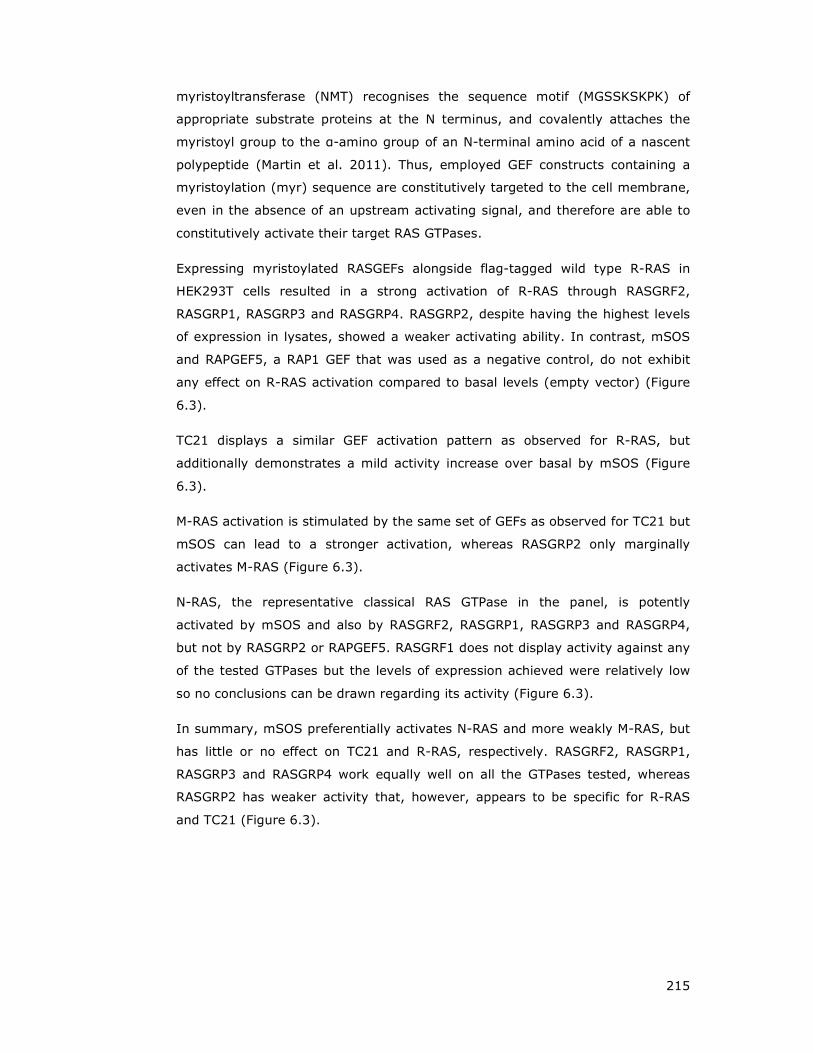
215
myristoyltransferase (NMT) recognises the sequence motif (MGSSKSKPK) of
appropriate substrate proteins at the N terminus, and covalently attaches the
myristoyl group to the α-amino group of an N-terminal amino acid of a nascent
polypeptide (Martin et al. 2011). Thus, employed GEF constructs containing a
myristoylation (myr) sequence are constitutively targeted to the cell membrane,
even in the absence of an upstream activating signal, and therefore are able to
constitutively activate their target RAS GTPases.
Expressing myristoylated RASGEFs alongside flag-tagged wild type R-RAS in
HEK293T cells resulted in a strong activation of R-RAS through RASGRF2,
RASGRP1, RASGRP3 and RASGRP4. RASGRP2, despite having the highest levels
of expression in lysates, showed a weaker activating ability. In contrast, mSOS
and RAPGEF5, a RAP1 GEF that was used as a negative control, do not exhibit
any effect on R-RAS activation compared to basal levels (empty vector) (Figure
6.3).
TC21 displays a similar GEF activation pattern as observed for R-RAS, but
additionally demonstrates a mild activity increase over basal by mSOS (Figure
6.3).
M-RAS activation is stimulated by the same set of GEFs as observed for TC21 but
mSOS can lead to a stronger activation, whereas RASGRP2 only marginally
activates M-RAS (Figure 6.3).
N-RAS, the representative classical RAS GTPase in the panel, is potently
activated by mSOS and also by RASGRF2, RASGRP1, RASGRP3 and RASGRP4,
but not by RASGRP2 or RAPGEF5. RASGRF1 does not display activity against any
of the tested GTPases but the levels of expression achieved were relatively low
so no conclusions can be drawn regarding its activity (Figure 6.3).
In summary, mSOS preferentially activates N-RAS and more weakly M-RAS, but
has little or no effect on TC21 and R-RAS, respectively. RASGRF2, RASGRP1,
RASGRP3 and RASGRP4 work equally well on all the GTPases tested, whereas
RASGRP2 has weaker activity that, however, appears to be specific for R-RAS
and TC21 (Figure 6.3).

216
Figure 6.3: Characterisation of RAS family GEF activity towards RAS and R-RAS
GTPases. (A) HEK293T cells were transfected with various myristoylated (myr) guanine
exchange factors (GEF) or empty vector alongside with flag-tagged R-RAS, TC21, M-RAS
or N-RAS. 32h post transfection, cells were serum deprived for 16 h and subsequently
lysed. Cell lysates were incubated with GST-RAF1 RBD fusion proteins immobilised on
glutathione beads and RAS family GTPases were detected by immunoblotting with a flag
antibody. (B) Summary of myr-GEF stimulating activity towards individual GTPases. R -
R-RAS, T – TC21, M – M-RAS, N – N-RAS. RasGRF - Ras-specific guanine nucleotide-
releasing factor, RasGRP - RAS guanyl nucleotide-releasing protein, mSOS – mouse Son
of Sevenless.
6.2 WNT ligands activate R-RAS
In order to test if WNT ligands are able to stimulate R-RAS GTPase activity,
various cell types were treated with recombinant WNT5a, and lysates subjected
to the GST-RAF1 RBD pull down assay approach.
There are 19 WNT genes in the human genome but my studies will focus on
those that have been previously reported to activate the PCP pathway and/or
affect cell motility, such as WNT5a and WNT7a, (Heisenberg et al. 2000; Veeman
et al. 2003a; Ulrich et al. 2005; Kikuchi and Yamamoto 2008).
WNT5a is known to activate PCP signalling through VANGL2 and to regulate cell
migration processes (Qian et al. 2007; Gao et al. 2011). In addition, WNT5a is
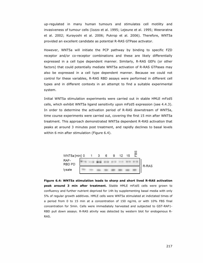
217
up-regulated in many human tumours and stimulates cell motility and
invasiveness of tumour cells (Iozzo et al. 1995; Lejeune et al. 1995; Weeraratna
et al. 2002; Kurayoshi et al. 2006; Pukrop et al. 2006). Therefore, WNT5a
provided an excellent candidate as potential R-RAS GTPase activator.
However, WNT5a will initiate the PCP pathway by binding to specific FZD
receptor and/or co-receptor combinations and these are likely differentially
expressed in a cell type dependent manner. Similarly, R-RAS GEFs (or other
factors) that could potentially mediate WNT5a activation of R-RAS GTPases may
also be expressed in a cell type dependent manner. Because we could not
control for these variables, R-RAS RBD assays were performed in different cell
types and in different contexts in an attempt to find a suitable experimental
system.
Initial WNT5a stimulation experiments were carried out in stable HMLE mFzd5
cells, which exhibit WNT5a ligand sensitivity upon mFzd5 expression (see 4.4.3).
In order to determine the activation period of R-RAS downstream of WNT5a,
time course experiments were carried out, covering the first 15 min after WNT5a
treatment. This approach demonstrated WNT5a dependent R-RAS activation that
peaks at around 3 minutes post treatment, and rapidly declines to basal levels
within 6 min after stimulation (Figure 6.4).
Figure 6.4: WNT5a stimulation leads to sharp and short lived R-RAS activation
peak around 3 min after treatment. Stable HMLE mFzd5 cells were grown to
confluency and further nutrient deprived for 14h by supplementing basal media with only
5% of regular growth additives. HMLE cells were WNT5a stimulated at indictated times of
a period from 0 to 15 min at a concentration of 150 ng/mL or with 10% FBS final
concentration for 5min. Cells were immediately harvested and subjected to GST-RAF1-
RBD pull down assays. R-RAS ativity was detected by western blot for endogenous R-
RAS.
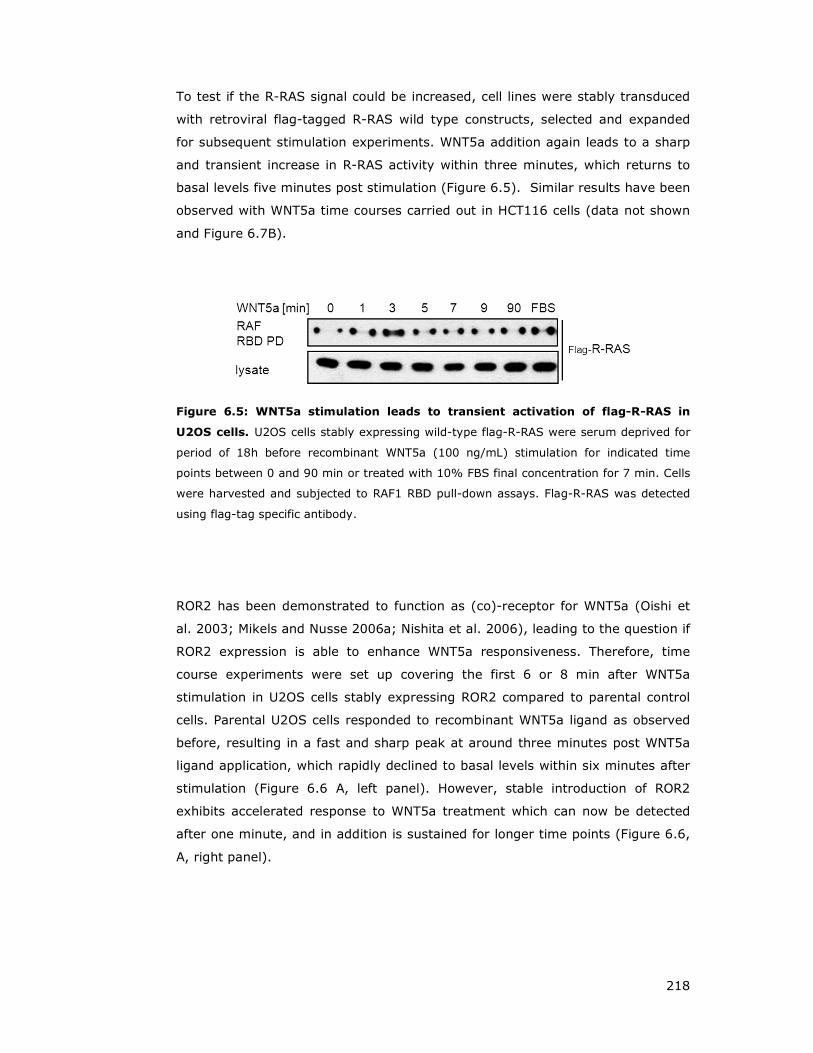
218
To test if the R-RAS signal could be increased, cell lines were stably transduced
with retroviral flag-tagged R-RAS wild type constructs, selected and expanded
for subsequent stimulation experiments. WNT5a addition again leads to a sharp
and transient increase in R-RAS activity within three minutes, which returns to
basal levels five minutes post stimulation (Figure 6.5). Similar results have been
observed with WNT5a time courses carried out in HCT116 cells (data not shown
and Figure 6.7B).
Figure 6.5: WNT5a stimulation leads to transient activation of flag-R-RAS in
U2OS cells. U2OS cells stably expressing wild-type flag-R-RAS were serum deprived for
period of 18h before recombinant WNT5a (100 ng/mL) stimulation for indicated time
points between 0 and 90 min or treated with 10% FBS final concentration for 7 min. Cells
were harvested and subjected to RAF1 RBD pull-down assays. Flag-R-RAS was detected
using flag-tag specific antibody.
ROR2 has been demonstrated to function as (co)-receptor for WNT5a (Oishi et
al. 2003; Mikels and Nusse 2006a; Nishita et al. 2006), leading to the question if
ROR2 expression is able to enhance WNT5a responsiveness. Therefore, time
course experiments were set up covering the first 6 or 8 min after WNT5a
stimulation in U2OS cells stably expressing ROR2 compared to parental control
cells. Parental U2OS cells responded to recombinant WNT5a ligand as observed
before, resulting in a fast and sharp peak at around three minutes post WNT5a
ligand application, which rapidly declined to basal levels within six minutes after
stimulation (Figure 6.6 A, left panel). However, stable introduction of ROR2
exhibits accelerated response to WNT5a treatment which can now be detected
after one minute, and in addition is sustained for longer time points (Figure 6.6,
A, right panel).
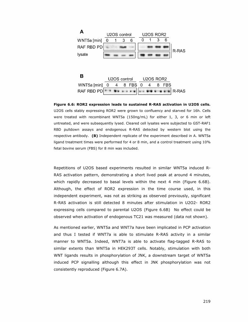
219
Figure 6.6: ROR2 expression leads to sustained R-RAS activation in U2OS cells.
U2OS cells stably expressing ROR2 were grown to confluency and starved for 16h. Cells
were treated with recombinant WNT5a (150ng/mL) for either 1, 3, or 6 min or left
untreated, and were subsequently lysed. Cleared cell lysates were subjected to GST-RAF1
RBD pulldown assays and endogenous R-RAS detected by western blot using the
respective antibody. (B) Independent replicate of the experiment described in A. WNT5a
ligand treatment times were performed for 4 or 8 min, and a control treatment using 10%
fetal bovine serum (FBS) for 8 min was included.
Repetitions of U2OS based experiments resulted in similar WNT5a induced R-
RAS activation pattern, demonstrating a short lived peak at around 4 minutes,
which rapidly decreased to basal levels within the next 4 min (Figure 6.6B).
Although, the effect of ROR2 expression in the time course used, in this
independent experiment, was not as striking as observed previously, significant
R-RAS activation is still detected 8 minutes after stimulation in U2O2- ROR2
expressing cells compared to parental U2OS (Figure 6.6B) No effect could be
observed when activation of endogenous TC21 was measured (data not shown).
As mentioned earlier, WNT5a and WNT7a have been implicated in PCP activation
and thus I tested if WNT7a is able to stimulate R-RAS activity in a similar
manner to WNT5a. Indeed, WNT7a is able to activate flag-tagged R-RAS to
similar extents than WNT5a in HEK293T cells. Notably, stimulation with both
WNT ligands results in phosphorylation of JNK, a downstream target of WNT5a
induced PCP signalling although this effect in JNK phosphorylation was not
consistently reproduced (Figure 6.7A).
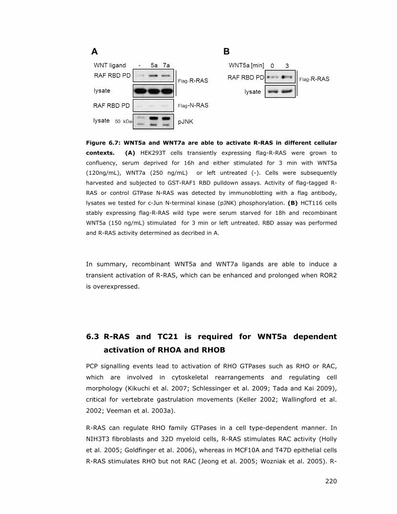
220
Figure 6.7: WNT5a and WNT7a are able to activate R-RAS in different cellular
contexts. (A) HEK293T cells transiently expressing flag-R-RAS were grown to
confluency, serum deprived for 16h and either stimulated for 3 min with WNT5a
(120ng/mL), WNT7a (250 ng/mL) or left untreated (-). Cells were subsequently
harvested and subjected to GST-RAF1 RBD pulldown assays. Activity of flag-tagged R-
RAS or control GTPase N-RAS was detected by immunoblotting with a flag antibody,
lysates we tested for c-Jun N-terminal kinase (pJNK) phosphorylation. (B) HCT116 cells
stably expressing flag-R-RAS wild type were serum starved for 18h and recombinant
WNT5a (150 ng/mL) stimulated for 3 min or left untreated. RBD assay was performed
and R-RAS activity determined as decribed in A.
In summary, recombinant WNT5a and WNT7a ligands are able to induce a
transient activation of R-RAS, which can be enhanced and prolonged when ROR2
is overexpressed.
6.3 R-RAS and TC21 is required for WNT5a dependent
activation of RHOA and RHOB
PCP signalling events lead to activation of RHO GTPases such as RHO or RAC,
which are involved in cytoskeletal rearrangements and regulating cell
morphology (Kikuchi et al. 2007; Schlessinger et al. 2009; Tada and Kai 2009),
critical for vertebrate gastrulation movements (Keller 2002; Wallingford et al.
2002; Veeman et al. 2003a).
R-RAS can regulate RHO family GTPases in a cell type-dependent manner. In
NIH3T3 fibroblasts and 32D myeloid cells, R-RAS stimulates RAC activity (Holly
et al. 2005; Goldfinger et al. 2006), whereas in MCF10A and T47D epithelial cells
R-RAS stimulates RHO but not RAC (Jeong et al. 2005; Wozniak et al. 2005). R-

221
RAS stimulation of RAC may be mediated by PI3Ks, as R-RAS GTPases are
strong activators of PI3K and several RACGEFs are activated in a PI3K-
dependent manner (Rodriguez-Viciana et al. 2004; Hawkins et al. 2006). On the
other hand, how R-RAS activates RHO is not known.
To assess RHO activation, pull-down assays using GST-fusion proteins using the
RHO-binding domain of Rhotekin that specifically bind RHO family members were
employed (Habas and He 2006; Mezzacappa et al. 2012). This activation-specific
probe functions analogous to RAS RBD constructs and was used to test the
possibility that R-RAS GTPases regulate the activation of RHO GTPases
downstream of WNT ligand stimulation. GST-Rhotekin RBD functionality and
specificity had already been tested in earlier experiments as part of the studies
on ARHGEF17/p164RhoGEF (Figure 3.14).
As mentioned above R-RAS has been reported to stimulate RHO in a cell type
dependent manner. This could be due to the differential expression of different
RHOGEFs in different cell types, only some of which may be regulated by R-RAS
and/or the WNT/PCP pathway. Because the RHOGEFs used by R-RAS or the
WNT/PCP pathway are not known, several cell types were screened for RHO
activation in different contexts.
Initially, I tested the panel of available Frizzled receptors in the absence or
presence of ectopically expressed ROR2 for their ability to activate RHOA in co-
transfection experiments in HEK293T cells. ROR2 expression by itself in HEK293T
is a strong RHO activator, achieving similar levels of active RHOA as FBS
stimulation (for 15 min) which was used as a positive control (Figure 6.8).
Overexpression of FZD2, FZD4, mFzd5 and FZD7 also stimulated RHOA
activation compared to empty vector control with FZD4 exhibiting the largest
effect. Interestingly, co-expression of ROR2 with FZD2 (but not other FZDs)
leads to a synergistic activation of RHOA. Treatment of mFzd5/ROR2 expressing
cells with recombinant WNT5a ligand only had a marginal effect compared to
unstimulated mFzd5/ROR2 samples (Figure 6.8).
In summary, ROR2 and Frizzled proteins can elevate RHOA activity to different
degrees and in some cases can cooperate to stimulate RHO activity, highlighting
a key role for ROR2 in the regulation of the RHO pathway.

222
Figure 6.8: ROR2 and Frizzled receptors stimulate RHOA activation. HEK293T cells
were transiently transfected with FZD2, FZD4, mFzd5 or FZD7 in combination with empty
vector (-) or ROR2. 36 hour later, cells were serum starved for 16h and harvested. Cell
lysates were incubated with GST-Rhotekin RBD beads and associated GTP-loaded
endogenous RHOA detected using a RHOA antibody. mFzd5 was transfected in duplicate
(with or without ROR2), and one set was stimulated with 100 ng/mL recombinant WNT5a
for 10 min prior to lysis, indicated with an asterisk (*).
Next, employing the synergistic FZD2/ROR2 combination, I wanted to test
whether WNT5a or WNT7a ligands could further enhance RHOA signalling in this
context (Figure 6.9).
As observed before (Figure 6.8) ROR2 expression by itself results in enhanced
RHOA precipitation, which is not further enhanced by WNT5a or WNT7a
stimulation. Expression of FZD2 also elevates RHOA activity, which is further
increased when ROR2 is co-expressed. But WNT5a or WNT7a treatment was not
able to further enhance RHOA activation in this experiment (Figure 6.9A).
However, there appears to be some variability within experiments with regards
to the effect of WNT addition, as in an independent experiment using the same
cell line and conditions, both WNT5a and WNT7a were able increase activation of
RHOA in ROR2 expressing cells (Figure 6.9B).
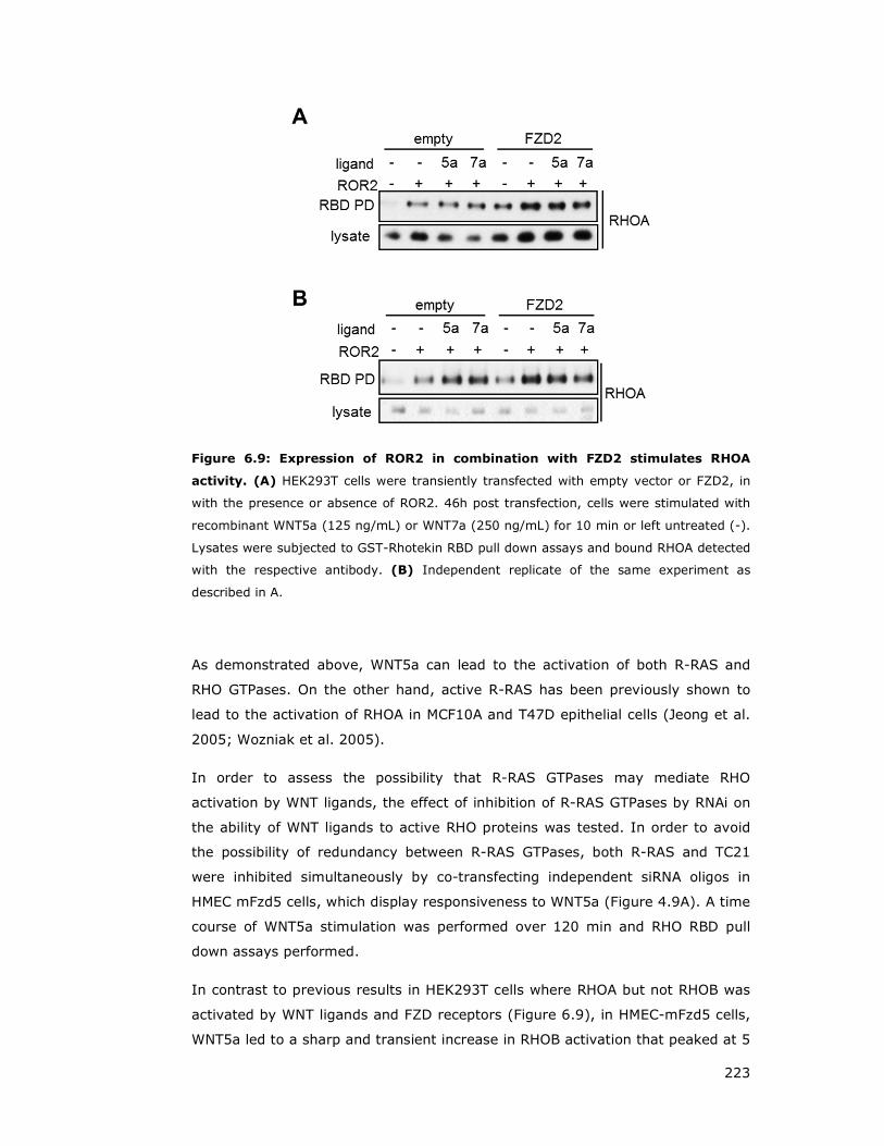
223
Figure 6.9: Expression of ROR2 in combination with FZD2 stimulates RHOA
activity. (A) HEK293T cells were transiently transfected with empty vector or FZD2, in
with the presence or absence of ROR2. 46h post transfection, cells were stimulated with
recombinant WNT5a (125 ng/mL) or WNT7a (250 ng/mL) for 10 min or left untreated (-).
Lysates were subjected to GST-Rhotekin RBD pull down assays and bound RHOA detected
with the respective antibody. (B) Independent replicate of the same experiment as
described in A.
As demonstrated above, WNT5a can lead to the activation of both R-RAS and
RHO GTPases. On the other hand, active R-RAS has been previously shown to
lead to the activation of RHOA in MCF10A and T47D epithelial cells (Jeong et al.
2005; Wozniak et al. 2005).
In order to assess the possibility that R-RAS GTPases may mediate RHO
activation by WNT ligands, the effect of inhibition of R-RAS GTPases by RNAi on
the ability of WNT ligands to active RHO proteins was tested. In order to avoid
the possibility of redundancy between R-RAS GTPases, both R-RAS and TC21
were inhibited simultaneously by co-transfecting independent siRNA oligos in
HMEC mFzd5 cells, which display responsiveness to WNT5a (Figure 4.9A). A time
course of WNT5a stimulation was performed over 120 min and RHO RBD pull
down assays performed.
In contrast to previous results in HEK293T cells where RHOA but not RHOB was
activated by WNT ligands and FZD receptors (Figure 6.9), in HMEC-mFzd5 cells,
WNT5a led to a sharp and transient increase in RHOB activation that peaked at 5
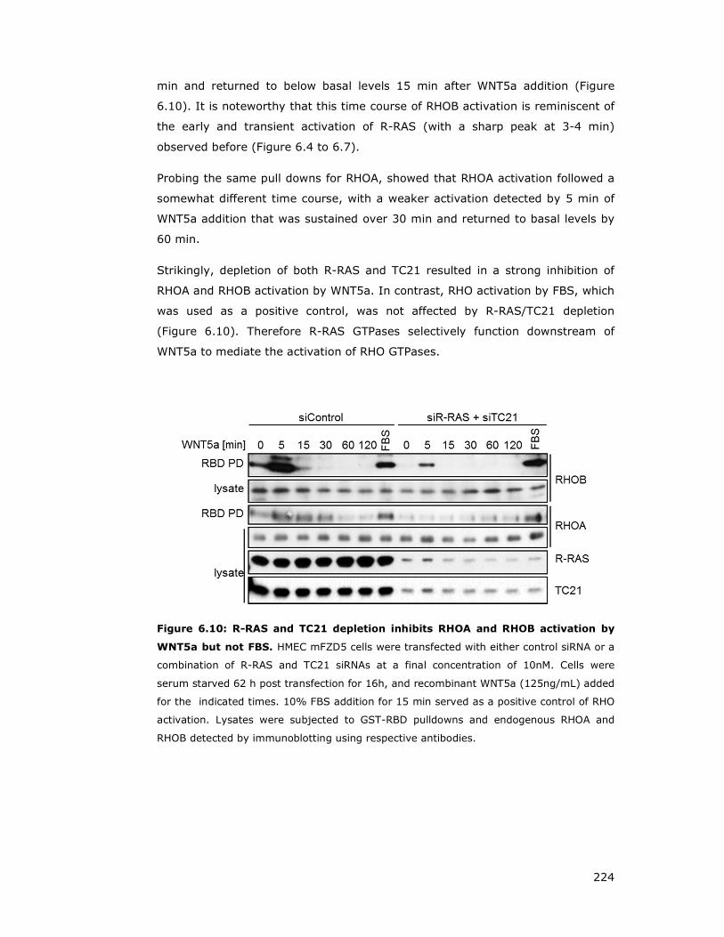
224
min and returned to below basal levels 15 min after WNT5a addition (Figure
6.10). It is noteworthy that this time course of RHOB activation is reminiscent of
the early and transient activation of R-RAS (with a sharp peak at 3-4 min)
observed before (Figure 6.4 to 6.7).
Probing the same pull downs for RHOA, showed that RHOA activation followed a
somewhat different time course, with a weaker activation detected by 5 min of
WNT5a addition that was sustained over 30 min and returned to basal levels by
60 min.
Strikingly, depletion of both R-RAS and TC21 resulted in a strong inhibition of
RHOA and RHOB activation by WNT5a. In contrast, RHO activation by FBS, which
was used as a positive control, was not affected by R-RAS/TC21 depletion
(Figure 6.10). Therefore R-RAS GTPases selectively function downstream of
WNT5a to mediate the activation of RHO GTPases.
Figure 6.10: R-RAS and TC21 depletion inhibits RHOA and RHOB activation by
WNT5a but not FBS. HMEC mFZD5 cells were transfected with either control siRNA or a
combination of R-RAS and TC21 siRNAs at a final concentration of 10nM. Cells were
serum starved 62 h post transfection for 16h, and recombinant WNT5a (125ng/mL) added
for the indicated times. 10% FBS addition for 15 min served as a positive control of RHO
activation. Lysates were subjected to GST-RBD pulldowns and endogenous RHOA and
RHOB detected by immunoblotting using respective antibodies.

225
6.4 Discussion
In this chapter I was able to show that WNT ligands alone or in combination with
the receptor tyrosine kinase ROR2 are able to activate R-RAS in a specific and
short lived manner. Furthermore, R-RAS and TC21 seem to be required in
WNT5a mediated RHOA/RHOB activation. These findings demonstrate a novel
link between R-RAS and WNT signalling networks.
The PCP pathway has been demonstrated in Drosophila to genetically interact
with the EGFR and Notch pathways (Brown and Freeman 2003; Gaengel and
Mlodzik 2003; Strutt and Strutt 2003). Interestingly, to date, the only R-RAS
activator described to stimulate R-RAS activity is the intracellular Notch fragment
(Hodkinson et al. 2007). Whereas well established growth factors such as EGF,
known to activate classical RAS proteins, fail to stimulate R-RAS activity.
Other potential candidates thought to be able to activate R-RAS, which were not
be tested in this thesis, are the migration regulators Trefoil Factors (TFFs)
(Taupin and Podolsky 2003), which have been demonstrated to act upstream of
VANGL1 (Kalabis et al. 2006). TFFs are known to synergise with the EGFR
pathway (Taupin and Podolsky 2003) and it is likely that directed migration is
regulated through cross-talk between multiple signalling pathways.
One of the main types of membrane receptors that activate RAS GTPases are
RTKs, which led to the appealing hypothesis that the tyrosine kinase ROR2 could
be functioning as a potential R-RAS activator. Our findings support this
possibility and suggest R-RAS interaction with VANGL1 could be modulated by
WNT ligands downstream of ROR2.
Studies showed that ROR2 can act as receptor for WNT5a through its
extracellular cysteine-rich domain and WNT5a mediated activation resulted in
ROR2 dependent phosphorylation of VANGL2 (Gao et al. 2011).
WNT5a can lead to the activation of R-RAS in multiple cell lines with a
characteristic activation pattern displaying a short lived burst of activation 3-4
minutes after WNT5a treatment. Stable expression of ROR2 in U2OS cells
exhibited an accelerated as well prolonged WNT5a mediated R-RAS stimulation,
compared to parental cells. This is the first time that WNT ligands have been
demonstrated to stimulate the activity of RAS family GTPases and confirm our
model that R-RAS GTPases can function downstream of WNT ligands in the
regulation of VANGL1 function.
226
I have been unable to detect activation of endogenous TC21 but this could be
due to the lower sensitivity of the TC21 antibody, which may be below the
threshold of detection achieved in our RBD assays. Notably however, TC21 (but
not R-RAS) can be detected in association with ROR2 in some contexts.
How WNT signalling components are able to activate R-RAS GTPases remains to
be determined. Regulation by GEFs or GAPs is a likely mechanism of R-RAS
activation. Although GEF activation is the most direct and common mechanism
for RAS protein activation (Quilliam et al. 2002), inhibition of GAPs by
extracellular stimuli also occurs. For example, p120GAP inhibition after
stimulation of T cells with phorbol ester or TCR engagement (Downward et al.
1990), Ca2+ regulation of the RASGAP, CAPRI (Lockyer et al. 2001), PI3-
kinase/AKT mediated regulation of the RHEB GAP, TSC2 (Castro et al. 2003) or
regulation of RAPGAP1 by G protein coupled receptors (Jordan et al. 1999).
Another example is CK1ε, regulating gastrulation movements through induced
phosphorylation of the RAPGAP SIPA1L1 (signal induced proliferation associated
protein 1 like 1), which in turn leads to reduced protein stability and thus results
in higher RAP1 activity (Tsai et al. 2007).
Despite the fact that the mechanism by which WNT5a activates R-RAS remains
to be determined, members of the RASGRP and RASGRF family of RAS family
GEFs are likely candidates for R-RAS activation. RASGRPs possess a pair of
atypical EF hands that may bind Ca2+ in vivo and contain a DAG-binding C1
domain (Buday and Downward 2008; Stone 2011). A rise in the intracellular
concentration of Ca2+ and DAG are therefore possible mechanisms that could
lead to activation of R-RAS GTPases. Furthermore, accumulating evidence links
Ca2+ signalling to WNT/PCP pathway, although the molecular links are not well
understood (refer to section 1.5.2 and 1.5.3) (De 2011; Stone 2011; Niehrs
2012).
Interestingly, in prostate PC3 cells, WNT5a addition lead to a sharp and transient
peak of Ca2+ release starting at around three min after stimulation, which
subsided within six min after WNT5a treatment (Wang et al. 2010). This
stimulation pattern is strikingly reminiscent of the time course of activation
observed for R-RAS in our studies. Future experiments will aim to determine the
link between Ca2+ signalling, R-RAS activation and the PCP pathway.
There are multiple reports linking the WNT/PCP pathway to activation of RHO
GTPases but the molecular mechanisms remains unclear. There are likely to be
multiple mechanisms operating in a cell type and context dependent manner. For

227
example, our data shows that WNT5a can activate RHOA but not RHOB in some
cells such as 293T whereas in others such as HMEC it activates RHOB more
strongly that RHOA. Strikingly, in at least some context and cell types, R-RAS
GTPases play a critical contribution to RHO activation downstream of WNT
ligands as simultaneous knock-down of both R-RAS and TC21 in HMEC-mFZD5
cells strongly inhibits RHOA and RHOB activation by WNT5a. Although the
mechanism by which R-RAS/TC21 regulate RHO activity remains to be
determined, based on our finding that ARHGEF17/p164RhoGEF functions as an
effector of TC21 (see section 3.3), this RHOGEF is an excellent candidate to
mediate this effect. Future studies should address this exciting possibility.
Figure 6.11: Chapter summary. (A) It has been demostrated that WNT ligands are
able to activate R-RAS, and the R-RAS subgroup member TC21 has been shown to be
ROR2 associated. In addition, enhanced expression of a subset of FZD proteins lead to
enhanced VANGL1-DVL2 interaction, sugesting that WNT signalling, R-RAS activation and
VANGL1 recruitment are co-ordinated processes required for PCP pathway activation. (B)
A potential mechanism of WNT signalling induced R-RAS GTPase activation could be
mediated through WNT stimulated intracellular Ca2+ accumulation, which in turn binds to
RASGRP family members, and thus leads to R-RAS activation and subsequent effector
interaction.
228
CHAPTER 7
Role of VANGL1 and R-
RAS in polarised
migration

229
7 Role of VANGL1 and R-RAS in polarised migration
In both developmental models and in cultured mammalian cells, the PCP/WNT
signalling pathway has been implicated in cell migration (Vladar et al. 2009;
Endo et al. 2012b). VANGL1 has been demonstrated to affect cell migration and
invasion in colorectal cancer cells (Lee et al. 2004; Lee et al. 2005), and VANGL2
has been implicated in WNT5a mediated migration and invasion (Phillips et al.
2005; Cantrell and Jessen 2010; Lindqvist et al. 2010; Kibar et al. 2011; Shafer
et al. 2011; Williams et al. 2012a). Therefore, I aimed to establish biological
assays in order to investigate if VANGL1 and R-RAS GTPases are involved WNT
ligand mediated cellular migration processes.
Cell migration is a complex process which is orchestrated through extensive
transient signalling networks (Vicente-Manzanares et al. 2005; Ridley 2011). In
general, cells start to polarise and protrude in the direction of migration before
modulating adhesion, following translocation of the cell body by retraction of the
rear (Ridley et al. 2003). In order to establish an experimental system to study
VANGL1 and R-RAS function in this process in a mammalian context, two
frequently used assays of directed or polarised migration were considered; the
scratch/wound closure assay and the transwell migration assay.
Wound scratch or wound healing assays have been widely employed to study
migration as well as the establishment of cell polarity during directed locomotion
(Nobes and Hall 1999; Etienne-Manneville and Hall 2001; Etienne-Manneville
and Hall 2003). Wound induction within a compact monolayer provides a
directional cue (or mechanotactic signal), which enables a cell to define a distinct
front and rear (Valster et al. 2005; Etienne-Manneville 2006; Liang et al. 2007).
This results in a strong actin polymerisation at the leading edge and creates
protrusive structures such as filopodia or lamellipodia, while the nucleus is
positioned in the centre of the cell and the centrosome reorients toward the
direction of migration, serving as a microtubule-organizing centre and elongating
microtubules to fill in protrusions (Ridley 2011).
The Boyden chamber approach, also termed transwell assay, monitors cell
migration towards a chemoattractant cue. This is achieved through two
chambers separated by a microporous membrane, in which cells are seeded in
the upper chamber and migrate towards chemotactic agents applied in the lower
chamber (Chen 2005; Falasca et al. 2011). This method further allows
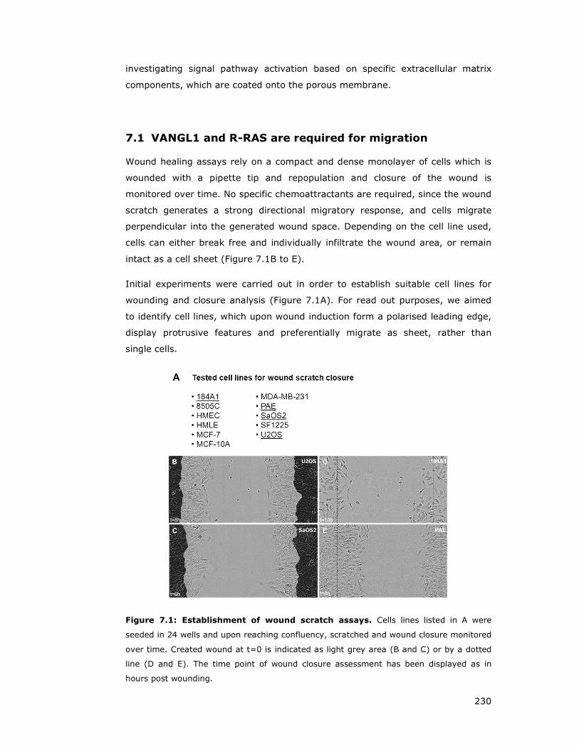
230
investigating signal pathway activation based on specific extracellular matrix
components, which are coated onto the porous membrane.
7.1 VANGL1 and R-RAS are required for migration
Wound healing assays rely on a compact and dense monolayer of cells which is
wounded with a pipette tip and repopulation and closure of the wound is
monitored over time. No specific chemoattractants are required, since the wound
scratch generates a strong directional migratory response, and cells migrate
perpendicular into the generated wound space. Depending on the cell line used,
cells can either break free and individually infiltrate the wound area, or remain
intact as a cell sheet (Figure 7.1B to E).
Initial experiments were carried out in order to establish suitable cell lines for
wounding and closure analysis (Figure 7.1A). For read out purposes, we aimed
to identify cell lines, which upon wound induction form a polarised leading edge,
display protrusive features and preferentially migrate as sheet, rather than
single cells.
Figure 7.1: Establishment of wound scratch assays. Cells lines listed in A were
seeded in 24 wells and upon reaching confluency, scratched and wound closure monitored
over time. Created wound at t=0 is indicated as light grey area (B and C) or by a dotted
line (D and E). The time point of wound closure assessment has been displayed as in
hours post wounding.

231
The osteosarcoma U2OS cell line exhibited all the desired features (Figure 7.1A)
and has been demonstrated to be a good system to study WNT5a mediated
signalling processes (Liu et al. 2007; Liu et al. 2008b; Enomoto et al. 2009;
Yamagata et al. 2012). Therefore, initial scratch wound experiments focused on
investigating the effect of loss of VANGL proteins or R-RAS GTPases on migratory
behaviour in U2OS cells. R-RAS GTPases or VANGL proteins were depleted using
an RNAi approach. 72h post transfection, confluent monolayers were wounded
with a tip, generating approximately 800µm wounds in width (Figure 7.2C and
D). Within 24h, cells usually repopulated the wound, through sheet-like
migration into the wound space (Figure 7.2C).
Cell lysates were assessed for siRNA mediated knock down efficiency by
immunoblotting. Overall, VANGL1, R-RAS and TC21 siRNA transfected samples
exhibited significant reduction of respective protein levels (Figure 7.2A).
Depletion of VANGL1, R-RAS or TC21 protein levels resulted in migration defects,
exhibited as delayed wound closure (Figure 7.2 B and C). The strongest effect
was observed in VANGL1 deficient conditions, that lead to a very strong block in
migration, observable as early as 2h post wounding (Figure 7.2B, purple). Knock
down of VANGL2 also inhibited migration, although not as strongly as VANGL1
(Figure 7.3B) and double knock down of VANGL1 and VANGL2 did not display an
additional effect (data not shown).
R-RAS and TC21 knock down both resulted in slightly delayed wound closure
compared to control cells (Figure 7.2B red, green, black). Notably, double knock
down samples of R-RAS/TC21 displayed a slightly stronger inhibitory effect
(Figure 7.2B, blue). However, this might be related to lower TC21 protein levels
in R-RAS/TC21 double knock down samples (Figure 7.2A).
Multiple siRNA oligomer sequences targeting the same gene were employed and
tested for efficiency and required concentration (data not shown). Analysis of
different VANGL1 RNAi oligomers confirmed the robust negative effect on
migration, supported by the observation that VANGL1 protein expression
correlates well with a less severe migration defect observed with a second less
efficient VANGL1 oligomer (oligo2), compared to oligo 1 (Figure 7.3B and C).

232
Figure 7.2: VANGL1 and R-RAS/TC21 are required for wound closure. U2OS cells
were transfected with siRNA oligomer targeting VANGL1 (V1), R-RAS or TC21 individually
or in combination as indicated at final concentration of 10 nM each. 72h post transfection,
cells were either harvested or scratched. (A) In order to assess knock down levels, cells
were lysed and targeted endogenous protein levels detected by immunoblotting, using
indicated antibodies. Detection of SCRIB or DLG1 served as loading control. (B)
Quantification of induced wound closure over a 24h period, assessed through wound
repopulation in percent (wound density). Data were obtained using a IncuCyte imaging
device (Essen Instruments) and plotted using Excel software (Microsoft) and represent
the mean relative wound density ± S.D. of two independent experiments. (C) Confluent
U2OS monolayers were wounded with a pipette tip and allowed to migrate for 24 h in the
presence of 10% serum. Images were aquired periodically every 2h using a imaging
IncuCyte (Essen Instruments) device. Dark grey areas represent the wound at the time
point of wounding (t=0), whereas light grey regions display the wound area and highlight
migrated cells after 14h. Yellow bar represents 200 µm. As control served a non-targeting
Alexa-fluorophore labeled siRNA.
Although R-RAS and TC21 knock down exhibited only mild effects on the rate of
wound closure compared to VANGL1 knock down conditions, this effect was more
apparent at earlier time points (Figure 7.2B) consistent with the possibility that
R-RAS/TC21 may be preferentially involved in the initial polarisation associated
with migration towards the wound. An enlarged phase contrast field of U2OS
cells during wound closure 4 h post scratching, depicts cell migration and
protrusion and further illustrates this point, as combinational knock down of R-
RAS and TC21 demonstrates a decreased amount of cell protrusion and
migration into the wound (Figure 7.4A, right panel and arrow).

233
Figure 7.3: VANGL1 knock down leads to a more severe migration defect than
VANGL2. Assessment of two individual VANGL1 siRNA oligomers (oligo 1 (1) and oligo
(2)) and VANGL2 (pooled oligmers (p)) on wound closure (10 nM final concentration
each). Confluent U2OS monolayers were wounded with a pipette tip and allowed to
migrate for 24 h in the presence of 10% serum. (A) Immunoblotting of cell lysates to
assess knock down efficency, as control served scramble (con1) or a non-targeting
sequence Alexa-fluorophore (con2). (B) Quantification of induced wound closure over a
24 h period, assessed through wound repopulation in percent (wound density). Data were
obtained using a IncuCyte imaging device (Essen Instruments) and plotted using Excel
software and represent the mean relative wound density ± S.D. of two independent
experiments. (C) Representative images of the repopulated wound. Dark grey areas
represent the wound at the time point of wounding (t=0), whereas light grey regions
illustrate the wound area and highlight migrated cells after 14h.
VANGL1 knock down cells fail to extend protrusions towards the wound (Figure
7.4A middle panel), highlighting a key role for VANGL1 in this type of polarised
migration.
Immunostaining of the actin skeleton (red) and the Golgi marker protein
(GM130, green) revealed that control U2OS cells displayed clear lamellipodial
protrusions towards the wound, exhibiting an extensive and elongated actin
polymerisation (Figure 7.4B, left image, red staining) as well as Golgi localisation
(green) anterior of the nucleus towards the wound, which marks the first
important step prior to actin polymerisation.

234
In comparison, lamellipodial protrusions are clearly shorter and compact in
VANGL1 and R-RAS/TC21 knock down cells and lack the extensive actin
polymerisation towards the wound detected in control cells (Figure 7.4B, middle
and right panel).
Figure 7.4: Loss of VANGL1 or R-RAS/TC21 inhibits membrane protrusion and
migration in scratch/wound closure assays that is particularly apparent at early
time points. U2OS cells were siRNA transfected and grown to confluency within 72h.
Cells were wounded with a plastic pipette tip and either monitored for wound closure over
time or immunostained for actin polymerisation and Golgi orientation. (A) siRNA
mediated downregulation of VANGL1 or R-RAS and TC21 severely disrupts cell migration
into the wound. The untouched cell monolayer at time of scratching is coloured in dark
grey, whereas newly migrated cells are highlighted in light grey and the outmost cell
border is emphasised with a white, dotted line for better visualisation. Control siRNA
transfected U2OS cells displayed fan-like lamellipodia structures at the leading edge.
Phase contrast images taken 4h after wound induction exhibit strong negative effect on
wound closure in siRNA VANGL1 treated cells and a milder but clear effect in the siRNA R-
RAS and TC21 sample, compared to non-targeting scrambled siRNA control (siControl).
Arrows depict lamellipodia protrusions into the wound. (B) Actin stainings of cells as in A
shows inhibition of membrane protrusions and lamellipodial extension at the leading edge
by VANGL1 or RRas/TC21 siRNAs. red - phalloidin, green-Golgi marker GM130, blue –
nucleus stain by Hoechst. Arrows depict areas of actin polymerisation and the direction of
wound triggered cell migration, which displays severely condensed and short lamellipodia
formation (illustrated by arrow shapes) in VANGL1 or R-RAS/TC21 knock down cells.
Yellow bar represents 60 µm.
235
Previous experiments were performed in the presence of 10% FBS. In order to
study a selective role of R-RAS GTPases in the migratory response specifically
stimulated by the WNT/PCP pathway, the ability of WNT ligands to stimulate
migration in the scratch assay was initially tested in this assay.
Unfortunately, using conditioned medium for an extensive panel of the WNT
ligands generated (section 2.4.8.1) no additional effect over control conditioned
medium was observed. Similarly, when purified WNT5a and WNT7a were used at
different concentrations on serum starved cells, no stimulation on migration
could be detected. These negative results precluded further studies using this
experimental system.
As an alternative assay to study polarised migration, the transwell assay was
also employed. Small interfering RNA transfected U2OS cells were seeded in the
upper chamber maintained in serum free media, while the lower chamber was
filled with regular growth media containing 10% FBS, generating a serum
gradient due to distinct pores within the membrane and thus providing a
chemotactic migration cue. Assay quantification was performed based on percent
of cell population travelled through the membrane towards the lower chamber,
normalised by initial cell seeding density.
Both knock down conditions, VANGL1 and R-RAS/TC21, displayed a lower
number of membrane passed cells, compared to the control transfected sample
(Figure 7.5). VANGL1 deficient cells showed a strong migratory reduction of
about 5 fold, whereas the R-RAS/TC21 combination resulted in milder decrease
of migrated cells. These results display a similar pattern as observed with wound
scratch closure assays, where VANGL1 knock-down resulted in a much stronger
inhibitory effect compared to R-RAS/TC21 depletion (Figure 7.2).
In order to study a selective role of R-RAS GTPases in cellular migration
specifically stimulated by the WNT/PCP pathway; experiments were set up using
the transwell assay approach to test the ability of WNT ligands to stimulate
migration.
Unfortunately, addition of different WNT ligands at different concentrations in the
absence of serum did not show a robust effect on migratory behaviour, in U2OS
or other cell lines tested.

236
Figure 7.5: VANGL1 or R-RAS/TC21 are required for transwell migration.
Migration of siRNA treated U2OS cells (either scramble (control), VANGL1 or combination
of R-RAS and TC21), 72h post transfection, towards 10% FBS growth medium was
assessed in a Boyden chamber assay using 24 well PET inserts of 8 µm pore size. (A)
50000 cells were seeded in the top part of a 24 well transwell insert and after
attachment, media was exchanged to serum free media. Left hand panel pictures were
taken 30 min after cell seeding in order to correct for potential discrepencies in cell
numbers. After 14h of incubation, the assay was terminated by removing non migrated
cells on the top of the tranwell membrane, and subsequent staining of migrated cells on
the membrane facing the lower chamber by crystal violet dye (right hand side panel). (B)
Quantification of chemotactic migration was performed by counting representative fields
of cells at 10x magnification, normalised by initial seeding densities and plotting the
numbers of total migrated cells in percent for each condition. Data represent the mean ±
standard error of the mean (SEM) from 3 independent experiments. (*p<0.001,
**p<0.05).

237
7.2 VANGL1 and R-RAS are required for scratch induced
polarisation
In order to further explore the possibility that VANGL1 and R-RAS function in the
context of the WNT/PCP pathway activation in order to control initial cell
polarisation which take place at the onset of migration, Golgi and MTOC re-
orientation at a scratch-induced wound were analysed.
U2OS cell based experiments were carried out in order to assess VANGL1 or R-
RAS/TC21 polarisation effects on Golgi orientation. Golgi structures within a cell
can be detected by immunostaining for a Golgi marker such as the cis-Golgi
protein GM130 (Nakamura et al. 1995).
In order to analyse and quantify Golgi immunostained samples, set criteria for
scoring parameters were determined, demanding that a cell is considered
polarised if the entire Golgi staining localises within a 120° angle towards the
wound edge (Figure 7.6A). A representative scored image is presented in Figure
7.6B. A time course assessing Golgi re-orientation after wound scratch,
comparing scramble control with VANGL1 knock down cells showed that VANGL1
deficient cells exhibited delayed Golgi re-orientation. These differences in
orientation were most apparent at 4h after wounding and thus this time point
was used for subsequent experiments (Figure 7.6C).
Knock-down of R-RAS, or TC21 had a similar inhibitory effect with 20-25% of
cells showing an oriented Golgi compared to ~40% of cells in scramble controls.
Simultaneous knock-down of R-RAS and TC21 had no further effect (Figure
7.6D). Knock-down of VANGL1 caused the biggest disruption, with only ~15%
cells showing wound oriented Golgi (Figure 7.6D).
Another way of determining wound cell orientation is MTOC staining using γ-
tubulin detection (Stearns et al. 1991; Etienne-Manneville 2006). Due to the fact
that the centrosome structure, which serves as MTOC towards the direction of
migration, is a very distinct and confined structure, I applied more stringent
scoring parameters. In order to be counted as correct polarised, MTOC staining
has to be localised within a 90° angle towards the wound (Figure 7.7A), which is
demonstrated on a representative image in Figure 7.7B.
Time course experiments were performed to determine the most suitable point
for cell fixation and MTOC assessment over the course of 7h post wounding.

238
Figure 7.6: VANGL1, SCRIB or R-RAS GTPases delay Golgi wound orientation.
U2OS cells were transfected with indicated siRNAs and grown to confluency. 72h post
transfection, cells were wounded and allowed to migrate for the indicated times. (A)
Scoring schematic. Correctly oriented cells (+) were counted when GM130 Golgi staining
(green) localised within the 120° wound oriented wedge. If GM130 Golgi staining localised
partly or completely outside the 120° criteria, cells were quantified as non oriented (-).
Cells were counted across multiple representative images per sample and more then 100
cells per conditon were assessed for analysis. (B) Example of a representative scored
GM130 image. Nuclei are stained with Hoechst dye (blue) and immunostained for the
Golgi marker GM130 (green). (C) Time course experiment to determine the most
appropriate time point for fixation of cells after wound scratch, comparing control
transfected versus VANGL1 siRNA treated cells. Cells were scratched and fixed either 2, 4
or 6 h after wounding and subsequently immunostained for visualisation of the nucleus
(blue) and the Golgi (green). (D) Quantification of Golgi orientation towards the wound 4
h post scratching were performed from immunostained samples using multiple images per
condition, aquired with a Zeiss microscope and analysed applying criteria as described in
A.

239
Figure 7.7: Loss of VANGL1 or R-RAS/TC21 disrupts MTOC polarisation towards a
wound. U2OS cells were seeded on coverslips, transfected with siRNA targeting either
SCRIB, VANGL1 or R-RAS and TC21 protein level expression at a final concentration of 10
nM. Cells were grown to confluency and wounded 72h post transfection. (A) Scoring
schematic. Correctly oriented cells (+) were counted when the distinctive γ-tubulin stain
(MTOC, microtubule organisation centre) located within a 90° wound oriented wedge. If
more than one MTOC stain was visible, the respective cell was not considered for analysis.
Individual samples were imaged across multiple fields along scratch induced wound, and
more then 100 cells per sample were analysed and counted for quantification. (B)
Example of a scored γ-tubulin image. Cells were nucleus counterstained with Hoechst
(blue) dye and immunostained using the MTOC marker γ-tubulin (green). Note that the
weak resiual staining of the cell body by γ-tubulin was utilised to assess the cell
positioning towards the wound. (C) Time course experiment to determine the most
suitable time point for fixation of cells after wound scratch induction. Cells were wound
scratched and fixed either 1, 2, 3, 4, 6 or 7 h after wound scratch generation. Cells were
immunostained for visualisation of the nucleus (Hoechst) and MTOC (γ-tubulin), and
wound orientation quantified as described in B. (D) Quantification of wound scratch cell
orientation using siRNA U2OS transfected samples, 90 min post wounding. Samples were
immunostained for the nucleus and MTOC location and various images were recorded
using a Zeiss fluorescent microscope. Wound cell orientation was quantified using criteria
as described in A. Red line marks 25%, which indicates the expected percentage of a non
polarised random cell population.

240
MTOC wound orientation appears to take place in a rapid fashion, showing the
highest orientation within one hour post wounding, with a decline during the
next hours (Figure 7.7C). Thus, experiments assessing MTOC orientation
towards the wound were harvested between one and two hours post wounding.
Analysis and quantification of MTOC orientation experiments confirmed Golgi
orientation results (Figure 7.6), demonstrating that SCRIB, VANGL1 or R-
RAS/TC21 knock down negatively affects cellular polarisation. In addition,
VANGL1 deficient cells demonstrated in both orientation assays the highest
defects with only 29% correctly aligned cells, while SCRIB or R-RAS/TC21 siRNA
treated cells displayed 35% or 41%, respectively (Figure 7.7D). These results
indicate that VANGL1 and R-RAS/TC21 are required for initial migration
processes such as correct polarisation towards as wound, consistent with our
observation that a lack of VANGL1 or R-RAS/TC21 results in delayed wound
closure.
Interestingly, immunoblotting for endogenous VANGL1 protein in SCRIB deficient
conditions revealed a gel-shift, compared to control transfected samples (Figure
7.8). It is known that VANGL2 phosphorylation occurs at the N-terminus in a
WNT5a mediated fashion through ROR2 (Gao et al. 2011). Since VANGL1 and
VANGL2 share the same N-terminal serine/threonine cluster, and VANGL1 is
reported to be N-terminal phosphorylated (Kalabis et al. 2006; Daub et al.
2008), this gel-shift suggests potential protein phosphorylation if SCRIB is
absent (Figure 7.8). SCRIB has been implicated in the regulation of directed cell
movements and interacts with VANGL1 (Dow et al. 2007; Anastas et al. 2011),
and thus this evidence implies a potential SCRIB dependent post-translational
VANGL1 regulatory mechanism.
Figure 7.8: Downregulation of SCRIB levels leads to VANGL1 gel migration shift.
U2OS cells were siRNA transfected as indicated above and harvested 72h later. Cell
lysates were immunoblotted for endogenous proteins with indiacted antibodies. Knock
down of SCRIB induced a gel-shift of VANGL1 (V1) protein, which was absent in scramble
or SHOC2 transfections, which interacts with SCRIB but not with VANGL1 and thus served
as control protein. V2-VANGL2.

241
7.3 VANGL1 depletion leads to increased invadosome
formation
Recent studies have implicated VANGL2 in the regulation of endocytosis and cell-
surface availability of membane-type-1 matrix metalloprotease (MMP-14), and in
formation or stability of invadopodia (Cantrell and Jessen 2010; Williams et al.
2012a; Williams et al. 2012b).
Regulation of MMPs and remodelling of ECM has been demonstrated to be
required for non-canonical WNT signalling and vertebrate gastrulation
movements (Coyle et al. 2008). This triggered the question if VANGL1 migratory
and invasive properties are mediated through similar signalling events as
reported for VANGL2.
To test if a R-RAS and VANGL1 are required for invasiveness, the human
osteosarcoma cell line SaOS2 was employed for invadopodium formation studies,
since this cell line shows invasive properties in vitro and metastasis in mouse
models (Dass et al. 2006; Guo et al. 2008).
The invadopodium formation assay relies on a fluorophore labelled matrix and
detection of potential extracellular matrix degradation through invadopodium
formation (Figure 7.9A) (Artym et al. 2009). In order to assess if VANGL1 and R-
RAS/TC21 exhibit an effect on ECM degradation, siRNA transfected SaOS2 cells,
which showed a significant down regulation of VANGL1, or R-RAS/TC21 (Figure
7.9B), were seeded on Alexa568 fluorophore labelled gelatin coverslips. After 9h
of incubation, coverslips were harvested and analysed for ECM degradation,
apparent through loss of staining and thus resulting in black spots (Figure 7.9C).
Quantification of ECM degradation ability was carried out by counting generated
spots across fields of cells, resulting in a spots per field read out. Control SaOS2
cells lead to an average of six generated spots per field, whereas down
regulation of VANGL1 shows a significant reduction by more than 80% in
generated spots. R-RAS/TC21 knock down samples also display a reduced spot
number of almost 50% (Figure 7.9D).

242
Figure 7.9: VANGL1 or R-RAS/TC21 knock down reduces invadosome formation.
(A) Schematic of experimental setup and time line. SaOS2 cells were transfected with
VANGL1, R-RAS and TC21 or scramble control siRNAs, and 72h later seeded at the same
density on Alexa568-gelatin coated coverslips prepared as outlined in section 2.6.4. Cells
were allowed to attach and to spread for 9h, during which time cells start to degrade the
Alexa-gelatin layer through formed invadosomes, resulting in formation of dark spots
within the red matrix layer. After the 9h incubation on Alexa coverslips, cells were fixed
and subsequently prepared for immunofluorescence. (B) Western blot analysis for
endogenous protein levels 81h post transfection using indicated antibodies. As loading
control served tubulin. (C) Representative immunofluorecent image of scramble (control),
R-RAS and TC21 or VANGL1 siRNA treated cell populations on coverslips and induced
matrix degradation spots (black). Actin cytoskeleton was counterstained using Phalloidin
(green) in order to assess if cell populations across conditions were comparable and
equally disributed. Scale bar represents 45 µm. (D) Quantification was performed by
counting yielded dark spots per field of cells (30 fields per condition) at a comparable
density per condition.
Due to reports that WNT5a/ROR2 signalling leads to MMP-13 (collagenase-3)
expression (Yamagata et al. 2012), and further observations that MMP-13 has
been implicated directly and indirectly to promote tumour angiogenesis (Kudo et
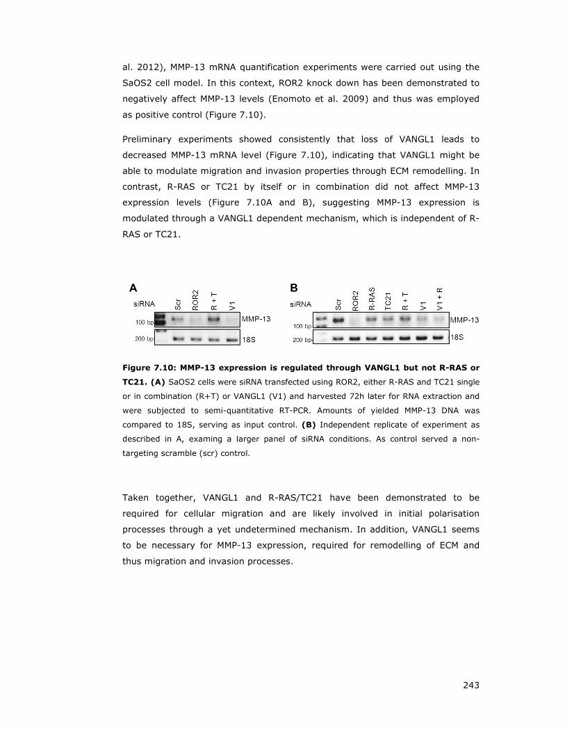
243
al. 2012), MMP-13 mRNA quantification experiments were carried out using the
SaOS2 cell model. In this context, ROR2 knock down has been demonstrated to
negatively affect MMP-13 levels (Enomoto et al. 2009) and thus was employed
as positive control (Figure 7.10).
Preliminary experiments showed consistently that loss of VANGL1 leads to
decreased MMP-13 mRNA level (Figure 7.10), indicating that VANGL1 might be
able to modulate migration and invasion properties through ECM remodelling. In
contrast, R-RAS or TC21 by itself or in combination did not affect MMP-13
expression levels (Figure 7.10A and B), suggesting MMP-13 expression is
modulated through a VANGL1 dependent mechanism, which is independent of R-
RAS or TC21.
Figure 7.10: MMP-13 expression is regulated through VANGL1 but not R-RAS or
TC21. (A) SaOS2 cells were siRNA transfected using ROR2, either R-RAS and TC21 single
or in combination (R+T) or VANGL1 (V1) and harvested 72h later for RNA extraction and
were subjected to semi-quantitative RT-PCR. Amounts of yielded MMP-13 DNA was
compared to 18S, serving as input control. (B) Independent replicate of experiment as
described in A, examing a larger panel of siRNA conditions. As control served a non-
targeting scramble (scr) control.
Taken together, VANGL1 and R-RAS/TC21 have been demonstrated to be
required for cellular migration and are likely involved in initial polarisation
processes through a yet undetermined mechanism. In addition, VANGL1 seems
to be necessary for MMP-13 expression, required for remodelling of ECM and
thus migration and invasion processes.

244
7.4 Discussion
PCP signalling in vertebrates plays a vital role in convergent extension and
gastrulation movements (Bisgrove and Yost 2006; Fliegauf et al. 2007; Zallen
2007; Tada and Kai 2009) through activation of RHO GTPases and regulation of
the formation and polarisation of actine rich membrane protrusions (Roszko et
al. 2009).
To study VANGL1 and R-RAS function in cell migration, wound closure and
transwell (Boyden chamber) assays were chosen (Etienne-Manneville 2006; Dow
et al. 2007) to assess migration and polarisation properties of VANGL1 or R-RAS
GTPases deficient cell lines. Following wounding or migration stimulation by
serum gradient, cells at the edge of the wound polarise the nucleus, actin
cytoskeleton and microtubule organising centre/Golgi network in the axis of
migration, a process known to involve RHO GTPases (Hall 2005; Ridley 2011).
Inhibition of either VANGL1 or R-RAS/TC21 expression with two different siRNA
oligos disrupted the scratch induced re-orientation of the Golgi and the
centrosome towards the wound, membrane protrusion at the leading edge and
inhibited directed migration when measured in transwell and scratch/wound
closure assays. The wound closure delay upon VANGL1 and to a milder degree of
R-RAS/TC21 was particularly apparent at early time points suggesting a possible
role of VANGL1 and R-RAS/TC21 in the initial scratch-induced polarisation. Taken
together these results strongly suggest that VANGL1 and R-RAS/TC21 play an
important role in polarised migration. This is consistent with previous reported
data using colorectal cancer cell lines, demonstrating that VANGL1 is critically
required for migration and invasion (Lee et al. 2004; Kho et al. 2009; Lee et al.
2009).
As outlined earlier, VANGL1 interacts with SCRIB, a protein with a well-
established role in polarity and migration (Qin et al. 2005b; Osmani et al. 2006;
Dow et al. 2007; Nola et al. 2008; Michaelis et al. 2013). Therefore, we
hypothesised that R-RAS may be involved in recruiting the VANGL1-SCRIB
complex to the membrane and thereby exert a role in polarised migration. In
support of this idea, SCRIB is found associated at the leading edge with VANGL1
(Anastas et al. 2011). In addition, it was noticed that knock down of R-
RAS/TC21 resulted in significantly shorter and compacter lamellipodial
protrusions. Consistent with observations that R-RAS is found associated to sites
of lamellipodia (Fernandez-Borja 2012).

245
In mammalian cells, SCRIB associates with the PIX/GIT complex that regulates
RAC/CDC42 and ARF GTPases (Audebert et al. 2004; Frank and Hansen 2008),
which has been implicated in the control of polarity associated with directed cell
migration (Qin et al. 2005b; Osmani et al. 2006; Dow et al. 2007; Michaelis et
al. 2013).
Upon depletion of SCRIB, VANGL1 exhibits an altered, retarded gel migration
shift. Since VANGL2 has been described to be phosphorylated (Gao et al. 2011)
and both VANGL proteins share a N-terminal serine/threonine cluster, it is
tempting to speculate that VANGL1 is phosphorylated by a kinase whose activity
is normally inhibited by SCRIB. The identity of this kinase and the regulatory
mechanisms involved remain to be determined.
Notably, VANGL2 has been reported to affect cell adhesion and the cytoskeleton
by recruiting RAC1 and targeting its activity in the cell to adherens junctions
(Lindqvist et al. 2010).
Using melanoma cell lines in a WNT5a/CXCL12-mediated cell migration model, it
was observed that WNT5a promotes cell polarity in a manner that requires a
chemokine CXCL12 gradient (Witze et al. 2008). CXCL12 signals through the
CXCR4 receptor and has been shown to promote cell invasion (Murakami et al.
2002; Bartolome et al. 2004). Based on experiments carried out by Witze and
colleagues, in combination with our observation that initial polarisation seems to
be strongly affected rather than migration, this suggests that the WNT/PCP
pathway is regulating polarisation and not migration.
Classical RAS proteins are activated in response to a wide variety of extracellular
signals and function as master regulators of cell proliferation. We propose that
R-RAS GTPases may function in a similar manner as master regulators of cell
migration.
VANGL2 has been implicated to modulate invasive properties through MMP
regulation (Cantrell and Jessen 2010; Williams et al. 2012a). Utilising the
invasive osteosarcoma cell line SaOS2, which has been established to show a
ROR2 dependent MMP-13 expression (Enomoto et al. 2009; Yamagata et al.
2012), it could be demonstrated that VANGL1 is required for ECM degradation
and MMP-13 expression.
MMP-13 facilitates local invasion (Milner and Cawston 2005) through its ability to
degrade fibrillar collagen (Uitto et al. 1998) and a wide variety of ECM
components (Ashworth et al. 1999; Tardif et al. 2004). MMP-13 has been
246
reported to be overexpressed in a variety of tumours (Freije et al. 1994; Pendas
et al. 2000; Elnemr et al. 2003; Culhaci et al. 2004; Corte et al. 2005; Luukkaa
et al. 2006) and has been correlated with tumour invasion, metastasis and poor
prognosis in patients (Johansson et al. 1997; Pendas et al. 2000; Krecicki et al.
2003; Roeb et al. 2004; Luukkaa et al. 2006). Expression studies in tumour cells
showed that MMP-13 is predominantly found in the tumour invasive front and to
a lower extent in stromal fibroblasts, surrounding tumour cells (Johansson et al.
1997; Ashworth et al. 1999).
WNT5a has been demonstrated to promote cell migration and invasion in
osteosarcoma cells or prostate carcinoma cells (Enomoto et al. 2009; Yamamoto
et al. 2010). In fact, constitutively active WNT5a/ROR2 is required for MMP-13
expression and invasive properties of osteosarcoma SaOS2 cells. It has been
established that for WNT5a/ROR2 dependent MMP-13 expression, SRC-family
protein tyrosine kinases (SFKs), DVL and RAC are required, mediated through c-
JUN and ATF2 transcription factors, which are recruited to the AP-1 promotor site
of MMP-13 (Enomoto et al. 2009; Yamagata et al. 2012). Notably, DVL2 and
RAC1 but not DVL3 are required for MMP-13 expression in SaOS2. In contrast,
studies carried out in U2OS cells reported that DVL3 and RAC1, but not DVL2 are
required for MMP-13 expression (Yamagata et al. 2012), illustrating the presence
of distinct signalling machineries in order to lead to MMP-13 expression in
osteosarcoma cell lines.
SiRNA mediated knock down of VANGL1 consistently showed a strong decrease
of gelatin degraded areas (spots) indicating that VANGL1 is required for ECM
degrading activities. This observation correlated well with a reduced level of
MMP-13 expression in VANGL1 deficient cells.
These findings are consistent with literature reports stating that ectopic VANGL1
promoted cell adhesion to fibronectin, and invasion of ECM, while loss of VANGL1
protein decreased adhesion and invasiveness (Lee et al. 2004). In addition,
ectopically expressed VANGL1 increased tumour growth rates in mice and
metastasis formation, whereas anti-sense VANGL1 mice exhibited a reduced
tumour volume and no metastasis formation (Lee et al. 2004).
MMP-13 was identified as a common up-regulated gene by cancer invasion-
related factors (Kudo et al. 2012). MMP-13 has been shown to mildly promoted
tumour invasion, and to be involved in tumour angiogenesis. Furthermore, MMP-
13 promoted capillary tube formation was mediated by activation of FAK and

247
ERK. Based on findings by Kudo and colleagues it was suggest that MMP-13 may
directly and indirectly promote tumour angiogenesis (Kudo et al. 2012).
MMP-14 is required for cell polarity and CE and exhibits a genetic interaction with
the WNT co-receptor GLYPICAN4 (Coyle et al. 2008). Studies investigating a
potential connection between PCP mediated cell migration and MMP-dependent
ECM remodelling, showed that VANGL2 regulates MMP-14 cell surface
expression, MMP-2 activation and invasion through an ECM barrier (Cantrell and
Jessen 2010). A subsequent study then revealed that VANGL2 directly regulates
cell surface levels of MMP-14 by controlling its trafficking to and from the plasma
membrane (Williams et al. 2012a). Moreover, VANGL2 dependent MMP-14
endocytosis requires focal adhesion kinase phosphorylation at Y397 (Williams et
al. 2012a). In summary, these findings suggest that VANGL2 mediated MMP-14
trafficking might be coordinated with integrin function and the formation of cell
matrix adhesions.
Therefore, experiments are required in order to test if VANGL1 mediated MMP-13
modulation might be regulated in a similar manner as discussed for VANGL2-
MMP-14. Due to time constraints, I was not able to investigate if VANGL1 is also
able to modulate MMP-14 localisation as observed for VANGL2.

248
CHAPTER 8
Final Discussion
and Summary
249
8 Final Discussion and Summary
8.1 Conclusions and and proposed model
To date, it is still poorly understood how signalling specificity is achieved among
the closely related GTPases of the RAS subfamily. It has been suggested that
individual GTPases have overlapping but specific blueprints of effector
interactions, and that their signalling and biological properties should be
considered in the context of the full spectrum of their many effector interactions
(Rodriguez-Viciana et al. 2004). A further degree of complexity is illustrated by
the observation that RAS family GTPases can cross-talk with each other and co-
operate in the activation of the same effector pathway (Rodriguez-Viciana et al.
2006).
Employing a proteomic approach in order to identify novel effectors of R-RAS
and TC21 led to the discovery of VANGL1 and ARHGEF17/p164RhoGEF,
respectively. Initial validation experiments demonstrated that
ARHGEF17/p164RhoGEF binds to both TC21 activated mutants and R-RAS L87 in
an activation and effector domain dependent manner. No interaction with M-
RAS, RAP1, RAP2 or N-RAS could be observed. Furthermore, I mapped the
TC21-ARHGEF17/p164RhoGEF interaction region to the ARHGEF C-terminus
downstream of the DH domain, and this interaction is direct as it can be
observed in in vitro interaction assays with recombinant proteins. Employing
RHO-RBD assays, I could confirm that ARHGEF17/p164RhoGEF acts as GEF for
RHOA.
Using activating or disrupting mutants of RAS family GTPases, I have shown that
VANGL1 has all the hallmarks of a bona fide effector of R-RAS and TC21. The
interaction is very specific for R-RAS subgroup members and VANGL1 does not
exhibit binding to other closely related RAS proteins such as N-RAS or RAP2.
VANGL1 binds preferentially to the activated mutants compared to the wild type
protein and the interaction is disrupted by mutations in the effector domain.
Furthermore, a GTP-dependent interaction can also be observed in vitro binding
assays using purified proteins, suggesting that the interaction is direct. Taken
together, these results indicate that VANGL1 is a direct and specific effector of R-
RAS and TC21.
VANGL1 is a membrane protein with four transmembrane domains (Iliescu et al.
2011). Interaction studies employing CAAX domain truncations showed that in
250
contrast to other effectors such as AF6 or BRAF, R-RAS need to be membrane
associated in order to interact with VANGL1.
Interaction studies analysing binding of VANGL1 point mutants identified in
human NTDs, demonstrated a defective DVL interaction with VANGL1 D259E
(Kibar et al. 2007). VANGL1 D259E also disrupts interaction with R-RAS. This
suggests that R-RAS and DVL proteins bind to the same region in the VANGL1 C-
terminus and therefore may compete for interaction. Notably, even though the
VANGL1 D259E mutation disrupted R-RAS and DVL3 binding, it did not affect the
interaction with other proteins such as SCRIB, thus indicating that R-RAS and
DVL bind to a different region than SCRIB and potentially linking R-RAS and DVL
to the pathogenesis of NTDs.
VANGL1 has no enzymatic domains or known function and it is likely to function
as a scaffold-type protein by interaction with other proteins. In this model R-
RAS/TC21 could regulate VANGL1 function at least partly by regulating its
localisation in some contexts: upon activation, RRAS/TC21 would recruit VANGL1
to specialised signalling platforms at the membrane micro-domains where they
reside. Furthermore, R-RAS/TC21 binding could also regulate the association of
VANGL1 with its interacting partners and thereby modulate its signalling
properties.
In order to shed more light into VANGL1 function and interaction partners, we
performed the tandem affinity method using VANGL1 as bait. This approach
resulted in the identification of various VANGL1 binding partners, including
several involved in polarity and vesicular trafficking. Although some of the
proteins tested in this study bind to both VANGL1 and VANGL2 isoforms (SCRIB,
DLG1, CASK, YIF1B, CELSR2-C2, RAP3GAP1, SYNTENIN-1), others show
different interaction affinities and suggest different functions for the two VANGL
proteins (e.g. DVL proteins and SYNJ2BP preferentially interact with VANGL1, as
does R-RAS and TC21) (Figure 8.1).
In order to test the hypothesis that R-RAS modulates VANGL1 interactor binding,
pull down experiments in the presence of activated or effector domain R-RAS
mutants were carried out. These studies revealed that indeed R-RAS is able to
either inhibit the interaction of VANGL1 with polarity proteins such as DVL,
SCRIB, DLG and CASK whereas it can stimulate VANGL1 interaction with the PCP
member CELSR2 or membrane trafficking proteins such as YIF1B or SYNJ2BP.

251
Although R-RAS has no effect on the VANGL1-PRICKLE1 interaction, R-RAS
GTPases partially inhibit the VANGL1 induced destabilisation of PRICKLE1/2
protein levels, which occurs by a mechanism that remains to be determined.
Taken together, these observations suggest that R-RAS regulates VANGL1
function by direct recruitment and spatial localization and/or modulation of its
interactome.
We propose a model where upon activation by extracellular signals, R-RAS
GTPases will recruit VANGL1 (and its associated proteins) to sites of activation.
This will not only lead to the localisation of VANGL1 complexes at spatially
distinct micro/nano-domains at the plasma membrane but also to a selective
‘rearrangement’ of the interacting partners within the complex to give rise to
VANGL1 complexes with specialised signalling properties. Furthermore, it will
provide a mechanism to spatially coordinate VANGL1 function with other
signalling pathways regulated by R-RAS GTPases that jointly contribute to
regulate polarity.
Figure 8.1: R-RAS-VANGL1 signalling model. See text for details.
DVL proteins form major signalling hubs, which interact with a multitude of
proteins, and mediate canonical and non-canonical WNT signalling (Boutros and
Mlodzik 1999; Gao and Chen 2010; Wynshaw-Boris 2012). VANGL1 interaction
with all three DVL proteins highlights its role downstream of WNT signalling.

252
Remarkably, R-RAS interaction is specific to VANGL1 and so is the DVL
interaction. Furthermore, R-RAS and DVL3 binding to VANGL1 is disrupted by the
point mutant D259E, suggesting that both proteins could be competing for the
same binding site. This is further confirmed by observations that active R-RAS
modulates VANGL1-DVL binding, suggesting a common ‘axis’ of VANGL1
function.
CELSR proteins are membrane adhesion proteins with key roles in PCP that
remain to be fully understood. CELSR1 is directly linked to cochlear stereocilia
bundle misorientation and neural tube closure defects (Curtin et al. 2003;
Devenport and Fuchs 2008; Robinson et al. 2011). CELSR2 and CELSR3 regulate
other PCP processes such as ciliogenesis, maintenance of dendrites, neuronal
migration and axon guidance (Shima et al. 2002; Tissir et al. 2005; Wada et al.
2006; Shima et al. 2007; Tissir et al. 2010). VANGL1 is able to bind to the
intracellular domain of CELSR2 but not CELSR1, which was also the only CELSR
isoform detected in VANGL1-TAP experiments. This finding is consistent with the
possibility that VANGL1 might not be directly involved in neurolation and related
CE (CELSR1 dependent functions) (Torban et al. 2008; Antic et al. 2010; Song et
al. 2010), but rather in PCP directed processes associated with ciliogenesis and
neuronal development and migration (CELSR2 dependent processes).
Multiple other novel VANGL1 - interacting proteins identified by TAP (NDFIP1,
SYNJ2BP, YIF1B, RAB3GAP) are involved in vesicle/membrane trafficking, and
imply additional roles for VANGL1 function that remains to be further studied.
VANGL1 might interact with these proteins as part of its regulated transport to
the plasma membrane as shown for Sec24B and VANGL2 transport (Wendeler et
al. 2007; Merte et al. 2011; Wansleeben et al. 2011). Another possibility is that
VANGL1 could have specific functions within other endomembrane compartments
such as ER, Golgi or endosomes where others and myself have shown that
VANGL1 can also localise. Whether R-RAS can modulate VANGL1 function at
these endomembrane compartments remains to be determined, but R-RAS as
well as other RAS family proteins, in addition to residing primarily at the plasma
membrane, have also been shown to localise at ER, Golgi and endosomes (Chiu
et al. 2002; Bivona and Philips 2003) (Conklin et al. 2010; Sandri et al. 2012).
In order to test the idea that WNT signalling modulates VANGL1 interaction with
endogenous binding partners was assessed in the presence of a panel of FZD
proteins and or ROR2. Interestingly stimulation of interaction of VANGL1 with
DVLs, but not with other proteins such as SCRIB, DLG1 and CASK (whose

253
interactions are mediated via PDZ domains) is enhanced by a subset of FZD
proteins as well as by ROR2. The underlying mechanism by which FZD and/or
ROR2 achieve this stimulatory effect remains to be identified but may be related
to DVL hyper-phosphorylation.
FZDs recruit DVL to the plasma membrane by a still poorly understood
mechanism. FZD proteins contain a C-terminal PDZ binding motif required for
recruitment of PDZ proteins (Wawrzak et al. 2009), as well as an additional
KTxxxW motif which is required for DVL translocation to the membrane and
subsequent phosphorylation (Rothbacher et al. 2000; Umbhauer et al. 2000;
Axelrod 2001; Cong et al. 2004b). The ability of mFzd5 to stimulate the
VANGL1-DVL2 interaction is abrogated in the mFzd5 KTxxxW motif W530G
mutant and thus correlates with the ability of mFZD5 to initiate DVL2
translocation to the plasma membrane for subsequent phosphorylation events.
This further suggests that DVL translocation and phosphorylation could be an
additional mechanism of recruitment of VANGL1 to sites of activation by WNT
ligands and of spatiotemporal control of VANGL1 function.
The role of DVL phosphorylation in WNT signalling remains still incompletely
understood, but a model has been proposed where DVL phosphorylation may be
a mechanism for the separate temporal control of β-catenin dependent and
independent WNT signalling. The interaction of DVL with VANGL1 correlates with
the phosphorylation of DVL that occurs at later time points of WNT stimulation.
The observation that WNT stimulation can lead to both the activation of R-RAS
GTPases and its association with VANGL1 on one hand and to the association of
VANGL1 with DVL on the other hand, with both interactions potentially being
mutually exclusive may seem counter-intuitive. However, the spatial-temporal
dynamics have to be considered. R-RAS proteins are activated within minutes of
WNT treatment, whereas the WNT induced increase in the interaction of VANGL1
with DVL is first detected after 60 minutes and therefore clearly displays a
delayed response that may correlate with DVL phosphorylation.
Of the tested panel of Frizzled receptors, FZD2, FZD4, mFzd5 and FZD7
stimulate the VANGL1-DVL2 interaction, whereas FZD3 and FZD6 did not display
a detectable interaction enhancement. Interestingly, FZD3 and FZD6 form a
distinct phylogenetic branch of Frizzled proteins (Huang and Klein 2004), and
have been demonstrated to be required with VANGL2, CELSR1 and PRICKLE2 for
inner ear stereociliary bundle orientation (Montcouquiol et al. 2006;
Montcouquiol et al. 2008).
254
As mentioned earlier, DVL interacts preferentially with VANGL1 compared to
VANGL2, and VANGL1 interacts with CELSR2 but not CELSR1. Taken together,
these observations suggest the possibility that a subset of FZD receptors,
possibly in combination with different CELSR co-receptors, may use different
VANGL isoforms to regulate various aspects of non-canonical signalling.
In order to assess if WNT ligands or the tyrosine kinase ROR2 could be
functioning as a potential R-RAS activators, RAS RBD assays were performed.
Our findings demonstrated that WNT5a leads to the activation of R-RAS in
multiple cell lines with a characteristic stimulation pattern displaying a short
lived burst of activation 3-4 minutes after WNT5a treatment. Stable expression
of ROR2 in U2OS cells exhibited an accelerated as well prolonged WNT5a
mediated R-RAS stimulation, compared to parental cells. This is the first time
that WNT ligands have been demonstrated to stimulate the activity of RAS family
GTPases and confirm our model that R-RAS GTPases can function downstream of
WNT ligands in the regulation of VANGL1 function.
Despite the fact that the mechanism by which WNT5a activates R-RAS remains
to be determined, Ca2+ dependent mechanisms provide a compelling possibility.
RASGRPs are the only known GEFs for R-RAS regulated by Ca2+ in vivo.
Furthermore, GAPs such as CAPRI and RASAL are also regulated by Ca2+.
Therefore, a rise in the intracellular concentration of Ca2+, could account for the
very transient activation of R-RAS observed by joint regulation of GEFs and
GAPs.
Multiple reports link Ca2+ signalling to WNT/PCP pathway, although the molecular
links are still not well understood (De 2011; Stone 2011; Niehrs 2012). A
compelling possibility is that Ca2+ dependent activation of R-RAS GTPases may
be the missing link between Ca2+ signalling and the PCP pathway. Further
experiments need to be performed to test this hypothesis. Additionally, how WNT
ligands regulate Ca2+ signalling remains to be determined.
There are multiple reports linking the WNT/PCP pathway to activation of RHO
GPTases but the molecular mechanisms remains unclear. There are likely to be
multiple mechanisms operating in a cell type and context dependent manner.
Strikingly, R-RAS GTPases play a critical contribution to RHO activation
downstream of WNT ligands as simultaneous knock-down of both R-RAS and
TC21 in HMEC-mFZD5 cells strongly inhibits RHOA and RHOB activation by
WNT5a. Although the mechanism by which R-RAS/TC21 regulate RHO activity
remains to be determined, based on our finding that ARHGEF17/p164RhoGEF
255
functions as an effector of TC21, this RHOGEF is an excellent candidate to
mediate this effect. Future studies should address this exciting possibility.
Our results strongly suggest that VANGL1 and R-RAS/TC21 play an important
role in polarised migration. This is consistent with previous reported data using
colorectal cancers cell lines, demonstrating that VANGL1 is critically required for
migration and invasion (Lee et al. 2004; Kho et al. 2009; Lee et al. 2009).
As outlined earlier, VANGL1 interacts with SCRIB, a protein with a well-
established role in polarity and migration (Qin et al. 2005b; Osmani et al. 2006;
Dow et al. 2007; Nola et al. 2008; Michaelis et al. 2013). SCRIB associates with
the PIX/GIT complex that regulates RAC/CDC42 and ARF GTPases (Audebert et
al. 2004; Frank and Hansen 2008). Therefore, we hypothesised that R-RAS may
be involved in recruiting the VANGL1-SCRIB complex to the membrane and
thereby help coordinate other members of the RAS superfamily of GTPases to
help coordinate polarization associated with migration.
In addition, VANGL1 seems to promote also R-RAS independent functions such
as ECM degrading activities (Lee et al. 2004) and MMP-13 expression regulation.
In conclusion, the discovery of WNT/ROR2 mediated activation of R-RAS GTPases
in combination with the identification of VANGL1 and ARHGRF/p164RhoGEF as
novel effectors of R-RAS GTPases provide an exciting new link between the R-
RAS subgroup and the WNT/PCP pathway. Perturbations in this pathway
contribute to the pathogenesis of several diseases including NTDs and to the
invasiveness of cancer cells. Identifying and characterising additional
components in this pathway will hopefully lead to the identification of new genes
involved in disease aetiology as well as reveal novel targets for drug discovery
and therapeutic intervention.

256
CHAPTER 9
References

257
9 References
Aberle, H., Bauer, A., Stappert, J., Kispert, A., and Kemler, R. 1997. beta-catenin is a
target for the ubiquitin-proteasome pathway. The EMBO journal 16(13): 3797-3804.
Ada-Nguema, A.S., Xenias, H., Hofman, J.M., Wiggins, C.H., Sheetz, M.P., and Keely, P.J.
2006. The small GTPase R-Ras regulates organization of actin and drives
membrane protrusions through the activity of PLCepsilon. J Cell Sci 119(Pt 7): 1307-1319.
Adler, P.N. 2002. Planar signaling and morphogenesis in Drosophila. Developmental cell 2(5): 525-535.
Adler, P.N., Krasnow, R.E., and Liu, J. 1997. Tissue polarity points from cells that have higher Frizzled levels towards cells that have lower Frizzled levels. Current biology
: CB 7(12): 940-949. Afzal, A.R., Rajab, A., Fenske, C.D., Oldridge, M., Elanko, N., Ternes-Pereira, E., Tuysuz,
B., Murday, V.A., Patton, M.A., Wilkie, A.O., and Jeffery, S. 2000. Recessive
Robinow syndrome, allelic to dominant brachydactyly type B, is caused by mutation of ROR2. Nat Genet 25(4): 419-422.
Ahearn, I.M., Haigis, K., Bar-Sagi, D., and Philips, M.R. 2012. Regulating the regulator:
post-translational modification of RAS. Nat Rev Mol Cell Biol 13(1): 39-51.
Ahearn, I.M., Tsai, F.D., Court, H., Zhou, M., Jennings, B.C., Ahmed, M., Fehrenbacher, N., Linder, M.E., and Philips, M.R. 2011. FKBP12 binds to acylated H-ras and
promotes depalmitoylation. Molecular cell 41(2): 173-185. Al-Shawi, R., Ashton, S.V., Underwood, C., and Simons, J.P. 2001. Expression of the Ror1
and Ror2 receptor tyrosine kinase genes during mouse development. Dev Genes Evol 211(4): 161-171.
Alarcon, B. and Martinez-Martin, N. 2011. RRas2, RhoG and T-cell phagocytosis. Small GTPases 3(2): 97-101.
Alexander, C.M., Reichsman, F., Hinkes, M.T., Lincecum, J., Becker, K.A., Cumberledge, S., and Bernfield, M. 2000. Syndecan-1 is required for Wnt-1-induced mammary
tumorigenesis in mice. Nat Genet 25(3): 329-332.
Amit, S., Hatzubai, A., Birman, Y., Andersen, J.S., Ben-Shushan, E., Mann, M., Ben-Neriah, Y., and Alkalay, I. 2002. Axin-mediated CKI phosphorylation of beta-
catenin at Ser 45: a molecular switch for the Wnt pathway. Genes Dev 16(9): 1066-1076.
Anastas, J.N., Biechele, T.L., Robitaille, M., Muster, J., Allison, K.H., Angers, S., and Moon, R.T. 2011. A protein complex of SCRIB, NOS1AP and VANGL1 regulates cell
polarity and migration, and is associated with breast cancer progression. Oncogene 31(32): 3696-3708.
Andersson, E.R., Bryjova, L., Biris, K., Yamaguchi, T.P., Arenas, E., and Bryja, V. 2010. Genetic interaction between Lrp6 and Wnt5a during mouse development. Dev Dyn
239(1): 237-245. Andre, P., Wang, Q., Wang, N., Gao, B., Schilit, A., Halford, M.M., Stacker, S.A., Zhang,
X., and Yang, Y. 2012. The Wnt coreceptor Ryk regulates Wnt/planar cell polarity by modulating the degradation of the core planar cell polarity component Vangl2.
J Biol Chem 287(53): 44518-44525.
Angers, S. and Moon, R.T. 2009. Proximal events in Wnt signal transduction. Nat Rev Mol
Cell Biol 10(7): 468-477.
Angers, S., Thorpe, C.J., Biechele, T.L., Goldenberg, S.J., Zheng, N., MacCoss, M.J., and Moon, R.T. 2006. The KLHL12-Cullin-3 ubiquitin ligase negatively regulates the
Wnt-beta-catenin pathway by targeting Dishevelled for degradation. Nat Cell Biol 8(4): 348-357.
Ansley, S.J., Badano, J.L., Blacque, O.E., Hill, J., Hoskins, B.E., Leitch, C.C., Kim, J.C.,
Ross, A.J., Eichers, E.R., Teslovich, T.M., Mah, A.K., Johnsen, R.C., Cavender,
J.C., Lewis, R.A., Leroux, M.R., Beales, P.L., and Katsanis, N. 2003. Basal body dysfunction is a likely cause of pleiotropic Bardet-Biedl syndrome. Nature
425(6958): 628-633. Antic, D., Stubbs, J.L., Suyama, K., Kintner, C., Scott, M.P., and Axelrod, J.D. 2010.
Planar cell polarity enables posterior localization of nodal cilia and left-right axis
determination during mouse and Xenopus embryogenesis. PLoS One 5(2): e8999.

258
Apolloni, A., Prior, I.A., Lindsay, M., Parton, R.G., and Hancock, J.F. 2000. H-ras but not K-ras traffics to the plasma membrane through the exocytic pathway. Molecular
and cellular biology 20(7): 2475-2487.
Arora, S., Matta, A., Shukla, N.K., Deo, S.V., and Ralhan, R. 2005. Identification of differentially expressed genes in oral squamous cell carcinoma. Mol Carcinog
42(2): 97-108. Artym, V.V., Yamada, K.M., and Mueller, S.C. 2009. ECM degradation assays for analyzing
local cell invasion. Methods Mol Biol 522: 211-219. Ashworth, J.L., Murphy, G., Rock, M.J., Sherratt, M.J., Shapiro, S.D., Shuttleworth, C.A.,
and Kielty, C.M. 1999. Fibrillin degradation by matrix metalloproteinases: implications for connective tissue remodelling. Biochem J 340 ( Pt 1): 171-181.
Audebert, S., Navarro, C., Nourry, C., Chasserot-Golaz, S., Lecine, P., Bellaiche, Y.,
Dupont, J.L., Premont, R.T., Sempere, C., Strub, J.M., Van Dorsselaer, A., Vitale,
N., and Borg, J.P. 2004. Mammalian Scribble forms a tight complex with the betaPIX exchange factor. Curr Biol 14(11): 987-995.
Axelrod, J.D. 2001. Unipolar membrane association of Dishevelled mediates Frizzled planar cell polarity signaling. Genes Dev 15(10): 1182-1187.
Bafico, A., Liu, G., Yaniv, A., Gazit, A., and Aaronson, S.A. 2001. Novel mechanism of
Wnt signalling inhibition mediated by Dickkopf-1 interaction with LRP6/Arrow. Nat
Cell Biol 3(7): 683-686.
Baines, A.T., Xu, D., and Der, C.J. 2011. Inhibition of Ras for cancer treatment: the search continues. Future Med Chem 3(14): 1787-1808.
Ballester, R., Furth, M.E., and Rosen, O.M. 1987. Phorbol ester- and protein kinase C-mediated phosphorylation of the cellular Kirsten ras gene product. The Journal of
biological chemistry 262(6): 2688-2695.
Barbacid, M. 1987. ras genes. Annu Rev Biochem 56: 779-827.
Barker, K.T. and Crompton, M.R. 1998. Ras-related TC21 is activated by mutation in a breast cancer cell line, but infrequently in breast carcinomas in vivo. Br J Cancer
78(3): 296-300. Barry, S.T. and Critchley, D.R. 1994. The RhoA-dependent assembly of focal adhesions in
Swiss 3T3 cells is associated with increased tyrosine phosphorylation and the recruitment of both pp125FAK and protein kinase C-delta to focal adhesions. J
Cell Sci 107 ( Pt 7): 2033-2045. Bartolome, R.A., Galvez, B.G., Longo, N., Baleux, F., Van Muijen, G.N., Sanchez-Mateos,
P., Arroyo, A.G., and Teixido, J. 2004. Stromal cell-derived factor-1alpha
promotes melanoma cell invasion across basement membranes involving stimulation of membrane-type 1 matrix metalloproteinase and Rho GTPase
activities. Cancer research 64(7): 2534-2543.
Bartsch, O., Kirmes, I., Thiede, A., Lechno, S., Gocan, H., Florian, I.S., Haaf, T., Zechner,
U., Sabova, L., and Horn, F. 2012. Novel VANGL1 Gene Mutations in 144 Slovakian, Romanian and German Patients with Neural Tube Defects. Mol
Syndromol 3(2): 76-81. Bastock, R., Strutt, H., and Strutt, D. 2003. Strabismus is asymmetrically localised and
binds to Prickle and Dishevelled during Drosophila planar polarity patterning. Development 130(13): 3007-3014.
Beales, P.L., Elcioglu, N., Woolf, A.S., Parker, D., and Flinter, F.A. 1999. New criteria for improved diagnosis of Bardet-Biedl syndrome: results of a population survey. J
Med Genet 36(6): 437-446. Bellaiche, Y., Beaudoin-Massiani, O., Stuttem, I., and Schweisguth, F. 2004. The planar
cell polarity protein Strabismus promotes Pins anterior localization during
asymmetric division of sensory organ precursor cells in Drosophila. Development 131(2): 469-478.
Bellaiche, Y., Gho, M., Kaltschmidt, J.A., Brand, A.H., and Schweisguth, F. 2001. Frizzled regulates localization of cell-fate determinants and mitotic spindle rotation during
asymmetric cell division. Nat Cell Biol 3(1): 50-57. Belo, J.A., Bachiller, D., Agius, E., Kemp, C., Borges, A.C., Marques, S., Piccolo, S., and
De Robertis, E.M. 2000. Cerberus-like is a secreted BMP and nodal antagonist not essential for mouse development. Genesis 26(4): 265-270.
Belotti, E., Puvirajesinghe, T.M., Audebert, S., Baudelet, E., Camoin, L., Pierres, M., Lasvaux, L., Ferracci, G., Montcouquiol, M., and Borg, J.P. 2012. Molecular
characterisation of endogenous Vangl2/Vangl1 heteromeric protein complexes. PLoS One 7(9): e46213.
Bernards, A. 2003. GAPs galore! A survey of putative Ras superfamily GTPase activating proteins in man and Drosophila. Biochim Biophys Acta 1603(2): 47-82.
259
Bernards, A. and Settleman, J. 2004. GAP control: regulating the regulators of small GTPases. Trends Cell Biol 14(7): 377-385.
Bernatik, O., Ganji, R.S., Dijksterhuis, J., Konik, P., Cervenka, I., Polonio, T., Krejci, P.,
Schulte, G., and Bryja, V. 2011. Sequential activation and inactivation of dishevelled in the Wnt/{beta}-catenin pathway by casein kinases. J Biol Chem.
Berrier, A.L., Mastrangelo, A.M., Downward, J., Ginsberg, M., and LaFlamme, S.E. 2000. Activated R-ras, Rac1, PI 3-kinase and PKCepsilon can each restore cell spreading
inhibited by isolated integrin beta1 cytoplasmic domains. J Cell Biol 151(7): 1549-1560.
Bhanot, P., Brink, M., Samos, C.H., Hsieh, J.C., Wang, Y., Macke, J.P., Andrew, D., Nathans, J., and Nusse, R. 1996. A new member of the frizzled family from
Drosophila functions as a Wingless receptor. Nature 382(6588): 225-230.
Bi, D., Nishimura, J., Niiro, N., Hirano, K., and Kanaide, H. 2005. Contractile properties of
the cultured vascular smooth muscle cells: the crucial role played by RhoA in the regulation of contractility. Circ Res 96(8): 890-897.
Bilic, J., Huang, Y.L., Davidson, G., Zimmermann, T., Cruciat, C.M., Bienz, M., and Niehrs, C. 2007. Wnt induces LRP6 signalosomes and promotes dishevelled-dependent
LRP6 phosphorylation. Science 316(5831): 1619-1622.
Bisgrove, B.W. and Yost, H.J. 2006. The roles of cilia in developmental disorders and
disease. Development 133(21): 4131-4143.
Bishop, A.L. and Hall, A. 2000. Rho GTPases and their effector proteins. Biochem J 348 Pt 2: 241-255.
Bivona, T.G., Perez De Castro, I., Ahearn, I.M., Grana, T.M., Chiu, V.K., Lockyer, P.J., Cullen, P.J., Pellicer, A., Cox, A.D., and Philips, M.R. 2003. Phospholipase
Cgamma activates Ras on the Golgi apparatus by means of RasGRP1. Nature
424(6949): 694-698.
Bivona, T.G. and Philips, M.R. 2003. Ras pathway signaling on endomembranes. Current opinion in cell biology 15(2): 136-142.
Bivona, T.G., Quatela, S.E., Bodemann, B.O., Ahearn, I.M., Soskis, M.J., Mor, A., Miura, J., Wiener, H.H., Wright, L., Saba, S.G., Yim, D., Fein, A., Perez de Castro, I., Li,
C., Thompson, C.B., Cox, A.D., and Philips, M.R. 2006. PKC regulates a farnesyl-electrostatic switch on K-Ras that promotes its association with Bcl-XL on
mitochondria and induces apoptosis. Molecular cell 21(4): 481-493. Blacque, O.E., Reardon, M.J., Li, C., McCarthy, J., Mahjoub, M.R., Ansley, S.J., Badano,
J.L., Mah, A.K., Beales, P.L., Davidson, W.S., Johnsen, R.C., Audeh, M., Plasterk,
R.H., Baillie, D.L., Katsanis, N., Quarmby, L.M., Wicks, S.R., and Leroux, M.R. 2004. Loss of C. elegans BBS-7 and BBS-8 protein function results in cilia defects
and compromised intraflagellar transport. Genes & development 18(13): 1630-
1642.
Blankesteijn, W.M., Essers-Janssen, Y.P., Ulrich, M.M., and Smits, J.F. 1996. Increased expression of a homologue of drosophila tissue polarity gene "frizzled" in left
ventricular hypertrophy in the rat, as identified by subtractive hybridization. J Mol Cell Cardiol 28(5): 1187-1191.
Blankesteijn, W.M., Essers-Janssen, Y.P., Verluyten, M.J., Daemen, M.J., and Smits, J.F. 1997. A homologue of Drosophila tissue polarity gene frizzled is expressed in
migrating myofibroblasts in the infarcted rat heart. Nat Med 3(5): 541-544. Blitzer, J.T. and Nusse, R. 2006. A critical role for endocytosis in Wnt signaling. BMC Cell
Biol 7: 28. Blomberg, N., Baraldi, E., Nilges, M., and Saraste, M. 1999. The PH superfold: a structural
scaffold for multiple functions. Trends Biochem Sci 24(11): 441-445.
Bohl, J., Brimer, N., Lyons, C., and Vande Pol, S.B. 2007. The stardust family protein MPP7 forms a tripartite complex with LIN7 and DLG1 that regulates the stability
and localization of DLG1 to cell junctions. J Biol Chem 282(13): 9392-9400. Bos, J.L. 1989. ras oncogenes in human cancer: a review. Cancer Res 49(17): 4682-
4689. Bos, J.L. and Pannekoek, W.J. 2012. Semaphorin signaling meets rap. Sci Signal 5(212):
pe6. Boutros, M. and Mlodzik, M. 1999. Dishevelled: at the crossroads of divergent intracellular
signaling pathways. Mech Dev 83(1-2): 27-37. Boutros, M., Paricio, N., Strutt, D.I., and Mlodzik, M. 1998. Dishevelled activates JNK and
discriminates between JNK pathways in planar polarity and wingless signaling. Cell 94(1): 109-118.
260
Bovolenta, P., Esteve, P., Ruiz, J.M., Cisneros, E., and Lopez-Rios, J. 2008. Beyond Wnt inhibition: new functions of secreted Frizzled-related proteins in development and
disease. Journal of cell science 121(Pt 6): 737-746.
Bradford, M.M. 1976. A rapid and sensitive method for the quantitation of microgram quantities of protein utilizing the principle of protein-dye binding. Anal Biochem
72: 248-254. Brown, K.E. and Freeman, M. 2003. Egfr signalling defines a protective function for
ommatidial orientation in the Drosophila eye. Development 130(22): 5401-5412. Brown, S.D., Twells, R.C., Hey, P.J., Cox, R.D., Levy, E.R., Soderman, A.R., Metzker,
M.L., Caskey, C.T., Todd, J.A., and Hess, J.F. 1998. Isolation and characterization of LRP6, a novel member of the low density lipoprotein receptor gene family.
Biochem Biophys Res Commun 248(3): 879-888.
Brtva, T.R., Drugan, J.K., Ghosh, S., Terrell, R.S., Campbell-Burk, S., Bell, R.M., and Der,
C.J. 1995. Two distinct Raf domains mediate interaction with Ras. The Journal of biological chemistry 270(17): 9809-9812.
Bryja, V., Andersson, E.R., Schambony, A., Esner, M., Bryjova, L., Biris, K.K., Hall, A.C., Kraft, B., Cajanek, L., Yamaguchi, T.P., Buckingham, M., and Arenas, E. 2009.
The extracellular domain of Lrp5/6 inhibits noncanonical Wnt signaling in vivo. Mol
Biol Cell 20(3): 924-936.
Bryja, V., Schulte, G., and Arenas, E. 2007a. Wnt-3a utilizes a novel low dose and rapid
pathway that does not require casein kinase 1-mediated phosphorylation of Dvl to activate beta-catenin. Cell Signal 19(3): 610-616.
Bryja, V., Schulte, G., Rawal, N., Grahn, A., and Arenas, E. 2007b. Wnt-5a induces Dishevelled phosphorylation and dopaminergic differentiation via a CK1-
dependent mechanism. J Cell Sci 120(Pt 4): 586-595.
Buday, L. and Downward, J. 1993. Epidermal growth factor regulates the exchange rate
of guanine nucleotides on p21ras in fibroblasts. Mol Cell Biol 13(3): 1903-1910. Buday, L. and Downward, J.. 2008. Many faces of Ras activation. Biochimica et biophysica
acta 1786(2): 178-187. Butz, S., Okamoto, M., and Sudhof, T.C. 1998. A tripartite protein complex with the
potential to couple synaptic vesicle exocytosis to cell adhesion in brain. Cell 94(6): 773-782.
Cadigan, K.M. and Liu, Y.I. 2006. Wnt signaling: complexity at the surface. J Cell Sci 119(Pt 3): 395-402.
Cain, R.J. and Ridley, A.J. 2009. Phosphoinositide 3-kinases in cell migration. Biol Cell
101(1): 13-29. Campbell, P.M. and Der, C.J. 2004. Oncogenic Ras and its role in tumor cell invasion and
metastasis. Semin Cancer Biol 14(2): 105-114.
Cantor, S.B., Urano, T., and Feig, L.A. 1995. Identification and characterization of Ral-
binding protein 1, a potential downstream target of Ral GTPases. Molecular and cellular biology 15(8): 4578-4584.
Cantrell, V.A. and Jessen, J.R. 2010. The planar cell polarity protein Van Gogh-Like 2 regulates tumor cell migration and matrix metalloproteinase-dependent invasion.
Cancer Lett 287(1): 54-61. Capelluto, D.G., Kutateladze, T.G., Habas, R., Finkielstein, C.V., He, X., and Overduin, M.
2002. The DIX domain targets dishevelled to actin stress fibres and vesicular membranes. Nature 419(6908): 726-729.
Capurro, M.I., Xiang, Y.Y., Lobe, C., and Filmus, J. 2005. Glypican-3 promotes the growth of hepatocellular carcinoma by stimulating canonical Wnt signaling. Cancer Res
65(14): 6245-6254.
Carreira-Barbosa, F., Concha, M.L., Takeuchi, M., Ueno, N., Wilson, S.W., and Tada, M. 2003. Prickle 1 regulates cell movements during gastrulation and neuronal
migration in zebrafish. Development 130(17): 4037-4046. Castellano, E. and Downward, J. 2010. Role of RAS in the regulation of PI 3-kinase. Curr
Top Microbiol Immunol 346: 143-169. Castro, A.F., Rebhun, J.F., Clark, G.J., and Quilliam, L.A. 2003. Rheb binds tuberous
sclerosis complex 2 (TSC2) and promotes S6 kinase activation in a rapamycin- and farnesylation-dependent manner. The Journal of biological chemistry
278(35): 32493-32496. Castro, A.F., Rebhun, J.F., and Quilliam, L.A. 2005. Measuring Ras-family GTP levels in
vivo--running hot and cold. Methods 37(2): 190-196. Cecconi, F., Di Bartolomeo, S., Nardacci, R., Fuoco, C., Corazzari, M., Giunta, L.,
Romagnoli, A., Stoykova, A., Chowdhury, K., Fimia, G.M., and Piacentini, M.

261
2007. A novel role for autophagy in neurodevelopment. Autophagy 3(5): 506-508.
Chae, J., Kim, M.J., Goo, J.H., Collier, S., Gubb, D., Charlton, J., Adler, P.N., and Park,
W.J. 1999. The Drosophila tissue polarity gene starry night encodes a member of the protocadherin family. Development 126(23): 5421-5429.
Chan, A.M., Miki, T., Meyers, K.A., and Aaronson, S.A. 1994. A human oncogene of the RAS superfamily unmasked by expression cDNA cloning. Proc Natl Acad Sci U S A
91(16): 7558-7562. Chan, D.W., Chan, C.Y., Yam, J.W., Ching, Y.P., and Ng, I.O. 2006. Prickle-1 negatively
regulates Wnt/beta-catenin pathway by promoting Dishevelled ubiquitination/degradation in liver cancer. Gastroenterology 131(4): 1218-1227.
Chang, E.H., Gonda, M.A., Ellis, R.W., Scolnick, E.M., and Lowy, D.R. 1982. Human
genome contains four genes homologous to transforming genes of Harvey and
Kirsten murine sarcoma viruses. Proc Natl Acad Sci U S A 79(16): 4848-4852. Chardin, P., Camonis, J.H., Gale, N.W., van Aelst, L., Schlessinger, J., Wigler, M.H., and
Bar-Sagi, D. 1993. Human Sos1: a guanine nucleotide exchange factor for Ras that binds to GRB2. Science 260(5112): 1338-1343.
Chen, H.C. 2005. Boyden chamber assay. Methods Mol Biol 294: 15-22.
Chen, M., Gutierrez, G.J., and Ronai, Z.A. 2011. Ubiquitin-recognition protein Ufd1
couples the endoplasmic reticulum (ER) stress response to cell cycle control. Proc
Natl Acad Sci U S A 108(22): 9119-9124. Chen, W., Hu, L.A., Semenov, M.V., Yanagawa, S., Kikuchi, A., Lefkowitz, R.J., and Miller,
W.E. 2001. beta-Arrestin1 modulates lymphoid enhancer factor transcriptional activity through interaction with phosphorylated dishevelled proteins. Proc Natl
Acad Sci U S A 98(26): 14889-14894.
Chen, W., ten Berge, D., Brown, J., Ahn, S., Hu, L.A., Miller, W.E., Caron, M.G., Barak,
L.S., Nusse, R., and Lefkowitz, R.J. 2003. Dishevelled 2 recruits beta-arrestin 2 to mediate Wnt5A-stimulated endocytosis of Frizzled 4. Science 301(5638): 1391-
1394. Chen, W.S., Antic, D., Matis, M., Logan, C.Y., Povelones, M., Anderson, G.A., Nusse, R.,
and Axelrod, J.D. 2008. Asymmetric homotypic interactions of the atypical cadherin flamingo mediate intercellular polarity signaling. Cell 133(6): 1093-
1105. Cheng, C.W., Yeh, J.C., Fan, T.P., Smith, S.K., and Charnock-Jones, D.S. 2008. Wnt5a-
mediated non-canonical Wnt signalling regulates human endothelial cell
proliferation and migration. Biochem Biophys Res Commun 365(2): 285-290. Cherfils, J. and Zeghouf, M. 2013. Regulation of small GTPases by GEFs, GAPs, and GDIs.
Physiol Rev 93(1): 269-309.
Cheyette, B.N., Waxman, J.S., Miller, J.R., Takemaru, K., Sheldahl, L.C., Khlebtsova, N.,
Fox, E.P., Earnest, T., and Moon, R.T. 2002. Dapper, a Dishevelled-associated antagonist of beta-catenin and JNK signaling, is required for notochord formation.
Dev Cell 2(4): 449-461. Chiu, V.K., Bivona, T., Hach, A., Sajous, J.B., Silletti, J., Wiener, H., Johnson, R.L., 2nd,
Cox, A.D., and Philips, M.R. 2002. Ras signalling on the endoplasmic reticulum and the Golgi. Nature cell biology 4(5): 343-350.
Cho, E., Feng, Y., Rauskolb, C., Maitra, S., Fehon, R., and Irvine, K.D. 2006. Delineation of a Fat tumor suppressor pathway. Nat Genet 38(10): 1142-1150.
Choy, E., Chiu, V.K., Silletti, J., Feoktistov, M., Morimoto, T., Michaelson, D., Ivanov, I.E., and Philips, M.R. 1999. Endomembrane trafficking of ras: the CAAX motif targets
proteins to the ER and Golgi. Cell 98(1): 69-80.
Chua, C.E., Lim, Y.S., and Tang, B.L. 2010. Rab35--a vesicular traffic-regulating small GTPase with actin modulating roles. FEBS Lett 584(1): 1-6.
Ciani, L. and Salinas, P.C. 2005. WNTs in the vertebrate nervous system: from patterning to neuronal connectivity. Nat Rev Neurosci 6(5): 351-362.
Ciechanover, A. 2005. Proteolysis: from the lysosome to ubiquitin and the proteasome. Nature reviews Molecular cell biology 6(1): 79-87.
Cirone, P., Lin, S., Griesbach, H.L., Zhang, Y., Slusarski, D.C., and Crews, C.M. 2008. A role for planar cell polarity signaling in angiogenesis. Angiogenesis 11(4): 347-
360. Ciruna, B., Jenny, A., Lee, D., Mlodzik, M., and Schier, A.F. 2006. Planar cell polarity
signalling couples cell division and morphogenesis during neurulation. Nature 439(7073): 220-224.

262
Clark, G.J., Kinch, M.S., Gilmer, T.M., Burridge, K., and Der, C.J. 1996. Overexpression of the Ras-related TC21/R-Ras2 protein may contribute to the development of
human breast cancers. Oncogene 12(1): 169-176.
Clark, R., Wong, G., Arnheim, N., Nitecki, D., and McCormick, F. 1985. Antibodies specific for amino acid 12 of the ras oncogene product inhibit GTP binding. Proc Natl Acad
Sci U S A 82(16): 5280-5284. Clevers, H. and Nusse, R. 2012. Wnt/beta-catenin signaling and disease. Cell 149(6):
1192-1205. Cliffe, A., Hamada, F., and Bienz, M. 2003. A role of Dishevelled in relocating Axin to the
plasma membrane during wingless signaling. Curr Biol 13(11): 960-966. Condeelis, J. 2001. How is actin polymerization nucleated in vivo? Trends Cell Biol 11(7):
288-293.
Cong, F., Schweizer, L., and Varmus, H. 2004a. Casein kinase Iepsilon modulates the
signaling specificities of dishevelled. Mol Cell Biol 24(5): 2000-2011. Cong, F., Schweizer, L., and Varmus, H. 2004b. Wnt signals across the plasma membrane
to activate the beta-catenin pathway by forming oligomers containing its receptors, Frizzled and LRP. Development 131(20): 5103-5115.
Conklin, M.W., Ada-Nguema, A., Parsons, M., Riching, K.M., and Keely, P.J. 2010. R-Ras
regulates beta1-integrin trafficking via effects on membrane ruffling and
endocytosis. BMC cell biology 11: 14.
Corbeel, L. and Freson, K. 2008. Rab proteins and Rab-associated proteins: major actors in the mechanism of protein-trafficking disorders. Eur J Pediatr 167(7): 723-729.
Corbit, K.C., Shyer, A.E., Dowdle, W.E., Gaulden, J., Singla, V., Chen, M.H., Chuang, P.T., and Reiter, J.F. 2008. Kif3a constrains beta-catenin-dependent Wnt signalling
through dual ciliary and non-ciliary mechanisms. Nat Cell Biol 10(1): 70-76.
Corte, M.D., Gonzalez, L.O., Corte, M.G., Quintela, I., Pidal, I., Bongera, M., and Vizoso,
F. 2005. Collagenase-3 (MMP-13) expression in cutaneous malignant melanoma. Int J Biol Markers 20(4): 242-248.
Costa, C., Barberis, L., Ambrogio, C., Manazza, A.D., Patrucco, E., Azzolino, O., Neilsen, P.O., Ciraolo, E., Altruda, F., Prestwich, G.D., Chiarle, R., Wymann, M., Ridley, A.,
and Hirsch, E. 2007. Negative feedback regulation of Rac in leukocytes from mice expressing a constitutively active phosphatidylinositol 3-kinase gamma. Proc Natl
Acad Sci U S A 104(36): 14354-14359. Courbard, J.R., Djiane, A., Wu, J., and Mlodzik, M. 2009. The apical/basal-polarity
determinant Scribble cooperates with the PCP core factor Stbm/Vang and
functions as one of its effectors. Dev Biol 333(1): 67-77. Coussens, L.M., Fingleton, B., and Matrisian, L.M. 2002. Matrix metalloproteinase
inhibitors and cancer: trials and tribulations. Science 295(5564): 2387-2392.
Cox, A.D. and Der, C.J. 2010. Ras history: The saga continues. Small GTPases 1(1): 2-
27. Coyle, R.C., Latimer, A., and Jessen, J.R. 2008. Membrane-type 1 matrix
metalloproteinase regulates cell migration during zebrafish gastrulation: evidence for an interaction with non-canonical Wnt signaling. Exp Cell Res 314(10): 2150-
2162. Cozier, G.E., Lockyer, P.J., Reynolds, J.S., Kupzig, S., Bottomley, J.R., Millard, T.H.,
Banting, G., and Cullen, P.J. 2000. GAP1IP4BP contains a novel group I pleckstrin homology domain that directs constitutive plasma membrane association. J Biol
Chem 275(36): 28261-28268. Cruciat, C.M. and Niehrs, C. 2012. Secreted and transmembrane Wnt inhibitors and
activators. Cold Spring Harb Perspect Med 3(3): a015081.
Culhaci, N., Metin, K., Copcu, E., and Dikicioglu, E. 2004. Elevated expression of MMP-13 and TIMP-1 in head and neck squamous cell carcinomas may reflect increased
tumor invasiveness. BMC Cancer 4: 42. Cullen, P.J., Hsuan, J.J., Truong, O., Letcher, A.J., Jackson, T.R., Dawson, A.P., and
Irvine, R.F. 1995. Identification of a specific Ins(1,3,4,5)P4-binding protein as a member of the GAP1 family. Nature 376(6540): 527-530.
Curtin, J.A., Quint, E., Tsipouri, V., Arkell, R.M., Cattanach, B., Copp, A.J., Henderson, D.J., Spurr, N., Stanier, P., Fisher, E.M., Nolan, P.M., Steel, K.P., Brown, S.D.,
Gray, I.C., and Murdoch, J.N. 2003. Mutation of Celsr1 disrupts planar polarity of inner ear hair cells and causes severe neural tube defects in the mouse. Curr Biol
13(13): 1129-1133. Dallol, A., Agathanggelou, A., Tommasi, S., Pfeifer, G.P., Maher, E.R., and Latif, F. 2005.
Involvement of the RASSF1A tumor suppressor gene in controlling cell migration. Cancer Res 65(17): 7653-7659.
263
Daneshmanesh, A.H., Porwit, A., Hojjat-Farsangi, M., Jeddi-Tehrani, M., Tamm, K.P., Grander, D., Lehmann, S., Norin, S., Shokri, F., Rabbani, H., Mellstedt, H., and
Osterborg, A. 2013. Orphan receptor tyrosine kinases ROR1 and ROR2 in
hematological malignancies. Leuk Lymphoma 54(4): 843-850. Dasgupta, B. and Gutmann, D.H. 2003. Neurofibromatosis 1: closing the GAP between
mice and men. Curr Opin Genet Dev 13(1): 20-27. Dass, C.R., Ek, E.T., Contreras, K.G., and Choong, P.F. 2006. A novel orthotopic murine
model provides insights into cellular and molecular characteristics contributing to human osteosarcoma. Clinical & experimental metastasis 23(7-8): 367-380.
Daub, H., Olsen, J.V., Bairlein, M., Gnad, F., Oppermann, F.S., Korner, R., Greff, Z., Keri, G., Stemmann, O., and Mann, M. 2008. Kinase-selective enrichment enables
quantitative phosphoproteomics of the kinome across the cell cycle. Mol Cell
31(3): 438-448.
Daulat, A.M., Luu, O., Sing, A., Zhang, L., Wrana, J.L., McNeill, H., Winklbauer, R., and Angers, S. 2012. Mink1 regulates beta-catenin-independent Wnt signaling via
Prickle phosphorylation. Mol Cell Biol 32(1): 173-185. Davidson, G., Wu, W., Shen, J., Bilic, J., Fenger, U., Stannek, P., Glinka, A., and Niehrs,
C. 2005. Casein kinase 1 gamma couples Wnt receptor activation to cytoplasmic
signal transduction. Nature 438(7069): 867-872.
De, A. 2011. Wnt/Ca2+ signaling pathway: a brief overview. Acta Biochim Biophys Sin
(Shanghai) 43(10): 745-756. De Cat, B., Muyldermans, S.Y., Coomans, C., Degeest, G., Vanderschueren, B., Creemers,
J., Biemar, F., Peers, B., and David, G. 2003. Processing by proprotein convertases is required for glypican-3 modulation of cell survival, Wnt signaling,
and gastrulation movements. J Cell Biol 163(3): 625-635.
de Curtis, I. 2001. Cell migration: GAPs between membrane traffic and the cytoskeleton.
EMBO reports 2(4): 277-281. de Lau, W.B., Snel, B., and Clevers, H.C. 2012. The R-spondin protein family. Genome
Biol 13(3): 242. De Marco, P., Merello, E., Rossi, A., Piatelli, G., Cama, A., Kibar, Z., and Capra, V. 2012.
FZD6 is a novel gene for human neural tube defects. Hum Mutat 33(2): 384-390. de Renzis, S., Sonnichsen, B., and Zerial, M. 2002. Divalent Rab effectors regulate the
sub-compartmental organization and sorting of early endosomes. Nature cell biology 4(2): 124-133.
de Rooij, J. and Bos, J.L. 1997. Minimal Ras-binding domain of Raf1 can be used as an
activation-specific probe for Ras. Oncogene 14(5): 623-625. De Toledo, M., Colombo, K., Nagase, T., Ohara, O., Fort, P., and Blangy, A. 2000. The
yeast exchange assay, a new complementary method to screen for Dbl-like
protein specificity: identification of a novel RhoA exchange factor. FEBS Lett
480(2-3): 287-292. Debnath, J., Muthuswamy, S.K., and Brugge, J.S. 2003. Morphogenesis and oncogenesis
of MCF-10A mammary epithelial acini grown in three-dimensional basement membrane cultures. Methods 30(3): 256-268.
DeFeo, D., Gonda, M.A., Young, H.A., Chang, E.H., Lowy, D.R., Scolnick, E.M., and Ellis, R.W. 1981. Analysis of two divergent rat genomic clones homologous to the
transforming gene of Harvey murine sarcoma virus. Proc Natl Acad Sci U S A 78(6): 3328-3332.
Delgado, P., Cubelos, B., Calleja, E., Martinez-Martin, N., Cipres, A., Merida, I., Bellas, C., Bustelo, X.R., and Alarcon, B. 2009. Essential function for the GTPase TC21 in
homeostatic antigen receptor signaling. Nat Immunol 10(8): 880-888.
Deng, X., Zhao, H.S., Peng, Z., Deng, W.W., Li, N., Guo, S., and Shi, T.P. 2011. [Study on the mechanism of C17orf62 induced cell death]. Beijing Da Xue Xue Bao
43(2): 168-172. DerMardirossian, C. and Bokoch, G.M. 2005. GDIs: central regulatory molecules in Rho
GTPase activation. Trends Cell Biol 15(7): 356-363. Descamps, B., Sewduth, R., Ferreira Tojais, N., Jaspard, B., Reynaud, A., Sohet, F.,
Lacolley, P., Allieres, C., Lamaziere, J.M., Moreau, C., Dufourcq, P., Couffinhal, T., and Duplaa, C. 2012. Frizzled 4 regulates arterial network organization through
noncanonical Wnt/planar cell polarity signaling. Circ Res 110(1): 47-58. Destaing, O., Planus, E., Bouvard, D., Oddou, C., Badowski, C., Bossy, V., Raducanu, A.,
Fourcade, B., Albiges-Rizo, C., and Block, M.R. 2010. beta1A integrin is a master regulator of invadosome organization and function. Mol Biol Cell 21(23): 4108-
4119.
264
Devenport, D. and Fuchs, E. 2008. Planar polarization in embryonic epidermis orchestrates global asymmetric morphogenesis of hair follicles. Nat Cell Biol
10(11): 1257-1268.
Diekmann, D., Brill, S., Garrett, M.D., Totty, N., Hsuan, J., Monfries, C., Hall, C., Lim, L., and Hall, A. 1991. Bcr encodes a GTPase-activating protein for p21rac. Nature
351(6325): 400-402. Diekmann, Y., Seixas, E., Gouw, M., Tavares-Cadete, F., Seabra, M.C., and Pereira-Leal,
J.B. 2011. Thousands of rab GTPases for the cell biologist. PLoS Comput Biol 7(10): e1002217.
Ding, Y., Zhang, Y., Xu, C., Tao, Q.H., and Chen, Y.G. 2013. HECT Domain-containing E3 ubiquitin ligase NEDD4L negatively regulates Wnt signaling by targeting
Dishevelled for proteasomal degradation. J Biol Chem.
Dissanayake, S.K., Wade, M., Johnson, C.E., O'Connell, M.P., Leotlela, P.D., French, A.D.,
Shah, K.V., Hewitt, K.J., Rosenthal, D.T., Indig, F.E., Jiang, Y., Nickoloff, B.J., Taub, D.D., Trent, J.M., Moon, R.T., Bittner, M., and Weeraratna, A.T. 2007. The
Wnt5A/protein kinase C pathway mediates motility in melanoma cells via the inhibition of metastasis suppressors and initiation of an epithelial to mesenchymal
transition. J Biol Chem 282(23): 17259-17271.
Doerks, T., Bork, P., Kamberov, E., Makarova, O., Muecke, S., and Margolis, B. 2000.
L27, a novel heterodimerization domain in receptor targeting proteins Lin-2 and
Lin-7. Trends Biochem Sci 25(7): 317-318. Dollar, G.L., Weber, U., Mlodzik, M., and Sokol, S.Y. 2005. Regulation of Lethal giant
larvae by Dishevelled. Nature 437(7063): 1376-1380. Dow, L.E., Kauffman, J.S., Caddy, J., Zarbalis, K., Peterson, A.S., Jane, S.M., Russell,
S.M., and Humbert, P.O. 2007. The tumour-suppressor Scribble dictates cell
polarity during directed epithelial migration: regulation of Rho GTPase recruitment
to the leading edge. Oncogene 26(16): 2272-2282. Downward, J. 2003. Targeting RAS signalling pathways in cancer therapy. Nat Rev Cancer
3(1): 11-22. Downward, J., Graves, J.D., Warne, P.H., Rayter, S., and Cantrell, D.A. 1990. Stimulation
of p21ras upon T-cell activation. Nature 346(6286): 719-723. Drivas, G.T., Shih, A., Coutavas, E., Rush, M.G., and D'Eustachio, P. 1990.
Characterization of four novel ras-like genes expressed in a human teratocarcinoma cell line. Mol Cell Biol 10(4): 1793-1798.
Du, S.J., Purcell, S.M., Christian, J.L., McGrew, L.L., and Moon, R.T. 1995. Identification
of distinct classes and functional domains of Wnts through expression of wild-type and chimeric proteins in Xenopus embryos. Mol Cell Biol 15(5): 2625-2634.
Easty, D.J., Mitchell, P.J., Patel, K., Florenes, V.A., Spritz, R.A., and Bennett, D.C. 1997.
Loss of expression of receptor tyrosine kinase family genes PTK7 and SEK in
metastatic melanoma. Int J Cancer 71(6): 1061-1065. Ebinu, J.O., Bottorff, D.A., Chan, E.Y., Stang, S.L., Dunn, R.J., and Stone, J.C. 1998.
RasGRP, a Ras guanyl nucleotide- releasing protein with calcium- and diacylglycerol-binding motifs. Science 280(5366): 1082-1086.
Ehrhardt, A., Ehrhardt, G.R., Guo, X., and Schrader, J.W. 2002. Ras and relatives--job sharing and networking keep an old family together. Exp Hematol 30(10): 1089-
1106. Eisenberg, S., Laude, A.J., Beckett, A.J., Mageean, C.J., Aran, V., Hernandez-Valladares,
M., Henis, Y.I., and Prior, I.A. 2013. The role of palmitoylation in regulating Ras localization and function. Biochem Soc Trans 41(1): 79-83.
Elbert, M., Cohen, D., and Musch, A. 2006. PAR1b promotes cell-cell adhesion and inhibits
dishevelled-mediated transformation of Madin-Darby canine kidney cells. Mol Biol Cell 17(8): 3345-3355.
Elnemr, A., Yonemura, Y., Bandou, E., Kinoshita, K., Kawamura, T., Takahashi, S., Tochiori, S., Endou, Y., and Sasaki, T. 2003. Expression of collagenase-3 (matrix
metalloproteinase-13) in human gastric cancer. Gastric Cancer 6(1): 30-38. Endo, M., Doi, R., Nishita, M., and Minami, Y. 2012a. Ror family receptor tyrosine kinases
regulate the maintenance of neural progenitor cells in the developing neocortex. J Cell Sci 125(Pt 8): 2017-2029.
Endo, M., Nishita, M., and Minami, Y. 2012b. Analysis of Wnt/planar cell polarity pathway in cultured cells. Methods Mol Biol 839: 201-214.
Endo, Y., Wolf, V., Muraiso, K., Kamijo, K., Soon, L., Uren, A., Barshishat-Kupper, M., and Rubin, J.S. 2005. Wnt-3a-dependent cell motility involves RhoA activation and is
specifically regulated by dishevelled-2. J Biol Chem 280(1): 777-786.
265
Enomoto, M., Hayakawa, S., Itsukushima, S., Ren, D.Y., Matsuo, M., Tamada, K., Oneyama, C., Okada, M., Takumi, T., Nishita, M., and Minami, Y. 2009.
Autonomous regulation of osteosarcoma cell invasiveness by Wnt5a/Ror2
signaling. Oncogene 28(36): 3197-3208. Erdmann, J., Grosshennig, A., Braund, P.S., Konig, I.R., Hengstenberg, C., Hall, A.S.,
Linsel-Nitschke, P., Kathiresan, S., Wright, B., Tregouet, D.A., Cambien, F., Bruse, P., Aherrahrou, Z., Wagner, A.K., Stark, K., Schwartz, S.M., Salomaa, V.,
Elosua, R., Melander, O., Voight, B.F., O'Donnell, C.J., Peltonen, L., Siscovick, D.S., Altshuler, D., Merlini, P.A., Peyvandi, F., Bernardinelli, L., Ardissino, D.,
Schillert, A., Blankenberg, S., Zeller, T., Wild, P., Schwarz, D.F., Tiret, L., Perret, C., Schreiber, S., El Mokhtari, N.E., Schafer, A., Marz, W., Renner, W., Bugert, P.,
Kluter, H., Schrezenmeir, J., Rubin, D., Ball, S.G., Balmforth, A.J., Wichmann,
H.E., Meitinger, T., Fischer, M., Meisinger, C., Baumert, J., Peters, A., Ouwehand,
W.H., Deloukas, P., Thompson, J.R., Ziegler, A., Samani, N.J., and Schunkert, H. 2009. New susceptibility locus for coronary artery disease on chromosome
3q22.3. Nat Genet 41(3): 280-282. Erdogan, M., Pozzi, A., Bhowmick, N., Moses, H.L., and Zent, R. 2007. Signaling pathways
regulating TC21-induced tumorigenesis. J Biol Chem 282(38): 27713-27720.
Erdogan, M., Pozzi, A., Bhowmick, N., Moses, H.L., and Zent, R.. 2008. Transforming
growth factor-beta (TGF-beta) and TGF-beta-associated kinase 1 are required for
R-Ras-mediated transformation of mammary epithelial cells. Cancer Res 68(15): 6224-6231.
Etheridge, S.L., Ray, S., Li, S., Hamblet, N.S., Lijam, N., Tsang, M., Greer, J., Kardos, N., Wang, J., Sussman, D.J., Chen, P., and Wynshaw-Boris, A. 2008. Murine
dishevelled 3 functions in redundant pathways with dishevelled 1 and 2 in normal
cardiac outflow tract, cochlea, and neural tube development. PLoS Genet 4(11):
e1000259. Etienne-Manneville, S. 2006. In vitro assay of primary astrocyte migration as a tool to
study Rho GTPase function in cell polarization. Methods in enzymology 406: 565-578.
Erdogan, M., Pozzi, A., Bhowmick, N., Moses, H.L., and Zent, R. 2008. Polarity proteins in migration and invasion. Oncogene 27(55): 6970-6980.
Etienne-Manneville, S. and Hall, A. 2001. Integrin-mediated activation of Cdc42 controls cell polarity in migrating astrocytes through PKCzeta. Cell 106(4): 489-498.
Etienne-Manneville, S. and Hall, A. 2002. Rho GTPases in cell biology. Nature 420(6916):
629-635. Etienne-Manneville, S. and Hall, A. 2003. Cdc42 regulates GSK-3beta and adenomatous
polyposis coli to control cell polarity. Nature 421(6924): 753-756.
Falasca, M., Raimondi, C., and Maffucci, T. 2011. Boyden chamber. Methods Mol Biol
769: 87-95. Fan, W.T., Koch, C.A., de Hoog, C.L., Fam, N.P., and Moran, M.F. 1998. The exchange
factor Ras-GRF2 activates Ras-dependent and Rac-dependent mitogen-activated protein kinase pathways. Curr Biol 8(16): 935-938.
Fanto, M. and McNeill, H. 2004. Planar polarity from flies to vertebrates. J Cell Sci 117(Pt 4): 527-533.
Fedorko, M. 1967. Effect of chloroquine on morphology of cytoplasmic granules in maturing human leukocytes--an ultrastructural study. J Clin Invest 46(12): 1932-
1942. Feiguin, F., Hannus, M., Mlodzik, M., and Eaton, S. 2001. The ankyrin repeat protein
Diego mediates Frizzled-dependent planar polarization. Developmental cell 1(1):
93-101. Feng, Y., Chen, M.H., Moskowitz, I.P., Mendonza, A.M., Vidali, L., Nakamura, F.,
Kwiatkowski, D.J., and Walsh, C.A. 2006. Filamin A (FLNA) is required for cell-cell contact in vascular development and cardiac morphogenesis. Proc Natl Acad Sci U
S A 103(52): 19836-19841. Fernandez-Borja, M. 2012. A tale of three GTPases and a RIN in endothelial cell adhesion.
Cell Res 22(10): 1426-1428. Fimia, G.M., Stoykova, A., Romagnoli, A., Giunta, L., Di Bartolomeo, S., Nardacci, R.,
Corazzari, M., Fuoco, C., Ucar, A., Schwartz, P., Gruss, P., Piacentini, M., Chowdhury, K., and Cecconi, F. 2007. Ambra1 regulates autophagy and
development of the nervous system. Nature 447(7148): 1121-1125. Fliegauf, M., Benzing, T., and Omran, H. 2007. When cilia go bad: cilia defects and
ciliopathies. Nat Rev Mol Cell Biol 8(11): 880-893.

266
Foldynova-Trantirkova, S., Sekyrova, P., Tmejova, K., Brumovska, E., Bernatik, O., Blankenfeldt, W., Krejci, P., Kozubik, A., Dolezal, T., Trantirek, L., and Bryja, V.
2010. Breast cancer-specific mutations in CK1epsilon inhibit Wnt/beta-catenin and
activate the Wnt/Rac1/JNK and NFAT pathways to decrease cell adhesion and promote cell migration. Breast Cancer Res 12(3): R30.
Ford, C.E., Qian Ma, S.S., Quadir, A., and Ward, R.L. 2012. The dual role of the novel Wnt receptor tyrosine kinase, ROR2, in human carcinogenesis. Int J Cancer.
Forrester, W.C., Dell, M., Perens, E., and Garriga, G. 1999. A C. elegans Ror receptor tyrosine kinase regulates cell motility and asymmetric cell division. Nature
400(6747): 881-885. Fox, J.W., Lamperti, E.D., Eksioglu, Y.Z., Hong, S.E., Feng, Y., Graham, D.A., Scheffer,
I.E., Dobyns, W.B., Hirsch, B.A., Radtke, R.A., Berkovic, S.F., Huttenlocher, P.R.,
and Walsh, C.A. 1998. Mutations in filamin 1 prevent migration of cerebral cortical
neurons in human periventricular heterotopia. Neuron 21(6): 1315-1325. Fradkin, L.G., Dura, J.M., and Noordermeer, J.N. 2010. Ryks: new partners for Wnts in
the developing and regenerating nervous system. Trends Neurosci 33(2): 84-92. Frank, S.R. and Hansen, S.H. 2008. The PIX-GIT complex: a G protein signaling cassette
in control of cell shape. Semin Cell Dev Biol 19(3): 234-244.
Franke, B., Akkerman, J.W., and Bos, J.L. 1997. Rapid Ca2+-mediated activation of Rap1
in human platelets. The EMBO journal 16(2): 252-259.
Freije, J.M., Diez-Itza, I., Balbin, M., Sanchez, L.M., Blasco, R., Tolivia, J., and Lopez-Otin, C. 1994. Molecular cloning and expression of collagenase-3, a novel human
matrix metalloproteinase produced by breast carcinomas. J Biol Chem 269(24): 16766-16773.
Fuerer, C., Habib, S.J., and Nusse, R. 2010. A study on the interactions between heparan
sulfate proteoglycans and Wnt proteins. Dev Dyn 239(1): 184-190.
Fujimura, L., Watanabe-Takano, H., Sato, Y., Tokuhisa, T., and Hatano, M. 2009. Prickle promotes neurite outgrowth via the Dishevelled dependent pathway in C1300
cells. Neurosci Lett 467(1): 6-10. Fukuda, M. 2008. Regulation of secretory vesicle traffic by Rab small GTPases. Cell Mol
Life Sci 65(18): 2801-2813. Fukuda, T., Chen, L., Endo, T., Tang, L., Lu, D., Castro, J.E., Widhopf, G.F., 2nd,
Rassenti, L.Z., Cantwell, M.J., Prussak, C.E., Carson, D.A., and Kipps, T.J. 2008. Antisera induced by infusions of autologous Ad-CD154-leukemia B cells identify
ROR1 as an oncofetal antigen and receptor for Wnt5a. Proc Natl Acad Sci U S A
105(8): 3047-3052. Furuhjelm, J. and Peranen, J. 2003. The C-terminal end of R-Ras contains a focal
adhesion targeting signal. Journal of cell science 116(Pt 18): 3729-3738.
Furushima, K., Yamamoto, A., Nagano, T., Shibata, M., Miyachi, H., Abe, T., Ohshima, N.,
Kiyonari, H., and Aizawa, S. 2007. Mouse homologues of Shisa antagonistic to Wnt and Fgf signalings. Developmental biology 306(2): 480-492.
Gaengel, K. and Mlodzik, M. 2003. Egfr signaling regulates ommatidial rotation and cell motility in the Drosophila eye via MAPK/Pnt signaling and the Ras effector
Canoe/AF6. Development 130(22): 5413-5423. Gan, X.Q., Wang, J.Y., Xi, Y., Wu, Z.L., Li, Y.P., and Li, L. 2008. Nuclear Dvl, c-Jun, beta-
catenin, and TCF form a complex leading to stabilization of beta-catenin-TCF interaction. J Cell Biol 180(6): 1087-1100.
Gao, B., Song, H., Bishop, K., Elliot, G., Garrett, L., English, M.A., Andre, P., Robinson, J., Sood, R., Minami, Y., Economides, A.N., and Yang, Y. 2011. Wnt signaling
gradients establish planar cell polarity by inducing Vangl2 phosphorylation
through Ror2. Dev Cell 20(2): 163-176. Gao, C., Cao, W., Bao, L., Zuo, W., Xie, G., Cai, T., Fu, W., Zhang, J., Wu, W., Zhang, X.,
and Chen, Y.G. 2010. Autophagy negatively regulates Wnt signalling by promoting Dishevelled degradation. Nat Cell Biol 12(8): 781-790.
Gao, C. and Chen, Y.G. 2010. Dishevelled: The hub of Wnt signaling. Cell Signal 22(5): 717-727.
Gawecka, J.E., Griffiths, G.S., Ek-Rylander, B., Ramos, J.W., and Matter, M.L. 2010. R-Ras regulates migration through an interaction with filamin A in melanoma cells.
PLoS One 5(6): e11269. Giles, R.H., van Es, J.H., and Clevers, H. 2003. Caught up in a Wnt storm: Wnt signaling
in cancer. Biochim Biophys Acta 1653(1): 1-24. Gobble, R.M., Qin, L.X., Brill, E.R., Angeles, C.V., Ugras, S., O'Connor, R.B., Moraco,
N.H., Decarolis, P.L., Antonescu, C., and Singer, S. 2011. Expression profiling of

267
liposarcoma yields a multigene predictor of patient outcome and identifies genes that contribute to liposarcomagenesis. Cancer Res 71(7): 2697-2705.
Goldfinger, L.E., Ptak, C., Jeffery, E.D., Shabanowitz, J., Han, J., Haling, J.R., Sherman,
N.E., Fox, J.W., Hunt, D.F., and Ginsberg, M.H. 2007. An experimentally derived database of candidate Ras-interacting proteins. Journal of proteome research
6(5): 1806-1811. Goldfinger, L.E., Ptak, C., Jeffery, E.D., Shabanowitz, J., Hunt, D.F., and Ginsberg, M.H.
2006. RLIP76 (RalBP1) is an R-Ras effector that mediates adhesion-dependent Rac activation and cell migration. J Cell Biol 174(6): 877-888.
Golubkov, V.S., Chekanov, A.V., Cieplak, P., Aleshin, A.E., Chernov, A.V., Zhu, W., Radichev, I.A., Zhang, D., Dong, P.D., and Strongin, A.Y. 2010. The Wnt/planar
cell polarity protein-tyrosine kinase-7 (PTK7) is a highly efficient proteolytic target
of membrane type-1 matrix metalloproteinase: implications in cancer and
embryogenesis. J Biol Chem 285(46): 35740-35749. Gomez, G.A. and Daniotti, J.L. 2005. H-Ras dynamically interacts with recycling
endosomes in CHO-K1 cells: involvement of Rab5 and Rab11 in the trafficking of H-Ras to this pericentriolar endocytic compartment. The Journal of biological
chemistry 280(41): 34997-35010.
Gonzalez-Sancho, J.M., Brennan, K.R., Castelo-Soccio, L.A., and Brown, A.M. 2004. Wnt
proteins induce dishevelled phosphorylation via an LRP5/6- independent
mechanism, irrespective of their ability to stabilize beta-catenin. Mol Cell Biol 24(11): 4757-4768.
Goodrich, L.V. and Strutt, D. 2011. Principles of planar polarity in animal development. Development 138(10): 1877-1892.
Gordon, M.D. and Nusse, R. 2006. Wnt signaling: multiple pathways, multiple receptors,
and multiple transcription factors. J Biol Chem 281(32): 22429-22433.
Gotoh, T., Niino, Y., Tokuda, M., Hatase, O., Nakamura, S., Matsuda, M., and Hattori, S. 1997. Activation of R-Ras by Ras-guanine nucleotide-releasing factor. J Biol Chem
272(30): 18602-18607. Graham, S.M., Oldham, S.M., Martin, C.B., Drugan, J.K., Zohn, I.E., Campbell, S., and
Der, C.J. 1999. TC21 and Ras share indistinguishable transforming and differentiating activities. Oncogene 18(12): 2107-2116.
Gravel, M., Iliescu, A., Horth, C., Apuzzo, S., and Gros, P. 2010. Molecular and cellular mechanisms underlying neural tube defects in the loop-tail mutant mouse.
Biochemistry 49(16): 3445-3455.
Gray, R.S., Roszko, I., and Solnica-Krezel, L. 2011. Planar cell polarity: coordinating morphogenetic cell behaviors with embryonic polarity. Developmental cell 21(1):
120-133.
Green, J.L., Kuntz, S.G., and Sternberg, P.W. 2008. Ror receptor tyrosine kinases:
orphans no more. Trends Cell Biol 18(11): 536-544. Griffiths, G.S., Grundl, M., Allen, J.S., 3rd, and Matter, M.L. 2011. R-Ras interacts with
filamin a to maintain endothelial barrier function. J Cell Physiol 226(9): 2287-2296.
Gubb, D. and Garcia-Bellido, A. 1982. A genetic analysis of the determination of cuticular polarity during development in Drosophila melanogaster. J Embryol Exp Morphol
68: 37-57. Gubb, D., Green, C., Huen, D., Coulson, D., Johnson, G., Tree, D., Collier, S., and Roote,
J. 1999. The balance between isoforms of the prickle LIM domain protein is critical for planar polarity in Drosophila imaginal discs. Genes & development 13(17):
2315-2327.
Guirao, B., Meunier, A., Mortaud, S., Aguilar, A., Corsi, J.M., Strehl, L., Hirota, Y., Desoeuvre, A., Boutin, C., Han, Y.G., Mirzadeh, Z., Cremer, H., Montcouquiol, M.,
Sawamoto, K., and Spassky, N. 2010. Coupling between hydrodynamic forces and planar cell polarity orients mammalian motile cilia. Nat Cell Biol 12(4): 341-350.
Guo, Y., Rubin, E.M., Xie, J., Zi, X., and Hoang, B.H. 2008. Dominant negative LRP5 decreases tumorigenicity and metastasis of osteosarcoma in an animal model.
Clin Orthop Relat Res 466(9): 2039-2045. Guyot, M.C., Bosoi, C.M., Kharfallah, F., Reynolds, A., Drapeau, P., Justice, M., Gros, P.,
and Kibar, Z. 2011. A novel hypomorphic Looptail allele at the planar cell polarity Vangl2 gene. Dev Dyn 240(4): 839-849.
Habas, R., Dawid, I.B., and He, X. 2003. Coactivation of Rac and Rho by Wnt/Frizzled signaling is required for vertebrate gastrulation. Genes Dev 17(2): 295-309.
Habas, R. and He, X. 2006. Activation of Rho and Rac by Wnt/frizzled signaling. Methods Enzymol 406: 500-511.

268
Habas, R., Kato, Y., and He, X. 2001. Wnt/Frizzled activation of Rho regulates vertebrate gastrulation and requires a novel Formin homology protein Daam1. Cell 107(7):
843-854.
Halford, M.M., Armes, J., Buchert, M., Meskenaite, V., Grail, D., Hibbs, M.L., Wilks, A.F., Farlie, P.G., Newgreen, D.F., Hovens, C.M., and Stacker, S.A. 2000. Ryk-deficient
mice exhibit craniofacial defects associated with perturbed Eph receptor crosstalk. Nat Genet 25(4): 414-418.
Hall-Glenn, F. and Lyons, K.M. 2011. Roles for CCN2 in normal physiological processes. Cell Mol Life Sci 68(19): 3209-3217.
Hall, A. 1998. Rho GTPases and the actin cytoskeleton. Science 279(5350): 509-514. Hall, A. 2005. Rho GTPases and the control of cell behaviour. Biochemical Society
transactions 33(Pt 5): 891-895.
Hall, A. 2012. Rho family GTPases. Biochem Soc Trans 40(6): 1378-1382.
Hamblet, N.S., Lijam, N., Ruiz-Lozano, P., Wang, J., Yang, Y., Luo, Z., Mei, L., Chien, K.R., Sussman, D.J., and Wynshaw-Boris, A. 2002. Dishevelled 2 is essential for
cardiac outflow tract development, somite segmentation and neural tube closure. Development 129(24): 5827-5838.
Hamel, B., Monaghan-Benson, E., Rojas, R.J., Temple, B.R., Marston, D.J., Burridge, K.,
and Sondek, J. 2011. SmgGDS is a guanine nucleotide exchange factor that
specifically activates RhoA and RhoC. J Biol Chem 286(14): 12141-12148.
Han, L. and Colicelli, J. 1995. A human protein selected for interference with Ras function interacts directly with Ras and competes with Raf1. Mol Cell Biol 15(3): 1318-
1323. Hanahan, D. and Weinberg, R.A. 2000. The hallmarks of cancer. Cell 100(1): 57-70.
Hanahan, D. and Weinberg, R.A. 2011. Hallmarks of cancer: the next generation. Cell
144(5): 646-674.
Hancock, J.F. 2003. Ras proteins: different signals from different locations. Nature reviews Molecular cell biology 4(5): 373-384.
Hancock, J.F. and Parton, R.G. 2005. Ras plasma membrane signalling platforms. Biochem J 389(Pt 1): 1-11.
Hancock, J.F., Paterson, H., and Marshall, C.J. 1990. A polybasic domain or palmitoylation is required in addition to the CAAX motif to localize p21ras to the plasma
membrane. Cell 63(1): 133-139. Hansen, M., Prior, I.A., Hughes, P.E., Oertli, B., Chou, F.L., Willumsen, B.M., Hancock,
J.F., and Ginsberg, M.H. 2003. C-terminal sequences in R-Ras are involved in
integrin regulation and in plasma membrane microdomain distribution. Biochem Biophys Res Commun 311(4): 829-838.
Hansen, M., Rusyn, E.V., Hughes, P.E., Ginsberg, M.H., Cox, A.D., and Willumsen, B.M.
2002. R-Ras C-terminal sequences are sufficient to confer R-Ras specificity to H-
Ras. Oncogene 21(28): 4448-4461. Hawkins, P.T., Anderson, K.E., Davidson, K., and Stephens, L.R. 2006. Signalling through
Class I PI3Ks in mammalian cells. Biochemical Society transactions 34(Pt 5): 647-662.
He, C. and Klionsky, D.J. 2009. Regulation mechanisms and signaling pathways of autophagy. Annu Rev Genet 43: 67-93.
He, F., Xiong, W., Yu, X., Espinoza-Lewis, R., Liu, C., Gu, S., Nishita, M., Suzuki, K., Yamada, G., Minami, Y., and Chen, Y. 2008. Wnt5a regulates directional cell
migration and cell proliferation via Ror2-mediated noncanonical pathway in mammalian palate development. Development 135(23): 3871-3879.
He, X., Semenov, M., Tamai, K., and Zeng, X. 2004. LDL receptor-related proteins 5 and
6 in Wnt/beta-catenin signaling: arrows point the way. Development 131(8): 1663-1677.
Heallen, T., Zhang, M., Wang, J., Bonilla-Claudio, M., Klysik, E., Johnson, R.L., and Martin, J.F. 2011. Hippo pathway inhibits Wnt signaling to restrain cardiomyocyte
proliferation and heart size. Science 332(6028): 458-461. Heasman, S.J. and Ridley, A.J. 2008. Mammalian Rho GTPases: new insights into their
functions from in vivo studies. Nat Rev Mol Cell Biol 9(9): 690-701. Heisenberg, C.P., Tada, M., Rauch, G.J., Saude, L., Concha, M.L., Geisler, R., Stemple,
D.L., Smith, J.C., and Wilson, S.W. 2000. Silberblick/Wnt11 mediates convergent extension movements during zebrafish gastrulation. Nature 405(6782): 76-81.
Hemsley, A., Arnheim, N., Toney, M.D., Cortopassi, G., and Galas, D.J. 1989. A simple method for site-directed mutagenesis using the polymerase chain reaction.
Nucleic Acids Res 17(16): 6545-6551.

269
Hendrickx, M. and Leyns, L. 2008. Non-conventional Frizzled ligands and Wnt receptors. Dev Growth Differ 50(4): 229-243.
Herman, I.M. 1993. Actin isoforms. Curr Opin Cell Biol 5(1): 48-55.
Herrmann, C., Martin, G.A., and Wittinghofer, A. 1995. Quantitative analysis of the complex between p21ras and the Ras-binding domain of the human Raf-1 protein
kinase. The Journal of biological chemistry 270(7): 2901-2905. Hino, S., Michiue, T., Asashima, M., and Kikuchi, A. 2003. Casein kinase I epsilon
enhances the binding of Dvl-1 to Frat-1 and is essential for Wnt-3a-induced accumulation of beta-catenin. The Journal of biological chemistry 278(16):
14066-14073. Hodkinson, P.S., Elliott, P.A., Lad, Y., McHugh, B.J., MacKinnon, A.C., Haslett, C., and
Sethi, T. 2007. Mammalian NOTCH-1 activates beta1 integrins via the small
GTPase R-Ras. J Biol Chem 282(39): 28991-29001.
Holly, S.P., Larson, M.K., and Parise, L.V. 2005. The unique N-terminus of R-ras is required for Rac activation and precise regulation of cell migration. Mol Biol Cell
16(5): 2458-2469. Hovens, C.M., Stacker, S.A., Andres, A.C., Harpur, A.G., Ziemiecki, A., and Wilks, A.F.
1992. RYK, a receptor tyrosine kinase-related molecule with unusual kinase
domain motifs. Proc Natl Acad Sci U S A 89(24): 11818-11822.
Hsieh, J.C., Rattner, A., Smallwood, P.M., and Nathans, J. 1999. Biochemical
characterization of Wnt-frizzled interactions using a soluble, biologically active vertebrate Wnt protein. Proc Natl Acad Sci U S A 96(7): 3546-3551.
Huang, H.C. and Klein, P.S. 2004. The Frizzled family: receptors for multiple signal transduction pathways. Genome Biol 5(7): 234.
Huang, Y., Saez, R., Chao, L., Santos, E., Aaronson, S.A., and Chan, A.M. 1995. A novel
insertional mutation in the TC21 gene activates its transforming activity in a
human leiomyosarcoma cell line. Oncogene 11(7): 1255-1260. Huff, S.Y., Quilliam, L.A., Cox, A.D., and Der, C.J. 1997. R-Ras is regulated by activators
and effectors distinct from those that control Ras function. Oncogene 14(2): 133-143.
Humbert, P.O., Grzeschik, N.A., Brumby, A.M., Galea, R., Elsum, I., and Richardson, H.E. 2008. Control of tumourigenesis by the Scribble/Dlg/Lgl polarity module.
Oncogene 27(55): 6888-6907. Hurlstone, A. and Clevers, H. 2002. T-cell factors: turn-ons and turn-offs. EMBO J
21(10): 2303-2311.
Ikeda, S., Kishida, S., Yamamoto, H., Murai, H., Koyama, S., and Kikuchi, A. 1998. Axin, a negative regulator of the Wnt signaling pathway, forms a complex with GSK-
3beta and beta-catenin and promotes GSK-3beta-dependent phosphorylation of
beta-catenin. The EMBO journal 17(5): 1371-1384.
Iliescu, A., Gravel, M., Horth, C., Apuzzo, S., and Gros, P. 2011. Transmembrane topology of mammalian planar cell polarity protein Vangl1. Biochemistry 50(12):
2274-2282. Inoki, K., Ouyang, H., Zhu, T., Lindvall, C., Wang, Y., Zhang, X., Yang, Q., Bennett, C.,
Harada, Y., Stankunas, K., Wang, C.Y., He, X., MacDougald, O.A., You, M., Williams, B.O., and Guan, K.L. 2006. TSC2 integrates Wnt and energy signals via
a coordinated phosphorylation by AMPK and GSK3 to regulate cell growth. Cell 126(5): 955-968.
Iozzo, R.V., Eichstetter, I., and Danielson, K.G. 1995. Aberrant expression of the growth factor Wnt-5A in human malignancy. Cancer Res 55(16): 3495-3499.
Ip, W., Wellman-Labadie, O., Tang, L., Su, M., Yu, R., Dutz, J., Wang, Y., Huang, S.,
Zhang, X., Huang, C., and Zhou, Y. 2011. Collagen triple helix repeat containing 1 promotes melanoma cell adhesion and survival. J Cutan Med Surg 15(2): 103-
110. Ishitani, T., Kishida, S., Hyodo-Miura, J., Ueno, N., Yasuda, J., Waterman, M., Shibuya,
H., Moon, R.T., Ninomiya-Tsuji, J., and Matsumoto, K. 2003. The TAK1-NLK mitogen-activated protein kinase cascade functions in the Wnt-5a/Ca(2+)
pathway to antagonize Wnt/beta-catenin signaling. Mol Cell Biol 23(1): 131-139. Ishitani, T., Ninomiya-Tsuji, J., Nagai, S., Nishita, M., Meneghini, M., Barker, N.,
Waterman, M., Bowerman, B., Clevers, H., Shibuya, H., and Matsumoto, K. 1999. The TAK1-NLK-MAPK-related pathway antagonizes signalling between beta-
catenin and transcription factor TCF. Nature 399(6738): 798-802. Itoh, K., Brott, B.K., Bae, G.U., Ratcliffe, M.J., and Sokol, S.Y. 2005. Nuclear localization
is required for Dishevelled function in Wnt/beta-catenin signaling. J Biol 4(1): 3.
270
Jaffe, A.B. and Hall, A. 2002. Rho GTPases in transformation and metastasis. Adv Cancer Res 84: 57-80.
Jaffe, A.B. and Hall, A. 2005. Rho GTPases: biochemistry and biology. Annu Rev Cell Dev
Biol 21: 247-269. Jenny, A. 2010. Planar cell polarity signaling in the Drosophila eye. Curr Top Dev Biol 93:
189-227. Jenny, A., Darken, R.S., Wilson, P.A., and Mlodzik, M. 2003. Prickle and Strabismus form
a functional complex to generate a correct axis during planar cell polarity signaling. EMBO J 22(17): 4409-4420.
Jeong, H.W., Nam, J.O., and Kim, I.S. 2005. The COOH-terminal end of R-Ras alters the motility and morphology of breast epithelial cells through Rho/Rho-kinase. Cancer
research 65(2): 507-515.
Jessen, J.R., Topczewski, J., Bingham, S., Sepich, D.S., Marlow, F., Chandrasekhar, A.,
and Solnica-Krezel, L. 2002. Zebrafish trilobite identifies new roles for Strabismus in gastrulation and neuronal movements. Nature cell biology 4(8): 610-615.
Johansson, N., Airola, K., Grenman, R., Kariniemi, A.L., Saarialho-Kere, U., and Kahari, V.M. 1997. Expression of collagenase-3 (matrix metalloproteinase-13) in
squamous cell carcinomas of the head and neck. Am J Pathol 151(2): 499-508.
Jordan, J.D., Carey, K.D., Stork, P.J., and Iyengar, R. 1999. Modulation of rap activity by
direct interaction of Galpha(o) with Rap1 GTPase-activating protein. The Journal
of biological chemistry 274(31): 21507-21510. Jullien-Flores, V., Dorseuil, O., Romero, F., Letourneur, F., Saragosti, S., Berger, R.,
Tavitian, A., Gacon, G., and Camonis, J.H. 1995. Bridging Ral GTPase to Rho pathways. RLIP76, a Ral effector with CDC42/Rac GTPase-activating protein
activity. The Journal of biological chemistry 270(38): 22473-22477.
Jun, J.I. and Lau, L.F. 2011. Taking aim at the extracellular matrix: CCN proteins as
emerging therapeutic targets. Nat Rev Drug Discov 10(12): 945-963. Jung, H., Kim, H.J., Lee, S.K., Kim, R., Kopachik, W., Han, J.K., and Jho, E.H. 2009.
Negative feedback regulation of Wnt signaling by Gbetagamma-mediated reduction of Dishevelled. Exp Mol Med 41(10): 695-706.
Junge, H.J., Yang, S., Burton, J.B., Paes, K., Shu, X., French, D.M., Costa, M., Rice, D.S., and Ye, W. 2009. TSPAN12 regulates retinal vascular development by promoting
Norrin- but not Wnt-induced FZD4/beta-catenin signaling. Cell 139(2): 299-311. Jura, N., Scotto-Lavino, E., Sobczyk, A., and Bar-Sagi, D. 2006. Differential modification
of Ras proteins by ubiquitination. Molecular cell 21(5): 679-687.
Kabeya, Y., Mizushima, N., Ueno, T., Yamamoto, A., Kirisako, T., Noda, T., Kominami, E., Ohsumi, Y., and Yoshimori, T. 2000. LC3, a mammalian homologue of yeast
Apg8p, is localized in autophagosome membranes after processing. The EMBO
journal 19(21): 5720-5728.
Kaech, S.M., Whitfield, C.W., and Kim, S.K. 1998. The LIN-2/LIN-7/LIN-10 complex mediates basolateral membrane localization of the C. elegans EGF receptor LET-
23 in vulval epithelial cells. Cell 94(6): 761-771. Kagermeier-Schenk, B., Wehner, D., Ozhan-Kizil, G., Yamamoto, H., Li, J., Kirchner, K.,
Hoffmann, C., Stern, P., Kikuchi, A., Schambony, A., and Weidinger, G. 2011. Waif1/5T4 inhibits Wnt/beta-catenin signaling and activates noncanonical Wnt
pathways by modifying LRP6 subcellular localization. Developmental cell 21(6): 1129-1143.
Kalabis, J., Rosenberg, I., and Podolsky, D.K. 2006. Vangl1 protein acts as a downstream effector of intestinal trefoil factor (ITF)/TFF3 signaling and regulates wound
healing of intestinal epithelium. J Biol Chem 281(10): 6434-6441.
Karner, C.M., Chirumamilla, R., Aoki, S., Igarashi, P., Wallingford, J.B., and Carroll, T.J. 2009. Wnt9b signaling regulates planar cell polarity and kidney tubule
morphogenesis. Nature genetics 41(7): 793-799. Karnoub, A.E. and Weinberg, R.A. 2008. Ras oncogenes: split personalities. Nat Rev Mol
Cell Biol 9(7): 517-531. Katoh, M. 2002. Molecular cloning and characterization of Strabismus 2 (STB2). Int J
Oncol 20(5): 993-998. Kazanskaya, O., Glinka, A., del Barco Barrantes, I., Stannek, P., Niehrs, C., and Wu, W.
2004. R-Spondin2 is a secreted activator of Wnt/beta-catenin signaling and is required for Xenopus myogenesis. Dev Cell 7(4): 525-534.
Kazerounian, S., Gerald, D., Huang, M., Chin, Y.R., Udayakumar, D., Zheng, N., O'Donnell, R.K., Perruzzi, C., Mangiante, L., Pourat, J., Phung, T.L., Bravo-Nuevo,
A., Shechter, S., McNamara, S., Duhadaway, J.B., Kocher, O.N., Brown, L.F., Toker, A., Prendergast, G.C., and Benjamin, L.E. 2013. RhoB differentially

271
controls Akt function in tumor cells and stromal endothelial cells during breast tumorigenesis. Cancer Res 73(1): 50-61.
Keely, P.J., Rusyn, E.V., Cox, A.D., and Parise, L.V. 1999. R-Ras signals through specific
integrin alpha cytoplasmic domains to promote migration and invasion of breast epithelial cells. J Cell Biol 145(5): 1077-1088.
Keller, R. 2002. Shaping the vertebrate body plan by polarized embryonic cell movements. Science 298(5600): 1950-1954.
Kelley, G.G., Reks, S.E., Ondrako, J.M., and Smrcka, A.V. 2001. Phospholipase C(epsilon): a novel Ras effector. EMBO J 20(4): 743-754.
Kelly, O.G., Pinson, K.I., and Skarnes, W.C. 2004. The Wnt co-receptors Lrp5 and Lrp6 are essential for gastrulation in mice. Development 131(12): 2803-2815.
Kho, D.H., Bae, J.A., Lee, J.H., Cho, H.J., Cho, S.H., Seo, Y.W., Ahn, K.Y., Chung, I.J.,
and Kim, K.K. 2009. KITENIN recruits Dishevelled/PKC delta to form a functional
complex and controls the migration and invasiveness of colorectal cancer cells. Gut 58(4): 509-519.
Khokhlatchev, A., Rabizadeh, S., Xavier, R., Nedwidek, M., Chen, T., Zhang, X.F., Seed, B., and Avruch, J. 2002. Identification of a novel Ras-regulated proapoptotic
pathway. Curr Biol 12(4): 253-265.
Khosravi-Far, R., Campbell, S., Rossman, K.L., and Der, C.J. 1998. Increasing complexity
of Ras signal transduction: involvement of Rho family proteins. Adv Cancer Res
72: 57-107. Khosravi-Far, R., Solski, P.A., Clark, G.J., Kinch, M.S., and Der, C.J. 1995. Activation of
Rac1, RhoA, and mitogen-activated protein kinases is required for Ras transformation. Mol Cell Biol 15(11): 6443-6453.
Kibar, Z., Bosoi, C.M., Kooistra, M., Salem, S., Finnell, R.H., De Marco, P., Merello, E.,
Bassuk, A.G., Capra, V., and Gros, P. 2009. Novel mutations in VANGL1 in neural
tube defects. Hum Mutat 30(7): E706-715. Kibar, Z., Salem, S., Bosoi, C.M., Pauwels, E., De Marco, P., Merello, E., Bassuk, A.G.,
Capra, V., and Gros, P. 2011. Contribution of VANGL2 mutations to isolated neural tube defects. Clin Genet 80(1): 76-82.
Kibar, Z., Torban, E., McDearmid, J.R., Reynolds, A., Berghout, J., Mathieu, M., Kirillova, I., De Marco, P., Merello, E., Hayes, J.M., Wallingford, J.B., Drapeau, P., Capra,
V., and Gros, P. 2007. Mutations in VANGL1 associated with neural-tube defects. N Engl J Med 356(14): 1432-1437.
Kibar, Z., Underhill, D.A., Canonne-Hergaux, F., Gauthier, S., Justice, M.J., and Gros, P.
2001a. Identification of a new chemically induced allele (Lp(m1Jus)) at the loop-tail locus: morphology, histology, and genetic mapping. Genomics 72(3): 331-
337.
Kibar, Z., Vogan, K.J., Groulx, N., Justice, M.J., Underhill, D.A., and Gros, P. 2001b. Ltap,
a mammalian homolog of Drosophila Strabismus/Van Gogh, is altered in the mouse neural tube mutant Loop-tail. Nat Genet 28(3): 251-255.
Kikuchi, A., Demo, S.D., Ye, Z.H., Chen, Y.W., and Williams, L.T. 1994. ralGDS family members interact with the effector loop of ras p21. Mol Cell Biol 14(11): 7483-
7491. Kikuchi, A. and Yamamoto, H. 2008. Tumor formation due to abnormalities in the beta-
catenin-independent pathway of Wnt signaling. Cancer Sci 99(2): 202-208. Kikuchi, A., Yamamoto, H., and Kishida, S. 2007. Multiplicity of the interactions of Wnt
proteins and their receptors. Cell Signal 19(4): 659-671. Kikuchi, A., Yamamoto, H., and Sato, A. 2009. Selective activation mechanisms of Wnt
signaling pathways. Trends Cell Biol 19(3): 119-129.
Kim, G.H., Her, J.H., and Han, J.K. 2008. Ryk cooperates with Frizzled 7 to promote Wnt11-mediated endocytosis and is essential for Xenopus laevis convergent
extension movements. J Cell Biol 182(6): 1073-1082. Kim, H. and McCulloch, C.A. 2011. Filamin A mediates interactions between cytoskeletal
proteins that control cell adhesion. FEBS letters 585(1): 18-22. Kim, K.A., Kakitani, M., Zhao, J., Oshima, T., Tang, T., Binnerts, M., Liu, Y., Boyle, B.,
Park, E., Emtage, P., Funk, W.D., and Tomizuka, K. 2005. Mitogenic influence of human R-spondin1 on the intestinal epithelium. Science 309(5738): 1256-1259.
Kimmelman, A.C., Nunez Rodriguez, N., and Chan, A.M. 2002. R-Ras3/M-Ras induces neuronal differentiation of PC12 cells through cell-type-specific activation of the
mitogen-activated protein kinase cascade. Mol Cell Biol 22(16): 5946-5961. Kimura, K., Tsuji, T., Takada, Y., Miki, T., and Narumiya, S. 2000. Accumulation of GTP-
bound RhoA during cytokinesis and a critical role of ECT2 in this accumulation. J Biol Chem 275(23): 17233-17236.
272
Kinashi, T., Katagiri, K., Watanabe, S., Vanhaesebroeck, B., Downward, J., and Takatsu, K. 2000. Distinct mechanisms of alpha 5beta 1 integrin activation by Ha-Ras and
R-Ras. J Biol Chem 275(29): 22590-22596.
Kinbara, K., Goldfinger, L.E., Hansen, M., Chou, F.L., and Ginsberg, M.H. 2003. Ras GTPases: integrins' friends or foes? Nat Rev Mol Cell Biol 4(10): 767-776.
Kishida, M., Hino, S., Michiue, T., Yamamoto, H., Kishida, S., Fukui, A., Asashima, M., and Kikuchi, A. 2001. Synergistic activation of the Wnt signaling pathway by Dvl
and casein kinase Iepsilon. J Biol Chem 276(35): 33147-33155. Kishida, S., Yamamoto, H., Ikeda, S., Kishida, M., Sakamoto, I., Koyama, S., and Kikuchi,
A. 1998. Axin, a negative regulator of the wnt signaling pathway, directly interacts with adenomatous polyposis coli and regulates the stabilization of beta-
catenin. The Journal of biological chemistry 273(18): 10823-10826.
Kitagawa, M., Hatakeyama, S., Shirane, M., Matsumoto, M., Ishida, N., Hattori, K.,
Nakamichi, I., Kikuchi, A., and Nakayama, K. 1999. An F-box protein, FWD1, mediates ubiquitin-dependent proteolysis of beta-catenin. The EMBO journal
18(9): 2401-2410. Kiyono, M., Kaziro, Y., and Satoh, T. 2000. Induction of rac-guanine nucleotide exchange
activity of Ras-GRF1/CDC25(Mm) following phosphorylation by the nonreceptor
tyrosine kinase Src. J Biol Chem 275(8): 5441-5446.
Kiyono, M., Satoh, T., and Kaziro, Y. 1999. G protein beta gamma subunit-dependent
Rac-guanine nucleotide exchange activity of Ras-GRF1/CDC25(Mm). Proc Natl Acad Sci U S A 96(9): 4826-4831.
Klein, P.S. and Melton, D.A. 1996. A molecular mechanism for the effect of lithium on development. Proc Natl Acad Sci U S A 93(16): 8455-8459.
Klimowski, L.K., Garcia, B.A., Shabanowitz, J., Hunt, D.F., and Virshup, D.M. 2006. Site-
specific casein kinase 1epsilon-dependent phosphorylation of Dishevelled
modulates beta-catenin signaling. FEBS J 273(20): 4594-4602. Klingensmith, J., Yang, Y., Axelrod, J.D., Beier, D.R., Perrimon, N., and Sussman, D.J.
1996. Conservation of dishevelled structure and function between flies and mice: isolation and characterization of Dvl2. Mech Dev 58(1-2): 15-26.
Klionsky, D.J. Abeliovich, H. Agostinis, P. Agrawal, D.K. Aliev, G. Askew, D.S. Baba, M. Baehrecke, E.H. Bahr, B.A. Ballabio, A. Bamber, B.A. Bassham, D.C. Bergamini, E.
Bi, X. Biard-Piechaczyk, M. Blum, J.S. Bredesen, D.E. Brodsky, J.L. Brumell, J.H. Brunk, U.T. Bursch, W. Camougrand, N. Cebollero, E. Cecconi, F. Chen, Y. Chin,
L.S. Choi, A. Chu, C.T. Chung, J. Clarke, P.G. Clark, R.S. Clarke, S.G. Clave, C.
Cleveland, J.L. Codogno, P. Colombo, M.I. Coto-Montes, A. Cregg, J.M. Cuervo, A.M. Debnath, J. Demarchi, F. Dennis, P.B. Dennis, P.A. Deretic, V. Devenish, R.J.
Di Sano, F. Dice, J.F. Difiglia, M. Dinesh-Kumar, S. Distelhorst, C.W. Djavaheri-
Mergny, M. Dorsey, F.C. Droge, W. Dron, M. Dunn, W.A., Jr. Duszenko, M. Eissa,
N.T. Elazar, Z. Esclatine, A. Eskelinen, E.L. Fesus, L. Finley, K.D. Fuentes, J.M. Fueyo, J. Fujisaki, K. Galliot, B. Gao, F.B. Gewirtz, D.A. Gibson, S.B. Gohla, A.
Goldberg, A.L. Gonzalez, R. Gonzalez-Estevez, C. Gorski, S. Gottlieb, R.A. Haussinger, D. He, Y.W. Heidenreich, K. Hill, J.A. Hoyer-Hansen, M. Hu, X. Huang,
W.P. Iwasaki, A. Jaattela, M. Jackson, W.T. Jiang, X. Jin, S. Johansen, T. Jung, J.U. Kadowaki, M. Kang, C. Kelekar, A. Kessel, D.H. Kiel, J.A. Kim, H.P. Kimchi, A.
Kinsella, T.J. Kiselyov, K. Kitamoto, K. Knecht, E. Komatsu, M. Kominami, E. Kondo, S. Kovacs, A.L. Kroemer, G. Kuan, C.Y. Kumar, R. Kundu, M. Landry, J.
Laporte, M. Le, W. Lei, H.Y. Lenardo, M.J. Levine, B. Lieberman, A. Lim, K.L. Lin, F.C. Liou, W. Liu, L.F. Lopez-Berestein, G. Lopez-Otin, C. Lu, B. Macleod, K.F.
Malorni, W. Martinet, W. Matsuoka, K. Mautner, J. Meijer, A.J. Melendez, A.
Michels, P. Miotto, G. Mistiaen, W.P. Mizushima, N. Mograbi, B. Monastyrska, I. Moore, M.N. Moreira, P.I. Moriyasu, Y. Motyl, T. Munz, C. Murphy, L.O. Naqvi, N.I.
Neufeld, T.P. Nishino, I. Nixon, R.A. Noda, T. Nurnberg, B. Ogawa, M. Oleinick, N.L. Olsen, L.J. Ozpolat, B. Paglin, S. Palmer, G.E. Papassideri, I. Parkes, M.
Perlmutter, D.H. Perry, G. Piacentini, M. Pinkas-Kramarski, R. Prescott, M. Proikas-Cezanne, T. Raben, N. Rami, A. Reggiori, F. Rohrer, B. Rubinsztein, D.C.
Ryan, K.M. Sadoshima, J. Sakagami, H. Sakai, Y. Sandri, M. Sasakawa, C. Sass, M. Schneider, C. Seglen, P.O. Seleverstov, O. Settleman, J. Shacka, J.J. Shapiro,
I.M. Sibirny, A. Silva-Zacarin, E.C. Simon, H.U. Simone, C. Simonsen, A. Smith, M.A. Spanel-Borowski, K. Srinivas, V. Steeves, M. Stenmark, H. Stromhaug, P.E.
Subauste, C.S. Sugimoto, S. Sulzer, D. Suzuki, T. Swanson, M.S. Tabas, I. Takeshita, F. Talbot, N.J. Talloczy, Z. Tanaka, K. Tanida, I. Taylor, G.S. Taylor,
J.P. Terman, A. Tettamanti, G. Thompson, C.B. Thumm, M. Tolkovsky, A.M. Tooze, S.A. Truant, R. Tumanovska, L.V. Uchiyama, Y. Ueno, T. Uzcategui, N.L.

273
van der Klei, I. Vaquero, E.C. Vellai, T. Vogel, M.W. Wang, H.G. Webster, P. Wiley, J.W. Xi, Z. Xiao, G. Yahalom, J. Yang, J.M. Yap, G. Yin, X.M. Yoshimori, T.
Yu, L. Yue, Z. Yuzaki, M. Zabirnyk, O. Zheng, X. Zhu, X. and Deter, R.L. 2008.
Guidelines for the use and interpretation of assays for monitoring autophagy in higher eukaryotes. Autophagy 4(2): 151-175.
Klionsky, D.J., Cuervo, A.M., and Seglen, P.O. 2007. Methods for monitoring autophagy from yeast to human. Autophagy 3(3): 181-206.
Kloog, Y. and Cox, A.D. 2000. RAS inhibitors: potential for cancer therapeutics. Mol Med Today 6(10): 398-402.
Kohn, A.D. and Moon, R.T. 2005. Wnt and calcium signaling: beta-catenin-independent pathways. Cell Calcium 38(3-4): 439-446.
Kolodkin, A.L., Matthes, D.J., and Goodman, C.S. 1993. The semaphorin genes encode a
family of transmembrane and secreted growth cone guidance molecules. Cell
75(7): 1389-1399. Komatsu, M. and Ruoslahti, E. 2005. R-Ras is a global regulator of vascular regeneration
that suppresses intimal hyperplasia and tumor angiogenesis. Nat Med 11(12): 1346-1350.
Korinek, V., Barker, N., Willert, K., Molenaar, M., Roose, J., Wagenaar, G., Markman, M.,
Lamers, W., Destree, O., and Clevers, H. 1998. Two members of the Tcf family
implicated in Wnt/beta-catenin signaling during embryogenesis in the mouse. Mol
Cell Biol 18(3): 1248-1256. Kozian, D.H. and Augustin, H.G. 1997. Transcriptional regulation of the Ras-related
protein TC21/R-Ras2 in endothelial cells. FEBS Lett 414(2): 239-242. Krapivinsky, G., Krapivinsky, L., Manasian, Y., Ivanov, A., Tyzio, R., Pellegrino, C., Ben-
Ari, Y., Clapham, D.E., and Medina, I. 2003. The NMDA receptor is coupled to the
ERK pathway by a direct interaction between NR2B and RasGRF1. Neuron 40(4):
775-784. Krecicki, T., Fraczek, M., Jelen, M., Podhorska, M., Szkudlarek, T., and Zatonski, T. 2003.
Expression of collagenase-1 (MMP-1), collagenase-3 (MMP-13) and tissue inhibitor of matrix metalloproteinase-1 (TIMP-1) in laryngeal squamous cell carcinomas.
Eur Arch Otorhinolaryngol 260(9): 494-497. Kroiher, M., Miller, M.A., and Steele, R.E. 2001. Deceiving appearances: signaling by
"dead" and "fractured" receptor protein-tyrosine kinases. Bioessays 23(1): 69-76. Kruger, R.P., Aurandt, J., and Guan, K.L. 2005. Semaphorins command cells to move. Nat
Rev Mol Cell Biol 6(10): 789-800.
Kubota, S. and Takigawa, M. 2011. The role of CCN2 in cartilage and bone development. J Cell Commun Signal 5(3): 209-217.
Kudo, Y., Iizuka, S., Yoshida, M., Tsunematsu, T., Kondo, T., Subarnbhesaj, A., Deraz,
E.M., Siriwardena, S.B., Tahara, H., Ishimaru, N., Ogawa, I., and Takata, T. 2012.
Matrix metalloproteinase-13 (MMP-13) directly and indirectly promotes tumor angiogenesis. J Biol Chem 287(46): 38716-38728.
Kuhl, M. 2004. The WNT/calcium pathway: biochemical mediators, tools and future requirements. Front Biosci 9: 967-974.
Kuhl, M., Geis, K., Sheldahl, L.C., Pukrop, T., Moon, R.T., and Wedlich, D. 2001. Antagonistic regulation of convergent extension movements in Xenopus by
Wnt/beta-catenin and Wnt/Ca2+ signaling. Mech Dev 106(1-2): 61-76. Kuhl, M., Sheldahl, L.C., Malbon, C.C., and Moon, R.T. 2000a. Ca(2+)/calmodulin-
dependent protein kinase II is stimulated by Wnt and Frizzled homologs and promotes ventral cell fates in Xenopus. J Biol Chem 275(17): 12701-12711.
Kuhl, M., Sheldahl, L.C., Park, M., Miller, J.R., and Moon, R.T. 2000b. The Wnt/Ca2+
pathway: a new vertebrate Wnt signaling pathway takes shape. Trends Genet 16(7): 279-283.
Kundu, M. and Thompson, C.B. 2008. Autophagy: basic principles and relevance to disease. Annu Rev Pathol 3: 427-455.
Kurayoshi, M., Oue, N., Yamamoto, H., Kishida, M., Inoue, A., Asahara, T., Yasui, W., and Kikuchi, A. 2006. Expression of Wnt-5a is correlated with aggressiveness of
gastric cancer by stimulating cell migration and invasion. Cancer Res 66(21): 10439-10448.
Kuriyama, M., Harada, N., Kuroda, S., Yamamoto, T., Nakafuku, M., Iwamatsu, A., Yamamoto, D., Prasad, R., Croce, C., Canaani, E., and Kaibuchi, K. 1996.
Identification of AF-6 and canoe as putative targets for Ras. J Biol Chem 271(2): 607-610.
274
Kwong, L., Wozniak, M.A., Collins, A.S., Wilson, S.D., and Keely, P.J. 2003. R-Ras promotes focal adhesion formation through focal adhesion kinase and p130(Cas)
by a novel mechanism that differs from integrins. Mol Cell Biol 23(3): 933-949.
Laird, D.J., Altshuler-Keylin, S., Kissner, M.D., Zhou, X., and Anderson, K.V. 2011. Ror2 enhances polarity and directional migration of primordial germ cells. PLoS Genet
7(12): e1002428. Lajoie, P. and Nabi, I.R. 2010. Lipid rafts, caveolae, and their endocytosis. Int Rev Cell
Mol Biol 282: 135-163. Lambert, J.M., Lambert, Q.T., Reuther, G.W., Malliri, A., Siderovski, D.P., Sondek, J.,
Collard, J.G., and Der, C.J. 2002. Tiam1 mediates Ras activation of Rac by a PI(3)K-independent mechanism. Nat Cell Biol 4(8): 621-625.
Lander, H.M., Milbank, A.J., Tauras, J.M., Hajjar, D.P., Hempstead, B.L., Schwartz, G.D.,
Kraemer, R.T., Mirza, U.A., Chait, B.T., Burk, S.C., and Quilliam, L.A. 1996. Redox
regulation of cell signalling. Nature 381(6581): 380-381. Lander, H.M., Ogiste, J.S., Pearce, S.F., Levi, R., and Novogrodsky, A. 1995a. Nitric
oxide-stimulated guanine nucleotide exchange on p21ras. The Journal of biological chemistry 270(13): 7017-7020.
Lander, H.M., Ogiste, J.S., Teng, K.K., and Novogrodsky, A. 1995b. p21ras as a common
signaling target of reactive free radicals and cellular redox stress. The Journal of
biological chemistry 270(36): 21195-21198.
Langenhan, T., Promel, S., Mestek, L., Esmaeili, B., Waller-Evans, H., Hennig, C., Kohara, Y., Avery, L., Vakonakis, I., Schnabel, R., and Russ, A.P. 2009. Latrophilin
signaling links anterior-posterior tissue polarity and oriented cell divisions in the C. elegans embryo. Dev Cell 17(4): 494-504.
Larive, R.M., Abad, A., Cardaba, C.M., Hernandez, T., Canamero, M., de Alava, E.,
Santos, E., Alarcon, B., and Bustelo, X.R. 2012. The Ras-like protein R-Ras2/TC21
is important for proper mammary gland development. Mol Biol Cell 23(12): 2373-2387.
Lawrence, P.A., Casal, J., and Struhl, G. 2004. Cell interactions and planar polarity in the abdominal epidermis of Drosophila. Development 131(19): 4651-4664.
Lawrence, P.A. and Shelton, P.M. 1975. The determination of polarity in the developing insect retina. J Embryol Exp Morphol 33(2): 471-486.
Lee, H.K., Chauhan, S.K., Kay, E., and Dana, R. 2011. Flt-1 regulates vascular endothelial cell migration via a protein tyrosine kinase-7-dependent pathway. Blood 117(21):
5762-5771.
Lee, J.H., Cho, E.S., Kim, M.Y., Seo, Y.W., Kho, D.H., Chung, I.J., Kook, H., Kim, N.S., Ahn, K.Y., and Kim, K.K. 2005. Suppression of progression and metastasis of
established colon tumors in mice by intravenous delivery of short interfering RNA
targeting KITENIN, a metastasis-enhancing protein. Cancer Res 65(19): 8993-
9003. Lee, J.H., Park, S.R., Chay, K.O., Seo, Y.W., Kook, H., Ahn, K.Y., Kim, Y.J., and Kim, K.K.
2004. KAI1 COOH-terminal interacting tetraspanin (KITENIN), a member of the tetraspanin family, interacts with KAI1, a tumor metastasis suppressor, and
enhances metastasis of cancer. Cancer Res 64(12): 4235-4243. Lee, J.K., Bae, J.A., Sun, E.G., Kim, H.D., Yoon, T.M., Kim, K., Lee, J.H., Lim, S.C., and
Kim, K.K. 2009. KITENIN increases invasion and migration of mouse squamous cancer cells and promotes pulmonary metastasis in a mouse squamous tumor
model. FEBS Lett 583(4): 711-717. Lee, O.K., Frese, K.K., James, J.S., Chadda, D., Chen, Z.H., Javier, R.T., and Cho, K.O.
2003. Discs-Large and Strabismus are functionally linked to plasma membrane
formation. Nat Cell Biol 5(11): 987-993. Lee, R.H., Iioka, H., Ohashi, M., Iemura, S., Natsume, T., and Kinoshita, N. 2007. XRab40
and XCullin5 form a ubiquitin ligase complex essential for the noncanonical Wnt pathway. EMBO J 26(15): 3592-3606.
Leevers, S.J., Paterson, H.F., and Marshall, C.J. 1994. Requirement for Ras in Raf activation is overcome by targeting Raf to the plasma membrane. Nature
369(6479): 411-414. Lehto, M., Laitinen, S., Chinetti, G., Johansson, M., Ehnholm, C., Staels, B., Ikonen, E.,
and Olkkonen, V.M. 2001. The OSBP-related protein family in humans. Journal of lipid research 42(8): 1203-1213.
Lehto, M., Mayranpaa, M.I., Pellinen, T., Ihalmo, P., Lehtonen, S., Kovanen, P.T., Groop, P.H., Ivaska, J., and Olkkonen, V.M. 2008. The R-Ras interaction partner ORP3
regulates cell adhesion. Journal of cell science 121(Pt 5): 695-705.

275
Lejeune, S., Huguet, E.L., Hamby, A., Poulsom, R., and Harris, A.L. 1995. Wnt5a cloning, expression, and up-regulation in human primary breast cancers. Clinical cancer
research : an official journal of the American Association for Cancer Research
1(2): 215-222. Leonoudakis, D., Conti, L.R., Anderson, S., Radeke, C.M., McGuire, L.M., Adams, M.E.,
Froehner, S.C., Yates, J.R., 3rd, and Vandenberg, C.A. 2004. Protein trafficking and anchoring complexes revealed by proteomic analysis of inward rectifier
potassium channel (Kir2.x)-associated proteins. J Biol Chem 279(21): 22331-22346.
Levental, I., Grzybek, M., and Simons, K. 2010. Greasing their way: lipid modifications determine protein association with membrane rafts. Biochemistry 49(30): 6305-
6316.
Lewis, J. and Davies, A. 2002. Planar cell polarity in the inner ear: how do hair cells
acquire their oriented structure? J Neurobiol 53(2): 190-201. Li, C., Chen, H., Hu, L., Xing, Y., Sasaki, T., Villosis, M.F., Li, J., Nishita, M., Minami, Y.,
and Minoo, P. 2008. Ror2 modulates the canonical Wnt signaling in lung epithelial cells through cooperation with Fzd2. BMC Mol Biol 9: 11.
Li, L., Hutchins, B.I., and Kalil, K. 2009a. Wnt5a induces simultaneous cortical axon
outgrowth and repulsive axon guidance through distinct signaling mechanisms. J
Neurosci 29(18): 5873-5883.
Li, L., Yuan, H., Xie, W., Mao, J., Caruso, A.M., McMahon, A., Sussman, D.J., and Wu, D. 1999. Dishevelled proteins lead to two signaling pathways. Regulation of LEF-1
and c-Jun N-terminal kinase in mammalian cells. J Biol Chem 274(1): 129-134. Li, Q., Shen, L., Xin, T., Xiang, W., Chen, W., Gao, Y., Zhu, M., Yu, L., and Li, M. 2009b.
Role of Scrib and Dlg in anterior-posterior patterning of the follicular epithelium
during Drosophila oogenesis. BMC Dev Biol 9: 60.
Li, X., Zhang, Y., Kang, H., Liu, W., Liu, P., Zhang, J., Harris, S.E., and Wu, D. 2005. Sclerostin binds to LRP5/6 and antagonizes canonical Wnt signaling. J Biol Chem
280(20): 19883-19887. Li, Z.D., Wu, Y., Bao, Y.L., Yu, C.L., Guan, L.L., Wang, Y.Z., Meng, X.Y., and Li, Y.X.
2009c. Identification and characterization of human ARIP2 and its relation to breast cancer. Cytokine 46(2): 251-259.
Liang, C.C., Park, A.Y., and Guan, J.L. 2007. In vitro scratch assay: a convenient and inexpensive method for analysis of cell migration in vitro. Nat Protoc 2(2): 329-
333.
Lijam, N., Paylor, R., McDonald, M.P., Crawley, J.N., Deng, C.X., Herrup, K., Stevens, K.E., Maccaferri, G., McBain, C.J., Sussman, D.J., and Wynshaw-Boris, A. 1997.
Social interaction and sensorimotor gating abnormalities in mice lacking Dvl1. Cell
90(5): 895-905.
Lin, S., Baye, L.M., Westfall, T.A., and Slusarski, D.C. 2010. Wnt5b-Ryk pathway provides directional signals to regulate gastrulation movement. J Cell Biol 190(2): 263-
278. Linder, S., Wiesner, C., and Himmel, M. 2011. Degrading devices: invadosomes in
proteolytic cell invasion. Annual review of cell and developmental biology 27: 185-211.
Lindqvist, M., Horn, Z., Bryja, V., Schulte, G., Papachristou, P., Ajima, R., Dyberg, C., Arenas, E., Yamaguchi, T.P., Lagercrantz, H., and Ringstedt, T. 2010. Vang-like
protein 2 and Rac1 interact to regulate adherens junctions. J Cell Sci 123(Pt 3): 472-483.
Liu, C., Li, Y., Semenov, M., Han, C., Baeg, G.H., Tan, Y., Zhang, Z., Lin, X., and He, X.
2002. Control of beta-catenin phosphorylation/degradation by a dual-kinase mechanism. Cell 108(6): 837-847.
Liu, C., Lin, C., Whitaker, D.T., Bakeri, H., Bulgakov, O.V., Liu, P., Lei, J., Dong, L., Li, T., and Swaroop, A. 2013. Prickle1 is expressed in distinct cell populations of the
central nervous system and contributes to neuronal morphogenesis. Hum Mol Genet.
Liu, W., Sato, A., Khadka, D., Bharti, R., Diaz, H., Runnels, L.W., and Habas, R. 2008a. Mechanism of activation of the Formin protein Daam1. Proc Natl Acad Sci U S A
105(1): 210-215. Liu, Y., Bhat, R.A., Seestaller-Wehr, L.M., Fukayama, S., Mangine, A., Moran, R.A.,
Komm, B.S., Bodine, P.V., and Billiard, J. 2007. The orphan receptor tyrosine kinase Ror2 promotes osteoblast differentiation and enhances ex vivo bone
formation. Mol Endocrinol 21(2): 376-387.

276
Liu, Y., Rubin, B., Bodine, P.V., and Billiard, J. 2008b. Wnt5a induces homodimerization and activation of Ror2 receptor tyrosine kinase. J Cell Biochem 105(2): 497-502.
Lledo, P.M., Johannes, L., Vernier, P., Zorec, R., Darchen, F., Vincent, J.D., Henry, J.P.,
and Mason, W.T. 1994. Rab3 proteins: key players in the control of exocytosis. Trends Neurosci 17(10): 426-432.
Lockyer, P.J., Bottomley, J.R., Reynolds, J.S., McNulty, T.J., Venkateswarlu, K., Potter, B.V., Dempsey, C.E., and Cullen, P.J. 1997. Distinct subcellular localisations of the
putative inositol 1,3,4,5-tetrakisphosphate receptors GAP1IP4BP and GAP1m result from the GAP1IP4BP PH domain directing plasma membrane targeting. Curr
Biol 7(12): 1007-1010. Lockyer, P.J., Kupzig, S., and Cullen, P.J. 2001. CAPRI regulates Ca(2+)-dependent
inactivation of the Ras-MAPK pathway. Current biology : CB 11(12): 981-986.
Logan, C.Y. and Nusse, R. 2004. The Wnt signaling pathway in development and disease.
Annu Rev Cell Dev Biol 20: 781-810. Long, J.M., LaPorte, P., Paylor, R., and Wynshaw-Boris, A. 2004. Expanded
characterization of the social interaction abnormalities in mice lacking Dvl1. Genes Brain Behav 3(1): 51-62.
Lowe, D.G., Capon, D.J., Delwart, E., Sakaguchi, A.Y., Naylor, S.L., and Goeddel, D.V.
1987. Structure of the human and murine R-ras genes, novel genes closely
related to ras proto-oncogenes. Cell 48(1): 137-146.
Lu, K.P., Finn, G., Lee, T.H., and Nicholson, L.K. 2007. Prolyl cis-trans isomerization as a molecular timer. Nature chemical biology 3(10): 619-629.
Lu, W., Yamamoto, V., Ortega, B., and Baltimore, D. 2004a. Mammalian Ryk is a Wnt coreceptor required for stimulation of neurite outgrowth. Cell 119(1): 97-108.
Lu, X., Borchers, A.G., Jolicoeur, C., Rayburn, H., Baker, J.C., and Tessier-Lavigne, M.
2004b. PTK7/CCK-4 is a novel regulator of planar cell polarity in vertebrates.
Nature 430(6995): 93-98. Luo, H., Hao, X., Ge, C., Zhao, F., Zhu, M., Chen, T., Yao, M., He, X., and Li, J. 2010.
TC21 promotes cell motility and metastasis by regulating the expression of E-cadherin and N-cadherin in hepatocellular carcinoma. Int J Oncol 37(4): 853-859.
Luukkaa, M., Vihinen, P., Kronqvist, P., Vahlberg, T., Pyrhonen, S., Kahari, V.M., and Grenman, R. 2006. Association between high collagenase-3 expression levels and
poor prognosis in patients with head and neck cancer. Head Neck 28(3): 225-234.
Luyten, A., Su, X., Gondela, S., Chen, Y., Rompani, S., Takakura, A., and Zhou, J. 2010.
Aberrant regulation of planar cell polarity in polycystic kidney disease. J Am Soc Nephrol 21(9): 1521-1532.
MacDonald, B.T., Tamai, K., and He, X. 2009. Wnt/beta-catenin signaling: components,
mechanisms, and diseases. Dev Cell 17(1): 9-26.
Macha, M.A., Matta, A., Sriram, U., Thakkar, A., Shukla, N.K., Datta Gupta, S., and Ralhan, R. 2010. Clinical significance of TC21 overexpression in oral cancer. J Oral
Pathol Med 39(6): 477-485. Macheda, M.L., Sun, W.W., Kugathasan, K., Hogan, B.M., Bower, N.I., Halford, M.M.,
Zhang, Y.F., Jacques, B.E., Lieschke, G.J., Dabdoub, A., and Stacker, S.A. 2012. The Wnt receptor Ryk plays a role in mammalian planar cell polarity signaling. J
Biol Chem 287(35): 29312-29323. Maestrini, E., Patrosso, C., Mancini, M., Rivella, S., Rocchi, M., Repetto, M., Villa, A.,
Frattini, A., Zoppe, M., Vezzoni, P., and et al. 1993. Mapping of two genes encoding isoforms of the actin binding protein ABP-280, a dystrophin like protein,
to Xq28 and to chromosome 7. Hum Mol Genet 2(6): 761-766.
Mak, B.C., Kenerson, H.L., Aicher, L.D., Barnes, E.A., and Yeung, R.S. 2005. Aberrant beta-catenin signaling in tuberous sclerosis. Am J Pathol 167(1): 107-116.
Malbon, C.C. and Wang, H.Y. 2006. Dishevelled: a mobile scaffold catalyzing development. Curr Top Dev Biol 72: 153-166.
Malliri, A., van der Kammen, R.A., Clark, K., van der Valk, M., Michiels, F., and Collard, J.G. 2002. Mice deficient in the Rac activator Tiam1 are resistant to Ras-induced
skin tumours. Nature 417(6891): 867-871. Mandai, K., Nakanishi, H., Satoh, A., Obaishi, H., Wada, M., Nishioka, H., Itoh, M.,
Mizoguchi, A., Aoki, T., Fujimoto, T., Matsuda, Y., Tsukita, S., and Takai, Y. 1997. Afadin: A novel actin filament-binding protein with one PDZ domain localized at
cadherin-based cell-to-cell adherens junction. J Cell Biol 139(2): 517-528. Marikawa, Y. and Elinson, R.P. 1998. beta-TrCP is a negative regulator of Wnt/beta-
catenin signaling pathway and dorsal axis formation in Xenopus embryos. Mechanisms of development 77(1): 75-80.
277
Marlow, F., Topczewski, J., Sepich, D., and Solnica-Krezel, L. 2002. Zebrafish Rho kinase 2 acts downstream of Wnt11 to mediate cell polarity and effective convergence
and extension movements. Curr Biol 12(11): 876-884.
Marte, B.M., Rodriguez-Viciana, P., Wennstrom, S., Warne, P.H., and Downward, J. 1997. R-Ras can activate the phosphoinositide 3-kinase but not the MAP kinase arm of
the Ras effector pathways. Curr Biol 7(1): 63-70. Martin, D.D., Beauchamp, E., and Berthiaume, L.G. 2011. Post-translational
myristoylation: Fat matters in cellular life and death. Biochimie 93(1): 18-31. Martinez-Martin, N., Fernandez-Arenas, E., Cemerski, S., Delgado, P., Turner, M., Heuser,
J., Irvine, D.J., Huang, B., Bustelo, X.R., Shaw, A., and Alarcon, B. 2011. T cell receptor internalization from the immunological synapse is mediated by TC21 and
RhoG GTPase-dependent phagocytosis. Immunity 35(2): 208-222.
Masckauchan, T.N., Agalliu, D., Vorontchikhina, M., Ahn, A., Parmalee, N.L., Li, C.M.,
Khoo, A., Tycko, B., Brown, A.M., and Kitajewski, J. 2006. Wnt5a signaling induces proliferation and survival of endothelial cells in vitro and expression of
MMP-1 and Tie-2. Mol Biol Cell 17(12): 5163-5172. Massimi, P., Zori, P., Roberts, S., and Banks, L. 2012. Differential regulation of cell-cell
contact, invasion and anoikis by hScrib and hDlg in keratinocytes. PLoS One 7(7):
e40279.
Matern, H., Yang, X., Andrulis, E., Sternglanz, R., Trepte, H.H., and Gallwitz, D. 2000. A
novel Golgi membrane protein is part of a GTPase-binding protein complex involved in vesicle targeting. EMBO J 19(17): 4485-4492.
Matheny, S.A., Chen, C., Kortum, R.L., Razidlo, G.L., Lewis, R.E., and White, M.A. 2004. Ras regulates assembly of mitogenic signalling complexes through the effector
protein IMP. Nature 427(6971): 256-260.
Matheny, S.A. and White, M.A. 2009. Signaling threshold regulation by the Ras effector
IMP. J Biol Chem 284(17): 11007-11011. Matsui, Y., Kikuchi, A., Araki, S., Hata, Y., Kondo, J., Teranishi, Y., and Takai, Y. 1990.
Molecular cloning and characterization of a novel type of regulatory protein (GDI) for smg p25A, a ras p21-like GTP-binding protein. Mol Cell Biol 10(8): 4116-
4122. Matsumoto, K., Asano, T., and Endo, T. 1997. Novel small GTPase M-Ras participates in
reorganization of actin cytoskeleton. Oncogene 15(20): 2409-2417. Matsuzaki, T., Hanai, S., Kishi, H., Liu, Z., Bao, Y., Kikuchi, A., Tsuchida, K., and Sugino,
H. 2002. Regulation of endocytosis of activin type II receptors by a novel PDZ
protein through Ral/Ral-binding protein 1-dependent pathway. J Biol Chem 277(21): 19008-19018.
Maurer-Stroh, S., Koranda, M., Benetka, W., Schneider, G., Sirota, F.L., and Eisenhaber,
F. 2007. Towards complete sets of farnesylated and geranylgeranylated proteins.
PLoS Comput Biol 3(4): e66. McCawley, L.J. and Matrisian, L.M. 2001. Matrix metalloproteinases: they're not just for
matrix anymore! Curr Opin Cell Biol 13(5): 534-540. McCormick, F. 1992. Coupling of ras p21 signalling and GTP hydrolysis by GTPase
activating proteins. Philos Trans R Soc Lond B Biol Sci 336(1276): 43-47; discussion 47-48.
McNeill, H. 2010. Planar cell polarity: keeping hairs straight is not so simple. Cold Spring Harbor perspectives in biology 2(2): a003376.
Meller, N., Merlot, S., and Guda, C. 2005. CZH proteins: a new family of Rho-GEFs. J Cell Sci 118(Pt 21): 4937-4946.
Meng, L., Sefah, K., O'Donoghue, M.B., Zhu, G., Shangguan, D., Noorali, A., Chen, Y.,
Zhou, L., and Tan, W. 2010. Silencing of PTK7 in colon cancer cells: caspase-10-dependent apoptosis via mitochondrial pathway. PLoS One 5(11): e14018.
Mercurio, S., Latinkic, B., Itasaki, N., Krumlauf, R., and Smith, J.C. 2004. Connective-tissue growth factor modulates WNT signalling and interacts with the WNT
receptor complex. Development 131(9): 2137-2147. Merte, J., Jensen, D., Wright, K., Sarsfield, S., Wang, Y., Schekman, R., and Ginty, D.D.
2011. Sec24b selectively sorts Vangl2 to regulate planar cell polarity during neural tube closure. Nat Cell Biol 12(1): 41-46; sup pp 41-48.
Mezzacappa, C., Komiya, Y., and Habas, R. 2012. Activation and function of small GTPases Rho, Rac, and Cdc42 during gastrulation. Methods Mol Biol 839: 119-
131. Michaelis, U.R., Chavakis, E., Kruse, C., Jungblut, B., Kaluza, D., Wandzioch, K.,
Manavski, Y., Heide, H., Santoni, M.J., Potente, M., Eble, J.A., Borg, J.P., and
278
Brandes, R.P. 2013. The Polarity Protein Scrib is Essential for Directed Endothelial Cell Migration. Circ Res.
Mikels, A.J. and Nusse, R. 2006a. Purified Wnt5a protein activates or inhibits beta-
catenin-TCF signaling depending on receptor context. PLoS Biol 4(4): e115. Mikels, A.J. and Nusse, R. 2006b. Wnts as ligands: processing, secretion and reception.
Oncogene 25(57): 7461-7468. Milburn, M.V., Tong, L., deVos, A.M., Brunger, A., Yamaizumi, Z., Nishimura, S., and Kim,
S.H. 1990. Molecular switch for signal transduction: structural differences between active and inactive forms of protooncogenic ras proteins. Science
247(4945): 939-945. Miller, J.R. 2002. The Wnts. Genome Biol 3(1): REVIEWS3001.
Milner, J.M. and Cawston, T.E. 2005. Matrix metalloproteinase knockout studies and the
potential use of matrix metalloproteinase inhibitors in the rheumatic diseases.
Curr Drug Targets Inflamm Allergy 4(3): 363-375. Minami, Y., Oishi, I., Endo, M., and Nishita, M. 2010. Ror-family receptor tyrosine kinases
in noncanonical Wnt signaling: their implications in developmental morphogenesis and human diseases. Dev Dyn 239(1): 1-15.
Minden, A., Lin, A., McMahon, M., Lange-Carter, C., Derijard, B., Davis, R.J., Johnson,
G.L., and Karin, M. 1994. Differential activation of ERK and JNK mitogen-activated
protein kinases by Raf-1 and MEKK. Science 266(5191): 1719-1723.
Mitchell, B., Stubbs, J.L., Huisman, F., Taborek, P., Yu, C., and Kintner, C. 2009. The PCP pathway instructs the planar orientation of ciliated cells in the Xenopus larval
skin. Curr Biol 19(11): 924-929. Mitin, N., Rossman, K.L., and Der, C.J. 2006. Signaling interplay in Ras superfamily
function. Curr Biol 15(14): R563-574.
Mitin, N., Rossman, K.L., and Der, C.J. 2012. Identification of a novel actin-binding
domain within the Rho guanine nucleotide exchange factor TEM4. PLoS One 7(7): e41876.
Miyazaki, K., Fujita, T., Ozaki, T., Kato, C., Kurose, Y., Sakamoto, M., Kato, S., Goto, T., Itoyama, Y., Aoki, M., and Nakagawara, A. 2004. NEDL1, a novel ubiquitin-protein
isopeptide ligase for dishevelled-1, targets mutant superoxide dismutase-1. J Biol Chem 279(12): 11327-11335.
Mizuno-Yamasaki, E., Rivera-Molina, F., and Novick, P. 2012. GTPase networks in membrane traffic. Annu Rev Biochem 81: 637-659.
Mizuno, T., Kaibuchi, K., Yamamoto, T., Kawamura, M., Sakoda, T., Fujioka, H.,
Matsuura, Y., and Takai, Y. 1991. A stimulatory GDP/GTP exchange protein for smg p21 is active on the post-translationally processed form of c-Ki-ras p21 and
rhoA p21. Proc Natl Acad Sci U S A 88(15): 6442-6446.
Mizushima, N. and Klionsky, D.J. 2007. Protein turnover via autophagy: implications for
metabolism. Annu Rev Nutr 27: 19-40. Mizushima, N. and Yoshimori, T. 2007. How to interpret LC3 immunoblotting. Autophagy
3(6): 542-545. Mizushima, N., Yoshimori, T., and Ohsumi, Y. 2011. The role of Atg proteins in
autophagosome formation. Annual review of cell and developmental biology 27: 107-132.
Mlodzik, M. 2002. Planar cell polarization: do the same mechanisms regulate Drosophila tissue polarity and vertebrate gastrulation? Trends Genet 18(11): 564-571.
Mohl, M., Winkler, S., Wieland, T., and Lutz, S. 2006. Gef10--the third member of a Rho-specific guanine nucleotide exchange factor subfamily with unusual protein
architecture. Naunyn Schmiedebergs Arch Pharmacol 373(5): 333-341.
Montcouquiol, M., Jones, J.M., and Sans, N. 2008. Detection of planar polarity proteins in mammalian cochlea. Methods Mol Biol 468: 207-219.
Montcouquiol, M., Rachel, R.A., Lanford, P.J., Copeland, N.G., Jenkins, N.A., and Kelley, M.W. 2003. Identification of Vangl2 and Scrb1 as planar polarity genes in
mammals. Nature 423(6936): 173-177. Montcouquiol, M., Sans, N., Huss, D., Kach, J., Dickman, J.D., Forge, A., Rachel, R.A.,
Copeland, N.G., Jenkins, N.A., Bogani, D., Murdoch, J., Warchol, M.E., Wenthold, R.J., and Kelley, M.W. 2006. Asymmetric localization of Vangl2 and Fz3 indicate
novel mechanisms for planar cell polarity in mammals. J Neurosci 26(19): 5265-5275.
Moodie, S.A., Willumsen, B.M., Weber, M.J., and Wolfman, A. 1993. Complexes of Ras.GTP with Raf-1 and mitogen-activated protein kinase kinase. Science
260(5114): 1658-1661.

279
Moon, R.T., Brown, J.D., and Torres, M. 1997. WNTs modulate cell fate and behavior during vertebrate development. Trends Genet 13(4): 157-162.
Mora, N., Rosales, R., and Rosales, C. 2007. R-Ras promotes metastasis of cervical cancer
epithelial cells. Cancer Immunol Immunother 56(4): 535-544. Moran, M.F., Polakis, P., McCormick, F., Pawson, T., and Ellis, C. 1991. Protein-tyrosine
kinases regulate the phosphorylation, protein interactions, subcellular distribution, and activity of p21ras GTPase-activating protein. Mol Cell Biol 11(4): 1804-1812.
Morin, P.J., Sparks, A.B., Korinek, V., Barker, N., Clevers, H., Vogelstein, B., and Kinzler, K.W. 1997. Activation of beta-catenin-Tcf signaling in colon cancer by mutations
in beta-catenin or APC. Science 275(5307): 1787-1790. Moskalenko, S., Henry, D.O., Rosse, C., Mirey, G., Camonis, J.H., and White, M.A. 2002.
The exocyst is a Ral effector complex. Nature cell biology 4(1): 66-72.
Moskalenko, S., Tong, C., Rosse, C., Mirey, G., Formstecher, E., Daviet, L., Camonis, J.,
and White, M.A. 2003. Ral GTPases regulate exocyst assembly through dual subunit interactions. The Journal of biological chemistry 278(51): 51743-51748.
Mossie, K., Jallal, B., Alves, F., Sures, I., Plowman, G.D., and Ullrich, A. 1995. Colon carcinoma kinase-4 defines a new subclass of the receptor tyrosine kinase family.
Oncogene 11(10): 2179-2184.
Movilla, N., Crespo, P., and Bustelo, X.R. 1999. Signal transduction elements of TC21, an
oncogenic member of the R-Ras subfamily of GTP-binding proteins. Oncogene
18(43): 5860-5869. Muller-Tidow, C., Schwable, J., Steffen, B., Tidow, N., Brandt, B., Becker, K., Schulze-
Bahr, E., Halfter, H., Vogt, U., Metzger, R., Schneider, P.M., Buchner, T., Brandts, C., Berdel, W.E., and Serve, H. 2004. High-throughput analysis of genome-wide
receptor tyrosine kinase expression in human cancers identifies potential novel
drug targets. Clin Cancer Res 10(4): 1241-1249.
Munoz, R., Moreno, M., Oliva, C., Orbenes, C., and Larrain, J. 2006. Syndecan-4 regulates non-canonical Wnt signalling and is essential for convergent and extension
movements in Xenopus embryos. Nat Cell Biol 8(5): 492-500. Murakami, T., Maki, W., Cardones, A.R., Fang, H., Tun Kyi, A., Nestle, F.O., and Hwang,
S.T. 2002. Expression of CXC chemokine receptor-4 enhances the pulmonary metastatic potential of murine B16 melanoma cells. Cancer research 62(24):
7328-7334. Murdoch, J.N., Henderson, D.J., Doudney, K., Gaston-Massuet, C., Phillips, H.M.,
Paternotte, C., Arkell, R., Stanier, P., and Copp, A.J. 2003. Disruption of scribble
(Scrb1) causes severe neural tube defects in the circletail mouse. Hum Mol Genet 12(2): 87-98.
Murphy, D.A. and Courtneidge, S.A. 2011. The 'ins' and 'outs' of podosomes and
invadopodia: characteristics, formation and function. Nature reviews Molecular
cell biology 12(7): 413-426. Muthuswamy, S.K. and Xue, B. 2012. Cell polarity as a regulator of cancer cell behavior
plasticity. Annu Rev Cell Dev Biol 28: 599-625. Na, H.W., Shin, W.S., Ludwig, A., and Lee, S.T. 2012. The cytosolic domain of protein-
tyrosine kinase 7 (PTK7), generated from sequential cleavage by a disintegrin and metalloprotease 17 (ADAM17) and gamma-secretase, enhances cell proliferation
and migration in colon cancer cells. J Biol Chem 287(30): 25001-25009. Nagase, H., Enghild, J.J., Suzuki, K., and Salvesen, G. 1990. Stepwise activation
mechanisms of the precursor of matrix metalloproteinase 3 (stromelysin) by proteinases and (4-aminophenyl)mercuric acetate. Biochemistry 29(24): 5783-
5789.
Nakamura, F., Stossel, T.P., and Hartwig, J.H. 2011. The filamins: organizers of cell structure and function. Cell Adh Migr 5(2): 160-169.
Nakamura, N., Rabouille, C., Watson, R., Nilsson, T., Hui, N., Slusarewicz, P., Kreis, T.E., and Warren, G. 1995. Characterization of a cis-Golgi matrix protein, GM130. J Cell
Biol 131(6 Pt 2): 1715-1726. Nam, J.S., Turcotte, T.J., Smith, P.F., Choi, S., and Yoon, J.K. 2006. Mouse cristin/R-
spondin family proteins are novel ligands for the Frizzled 8 and LRP6 receptors and activate beta-catenin-dependent gene expression. J Biol Chem 281(19):
13247-13257. Narimatsu, M., Bose, R., Pye, M., Zhang, L., Miller, B., Ching, P., Sakuma, R., Luga, V.,
Roncari, L., Attisano, L., and Wrana, J.L. 2009. Regulation of planar cell polarity by Smurf ubiquitin ligases. Cell 137(2): 295-307.
Neel, N.F., Lapierre, L.A., Goldenring, J.R., and Richmond, A. 2007. RhoB plays an essential role in CXCR2 sorting decisions. J Cell Sci 120(Pt 9): 1559-1571.

280
Neel, N.F., Martin, T.D., Stratford, J.K., Zand, T.P., Reiner, D.J., and Der, C.J. 2011. The RalGEF-Ral Effector Signaling Network: The Road Less Traveled for Anti-Ras Drug
Discovery. Genes & cancer 2(3): 275-287.
Niehrs, C. 2012. The complex world of WNT receptor signalling. Nat Rev Mol Cell Biol 13(12): 767-779.
Nimnual, A. and Bar-Sagi, D. 2002. The two hats of SOS. Sci STKE 2002(145): pe36. Nishigaki, M., Aoyagi, K., Danjoh, I., Fukaya, M., Yanagihara, K., Sakamoto, H., Yoshida,
T., and Sasaki, H. 2005. Discovery of aberrant expression of R-RAS by cancer-linked DNA hypomethylation in gastric cancer using microarrays. Cancer Res
65(6): 2115-2124. Nishita, M., Enomoto, M., Yamagata, K., and Minami, Y. 2010a. Cell/tissue-tropic
functions of Wnt5a signaling in normal and cancer cells. Trends Cell Biol 20(6):
346-354.
Nishita, M., Itsukushima, S., Nomachi, A., Endo, M., Wang, Z., Inaba, D., Qiao, S., Takada, S., Kikuchi, A., and Minami, Y. 2010b. Ror2/Frizzled complex mediates
Wnt5a-induced AP-1 activation by regulating Dishevelled polymerization. Mol Cell Biol 30(14): 3610-3619.
Nishita, M., Yoo, S.K., Nomachi, A., Kani, S., Sougawa, N., Ohta, Y., Takada, S., Kikuchi,
A., and Minami, Y. 2006. Filopodia formation mediated by receptor tyrosine kinase
Ror2 is required for Wnt5a-induced cell migration. J Cell Biol 175(4): 555-562.
Niu, L.J., Xu, R.X., Zhang, P., Du, M.X., and Jiang, X.D. 2012. Suppression of Frizzled-2-mediated Wnt/Ca(2)(+) signaling significantly attenuates intracellular calcium
accumulation in vitro and in a rat model of traumatic brain injury. Neuroscience 213: 19-28.
Nobes, C.D. and Hall, A. 1999. Rho GTPases control polarity, protrusion, and adhesion
during cell movement. J Cell Biol 144(6): 1235-1244.
Nola, S., Sebbagh, M., Marchetto, S., Osmani, N., Nourry, C., Audebert, S., Navarro, C., Rachel, R., Montcouquiol, M., Sans, N., Etienne-Manneville, S., Borg, J.P., and
Santoni, M.J. 2008. Scrib regulates PAK activity during the cell migration process. Human molecular genetics 17(22): 3552-3565.
Nomachi, A., Nishita, M., Inaba, D., Enomoto, M., Hamasaki, M., and Minami, Y. 2008. Receptor tyrosine kinase Ror2 mediates Wnt5a-induced polarized cell migration
by activating c-Jun N-terminal kinase via actin-binding protein filamin A. J Biol Chem 283(41): 27973-27981.
Nourry, C., Grant, S.G., and Borg, J.P. 2003. PDZ domain proteins: plug and play! Sci
STKE 2003(179): RE7. Nunez Rodriguez, N., Lee, I.N., Banno, A., Qiao, H.F., Qiao, R.F., Yao, Z., Hoang, T.,
Kimmelman, A.C., and Chan, A.M. 2006. Characterization of R-ras3/m-ras null
mice reveals a potential role in trophic factor signaling. Mol Cell Biol 26(19):
7145-7154. O'Shaughnessy, R.F., Yeo, W., Gautier, J., Jahoda, C.A., and Christiano, A.M. 2004. The
WNT signalling modulator, Wise, is expressed in an interaction-dependent manner during hair-follicle cycling. J Invest Dermatol 123(4): 613-621.
Oertli, B., Han, J., Marte, B.M., Sethi, T., Downward, J., Ginsberg, M., and Hughes, P.E. 2000. The effector loop and prenylation site of R-Ras are involved in the
regulation of integrin function. Oncogene 19(43): 4961-4969. Oh, J.S., Manzerra, P., and Kennedy, M.B. 2004. Regulation of the neuron-specific Ras
GTPase-activating protein, synGAP, by Ca2+/calmodulin-dependent protein kinase II. J Biol Chem 279(17): 17980-17988.
Ohba, Y., Mochizuki, N., Yamashita, S., Chan, A.M., Schrader, J.W., Hattori, S.,
Nagashima, K., and Matsuda, M. 2000. Regulatory proteins of R-Ras, TC21/R-Ras2, and M-Ras/R-Ras3. J Biol Chem 275(26): 20020-20026.
Ohkawara, B., Glinka, A., and Niehrs, C. 2011. Rspo3 binds syndecan 4 and induces Wnt/PCP signaling via clathrin-mediated endocytosis to promote morphogenesis.
Dev Cell 20(3): 303-314. Ohkawara, B., Yamamoto, T.S., Tada, M., and Ueno, N. 2003. Role of glypican 4 in the
regulation of convergent extension movements during gastrulation in Xenopus laevis. Development 130(10): 2129-2138.
Ohta, Y., Hartwig, J.H., and Stossel, T.P. 2006. FilGAP, a Rho- and ROCK-regulated GAP for Rac binds filamin A to control actin remodelling. Nat Cell Biol 8(8): 803-814.
Ohta, Y., Suzuki, N., Nakamura, S., Hartwig, J.H., and Stossel, T.P. 1999. The small GTPase RalA targets filamin to induce filopodia. Proceedings of the National
Academy of Sciences of the United States of America 96(5): 2122-2128.

281
Oinuma, I., Ishikawa, Y., Katoh, H., and Negishi, M. 2004. The Semaphorin 4D receptor Plexin-B1 is a GTPase activating protein for R-Ras. Science 305(5685): 862-865.
Oinuma, I., Ito, Y., Katoh, H., and Negishi, M. 2010. Semaphorin 4D/Plexin-B1 stimulates
PTEN activity through R-Ras GTPase-activating protein activity, inducing growth cone collapse in hippocampal neurons. J Biol Chem 285(36): 28200-28209.
Oishi, I., Suzuki, H., Onishi, N., Takada, R., Kani, S., Ohkawara, B., Koshida, I., Suzuki, K., Yamada, G., Schwabe, G.C., Mundlos, S., Shibuya, H., Takada, S., and
Minami, Y. 2003. The receptor tyrosine kinase Ror2 is involved in non-canonical Wnt5a/JNK signalling pathway. Genes Cells 8(7): 645-654.
Oishi, I., Takeuchi, S., Hashimoto, R., Nagabukuro, A., Ueda, T., Liu, Z.J., Hatta, T., Akira, S., Matsuda, Y., Yamamura, H., Otani, H., and Minami, Y. 1999. Spatio-
temporally regulated expression of receptor tyrosine kinases, mRor1, mRor2,
during mouse development: implications in development and function of the
nervous system. Genes Cells 4(1): 41-56. Okamoto, K., Narayanan, R., Lee, S.H., Murata, K., and Hayashi, Y. 2007. The role of
CaMKII as an F-actin-bundling protein crucial for maintenance of dendritic spine structure. Proc Natl Acad Sci U S A 104(15): 6418-6423.
Okino, K., Nagai, H., Hatta, M., Nagahata, T., Yoneyama, K., Ohta, Y., Jin, E., Kawanami,
O., Araki, T., and Emi, M. 2003. Up-regulation and overproduction of DVL-1, the
human counterpart of the Drosophila dishevelled gene, in cervical squamous cell
carcinoma. Oncol Rep 10(5): 1219-1223. Oldridge, M., Fortuna, A.M., Maringa, M., Propping, P., Mansour, S., Pollitt, C., DeChiara,
T.M., Kimble, R.B., Valenzuela, D.M., Yancopoulos, G.D., and Wilkie, A.O. 2000. Dominant mutations in ROR2, encoding an orphan receptor tyrosine kinase, cause
brachydactyly type B. Nat Genet 24(3): 275-278.
Osmani, N., Vitale, N., Borg, J.P., and Etienne-Manneville, S. 2006. Scrib controls Cdc42
localization and activity to promote cell polarization during astrocyte migration. Current biology : CB 16(24): 2395-2405.
Ott, I., Fischer, E.G., Miyagi, Y., Mueller, B.M., and Ruf, W. 1998. A role for tissue factor in cell adhesion and migration mediated by interaction with actin-binding protein
280. The Journal of cell biology 140(5): 1241-1253. Otto, E.A., Schermer, B., Obara, T., O'Toole, J.F., Hiller, K.S., Mueller, A.M., Ruf, R.G.,
Hoefele, J., Beekmann, F., Landau, D., Foreman, J.W., Goodship, J.A., Strachan, T., Kispert, A., Wolf, M.T., Gagnadoux, M.F., Nivet, H., Antignac, C., Walz, G.,
Drummond, I.A., Benzing, T., and Hildebrandt, F. 2003. Mutations in INVS
encoding inversin cause nephronophthisis type 2, linking renal cystic disease to the function of primary cilia and left-right axis determination. Nat Genet 34(4):
413-420.
Park, M. and Moon, R.T. 2002. The planar cell-polarity gene stbm regulates cell behaviour
and cell fate in vertebrate embryos. Nat Cell Biol 4(1): 20-25. Pasterkamp, R.J. 2005. R-Ras fills another GAP in semaphorin signalling. Trends Cell Biol
15(2): 61-64. Patthy, L. 2000. The WIF module. Trends Biochem Sci 25(1): 12-13.
Pei, J. and Grishin, N.V. 2012. Unexpected diversity in Shisa-like proteins suggests the importance of their roles as transmembrane adaptors. Cellular signalling 24(3):
758-769. Pendas, A.M., Uria, J.A., Jimenez, M.G., Balbin, M., Freije, J.P., and Lopez-Otin, C. 2000.
An overview of collagenase-3 expression in malignant tumors and analysis of its potential value as a target in antitumor therapies. Clin Chim Acta 291(2): 137-
155.
Peradziryi, H., Kaplan, N.A., Podleschny, M., Liu, X., Wehner, P., Borchers, A., and Tolwinski, N.S. 2011. PTK7/Otk interacts with Wnts and inhibits canonical Wnt
signalling. EMBO J 30(18): 3729-3740. Peradziryi, H., Tolwinski, N.S., and Borchers, A. 2012. The many roles of PTK7: a
versatile regulator of cell-cell communication. Arch Biochem Biophys 524(1): 71-76.
Peters, J.M., McKay, R.M., McKay, J.P., and Graff, J.M. 1999. Casein kinase I transduces Wnt signals. Nature 401(6751): 345-350.
Pfeffer, S.R. 2001. Rab GTPases: specifying and deciphering organelle identity and function. Trends in cell biology 11(12): 487-491.
Phillips, H.M., Murdoch, J.N., Chaudhry, B., Copp, A.J., and Henderson, D.J. 2005. Vangl2 acts via RhoA signaling to regulate polarized cell movements during development
of the proximal outflow tract. Circ Res 96(3): 292-299.
282
Piccolo, S., Agius, E., Leyns, L., Bhattacharyya, S., Grunz, H., Bouwmeester, T., and De Robertis, E.M. 1999. The head inducer Cerberus is a multifunctional antagonist of
Nodal, BMP and Wnt signals. Nature 397(6721): 707-710.
Pinson, K.I., Brennan, J., Monkley, S., Avery, B.J., and Skarnes, W.C. 2000. An LDL-receptor-related protein mediates Wnt signalling in mice. Nature 407(6803): 535-
538. Ponting, C.P. 1997. Evidence for PDZ domains in bacteria, yeast, and plants. Protein Sci
6(2): 464-468. Ponting, C.P. and Benjamin, D.R. 1996. A novel family of Ras-binding domains. Trends in
biochemical sciences 21(11): 422-425. Pozzi, A., Coffa, S., Bulus, N., Zhu, W., Chen, D., Chen, X., Mernaugh, G., Su, Y., Cai, S.,
Singh, A., Brissova, M., and Zent, R. 2006. H-Ras, R-Ras, and TC21 differentially
regulate ureteric bud cell branching morphogenesis. Mol Biol Cell 17(4): 2046-
2056. Prebet, T., Lhoumeau, A.C., Arnoulet, C., Aulas, A., Marchetto, S., Audebert, S., Puppo,
F., Chabannon, C., Sainty, D., Santoni, M.J., Sebbagh, M., Summerour, V., Huon, Y., Shin, W.S., Lee, S.T., Esterni, B., Vey, N., and Borg, J.P. 2010. The cell
polarity PTK7 receptor acts as a modulator of the chemotherapeutic response in
acute myeloid leukemia and impairs clinical outcome. Blood 116(13): 2315-2323.
Prior, I.A., Harding, A., Yan, J., Sluimer, J., Parton, R.G., and Hancock, J.F. 2001. GTP-
dependent segregation of H-ras from lipid rafts is required for biological activity. Nature cell biology 3(4): 368-375.
Pukrop, T., Klemm, F., Hagemann, T., Gradl, D., Schulz, M., Siemes, S., Trumper, L., and Binder, C. 2006. Wnt 5a signaling is critical for macrophage-induced invasion of
breast cancer cell lines. Proc Natl Acad Sci U S A 103(14): 5454-5459.
Putz, U., Howitt, J., Lackovic, J., Foot, N., Kumar, S., Silke, J., and Tan, S.S. 2008. Nedd4
family-interacting protein 1 (Ndfip1) is required for the exosomal secretion of Nedd4 family proteins. J Biol Chem 283(47): 32621-32627.
Pyagay, P., Heroult, M., Wang, Q., Lehnert, W., Belden, J., Liaw, L., Friesel, R.E., and Lindner, V. 2005. Collagen triple helix repeat containing 1, a novel secreted
protein in injured and diseased arteries, inhibits collagen expression and promotes cell migration. Circ Res 96(2): 261-268.
Qian, D., Jones, C., Rzadzinska, A., Mark, S., Zhang, X., Steel, K.P., Dai, X., and Chen, P. 2007. Wnt5a functions in planar cell polarity regulation in mice. Dev Biol 306(1):
121-133.
Qin, Y., Capaldo, C., Gumbiner, B.M., and Macara, I.G. 2005a. The mammalian Scribble polarity protein regulates epithelial cell adhesion and migration through E-
cadherin. J Cell Biol 171(6): 1061-1071.
Qin, Y., Capaldo, C., Gumbiner, B.M., and Macara, I.G. 2005b. The mammalian Scribble
polarity protein regulates epithelial cell adhesion and migration through E-cadherin. The Journal of cell biology 171(6): 1061-1071.
Quilliam, L.A., Rebhun, J.F., and Castro, A.F. 2002. A growing family of guanine nucleotide exchange factors is responsible for activation of Ras-family GTPases.
Prog Nucleic Acid Res Mol Biol 71: 391-444. Radhakrishna, H., Al-Awar, O., Khachikian, Z., and Donaldson, J.G. 1999. ARF6
requirement for Rac ruffling suggests a role for membrane trafficking in cortical actin rearrangements. Journal of cell science 112 ( Pt 6): 855-866.
Raftopoulou, M. and Hall, A. 2004. Cell migration: Rho GTPases lead the way. Dev Biol 265(1): 23-32.
Rajagopalan, H., Bardelli, A., Lengauer, C., Kinzler, K.W., Vogelstein, B., and Velculescu,
V.E. 2002. Tumorigenesis: RAF/RAS oncogenes and mismatch-repair status. Nature 418(6901): 934.
Rattner, A., Hsieh, J.C., Smallwood, P.M., Gilbert, D.J., Copeland, N.G., Jenkins, N.A., and Nathans, J. 1997. A family of secreted proteins contains homology to the
cysteine-rich ligand-binding domain of frizzled receptors. Proc Natl Acad Sci U S A 94(7): 2859-2863.
Rawls, A.S. and Wolff, T. 2003. Strabismus requires Flamingo and Prickle function to regulate tissue polarity in the Drosophila eye. Development 130(9): 1877-1887.
Ren, X.D. and Schwartz, M.A. 2000. Determination of GTP loading on Rho. Methods Enzymol 325: 264-272.
Repasky, G.A., Chenette, E.J., and Der, C.J. 2004. Renewing the conspiracy theory debate: does Raf function alone to mediate Ras oncogenesis? Trends Cell Biol
14(11): 639-647.

283
Resat, H., Straatsma, T.P., Dixon, D.A., and Miller, J.H. 2001. The arginine finger of RasGAP helps Gln-61 align the nucleophilic water in GAP-stimulated hydrolysis of
GTP. Proc Natl Acad Sci U S A 98(11): 6033-6038.
Resh, M.D. 1999. Fatty acylation of proteins: new insights into membrane targeting of myristoylated and palmitoylated proteins. Biochimica et biophysica acta 1451(1):
1-16. Rey, I., Taylor-Harris, P., van Erp, H., and Hall, A. 1994. R-ras interacts with rasGAP,
neurofibromin and c-raf but does not regulate cell growth or differentiation. Oncogene 9(3): 685-692.
Reya, T. and Clevers, H. 2005. Wnt signalling in stem cells and cancer. Nature 434(7035): 843-850.
Reynolds, A., McDearmid, J.R., Lachance, S., De Marco, P., Merello, E., Capra, V., Gros,
P., Drapeau, P., and Kibar, Z. 2010. VANGL1 rare variants associated with neural
tube defects affect convergent extension in zebrafish. Mech Dev 127(7-8): 385-392.
Richardson, M.M., Jennings, L.K., and Zhang, X.A. 2011. Tetraspanins and tumor progression. Clin Exp Metastasis 28(3): 261-270.
Rida, P.C. and Chen, P. 2009. Line up and listen: Planar cell polarity regulation in the
mammalian inner ear. Seminars in cell & developmental biology 20(8): 978-985.
Ridley, A.J. 1999. Rho family proteins and regulation of the actin cytoskeleton. Prog Mol
Subcell Biol 22: 1-22. Ridley, A.J. 2001a. Rho family proteins: coordinating cell responses. Trends Cell Biol
11(12): 471-477. Ridley, A.J. 2001b. Rho GTPases and cell migration. Journal of cell science 114(Pt 15):
2713-2722.
Ridley, A.J. 2001c. Rho proteins: linking signaling with membrane trafficking. Traffic 2(5):
303-310. Ridley, A.J. 2006. Rho GTPases and actin dynamics in membrane protrusions and vesicle
trafficking. Trends Cell Biol 16(10): 522-529. Ridley, A.J. 2011. Life at the leading edge. Cell 145(7): 1012-1022.
Ridley, A.J. and Hall, A. 1992. The small GTP-binding protein rho regulates the assembly of focal adhesions and actin stress fibers in response to growth factors. Cell
70(3): 389-399. Ridley, A.J., Schwartz, M.A., Burridge, K., Firtel, R.A., Ginsberg, M.H., Borisy, G.,
Parsons, J.T., and Horwitz, A.R. 2003. Cell migration: integrating signals from
front to back. Science 302(5651): 1704-1709. Rincon-Arano, H., Rosales, R., Mora, N., Rodriguez-Castaneda, A., and Rosales, C. 2003.
R-Ras promotes tumor growth of cervical epithelial cells. Cancer 97(3): 575-585.
Rivera, C., Simonson, S.J., Yamben, I.F., Shatadal, S., Nguyen, M.M., Beurg, M.,
Lambert, P.F., and Griep, A.E. 2013. Requirement for Dlgh-1 in planar cell polarity and skeletogenesis during vertebrate development. PLoS One 8(1): e54410.
Roberts, P.J. and Der, C.J. 2007. Targeting the Raf-MEK-ERK mitogen-activated protein kinase cascade for the treatment of cancer. Oncogene 26(22): 3291-3310.
Robinson, A., Escuin, S., Doudney, K., Vekemans, M., Stevenson, R.E., Greene, N.D., Copp, A.J., and Stanier, P. 2011. Mutations in the planar cell polarity genes
CELSR1 and SCRIB are associated with the severe neural tube defect craniorachischisis. Hum Mutat 33(2): 440-447.
Robitaille, J., MacDonald, M.L., Kaykas, A., Sheldahl, L.C., Zeisler, J., Dube, M.P., Zhang, L.H., Singaraja, R.R., Guernsey, D.L., Zheng, B., Siebert, L.F., Hoskin-Mott, A.,
Trese, M.T., Pimstone, S.N., Shastry, B.S., Moon, R.T., Hayden, M.R., Goldberg,
Y.P., and Samuels, M.E. 2002. Mutant frizzled-4 disrupts retinal angiogenesis in familial exudative vitreoretinopathy. Nat Genet 32(2): 326-330.
Rodriguez-Viciana, P. and McCormick, F. 2006. Characterization of interactions between ras family GTPases and their effectors. Methods in enzymology 407: 187-194.
Rodriguez-Viciana, P., Oses-Prieto, J., Burlingame, A., Fried, M., and McCormick, F. 2006. A phosphatase holoenzyme comprised of Shoc2/Sur8 and the catalytic subunit of
PP1 functions as an M-Ras effector to modulate Raf activity. Mol Cell 22(2): 217-230.
Rodriguez-Viciana, P., Sabatier, C., and McCormick, F. 2004. Signaling specificity by Ras family GTPases is determined by the full spectrum of effectors they regulate. Mol
Cell Biol 24(11): 4943-4954. Rodriguez-Viciana, P., Tetsu, O., Oda, K., Okada, J., Rauen, K., and McCormick, F. 2005.
Cancer targets in the Ras pathway. Cold Spring Harb Symp Quant Biol 70: 461-467.

284
Rodriguez-Viciana, P., Warne, P.H., Dhand, R., Vanhaesebroeck, B., Gout, I., Fry, M.J., Waterfield, M.D., and Downward, J. 1994. Phosphatidylinositol-3-OH kinase as a
direct target of Ras. Nature 370(6490): 527-532.
Rodriguez-Viciana, P., Warne, P.H., Khwaja, A., Marte, B.M., Pappin, D., Das, P., Waterfield, M.D., Ridley, A., and Downward, J. 1997. Role of phosphoinositide 3-
OH kinase in cell transformation and control of the actin cytoskeleton by Ras. Cell 89(3): 457-467.
Roeb, E., Arndt, M., Jansen, B., Schumpelick, V., and Matern, S. 2004. Simultaneous determination of matrix metalloproteinase (MMP)-7, MMP-1, -3, and -13 gene
expression by multiplex PCR in colorectal carcinomas. Int J Colorectal Dis 19(6): 518-524.
Rojas, R.J. and Santos, E., ed. 2006. Proteins and Cell Regulation. Springer.
Rosenfeld, J., Capdevielle, J., Guillemot, J.C., and Ferrara, P. 1992. In-gel digestion of
proteins for internal sequence analysis after one- or two-dimensional gel electrophoresis. Anal Biochem 203(1): 173-179.
Rossman, K.L., Der, C.J., and Sondek, J. 2005. GEF means go: turning on RHO GTPases with guanine nucleotide-exchange factors. Nature reviews Molecular cell biology
6(2): 167-180.
Rosso, S.B., Sussman, D., Wynshaw-Boris, A., and Salinas, P.C. 2005. Wnt signaling
through Dishevelled, Rac and JNK regulates dendritic development. Nat Neurosci
8(1): 34-42. Roszko, I., Sawada, A., and Solnica-Krezel, L. 2009. Regulation of convergence and
extension movements during vertebrate gastrulation by the Wnt/PCP pathway. Seminars in cell & developmental biology 20(8): 986-997.
Rothbacher, U., Laurent, M.N., Deardorff, M.A., Klein, P.S., Cho, K.W., and Fraser, S.E.
2000. Dishevelled phosphorylation, subcellular localization and multimerization
regulate its role in early embryogenesis. EMBO J 19(5): 1010-1022. Rowe, A. and Jackson, P. 2006. Expression of KITENIN, a KAI1/CD82 binding protein and
metastasis enhancer, in bladder cancer cell lines: relationship to KAI1/CD82 levels and invasive behaviour. Oncol Rep 16(6): 1267-1272.
Rubinstein, E. 2011. The complexity of tetraspanins. Biochem Soc Trans 39(2): 501-505. Rumenapp, U., Freichel-Blomquist, A., Wittinghofer, B., Jakobs, K.H., and Wieland, T.
2002. A mammalian Rho-specific guanine-nucleotide exchange factor (p164-RhoGEF) without a pleckstrin homology domain. Biochem J 366(Pt 3): 721-728.
Ryu, H.S., Park, Y.L., Park, S.J., Lee, J.H., Cho, S.B., Lee, W.S., Chung, I.J., Kim, K.K.,
Lee, K.H., Kweon, S.S., and Joo, Y.E. 2010. KITENIN is associated with tumor progression in human gastric cancer. Anticancer Res 30(9): 3479-3486.
Sabio, G., Arthur, J.S., Kuma, Y., Peggie, M., Carr, J., Murray-Tait, V., Centeno, F.,
Goedert, M., Morrice, N.A., and Cuenda, A. 2005. p38gamma regulates the
localisation of SAP97 in the cytoskeleton by modulating its interaction with GKAP. EMBO J 24(6): 1134-1145.
Saez, R., Chan, A.M., Miki, T., and Aaronson, S.A. 1994. Oncogenic activation of human R-ras by point mutations analogous to those of prototype H-ras oncogenes.
Oncogene 9(10): 2977-2982. Saito, Y., Oinuma, I., Fujimoto, S., and Negishi, M. 2009. Plexin-B1 is a GTPase activating
protein for M-Ras, remodelling dendrite morphology. EMBO Rep 10(6): 614-621. Sakanaka, C., Leong, P., Xu, L., Harrison, S.D., and Williams, L.T. 1999. Casein kinase
iepsilon in the wnt pathway: regulation of beta-catenin function. Proc Natl Acad Sci U S A 96(22): 12548-12552.
Sakane, H., Yamamoto, H., Matsumoto, S., Sato, A., and Kikuchi, A. 2012. Localization of
glypican-4 in different membrane microdomains is involved in the regulation of Wnt signaling. J Cell Sci 125(Pt 2): 449-460.
Sala-Valdes, M., Ailane, N., Greco, C., Rubinstein, E., and Boucheix, C. 2012. Targeting tetraspanins in cancer. Expert Opin Ther Targets 16(10): 985-997.
Sanchez-Alvarez, L., Visanuvimol, J., McEwan, A., Su, A., Imai, J.H., and Colavita, A. 2011. VANG-1 and PRKL-1 cooperate to negatively regulate neurite formation in
Caenorhabditis elegans. PLoS Genet 7(9): e1002257. Sandilands, E., Brunton, V.G., and Frame, M.C. 2007. The membrane targeting and
spatial activation of Src, Yes and Fyn is influenced by palmitoylation and distinct RhoB/RhoD endosome requirements. J Cell Sci 120(Pt 15): 2555-2564.
Sandri, C., Caccavari, F., Valdembri, D., Camillo, C., Veltel, S., Santambrogio, M., Lanzetti, L., Bussolino, F., Ivaska, J., and Serini, G. 2012. The R-Ras/RIN2/Rab5
complex controls endothelial cell adhesion and morphogenesis via active integrin endocytosis and Rac signaling. Cell Res 22(10): 1479-1501.

285
Santy, L.C. and Casanova, J.E. 2001. Activation of ARF6 by ARNO stimulates epithelial cell migration through downstream activation of both Rac1 and phospholipase D. The
Journal of cell biology 154(3): 599-610.
Saras, J. and Heldin, C.H. 1996. PDZ domains bind carboxy-terminal sequences of target proteins. Trends Biochem Sci 21(12): 455-458.
Sarkar, S., Perlstein, E.O., Imarisio, S., Pineau, S., Cordenier, A., Maglathlin, R.L., Webster, J.A., Lewis, T.A., O'Kane, C.J., Schreiber, S.L., and Rubinsztein, D.C.
2007. Small molecules enhance autophagy and reduce toxicity in Huntington's disease models. Nat Chem Biol 3(6): 331-338.
Sasaki, A.T., Carracedo, A., Locasale, J.W., Anastasiou, D., Takeuchi, K., Kahoud, E.R., Haviv, S., Asara, J.M., Pandolfi, P.P., and Cantley, L.C. 2011. Ubiquitination of K-
Ras enhances activation and facilitates binding to select downstream effectors.
Science signaling 4(163): ra13.
Sato, A., Yamamoto, H., Sakane, H., Koyama, H., and Kikuchi, A. 2010. Wnt5a regulates distinct signalling pathways by binding to Frizzled2. EMBO J 29(1): 41-54.
Sawada, J., Urakami, T., Li, F., Urakami, A., Zhu, W., Fukuda, M., Li, D.Y., Ruoslahti, E., and Komatsu, M. 2012. Small GTPase R-Ras regulates integrity and functionality
of tumor blood vessels. Cancer Cell 22(2): 235-249.
Schlessinger, K., Hall, A., and Tolwinski, N. 2009. Wnt signaling pathways meet Rho
GTPases. Genes Dev 23(3): 265-277.
Schlessinger, K., McManus, E.J., and Hall, A. 2007. Cdc42 and noncanonical Wnt signal transduction pathways cooperate to promote cell polarity. J Cell Biol 178(3): 355-
361. Schmidt, A. and Hall, A. 2002. Guanine nucleotide exchange factors for Rho GTPases:
turning on the switch. Genes Dev 16(13): 1587-1609.
Schulte, G. and Bryja, V. 2007. The Frizzled family of unconventional G-protein-coupled
receptors. Trends Pharmacol Sci 28(10): 518-525. Schulte, G., Bryja, V., Rawal, N., Castelo-Branco, G., Sousa, K.M., and Arenas, E. 2005.
Purified Wnt-5a increases differentiation of midbrain dopaminergic cells and dishevelled phosphorylation. J Neurochem 92(6): 1550-1553.
Schwabe, G.C., Tinschert, S., Buschow, C., Meinecke, P., Wolff, G., Gillessen-Kaesbach, G., Oldridge, M., Wilkie, A.O., Komec, R., and Mundlos, S. 2000. Distinct
mutations in the receptor tyrosine kinase gene ROR2 cause brachydactyly type B. Am J Hum Genet 67(4): 822-831.
Schwarz-Romond, T., Fiedler, M., Shibata, N., Butler, P.J., Kikuchi, A., Higuchi, Y., and
Bienz, M. 2007. The DIX domain of Dishevelled confers Wnt signaling by dynamic polymerization. Nat Struct Mol Biol 14(6): 484-492.
Schwarz-Romond, T., Merrifield, C., Nichols, B.J., and Bienz, M. 2005. The Wnt signalling
effector Dishevelled forms dynamic protein assemblies rather than stable
associations with cytoplasmic vesicles. J Cell Sci 118(Pt 22): 5269-5277. Seglen, P.O. and Gordon, P.B. 1982. 3-Methyladenine: specific inhibitor of
autophagic/lysosomal protein degradation in isolated rat hepatocytes. Proceedings of the National Academy of Sciences of the United States of America 79(6): 1889-
1892. Seifert, J.R. and Mlodzik, M. 2007. Frizzled/PCP signalling: a conserved mechanism
regulating cell polarity and directed motility. Nat Rev Genet 8(2): 126-138. Self, A.J., Caron, E., Paterson, H.F., and Hall, A. 2001. Analysis of R-Ras signalling
pathways. J Cell Sci 114(Pt 7): 1357-1366. Self, A.J., Paterson, H.F., and Hall, A. 1993. Different structural organization of Ras and
Rho effector domains. Oncogene 8(3): 655-661.
Semenov, M., Tamai, K., and He, X. 2005. SOST is a ligand for LRP5/LRP6 and a Wnt signaling inhibitor. J Biol Chem 280(29): 26770-26775.
Semenov, M.V., Tamai, K., Brott, B.K., Kuhl, M., Sokol, S., and He, X. 2001. Head inducer Dickkopf-1 is a ligand for Wnt coreceptor LRP6. Curr Biol 11(12): 951-961.
Sethi, T., Ginsberg, M.H., Downward, J., and Hughes, P.E. 1999. The small GTP-binding protein R-Ras can influence integrin activation by antagonizing a Ras/Raf-initiated
integrin suppression pathway. Mol Biol Cell 10(6): 1799-1809. Setou, M., Nakagawa, T., Seog, D.H., and Hirokawa, N. 2000. Kinesin superfamily motor
protein KIF17 and mLin-10 in NMDA receptor-containing vesicle transport. Science 288(5472): 1796-1802.
Shafer, B., Onishi, K., Lo, C., Colakoglu, G., and Zou, Y. 2011. Vangl2 promotes Wnt/planar cell polarity-like signaling by antagonizing Dvl1-mediated feedback
inhibition in growth cone guidance. Dev Cell 20(2): 177-191.
286
Shang, X., Cancelas, J.A., Li, L., Guo, F., Liu, W., Johnson, J.F., Ficker, A., Daria, D., Geiger, H., Ratner, N., and Zheng, Y. 2011. R-Ras and Rac GTPase cross-talk
regulates hematopoietic progenitor cell migration, homing, and mobilization. The
Journal of biological chemistry 286(27): 24068-24078. Sharma, R., Sud, N., Chattopadhyay, T.K., and Ralhan, R. 2005. TC21/R-Ras2
upregulation in esophageal tumorigenesis: potential diagnostic implications. Oncology 69(1): 10-18.
Sheldahl, L.C., Park, M., Malbon, C.C., and Moon, R.T. 1999. Protein kinase C is differentially stimulated by Wnt and Frizzled homologs in a G-protein-dependent
manner. Curr Biol 9(13): 695-698. Sheldahl, L.C., Slusarski, D.C., Pandur, P., Miller, J.R., Kuhl, M., and Moon, R.T. 2003.
Dishevelled activates Ca2+ flux, PKC, and CamKII in vertebrate embryos. J Cell
Biol 161(4): 769-777.
Shibamoto, S., Higano, K., Takada, R., Ito, F., Takeichi, M., and Takada, S. 1998. Cytoskeletal reorganization by soluble Wnt-3a protein signalling. Genes Cells
3(10): 659-670. Shima, Y., Copeland, N.G., Gilbert, D.J., Jenkins, N.A., Chisaka, O., Takeichi, M., and
Uemura, T. 2002. Differential expression of the seven-pass transmembrane
cadherin genes Celsr1-3 and distribution of the Celsr2 protein during mouse
development. Dev Dyn 223(3): 321-332.
Shima, Y., Kawaguchi, S.Y., Kosaka, K., Nakayama, M., Hoshino, M., Nabeshima, Y., Hirano, T., and Uemura, T. 2007. Opposing roles in neurite growth control by two
seven-pass transmembrane cadherins. Nat Neurosci 10(8): 963-969. Shimizu, H., Julius, M.A., Giarre, M., Zheng, Z., Brown, A.M., and Kitajewski, J. 1997.
Transformation by Wnt family proteins correlates with regulation of beta-catenin.
Cell Growth Differ 8(12): 1349-1358.
Shin, W.S., Maeng, Y.S., Jung, J.W., Min, J.K., Kwon, Y.G., and Lee, S.T. 2008. Soluble PTK7 inhibits tube formation, migration, and invasion of endothelial cells and
angiogenesis. Biochem Biophys Res Commun 371(4): 793-798. Shnitsar, I. and Borchers, A. 2008. PTK7 recruits dsh to regulate neural crest migration.
Development 135(24): 4015-4024. Sibony-Benyamini, H. and Gil-Henn, H. 2012. Invadopodia: the leading force. European
journal of cell biology 91(11-12): 896-901. Sidhu, R.S., Clough, R.R., and Bhullar, R.P. 2005. Regulation of phospholipase C-delta1
through direct interactions with the small GTPase Ral and calmodulin. The Journal
of biological chemistry 280(23): 21933-21941. Simons, K. and Ikonen, E. 1997. Functional rafts in cell membranes. Nature 387(6633):
569-572.
Simons, M., Gloy, J., Ganner, A., Bullerkotte, A., Bashkurov, M., Kronig, C., Schermer, B.,
Benzing, T., Cabello, O.A., Jenny, A., Mlodzik, M., Polok, B., Driever, W., Obara, T., and Walz, G. 2005. Inversin, the gene product mutated in nephronophthisis
type II, functions as a molecular switch between Wnt signaling pathways. Nat Genet 37(5): 537-543.
Simons, M. and Mlodzik, M. 2008. Planar cell polarity signaling: from fly development to human disease. Annu Rev Genet 42: 517-540.
Slusarski, D.C., Corces, V.G., and Moon, R.T. 1997a. Interaction of Wnt and a Frizzled homologue triggers G-protein-linked phosphatidylinositol signalling. Nature
390(6658): 410-413. Slusarski, D.C., Yang-Snyder, J., Busa, W.B., and Moon, R.T. 1997b. Modulation of
embryonic intracellular Ca2+ signaling by Wnt-5A. Dev Biol 182(1): 114-120.
Small, J.V., Stradal, T., Vignal, E., and Rottner, K. 2002. The lamellipodium: where motility begins. Trends Cell Biol 12(3): 112-120.
Song, C., Hu, C.D., Masago, M., Kariyai, K., Yamawaki-Kataoka, Y., Shibatohge, M., Wu, D., Satoh, T., and Kataoka, T. 2001. Regulation of a novel human phospholipase
C, PLCepsilon, through membrane targeting by Ras. J Biol Chem 276(4): 2752-2757.
Song, H., Hu, J., Chen, W., Elliott, G., Andre, P., Gao, B., and Yang, Y. 2010. Planar cell polarity breaks bilateral symmetry by controlling ciliary positioning. Nature
466(7304): 378-382. Song, H.H., Shi, W., Xiang, Y.Y., and Filmus, J. 2005. The loss of glypican-3 induces
alterations in Wnt signaling. J Biol Chem 280(3): 2116-2125. Songyang, Z., Fanning, A.S., Fu, C., Xu, J., Marfatia, S.M., Chishti, A.H., Crompton, A.,
Chan, A.C., Anderson, J.M., and Cantley, L.C. 1997. Recognition of unique carboxyl-terminal motifs by distinct PDZ domains. Science 275(5296): 73-77.

287
Sorkin, A. and von Zastrow, M. 2009. Endocytosis and signalling: intertwining molecular networks. Nature reviews Molecular cell biology 10(9): 609-622.
Spaargaren, M. and Bischoff, J.R. 1994. Identification of the guanine nucleotide
dissociation stimulator for Ral as a putative effector molecule of R-ras, H-ras, K-ras, and Rap. Proc Natl Acad Sci U S A 91(26): 12609-12613.
Spaargaren, M., Martin, G.A., McCormick, F., Fernandez-Sarabia, M.J., and Bischoff, J.R. 1994. The Ras-related protein R-ras interacts directly with Raf-1 in a GTP-
dependent manner. Biochem J 300 ( Pt 2): 303-307. St Croix, B., Rago, C., Velculescu, V., Traverso, G., Romans, K.E., Montgomery, E., Lal,
A., Riggins, G.J., Lengauer, C., Vogelstein, B., and Kinzler, K.W. 2000. Genes expressed in human tumor endothelium. Science 289(5482): 1197-1202.
Stacker, S.A., Hovens, C.M., Vitali, A., Pritchard, M.A., Baker, E., Sutherland, G.R., and
Wilks, A.F. 1993. Molecular cloning and chromosomal localisation of the human
homologue of a receptor related to tyrosine kinases (RYK). Oncogene 8(5): 1347-1356.
Stambolic, V., Ruel, L., and Woodgett, J.R. 1996. Lithium inhibits glycogen synthase kinase-3 activity and mimics wingless signalling in intact cells. Curr Biol 6(12):
1664-1668.
Stamos, J.L. and Weis, W.I. 2013. The beta-catenin destruction complex. Cold Spring
Harbor perspectives in biology 5(1): a007898.
Stearns, T., Evans, L., and Kirschner, M. 1991. Gamma-tubulin is a highly conserved component of the centrosome. Cell 65(5): 825-836.
Steinert, S., Lee, E., Tresset, G., Zhang, D., Hortsch, R., Wetzel, R., Hebbar, S., Sundram, J.R., Kesavapany, S., Boschke, E., and Kraut, R. 2008. A fluorescent
glycolipid-binding peptide probe traces cholesterol dependent microdomain-
derived trafficking pathways. PLoS One 3(8): e2933.
Stenmark, H. 2009. Rab GTPases as coordinators of vesicle traffic. Nat Rev Mol Cell Biol 10(8): 513-525.
Sternlicht, M.D., Lochter, A., Sympson, C.J., Huey, B., Rougier, J.P., Gray, J.W., Pinkel, D., Bissell, M.J., and Werb, Z. 1999. The stromal proteinase MMP3/stromelysin-1
promotes mammary carcinogenesis. Cell 98(2): 137-146. Stokoe, D., Macdonald, S.G., Cadwallader, K., Symons, M., and Hancock, J.F. 1994.
Activation of Raf as a result of recruitment to the plasma membrane. Science 264(5164): 1463-1467.
Stone, J.C. 2011. Regulation and Function of the RasGRP Family of Ras Activators in
Blood Cells. Genes Cancer 2(3): 320-334. Strutt, D.I. 2001. Asymmetric localization of frizzled and the establishment of cell polarity
in the Drosophila wing. Molecular cell 7(2): 367-375.
Strutt, H. and Strutt, D. 2003. EGF signaling and ommatidial rotation in the Drosophila
eye. Curr Biol 13(16): 1451-1457. Stuebner, S., Faus-Kessler, T., Fischer, T., Wurst, W., and Prakash, N. 2010. Fzd3 and
Fzd6 deficiency results in a severe midbrain morphogenesis defect. Dev Dyn 239(1): 246-260.
Subbaiah, V.K., Kranjec, C., Thomas, M., and Banks, L. 2011. PDZ domains: the building blocks regulating tumorigenesis. Biochem J 439(2): 195-205.
Sun, P., Watanabe, H., Takano, K., Yokoyama, T., Fujisawa, J., and Endo, T. 2006. Sustained activation of M-Ras induced by nerve growth factor is essential for
neuronal differentiation of PC12 cells. Genes Cells 11(9): 1097-1113. Sun, T.Q., Lu, B., Feng, J.J., Reinhard, C., Jan, Y.N., Fantl, W.J., and Williams, L.T. 2001.
PAR-1 is a Dishevelled-associated kinase and a positive regulator of Wnt
signalling. Nat Cell Biol 3(7): 628-636. Sussman, D.J., Klingensmith, J., Salinas, P., Adams, P.S., Nusse, R., and Perrimon, N.
1994. Isolation and characterization of a mouse homolog of the Drosophila segment polarity gene dishevelled. Dev Biol 166(1): 73-86.
Suzuki, J., Kaziro, Y., and Koide, H. 2000. Positive regulation of skeletal myogenesis by R-Ras. Oncogene 19(9): 1138-1146.
Tada, M. and Kai, M. 2009. Noncanonical Wnt/PCP signaling during vertebrate gastrulation. Zebrafish 6(1): 29-40.
Tada, M. and Kai, M. 2012. Planar cell polarity in coordinated and directed movements. Curr Top Dev Biol 101: 77-110.
Takahashi, M., Fujita, M., Furukawa, Y., Hamamoto, R., Shimokawa, T., Miwa, N., Ogawa, M., and Nakamura, Y. 2002. Isolation of a novel human gene, APCDD1, as a
direct target of the beta-Catenin/T-cell factor 4 complex with probable involvement in colorectal carcinogenesis. Cancer research 62(20): 5651-5656.

288
Takai, Y., Sasaki, T., and Matozaki, T. 2001. Small GTP-binding proteins. Physiol Rev 81(1): 153-208.
Takaya, A., Kamio, T., Masuda, M., Mochizuki, N., Sawa, H., Sato, M., Nagashima, K.,
Mizutani, A., Matsuno, A., Kiyokawa, E., and Matsuda, M. 2007. R-Ras regulates exocytosis by Rgl2/Rlf-mediated activation of RalA on endosomes. Mol Biol Cell
18(5): 1850-1860. Takeuchi, M., Nakabayashi, J., Sakaguchi, T., Yamamoto, T.S., Takahashi, H., Takeda, H.,
and Ueno, N. 2003. The prickle-related gene in vertebrates is essential for gastrulation cell movements. Curr Biol 13(8): 674-679.
Tall, G.G., Barbieri, M.A., Stahl, P.D., and Horazdovsky, B.F. 2001. Ras-activated endocytosis is mediated by the Rab5 guanine nucleotide exchange activity of
RIN1. Dev Cell 1(1): 73-82.
Tamagnone, L., Artigiani, S., Chen, H., He, Z., Ming, G.I., Song, H., Chedotal, A.,
Winberg, M.L., Goodman, C.S., Poo, M., Tessier-Lavigne, M., and Comoglio, P.M. 1999. Plexins are a large family of receptors for transmembrane, secreted, and
GPI-anchored semaphorins in vertebrates. Cell 99(1): 71-80. Tamai, K., Semenov, M., Kato, Y., Spokony, R., Liu, C., Katsuyama, Y., Hess, F., Saint-
Jeannet, J.P., and He, X. 2000. LDL-receptor-related proteins in Wnt signal
transduction. Nature 407(6803): 530-535.
Tamai, K., Zeng, X., Liu, C., Zhang, X., Harada, Y., Chang, Z., and He, X. 2004. A
mechanism for Wnt coreceptor activation. Mol Cell 13(1): 149-156. Tanegashima, K., Zhao, H., and Dawid, I.B. 2008. WGEF activates Rho in the Wnt-PCP
pathway and controls convergent extension in Xenopus gastrulation. EMBO J 27(4): 606-617.
Tardif, G., Reboul, P., Pelletier, J.P., and Martel-Pelletier, J. 2004. Ten years in the life of
an enzyme: the story of the human MMP-13 (collagenase-3). Mod Rheumatol
14(3): 197-204. Taupin, D. and Podolsky, D.K. 2003. Trefoil factors: initiators of mucosal healing. Nat Rev
Mol Cell Biol 4(9): 721-732. Tavares, A.T., Andrade, S., Silva, A.C., and Belo, J.A. 2007. Cerberus is a feedback
inhibitor of Nodal asymmetric signaling in the chick embryo. Development 134(11): 2051-2060.
Tawa, N.E., Jr., Odessey, R., and Goldberg, A.L. 1997. Inhibitors of the proteasome reduce the accelerated proteolysis in atrophying rat skeletal muscles. J Clin Invest
100(1): 197-203.
Taylor, J., Abramova, N., Charlton, J., and Adler, P.N. 1998. Van Gogh: a new Drosophila tissue polarity gene. Genetics 150(1): 199-210.
Theisen, H., Purcell, J., Bennett, M., Kansagara, D., Syed, A., and Marsh, J.L. 1994.
dishevelled is required during wingless signaling to establish both cell polarity and
cell identity. Development 120(2): 347-360. Tian, X., Gotoh, T., Tsuji, K., Lo, E.H., Huang, S., and Feig, L.A. 2004. Developmentally
regulated role for Ras-GRFs in coupling NMDA glutamate receptors to Ras, Erk and CREB. EMBO J 23(7): 1567-1575.
Tissir, F., Bar, I., Jossin, Y., De Backer, O., and Goffinet, A.M. 2005. Protocadherin Celsr3 is crucial in axonal tract development. Nat Neurosci 8(4): 451-457.
Tissir, F. and Goffinet, A.M. 2005. Planar cell polarity signaling in neural development. Curr Opin Neurobiol 20(5): 572-577.
Tissir, F. and Goffinet, A.M. 2006. Expression of planar cell polarity genes during development of the mouse CNS. Eur J Neurosci 23(3): 597-607.
Tissir, F., Qu, Y., Montcouquiol, M., Zhou, L., Komatsu, K., Shi, D., Fujimori, T., Labeau,
J., Tyteca, D., Courtoy, P., Poumay, Y., Uemura, T., and Goffinet, A.M. 2010. Lack of cadherins Celsr2 and Celsr3 impairs ependymal ciliogenesis, leading to fatal
hydrocephalus. Nat Neurosci 13(6): 700-707. Tommasi, S., Dammann, R., Zhang, Z., Wang, Y., Liu, L., Tsark, W.M., Wilczynski, S.P.,
Li, J., You, M., and Pfeifer, G.P. 2005. Tumor susceptibility of Rassf1a knockout mice. Cancer Res 65(1): 92-98.
Topol, L., Jiang, X., Choi, H., Garrett-Beal, L., Carolan, P.J., and Yang, Y. 2003. Wnt-5a inhibits the canonical Wnt pathway by promoting GSK-3-independent beta-catenin
degradation. J Cell Biol 162(5): 899-908. Torban, E., Iliescu, A., and Gros, P. 2012. An expanding role of Vangl proteins in
embryonic development. Curr Top Dev Biol 101: 237-261. Torban, E., Patenaude, A.M., Leclerc, S., Rakowiecki, S., Gauthier, S., Andelfinger, G.,
Epstein, D.J., and Gros, P. 2008. Genetic interaction between members of the

289
Vangl family causes neural tube defects in mice. Proc Natl Acad Sci U S A 105(9): 3449-3454.
Torban, E., Wang, H.J., Groulx, N., and Gros, P. 2004. Independent mutations in mouse
Vangl2 that cause neural tube defects in looptail mice impair interaction with members of the Dishevelled family. J Biol Chem 279(50): 52703-52713.
Torban, E., Wang, H.J., Patenaude, A.M., Riccomagno, M., Daniels, E., Epstein, D., and Gros, P. 2007. Tissue, cellular and sub-cellular localization of the Vangl2 protein
during embryonic development: effect of the Lp mutation. Gene Expr Patterns 7(3): 346-354.
Torres, M.A., Yang-Snyder, J.A., Purcell, S.M., DeMarais, A.A., McGrew, L.L., and Moon, R.T. 1996. Activities of the Wnt-1 class of secreted signaling factors are
antagonized by the Wnt-5A class and by a dominant negative cadherin in early
Xenopus development. J Cell Biol 133(5): 1123-1137.
Toyofuku, T., Zhang, H., Kumanogoh, A., Takegahara, N., Suto, F., Kamei, J., Aoki, K., Yabuki, M., Hori, M., Fujisawa, H., and Kikutani, H. 2004. Dual roles of Sema6D in
cardiac morphogenesis through region-specific association of its receptor, Plexin-A1, with off-track and vascular endothelial growth factor receptor type 2. Genes
Dev 18(4): 435-447.
Traub, L.M. 2009. Tickets to ride: selecting cargo for clathrin-regulated internalization.
Nature reviews Molecular cell biology 10(9): 583-596.
Tree, D.R., Ma, D., and Axelrod, J.D. 2002. A three-tiered mechanism for regulation of planar cell polarity. Seminars in cell & developmental biology 13(3): 217-224.
Tsai, I.C., Amack, J.D., Gao, Z.H., Band, V., Yost, H.J., and Virshup, D.M. 2007. A Wnt-CKIvarepsilon-Rap1 pathway regulates gastrulation by modulating SIPA1L1, a Rap
GTPase activating protein. Dev Cell 12(3): 335-347.
Tsang, M., Lijam, N., Yang, Y., Beier, D.R., Wynshaw-Boris, A., and Sussman, D.J. 1996.
Isolation and characterization of mouse dishevelled-3. Dev Dyn 207(3): 253-262. Uematsu, K., He, B., You, L., Xu, Z., McCormick, F., and Jablons, D.M. 2003. Activation of
the Wnt pathway in non small cell lung cancer: evidence of dishevelled overexpression. Oncogene 22(46): 7218-7221.
Uitto, V.J., Airola, K., Vaalamo, M., Johansson, N., Putnins, E.E., Firth, J.D., Salonen, J., Lopez-Otin, C., Saarialho-Kere, U., and Kahari, V.M. 1998. Collagenase-3 (matrix
metalloproteinase-13) expression is induced in oral mucosal epithelium during chronic inflammation. Am J Pathol 152(6): 1489-1499.
Ulrich, F., Krieg, M., Schotz, E.M., Link, V., Castanon, I., Schnabel, V., Taubenberger, A.,
Mueller, D., Puech, P.H., and Heisenberg, C.P. 2005. Wnt11 functions in gastrulation by controlling cell cohesion through Rab5c and E-cadherin. Dev Cell
9(4): 555-564.
Umbhauer, M., Djiane, A., Goisset, C., Penzo-Mendez, A., Riou, J.F., Boucaut, J.C., and
Shi, D.L. 2000. The C-terminal cytoplasmic Lys-thr-X-X-X-Trp motif in frizzled receptors mediates Wnt/beta-catenin signalling. EMBO J 19(18): 4944-4954.
Usui, T., Shima, Y., Shimada, Y., Hirano, S., Burgess, R.W., Schwarz, T.L., Takeichi, M., and Uemura, T. 1999. Flamingo, a seven-pass transmembrane cadherin,
regulates planar cell polarity under the control of Frizzled. Cell 98(5): 585-595. Valster, A., Tran, N.L., Nakada, M., Berens, M.E., Chan, A.Y., and Symons, M. 2005. Cell
migration and invasion assays. Methods 37(2): 208-215. van Amerongen, R. 2012. Alternative Wnt pathways and receptors. Cold Spring Harb
Perspect Biol 4(10). van Amerongen, R., Mikels, A., and Nusse, R. 2008. Alternative wnt signaling is initiated
by distinct receptors. Sci Signal 1(35): re9.
van Bokhoven, H., Celli, J., Kayserili, H., van Beusekom, E., Balci, S., Brussel, W., Skovby, F., Kerr, B., Percin, E.F., Akarsu, N., and Brunner, H.G. 2000. Mutation of
the gene encoding the ROR2 tyrosine kinase causes autosomal recessive Robinow syndrome. Nat Genet 25(4): 423-426.
Van Keymeulen, A., Wong, K., Knight, Z.A., Govaerts, C., Hahn, K.M., Shokat, K.M., and Bourne, H.R. 2006. To stabilize neutrophil polarity, PIP3 and Cdc42 augment
RhoA activity at the back as well as signals at the front. J Cell Biol 174(3): 437-445.
Vanhaesebroeck, B., Stephens, L., and Hawkins, P. 2012. PI3K signalling: the path to discovery and understanding. Nat Rev Mol Cell Biol 13(3): 195-203.
Varelas, X., Miller, B.W., Sopko, R., Song, S., Gregorieff, A., Fellouse, F.A., Sakuma, R., Pawson, T., Hunziker, W., McNeill, H., Wrana, J.L., and Attisano, L. 2010. The
Hippo pathway regulates Wnt/beta-catenin signaling. Dev Cell 18(4): 579-591.
290
Vavvas, D., Li, X., Avruch, J., and Zhang, X.F. 1998. Identification of Nore1 as a potential Ras effector. J Biol Chem 273(10): 5439-5442.
Veeman, M.T., Axelrod, J.D., and Moon, R.T. 2003a. A second canon. Functions and
mechanisms of beta-catenin-independent Wnt signaling. Dev Cell 5(3): 367-377. Veeman, M.T., Slusarski, D.C., Kaykas, A., Louie, S.H., and Moon, R.T. 2003b. Zebrafish
prickle, a modulator of noncanonical Wnt/Fz signaling, regulates gastrulation movements. Curr Biol 13(8): 680-685.
Vega, F.M., Colomba, A., Reymond, N., Thomas, M., and Ridley, A.J. 2012. RhoB regulates cell migration through altered focal adhesion dynamics. Open Biol 2(5):
120076. Vega, F.M. and Ridley, A.J. 2008. Rho GTPases in cancer cell biology. FEBS Lett 582(14):
2093-2101.
Verkaar, F., van Rosmalen, J.W., Smits, J.F., Blankesteijn, W.M., and Zaman, G.J. 2009.
Stably overexpressed human Frizzled-2 signals through the beta-catenin pathway and does not activate Ca2+-mobilization in Human Embryonic Kidney 293 cells.
Cell Signal 21(1): 22-33. Verstreken, P., Koh, T.W., Schulze, K.L., Zhai, R.G., Hiesinger, P.R., Zhou, Y., Mehta,
S.Q., Cao, Y., Roos, J., and Bellen, H.J. 2003. Synaptojanin is recruited by
endophilin to promote synaptic vesicle uncoating. Neuron 40(4): 733-748.
Vicente-Manzanares, M., Webb, D.J., and Horwitz, A.R. 2005. Cell migration at a glance. J
Cell Sci 118(Pt 21): 4917-4919. Vigil, D., Cherfils, J., Rossman, K.L., and Der, C.J. 2010. Ras superfamily GEFs and GAPs:
validated and tractable targets for cancer therapy? Nature reviews Cancer 10(12): 842-857.
Vincent, J.P. and Beckett, K. 2011. Off-track takes Frizzled off the canonical path. EMBO J
30(18): 3665-3666.
Vinson, C.R. and Adler, P.N. 1987. Directional non-cell autonomy and the transmission of polarity information by the frizzled gene of Drosophila. Nature 329(6139): 549-
551. Vladar, E.K., Antic, D., and Axelrod, J.D. 2009. Planar cell polarity signaling: the
developing cell's compass. Cold Spring Harbor perspectives in biology 1(3): a002964.
Vojtek, A.B., Hollenberg, S.M., and Cooper, J.A. 1993. Mammalian Ras interacts directly with the serine/threonine kinase Raf. Cell 74(1): 205-214.
Vonica, A. and Brivanlou, A.H. 2007. The left-right axis is regulated by the interplay of
Coco, Xnr1 and derriere in Xenopus embryos. Developmental biology 303(1): 281-294.
Vos, M.D., Martinez, A., Ellis, C.A., Vallecorsa, T., and Clark, G.J. 2003. The pro-apoptotic
Ras effector Nore1 may serve as a Ras-regulated tumor suppressor in the lung. J
Biol Chem 278(24): 21938-21943. Vu, T.H., Shipley, J.M., Bergers, G., Berger, J.E., Helms, J.A., Hanahan, D., Shapiro, S.D.,
Senior, R.M., and Werb, Z. 1998. MMP-9/gelatinase B is a key regulator of growth plate angiogenesis and apoptosis of hypertrophic chondrocytes. Cell 93(3): 411-
422. Wada, H., Iwasaki, M., Sato, T., Masai, I., Nishiwaki, Y., Tanaka, H., Sato, A., Nojima, Y.,
and Okamoto, H. 2005. Dual roles of zygotic and maternal Scribble1 in neural migration and convergent extension movements in zebrafish embryos.
Development 132(10): 2273-2285. Wada, H., Tanaka, H., Nakayama, S., Iwasaki, M., and Okamoto, H. 2006. Frizzled3a and
Celsr2 function in the neuroepithelium to regulate migration of facial motor
neurons in the developing zebrafish hindbrain. Development 133(23): 4749-4759.
Wagner, G., Peradziryi, H., Wehner, P., and Borchers, A. 2010. PlexinA1 interacts with PTK7 and is required for neural crest migration. Biochem Biophys Res Commun
402(2): 402-407. Walker, S.A., Kupzig, S., Bouyoucef, D., Davies, L.C., Tsuboi, T., Bivona, T.G., Cozier,
G.E., Lockyer, P.J., Buckler, A., Rutter, G.A., Allen, M.J., Philips, M.R., and Cullen, P.J. 2004. Identification of a Ras GTPase-activating protein regulated by receptor-
mediated Ca2+ oscillations. EMBO J 23(8): 1749-1760. Wallingford, J.B. 2004. Closing in on vertebrate planar polarity. Nat Cell Biol 6(8): 687-
689. Wallingford, J.B. 2010. Planar cell polarity signaling, cilia and polarized ciliary beating.
Curr Opin Cell Biol 22(5): 597-604.

291
Wallingford, J.B. 2012. Planar cell polarity and the developmental control of cell behavior in vertebrate embryos. Annu Rev Cell Dev Biol 28: 627-653.
Wallingford, J.B., Fraser, S.E., and Harland, R.M. 2002. Convergent extension: the
molecular control of polarized cell movement during embryonic development. Dev Cell 2(6): 695-706.
Wallingford, J.B. and Habas, R. 2005. The developmental biology of Dishevelled: an enigmatic protein governing cell fate and cell polarity. Development 132(20):
4421-4436. Wallingford, J.B. and Mitchell, B. 2011. Strange as it may seem: the many links between
Wnt signaling, planar cell polarity, and cilia. Genes Dev 25(3): 201-213. Wang, H., Zhang, W., Zhao, J., Zhang, L., Liu, M., Yan, G., Yao, J., Yu, H., and Yang, P.
2012. N-Glycosylation pattern of recombinant human CD82 (KAI1), a tumor-
associated membrane protein. J Proteomics 75(4): 1375-1385.
Wang, H.X., Li, Q., Sharma, C., Knoblich, K., and Hemler, M.E. 2011. Tetraspanin protein contributions to cancer. Biochem Soc Trans 39(2): 547-552.
Wang, J., Mark, S., Zhang, X., Qian, D., Yoo, S.J., Radde-Gallwitz, K., Zhang, Y., Lin, X., Collazo, A., Wynshaw-Boris, A., and Chen, P. 2005. Regulation of polarized
extension and planar cell polarity in the cochlea by the vertebrate PCP pathway.
Nat Genet 37(9): 980-985.
Wang, Q., Symes, A.J., Kane, C.A., Freeman, A., Nariculam, J., Munson, P., Thrasivoulou,
C., Masters, J.R., and Ahmed, A. 2010. A novel role for Wnt/Ca2+ signaling in actin cytoskeleton remodeling and cell motility in prostate cancer. PLoS One 5(5):
e10456. Wang, Y., Guo, N., and Nathans, J. 2006. The role of Frizzled3 and Frizzled6 in neural
tube closure and in the planar polarity of inner-ear sensory hair cells. J Neurosci
26(8): 2147-2156.
Wang, Y. and Nathans, J. 2007. Tissue/planar cell polarity in vertebrates: new insights and new questions. Development 134(4): 647-658.
Wang, Z., Tseng, C.P., Pong, R.C., Chen, H., McConnell, J.D., Navone, N., and Hsieh, J.T. 2002. The mechanism of growth-inhibitory effect of DOC-2/DAB2 in prostate
cancer. Characterization of a novel GTPase-activating protein associated with N-terminal domain of DOC-2/DAB2. J Biol Chem 277(15): 12622-12631.
Wansleeben, C., Feitsma, H., Montcouquiol, M., Kroon, C., Cuppen, E., and Meijlink, F. 2011. Planar cell polarity defects and defective Vangl2 trafficking in mutants for
the COPII gene Sec24b. Development 137(7): 1067-1073.
Warne, P.H., Viciana, P.R., and Downward, J. 1993. Direct interaction of Ras and the amino-terminal region of Raf-1 in vitro. Nature 364(6435): 352-355.
Wawrzak, D., Luyten, A., Lambaerts, K., and Zimmermann, P. 2009. Frizzled-PDZ scaffold
interactions in the control of Wnt signaling. Adv Enzyme Regul 49(1): 98-106.
Wawrzak, D., Metioui, M., Willems, E., Hendrickx, M., de Genst, E., and Leyns, L. 2007. Wnt3a binds to several sFRPs in the nanomolar range. Biochem Biophys Res
Commun 357(4): 1119-1123. Weeraratna, A.T., Jiang, Y., Hostetter, G., Rosenblatt, K., Duray, P., Bittner, M., and
Trent, J.M. 2002. Wnt5a signaling directly affects cell motility and invasion of metastatic melanoma. Cancer Cell 1(3): 279-288.
Wehner, P., Shnitsar, I., Urlaub, H., and Borchers, A. 2011. RACK1 is a novel interaction partner of PTK7 that is required for neural tube closure. Development 138(7):
1321-1327. Wei, Q., Yokota, C., Semenov, M.V., Doble, B., Woodgett, J., and He, X. 2007. R-
spondin1 is a high affinity ligand for LRP6 and induces LRP6 phosphorylation and
beta-catenin signaling. J Biol Chem 282(21): 15903-15911. Wei, W., Li, M., Wang, J., Nie, F., and Li, L. 2012. The E3 ubiquitin ligase ITCH negatively
regulates canonical Wnt signaling by targeting dishevelled protein. Mol Cell Biol 32(19): 3903-3912.
Welch, H.C., Coadwell, W.J., Stephens, L.R., and Hawkins, P.T. 2003. Phosphoinositide 3-kinase-dependent activation of Rac. FEBS Lett 546(1): 93-97.
Wellbrock, C., Karasarides, M., and Marais, R. 2004. The RAF proteins take centre stage. Nature reviews Molecular cell biology 5(11): 875-885.
Wen, S., Zhu, H., Lu, W., Mitchell, L.E., Shaw, G.M., Lammer, E.J., and Finnell, R.H. 2010. Planar cell polarity pathway genes and risk for spina bifida. Am J Med Genet
A 152A(2): 299-304. Wendeler, M.W., Paccaud, J.P., and Hauri, H.P. 2007. Role of Sec24 isoforms in selective
export of membrane proteins from the endoplasmic reticulum. EMBO Rep 8(3): 258-264.

292
Wennerberg, K. and Der, C.J. 2004. Rho-family GTPases: it's not only Rac and Rho (and I like it). J Cell Sci 117(Pt 8): 1301-1312.
Wennerberg, K., Rossman, K.L., and Der, C.J. 2005. The Ras superfamily at a glance. J
Cell Sci 118(Pt 5): 843-846. Westfall, T.A., Brimeyer, R., Twedt, J., Gladon, J., Olberding, A., Furutani-Seiki, M., and
Slusarski, D.C. 2003. Wnt-5/pipetail functions in vertebrate axis formation as a negative regulator of Wnt/beta-catenin activity. J Cell Biol 162(5): 889-898.
Wharton, K.A., Jr. 2003. Runnin' with the Dvl: proteins that associate with Dsh/Dvl and their significance to Wnt signal transduction. Dev Biol 253(1): 1-17.
Wheeler, A.P. and Ridley, A.J. 2004. Why three Rho proteins? RhoA, RhoB, RhoC, and cell motility. Exp Cell Res 301(1): 43-49.
Wheeler, A.P. and Ridley, A.J. 2007. RhoB affects macrophage adhesion, integrin
expression and migration. Exp Cell Res 313(16): 3505-3516.
White, M.A., Nicolette, C., Minden, A., Polverino, A., Van Aelst, L., Karin, M., and Wigler, M.H. 1995. Multiple Ras functions can contribute to mammalian cell
transformation. Cell 80(4): 533-541. Willard, F.S., Willard, M.D., Kimple, A.J., Soundararajan, M., Oestreich, E.A., Li, X., Sowa,
N.A., Kimple, R.J., Doyle, D.A., Der, C.J., Zylka, M.J., Snider, W.D., and
Siderovski, D.P. 2009. Regulator of G-protein signaling 14 (RGS14) is a selective
H-Ras effector. PLoS One 4(3): e4884.
Willard, M.D., Willard, F.S., Li, X., Cappell, S.D., Snider, W.D., and Siderovski, D.P. 2007. Selective role for RGS12 as a Ras/Raf/MEK scaffold in nerve growth factor-
mediated differentiation. EMBO J 26(8): 2029-2040. Willert, K. and Nusse, R. 1998. Beta-catenin: a key mediator of Wnt signaling. Curr Opin
Genet Dev 8(1): 95-102.
Williams, B.B., Cantrell, V.A., Mundell, N.A., Bennett, A.C., Quick, R.E., and Jessen, J.R.
2012a. VANGL2 regulates membrane trafficking of MMP14 to control cell polarity and migration. J Cell Sci 125(Pt 9): 2141-2147.
Williams, B.B., Mundell, N., Dunlap, J., and Jessen, J. 2012b. The planar cell polarity protein VANGL2 coordinates remodeling of the extracellular matrix. Commun
Integr Biol 5(4): 325-328. Williams, J.G., Pappu, K., and Campbell, S.L. 2003. Structural and biochemical studies of
p21Ras S-nitrosylation and nitric oxide-mediated guanine nucleotide exchange. Proceedings of the National Academy of Sciences of the United States of America
100(11): 6376-6381.
Winkler, S., Mohl, M., Wieland, T., and Lutz, S. 2005. GrinchGEF--a novel Rho-specific guanine nucleotide exchange factor. Biochem Biophys Res Commun 335(4):
1280-1286.
Winter, C.G., Wang, B., Ballew, A., Royou, A., Karess, R., Axelrod, J.D., and Luo, L. 2001.
Drosophila Rho-associated kinase (Drok) links Frizzled-mediated planar cell polarity signaling to the actin cytoskeleton. Cell 105(1): 81-91.
Witte, F., Bernatik, O., Kirchner, K., Masek, J., Mahl, A., Krejci, P., Mundlos, S., Schambony, A., Bryja, V., and Stricker, S. 2010. Negative regulation of Wnt
signaling mediated by CK1-phosphorylated Dishevelled via Ror2. FASEB J 24(7): 2417-2426.
Witze, E.S., Litman, E.S., Argast, G.M., Moon, R.T., and Ahn, N.G. 2008. Wnt5a control of cell polarity and directional movement by polarized redistribution of adhesion
receptors. Science 320(5874): 365-369. Wolff, T. and Rubin, G.M. 1998. Strabismus, a novel gene that regulates tissue polarity
and cell fate decisions in Drosophila. Development 125(6): 1149-1159.
Wong, G.T., Gavin, B.J., and McMahon, A.P. 1994. Differential transformation of mammary epithelial cells by Wnt genes. Mol Cell Biol 14(9): 6278-6286.
Wong, H.C., Bourdelas, A., Krauss, A., Lee, H.J., Shao, Y., Wu, D., Mlodzik, M., Shi, D.L., and Zheng, J. 2003. Direct binding of the PDZ domain of Dishevelled to a
conserved internal sequence in the C-terminal region of Frizzled. Mol Cell 12(5): 1251-1260.
Wozniak, M.A., Kwong, L., Chodniewicz, D., Klemke, R.L., and Keely, P.J. 2005. R-Ras controls membrane protrusion and cell migration through the spatial regulation of
Rac and Rho. Mol Biol Cell 16(1): 84-96. Wright, L.P. and Philips, M.R. 2006. Thematic review series: lipid posttranslational
modifications. CAAX modification and membrane targeting of Ras. J Lipid Res 47(5): 883-891.
Wright, T.M., Brannon, A.R., Gordan, J.D., Mikels, A.J., Mitchell, C., Chen, S., Espinosa, I., van de Rijn, M., Pruthi, R., Wallen, E., Edwards, L., Nusse, R., and Rathmell,
293
W.K. 2009. Ror2, a developmentally regulated kinase, promotes tumor growth potential in renal cell carcinoma. Oncogene 28(27): 2513-2523.
Wu, C., Wei, W., Li, C., Li, Q., Sheng, Q., and Zeng, R. 2012. Delicate analysis of post-
translational modifications on Dishevelled 3. J Proteome Res 11(7): 3829-3837. Wu, J. and Mlodzik, M. 2008. The frizzled extracellular domain is a ligand for Van
Gogh/Stbm during nonautonomous planar cell polarity signaling. Dev Cell 15(3): 462-469.
Wu, J. and Mlodzik, M. 2009. A quest for the mechanism regulating global planar cell polarity of tissues. Trends in cell biology 19(7): 295-305.
Wu, Y.T., Tan, H.L., Shui, G., Bauvy, C., Huang, Q., Wenk, M.R., Ong, C.N., Codogno, P., and Shen, H.M. 2010. Dual role of 3-methyladenine in modulation of autophagy
via different temporal patterns of inhibition on class I and III phosphoinositide 3-
kinase. The Journal of biological chemistry 285(14): 10850-10861.
Wynshaw-Boris, A. 2012. Dishevelled: in vivo roles of a multifunctional gene family during development. Curr Top Dev Biol 101: 213-235.
Xu, K., Chong, D.C., Rankin, S.A., Zorn, A.M., and Cleaver, O. 2009. Rasip1 is required for endothelial cell motility, angiogenesis and vessel formation. Developmental
biology 329(2): 269-279.
Xu, K., Sacharidou, A., Fu, S., Chong, D.C., Skaug, B., Chen, Z.J., Davis, G.E., and
Cleaver, O. 2011. Blood vessel tubulogenesis requires Rasip1 regulation of
GTPase signaling. Dev Cell 20(4): 526-539. Xu, L., Lubkov, V., Taylor, L.J., and Bar-Sagi, D. 2010. Feedback regulation of Ras
signaling by Rabex-5-mediated ubiquitination. Current biology : CB 20(15): 1372-1377.
Xu, Q., Wang, Y., Dabdoub, A., Smallwood, P.M., Williams, J., Woods, C., Kelley, M.W.,
Jiang, L., Tasman, W., Zhang, K., and Nathans, J. 2004. Vascular development in
the retina and inner ear: control by Norrin and Frizzled-4, a high-affinity ligand-receptor pair. Cell 116(6): 883-895.
Yadav, K.K. and Bar-Sagi, D. 2010. Allosteric gating of Son of sevenless activity by the histone domain. Proceedings of the National Academy of Sciences of the United
States of America 107(8): 3436-3440. Yagyu, R., Hamamoto, R., Furukawa, Y., Okabe, H., Yamamura, T., and Nakamura, Y.
2002. Isolation and characterization of a novel human gene, VANGL1, as a therapeutic target for hepatocellular carcinoma. Int J Oncol 20(6): 1173-1178.
Yamagata, K., Li, X., Ikegaki, S., Oneyama, C., Okada, M., Nishita, M., and Minami, Y.
2012. Dissection of Wnt5a-Ror2 signaling leading to matrix metalloproteinase (MMP-13) expression. J Biol Chem 287(2): 1588-1599.
Yamagishi, H., Garg, V., Matsuoka, R., Thomas, T., and Srivastava, D. 1999. A molecular
pathway revealing a genetic basis for human cardiac and craniofacial defects.
Science 283(5405): 1158-1161. Yamaguchi, T.P., Bradley, A., McMahon, A.P., and Jones, S. 1999. A Wnt5a pathway
underlies outgrowth of multiple structures in the vertebrate embryo. Development 126(6): 1211-1223.
Yamamoto, A., Nagano, T., Takehara, S., Hibi, M., and Aizawa, S. 2005. Shisa promotes head formation through the inhibition of receptor protein maturation for the
caudalizing factors, Wnt and FGF. Cell 120(2): 223-235. Yamamoto, A., Tagawa, Y., Yoshimori, T., Moriyama, Y., Masaki, R., and Tashiro, Y. 1998.
Bafilomycin A1 prevents maturation of autophagic vacuoles by inhibiting fusion between autophagosomes and lysosomes in rat hepatoma cell line, H-4-II-E cells.
Cell Struct Funct 23(1): 33-42.
Yamamoto, H., Oue, N., Sato, A., Hasegawa, Y., Matsubara, A., Yasui, W., and Kikuchi, A. 2010. Wnt5a signaling is involved in the aggressiveness of prostate cancer and
expression of metalloproteinase. Oncogene 29(14): 2036-2046. Yamamoto, S., Nishimura, O., Misaki, K., Nishita, M., Minami, Y., Yonemura, S., Tarui, H.,
and Sasaki, H. 2008. Cthrc1 selectively activates the planar cell polarity pathway of Wnt signaling by stabilizing the Wnt-receptor complex. Dev Cell 15(1): 23-36.
Yamamoto, T., Kaibuchi, K., Mizuno, T., Hiroyoshi, M., Shirataki, H., and Takai, Y. 1990. Purification and characterization from bovine brain cytosol of proteins that
regulate the GDP/GTP exchange reaction of smg p21s, ras p21-like GTP-binding proteins. J Biol Chem 265(27): 16626-16634.
Yamanaka, H., Moriguchi, T., Masuyama, N., Kusakabe, M., Hanafusa, H., Takada, R., Takada, S., and Nishida, E. 2002. JNK functions in the non-canonical Wnt
pathway to regulate convergent extension movements in vertebrates. EMBO Rep 3(1): 69-75.
294
Yamazaki, D., Kurisu, S., and Takenawa, T. 2005. Regulation of cancer cell motility through actin reorganization. Cancer science 96(7): 379-386.
Yanagawa, S., Matsuda, Y., Lee, J.S., Matsubayashi, H., Sese, S., Kadowaki, T., and
Ishimoto, A. 2002. Casein kinase I phosphorylates the Armadillo protein and induces its degradation in Drosophila. EMBO J 21(7): 1733-1742.
Yanez-Mo, M., Barreiro, O., Gordon-Alonso, M., Sala-Valdes, M., and Sanchez-Madrid, F. 2009. Tetraspanin-enriched microdomains: a functional unit in cell plasma
membranes. Trends Cell Biol 19(9): 434-446. Yang, S.H., Sharrocks, A.D., and Whitmarsh, A.J. 2013. MAP kinase signalling cascades
and transcriptional regulation. Gene 513(1): 1-13. Ye, X., Wang, Y., Cahill, H., Yu, M., Badea, T.C., Smallwood, P.M., Peachey, N.S., and
Nathans, J. 2009. Norrin, frizzled-4, and Lrp5 signaling in endothelial cells
controls a genetic program for retinal vascularization. Cell 139(2): 285-298.
Yokoyama, N. and Malbon, C.C. 2007. Phosphoprotein phosphatase-2A docks to Dishevelled and counterregulates Wnt3a/beta-catenin signaling. J Mol Signal 2:
12. Yokoyama, N., Markova, N.G., Wang, H.Y., and Malbon, C.C. 2012. Assembly of
Dishevelled 3-based supermolecular complexes via phosphorylation and Axin.
Journal of molecular signaling 7(1): 8.
Yorimitsu, T. and Klionsky, D.J. 2005. Autophagy: molecular machinery for self-eating.
Cell Death Differ 12 Suppl 2: 1542-1552. Yoshikawa, S., McKinnon, R.D., Kokel, M., and Thomas, J.B. 2003. Wnt-mediated axon
guidance via the Drosophila Derailed receptor. Nature 422(6932): 583-588. Yost, C., Torres, M., Miller, J.R., Huang, E., Kimelman, D., and Moon, R.T. 1996. The axis-
inducing activity, stability, and subcellular distribution of beta-catenin is regulated
in Xenopus embryos by glycogen synthase kinase 3. Genes Dev 10(12): 1443-
1454. Yu, H., Ye, X., Guo, N., and Nathans, J. 2012. Frizzled 2 and frizzled 7 function
redundantly in convergent extension and closure of the ventricular septum and palate: evidence for a network of interacting genes. Development 139(23): 4383-
4394. Zallen, J.A. 2007. Planar polarity and tissue morphogenesis. Cell 129(6): 1051-1063.
Zeng, X., Tamai, K., Doble, B., Li, S., Huang, H., Habas, R., Okamura, H., Woodgett, J., and He, X. 2005. A dual-kinase mechanism for Wnt co-receptor phosphorylation
and activation. Nature 438(7069): 873-877.
Zhang, L., Gao, X., Wen, J., Ning, Y., and Chen, Y.G. 2006. Dapper 1 antagonizes Wnt signaling by promoting dishevelled degradation. J Biol Chem 281(13): 8607-
8612.
Zhang, X., Abreu, J.G., Yokota, C., MacDonald, B.T., Singh, S., Coburn, K.L., Cheong,
S.M., Zhang, M.M., Ye, Q.Z., Hang, H.C., Steen, H., and He, X. 2012. Tiki1 is required for head formation via Wnt cleavage-oxidation and inactivation. Cell
149(7): 1565-1577. Zhang, X., Zhu, J., Yang, G.Y., Wang, Q.J., Qian, L., Chen, Y.M., Chen, F., Tao, Y., Hu,
H.S., Wang, T., and Luo, Z.G. 2007. Dishevelled promotes axon differentiation by regulating atypical protein kinase C. Nat Cell Biol 9(7): 743-754.
Zhang, X.A., He, B., Zhou, B., and Liu, L. 2003. Requirement of the p130CAS-Crk coupling for metastasis suppressor KAI1/CD82-mediated inhibition of cell
migration. J Biol Chem 278(29): 27319-27328. Zhang, X.F., Settleman, J., Kyriakis, J.M., Takeuchi-Suzuki, E., Elledge, S.J., Marshall,
M.S., Bruder, J.T., Rapp, U.R., and Avruch, J. 1993. Normal and oncogenic p21ras
proteins bind to the amino-terminal regulatory domain of c-Raf-1. Nature 364(6435): 308-313.
Zhang, Z., Vuori, K., Wang, H., Reed, J.C., and Ruoslahti, E. 1996. Integrin activation by R-ras. Cell 85(1): 61-69.
Zheng, C.F., Ohmichi, M., Saltiel, A.R., and Guan, K.L. 1994. Growth factor induced MEK activation is primarily mediated by an activator different from c-raf. Biochemistry
33(18): 5595-5599. Zheng, C.Y., Seabold, G.K., Horak, M., and Petralia, R.S. 2011. MAGUKs, synaptic
development, and synaptic plasticity. Neuroscientist 17(5): 493-512. Zheng, Y. 2001. Dbl family guanine nucleotide exchange factors. Trends Biochem Sci
26(12): 724-732. Zhou, W., Lin, L., Majumdar, A., Li, X., Zhang, X., Liu, W., Etheridge, L., Shi, Y., Martin,
J., Van de Ven, W., Kaartinen, V., Wynshaw-Boris, A., McMahon, A.P., Rosenfeld, M.G., and Evans, S.M. 2007. Modulation of morphogenesis by noncanonical Wnt

295
signaling requires ATF/CREB family-mediated transcriptional activation of TGFbeta2. Nat Genet 39(10): 1225-1234.
Zhu, L., Marvin, M.J., Gardiner, A., Lassar, A.B., Mercola, M., Stern, C.D., and Levin, M.
1999. Cerberus regulates left-right asymmetry of the embryonic head and heart. Current biology : CB 9(17): 931-938.
Zhu, W., Shiojima, I., Ito, Y., Li, Z., Ikeda, H., Yoshida, M., Naito, A.T., Nishi, J., Ueno, H., Umezawa, A., Minamino, T., Nagai, T., Kikuchi, A., Asashima, M., and
Komuro, I. 2008. IGFBP-4 is an inhibitor of canonical Wnt signalling required for cardiogenesis. Nature 454(7202): 345-349.
Zhu, Y., Shen, T., Liu, J., Zheng, J., Zhang, Y., Xu, R., Sun, C., Du, J., Chen, Y., and Gu, L. 2013. Rab35 is required for Wnt5a/Dvl2-induced Rac1 activation and cell
migration in MCF-7 breast cancer cells. Cell Signal.
Zhu, Y., Tian, Y., Du, J., Hu, Z., Yang, L., Liu, J., and Gu, L. 2012. Dvl2-dependent
activation of Daam1 and RhoA regulates Wnt5a-induced breast cancer cell migration. PLoS One 7(5): e37823.
Zou, J.X., Liu, Y., Pasquale, E.B., and Ruoslahti, E. 2002. Activated SRC oncogene phosphorylates R-ras and suppresses integrin activity. J Biol Chem 277(3): 1824-
1827.
Zou, J.X., Wang, B., Kalo, M.S., Zisch, A.H., Pasquale, E.B., and Ruoslahti, E. 1999. An
Eph receptor regulates integrin activity through R-Ras. Proc Natl Acad Sci U S A
96(24): 13813-13818.