A_study_of_the_cell_biology_of.pdf - UCL Discovery
296
A STUDY OF THE CELL BIOLOGY OF MOTILITY IN Eimeria tenella SPOROZOITES by David Robert Bruce Department of Biology University College London A thesis presented for the degree of Doctor of Philosophy in the University of London 2000
-
Upload
khangminh22 -
Category
Documents
-
view
1 -
download
0
Transcript of A_study_of_the_cell_biology_of.pdf - UCL Discovery

A STUDY OF THE CELL
BIOLOGY OF MOTILITY IN
Eimeria tenella SPOROZOITES
by
David Robert Bruce
Department of Biology
University College London
A thesis presented for the degree of
Doctor of Philosophy
in the
University of London
2000

ProQuest Number: U643145
All rights reserved
INFORMATION TO ALL USERS The quality of this reproduction is dependent upon the quality of the copy submitted.
In the unlikely event that the author did not send a complete manuscript and there are missing pages, these will be noted. Also, if material had to be removed,
a note will indicate the deletion.
uest.
ProQuest U643145
Published by ProQuest LLC(2016). Copyright of the Dissertation is held by the Author.
All rights reserved.This work is protected against unauthorized copying under Title 17, United States Code.
Microform Edition © ProQuest LLC.
ProQuest LLC 789 East Eisenhower Parkway
P.O. Box 1346 Ann Arbor, Ml 48106-1346

ABSTRACT
A study on the cell biology of motility in Eimeria tenella sporozoites
Eimeria tenella is an obligate intracellular parasite within the phylum Apicomplexa. It is
the causative agent of coccidiosis in domesticated chickens and under modem farming conditions
can have a considerable economic impact. Motility is employed by the sporozoite to effect release
from the sporocyst and enable invasion of appropriate host cells and occurs at an average speed of
16.7 ± 6 pm s'\ Frame by frame video analysis of gliding motility shows it to be an erratic non
substrate specific process and this observation was confirmed by studies of bead translocation across
the cell surface occurring at an average speed of 16.9 ± 7.6 pms'^
Incubation with cytochalasin D, 2,3-butanedione monoxime and colchicine, known
inhibitors of the motility associated proteins actin, myosin and tubulin respectively, indicated that it
is an actomyosin complex which generates the force to power sporozoite motility.
Western blotting analysis confirmed the presence of actin with an apparent molecular
weight of 43kDa and an unconventional myosin with an apparent molecular weight of 93kDa.
Antibodies against the actin binding proteins spectrin, vinculin, filamin, a-actinin, cofilin and
tropomyosin failed to recognise any polypeptides in whole cell extracts.
Immunofiuoresence studies showed actin was found predominantly in the anterior third of
the sporozoite. Myosin appeared to have a more widespread distribution, with a strong signal found
at the margins of the cell.

Genomic DNA samples were prepared and two degenerate primers against highly
conserved regions of the myosin head were used in a polymerase chain reaction (PCR) to probe for
the presence of myosin genes. These PCR products were inserted into suitable plasmids followed by
amplification in bacteria. Selection of appropriate bacterial colonies and subsequent DNA sequence
analysis identified a clone with significant homology to a Homo sapiens myosin II gene previously
described.
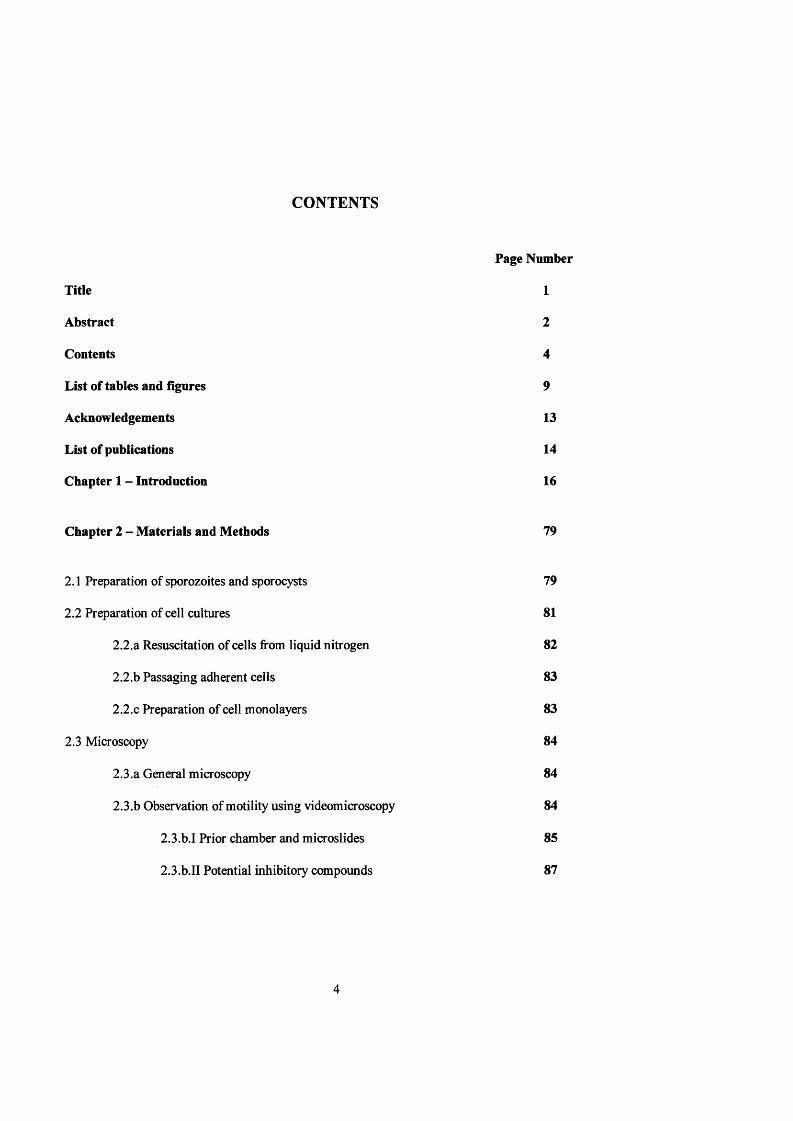
CONTENTS
Page Number
Title 1
Abstract 2
Contents 4
List of tables and figures 9
Acknowledgements 13
List of publications 14
Chapter 1 - Introduction 16
Chapter 2 - Materials and Methods 79
2.1 Preparation of sporozoites and sporocysts 79
2.2 Preparation of cell cultures 81
2.2.a Resuscitation of cells jfrom liquid nitrogen 82
2.2.b Passaging adherent cells 83
2.2.C Preparation of cell monolayers 83
2.3 Microscopy 84
2.3.a General microscopy 84
2.3.b Observation of motility using videomicroscopy 84
2.3.b.I Prior chamber and microslides 85
2.3.b.II Potential inhibitory compounds 87

2.3.b.III Motility studies 88
2.3.b.IV Sperm tracker analysis 90
2.3.b.VExcystation studies 91
2.3.b.VI Invasion studies 92
2.3.b, VII Calculation of speeds 94
2.3.c Immunofiuoresence microscopy 95
2.3.C.I Preparation of coated coverslips 95
2.3.c.II Staining protocol 96
2.3.d Electron microscopy 97
2.3.d.I Fixation and embedding of material 97
2.3.d.II Sectioning 99
2.3.d.III Staining thin sections 99
2.3.d.IV Immuno-electron microscopy 99
2.4 Identification of cell proteins 100
2.4.a Preparation of polyacrylamide gels 100
2.4.b Preparation of protein samples for SDS electrophoresis 101
2.4.C Transfer of proteins to nitrocellulose membranes 104
2.4.d Western blotting 106
2.4.d.I Antibody treatment of nitrocellulose sheets 106
2.4.d.II Detection protocols 109
2.4.d.III F-actin overlay 111
2.4.e Chromatography 112
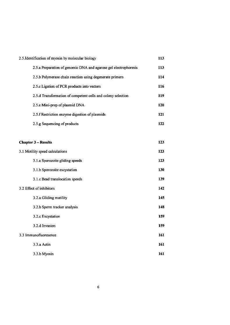
2.5.Identifîcation of myosin by molecular biology 113
2.5.a Preparation of genomic DNA and agarose gel electrophoresis 113
2.5.b Polymerase chain reaction using degenerate primers 114
2.5.C Ligation of PCR products into vectors 116
2.5.d Transformation of competent cells and colony selection 119
2.5.e Mini-prep of plasmid DNA 120
2.5.f Restriction enzyme digestion of plasmids 121
2.5.g Sequencing of products 122
Chapter 3 - Results 123
3.1 Motility speed calculations 123
3.1 .a Sporozoite gliding speeds 123
3.1 .b Sporozoite excystation 130
3.1.C Bead translocation speeds 139
3.2 Effect of inhibitors 142
3.2.a Gliding motility 145
3.2.b Sperm tracker analysis 148
3.2.C Excystation 159
3.2.d Invasion 159
3.3 Immunofiuoresence 161
3.3.a Actin 161
3.3.b Myosin 161

3.4 Cell ultrastructure 164
3.5 Western blotting 181
3.5.a Actin 181
3.5.b Myosin 183
3.5.C Actin associated proteins (AAPs) 186
3.6 Fast Protein Liquid Chromatography (FPLC) 188
3.7 Identification of myosin sequence 189
3.7.a Extraction of genomic DNA 189
3.7.b PCR products 189
3.7.C Selection of inserts 191
3.7.d Sequence data analysis 191
Chapter 4 Discussion 198
4.1 Speed of movement 198
4.2 Erratic nature of movement 199
4.3 Cell ultrastructure 201
4.4 Cell-substratum interactions 202
4.5 Bead-cell surface interactions 230
4.6 Excystation 231
4.7 Nature of the motor 234
4.8 Invasion 241

References 245
Appendices 288
1 Chemicals 288
2 Standard proteins 291
3 Antibodies 292
(a) Primary antibodies 292
(b) Secondary antibodies 293
4 Molecular biology 294
5 Miscellaneous 295

LIST OF FIGURES AND TABLES
Figures:
Page Number
Figure 1.1: A cartoon of a typical apicomplexan zoite 17
Figure 1.2: The classical coccidian life 19
Figure 1.3: The life cycle of Eimeria tenella in the caecum of the chicken 22
Figure 1.4: Model of the moving junction of invasion 29
Figure 1.5: Cartoon of a Gregarina blaberae fold 34
Figure 1.6: The cellular functions of the 15 myosin classes 53
Figure 1.7: Hydrolysis of ATP by the myosin head 55
Figure 1.8: Similarities in the basic structure of kinesin myosin 58
Figure 1.9: Myosin neck domains 58
Figure 1.10: A phylogenetic tree of myosin motor domains 76
Figure 2.1 : Diagram of the Prior perfusion chamber apparatus 86
Figure 2.2: Diagram of the protein transfer ‘sandwich’ apparatus 105
Figure 2.3: Diagram of the slot blot apparatus 107
Figure 2.4: Schematic diagram of the HRP conjugate detection system 110
Figure 2.5: Generation of new DNA strands during PCR 117
Figure 3.1(a) & (b): Graph of cumulative & individual distances moved by zoites 127
Figure 3.2: Cartoons of RIM studies on gliding sporozoites 129
Figure 3.3: Excystation of two sporozoites from a single sporocyst 132
Figure 3.4: High magnification observation of an excystation 133
Figure 3.5: Graph of individual and cumulative during excystation 137

Figure 3.6(a) & (b): Graph of individual & cumulative distances moved by a 2/xm bead 141
Figure 3.7: Sequential photographs of a bead translocation 143
Figure 3.8: Cartoon of the bead translocation shown in figure 3.7 144
Figure 3.9(a) & (b): Inhibition of sporozoite motility in CD & BDM 146
Figure 3.10: Graph of Hobson Sperm Tracker analysis 149
Figure 3.11(a) & (b): Sporozoite excystation control conditions & in DMSO 153
Figure 3.12(a) & (b): Sporozoite excystation in 0.1 &1 /igml ‘ Cytochalasin D 154
Figure 3.13(a) & (b): Sporozoite excystation in 75 &100mM BDM 155
Figure 3.14(a) & (b): Sporozoite excystation in ImM Colchicine & l/xgml'* Salinomycin 156
Figure 3.15: Mean % invasion of MDBK cells by sporozoites 158
Figure 3.16: Photograph of actin immunofiuoresence 161
Figure 3.17: Photograph of myosin immunofiuoresence 162
Figure 3.18: TEM section through an uninvaded CaC02 cell 164
Figure 3.19: TEM of a flexing sporozoite 166
Figure 3.20: Cartoon of the flexing sporozoite in figure 3.19 167
Figure 3.21 : CaC02 cells 20 minutes post invasion 169
Figure 3.22: TEM of a CaC02 cell 20 minutes post invasion 170
Figure 3.23: TEMs of a CaC02 cell 20 minutes post invasion 171
Figure 3.24: TEM of a possible sporozoite Golgi complex 172
Figure 3.25: TEM of a CaC02 60 minutes post invasion 173
Figure 3.26: High magnification TEM of in intracellular 174
Figure 3.27: TEM of the anterior region of a sporozoite 175
Figure 3.28: TEM of a sporozoite at the base of an INT 407 cell 176
10
Figure 3.29: A high resolution TEM of the apical tip of a sporozoite 177
Figure 3.30: A high magnification TEM of a horizontal sporozoite section 178
Figure 3.31 : A high magnification TEM of an intracellular sporozoite 179
Figure 3.32: Anti-actin Western blot and corresponding Coomassie stained gel 181
Figure 3.33: Actin dilution series 183
Figure 3.34: Anti-myosin Western blot 184
Figure 3.35: Anti-myosin Western blot using two anti-myosin antibodies 186
Figure 3.36: UV illuminated photograph of Eimeria total genomic DNA 189
Figure 3.37: UV illuminated photograph of PCR products 190
Figure 3.38: UV illuminated photograph of restriction enzyme digests and inserts 192
Figure 3.39: Sequences for the Eimeria tenella cloned insert DB-07 193
Figure 3.40: Alignment of the Eimeria tenella insert protein 195
Figure 3.41: Alignment with Plasmodium falciparum & Toxoplasma gondii 196
Figure 4.1 : Interaction of microneme components, sporozoite motor and substratum 206
Figure 4.2: Determination of contact size between a 2/im bead and sporozoite surface 209
Figure 4.3: Gliding sporozoite displacing its body volume of medium 212
Figure 4.4: Models of actin & myosin interacting with an extracellular substrate 221
Figure 4.5(a) & (b): Models of single and multiple microfilament and labile myosins 224
Figure 4.6(a) & (b): Models of zoite-substrata/bead contact area 226
Figure 4.7: Cytoskeletal changes mediated by the small OTP binding protein, Rho 229
Figure 4.8: Cartoon of sporozoite compression 233
Figure 4.9: Sporozoite invasion of a target cell 243
11

Tables:
Table 1.1; Classification of the Coccidia 21
Table 1.2: Myosin class size 72
Table 3.1: Characteristics of Eimeria sporozoites gliding on a glass substratum 124
Table 3.2: Characteristics of Eimeria sporozoites gliding on a MDBK monolayer 125
Table 3.3: Characteristics of Eimeria sporozoite gliding in PBS on a glass substratum 126
Table 3.4: Characteristics of Eimeria sporozoite excystation 134
Table 3.5: Characteristics of Eimeria sporozoite excystation at 5 second intervals 136
Table 3.6: Characteristics of 2/xm bead translocations 139
Table 3.7: Characteristics of 2/[xm bead translocations at 0.08 second intervals 140
Table 3.8: Motility indices for sporozoites incubated in cytochalasin D 145
Table 3. 9: Motility indices for sporozoites incubated in BDM 147
Table 3.10: Motility indices for sporozoites incubated in Colchicine 147
Table 3.11: Motility indices for reversibility of inhibitory effects 148
Table 3.12: Sperm tracker analysis of sporozoite motility in potential inhibitors 150
Table 3.13: Actual number of motility events recorded on the sperm tracker video 152
Table 3.14: Mean sporozoite invasion rates 159
Table 4.1 : Energy requirements in Plasmodium and Eimeria sporozoites 215
12

ACKNOWLEDGEMENTS
I would first like to thank my supervisor. Dr. Conrad King for all his help and patience
over the last few years. He has always been available with a fnendly word of advice and good
humour, and it has been much appreciated.
I would also like to thank those people who, in various moments have acted as second
supervisors; Mr. Terry Preston (UCL), Dr. Peter Daszak (Kingston University), Dr. Bob Thong
(Pfizer) and Mr. Adrian Thompson (Pfizer). Once again their help and friendly advice has been of
great importance to me. I would also like to express my gratitude to Drs. Jenny Pinder, Karen
May, Joe Bateman, David Stevens and Adam Watson who have helped me with many of the
technological aspects of the molecular biology investigations, and who have been invaluable
sounding boards.
1 would also like to take this opportunity to thank the numerous friends who have put up
with many of my worst excesses over the duration of my studies. I would especially like to say a
huge thank you to Tin and Niamh, Gales (especially for proof reading duties), Steve and Jools,
Cleo, Nickie, Annette, F liss, the Hartbum boys and Dan and Rob (especially for help with some
of the artwork). I would also like to acknowledge the BBSRC and Pfizer Ltd., Sandwich, Kent
for providing the funds, without which none of this would have been possible.
I would finally like to say a massive thank you to my Mum and Dad for their constant
love and support from the very beginning. Even when I lost faith you didn’t, and that was
fantastic.
For JRW:
as always, the inspiration.
13

PUBLICATIONS/PRESENTATIONS
King C.A., Bruce D. & Sleep J.
Actin-myosin powered cell movement in the Apicomplexan: Two case studies Eimeria and
Gregarinia.
COST 820 - “Vaccines Against Animal Coccidioses - Progress and Prospects” Dublin, June 2000
King C.A., Bruce D. & Sleep J.
Cell Movement in apicomplexan Protozoa- Two case studies of this little studied motility.
Presented at the Spring meeting of the British Society of Cell Biology, April 1999
King C.A., Bruce D. & Sleep J.
Myosin and motility in the apicomplexan Protozoa.
Presented at the Spring meeting of the British Section of the Society of Protozoologists; March
1999
King C.A., Bruce, D. & Hague, J. (1998)
Evidence for the role of myosin in the motility of Eimeria tenella sporozoites. J. Euk. Microbiol.
45 6a
King, C.A. & Bruce, D.
Role for actin and myosin in the motility of Eimeria tenella sporozoites - manifestations in overt
gliding and sporozoite escape from sporocysts.
Presented at the Vllth International Coccidiosis Conference and European Union COST820
Workshop, Oxford, September, 1997
14

Bruce, D. & King, C.A.
Manifestations of cell motility in Eimeria tenella sporozoites: analysis of overt gliding and
microbead translocation.
Presented at the Spring meeting of the British Section of the Society of Protozoologists; March
1997
Bruce, D.R., King, C.A. and Daszak, P.
Actin binding proteins in Eimeria tenella sporozoites.
Presented at the Spring meeting of the British Society of Parasitologists; April 1996.
15
CHAPTER 1: INTRODUCTION
Eimeria tenella is an obligate intracellular parasite within the phylum Apicomplexa
and is the causative agent of the disease known as coccidiosis. Its definitive host is the domestic
chicken Gallus gallus. Under battery farming conditions it can cause substantial economic loss
such that in the USA alone, coccidiostats worth $250-$300 million annually are routinely added
to commercial feed preparations (Parry et al, 1992).
The phylum Apicomplexa was established in 1970 by The Society of Protozoologists
(Levine, 1970), and further revised in 1980 by Levine (Levine et al, 1980) in an attempt to try
and clarify the confusion which existed in the literature surrounding the organisms described as
the “Sporozoa.” Early electron micrographs identified the presence of the organelles of the
apical complex and this gave the phylum its name (see figure 1.1). These organelles and their
importance are discussed in more detail later.
The classical coccidian life cycle has three major phases: merogony, gametogony and
sporogony (see figure 1.2). Merogony refers to the rounds of intracellular, asexual replication
immediately following target cell invasion, gametogony to the development of gametes from
second or third generation merozoites, and sporogony to sporulation from the zygote (Schmidt
& Roberts, 1996). Both merogony and gametogony are exclusively intracellular processes,
whereas the sporulation seen in sporogony can occur outside the body of the host.
Despite these basic similarities in life cycle, there exists in this group an important
difference in the number of hosts involved in the successful completion of the life cycle. Eimeria
spp. complete their life cycle in a single host. This is in contrast to the more opportunistic
16

conoid
rhoptry
micropore
densegranules
innermembrane
complex
mitochondrion
m
Q
polar ring
micronemes
plasmamembrane
Golgi complex
nucleus
apicoplast
posterior polar ring
Figure 1.1: A cartoon of a typical apicompexan zoite showing the major features of the apical complex (i.e. conoid, polar ring, rhoptries and micronemes). (Original but based on Scholtyseck, 1979).
17
pathogen Cryptosporidium which can infect and successfully develop in many mammalian
species, including man. Plasmodium spp. undergo distinct developmental stages in both a
vertebrate and invertebrate host, and again seem to have highly defined preferences for specific
cell types. Toxoplasma can develop successfully in a wide variety of hosts and does not seem to
be subject to a rigorous host cell specificaticm (Roberts & Schmidt, 1996).
Within the phylum there are two classes; the Perkin sea (containing only two species of
the genus Perkinsus) and the Sporozoea, Wiich contains three subclasses; the Gregarinia, the
Coccidia and the Piroplasmia.
The Gregarinia caitain the majority of the gregarines, whidi are often to be found
adopting an extracellular parasitic mode in the gut and body cavities of arthropods and annelids.
The first account of a gregarine was given in 1787 by Cavolini vho observed what is now
recognised as Cephaloidophorus conformis (Diesing, 1851) undergoing syzygy (a process in
which the gregarine forms a permanent end-to-end association with another gregarine) in the
stomach of the Mediterranean crab Pachygrapusus marmoratus but he perceived this to be a
two-segmented tapeworm. In essence the real discovery of the gregarines was achieved by Léon
Dufour, an insect anatonist, Wio after describing many species gave them the generic name
Gregarina (Hammond & Long, 1973).
Gregarines are generally monoxenous i.e. t h ^ complete their life cycle within one host
species, and have a simple life cycle. Once the host ingests a spore the sporozoites hatch out and
generally invade ^propriate intestinal epithelium cells. Each sporozoite then begins to develop
18

sporozoite
trophozoite
meront merogony
merozoite
microgamétocyte macrogamétocyte
gametogonymicrogamete macrogamete
zygote
oocystsporoblast sporogony
sporocystsporozoite
Figure 1.2: The classical coccidian life cycle showing the three major phases; merogony, gametogony and sporogony. Original but based on “The life history of the Telosporidea” in: How to Know the Protozoa, T.L. Jahn (1949).
19
into a trophozoite, which enlarges and eventually projects into the gut lumen. The trophozoite is
divided by septa into three sections; the anterior epimerite, which maintains the attachment
between gregarine and host cell, the central protomerite and the posterior deutomerite. The
gregarine subsequently detaches from the host cell, enters the gut lumen, and the epimerite is
lost. In the gut lumen the gregarine forms a permanent end-to-end association with another
gregarine which is termed syzygy. This represents the commencement of the sexual phase of the
life cycle. Subsequently the protomerite and deutomerite of each organism merge forming a
gametocyte. The elements of the two different gregarines form paired associations which
eventually leads to the budding off of gametes from each gametocyte. Each gamete then fuses
with a gamete from the second gregarine to form a zygote. The many zygotes form resistant
outer shells and develop into sporocysts within which sporozoites develop. Under favourable
environmental conditions in the external environment, spore tubes develop through which
infective spores are released (Canning, 1956).
An interesting feature of the gregarine life cycle is the host-parasite synchronisation
which exists in many species e.g. Gregarina gamhami from the grasshopper Locusta migratori
(Corbel, 1964). In this example the gregarine life cycle is completed in 11-13 days with the
majority of cysts being shed at the end of the fifth larval stage. This coincides with the shedding
of the insect gut lining. Hence the gregarines are present in the lumen of the gut, where syzygy
and subsequent sporocyst development can occur. That these two events seem to be linked was
demonstrated by Corbel (1964) who observed that a delay in moulting followed removal of the
host’s ventral gland, no massive discharge of cysts being observed until moulting was
subsequently induced.
20

The subclass Coccidia is further classified as shown in table 1.1:
Subclass Order Suborder Genus
Coccidia Eucoccidiida AdeieinaEimeriina
Haemospororina
AdelinaEimeriaToxoplasmaNeosporaIsosporaCryptosporidium
Plasmodium
Table 1.1: Classification of the Coccidia.
The Coccidia were first discovered by Leeuwenhoek in 1674 (Smyth, 1994) when he
observed oocysts of Eimeria steidei in the bile of a rabbit (Levine et al, 1980). It was Ifeke in
1839 who first described them, although he thought the oocysts were probably pus particles from
a hepatic carcinoma.
The coccidian life cycle was elucidated step by step and was first fully worked out by
Kloss in 1855 for the snail coccidium which Schneider later named Kloss helicana (Smyth,
1994). In 1870 Eimer described the life cycle of Gregarina falciformis in the mouse, which
Schneider (1875) subsequently renamed Eimeria falciformis (Smyth, 1994). This was adopted as
the type species for the new genus. Eimer also held that the infection was spread through the
transmission of the oocysts fi*om mouse to mouse, a view which did not receive broad
endorsement until the turn of the century (Hammond & Lcmg, 1973). Within the Eimeriina
some genera are monoxenous e.g. Eimeria, Cryptosporidium and others are heteroxenous e.g.
Toxoplasma. In the classical Eimerian life cycle (see figure 1.3) the infection is transmitted by
the faecal-oral route and begins when an animal ingests a sporulated oocyst.
21

merozoite
0 repeated gametocyteormationmerogon
©
sporozoite
pnsporulated oocyst
sporulated oocyst
©0
micro-gametogenesis
g)
îmacro-gametogenesis
fertilisationSporogony - (outside host)
developingzygote
Figure 1.3: The classical coccidian life cycle as represented by the life cycle of Eimeria tenella in the caecum of the chicken. (Original, but partly based on Hoare, 1949).
92

This subsequently ruptures to release four sporocysts each containing two sporozoites.
Following the action of bile and trypsin on the sporocyst the sporozoites become motile, escape
from the sporocyst and invade an appropriate host cell (for Eimeria tenella this is a caecal
epithelium cell). In this intracellular site the parasite resides within a parasitophorous vacuole
(which contains both host and parasite derived components) and begins to divide by merogony,
yielding a large number of first generation merozoites (Schmidt & Roberts, 1996). These
merozoites escape from the host cell by destroying it and invade neighbouring cells. In these
cells a further round of merogony can occur. They escape as before and further rounds of
asexual replication can occur or they may enter the sexual phase of the life cycle. If they enter
the sexual phase, the merozoites enter a new host cell as before but undergo gametogony
developing into either a single macrogamete (essentially the female gamete) or numerous
flagellated microgametes (essentially the male gametes). The microgametes lyse their host cells
and travel through the gut lumen in order to locate a cell containing a macrogamete. They
penetrate this cell and fertilise the macrogamete to form a zygote which subsequently develops
into an oocyst. The oocyst breaks out from the cell and passes out of the host in the faeces. If the
conditions are correct it will undergo sporulation. The sporulated oocyst will contain the
characteristic four sporocysts each containing two sporozoites (Hammond & Long, 1973).
Eimeria infections are generally self-limiting and in the wild pose little threat to the wellbeing
of the host as some degree of immunity is developed. Cuckler & Malanga (1956) observed the
development of an immune response in chickens to Eimeria acervulina on the eighth day
following the initial challenge. A subsequent oocyst challenge on the twenty-first day showed
the chickens to be immune. However in battery farming conditions where infections can be
frequent, the pathology observed can be very severe. With large numbers of merozoites lysing
epithelium cells there can be a significant reduction in feed conversion rates and this may have a
23

substantial economic impact. In extreme cases the infection may be so severe that the bird dies
(Hammond & Long, 1973).
Cryptosporidium spp. follow a broadly similar life cycle but there are a number of
subtle differences. When a Cryptosporidium sporozoite enters a cell it does not penetrate any
great distance into the host cell cytoplasm. It undergoes division and development lying just
under the cell membrane in a manner which has been described as “intracellular but
extracytoplasmic” (Canning, 1990). In addition the oocyst is fully sporulated when released into
the gut lumen and contains four naked sporozoites i.e. there are no sporocysts. A further
interesting feature is that two types of oocyst are produced: thick-walled oocysts and thin-walled
oocysts. The thick-walled oocysts comprise approximately 80% of those produced and are the
type which are passed out in the faeces to enable transmission to other hosts. The thin-walled
oocysts are fragile and generally rupture inside the gut lumen, releasing infectious sporozoites
which are responsible for autoinfection. It is this contrast with the self-limiting infection seen in
Eimeria that has seen Cryptosporidium become a significant consideration in the treatment of
immunocompromised individuals. Approximately 10% of AIDS patients in the USA develop
cryptosporidiosis (Petersen, 1993) and the often severe diarrhoea that the infection causes is a
major complication in the treatment of these individuals.
Despite acknowledgement of its potential pathogenicity in man it is only relatively
recently that Toxoplasma has been recognised as a coccidian parasite, having previously been
placed in various taxonomic groups, including the fungi. Hutchison (1965) fed mice infected
with Toxoplasma to cats and the cat faeces was seen to be infective to mice. It was initially
thought that transmission was facilitated via Toxocara cati (nematode) eggs but subsequent
experiments with worm-free cats showed this not to be the case and further work identified the
Toxoplasma oocyst in the faeces (Sheffield & Melton, 1968; Work & Hutchison \969a&b'.
24

Frenkel, 1970; Frenkel et al, 1969). The paucity of information about Toxoplasma until recent
times is all the more astonishing when one examines the rate of infection in humans.
Transmission is through the faecal-oral route, eating undercooked meat or by transplacental
transmission. In some areas of the USA seropositive individuals represent 30% of the
population, and in the UK values range from 0.1% in 1 year olds to 45% in the over 50’s
(Canning, 1990). In France, where meat is often less thoroughly cooked, some studies have
indicated seropositive levels of between 80-90%. Childs & Seegar (1987) found that 14.5% of
cats in the USA were infected with Toxoplasma and a value of 78% was seen in cats in Beirut
(Deeb et al, 1985). With the discovery of HIV and AIDS in the mid-1970s, research into all
aspects of Toxoplasma infections has increased substantially as a result of the impact that this
parasite can have on immunocompromised patients. The definitive host for Toxoplasma gondii
is the cat. Following ingestion of an infective stage (either an oocyst or cyst - see later) the zoites
invade an intestinal epithelium cell and undergo multiplication in an unusual process known as
endodyogeny. In this process two daughter cells are formed inside the mother cell which is
subsequently consumed by the developing daughter cells (Goldman et al, 1958). As in other
coccidians this asexual cycle is followed by macro- and microgametogenesis, fertilisation and
the formation of an oocyst. The unsporulated oocyst passes out in the faeces and under the
correct conditions will develop into the infective oocyst containing two sporocysts each
containing four sporozoites (Hammond & Long, 1973). In addition to the intestinal
development, some zoites invade other areas of the body and undergo cell division in these
tissues. Generally by the end of the second week post-infection these lesions have subsided but
may persist as cysts in the brain and muscles of the cat (Dubey & Frenkel, 1972).
One of the most striking features of Toxoplasma is its ability to invade numerous cell
types in many different hosts. At least 300 mammalian and 30 avian species have been
25
identified as intermediate hosts (Schmidt & Roberts, 1996). However in these animals it is
unable to complete the sexual phase of its life cycle and the zoites undergo a rapid succession of
endopolygenic divisions to form a pseudocyst containing many tachyzoites. This cell containing
the pseudocyst eventually ruptures, releasing the tachyzoites which then invade further cells. At
this stage immunity to Toxoplasma begins to develop and proliferation is slowed down,
eventually leading to the development of true cysts containing bradyzoites. These cysts are found
in all parts of the body including muscle and the central nervous system. They are up to 60pm in
diameter and may persist for the lifetime of the host (Canning, 1990). It is the reactivation of
these cysts that can present serious problems in immunocompromised hosts. In humans the
disease is generally asymptomatic but may be accompanied by some fever and swelling.
However in immunodeficient individuals the disseminated toxoplasmosis can lead to blindness
and if present in the CNS may cause fatal encephalitis (Hughes, 1985; Holliman 1988).
Congenital toxoplasmosis is most likely to occur when the mother acquires the infection for the
first time and transmission to the foetus is via the placenta. The likelihood of foetal infection can
be as high as 45% and of those infants born 60% will present no clinical symptoms. However
30% of those remaining will develop serious ocular and neurological complications resulting in
mental and visual impairment and some 9% of these will die (Apt, 1985; Hughes, 1985).
In many ways Toxoplasma has become the model apicomplexan. There exists a large
amount of data concerning many aspects of this organism and its host cell promiscuity allows in
vitro studies to be performed easily. In addition in recent years it has been possible to genetically
manipulate the parasite (Klein & Fitzpatrick-McElligott, 1993; O’Neill et al, 1993) and these
techniques are likely to significantly advance our understanding of both Toxoplasma and the
other coccidians.
26

It is difficult to overestimate the impact that malaria has had on human health. In
Afi'ica alone there are at least 1 millim fatalities fi’om mœe than 100 million cases of malaria
recorded annually. The morbidity caused by the infection has far reaching implications on the
social and economic development of communities in countries where the balance between
poverty and prosperity is often already precarious. Increasingly problems are being eneountered
with multi-drug resistant strains of Plasmodium^ and it is clear that malaria will caitinue to be a
huge public health problem for many years to come.
In the 1890s Patriek Manson postulated that mosquitoes were responsible for the
transmission of the filarial worms which caused elephantiasis and suggested that this
mechanism was also responsible for the transmission of malaria (Hammond & Long, 1973). In
1895 Ronald Ross, a British Army doctor posted to India, began work on mosquitoes fed blood
fi'om patients infected with malaria but was unable to discover parasites within the mosquitoes.
This is not surprising as he had been using the wrong mosquitoes but, in June 1897 he acquired
a sample of anopheline mosquitoes (subsequently identified as Anopheles stephensi) and
following a blood meal from a patient infected with malaria, was able to recognise the
characteristic haemozoin granules in the stonach of two of the mosquitoes. Ifc was transferred
shortly after this diseovery and had to continue his work on P. relictum in sparrows. It was in
this species that he found the sporozoites in the mosquito salivary glands and by successfiilly
using these sporozoites to infect other sparrows he proved the role of the mosquito as the vector
for the transmission of malaria. Around this time Grassi and his co-workers confirmed this work
in the human malaria parasites, P. falciparum and P. vivax in the mosquito, A. claviger
(Hammond & Long, 1973).
The eluei dation of the entire malaria life cycle was completed in 1934 when Raffaele
described schizogony in reticulo-endothelial cells in bone marrow and other organs in the avian
27

malaria P. elongatum and this exo-erythrocytic development was confirmed for mammalian
malaria in 1948 by Short and Garnham who observed P. cynomolgi in monkey liver tissues
(Hammond & Long, 1973).
In man the infection is initiated when a female anopheline mosquito injects saliva
containing Plasmodium sporozoites into the capillary from which she is preparing to feed. These
sporozoites circulate in the blood and upon reaching the liver, invade an appropriate hepatic cell
within 30-60 minutes of their injection. Here they undergo a schizogonie multiplication to form
merozoites or occasionally hypnozoites, which develop much more slowly and can lead to future
relapses. The merozoites lyse the host cell and pass into the blood stream where they attach to
erythrocytes with the aid of a surface coat (Bannister & Mitchell, 1995), and orientate
themselves so that the apical end of the parasite lies next to the red cell membrane. Invasion is
complete within a minute and leads to the development of a parasitophorous vacuole around the
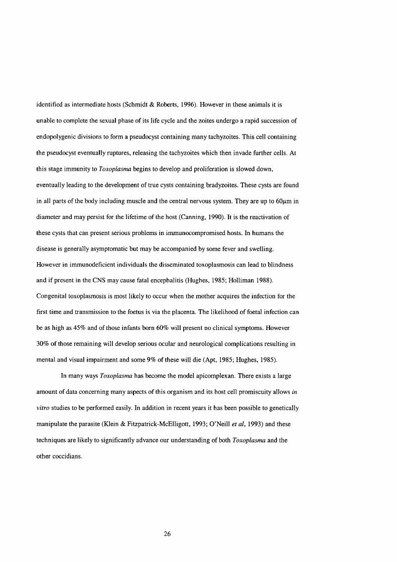
parasite within the red blood. During the invasion process a moving junction is formed (Aikawa
et at, 1978) and the surface coat is shed (Hermentin, 1987; Perkins, 1989). The moving junction
(see figure 1.4) is thought to be formed by the interaction of a subpellicular motor mechanism
linked to the red blood cell surface, via a parasite derived linker protein such as merozoite
capping protein (MCP-1) (Klotz et at, 1995). This motor mechanism and its impact on parasite
gliding motility and host cell invasion will be discussed later. In the erythrocyte the parasite
enlarges to occupy most of the cellular volume and then undergoes differentiation to form
between 8 and 24 nuclei. Each nucleus becomes associated with a cytoplasmic bud and
subsequently develops into a merozoite. Until recently it had been assumed that the merozoites
left the erythrocyte explosively, by inducing the rupture of the cell (Dvorak et al, 1975;
Hermentin & Enders, 1984). However recent work by Clavijo et al (1999) has suggested that
when the parasite leaves the cell it does so in a manner which maintains the structural integrity
28
parasite
plasmalemma
host cell — cytoplasm — nascent
parasitophorous vacuole
# ••
(a)microfilament microneme
protein
parasitecytoplasm
myosin target cell surface ligand
host cell cytoplasm
(b)
extracellular microfilament space
inner sporozoite target cellmembrane plasma plasma
complex membrane membrane
Figure 1.4: Model of the moving junction of invasion formed between the invading parasite and the host cell surface, (a) shows the parasite invading the red cell and the developing parasitophorous vacuole and (b) shows a cartoon of the possible molecular arrangement at the site of parasite/host cell contact. The bridge that links the two is the membrane bound microneme protein. (Original but based on Finder, et al, 1998).
29

of the erythrocyte. These observations would strengthen the validity of the model they proposed
(Clavijo et al, 1998) that suggested that the PVM fuses with the erythrocyte plasma membrane,
forming a compartment which is continuous with the external environment. This would also
facilitate the release of the haemozoin granules.
Under an unknown stimulus, induction of gametocytogenesis occurs within the
vertebrate host, leading to the developmait of micro- and macrogamétocytes. These are ingested
by a feeding mosquito when a blood meal is taken. Within 10 minutes of ingestion the
microgamétocyte undergoes the first of three mitotic divisions leading to the development of 8
haploid nuclei, each of which associates with one of the recently developed axonemal structures.
These fully formed microgametes escape from the erythrocyte in a somewhat explosive manner
(Smyth, 1994). Macrogamete formation is a much less complex process, simply involving the
loss of the red cell membrane. The microgametes swim through the blood meal, fertilise the
macrogamete and form a zygote which subsequently develops into the distinctive oval ookinete
(Sinden & Croll, 1975).
The ookinete is motile and possesses the characteristic apical organelles found in other
invasive stages of Plasmodium (Aikawa & Seed, 1980). It glides through the gut (Freyvogel,
1966) and then passes through the epithelial layer, where it begins to differentiate into an oocyst
at the interface between the basal area of the gut epithelium and the haemocoel. Recent work on
ookinete invasion has identified the presence of a molecule with significant structural homology
to both circumsporozoite protein (CSP) and thrombospcaidin-related adhesive protein (TRAP)
(Yuda et al, 1999). These proteins are implicated in Plasmodium sporozoite gliding motility and
red cell invasion. This suggests a common motility generating mechanism in these different
developmental stages. Numerous sporozoites (up to 1000) develop inside the oocyst and after
about 10-20 days the oocyst bursts, releasing the zoites into the insect haemocoel from where
30

th ^ travel to the salivary glands. Here they can exist either extra- or intracellularly. When the
mosquito next feeds these sporozoites are injected into the blood along with the saliva
(Gamham, 1988).
The subclass Piroplasmia contains two important animal pathogens; Theileria and
Babesia but it is only Theileria that will be discussed here. This organism is a major pathogen of
cattle on the continents of Africa and Asia, transmission to cattle being effected by ixodid ticks.
Theileria parva is transmitted to the African Cape bufialo Syncenis caffer by the tick
Rhipicephalus appendiculatus and causes the usually fatal East Coast fever which is endemic to
sizeable parts of eastern, central and southern Africa (Smyth, 1994). It can cause considerable
loss of livestock and is therefore of great economic significance in the region, since it places
substantial restraints on the development of herds (Mukhebi et al, 1992; Norval et al, 1992).
Theileria annulata is transmitted to Bubalus btibalis, the Asiatic or water buffalo by Hyalomma
spp. and causes theilerosis across a wide geographical range from the Mediterranean through to
China (Dolan, 1989). The infection begins when sporozoites are injected into the bloodstream of
the cattle along with saliva when a tick feeds. Spœozoites invade lymphocytes, with cell entry
complete within 10 minutes (Fawcett et al, 1982). The sporozoites then undergo repeated rounds
of schizogony in vdiich the host cell is induced to divide, a schizont dividing with each of the
new daughter cells and subsequently producing numerous merozoites.
It is this repeated lymphoid schizogony Wiich is the major cause of pathology, but as
later in the infection erythrocytes can also be infected, some anaemia may occur (Irvin &
Morrison, 1987). The merozoites invade erythroc^es and develop into spherical bodies, from
31

which the gametes will develop following ingestion as a tick blood meal. Gametogenesis and
fertilisation occur in the tick gut.
The spherical bodies contained in infected erythrocytes begin to develop into the two
morphologically distinct gametocytes. Macrc^ametes seem to form by an enlargement of these
spherical bodies, although they may simply be cells incapable of further development (Melhom
& Schein, 1984). Once the red blood cells are lysed in the tick intestine, microgametes develop
from the ray bodies classically seen in tick intestinal smears, each yielding four gametes.
Fertilisation occurs wtien two gametes come into cmtact with each other, eventually leading to
the fusion of the two nuclei.
The fused cells now develop, with a large degree of synchronicity (Melhom et al, 1979)
into the club-shaped kinete which enters the haemolymph and invades an appropriate salivary
gland cell. The invasion of the salivary gland cells is closely tied to the life cycle of the host and
is only possible after the tick has moulted (Melhom & Schein, 1984). The salivary glands of
ixodid ticks become reduced following a feed and only develop again post moult when they
become reattached to a suitable animal (Fawcett, et al, 1981a&6, 1982).The kinete then grows
to occupy most of the host cell volume and undergoes divisicxi to produce up to 50,000
sporozoites per cell (Melhom & Schein, 1984). This can lead to an enormous innoculum when
the tick next feeds on a host and this is an element of the severe pathogenicity of theileriosis.
There is no transo\^rial transmission, with the infection being transmitted trans-
stadially e.g. from nymph to adult or from larva to nymph, which is in marked contrast to the
situation in Babesia (Shaw, 1991).
32
The ultrastructure of the Apicomplexa is the defining feature of the phylum. The name
is derived from the organelles comprising the apical complex which is found at the anterior end
of the parasite (see figure 1.1). With advances in electron microscopy in the 1960s allowing
good preservation of protozoan cells and the organelles therein, a number of structural features
became apparent and this radically altered the understanding of the biology of apicomplexan
cells. The main features that are commonly seen in the Apicomplexa are the nucleus,
mitochondrion, a tri-laminar outer cell membrane, rhoptries, micronemes, dense granules,
conoid and associated structures, Golgi apparatus, residual bodies, amylopectin storage granules
and the newly discovered apicoplast. These features are characteristic of the parasites, but not all
of the species contain all the structures and it is clear that some features are absent in different
developmental stages of the same species (Hammond & Long, 1973). The first structure of
interest is the tri-laminar pellicle consisting of an external plasmalemma under which lies two
cytomembranes, resulting in the characteristic three-cortical-membrane pattern. This unique
arrangement was first shown in the coccidian Coleotropha durchoni by Vivier (1963) and
shortly afterwards was described in the gregarine Selenidium hollandei (Vivier & Schrevél,
1964) eventually being recognised as a common feature of the Sporozoa (Vivier et al, 1970).
Freeze fracture studies of the cell surface of Gregarina blabarae by Schrevél et al (1983) led to
the conclusion that the two underlying membranes formed a continuum, being fused at the
anterior and posterior extremities and separated by a flattened vesicle. However similar freeze
fracture studies on T. gondii suggest that in this organism, the inner membrane complex is
formed from a series of flattened vesicles, and somewhat resembles a pavement (Vivier &
Petitprez, 1969). It would also appear that underlying each of the individual pellicular strips is a
subpellicular microtubule, which would suggest a structural role for this component of the
cytoskeleton (Dubremetz & Torpier, 1978).
33

rippled dense structures
12nm filaments
PFp
plasma membrane
PFecinternal lamina
EFic yEFec
cortical cytomembranes
pore-like structure internal lamina
Figurel.5: Cartoon of a Gregarina blaberae fold, illustrating the spatial relationship between the three cortical membranes and associated structures such as the 12nm filaments, as revealed by freeze fracture studies (Schrevél et al̂ 1983). This trilaminar membrane is common to both the Gregarinia and the Coccidia.(Where EFp = plasma membrane: exoplasmic fracture face; PFp = plasma membrane: protoplasmic fracture face; PFec = external cytomembrane: protoplasmic fracture face; EFec = external cytomembrane: exoplasmic fracture face; EFic = internal cytomembrane: exoplasmic fracture face and PFic = internal cytomembrane: protoplasmic fracture face.)
34

In the Coccidia and the Piroplasmia the cell surface is smooth, but in the Gregarinia there
exists a wide diversity of cell forms which are used as the basis for classification. The pellicle of the
gregarines tends to be folded longitudinally (see figure 1.5) and these folds have been implicated in
the gliding motility exhibited in many, but not all, gregarine trophozoites. The nature of the folds
can alter depending upon the age of the parasite, the topographical region of the cell and the degree
of sexual differentiation of the cell. Associated with these folds are three types of structures: 12nm
filaments, apical rippled dense structures and the internal lamina.
The 12 nm filaments have similar biochemical properties to intermediate filaments
(Schrével et al, 1983) and seem to play a scaffolding role, running along the longitudinal axis of the
fold just under the cytomembranes. Mature trophozoites contain two types of fold and these differ in
their height and 12nm filament distribution. In the deutomerite region folds are high (l-2pm) and
contain between 10-12 filaments, whereas folds of the protomerite tend to be smaller (0.6-0.8pm),
more rounded and contain between 14-16 filaments (Schrével et al, 1983). Apical rippled dense
structures are seen at the tip of folds and lie between the plasmalemma and the outer cytomembrane
(Schrével et al, 1983). The internal lamina was named by Schrével et al (1983) and is an electron-
opaque layer of between 15-30 nm which runs under the two cytomembranes in the folds, and also
along the base of the folds, effectively linking the folds together. On the inner, cytoplasmic side of
the folds run a series of microtubules. There are conflicting reports as to the orientation of these
microtubules. It has been reported that they are arranged in parallel orientated along the
longitudinal axis (Vivier & Schrével, 1964), but it has also been shown that they are arranged at 90®
to the longitudinal axis, resembling barrel hoops (Baines, 1988). They are thought to have a
scaffolding function.
35
In the Coccidia the two cytomembranes of the inner pellicular complex are thickened at
each end of the cell, forming die anterior and posterior polar rings. The anterior polar ring is thought
to anchor the sub-pellicular microtubules (Roberts & Hammond, 1970), and may also have a role in
the support of the conoid. The conoid was first observed in Toxoplasma gondii by Gustafson et al
(1954) and has subsequently been found in all coccidians and gregarines so far examined.
The conoid appears as a hollow cone of 6-8 spirally arranged microtubules with two
preconoidal rings immediately anterior to the conoid. These rings are linked to the conoid by an
osmiophilic layer, and in some Eimeria species and T. gondii, the whole structure seems to be
covered with a canopy like membrane containing an aperture at the anterior tip, through which the
rhoptiy ducts pass (Sheffield & Hammond, 1966; Scholtyseck et al, 1970). The size of the conoid
varies 6om species to species, ranging from 1-5% of the total cell body length. The location of the
conoid in comparison with the anterior polar ring varies and it has been suggested that this organelle
can be protruded and retracted. It may therefore have a function in host cell penetration (McLaren &
Paget, 1968; Roberts & Hammond, 1970).
The subpellicular microtubule network originates in the conoid and extends towards the
posterior of the zoite. In Eimeria tenella this array extends two-thirds of the length of the
sporozoite (Russell & Sinden, 1982), apparently terminating adjacent to the posterior of the
nucleus. The number of microtubules present varies greatly depending upon the species and
developmental stage involved. Both sporozoites (Ryley, 1969) and merozoites (McLaren & Paget,
1968) of Eimeria tenella contain 24 microtubules whereas Eimeria nieschulzi sporozoites
(Colley, 1967) and merozoites (Colley, 1968) are seen to possess 25 microtubules. T. gondii
merozoites (Sheffield & Melton, 1968) have 22 microtubules, merozoites o f P. fallax (Aikawa,
1967) have
36

between 24-26 and it would seem that Theileria parva has no subpellicular microtubules at any
stage in its life cycle (Shaw, 1997).
Mitochondria in apicomplexan species are of small size, generally number between one
and three and are often seen to be Y-shaped with characteristic tubular cristae. An interesting
exception is the situation in Cryptosporidium. Merozoites of C. muris have been shown to
possess mitochondria (Uni et al, 1987) but sporozoites of C. parvum appear to lack these
organelles. This observation correlates with the apparent absence of the Krebs cycle (Denton et
al, 1996; Coombs et al, 1997; Entrala & Mascaro, 1997) and lack of sensitivity to respiratory
inhibitors (Brown et al, 1996). However a recent paper in which a C. parvum genomic DNA
library was probed for mitochondrion specific genes, suggested the presence of at least three
such genes (Riordan et al, 1999). Sporozoites of T. parva contain a single mitochondrion which
is devoid of tubular cristae ( Shaw, 1997).
Micropores are commonly seen in all stages of the Sporozoa and are formed from an
invagination of the pellicle. There seems to be no distinct pattern to their distribution and they
are generally about 200nm in diameter at the pellicle surface and extend about 350nm into the
cell. In most cases only one micropore is seen but in some species numerous organelles have
been recorded e.g. E. alabamensis (Sampson & Hammond, 1971). The fact that these organelles
are not confined to motile stages suggests that they have a fundamental role to play in the
development of the parasite and it is thought they are involved in the uptake of nutrients from
the host-cell cytoplasm (Chobotar & Scholtyseck , 1982). Ryley (1969) observed vesicles lying
under the micropore in sporozoites of E. tenella which appeared to be undergoing digestion.
In sporozoites of Cryptosporidium, Toxoplasma and Eimeria the nucleus resides in the
rear two-thirds of the cell and in Cryptosporidium and Eimeria is sandwiched between the
anterior and posterior refi*actile bodies. The refi-actile bodies are electron dense structures which
37
are thought to act as energy reserves whilst the parasite is in the oocyst or undergoing
schizogony (Chobotar & Scholtyseck , 1982). There is some evidence that proteinaceous
components of the reff actile bodies are involved in the development of immunity to E. tenella in
chickens (Danforth & Augustine, 1989).
Sporozoites of T. parva differ significantly to the cells described above. They contain an
eccentric nucleus occupying up to 50% of the total cell volume. There is no obvious nucleolus or
evidence of condensed chromatin (Shaw, 1991).
In recent years a number of apicomplexan parasites have been recorded as containing a
plastid-like structure with a 35kb circular genome (McFadden et al, 1996) termed the
apicoplast. This has a probable green algal origin (Kohler et al, 1997). The original
identification was based on the presence of multiple surrounding membranes (Kohler et al,
1997; Hackstein et al, 1998). It has been assumed that the plastid was acquired by secondary
endosymbiosis, i.e. the ancestral apicomplexan ingested a eukaryote which itself had ingested an
alga (Kohler et al, 1997). Absolute confirmation of the presence of an apicoplast can only be
provided by in situ hybridisation and thus far this has only been achieved with Toxoplasma
(McFadden et al, 1996). The function of the apicoplast is as yet unknown but one can assume
that since this organelle continues to replicate (by binary fission and relatively early in the
replication cycle - Kohler et al, 1997), it must have an important role or roles in the successful
completion of the life cycle. It is also likely that the original piece of DNA was larger than the
35kb remnant present today and that, in line with the situation in other endosymbiotic genomes
some of the original functions have been lost or passed to the nucleus (Palmer, 1985; Gray,
1989). In addition to photosynthesis, plastids are also capable of amino acid and fatty acid
biosynthesis, nitrate and sulphate assimilation and the storage of starch (Hrazdina & Jensen,
1992). It may be that one or more of these metabolic processes are controlled by the apicoplast.
38

Perhaps the most important feature of the apicoplast is that it potentially provides an excellent
target for chemotherapy (Soldati, 1999). It has been suggested that this organelle is the target for
the macrolide antibiotics in Toxoplasma (Beckers et al, 1995: Fichera et al, 1995), and
rifampicin in Plasmodium infections (Pukrittayamee et al, 1994).
Three further organelles that are unique to the apicomplexa are the micronemes,
rhoptries and the dense granules/bodies. They are bounded by membranes and their contents are
thought to be involved in target cell invasion and the initiation and maintenance of the
parasitophorous vacuole in which the intracellular stages of the parasite resides. Micronemes are
small (~ 60-90nm in diameter) spherical organelles which lie adjacent to the rhoptries at the
anterior end of the parasite. They were first discovered in Toxoplasma by Gustafson et al (1954)
and named “toxonemes”. Subsequent identification of similar structures in other coccidians led
to a profusion of names but the term microneme was proposed by Jacobs in 1967 and has passed
into general use. The first microneme derived proteins discovered were adhesive proteins found
in merozoites of P. knowlesi and P. falciparum (Camus & Hadley, 1985; Adams et al, 1992).
Further work showed that despite differences in ligand binding specificity they were part of a
highly conserved protein family in the genus Plasmodium. Subsequently a second family of
highly conserved proteins were discovered to be present in the micronemes of Plasmodium
(Robson et al, 1988), Eimeria (Tomley et al, 1991) and Toxoplasma (Wan et al, 1997) which
contained a number of thrombospondin-like domains. In Plasmodium these are responsible for
host cell binding (Frevert et al, 1993). Thrombospondin is an extracellular matrix protein which
can interact and bind to integrins. Integrins are the principal animal cell receptors for binding
extracellular matrix proteins (Alberts et al, 1994). The presence of a thrombospondin-like
protein on the surface of the parasite would clearly aid attachment to an appropriate target cell.
39

Rhoptries are club-shaped organelles located in the anterior third of the parasite and
possess ducts which pass through the conoid and associated structures. They were first identified
in Haemamoeba (Plasmodium) gallinacea by Garnham et al (1960) who labelled them as paired
organelles, and were subsequently discovered to be present in all the motile, invasive stages of
coccidian parasites. In light of this in 1967 Sénaud coined the term rhoptry to describe these
structures from the Greek rho meaning club-shaped (Hammond & Long, 1973). Between 2 and
20 individual rhoptries have been reported for different species of coccidia (Chobotar &
Scholtyseck, 1982) with 6-8 being seen in sporozoites of Eimeria (Scholtyseck, 1973). In
Plasmodium sporozoites and merozoites 2 rhoptries per parasite are commonly seen, but
interestingly in T. gondii tachyzoites 6-12 rhoptries have been observed in intracellular
replicative phases as well as actively invasive stages (Lingelbach & Joiner, 1998). Merozoites of
Cryptosporidium have been reported to possess 2 rhoptries (Current, 1989) and a recent study by
Tetley et al (1998) found the sporozoite of Cryptosporidium parvum to contain only 1 rhoptry.
Original theories on the role of the rhoptries postulated that they were in some way connected
with the functioning of the conoid (Scholtyseck, 1979). However, the occurrence of rhoptries in
those stages of the parasite that are motile and invasive strongly suggests that they are involved
in host cell entry. Consequently a large amount of work has been directed towards elucidating
the composition, function and fate of the rhoptry contents. It would seem that the make-up of the
rhoptry contents is complex and in keeping with many of the other features of the Apicomplexa,
there is a fair degree of variation from species to species. Plasmodium and Toxoplasma rhoptries
contain 12-15 major proteins whereas E. nieschulzi contains only 4 major proteins (Perkins,
1992). Analysis of these proteins has shown that they contain a high proportion of
intradisulphide bonds indicating that they are highly folded and compact (Cooper et al, 1988;
Sam-Yellowe et al, 1988; Sadak et al, 1988). Further evidence points to the rhoptries being part
40
of a classical secretory pathway. In ultrastructural studies of Plasmodium small vesicles were seen
to fuse with the rhoptries (Bannister & Mitchell, 1995). Rhoptry proteins are synthesised with a N
terminal sequence which aids transfer across endoplasmic reticulum membranes (Lingelbach,
1993) and also post-translational maturation of rhoptry proteins is inhibited by treatment with
brefeldin-A or incubation at 15°C (Ogun & Holder, 1994) - known inhibitors o f the classical
secretory pathway (i.e. nucleus E R -^ Golgi -> target site).
In other eukaryotic organisms secreted proteins are ultimately derived from the Golgi
apparatus and it has been proposed that this is also the case for the Apicomplexa (Porchet &
Torpier, 1977). However there is no evidence for the presence of a Golgi network in C. parvum
(Tetley et al, 1998). It has been suggested that, as the classical stacked Golgi cistemae are never
seen in Plasmodium, these organisms also lack Golgi (Banting et al, 1995), However localisation
studies with the Rab family of proteins indicates that the parasite does indeed contain Golgi
(Ward et al, 1997), The Rab family of proteins are monomeric GTPases, involved in the correct
targeting of transport vesicles to their appropriate docking site (Alberts et al, 1994),
As mentioned previously the protein content of the rhoptries, micronemes and dense
granules are highly complex but also very specific (Perkins, 1992), In addition they are formed
sequentially through merogony and this argues for a mechanism with a high degree of regulation
of protein synthesis, packaging and targeting (Ward et al, 1997),
There are three mechanisms which bring about organelle protein targeting. The first is
the secretory pathway which involves the newly translated proteins localising to the endoplasmic
reticulum, where they are subsequently packaged as either membrane-bound or soluble proteins
within a membrane vesicle (Walter & Blobel, 1981), From here they translocate through other
subcellular compartments, including the Golgi, and ultimately these vesicles fuse together to
41
form a new organelle or fuse with an existing one to enlarge it. This is seen in the generation of
lysosomes and secretory granules (Farquhar, 1985). In the second mechanism the proteins are
synthesised as soluble cytoplasmic proteins and are imported into the pre-existing organelle
post-translationally. In the case of the mitochondria, this process is facilitated by the presence of
N-terminal signal sequences known as mitochondrial targeting sequences or presequences
(Schatz & Doberstein, 1996; Neupert, 1997; Pfanner et al, 1997). These extensions are typically
15-40 amino acids in length and are composed of predominantly positively charged and
hydroxylated residues (mainly serine). Since these presequences can form amphipathic a-helices
it is thought to be important for their recognition by either the outer or inner mitochondrial
complex membranes (von Heijne, 1986; Roise & Schatz, 1988). Progeny organelles are
generated by membrane fission (Lazarow & Kukui, 1985), and it is by this route that
peroxisomes and to a large extent, mitochondria receive their proteins (Hartl & Neupert, 1990;
Fukui et al, 1984). The third mechanism is the targeting of mRNA molecules to the site of
action of the protein, so that the polypeptide is synthesised at the exact location where it will
perform its function. How the mRNA is delivered to the site is as yet unclear, but may involve
movement along the microtubules or actin filaments (Singer, 1996). Studies on the transport of
the mRNA for P-actin to the growth cone of the developing axon, have shown that these
molecules travel along microtubules and are somewhat granular (these granules also contain
other components necessary for translation e.g. ribosomes, tRNA synthetase, etc.) (Singer,
1996). Interestingly, in fibroblasts the P-actin mRNA seems to be transported along
microfilaments, and tends to be less clustered (Singer, 1996).
Howard & Schmidt (1995) examined the localisation of the Plasmodium rhoptry
associated protein-1 (Rap-1) gene product to study rhoptry maturation. The Rap-1 gene encodes
for an 86kDa precursor protein (Pr86). Upon maturation this is processed to an 82kDa protein
42
(p82) found in the rhoptry. Both treatment of cells with brefeldin-A, and incubation at low
temperature prevented the processing of the mature protein, suggesting that rhoptry biogenesis
is at least partially controlled by the classical secretory pathway, and involves the Golgi
apparatus. Since Cryptosporidium is seen to possess a rhoptry, the apparent lack of a structurally
distinct Golgi complex is all the more puzzling and needs to be investigated further.
The micronemes, rhoptries and dense granules are of vital importance in host cell
invasion and the initiation and modification of the parasitophorous vacuole. There is a defined
sequential release of the components of the three organelles (Carruthers & Sibley, 1997). The
microneme contents are discharged upon contact with an appropriate cell or substratum, the
rhoptry contents are discharged at the onset of host cell invasion and the dense granule contents
are released once the parasite is fully internalised in the parasitophorous vacuole (Carruthers &
Sibley, 1997). These processes seem to be under the control of specific signals.
The main function of the proteins present within the micronemes, rhoptries and dense
granules appears to be to aid target cell invasion, and the initiation and maintenance of the
parasitophorous vacuole (PV) (Scholtyseck & Piekarski, 1965). This is a structure unique to the
Apicomplexa, and allows the parasite to replicate within the host cell whilst effectively
circumventing the host immune response. This is demonstrated by the failure of the PV to
acidify (Sibley et al, 1985) or to fuse with other endocytic vesicles (Jones & Hirsh, 1972; Joiner
et al, 1990). Each of the parasite organelles release their contents at a specific stage of the
invasion process and a number of the proteins involved have been characterised, providing
important clues as to their role in host cell invasion.
Microneme proteins seem to be released upon the binding of the parasite to the host
cell. On initial contact with a suitable cell the parasite orientates itself such that the anterior tip.
43

and consequently the organelles of the apical complex, are in close apposition with the host cell
membrane. Exocytosis of microneme proteins has been reported for Eimeria (Tomley et al,
1996), Plasmodium (Spaccapelo et al, 1997) and T. gondii (Carruthers & Sibley, 1997). As
outlined above the microneme proteins so far characterised contain highly conserved binding
domains typified by the thrombospondin-related anonymous protein (TRAP) family (Robson et
al, 1988) and the circumsporozoite protein (CSP) (Yoshida et al, 1980). Both are found in
Plasmodium and play an important role in hepatocyte binding (Nussenzweig & Nussenzweig,
1985). These proteins are conserved in all species of Plasmodium so far studied (McCutchan et
al, 1996; Templeton & Kaslow, 1997) and as outlined above it seems other apicomplexans
possess microneme proteins with significant sequence homology to TRAP and CSP. However
the T. gondii micronemal protein M lCl lacks significant homology with both CSP and TRAP. It
is suggested that this may be the basis for the difference in host cell range, with T. gondii MICl
recognising a ubiquitous surface receptor (Formaux et al, 1996), and hence possessing a
mechanism to recognise, bind to and invade many different cell types.
It is thought that these proteins are released from the micronemes, pass out onto the
surface of the parasite, where a possible domain near to the C-terminal end binds to the outer
membrane of the parasite, promoting the close association between the host and parasite
plasmalemmae (Dubremetz et al, 1998). Similar studies have shown that two proteins found in
the micronemes, the Duffy binding protein in P. knowlesi (Adams et al, 1990) and EBA-175 in
P. falciparum (Sim et al, 1992), are involved in recognition and attachment of merozoites to
erythrocytes. Invasion is characteristically complete within 30 - 40 seconds (Morisaki et al,
1995).
Studies on the T. gondii micronemal protein MIC2 showed that none of this protein
was detectable on extracellular tachyzoites. When a parasite was in contact with a host cell
44

MIC2 was present on the parasite cell surface, particularly at the anterior tip (Carruthers &
Sibley, 1997). Further studies have shown that the micronemal proteins do not enter the
developing PV (Wan et al, 1997) but remain associated with the tight junction complex and are
eventually shed from the posterior of the zoite when invasion is complete (Carruthers & Sibley,
1997).
An important recent observation has been that P. berghei TRAP knock-out sporozoites
are incapable of gliding motility and are therefore unable to invade host cells (Sultan et at,
1997). These observations clearly indicate an important role for zoite motility in successful host
cell invasion and also strongly suggest that the microneme contents are vital components in the
motility mechanism. Motility strategies will be discussed later.
Discharge of the rhoptry contents occurs soon after the exocytosis of the micronemes
and appears to be involved in the development of the nascent parasitophorous vacuole.
Treatment of Plasmodium merozoites with cytochalasin D, which inhibits motility and invasion
but not attachment and rhoptry discharge, led to the formation within erythrocytes of vesicular
structures analogous to the PV, and this would suggest that these structures are rhoptry derived
(Aikawa et al, 1981). Transmission EM pictures of P. falciparum (Stewart et al, 1986), P.
knowlesi ( Bannister et al, 1986) and T. gondii (Nichols et al, 1983) recording rhoptry discharge
into the host cell, show associated membranous material within the PV. Work undertaken by
Sam-Yellowe et al, (1988) showed that in P. falciparum invasion of erythrocytes a 110 kDa
rhoptry protein was secreted into the PV in association with this membranous material.
As the parasitophorous vacuole is a unique compartment in biology, much work has
been done to try and elucidate its composition and more importantly the relative contribution of
the host and parasite components to the eventual make-up of the parasitophorous vacuole
membrane (PVM). Suss-Toby et al, (1996) estimated the surface area of the T. gondii PVM to
45

be 30-33/im2, which represents a very large surface area when one considers that firstly, the
entire vacuole is formed de novo in 10-20 seconds, and secondly, the average surface area of the
apical face of an epithelium cell is approximately 150/xm ̂(based on the assumption that the
dimensions of a classical epithelium cell are 12/rm x 12/rm). Studies in Plasmodium on the
origin of the PVM lipids have presented a slightly confusing picture. Recent studies in which
non-exchangeable fluorescent lipid probes were introduced into erythrocyte membranes
indicated that the PVM was largely composed of host derived material (Ward et al, 1993;
Pouvelle et al, 1994). However work by Dluzewski et al, (1995) on the loss of cell surface area
in newly infected red blood cells, appears to suggest no major internalisation of host cell
membrane - an apparent contradiction. Studies on Toxoplasma using cytochalasin D treated
parasites show empty vesicular structures (Carruthers & Sibley, 1997) in host cells but so far no
lipid probe studies are available. However an experiment on cell invasion using patch clamping
techniques to measure the host cell surface capacitance has been performed (Suss-Toby et al,
1996). As cell surface capacitance is directly correlated to cell surface area any change in that
surface area following invasion will be detected as a change in capacitance. The experiment
concluded that, as there was no increase in capacitance during formation of the PVM but upon
closure there was an observable decrease, it is reasonable to assume that the PVM was formed
largely from the host cell membrane. However this technique still allows for up to 20% of the
PVM to be derived from the parasite, and as there is phospholipase activity associated with
invasion in T. gondii (Saffer & Schwartzman, 1991) it is therefore possible that the PVM lipids
originate from both the parasite and host.
In contrast to the situation with lipids it is clear that host proteins are excluded from
the forming vacuole in both P. falciparum (Atkinson et al, 1987; Dluzewski et al, 1989; Ward
et al, 1993) and T. gondii (Lingelbach & Joiner, 1998). The site of this exclusion is the tight
46

junction developed between the host cell and the invading parasite. Consequently it can be
assumed that all the proteins within the vacuole and the PVM are parasite derived.
The multigene family p235 of P. yoelii yoelii encodes a group of rhoptry proteins with
a Mr of 235,000 (Keen et al, 1990; Borre et al, 1995) which are thought to determine the subset
of erythrocyte infected (Holder & Freeman, 1981; Freeman et al, 1980), and one p235 protein
has been shown to bind to the erythrocyte surface membrane (Ogun & Holder, 1996). Up to 50
copies of these genes are present per genome and there are at least 11 distinct py235 proteins
(Borre et al, 1995). Recent work by Prieser et al (1999) has shown an elegant mechanism of
clonal phenotypic variation in these proteins. Taking a single schizont they extracted the
individual merozoites and subjected each one to reverse transcription with PCR (RT-PCR). They
showed that each merozoite contained only one transcript and that these varied from merozoite
to merozoite, the total number of different transcripts correlating to the number of nuclei in the
original schizont. This would seem to provide the parasite with an excellent mechanism to avoid
the host immune response.
In T. gondii at least 10 rhoptry proteins have been identified (Leriche & Dubremetz,
1991) and labelled Ropl—>10. The Ropl gene has been cloned (Ossorio et al, 1992) and the
protein it encodes has been identified as a penetration enhancing factor (Schwartzman, 1986).
This is consistent with the observation that it possesses no putative transmembrane domains
(Beckers et al, 1994; Saffer et al, 1992). Studies on Ropl have shown it to be at its highest
concentration in the PV immediately following invasion (Morisaki et al, 1995) and that it is no
longer detectable within the vacuole 6 hours post-invasion (Saffer et al, 1992). It would also
seem that Ropl is not essential for successful host cell invasion since Ropl null mutants are
capable of entering cells in vitro (Soldati et al, 1995). Analysis of the rhoptry genes Rop 2, 4, 7
& 8 show that at the nucleotide level there is between 55-60% sequence consensus and that all
47
four proteins are highly basic and possess putative transmembrane domains. These proteins are
seen in the PV shortly after invasion and are detectable on the cytoplasmic face of the PVM
where they appear to behave as integral membrane proteins (Beckers et al, 1994). The PVM in
Eimeria nieschulzi is also enriched with rhoptry derived proteins (Rick et al, 1998), and
although their function is as yet not entirely resolved, they are thought to be responsible for the
development of small pores in the membrane that selectively allow small molecules from the
host cell, into the vacuole (Schwab et al, 1994; Werner-Meier & Entzeroth, 1997), or may help
mediate the association of host cell endoplasmic reticulum and mitochondria with the PVM
(Sinai et al, 1997). The PVM acts as a molecular sieve and has been demonstrated by the
observation in T. gondii that the PVM is permeable to charged and uncharged molecules smaller
than 1400 Da (Schwab et al, 1994). Similarly, host cell microinjection experiments with E.
nieschulzi showed that molecules of 850 Da can pass across the PVM whilst those of 10,000 Da
are excluded (Werner-Meier & Entzeroth, 1997).
Particularly within the cyst-forming Coccidia, post invasion modification of the
parasitophorous vacuole takes place. The PVM of Toxoplasma develops an intravacuolar
network, derived from the secretion of multilamellar vesicles from the posterior of the enclosed
parasite (Sibley et al, 1995), and involves the proteins of the dense granules (Dubremetz, 1998).
In Plasmodium the vacuole enlarges and extends a tubovesicular network into the host cell
cytosol (Lingelbach & Joiner, 1998). Post invasion modification of the PV in Eimeria has so far
not been conclusively demonstrated. The presence and subsequent exocytosis of dense granules
has been described in E. tenella (Daszak et al, 1994) and E. papillata (Entzeroth et al, 1993),
and electron dense material has been reported in a number of Eimeria species (Scholtyseck &
Strout, 1968; Entzeroth et al, 1981; Mattig et al, 1995) within the vacuole. A number of dense
granule proteins have been identified in T. gondii and monitoring of the levels of one of these
48
proteins, Gral, suggests that dense granule exocytosis occurs only after the parasite is fully
internalised within the PV (Carruthers & Sibley, 1997). An interesting feature of dense granule
exocytosis is that there appears to be a localised docking site at the apical end of the parasite
where all the dense granules bind to the cell membrane and exocytose (Dubremetz et al, 1993).
This seems to be a high throughput region with a third of all granules being exocytosed within
20 minutes (Carruthers & Sibley, 1997). The contents of the dense granules are homogenous but
once released into the vacuolar lumen they undergo specific trafficking, with Gra 3 & 5 locating
to the PVM, Gra 2, 4 & 6 associating with the intravacuolar network and Gral remaining
within the vacuolar lumen (Achbaraou, 1991; Cesbron-Delauw, 1993).
The invasion mechanism of Theileria differs significantly in a number of fundamental
considerations from those seen in other apicomplexans as outlined above. The first important
difference is that the invasion is a passive rather than an active process by the parasite. Pre
treatment of sporozoites with cytochalasin D did not alter the numbers of intracellular parasites
observed but preincuabtion of lymphocytes with cytochalasin D showed a marked decrease in the
number of sporozoites entering cells (Shaw et al, 1991). This would suggest an important role
for the host cell actin cytoskeleton in the mechanism of entry.
Sporozoites are injected into the bloodstream during tick feeding, and as they are non-
motile the encounter with an appropriate lymphocyte is a chance event. The lack of motility
infers that the parasite does not reorientate itself to enter the host cell, and also suggests that
sporozoite mediated motility plays no part in host cell penetration. Upon encountering a suitable
cell, sporozoite recognition and binding appears to be immediate (Shaw et al, 1991) and there
follows the formation of a close junction between the host and parasite membranes characterised
by the appearance of a 6nm layer of electron dense material (Shaw, 1997). The sporozoite then
49
seems to ‘sink’ into the cell by the circumferential zippering of the of the host and parasite cell
membranes, with the parallel loss of the sporozoite surface coat (Webster et al, 1985). Once
fully internalised the rhoptries and micronemes discharge their contents, leading to the
development on the sporozoite surface of a fuzzy coat. The host cell membrane then undergoes
degradation allowing the sporozoite to enter the host cell cytoplasm (Shaw, 1997). Subsequently
the parasite becomes surrounded by an ordered array of host derived microtubules which become
closely associated with the sporozoites fuzzy coat. This seems to be a similar process to Theileria
merozoite entry into bovine erythrocytes (Shaw & Tilney, 1995).
A further consideration of the invasion process is the impact on the target cell
cytoskeleton, since the parasitophorous vacuole and internalised parasite represent a sizeable
volume and are likely to cause a significant degree of cytoplasmic deformation. This is clearly
not an issue in the biology of Cryptosporidium since it remains outside the main cell cytoplasm,
but for other Apicomplexa it remains to be seen what impact invasion has on host cell
cytoarchitecture.
It is apparent that for apicomplexan parasites to be successfully internalised into an
appropriate host cell they need to be covered in some sort of surface coat. This has been shown
in electron microscopic studies of P. falciparum, which reveals that the merozoites are covered
in a thick bristly coat (Bannister & Mitchell, 1995). This coat not only functions as a
mechanism by which host cell recognition and binding can be achieved, but is also essential for
the generation of gliding motility which appears to be vital for active host cell penetration. It is
also clear that this surface coat is parasite derived and consists of various microneme proteins.
This presents an interesting cell biological problem in that it would appear that these proteins
50
contain transmembrane domains but exist within the lumen of the microneme and upon
exocytosis become embedded in the membrane of the parasite.
Motility is seen in many types of cell and is often vital to the successful development
and survival of the cell. Movement can be generated in a number of ways. The most obvious of
these is the use of a specific force generating organelles such as a flagella and cilia. This can be
clearly observed in organisms such as Chlamydomonas and can generate speeds of up to 60/xms'
' (Goldstein, 1992). Cells can also utilise amoeboid locomotion in which the cell body is seen to
crawl across a substrate, and this can occur at speeds of around 1-2/rms'^ (King et al, 1979).
However in the context of sporozoite motility there exists no discernible organelle of propulsion,
nor change in the shape of the cell and this type of movement is described as overt gliding
motility (King, 1988). The important implication of this observation is that there must exist
within the parasite some mechanism which can enable interaction with an appropriate substrate,
and thus generate motility. Interestingly, Chlamydomonas can also exhibit gliding motility when
its flagella are in contact with a substrate (Hader & Hoiczyk, 1992).
Gliding motility is fundamental to the successful completion of the coccidian life cycle,
and potentially impacts at three stages. It may enable the sporozoite to escape from the sporocyst
when activated in the gut. Once the sporozoite has excysted it is imperative for it to find an
appropriate target cell to invade, and this is facilitated by the ability to glide through the gut
contents. Finally, once an appropriate cell has been located, the zoite uses gliding motility to
power itself into the target cell. The mechanism by which overt gliding motility is generated in
the Apicomplexa, and the role of the micronemes in the process is still subject to considerable
debate. General studies on cell motility of eukaryotic cells (Preston et al, 1990) have indicated
the presence of two cytoskeletal motor systems; microtubules and associated motor proteins
(kinesins and dyneins), and actin microfilaments (interacting with myosin molecules).
51

Superficially the apparatus of the two protein motor systems bear a number of similarities. Both
consist of an elongated filament (= trackway) formed fi'om the polymerisation of many subunits.
In the microfilament the subunit is globular actin, and microtubules are formed fi-om the
polymerisation of tubulin heterodimers. The two structures also possess an intrinsic polarity
which arises fi-om the mechanism of monomer addition. This polarity is represented by the
dedication of the faster growing end of the filament as the ‘+’ or ‘plus’ end and the slower
growing end as the or ‘minus’ end.
Actin polymerisation occurs under specific conditions and involves the binding of an
actin monomer (G-actin) and an associated bound ATP molecule to the elongating microfilament.
Upon binding to the filament this ATP is hydrolysed (although not immediately). Since ATP
hydrolysis occurs at a slower rate than filament elongation both ends of the forming
microfilament develop an ATP cap, but under physiological conditions this only persists at the
plus end (Huxley, 1969). G-actin.ATP has a larger association constant for filamentous actin than
G-actin. ADP and consequently there is a net addition of monomers to the + end and a net loss
fi-om the - end in a process knovm as treadmilling. A similar situation is seen when one examines
the growth of microtubules in which the development of filaments with designated + and - ends is
also observed, although since the - end of the microtubule tends to be embedded in structures
such as the microtubule organising centre (MTOC) the capacity for treadmilling to occur here
seems to be substantially reduced. It is also seen that the nucleotide involved in microtubule
polymerisation is GTP rather than ATP.
In the Apicomplexa the MTOC is the conoid, since this is where the subpellicular
microtubule array seems to be anchored (Hammond & Long, 1973). During the development of
the sporozoites firom the sporoblast, the conoid and the microtubules are synthesised at an early
stage, with the microtubules eventually extending for two-thirds of the final zoite length
52

MYOSIN CLASS FUNCTION(S) REFERENCE
I Membrane extension
Actin rearrangement
Wessels et al, (1996)/ Wang et al, (1996)
II Muscle contraction Huxley, (1969)
Cytokinesis DeLozanne & Snudich. 0987)in Phototransduction Montell & Rubin, (1988)
IV Unknown Horowitz & Hammer, (1990)
V Vesicle trafficking Wu et al, (1997)/Evans et al, (1997)
Organelle localisation Takagishi et al, (1996)VI Vesicle/particle movement Mooseker & Cheney, (1996)
Anchoring of stereocilia Hasson et al, (1997)VII Membrane movement Liu et al, (1997)/Hasson et al, (1995)
VIII Plant myosins - unknown function
Knight & Kendrick-Jones (1993)/Kinkema et al, (1994)
IX Signal transduction
Actin rearrangement
Mooseker & Cheney, (1996)
X Unknown Sole et al, (1994)
XI Plant myosins with distinct expression patterns but unknown functionfs)
Moepps et al, (1993)
Kinkema et al, (1994)xn Caenorhabditis elegans protein
- unknown function & locationSellers et al, (1996)
xm Rapid cytoplasmic streaming in the alga Chara
Yamamoto et al, (1995)/Higashi-Fujime et al, (1995)
XIV Toxoplasma gondii & Plasmodium falciparum myosin
- unknown function
Heintzelman & Schwartzman, (1997)/Pinder era/, (1998)
XV Auditory functioning Probst et al, (1998)/Wang et al, (1998)
Figure 1.6: The diverse range of cellular functions in which members of the 15 myosin classes are involved.
53

(Hammond & Long, 1973). It is thus the case that the distal ends of the microtubules are in the
region that comes to be occupied by the nucleus and the Golgi, and it is not unreasonable to
suggest that the microtubules, and associated motors are subsequently involved in the transport
of newly synthesised proteins and nucleic acids within the developing zoite.
In essence both microtubules and microfilaments provide a trackway along which
various motor proteins can move. The myosin family of proteins interact with microfilaments to
produce movement, whereas the microtubule system has two associated motor protein families:
the dyneins and the kinesins (Preston et al, 1990).
For many years it was thought that myosin existed in only one form, conventional
myosin or myosin II, which was classically derived fi'om preparations of striated muscle cells.
However in the early I970’s Acanthamoeba myosin-I was discovered (Pollard & Korn, 1973),
and this was the first of the unconventional myosins to be identified. It was subsequently shown
that a Dictyostelium amoeba lacking a functional myosin-II could still undertake many actin-
based movements (Titus et at, 1989; Pollard et al, 1991), and there followed a considerable
expansion in the field of unconventional myosin study. At the present time 15 myosin classes
are recognised (Sellers, 1999). With the exception of myosins-I & II, these have been assigned a
Roman numeral in accordance with the order of their discovery (see figure 1.6). This will be
discussed in greater detail later in this chapter.
To enable myosin to act as a motor the molecule must be capable of generating force,
and this it achieves by the hydrolysis of a single molecule of ATP (see figure 1.7). Upon binding
to the myosin the ATP molecule is cleaved to yield an ADP.Pj.myosin complex which
subsequently interacts with suitable ‘monomer’ subunit on the microfilament. At this stage the
interaction between the myosin and actin filament is weak, but upon the release of the Pj from
the interaction a tighter binding between the myosin head and the microfilament subunit results.
54

F-actinMyosin head group (S^) -
MgATP1
1 2
MgADRPi
MgADP # ^Pi
Figure 1.7: Diagrammatic representation of the hydrolysis of ATP by the myosin head, and the subsequent conformational change that brings about directed movement along an actin filament. (From “The Cytoskeleton and Cell Motility”, Preston, King & Hyams, 1990).
55

The subsequent release of the bound ADP causes a further conformational change in the myosin
head and it is this that results in movement in a process termed the powerstroke. The binding of
second ATP molecule to the myosin releases the myosin from its actin interaction and enables
the cycle to be repeated (Preston et al, 1990). A further consideration in this process is the effect
of the number of head domains each myosin molecule possesses. Theoretically a double-headed
myosin is more likely to remain in contact with the microfilament simply as a result of having a
second head domain site capable of immediately interacting with the microfilament, following
the completion of the powerstroke of the first. However, single-headed myosins exist and are
able to carry out numerous functions in vivo. Thus it is clear that some strategy has evolved in
order to facilitate this. How this is brought about is unknown, but may be the result of some
subtle alteration in biochemistry of the interaction, or may be a function of a particular physical
orientation. Since both the ATP and actin-binding sites are highly conserved in all the myosin
classes so far discovered, it is reasonable to assume that this process is fundamental to the
correct functioning of all myosin motors, regardless of their in vivo role. Classical studies of
force generation in the myosin-II molecule have shown that this is due to conformational
changes in the core of the motor domain, brought about by hydrolysis of ATP. These small
changes are subsequently communicated to the base of a long a-helix at the carboxy terminal
end of the myosin head, which acts as a lever. These small changes are translated into a rotation
of the lever via a subdomain termed the converter (Dominguez et al, 1998), bringing about
movement of the distal end of the molecule over several nanometres. Until recently all myosins
were seen to move unidirectionally along microfilaments, travelling towards the barbed end (+
end) of the filament. However a recent report has shown that myosin-VI is capable of acting as a
pointed end directed motor (Wells et al,l999). This gives the actomyosin systems a
bidirectionality that has often been thought to exist but has been difficult to prove. Wells et al
56

(1999) decided to screen the known myosin sequences for differences in the converter domain,
as it was felt that this was the most likely site for change to occur which would facilitate
pointed-end (- end) directed movement. In myosin-VI the converter seemed to contain a 53
amino acid insertion which could enable the lever arm to rotate in the opposite direction, and
hence generate pointed-end-directed movement.
In the various members of the kinesin family, which are capable of bidirectional
movement along microtubules, the motor core structures are virtually identical (Kull et al, 1996;
Sablin et al, 1996). However there is strong evidence to suggest that the neck domain is vital in
determining directionality (Henningsen & Schliwa, 1997; Case et al, 1997; Endow & Waligora,
1998; Sablin et al, 1998). The microtubule system can interact with two families of motor
protein which generally move in opposite directions along the microtubules. Kinesins are seen to
move primarily towards the + end (although some kinesin molecules have been shown to move
towards the - end - Bloom & Endow, 1995) and the dyneins move towards the - end of the
microtubule.
Interestingly crystallographic examinations of myosin and kinesin molecules show a
remarkable structural similarity (see figure 1.8) which is perhaps unsurprising given their
similar functions, but amino acid sequence analysis shows that the only area of homology
appears to be the nucleotide-binding region (Howard, 1997). Despite this lack of primary
sequence homology the core 180 amino acids in both the kinesin and myosin head structures
have the same fold (Kull et al, 1996) which may argue for a common ancestor protein for these
two molecules (Kull et al, 1998).
Perhaps unsurprisingly, as a result of these apparent morphological similarities there
exists a significant degree of similarity in the way in which the motors interact with their
appropriate trackways. In essence the motor head binds in an ATP-dependent manner to the
57
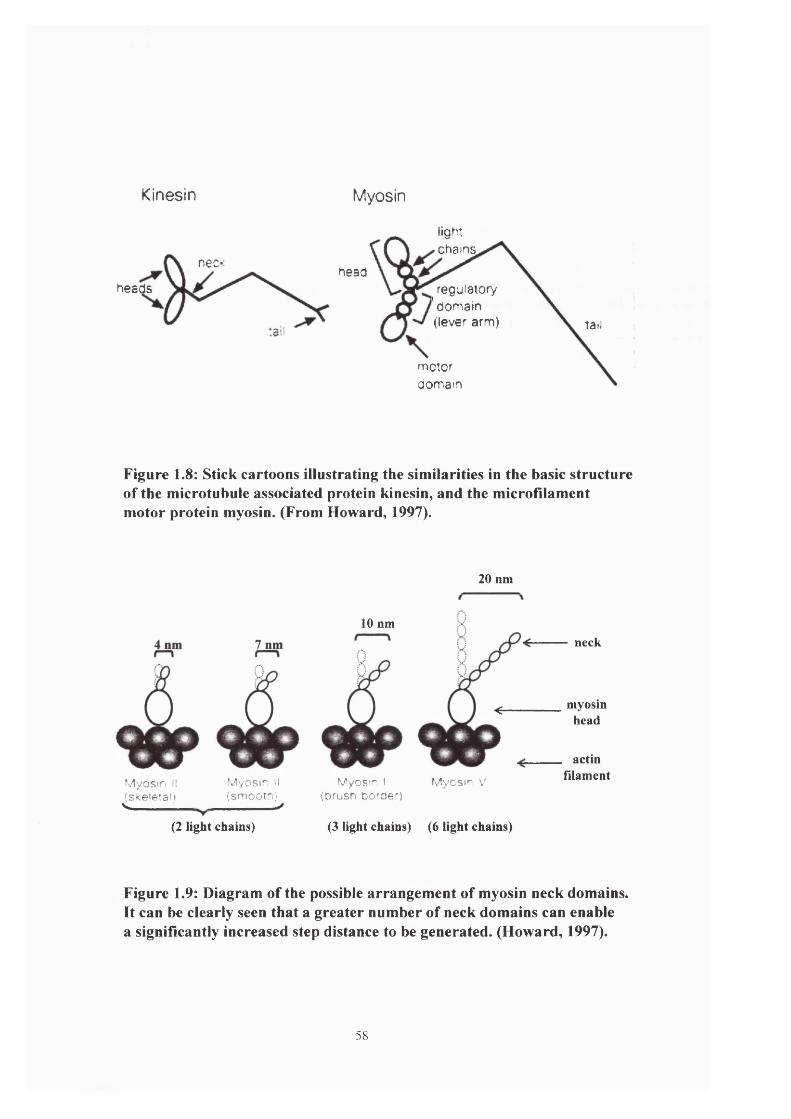
Kinesin Myosin
lightchains
nec<head
heads regulatory ’ domain (lever arm) tail
motordomain
Figure 1.8: Stick cartoons illustrating the similarities in the basic structure of the microtubule associated protein kinesin, and the microfilament motor protein myosin. (From Howard, 1997).
20 nm
10 nm
Myosin I ^s<eletal.i
(2 light chains)
Myosin II (smootn) ^
neck
^__________myosinhead
MyOS'n I (brush border)
Mvcsin V
actinfilament
(3 light chains) (6 light chains)
Figure 1.9: Diagram of the possible arrangement of myosin neck domains. It can be clearly seen that a greater number of neck domains can enable a significantly increased step distance to be generated. (Howard, 1997).
58

microtubule or microfilament and following hydrolysis of the ATP molecule, undergoes a
conformational change which brings about movement. The major difference in the two systems
can be expressed in terms of the ‘duty ratio’ of the motor molecules. This duty ratio is a
determination of the fraction of the time that the motor domain spends attached to the filament
(Dillon & Murphy, 1982;Howard, 1997), and is measured over a range from close to 1, where
the head is mainly attached to the filament, to close to 0 when the head is mainly detached from
the filament.
A conventional two-headed kinesin molecule remains attached to the microtubule and
consequently has a duty ratio of 0.5 (implying that one head is always in contact with the
microtubule). If neither head domain were attached the molecule would diffuse away from the
filament. A potential problem exists here in that the kinesin working stroke is ~8nm and the
distance between adjacent binding sites (i.e. individual tubulin heterodimer units) is 8nm. This
it seems to overcome by a conformational change in the bound head mediated by ATP hydrolysis
(Hackney, 1994; Ma & Taylor,
1997 ) which enables the unbound head to swing forward and maintain the contact with the
microtubule (Hackney, 1994; Hirose et al, 1996; Howard, 1996). This is a motion similar to the
‘hand-over-hand’ climbing of a rope (Howard et al, 1989; Schnapp et al, 1990). The kinesin
molecule is therefore able to act as a processive motor, an important feature in one of its primary
roles as a transporter of organelles. This arrangement allows a single motor molecule to drive its
cargo a considerable distance along the filament.
Since skeletal muscle myosin exists in large assemblies with many available head
domains, the duty ratio of each individual myosin head must be small, ~ 0.01 - 0.1 (Uyeda et al,
1990; Liebler & Huse, 1993). This appears to be a function of the large discrepancy that exists
59

between the binding site separation distance along the microfilament of 36nm and the working
stroke of 4-7nm (Molloy et al, 1995; Mehta et al, 1997). A large number of myosin heads would
ensure that at any given moment some of the myosin heads in the assembly are undergoing the
powerstroke. Consequently the distance travelled during this movement would propel forward a
different subset of myosins, which would then be in register to subsequently interact with the
actin filament and thus produce further movement. In muscle cells this may be an adaptation for
speed and high load, since this system would provide a large pool of primed heads within a short
distance of the binding sites. This could enable muscle to respond quickly and with power. This
is clearly adequate for the duty ratio considerations of myosin-II in muscle cells but the situation
with respect to many of the recently discovered myosins is uncertain.
An interesting adaptation seems to occur in the case of myosin-V, which is thought to
act as a vesicle transporter in yeast (Govindan et al, 1995) and mice (Cheney et al, 1993).
Sequence analysis has shown that this molecule possesses 6 light chains per heavy chain (see
figure 1.9) forming a neck which, when examined under the electron microscopy was calculated
to ~25nm in length (Cheney et al, 1993). This could enable the molecule to perform a much
longer working stroke than other myosin molecules and is almost capable of spanning the entire
binding site separation distance. Since it is a double-headed molecule these observations would
suggest that it could have a duty ratio of 0.5, indicative of a processive motor. In vitro and in
vivo measurements of the speed of myosin-V mediated movements have shown it to be a much
slower motor than other myosins (Cheney et al, 1993; Govindan et al, 1995) which would
confirm the theory that long necks are an adaptation for processivity rather than speed.
Studies in vivo of the two cytoskeletal networks suggest that although they appear to be
disparate systems, strong evidence exists for the presence of both actin- and microtubule-
dependent motors on the same organelles (Kuznetsov et al, 1992; Path et al, 1994). Two models
60

developed from studies in the giant squid axon have been suggested to explain this apparent
interrelationship. The first suggests that since microtubules are long (>25/rm) they are
responsible for transporting organelles large distances along the length of the axon, whilst the
shorter, cross-linked network of actin filaments allows the organelles to be moved to local sites
along the axon (Atkinson et al, 1992). Alternatively the microtubules may allow the
translocation of products of the Golgi to other organelles (e.g. rhoptries, micronemes, dense
bodies) whilst the 3D actin mesh maintains the appropriate spatial distribution (Aniento et at,
1993). Both the microtubule and microfilament networks are fundamental to the correct
functioning and survival of cells. Microtubule-based transport is responsible for important
processes such as chromosome segregation during mitosis and meiosis, localisation of some
cytoplasmic mRNA and transport of intracellular membrane organelles (Vale & Fletterick,
1997). The actin/myosin system is implicated in many different intracellular processes ranging
from force generation and overt cell motility to signal transduction and endocytosis (Sellers,
1999).
Inhibition studies using known inhibitors of actin and myosin functions have shown a
substantial reduction and even a complete cessation of overt gliding motility in Eimeria tenella
and other Apicomplexans (Dubremetz & Ferreira, 1978; Sibley, 1995; Miller et at, 1979;
Dluzewski, 1992). Similar treatment with inhibitors of microtubule functions resulted in no
change in gliding velocity or frequency, but interestingly was seen to have an impact upon host
cell invasion by the parasite (Bejon et al, 1998). Therefore in the context of the generation of
gliding motility it would be reasonable to assume that it is the actomyosin complex that is
responsible for this phenomenon. Consequently the remainder of this chapter shall focus on the
possible mechanisms involving actin and myosin that could be employed to generate gliding
motility.
61

King (1988) first proposed the concept of a linear motor based on an actin-myosin
interaction in apicomplexans. Transmembrane components located in the plasma membrane
could form a bridge between the cytoplasmic motor mechanism and the external ligand (e.g.
substratum, latex bead). In the light of subsequent discoveries Dubremetz et al (1998) further
refined this model by suggesting that it is the microneme proteins which, when exocytosed, bind
to the parasite membrane and act as the ligand for the substrate/motor interaction. They may do
this directly or they may interact with proteins anchored in the cell surface membrane which
subsequently change conformation and bind to the underlying motor apparatus. Possible
candidate proteins for this interaction are the lipidated proteins containing the complex
glycoylphosphatidylinositol (GPI) moiety. These are cell surface bound proteins (Casey, 1995)
and are known to participate in a number of cellular processes such as nutrient uptake, cell
adhesion and membrane signalling events (Englund, 1993; Anderson, 1994). The signalling
mechanism has been best characterised in T cells where antibodies against specific GPI proteins
cross-linked these antigens and by an as yet unknown mechanism, led to the tyrosine
phosphorylation of intracellular proteins (Brown, 1993).
All surface proteins so far identified in the Apicomplexa are GPI proteins (Dubremetz
et al, 1998) and have no cytoplasmic domain. It is possible therefore that the microneme
proteins (which themselves have a C-terminal transmembrane domain but no GPI homology)
become associated with the parasite membrane interact with the GPI proteins, causing a
conformational change in the microneme derived proteins, which in turn interact with an
underlying motor complex to generate motility and hence facilitate invasion. This hypothesis is
made more attractive by data showing that invasion can be inhibited by anti-surface protein
antibodies (Grimwood & Smith, 1992; Mineo & Casper, 1994).
62

The moving junction formed at the point of invasion between the entering zoite and
host cell, is still somewhat of a mystery. Little is known about its composition but the
circumstantial evidence points to the microneme proteins as being an integral part of the
complex.
Studies on gliding motility in the sporozoite of Plasmodium have shown that as the cell
moves across the substratum a trail is left behind and subsequent examination of this material
showed it to be composed of the circumsporozoite protein (Stewart & Vanderberg, 1991).
Similar studies on Eimeria nieschulzi (Entzeroth et al, 1989) also showed the secretion of trails
during sporozoite motility. These observations strengthen the idea that proteins of parasite
origin are vital for the generation of gliding motility.
In the Apicomplexa, with the exception of the flagellated microgametes, the motile
stages possess no discernible organelles of motility. Cells lacking an obvious organelle adapted
for motility (e.g. cilia, flagella), adopt one of two main strategies to bring about directed
motility. The first, and perhaps simplest is the polymerisation of filaments from a pool of
monomers. The polymerisation of actin is utilised by certain pathogenic bacteria to enter and
travel through host cells (Cossart, 1995) and also plays a role in the protrusion of lamellipodia
during crawling locomotion of cultured cells (Condeelis, 1993). An analogous process occurs in
the amoeboid movement of nematode sperm generated by the polymerisation of the nematode
major sperm protein (MSP) (Roberts & Stewart, 1997). In this cell amoeboid locomotion occurs
despite the observation that they contain neither microfilaments nor myosin molecules (Roberts
& Stewart, 1997).
63

The second strategy involves the movement of motor proteins along a polarised
filament. This mechanism is exemplified by the case of myosin moving along an actin filament
and also dynein and kinesin travelling along a microtubule (or vice versa).
Perhaps the simplest method used to achieve motility is directed protein
polymerisation. This is a technique exploited by the bacteria Listeria monocytogenes which
moves through the host cell cytosol at speeds of about 0.4/xm sec ’ (Gouin et al, 1999), and
invades neighbouring cells by inducing the polymerisation of an actin tail which propels the cell
forward (Tilney & Portnoy, 1989; Mounier et at, 1990). Much work has been done on this
organism, not only because it is a serious pathogen but also since it presents an excellent
opportunity to study the mechanisms underlying the polymerisation of actin in a much simpler
context than that which exists in a lamellipodium.
Further analysis of the Listeria actin tail showed it to be composed of two distinct
populations of actin filaments. Initial experiments which decorated the actin filaments with the
SI fi-agment of myosin showed them to be small (approximately 0.2^m) and orientated with the
faster growing barbed end towards the bacterium (Tilney & Portnoy, 1989; Tilney et al,
1992a&b). However more recent studies have also revealed the presence of long axial filaments
which were obscured in the proximal region by the profusion of shorter filaments, and suggests
that these shorter filaments are randomly orientated (Sechi et al, 1997). The basis for this actin
polymerisation seems to be the protein encoded by the actA gene, since mutations in this gene
prevent actin assembly, tail formation and cell movement (Kocks et al, 1992; Domann et al,
1992). The ActA protein exists as a dimer (Dramsi & Cossart, 1998) protruding fi'om the
bacterial cell wall and is not released during tail formation (Kocks et al, 1993; Niebuhr et al,
1993). It also does not have a uniform distribution, being absent from the septum in dividing
cells and remaining absent from the newly formed pole, whilst increasing in density at the old
64

pole (Kocks et al, 1993). Sequence analysis of the actA gene has revealed a number of peptide
sequences similar to known actin-binding sites and a central proline rich sequence (Kocks et al,
1992).The observation that bacteria grown in broth are unable to nucleate actin polymerisation
suggests that ActA itself is not the nucleator but needs to recruit a nucleator or nucleator
complex from the host cell (Tilney et al, 1992a).
Nematode sperm are unlike many other sperm since they possess neither an acrosome
nor axoneme and instead of swimming they crawl along the uterine wall (Roberts & Stewart,
1995; Theriot, 1997). In vitro these sperm cells have been observed to crawl across substrata at
speeds of up to 0.5/xm sec ' (Roberts & Stewart, 1997). In addition, they seem to contain no actin
filaments or myosin (Nelson & Ward, 1981; Nelson et al, 1982; Roberts & Stewart, 1997) and
instead base their motility on a 14kDa protein named the major sperm protein (MSP). This MSP
was first identified in Caenorhabditis elegans and found to comprise -15% of the total protein
in the cell (Klass & Hirsh, 1981). Studies on the parasitic nematode A.vcam suum showed that
MSP was the main constituent of the cytoskeleton (Sepsenwol et al, 1989) and that this protein
was responsible for the production of the motility reminiscent of the actin-rich amoeboid cells
i.e. assembly of filaments at the leading edge of the cell causes extension of a pseudopod which
subsequently attaches to the substratum and pulls the cell forward. Actin is involved in a number
of other cellular processes and this can make detailed studies of its function in individual
processes difficult. MSP provides a simple model to elucidate the mechanisms behind this form
of amoeboid motility.
Despite the complexity of the processes involved it is clearly important to try and
understand the mechanisms of actin-based cell motility. Extensive studies have been performed
on motile cells such as fibroblasts, white blood cells and the free-living amoeba Dictyostelium
65

discoideum, and these are beginning to elucidate the mechanisms involved in the generation of
actin-based motility.
Most motile cells possess protrusive structures at the leading edge which are highly
dynamic and contain large numbers of microfilaments, seemingly orientated with their barbed
ends in the direction of protrusion (Small, 1988). The most basic of these structures is a thin,
finger-like protrusion termed a filopodium, which can extend tens of microns from the main cell
body. In these filopodia actin is arranged in a tight bundle of more than 15 microfilaments
(Lewis & Bridgman, 1992) bound together with actin-associated proteins such as fimbrin
(Matsudaira, 1994).
Lamellipodia are thin sheets which protrude from the leading edge of motile cells such
as cultured fibroblasts, and are responsible for the characteristic ruffled appearance at fibroblast
leading edges. The ruffles are lamellipodia that have detached from the substrate and are
moving backwards over the dorsal surface of the cell. Actin in these structures is organised in an
orthogonal web formed from two sets of filaments (Small, 1988), such that the filaments are at
45° to the direction of protrusion (Small et al, 1995). In all these structures the important
question is how is membrane protrusion brought about? In essence there are two main strategies
that are thought to be employed; directed actin polymerisation and force generated through
motor proteins.
The first consideration in understanding the mechanisms underlying the movement of
cell protrusions is ascertaining how actin polymerisation is brought about. Are the actin
filaments in these structures formed de novo i.e. nucleated, or do they elongate from pre-existing
filaments? In classical studies of the Thyone sperm acrosomal process it was concluded that
microfilaments are nucleated from a stable filament bundle present in the sperm nucleus and
continue to extend by monomer addition to the barbed ends (Tilney & Inoue, 1982). Ultimately
66

this generates a bundle containing very long actin filaments. Small et al, (1995) examined the
lamellipodia of fish keratocytes and suggested that the actin filaments therein extend the entire
length of this structure, indicative of filament elongation. It has subsequently been claimed that
the techniques used to prepare the cells may have selectively removed smaller microfilaments.
However, earlier fluorescence activation experiments in similar cells showed actin
filaments turning over very rapidly and this was interpreted as the presence of a population of
short filaments, generated by frequent nucléation (Theriot & Mitchison, 1991). Again the
interpretation of these findings were subsequently challenged (Small, 1994) and it is clear that
this question is far from being answered, although some cells have been shown to contain both
short and long filaments, albeit with different spatial distributions (Lewis & Bridgman, 1992).
In Dictyostelium and polymorphonuclear leukocytes (PMN) addition of a suitable
chemoattractant leads to the doubling of F-actin levels within 10 seconds (Omann et al, 1987;
Schleicher & Noegel, 1992). There also seems to be a correlation between the speed of
movement of the cell and the speed of actin turnover (Zigmond, 1993). In rapidly moving PMNs
moving at ~0.2/rm sec ', the F-actin has a half-life of approximately 3 seconds (Cassimeris et al,
1990) whereas in a fibroblast moving 0.08/xm min ', the F-actin half-life is increased to 181
seconds (Theriot & Mitchison, 1992). Since many of the invasive stages of the Apicomplexa
move at considerable speeds (up to 20/ims '), this may explain why it has so far proved
impossible to detect and stain microfilamerits in these cells.
In any protrusive structure, regardless of the mechanism of actin polymerisation,
sufficient force must be generated to enable protrusion to take place. The mechanisms by which
this force is generated are thought to be brought about by actin polymerisation itself, or by the
use of motor proteins. A further consideration is the origin of the force generation. It can be
envisaged that the force could develop either in the cell cortex and be transmitted forward to the
67

site of protrusion, or may be generated in the area of the leading edge. In Amoeba observations
favour local force generation (Grebecki, 1994).
Studies have shown that sufficient energy is available from the polymerisation of actin
to distort the cell membrane (Hill & Kirschner, 1982; Cooper, 1991; Tilney et al, 1973) and this
is consistent with the polarity of the filaments. Indeed polymerisation of pure actin contained
within a lipid vesicle has been shown to be able to bring about the distortion of the membrane
(Cortese et al, 1989; Miyati & Hotani, 1992), as has been demonstrated with a number of other
proteins including tubulin (Hotani & Miyamoto, 1990).
The alternative model for force generation most likely involves a barbed end-directed
protein motor (most probably a myosin) utilising nucleotide hydrolysis to provide the energy to
deform the cell membrane. It is clear that sufficient force is available from the actin/myosin
crossbridge cycle (Oster & Perelson, 1993) but the most important question is, does the myosin
exist in the right locations to bring about protrusion? Localisation studies on myosin 11 showed it
to be present in cultured cells mainly behind the leading edge (Conrad et al, 1989), and in
amoeboid cells primarily in the cortex and trailing edge of the cells (Baines & Korn, 1990;
Fukui et al, 1989; Carboni & Condeelis, 1985). These observations do not necessarily preclude
myosin 11 from being involved in protrusion, however in Dictyostelium cells in which the single
myosin 11 gene has been deleted (DeLozanne & Spudich, 1987) or the expression inhibited by
antisense RNA (Knecht & Loomis, 1987) amoebae are still capable of pseudopod extension
(Wessels et al, 1988).
In cultured cells myosin 1 localises to the cell periphery, pseudopodia and filopodia and
to the cytoplasm surrounding the nucleus (Wagner et al, 1992; Conrad et al, 1993).
Acanthamoeba myosin lA & IB are found throughout the cytoplasm and close to the cell
membrane (Hagen et al, 1986; Miyata et al, 1989) whilst myosin 1C is also found in close
68

apposition to the cell membrane and particularly the contractile vacuole (Baines & Korn, 1990).
In Dictyostelium myosin I localises to the leading edge and pseudopodia as well as to the
posterior cytoplasm (Fukui et al, 1989). These results suggest that myosin I binds to cell
membranes and plays an important part in vesicle transport (Cheney & Mooseker, 1992), and is
also implicated in the generation of force in protrusive structures (Condeelis, 1993). However
the function of myosin I at the leading edge is still far from clear.
Mutant Dictyostelium cells whose myosin IB gene had been disrupted by homologous
recombination (Jung & Hammer, 1990) were still capable of wild type levels of pseudopod
extension and cell migration. However they exhibited a reduction in intracellular transport and
an increase in the extension of lateral pseudopodia (Wessels et al, 1991). Subsequently, similar
results were obtained from cells lacking a functional myosin lA gene (Titus et al, 1993)
suggesting that other myosin I isoforms may compensate for the loss a single myosin I isoform.
Double deletion experiments in which cells lacked either myosin lA & IB or IB & IC led to the
development of cells that also extend greater numbers of lateral pseudopodia (Novak & Titus,
1992). These findings tend to suggest that there is no really compelling evidence either for or
against the involvement of myosin I in the development of cell protrusions, although there is
evidence for a role in cell surface retraction and intracellular vesicular transport (Condeelis,
1993). Both processes are presumably important in cell motility. In the short term at least, the
situation is likely to remain unresolved due to the explosion in the number of unconventional
myosins discovered in recent years (see later for more detailed analysis), many of which are
likely to have overlapping distributions and functions.
The second important consideration in the generation of protrusive structures is the
mechanism by which membrane deformation is brought about. Models based on myosin I
involvement envisage either the motor protein sliding entire actin filaments forward (Egelhoff &
69

Spudich, 1991) or a situation where myosin I attaches to a membrane-associated helmet and
moves the membrane forward relative to the barbed end of the filament, thus creating space for
monomer addition and consequent protrusion elongation (Mitchison & Kirschner, 1988; Sheetz
et al, 1992). Since actin alone can bring about membrane deformation (Cortese et al, 1989;
Miyata & Hotani, 1992) and Dictyostelium myosin I null mutants still produce protrusive
structures (Jung & Hammer, 1990; Wessels et al, 1991; Novak & Titus, 1992; Titus et al, 1993)
it is probable that mechanisms based on motor protein generated force are not responsible for
membrane deformation. This would suggest that actin polymerisation brings about membrane
deformation.
The Brownian Ratchet Model proposed by Peskin et al (1993) suggests that natural
oscillation of the plasma membrane may be sufficient to allow the addition of an actin monomer
to the growing end of a microfilament. Once bound this monomer would prevent the membrane
from regaining its starting position and hence the protrusion would advance. Studies on filopod
extension in neuronal growth cones have shown that at room temperature the membrane
vibrates at both a speed and a distance which could facilitate monomer addition (Fricke &
Sackmann, 1984). However, it has been suggested that the stiffness of the plasma membrane
may be such that it is unable to deform at an adequate rate (Mitchison & Cramer, 1996).
Alternatively thermally driven changes may be brought about by bending of the actin filament
(Peskin et al, 1993) which would temporarily shorten the effective length of the filament. It has
also been proposed that membrane deformation could be a result of osmotic swelling following
the release of bound ions and water from actin during the polymerisation process (Oster, 1988).
In recent years much work has focussed on the signalling mechanisms that transduce
external stimuli through the plasma membrane, into the cytoplasm where they can elicit an
appropriate response. In this context the guanine-nucleotide-binding (G) proteins have been
70

shown to be very important, since they act as molecular switches at the cell surface to translate
an external stimulus into an intracellular reaction (Bos & Zwartkruis, 1999). In Dictyostelium
cyclic AMP binding to its receptor stimulates the activation of a heterotrimeric G protein and
this in turn stimulates the release of profilin bound to the plasma membrane, into the cytoplasm.
Profilin is an actin-associated protein which catalyses ATP-ADP exchange on actin monomers
and consequently induces microfilament assembly (Alberts et al, 1994). It also seems that G
protein activation initiates a protein cascade with a number of molecules being released and
impacting upon the dynamics of the actin cytoskeleton. Two families of these downstream
effector proteins which have attracted great attention are the Rac family and the Rho family. It
has been demonstrated that injection of Rac into cultured fibroblasts leads to an increase in the
formation of lamellipodia (Hall, 1998). It remains to be seen if these, or similar proteins exist in
the Apicomplexa, but given the ubiquity of these molecules (Hall, 1998) it would seem
reasonable to assume that they do.
The interaction of actin and myosin has been shown to be responsible for many more
cellular processes than just cell motility. The actin cytoskeleton itself helps maintain the shape
of the cell and in conjunction with myosin it has been seen to participate in such diverse cellular
functions such as cytokinesis, intracellular trafficking and nutrient uptake. In higher organisms
this diversification is seen in the requirement of myosin in processes ranging from muscle
contraction to auditory function (Baker & Titus, 1998).
In recent years there has been a revolution in the field of cell biology due to the
discovery of multiple unconventional myosins that carry out a wide range of cellular functions.
Considerable detail is known about conventional myosin (myosin II). It is a hexamer formed
from two large, identical heavy chains (M̂ ~ 200,000) and two pairs of light chains (Mr ~
71

20,000). Table 1.2 shows a comparison of the size of conventional myosin with some of the
unconventional myosins.
MYOSIN CLASS ORGANISM HEAVY CHAIN SIZE
I Dictyostelium 124kDa
II Chicken skeletal 222kDa
VI Pig 145kDa
IX Rat 225kDa
XI Arabidopsis 173kDa
XIV Apicomplexa 93kDa
Table 1.2: Myosin class size comparison (data from Mooseker & Cheney, 1995; Heintzelman & Schwartzman, 1997; Finder et al, 1998).
At the amino terminal end of the myosin II heavy chain, the protein folds to produce a
globular head, and it is here that both the ATP-binding site and the actin-binding site are
located. Beyond this globular head domain is an a-helical tail which interacts with the tail
domain of the second heavy chain, and through a sequence of non-covalent bonds forms a rod
which extends to the carboxy-terminal end. Connecting the head and tail domains is the neck
region, formed from a single a-helical strand of the heavy chain and which is stabilised by the
binding of the light chains. In non-muscle cells it is phosphorylation of these light chains that
control the regulation of the myosin II molecule (Burridge, 1999). Addition of a phosphate
group by myosin light chain kinase (MLCK) stimulates the ATPase activity of the myosin
molecule, causing it to unravel and associate into the classical bipolar filaments (Sanders et al,
1999; Somylo & Somylo, 1994). Similarly, removal of a phosphate group by myosin light chain
72
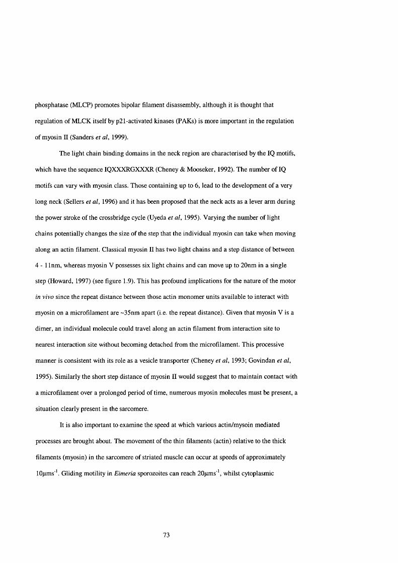
phosphatase (MLCP) promotes bipolar filament disassembly, although it is thought that
regulation of MLCK itself by p21-activated kinases (PAKs) is more important in the regulation
of myosin II (Sanders et al, 1999).
The light chain binding domains in the neck region are characterised by the IQ motifs,
which have the sequence IQXXXRGXXXR (Cheney & Mooseker, 1992). The number of IQ
motifs can vary with myosin class. Those containing up to 6, lead to the development of a very
long neck (Sellers et al, 1996) and it has been proposed that the neck acts as a lever arm during
the power stroke of the crossbridge cycle (Uyeda et al, 1995). Varying the number of light
chains potentially changes the size of the step that the individual myosin can take when moving
along an actin filament. Classical myosin II has two light chains and a step distance of between
4 - 1 Inm, whereas myosin V possesses six light chains and can move up to 20nm in a single
step (Howard, 1997) (see figure 1.9). This has profound implications for the nature of the motor
in vivo since the repeat distance between those actin monomer units available to interact with
myosin on a microfilament are ~35nm apart (i.e. the repeat distance). Given that myosin V is a
dimer, an individual molecule could travel along an actin filament from interaction site to
nearest interaction site without becoming detached from the microfilament. This processive
manner is consistent with its role as a vesicle transporter (Cheney et al, 1993; Govindan et al,
1995). Similarly the short step distance of myosin II would suggest that to maintain contact with
a microfilament over a prolonged period of time, numerous myosin molecules must be present, a
situation clearly present in the sarcomere.
It is also important to examine the speed at which various actin/mysoin mediated
processes are brought about. The movement of the thin filaments (actin) relative to the thick
filaments (myosin) in the sarcomere of striated muscle can occur at speeds of approximately
lO/rms '. Gliding motility in Eimeria sporozoites can reach 20/xms'*, whilst cytoplasmic
73

streaming seen in the alga Chara can achieve speeds of up to 80/xms ' (Preston et al, 1992). In
contrast some actin/myosin interactions are relatively slow, e.g. myosin V mediated vesicle
transport occurs at 0.2/xms ' (Govindan et al, 1993; Cheney et al, 1993). It is unclear how these
substantial differences in speed can be achieved by what are essentially very similar molecules,
but it may reflect a subtle alteration in the ATPase activity of the individual myosins.
Structural features of the tail domain allow myosin II molecules to further interact in a
precise manner, leading to the development of a stack of anti-parallel monomers. This filament
is orientated such that the globular heads are at each end and the central shaft is bare as it
consists only of the a-helical tails. In muscle cells this structure is the basis for the sarcomere. In
non-muscle cells such as Dictyostelium amoeba, myosin II localises to the actin ring which
constricts at the centre of the two dividing cells during cytokinesis (DeLozanne & Spudich,
1987). It is also thought to be involved in other cellular functions including the maintenance of
cortical tension, capping of surface receptors and certain features of cell motility (Sellers et al,
1996).
In 1973 Pollard & Korn discovered a novel myosin in Acanthamoeba which did not
resemble the conventional two-headed, filament forming myosins and termed this isoform
myosin I. Biochemical studies on this molecule revealed that although it was much smaller (it
exists as a monomer of - Mr 120,000) and unable to form filaments, it was nonetheless able to
crosslink actin filaments. Despite this report, research in the field of unconventional myosins
remained relatively unpopular until two important discoveries were made. The first was the
observation that Dictyostelium cells lacking myosin II were still able to perform a large number
of actin-based functions, and the second was the discovery in Drosophila of a number of myosin
genes which encoded actin-based motors with predicted structures considerably different to both
myosin I and myosin II (Montell & Rubin, 1988).
74

Using current molecular biology techniques such as polymerase chain reaction (PCR)
screens has revealed a large number of genes encoding for myosins with significantly different
structures to myosin I & II, in a wide variety of species. Individual organisms have also been
observed to contain multiple myosin genes (Bement et al, 1994). PCR primers were designed
around the amino acid sequence of the nucleotide-binding site, which appears to very highly
conserved across all myosin classes, and is based on the GESGAGKT motif. These screens also
revealed that the myosin head domain is highly conserved in all myosins so far identified and it
is the divergence of the tail sequence that separates the myosin classes. Using the CLUSTAL V
programme (Higgins et al, 1992; Cheney et al, 1993) produced a phylogenetic tree based on
sequence divergence in the head domain (see figure 1.10), in which there was close correlation
to those classes based on tail domain structure. It is this tail domain which determines the
function of the individual myosins.
At present 15 different classes of myosin are recognised (Baker & Titus, 1998; Sellers,
1999). With the exception of myosin I & II these have been assigned a roman numeral in line
with their order of discovery (Mooseker & Cheney, 1995; Cope et al, 1996) (see figure 1.7). In a
study on Dictyostelium Titus et al (1994) presented evidence for the presence of 10 different
myosin classes, only one of which appeared to be conventional. In vertebrate cells (human
epithelial, liver & white blood cells and porcine epithelial cells) evidence exists for the presence
of at least 11 myosin genes, two of which are myosin II (Bement et al, 1994). Experiments
involving Arabidopsis have indicated the presence of at least 8 myosin genes, none seemingly
conventional (Kinkema et al, 1994).
Classes XIV & XV are the most recent classes to be discovered (Mermall et al, 1998;
Sellers, 1999) and it is class XIV that is perhaps the most pertinent to this investigation, since it
has so far been identified only in Toxoplasma gondii (Heintzelman & Schwartzman, 1997) and
75

XIIIVIIIXI
IV
S m ooth an d nonm uscle
Invertebratem uscle
X V
VIIC ard iacm u sc le
Skeletal m u sc le
IXVI
XIVIII XII
Figure 1.10: A phylogenetic tree generated through the ClustalW analysis of myosin motor domain sequence comparison. The highlighted group is the branch representing the Apicomplexan myosins. (From Sellers, 1999).
76
Plasmodium falciparum', (Finder et al, 1998). In Toxoplasma three unconventional myosins
were identified; TgM-A, TgM-B and TgM-C. Of these TgM-A proved to be a most remarkable
molecule due to its small heavy chain size (~ 93kDa) and the observation that, since no IQ
motifs were found it presumably lacks a neck domain. The tail domain is also very small (57
amino acid residues) and is highly basic, suggesting a possible membrane binding function.
TgM-B (-1 1 4 kDa) & TgM-C (-125 kDa) seem to arise from differential RNA splicing, since
they share an identical head domain, and only diverge after 245 residues of the tail domain. In
follow up studies to their original findings, Heintzelman & Schwartzman (1999) have
undertaken localisation studies on these unconventional myosins. In tachyzoites it would appear
that both TgM-A & TgM-C are membrane associated, since it proved necessary to treat the
cellular extracts with detergent to solublise these proteins. However once within the
parasitophorous vacuole the pattern of distribution changes, as visualised with
immunofluorescent techniques . TgM-A is to be found at the apical pole of the parasite, and
once the cells are seen to begin the movement which presages their escape from the host cell, the
myosin locates to the membrane in defined patches. This is consistent with a role in the
generation of gliding motility. TgM-C is localised to the juxtanuclear region, a position
consistent with an involvement in aspects of Golgi apparatus function and protein trafficking.
Myosins classes I, II & VI from a number of organisms have also been shown to be associated
with the Golgi complex or Golgi derived vesicles (Path & Burgess, 1993; Dconen et al, 1997;
Buss et al, 1998; Skowron & Mooseker, 1999). An antibody raised against the Pf-myol gene
product was used in immunofluorescent microscopy of Plasmodium merozoites (Finder et al,
1998) and was seen to localise predominantly to the periphery of the cell. The same antibody
was used in immuno-electron microscopy and also localised to the cell periphery, especially in
the region surrounding the apical prominence. It seemed to be associated with either the plasma
77

membrane itself, or the outer membrane of the subplasmalemmal cisternae. These observations
would provide a candidate motor mechanism for the protozoan cell motility motor proposed by
King (1988).
Even a cursory examination of the literature dealing with various aspects of the phylum
Apicomplexa shows that whilst there are many unifying features between the various species,
there is a great amount of variation. However the recent work which has suggested that the
phylum Apicomplexa contain a unique subclass of the myosin family may have important
implications for the study of cell motility. Sporozoites of Eimeria tenella move at considerable
speeds which would seem to be outside our present understanding of myosin biochemistry.
Further investigation of myosin XIV may yield important information as to how the individual
myosin classes have been adapted to undertake their diverse cellular functions.
78

CHAPTER 2; MATERIALS AND METHODS
2.1 Preparation of sporozoites and sporocysts
Oocysts of Eimeria tenella (strain ETl) in 2% chromic acid solution were provided by
Pfizer Ltd., Sandwich. They had been prepared as follows:- faeces was collected from infected
birds over a period of 24-48 hours and suspended in a sterile plastic beaker with tap water. This
mixture was then homogenised with a Waring blender and filtered through two layers of butter
muslin. The filtrate was then centrifuged (MSE Chillspin at 2000rpm for 10 minutes), the
supernatant discarded and the pellet, plus a small amount of residual liquid resuspended in
saturated NaCl solution. Oocysts were collected from the top of the liquid using a syringe with a
long needle, placed in distilled water and centrifuged as before to yield an oocyst pellet. The
oocysts were washed free from salt solution by three cycles of centrifugation and resuspension in
distilled water and finally resuspended in 2% chromic acid solution at a final concentration of
about 0.25 x 10 ̂oocysts ml '. Sporulation of the oocysts was achieved by incubating the culture
with forced aeration at 30°C for 48 hours in a shaking water bath. When approximately 80% of
the oocysts had sporulated the suspension was removed from the water bath and stored at 4°C
until required. The oocysts remained viable for up to 9 months.
Clean oocysts were obtained following the protocol of Shirley (1995). The oocysts were
pelleted at 12,000g for 5 minutes (MSE Microcentaur) and washed three times in phosphate
buffered saline (PBS - pH7.4 - see appendix for details). Following the final wash the pellet was
resuspended in 1ml of saturated NaCl solution onto which 0.5ml of deionised water was gently
overlain. This was centrifuged at 12,000g for 5 minutes after which time the oocysts appeared as
an off-white layer at the salt/water interface. These were carefully removed to a clean
79

polypropylene tube. An equal volume of 400/xm diameter glass beads (Sigma) were added to the
tube and this was agitated on an Autovortexer SA6 (Stuart Scientific) at maximum speed for 2
minutes to rupture the oocysts and release the sporocysts. The level of oocyst rupture was
monitored by placing a small sample onto a glass microscope slide, covered with a 13mm
diameter glass coverslip and then observed using a x20 objective lens. When approximately 85%
of the oocysts were observed to have ruptured, the liquid (~ 0.5ml) was drawn off and combined
with 0.5ml of 2.5mgml ' trypsin and 0.5ml of 5mgml ’ bile and placed in a shaking water bath
(The Mickle Laboratory Engineering Co., setting 7) at 41°C for 50 minutes or until sufficient
sporozoites had excysted, again approximately 85%, (as visualised using the method outlined
above). If there was insufficient oocyst rupture the tube was vortexed for a further 30 seconds
and then treated as above.
The mixture containing free sporozoites was then passed through a cotton wool filter
prepared by transferring sufficient cotton wool to a 10ml plastic syringe to provide a 2ml bed
volume when lightly compressed with the plunger. This filter was first cleaned by the addition
of 5ml of 70% ethanol and was then washed by the addition of two 5ml aliquots of PBS. Once
all the sporozoite solution had passed through the filter a further 5ml PBS was added to elute
any remaining zoites. The filtrate was then spun at 3000g in a MSE Chillspin for 3 minutes, the
supernatant discarded and the cells resuspended in 0.5ml of an appropriate solution.
80

2.2 Preparation of cell cultures
For studies on invasion five cell lines were used. These were;
CACO-2 - human colonic carcinoma cell line
grown in minimal essential medium (MEM) + 10% foetal calf serum
(PCS) +1% non-essential amino acids (NEAA),
MDBK - Madin & Darby Bovine Kidney Cells
grown in RPMI with Glutamax I + 5% PCS,
CEV/A2 - chick embryonic tumour cell line
grown in medium 199 (M l99) + 5% PCS,
INT 407 - human Caucasian embryo intestinal cell line
grown in Eagle’s MEM + 10% PCS + 1% NEAA + 2mM glutamine
CPCC - chick primary caecal cell line (see below)
grown in Dulbecco’s MEM with Hepes and Glutamax I + 6% PCS
+ 4% chicken serum + 0.1% penicillin / streptomycin solution.
The CACO-2, MDBK and CEV/A2 cell lines were obtained from Pfizer Ltd and the
INT 407 from Dr. Peter Daszak (Kingston University). The MDBK cells were used for studies
on motility, invasion and inhibition of invasion. These cell lines were kept at -70°C until
required.
The chick primary caecal cell line was freshly prepared from 17 day old eggs. The
embryos were removed from the eggs, the intestines excised and washed 3 times in cold wash
buffer (PBS + 0.1% penicillin/streptomycin solution). Intestinal caeca were then removed,
washed a further 3 times and placed in a sterile plastic petri dish. Using 2 sterile scalpels angled
81

at 90° to each other the organs were chopped up for 3 minutes and then washed 3 times in cold
wash buffer. The tissue was placed in a sterile 250ml conical flask with 25ml of dissociation
buffer (PBS + 0.1% penicillin/streptomycin + 0.0625% trypsin) and stirred gently for 30
minutes at 37°C. This suspension was filtered through sterile Nyblot cloth (nylon muslin) and
the tissue returned to the flask for a further dissociation as described above. The filtered liquid
was mixed with 25ml of medium (identified above) and spun at 30g for 5 minutes in a benchtop
centrifuge. This procedure was undertaken 3 times in all and following the final spin the
supernatants from the 3 tubes were decanted and the cell pellets resuspended in medium and
combined in one tube. 100/xl of this suspension was then aliquoted into each well of a 24 well,
flat-bottomed cell culture plate (Nunc™) and incubated at 37°C in an air incubator (5% CO2)
for 48 hours prior to the invasion experiment.
2.2.a Resuscitation of cells from liquid nitrogen
A 0.5ml ampuole containing 100/xl of cell suspension was removed from storage at -
70°C in liquid nitrogen and left at room temperature for 1 minute. This was then transferred to a
37°C water bath until the contents were fully thawed (1-2 minutes) care being taken not to
immerse the ampuole completely. The ampuole was then wiped with 70% ethanol, placed in a
sterile flow hood and the cell suspension pipetted into a sterile 15ml universal tube containing
10ml of the appropriate, pre-warmed specified media. This was then centrifuged for 5 minutes
at 3000g in a MSE Centaur benchtop centrifuge. The supernatant was removed and the cell
pellet resuspended in 30ml of the appropriate media (described above) and pipetted into a
sterile, flat-bottomed incubation flask (Nunc™) which was then placed in an incubator at 37°C
until the cells grew to confluence.
82

2.2.b Passaging adherent cells
In a sterile flow hood the medium was removed from the flasks containing confluent
cells which were then gently washed with warm PBS. The PBS was poured off, replaced with
2ml of PBS containing 0.05% trypsin and 0.01% EDTA and the flask placed in the 37°C
incubator for 2 minutes. Microscope observation was used to monitor the level of detachment of
cells from the substrate. If fewer than -80% of cells had detached the flask was gently tapped on
the bench to release the remaining cells. 20ml of appropriate medium supplemented with 10%
PCS was added to the flask, the contents decanted into a sterile 50ml universal tube and spun at
3000g for 5 minutes in a benchtop centrifuge. The supernatant was removed, the pellet
resuspended in medium and divided into 4 fresh Nunc™ flasks.
2.2.C Preparation of cell monolayers
For motility studies cells were grown up on 32mm diameter coverslips (Chance Propper
#2). These had been flame sterilised and placed in the wells of a 6 well plate (Nunc™). The 6
well plates were used as the well diameter was 35mm, therefore allowing the coverslips to be
easily manipulated. The cells were grown to confluence and when required the coverslips were
carefully removed from the well and incorporated into a Prior perfusion chamber set up as
described in section 2.3.b.I.
The cells used for electron microscopic studies of invasion were grown in 12 well plates
(Nunc™) without coverslips. Following incubation with sporozoites they were fixed and
detached from the substrate as described below (section 2.3.d.I).
83
For invasion inhibition studies cells were grown in 96 well plates, incubated as
appropriate and fixed and analysed in situ again as described below (section 2.3.b.VI).
2.3 Microscopy
2.3.a General microscopy
Generally observations were made using an Olympus IMT-2 inverted microscope.
Initial observations of the immunofluorescent samples were also undertaken on this microscope
but for greater resolution and image capture more detailed work was done on a Zeiss Axiophot
microscope.
2.3.b Observation of motility using videomicroscopy
Motility was observed and recorded from the microscope using a video camera (Hitachi
CC-TV camera - HV-735K) linked to both a VHS video recorder (Panasonic AG-6010) and a
television monitor (Hitachi). A VTGG-22 video timer (FOR-A Company Ltd.) was plugged into
the system to allow definitive video independent time measurements to be made when necessary.
Information on the VHS tapes could be recorded onto U-matic tapes using a Sony VO-9660P
video recorder and this enabled changes over short time periods to be quantified. Outline
drawings of individual sporozoites on acetate sheets placed over the monitor screen allowed the
progress of each motility event to be followed ‘frame’ by ‘frame’. Subsequent measurement of
these drawings allowed accurate speed calculations to be made.
84

Before all microscopical observations a slide graticule at the appropriate magnification
was recorded to allow accurate measurements to be made subsequently.
2.3.b.I Prior chamber and microslides
Motility was observed using samples contained within either a Prior perfusion chamber
essentially as described by Preston & King (1978) (see figure 2.1) or glass microslides. In
general the use of the microslides was restricted to the motility and excystation inhibition assays.
Glass microslides (5cm in length) with an internal separation distance of 0.2mm were
obtained from CamLab. They held a volume of 20/il and were ideal for the inhibitor studies as a
representative samples could be removed from the experimental tubes without significantly
reducing the overall volume. Microslides could only be used at a lower magnification (x20) but
were able to be used to take sequential samples from tubes being incubated in a shaking water
bath.
The Prior chamber was used to observe motility for a number of reasons. The use of
standard microscopic coverslips allowed a high magnification (xlOO) lens to be used and this
enabled accurate measurements of motility to be made. The volume of the chamber was -1ml
which in comparison to the size of a sporozoite (12/xm x 3/rm) represented a large volume.
Consequently relatively large numbers of cells could be assessed in any one sample. The use of a
reservoir of buffer meant that the Eimeria samples could be readily cleaned by flushing through
with fresh buffer. In the same way this system was used to perform washout experiments in
which potential inhibitors were assessed for the reversibility of their actions.
The Prior chamber was assembled by first placing a 32mm diameter coverslip, which
had been immersed in 70% ethanol and wiped dry, into the lower metal ring clamp. On top of
85
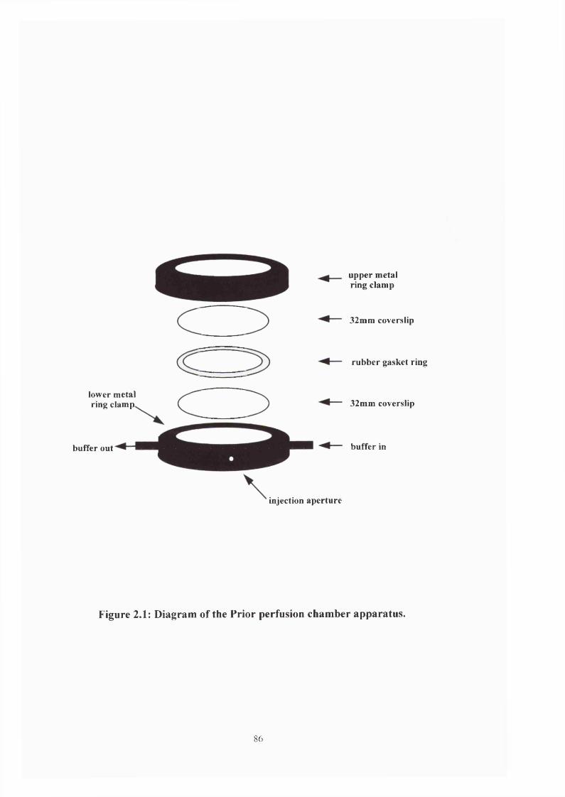
lower metal ring clamp
buffer out
upper metal ring clamp
32mm coverslip
rubber gasket ring
32mm coverslip
buffer in
injection aperture
Figure 2.1: Diagram of the Prior perfusion chamber apparatus.
86
this was placed the rubber ring gasket which had been lightly greased with a small amount of
silicone grease. A syringe needle bent halfway along its length at approximately 70° was
introduced into each of the ports protruding from the lower ring clamp. This was to allow
solutions to be passed through the perfusion chamber once it had been closed. These needles
were then gently pushed through the rubber ring until all of the needle aperture was visible. The
second 32mm coverslip (also cleaned with 70% ethanol) was then placed on top, followed by the
upper metal ring clamp and the entire apparatus screwed together.
Once the chamber had been assembled it was filled with PBS warmed to 41 °C from a
reservoir (a 10ml syringe) connected to one of the ports with a length of rubber tubing.
Depending upon the experiment sporozoite or sporocyst suspensions were injected into the
chamber via a small aperture in the lower ring clamp. In the same way suspensions of latex
beads were also introduced into the chamber. The objective stage and the Prior chamber were
maintained at a constant temperature throughout the experiments using warm air provided by an
Accuron X120 heater (Cambridge, England).
2.3.b.II Potential inhibitory compounds
As outlined in the introduction sporozoite motility is important in vivo at 3 distinct
stages;
(1) excystation
(2) gliding through the caecal lumen (and also across the caecal surface)
(3) target cell invasion
87

Consequently a sequence of experiments were performed using inhibitors of known
cytoskeletal components and established coccidiostats, to try and identify the role of the parasites
cytoskeleton in these important events.
The cytochalasins are a class of compounds derived fix>m fungi and are known to prevent
actin polymerisation (Brown & Spudich, 1981; Cooper, 1987). Both cytochalasin B & D (Sigma)
were used in initial experiments but their efficacy appeared to be very similar and so in subsequent
studies only cytochalasin D was used. Concentrations of O.Spgml'* and O.lpgml'* were used. These
were prepared using a stock lmgml ‘ solution made up in DMSO (Sigma). DMSO controls were
used as required.
2,3 butane dionemonoxime (BDM - Sigma) is a myosin ATPase inhibitor (Mulieri &
Alpert, 1984), preventing the dissociation of the phosphate from the ADP-P/myosin complex
thereby ‘locking’ the myosin molecule onto the actin filament, and was used at concentrations of
lOOmM, 75mM, 50mM & 25mM. These experimental solutions were derived from a stock solution
of IM BDM dissolved in PBS at 50®C. Colchicine (Sigma) is derived from the autumn crocus
Colchicum autumnale and is known to act on microtubules by binding with tubulin causing a
conformational change which induces microtubule depolymerisation (Margolis & Wilson, 1977)
and was used at a concentration of ImM.
Salinomycin (Sigma) is an ionophore and is routinely used as a coccidiostat. In this study it
was used at a concentration of 1 pgml‘‘.
2.3.b.IU Motility studies
Motility studies were undertaken using the Prior perfusion chamber. The apparatus
(described in section 2.3.b.I) was assembled, placed on the heated microscope objective stage
88

and the chamber filled with PBS pre-warmed to 41°C. 100/d of sporozoite suspension (see
section 2.1) was injected into the chamber and allowed to settle onto the lower coverslip for 5
minutes. Using the lOOx objective lens under oil immersion individual sporozoites were
recorded onto VHS videotape for analysis.
In addition studies were performed observing the translocation of latex beads (Sigma).
A number of sizes of beads were tried and 2/im diameter beads were selected as they were large
enough for their progress to be easily monitored without obscuring the sporozoite. Sporozoites
were injected into the Prior chamber and allowed to settle. The bead suspension was diluted
1:100 in PBS, 100/d of this was injected into the chamber and the beads allowed to settle for 5
minutes. Bead translocation was then recorded onto VHS videotape for subsequent analysis.
Motility inhibition studies were undertaken in 1.5ml Eppendorf tubes containing 200/d
of potential inhibition solution (see appendix for detailed compositions). The experiment was
started with the addition of 100/d of sporozoite suspension to the tube which was then mixed
and placed in a shaking water bath at 41°C. After 5 minutes a small sample was drawn up in a
microslide (approximately four-fifths of the way up the microslide) and placed on the objective
stage of the IMT-2 which was maintained at 41°C. The sporozoites were allowed to settle for 1
minute and were then observed for 3 minutes, during which the total number of sporozoites and
number of motile sporozoites was recorded. This was repeated every 10 minutes for 1 hour. The
experiment was restricted to 1 hour as it was felt a prolonged incubation would not accurately
reflect the situation in vivo.
Each experiment was repeated 3 times. In addition 3 control experiments were
performed; tubes without inhibitor to provide a motility reference point, tubes containing
suitable dilutions of DMSO but no inhibitor to ensure that the solvent was non-inhibitory and a
sucrose control to identify any potential effects on motility due to changes in buffer osmolarity.
89

The reversibility of each compound was also assessed. Sporozoites were incubated with
the highest drug concentration for each compound and after 20 minutes the cells were scored for
motility (T = 0 minutes), spun down, washed twice in fresh PBS and resuspended in 150/il of
PBS. The cells were immediately scored for motility (T = 5 minutes) and again (T = 25 minutes)
after a further 20 minutes had elapsed.
In addition reflection interference microscopy (RIM) (Curtis, 1964) was performed on
excysted sporozoites gliding in PBS across the glass slides. This technique allowed an
evaluation of the size and location of the cell/ substratum contact to be made.
2.3.b.IV Sperm tracker analysis
The Hobson Sperm Tracker was originally designed to aid analysis of the viability of
samples of sperm. However it has subsequently been adapted to allow analysis of both motile
bacteria and protozoa. In this experiment the equipment was used to assess sporozoite motility
in samples which had been incubated in a number of potential inhibitory compounds for various
periods of time.
Sporozoites were incubated in PBS (as the control), 25mM BDM, 50mM BDM or
0.5/igml * Cytochalasin D for 10 minutes, 30 minutes and 60 minutes as described in section
2.3.b.III. At each time point the samples were recorded for 3 minutes onto VHS video tape and
it was these recordings that were analysed using the sperm tracking software. Cells were also
incubated in 75mM BDM and lOOmM BDM after 10 minutes only, as previous studies have
shown little or no sporozoite motility in these samples following prolonged incubation. To
compare the efficacy of the two compounds a recording of sporozoites incubated in 0.5/tgml'*
Cytochalasin B for 10 minutes was also analysed.
90
2.3.b.V Excystation studies
Sporozoite excystation from sporocysts was observed using the Prior perfusion chamber
apparatus on the heated microscope stage. To bring about excystation the warm PBS was
supplemented with bile at 5mgml'^ and trypsin at 2.5mgml * and perfused into the chamber.
100/xl of sporocyst suspension (see section 2.1) was then injected into the chamber and allowed
to settle onto the lower coverslip for 5 minutes. The process of excystation was then recorded
onto VHS video tape for further analysis.
Studies on the inhibition of excystation were undertaken in 1.5ml Eppendorf tubes
containing 100/d of potential inhibition solution and 100/d of 5mgml'^ bile/2.5mgml ‘ trypsin
solution (see appendix for detailed compositions). The experiment was started with the addition
of 100/d of sporozoite suspension to the tube which was then inverted and placed in a shaking
water bath at 41°C. After 10 minutes a small sample was drawn up in a microslide and placed
on the objective stage of the IMT-2 microscope which was maintained at 41°C. The sporocysts
were allowed to settle for 1 minute and were then observed for 3 minutes, during which 3
parameters were recorded;
(1) the total number of sporocysts
(2) the number of sporocysts which contained tumbling sporozoites
(3) the number of free sporozoites in the solution.
This was repeated every 10 minutes for 1 hour. This experiment was also restricted to 1
hour for the reasons outlined in section 2.3.b.III.
91

All experiments were repeated 3 times and in addition 2 control experiments were
performed; tubes containing no inhibitor, to provide a suitable base value for excystation and
tubes containing DMSO but no inhibitor to ascertain any cytotoxic effects of the solvent.
2.3.b.VI Invasion studies
The observation of invasion required a slight modification to the standard Prior
chamber set up where the chamber is ‘closed’. It was felt that the use of a closed chamber with
medium perfusing through it could dislodge the cells and so the upper coverslip was omitted. In
the invasion studies an ‘open’ chamber was used in which only the lower coverslip was
assembled into the Prior chamber. This coverslip had been previously incubated with cell
suspension so that it had a monolayer of MDBK cells growing on it. Consequently both the
medium and the sporozoite suspension were added dropwise to the chamber using a Pasteur
pipette. This was then placed on a heated microscope stage and motility and invasion recorded
onto VHS videotape.
Studies into the inhibition of target cell invasion not only attempted to identify the
cytoskeletal components of the invasion process but also tried to ascertain if the host cell
cytoskeleton was implicated. Consequently 2 approaches were adopted; pre-incubating the cell
monolayers for 20 minutes & 60 minutes and pre-incubating the sporozoites for 20 minutes &
60 minutes.
MDBK monolayers were grown in 96 well plates until confluent (see section 2.2.c). If
pre-incubation of the cells was required the medium was aspirated from each well and replaced
with 200jul of the appropriate solution (see appendix for details) and then incubated at 4I°C for
30 minutes in an air incubator (5% CO2). Following incubation the drug/medium was removed
92
and replaced with 150/xl of fresh medium and 50/xl of sporozoite preparation. The plates were
then spun at 3000g on a Jouan MP4P plate spinner to bring the zoites down and incubated at
41°C for either 20 or 60 minutes. Following incubation the medium/drug was discarded, the
cells washed twice in 100/il of PBS and then fixed in 100/rl of 100% methanol for 5 minutes at
room temperature.
Sporozoites were pelleted and resuspended in an appropriate solution (see appendix for
detail) and incubated at 41“C for 30 minutes. Following incubation 200/rl of each solution was
added to the relevant wells (previous medium having been removed) and incubated at 41“C for
either 20 minutes or 60 minutes. Following incubation the drug/medium was removed, the cells
washed twice in PBS and then fixed in lOOpil of methanol for 5 minutes at room temperature,
the methanol removed and the plates sealed and stored at 4°C until required for assay.
In order to assay the inhibition of cell invasion, the cells were treated with a primary
anti-sporozoite antibody (raised in rabbit using a whole sporozoite extract as the immunogen
and donated by Pfizer, Ltd.) and a goat anti-rabbit secondary FITC conjugated antibody
(Sigma), and then counted using a fluorescence microscope to determine the number of
internalised sporozoites.
The experimental plates were removed from the fridge and 100/xl of PBS +1% FCS +
0.5% BSA (cooled to 4°C) was added to each well. These were left for 40 minutes to rehydrate
the cells and the buffer was removed by inverting and shaking the plates. 100/d of primary
antibody solution diluted 1:1000 in PBS + 1% FCS + 0.5% BSA was added to each well and
incubated overnight at 4“C. The primary antibody was discarded by inverting and shaking the
plates and the cells were washed 3 times in 250/il of PBS + 1 % FCS + 0.5% BSA using a
Titertek M96V plate washer. Following the final wash 100/d of secondary antibody diluted 1:50
with PBS + 1% FCS + 0.5% BSA (warmed to 25“C) was added and the plates incubated at 25°C
93

for 2 hours. The secondary antibody solution was removed and the cells washed 3 times in 200/xl
of PBS + 1% FCS + 0.5% BSA. After the final wash the plates were tapped on a paper towel to
remove as much moisture as possible and were then examined on a Zeiss Axiovert 10
microscope under a x32 objective lens. One central field per well was examined and the number
of intracellular sporozoites counted.
2.3.b.VIl Calculation of speeds
Speed measurements were made using the motility recordings. These were replayed on
a U-matic video player linked to a Hitachi monitor onto which an acetate sheet had been affixed.
When an appropriate sporozoite was identified the sequence was manually advanced frame by
frame and the position of the anterior and posterior of the cell marked on the acetate sheet with
an indelible marker. For the measurement of bead translocation the centre of the latex bead was
also marked.
For the calculation of gliding speeds the motility sequence was stopped and marked
every 0.2 seconds and for the bead translocation every 0.08 seconds. The distances moved by the
anterior and posterior points were then measured and an average taken of the two distances. In
the case of bead translocations the anterior and posterior distances were measured to ensure that
the real bead distance was being measured and not the cumulative effect of sporozoite motility
and bead translocation.
94

2.3.C Immunofluoresence microscopy
2.3.C.1 Preparation of coated coverslips
The interaction between sporozoites and the substrate was weak and therefore
coverslips were coated to increase cell adhesion. The immunofluoresence protocols described
below were found on a Harvard University Medical School website maintained by Louise
Cramer & Arshad Desai (http://skye.med.harvard.edu/Protocols/genl.html).
32mm coverslips (Chance Propper #2) were heated in a loosely covered glass beaker in
IM HCl at 50-60°C for about 6 hours. When cooled they were washed once in distilled water
and once in deionised water then rinsed in 70% ethanol and left to dry between 2 sheets of
Whatman filter paper. They were stored in a sterile Petri dish for up to a year.
Coverslips were then coated in poly-L-lysine (Sigma - molecular weight = 70,000 -
150,000). Up to 50 coverslips were placed in a Petri dish containing 15ml of Imgml * poly-L-
lysine solution in distilled water and swirled gently on a flat bed shaker (RlOO Rotatest shaker,
Luckham Ltd.) for 30 minutes. The poly-lysine solution was then poured off and stored at-20°C
(it can be used 3-4 times). The coverslips were washed once in distilled water and at least 5
times in deionised water as the free polyamino acid is cytotoxic. If the coverslips were for
immediate use they were rinsed in 100% ethanol, placed in a Petri dish and dried in a drying
oven. If they were to be used later they were also washed in 100% ethanol but dried between two
sheets of Whatman filter paper.
95

2.3.C.I1 Staining protocol
The sporozoite suspension was prepared as described in section 2.1 and following the
final spin the cell pellet was resuspended in 500/rl of Tris buffered saline (TBS - 0.15M NaCl &
0.02M Tris-HCl - pH 7.4). 50/xl of this suspension was dropped onto a poly-L-lysine coated
coverslip and the zoites were allowed to settle and attach to the coverslips for 5 minutes. The
attached cells were washed by holding the coverslip upright with a pair of tweezers and gently
pipetting a stream of TBS across the surface.
The cells were then fixed in a 4% formaldehyde solution in cytoskeleton buffer (lOmM
2-[N-Morpholino] ethanesulfonic acid (MES), 38mM KCl, 3mM MgCb, 2mM ethyleneglycol-
bis-(P-amino-ethylether) N,N’-tetra acetic acid (EGTA) & 0.32M sucrose) for 20 minutes,
washed in TBS as before and further fixed in 100% methanol at -20°C for 2 minutes. After
another wash in TBS the cells were permeabilised by incubation in TBS & 0.5% Triton X-100
for JO minutes followed by 3 washes in TBS & 0.1% Triton X-100.
Following fixation and permeabilisation the cells were blocked in antibody diluting
solution
(‘Abdil ’ - TBS plus 0.1% Triton X-100, 2% bovine serum albumin & 0.1% sodium azide) and
incubated in the primary antibody diluted 1:100 in Abdil for 90 minutes. The coverslip was then
washed 5 times in TBS & 0.1% Triton X-100 and incubated in fluorescent isothiocyanate
(FITC) conjugated secondary antibody diluted 1:100 in Abdil for 45 minutes. After a final wash
in TBS & 0.1% Triton X-100 the coverslip was allowed to dry and inverted on 10/d of freshly
prepared mounting medium (see appendix) placed on a microscope slide wiped clean with 70%
ethanol. The coverslip was gently depressed to expunge air bubbles and sealed with clear nail
96

varnish. Once the varnish had dried both sides of the slide were wiped with 70% ethanol and the
sample examined.
2.3.d Electron microscopy
2.3.d.I Fixation and embedding of material
The fixation of invaded cells and free sporozoites required different conditions and
resins in order to maintain the antigenicity of the tissue for immunogold treatment.
Invaded cells were first drained of as much remaining medium as possible and were
fixed in Karnovsky’s fixative (Karnovsky, 1965 - see appendix for details) for 30 minutes
followed by a further fixation in fresh fixative for 1 hour. The cells were then washed 3 times in
O.IM phosphate buffer (see appendix) and fixed in a 1% solution of osmium tetroxide in
phosphate buffer for 45 minutes. Following a brief (1 minute) wash in distilled water the cells
were placed in 2% uranyl acetate solution for 2 hours.
Dehydration of the cells was undertaken by incubating them for 10 minutes in 20%
ethanol, then 10 minutes in 30% ethanol, 10 minutes in 40% ethanol and 10 minutes in 60%
ethanol, all at room temperature. This was followed by 10 minutes in 70% ethanol and 10
minutes in 90% ethanol at 4“C and finally 10 minutes in 100% ethanol at 0”C.
The 100% ethanol was removed and then gently replaced with propylene oxide (BDH -
AnalaR). After two minutes the cell layer was gently agitated with a wire loop until the
monolayer lifted from the bottom of the well and this sheet of tissue was placed in a 1.5ml
Eppendorf tube containing 1ml of propylene oxide, gently spun down (3000g for 2 minutes in a
MSE benchtop centrifuge) and washed again in 1ml of propylene oxide.
97
A stock of Araldite 6005 resin (Sigma) was prepared following the protocol of Parsons
& Darden (1961) by thoroughly mixing together 10ml of Araldite, 10ml of 2-dodecen-l-yl
succinic anhydride, 1ml of dibutyl phthalate and 0.2ml of N,N-dimethyl-benzylamine.
Following the second propylene oxide wash the tissue was placed in 0.5ml of fresh propylene
oxide and 0.5ml of stock Araldite 6005 resin and left at room temperature for 1 hour. This was
replaced with pure Araldite for 1 hour and after one further change of resin the tissue and resin
were allowed to harden in an oven at 65°C for 48 hours.
Sporozoites were prepared as described in section 2.1 and the final pellet resuspended
in a 1.5ml Eppendorf tube in 1ml of Karnovsky’s fixative (diluted 1:8 with PBS) and left at
room temperature for 15 minutes. The parasites were then washed 3 times in 1ml of cold (4°C)
PBS and placed in 30% ethanol at 4“C for 30 minutes, followed by 1 hour in 50% ethanol at -
20°C and a further hour in 70% ethanol at -20°C. The dehydration process was completed by 3
incubations in 100% ethanol for 10 minutes each at -20°C. The ethanol was then gently poured
off and replaced with a 2:1 solution of medium grade LR White resin (London Resin Co. Ltd.):
100% ethanol and placed at -20”C for 1 hour. This was then replaced with pure LR White and
placed at -20°C for 1 hour, this process being repeated twice. Following the final rinse the tissue
was removed from the Eppendorf and placed in a gelatin capsule (size 00 - Agar Aids) which
was filled as full as possible with fresh resin and the cap snapped shut. As little air as possible
was allowed to remain in the tube as this inhibits the polymerisation process. The capsule was
then placed in an oven at 50°C for 24 hours until hard.
98

2.3.d.II Sectioning
Thin sections (~90nm) were cut from the resin block using a DuPont diamond knife
filled with deionised water on a Reichert-Jung Ultracut E microtome. These sections were then
floated onto nickel grids (Agar, Cambridge) and allowed to air dry before staining or antibody
treatment.
2.3.d.IIl Staining thin sections
As the sample had already been stained with uranyl acetate en bloc, it was therefore
only necessary to further stain the thin sections with Reynolds’ lead citrate (Reynolds, 1963). A
strip of dental wax was placed onto the lower chamber of a petri dish and onto this was added
individual drops of the lead citrate solution. To ensure that no lead carbonate precipitates were
formed on the thin sections, the dental wax was surrounded with sodium hydroxide pellets.
Grids containing thin sections were then inverted onto these drops, the top of the petri dish
placed over the wax strip and the grids left to stain for 10 minutes. The grids were then washed
in deionised water and allowed to air dry.
2.3.d.rV Immuno-electron microscopy
The thin sections on nickel grids were blocked by placing shiny side down on the
surface of 4 drops (as dispensed from a 10ml syringe) of PBS/0.1% BSA/6% goat serum on a
small piece of dental modeling wax. After 45 minutes the grids were carefully removed and
placed shiny side down on 4 drops of primary antibody solution at a diluted 1:250 in PBS/0.1%
99

BSA, on a separate piece of wax for 1 hour. They were then washed 7 times in 4 drops of PBS
by placing on the surface and leaving for 5 minutes. The grids were not blotted dry following the
first wash but were after the subsequent 6 washes, using fibre-free filter paper. They were then
placed in 4 drops of secondary antibody diluted 1:500 in PBS/0.1% BSA. After an hour they
were removed from the antibody solution and washed 3 times for 5 minutes in PBS, again
without blotting following the first wash but with blotting on the final 2 washes. Over each grid
was then run 8 drops of distilled water and they were then blotted and allowed to dry.
2.4 Identification of cell proteins
2.4.a Preparation of polyacrylamide gels
To probe for proteins present within the sporozoites, samples were run on 8%
polyacrylamide gels contained within a Bio Rad Mini-Protean® II Dual Slab Cell system. Initial
experiments were performed using a variety of acrylamide concentrations (6 - 12%) and it was
found that 8% gels gave the best band separation.
The casting blocks were assembled as laid out in the manufacturer’s instructions and
gels were freshly prepared for each experiment as follows.
4ml of stock acrylamide (40% acrylamide: 1.4% bis-acrylamide) was mixed with 4ml
of resolving gel buffer (3M Tris-HCl, pH 8.8), and 25/Jil of N,N,N,N-Tetramethlethylenediamine
(TEMED) and this was made up to a final volume of 20ml with deionised water. Polymerisation
was initiated by the addition of I50/il of freshly prepared 10% ammonium persulphate solution
and this mixture poured into the gel apparatus until it reached approximately four-fifths of the
100

way up the smaller of the two glass plates. The solution was immediately overlain with
isopropanol to ensure that the resolving gel had a straight surface. Once polymerisation was
complete the isopropanol was poured off, the gel surface washed with a stream of deionised
water and excess liquid removed with Whatman #1 filter paper (care being taken not to touch
the gel surface).
The stacking gel was then prepared as follows. 1.25ml of the stock acrylamide was
mixed with 2ml of stacking gel buffer (0.5M Tris-HCl, pH6.8), and 12.5/d of TEMED and made
up to a final volume of 10ml with deionised water. Polymerisation was initiated by the addition
of 75/il of 10% ammonium persulphate and then poured on top of the resolving gel to fill the
remaining space. Generally a 10 well plastic comb was placed in the stacking gel solution to
form the wells into which the samples were loaded. As the stacking gel shrinks whilst it is
polymerising the level was regularly topped up with the remaining gel solution until
polymerisation was complete. Once complete the plastic combs were removed and any residual
unpolymerised acrylamide aspirated from the wells using a 1ml syringe.
The gels were then removed from the casting block and placed in the running chamber.
They were then completely covered with running buffer (3.5mM sodium dodecyl sulphate
(SDS), 20mM Tris & 0.19M glycine) and placed at 4®C for 60 minutes prior to sample loading.
2.4.b Preparation of protein samples for SDS electrophoresis
Initially protein samples were prepared from intact oocysts following the protocol of
Preston & King, (1992). This involved resuspending the final oocyst volume in 3 volumes of
cold (0°C) TEDPS buffer (lOmM Tris, ImM EDTA, O.lmM dithioerythritol, ImM sodium
101

pyrophosphate and 0.34M sucrose) containing protease inhibitors (PI - Boehringer Mannheim -
see appendix for details) and double this volume of 400^m diameter glass beads (Sigma). This
was then subjected to 10 cycles of 30 seconds on ice followed by 30 seconds of rupture on an
autovortexer. The liquid was then drawn off (usually ~ 4ml) and spun at 90,000g for 1 hour at
4"C on a Beckman TL-100 ultracentrifuge. This supernatant was then used as the Eimeria
protein extract.
The protein content of each sample was assessed using the Bio-Rad protein assay based
on the technique outlined by Bradford (1976). A serial dilution of BSA (lOmgml ' - 0.5mgml ’)
was made up in PEM (lOOmM PIPES, ImM MgSO^ & ImM EGTA) and each 100/xl of sample
mixed well with 2ml of a 1:6 dilution of Bradford’s reagent. The optical density of each mixture
was read at 595nm and a standard curve plotted. Eimeria samples were diluted 1:10 with ddH20
and 100/xl thoroughly mixed with 2ml of the 1:6 dilution of Bradford’s reagent. These were also
read at 595nm and the protein concentration calculated by reference to the protein standard
curve.
It proved difficult to identify proteins successfully from this sample and analysis
showed both a low protein concentration and some degree of proteolytic degradation.
Consequently subsequent experiments were undertaken with a sporozoite derived extract using
the technique outlined below.
Once the sporozoites had been excysted as described in section 2.1, the final pellet was
resuspended in a fresh Eppendorf tube in 250/xl of TEDPS buffer + PI and 250/d of sample
buffer (50mM NaiCOs, 2% SDS, 12% sucrose and 0.1% bromophenol blue). A small amount of
solid dithioerythritrol (Sigma) was added and the solution then heated to 100 C for 3 minutes
and allowed to cool before being loaded into the wells of the polyacrylamide gel at a volume of
15/d per well.
102

Both high range and low range ECL molecular weight markers were obtained from
Amersham Ltd. and broad range molecular weight markers were obtained from Bio Rad. These
were prepared by mixing 1^1 of protein marker with 9/rl of sample buffer, adding a small
amount of dithioerythritol and heating to 100”C for 3 minutes. Once cooled the entire lO/il was
then loaded into an appropriate well.
Standard proteins were prepared following the same protocol but using 2/il of protein
per sample. Actin (3.6 fig /xl'*) was derived from rabbit muscle and was donated by Dr.
Sutherland Mclver (University of Edinburgh). Chicken gizzard myosin (2.4 fig /xl *), chicken
gizzard filamin (2.6 fig fiV^), a-actinin (3 fig fiV^), human erythrocyte spectrin (0.316 fig fiV^)
were all obtained from Sigma.
When the wells had been loaded the gel tank was connected to the power pack
(Vokam® 500-500, Shandon) and a charge of 100A applied until all the sample had left each
well and once this was achieved the voltage was increased to 180A. The power was switched off
when the dye front was approximately 5-8mm from the end of the gel (generally 45-50 minutes)
and the clamp containing the glass plates and the gel removed from the running tank. The
whole gel was removed from the glass plates and the stacking gel carefully excised with a clean
scalpel. The remaining resolving gel was then placed in either a solution of Coomassie brilliant
blue (CBB - 0.25% Coomassie brilliant blue R250, 50% methanol & 10% acetic acid) or placed
directly in the Western blot apparatus (see section 2.4.c).
The gels were stained with CBB as follows. The gels were left in the CBB solution
overnight with gentle agitation and were then washed in destain solution (20% industrial
methylated spirits (IMS), 10% glacial acetic acid, 70% distilled H2O) until sufficient
background staining was removed to allow the protein bands to be readily observed.
103

2.4.C Transfer of proteins to nitrocellulose membranes
Into the transfer clamp was placed a Brillo pad and a same-sized piece of Whatman #1
filter paper both pre-soaked in cold transfer buffer (3.5mM SDS, 15mM Tris, 0.15M glycine &
20% IMS). The protein gel was then gently placed on top of the filter paper ensuring that no air
bubbles were present between the gel and the paper, and was overlain with a piece of
nitrocellulose paper (Sigma) also soaked in cold transfer buffer. Any air bubbles were gently
expunged and a syringe needle was used to prick the nitrocellulose sheet along the line of the
dye front. A further piece of filter paper and another brillo pad were placed on top of the
nitrocellulose and the whole clamp placed into the running apparatus (see figure 2.2). The cold
transfer buffer was then poured into the apparatus, which was then placed in iced water. A
charge of 100A was then applied for 2 hours, care being taken to ensure that at all times ice was
present in the surrounding water to maintain the voltage below 350v. This was to reduce
distortion of the protein bands which can occur if electrical resistance heats the gel excessively.
After 2 hours the power was switched off, the nitrocellulose sheet removed from the
blot apparatus and placed in a solution of Ponceau S stain (0.2% Ponceau S, 3% trichloroacetic
acid & 3% sulphosalicylic acid) for 2 minutes to check that the protein transfer had been
successful. The sheet was then washed in deionised water until the protein bands could be seen
prominently and then allowed to dry.
Using a modified version of this basic protocol, a preliminary screen was undertaken of
6 anti-myosin antibodies with a 48 well (6 columns x 8 rows) Bio-Dot Slot format apparatus
(Bio Rad).
As shown in figure 2.3 a sheet of nitrocellulose paper was placed on top of 3 sheets of
Bio-Dot slot format filter paper which had been pre-soaked in PBS. This was then placed
104
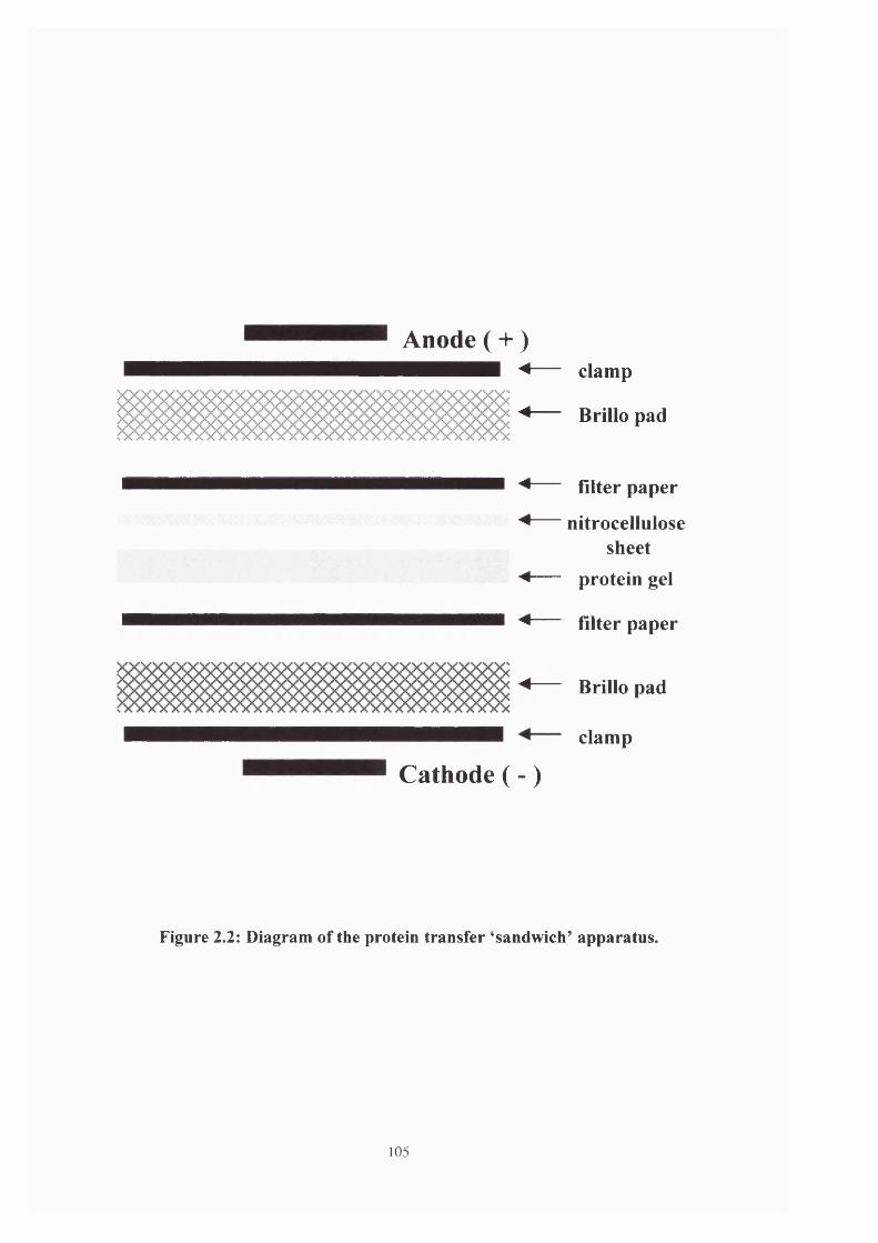
Anode ( + )
Cathode ( - )
clamp
Brillo pad
filter paper
nitrocellulosesheet
protein gel
filter paper
Brillo pad
clamp
Figure 2.2: Diagram of the protein transfer ^sandwich’ apparatus.
105

between the upper and lower clamps, screwed tightly together and connected to the vacuum
pump. A chicken gizzard myosin standard dilution series ranging from 240 /igml * to 0.24 /igml'
* was prepared in the electrophoresis buffer described previously, to which was added a small
amount of solid DTE and then heated to 100“C for 3 minutes. Once cooled 100/d of myosin
solution was applied to the appropriate well and the liquid drawn through the nitrocellulose
sheet using suction. The sheet was then allowed to dry (normally about 20 minutes) and then
stained in the usual way with Ponceau S.
Each of the 48 individual patches of myosin transferred to the nitrocellulose sheet had
an area of 0.9cm^ and a width of 5mm. From each of the six columns formed by the slot blot
apparatus a strip of nitrocellulose was excised which was 3mm in width. Each of these strips
had 8 individual patches of myosin on them ranging in concentration from 240 /tgml ' to 0.24
/igml *. These strips were then Western blotted as outlined in section 2.4.d.
2.4.d Western blotting
2.4.d.l Antibody treatment of nitrocellulose sheets
Identification of actin and the actin-associated proteins from the sporozoite samples
was undertaken by Western blotting of the nitrocellulose sheets. The nitrocellulose sheets with
transferred protein were marked at the top of the original gel with a pencil line, and from each
5mm wide sample lane a 3mm wide strip was excised to counter any potential band distortion
effects. These strips were then pricked with a syringe needle a different number of times (to aid
post treatment identification) and placed in an incubation tray. 1ml of blocking solution (PBS
containing 5% milk protein (Marvel skimmed milk powder) and 0.1% Tween 2 - Sigma) was
106

upper clamp with wells
nitrocellulose sheet
filter paper (x3)
lower clamp
to vacuum apparatus
Figure 2.3: Diagram of the slot blot apparatus.
107

added to each strip and this was incubated at room temperature for 2 hours with gentle shaking
on a flat bed shaker (RlOO Rotatest shaker, Luckham Ltd.).
After 2 hours the blocking solution was removed and replaced with the primary
antibody made up in the blocking solution. In general the primary antibodies (see appendix for
details) were used at concentrations within the range 1:500 - 1:2000. The strips were again
incubated at room temperature for 2 hours with gentle shaking. The primary antibody solutions
were drawn off and stored at -20°C (they were generally used 4 or 5 times) and the strips washed
3 times in fresh blocking solution, the first wash for 15 minutes and the last 2 for 5 minutes.
The ECL molecular weight markers can be inactivated by further treatment with milk protein
and so were washed in PBS/0.1% Tween 20 only.
The final wash solution was discarded and the strips incubated in horseradish
peroxidase (HRP) coupled secondary antibody at room temperature with gentle shaking for 1
hour. In general the secondary antibodies (see appendix for details) were used within the
dilution range 1:1000- 1:4000. The ECL molecular weight markers were incubated in a 1:3000
solution of streptavidin-HRP also for 1 hour with gentle shaking. Following incubation the two
solutions were discarded and the strips washed 3 times in fresh blocking solution, the first wash
for 15 minutes and the last 2 for 5 minutes and then washed 3 times in PBS & 0.1% Tween,
again the first wash for 15 minutes and the last 2 for 5 minutes. The molecular weight markers
were washed following the same protocol but for the first 3 washes the blocking solution was
replaced with PBS/0.1% Tween.
The strips were then ready to undergo the detection process.
108

2.4.d.ll Detection protocols
The basis of both the detection protocols was the HRP/hydrogen peroxide catalysed
oxidation process (see figure 2.4).
The first technique catalysed the oxidation of solutions which yield a dark blue
insoluble product as a positive result. This was performed as follows. A tablet of 4-chloro-l-
napthol (Sigma) was dissolved in 10ml of IMS. 2ml of this solution was added to 10ml of PBS,
mixed thoroughly and poured into a plastic 9cm diameter petri dish (Sterilin). Immediately
before use 40/xl of 30% hydrogen peroxide solution (Sigma) was added and mixed by gentle
swirling. The nitrocellulose strips were then added and the dish again gently swirled until dark
blue bands became visible. The reaction was halted by pouring off the detection solution and
washing the strips in fresh PBS. Positive strips were then photographed and stored at 4°C.
Initial experiments using this detection method gave some promising results particularly in the
detection of actin. However it was a difficult system to control and it was felt a more sensitive
and adaptable technique would yield more accurate results.
The second technique involved the catalysed oxidation of luminol in alkaline
conditions known as enzymatic chemiluminesence (ECL). Immediately following oxidation the
luminol is in an excited state and decays to ground state by the emission of light. If this is
performed in the presence of an enhancer (e.g. phenols) the light emission can be increased
-1000 fold and the output prolonged. The light emitted has a wavelength of 428nm which can
be detected by exposure to blue-light sensitive autoradiography film.
The ECL detection system was obtained fi*om Amersham Ltd. and appropriate volumes
of the two detection reagents were mixed to give a final volume of 0.125ml per cm^ of
nitrocellulose sheet. The nitrocellulose strips which had been prepared as outlined above were
109
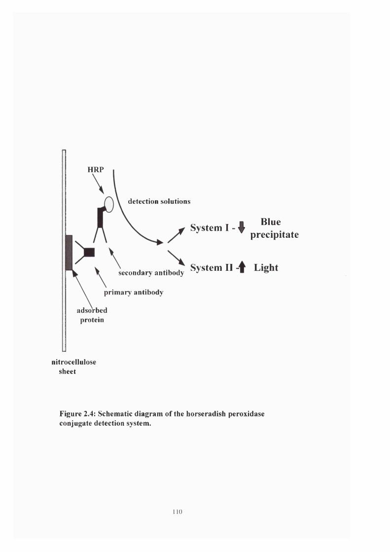
HRP
detection solutions
System I - ^
System IIsecondary antibody
primary antibody
adsorbedprotein
Blue precipitate
nitrocellulosesheet
Figure 2.4: Schematic diagram of the horseradish peroxidase conjugate detection system.
110

placed protein side uppermost on the lower half of an appropriate sized piece of SaranWrap™
and the detection mixture applied via a glass Pasteur pipette. They were incubated for 1 minute
at room temperature after which the excess liquid was drained off. The upper half of the
SaranWrap™ was then folded over the top of the strips and the edges sealed by further folds.
This envelope was then placed in a film cassette with protein side uppermost, and under a safe
red light a sheet of photographic film (Hyperfilm ECL - Amersham Ltd.) was placed on top of
the envelope and exposed for 15 seconds. This film was then removed, developed using an X-
OGRAPH Compact X2 automated developer, and replaced by a second piece of film. Following
development of the original film the strength of the signal was evaluated and the length of
exposure for the second and any subsequent films calculated accordingly.
2.4.d.III F-actin overlay
An alternative method used to probe protein samples immobilised on nitrocellulose
membranes is the F-actin overlay assay (Luna, 1998). This involved incubating the
nitrocellulose sheet with '^^I-labelled F-actin (50 - lOOjagml *) at about 1ml per lOcm^
nitrocellulose for 2 hours at 20°C. The sheet could then be washed up to 5 times in PBS and
placed on photographic film for 2 - 2 4 hours at -80°C.
Whole sporozoite samples were prepared and sent to Dr. Elizabeth Luna (Mass., USA)
to perform the assay. Unfortunately this did not prove successful.
I l l

2.4.e Chromatography
The protein constitution of the Eimeria sample was further analysed using fast protein
liquid chromatography (FPLC). Proteins were separated on the basis of their charge on a
MonoQ column
(Pharmacia) or on the basis of their size on a gel filtration column (Pharmacia). Protein samples
prepared from whole oocysts as described in section 2.4.b and 1ml samples were injected into
the chromatography column. This was then passed through the column and samples collected at
the rate of 1ml min ‘.
These samples were then concentrated following the method of Wessel & Fliigge
(1984). 4ml of methanol was added to 1ml of chromatography sample in a 15ml polypropylene
tube and this mixture vortexed briefly (SA6 Autovortexer - Stuart Scientific) and then spun at
9,000g for 10 seconds in a centrifuge (MSE Chillspin). 1ml of chloroform was then added, the
solution mixed and spun again at 9,000g for 10 seconds and then 3ml of distilled water was
added. This was vortexed vigorously, spun at 9,000g for 1 minute and the upper phase removed
using a Pasteur pipette. 3ml of methanol was added and following a final spin at 9,000g for 2
minutes the supernatant was removed and the protein pellet allowed to air dry.
For further analysis the pellet was resuspended in 50/xl of electrophoresis sample buffer
(see section 2.4.b) and run on an 8% acrylamide gel following the protocol in section 2.4.a.
112

2.5 Identification of myosin by molecular biology
2.5.a Preparation of genomic DNA & agarose gel electrophoresis
DNA was extracted following the method described by Grief, et al (1996). 10 ̂ oocysts
were prepared following the standard purification protocol outlined above (see section 2.1) and
after the final spin the pellet was resuspended in a small volume of PBS ~ 20pl) and 2x volume
of 400/xm diameter glass beads (Sigma). This suspension was then vortexed at maximum speed
for 4 minutes on a SA6 Autovortxer (Stuart Scientific) and the lysate resuspended in 500pl TEN
buffer (lOmM Tris-HCl/5mM EDTA/lOOmM NaCl, pH 7.5). The cell debris and glass beads
were removed by a spin at 12,000g for 2 minutes in a MSE benchtop centrifuge.
The supernatant was removed to a fresh Eppendorf tube and 25pl of 10% SDS and
25pl of Pronase (20 mgml ') were added. This mixture was then incubated in a water bath at
37°C for 3 hours. Following lysis an equal volume of phenol was added ( - SOOpl), the solution
vortexed and 500pl of chloroformiisoamylalcohol (26:1) added and again vortexed. This was
then spun at 12,000g for 5 minutes and the upper layer removed to a fresh tube, care being taken
not to take up any of the protein layer at the interface. The phenol/chloroform extraction process
was then repeated.
To this clear supernatant (~ 400/xl) was added an equal volume of isopropanol and one-
tenth volume of 3M sodium acetate and the DNA allowed to precipitate at -20 °C overnight. The
tube was then spun at 12,000g for 10 minutes and the pellet resuspended in 70% ethanol, spun
down again and as much of the liquid removed as possible without disturbing the pellet. The
remaining ethanol was evaporated off in a drying oven. The pellet was finally resuspended in
50pl TE buffer (lOmM Tris-HCl/O.lmM EDTA, pH 7.5) containing RNAse. 2pl of this solution
113

was then mixed with 8/xl of 6x agarose gel loading buffer (AGLB - 60% glycerol, 6GmM EDTA,
0.2% bromophenol blue & 0.2% xylene cyanol FF) run on a 50ml, 0.8% agarose gel containing
ethidium bromide (2pl at 2mgml ‘) to roughly quantify the amount of DNA recovered.
The sample was then subjected to electrophoresis in a Hoefer (Newcastle-under-Lyme)
10cm cooled minigel apparatus at 75V constant voltage for IV2 - 2 hours. In addition 2 DNA
standard markers were also run to enable the size of the experimental DNA to be estimated.
These were obtained from MBI Fermentas and were the GeneRuler™ 100bp+ DNA ladder and
the GeneRuler™ Ikb DNA ladder (see appendix for further details). Following electrophoresis
the DNA bands were visualised by illuminating the gels with UV light at 302nm on a UVP Inc.
(California) TM-20 transilluminator. Photographic records of gels were taken using a Sony UP-
890CF Video Graphic Printer.
2.5.b Polymerase chain reaction using degenerate primers
The polymerase chain reaction (PGR) was performed using degenerate primers as
previously reported by Bement et al (1994), as the target DNA sequence was unknown.
Degenerate primers are actually a mixture of primers with a similar sequence but each
with a variation at one or more positions and are ideal when searching for novel members of a
known family of genes (Wilks, 1989). Due to the degeneracy of the primers, mismatching of the
primers with the target DNA can occur readily under standard PGR conditions and so a ‘hot-
start’ protocol was adopted. This hot-start entailed an initial DNA denaturing step before the
addition of the polymerase enzyme.
As shown in figure 2.5 in the first cycle the double stranded DNA is denatured and the
2 primers bind at opposite ends of the target region and are extended by the polymerase giving
114

new DNA strands of variable length. In the second cycle both the original template strands and
the newly synthesised strands are denatured which gives 4 primer sites onto which the primers
anneal. Primers which bind to the strands newly synthesised in cycle 1 will be extended only as
far as the end of the template, leading to a precise copy of the target region. In the third cycle
double stranded DNA molecules are produced which are identical to the target DNA.
Subsequent PCR cycles lead to an exponential doubling of the target region and consequently
the original template DNA strands and those of variable length become negligible (see figure
2.5).
The degenerate primers were obtained from PE - Applied Biosystems U.K. The forward
primer was EAF-A directed against the highly conserved ‘LEAF’ motif on the myosin head
domain and the reverse primer was ATP-3 directed against the ATP binding site on the myosin
head domain (see appendix for detailed description of the primers). Taq polymerase (Promega)
was used to perform the PCR.
The PCR reaction mixture was as follows; 2.5pl of 25mM magnesium chloride, Ipl of
the genomic DNA sample, Ipl of stock deoxy-nucleotide triphosphate (dNTP) solution (5mM
dATP, dTTP, dGTP & dCTP - Promega), 3pl of EAF-A, 3pl of ATP-3, 5pi of Taq polymerase
buffer and 33pl of sterile deionised water were mixed in a thin walled 0.5ml polypropylene PCR
tube and placed in a Hybaid thermocycler. In addition a tube containing Ipl of
Schizosaccharomyces pombe genomic DNA (a gift from Dr. Karen May, University of
Edinburgh) was prepared as a positive control and a tube containing no DNA was prepared as a
negative control.
Initial PCR attempts showed a large degree of non-specific amplification and so to
overcome this problem the first cycles of primer annealing were undertaken at a lowered
temperature which was then increased in subsequent cycles to improve the specificity of binding.
115

The tubes were heated to 95"C for 2 minutes to denature the double stranded DNA and then
O.Sjil of Taq polymerase was added and the whole solution overlain with 50pl of mineral oil to
prevent evaporation. The tubes were then subjected to 10 cycles of denaturing at 94"C for 30
seconds, primer annealing at 40”C for 90 seconds and primer extension at 72“C for 90seconds
followed by 25 cycles of denaturing at 94“C for 30 seconds, primer annealing at 50“C for 90
seconds and primer extension at 72°C for 90 seconds and a final amplification step of 5 minutes
at 72°C. The reaction mix was then pipetted into a fresh tube without transferring any of the
mineral oil and stored at -20°C.
To monitor the success of the PCR reactions 2\x\ of each solution was mixed with 8pi of
AGLB and run on a 0.8% agarose gel (-50ml volume) containing ethidium bromide (2 pi at
2m gm r‘) as described in section 2.5.a.
2.5.C Ligation of PCR products into vectors
Initially two approaches were adopted to try and ligate the PCR products into the
pBluescript plasmid vector (0.5pgml ‘ - a gift from Dr. Karen May, University of Edinburgh).
Following PCR treatment with Taq polymerase (derived from Thermus aquaticus,
strain YTl - Promega), Eimeria DNA formed PCR products with a small 5’ polyA tail. This
made successful vector/construct ligation difficult, since the EcoRV digested plasmid is left
blunt ended i.e. neither strand has a nucleotide overhang. Subsequent treatment of the PCR
product with T4 polymerase left the Eimeria DNA blunt-ended.
Alternatively the vector was treated with Taq polymerase in a solution of dTTP which
gave the cut plasmid a small polyT tail (Marchuk et al, 1991), hence both pieces of DNA have
‘sticky’ ends theoretically increasing the likelihood of a successful ligation.
116
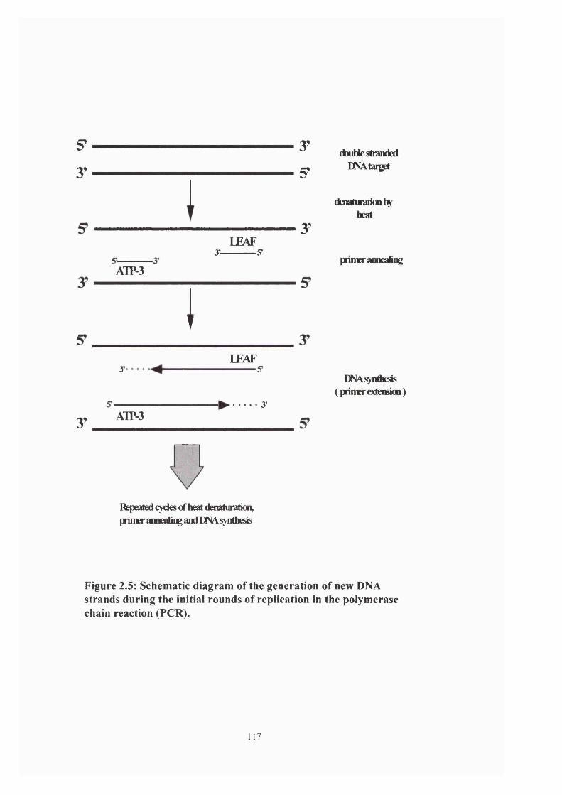
5?
y
AlP-3
3"
y AlP-3
LEAF3’------------S’
ILEAF
3’
Repeated cycles cf heat denaturaticn, prinH* ainealiiç aid DNA synthesis
3’
S’
3’
double stranded ENAtai]get
denaturatioDfayheat
pnmerameding
DNA synthesis (primer extension)
Figure 2.5: Schematic diagram of the generation of new DNA strands during the initial rounds of replication in the polymerase chain reaction (PCR).
117

For both treatments the vector was first cut by placing 4/xl of pBluescript suspension in
a sterile 1.5ml Eppendorf tube, then mixed with 3/xl of lOx REAct®2 buffer (Gibco BRL), 1/xl of
EcoRV restriction enzyme and 22/xl of sterile deionised water and incubated at 37°C for 3 hours.
This mixture was then heat inactivated by incubating at 70°C for 15 minutes and split into 2
equal aliquots. One of these aliquots was subsequently incubated in lOmM dTTP and Taq
polymerase at 70°C for 2 hours in order to generate a polyT tail on the cut ends of the vector.
Both the cut vectors were then loaded onto a 2% agarose gel to analyse and subsequently purify
the DNA.
Under UV illumination the position of the appropriate bands was recorded and the
DNA carefully excised using a clean razor blade. The DNA was then purified from the agarose
gel using a Qiaquick Gel Extraction Kit (Qiagen) according to the manufacturer’s protocol and
resuspended in 50/xl of TE buffer.
In separate tubes 2/d of both the cut vectors and the PCR products were mixed with S/zl
of AGLB and loaded into a 2% agarose gel to ascertain the relative concentrations of DNA in
order to optimise the ratio of PCR product;vector for the ligation reactions. Following
electrophoresis it was decided that a 2:1 ratio of PCR product:vector would provide the best
mixture for successful ligations.
Therefore 1/tl of vector was mixed with 2/il of PCR product, 2/il of lOx T4 DNA ligase
buffer (300mM Tris-HCl, lOOmM MgCl%, 100 mM DTT & lOmM ATP - pH 7.8), 1/il of T4
DNA ligase (Promega) and 14/il of deionised water and incubated overnight at 16°C. The
plasmids were then transformed into competent Escherichia coli cells (see section 2.5.d).
After a number of unsuccessful attempts at ligation using these methods it was felt that
more success might be achieved using a specifically designed T-A vector i.e. a plasmid which
has been engineered to contain a polyT tail. Consequently the pGEM®-T vector system was
118
used (Promega) and the following ligation protocol was set up; 0.5/xl of lOx T4 DNA ligase
buffer, 0.2/xl of pGEM®-T vector, 1/il of PCR product, 0.5/xl of T4 DNA ligase (3 Wu /xl * -
Weiss units, where 1 Wu is defined as the amount of enzyme required to ligate 50% À DNA in
30 minutes at 16°C) and 2.8/xl of deionised water. In addition 2 control tubes were prepared; a
positive control containing control insert DNA but no PCR product and a negative control
containing neither control nor PCR product DNA. These reactions were mixed by gentle
pipetting, incubated overnight at 4°C and were then used to transform competent bacterial cells.
2.5.d Transformation of competent cells and colony selection
Original transformation experiments used the method described by Hanahan, (1983),
but due to the lack of suitable colonies derived using this technique a second strategy was
adopted.
Supercompetent cells were obtained from Promega {E. coli - strain JM109) and
transformations undertaken as described in the manufacturer’s protocol, essentially a
modification of the Hanahan, (1983) method outlined below. An appropriate number of
polypropylene culture tubes (Falcon) were placed on ice. The frozen competent cells were
thawed by placing on ice for 5 minutes, gently mixed and 100/xl of the cell suspension added to
each of the chilled culture tubes. 50ng of the cloned Eimeria DNA in 10/xl of sterile deionised
water was added to the cell suspension, mixed by gently flicking the tube and then returned to
the ice for 10 minutes. The cells were heat-shocked by placing them in a water bath at exactly
42°C for 45 seconds and returned to ice for a further 2 minutes. To each of the tubes was added
900/xl of SOC medium (see appendix) and the cells were then incubated at 37°C for 1 hour on a
flat bed shaker at 225rpm.
119
lOOjLil of these cells were plated out on LB plates containing 100/tgml^ ampicillin,
SO/xgrnl'i 5-chromo-4-chloro-3-indolyl-P-D-galactoside (X-Gal) and 0.5mM isopropylthio-P-D-
galactoside (EPTG). The plates were then incubated for 16-24 hours at 37°C. Individual plates
were then scored for the number of blue and white colonies, and appropriate numbers of white
colonies were selected for subsequent mini-prep and restriction enzyme digest analysis, in order
to confirm the presence of DNA inserts.
2.5.6 Mini-prep of plasmid DNA
A single white colony of transformed E.coli was inoculated into 10ml of LB medium
(1% Bacto®-Tryptone, 0.5% Bacto®-Yeast extract + 0.5% NaCl) + ampicillin (lOO/igml *) and
incubated overnight at 37°C, 225 rpm on a flat bed shaker. This was then spun down on a
Beckman centrifuge, the supernatant decanted off and the tube spun again and the remaining
supernatant carefully pipetted off.
The plasmid DNA was then extracted following the alkaline lysis method using a
QIAprep Spin Miniprep Kit (Qiagen) according to the manufacturer’s protocol as follows. The
pellet of bacterial cells was resuspended in 250/d of buffer PI and to this 250/d of buffer P2 was
added, the tubes gently inverted 4-6 times and allowed to stand for no longer than 5 minutes.
350/d of buffer N3 was then added, the tubes again inverted 4-6 times and then spun in a
microfuge at 13,000 rpm for 10 minutes. The resultant supernatant was carefully pipetted into a
QIAprep column, which had been placed in a 1.5ml Eppendorf tube, and which was then
centrifuged for 1 minute at 13,000rpm. The column was washed with 0.75ml of buffer PE,
centrifuged for 1 minute, the flow through discarded and the column spun for a further minute.
The column was then placed in a clean 1.5ml Eppendorf and the DNA eluted by the addition of
120

50/xl of buffer EB (lOmM Tris-HCl, pH 8.5), which was allowed to stand for 1 minute before a
final spin for 1 minute. The sample was then stored at -20°C until required.
2.5.f Restriction enzyme digestion of plasmids
Restriction enzymes are bacterially derived enzymes which cut highly specific regions
of DNA according to the presence and detection of the 4 nucleotide bases. The plasmid was
designed so that in both the regions flanking the multiple cloning site (MGS) there are a number
of different restriction enzyme sites which can be used to cut out any Eimeria DNA which has
been ligated into the MCS. In this way it is possible to determine if ligation has been successful.
Two restriction enzymes were used (see appendix for details); Ndel (New England
Biolabs®) and Ncol (New England Biolabs®). In a 0.5ml Eppendorf tube 5/il of plasmid DNA
prepared by mini-prep was mixed with 0.5/xl of each enzyme, 1/xl of lOx NEB 4 buffer (20mM
Tris-acetate, lOmM magnesium acetate, 50mM potassium acetate & ImM DTT - pH 7.9 - New
England Biolabs®) and 3/xl of sterile deionised water. This was spun briefly (~5 seconds) in a
MSE benchtop centrifuge to collect the contents together and incubated at 37“C for 2 hours.
Following the digestion 2/xl of the reaction mixture was mixed with 8/xl of AGLB and loaded
onto a 0.8% agarose gel containing ethidium bromide (2 pi at 2mgml ’) and electrophoresised.
The gel was then illuminated with UV light to check for inserts.
2.5.g Sequencing of products
Plasmids which had been shown to contain appropriately sized inserts were run uncut
on an 0.8% agarose gel along with a sample of X DNA (New England Biolabs®) which had
121
been digested with Hindlll (New England Biolabs®) in order to calculate if the DNA
concentration was adequate for the sequencing reaction.
The gel was run in the usual manner and photographed. From this picture it was
estimated which X DNA band the plasmid band was most similar to in band intensity and using
the following equation a concentration value was calculated;
[DNA] = (size of X band / size of X DNA) x amount of plasmid DNA loaded.
For successful DNA sequencing a concentration of 250ng|ir* of plasmid DNA was
required, along with a solution of 3.2pmolpT^ of the original PCR primers. Eppendorf tubes
containing the appropriate solutions were sent to The Advanced Biotechnology Centre at Charing
Cross & Westminster Hospital for automated sequencing using an ABL Prism 377 Genetic
Analyser (Perkin Elmer). The reaction cycles were set up in a reaction volume of 20pl containing
plasmid DNA, PCR primers, AmpliTaq FS, reaction buffer, dNTPs and BigDye labelled
terminator ddNTPs.
The sequencing reaction also relies on the amplification of DNA strands, except that at
some point the amplification is terminated by the addition of a dye-labeled nucleotide. Each
strand will be terminated at a different base along the sequence and therefore a population of
variable length DNA strands is generated. Chromatography allows these to be separated
according to their size, and since each of the four nucleotides is labeled with a different dye,
fluorescent analysis of the chromatographic sample enables the sequence of the entire DNA
strand to be ascertained.
122
CHAPTER 3: RESULTS
3.1 Motility speed calculations
3.1.a Sporozoite gliding speeds
An important feature of the gliding motility of Eimeria tenella sporozoites is its relative
infrequency. Generally, observations at a high magnification produced an objective field which
contained approximately 20 sporozoites. When these were observed for the standard period of 3
minutes about 80% of the cells moved but generally only did so once during that period. The
implication of this is that in the 3 minute observation period, gliding of a single zoite lasted only
0.8 seconds and therefore the sporozoite was only moving for approximately 0.4% of the time
period.
This observation is in marked contrast to the situation in other motile cells (e.g. avian
fibroblasts -Dunn, 1980; Heaysman et al, 1982 & Naegleria amoeba King et at, 1979) where
cell motility is generally a continuous process and a relatively constant speed is established
dependent on conditions.
From the data shown in table 3.1 it was seen that gliding motility occurs at an average
speed of 16.7 ± 6/rms'^ but can range from 7.7 - 27.4/ims *. Each motility event lasted for an
average of 0.8 ± 0.2 seconds and in that time the sporozoite essentially moved one body length
1.e. 12.7 ± 2.6/xms ‘ (107 ± 21.9 % of body length).
123

Sporozoite
#
Sporozoite
length (/tm)
Distance
moved
(/tm)
Time
(seconds)
Speed
(normalised to
/tms'O
% Body length
moved
1 17.7 16.4 0.6 27.4 92.7
2 11.6 8.1 1.1 7.7 70.4
3 11.6 9.8 0.9 10.9 84.5
4 10.6 12.5 1 12.5 118
5 11.2 13.4 0.6 22.4 119.6
6 10.7 14.2 0.7 20.3 132.7
7 11.8 15 0.9 16.7 127.1
8 12.4 11.8 0.6 19.6 95.2
9 11.4 15.2 1 15.2 133.7
10 10.6 10.6 0.7 15.2 100
Mean ± SD 12±2.1 12.7 ±2.6 0.8 ±0.2 16.7 ± 6 107.3 ±21.9
Table 3.1: Characteristics of Eimeria sporozoites gliding in PBS on a glass substratum.
Sporozoites gliding across the surface of cell monolayers (see table 3.2) recorded an
average speed of 20.8 ± 4.1/xms'^ with a range of speeds from 17.5 - 27.5/xms ‘. However,
although the gliding speeds on glass and cells were similar, sporozoites on cell monolayers
tended to glide for longer periods (1 ± 0.3 seconds) and consequently travelled further, on
average 20.2 ± 5.9/xm.
124

Observation of sporozoites gliding on cell monolayers was undertaken using a x20
objective lens and consequently it proved difficult to accurately measure the length of individual
parasites. Therefore the calculations of average % body length travelled were made using the
sporozoite length measurement from table 1 i.e. 12^m, as it was felt that this was a
representative value. Using this value it was observed that sporozoites on MDBK cells on
average travelled over IV2 times their body length - 168.3% ± 48.9%.
Sporozoite
#
Distance moved
(/im)
Time
(seconds)
Speed
(fim s'l)
% body length
moved
1 30.5 1.4 21.8 254.2
2 22 0.8 27.5 183.3
3 19.3 1 19.3 160.8
4 21.8 1.6 13.6 181.7
5 11.3 0.6 18.8 94.2
6 16.8 0.8 20.9 140
7 11.8 0.6 19.6 98.3
8 21.5 0.8 26.9 179.2
9 21 1.2 17.5 175
10 26 1.2 21.7 216.7
Mean ± SD 20.2 ± 5.9 1 ±0.3 20.8 ±4.1 168.3 ±48.9
Table 3.2: Characteristics of Eimeria sporozoites gliding in medium on a MDBK
monolayer.
125

Closer analysis of the individual motility events showed them to be much more erratic
than initial appearances would suggest. Table 3.3 shows the gliding motility of 10 sporozoites
analysed at 0.1 second intervals. These 10 sporozoites are the same cells from which the gliding
on glass data was calculated.
Time
(sec)
Distance moved (/im) by the sporozoite per unit time (0.1 sec)
#1 #2 #3 #4 #5 #6 #7 #8 #9 #10
0 0 0 0 0 0 0 0 0 0 0
0.1 3.3 1.6 1.2 1.1 3 0.8 0.7 0.9 1.2 2.4
0.2 2.7 1.8 1.3 1.8 3 2 1.1 2 1.3 1.2
0.3 2.7 1.1 1.2 1.9 2.3 1.9 2.3 2.3 2.2 1.1
0.4 2.7 1.3 1.3 1.7 1.6 2 3.4 2.3 2.4 1.6
0.5 3.1 0.6 1 1.6 2.2 3.1 1.5 2.1 1.2 0.8
0.6 1.9 0 1.1 1.4 1.3 2.1 1.9 1.9 1.4 0.8
0.7 0 0 0 1.7 0 2.5 1.5 0 2.3 0.8
0.8 - 1.1 1.3 0 - 0 1.6 - 1 0
0.9 - 0 1.6 1.6 - - 1.2 - 1.6 -
1.0 - 0 0 1.6 - - 0 - 0 -
1.1 - 0.7 - 0 - - - - - -
1.2 - 0 - - - - - - - -
Total
pim
17.4 8.2 10 14.4 13.4 14.4 15.2 11.5 14.6 8.7
Table 3.3: Characteristics of Eimeria sporozoite gliding motility in PBS on a glass substratum analysed at 0.1 second intervals.
126

0 0.1
I#
I
0.4 0.5 0.6 0.7 0.8 0.9
Time ( seconds )
12
10
8
6
4
2
0
I*6
II5
individual
cum ulative
Figure 3.1(a): Graph of cumulative & individual distances moved by sporozoite #3 (from table 1) gliding on a glass substratum, analysed at 0.1 second intervals.
3.5
3
-C' 2.5
■I 1.5
5 10.5
00.1 0.2 0.3 0.4 0.5
Time (seconds)
0.6 0.7 0.8
16
14
12
10
8
6
4
2
0
individual
— cum ulative
Figure 3.1(b): Graph of cumulative & individual distances moved by sporozoite #6 (from table 1) gliding on a glass substratum, analysed at 0.1 second intervals.
127
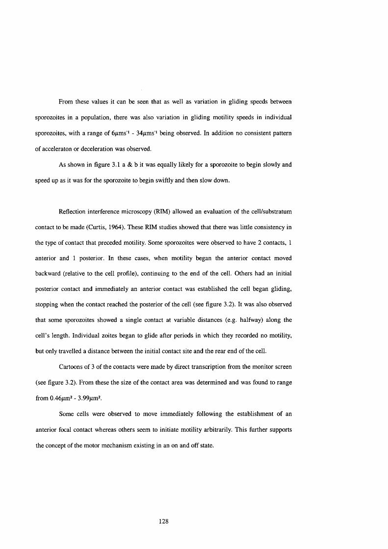
From these values it can be seen that as well as variation in gliding speeds between
sporozoites in a population, there was also variation in gliding motility speeds in individual
sporozoites, with a range of 6/ims‘* - 34/xms' ̂ being observed. In addition no consistent pattern
of acceleraton or deceleration was observed.
As shown in figure 3.1 a & b it was equally likely for a sporozoite to begin slowly and
speed up as it was for the sporozoite to begin swiftly and then slow down.
Reflection interference microscopy (RIM) allowed an evaluation of the cell/substratum
contact to be made (Curtis, 1964). These RIM studies showed that there was little consistency in
the type of contact that preceded motility. Some sporozoites were observed to have 2 contacts, 1
anterior and 1 posterior. In these cases, when motility began the anterior contact moved
backward (relative to the cell profile), continuing to the end of the cell. Others had an initial
posterior contact and immediately an anterior contact was established the cell began gliding,
stopping when the contact reached the posterior of the cell (see figure 3.2). It was also observed
that some sporozoites showed a single contact at variable distances (e.g. halfway) along the
cell’s length. Individual zoites began to glide after periods in which they recorded no motility,
but only travelled a distance between the initial contact site and the rear end of the cell.
Cartoons of 3 of the contacts were made by direct transcription from the monitor screen
(see figure 3.2). From these the size of the contact area was determined and was found to range
from 0.46/im^ - 3.99/tm^.
Some cells were observed to move immediately following the establishment of an
anterior focal contact whereas others seem to initiate motility arbitrarily. This further supports
the concept of the motor mechanism existing in an on and off state.
128
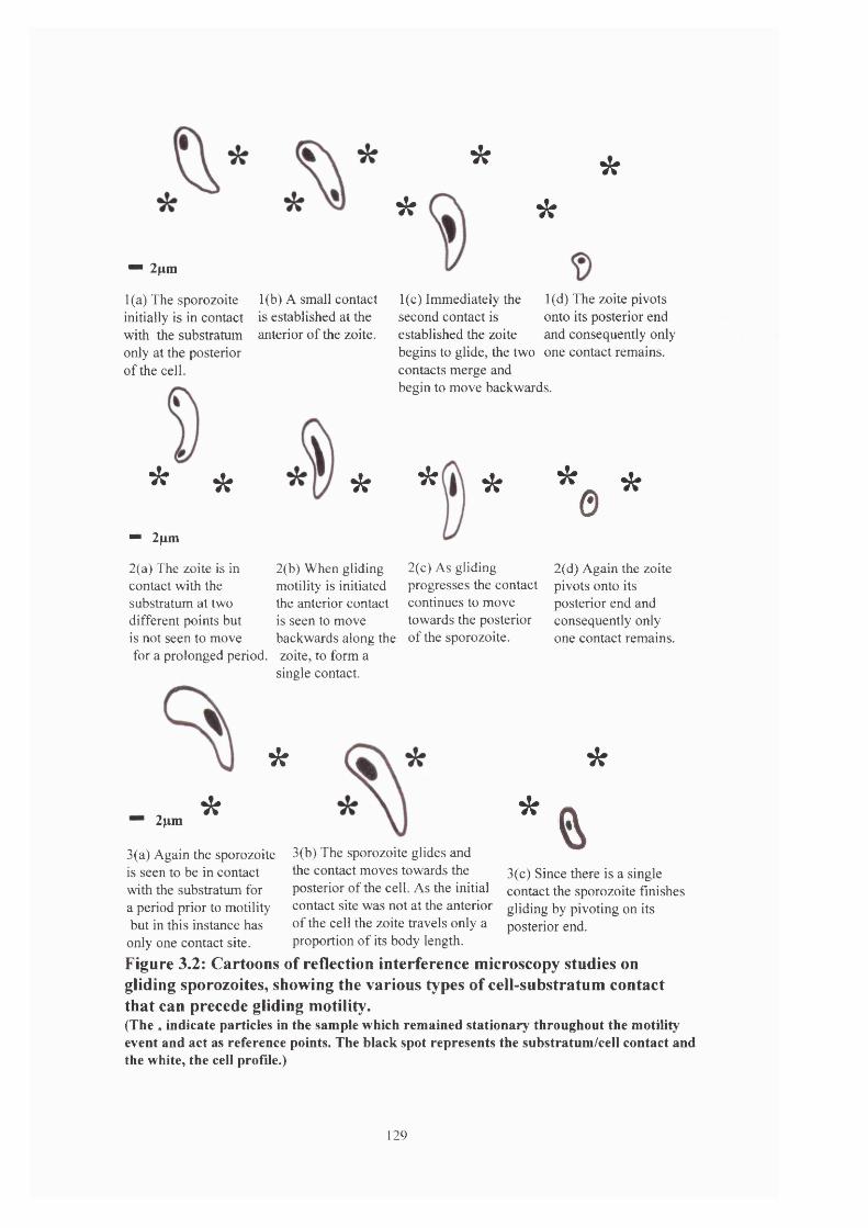
" 2^m
1 (a) The sporozoite initially is in contact with the substratum only at the posterior of the cell.
1(b) A small contact is established at the anterior of the zoite.
*
*
**
1(c) Immediately the second contact is established the zoite
1 (d) The zoite pivots onto its posterior end and consequently only
begins to glide, the two one contact remains, contacts merge and begin to move backwards.
* * *
"" 2pm
2(a) The zoite is in contact with the substratum at two different points but is not seen to move for a prolonged period.
*
2pm*
* *
2(b) When gliding motility is initiated the anterior contact is seen to move backwards along the zoite, to form a single contact.
2(c) As gliding progresses the contact continues to move towards the posterior of the sporozoite.
% *
2(d) Again the zoite pivots onto its posterior end and consequently only one contact remains.
*
*
3(a) Again the sporozoite is seen to be in contact with the substratum for a period prior to motility but in this instance has only one contact site.
%3(c) Since there is a single contact the sporozoite finishes gliding by pivoting on its posterior end.
3(b) The sporozoite glides and the contact moves towards the posterior of the cell. As the initial contact site was not at the anterior of the cell the zoite travels only a proportion of its body length.
Figure 3.2: Cartoons of reflection interference microscopy studies on gliding sporozoites» showing the various types of cell-substratum contact that can precede gliding motility.(The * indicate particles in the sample which remained stationary throughout the motility event and act as reference points. The black spot represents the substratum/cell contact and the white, the cell profile.)
129

RIM is a two-dimensional depiction of the sporozoite/substratum contact during
motility, but of course motility is a three-dimensional phenomenon. By carefully plotting
successive points of contact in relation to the cell profile it can be seen that the sporozoite moves
in a corkscrew motion, with the band of contact somewhat resembling the stripes on a barber’s
pole. This form of motility may provide extra torsion to facilitate host cell entry.
3.1.b Sporozoite excystation
Observation of sporozoites tumbling within sporocysts suggests that excystation could
be a process driven by sporozoite motility.
When sporozoites are immersed in a suitable excystation medium (e.g. warm PBS
containing bile and trypsin) there is a lag phase before the commencement of sporozoite motility
that precedes sporozoite escape. This lag phase usually lasts about 10 minutes, and during this
period it is observed that the plug of material at the pointed end of the sporocyst changes from
opaque to translucent, concomitant with the onset of motility. This would suggest that it is the
digestion of this plug and the entry of bile and trypsin into the sporocyst that are the
environmental cues for the initiation of motility. Further analysis has shown that trypsin appears
to be the most important component in this process since incubation with trypsin alone can
bring about excystation, albeit over a longer period of time (personal observation). Similar
incubation in bile alone does not lead to excystation. Consequently it can be assumed that the
sporocyst plug is composed of mainly proteinaceous material with some lipid content.
Once the sporozoites begin moving inside the sporocyst they exhibit the characteristic
tumbling in which they twist around each other. There seems to be no discernible pattern to this
tumbling with periods of prolonged or momentary motility followed by periods of prolonged or
momentary inactivity. As a result of the shape of the sporocyst the zoites are repeatedly directed
130

into the end of the cyst, and eventually the cyst plug degrades sufficiently to allow the anterior
tip of the sporozoite to be inserted into the aperture. Occasionally the sporozoite tip is seen to
protrude through the aperture then retract to continue tumbling, but generally the sporozoite
produces sufficient traction at the first attempt to escape from the sporocyst.
Perhaps the most striking feature of the excystation process is the degree of
compression of the sporozoite cellular components, particularly the nucleus, have to undergo in
order to escape through the pore in the sporocyst wall (see figures 3.3 & 3.4). The aperture in
the sporocyst through which the sporozoite must excyst is approximately 0.4/xm in diameter
(although the optical conditions made it difficult to measure the aperture as accurately as one
would wish - see figures 3.3 (a) & 3.4 (f)), whereas an Eimeria tenella sporozoite has a
diameter of about 2.5/xm. In a study on E. larimensis sporocysts were seen to be 15/xm x 8/xm
(with the sporozoites approximately 14/xm x 4.5 fxm) and the anterior tip was filled by the
posterior part of the Steida body measuring 1.3/xm across the top and 1.8/xm across the base
(Roberts et al, 1970). This clearly demonstrates that for the cell to successfully achieve
excystation it must undergo a significant degree of distortion (since there is presumably no
change in cell volume). How this is brought about is unclear, but it is probable that this ability is
also utilised during the host cell invasion process.
When a sporozoite leaves the sporocyst a ‘kink’ is often seen in the sporocyst wall close
to the aperture through which the sporozoite escapes (see figure 3.4 (e)). In addition it is
generally noted that the residual cytoplasm that exists in the mature sporocyst is retained within
the sporocyst during and after excystation is complete (see figures 3.3 (g) & (h)). Both these
observations would suggest that excystation is an active process powered by sporozoite motility,
rather than a passive process facilitated by changes in osmotic pressure within the sporocyst
forcing the zoite out.
131
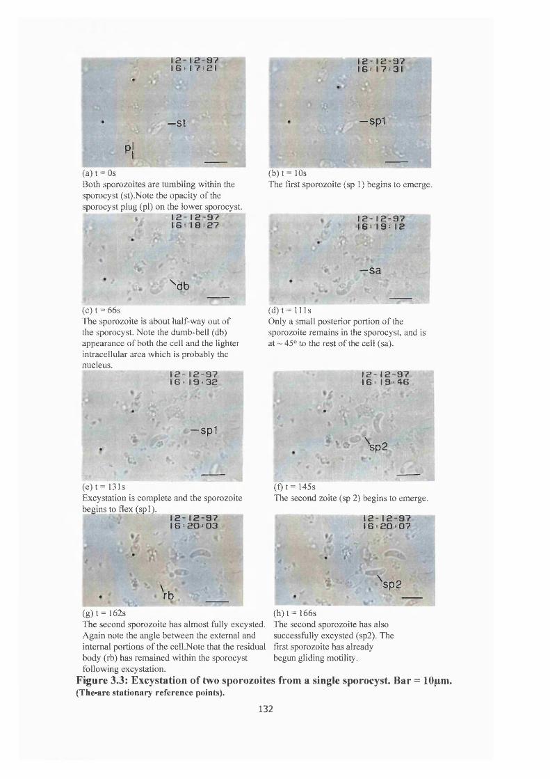
12 1 2 - 9 7 1 6 : 1 7 : 2 1
1 2 - 1 2 - 9 7 16 17 31
—St -sp1
p!
(a) t = OsBoth sporozoites are tumbling within thesporocyst (st).Note the opacity of thesporocyst plug (pi) on the lower sporocyst.
$ 1 2 - 1 2 - 9 7I 6 : I 8 : 2 7
(b) t = 10sThe first sporozoite (sp 1 ) begins to emerge.
1 2 - 1 2 - 9 7 16 1 9 : 1 2
\ d b
(c) t = 66sThe sporozoite is about half-way out of the sporocyst. Note the dumb-bell (db) appearance of both the cell and the lighter intracellular area which is probably the nucleus.
1 2 - 1 2 - 9 7 I 6 : 1 9 : 3 2
— S 3
(d) t = 11 IsOnly a small posterior portion of the sporozoite remains in the sporocyst, and
45® to the rest of the cell (sa).IS
at
1 2 - 1 2 - 9 7 1 6 : 1 9 : 4 6
— sp1
% 'Ii
(e) t = 131sExcystation is complete and the sporozoite begins to flex (spl).
1 2 - 1 2 - 9 7 I 6 : 2 0 : 0 3
\rb
\ sp2
CD t= 145sThe second zoite (sp 2) begins to emerge.
1 2 - 1 2 - 9 7 16 : 2 0 : 0 7
r
\ sp2
(g) t= 162sThe second sporozoite has almost fully excysted.Again note the angle between the external and internal portions of the cell.Note that the residual body (rb) has remained within the sporocyst following excystation.
Figure 3.3: Excystation of two sporozoites from a single sporocyst. Bar = 10pm. (The»are stationary reference points).
(h ) t= 166sThe second sporozoite has also successfully excysted (sp2). The first sporozoite has already begun gliding motility.
132
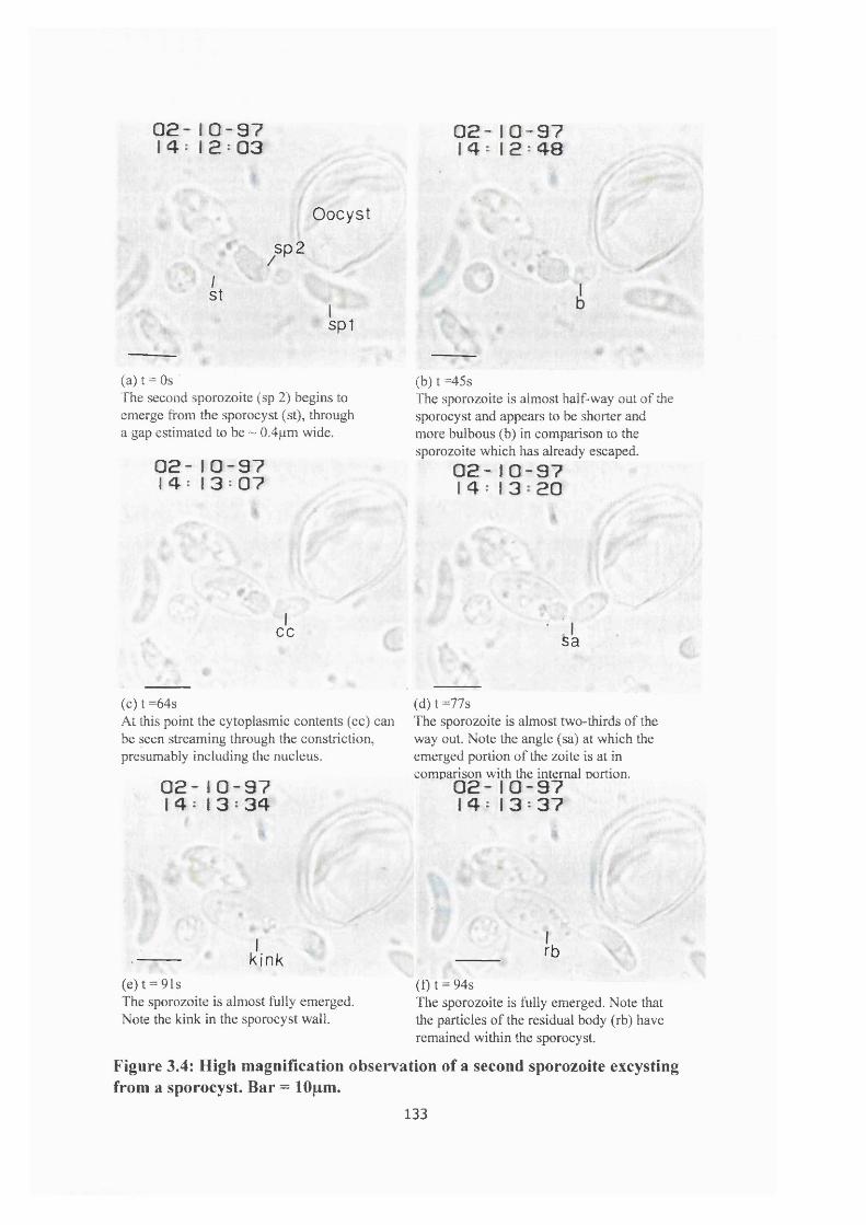
0 2 - 1 0 - 9 7 14: I 2 : 0 3
0 2 - 1 0 - 9 7 I 4 : 1 2 : 4 8
Oocyst
sp2
It
spl
(a) t = OsThe second sporozoite (sp 2) begins to emerge from the sporocyst (st), through a gap estimated to be ~ 0.4pm wide.
0 2 - 1 0 - 9 7 1 4 : 1 3 : 0 7
(b) t =45sThe sporozoite is almost half-way out of the sporocyst and appears to be shorter and more bulbous (b) in comparison to the sporozoite which has already escaped.
0 2 - 1 0 - 9 7 I 4 : I 3 2 0
cc sa
(c) t =64sAt this point the cytoplasmic contents (cc) can be seen streaming through the constriction, presumably including the nucleus.
0 2 - 1 0 - 9 7 I 4 : 1 3 : 3 4
(d) t =77sThe sporozoite is almost two-thirds of the way out. Note the angle (sa) at which the emerged portion of the zoite is at in comparison with the internal portion.
0 2 - 1 0 - 9 7 I 4 : I 3 : 3 7
kink(e)t = 91sThe sporozoite is almost fully emerged. Note the kink in the sporocyst wall.
rb
(t) t = 94sThe sporozoite is fully emerged. Note that the particles of the residual body (rb) have remained within the sporocyst.
Figure 3.4: High magnification observation of a second sporozoite excysting from a sporocyst. Bar = 10pm.
133

The first and most obvious observation about sporozoite excystation from the sporocyst
is that it is a considerably slower process than overt gliding motility. On average (n = 8)
excystation proceeds at 0.2 ± 0.12jLims ’ as compared to a value of 16.7 ± 6/ims * for a sporozoite
gliding across a substrate. It is also observed that on average (n = 8) excystation takes 78 ± 33
seconds for each sporozoite (see table 3.4).
Sporozoite
#
Time taken to
excyst (seconds)
Speed
(/tms'^)
la 120 0.1
lb 64 0.19
2a 80 0.15
2b 99 0.12
3a 114 O il
3b 28 0.43
4a 81 0.15
4b 35 0.32
Mean ± SD 78 ±33 0.2 ±0.12
Table 3.4: Characteristics of Eimeria sporozoite excystation from the sporocyst in excystation medium, a' denotes the first sporozoite to emerge, and h* the second.
134
However if the excystations are examined individually it can be seen that there is a
difference in the excystation profiles of the first sporozoite to emerge compared with the second.
The first sporozoite to emerge is slower, averaging 0.13 ± 0.03/rms * and takes 99 ± 21
seconds to complete the excystation compared to the second zoite which averages 0.27 ±
0.14/xms ' and excysts within 57 ± 32 seconds. The time between the end of the first excystation
and the beginning of the second shows no discernible pattern, values of 40 seconds and 13
minutes having been recorded.
Examination of the excystation process at short time intervals (table 3.5 & figure 3.5)
shows it to be erratic with long periods of inactivity punctuated by short bursts of motility with
variable speed.
135

Time
(seconds)
Total cumulative
distance (/im)
Individual
distance (/tm)
Speed
0 0 0 05 3 3 0.610 3 0 015 3 0 020 3 0 025 3 0 030 3.5 0.5 0.135 3.5 0 040 3.5 0 045 5.3 1.8 0.3650 5.3 0 060 5.5 0.2 0.0465 5.5 0 070 5.5 0 075 5.5 0 080 6.5 1 0.285 8.4 1.9 0.3890 8.4 0 095 9.1 0.7 0.14100 9.1 0 0105 9.1 0 0110 9.1 0 0115 9.1 0 0120 9.1 0 0125 9.5 0.4 0.08130 10.4 0.9 0.18135 12 1.6 0.32
Table 3.5: Characteristics of Eimeria sporozoite excystation in excystation medium, analysed at 5 second intervals.
136

Indiv idual
Cum ulative
Time (seconds)
Figure 3.5: Graph showing the individual and cumulative mean distances moved through the sporocyst aperture, by a single sporozoite during the process of excystation.See the text for a detailed explanation of these observations.
137
As can be seen from the graph (figure 3.5), there seems to be an initial burst of activity
followed by a prolonged period of inactivity. It is likely that this is a consequence of the
difference in the circumference of the sporozoite and the size of the aperture in the sporocyst
wall through which the zoite escapes. During the initial phase of excystation the sporozoite
proceeds through the aperture smoothly, only becoming arrested when the sporozoite
circumference exceeds that of the sporocyst aperture. The sporozoite appears to ‘jam ’ at a
particular point and cannot continue to excyst until sufficient force has been generated by the
motor to overcome this problem. It is also clear from the graph that this occurs a number of
times during the excystation process. The final phase of this process tends to proceed relatively
swiftly, presumably since the circumference of the most posterior portion of the sporozoite is
smaller than the sporocyst wall aperture.
3.1.C Bead translocation speeds
Studies of the translocation of 2/im diameter latex beads across the pellicle of
sporozoites show close similarities to the findings of the gliding studies. The average
translocation speed was 16.9 ± 7.6^ms * and individual speeds varied from 9.9 - 28.8pims *
(table 3.6) which are comparable with the values observed for gliding motility. In the following
table distance is defined as the length that the bead travelled along the sporozoite. Since a
number of the beads made their initial contact at a point below the anterior tip they were
subsequently only translocated from the contact point to the posterior of the cell. Consequently
the total distance they travelled is often smaller than the classical sporozoite length of I2/xm.
138

Sporozoite
#
Time
(seconds)
Total distance
(#tm)
Speed (normalised
to pms*)
1 0.4 7 17.5
2 0.72 12.4 17.2
3 0.56 12 21.4
4 0.56 12 21.4
5 0.56 5.6 9.9
6 0.64 11.4 17.9
7 0.4 8.7 21.8
8 0.48 13.8 28.8
9 0.32 5.6 17.5
10 0.54 9.2 17
M ean ± SD 0.52 ±0.1 11.7 ±4.3 16.9 ±7.6
Table 3.6: Characteristics of 2fim diam eter bead translocations over the pellicle of Eimeria sporozoites in PBS.
139

Time
( sec )
Distance moved (/im) by the latex bead per unit time (0.08 sec)
#1 #2 #3 #4 #5 #6 #7 #8 #9 #10
0 0 0 0 0 0 0 0 0 0 0
0.08 1.7 1.4 2.3 0.9 1.1 1.9 1.6 2.3 2.3 1.3
0.16 2 1.7 1.4 1.9 1.1 1.4 1.4 2.3 2 1.4
0.24 1.6 1.7 2.6 2.1 1.1 1.9 2.4 2.9 1.3 1.7
0.32 1.7 2 2.3 1.8 1.1 1.9 1.3 2.9 0 2
0.40 0 1.7 1.7 1.7 0 1.9 0 3.5 - 1.3
0.48 - 1.6 1.7 1.6 1 1.1 - 0 - 1.4
0.56 - 1.1 0 0 0 1.4 - - - 0
0.64 - 1.1 - - - 0 - - - -
0.72 - 0 - - - - - - - -
Total
pim
7 12.3 12 12 5.4 11.5 8.7 13.9 5.6 9.1
Table 3.7: Characteristics of 2/im diameter bead translocations over the pellicle of Eimeria sporozoites in PBS, analysed at 0.08 second intervals.
The data contained in table 3.7 complements the findings of the gliding motility
analysis. There again seemed to be no consistent pattern of bead acceleration and deceleration,
with beads equally likely to speed up or slow down in successive intervals. Figures 3.6 a & b
illustrate the differences that could be seen in individual bead translocations.
140

2.5
2 T
,5
0.5 !
00 0.08 0.16 0.24 0.32 0.4 0.48 0.56 0.64 0.72
Time (seconds)
14
12
10
8
6
4
2
0
2iSi1I
C Z 3 Individual
— ♦ — C um ulative
Figure 3.6(a): Graph of individual & cumulative distances travelled by a 2pm bead translocating over the pellicle of sporozoite #2 (from table 6).
Time (seconds)
5
3
,5
2
,5
5
00.32 0.48 0.540 0.16 0.24 0.4
16
14
12
10
6
- 4
I 21
: 0
%Si1Iu
■ ■ B Individual
C um ulative
Figure 3.6(b): Graph of individual & cumulative distances travelled by a 2pm bead translocating over the pellicle of sporozoite #8 (from table 6).
141

The bead translocation studies also give support to the idea of a motor system which
needs to be ‘switched on’ before motility can commence. Although the optical resolution was not
ideal it appeared that on some occasions beads were seen to be in contact with the sporozoite
pellicle for prolonged periods of time before translocation occurred. Interestingly, in the case of
sporozoites in which translocation followed gliding, translocation occurred immediately the
sporozoite made contact with the bead.
In addition, bead translocation may also have indicated the orientation of the motor
apparatus. In the case of beads which had been translocated the full length of the cell (50% of
those observed, as shown in table 7) there was an approximate 180° difference between the
anterior starting position and the posterior finishing position of the bead (see figures 3.7 & 3.8).
Beads which had been translocated only a proportion of the cell were consequently displaced only
a proportion of the 180°.
This observation would suggest that the sporozoites have gyre and it may be that is of
importance in the invasion of host cells. Ultrastructural studies of sporozoites have shown that the
sub-pellicular microtubule network runs in an orientation very similar to that which has been
described in the introduction (Colley, 1967; McLaren & Paget, 1968; Ryley, 1969; Russell &
Sinden, 1982).
3.2 Effect of inhibitors
Inhibition studies were undertaken to identify some of the proteins involved in the
processes of sporozoite gliding motility, sporozoite excystation and host cell invasion. No
inhibition of bead translocation was attempted as it was felt that it would be very difficult to
142

. bsp
(a) t = OsThe 2\xm latex bead comes into contact with the sporozoite pellicle at the anterior of the cell.
'
(b) t = 0.16sThe bead has become associated with the pellicle and has begun to be translocated towards the posterior of the cell.
sp
(c) t = 0.24sAs the bead travels towards the posterior it also moves apross the sporozoite from left to right.
sp
° - K;( rBr '̂9
& "■
(e) t = 0.56sThe bead continues to be translocated in this anterior-posterior / left-right direction.
1sp bo
' I^ —
(d) t = 0.4sThe bead has now travelled about two-thirds of the length of the cell and lies in the centre of the sporozoite.
O «sp ,b
è a r * .
(f) t = 0.64sThe translocation is complete, with the bead at the anterior of the sporozoite and on the opposite side to the initial contact.
Figure 3.7: Sequential photographs of the translocation of a single, 2pm diameter latex bead over the pellicle of a stationary Eimeria sporozoite. (b = latex bead; sp = sporozoite; the two circles indicate beads which remained static during the translocation, and therefore provide stationary reference points. Bar = 10pm.)
143
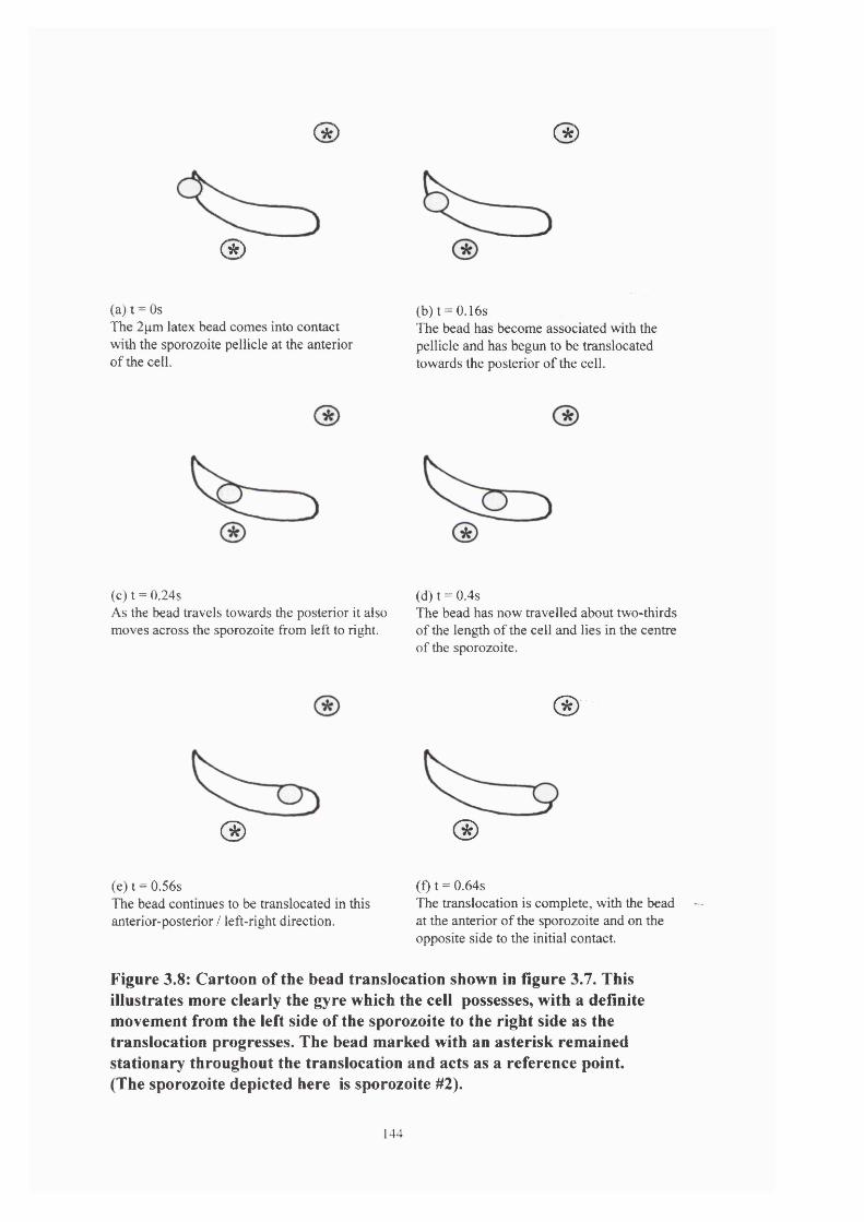
( * ) (*)
(* )
(a) t = OsThe 2p.m latex bead comes into contact with the sporozoite pellicle at the anterior of the cell.
(b) t = 0.16sThe bead has become associated with the pellicle and has begun to be translocated towards the posterior of the cell.
(c) t = 0.24sAs the bead travels towards the posterior it also moves across the sporozoite from left to right.
(d) t = 0.4sThe bead has now travelled about two-thirds of the length of the cell and lies in the centre of the sporozoite.
®
® ®(e) t = 0.56sThe bead continues to be translocated in this anterior-posterior / left-right direction.
(f) t = 0.64sThe translocation is complete, with the bead at the anterior of the sporozoite and on the opposite side to the initial contact.
Figure 3.8: Cartoon of the bead translocation shown in figure 3.7. This illustrates more clearly the gyre which the cell possesses» with a definite movement from the left side of the sporozoite to the right side as the translocation progresses. The bead marked with an asterisk remained stationary throughout the translocation and acts as a reference point. (The sporozoite depicted here is sporozoite #2).
144

identify potential inhibitory effects as bead translocation was a very infrequent occurrence even in
in vitro conditions without the presence of inhibitors.
Statistical analysis of the data was undertaken using Spearman’s rank correlation
coefficient and analysis of variance (anova).
3.2.a Gliding motility
To make meaningful comparisons between the inhibitors the observed values for
sporozoite gliding motility were converted into motility indices. This was calculated by taking the
mean control value and converting it to 100%. Using this base value the motility observed for
each drug was determined by converting the observed values for sporozoites in inhibitors, into a
value which represented the percentage motility as compared with the control samples.
In the case of cytochalasin D, as shown in table 3.8 & figure 3.9, it was seen that at all
concentrations used, inhibition was evident within 5 minutes of the introduction of the
sporozoites to the drug {p< 0.05). Over 70% of sporozoites were inhibited at 1 pg ml-' and almost
25% inhibition at 0.1 pg ml-' and by the end of the experiment essentially all sporozoites at 1 pg
ml-' were inhibited and at 0.1 pg ml-' nearly 50% were inhibited. The parasite also showed a dose
dependent response with inhibition being most marked at the highest concentration.
TIME 1.0 pg ml^ 0.5 pg ml^ 0.1 pg ml'*
(mins) Mean%
SD Mean%
SD Mean%
SD
5 29.2 ± 6.6 45.8 ±2.2 75.5 ±2.220 9.8 ±4.9 26.2 ±6.4 65.4 ±5.960 1.3 ±2.2 12.6 ± 0.5 52.7 ±5.1
Table 3.8: Motility indices for sporozoites incubated in cytochalasin D
145

I
120
100
60
i * 40
I 20
0CONTROL
Jé0. lug/ml 0.5ug/ml
Cyotchalasin D concentration
B 5 minutes □ 20 minutes B 60 minutes
l.Oug/ml
Figure 3.9(a): Inhibition of sporozoite motility in the presence of cytochalasin D.
160
140
120
100
I
§ 60
S 40
20
0
H 5 minutes □ 20 minutes ■ 60 minutes
1 WkCONTROL 25mM 50mM 75mM
BDM concentration
lOOmM
Figure 3.9(b): Inhibition of sporozoite motility in the presence of BDM.
146

Inhibition of sporozoite motility in the presence of BDM was more marked at the highest
experimental drug concentrations used (table 3.9 & figure 3.9). After 5 minutes incubation in
25mM BDM it seemed that the drug elicited an activation response since motility exceeded the
control values. By the end of the experiment only about 25% inhibition was recorded. Incubation
in 50mM BDM also showed this initial increase in activity but at 20 minutes this had fallen
significantly, with nearly 50% inhibition and following incubation for 1 hour 80% of sporozoites
were inhibited (/?< 0.05).
TIM E 25mM 50mM 75mM lOOmM
(mins) Mean%
SD Mean%
SD Mean%
SD Mean%
SD
5 112.1 ±7.1 139.5 ± 3.0 45.6 ±3.1 23.5 ±8.320 84.0 ±18.1 54.8 ±4.2 20.9 ± 1.5 8.6 ±2.960 77.8 ±5.7 20.0 ±5.4 9.8 ±1.6 7.0 ± 0.8
Table 3. 9: Motility indices for sporozoites incubated in BDM
Incubation at the higher concentrations of BDM leads to substantial inhibition of
motility (p< 0.05). With 75mM BDM over 50% of sporozoites were inhibited within 5 minutes
and after 1 hour less than 10% were showing evidence of motility. At lOOmM BDM 75% of
sporozoites were inhibited after 5 minutes with less than 10% remaining motile by the end of the
experiment.
Experiments with 0.1 mM colchicine showed no apparent inhibition of motility with
nearly 100% motility recorded after 1 hour (table 3.10).
147
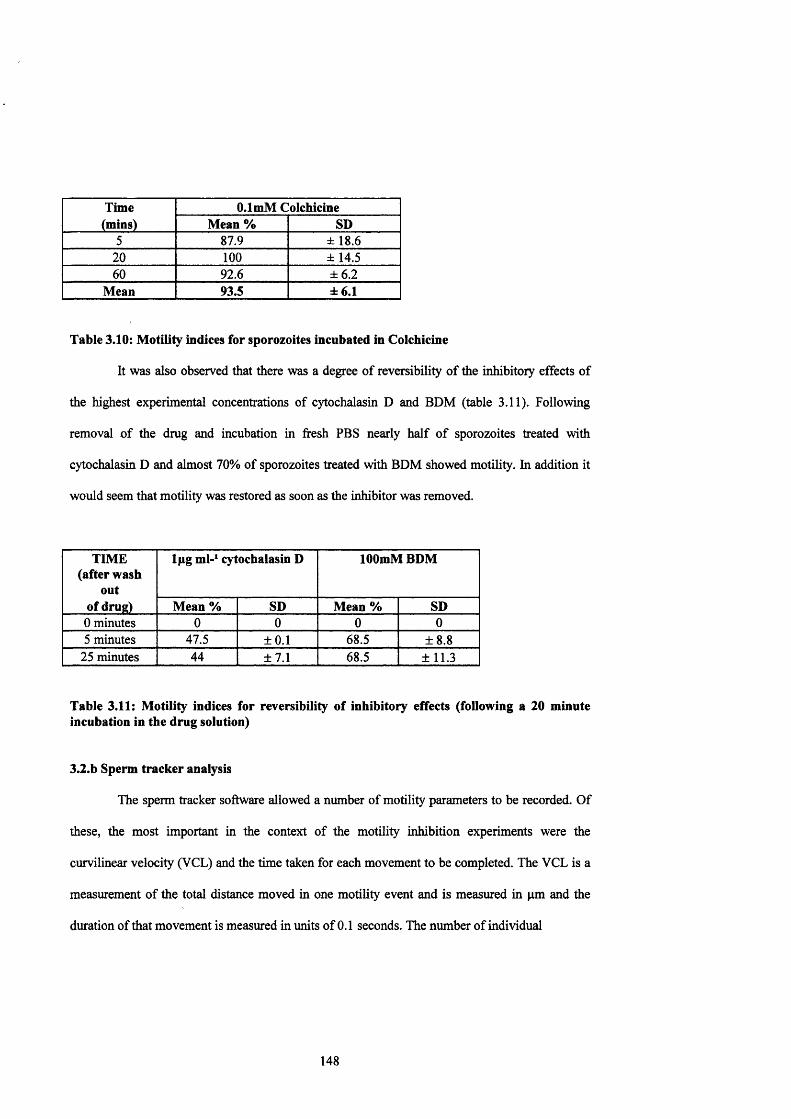
Time O.lmM Colchicine(mins) Mean % SD
5 87.9 ± 18.620 100 ±14.560 92.6 ± 6.2
Mean 93.5 ± 6.1
Table 3.10; Motility indices for sporozoites incubated in Colchicine
It was also observed that there was a degree of reversibility of the inhibitory effects of
the highest experimental concentrations of cytochalasin D and BDM (table 3.11). Following
removal o f the drug and incubation in fresh PBS nearly half of sporozoites treated with
cytochalasin D and almost 70% of sporozoites treated with BDM showed motility. In addition it
would seem that motility was restored as soon as the inhibitor was removed.
TIME (after wash
out of drug)
Ipg ml-^ cytochalasin D lOOmM BDM
Mean % SD Mean % SD0 minutes 0 0 0 05 minutes 47.5 ±0.1 68.5 ±8.8
25 minutes 44 ±7.1 68.5 ± 11.3
Table 3.11: Motility indices for reversibility of inhibitory effects (following a 20 minute incubation in the drug solution)
3.2.b Sperm tracker analysis
The sperm tracker software allowed a number of motility parameters to be recorded. Of
these, the most important in the context of the motility inhibition experiments were the
curvilinear velocity (VCL) and the time taken for each movement to be completed. The VCL is a
measurement of the total distance moved in one motility event and is measured in pm and the
duration of that movement is measured in units of 0.1 seconds. The number of individual
148

Average sporozoite speed (pm sec )
0
10
1If9
Control 10 mins
Control 30 mins
Control 60 mins
25mM BDM 10 mins
25mM BDM 30 mins
25mM BDM 60 mins
50mM BDM 10 mins
50m M BDM 30 mins
50mM BDM 60 mins
75mM BDM 10 mins
lOOraM BDM 10 mins
C B0,5figm l-l 10 mins
CD 0,5figml-l 10 mins
CD 0,5figml-l 30 mins
CD 0,5^gml-l 60 mins
Figure 3.10: Graph showing the average sporozoite gliding motility speeds, as determined by Hobson Sperm Tracker analysis, for cells incubated in potential inhibitors.
149
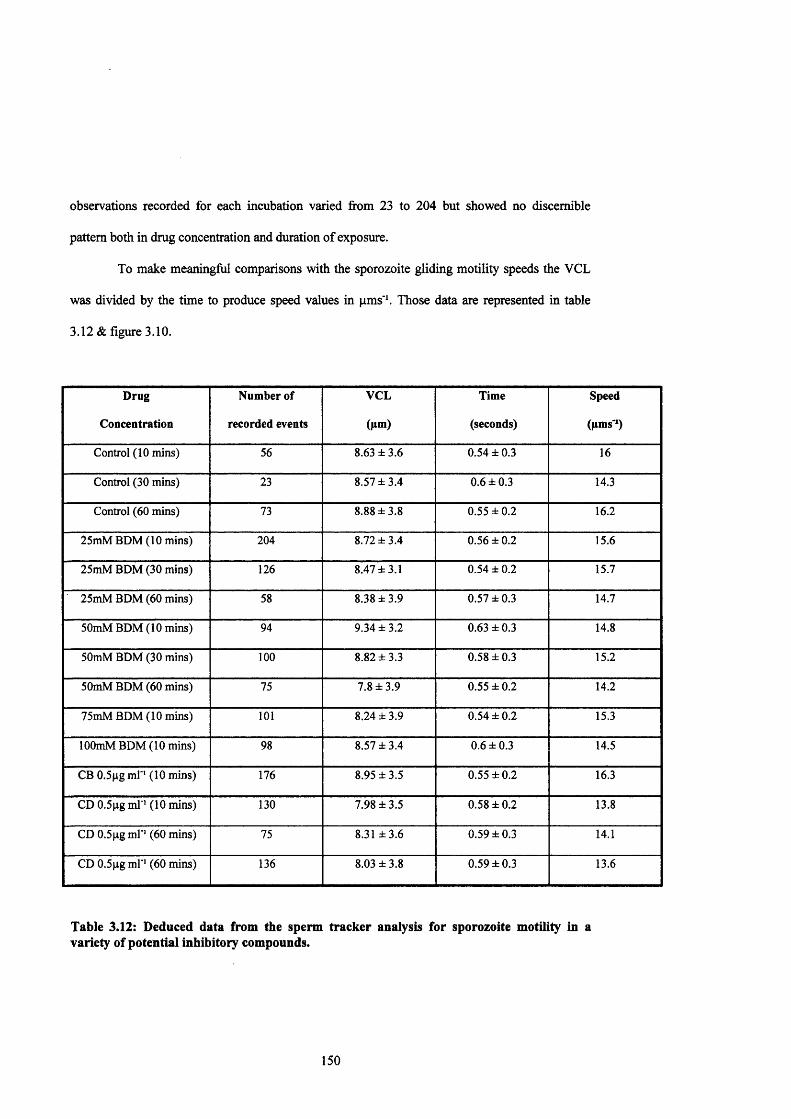
observations recorded for each incubation varied from 23 to 204 but showed no discernible
pattern both in drug concentration and duration of exposure.
To make meaningful comparisons with the sporozoite gliding motility speeds the VCL
was divided by the time to produce speed values in pm s'\ Those data are represented in table
3.12 & figure 3.10.
Drug
Concentration
Number of
recorded events
VCL
(pm)
Time
(seconds)
Speed
(pms'O
Control (10 mins) 56 8.63 ± 3.6 0.54 ± 0.3 16
Control (30 mins) 23 8.57 ±3.4 0.6 ± 0.3 14.3
Control (60 mins) 73 8.88 ±3.8 0.55 ±0.2 16.2
25mM BDM (10 mins) 204 8.72 ±3.4 0.56 ±0.2 15.6
25mM BDM (30 mins) 126 8.47 ±3.1 0.54 ± 0.2 15.7
25mM BDM (60 mins) 58 8.38 ±3.9 0.57 ±0.3 14.7
50mM BDM (10 mins) 94 9.34 ±3.2 0.63 ±0.3 14.8
50mM BDM (30 mins) 100 8.82 ±3.3 0.58 ± 0.3 15.2
50mM BDM (60 mins) 75 7.8 ±3.9 0.55 ±0.2 14.2
75mM BDM (10 mins) 101 8.24 ± 3.9 0.54 ±0.2 15.3
lOOmM BDM (10 mins) 98 8.57 ±3.4 0.6 ± 0.3 14.5
CB O.Sggml"’ (10 mins) 176 8.95 ±3.5 0.55 ±0.2 16.3
CD 0.5|tg ml ' (10 mins) 130 7.98 ±3.5 0.58 ± 0.2 13.8
CD O.Sgg ml ' (60 mins) 75 8.31 ±3.6 0.59 ±0.3 14.1
CD O.Sgg ml ' (60 mins) 136 8.03 ± 3.8 0.59 ±0.3 13.6
Table 3.12: Deduced data from the sperm tracker analysis for sporozoite motility in a variety of potential inhibitory compounds.
150

It is clear from these data that regardless of the conditions of incubation the gliding
speeds observed in motile sporozoites are broadly similar to the values seen in section 3.La,
Sporozoite gliding motility on a glass substrate was seen to occur at speeds between 7.7pms‘‘ -
27.4pms‘‘ with an average of 16.7 ± 6pms‘‘ whereas from the sperm tracker analysis the gliding
speed ranged from 13.8pms ' - 16.3pms' and averaged 14.9 ± 0.9pms'\ These observations
suggest that the inhibitory compounds used have an ‘all or nothing’ effect on the sporozoites, in
that the sporozoites are either unaffected or inhibited but do not undergo a gradual reduction in
gliding efficiency. This provides further proof for the theory that the motor mechanism exists in
an ‘on or ofT state.
At first sight the observation that similar numbers o f sporozoites are moving in each
sample may appear to be at variance with the data extracted from the inhibition studies. However
the inhibition studies were designed so that each individual sporozoite in a field could be scored
for motility whereas the sperm tracker counts the total number of movements in that field. It is
probable therefore that these seemingly confusing results are obtained from a small number of
highly motile sporozoites which at these concentrations remain unaffected by the inhibitors.
Subsequent analysis of the video sequences used for the sperm tracker analysis under the
inhibition study protocol yielded two observations. Firstly that the recorded inhibition was
comparable with the earlier studies, and secondly that in a number of cases highly motile
sporozoites were observed. The video footage of inhibition at lOOmM BDM for 10 minutes and
75mM BDM for 10 minutes both showed the expected levels of inhibition but also contained 1
sporozoite which was seen to twirl for the entire 3 minute period (see table 13). Under the
parameters set for the observation of sporozoites it is probable that these continuously motile cells
were repeatedly scored, hence the apparent contradiction.
151

Drug
concentration
Number of
sporozoites
Number of motile
sporozoites
% motile
sporozoites
Observations
Control @10 mins 30 30 100
Control @ 30 mins 38 38 100
Control @ 60 mins 37 37 100
lOOmM BDM @10 mins 26 1 4 continuous swirling
75mM BDM @ 10 mins 27 5 19 1 continually swirling
50mM BDM @ 10 mins 20 13 65 1 continually swirling
50mM BDM @ 30 mins 24 16 67
50mM BDM @ 60 mins 28 14 50
25mM BDM @ 10 mins 34 34 100
25mM BDM @ 30 mins 29 27 93
25mM BDM @ 60 mins 20 12 60
Table 3.13: Actual number of motility events recorded on the sperm tracker video.
The initial objective of the sperm tracker analysis was to try and determine if cells
incubated in the presence of an inhibitor showed any difference in their motility compared to
control cells, but as a result of the limitations of the system it proved difficult to ascertain these
measurements with any real confidence. However it proved an accurate mechanism for the
determination of gliding motility speeds in control sporozoite samples.
152

60
50
40SI 30 =) z
20
10
0 ITT
B Cysts
■ Tumbling
□ Free Zoites
30 60 90
INCUBATION n M E ( m in u te s )
120
Figure 3.11(a): Values observed for sporozoite excystation from sporocysts, under control conditions.
60
50
40gi 30
^ 20
10
030 60 90
INCUBATION T1ME( minutes )
B Cysts
■ Tumbling
□ Free Zoites
120
Figure 3.11(b): Values observed for sporozoite excystation from sporocysts,in a 1% DMSO solution.
153

r60
50
a "I 30 =^ 20
10
0 T T
0 30 60 90
INCUBATION TIM E( m in u te s )
120
□ Cysts
■ Tumbling
□ Free Zoites
Figure 3.12(a): Values observed for sporozoite excystation from sporocysts, in a solution of 0.1 pgml"’ Cytochalasin D.
70
60
50
sn 40Z3 30Z
20
10
0
■ Cysts■ Tumbling
□ Free Zoites
30 60 90
INCUBATION TIM E( m in u te s )
120
Figure 3.12(b): Values observed for sporozoite excystation from sporocysts,in a solution of 1 pgml^ Cytochalasin D.
154
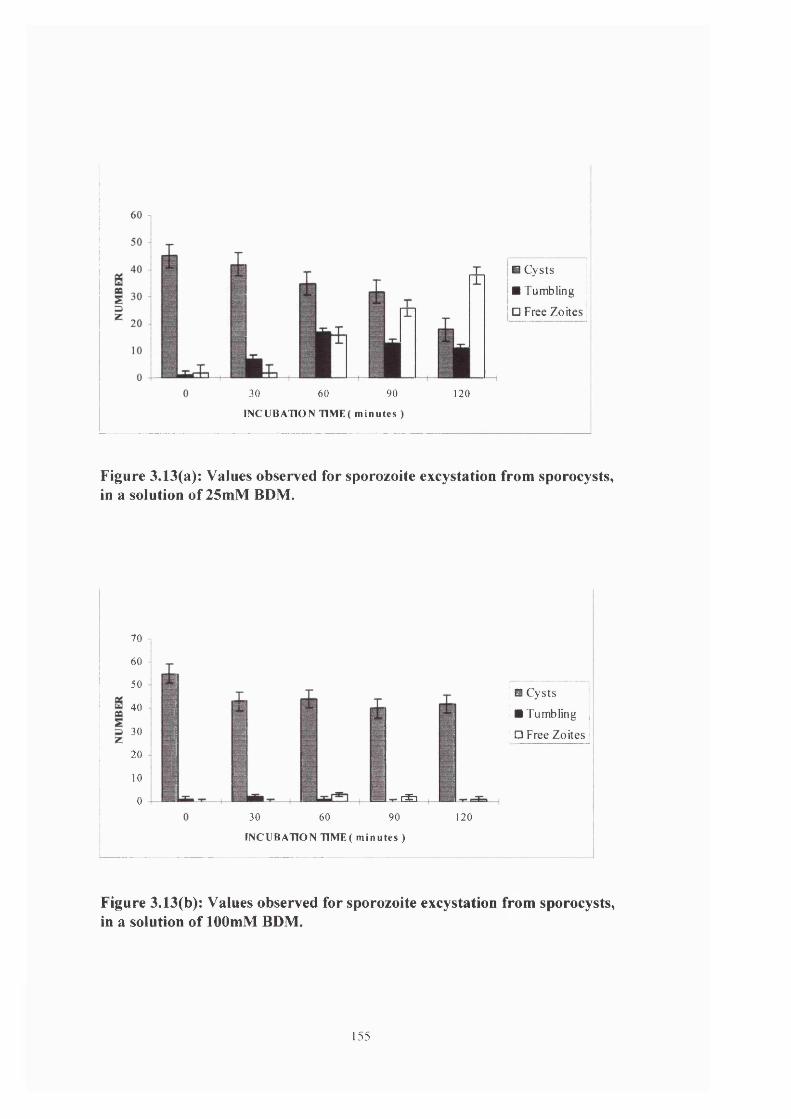
30 60 90
INCUBATION T11V1E( m in u te s )
B Cysts B Tumbling
□ Free Zoites
120
Figure 3.13(a): Values observed for sporozoite excystation from sporocysts, in a solution of 25mM BDM.
70
60
50
40
30
20
10
0 r £ i
B Cysts
B Tumbling
□ Free Zoites
30 60 90
INCUBATION TTME( m in u te s )
120
Figure 3.13(b): Values observed for sporozoite excystation from sporocysts,in a solution of lOOmM BDM.
155

80
70
60
S 50
I 40az 30
20
10
0
T
T
a Cysts ■ Tumbling
□ Free Zoites
30 60 90
INCUBATION T1ME( m in u te s )
120
Figure 3.14(a): Values observed for sporozoite excystation from sporocysts, in a solution of ImM Colchicine.
60
50
Sg 30
^ 20
10
0 T T0
B Cysts
a Tumbling
□ Free Zoites
30 60 90
INCUBATION T1IV1E( m in u te s )
120
Figure 3.14(b): Values observed for sporozoite excystation from sporocysts,in a solution of 1 pgml ' Salinomycin.
156
3.2.C Excystation
Inhibition of sporozoite excystation from sporocysts was evident with cytochalasin D at
a concentration of l/xgml'*, (equivalent to ~ 2piM) and BDM at lOOmM (figures 3.11, 3.12 &
3.13). Both compounds prevented almost all sporozoites from excysting within the 2 hours of
the experimental incubation. In addition it appeared that the inhibitors were able to permeate
into the sporocyst at an early stage in the incubation, thereby preventing the characteristic
sporozoite tumbling from occurring. This process classically precedes excystation.
Incubation in ImM colchicine seemed to have no effect on excystation (figure 3.14),
with both the rate of tumbling and the number of free sporozoites closely resembling the values
seen in the control experiments.
3.2.d Invasion
The first thing of importance which is obvious from these results (table 3.14 & figure
3.15) is that preincubation of the cell monolayer has no impact on subsequent sporozoite
invasion rates. All compounds at all concentrations showed similar invasion rates to those seen
in the control wells.
157

120
100
o 80
g 60
"Ë. 40
20
■ 20 minutes PK7 B 60 minutes PIC □ 20 minutes PIS B 60 minutes PIS
CONTROL CD@ l.Opg/ml
Drug concentration
CD @ 0.1 |ag/ml
120
100
I 80
^ 60L.20
■ 20 minutes PIC B9 60 minutes PIC □ 20 minutes PIS B 60 minutes PIS
lOOmMBDM 25mMBDM ImMColchicine
IjiMSalinomycin
Drug concentration
Figure 3.15: Values for mean % invasion of MDBK cells by sporozoites, following incubation of cells or zoites in potential inhibitory compounds. (PIC == preincubation of cells, PIS = preincubation of sporozoites.)
158

Drug
Concentration
Incubation conditions and time
20 minutes
preincubated cells
(PIC)
60 minutes
preincubated cells
(PIC)
20 minutes
preincubated zoites
(PIS)
60 minutes
preincubated zoites
(PIS)
Control 80.8 99.2 75.6 92.1
CD @ IjLigml'^ 95.6 98.5 46.2 42.2
CD @ 0.1 ̂ gml'^ 94 97.3 40.8 43.3
BDM @ lOOmM 80.7 96.5 29.2 37.9
BDM @ 25mM 68.1 91.4 33.7 48.4
Colchicine @ ImM 90.9 89 46.7 52.3
Salinomycin @ 1/xM 76.7 84.1 31 43.3
Table 3.14: Mean sporozoite invasion rates
However preincubation of the sporozoites resulted in significant inhibition in invasion
with all compounds showing a broadly similar level of activity. This is an interesting
observation since the studies on the inhibition of gliding and excystation showed the parasites
exhibiting a dose dependent response.
159
3.3 Immunofluoresence
3.3.a Actin
Treatment of fixed sporozoites with the Amersham N350 monoclonal anti-actin primary
antibody and a fluorescent conjugated secondary antibody gave a distinct staining pattern.
The fluorescent staining seems to be largely localised to the anterior portion of the cell
(see figure 3.16). Interestingly there is little or no staining at the extreme anterior of the cell in the
region occupied by the apical complex, or in the extreme posterior region o f the sporozoite. The
lack of staining in the region of the posterior retractile body (except in the region of the pellicle)
would suggest that this structure contains little actin, and may indicate that the actin is located in
the cytosol, since actin staining localised to the cell membrane would be unaffected by the
presence of this organelle. However in the region occupied by the anterior refractile body there is
strong staining, suggesting that actin may be associated with this structural feature. Given that
little is known about the function of the refi-actile bodies this apparent difference in cytoskeletal
associations may help to begin elucidate their intracellular roles.
3.3.b Myosin
Fixed sporozoites were probed with the Mitchison antibody to myosin and an
appropriate fluorescently labelled secondary antibody.
Sporozoites treated with the anti-myosin antibody showed a peripheral and distinctly
punctate pattern o f staining (see figure 3.17). The pattern of staining in anti-myosin antibody
treated sporozoites may suggest a membranous location for the myosin, since the signal does not
appear to be obscured by any of the cell’s organelles. The staining is positive in the perinuclear
region
160
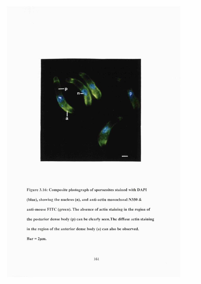
— p
" 1
Figure 3.16: Composite photograph of sporozoites stained with DAPI
(blue), showing the nucleus (n), and anti-actin monoclonal N350 &
anti-mouse FITC (green). The absence of actin staining in the region of
the posterior dense body (p) can be clearly seen.The diffuse actin staining
in the region of the anterior dense body (a) can also be observed.
Bar = 2pm.
161
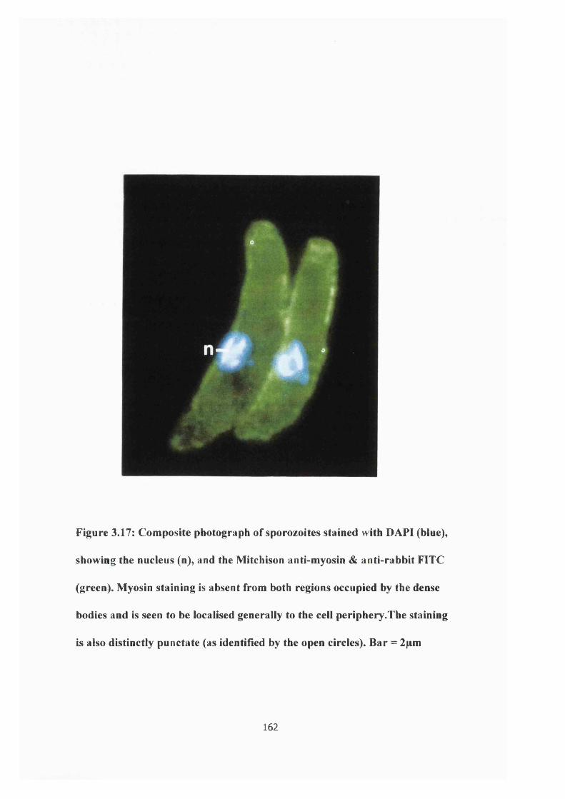
4 ê #
Figure 3.17: Composite photograph of sporozoites stained with DAPI (blue),
showing the nucleus (n), and the Mitchison anti-myosin & anti-rabbit FITC
(green). Myosin staining is absent from both regions occupied by the dense
bodies and is seen to be localised generally to the cell periphery.The staining
is also distinctly punctate (as identified by the open circles). Bar = 2pm
162

but weak elsewhere in the central region of the sporozoite. As discussed in the introduction, the
cytoskeleton is thought to have a role in many diverse cellular functions, and the localisation of
myosin to the nucleus may indicate that this protein has a role in the transport of nuclear
derived components to their sites of action e.g. the organelles of the apical complex. It may also
indicate that the myosin may be transported to the apical complex from this site, or that it may
be Golgi derived.
3.4 Cell ultrastructure
The original aim of these EM studies was to look for sporozoites in the act of target cell
invasion, and subsequently to probe that sporozoite with the anti-actin and anti-myosin
antibodies used in the Western blotting and immunofluoresence experiments. It was felt that if
one could ascertain the location of these proteins in the sporozoite during the process of
invasion, then it would give an indication, at the ultrastructural level, as to the possible role of
actin and myosin in target cell invasion.
Unfortunately, despite examining many samples of different incubation lengths and
using a variety of cell lines, it proved impossible to catch a sporozoite at the precise moment of
penetration. Similarly despite repeated modifications of all aspects of the sample preparation, it
proved impossible to prepare sporozoite thin sections that would firstly be amenable to
immunological examination, and secondly, that would withstand the harsh conditions of the
electron beam.
Despite these obvious limitations it was possible to derive a number of electron
micrographs (figures 3 .1 8 -3 .3 1 ) that show many of the ultrastructural features of sporozoites
which are characteristic of the Apicomplexa. In addition this study also illustrated the relatively
163

*$ ft
0
Figure 3.18: TEM section through an uninvaded CaC02 cell. Note the
dense array of microvilli, the mesh-like network that forms the cortical
layer and the tight junction (tj) between cells. (xl9,000 ).
164

wide host cell range which can support invasion by Eimeria tenella sporozoites. This contrasts
with the small host range of invasive stages of Plasmodium spp. and complements the findings
of the host cell promiscuity of Toxoplasma gondii. The individual micrographs are described
and discussed below.
Figure 3.18:
Due to the fixation conditions the glycocalyx that one would expect to be present on the
surface of the cell is absent. However in CaC02 cells it is easy to identify both a microvillar
layer about 2.6/xm in width and a cortical layer 0.8/xm wide associated with the apical surface.
Classically one would expect the microvillus of an epithelial brush border cell to be
approximately 0.1/rm wide, 1/im in length and have a density of about 60 microvilli per
(Fawcett, 1981). This discrepancy is likely to have arisen from the nature of the sectioning
process, in which it is very difficult to obtain a true cross section, with most sections being cut at
an oblique angle, thus altering the apparent cellular dimensions. It may also be a function of the
difference seen in cultured against non-cultured cells as a result of the dense packing associated
with in vivo epithelial cells A tight junction between two adjacent cells can also be clearly seen
and measures just under a micron in length.
These features of the cell are important considerations in respect of successful target
cell invasion by a sporozoite. Since sporozoites are considerably larger than the individual
microvilli it is probable that they invade the cell towards the base of the microvillar layer, in the
crypts between microvilli. This is better illustrated in figures 3.20 & 3.21 where a distinct
difference in the degree of target cell-sporozoite contact can be seen. Once this layer has been
negotiated, the dense, mesh-like network of the cortical layer presents a further barrier to the
165
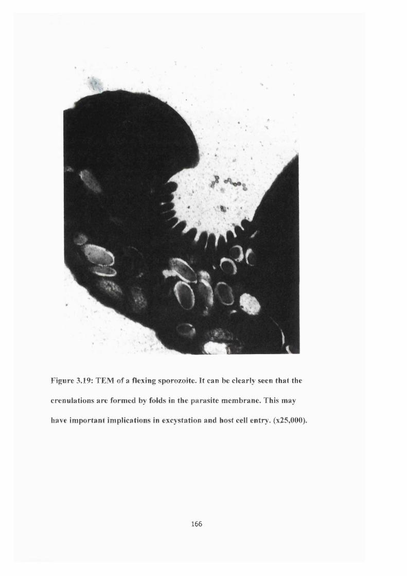
5 r
Figure 3.19: TEM of a flexing sporozoite. It can be clearly seen that the
crenuiations are formed by folds in the parasite membrane. This may
have important implications in excystation and host cell entry. (x25,000).
166
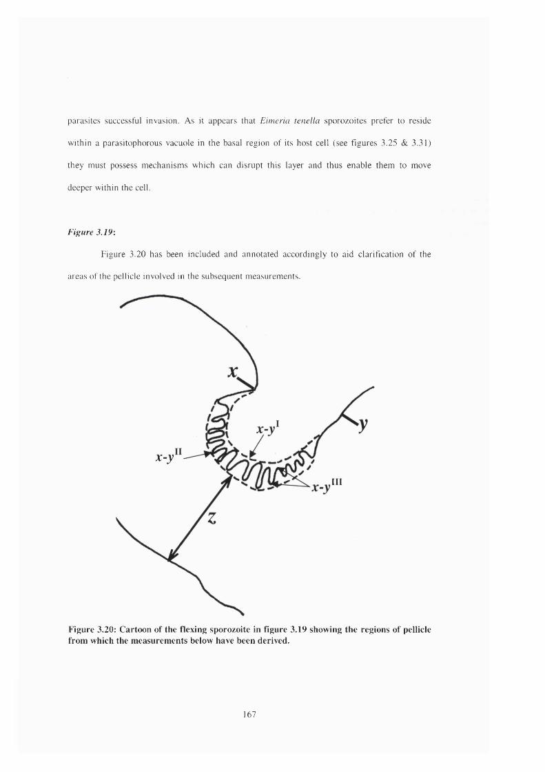
parasites successful invasion. As it appears that Eimeria tenella sporozoites prefer to reside
within a parasitophorous vacuole in the basal region of its host cell (see figures 3.25 & 3.31)
they must possess mechanisms which can disrupt this layer and thus enable them to move
deeper within the cell.
Figure 3.19:
Figure 3.20 has been included and annotated accordingly to aid clarification of the
areas of the pellicle involved in the subsequent measurements.
x-y
III
Figure 3.20: Cartoon of the flexing sporozoite in figure 3.19 showing the regions of pellicle from which the measurements below have been derived.
167
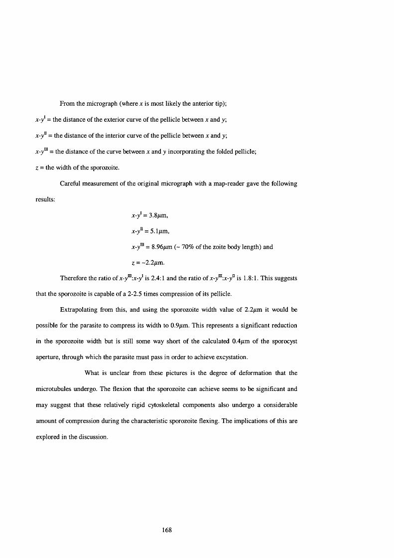
From the micrograph (where x is most likely the anterior tip);
x-y' = the distance of the exterior curve of the pellicle between x and y;
x-y° = the distance of the interior curve of the pellicle between x and y;
x-y™ = the distance of the curve between x and y incorporating the folded pellicle;
z = the width of the sporozoite.
Careful measurement of the original micrograph with a map-reader gave the following
results:
x-y' = 3.8/rm,
x-y" = 5.1/xm,
x-y'" = 8.96/xm (~ 70% of the zoite body length) and
z = -2.2fim.
Therefore the ratio of x-y'":x-y' is 2.4:1 and the ratio of x-y'":x-y" is 1.8:1. This suggests
that the sporozoite is capable of a 2-2.5 times compression of its pellicle.
Extrapolating from this, and using the sporozoite width value of 2.2/rm it would be
possible for the parasite to compress its width to 0.9/xm. This represents a significant reduction
in the sporozoite width but is still some way short of the calculated 0.4jam of the sporocyst
aperture, through which the parasite must pass in order to achieve excystation.
What is unclear from these pictures is the degree of deformation that the
microtubules undergo. The flexion that the sporozoite can achieve seems to be significant and
may suggest that these relatively rigid cytoskeletal components also undergo a considerable
amount of compression during the characteristic sporozoite flexing. The implications of this are
explored in the discussion.
168
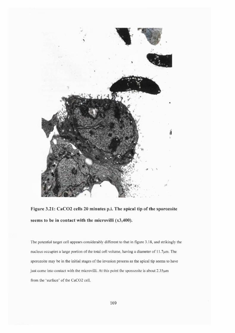
*«
w l i t l •
Figure 3.21: CaC02 cells 20 minutes p.i. The apical tip of the sporozoite
seems to be in contact with the microvilli (x3,400).
The potential target cell appears considerably different to that in figure 3.18, and strikingly the
nucleus occupies a large portion of the total cell volume, having a diameter of 11.7pm. The
sporozoite may be in the initial stages of the invasion process as the apical tip seems to have
just come into contact with the microvilli. At this point the sporozoite is about 2.35pm
from the ‘surface’ of the CaC02 cell.
169
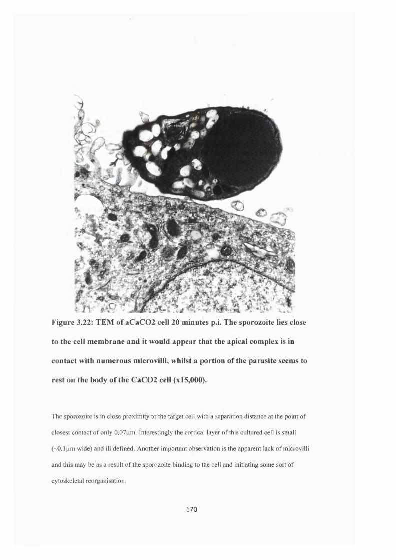
h
S
<C)
Figure 3.22; TEM of aCaC02 cell 20 minutes p.i. The sporozoite lies close
to the cell membrane and it would appear that the apical complex is in
contact with numerous microvilli, whilst a portion of the parasite seems to
rest on the body of the CaC02 cell (x l5,000).
The sporozoite is in close proximity to the target cell with a separation distance at the point of
closest contact of only 0.07pm. Interestingly the cortical layer of this cultured cell is small
(~0.1 pm wide) and ill defined. Another important observation is the apparent lack of microvilli
and this may be as a result of the sporozoite binding to the cell and initiating some sort of
cytoskeletal reorganisation.
170

(a)
(b) - 7 / 1 7 - iSBR bggFigure 3.23: TEMs of a CaC02 ceil 20 minutes p.i. At these magnifications
the posterior of the sporozoite is not observable but the cell contains a
sporozoite which has been sectioned through the anterior and posterior
portions suggesting it is resting in a curved manner in the parasitophorous
vacuole. Higher magnification shows the conoid (c) and the micronemes
(mn). It is also possible to see the PVM (pvm). (a - xl5,000; b - x57,000).
The CaC02 cell has a much reduced microvillar layer (~ 0.8pm width) which may be a
result of microvillus shedding/resorption following sporozoite invasion.
171

Figure 3.24: TEM of a possible sporozoite Golgi complex (Gc) situated
adjacent to the parasite nucleus (N). At this point the parasite nucleus is
about 0.8pm (x71,000). (This is a higher magnifîcation micrograph of the sporozoite
pictured in figure 3.31).
172

Figure 3.25: TEM of a CaC02 60 minutes p.i. A sporozoite lying basally
(by reference to the upper microvillar layer) within a parasitophorous
vacuole. Both the anterior (adb) and posterior dense bodies (pdb) can be
clearly seen, with the parasite nucleus (N) in-between. In this micrograph
the parasite nucleus is 1.05;xm in width (x5,700).
173

Figure 3.26: High magnification TEM micrograph of in intracellular
sporozoite which has been sectioned through the apical tip. The rhoptries
(rh) can be clearly seen and the banding seen could be the microtubules,
since they seem to possess the correct curvature. (x9,100).
At its widest point the sporozoite appears to be about 3.5pm (which again may be a function
of the sectioning process) and in this section the uppermost tip of the sporozoite is 1.6pm.
174

4
mm
Figure 3.27: TEM of the anterior region of a sporozoite which has possibly
just invaded the INT 407 ceil, possibly leaving behind the membranous
material (mm) seen outside the cell, (x l9,000).
Again the cell appears to be devoid of microvilli following successful internalisation
by the parasite.
175

Figure 3.28: TEM of a sporozoite at the base of an INT 407 cell.
The conoid (c), rhoptries (rhl & 2) and micronemes (mn) are clearly
visible, (x l9,000).
From measurements obtained using this micrograph, rhl is 1.8pm long and 160nm at its
widest point, and rh2 is 1.6pm in length and lOOnm at its widest point. It is also seen that the
gap at the neck of the conoid is ~ 150nm.
176
f .
Figure 3.29: A high resolution TEM micrograph of the apical tip of a
sporozoite residing in a parasitophorous vacuole within an INT 407 cell.
The parasite plasma membrane (pm) and the two membranes of the inner
membrane complex (IMC) can be easily distinguished. (x71,000).
To obtain a representative value for the widtli of each membrane, measurements were taken at
8 random points along the entire length of each membrane. All three membranes are on average
7nm wide (plasma membrane = 7.2± 0.4nm; outer IMC = 6.9± 0.2nm; inner IMC = 7.2± 0.3nm).
Again taking measurements at 8 different points along the plasma membrane, the gap between
the plasma membrane and the IMC is 18± 2.1nm. In addition the gap between the two ends of
the plasma membrane at the apical end of the parasite is ~340nm. The two ‘arms' of the conoid
(c) formed by the sectioning process are 295nm in length, 42nm wide and are separated b y a gap
of 200nm. Numerous micronemes (mn) can be seen and on average these measure 327± 78nm in
length and 89 ± l lnm at their widest point (n=9). The membrane of the parasitophorous vacuole
can also be clearly seen.
177
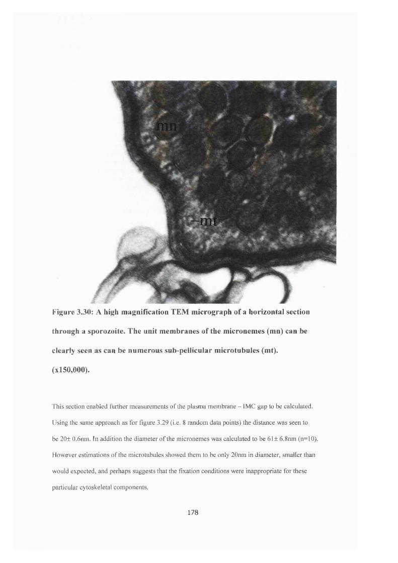
Figure 3.30: A high magnification TEM micrograph of a horizontal section
through a sporozoite. The unit membranes of the micronemes (mn) can be
clearly seen as can be numerous sub-pellicular microtubules (mt).
(xl50,000).
This section enabled further measurements of the plasma membrane - IMC gap to be calculated.
Using the same approach as for figure 3.29 (i.e. 8 random data points) the distance was seen to
be 20± 0.6nm. In addition the diameter of the micronemes was calculated to be 61± 6.8nm (n=10).
However estimations of the microtubules showed them to be only 20nm in diameter, smaller tlian
would expected, and perhaps suggests that the fixation conditions were inappropriate for these
particular cytoskeletal components.
178
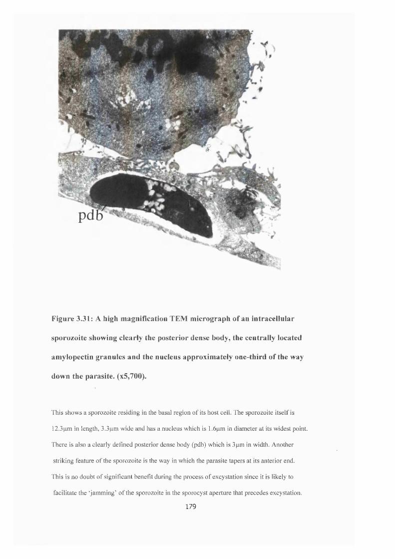
wiMm
f
Figure 3.31: A high magnification TEM micrograph of an intracellular
sporozoite showing clearly the posterior dense body, the centrally located
amylopectin granules and the nucleus approximately one-third of the way
down the parasite. (x5,700).
This shows a sporozoite residing in the basal region of its host ceil. The sporozoite itself is
12.3pm in length, 3.3pm wide and has a nucleus which is 1.6pm in diameter at its widest point.
There is also a clearly defined posterior dense body (pdb) which is 3pm in width. Another
striking featuie of the sporozoite is the way in which the parasite tapers at its anterior end.
This is no doubt of significant benefit during the process of excystation since it is likely to
facilitate the ‘jamming’ of the sporozoite in the sporocyst aperture that precedes excystation.
179

3.5 Western blotting
3.5.a Actin
Following the detection protocol using the 4-chloro-1 -naphthol methodology and rabbit
polyclonal anti-actin yielded rather disappointing results. The antibody identified very distinctly
the standard actin which migrated to the classical actin value of 43kDa. However, with the
Eimeria sample the antibody recognised a band which co-migrated with the standard actin but
also recognised a number of bands of both higher and lower apparent molecular weight. The
number and intensity of these extraneous bands could be altered by manipulating the incubation
and detection conditions but it was never possible to eradicate them entirely (data not shown).
Initial experiments using the polyclonal antibody and the ECL detection method also
showed a strong signal at approximately 43kDa but it proved impossible to eliminate the extra
bands. It was felt that a more specific antibody may facilitate the identification process and
therefore a mouse monoclonal anti-actin was obtained from Amersham (N350).
Sporozoite samples probed with this monoclonal antibody at a dilution of 1:2000 and a
secondary anti-mouse HRP conjugate at a concentration of 1:1000, and exposed for 15 seconds
showed a very clean blot with a single, distinct band co-migrating with the standard actin to a
value of 43kDa (see figure 3.32).
In addition to identifying the actin in the sporozoite samples the amount of actin in an
individual sporozoite was estimated following the treatment of the actin dilution series with the
Amersham monoclonal antibody. A sporozoite sample containing a known number of cells
(1x10^) was prepared as described in section 2.4.b and run on a polyacrylamide gel in which
adjacent wells contained progressively smaller amounts of standard actin (36pgml'' - 3.6pgml '
protein). The proteins were then transferred onto a nitrocellulose sheet and probed with the anti-
180
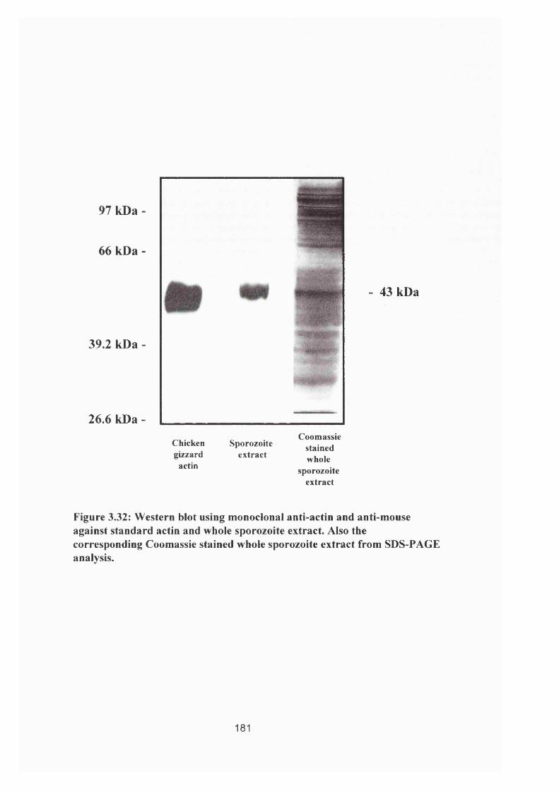
97 k D a -
66 kD a -
39.2 kD a -
26.6 kDa -
Chickengizzard
actin
Sporozoiteextract
Coomassiestainedwhole
sporozoiteextract
- 43 kDa
Figure 3.32: Western blot using monoclonal anti-actin and anti-mouse against standard actin and whole sporozoite extract. Also the corresponding Coomassie stained whole sporozoite extract from SDS-PAGE analysis.
181
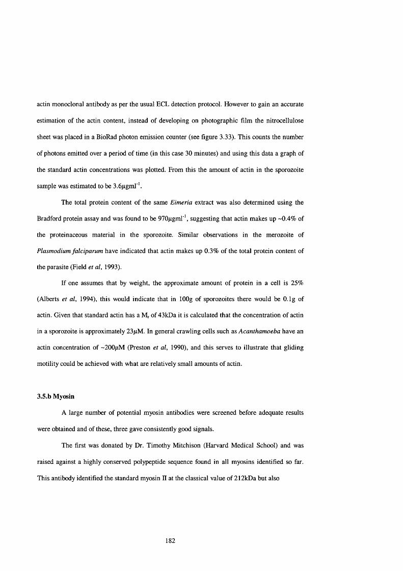
actin monoclonal antibody as per the usual ECL detection protocol. However to gain an accurate
estimation of the actin content, instead of developing on photographic film the nitrocellulose
sheet was placed in a BioRad photon emission counter (see figure 3.33). This counts the number
of photons emitted over a period of time (in this case 30 minutes) and using this data a graph of
the standard actin concentrations was plotted. From this the amount of actin in the sporozoite
sample was estimated to be 3.6jLigml '.
The total protein content of the same Eimeria extract was also determined using the
Bradford protein assay and was found to be 970jugml ', suggesting that actin makes up -0.4% of
the proteinaceous material in the sporozoite. Similar observations in the merozoite of
Plasmodium falciparum have indicated that actin makes up 0.3% of the total protein content of
the parasite (Field et al, 1993).
If one assumes that by weight, the approximate amount of protein in a cell is 25%
(Alberts et al, 1994), this would indicate that in lOOg of sporozoites there would be O.lg of
actin. Given that standard actin has a Mr of 43kDa it is calculated that the concentration of actin
in a sporozoite is approximately 23/xM. In general crawling cells such as Acanthamoeba have an
actin concentration of ~200/xM (Preston et al, 1990), and this serves to illustrate that gliding
motility could be achieved with what are relatively small amounts of actin.
3.5.b Myosin
A large number of potential myosin antibodies were screened before adequate results
were obtained and of these, three gave consistently good signals.
The first was donated by Dr. Timothy Mitchison (Harvard Medical School) and was
raised against a highly conserved polypeptide sequence found in all myosins identified so far.
This antibody identified the standard myosin II at the classical value of 212kDa but also
182

97 kDa-
68 kDa-
46 kDa -
31 kDa -
- 43 kDa
Sp 1 2 3 4 5 6
Figure 3.33: Western blot using monoclonal anti-actin and anti-mouse against whole sporozoite extract and actin dilution series. Sp= sporozoite extract, 1= 36pg actin, 2= 28.8pg actin, 3= 21.6pg actin, 4= 14.4pg actin, 5= 7.2pg actin & 6= 3.6pg actin.
183

212 kDa -
116 kDa
97 kDa
66 kDa -
- 212 kDa
-93 kDa
Chickengizzardmyosin
Wholesporozoite
extract
Figure 3.34: Western blots using the Mitchison anti-myosin and anti-rabbit against a classical myosin II standard (chicken gizzard myosin - Sigma) and the whole sporozoite extract.
184

identified a much smaller myosin in the sporozoite extract at a value of 93kDa (see figure 3.34).
Recent work in the related Apicomplexans Toxoplasma gondii (Schwartzman et at, 1998) &
Plasmodium falciparum (Finder et al, 1998) has also identified a 93kDa myosin like protein.
The second two antibodies were donated by Dr. Jeni Finder (Kings College, London),
one of which was raised against Plasmodium falciparum myosin and the second against a highly
conserved polypeptide sequence in the head region of all myosins so far identified. Both these
antibodies identified a 93kDa band in the sporozoite extract (see figure 3.35). This second
antibody gave relatively poor results and so was not used in subsequent experiments.
It is interesting to note that these antibodies, although raised against a highly conserved
myosin polypeptide sequence (CNFILEAFGNAKTIKNNNSSRRGKY for the Mitchison
antibody and LEAFGNAKTIRNNN for the Finder antibody), identified only the single band
from the whole sporozoite extracts. Genetic analysis of other organisms have shown them to
contain numerous classes of myosins (e.g. Dictyostelium discoideum has been reported to
possess between 5 (Titus et al, 1989; Jung & Hammer, 1990) and 9 (Jung et al, 1993; Titus et
al, 1994) distinct myosin classes), and it is possible that this is also the case for Eimeria. In light
of this assertion it is intriguing that Western blotting identified only a single band and it may be
an indication that there may be a stage specific expression of myosin genes in Eimeria tenella.
3.5.C Actin associated proteins (AAPs)
A wide range of antibodies raised against known AAFs were used to probe the
sporozoite blots but under these conditions none gave consistently reliable results. Antibodies
against a-actinin, actin depolymerising factor, cofilin, filamin, spectrin & tropomyosin were
tested.
185

in the related Apicomplexans Toxoplasma gondii (Schwartzman et al, 1998) & Plasmodium
falciparum (Finder et al, 1998) has also identified a 93kDa myosin like protein.
The second two antibodies were donated by Dr. Jeni Finder (Kings College, London), one
of which was raised against Plasmodium falciparum myosin and the second against a highly
conserved polypeptide sequence in the head region of all myosins so far identified. Both these
antibodies identified a 93kDa band in the sporozoite extract (see figure 3.35). This second antibody
gave relatively poor results and so was not used in subsequent experiments.
It is interesting to note that these antibodies, although raised against a highly conserved
myosin polypeptide sequence (CNFILEAFGNAKTIKNNNSSRRGKY for the Mitchison antibody
and LEAFGNAKTIRNNN for the Finder antibody), identified only the single band from the whole
sporozoite extracts. Genetic analysis of other organisms have shown them to contain numerous
classes of myosins (e.g. Dictyostelium discoideum has been reported to possess between 5 (Titus et
al, 1989; Jung & Hammer, 1990) and 9 (Jung et al, 1993; Titus et al, 1994) distinct myosin classes),
and it is possible that this is also the case for Eimeria. In light of this assertion it is intriguing that
Western blotting identified only a single band and it may be an indication that there may be a stage
specific expression of myosin genes in Eimeria tenella.
3.5.C Actin associated proteins (AAPs)
A wide range of antibodies raised against known AAFs were used to probe the sporozoite
blots but under these conditions none gave consistently reliable results. Antibodies against a-actinin,
actin depolymerising factor, cofilin, filamin, spectrin & tropomyosin were tested.
As shown in section 3.5.a the amount of actin in an individual sporozoite is small
compared with standard figures from vertebrate and invertebrate crawling cells (e.g. Acanthamoeba
has an actin concentration of approximately 170pM - Alberts et al, 1994), and it is probable that
each AAP will exist at a cell at a ratio lower than 1:1 actin: AAF (depending on the
186
As shown in section 3.5.a the amount of actin in an individual sporozoite is small
compared with standard figures from vertebrate and invertebrate crawling cells (e.g.
Acanthamoeba has an actin concentration of approximately 170/iM - Alberts et al, 1994), and it
is probable that each AAP will exist at a cell at a ratio lower than 1:1 actin: AAP (depending on
the association constant between actin and the specific AAP), the actual amount of a given AAP
might be very low. Therefore it is possible that the concentration of those AAPs probed for is
just too low to allow detection by Western blotting with these antibodies.
3.6 Fast Protein Liquid Chromatography (FPLC)
Samples from chromatographic analysis were initially assessed using a
spectrophotometer to determine the level of proteins in that sample. Aliquots which showed a
relatively high protein content were then Western blotted using the Amersham anti-actin
monoclonal and developed using the ECL detection method.
Neither Eimeria samples from the MonoQ columns nor from the gel filtration columns
showed any signal even following prolonged exposure (+10 minutes) to the photographic film.
However the samples used in the FPLC experiments were whole oocyst derived preparations and
as described previously this method of preparation was shown to be subject to substantial
proteolytic degradation. Consequently there may have been little intact protein left to bind and
be eluted from the columns.
187

3.7 Identification of myosin sequence
3.7.a Extraction of genomic DNA
From the genomic DNA shown in figure 3.36 the Eimeria band measured in excess of
1Mb in size. The reported sizes of the 14 Eimeria tenella chromosomes ranges from 1Mb to
6Mb (Shirley, 1994). However the concentration of agarose gel used was not optimised for
separation of such large components and it is possible that this band represents the chromosomal
DNA.
3.7.b PCR products
The use of degenerate primers to screen for the presence of myosin genes has been
reported in both protozoa and mammalian cell types (Garcés & Gavin, 1998; Bement et al,
1994). These primers were also based on the highly conserved myosin amino acid sequences
GESGAGKT & LEAF.
PCR produced two appropriately sized candidate bands (as shown in diagram 3.37).
Since the two primers were separated by 53 amino acid residues appropriate PCR products were
to be expected to measure approximately 160 bp in length. The two candidate bands appear to be
-ISO bp (band 1) and -160 bp (band 2). These were then purified and used in the subsequent
transformation reactions.
3.7.C Selection of inserts
Blue/white selection yielded a large number of white colonies and double restriction
enzyme digests of the purified plasmids confirmed the presence of appropriately sized inserts
(figure 3.38) in a number of the selected plasmids. Excision from the MCS by digestion with the
188

3000 bp-
1500 bp -600 bp- 500 bp-
400 bp -
300 bp -
200 bp-
100 bp-
sampleloading
well
DNAladder
E i m e r iagenomic
DNA
Figure 3.36: Photograph of a UV illuminated 0.8% agarose gel of Eimeria total genomic DNA and a 1 Mb DNA marker ladder.
189

B and 1 (-180 bp) -
B and 2 ( - 160 bp)-
- 3000 bp -1500 bp
- 600 bp- 500 bp
- 400 bp
- 300 bp
- 200 bp
- 100 bp
PC Rp ro d u c ts
DNAlad d e r
Figure 3.37: UV illuminated photograph of a 0.8% agarose gel of PCR products from Eimeria genomic DNA. The two highlighted bands were excised from the gel and used for the transformation experiments.
190

restriction enzymes, Ncol & Ndel yielded sections of insert DNA with small regions of plasmid
DNA at either end, since both enzymes cut a small distance from the MCS. In the case of Ncol this
is 15 bp and for Ndel 43 bp. Therefore, as the original inserts were approximately 160-180 bp in
length, appropriately sized inserts would be in the region of -220-240 bp in length.
Those plasmids containing inserts of an appropriate length were selected, purified and
sequenced.
3.7.d Sequence data analysis
With these sequence data a number of DNA and protein sequence databases were searched
to try and identify regions of homology between known nucleotide and amino acid sequences and the
information from the Eimeria sequences.
Using the basic local alignment tool (BLAST) programme (Altschul et al, 1990) inserts
identified as above were compared against sequences in the databases using the BLASTX
programme. Using this technique the protein translation of 4 sequences (DB-01, DB-07, DB-11 &
DB-12) were shown to have significant homology to reported myosin sequences (see figure 3.41).
DB-01, DB-07 & DB-11 all have homology to the reported sequence for human smooth
muscle myosin heavy chain, the values being 48% for DB-07 and 40% for DB-1 & DB-11. The
nucleic acid and translated amino acid sequences encoded by DB-07 can be seen in figure 3.39. The
DB-12 sequence has 35% homology with Dictyostelium discoideum myosin IE heavy chain. In
these sequences there are two main areas of sequence homology; the region encoding the
GESGAGK motif which comprises the ATP binding site and which was used to provide the forward
primer for the PCR, and the region encoding the LEAF motif, which was used to provide the reverse
primer for PCR. In addition the GESGAGK motif is also the region which
191

1000 bp -
800 bp -
600 bp - 500 bp -
400 bp -
300 bp -
200 bp -
1A3 1A4 1A5 2A1 2A2 2 A3 2A4 2A5
Figure 3.38: UV illuminated photograph of restriction enzyme digests and the DNA inserts contained in the MCS. (The clone numbers 1A3,2A1, etc. refer to the number of the agar plate each colony was selected from (lA , 2A, etc.) and the individual colony number selected from each plate (1 ,2 ,3 , etc.)).
192

GGG GAG TCG GGG GCG GGT AAG ACG GAG ACA ACG AAG TTA ATT G E S G A G K T E T T K L I
CTA CAG TTT CTC GCA GTT GCG GGG GGA AGT GAT GCG GGC TGC L Q F L A V A G G S D A G C
AGG GAA GAA CCT GAG NCA GGT CAN ATC CAG CAA CGG GTC CTT R E E P A X G X I Q Q R V L
NAG GCA AAT NCT GTT CTC GAA GCC TTC GGC AAA GCC ACA ATG X A N X V L E A F G K A T M
Figure 3.39: Nucleotide and translated amino acid sequences for the Eimeria tenella cloned insert DB-07. The underlined regions are the GESGAGKT and EAFGKANT motifs on which the degenerate PCR primers were based.
193
acted as the antigen to raise the anti-myosin antibodies that were used to probe both the Western
blots and the samples for immunofluoresence.
With the availability of the complete DNA and protein sequences for the myosin XFVs
found in T. gondii and P. falciparum, it was also possible to compare these sequences with the
Eimeria derived sequences (see figure 3.41). The T. gondii and P. falciparum protein sequences
are very similar, with only 5 non-homologous amino acids between the GESGAGK and LEAF
motifs.
Since DB-07 had the greatest degree of homology this was used for analysis with the
other Apicomplexan sequences. DB-07 has a number of insertions compared to the Toxoplasma
and Plasmodium sequences and consequently it is impossible to align the sequences so that both
the GESGAGK and LEAF motifs are in concert. Therefore figure 3.41 shows two sets of
alignments, one based on GESGAGK and the other on LEAF. As in the previous alignment
studies there is a substantial degree of homology based around these motifs and serves to further
emphasis the highly conserved nature, and presumed functional importance of these two
stretches of the protein molecule.
Given the results of the Western blotting experiments which suggested the presence in
Eimeria of a myosin of comparable size to the myosin XIV in T. gondii and P. falciparum, it
would be important to attempt to confirm the existence of a myosin XTV in Eimeria and to
subsequently compare the sequences from all three Apicomplexans.
Since the ATP-binding domain of the molecule is fundamental to its correct
functioning as a molecular motor, it is unsurprising that this stretch of DNA appears to be so
highly conserved, and hence high degrees of homology are encountered when comparing
sequence data. In the light of this observation it would be interesting to continue these studies in
attempt to obtain the entire sequence, thus enabling a full functional comparison to be
194

DB-01 33 GERGAGRRSTTKLILQFLAVXGGSDA6CREEPEAGTIQQRVLEANPVLEAFGKANTITSAAACRH. sapiens 63 GESGAGKTENTKLVIQFLAWASSHKGKKDTSITGELEKQLLQANPILEAFGNAKTVKNDNSSR
DB-07H, sapiens
DB 11H. sapiens
209 GESGAGKTETTKLILQFLAVAGGSDAGCREEPEXGTIQQRVLXANXVLEAFGKANTM 60 GESGAGKTENTKWIQYLAWASSHKGKKDTSITGELEKQLLQANPILEAFGNAKTV
3360
DB-12 24D. discoideum
GERGAGRRSTTKLILQFLAVXGGSDAGCREEPEAGTIQQRVLEANPVLEAFGKANTITSA GE SGAGKTENTKKVIQYLAWAS SHKGKKDT SITGELEKQLLQANPILEAFGNAKTVKND
GQWGAGKTXAAKXXMXXLTXXQGVDGNGVAKRSAXNQXLATNPIMEAFGXVNT GESGAGKTEASKKIMQFLTFVSSNQSPNGERISKMLIRFQSNPLLEAFGNAKT
i nCN
Key:Black = no homology (i.e. fewer than two of the codon bases are identical) Blue = positive (i.e. two of the three codon bases are identical)Red = identity (i.e. all three codon bases are identical)
Figure 3.40: Alignment of the Eimeria tenella insert protein translations to compare the relative positions of each sequence.

Plasmodium
Toxoplasma 193
GESGAGKTEATKQIMRYFASSKSGNMDLRIQTAIMAANPVLEAFGNAKTIRNNNSSRFGRF
GESGAGKTEATKQIMRYFAAAKTGSMDLRIQNAIMAANPVLEAFGNAKTIRNNNSSRFGRF
DB-07 209 GESGAGKTE TTKLILQFLAVAGGSDAGCREE PEXGTIQQRVLXANXVLEAFGKANTM
Plasmodium
Toxoplasma 193
DB-07 209
GESGAGKTEATKQIMRYFASSKSGNMDLRIQTAIMAANPVLEAFGNAKTIRNNNSSRFGRF
GE SGAGKTEATKQIMRYFAAAKTGSMDLRIQNAIMAANPVLEAFGNAKTIRNNNS SRFGRF
GESGAGKTETTKLILQFLAVAGGSDAGCREEPEXGTIQQRVLXANXVLEAFGKANTM
Key:Black = no homologyRed = identity between all three Apicomplexans Blue = identity between Plasmodium & Toxoplasma
Figure 3.41: Alignment of the published sequences for Plasmodium falciparum & Toxoplasma gondii with an Eimeria tenella insert (DB-07). Since the Eimeria insert seems to contain a number of extra amino acids between the two primer sites, the same insert is aligned twice, in order that the homology associate with the GESGAGK and LEAF motifs can both be compared with the appropriate regions from the sequences of the other two Apicomplexans.

undertaken. Once this information is available it is possible to perform in situ hybridisation
studies, which would provide valuable information on the intracellular localisation of the
myosin messenger RNA(s).
197

CHAPTER 4: DISCUSSION
4.1 Speed of movement
Sporozoites of Eimeria tenella can translocate across a substratum at relatively high
speeds. The speed of crawling movement by cells is considerably lower (in fibroblasts at speeds
of about 0.02 pms’* - King, 1988) and the substratum dependent motility observed in
Chlamydomonas flagella generates speeds of 1-2 pm s'\ However the overt gliding motility of E.
tenella sporozoites occurs at approximately 20 pm s'\ This is also significantly faster than most
other Apicomplexans such as Plasmodium sporozoites (~ 1 pms‘* - Vanderberg, 1974) and
Gregarina (~ 5-10 pms ' - although the giant gregarine Porospora can achieve speeds of up to 50
pms'* - King, 1994) and the actin-myosin generated contraction of striated muscle sarcomere
which occurs at about 15 pms‘‘ (Huxley, 1973). In higher organisms such high speeds are only
witnessed in cytoplasmic streaming in algae and plants, where organelles are seen to move at up
to 80 fims’‘ (Allen & Allen, 1978). It is possible that the mechanisms employed by the
Apicomplexa to generate high motility rates might be using similar mechanisms. Since such
variable speeds are recorded between apicomplexan cells, significant differences must exist in the
fine detail of the motor apparatus.
Another important feature of sporozoite gliding motility is its generally unidirectional
aspect. In the case of the Apicomplexa this is anterior first, arguing for a defined axis along which
the motor operates.
198

4.2 Erratic nature of movement
Analysis of sporozoite gliding motility over consecutive small fractions of a second
shows it to be a much more erratic process than it initially appears, with sporozoites speeding up
and slowing down from one time interval to the next in an apparently random fashion. This also
seems to be the case in Gregarina trophozoites (Preston et al, 1990) and Plasmodium
sporozoites (Preston & King, 1996).
The finding of ‘erratic motility’ lends credence to the idea of a motor which can exist
in an ‘on’ or ‘off state. Observation of E. tenella sporozoites for prolonged periods showed
motility to be a relatively infrequent occurrence (in this instance sporozoites observed for 3
minutes tended to be active for less than a second, = 0.4% of the observation period). When
viewed in a wider biological context the existence of a motor seems configured in this manner
should not be particularly surprising. In the case of striated muscle the cells spend most of their
time in the ‘off state, only becoming ‘on’ when the muscle contracts as a result of Ca^^ influx.
The stimulus for the initiation of motility in zoites remains as yet unknown. Studies on the
motility of T. gondii tachyzoites have shown that to stimulate motility in the absence of host
cells it is necessary to add Ca^^ and ATP to the medium following mild trypsinisation
(Mondragon et al, 1994), but motility readily occurs in E. tenella sporozoites without any such
additions.
The observation that both E. tenella sporozoites and T. gondii tachyzoites generally
translocate only one body length per motility event is in contrast to observations on Plasmodium
sporozoites which are seen to glide in a circular or sinusoidal fashion for prolonged periods.
Gregarine trophozoites also translocate across substrata for distances far exceeding one body
199
length. This is an intriguing observation and a possible explanation may lie in the relative
cytoskeletal architecture of the Apicomplexa.
Both E. tenella sporozoites and T. gondii tachyzoites possess an array of subpellicular
microtubules which extend from the conoid posteriorly for about two-thirds of the length of the
cell. These microtubules are helically arranged and may play a part in the generation of the
characteristic corkscrew motion exhibited by these cell types. This helical array (and
consequently gyre) is not seen in either Gregarina trophozoites or Plasmodium sporozoites where
it is easy to envisage a situation in which a cell-substratum contact in the same plane is constantly
maintained, with a new contact being established at the anterior of the cell before the previous
contact has been lost at the posterior end.
However in the case of cells with a helical microtubule array it is equally easy to
visualise a situation where the cells establish a contact with the underlying substratum at the
anterior end. As a function of the cell shape and corkscrew motion involved establishment of a
second contact at a more anterior situation becomes a significant problem. The cell gyre will
distance the anterior tip from the substratum, thus making the establishment of a subsequent
anterior point of attachment difficult. Consequently motility ceases when the single contact
reaches the posterior o f the cell, hence the parasite undergoes a bodylength translocation. In
addition to gliding motility both sporozoites and tachyzoites also exhibit pivoting, a process in
which the cell lifts from the substratum eventually coming to rest upright on its posterior pole,
and is presumed to be an aberrant form of gliding motility.
Close scrutiny of pivoting T. gondii tachyzoites by computer-enhanced video
microscopy has enabled detailed studies to be performed which have yielded important
information regarding the forces involved in the generation of both pivoting, and by
extrapolation, gliding motility. Frixione et at, (1996) have suggested that two forces are
200

operating upon the parasite in order to generate both gliding and pivoting. The first is a clockwise
torque which causes torsion, spinning and pivoting and the second is a longitudinal force which
causes the tachyzoite to bend, contract and tilt. These workers further suggest that contraction of
an actin-myosin motor system could exert sufficient pressure on the subpellicular microtubule
network to generate both torque and bending. Subsequent relaxation of this pressure would enable
the tachyzoite to spring back to its original shape. They also observed that pivoting parasites
tended to sit on a blob of unspecified material. This may represent a localised accumulation of the
membranous network containing a 30 kDa protein that is secreted from the tachyzoite and
associates with the membrane (Sibley et al, 1986). Similar observations were found in other
apicomplexans (Entzeroth et al, 1989; Arrowood et al, 1991; Stewart & Vanderberg, 1991,1992)
and this is discussed in more detail later. It would seem that on occasions the interaction between
the secreted network, the cell and the substratum is too strong, and the tachyzoite becomes
anchored to the substratum at its posterior end. This is consistent with the concept of the motor
establishing a single contact and translocating over the cell in an anterior-posterior direction.
4.3 Cell Ultrastructure
The original intention of the ultrastructure studies was to investigate the
immunochemical basis of the invasion process. This proved to be impossible to achieve but a
number of the resulting TEMs yielded some important observations.
Measurement of the gap between the plasma membrane and the inner membrane
complex show it to be about 20 nm (see figures 3.29 & 3.30). In the context of localising the
201

putative actin/ myosin motor this is important since a myosin head domain is classically 14nm
long and a microfilament 7nm wide. This would suggest that there exists sufficient room in this
gap for an actin/myosin motor to function without impedance.
It can also be seen that the conoid is about 300nm wide (see figure 3.29) suggesting that
this structure can pass through the 0.4pm wide sporocyst aperture without the need for
compression . The conoid may play an important role in the initial localisation of the anterior end
of the sporozoite in the sporocyst aperture.
Another striking feature evident from the TEMs is the clearly defined, large Golgi
apparatus seen in the sporozoite (see figure 3.24). Since both the micronemes and rhoptries are
secretory vesicles it is not perhaps surprising that there should be a such a well defined
development of an organelle involved in protein export. It is possible that the cytoskeletal
proteins and their associated motors play some role in the passage of proteins towards the apical
complex.
Also clearly defined is the cortical layer o f the target cell (see figure 3.18). This a dense
network which represents a significant barrier to the passage of the parasite into the host cell.
Disruption of this layer seems to be mediated by proteins secreted by the parasite rhoptries, and it
seems that the cytoskeletal reorganisation initiated by these proteins can also lead to the loss of
the microvilli (see figure 3.25).
4.4 Ceil-substratum interactions
Since sporozoites are incapable of generating motility whilst floating in the bulk-phase
of a medium it is clear that interaction with the substratum is obligatory for the development of
202

forward propulsion. In crawling cells such as fibroblasts and amoebae the contact is well
characterised.
RIM studies on the amoeboid phenotype of Naegleria gruberi have shown that
locomoting cells in low ionic strength media exhibit two distinct types of contact with the
substratum (Preston & King, 1978). The first, termed the associated contact, forms some
distance from the substratum (~ lOOnm) and can occupy up to a third of the entire cell profile.
Within this ventral area filopodia are extended which interact with the underlying substratum to
form focal contacts. During locomotion the associated contact moves freely across the
substratum surface, but the focal contacts, due to their stable nature, remain bound to the
substratum forming a series of ‘footprints’ (Preston & King, 1978). In vertebrate cells focal
contacts are the site at which stress fibers insert into the plasma membrane via an amorphous
patch of material known as the adhesion plaque (Abercrombie et al, 1971; Izzard & Lochner,
1976). This plaque is a complex, electron dense assemblage of a variety of extracellular
materials, integral membrane proteins and cytoplasmic proteins (Burridge, 1986). Two
prominent actin-associated proteins found in the adhesion plaques are talin and vinculin
(Burridge, 1986). It is thought that talin functions in the end-on organisation of actin filaments
at the membrane (Burridge & Connell, 1983). The focal contact is rapidly formed (in about 1
second) and remains stable against the continued movement of the adjacent lamellipodium
(Izzard, 1988).
Focal contacts do not appear to be generated upon sporozoite contact with a substratum
and also seem to be absent during gliding motility. Given the complexity of the adhesion
plaques seen in established focal contacts, it is possible that the faster moving sporozoites have
no need for such a rigid cell/substratum contact.
203

As sporozoite gliding motility is generally not initiated immediately upon contact with
the substratum, it is clear that the stimulus for motility is a more complex mechanism than the
simple transduction of a signal through the pellicle following cell-substratum contact. This
suggests a mechanism in which transmembrane components form a link between the substratum
and the sporozoite motor apparatus, and thus initiate motility. As gliding motility can be non
substratum specific, the nature of the transmembrane component(s) which recognises the
extracellular ligand could also be fairly non-specific.
In sporozoites of Eimeria nieschulzi (Entzeroth et al, 1989), Cryptosporidium parvum
(Arrowood et al, 1991) and Plasmodium berghei (Stewart & Vanderberg, 1988; 1991) sporozoite
immunolocalisation studies have shown that proteinaceous material is shed fi'om the cell during
gliding motility and forms a trail posterior to the sporozoite which exactly follows the path taken
by the cell across the substratum. In P. berghei this has been identified as the circumsporozoite
protein (CSP) which has been localised to the rhoptries and micronemes, and CSP seems to be
associated with membranous material whilst in these organelles and also when present in the
extracellular ‘phase’. Under a particular stimulus (which may be albumin, thereby indicating that
the parasite is in the blood) the CSP/membrane conjugation is either released firom the anterior
end of the sporozoite and becomes associated with the plasmalemma of the parasite, or is inserted
directly into the plasmdemma. Gliding sporozoites then travel forward and the CSP is seen to
move posterior relative to the sporozoite. In cells which are not actively gliding the CSP can be
seen to translocate over the pellicle of the parasite (by way of anti-CSP antibody), accumulating
at the posterior of the cell in a process known as capping (Stewart & Vanderberg, 1988,1991).
However in actively gliding sporozoites the CSP seems to form a static attachment with the
substratum, and the cell essentially moves forward over the point of attachment (Stewart &
Vanderberg, 1988,1991) (see figure 4.1). Both of these translocation
204

mechanisms have been demonstrated to be cytochalasin B sensitive, indicating that this
movement is probably actin-based. The finding that the CSP remains on the substratum following
sporozoite motility suggests that it is able to form fairly strong attachments widi the substratum.
Sequence analysis of the CSP has identified a hydrqDhobic carboxy terminal domain which is
There has been little consideration o f the role of the membranous material in this process
but it clearly has an important function to play. Two possibilities suggest themselves.
(i) the first involves the integration of the secreted membrane/protein conjugate into the
plasmalemma, where the membranous material would provide an anchor for the protein which is
then free to form associations with the substratum. The fluid nature o f the plasma membrane
(Singer & Nicholson, 1972) would allow the protein to move freely (in a two-dimensional
manner) through the plasmalemma whilst maintaining contact with both the substratum and the
motor apparatus.; and
(ii) a second possibility essentially reverses the first in that it is the protein which
becomes embedded in the plasma membrane directly and the membranous material that interacts
with the substratum, acting almost like a sheath in which the parasite moves.
The shedding of the CSP (and possibly the associated membrane) from the posterior of
the cell can lead to the build up of material at the posterior of the cell. This can be seen clearly in
the gregarines, in which studies involving the use of latex beads can lead to the development of a
trail of the beads behind the cell which can be several microns long. However gregarine studies
are complicated by the production of copious amounts of a mucus-like material. In pivoting
tachyzoites of T. gondii the cell can often be seen resting on a blob of material presumed to be
analogous to CSP (Frixione et al, 1996). Pivoting is in part, thought to be brought about by an
overly tenacious attachment of the zoite to the substratum surface.
205

micronemerhoptries
ijj ̂ } and insertion||:«^aiicroneme discharge
substratum
(b)
tstationary reference point
translocation of microneme contents gliding motility of sporozoite
►
Figure 4.1: Interaction of microneme components, sporozoite motor and substratum
In scenario (a), microneme components (me) are translocated to the posterior without gliding motility occurring. (Note stationary reference point marked by arrow). In scenario (b), microneme components form a link between the sporozoite motor and the substratum, which enables the sporozoite to undertake gliding motility.
206

Using this information one can construct a model in which the CSP conjugate is
released from the rhoptries/micronemes and becomes associated with the sporozoite at the
anterior plasmalemma via its carboxy terminal end and the substratum via its amino terminal
end. Following membrane integration the protein connects either directly or through a
secondary ligand, to an actin-based motor and forms the bridge with the substratum that the
parasite requires to develop forward traction.
This may be one mechanism that contributes to the variation in motility between
different apicomplexan species. It is not difficult to envisage a situation where different rates of
release of a suitable transmembrane protein could lead to variable motility patterns. For
instance, E. tenella sporozoites may move too fast to allow a second release of protein and
subsequent attachment before the first cell surface components are shed at the posterior. In
slower moving cells this lack of cohesion may not occur. This of course rests on the assumption
that the release of CSP and analogous proteins is periodic rather than continuous.
The implication of the translocation mechanism is that the attachment formed between
the substratum, transmembrane components and the underlying motor, once formed, can remain
a continuum until it reaches the posterior of the cell, although it may not be constantly in an
active configuration (hence the stop/start nature of motility). This seems reasonable given the
observations on capping and bead translocation but raises the issue of the recycling of the
components of the motor apparatus. Following translocation the myosin and/or actin molecules
might be redistributed at the posterior of the cell. There seems to be quite small amounts of these
proteins so it might be important for the molecules to be recycled to the anterior of the cell to
enable further motility to take place. It may be that simple diffusion can account for this
recycling, but given the speed of the motile process it is probable that some other mechanism is
involved. Recent work on T. gondii has suggested the presence of an actin-like filament network
207
extending from the posterior ring (Chavez et al, 1998) and this may provide a suitable ‘route’
along which the myosin molecules can travel, and thus be recycled to the anterior of the cell.
In the microscopic environment in which the sporozoites exist it is important to
understand the nature of the forces that impact upon the generation of motility and how much
energy is required to overcome these forces. Such considerations are based on Reynolds number
(Re), which compares the viscous forces of the medium to the forces of inertia generated by the
motility of the organism. A low Re (<1.0) indicates that viscous forces predominate whereas at
high Re (>100) the forces of inertia are the more important consideration. Preston & King
(1996) calculated that for a gliding Plasmodium sporozoite 10/xm long gliding at l/xms * through
an aqueous medium the Re would be 10'^, and for a gliding Eimeria sporozoite 12/im long
gliding at 20pims * through an aqueous medium the Re would be ~ 2 x lO '*. It is clear therefore
that sporozoite motility is dependent on overcoming viscous forces. What this essentially means
is that inertial force does not impact upon sporozoite gliding motility. Thus gliding can only
occur when force is being generated between the cell and the substratum.
In order to calculate the energetics involved in the process of gliding motility it is first
necessary to ascertain the dimensions and nature of the contact between the cell and the
substratum.
From the RIM cartoons in figure 3.2 the size of the cell/ substratum contact area was
calculated to vary from about 0.46/im^ when the zoite was pivoting (figure 3.2 - 1(a)) to about
4/xm ̂during gliding motility (figure 3.2 - 3(b)). Since this is a broad range calculations were
undertaken using both the highest and lowest values, and also those determined for the bead
translocation contact area. As illustrated in figure 4.2, since the cell is likely to possess a
glycocalyx of some sort, the bead will probably have a direct contact with this layer. Reflection
interference microscopy studies on Dictyostelium amoebae determined that the glycocalyx was
208

30nm in width (G ingell & Vince, 1982). If we assum e that this value is sim ilar for a sporozoite,
and m olecular contact could occur at a d istance o f 30nm from the point of closest approach, for
a 2/>tm diam eter latex bead the contact area would be ~500nm wide (since the bead is obviously a
sphere its area of contact with the surface of the sporozoite would be 0.2/rm^ - see figure 4.2). By
the same token, since the cross section of a sporozoite is essentially circular, it is reasonable to
expect the width of the cell/substratum contact to be of the order o f ~500nm (and consequently
the contact area to be 0.2/im^).
a-
(1/im)
b- (1-x)
1/rm
glycocalyxplasma
membrane2c
Figure 4.2: Determination of the size of contact between a 2^m diameter latex bead and the sporozoite surface.
209
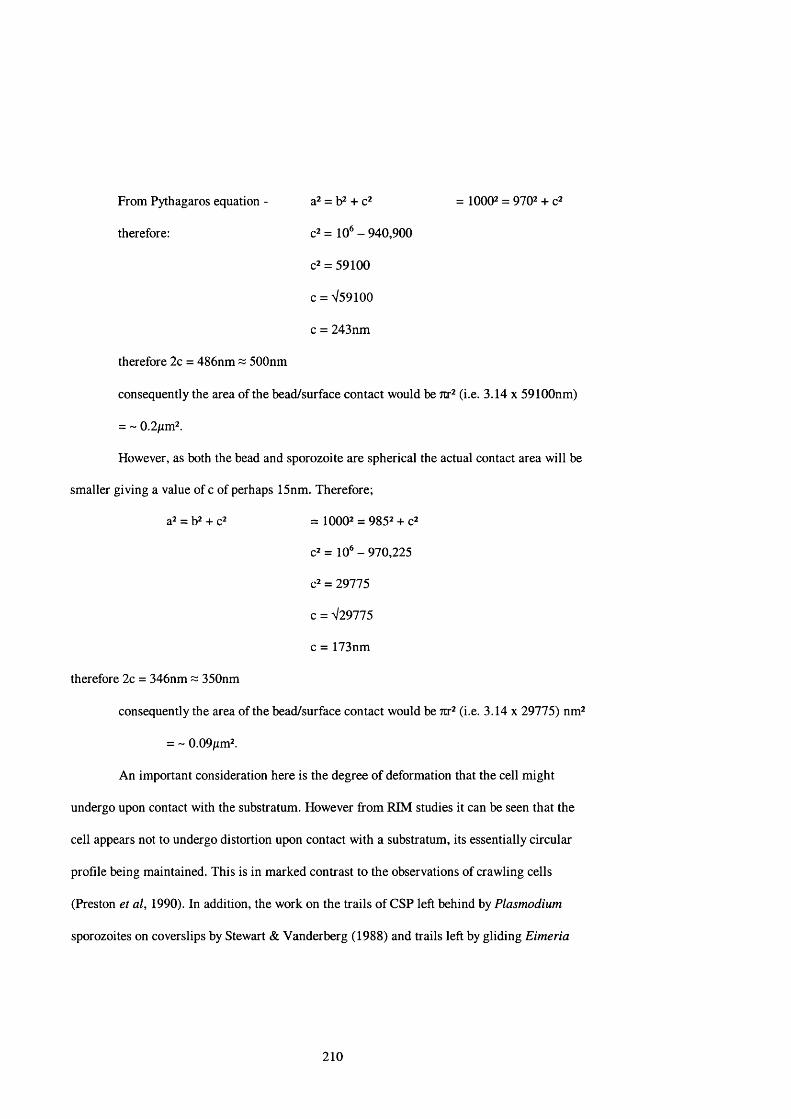
From Pythagaros equation - a ̂= = 1000^ = 970^ +
therefore: = 10 ̂- 940,900
c2 = 59100
c = V59100
c = 243nm
therefore 2c = 486nm ~ 500nm
consequently the area of the bead/surface contact would be Ttr̂ (i.e. 3.14 x 59100nm)
= ~ 0.2/im^.
However, as both the bead and sporozoite are spherical the actual contact area will be
smaller giving a value of c of perhaps 15nm. Therefore;
a2 = b2 + c2 = 10002 = 9852 + fZ
c2= 1 0 * -970,225
c2 = 29775
c = V29775
c = 173nm
therefore 2c = 346nm ~ 350nm
consequently the area of the bead/surface contact would be Ttr̂ (j g 3.14 x 29775) nm^
= - 0.09/xm2.
An important consideration here is the degree of deformation that the cell might
undergo upon contact with the substratum. However from RIM studies it can be seen that the
cell appears not to undergo distortion upon contact with a substratum, its essentially circular
profile being maintained. This is in marked contrast to the observations of crawling cells
(Preston et al, 1990). In addition, the work on the trails of CSP left behind by Plasmodium
sporozoites on coverslips by Stewart & Vanderberg (1988) and trails left by gliding Eimeria
210

nieschulzi (Entzeroth et al, 1989) sporozoites have shown that the width of these trails is less
than the width of the sporozoite. In the case of E. nieschulzi the trails were observed to be 20-
60/im in length and 0.3-0.5jum wide, compared to a sporozoite width in the region of 2[im. It
was also observed that the Eimeria trails were the same density at the beginning of the trail as at
the end. This strengthens the idea that only a portion of the ventral surface of the cell is in
contact with the substratum, and that the cell does not distort on contact with the substratum,
and it is therefore possible to apply the calculations from figure 4.2 to the ventral cell surface of
the zoite. This also negates the idea of an all encompassing sheath in which the zoite travels.
For the parasite to glide it must first overcome the forces of viscous drag (F’) and
viscous force (F” ). Viscous drag is a measure of the force necessary to overcome the viscosity of
the surrounding medium and viscous force is a measure of the force required to overcome the
shear forces present in the gap between the cell and the substratum.
The following calculations must be treated with some caution since they are based on
the assumption that it is a sphere that is generating sufficient force to displace the medium in
front of it. Of course in the case of the sporozoites of both Eimeria and Plasmodium the actual
shape of the cell is more like a cylinder (see figure 4.3). Nonetheless these calculations are
useful as they give a broad indication as to the kind of energy involved in the generation of
gliding motility and bead translocation.
211

displacement of medium
substratum
Direction of gliding motility
Figure 4.3: Illustration of the need of the gliding sporozoite to displace its body volume of medium in front of it in order for movement to occur.
212

Viscous drag can be calculated from Stokes’ equation (Preston et al, 1990);
F’ = 67tT|rv
For an Eimeria tenella sporozoite the cell radius (r) = 1 x 10'^ m; the viscosity of water
(T|) = 10 ̂Nsm'^ and the velocity of the sporozoite (v) = 2 x 10 ̂ms ' (see results chapter).
Consequently
F’ = 3.75 X lO '^N
This is also calculated to be the force required to translocate a 2/im bead over the
surface of an Eimeria sporozoite at 20/xms '.
For a Plasmodium sporozoite travelling at 1/xms ' F’ = 7.5 x lO '^N (Preston & King,
1996).
Once the force required has been calculated the energy requirement can also
be calculated by multiplying F’ by the distance moved per second (to give E ’). Therefore for an
Eimeria sporozoite;
E ’ = 3.75 X lO '^N X (2 X lO ^m) Js '
E ’ = 0.75 X 10 '7 Js '
Again the same value is obtained for the 2fim bead, whilst for the Plasmodium
sporozoite a value of 0.75 x 10'^" Is ' is obtained (Preston & King, 1996), i.e. the Eimeria
sporozoite requires 10 ̂times more energy.
213

Viscous force can be estimated from the following equation;
F” = Any h
Where A = possible area of the cell surface in contact with the substratum (for an
Eimeria sporozoite, as estimated above is between OAôfxm^ and 4/im^), r| = viscosity of water
(10'^ Nsm'2), V = velocity (in m sec ‘) and h = cell-substratum separation distance (30nm).
Therefore, using the highest and lowest contact area values there will be two scenarios;
Case (i): F” = 0.31 x lO '^N
Case (ii): F” = 2.68 x lO '^N
For a Plasmodium sporozoite with an estimated cell surface contact area of 3/xm^
F ” = 0.1 X lO '^N (Preston & King, 1996).
For the 2/rm latex bead the surface area in contact with the Eimeria sporozoite pellicle
will be circular and can be calculated using Tcr̂ (giving a value of ~ 0.2^m^ - see above
calculations) and thus
F” =0.124 X lO '^N
Again the energy requirement can also be calculated by multiplying F” by the distance
moved per second (to give E ” ). Therefore for an Eimeria sporozoite;
Case (i): E " =0.31 x lO '^N x (20 x 10'W ‘)
E” = 6.2 X 10 *̂ Js '
Case (ii): E” = 2.68 x lO '^N x (20 x 10'W )
E ” = 5.38 X 10 '^ Js '
And for the translocation of the 2/rm bead the energy required is;
E ” = 0.124 X lO '^N X (20 x 10'W )
E ” = 2.48x 10 '* Js '
214

Preston & King (1996) calculated this value to be 1 x 10 Js * for the sporozoite of
Plasmodium. Interestingly the hydrolysis of 1 molecule of ATP yields 1 x 10 *̂ J (assuming
100% efficiency). To enable a ready comparison this data and the theoretical ATP requirements
are illustrated in the table below;
Type of
movement
Energy required to
overcome viscous
drag (E’)
ATP
required
Energy required to
overcome viscous
force (E")
ATP
required
Eimeria - sporozoite
Case (i) 0.75 X 10 ' ̂Js ‘ 1 6.2 X 10 '® Js ' 62
Case (ii) 0.75 X 10 *̂ Js ' 1 5.38 X 10 '̂ Js ' 538
Eimeria - 2/tm bead 0.75 X 10 '̂ Js ' 1 2.48 X 10 '® Js ' 25
Plasmodium - sporozoite 0.75 X 10'"° Js ' 1 - 1.1 X 10 Js ' 1
Plasmodium - 1/tmbead 0.75 X 10"°Js' 1 - 1.1 X 10 '̂ Js ' 1
Table 4.1: Relative energy requirements for overt gliding and bead translocation in Plasmodium and Eimeria sporozoites.
As these cell motility observations were made on sporozoites using an inverted
microscope one must also consider the influence of gravity on the grip between cell and
substratum and consequently, the generation of motility. Studies on the amoeba Naegleria
gruberi (Davies et al, 1981) showed that the speed of ameoboid locomotion was the same for
cells attached to the upper and lower coverslips of a Prior chamber, suggesting that the adhesion
between the cell and substratum is considerably stronger than the force of gravity acting upon
the amoeba. This is an important observation since it illustrates the strength of the grip at the
contact site and has implications for events at the posterior of the cell after gliding. Observation
215

of trails left behind the motile sporozoite suggests that once at the end of the cell the strong cell-
substratum adhesion leads to the contact being essentially ripped from the plasma membrane
and brings with it an amount of membranous material. The continuous loss of membrane would
clearly be an untenable system unless a mechanism existed which enabled the lost material to be
replaced. As described above the micronemal contents discharged in association with gliding
and invasion include membranous material, and this would provide a ready source of material to
replace that which has been lost during gliding. These considerations place considerable doubt
on models involving viscous force, as viscous force is a measure of the shear force between the
plasmalemma and the substratum with which it is interacting. Consequently, if this space is
occupied by an essentially static layer in which negligible shear occurs, then viscous force might
have little impact. Instead the interaction is likely to be protein assemblages with an intimate
substratum association moving through lipid bilayers. It is therefore probable that E ” is an
exaggerated figure.
The models of cell/substratum contact employed in the above calculations are based on
the assumption that the motile cells are essentially ‘sausage-shaped’ and that the cell/substratum
contact exists for virtually the entire length of the sporozoite. This is probably accurate in the
case of Plasmodium but is likely to be incorrect for Eimeria sporozoites, which more closely
resemble a ‘twisted banana’ than a ‘sausage’. RIM showed the nature of the contact in Eimeria
sporozoites to be one or two, relatively small, clearly defined circular or ellipsoidal areas (see
figure 3.2) which translocated backwards over virtually the entire length of the sporozoite. What
is less clear is how this would impact on the energy requirement for gliding motility. At first
glance it would be reasonable to assume that the overall energy requirement would be reduced
by a smaller contact area. However since the area of the pellicle involved in substratum
interaction does not remain the same, but moves along the sporozoite in an anterior-posterior
216

direction, the total surface area involved in the generation of gliding motility could well be
similar to that seen for a permanent longitudinal contact, as demonstrated in Plasmodium
sporozoites.
Given that the hydrolysis of one molecule of ATP can theoretically yield 1 x 10 *’ J and
the energy required by a Plasmodium sporozoite to overcome viscous drag and viscous force (E’
& E ” ) is approximately 1 x 10'” Js ', it can be suggested that one molecule of ATP per second
could power sporozoite gliding and bead translocation (Preston & King, 1996). Applying this
rationale to the data calculated above, suggests that for a gliding Eimeria sporozoite upto 539
ATP molecules per second are required to overcome viscous drag and viscous force, and to
power bead translocation 26 ATP molecules per second are necessary. Using this information
one can begin to explore ideas on the molecular model generating gliding motility.
Given the assumption that filamentous actin is involved in this force generation the
obvious model that presents itself is one in which myosin molecules are recruited to the contact
site and interact with a stable actin trackway. This sort of arrangement has been seen
experimentally in the classical studies of Sheetz & Spudich (1983a). In these experiments
fluorescent beads were coated with heavy meromyosin (HMM) and placed onto a preparation of
a Nitella cell which had been split open and whose cytoplasmic contents had been washed away,
leaving exposed two microfilament arrays separated by an indifferent zone. The beads were then
seen to move along the two microfilament arrays in opposite directions, but with all beads on
each side of the indifferent zone moving in one direction only.
217

The myosin step distance produced by the hydrolysis of 1 molecule of ATP in striated
muscle myosin II has been calculated to be lln m (Finer et al, 1994). Using this figure
movement of a latex bead at 20/xms'* would require;
total distance = 20,000nm = 1818 individual ATP cleavage steps/sec 'step distance lln m
This is in comparison to the value calculated above that shows 1 ATP can provide
sufficient energy to overcome the forces of viscous drag and upto 539 molecules of ATP to
overcome viscous drag & viscous force.
Furthermore, using the generalisation that myosin molecules can undergo 40 cycles of
ATP hydrolysis per second (Huxley, 1969);
number of ATP molecules = 1818 = a minimum of 45 myosin moleculesnumber of cycles per second 40
In these considerations it is also important to take account of the on/off ratio of the
myosin molecule. For classical muscle myosin-II it is thought the off ratio is very high, being of
the order 0.95:0.05 (Howard, 1997), suggesting that at any given time most of the myosin
molecules are not in contact with the actin filament. In these systems it is therefore necessary to
have a high number of myosin molecules present so at least one molecule is ‘on’ since if all the
myosins were ‘off the contact would be lost and the bead could become detached from the cell.
218

Using the ratio above;
100 myosins = 95 off: 5 on
and therefore, using the minimalist value of;
45 myosins at the interaction site = 42.6 off: 2.4 on
i.e. there are always some myosin molecules locked onto the actin filament. (It must be
remembered however that apicomplexan myosins might have characteristics very different from
those used in the above calculations).
If we accept these figures, the next major consideration is the way in which these
molecules are organised at the interaction site. An important issue is to understand if it is the
myosin or the actin that is ‘mobile’ relative to the cell profile. In both scenarios it might be
expected that substantial amounts of either protein in well-organised arrays, would be necessary
to bring about motility. This seems unlikely since, although the immunoelectron micrograph
data is so far inconclusive, there is no evidence of such an ordered array of myosin or actin
molecules. In addition the immunofluoresence studies on Eimeria sporozoites showed the actin
staining to be somewhat diffuse and the myosin staining to be distinctly punctate. An attractive
model involves a stable actin trackway associating with mobile myosin molecules along which
the contact moves, reminiscent of the classical data presented by Sheetz and Spudich (1983).
Although microfilament arrays have not yet been described in apicomplexans it is possible that
actin filaments can be synthesised de novo at a rate sufficient to provide a trackway required for
gliding to occur (this will be discussed later).
This sort of arrangement is essentially a non-processive motor. Although myosin can
act as a processive motor (Howard, 1997) it is generally perceived as a non-processive motor. In
addition recent laser trap studies (King et al, 1999) using latex beads translocating over the
219

surface of gregarines indicated that a force of 50pN was required to overcome the force exerted
by the laser. Given that the classical value for the force generated by a single myosin head
following ATP hydrolysis is about 5pN, it is clear that at least 10 myosin molecules can be
actively involved in this interaction site, at any given time. One myosin molecule would be
insufficient as it would move one step distance and fall off. However the fact that the force
generation system acting on the head of the bead does eventually overcome the force of the laser
indicates multiple myosins are present at the interaction site, with each individual myosin
exerting its force until the cumulative effect is sufficient to overcome the energy barrier dictated
by the laser.
From these assertions a model can be constructed which involves a pellicular actin
trackway (possibly synthesised de novo and with a rapid turnover) along which the interaction
site travels, with a clearly defined axis (see figure 4.4a). The interaction site is composed of
myosin molecules undergoing actin/ATP-mediated movement, which are associated with the
plasma membrane and are also linked directly or through transmembrane components to the
latex bead on the sporozoite surface. It is likely that the myosins are in the form of a plaque
containing many myosin molecules, a sufficient number of which will be undertaking ATP
hydrolysis, and hence movement, at any given moment. Such plaques are seen elsewhere in the
cytoskeletal organisation of cellular structures e.g. Chlamydomonas flagella (Cole et al, 1998).
Here intraflagellar transport (IFT) particles formed from FLAIO kinesin-II (a kinesin found in
temperature sensitive Chlamydomonas mutants), complex A and complex B develop at the
flagella basal bodies. These IFT particles form linear arrays or ‘rafts’ which are transported by
the FLAIO kinesin-II along the B tubules of the microtubule outer doublet (Kozminski et al,
1995). Once at the tip the rafts dissociate into smaller oligomers and are transported to the
flagella base by a retrograde motor thought to be cytoplasmic dyenin (Pazour et al, 1998). In
220

microfilament‘anchor’
transmembranecomponent
myosin
microfilament
innermembrane
complex
plasmamembrane
(a) Actin trackway and mobile myosin
transmembrane myosin component (i)
innermembrane
complex
microfilamentplasma
membrane
SUBSTRATE
transmembrane component (ii)
SUBSTRATE
(b) Sequestered pellicular myosin
Figure 4.4: The two postulated models involving actin & myosin interacting with an extracellular substrate.
221

Chlamydomonas these rafts are thought to play a role in providing material for flagellum
elongation. In the Apicomplexa it is easy to visualise a similar system based on myosin, in
which a plaque/raft forms at the point of contact with a substratum, remains attached to the
substratum and within the plasmalemma until the contact reaches the posterior of the cell,
dissociates once the contact is lost and leads to the recycling of the components (especially the
motor proteins) to the anterior of the cell.
A significant limitation to this actin trackway/mobile myosin system is encountered
when one begins to look at the actin-myosin interaction at the molecular level. The two strands
of the actin filament crossover every 35nm so that the filament is orientated such that a myosin
molecule could only interact with an appropriate actin molecule once every 35nm. If we bear in
mind that the myosin step distance is llnm this represents a significant barrier to the
continuous progression of the myosin (and consequently the contact site) along the
microfilament. It is possible that the actin filament might rotate whilst interacting with the
myosin so that when one step has been completed another appropriate myosin is readily
available to bind to the released myosin. However this would be at odds with the idea of a stable
actin trackway which is anchored to the inner membrane complex and indeed, experimentation
using myosin coated substrata has shown that actin filaments exhibit little or no rotation (Suzuki
et al, 1996; Sase et al, 1997).
One solution to this problem could be to organise the myosins such that they appear
every 35nm, and are hence in the correct position to undergo the next powerstroke. Using the
calculation that 45 myosins are involved in translocating a latex bead at 20fim s'\ it can be seen
that the contact site to enable 35nm spacing of the myosins would be over 1.5/xm, an area much
larger than the postulated 0.2/xm^ described above. It would also be possible that the myosins are
spaced 24nm apart (see figure 4.5a), since upon completion of an l ln m step the next myosin
222

would automatically be in register with the next appropriate actin, but again this would demand
a contact site in excess of l/im.
These ideas are based on the assumption that the myosin is walking down a single actin
filament. If, however one assumes there are multiple actin filaments one can readily construct a
model in which the step distance limitations can be overcome. These actin filaments could be
aligned such that each myosin interaction site on the individual microfilament occurs lln m
further ‘upstream’ from that on the adjacent microfilament, thus when one step has been
completed, the myosin is immediately present at a site conducive to another step. For an
individual myosin to bring about movement in this model would take a large number of
microfilaments arranged like a sheet under the plasma membrane, but one could envisage a
model in which an individual myosin undergoes a number of steps and then ‘rests’ (see figure
4.5b). During this period the myosin would be able to ‘diffuse’ through the plasma membrane to
a position where it could begin to interact with the underlying microfilaments again (again
reminiscent of the Chlamydomonas IFT particles). Since there are postulated to be at least 45
individual myosins present at the site of contact and 2.4 of them will be ‘on’, there would seem
to be adequate provision of activated myosin molecules for this kind of mechanism to achieve
movement successfully.
If one takes the upper estimation of the actin filament width i.e. lOnm it would
theoretically be possible to pack 50 microfilaments into the 0.2/im^ contact site. Since this
represents a large amount of highly ordered actin this figure is unlikely to be correct given that
microfilaments have thus far evaded identification in the sporozoites by both immunoEM and
immnofluorescent studies, but it is plausible that a smaller number of filaments could be
involved, forming such arrays yet remaining undetectable by conventional methods. It is
possible that as few as three or four actin filaments would be required (see figure 4.6(b)). These
223

direction of bead movement
latex bead
membrane ■lag'
transmembrane componentK
microfilament
myosin
24nm= ‘appropriate’ actin monomer
36nm
Figure 4.5(a): Model involving a single microfilament and an ordered myosin cascade.
microfilament
Key
= myosin powerstroke
= movement of myosin from one microfilament to an adjacent one
= actin associated protein, e.g. villin, fimbrin
myosm
Figure 4.5(b): Model involving multiple microfilaments and labile myosins which can switch to adjacent microfîlaments following completion of the powerstroke.
224

Bead surfacetrans-
membranecomponent
— myosin
microfilament
plasmaX membrane
/ \
20nm
\ ( outer ' membrane
oflM C
Figure 4.6(a): In the area of contact the myosins are attached via the tail domain to the plasma membrane, and hang down rather like the tentacles of a jellyfish. Note the random distribution of the myosins.(The inter-membrane gap distance is derived from the EM measurements.)
microfilament arraynon-contact__
plasma membranemyosins in contact
with actin
outer limit ofbead/plasma membrane
contactindividual myosin head
Figure 4.6(b): Longitudinal section through a model contact site illustrating the random distribution of the myosin molecules. Arbitrary superimposition of just four microfilaments generates up to 16 potential sites of actin/myosin interaction.
226

Given the paucity of knowledge of the biochemistry of the Apicomplexan myosins it is
difficult to construct a wholly convincing model of the mechanism employed in the generation
of motility. Using assumptions gleaned from work on classical myosin II can begin to indicate
potential interactions but it is clear that this is far from ideal. Large amounts of myosin II exist
in the sarcomere of muscle cells and consequently a low on: off ratio can be tolerated.
As described above there is the potential for the hydrolysis of up to 1800 ATP
molecules per second in the contact site plaque, but from first principles it has been shown that
to enable bead translocation only 11 molecules of ATP would be required to overcome the
attendant physical forces. This represents an enormous excess of energy. Studies involving the
use of various sized beads translocating over the surface of gregarines suggested that the speed
of movement is independent of bead size (King, 1981). Similar observations were made by
Sheetz & Spudich (19836) in follow up work to their original study using beads ranging from
0.6/xm - 120/rm in diameter. The obvious question that arises from these observations is, why is
there such an excess of energy?
In vivo the generation of gliding motility is important at three key points in the life
cycle of the sporozoite; to excyst, to glide to an appropriate host cell and then to invade that cell.
As described in the introduction the processes of excystation and invasion could involve a
considerable amount of work output on the part of the parasite. Excystation requires the
sporozoite to squeeze through a relatively small space and invasion requires the sporozoite to
deform both the outer cell surface and underlying cytoskeleton of the host cell. It can be
suggested that these processes need much more energy than to translocate beads along the outer
membrane of the sporozoite. However it would be difficult to measure the energy requirement of
both excystation and target cell invasion. The laser trap (King & Sleep, 1999) experiments are
paralleled by the observation of excystation in which the sporozoite seems to move in steps (see
227
section 3.1.b), presumably as a result of generating sufficient power to enable the restraining
forces to be overcome.
The main weakness of this whole conjecture is that so little is known about the myosin
molecules within the Apicomplexa. Structural and localisation studies are beginning to provide
answers but a greater understanding of the biochemistry of the Apicomplexan myosins would
clearly lead to the development of more accurate and less ambiguous models of the mechanisms
involved in motility generation. For instance, knowledge about the step distance and ATP
turnover rates of these myosins would clarify a great deal of the conjecture currently
surrounding this field and is clearly a key feature of any model which attempts to understand the
nature of an actomyosin motor.
A further complication is the concept of viscous force in this system. Although
arguments have been presented the for energy requirements in which both viscous drag and
viscous force are important, it is quite possible that viscous force plays an indeterminate role in
the gliding motility of sporozoites. This is due to the nature of the contact between the cell and
the substratum. If the cell forms a very strong contact with the substratum through a complex
involving the motor, the plasma membrane and various transmembrane components shear forces
are likely to be negligible and certainly very difficult to calculate.
Perhaps the most enigmatic feature of this whole system is its unpredictability. A bead
can be resting on a sporozoite for a prolonged period and then suddenly begin to move, or can be
translocated immediately upon contact with the parasite cell surface and it is unclear as to what
provides the stimulus for the motor to begin working.
It is probable that the stimulus is generated at the plasma membrane/substratum
interface but it remains to be seen if this is a simple transduction or a more complex biochemical
pathway. Recent work (Maekawa et al, 1999) has underlined the importance of the small GTP
228

binding protein Rho in the control of actomyosin complexes and the reorganisation of the actin
cytoskeleton (see figure 4.7).
Rho
mDia
Profilin
iActinpolymerisation
ROCK
Myosin phosphatase
Myosin light chain phosphorylation
Increased actomyosin contractility
Reorganisation of actin cytoskeleton
LIM-kinase
iCofîlinphosphorylation
IInhibition of actin depolymerisation
Disassembly
Figure 4.7: Diagram of the cytoskeletal changes mediated in part by the small GTP binding protein, Rho (From Maekawa et al, 1999).
The stimulation of Rho by endogenous factors such as serum and lysophosphatidic acid
results in the phosphorylation of the ROCK family of serine-threonine protein kinases and their
mammalian homologue, mDia. ROCK is then capable of phosphorylating both myosin
phosphatase and LIM-kinase which subsequently brings about increased actomyosin
contractility and the inhibition of actin depolymerisation respectively. The phosphorylation of
229

mDia brings about the recruitment of profilin which in turn promotes the polymerisation of
actin. The regulation of myosin light chain phosphorylation by ROCK represents an effective
system by which the observed on/off nature of the myosin motor could be controlled.
It remains to be seen if these, or analogous proteins exist in the Apicomplexa and even
if they do it is difficult to envisage their activation by a stimulus as relatively specific as serum.
However, it is possible that the establishment of a link between sporozoite and substratum could
begin a cascade that could include such proteins. Alternatively this pathway could be directly
controlled by myosins themselves.
A number of myosins have been found to possess motifs and domains usually
implicated in the mediation of protein-protein interaction between signal transduction factors.
These include the pleckstrin homology of myosin X (Sole et al, 1994) and the chimaerin-like
GTPase-activating-protein (GAP) domain for Rho GTPases of myosin IX (Reinhard et al, 1995;
Müller et al, 1997). Mammalian cells overexpressing a rat myosin IX, myr5, lose stress fibers
and focal contacts and eventually round up (Müller et al, 1997), correlating with observations of
the effect of Rho inactivation.
This of course brings us back to the thorny issue of what it is that actually induces the
sporozoite/bead to begin translocation.
4.5 Bead-cell surface interactions
Data from observations of bead translocations serve to strengthen many of the
assumptions made from the study of overt gliding motility. Since beads are seen to translocate at
roughly the same speed as gliding motility occurs (both ~ 20/xms'^), this argues for the existence
230

of a common motor. A major difference however is likely to be the surface area of the contact
between the cell and the bead/substratum. In a gliding E. tenella sporozoite using a contact of
12/xm X 0.2/rm the total surface area of the contact would be 2.4/rm^. The energy required to
translocate a 2^m diameter latex bead travelling at 20/xms ‘ (i.e. to overcome the physical forces
of viscous force and viscous drag) would be 2.48 x 10''® Js'* and would need the hydrolysis of 25
molecules of ATP per second, in comparison with upto 538 ATP molecules (see table 4.1)
required to power gliding motility at the same speed.
Closer examination of bead translocation also shows it to be a highly erratic process in
the same way as gliding motility. The bead can only proceed once sufficient energy had been
generated by the motor at the contact site to overcome the viscous force and hence allow
progression of the bead.
In addition the helical progression of the bead over the pellicle illustrates very clearly
the gyre of the cell. A bead can translocate the entire length of the cell coming to rest 180° from
its initial point of attachment. It also illustrates that the bead can remain bound for the entire
length of the sporozoite. This strengthens the idea that the motor operates in a direction similar
to the orientation of the subpellicular microtubules. However microtubules cannot be directly
involved in the generation of motility since they are seen to end at approximately two-thirds of
the sporozoite length but the translocated bead can progress to the very posterior of the cell.
4.6 Excystation
Excystation inhibition studies showed that this process was inhibited by both
cytochalasin D & BDM but not by treatment with colchicine. This argues for the involvement of
231

both actin and myosin in escape from the sporocyst, but negates an active role for the
microtubules. Observations of the process have also indicated that it is sporozoite motility and
not osmotic pressure that brings about sporozoite excystation.
The large discrepancy between the sporocyst aperture and the sporozoite diameter
presents a number of very different problems from those presented by the generation of gliding
motility. The sporozoite and its intracellular organelles must undergo a substantial amount of
compression in order to excyst and enter the environment outside the sporocyst. Ultrastructural
studies show the sporozoites to possess a subpellicular microtubule network which could be
reasonably expected to act as a fairly rigid ‘corset’, further complicating ideas on cell distortion
and compression.
TEM photographs of flexing sporozoites have shown that the trilaminar pellicle of E.
tenella is capable of a sizeable degree of folding/compression, albeit in an anterior-posterior
direction. However it is possible that the force developed by a sporozoite passing through the
sporocyst aperture is sufficient to bring about the folding of the parasite membrane (see diagram
4.8). What is more difficult to imagine is the impact of the shear forces on the cell organelles
(especially the nucleus) as they pass through the sporocyst aperture. However this type of
compression is not without precedent. Studies on neutrophils (Mandeville, et al, 1997) and
macrophages (Haston, et al, 1982) have shown that these cells with a nucleus width of
approximately 10/xm are capable of passing through 3/xm diameter Micropore filters, seemingly
without any damage being done to the structural integrity of the nuclear material. It is unclear as
to how this is achieved but since the nucleus is so fundamental to the correct functioning of the
cell, it could be that similar protective systems exist in many types of cell from many different
organisms.
232

(i) (ii)
B A B
B B
plasmamembrane
inner membrane complex
Figure 4.8: The hole in the sporocyst wall through which the 2pm diameter sporozoite must pass in order to successfully excyst, has been estimated to be ~ 0.4pm in diameter (see results section). This represents a significant discrepancy. Having seen from the electron micrographs that the sporozoite can form folds in its pellicle in a lateral direction, it is possible that it could also form longitudinal folds. As shown in (ii) this could provide an effective mechanism by which a reduction in the parasites cell diameter could be achieved.
233

A further consideration in the flexing of the sporozoite is the impact that this may have
on the microtubule ‘corset’. Traditional theories on the nature of microtubules suggest that they
are fairly rigid features which lend a large amount of structural integrity to cells. Since flexing
sporozoites often almost touch anterior tip to posterior tip the microtubules must be capable of a
considerable degree of both compression (on the ‘inner’ surface of the fold) and elongation (on
the ‘outer’ surface). It may be that some other mechanism has developed in which the
microtubules are displaced away from the area of greatest contortion. In the case of the
longitudinal folding postulated to be involved in the excystation process, considerations of
microtubule flexibility are probably redundant as the compression is lateral-dorsal.
4.7 Nature of the motor
The motility inhibition studies showed that cytochalasin D and BDM were both
inhibitors of motility, but that treatment with colchicine had no effect on sporozoite gliding
motility. This suggests that actin and myosin are involved in the generation of the force that
powers motility, and negates a direct role for the microtubule network in this process.
Subsequent Western blotting of sporozoite derived protein samples showed the presence of actin
at the classical value of 43kDa confirming the findings of Baines & King (19896). Treatment of
similar samples with a range of anti-myosin antibodies detected the presence of a novel
unconventional myosin with a heavy chain molecular weight of 93kDa. In relation to the
classical myosin II found in the muscle sarcomere this is very small myosin molecule, and would
be the smallest myosin yet to be discovered. Recent work by Heintzelman & Schwartzman
234
(1997) in Toxoplasma gondii and Finder et al (1998) in Plasmodium falciparum has also
revealed the presence of a 93kDa myosin in these related apicomplexans.
Immunolocalisation studies using fluorescently labelled antibodies yielded actin
staining patterns that suggested that actin was mainly present in the cytoplasm, and that the
signal was particularly strong at the anterior end of the parasite (see figure 3.16). Similar results
have been reported for T. gondii (Dobrowolski et al, 1997), but a more diffuse pattern of actin
staining has been reported in sporozoites of Cryptosporidium parvum (Forney et al, 1998).
Immunofluoresence and immunoelectron microscope studies on Toxoplasma
trophozoites indicated a distinct pattern of staining at the anterior pole (Yasuda et al, 1988;
Endo et al, 1988). In these studies gold particles were seen along the inner surface of the inner
membrane complex, in the preconoidal and polar rings and within the conoid. Using
fluorescently labelled DNAse I Dobrowolski et al, (1997) identified clusters of globular actin
located primarily in the anterior half of Toxoplasma. This indicates there exists a large pool of
actin monomers, presumably from which microfilaments could be assembled de novo,
suggesting a highly dynamic system and perhaps explaining why actin filaments have not been
seen in the Apicomplexa. Karpova et al, (1995) have theorised that G-actin concentration is
closely correlated to cell motility rates, in that highly motile cells seem to have relatively large
amounts of monomeric actin, whereas more sedentary cells such as yeast are seen to possess
large microfilament arrays.
The studies with anti-myosin antibodies showed punctate staining primarily located to
peripheral regions of the sporozoite, and a pattern which was also consistent with a membranous
location. Similar studies in T gondii showed myosin to be located primarily at the anterior pole
of the cell (Schwartzman & Pfefferkorn, 1983) and this also seems to be the case for P.
falciparum (Finder et al, 1998).
235

Immunoelectron micrographs of P. falciparum merozoites indicated that the myosin
was located just under the plasma membrane, but it was unclear as to whether the myosin was
associated with the cytoplasmic surface of the plasma membrane or the outer membrane of the
inner membrane complex (Finder et at, 1998). However the absence of gold labelling in the area
of the apical prominence may indicate that the myosin is primarily associated with the inner
membrane complex. Similar results were obtained from immunoelectron localisation studies
with T. gondii (Dobrowolski et at, 1997).
Actin is a highly conserved protein throughout evolution. The actin molecules found in
organisms such as Dictyostelium and Acanthamoeba share 95% amino acid sequence homology
with vertebrate P-actin (Wesseling et al, 1988a). In the Apicomplexa P. falciparum contains 2
actin genes (Wesseling et al, 19886), which share 79% homology, the lowest value so far
recorded for actin genes from the same organism. They are also seen to have a stage specific
expression, with pfact-I h&ing transcribed in both sexual and asexual stages and pfact-II being
transcribed only in the gametocytes and gametes/zygotes (Wesseling et al, 1989). However T.
gondii contains only 1 actin gene, ACTl, (Dobrowolski et al, 1997) which has 93.1% homology
with pfact-I and 88.1% homology with the single C. parvum actin gene (Kim et al, 1992).
As outlined in the introduction the myosin family of molecules continues to grow in
complexity. In keeping with most cell types examined so far E. tenella is likely to contain
multiple myosin genes. The identification of an E. tenella gene with significant homology to the
ATP-binding site of myosin II may seem initially to be at variance with the Western blotting
data which suggested the presence of a myosin of 93kDa. It is possible that the myosin Il-like
myosin in E. tenella is involved in cytokinesis, as is the case in Dictyostelium and yeast. This
might suggest a stage-specific myosin expression pattern and as sporozoites do not undergo
division, expression of the myosin II gene in this stage may be unnecessary. A more exhaustive
236

search of an Eimeria cDNA library would doubtless identify a number of other actin genes.
Recent work in T. gondii (Hettmann et al, 2000) has suggested that this organism alone contains
at least five myosin XTVs. Given the similarities between the apicomplexans it is highly likely
that Eimeria contains at least one myosin XIV in its repertoire.
In P. falciparum the 93kDa myosin (designated Pf-myol) was shown to have a stage-
specific expression, with the protein being seen in mature schizonts and also merozoites, and
disappearing following erythrocyte invasion (Finder et al, 1998).
Identification of actin and myosin as the most likely candidates for the composition of
the motor naturally leads to considerations of the format that this interaction takes. Two models
make themselves apparent immediately; one in which a stable ‘actin trackway’ exists along
which the mobile myosins travel and a second which envisages ‘sequestered pellicular myosin’
interacting with mobile actin filaments.
In the first model a stable actin filament is associated with the outer membrane of the
inner membrane complex and mobile myosin molecules move along this filament (King, 1988).
These myosin molecules are in turn associated with transmembrane components which form the
link with the external substratum. In addition it has been suggested that the actin filaments are
anchored to an underlying network of microtubules or intermediate filaments which could
provide an added robustness to the whole structure (King, 1988). Movement of the myosin
molecules along the actin filament could bring about the movement of the sporozoite forward
relative to the substratum.
In the second model it is the myosins which are associated with the inner membrane
complex (bound and non-mobile) and the microfilaments which are mobile and link with the
substratum interactions. Subsequent activation of the myosin brings about the movement of the
237

actin filament and consequently the forward translocation of the cell/backward translocation of a
bead.
A major objection to the actin trackway theory is that actin filaments have yet to be
identified in motile stages of the Apicomplexa. However in situ permanent microfilaments are
not necessarily a prerequisite for this mechanism. Actin polymerisation rates might not be able
to support gliding motility at 20/ims'‘. In fast moving polymorphonuclear leukocytes locomoting
at 0.5/xms ‘, actin polymerisation occurs at 5/xms * (Zigmond, 1993), Listeria moving through
the cytosol of infected macrophages do so at speeds of 0.6 - 6jnms * (Cossart, 1995) and the actin
involved in the Thyone sperm acrosomal process elongation generates speeds of 10/ims * (Tilney
& Inoue, 1982). However it is possible that a pool of short actin filaments, small enough to
avoid detection by traditional methods exist, and that the polymerisation of these into an
elongate microfilament can occur at rates sufficient to support gliding motility. Alternatively
Tardieux et al, (1998) identified the presence of a coronin-like protein in P. falciparum
schizonts which would be expected to promote rapid actin polymerisation and cross-linking.
Sequence analysis of the T. gondii myosin TgM-A has shown this molecule to contain a
presumptive membrane binding domain in its tail (Heintzelman & Schwartzman, 1996). In
addition Pf-myoI separates with the insoluble fraction of a cell lysate, suggesting a membrane
bound location (Finder et al, 1998; Hettmann et al, 2000). Recent further analysis of this
molecule (Soldati - personal communication) have shown it to be similar to classical myosin II
smooth muscle myosin in that it has a short ‘on’ period in respect to the actin/myosin
interaction.
Assuming that the myosin is indeed membrane bound the question is, with which
membrane is it associated?
238
Immunoelectron microscopy of osmotically swollen T. gondii tachyzoites showed that
actin was associated with the inner membrane complex and myosin with the plasma membrane
(Endo et al, 1988; Dobrowolski et al, 1997), however the observation in P. falciparum
merozoites that myosin was absent from the apical prominence argues for the presence of
myosin associated with the outer face of the inner membrane complex (Finder et al, 1998). It is
possible that the sensitivity of these immunoelectron microscopic techniques is inadequate to
accurately determine the exact location of these proteins.
A further important aspect of the actomyosin motor is the mechanism by which the
myosin MgATPase activity is regulated. In conventional myosins this is classically regulated
through Ca^^ activation, in one of two ways. The first is actin-based regulation in which the Ca^^
binds to troponin C in the troponin-tropomyosin complex, causing a conformational change
which allows the actin and myosin to interact and therefore produce movement in striated
muscle. The second is myosin light chain kinase (MLCK) regulation in which either
Ca^Vcalmodulin-dependent MLCK phosphorylates a serine residue in the regulatory light chain
(RLC) (Sellers & Goodson, 1995). Phosphorylation can be regulatory, as in the case of avian
smooth muscle myosin (Pearson et al, 1984) or modulatory, as seen in the RLC of skeletal
muscle myosin (Pemrick, 1980). Further work on the regulation of myosin in smooth muscle
cells has shown that the myosin-binding subunit (MBS) of myosin phosphatase assembles a
multienzyme complex (Surks et al, 1999). This complex contains a phosphatase and at least two
kinases with opposite regulatory effects on the myosin light-chain phosphatase, PPIM. One of
these kinases, cGMP-dependent protein kinasela (cGKIa), is vital in the cGMP-dependent
phosphorylation of myosin light chain, and uncoupling of the MBS-cGKIa interaction prevents
dephosphorylation, hence the muscle cell remains in a relaxed state.
239
The recent explosion in the number of myosin classes identified has predictably led to a
greater number of investigations into the mechanisms by which these molecules are regulated.
Perhaps inevitably there seems to be a degree of variation between some myosin classes and also
between the same myosin classes in different organisms.
Most conventional non-muscle myosins are regulated by Ca^Vcalmodulin-dependent
MLCK phosphorylation (Brzeska & Korn, 1996) and since all unconventional myosins contain
at least one light chain which appears to be calmodulin, it is reasonable to assume that these
molecules are regulated in the same way (Mooseker & Cheney, 1995).
However, studies on the myosins of the amoeba Acanthamoeba have shown that these
are regulated exclusively by heavy chain phosphorylation (Brzeska & Korn, 1996). In
Acanthamoeba myosin I this occurs in the S-1 domain and involves the phosphorylation of a
single serine/threonine residue (Brzeska & Korn, 1996), which in the conventional myosin
occurs near the end of the C-terminal domain (Korn & Hammer, 1988). So far only the myosins
of Acanthamoeba, Dictyostelium and Physarum have definitely been shown to be regulated by
heavy chain phosphorylation (Brzeska & Korn, 1996).
There has been little work undertaken on the mechanisms of regulation of myosins
outside class I & II myosins, although myosin V from chicken seems to be regulated by Ca^^
(Nascimento et at, 1996). Given the diversity in form and function of the newly discovered
myosins it would not be surprising if a number of different biochemical strategies were
employed in the regulation of these molecules. However, the extremely conserved nature many
of the sites fundamental to the correct functioning of the myosin molecules, e.g. the actin-
binding site & the ATP-binding site, suggest that the regulatory mechanisms are likely to be
broadly similar from myosin class to myosin class, and from organism to organism.
240
In the motor apparatus models outlined above it is suggested that the myosin molecules
involved in the generation of motility might exist as a ‘plaque’ within the plasma membrane (of
the Chlamydomonas IFT particles discussed earlier). If this is the case this may subtly alter the
way in which these molecules are regulated. As in other motile plaques there are likely to be a
number of associated proteins and perhaps other structural features, some of which could
reasonably be expected to impact upon the activity of the myosins.
4.8 Invasion
The invasion inhibition studies undertaken yielded two important observations. Firstly
preincubation of cell cultures with potential cytoskeletal inhibitors did not alter the efficacy of
host cell invasion by the parasite (Sibley, 1995), thus indicating that it is the correct functioning
of the sporozoite cytoskeleton that is vital for this process to be achieved. The second
observation was that, in addition to inhibition by cytochalasin D & BDM, target cell invasion
was also inhibited to a comparable level by treatment with colchicine (Bejon et al, 1999). This
would suggest that the invasion process involves both the actomyosin system and the
microtubule network. Since the microtubules have been shown not to be directly involved in the
generation of the force that drives motility it is clear that they are involved in some other aspect
of invasion. Potential roles could include a part in the normal functioning of the conoid or an
ability to move specific organelles and molecules involved in the invasion process to positions
where they could have their impact.
Studies on both Plasmodium falciparum and Toxoplasma gondii have shown a defined
sequence of organelle discharge during both the host cell invasion process and the induction and
241

maintenance of the parasitophorous vacuole (Carruthers & Sibley, 1997; Sibley et al, 1998),
which appears to be the following: micronemes, then rhoptries and finally the dense granules.
From the point of view of the penetrative act of invasion it would seem that the proteins
contained within the micronemes are the most important.
If one thinks of a classical epithelium cell then there essentially three barriers that the
parasite must negotiate in order to be successfully internalised. These are the glycocalyx, the
apical microvilli and the dense cortical network lying under the cell plasma membrane. In
ookinetes of P. gallinaceum it appears that a parasite-derived chitinase is responsible for a
disruption in the peritrophic membrane (Huber et al, 1991). The motile ookinetes also possess
the characteristic organelles of the apical complex and it is possible that other invasive stages of
the Apicomplexa contain within their micronemes proteins with enzymatic activity capable of
breaking down the polysaccharides of the glycocalyx.
To some extent the microvillar layer represents less of a barrier than the other two,
since the sporozoites could (and seemingly do) glide across the microvillar surface and invade
the epithelial at the base between two microvilli. Alternatively there could be a general
disruption of the actin cytoskeleton in the regions of the cell, resulting in the loss of the
microvilli. This can be seen in the electron micrographs in figures 3.18 and 3.25.
A more difficult obstacle is presented by the dense cortical layer, and again here it
seems unlikely that gliding motility alone can bring about such swift cell entry. Observations on
cytochalasin D treated T. gondii tachyzoites have shown that although these are incapable of
gliding motility they still retain the ability to induce host cell vacuolation (Dobrowolski &
Sibley, 1997). This suggests the release of a parasite derived factor(s) responsible for host cell
membrane disruption, with a microneme derived protein being the obvious candidate.
242

A'ê
(i) The zoite comes into contact with a host cell and the microneme proteins form a link between the motor and the target cell surface.
'ê
(ii) The host cell begins to vacuolate as a result o f the release of the rhoptry contents. A tight junction is formed at the point of zoite/cell contact with the microneme protein acting as the bridge. (Vacuolation is the first stage of parasitophorous vacuole formation.)
rn r~i
(iii) The nascent parasitophorous vacuole continues to enlarge as the zoite begins to enter the target cell.
(iv) The parasitophorous vacoule is pinched off from the host cell membrane, becoming a separate entity. The microneme proteins remain on the cell surface and do
not enter the vacuole.
Figure 4.9: Sporozoite invasion of a target cell. This involves the development of a parasitophorous vacuole and a tight junction. The components of the tight junction remain on the target cell surface following successful invasion.
243

In T. gondii the microneme localised protein MIC2 is released onto the surface of the
tachyzoite upon contact with an appropriate host cell (Carruthers & Sibley, 1997), and local
vacuolation of the host cell membrane generates an invagination within the host cell, but
separated from the host cell cytoplasm, which the parasite can subsequently occupy. This
process is actin independent, since T. gondii tachyzoites pre-treated with cytochalasin D were
still capable of inducing host cell vacuolation (Dobrowolski & Sibley, 1997). A moving junction
is formed as a ring between the tachyzoite pellicle and the host cell membrane and it seems that
the link between these two membranes is M1C2. As the invasion progresses the moving junction
is seen to move backward relative to the tachyzoite. The M1C2 jacket is not internalised along
with the parasite but rather is ‘filtered off at the junction and remains on the surface of the
invaded cell (see figure 4.9). A similar process occurs in the merozoites of Plasmodium (Aikawa
et at, 1978), and here it is the ‘merozoite capping protein’ MCP-1 which forms the link between
the two cells (Klotz et at, 1995).
As outlined in chapter 1, the M1C2 and MCP-1 proteins have been shown to possess
membrane-binding domains, which suggests that upon release from the micronemes they
become integrated into the pellicle of the parasite, but are also thought to bind to markers on the
host cell surface. In this way they essentially form a continuum between the parasite motor
apparatus and the host cell surface, and this provides a substratum for the zoite to interact with,
thereby generating the force necessary to enable the parasite to successfully penetrate the cell.
The nature of this junction is similar to that proposed for the site of bead/pellicle interaction (see
figure 4.4a) with the bead obviously replaced by the target cell surface (see also figure 1.10), or
indeed the interaction between the sporozoite and sporocyst wall during excystation. Following
successful invasion, the components of the dense granules will modify the parasitophorous
vacuole to enable the parasite to begin to embark on the next stage in its development.
244
REFERENCES
Abercrombie, M., Heaysman, J.E.M. & Pegrum, S.M. (1971) The locomotion of fibroblasts in
culture. Exp. Cell Res. 67: 359-367.
Achbarou, A., Mercereau-Puijalon, O., Sadak, A., Fortier, B., Leriche, M.A. & Camus, D. (1991)
Differential targeting of dense granule proteins in the parasitophorous vacuole of Toxoplasma
gondii. Parasitology 103: 321-329.
Adams, J.H., Hudson, D.E., Torii, M., Ward, G.E., Wellems, T.E., Aikawa, M. & Miller, L.H.
(1990) The Dufly receptor family of Plasmodium knowlesi is located within the micronemes of
invasive malaria merozoites. Cell 63: 141-153.
Adams, J.H., Sim, B.K.L., Dolan, S.A., Fang, X.D., Kaslow, D C. & Miller, L.H. (1992) A family of
erythrocyte binding proteins of malaria parasites. Proc. Natl. Acad. Sci. USA 89: 7085-7089.
Aikawa, M. (1967) Ultrastructure of the pellicular complex of Plasmodium fallax. J. Cell Biol. 35:
103-113.
Aikawa, M., Miller, L.H., Johnson, J. & Rabbege, J. (1978) Erythrocyte entry by malaria parasites.
A moving junction between erythrocyte and parasite. J. Cell Biol. 77: 77-82.
Aikawa, M. & Seed, T.M. (1980) Morphology of Plasmodia. In: Malaria (ed. J.P. Kreier), vol. I,
285-344. Academic Press, New York.
Aikawa, M., Miller, L.H., Rabbege, J R. & Epstein, N. (1981) Freeze fi-acture study on erythrocyte
membrane during malarial parasite invasion. J. Cell Biol. 91: 55-62.
Alberts, B., Bray, D., Lewis, J., Raff, M., Roberts, K. & Watson, J.D. (1994) Molecular Biology of
the Cell. 3"̂ ̂Edition. Garland Publishing Inc., New York & London.
245

Allen, N.S. & Allen, R.D. (1978) Cytoplasmic streaming in green plants. Ann. Rev. Biophys.
Bioeng. 7: 469-495.
Altschul, S.F., Gish, W., Miller, W., Myers, E.W. & Lipman, D.J. (1990) Basic local alignment
search tool. J. Mol. Biol. 215: 403-410.
Aniento, F., Emans, N., Griffiths, G. & Gruenberg, J. (1993) Cytoplasmic dyenin-dependent
vesicular transport from early to late endosomes. J. Cell Biol. 123; 1373-1387.
Apt, W. (1985) Toxplasmosis in developing countries. Para. Today 1: 44-46.
Arrowood, M.J., Sterling, C.R. & Healey, M.C. (1991) Immunofluorescent microscopical
visualization of trails left by gliding Cryptosporidium parvum sporozoites. J. Parasitol. 77: 315-317.
Atkinson, C.T., Aikawa, M., Perry, G., Fujino, T., Bennet, V., Davidson, & Howard, R.J. (1987)
Ultrastructural localization of erythrocyte cytoskeleton and integral membrane proteins in
Plasmodium falciparum infected erythrocytes. Eur. J. Cell Biol. 45: 192-199.
Atkinson, S.J., Doberstein, S.K. & Pollard, T.D. (1992) Moving off the beaten tracks. Curr. Biol. 2:
326-328.
Baines, I. (1988) Studies on the motility and cytoskeleton of two apicomplexan protozoans
(Greagarina and Eimeria) Ph.D. Dissertation, University of London.
Baines, I. & King, C.A. (1989) Demonstration of actin in sporozoites of the protozoon Eimeria. Cell
Biol. Int. Rep. 13: 639-641.
Baines, I. & Korn, E. (1990) Localization of myosin IC and myosin II in Acanthamoeba castellanii
by indirect immunofluoresence and immunogold electron microscopy. J. Cell Biol. I l l : 1895-1904.
Baker, J.P. & Titus, M.A. (1998) Myosins: matching functions with motors. Curr. Opin. Cell Biol.
10: 80-86.
246

Bannister, L.H,, Mitchell, G.H., Butcher, G.A. & Dennis, E.D. (1986) Lamellar membranes
associated with rhoptries in erythrocytic merozoites of Plasmodium knowlesi: a clue to the
mechanism of invasion. Parasitology 92: 947-961.
Bannister, L.H. & Mitchell, G.H (1995) The role of the cytoskeleton in Plasmodium falciparum
merozoite biology: an electron-microscopic view. Annals Trop. Med. & Parasitol. 89: 105-111.
Banting, G., Benting, J. & Lingelbach, K. (1995) A minimalist view of the secretory pathway in
Plasmodium falciparum. Trends Cell Biol. 5: 340-343.
Beckers, C.J.M., Dubremetz, J.F., Mercereau-Puijalon, O. & Joiner, K.A. (1994) The Toxoplasma
gondii rhoptry protein ROP2 is inserted into the parasitophorous vacuole membrane, surrounding
the intracellular parasite, and is exposed to the host cell cytoplasm. J. Cell Biol. 127: 947-961.
Beckers, C.J., Roos, D.S., Donald, R.G., Luft, B.J., Schwab, J.C., Cao, Y. & Joiner, K.A. (1995)
Inhibition of cytoplasmic and organellar protein synthesis in Toxoplasma gondii. Implications for
the target of macrolide antibiotics. J. Clin. Invest. 95: 367-76.
Bejon, P.A., Bannister, L.H., Fowler, R.E., Fookes, R.E., Webb, S.E., Wright, A. & Mitchell, G.H.
(1997) A role for microtubules in Plasmodium falciparum merozoite invasion.
Bement, W. M., Hasson, T., Wirth, J. A., Cheney, R. E & Mooseker, M.S. (1994) Identification and
overlapping expression of multiple unconventional myosin genes in vertebrate cell types. Proc. Natl.
Acad. Sci. USA. 91: 6549-6553.
Bloom, G.S. & Endow, S.A. (1995) Motor proteins 1: kinesins. Protein Profile 2: 1105-1171.
Borre, M.B., Owen, C.A., Keen, J.K., Sinha, K.A., & Holder, A.A. (1995) Multiple genes code for
high-molecular-mass rhoptry proteins of yoe/n. Mol. Biochem. Parasitol. 70: 149-155.
Bos, J.L. & Zwartkruis, J.T. (1999) Rhapsody in G proteins. Nature 400: 820-821.
247

Bradford, M.M. (1976) A rapid and sensitive method for the quantitation of microgram quantities of
protein utilizing the principle of protein-dye binding. Anal. Biochem. 72: 248-254.
Brown, D. (1993) The tyrosine kinase connection - how GPI-anchored proteins activate T-cells.
Curr. Opin. Immunol. 5: 349-354.
Brown, S.S. & Spudich, J.A. (1981) Mechanism of action of cytochalasin: evidence that it binds to
actin filament ends. J. Cell Biol. 88: 487-491.
Brown, S.M. A., Me Donald, V. Denton, H. & Coombs, G.H. (1996) The use of a new viability of
assay to determine the susceptibility of Cryptosporidium and Eimeria sporozoites to respiratory
inhibitors and extremes of pH. FEMS Microbiol. Letts. 142: 203-208.
Brzeska, H. & Korn, E.D. (1996) Regulation of class I and class II myosins by heavy chain
phosphorylation. J. Biol. Chem. 271: 16983-16986.
Burridge, K, (1986) Substrate adhesions in normal and transformed fibroblasts: Organization and
regulation of cytoskeletal, membrane and extracellular matrix components at focal contacts. Cancer
Rev. 4: 18-78.
Burridge, K. (1999) Crosstalk Between Rac and Rho. Science 283: 2028-2029.
Burridge, K. & Connell, L. (1983) A new protein of adhesion plaques and ruffling membranes. J.
Cell Biol. 97: 359-367.
Buss, F., Kendrick-Jones, J., Lionne, C., Knight A.E., Cote, G.P. & Luzio, J.P. (1998) The
localization of myosin VI at the Golgi complex and the leading edge of fibroblasts and its
phosphorylation and recruitment into membrane ruffles of A431 cells after growth factor
stimulation. J. Cell Biol. 143: 1535-1545.
248

Camus, D C. & Hadley, T.J, (1985) A Plasmodium falciparum antigen that bids to host erythrocytes
and merozoites. Science 230: 553-555.
Canning, E.U. (1956) A new eugregarine of locusts, Gregarina gamhami n.sp., parasitic in
Schistocerca gregaria Forsk. J. Protozool. 3: 50-62.
Canning, E.U. (1990) Protozoan infections. Trans. Royal Soc. Trop. Med. & Hyg. 84 (suppl. 1): 19-
24.
Carboni, J. & Condeelis, J. (1985) Ligand-induced changes in the location of actin, myosin, a-
actinin and 120K protein in amoebae of Dictyostelium discoideum. J. Cell Biol. 100: 1884-1893.
Carruthers, V.B & Sibley, L.D. (1997) Sequential protein secretion from three distinct organelles of
Toxoplasma gondii accompanies invasion of human fibroblasts. Eur. J. Cell Biol. 73: 114-123.
Case, R.B., Pierce, D.W., HomBooher, N., Hart, C.L. & Vale, R.D. (1997) The directional
preference of kinesin motors is specified by an element outside of the motor catalytic domain. Cell:
90: 959-966.
Casey, P.J. (1995) Protein lipidation in cell signaling. Science 268: 221-225.
Cassimeris, L., McNeill, H. & Zigmond, S.H. (1990) Chemoattractant-stimulated
polymorphonuclear leukocytes contain two populations of actin filaments that differ in their spatial
distributions and relative stabilities. J. Cell Biol. 110: 1067-1075.
Cesbron-Delauw, M,F., Mercier, C., Lecordier, L. Darcy, F. & Capron, F.A. (1993) Dense granule
antigens of Toxoplasma gondii. In: Toxoplasmosis (ed - J.E. Smith) pp33-42. Springer-Verlag.
Berlin, Heidelberg, New York.
Chavez, S., Guerrero, A.L. & Mondragon, R. (1998) Structural evidences of the organization of
actin-like filaments in Toxoplasma gondii. ICEM14, Electron Microscopy 1998: 263-264.
249

Cheney, R. & Mooseker, M. (1992) Unconvraitional myosins, Curr. Opin, Cell Biol, 4: 27-35,
Cheney, R,E,, Riley, M,A, & Mooseker, M,S, (1993) Phylogenetic analysis of the myosin
superfamily. Cell Motil, Cytoskel, 25: 215-223,
Cheney, R.E,, O’Shea, M,K,, Heuser, J,E,, Coelho, M,V,, Wolenski, J,S,, Espreafico, E,M,,
Forscher, ?,, Larson, R.E, & Mooseker, M,S, (1993) Brain myosin-V is a two-headed
unconventional myosin with motor activity. Cell 75: 13-23,
Childs, J,E, & Seegar, W,S, (1987) Epidemiological observations on infection with Toxoplasma
gondii in three species of urban mammals in Baltimore, Maryland, USA, Int, J, Zoonoses 13: 249-
261,
Clavijo, C,A,, Mora, C,A, & Winograd, E, (1998) Identification of novel membrane structures in
Plasmodium falciparum infected erythrocytes, Mem, Inst, Oswaldo Cruz, 93: 115-120,
Chobotar, W, & Scholtyseck, E, (1982) Ultrastructure, In: Biology of the Coccidia, pp, 101-165,
Long, P L, (ed), Baltimore: University Park Press,
Cole, D,G,, Diener, D R,, Himelblau, A,L,, Beech, P L,, Fuster, J,C, & Rosenbaum, J,L, (1998)
Chlamydomonas kinesin-II-dependent intraflagellar transport (IFT): IFT particles contain proteins
required for ciliary assembly in Caenorhabditis elegans sensory neurons, J. Cell Biol, 141: 993-
1008,
Colley, F,C, (1967) Fine structure of microgamétocytes and macrogametes of Eimeria nieschulzi. J,
Protozool, 14: 663-74,
Colley, F,C, (1968) Fine structure of schizonts and merozoites of Eimeria nieschulzi. J, Protozool,
15: 374-82,
250

Condeelis, J. (1993) Life at the leading edge: The formation of cell protrusions. Ann, Rev. Cell Biol.
9:411-444.
Conrad, P., Nederlof, M., Herman, I. & Taylor, D. (1989) Correlated distribution of actin, myosin,
and microtubules at the leading edge of Swiss 3T3 fibroblasts. Cell Motil. Cytoskel. 14: 527-543.
Conrad, P., Guiliano, K., Fisher, G., Collins, K., Matsudira, P. & Taylor, D. (1993) Relative
distribution of actin, myosin I and myosin II during the wound healing response of fibroblasts. J.
Cell Biol. 120: 1381-1391.
Coombs, G.H. Denton, H., Brown, S.M.A & Thong, K.-W. (1997) Biochemistry of the coccidia.
Adv. Parasitol. 39: 141-226.
Cooper, J.A. (1987) Effects of cytochalasin and phalloidin on actin. J. Cell Biol. 118: 1473-1478.
Cooper, J. (1991) The role of actin polymerization in cell motility. Ann. Rev. Physiol. 53: 585-605.
Cooper, J.A., Ingramm L.T., Bushell, G.R., Fardoulys, C.A., Stenzel, D., Schofield, L. & Saul A.J.
(1988) The 140 130 105 kilodalton protein complex in the rhoptries of Plasmodium falciparum
consists of discrete polypeptides. Mol. Biol. Parasitol. 29: 251-260.
Cope, M.J.T.V, Whisstock, J., Payment, I & Kendrick-Jones, J. (1996) Conservation within the
myosin motor domain: implication for structure and function. Structure 4: 969-987.
Corbel, J.C (1964) Infestions expérimentales de Locusta migratori L. (Insect, Orthoptére) pour
Gregarina garnhami Carming (Sporozoaire, Grégarinomorphe): relation entrele cycle de l’hôte et
celui du parasite. Comptes Rendus de Séances de l’Académie des Sciences. 295D: 207 - 210.
Cortese, J., Schwab, B., Frieden, C. & Elson, E. (1989) Actin polymerization induces a shape
change in actin-containing vesicles. Proc. Natl. Acad. Sci. USA 86: 5773-5777.
Cossart, P. (1995) Actin-based bacterial motility. Curr. Opin. Cell Biol. 7: 94-101.
251

Cuckler, A C. & Malanga, C.M. (1956) The effect of nicarbazin on the development of immunity to
avian coccidia, J. Parasitol. 42: 593-605.
Current, W.L. (1989) Cryptosporidium spp. In: Parasitic Infections of the Immunocompromised
Host, pp 251-341. Walzer, P.M & Genta, R.M. (eds) New York and Basel: Marcel Dekker,
Curtis, A.S.G. (1964) The mechanism of adhesion of cells to glass - A study of interference
reflection microscopy. J. Cell Biol. 20: 199-216.
Danforth, H.D. & Augustine, P C. (1989) Eimeria tenella: use of a monoclonal antibody in
determining the intracellular fate of the refractile body organelles and the effect on in vitro
development. Exp, Parasitol. 68: 1-7.
Daszak, P., Ball, S. J., Pittilo, R.M. & Norton, C.C. (1993) Ultrastructural evidence for dense
granule exocytosis by first-generation merozoites of Eimeria tenella in vivo. Parasitol Res. 79: 256-
258.
Davies, A.H., Preston, T.M. & King, C.A. (1981) Lack of gravity effect on the speed of amoeboid
locomotion in Naegleria gruberi. Experientia 37: 708-709.
Deeb, B.J., Sulfan, M.M. & Diagiacomo, R.F. (1985) Toxoplasma gondii infections in cats in Beirut,
Lebanon. J. Trop. Med. & Hyg. 88: 301-306.
DeLozanne, A. & Spudich, J, (1987) Disruption of the Dictyostelium myosin heavy chain gene by
homologous recombination. Science 236: 1086-1091.
Denton, H., Brown, S.M.A, Roberts, C.W., Alexander, J.A., McDonald, V.D., Thong, K. &
Coombs, G.H. (1996) Comparison of the phosphofructokinase and pyruvate kinase activities of
Cryptosporidium parvum, Eimeria tenella and Toxoplasma gondii. Mol. Biochem. Parasitol. 76:
23-29.
252
Dillon, P.F. & Murphy, R.A, (1982) High force development and cross-bridge attachment in smooth
muscle from swine carotid arteries. Circ. Res. 50: 799-804.
Dluzewski, A.R., Fryer, P R., Griffiths, S., Wilson, R.J.M. & Gratzer, W.B. (1989) Red cell protein
distribution during malarial invasion. J. Cell Sci. 92: 691-699.
Dluzewski, A.R., Mitchell, G.H., Fryer, P R., Griffiths, S., Wilson, R.J. & Gratzer, W.B. (1992)
Origins of the parasitophorous vacuole membrane of the malaria parasite, Plasmodium falciparum,
in human red blood cells. J. Cell Sci. 102: 527-532.
Dluzweski, A.R., Zicha, D., Dunn, G.A. & Gratzer, W.B. (1995) Origins of the parasitophorous
vacuole membrane of the malaria parasite: surfece area of the parasitised cell. Eur. J. Cell Biol. 68:
446-449.
Dobrowolski, J.M. & Sibley, L.D. (1996) Toxoplasma invasion of mammalian cells is powered by
the actin cytoskeleton of the parasite. Cell 84: 933-939.
Dobrowolski, J.M. Carruthers, V.B & Sibley, L.D. (1997) Participation of myosin in gliding motility
and host cell invasion by Toxoplasma gondii. Molec. Microbiol. 26: 163-173.
Dobrowolski, J.M, Niesman, I.R. & Sibley, L.D. (1997) Actin in the parasite Toxoplasma gondii is
encoded by a single copy gene, ACTl and exists primarily in a globular form. Cell Motil. Cytoskel.
37: 253-262.
Dolan, T.T. (1989) Theileriasis: a comprehensive review. Rev. Sci. Tech. (Office Int. Epizooties). 8:
11-36.
Domann, E., Wehland, J., Rohde, M., Pistor, S., Haiti, M., Leimester-Wâchter, M., Wuenscher, M.
& Chakraborty, T. (1992) A novel bacterial virulence gene in Listeria monocytogenes required for
253

host cell microfilament interaction with homology to the proline-rich region of vinculin. EMBO J.
11: 1981-1990.
Dominguez, R., Freyzon, Y., Trybus, K.M. & Cohen, C. (1998) Crystal structure of a vertebrate
smooth muscle myosin motor domain and its complex with the essential light chain: visualization of
the pre-power stroke state. Cell 94: 559-571.
Dramsi, S. & Cossart, P. (1998) Intracellular pathogens and the actin cytoskeleton. Ann. Rev. Cell
Dev. Biol. 14: 137-166.
Dubey, J.P. & Frenkel, J.K (1972) Cyst induced toxoplasmosis in cats. J. Protozool. 19: 155-177.
Dubremetz, J.F. (1998) Host cell invasion by Toxoplasma gondii. Trends Microbiol. 6: 27-30.
Dubremetz, J.F. & Ferreira, E. (1978) Capping of cationised ferritin by coccidian zoites. J.
Protozool. 25: 9B.
Dubremetz, J.F. & Torpier, G. (1978) Freeze fi-acture study of the pellicle of an eimerian sporozoite
(Protozoa, Coccidia). J. Ultrastruct. Res. 62: 94-109.
Dubremetz, J.F., Achbarou, A., Bermudes, D. & Joiner, K.A. (1993) Kinetics and pattern of
organelle exocytosis during Toxoplasma gondii host~QQ\\ interaction. Parasitol. Res. 79:401-408.
Dubremetz, J.F., Garcia-Réguet, N., Conseil, V. & Fourmaux, M.N. (1998) Apical organelles and
host cell invasion by Apicomplexa. Int. J. Parasitol. 28: 1007-1013.
Dvorak, J.A., Miller. J.H., Whitehouse, W.C. & Shiroishi, T. (1975) Invasion of erythrocytes by
malaria parasites. Science 187: 748-749.
Egelhoff, T. & Spudich, J. (1991) Molecular genetics of cell migration: Dictyostelium, a model
system. Trends Genet. 7: 161-166.
254

Endo, T., Yagita, K., Yasuda, T. & Nakamura, T. (1987) Detection and localization of actin in
Toxoplasma gondii. Para. Res. 75: 102-106.
Endow, S.A. & Waligora K.W. (1998) Determinants of kinesin motor polarity. Science 281: 1200-
1202.
England, P.T. (1993) The structure and biosynthesis of glycosyl phosphatidylinositol protein
anchors. Ann. Rev. Biochem. 62: 121-138.
Entrai a, E. & Mascaro, C. (1997) Glycolytic enzyme activities in Cryptosporidium parvum oocysts.
FEMS Microbiol Lett. 151: 51-57.
Entzeroth, R., Scholtyseck, E. & Sezen, I.Y. (1981) Fine structural study of Eimeria truncata from
the domestic goose (Anser anser dom.). Z. Parasitenkd. 66: 1-7.
Entzeroth, R., Zgrzebski, G & Dubremetz, J.F. (1989) Secretions of trails during gliding motility of
Eimeria nieschulzi (Apicomplexa, Coccidia) sporozoites visualized by a monoclonal antibody and
immuno-gold-silver enhancement. Para. Res. 76: 174-175.
Entzeroth, R., Stefan, C., Chobotar, B. & Dubremetz, J.F. (1993) Evidence of secretion of an
internal antigen of Eimeria papillata into cultured cells. Arch. Protistenk. 143: 347-351.
Evans, L.L., Hammer, J. & Bridgman, P C, (1997) Subcellular localization of myosin V in nerve
growth cones and outgrowth from dilute-lethal neurons. J. Cell Sci. 110: 439-449.
Farquhar, M.G. (1985) Progress in unravelling pathways of Golgi traffrc. Ann. Rev. Cell Biol. 1:
489-530.
Fath, K.R & Burgess, D R. (1993) Golgi-derived vesicles from developing epithelial cells bind actin
filaments and possess myosin-I as a cytoplasmically orientated peripheral membrane protein. J. Cell.
Biol. 120: 117-127.
255

Fath, K.R., Trimbur, G.M. & Burgess, D R. (1994) Molecular motors are differentially distributed
on Golgi membranes from polarized epithelial cells. J. Cell Biol. 126: 661-675.
Fawcett, D. (1981) The Cell. W.B. Saunders & Company, Philadelphia, London & Toronto.
Fawcett, D.W., Doxsey, S., Stagg, D.S. & Young, A.S. (1982) The entry of sporozoites of Theileria
parva into bovine lymphocytes in vitro. Eur. J.Cell Biol. 27: 10-21.
Field, S.J., Finder, J.C., Clough, B., Dluzewski, A.R., Wilson, R.J.M. & Gratzer, W.B. (1993) Actin
in the merozoite of the malaria parasite, Plasmodium falciparum. Cell. Motil. Cytoskel, 25: 43-48.
Fichera, M.E., Bhopale, M.K. & Roos, D.S. (1995) In vitro assays elucidate peculiar kinetics of
clindamycin action against Toxoplasma gondii. Antimicrob. Agents Chemother. 39: 1530-7.
Finer, J.T., Simmons, R.M. & Spudich, J.A. (1994) Single myosin molecule mechanics: piconewton
forces and nanometre steps. Nature 368: 113-119.
Formaux, M.N., Achbarou, A., Mercereau-Puijalon, O., (1996) The MICl microneme protein of
Toxoplasma gondii contains a duplicated receptor-like domain and binds to host cell surface. Mol.
Biochem. Parasitol. 83: 201-210.
Forney, J R., Vaughan, D.K., Yang, S. & Healey, M.C. (1998) Actin-dependent motility in
Cryptosporidium parvum sporozoites. J. Parasitol. 84: 908-913.
Freeman, R.R., Trejdosiewicz, A.J. & Cross, G.A.M. (1980) Protective monoclonal antibodies
recognising stage-specific merozoite antigens of a rodent malaria parasite. Nature 284: 366-368.
Frenkel, J.K. (1970) Pursuing Toxoplasma. J. Infect. Dis. 122: 553-559.
Frenkel J.K., Dubey, J.P. & Miller, N.L. (1969) Toxoplasma gondii: faecal forms separated from
eggs of the nematode Toxocara cati. Science 164: 432-434.
256

Frevert, U., Sinnis, P., Cerami, C., Shreffler, W., Takacs, B, & Niissenzweig, V. (1993) Malaria
circumsporozoite protein binds to heparan sulfate proteoglycans associated with the surface
membrane of hepatocytes. J. Exp, Med. 177: 1287-1298.
Freyvogel, T.A. (1966) Shape, movement in situ and locomotion of plasmodial ookinetes. Acta
Tropica 23: 201-220.
Fricke, K. & Sackmann, E. (1984) Variation of frequency spectrum of erythrocytic flickering caused
by aging, osmolarity, temperature and pathological changes. Biochem. Biophys. Acta. 803: 145-152.
Frixione, E, Mondragon, R. & Meza, I. (1996) Kinematic analysis of Toxoplasma gondii motility.
Cell Motil. Cytoskel. 34: 152-163.
Fujiki, Y., Rachubinski, R.A. & Lazarow, P.B. (1984) Synthesis of a major integral membrane
polypeptide of rat liver peroxisomes on free polysomes. Proc. Natl. Acad. Sci. USA 81: 7127-7131.
Fujiki, Y., Lynch, T., Brzeska, H. & Korn, E. (1989) Myosin I is located at the leading edges of
locomoting Dictyostelium amoebae. Nature 341: 328-331.
Garcés, J. & Gavin, R.H (1998) A PCR screen identifies a novel, unconventional myosin heavy
chain gene (MYOl) in Tetrahymena thermophila. J. Euk. Microbiol. 45: 252-259.
Gamham, P.C.C., Bird, R.G. & Baker, J.R. (1960) Electron microscope studies of motile stages of
malaria parasites. 1 The fine structure of the sporozoites of Haemamoeba {Plasmodium) gallinacea.
Trans. Roy. Soc. Trop. Med. Hyg. 54: 274-278.
Gamham, P.C.C. (1988) Malaria parasites of amn: life cycles and morphology (excluding
ultrastructure). In: Malaria (eds. W.H. Wemsdorfer & I. McGregor), vol. I, 61-96. Churchill-
Livingstone, Edinburgh.
257

Gingell, D. & Vince, S. (1982) Cell-glass separation depends on salt concentration and valency:
measurements on Dictyostelium amoebae by finite aperture interferometry, J. Cell Sci. 54: 299-310.
Goldman, M., Carver, R.K & Sulzman, A.J. (1958) Reproduction of Toxoplasma gondii by internal
budding. J. Parasitol. 44: 161-171.
Goldstein, S.F. (1992) Flagellar Beat Patterns in Algae. In: Algal Cell Motility. Michael Melkonian
(ed). Routledge, Chapman & Hall, Inc.
Gouin, E., Gantelet, C., Lasa, I., Ohayon, H., Villiers, V., Gounon, P., Sansonetti, P.J. & Cossart, P.
(1999) A comparative study of the actin-based motilities of the pathogenic bacteria Listeria
monocytogenes. Shigella flexneri and Rickettsia conorii. J. Cell Sci. 112: 1697-1708.
Govindan, B., Bowser, R. & Novick, P. (1995) The role of Myo2, a yeast class V myosin, in
vesicular transport. J. Cell Biol. 128: 1055-1068.
Gray, M.W. (1989) Origin and evolution of mitochondrial DNA. Ann. Rev. Cell Biol. 5: 25-50.
Grebecki, A. (1994) Membrane and cytoskeletal flow in motile cells with emphasis on the
contribution of fi-ee-living amoeba. Int. Rev. Cytol. 148: 37-79.
Greif, G., Stephan, B. & Haberkom, A. (1996) Intraspecific polymorphisms of Eimeria species due
to resistance against anti-coccidial drugs. Parasitol. Res. 82: 706-714.
Grimwood, J. & Smith, J.E. (1992) Toxoplasma gondii - the role of a 30kDa surface protein in host
cell invasion. Exp. Parasitol. 74: 106-111.
Gustafson, P.V., Agar, H.D. & Cramer, D.I. (1954) An electron microscope study of Toxoplasma.
Am. J. Trop. Med. Hyg. 3: 223-228.
Hackney, D.D. (1994) Evidence for alternating head catalysis by kinesin during microtubule-
stimulated ATP hydrolysis. Proc. Natl. Acad. Sci. USA 91: 6865-6869.
258

Hackstein, Voncken, F.G.J., Vogels, G.D., Rosenberg, J. & Mackenstedt, U. (1998)
Hydrogensomes and plastid-like organelles in amoeboflagellates, chytrids, and apicomplexan
parasites. In: Evolutionary Relationships among Protozoa, pp. 149-167. (Coombs, G.H., Vickerman,
K., Sleigh, M.A. & Warren, A. - eds) London: Chapman & Hall.
Hâder, D-P. & Hoicyzk, E. (1992) Gliding Motility. In: Algal Cell Motility. Michael Melkonian
(ed). Routledge, Chapman & Hall, Inc.
Hagen, S., Kiehart, D., Kaiser, D. & Pollard, T. (1986) Characterization of monoclonal antibodies to
Acanthamoeba myosin I that cross-react with both myosin II and low molecular mass nuclear
proteins. J. Cell Biol. 103: 2121-2128.
Hall, A. (1998) Rho GTPases and the actin cytoskeleton. Science 279: 509-514.
Hammond, D.M. & Long, P.L. (1973) The Coccidia: Eimeria, Isospora, Toxoplasma and Related
Genera. University Park Press, Baltimore & Butterworth & Co. (Publishers), London.
Hanahan, D. (1983) Studies on the transformation of Escherichia coli with plasmids. J. Mol. Biol.
166: 557-580.
Hartl, F.-U. & Neupert, W. (1990) Protein sorting to mitochondria: evolutionary conservations of
folding and assembly. Science 247: 930-938.
Hasson, T., Heintzelman, M.B., Santos-Sacchi, J., Corey, D P. & Mooseker, M.S. (1997) Expression
in the cochlea and retina of myosin Vila, the gene product defective in Usher syndrome IB. Proc.
Natl. Acad. Sci. USA 92: 9815-9819.
Hasson, T., Gillespie, P.G., Garcia, J.A., MacDonald, R.B., Zhao, Y., Yee, A.G., Mooseker, M.S., &
Corey, D P. (1995) Unconventional myosins in inner-ear sensory epithelia. J. Cell Biol. 137: 1287-
1307.
259

Haston, W.S., Shields, J.M. & Wilkinson, P.C. (1982) Lymphocyte locomotion and attachment on
two-dimensional surfaces and in 3-dimensional matrices. J. Cell Biol. 92: 747-752.
Heaysman, J.E., Pegrum, S.M. & Preston, T.M. (1982) Spreading chick heart fibroblasts. A
correlated study using phase contrast microscopy, RIM, TEM and SEM. Exp. Cell Res. 140: 85-93.
Heintzelman, M.B. & Schwartzman, J.D. (1997) A novel class of unconventional myosins fi'om
Toxoplasma gondii. J. Mol. Biol. 271:139-146.
Heintzelman, M.B. & Schwartzman, J.D. (1999) Characterization of myosin-A and myosin-C: two
class XIV unconventional myosins from Toxoplasma gondii. Cell Motil. Cytoskeleton 44: 58-67.
Henningsen, U. & Schliwa, M. (1997) Reversal in the direction of movement of a molecular motor.
Nature 389: 93-96.
Hermentin, P. (1987) Malaria invasion of human erythrocytes. Para. Today 3:52-55.
Hermentin, P. & Enders, B. (1984) Erythrocyte invasion by malaria {Plasmodium falciparum)
merozoites: recent advances in the evaluation of receptor sites. Behring. Inst. Mitt. 76: 121-141.
Hettmann, C., Herm, A., Geiter, A., Frank, B., Schwarz, E., Soldati, T. & Soldati, D. (2000) A
Dibasic Motif in the Tail of a Class XIV Apicomplexan Myosin Is an Essential Determinant of
Plasma Membrane Localization. Mol. Biol. Cell 11: 1385-1400.
Higashi-Fujime, S., Ishikawa, R., Iwasawa, H., Kagami, O., Kurimoto, E., Kohama, K. & Hozumi,
T. (1995) The fastest actin-based motor protein from the green algae, Chara, and its distinct mode
of interaction with actin. FEBS 375: 151-154.
Hill, T. & Kirschner, M. (1982) Bioenergetics and kinetics of microtubule and actin filament
assembly-disassembly. Int. Rev. Cytol. 78: 1-123.
260

Higgins, D.G., Bleasby, A.J. & Fuchs, R. (1992) CLUSTAL-V - Improved software for multiple
sequence alignment. Com. Appl. Bio. 8: 189-191.
Hirose, K., Lockhart, A., Cross, R.A. & Amos, L.A. (1996) Three-dimensional cryoelectron
microscopy of dimeric kinesin and ncd motor domains on microtubules. Proc. Natl. Acad. Sci. USA
93: 9539-9544.
Holder, A.A. & Freeman, R.R. (1981) Immunization against blood-stage rodent malaria using
purified parasite antigens. Nature 294: 361-364.
Hoare, C.A. (1949) Handbook of Medical Protozoology. Ballière, Tindall & Cox, London.
Holliman, R.E. (1988) Toxoplasmosis and the acquired immune deficiency syndrome. J. Infection
16: 121-128.
Horowitz, J.A. & Hammer, J.A. (1990) A wQVf Acanthamoeba myosin heavy chain. J. Biol. Chem.
265: 14625-14632.
Hotani, H & Miyamoto, H. (1990) Dynamic features of microtubules as visualized by dark-field
microscopy. Adv. Biophys. 26: 135-156.
Howard, J. (1996) The movement of kinesin along microtubules. Ann. Rev. Physiol. 58: 703-729.
Howard, J. (1997) Molecular motors: structural adaptations to cellular fimctions. Nature 389: 561-
567.
Howard, J., Huspeth, A.J. & Vale, R.D. (1989) Movement of microtubules by single kinesin
molecules. Nature 342: 154-158.
Howard, R.F & Schmidt, C.M. (1995) The secretory pathway oi Plasmodium falciparum regulates
transport of p82/RAP-l to the rhoptries. Mol. Biochem. Parasitol. 74: 43-54.
Hrazdina, G. & Jensen, R.A. (1992) Spatial organization of enzymes in plant metabolic pathways
261

Ann. Rev. Plant Physiol & Plant Mol. Biol. 43: 241-267.
Huber, M., Cabib, E. & Miller, L.H. (1991) Malaria parasite cbitmase and penetration of the
mosquito peritropbic membrane. Proc. Natl. Acad. Sci. USA 88: 2807-2810.
Hughes, H.P.A. (1985) Toxoplasmosis - a neglected disease. Para. Today 1: 41-44.
Hutchison, W.M. (1965) Experimental transmission of Toxoplasma gondii. Nature 206: 961-962.
Huxley, H.E. (1969) The mechanism of muscular contraction. Science 164: 1356-1366.
Huxley, H E. (1973) Muscular contraction and cell motility. Nature 243: 445-449.
Ikonen, E., de Almeid, J.B., Fath, K.R., Burgess, D R. Ashman, K., Simons, K. & Stow, J.L. (1997)
Myosin-II is associated with Golgi membranes: identification of p200 as non-muscle myosin II on
Golgi-derived vesicles. J. Cell Sci. 110: 2155-2164.
Irvin, A.D. & Morrison, W.I. (1987) Immunopathology, immunology and immunoprophylaxis of
Theileria infections. In: Immune Reponses in Parasitic Infections: Immunopathology, Immunology
and Immunoprophylaxis. (Edited by Soulsby, E.J.L.) pp223-274. CRC Press, Boca Raton.
Izzard, C.S. (1988) A Precursor of the Focal Contact in Cultured Fibroblasts. Cell Motil. Cytoskel.
10: 137-142.
Izzard, C.S. & Lochner, L.R. (1976) Formation of cell substrate contacts during fibroblast motility:
an interference-reflexion study.. Cell Sci. 42: 81-116.
Jahn, T.L. & Jahn, F.L. (1949) How to Know the Protozoa. Wm. C. Brown Co., Publishers,
Dubuque, Iowa.
Joiner, K.A., Furhman, S.A., Miettinen, H.M., Kasper, L.H. & Mellman, I. (1990) Toxoplasma
gondii: fusion competence of parasitophorous vacuoles in Fc-receptor transfected fibroblasts. Science
249: 641-646.
262
Jones, T.C., & Hirsch, J.G. (1972) The interaction of Toxoplasma gondii and mammalian cells. II.
The absence of lysosomal fusion with phagocytic vacuoles containing living parasites. J. Exp. Med.
136: 1173-1194.
Jung, G. & Hammer, J.A. Ill (1990) Generation and characterization of Dictyostelium cells deficient
in myosin I heavy chain isoform. J. Cell Biol. 110: 1955-1964.
Jung, G., Fukui, Y., Martin, B & Hammer, J.A. Ill (1993) Sequence, expression pattern,
intracellular-localization, and targeted disruption of the Dictyostelium myosin Id heavy-chain
isoform. J. Biol. Chem. 268: 14981-14990.
Kamovsky, M.J. (1965) A formaldehyde-glutaraldehyde fixative of high osmolality for use in
electron microscopy. J. Cell Biol. 27: 137a
Karpova, T.S., Tatchell, K. & Cooper, J.A. (1995) Actin filaments in yeast are unstable in the
absence of capping protein or fimbrin. J. Cell Biol. 131: 1483-1493.
Keen, J.K., Holder, A. A., Playfair, J., Lockyer, M., & Lewis, A. (1990) Identification of a gene for
the Plasmodium yoelii rhoptry protein. Multiple copies in the parasite genome. Mol. Biochem.
Parasitol. 42: 241-246.
Kim, K., Gooze, L., Petersen, C., Gut, J. & Nelson, R.G. (1992) Isolation, sequence and karyotype
analysis of the actin gene of Cryptosporidium parvum. Mol. Biochem. Parasitol. 50: 105-114.
King, C.A. (1981) Cell-surface interaction of the protozoan Gregarina with concanavalin-A beads -
implications for models of gregarine gliding. Cell Biol. Int. Rep. 5: 297-305.
King, C.A. (1988) Cell motility of the sporozoan Protozoa. Para. Today 4: 315-318.
King, C.A. (1994) Gliding motility of gregarine apicomplexans. J. Euk. Microbiol. 42: 1 A.
263

King, C.A., Westwood, R., Cooper, L. & Preston, T.M. (1979) Speed of locomotion of the soil
amoeba Naegleria gruberi in media of different ionic compositions with special reference to
interactions with the substratum. Protoplasma 99: 323-334.
King, C.A., Bruce, D. & Sleep, J. (1999) Myosin and motility in the apicomplexan protozoa. J. Euk.
Microbiol, (in press).
Kinkema, M., Wang, H. & Schiefelbein, J. (1994) Molecular analysis of the myosin gene family in
Arabidopsis thaliana. Plant Mol. Biol. 26: 2239-2253.
Klass, M.R. & Hirsh, D. (1981) Sperm isolation and biochemical-analysis of the major sperm
protein from Caenorhabditis elegans: Dev, Biol. 84: 299-312.
Klein, T.M. & Fitzpatrick-McElligott, S. (1993) Particle bombardment: a universal approach for
gene transfer to cells and tissues. Curr. Opin. Biotechnol. 4: 583-90
Klotz, F.W., Hadley, T.J., Aikawa, T.J., Leech, J., Howard, R.J. & Miller, L.H. (1995) A 60-kDa
Plasmodium falciparum protein at the moving junction formed between merozoite and erythrocyte
during invasion. Mol. Biochem. Parasitol. 36: 177-186.
Knight, A.E. & Kendrick-Jon es, J. (1993) A myosin-like protein from a higher plant. J. Mol. Biol.
231: 148-154.
Knecht, D. & Loomis, W. (1987) Antisense RNA inactivation of myosin heavy chain gene
expression in Dictyostelium discoideum. Science 236: 1081-1086.
Kocks, C., Gouin, E., Tabouret, M., Berche, P., Ohayon, H. & Cossart, P. (1992) Listeria
monocytogenes induced actin assembly requires the ActA gene product, a surface protein. Cell 68:
521-531.
264

Kocks, C., Hellio, R., Gounon, P., Ohayon, H. & Cossart, P. (1993) Polarized distribution of Listeria
monocytogenes surface protein ActA at the site of directional assembly. J. Cell Sci. 105: 699-710.
Kohler, S., Delwiche, C.F., Denny, P.W., Tilney, L.G., Webster, P., Wilson, R.J., Palmer, J.D. &
Roos, D.S. (1997) A plastid of probable green algal origin in Apicomplexan parasites. Science
275:1485-9.
Korn, E D . & Hammer, J.A. Ill (1988) Myosins of non-muscle cells. Ann. Rev. Biophys. Biophys.
Chem. 17: 23-45.
Kozminski, K.G., Beech, P.L. & Rosenbaum, J.L. (1995) The Chlamydomonas kinesin-like protein
FLAIO is involved in motility associated v^ith the flagellar membrane. J. Cell Biol. 131: 1517-1527.
Kull, F.J., Sablin, E.P., Lau, R., Fletterick, R.J. & Vale, R.D. (1996) Crystal structure of the kinesin
motor domain reveals a structural similarity to myosin. Nature 380: 550-555.
Kull, F.J., Vale, R.D. & Fletterick, R.J. (1998) The case for a common ancestor: kinesin and myosin
motor proteins and G proteins. J. Mus. Res. Cell Motil. 19: 877-886.
Kuznetsov, S.A., Langford, G.M. & Weiss, D.G. (1992) Actin-dependent organelle movement in
squid axoplasm. Nature 356: 711-725.
Lazarow, P.B. & Fujiki, Y. (1985) Biogenesis of peroxisomes. Ann. Rev. Cell Biol. 1: 489-530.
Leriche, M.A. & Dubremetz, J.F (1991) Characterization of the protein contents of rhoptries and
dense granules of Toxoplasma gondii tachyzoites by subcellular fractionation and monoclonal
antibodies. Mol. Biochem. Parasitol. 45: 249-259.
Levine, N.D. (1970) Taxonomy of the Sporozoa. J. Parasitol. 56: 208-209.
Levine, N.D. (Chairman) Corliss, J.O., Cox, F.E.G., Deroux, G., Grain, J., Honigberg, B.M.,
Leedale, G.F., Loeblich, III, A.R., Lom, J., Lynn, D., Merinfreld, E.G., Page, F.C., Poljansky, G.,
265
Sprague, V., Vavra, J, & Wallace, J.G. (1980) A newly revised classification of the protozoa. (The
Committee on Systematics and Evolution of the Society of Protozoologists). J. Protzool. 27: 37-58.
Lewis, A.K. & Bridgman, P.C. (1992) Nerve growth cone lamellipodia contain two populations of
actin filament that differ in organization and polarity. J. Cell Biol. 119: 1219-1243.
Liebler, S. & Huse, D. (1993) Porters versus rowers: a unified stochastic model of motor proteins. J.
Cell Biol. 121: 1357-1368.
Lingelbach, K.R. (1993) Plasmodium falciparum: a molecular view of protein transport from the
parasite into the host erythrocyte. Exp. Parasitol. 76: 318-27.
Lingelbach, K. & Joiner, K.A. (1998) The parasitophorous vacuole membrane surrounding
Plasmodium and Toxoplasma: an unusual compartment in infected cells. J. Cell Sci. I l l : 1467-
1475.
Liu, X., Vansant, G., Udovichenko, I.P., Wolfrum, U. & Williams, D.S. (1997) Myosin Vila , the
product of the Usher IB syndrome gene, is concentrated in the connecting cilia of photoreceptor
cells. Cell Motil. Cytoskel. 37: 240-252.
Luna, E. (1998) F-Actin Overlay. Methods in Enzymology: 298: 32-42.
Ma. Y.-Z. & Taylor, E.W. (1997) Interacting head mechanism of microtubule-kinesin ATPase. J.
Biol. Chem. 272: 724-730.
Maekawa, M., Ishizaki, T., Boku, S., Watanabe, N., Fujita, A., Iwamatsu, A., Obinata, T., Ohashi,
K., Mizuno, K. & Narumiya, S. (1999) Signalling from Rho to the actin cytoskeleton through
protein kinases ROCK and LIM-kinase. Science 285: 895-898.
266

Mandeville, Lawson, M.A. & Maxfield, F.R. (1997) Dynamic imaging of neutrophil
migratiœi in three dimensions: Mechanical interactions between cells and matrix. J. Leukocyte Biol.
61: 188-200.
Marchuk, D., Drumm, M., Saulino, A. & Collins, F.S. (1991) Construction of T-vectors, a rapid and
general system for direct cloning of unmodified PCR products. Nucleic Acids Res. 19: 1154
Margolis, R.L. & Wilson, L. (1977) Addition of colchicine-tubulin complex to microtubule ends:
The mechanism of substoichometric colchicine poisoning. Proc. Natl. Acad. Sci. USA 74: 3466-
3470.
Matsudaira, P. (1994) Actin crosslinking proteins at the leading edge. Semin. Cell Biol. 5 : 165-174.
Mattig, F.R,, Chobotar, B. & Entzeroth, R, (1985) Light and electron microscope study of the first
generation development o f Eimeria papillata (Apicomplexa: Eimeriidae) in polarized MDCK cells
in vitro. Arch. Protistenkd. 146: 59-67.
McCutchan, T.F., Kissinger, J.C., Touray, M.G., Rogers, M.G., Li, J., Sullivan, M., Braga, E.M.,
Krettli, A.U. & Miller, L.H. (1996) Comparison of circumsporozoite proteins fi-om avian and
mammalian malarias: biological and phylogenetic implications. Proc. Natl. Acad. Sci. USA 93:
11889-11894.
McFadden, G.I., Reith, M.E., Mulholland, J. & Lang-Unnasch, N. (1996) Plastid in human
parasites. Nature 381: 482.
McLaren, D.J. & Paget, G.E. (1968) A fine structural study on the merozoite of Eimeria tenella with
special reference to the conoid apparatus. Parasitology 58: 61-71.
Mehta, A.D., Fiber, J.T. & Spudich, J.A. (1997) Detection of single molecule interactions using
correlated thermal diffusion. Proc. Natl. Acad. Sci. USA 94: 7927-7931.
267

Melhom, H., Schein, E. & Wamecke, M. (1979) Electron microscopic studies on Theileris ovis:
development of kinetes in the gut of the vector tick, Rhipicephalus evertsi evertsi, and their
transformation within cells of the salivary glands. J. Protozool. 26: 377-385.
Melhom, R & Schein, E. (1984) The Piroplasms: Life Cycle and Sexual Stages. Adv. Parasitol. 23:
37-103.
Mermall, V., Post, P.L & Mooseker, M. S. (1998) Unconventional myosins in cell movement,
membrane traffic and signal transduction. Science 279: 527-533.
Miller, L .R , Aikawa, M., Johnson, J.G. & Shiroishi, T. (1979) Interaction between cytochalasin B-
treated malarial parasites and erythrocytes. Attachment and junction formation. J. Exp. Med. 149:
172-184.
Mineo, J.R. & Casper, L.H. (1994) Attachment of Toxoplasma to host cells involves major surface
protein SAG-1 (P30). Exp. Parasitol. 79(P30): 11-20.
Mitchison, T, & Kirschner, M. (1988) Cytoskeletal dynamics and nerve growth. Neuron 1: 761-771.
Mitchison, T.J. & Cramer, L.P. (1996) Actin-based cell motility and locomotion. Cell 84:371-379.
Miyata, R , Bowers, B, & Kom, E. (1989) Plasma membrane association of Acanthamoeba myosin
I. J. Cell Biol. 109: 1519-1528.
Miyati, H. & Hotani, H. (1992) Morphological changes in liposomes caused by polymerization of
encapsulated actin and spontaneous formation of actin bundles. Proc. Natl. Acad. Sci. USA 89:
11547-11551.
Moepps, B., Conrad, S. & Schraudolf, H. (1993) PCR-dependent amplification and sequence
characterization of partial cDNAs encoding myosin-like proteins in Anemia phyllitidis (L) Sw. and
Arabidopsis thaliana (L) Heynh. Plant Mol. Biol. 21: 1077-1083.
268

Molloy, J.E., Bums, J.E., Kendrick-Jones, J., Tregear, R.T. & White, D.C.S. (1995) Force and
movement produced by a single myosin head. Nature 378: 209-212.
Mondragon, R., Meza, I. & Frixione, E. (1994) Divalent cation and ATP dependent motility in
Toxoplasma gondii tachyzoites after mild treatment with trypsin. J. Euk. Micobiol. 41: 330-337.
Montell, C. & Rubin, G.M. (1988) The Drosophila ninaC locus encodes two photoreceptor cell
specific proteins with domains homologous to protein kinases and the myosin heavy chain head.
Cell 52: 757-772.
Mooseker, M.S. & Cheney, R.E. (1995) Unconventional myosins. Aim. Rev. Cell Dev. Biol. 11:
633-675.
Morisaki, J.H., Heuser, J.E. & Sibley, L.D. (1995) Invasion of Toxoplasma gondii occurs by active
penetration of the host cell. J. Cell Sci. 108: 2457-2464.
Mounier, J., Ryter, A., Coquis-Rondon, M. & Sansonetti, P.J. (1990) Intracellular and cell-to-cell
spread of Listeria monocytogenes involves interaction with F-actin in the enterocyte-like line Caco-
2. Infect. Immun. 58: 1048-1058.
Mukhebi, A.W., Perry, B.D. & Kruska, R. (1992) Estimated economics of theilerosis control in
Afi’ica. Preventive Vet. Med. 12: 73-85.
Mulieri, L.A. (1984) Differential effects of BDM on activation and contraction. Biophys. J. 45: 47a
Millier, R.T., Honnert, U., Reinhard, J., & Bahler, M. (1997) The rat myosin myr5 is a GTPase-
activating protein for Rho in vivo: essential role of arginine 1695. Mol. Biol. Cell 8: 2039-2053.
Nascimento, A.A.C., Cheney, R.E., Tauhata, B.F., Larson, R.E. & Mooseker, M.S. (1996)
Enzymatic characterization and fimctional domain mapping of brain myosin-V. J. Biol. Chem. 271:
17561-17569.
269

Nelson, G.A., & Ward, S (1981) Amoeboid motility and actin in Ascaris lumbricoides sperm. Exp.
Cell Res. 131: 149-160.
Nelson, G.A., Roberts, T.M. & Ward, S (1982) Caenorhabditis elegans spermatozoan locomotion -
amoeboid movement with almost no actin. J. Cell Biol. 92: 121-131.
Neupert, W. (1997) Protein import into mitochondria. Aim. Rev. Biochem. 66: 863-917.
Nichols, B.A., Chiappino, M.L. & O’Connor, G.R. (1983) Secretion from the rhoptries of
Toxoplasma gondii during host cell invasion. J. Ultrastruct. Res. 84: 85-98.
Niebuhr, K., Chakraborty, T., Rohde, M., Gazlig, T., Jansen, B., Kollner, P. & Wehland, J. (1993)
Localisation of the ActA polypeptide of Listeria monocytogenes in infected tissue culture lines: ActA
is not associated with actin ‘comets’. Infect. Immun. 61: 2793-2802.
Novak, K. & Titus, M. (1992) Dictyostelium cells devoid of two unconventional myosins show
defects in growth. Mol. Biol. Cell 3: 3a.
Norval, R.A.Ï., Perry, B.D. & Young, A.S. (1992) The Epidemiology of Theilerosis in Africa.
Academic Press, London.
Nussenzweig, R.S., & Nussenzweig, V. (1985) Circumsporozoite proteins of malaria parasites. Cell
42: 401-403.
Ogun, S.A. & Holder, A. A. (1994) Plasmodium yoelii: brefeldin-A sensitive processing of proteins
targeted to the rhoptries. Exp. Parasitol. 79: 270-278.
Ogun, S.A. & Holder, A. A. (1996) A high molecular mass rhoptry protein of Plasmodium yoelli.
Mol. Biochem. Parasitol. 76: 321-324.
Omann, G.M., Allen, R.A., Bokoch, G.M., Painter, R.G., Traynor, A.E. & Sklar, L.A. (1987) Signal
transduction and cytoskeletal activation in the neutrophil. Physiol. Rev. 67: 285-322.
270
O’Neill C., Horvath, G.V., Horvath, E., Dix, P.J. & Medgyesy, P. (1993) Chloroplast transformation
in plants: polyethylene glycol (PEG) treatment of protoplasts is an altemativeto biolistic delivery
systems. Plant J. 3: 729-738.
Ossorio, P.N., Schwartzman, J.D. & Boothroyd, J.C, (1992) A Toxoplasma gondii rhoptry protein
associated with host cell penetration has unusual charge asymmetry. Mol. Biochem. Parasitol. 50: 1-
16.
Oster, G. (1988) Biophysics of the leading lamella. Cell Motil. Cytoskel. 10: 164-171.
Oster, G. & Perelson, A. (1993) Cell protrusions. In: Lecture Notes In Biomathematics, ed. S. Levin,
Vol. 100. New York/Berlin: Springer-Verlag.
Palmer, J.D. (1985) Comparative organization of chloroplast genomes. Ann. Rev. Genet. 19: 325-
354.
Parry, S., Barratt, M.E.J., Jones, S., McKee, S. & Murray, J.D. (1992) Modelling coccidial infection
in chickens: Emphasis on vaccination by in-feed delivery of oocysts. J. Theoret. Biol. 157: 407-425.
Parsons, D.F. & Darden, E.B. (1961) Optimal conditions for methacrylate embedding of certain
tissues and cells sensitive to polymerisation damage. Expl. Cell Res. 24: 466.
Pazour, G.J., Wilkerson, C.G. & Witman, G.B (1998) A dynein light chain is essential for
retrograde particle movement of intraflagellar transport (IFT). J. Cell Biol. 141: 979-992.
Pearson, R.B., Jakes, R., John, M., Kendrick-Jones, J. & Kemp, B.E. (1984) Phosphorylation site
sequence of smooth muscle myosin light chain (Mr = 20, 000). FEBS Lett. 168: 108-112.
Pemrick, S.M. (1980) The phosphorylated L2 light chain of skeletal myosin is a modifier of the
actomyosin ATPase. J. Biol. Chem. 255: 8836-8841.
Perkins, M.E. (1989) Erythrocytic invasion by the malarial merozoite. Expl. Para. 69: 94-99.
271

Perkins, M.E. (1992) Rhoptry Organelles of Apicomplexan Parasites. Para. Today 8: 28-32.
Peskin, C.S., Odell, G.M. & Oster, G.F. (1993) Cellular motions and thermal fluctuations: the
Brownian ratchet. Biophys. J. 65: 316-324.
Petersen, C. (1993) Cellular biology oi Cryptosporidium parvum. Para. Today 9: 87-91.
Pfanner, N., Craig, E.A. & Honlinger, A. (1997) Mitochondrial preprotein translocase. Ann. Rev.
Cell Develop. Biol. 13: 25-51.
Pinder, J.C., Fowler, R.E., Dluzewski, A.R., Bannister, L.H., Lavin, F.M., Mitchell, G.H., Wilson,
R.J.M. & Gratzer, W.B. (1998) Actomyosin in the merozoite of the malaria parasite, Plasmodium
falciparum: implications for red cell invasion. J. Cell Sci. I l l : 1831-1839.
Preston, T.M. & King, C.A. (1978) An experimental study of the interaction between the soil
amoeba Naegleria gruberi and a glass substrate during amoeboid locomotion. J. Cell Sci. 34: 145-
158.
Preston, T.M., King, C.A. & Hyams, J.S. (1990) The Cytoskeleton and Cell Motility. Blackie,
Glasgow and London.
Preston, T.M., & King, C.A (1992) Evidence for the expression of actomyosin in the infective stage
of the sporozoan protist Eimeria. Cell Biol. Int. Rep. 16:377-381.
Preston, T.M. & King, C.A. (1996) Strategies for cell-substratum dependent motility among
Protozoa. Acta Protozool. 35: 3-12.
Preiser, P R., Jarra, W., Capoid, T. & Snounou, G. (1999) A rhoptry protein associated mechanism
of clonal phenotypic variation in rodent malaria. Nature 398: 618-622.
Probst, F.J., Fridell, R.A., Raphael, Y., Saunders, T.L., Wang, A., Liang, Y., Morell, R.J.,
Touchman, J.W., Lyons, R.H., Noben-Trauth, K., Friedman, T.B. & Camper, S.A (1998) Correction
272

of deafiiess in shaker-2 mice by an unconventional myosin in a BAC transgene. Science 280: 1444-
1447.
Ihikrittayamee, S., Vivaran, C., Charoenlarp, P., Yeamput, C., Wilson, R.J.M. & White, N.J. (1994)
Antimalarial Effects of Rifampin in Plasmodium vivax Malaria. 38: 511-514.
Pollard, T.D. & Kom, E.D. (1973) Acanthamoeba myosin: I. Isolation from Acanthamoeba
castellanii of an enzyme similar to muscle myosin. J. Biol. Chem. 248:4682-4690.
Pollard, T.D. & Cooper, J.A. (1986) Actin and actin-binding proteins. A critical evaluation of
mechanisms and functions. Ann. Rev. Biochem. 55: 987-1035.
Pollafd, T.D., Doberstein, S.K. & Zot, H.G. (1991) Myosin-I. Ann. Rev. Physiol. 53: 653-681.
Porchet, E. & Torpier, G. (1977) Etude du germe infectieux de Sarcocystis tenella et Toxoplasma
gondii par la technique du cryodécapage. Z. Parasitkend. 54: 101-124.
Pouvelle, B., Gormley, J. & Taraschi, T. (1994) Characterisation of trafficking pathways and
membrane genesis in malaria-infected erythrocytes. Mol. Biochem. Parasitol. 66: 83-96.
Reinhard, J., Scheel, A.A., Diekmann, D., Hall, A., Ruppert, C. & Bahler, M. (1995) A novel type
of myosin implicated in signalling by rho family GTPases. EMBO J. 14: 697-704.
Reynolds, E.S. (1963) The use of lead citrate at high pH as an electron-opaque stain in electron
microscopy. 208-212.
Rick, B., Dubremetz, J.F. & Entzeroth, R. A. (1998) A merozoite-specific 22-kDa rhoptry protein of
the coccidium Eimeria nieschulzi (Sporozoa, Coccidia) is exocytosed in the parasitophorous vacuole
upon host cell invasion. Para. Res. 84: 291-296.
273
Riordan, C.E., Langreth, S.G., Sanchez, L.D., Kayser, O. & Keithly, J.S. (1999) Preliminary
Evidence for a Mitochondrion in Cryptosporidium parvum: Phylogenetic and Therapeutic
Implications. J. Euk. Microbiol. 46: 52S.
Roberts, W.L. & Hammond, D.M. (1970) Ultrastructural and cytologic studies of the sporozoites of
four Eimeria species. J. Protozool 17 (1); 86-89.
Roberts, W.L., Speer, C.A. & Hammond, D.M. (1970) Electron and light microscope studies of the
oocyst walls, sporocysts, and excysting sporozoites of Eimeria callospermophili and E. larimensis. J.
Parasitol. 56: 918-926.
Roberts, T.M. & Stewart, M. (1997) Nematode sperm: amoeboid movement without actin. Trends
Cell Biol. 7: 368-373.
Robson, K.J.H., Hall, J.R., Jennings, M.W., Harris, T.J.R., Marsh, K., Newbold, C.I., Tate, V.E. &
Weatherall, D.J. (1988) A highly conserved amino-acid sequence in thrombospondin, properdin and
in proteins from the blood stages of a human malaria parasite. Nature 335: 79-82.
Roise, D. & Schatz, G. (1988) Mitochondrial presequences. J. Biol. Chem. 263: 4509-4511.
Russell, D.G. & Sinden, R E. (1982) Three-dimensional study of the intact cytoskeleton of coccidian
sporozoites. Int. J. Parasitol. 12: 221-226.
Ryley, J.F (1969) Ultrastructural studies on the sporozoite of Eimeria tenella. Parasitology 59: 67-
72.
Sablin, E.P., Kull, F.J., Cooke, R., Vale, R.D. & Fletterick, R.J. (1996) Crystal structure of the
motor domain of the kinesin-related motor ncd. Nature :380: 555-559.
Sablin, E.P., Case, R.B., Dai, S.C., Hart, C.L., Ruby, A., Vale, R.D. & Fletterick, R.J. (1998)
Direction determination in the minus-end-directed kinesin motor ncd. Nature: 395: 813-816.
274
Sadak, A., Taghy, Z., Fortier, B. & Dubremetz, J.F. (1988) Characterization of a family of rhoptry
proteins of Toxoplasma gondii. Mol. Biochem. Parasitol. 29: 203-11.
Saffer, L.D. & Schwartzman, J.D. (1991) A soluble phospholipase of Toxoplasma gondii associated
with host cell penetration, J. Protozool. 38: 454-460.
Saffer, L.D., Mercereau-Puijalon, O., Dubremetz, J.F. & Schwartzman, J.D. (1992) Localization of a
Toxoplasma gondii rhoptry protein by immunoelectron microscopy during and after host cell
penetration. J. Prtozool. 39: 526-530.
Sampson, J.R. & Hammond, D.M, (1971) Ingestion of host-cell cytoplasm by micropores in Eimeria
alabamensis. J. Parasitol. 57: 1133-5.
Sam-Yellowe, T., Shio, H., Perkins, M.E. (1988) Secretion of Plasmodium falciparum rhoptry
protein into the plasma membrane of the host erythrocytes. J. Cell Biol. 106: 1507-1513.
Sanders, L.C., Matsumura, F., Bokoch, G.M. & de Lanerolle, P. (1999) Inhibition of Myosin Light
Chain Kinase by p21-Activated Kinase, Science 268: 2083-2085.
Sase, I., Miyata, H., Ishiwata, S. & Kinosita, K. Jr (1997) Axial rotation of sliding actin filaments as
revealed by single-fluorophore imaging. Proc. Natl. Acad. Sci. USA 94: 5646-5650.
Schatz, G. & Dobberstein, B. (1996) Common principles of protein translocation across membranes
Science 271: 1519-1526.
Schleicher, M. & Noegel, A. A. (1992) Dynamics of the Dictyostelium cytoskeleton during
chemotaxis. New Biol. 4: 461-472.
Schmidt, G.D. & Roberts, L.S. (1996) Foundations of Parasitology (eds.) Fifth Edition. Wm. C.
Brown Publishers. Dubuque, Iowa, USA
275

Schnapp, B.J., Crise, B., Sheetz, M.P., Reese, T.S. & Khan, S. (1990) Delayed start-up of kinesin-
driven microtubule gliding following inhibition by adenosine 5’-[P,y-imido] triphosphate. Proc.
Natl. Acad. Sci. USA 87: 10053-10057.
Scholtyseck, E. (1973) The significance of endodyogeny and schizogony in the Coccidia and other
Sporozoa. Z Parasitenkd. 42 :87-104.
Scholtyseck, E. (1979) Fine Structure of Parasitic Protozoa. Berlin, Heidelberg & New York:
Springer.
Scholtyseck, E. & Piekarski, G (1965) Elektronenmikroskopische Untersuchungen über die
Merozoiten von Eimeria steidae und E. perforans und Toxoplasma gondii. Zer systematischen
Stellung von T. gondii. Z. Parasitenkd. 26: 91-115.
Scholtyseck, E. & Strout, R.G. (1968) Feinbstrukturuntersuchungen über die Nahrungsaufiiahme bei
Coccidien in Gewebekulturen {Eimeria tenelld). Z. Parasitenkd. 30: 291-300.
Scholtyseck, E., Mehlhom, H. & Friedhoff, K. (1970) The fine structure of the conoid of sporozoa
and related organisms.. Z. Parasitenkd. 34: 68-94.
Schrével J., Caigneux, E., Gros, D. & Phillipe, M. (1983) The three cortical membranes of the
gregarines. I Ultrastructural organization of Gregarina blaberae. J. Cell Sci. 61: 151-174.
Schwab, J.C., Beckers, C.J.M. & Joiner, K.A. (1994) The parasitophorous vacuole membrane
surrounding intracellular Toxoplasma gondii functions as a molecular sieve. Proc. Natl. Acad. Sci.
USA 91: 509-513.
Schwartzman, J.D. (1986) Inhibition of a penetration-enhancing factor of Toxoplasma gondii by
monoclonal antibodies specific for rhoptries. Infect. Immun. 51: 760-764.
276

Schwartzman, J.D. & PfefiFerkom, E.R. (1983) Immunofluorescent localization of myosin at the
anterior pole of the coccidian, Toxoplasma gondii. J. Protozool. 30: 657-661.
Sechi, A.S., Wehland, J. & Small, J.V. (1997) The isolated comet tail pseudopodium of Listeria
monocytogenes: a tail of two actin filament populations, long and axial and short and random. J.
Cell Biol. 137: 155-167.
Sellers, J.R. & Goodson, H.V (1995) In Protein Profile (Sheterline, P., ed) Vol. 2: 1323-1423.
Academic Press, London.
Sellers, J.R., Goodson, H.V. & Wang, F. (1996) A myosin family reunion. J. Mus. Res. Cell Motil.
17: 7-22.
Sellers, J.R. (1999) Myosins (2nd ed) Protein Profile Series. Oxford University Press.
Sepsenwol, S. Ris, R. & Roberts, T.M. (1989) A unique cytoskeleton associated with crawling in the
ameboid sperm of the nematode, Ascaris suum. J. Cell Biol. 108: 55-66
Shaw, M.K. (1997) The Same but Different: the Biology of Theileria Sporozoite Entry into Bovine
Cells. Int. J. Parasitol. 27: 457-474.
Shaw, M.K., Tilney, E.G. & Musoke, A.J. (1991) The entry of Theileria parva sporozoites into
bovine lymphocytes: evidence for MHC class I involvement. J. Cell Biol. 113: 87-101.
Shaw, M.K. & Tilney, L.G. (1995) The entry of Theileria parva merozoites into bovine erythrocytes
occurs by a process similar to sporozoite invasion of lymphocytes. Parasitology 111: 455-461.
Sheetz, M.P. & Spudich, J.A. (1983a) Movement of myosin-coated fluorescent beads on actin cables
in vitro. Nature 303: 31-35.
Sheetz, M.P. & Spudich, J.A. (19836) Movement of myosin-coated structures on actin cables. Cell
Motil. 3: 485-489.
277

Sheetz, M., Wayne, D. & Pearlman, A. (1992) Extension of filopodia by motor-dependent actin
assembly. Cell Motil. Cytoskel. 22: 160-169.
Sheffield, H.G. & Hammond, D M. (1966) Fine structure of first-generation merozoites of Eimeria
bovis. J. Parasitol. 52: 595-606.
Sheffield, H.G. & Melton, M.L. (1968) The fine structure and reproduction of Toxoplasma gondii. J.
Parasitol. 54: 209-26.
Shirley, M.W. (1994) The genome of Eimeria tenella: further studies on its molecular organisation.
Parasitol. Res. 80: 366-373.
Shirley, M.W. (1995) Eimeria species and strains of chickens. In: Biotechnology Guidelines on
Techniques in Coccidiosis Research. (Eckert, J., Braun, R, Shirley, M.W. & Coudert, P - eds.) pp 1-
24. Luxembourg: The European Commission DGXII.
Sibley, L.D. (1995) Invasion of vertebrate cells by Toxoplasma gondii. Trends in Cell Biology 5:
159-132.
Sibley, L.D., Krahenbuhl, J.A., Adams, G.M.W. & Weidner, E. (1985) Toxoplasma modifies
macrophage phagosomes by secretion of a vesicular network rich in surface proteins. J. Cell Biol.
103: 867-874.
Sibley, L.D., Weidner, E. & Krahenbuhl, J.L (1985) Phagosome acidification blocked by
intracellular Toxoplasma gondii. Nature 315: 416-419.
Sibley L.D., Niesman, I.R., Parmley, S.F. & Cesbron-Delauw (1995) Regulated secretion of multi-
lamellar vesicles leads to formation of a tubulo-vesicular network in host cell vacuoles occupied by
Toxoplasma gondii. J. Cell Sci. 108: 1669-1677.
278

Sibley, L.D. Hâkansson, S. & Carruthers, V.B. (1998) Gliding motility: An efficient mechanism for
cell penetration. Current Biology 8: R12-R14.
Sim, K.L.B., Toyoshima, T., Haynes, J.D. & Aikawa, M. (1992) Localization of the 175-kilodalton
erythrocyte binding antigen in micronemes of Plasmodium falciparum merozoites. Mol. Biochem.
Parasitol. 51: 157-160.
Sinai, A.?., Webster, P & Joiner, K.A. (1997) Association of host cell mitochondria and
endoplasmic reticulum with the Toxoplasma gondii parasitophorous vauole - a high affinity
interaction. J. Cell Sci. 110: 2117-2128.
Sinden, R.E. & Croll, N.A. (1975) Cytology and kinetics of microgametogenesis and fertilization in
Plasmodium yoelli nigeriensis. Parasitology 70: 53-65.
Singer, R.H. (1996) RNA traffic report. Trends Cell Biol. 6: 486-489.
Singer, S.J, & Nicholson, G.L. (1972) The fluid mosaic model of the structure of cell membranes.
Science 175: 720-731.
Skowron, J.F. & Mooseker, M.S. (1999) Cloning and characterisation of mouse brush border
myosin-I in adult and embryonic intestine. . Exp. Zool. 283: 242-257.
Small, J.V. (1988) The actin cytoskeleton. Electron Microsc. Rev. 1: 155-174.
Small, J.V. (1994) Lamellipodia architecture: actin filament turnover and the lateral flow of actin
filaments during motility. Seminars Cell Biol. 5: 157-163.
Small, J.V., Herzog, M. & Anderson, K. (1995) Actin filament organization in the fish keratocyte
lamellipodium. J. Cell Biol. 129: 1275-1286.
Smyth, J.D. (1994) An Introduction to Animal Parasitology. 3*̂** Edition. Edward Arnold, London.
279

Sole, D.K., Derfler, B.H., Duyk, G.M. & Corey, D P. (1994) Molecular cloning of myosins from the
bullfrog saccular macula: a candidate for the hair cell adaptation. Aud. Neurosci. 1: 63-75.
Soldati, D., Kim, K., Kampmeier, J., Dubremetz, J.F. & Boothroyd, J.C. (1995) Complementation of
a Toxoplasma gondii ROPl knock-out mutant using phleomycin selection. Mol. Biochem. Parasitol.
51: 157-160.
Soldati, D. (1999) The Apicoplast as a Potential Therapeutic Target in Toxoplasma and Other
Apicomplexan Parasites. Para. Today 15: 5-7.
Somlyo, A.P. & Somlyo, A.V. (1994) Signal-transduction and regulation in smooth-muscle. Nature
372: 231-236.
Spaccapelo, R., Naitza, S., Robson, K.J. & Crisanti, A. (1997) Thrombospondin-related adhesive
protein (TRAP) of Plasmodium berghei and parasite motility. Lancet 350: 335.
Stewart, M.J. Schulman, S. & Vanderberg, J.P. (1986) Rhoptry secretion of membranous whorls by
Plasmodium falciparum merozoites. Am. J. Trop. Med. Hyg. 35: 37-44.
Stewart, M.J. & Vanderberg, J.P. (1988) Malaria sporozoites leave behind trails of circumsporozoite
protein during gliding motility. J. Protozool. 35: 389-393.
Stewart, M.J. & Vandenberg, J.P. (1991) Malaria sporozoites release circumsporozoite protein from
their anterior end and translocate it along their surface. J. Protozool. 38: 411-421.
Stewart, M.J. & Vandenberg, J.P. (1992) Electron microscopic analysis of circumsporozoite protein
trail formation by gliding malaria sporozoites. J. Protozool. 39: 663-671.
Sultan, A.A., Thathy, V., Frevert, U., Robson, K.J.H., Crisanti, A., Nussenzweig, V., Nussenzweig,
R.S. & Ménard, R. (1997) TRAP is necessary for gliding motility and infectivity of plasmodium
sporozoites. Cell 90: 511-522.
280

Surks, H.K., Mochizuki, N., Kasai, Y., Georgescu, S.P., Tang, K.M., Ito, M., Lincoln, T.M. &
Mendelsohn, M.E. (1999) Regulation of myosin phosphatase by a specific interaction with cgmp-
dependent protein kinase la . Science 286: 1583-1587.
Suss-Toby, E., Zimmerberg, J. & Ward, G.E. (1996) Toxoplasma invasion: the parasitophorous
vauole is formed from host cell plasma membrane and pinches off via a fusion pore. Proc. Natl.
Acad. Sci. USA 93: 8413-8418.
Suzuki, Miyata, H., Ishiwata, S. & Kinosita, K. Jr (1996) Preparation of bead-tailed actin filaments:
estimation of the torque produced by the sliding force in an in vitro motility assay. Biophys. J. 70:
401-408.
Takagashi, Y., Oda, S., Hayasaka, S., Dekker-Ohno, K., Shikata, T., Inouye, M. & Yamamura, H.
(1996) The dilute-lethal (d )̂ gene attacks a Ca^^ store in the dendritic spine of Purkinje cells in
mice. Neurosci. Letts. 215: 169-172.
Tardieux, I., Baines, I., Mossakowska, M., & Ward G.E. (1998) Actin-binding proteins of invasive
malaria parasites and the regulation of actin polymerization by a complex of 32/34-kDa proteins
associated with heat shock protein 70kDa. Mol. Biochem. Parasitol. 93: 295-308.
Tetley, L.. Brown, S.M.A., Me Donald, V. & Coombs, G.H. (1998) Ultrastructural analysis of the
sporozoite of Cryptosporidium parvum. Microbiology 144: 3249-3255.
Templeton, T.J. & Kaslow, D.C. (1997) Cloning and cross-species comparison of the
thrombospondin related anonymous protein (TRAP) gene from Plasmodium knowlesi, Plasmodium
vivax and Plasmodium gallinaceum. Mol. Biochem. Parasitol. 84: 13-24.
Theriot, J.A. & Mitchison, T.J (1991) Actin microfilament dynamics in locomoting cells. Nature
352: 126-131.
281

Theriot, J.A. & Mitch i son, T.J (1992) Comparison of actin cell surface dynamics in motile
fibroblasts. J. Cell Biol. 119: 367-377.
Tilney, L., Hotano, S., Ishikawa, A. & Mooseker, M. (1973) The polymerization of actin: its role in
the generation of the acrosomal process of certain echinodermal sperm. J. Cell Biol. 59: 109-126.
Tilney, L.G. & Inoue, S. (1982) Acrosomal reaction of the thyone sperm. II The kinetics and
possible mechanism of acrosomal process elongation. J. Cell Biol. 93: 820-827.
Tilney, L.G. & Portnoy, D.A. (1989) Actin filaments and the growth, movement and spread of the
intracellular bacterial parasite Zw tm a monocytogenes. J. Cell Biol. 109: 1597-1608.
Tilney, L.G. DeRosier, D.J. & Tilney, M.S. (1992a) How Listeria exploits host cell actin to form its
own cytoskelton. I. Formation of a tail and how that tail might be involved in movement. J. Cell
Biol. 118:71-81.
Tilney, L.G., DeRosier, D.J., Weber, A. & Tilney, M.S. (1992b) How Listeria exploits host cell actin
to form its own cytoskeleton. II. Nucléation, actin filament polarity, filament assembly, and evidence
for a pointed end capper. J. Cell Biol. 118: 83-93.
Titus, M.A., Warwick, H.M. & Spudich, J. A. (1989) Multiple actin-based motor genes in
Dictyostelium. Cell Regul. 1: 55-63.
Titus, M., Wessels, D., Spudich, J. & Soil, D. (1993) The unconventional myosin encoded by the
myo A gene plays a role in Dictyostelium motility. Mol. Biol. Cell 4: 233-246.
Titus, M.A., Kuspa, A. & Loomis, W.F. (1994) Discovery of myosin genes by physical mapping in
Dictyostelium. Proc. Natl. Acad. Sci. USA 91: 9446-9450.
282

Tomley, P.M., Clarke, L.E., Kawazoe, U. Dijkema, R. & Kok, JJ . (1991) Sequence of the gene
encoding an immunodominant microneme protein of Eimeria tenella. Mol. Biochem. Parasitol. 49:
227-288.
Uni, S., Iseki, M., Maekawa, T., Moriya, K. & Takada, S. (1987) Ultrastructure of Cryptosporidium
parvum (strain RN 66) parasitising the murine stomach. Parasitol. Res. 74: 123-132.
Uyeda, T.Q.P, Kron, S.J. & Spudich, J.A. (1990) Myosin step size estimation from slow sliding
movement of actin over low densities of heavy meromyosin. J. Mol. Biol. 214: 699-710.
Uyeda, T.Q.P, Abramson, P.D. & Spudich, J.A. (1995) Role of myosin neck region as a lever arm to
generate movement. Mol. Biol. Cell 6: 146a.
Vale, R.D. & Fletterick, R.J. (1997) The design plan of kinesin motors. Ann. Rev. Cell Dev. Biol.
13: 745-777.
Vanderburg, J.P. (1974) Studies on the Motility of Plasmodium sporozoites. J. Protozool. 21: 527-
537.
Vivier, E. (1963) Première observations sur I’ultrastructure d’une Coccide: Coleotropha durchoni
Vivier, parasite de Nereis diversicolor (Annélide Polychète). Annals Inst. Pasteur, Lille 14: 217-
226.
Vivier, E. & Schrével J. (1964) Etude, au microscope électronique, d’une grégarine du genre
Selenidium, parasite de Sabellaria alveolata L. J. Micros. Paris 3: 651-670.
Vivier, E. & Petitprez, A. (1969) Le complexe membranaire superficiel et son complexe lors de
l’élaboration des individus-fils de Toxoplasma gondii. J. Cell Biol. 43: 329-342.
283
Vivier, E., Devauchelle, G., Petitprez, A., Porchot-Henerre, E., Prensier, G., Schrével J. & Vinckier,
D. (1970) Observation de cytologie comparée chez les Sporozoaires. I Les structures superficielles
chez les formes végétives, Protistologica 6: 127-150.
Von Heijne, G. (1986) Mitochondrial targeting sequences may form amphiphilic helices EMBO J. 6:
1335-1342.
Wagner, M., Barylko, B. & Albanesi, J. (1992) Tissue distribution and subcellular localization of
mammalian myosin I. J. Cell Biol. 119: 163-170.
Walter, P. & Blobel, G. (1981) Translocation of proteins across the endoplasmic reticulum II. Signal
recognition protein (SRP) mediates the selective binding to the microsomal membranes of in-vitro
assembled polysomes synthesizing secretory protein. J. Cell Biol. 91: 551-556.
Wan, K.L., Carruthers, V.B., Sibley, L.D. & Ajioka, J.W. (1997) Molecular characterization of an
expressed sequence tag locus of Toxoplasma gondii encoding the micronemal protein MIC2, Mol.
Biochem. Parasitol. 84: 203-214.
Wang, F.S., Wolenski, J.S., Cheney, R.E., Mooseker, M.S. & Jay, D.G. (1996) Function of myosin-
V in filopodial extension of neuronal growth cones. Science 276: 660-663.
Wang, A., Liang, Y., Fridell, R.A., Probst, F.J., Wilcox, E.R., Touchman, J.W., Morton, C.C.,
Morell, R.J., Noben-Trauth, K., Camper, S.A. & Friedman, T.B. (1998) Association of
unconventional myosin myol5 mutation with human nonsyndromic deafiiess DFNB3. Science 280:
1447-1451.
Ward, G.E., Miller, L.H. & Dvorak, J.A. (1993) The origin of the parasitophorus vacuole membrane
lipids in malaria-infected erythrocytes. J. Cell Sci. 106: 237-248.
284
Ward, G.E., Tilney, L.G. & Langsley, G. (1997) Rab GTPases and the unusual secretory pathway of
Plasmodium. Para today 13: 57-62.
Webster, P., Dobbelaa-e, D.A.E. & Fawcett, D.W. (1985) The entry of sporozoites of Theileria
parva into bovine lymphocytes in vitro: immunoelectron microscopic observations. Eur. J. Cell Biol.
36: 157-162.
Wells, A.L., Lin, A.W., Chen, L-Q., Safer, D., Cain, S.M., Hasson, T., Carragher, B.O., Milligan,
R.A. & Sweeney, H.L. (1999) Myosin VI is an actin-based motor that moves backwards. Nature 401:
505-508.
Wemer-Meier, R, & Entzeroth, R. (1997) Diffusion of microinjected markers across the
parasitophorous vacuole membrane in cells infected with Eimeria nieschulzi (Coccidia,
Apicomplexa). Parasitol. Res. 83:611-613.
Wessel, D. & Flügge, U.I. (1984) A method for the quantitative recovery of proteins in dilute
solution in the presence of detergents and lipids. Anal. Biochem. 138: 141-143.
Wessels, D., Soil, D., Knecht, D., Loomis, W., Delozarme, A. & Spudich, J. (1988) Cell motility and
chismotaxis in Dictyostelium amoebae lacking myosin heavy chain. Dev. Biol. 128: 164-177.
Wessels, D., Murray, J., Jung, G., Hammer, J. & Soil, D. (1991) Myosin IB null mutants of
Dictyostelium exhibit abnormalities in motility. Cell Motil. Cytoskel. 20: 301-315.
Wessels, D., Titus, M.A., & Soil, D R. (1996) A Dictyostelium myosin I plays a crucial role in
regulating the frequency of pseudopods formed on the substratum. Cell Motil. Cytoskel. 33: 64-79.
Wesseling, J.G., de Ree, J.M., Ponnudurai, T., Smits, M.A. & Schoenmakers, J.G.G. (1988a)
Nucleotide sequence and deduced amino acid sequence of a Plasmodium falciparum actin gene.
Mol. Biochem. Parasitol. 27: 313-320.
285

Wesseling, J.G., Smits, M.A. & Schoenmakers, J.G.G. (19886) Extremely diverged actin proteins in
Plasmodium falciparum. Mol. Biochem. Parasitol. 30: 143-154.
Wesseling, J.G., Snijders, P.J.F., van Someren, P., Jansen, J., Smits, M.A. & Schoenmakers, J.G.G.
(1989) Stage-specific expression and genomic organisation of the actin genes of the malaria parasite
Plasmodium falciparum. Mol. Biochem. Parasitol. 35: 167-176.
Wilks, A.F. (1989) Two putative protein-tyrosine kinases identified by application of the polymerase
chain reaction. Proc. Natl. Acad. Sci. USA 86: 1063-1067.
Winograd, £., Clavijo, C.A., Bustamante, L.Y. & Jaramillo, M. (1999) Release of merozoites fi-om
Plasmodium falciparumAnfQcXtd erythrocytes could be mediated by a non-explosive event. Parasitol.
Res. 85: 621-624.
Work, K. & Hutchison, W.M. (1969a) A new cystic form of Toxoplasma gondii. Acta Path
Microbiol. Scan. 77: 191-192.
Work, K. & Hutchison, W.M. (19696) The new cyst of Toxoplasma gondii. Acta Path Microbiol
Scan. 77: 414-424.
Wu, X, Bowers, B., Wei, Q., Kocher, B. & Hammer, J.A. Ill (1997) Myosin V associates with
melanosomes in mouse melanocytes: evidence that myosin V is an organelle motor. J. Cell Sci. 110:
847-859.
Yamamoto, K., Kikuyama, M., Sutoh-Yamamoto, N., Kamitsubo, E. & Katayama, E. (1995)
Myosin fi’om alga Chara: unique structure revealed by electron microscopy. J. Mol. Biol. 254: 109-
112.
Yasuda, T., Yagita, K., Nakamura, T. & Endo, T. (1988) Immunocytochemical localization of actin
in Toxoplasma gondii. Para. Res. 75: 105-113.
286
Yoshida, N., Nussenzweig, R.S., Potocnjak, P., Aikawa, M. & Nussenzweig, V. (1980) Hybridoma
produces protective antibodies directed against the sporozoite stage of malaria parasite. Science 207;
71-75.
Yuda, M., Sawai, T & Chinzei, Y. (1999) Structure and expression of an adhesive protein-like
molecule of mosquito-invasive stage malarial parasite. J. Exptl. Med. 189: 1947-1952.
Zigmond, S.H. (1993) Recent Quantitative Studies of Actin Filament Turnover During Cell
Locomotion. Cell Motil. Cytoskel. 25: 309-316.
287
APPENDICES
1 Chemicals
Acrylamide & bis-acrylamide @ 40%: 1.4% - Sigma# A-7802
Agarose - Sigma# A-6877
Ammonium persulphate - Bio-Rad# 161-0700
Ampicillin - Sigma# A-9393
Araldite 6005 resin - Sigma #A-3433
Bile (chicken) - Sigma #B-8897
Boric acid - Sigma# B-0252
Bromophenol blue powder - Sigma #B-6131
2,3 Butane dionemonoxime - Sigma #B-0753
Bovine serum albumin - Sigma #A-3350
4-chloro-1 -naphthol tablets - Sigma #€-6788
Colchicine - Sigma #C-9754
Cytochalasin D (from Zygosporium mansoni) - Sigma #C-8273
4’,6 - Diamidino-2-phenylindole (DAPI) - Sigma #D-9542
N,N-Dimethyl-benzylamine - Sigma #D-2013
2-Dodecen-l-yl succinic anhydride - Sigma #D-9415
Diaminoethanetetra-acetic acid (EDTA) - AnalaR #10428
Dibutyl phthalate - Sigma #D-2270
Dimethyl sulfoxide - Koch Light Ltd. #2228-90
288

Dithioerythritol - Sigma #D-8255
ECL detection reagents - Amersham International pic #RPN 2106
Ethyleneglycol-bis-(P-amino-ethylether)N,N’-tetra acetic acid (EGTA) - Sigma #E-3251
Ethanol - BDH #10107
Ethidium bromide - Sigma# E-8751
Glass beads (425 - 600 pm diameter) - Sigma #G-9268
Glutaraldehyde - Agar Aids #R1011
Glycine - Sigma #G-7126
Goat serum - Sigma #S-6898
Hydrogen peroxide (30% solution) - Sigma #H-1009
Industrial methylayted spirits
Isopropanol - BDH #10345
Latex beads - Sigma #L-2653
LR White resin (medium grade) - London Resin Co. Ltd. # R1281
Magnesium chloride - General Chemical Company Ltd., Wembley #
2-[N-morpholino] ethanesulfonic acid - Sigma #M-5287
Milk protein - Marvel dried skimmed milk powder
Osmium tetroxide - Sigma #0-0631
p-Phenylenediamine (Antifade) - Sigma #P-6001
Phosphate buffered saline tablets - Sigma #P-4417
(pH7.4 - 1 tablet in 200ml = O.OIM Phosphate buffer, 0.0027M KCl, 0.137M NaCl)
Photographic films - Amersham International pic #RPN 2103H
289

Potassium chloride - AnalaR #
Ponceau S powder - Sigma #P-3504
Poly-L-lysine - Sigma #P-1274
Polyoxyethylenesorbitan-monolaurate - Sigma #P-1379
Protease inhibitors (Complete’*' )̂ - Boehringer Mannheim #1 697 498
(broad inhibitory specificity for cysteine-, serine- and metallo-proteases)
Rabbit serum - #R-4505
Salinomycin - Sigma #S-4526
Sodium azide - Sigma #S-8032
Sodium chloride - BDH AnalaR #10241
Sodium dodecyl sulphate - Bio Rad # 161 0301
Streptavidin-HRP - Amersham International pic #RPN 1231
Sucrose - Sigma #S-9378
Sulphosalicylic acid - BDH #10346
N,N,N,N-Tetramethylethylenediamine - Sigma #T-8133
Trichloroacetic acid - BDH #23048
Triton X-100 - Sigma #X-100
Tris-base - Sigma #T-1503
Tris-HCl - Sigma #T-3253
Trypsin (bovine pancreas) - Sigma #T-4665
290

2 Standard proteins
Low range biotinylated proteins - Boehringer Mannheim # 1 624 237
Protein Molecular weight (kDa)
Phosphorylase b 97 000
Bovine serum albumin 66 000
Aldolase 39 200
Triosephosphatisomerase 20 100
Cytochrome C 12 100
High range biotinylated proteins - Boehringer Mannheim # 1 624 229
Protein Molecular weight (kDa)
Myosin 212 000
P - galactosidase 116 000
Phosphorylase b 97 000
Bovine serum albumin 66 000
Aldolase 39 200
Myosin (chicken muscle) - Sigma #M-1270
Filamin (chicken gizzard) - Sigma #F-9886
a-actinin (chicken gizzard) - Sigma A#-9776
Spectrin (human erythrocyte) - Sigma #S-3644
291

3 Antibodies
(a) P rim ary antibodies
Anti-actin (polyclonal) - Sigma #A-2668
developed in rabbit using actin from chicken back muscle
Anti-actin (monoclonal) - Amersham International pic - #N350
developed in mouse using actin from chicken gizzard
Anti-a-actinin (polyclonal) - Sigma #A-2543
developed in rabbit using a-actinin from chicken gizzard
Anti-chicken spectrin (polyclonal) - Sigma #S-1390
developed in rabbit using a & P chains from chicken erythrocytes
Anti-filamin (polyclonal) - Sigma #F-2762
developed in goat using filamin from chicken gizzard
Anti-skeletal myosin (polyclonal) - Sigma #M-7523
developed in rabbit using heavy & light chains from human skeletal muscle
Anti-smooth & skeletal myosin (polyclonal) - Sigma #M-7648
developed in rabbit using heavy & light chains from bovine uterus
Anti-myosin - from Tim Mitchison (Harvard Medical School)
developed in rabbit against a 22 amino acid sequence highly conserved in all known
myosin families - CNPILEAFGNAKTIKNNNSSRRGKY
Anti-myosin - from Jenny Finder (Kings College, London)
(a) developed in rabbit against a 14 amino acid sequence highly conserved in all known
myosin families - LEAFGNAKTIRNNN
292
(b) developed in rabbit against Plasmodium falciparum myosin
Anti-tropmyosin (polyclonal) - Sigma #T-3651
developed in rabbit using tropmyosin from chicken gizzard
(b) Secondary antibodies
Anti-goat HRP conjugate - Sigma #A-4714
developed in rabbit using whole IgG molecule
Anti-rabbit HRP conjugate - Amersham International pic #NA 934
developed in donkey using whole IgG molecule
Anti-mouse HRP conjugate - Sigma #A-5278
developed in goat using whole IgG molecule
Anti-mouse FITC conjugate - Sigma #F-7506
developed in rabbit using whole IgG molecule
Anti-rabbit FITC conjugate - Sigma - #F-6005
developed in goat using whole IgG molecule
Anti-mouse gold conjugate - BioCell Research Labs # EMGAF.IO
goat anti-mouse IgG & IgM - lOnm gold particles
Anti-rabbit gold conjugate - BioCell Research Labs # EMGAR.10
goat anti-rabbit IgG - lOnm gold particles
293
4 Molecular biology
Bacto®-Tryptone - Difco #0123-17-3
Bacto®-Yeast extract - Difco #0127-17-9
5-chromo-4-chloro-3-indolyl-P-D-galactoside (X-Gal) - Promega #V3941
EcoRV - derived from E.coli strain J62 pLG74 - Promega #R6351
GeneRuler™ Ikb DNA ladder - MBI Fermentas #SM0311
GeneRuler™ 100bp+ DNA ladder - MBI Fermentas #SM0321
Isopropylthio-P-D-galactoside (IPTG) - Promega #V3951
LB plates with ampicillin - add 15g agar to 11 of LB medium, cool to 50°C and add
ampicillin to a final concentration of 100pgml'\ Pour 30-35ml
of medium into 85mm petri dishes and when solidified store @
4°C.
Qiaquick Gel Extraction Kit - Qiagen #28704
Qiaprep Spin Miniprep Kit - Qiagen #27104
Ndel - derived from Neisseria denitrificans - New England Biolabs #1938
Ncol - derived from Nocardia coralina - New England Biolabs #1118
pGEM®-T Vector System II - Promega #A3610
RNAse - Promega #M4261
294

s o c medium - for 100ml; add 2g Bacto®-Tryptone, 0.5g Bacto®-Yeast extract, 1ml IM
NaCl and 0.25ml IM KCL to 97ml dH20, autoclave & cool to
room temperature. Add 1ml 2M Mg^^ stock (IM MgCL / IM
MgSO^) & 1ml 2M glucose solution. Bring to final volume of
100ml with sterile dH20 & filter through 0.2pm filter unit. Final
pH should be 7.0.
Supercompetent cells - E. coli JM109 - Promega #L2001
T4 DNA ligase - Promega #M1801
T4 DNA polymerase - Promega #M4211
PGR Primers:
Forward (‘ATP-3’) GGIGA(G/A)(A/T)(G/C)IGGIGCIGGXAA(G/A)(A/C)
Reverse (‘EAF-A) GT(C/T)TTIGC(G/A)AAIGC(C/T)TC
where I = inosine.
5 Miscellaneous
Bio-Dot Slot Format filter papers - Bio Rad #162-0161
Gelatin capsules - Agar Aids #G292-08
Glass coverslips - Chance Propper Ltd.: 13mm diameter - #1
32mm diameter - #2
Glass microslides - Camlab # VD/3520-050 (50mm x 0.6mm x 2.4mm)
Glass microscope slides - BDH #406/0180/04
Flat-bottomed cell culture flasks (25cm^) - Nunc™ # 402/0313/074
295
Flat-bottomed cell culture flasks (80cm^) - Nunc™ # 402/0313/078
Immunofluoresence mounting medium:
mix l^il of Imgml'* DAPI with 99pi of lOmgmH Antifade and mix lOpl of this with 90pl
of elvandol - prepare fresh each time.
Kamovsky’s fixative:
to make 50ml of fixative 25ml of stock paraformaldehyde solution (8g of paraformaldehyde
in 100ml distilled water heated to 70°C + 12 drops IM NaOH) was mixed with 10ml of
25% glutaraldehyde solution and brought to a final volume of 50 ml with 0. IM phosphate
buffer (pH 7.4 - 80:20 disodium hydrogen phosphate : sodium dihydrogen phosphate).
296