Death effector domain protein PEA-15 potentiates Ras activation of extracellular signal...
10
Molecular Biology of the Cell Vol. 11, 2863–2872, September 2000 Death Effector Domain Protein PEA-15 Potentiates Ras Activation of Extracellular Signal Receptor-activated Kinase by an Adhesion-independent Mechanism Joe W. Ramos,* Paul E. Hughes,* Mark W. Renshaw,* Martin A. Schwartz,* Etienne Formstecher, ‡ Herve ´ Chneiweiss, ‡ and Mark H. Ginsberg* *Department of Vascular Biology, The Scripps Research Institute, La Jolla, California 92037; and ‡ Institut National de la Sante ´ et de la Recherche Medicale ´ U114-Chaire de Neuropharmacologie, Colle `ge de France, Paris, France Submitted December 7, 1999; Revised May 10, 2000; Accepted June 16 2000 Monitoring Editor: Carl-Henrik Heldin PEA-15 is a small, death effector-domain (DED)–containing protein that was recently demon- strated to inhibit tumor necrosis factor-a–induced apoptosis and to reverse the inhibition of integrin activation due to H-Ras. This led us to investigate the involvement of PEA-15 in Ras signaling. Surprisingly, PEA-15 activates the extracellular signal receptor-activated kinase (ERK) mitogen-activated protein kinase pathway in a Ras-dependent manner. PEA-15 expression in Chinese hamster ovary cells resulted in an increased mitogen-activated protein kinase kinase and ERK activity. Furthermore, PEA-15 expression leads to an increase in Ras guanosine 59-triphos- phate loading. PEA-15 bypasses the anchorage dependence of ERK activation. Finally, the effects of PEA-15 on integrin signaling are separate from those on ERK activation. Heretofore, all known DEDs functioned in the regulation of apoptosis. In contrast, the DED of PEA-15 is essential for its capacity to activate ERK. The ability of PEA-15 to simultaneously inhibit apoptosis and potentiate Ras-to-Erk signaling may be of importance for oncogenic processes. INTRODUCTION PEA-15 is a 15-kDa protein that was originally identified as a major astrocytic phosphoprotein (Araujo et al., 1993; Es- telles et al., 1996). The first 80 amino acids of PEA-15 corre- spond to the canonical death effector domain (DED) se- quence found in proteins that regulate apoptotic-signaling pathways (Boldin et al., 1995; Chinnaiyan et al., 1995; Chin- naiyan et al., 1996). The DED of PEA-15 can bind to the DEDs of both Fas associated death domian (FADD) and caspase 8 (Condorelli et al., 1999; Kitsberg et al., 1999). The remaining 51 amino acids contain a serine (S104) that is phosphorylated by protein kinase C (Araujo et al., 1993) and a serine (S116) phosphorylated by calcium calmodulin kinase II (Estelles et al., 1996). The 39-untranslated region of PEA-15 also was independently cloned as mammary-transforming gene 1 (Bera et al., 1994). PEA-15 mRNA is widely expressed in several tissues in addition to astrocytes, including lung, heart, spleen, kidney, thymus, and muscle, whereas the protein has been detected in astrocytes, lung, eye, and fibro- blasts (Danziger et al., 1995; Estelles et al., 1996). We previously isolated PEA-15 in an expression-cloning strategy in which we looked for proteins that block an H-Ras-to-integrin signal (Ramos et al., 1998). Activation of the small guanosine 59-triphosphate (GTP)– binding protein H-Ras, or its effector kinase c-Raf-1, initiates a signaling pathway that suppresses integrin–ligand binding (activa- tion) (Hughes et al., 1997). This pathway can regulate cell shape and fibronectin matrix assembly (Hughes et al., 1997). Conversely, expression of a constitutively active form of the small GTPase R-Ras enhances integrin–ligand binding in the normally nonadherent cell lines U937 and 32D.3. R-Ras also is reported to regulate integrin–ligand binding in Chinese hamster ovary (CHO) cell lines (Zhang et al., 1996). In addi- tion, expression of activated R-Ras in CHO cells blocks the H-Ras-to-integrin–signaling pathway (Sethi et al., 1999). R- Ras is homologous to H-Ras but has an extra 26 amino acid residues at the amino terminus (Lowe et al., 1987). Further- more, R-Ras is regulated by activators and effectors distinct from those that regulate H-Ras function (Huff et al., 1997). Thus, two Ras proteins can have opposing effects on inte- grin–ligand binding. We reported that PEA-15 blocks the H-Ras-to-integrin signal by activating a pathway dependent on R-Ras (Ramos et al., 1998). PEA-15 also affects glucose transport in skeletal muscle cells (Condorelli et al., 1998). Overexpression of PEA-15 in L6 skeletal muscle cells increases the number of glucose trans- porter (Glut)-1 transporters on the plasma membrane and ² Corresponding author. E-mail address: [email protected]. © 2000 by The American Society for Cell Biology 2863
Transcript of Death effector domain protein PEA-15 potentiates Ras activation of extracellular signal...
Molecular Biology of the CellVol. 11, 2863–2872, September 2000
Death Effector Domain Protein PEA-15 Potentiates RasActivation of Extracellular Signal Receptor-activatedKinase by an Adhesion-independent MechanismJoe W. Ramos,*† Paul E. Hughes,* Mark W. Renshaw,* Martin A. Schwartz,*Etienne Formstecher,‡ Herve Chneiweiss,‡ and Mark H. Ginsberg*
*Department of Vascular Biology, The Scripps Research Institute, La Jolla, California 92037; and‡Institut National de la Sante et de la Recherche Medicale U114-Chaire de Neuropharmacologie,College de France, Paris, France
Submitted December 7, 1999; Revised May 10, 2000; Accepted June 16 2000Monitoring Editor: Carl-Henrik Heldin
PEA-15 is a small, death effector-domain (DED)–containing protein that was recently demon-strated to inhibit tumor necrosis factor-a–induced apoptosis and to reverse the inhibition ofintegrin activation due to H-Ras. This led us to investigate the involvement of PEA-15 in Rassignaling. Surprisingly, PEA-15 activates the extracellular signal receptor-activated kinase (ERK)mitogen-activated protein kinase pathway in a Ras-dependent manner. PEA-15 expression inChinese hamster ovary cells resulted in an increased mitogen-activated protein kinase kinase andERK activity. Furthermore, PEA-15 expression leads to an increase in Ras guanosine 59-triphos-phate loading. PEA-15 bypasses the anchorage dependence of ERK activation. Finally, the effectsof PEA-15 on integrin signaling are separate from those on ERK activation. Heretofore, all knownDEDs functioned in the regulation of apoptosis. In contrast, the DED of PEA-15 is essential for itscapacity to activate ERK. The ability of PEA-15 to simultaneously inhibit apoptosis and potentiateRas-to-Erk signaling may be of importance for oncogenic processes.
INTRODUCTION
PEA-15 is a 15-kDa protein that was originally identified asa major astrocytic phosphoprotein (Araujo et al., 1993; Es-telles et al., 1996). The first 80 amino acids of PEA-15 corre-spond to the canonical death effector domain (DED) se-quence found in proteins that regulate apoptotic-signalingpathways (Boldin et al., 1995; Chinnaiyan et al., 1995; Chin-naiyan et al., 1996). The DED of PEA-15 can bind to the DEDsof both Fas associated death domian (FADD) and caspase 8(Condorelli et al., 1999; Kitsberg et al., 1999). The remaining51 amino acids contain a serine (S104) that is phosphorylatedby protein kinase C (Araujo et al., 1993) and a serine (S116)phosphorylated by calcium calmodulin kinase II (Estelles etal., 1996). The 39-untranslated region of PEA-15 also wasindependently cloned as mammary-transforming gene 1(Bera et al., 1994). PEA-15 mRNA is widely expressed inseveral tissues in addition to astrocytes, including lung,heart, spleen, kidney, thymus, and muscle, whereas theprotein has been detected in astrocytes, lung, eye, and fibro-blasts (Danziger et al., 1995; Estelles et al., 1996).
We previously isolated PEA-15 in an expression-cloningstrategy in which we looked for proteins that block an
H-Ras-to-integrin signal (Ramos et al., 1998). Activation ofthe small guanosine 59-triphosphate (GTP)–binding proteinH-Ras, or its effector kinase c-Raf-1, initiates a signalingpathway that suppresses integrin–ligand binding (activa-tion) (Hughes et al., 1997). This pathway can regulate cellshape and fibronectin matrix assembly (Hughes et al., 1997).Conversely, expression of a constitutively active form of thesmall GTPase R-Ras enhances integrin–ligand binding in thenormally nonadherent cell lines U937 and 32D.3. R-Ras alsois reported to regulate integrin–ligand binding in Chinesehamster ovary (CHO) cell lines (Zhang et al., 1996). In addi-tion, expression of activated R-Ras in CHO cells blocks theH-Ras-to-integrin–signaling pathway (Sethi et al., 1999). R-Ras is homologous to H-Ras but has an extra 26 amino acidresidues at the amino terminus (Lowe et al., 1987). Further-more, R-Ras is regulated by activators and effectors distinctfrom those that regulate H-Ras function (Huff et al., 1997).Thus, two Ras proteins can have opposing effects on inte-grin–ligand binding. We reported that PEA-15 blocks theH-Ras-to-integrin signal by activating a pathway dependenton R-Ras (Ramos et al., 1998).
PEA-15 also affects glucose transport in skeletal musclecells (Condorelli et al., 1998). Overexpression of PEA-15 in L6skeletal muscle cells increases the number of glucose trans-porter (Glut)-1 transporters on the plasma membrane and† Corresponding author. E-mail address: [email protected].
© 2000 by The American Society for Cell Biology 2863
inhibits insulin-stimulated glucose transport and cell-sur-face recruitment of glucose transporter (Glut)-4. Further-more, PEA-15 expression is elevated in the skeletal muscleand adipose tissue of patients with type II diabetes (Con-dorelli et al., 1998). The molecular mechanisms of PEA-15function in these systems have not been characterized.
The connections between integrin affinity and the mito-gen-activated protein (MAP) kinase pathway led us to ex-amine the effects of PEA-15 on MAP kinases. We report thatPEA-15 activates the extracellular signal receptor-activatedkinase (ERK) MAP kinase pathway in a Ras-dependent man-ner. Moreover, PEA-15 activation of ERK was independentof cell adhesion. The DED of PEA-15 was necessary for ERKactivation. Thus, our data provide evidence for a new role ofDEDs in the activation of MAP kinase cascades. Hence,PEA-15 may serve to connect cell death pathways and theERK MAP kinase pathway.
MATERIALS AND METHODS
Cell Cultureabpy-Cells are a CHO cell line that expresses the polyoma large Tantigen and a constitutively active recombinant chimeric integrin(aIIba6Ab3b1) (Baker et al., 1997). NIH3T3, BT20, MDA-MB-231, andMCF-7 cells were obtained from the American Type Tissue CultureCollection (Rockville, MD). abpy-Cells were maintained in DMEM(Biowhittaker, Walkersville, MD.) supplemented with 10% fetal calfserum (Biowhittaker, Walkersville, MD.), 1% nonessential aminoacids (NEAA; Life Technologies, Gaithersburg, MD), 1% glutamine(Sigma, St. Louis, MO), 1% penicillin and streptomycin (Sigma), and700 mg/ml G418 (LifeTechnologies). NIH3T3 cells were maintainedin DMEM supplemented with 10% calf serum, 1% NEAA, 1% glu-tamine, and 1% penicillin and streptomycin. Jurkat cells were main-tained in RPMI (Biowhittaker) supplemented with 10% calf serum,1% NEAA, 1% glutamine, and 1% penicillin and streptomycin.
Antibodies, Reagents, and cDNA ConstructsThe anti-PEA-15 polyclonal antibody (3099) was raised in rabbitsagainst the thyroglobulin (Sigma)-conjugated peptide EEEIIKLAP-PPKKA. Rabbits were immunized with peptide conjugate (100 mg)in Freund’s complete adjuvant and then received two subsequentinjections of conjugate in Freund’s incomplete adjuvant at 3-wkintervals. Sera were tested for reactivity with the peptide by using adirect enzyme-linked immunosorbent assay. Reactivity with trans-fected and endogenous full-length hamster PEA-15 was verified byWestern blotting. The activation-specific anti-aIIbb3 monoclonal an-tibody PAC1 (Shattil et al., 1985) was generously provided by Dr. S.Shattil (The Scripps Research Institute, La Jolla, CA). The anti-aIIbb3monoclonal antibody anti-LIBS6 has been described previously(Frelinger et al., 1991). The anti-Tac antibody 7G7B6 was obtainedfrom the American Tissue Culture Collection. The 7G7B6 was bio-tinylated with biotin-N-hydroxy-succinimide (Sigma) according tothe manufacturer’s instructions. The polyclonal antibodies anti-c-Jun NH2 terminal kinase (JNK), anti-p38, anti-phospho-Erk1/2, an-ti-phospho-JNK, and anti-phospho-p38 were purchased from Pro-mega (Madison, WI). The polyclonal anti-Erk1/2 was purchasedfrom Santa Cruz Biotechnology (Santa Cruz, CA). The mouse mono-clonal anti-hemagglutinin (HA) antibody (12CA5) was producedand purified in our laboratory (Field et al., 1988). The aIIbb3-specificpeptide inhibitor Ro43-5054 (Alig et al., 1992) was a generous gift ofB. Steiner (F. Hoffman, La Roche, Basel, Switzerland).
The cDNA encoding PEA-15 was cloned from a CHO cDNAlibrary as previously described (Ramos et al., 1998). The PEA-15mutants PEA-15-DED, PEA-15-CTERM, PEA-15-D74A, and DD-PEA-15 were previously described (Ramos et al., 1998). Tac-a5
(LaFlamme et al., 1994) was generously provided by Drs. S.LaFlamme and K. Yamada (National Institutes of Health, Bethesda,MD). pCGN-RafN4(23-284) (Brtva et al., 1995) and HA-ERK (Ren-shaw et al., 1996) were generous gifts from Dr. C. J. Der (Universityof North Carolina, Chapel Hill, NC) and Dr. Mark Renshaw (TheScripps Research Institute), respectively. Dr. G. Bokoch (The ScrippsResearch Institute, La Jolla, CA.) kindly provided both pCMV5-Cdc42(Q61L) and pCMV5-H-RasT17N. pcDNA3-R-Ras(G38V) andpcDNA3-R-Ras(T43N) (Zhang et al., 1996) were gifts from Dr. E.Ruoslahti (The Burnham Institute, La Jolla, CA) with permissionfrom Dr. A. Hall (University of London, London, England). Dr. V.Dixit (Genentech, South San Francisco, CA) kindly provided thepcDNA3-E8 construct.
Measurement of ERK, Mitogen-activated ProteinKinase Kinase (MEK), JNK, and p38 ActivityFor ERK kinase assays, abpy-cells were transfected with HA-ERK2(2 mg) along with test cDNA such as pcDNA3-PEA15 (3 mg) byusing Lipofectamine (20 ml/plate; LifeTechnologies). In instanceswhere more than one test plasmid was used, the amount of DNAtransfected was standardized by addition of pcDNA1 control vec-tor. In some experiments, transfections were done in duplicate toallow analysis of both PAC1 binding and kinase activity. Cells werelysed 48 h after transfection in ice-cold M2 buffer (0.5% NP-40, 20mM Tris, pH 7.6, 250 mM NaCl, 5 mM EDTA, 3 mM ethyleneglycol-bis(b-aminoethyl ester)-N,N,N9,N9-tetraacetic acid, 20 mMsodium phosphate, 20 mM sodium pyrophosphate, 3 mM b-glycer-ophosphate, 1 mM sodium orthovanadate, 1 mM phenylmethylsul-fonyl fluoride, 10 mM NaF, and 10 mg/ml each of leupeptin andaprotinin). ERK2 activity was measured by an immune-complexkinase assay (from 100 mg of cell lysate protein) by using myelinbasic protein as a substrate (Renshaw et al., 1996). ERK2 activity wasdetermined by autoradiography followed by scanning densitome-try. Alternatively, ERK1/2 activity was determined by Western blotof 20 mg of cell lysate protein by using antibodies specific forphosphorylated ERK1/2 (Promega). MEK inhibitor U0126 (Pro-mega) was used at 50 mM for 24 h to inhibit MEK activity.
Activity of MEK2 was assessed by cotransfection of HA-MEK2with test plasmids, followed by cell lysis in M2 buffer, and anti-HAimmunoprecipitation. MEK2 activity was measured by an immune-complex kinase assay (from 100 mg of cell lysate protein) withglutathione-S-transferase (GST)-ERK2 as the substrate. Alterna-tively, the same lysates (20 mg) were Western blotted with antibod-ies specific for phosphorylated MEK (no. 9121S; New England Bio-labs, Beverly, MA). For measurement of JNK and p38 activity,abpy-cells were transfected and lysed as described above. Lysates(20 mg) were Western blotted with antibodies specific for phosphor-ylated JNK or p38 (Promega). Western blots were developed byenhanced chemiluminescence.
ERK activity in attached versus suspended NIH3T3 cells wasmeasured with an in-gel kinase assay as previously described (Ren-shaw et al., 1996). For attached cells, NIH3T3 cells in DMEM with0.4% calf serum were plated in 60-mm tissue culture dishes at ;80%confluence. For suspended cells, an equal number of NIH3T3 cells inDMEM with 0.4% calf serum and 0.5% methylcellulose was platedin 10-cm dishes coated with 1 ml of 1% agarose equilibrated withDMEM. In each instance, cells were then incubated for 24 h andlysed as described above. Some cells were serum stimulated for 10min with 10% serum just before lysis as indicated in the text.
Ras GTP LoadingRas GTP loading was measured as previously reported (de Rooijand Bos, 1997; Marais et al., 1998). Cell lysates were prepared at 4°C.Cells were washed twice with cold phosphate-buffered saline andeach 100-mm dish was extracted in 300 ml of extraction buffer (20mM Tris, pH 7.5, 1 mM EDTA, 10% glycerol, 1% Triton X-100, 100mM KCl, 5 mM MgCl2, 0.05% 2-mercaptoethanol, and protease
Ramos et al.
Molecular Biology of the Cell2864
inhibitors). DNA was sheared and extracts clarified by centrifuga-tion at 13,000 3 g for 2 min. Extracts were then incubated withSepharose beads coated with a bacterially expressed Ras-bindingdomain of Raf (GSTRBD) to pulldown GTP-loaded Ras. GTP-loadedRas was then revealed by immunoblotting with a pan Ras antibody(no. R02120; Transduction Laboratories, Lexington, KY). Lysate(10%) also was blotted to determine endogenous levels of Rasexpression.
Flow CytometryAnalytical two-color flow cytometry was done as previously de-scribed (O’Toole et al., 1994). In transiently transfected abpy-cells,PAC1 binding was determined for transfected cells (cells positivefor the cotransfected Tac-a5 as measured by 7G7B6 binding). Inte-grin activation was quantitated in the form of an activation indexdefined as 100 3 (F 2 Fr)/(FLIBS6 2 Fr); in which F is the medianfluorescence intensity (MFI) of PAC1 binding; Fr is the MFI of PAC1binding in the presence of competitive inhibitor (Ro43-5054, 1 mM);and FLIBS6 is the MFI in the presence of anti-LIBS6 (2 mM).
RESULTS
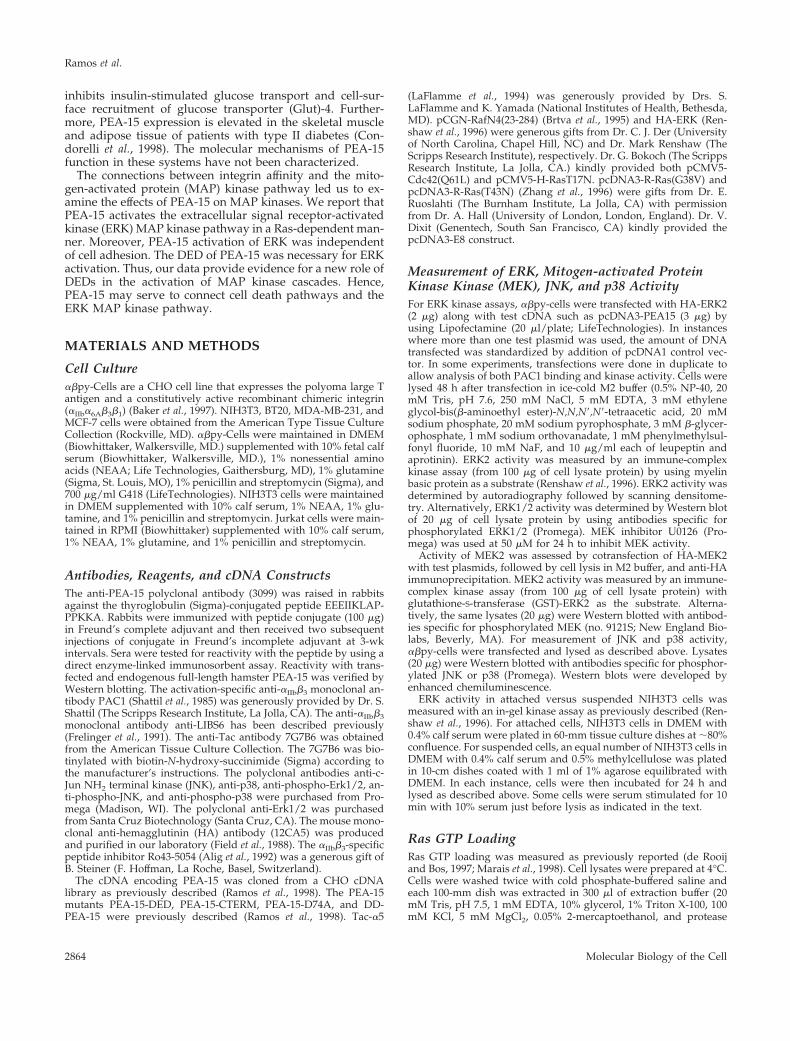
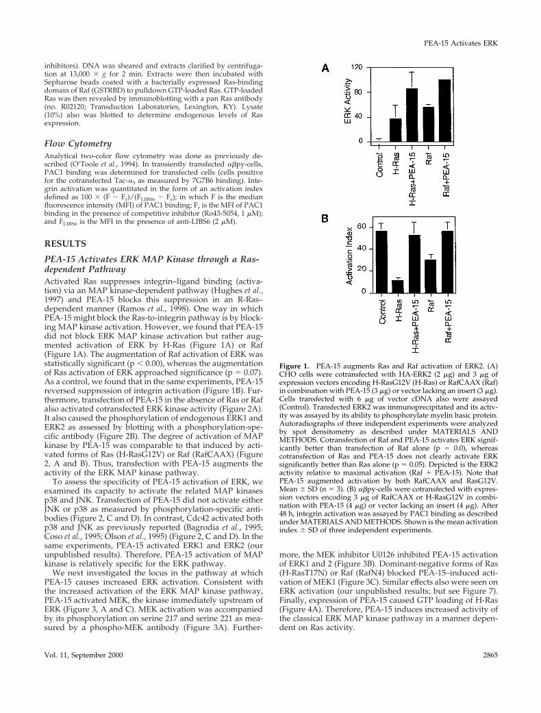
PEA-15 Activates ERK MAP Kinase through a Ras-dependent PathwayActivated Ras suppresses integrin–ligand binding (activa-tion) via an MAP kinase-dependent pathway (Hughes et al.,1997) and PEA-15 blocks this suppression in an R-Ras–dependent manner (Ramos et al., 1998). One way in whichPEA-15 might block the Ras-to-integrin pathway is by block-ing MAP kinase activation. However, we found that PEA-15did not block ERK MAP kinase activation but rather aug-mented activation of ERK by H-Ras (Figure 1A) or Raf(Figure 1A). The augmentation of Raf activation of ERK wasstatistically significant (p , 0.00), whereas the augmentationof Ras activation of ERK approached significance (p 5 0.07).As a control, we found that in the same experiments, PEA-15reversed suppression of integrin activation (Figure 1B). Fur-thermore, transfection of PEA-15 in the absence of Ras or Rafalso activated cotransfected ERK kinase activity (Figure 2A).It also caused the phosphorylation of endogenous ERK1 andERK2 as assessed by blotting with a phosphorylation-spe-cific antibody (Figure 2B). The degree of activation of MAPkinase by PEA-15 was comparable to that induced by acti-vated forms of Ras (H-RasG12V) or Raf (RafCAAX) (Figure2, A and B). Thus, transfection with PEA-15 augments theactivity of the ERK MAP kinase pathway.
To assess the specificity of PEA-15 activation of ERK, weexamined its capacity to activate the related MAP kinasesp38 and JNK. Transfection of PEA-15 did not activate eitherJNK or p38 as measured by phosphorylation-specific anti-bodies (Figure 2, C and D). In contrast, Cdc42 activated bothp38 and JNK as previously reported (Bagrodia et al., 1995;Coso et al., 1995; Olson et al., 1995) (Figure 2, C and D). In thesame experiments, PEA-15 activated ERK1 and ERK2 (ourunpublished results). Therefore, PEA-15 activation of MAPkinase is relatively specific for the ERK pathway.
We next investigated the locus in the pathway at whichPEA-15 causes increased ERK activation. Consistent withthe increased activation of the ERK MAP kinase pathway,PEA-15 activated MEK, the kinase immediately upstream ofERK (Figure 3, A and C). MEK activation was accompaniedby its phosphorylation on serine 217 and serine 221 as mea-sured by a phospho-MEK antibody (Figure 3A). Further-
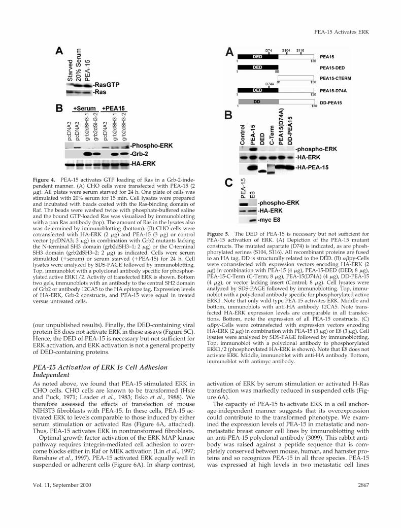
more, the MEK inhibitor U0126 inhibited PEA-15 activationof ERK1 and 2 (Figure 3B). Dominant-negative forms of Ras(H-RasT17N) or Raf (RafN4) blocked PEA-15–induced acti-vation of MEK1 (Figure 3C). Similar effects also were seen onERK activation (our unpublished results; but see Figure 7).Finally, expression of PEA-15 caused GTP loading of H-Ras(Figure 4A). Therefore, PEA-15 induces increased activity ofthe classical ERK MAP kinase pathway in a manner depen-dent on Ras activity.
Figure 1. PEA-15 augments Ras and Raf activation of ERK2. (A)CHO cells were cotransfected with HA-ERK2 (2 mg) and 3 mg ofexpression vectors encoding H-RasG12V (H-Ras) or RafCAAX (Raf)in combination with PEA-15 (3 mg) or vector lacking an insert (3 mg).Cells transfected with 6 mg of vector cDNA also were assayed(Control). Transfected ERK2 was immunoprecipitated and its activ-ity was assayed by its ability to phosphorylate myelin basic protein.Autoradiographs of three independent experiments were analyzedby spot densitometry as described under MATERIALS ANDMETHODS. Cotransfection of Raf and PEA-15 activates ERK signif-icantly better than transfection of Raf alone (p 5 0.0), whereascotransfection of Ras and PEA-15 does not clearly activate ERKsignificantly better than Ras alone (p 5 0.05). Depicted is the ERK2activity relative to maximal activation (Raf 1 PEA-15). Note thatPEA-15 augmented activation by both RafCAAX and RasG12V.Mean 6 SD (n 5 3). (B) abpy-cells were cotransfected with expres-sion vectors encoding 3 mg of RafCAAX or H-RasG12V in combi-nation with PEA-15 (4 mg) or vector lacking an insert (4 mg). After48 h, integrin activation was assayed by PAC1 binding as describedunder MATERIALS AND METHODS. Shown is the mean activationindex 6 SD of three independent experiments.
PEA-15 Activates ERK
Vol. 11, September 2000 2865
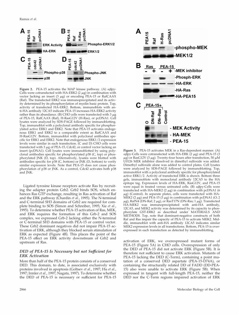
Ligated tyrosine kinase receptors activate Ras by recruit-ing the adapter protein Grb2. Grb2 binds SOS, which en-hances Ras GTP exchange, resulting in Ras activation of Rafand the ERK pathway (Chardin et al., 1995). The N-terminaland C-terminal SH3 domains of Grb2 are required for com-plete binding to SOS (Simon and Schreiber, 1995; Xie et al.,1995). To determine whether PEA-15 activation of Ras, MEK,and ERK requires the formation of this Grb-2 and SOScomplex, we expressed Grb-2 lacking either the N-terminalor C-terminal SH3 domains with PEA-15 or control vector.These Grb2 dominant negatives did not impair PEA-15 ac-tivation of ERK, although they blocked serum stimulation ofERK as expected (Figure 4B). This places the point of thePEA-15 effect on ERK activity downstream of Grb2 andupstream of Ras.
DED of PEA-15 Is Necessary but not Sufficient forERK ActivationMore than half of the PEA-15 protein consists of a conservedDED. This domain, to date, is associated exclusively withproteins involved in apoptosis (Goltsev et al., 1997; Hu et al.,1997; Irmler et al., 1997; Nagata, 1997). To determine whetherthe DED of PEA-15 is necessary or sufficient for PEA-15
activation of ERK, we overexpressed mutant forms ofPEA-15 (Figure 5A) in CHO cells. Overexpression of onlythe DED of PEA-15 did not activate ERK (Figure 5B). It istherefore not sufficient to cause ERK activation. Mutants ofPEA-15 lacking the DED (C-Term), containing a point mu-tation of a conserved DED aspartate (PEA-15-D74A), orcontaining the structurally related DD of FADD (DD-PEA-15) also were unable to activate ERK (Figure 5B). Whenexpressed in tangent with full-length PEA-15, neither theDED nor the C-Term regions impaired activation of ERK
Figure 2. PEA-15 activates the MAP kinase pathway. (A) abpy-Cells were cotransfected with HA-ERK2 (2 mg) in combination withvector lacking an insert (3 mg) or encoding PEA-15 or RafCAAX(Raf). The transfected ERK2 was immunoprecipitated and its activ-ity determined by its phosphorylation of myelin basic protein. Top,activity of transfected HA-ERK2. Bottom, immunoblots with an-ti-HA antibody 12CA5 indicate PEA-15 increases HA-ERK2 activityrather than its abundance. (B) CHO cells were transfected with 3 mgof PEA-15, RafCAAX (Raf), H-RasG12V (H-Ras), or pcDNA1. Celllysates were analyzed by SDS-PAGE followed by immunoblotting.Top, immunoblot with a polyclonal antibody specific for phosphor-ylated active ERK1 and ERK2. Note that PEA-15 activates endoge-nous ERK1 and ERK2 to a comparable extent as RafCAAX andH-RasG12V. Bottom, immunoblot with polyclonal antibodies spe-cific for ERK1 and ERK2. Note that endogenous ERK1/2 expressionlevels were similar in each transfection. (C and D) CHO cells weretransfected with 3 mg of PEA-15, Cdc42, or control vector lacking aninsert (pcDNA1). Cell lysates were immunoblotted by using poly-clonal antibodies specific for phosphorylated p38 (C, top) or phos-phorylated JNK (D, top). Alternatively, lysates were blotted withantibodies specific for p38 (C, bottom) or JNK (D, bottom) to verifysimilar expression levels. Note that PEA-15 does not cause phos-phorylation of p38 or JNK. As a control, Cdc42 activates both p38and JNK.
Figure 3. PEA-15 activates MEK in a Ras-dependent manner. (A)abpy-Cells were cotransfected with HA-ERK (2 mg) and PEA-15 (3mg) or RasG12V (3 mg). Twenty-four hours after transfection, 50 mMU0126 MEK inhibitor dissolved in dimethyl sulfoxide was added.Dimethyl sulfoxide alone was added to control plates. Cell lysateswere analyzed by SDS-PAGE followed by immunoblotting. Top,immunoblot with a polyclonal antibody specific for phosphorylatedactive ERK1/2. Activity of transfected ERK is shown. Bottom threegels, immunoblots with monoclonal antibody 12CA5 to the HAepitope tag. Expression levels of HA-ERK, RasG12V, and PEA-15were equal in treated versus untreated cells. (B) abpy-Cells weretransfected with HA-MEK2 (2 mg) in combination with pcDNA1 (6mg) (Control). In separate plates, cells were transfected with HA-MEK2 (2 mg) and PEA-15 (3 mg) in combination with pcDNA1 (O; 2mg), RafN4 (DN-Raf; 2 mg), or RasT17N (DN-Ras; 1 mg). TransfectedHA-MEK2 was immunoprecipitated with anti-HA antibody,12CA5, and MEK2 activity was determined by its capacity to phos-phorylate GST-ERK2 as described under MATERIALS ANDMETHODS. Top, note that dominant-negative constructs of bothRaf and Ras impair the capacity of PEA-15 to activate MEK2. Mid-dle, immunoblot with anti-HA antibody 12CA5 indicates similarMEK2 expression levels in all transfections. Bottom, PEA-15 is over-expressed in each transfection as detected by immunoblotting.
Ramos et al.
Molecular Biology of the Cell2866
(our unpublished results). Finally, the DED-containing viralprotein E8 does not activate ERK in these assays (Figure 5C).Hence, the DED of PEA-15 is necessary but not sufficient forERK activation, and ERK activation is not a general propertyof DED-containing proteins.
PEA-15 Activation of ERK Is Cell AdhesionIndependentAs noted above, we found that PEA-15 stimulated ERK inCHO cells. CHO cells are known to be transformed (Hsieand Puck, 1971; Leader et al., 1983; Esko et al., 1988). Wetherefore assessed the effects of transfection of mouseNIH3T3 fibroblasts with PEA-15. In these cells, PEA-15 ac-tivated ERK to levels comparable to those induced by eitherserum stimulation or activated Ras (Figure 6A, attached).Thus, PEA-15 activates ERK in nontransformed fibroblasts.
Optimal growth factor activation of the ERK MAP kinasepathway requires integrin-mediated cell adhesion to over-come blocks either in Raf or MEK activation (Lin et al., 1997;Renshaw et al., 1997). PEA-15 activated ERK equally well insuspended or adherent cells (Figure 6A). In sharp contrast,
activation of ERK by serum stimulation or activated H-Rastransfection was markedly reduced in suspended cells (Fig-ure 6A).
The capacity of PEA-15 to activate ERK in a cell anchor-age-independent manner suggests that its overexpressioncould contribute to the transformed phenotype. We exam-ined the expression levels of PEA-15 in metastatic and non-metastatic breast cancer cell lines by immunoblotting withan anti-PEA-15 polyclonal antibody (3099). This rabbit anti-body was raised against a peptide sequence that is com-pletely conserved between mouse, human, and hamster pro-teins and so recognizes PEA-15 in all three species. PEA-15was expressed at high levels in two metastatic cell lines
Figure 4. PEA-15 activates GTP loading of Ras in a Grb-2-inde-pendent manner. (A) CHO cells were transfected with PEA-15 (2mg). All plates were serum starved for 24 h. One plate of cells wasstimulated with 20% serum for 15 min. Cell lysates were preparedand incubated with beads coated with the Ras-binding domain ofRaf. The beads were washed twice with phosphate-buffered salineand the bound GTP-loaded Ras was visualized by immunoblottingwith a pan Ras antibody (top). The amount of Ras in the lysates alsowas determined by immunoblotting (bottom). (B) CHO cells werecotransfected with HA-ERK (2 mg) and PEA-15 (3 mg) or controlvector (pcDNA3; 3 mg) in combination with Grb2 mutants lackingthe N-terminal SH3 domain (grb2dSH3–1; 2 mg) or the C-terminalSH3 domain (grb2dSH3–2; 2 mg) as indicated. Cells were serumstimulated (1serum) or serum starved (1PEA-15) for 24 h. Celllysates were analyzed by SDS-PAGE followed by immunoblotting.Top, immunoblot with a polyclonal antibody specific for phosphor-ylated active ERK1/2. Activity of transfected ERK is shown. Bottomtwo gels, immunoblots with an antibody to the central SH2 domainof Grb2 or antibody 12CA5 to the HA epitope tag. Expression levelsof HA-ERK, Grb-2 constructs, and PEA-15 were equal in treatedversus untreated cells.
Figure 5. The DED of PEA-15 is necessary but not sufficient forPEA-15 activation of ERK. (A) Depiction of the PEA-15 mutantconstructs. The mutated aspartate (D74) is indicated, as are phosh-phorylated serines (S104, S116). All recombinant proteins are fusedto an HA tag. DD is structurally related to the DED. (B) abpy-Cellswere cotransfected with expression vectors encoding HA-ERK (2mg) in combination with PEA-15 (4 mg), PEA-15-DED (DED; 8 mg),PEA-15-C-Term (C-Term; 8 mg), PEA-15(D74A) (4 mg), DD-PEA-15(4 mg), or vector lacking insert (Control; 8 mg). Cell lysates wereanalyzed by SDS-PAGE followed by immunoblotting. Top, immu-noblot with a polyclonal antibody specific for phosphorylated activeERK1. Note that only wild-type PEA-15 activates ERK. Middle andbottom, immunoblots with anti-HA antibody 12CA5. Note trans-fected HA-ERK expression levels are comparable in all transfec-tions. Bottom, note the expression of all PEA-15 constructs. (C)abpy-Cells were cotransfected with expression vectors encodingHA-ERK (2 mg) in combination with PEA-15 (3 mg) or E8 (3 mg). Celllysates were analyzed by SDS-PAGE followed by immunoblotting.Top, immunoblot with a polyclonal antibody to phosphorylatedERK1/2 (phosphorylated HA-ERK is shown). Note that E8 does notactivate ERK. Middle, immunoblot with anti-HA antibody. Bottom,immunoblot with antimyc antibody.
PEA-15 Activates ERK
Vol. 11, September 2000 2867
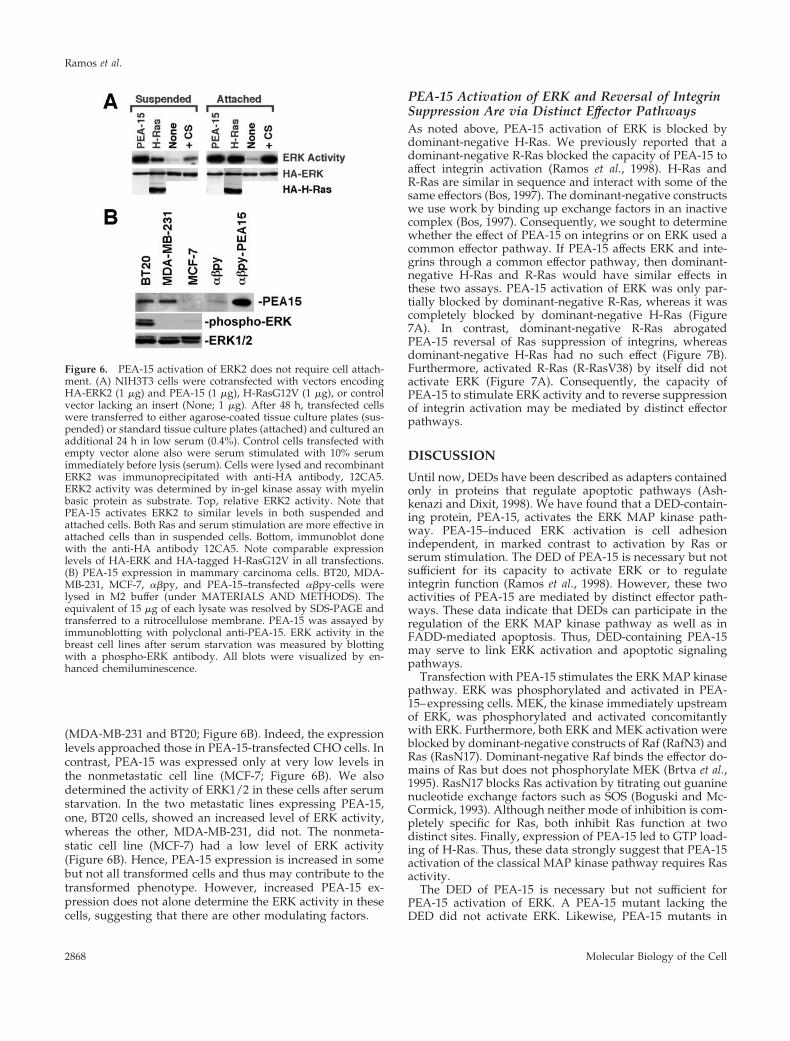
(MDA-MB-231 and BT20; Figure 6B). Indeed, the expressionlevels approached those in PEA-15-transfected CHO cells. Incontrast, PEA-15 was expressed only at very low levels inthe nonmetastatic cell line (MCF-7; Figure 6B). We alsodetermined the activity of ERK1/2 in these cells after serumstarvation. In the two metastatic lines expressing PEA-15,one, BT20 cells, showed an increased level of ERK activity,whereas the other, MDA-MB-231, did not. The nonmeta-static cell line (MCF-7) had a low level of ERK activity(Figure 6B). Hence, PEA-15 expression is increased in somebut not all transformed cells and thus may contribute to thetransformed phenotype. However, increased PEA-15 ex-pression does not alone determine the ERK activity in thesecells, suggesting that there are other modulating factors.
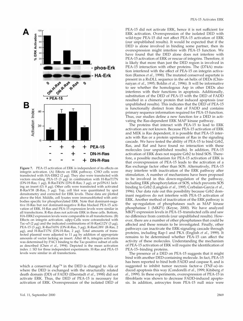
PEA-15 Activation of ERK and Reversal of IntegrinSuppression Are via Distinct Effector PathwaysAs noted above, PEA-15 activation of ERK is blocked bydominant-negative H-Ras. We previously reported that adominant-negative R-Ras blocked the capacity of PEA-15 toaffect integrin activation (Ramos et al., 1998). H-Ras andR-Ras are similar in sequence and interact with some of thesame effectors (Bos, 1997). The dominant-negative constructswe use work by binding up exchange factors in an inactivecomplex (Bos, 1997). Consequently, we sought to determinewhether the effect of PEA-15 on integrins or on ERK used acommon effector pathway. If PEA-15 affects ERK and inte-grins through a common effector pathway, then dominant-negative H-Ras and R-Ras would have similar effects inthese two assays. PEA-15 activation of ERK was only par-tially blocked by dominant-negative R-Ras, whereas it wascompletely blocked by dominant-negative H-Ras (Figure7A). In contrast, dominant-negative R-Ras abrogatedPEA-15 reversal of Ras suppression of integrins, whereasdominant-negative H-Ras had no such effect (Figure 7B).Furthermore, activated R-Ras (R-RasV38) by itself did notactivate ERK (Figure 7A). Consequently, the capacity ofPEA-15 to stimulate ERK activity and to reverse suppressionof integrin activation may be mediated by distinct effectorpathways.
DISCUSSION
Until now, DEDs have been described as adapters containedonly in proteins that regulate apoptotic pathways (Ash-kenazi and Dixit, 1998). We have found that a DED-contain-ing protein, PEA-15, activates the ERK MAP kinase path-way. PEA-15–induced ERK activation is cell adhesionindependent, in marked contrast to activation by Ras orserum stimulation. The DED of PEA-15 is necessary but notsufficient for its capacity to activate ERK or to regulateintegrin function (Ramos et al., 1998). However, these twoactivities of PEA-15 are mediated by distinct effector path-ways. These data indicate that DEDs can participate in theregulation of the ERK MAP kinase pathway as well as inFADD-mediated apoptosis. Thus, DED-containing PEA-15may serve to link ERK activation and apoptotic signalingpathways.
Transfection with PEA-15 stimulates the ERK MAP kinasepathway. ERK was phosphorylated and activated in PEA-15–expressing cells. MEK, the kinase immediately upstreamof ERK, was phosphorylated and activated concomitantlywith ERK. Furthermore, both ERK and MEK activation wereblocked by dominant-negative constructs of Raf (RafN3) andRas (RasN17). Dominant-negative Raf binds the effector do-mains of Ras but does not phosphorylate MEK (Brtva et al.,1995). RasN17 blocks Ras activation by titrating out guaninenucleotide exchange factors such as SOS (Boguski and Mc-Cormick, 1993). Although neither mode of inhibition is com-pletely specific for Ras, both inhibit Ras function at twodistinct sites. Finally, expression of PEA-15 led to GTP load-ing of H-Ras. Thus, these data strongly suggest that PEA-15activation of the classical MAP kinase pathway requires Rasactivity.
The DED of PEA-15 is necessary but not sufficient forPEA-15 activation of ERK. A PEA-15 mutant lacking theDED did not activate ERK. Likewise, PEA-15 mutants in
Figure 6. PEA-15 activation of ERK2 does not require cell attach-ment. (A) NIH3T3 cells were cotransfected with vectors encodingHA-ERK2 (1 mg) and PEA-15 (1 mg), H-RasG12V (1 mg), or controlvector lacking an insert (None; 1 mg). After 48 h, transfected cellswere transferred to either agarose-coated tissue culture plates (sus-pended) or standard tissue culture plates (attached) and cultured anadditional 24 h in low serum (0.4%). Control cells transfected withempty vector alone also were serum stimulated with 10% serumimmediately before lysis (serum). Cells were lysed and recombinantERK2 was immunoprecipitated with anti-HA antibody, 12CA5.ERK2 activity was determined by in-gel kinase assay with myelinbasic protein as substrate. Top, relative ERK2 activity. Note thatPEA-15 activates ERK2 to similar levels in both suspended andattached cells. Both Ras and serum stimulation are more effective inattached cells than in suspended cells. Bottom, immunoblot donewith the anti-HA antibody 12CA5. Note comparable expressionlevels of HA-ERK and HA-tagged H-RasG12V in all transfections.(B) PEA-15 expression in mammary carcinoma cells. BT20, MDA-MB-231, MCF-7, abpy, and PEA-15–transfected abpy-cells werelysed in M2 buffer (under MATERIALS AND METHODS). Theequivalent of 15 mg of each lysate was resolved by SDS-PAGE andtransferred to a nitrocellulose membrane. PEA-15 was assayed byimmunoblotting with polyclonal anti-PEA-15. ERK activity in thebreast cell lines after serum starvation was measured by blottingwith a phospho-ERK antibody. All blots were visualized by en-hanced chemiluminescence.
Ramos et al.
Molecular Biology of the Cell2868
which a conserved Asp74 in the DED is changed to Ala orwhere the DED is exchanged with the structurally relateddeath domain (DD) of FADD (Eberstadt et al., 1998) did notactivate ERK. Thus, the DED of PEA-15 is necessary foractivation of ERK. Overexpression of the isolated DED of
PEA-15 did not activate ERK, hence it is not sufficient forERK activation. Overexpression of the isolated DED withwild-type PEA-15 did not affect PEA-15 activation of ERK(our unpublished results). It would be expected that if theDED is alone involved in binding some partner, then itsoverexpression might interfere with PEA-15 function. Wehave found that the DED alone does not interfere withPEA-15 activation of ERK or rescue of integrins. Therefore, itis likely that more than just the DED region is involved inPEA-15 interaction with other proteins. The (D74A) muta-tion interfered with the effect of PEA-15 on integrin activa-tion (Ramos et al., 1998). The mutated conserved aspartate ispresent in a RxDLL sequence in the a6 helix of DEDs (Chin-naiyan et al., 1995; Boldin et al., 1996). It will be informativeto see whether the homologous Asp in other DEDs alsointerferes with their functions in apoptosis. Additionally,substitution of the DED of PEA-15 with the DED of FADDresulted in a chimeric protein that induced apoptosis (ourunpublished results). This indicates that the DED of PEA-15is functionally distinct from that of FADD and containsprimary sequence information required for PEA-15 function.Thus, our studies define a new function for a DED in acti-vating the Ras-dependent ERK MAP kinase pathway.
The proteins that interact with PEA-15 to lead to ERKactivation are not known. Because PEA-15 activation of ERKand MEK is Ras dependent, it is possible that PEA-15 inter-acts with Ras or a protein upstream of Ras in the signalingcascade. We have tested the ability of PEA-15 to bind Grb2,Ras, and Raf and have found no interaction with thesemolecules (our unpublished results). In addition, PEA-15activation of ERK does not require Grb2 to bind SOS. There-fore, a possible mechanism for PEA-15 activation of ERK isthat overexpression of PEA-15 leads to the activation of aRas exchange factor other than SOS. Alternatively, PEA-15may interfere with inactivation of the ERK pathway afterstimulation. A number of mechanisms have been proposedto be involved in this down-regulation of the pathway,including ERK phosphorylation of SOS, which prevents SOSbinding to Grb2 (Langlois et al., 1995; Corbalan-Garcia et al.,1996). Our data rule out this possibility because Grb2 dom-inant negatives do not interfere with PEA-15 activation ofERK. Another method of inactivation of the ERK pathway isthe up-regulation of phosphatases such as MAP kinasephosphatase 1 (MKP1) (Keyse, 2000). We have analyzedMKP1 expression levels in PEA-15–transfected cells and sawno difference from controls (our unpublished results). How-ever, there are a number of other phosphatases that could beaffected and these remain to be investigated. Finally, otherpathways can inactivate the ERK-signaling cascade throughproteins, including Rap-1 and PKA (English et al., 1999). Itremains to be determined whether PEA-15 can affect theactivity of these molecules. Understanding the mechanismof PEA-15 activation of ERK will require the identification ofPEA-15–binding proteins.
The presence of a DED on PEA-15 suggests that it mightbind with another DED-containing molecule. In fact, PEA-15has been reported to bind both FADD and caspase 8, and issuggested to inhibit tumor necrosis factor-a (TNF-a)–in-duced apoptosis this way (Condorelli et al., 1999; Kitsberg etal., 1999). In these experiments, overexpression of PEA-15 infibroblasts was shown to decrease FADD-induced apopto-sis. In addition, astrocytes from PEA-15 null mice were
Figure 7. PEA-15 activation of ERK is independent of its effects onintegrin activation. (A) Effects on ERK pathway. CHO cells weretransfected with HA-ERK2 (2 mg). They also were transfected withvectors encoding PEA-15 (3 mg) in combination with H-RasT17N(DN-H-Ras; 1 mg), R-RasT43N (DN-R-Ras; 3 mg), or pcDNA1 lack-ing an insert (O; 8 mg). Other cells were transfected with activatedR-RasV38 (R-Ras, 3 mg). Top, cell blot was quantitated by spotdensitometry and corrected for ERK levels. These data are plottedabove the blot. Middle, cell lysates were immunoblotted with anti-bodies specific for phosphorylated ERK. Note that dominant-nega-tive H-Ras but not dominant-negative R-Ras blocked PEA-15 acti-vation of ERK. H-Ras and PEA-15 expression levels were similar inall transfections. R-Ras does not activate ERK in these cells. Bottom,HA-ERK2 expression levels were comparable in all transfections. (B)Effects on integrin activation. abpy-Cells were cotransfected withTac-a5 (2 mg) and the indicated combinations of H-RasG12V (3 mg),PEA-15 (3 mg), R-RasT43N (DN-R-Ras, 3 mg), R-RasG38V (R-Ras, 2mg), and H-RasT17N (DN-H-Ras, 2 mg). Total amounts of trans-fected plasmid were adjusted to 11 mg by addition of appropriateamounts of vector lacking an insert. After 48 h, integrin activationwas determined by PAC1 binding to the Tac-positive subset of cellsas described (Chen et al., 1994). Depicted is the mean activationindex 6 SD for three independent experiments. H-Ras and PEA-15levels were similar in all transfections.
PEA-15 Activates ERK
Vol. 11, September 2000 2869
sensitive to TNF-a, whereas the wild-type astrocytes werenot. Transfection of PEA-15 into the knockout astrocytesrendered them resistant to TNF-a–induced apoptosis. It maybe that the anti-apoptotic effects of PEA-15 are in part due tothe activation of MEK and ERK. Indeed, TNFR1 receptorscan bind an intracellular molecule named MADD (MAPkinase-activating death domain) and signal this way to ac-tivate ERK MAP kinase and cPLA2 (Schievella et al., 1997).Thus, PEA-15 may tune TNF signaling by simultaneouslyblocking the apoptotic cascade and increasing the ERK-related survival pathways.
In addition to regulating cell death, DED adapter proteinssuch as FADD may promote activation of ERK and prolif-eration. For example, ligation of Fas receptor (CD95) acti-vates H-Ras and ERK in Jurkat T cells (Gulbins et al., 1995)through an unknown mechanism. Moreover, T cells lackingFADD or expressing a dominant-negative form of FADD aredefective in proliferation (Walsh et al., 1998), further impli-cating FADD signaling in pathways that control cell prolif-eration. Newton and colleagues postulated the existence ofan adapter protein that contains a DED and stimulates T-cellproliferation (Newton et al., 1998). PEA-15 is a DED-contain-ing protein that augments ERK activity in a Ras-dependentmanner. The DED of PEA-15 also can bind to the DEDs ofFADD and caspase 8 (Condorelli et al., 1999; Kitsberg et al.,1999). Consequently, our data suggest that PEA-15 is a po-tential link between apoptotic signaling and the MAP kinasepathway.
The affects of PEA-15 on ERK and integrin activation arevia distinct pathways. We previously demonstrated thatH-Ras blocks integrin signaling, but at the same time anexpression of a constitutively active form of the small GT-Pase, R-Ras, enhances integrin–ligand binding in the nor-mally nonadherent cell lines U937 and 32D.3. R-Ras also isreported to regulate integrin–ligand binding in CHO celllines (Zhang et al., 1996). We reported that PEA-15 blocks theH-Ras-to-integrin signal by activating a pathway dependenton R-Ras (Ramos et al., 1998). Indeed, the effects of PEA-15on integrin activation were blocked by dominant-negativeR-Ras. R-Ras is homologous to H-Ras, but has an extra 26amino acid residues at the amino terminus (Lowe et al.,1987). Furthermore, R-Ras can be regulated by activatorsand effectors distinct from those that regulate H-Ras func-tion (Huff et al., 1997). PEA-15 activation of ERK was blockedby dominant-negative H-Ras. However, dominant-negativeR-Ras had little effect on PEA-15 activation of ERK. Acti-vated R-Ras also did not promote ERK activation. Thus,PEA-15 represents a novel stimulator of distinct pathwaysthat depend on activity of either H-Ras or R-Ras.
PEA-15 activation of the MAP kinase pathway is anchor-age independent. PEA-15–activated ERK equally well inboth suspended and adherent cells. In contrast, efficient ERKactivation by Ras or serum requires cell adhesion (Lin et al.,1997; Renshaw et al., 1997). Activated ERK contributes to cellproliferation and anchorage-independent growth correlateswith tumorigenicity (Freedman and Shin, 1974). Therefore,increased PEA-15 could contribute to tumorigenicity. In-deed, the conserved 39-untranslated region of PEA-15 wasidentified as a transforming gene called mammary-trans-forming gene 1 (Bera et al., 1994). In addition, we find thatPEA-15 is expressed in some metastatic breast cancer celllines (MDA-MB-231 and BT20) at levels comparable to those
in the transfected CHO cells. In contrast, PEA-15 is poorlyexpressed in a nonmetastatic line (MCF-7). We found thatelevated expression of PEA-15 correlated with elevated ac-tivity of ERK in one of these lines (BT20) but not the other(MDA-MB-231). This suggests that other factors influencethe basal level of ERK activity in addition to PEA-15. PEA-15also is present in certain other human carcinoma lines de-rived from larynx, cervix, and skin (Condorelli et al., 1998).Our finding that PEA-15 activation of ERK is adhesion in-dependent suggests that high level PEA-15 expression inthese cancer cells could contribute to their pathogenic po-tential. We are currently exploring this hypothesis.
ACKNOWLEDGMENTS
This is publication number 11856-VB from The Scripps ResearchInstitute. We thank our colleagues for their generosity in providingthe reagents acknowledged under MATERIALS AND METHODS.We also are grateful to Dr. Sandy Shattil for critical review of themanuscript. J.W.R. and P.E.H. are fellows of the Leukemia Societyof America. M.W.R. was supported by a National Research ServiceAward from the National Institutes of Health. This work wasfunded by a grant from the National Institutes of Health and byfunds received from the Cancer Research Fund, under interagencyagreement 97-12013 (University of California contract 98-00924V)with the Department of Health Services, Cancer Research Program.
REFERENCES
Alig, L., Edenhofer, A., Hadvary, P., Hurzeler, M., Knopp, D.,Muller, M., Steiner, B., Trzeciak, A., and Weller, T. (1992). Lowmolecular weight, non-peptide fibrinogen receptor antagonists.J. Med. Chem. 35, 4393–4407.
Araujo, H., Danziger, N., Cordier, J., Glowinski, J., and Chneiweiss,H. (1993). Characterization of PEA-15, a major substrate for proteinkinase C in astrocytes. J. Biol. Chem. 268, 5911–5920.
Ashkenazi, A., and Dixit, V.M. (1998). Death receptors: signalingand modulation. Science 281, 1305–1308.
Bagrodia, S., Derijard, B., Davis, R.J., and Cerione, R.A. (1995).Cdc42 and PAK-mediated signaling leads to Jun kinase and p38mitogen-activated protein kinase activation. J. Biol. Chem. 270,27995–27998.
Baker, E.K., Tozer, E.C., Pfaff, M., Shattil, S.J., Loftus, J.C., andGinsberg, M.H. (1997). A genetic analysis of integrin function:Glanzmann thrombasthenia in vitro. Proc. Natl. Acad. Sci. USA 94,1973–1978.
Bera, T.K., Guzman, R.C., Miyamoto, S., Panda, D.K., Sasaki, M.,Hanyu, K., Enami, J., and Nandi, S. (1994). Identification of a mam-mary transforming gene (MAT1) associated with mouse mammarycarcinogenesis. Proc. Natl. Acad. Sci. USA 91, 9789–9793.
Boguski, M.S., and McCormick, F. (1993). Proteins regulating Rasand its relatives. Nature 366, 643–654.
Boldin, M.P., Goncharov, T.M., Goltsev, Y.V., and Wallach, D.(1996). Involvement of MACH, a novel MORT1/FADD-interactingprotease, in Fas/APO-1- and TNF receptor-induced cell death. Cell85, 803–815.
Boldin, M.P., Varfolomeev, E.E., Pancer, Z., Mett, I.L., Camonis, J.H.,and Wallach, D. (1995). A novel protein that interacts with the deathdomain of Fas/APO1 contains a sequence motif related to the deathdomain. J. Biol. Chem. 270, 7795–7798.
Bos, J.L. (1997). Ras-like GTPases. Biochim. Biophys. Acta 1333,M19–M31.
Ramos et al.
Molecular Biology of the Cell2870
Brtva, T.R., Drugan, J.K., Ghosh, S., Terrell, R.S., Campbell-Burk, S.,and Bell, R.M., Der, C.J. (1995). Two distinct Raf domains mediateinteraction with Ras. J. Biol. Chem. 270, 9809–9812.
Chardin, P., Cussac, D., Maignan, S., and Ducruix, A. (1995). TheGrb2 adaptor. FEBS Lett. 369, 47–51.
Chen, Y.-P., O’Toole, T.E., Shipley, T., Forsyth, J., LaFlamme, S.E.,Yamada, K.M., Shattil, S.J., and Ginsberg, M.H. (1994). “Inside-out”signal transduction inhibited by isolated integrin cytoplasmic do-mains. J. Biol. Chem. 269, 18307–18310.
Chinnaiyan, A.M., O’Rourke, K., Tewari, M., and Dixit, V.M. (1995).FADD, a novel death domain-containing protein, interacts with thedeath domain of Fas and initiates apoptosis. Cell 81, 505–512.
Chinnaiyan, A.M., Tepper, C.G., Seldin, M.F., O’Rourke, K., Kisch-kel, F.C., Hellbardt, S., Krammer, P.H., Peter, M.E., and Dixit, V.M.(1996). FADD/MORT1 is a common mediator of CD95 (Fas/APO-1)and tumor necrosis factor receptor-induced apoptosis. J. Biol. Chem.271, 4961–4965.
Condorelli, G., Vigliotta, G., Cafieri, A., Trencia, A., Andalo, P.,Oriente, F., Miele, C., Caruso, M., Formisano, P., and Beguinot, F.(1999). PED/PEA-15: an anti-apoptotic molecule that regulatesFAS/TNFR1-induced apoptosis. Oncogene 18, 4409–4415.
Condorelli, G., Vigliotta, G., Iavarone, C., Caruso, M., Tocchetti,C.G., Andreozzi, F., Cafieri, A., Tecce, M.F., Formisano, P., Beguinot,L., and Beguinot, F. (1998). PED/PEA-15 gene controls glucosetransport and is overexpressed in type 2 diabetes mellitus. EMBO J.17, 3858–3866.
Corbalan-Garcia, S., Yang, S.S., Degenhardt, K.R., and Bar-Sagi, D.(1996). Identification of the mitogen-activated protein kinase phos-phorylation sites on human Sos1 that regulate interaction withGrb2. Mol. Cell. Biol. 16, 5674–5682.
Coso, O.A., Chiariello, M., Yu, J.C., Teramoto, H., Crespo, P., Xu, N.,Miki, T., and Gutkind, J.S. (1995). The small GTP-binding proteinsRac1 and Cdc42 regulate the activity of the JNK/SAPK signalingpathway. Cell 81, 1137–1146.
Danziger, N., Yokoyama, M., Jay, T., Cordier, J., Glowinski, J., andChneiweiss, H. (1995). Cellular expression, developmental regula-tion, and phylogenic conservation of PEA-15, the astrocytic majorphosphoprotein and protein kinase C substrate. J. Neurochem. 64,1016–1025.
de Rooij, J., and Bos, J.L. (1997). Minimal Ras-binding domain ofRaf1. can be used as an activation-specific probe for Ras. Oncogene14, 623–625.
Eberstadt, M., Huang, B., Chen, Z., Meadows, R.P., Ng, S.C., Zheng,L., Lenardo, M.J., and Fesik, S.W. (1998). NMR structure and mu-tagenesis of the FADD (Mort1) death-effector domain. Nature 392,941–945.
English, J., Pearson, G., Wilsbacher, J., Swantek, J., Karandikar, M.,Xu, S., and Cobb, M.H. (1999). New insights into the control of MAPkinase pathways. Exp. Cell Res. 253, 255–270.
Esko, J.D., Rostand, K.S., and Weinke, J.L. (1988). Tumor formationdependent on proteoglycan biosynthesis. Science 241, 1092–1096.
Estelles, A., Yokoyama, M., Nothias, F., Vincent, J.D., Glowinski, J.,Vernier, P., and Chneiweiss, H. (1996). The major astrocytic phos-phoprotein PEA-15 is encoded by two mRNAs conserved on theirfull length in mouse and human. J. Biol. Chem. 271, 14800–14806.
Field, J., Nikawa, J., Broek, D., MacDonald, B., Rodgers, L., Wilson,I.A., Lerner, R.A., Wigler, M. (1988). Purification of a RAS-respon-sive adenyl cyclase complex from Saccharomyces cerevisiae by use ofan epitope addition method. Mol. Cell. Biol. 8, 2159–2165.
Freedman, V.H., and Shin, S.I. (1974). Cellular tumorigenicity innude mice: correlation with cell growth in semi-solid medium. Cell3, 355–359.
Frelinger, A.L. III, Du, X., Plow, E.F., and Ginsberg, M.H. (1991).Monoclonal antibodies to ligand-occupied conformers of integrinaIIbb3 (glycoprotein IIb-IIIa) alter receptor affinity, specificity, andfunction. J. Biol. Chem. 266, 17106–17111.
Goltsev, Y.V., Kovalenko, A.V., Arnold, E., Varfolomeev, E.E., Bro-dianskii, V.M., and Wallach, D. (1997). CASH, a novel caspasehomologue with death effector domains. J. Biol. Chem. 272, 19641–19644.
Gulbins, E., Bissonnette, R., Mahboubi, A., Martin, S., Nishioka, W.,Brunner, T., Baier, G., Baier-Bitterlich, G., Byrd, C., Lang, F. (1995).FAS-induced apoptosis is mediated via a ceramide-initiated RASsignaling pathway. Immunity 2, 341–351.
Hsie, A.W., and Puck, T.T. (1971). Morphological transformation ofChinese hamster cells by dibutyryl adenosine cyclic 39:59-mono-phosphate and testosterone. Proc. Natl. Acad. Sci. USA 68, 358–361.
Hu, S., Vincenz, C., Buller, M., and Dixit, V.M. (1997). A novelfamily of viral death effector domain-containing molecules thatinhibit both CD-95- and tumor necrosis factor receptor-1-inducedapoptosis. J. Biol. Chem. 272, 9621–9624.
Huff, S.Y., Quilliam, L.A., Cox, A.D., and Der, C.J. (1997). R-Ras isregulated by activators and effectors distinct from those that controlRas function. Oncogene 14, 133–143.
Hughes, P.E., Renshaw, M.W., Pfaff, M., Forsyth, J., Keivens, V.M.,Schwartz, M.A., and Ginsberg, M.H. (1997). Suppression of integrinactivation: a novel function of a Ras/Raf-initiated MAP kinasepathway. Cell 88, 521–530.
Irmler, M., Thome, M., Hahne, M., Schneider, P., Hofmann, K.,Steiner, V., Bodmer, J.L., Schroter, M., Burns, K., Mattmann, C.,Rimoldi, D., French, L.E., and Tschopp, J. (1997). Inhibition of deathreceptor signals by cellular FLIP. Nature 388, 190–195.
Keyse, S.M. (2000). Protein phosphatases and the regulation ofmitogen-activated protein kinase signaling. Curr. Opin. Cell Biol.12, 186–192.
Kitsberg, D., Formstecher, E., Fauquet, M., Kubes, M., Cordier, J.,Canton, B., Pan, G., Rolli, M., Glowinski, J., and Chneiweiss, H.(1999). Astrocytes lacking the neural death-effector-domain proteinPEA-15 show increased susceptibility to TNF alpha. J. Neurosci. 19,8244–8251.
LaFlamme, S.E., Thomas, L.A., Yamada, S.S., and Yamada, K.M.(1994). Single subunit chimeric integrins as mimics and inhibitors ofendogenous integrin functions in receptor localization, cell spread-ing and migration, and matrix assembly. J. Cell Biol. 126, 1287–1298.
Langlois, W.J., Sasaoka, T., Saltiel, A.R., and Olefsky, J.M. (1995).Negative feedback regulation and desensitization of ins. J. Biol.Chem. 270, 25320–25323.
Leader, W.M., Stopak, D., and Harris, A.K. (1983). Increased con-tractile strength and tightened adhesions to the substratum resultfrom reverse transformation of CHO cells by dibutyryl cyclic aden-osine monophosphate. J. Cell Sci. 64, 1–11.
Lin, T.H., Chen, Q., Howe, A., and Juliano, R.L. (1997). Cell anchor-age permits efficient signal transduction between ras and its down-stream kinases. J. Biol. Chem. 272, 8849–8852.
Lowe, D.G., Capon, D.J., Delwart, E., Sakaguchi, A.Y., Naylor, S.L.,and Goeddel, D.V. (1987). Structure of the human and murine R-Rasgenes, novel genes closely related to ras proto-oncogenes. Cell 48,137–146.
Marais, R., Light, Y., Mason, C., Paterson, H., Olson, M.F., andMarshall, C.J. (1998). Requirement of Ras-GTP-Raf complexes foractivation of Raf-1 by protein kinase C. Science 280, 109–112.
Nagata, S. (1997). Apoptosis by death factor. Cell 88, 355–365.
Newton, K., Harris, A.W., Bath, M.L., Smith, K.C., and Strasser, A.(1998). A dominant interfering mutant of FADD/MORT1 enhances
PEA-15 Activates ERK
Vol. 11, September 2000 2871
deletion of autoreactive thymocytes and inhibits proliferation ofmature T lymphocytes. EMBO J. 17, 706–718.
O’Toole, T.E., Katagiri, Y., Faull, R.J., Peter, K., Tamura, R.N., Quar-anta, V., Loftus, J.C., Shattil, S.J., and Ginsberg, M.H. (1994). Integrincytoplasmic domains mediate inside-out signal transduction. J. CellBiol. 124, 1047–1059.
Olson, M.F., Ashworth, A., and Hall, A. (1995). An essential role forRho, Rac, and Cdc42 GTPases in cell cycle progression through G1.Science 269, 1270–1272.
Ramos, J.W., Kojima, T.K., Hughes, P.E., Fenczik, C.A., and Gins-berg, M.H. (1998). The death effector domain of PEA-15 is involvedin its regulation of integrin activation. J. Biol. Chem. 273, 33897–33900.
Renshaw, M.W., Lea-Chou, E., and Wang, J.Y.J. (1996). Rac is re-quired for v-Abl tyrosine kinase to activate mitogenesis. Curr. Biol.6, 76–83.
Renshaw, M.W., Ren, X.D., and Schwartz, M.A. (1997). Growthfactor activation of MAP kinase requires cell adhesion. EMBO J. 16,5592–5599.
Schievella, A.R., Chen, J.H., Graham, J.R., and Lin, L.L. (1997).MADD, a novel death domain protein that interacts with the type 1
tumor necrosis factor receptor and activates mitogen-activated pro-tein kinase. J. Biol. Chem. 272, 12069–12075.
Sethi, T., Ginsberg, M.H., Downward, J., and Hughes, P.E. (1999).The small GTP-binding protein R-Ras can influence integrin activa-tion by antagonizing a Ras/Raf-initiated integrin suppression path-way. Mol. Biol. Cell 10, 1799–1809.
Shattil, S.J., Hoxie, J.A., Cunningham, M., and Brass, L.F. (1985).Changes in the platelet membrane glycoprotein IIb-IIIa Complexduring platelet activation. J. Biol. Chem. 260, 11107–11114.
Simon, J.A., and Schreiber, S.L. (1995). Grb2 SH3 binding to peptidesfrom Sos: evaluation of a general model for SH3-ligand interactions.Chem. Biol. 2, 53–60.
Walsh, C.M., Wen, B.G., Chinnaiyan, A.M., O’Rourke, K., Dixit,V.M., and Hedrick, S.M. (1998). A role for FADD in T cell activationand development. Immunity 8, 439–449.
Xie, Y., Pendergast, A.M., and Hung, M.C. (1995). Dominant-nega-tive mutants of Grb2 induced reversal of the transformed pheno-types caused by the point mutation-activated Rat HER-2/Neu.J. Biol. Chem. 270, 30717–30724.
Zhang, Z., Vuori, K., Wang, H.-G., Reed, J.C., and Ruoslahti, E.(1996). Integrin activation by R-Ras. Cell 85, 61–69.
Ramos et al.
Molecular Biology of the Cell2872