Promigratory and procontractile growth factor environments differentially regulate cell...
20
Promigratory and Procontractile Growth Factor Environments Differentially Regulate Cell Morphogenesis Sangmyung Rhee 1,2 , Chin-Han Ho 2 , and Frederick Grinnell 2,3 1 Department of Life Science, Chung-Ang University, Seoul, South Korea 2 Department of Cell Biology, UT Southwestern Medical Center, 5323 Harry Hines blvd. Dallas, Texas 75390-9039 Abstract 3D cell-matrix cultures provide a useful model to analyze and dissect the structural, functional, and mechanical aspects of cell-matrix interactions and motile behavior important for cell and tissue morphogenesis. In the current studies we tested the effects of serum and physiological growth factors on the morphogenetic behavior of human fibroblasts cultured on the surfaces of 3D collagen matrices. Fibroblasts in medium containing serum contracted into clusters, whereas cells in medium containing platelet derived growth factor (PDGF) were observed to migrate as individuals. The clustering activity of serum appeared to depend on lysophosphatidic acid, required cell contraction based on inhibition by blocking Rho kinase or myosin II, and was reversed upon switching to PDGF. Oncogenic Ras transformed human fibroblasts did not exhibit serum-stimulated cell clustering. Our findings emphasize the importance of cell specific promigratory and procontractile growth factor environments in the differential regulation of cell motile function and cell morphogenesis. Keywords Platelet-derived growth factor; lysophosphatidic acid; 3D collagen matrix; cell motility; cell contraction INTRODUCTION 3D cell-matrix cultures provide useful models to analyze and dissect the structural, functional, and mechanical aspects of cell-matrix interactions and motile behavior important for cell and tissue morphogenesis [1–5]. Unlike conventional 2D cell cultures (e.g., plastic or glass), cells can penetrate into 3D matrices, cell-matrix adhesive interactions are localized to matrix elements, and matrix fibrils can be reorganized mechanically. Mechanical remodeling of the extracellular matrix has been implicated in diverse aspects of normal tissue physiology and pathology including wound repair [6–9], fibrosis [10,11], tumorigenesis [12–14], and aging [15]. Matrix remodeling also has become an important design consideration for tissue engineering [16–18]. 3Corresponding author: Tel: 214-648-2181; Fax: 214-648-6712, [email protected]. Publisher's Disclaimer: This is a PDF file of an unedited manuscript that has been accepted for publication. As a service to our customers we are providing this early version of the manuscript. The manuscript will undergo copyediting, typesetting, and review of the resulting proof before it is published in its final citable form. Please note that during the production process errors may be discovered which could affect the content, and all legal disclaimers that apply to the journal pertain. NIH Public Access Author Manuscript Exp Cell Res. Author manuscript; available in PMC 2011 January 15. Published in final edited form as: Exp Cell Res. 2010 January 15; 316(2): 232–244. doi:10.1016/j.yexcr.2009.09.021. NIH-PA Author Manuscript NIH-PA Author Manuscript NIH-PA Author Manuscript
Transcript of Promigratory and procontractile growth factor environments differentially regulate cell...

Promigratory and Procontractile Growth Factor EnvironmentsDifferentially Regulate Cell Morphogenesis
Sangmyung Rhee1,2, Chin-Han Ho2, and Frederick Grinnell2,31 Department of Life Science, Chung-Ang University, Seoul, South Korea2 Department of Cell Biology, UT Southwestern Medical Center, 5323 Harry Hines blvd. Dallas,Texas 75390-9039
Abstract3D cell-matrix cultures provide a useful model to analyze and dissect the structural, functional, andmechanical aspects of cell-matrix interactions and motile behavior important for cell and tissuemorphogenesis. In the current studies we tested the effects of serum and physiological growth factorson the morphogenetic behavior of human fibroblasts cultured on the surfaces of 3D collagen matrices.Fibroblasts in medium containing serum contracted into clusters, whereas cells in medium containingplatelet derived growth factor (PDGF) were observed to migrate as individuals. The clusteringactivity of serum appeared to depend on lysophosphatidic acid, required cell contraction based oninhibition by blocking Rho kinase or myosin II, and was reversed upon switching to PDGF.Oncogenic Ras transformed human fibroblasts did not exhibit serum-stimulated cell clustering. Ourfindings emphasize the importance of cell specific promigratory and procontractile growth factorenvironments in the differential regulation of cell motile function and cell morphogenesis.
KeywordsPlatelet-derived growth factor; lysophosphatidic acid; 3D collagen matrix; cell motility; cellcontraction
INTRODUCTION3D cell-matrix cultures provide useful models to analyze and dissect the structural, functional,and mechanical aspects of cell-matrix interactions and motile behavior important for cell andtissue morphogenesis [1–5]. Unlike conventional 2D cell cultures (e.g., plastic or glass), cellscan penetrate into 3D matrices, cell-matrix adhesive interactions are localized to matrixelements, and matrix fibrils can be reorganized mechanically. Mechanical remodeling of theextracellular matrix has been implicated in diverse aspects of normal tissue physiology andpathology including wound repair [6–9], fibrosis [10,11], tumorigenesis [12–14], and aging[15]. Matrix remodeling also has become an important design consideration for tissueengineering [16–18].
3Corresponding author: Tel: 214-648-2181; Fax: 214-648-6712, [email protected]'s Disclaimer: This is a PDF file of an unedited manuscript that has been accepted for publication. As a service to our customerswe are providing this early version of the manuscript. The manuscript will undergo copyediting, typesetting, and review of the resultingproof before it is published in its final citable form. Please note that during the production process errors may be discovered which couldaffect the content, and all legal disclaimers that apply to the journal pertain.
NIH Public AccessAuthor ManuscriptExp Cell Res. Author manuscript; available in PMC 2011 January 15.
Published in final edited form as:Exp Cell Res. 2010 January 15; 316(2): 232–244. doi:10.1016/j.yexcr.2009.09.021.
NIH
-PA Author Manuscript
NIH
-PA Author Manuscript
NIH
-PA Author Manuscript

Compared to cell migration on 2D surfaces [19,20], tissue cells interacting with 3D matricesexhibit greater plasticity and multiple mechanisms of migration [21–25]. Local matrixremodeling can produce collagen fibril alignment and formation of tension lines along whichcell migration occurs [26–31]. If the matrix is stiff enough to resist cell tractional force, thenthe cells move. If the matrix cannot resist cell tractional force, then the matrix moves [32].
Fibroblast migration frequently has been analyzed in serum-containing medium, and platelet-derived growth factor (PDGF) has been reported to be the major promigratory factor forfibroblasts in serum [33,34]. However, in experiments using 3D collagen matrix assays offibroblast migration and contraction, we observed that unlike PDGF, serum stimulationestablished a procontractile rather than promigratory environment [35]. If human fibroblastswere transformed by oncogenic Ras, then serum became as promigratory as PDGF [36].
Fibroblasts and endothelial cells cultured in serum-containing medium on polyacrylamidesurfaces contract into cell clusters if the surface is sufficiently compliant [37,38]. In light ofour previous observations, we speculated that cells cultured on the surfaces of collagen matricesmight exhibit differential responses to serum and PDGF growth factor environments. Thecurrent work was carried out to test this possibility. We found that fibroblasts in serum mediumformed clusters on 1.5 mg/ml collagen matrices, exhibited less clustering on 4 mg/ml matrices,and did not cluster on collagen-coated coverslips. Cells in PDGF medium, on the other hand,migrated as individuals and did not form clusters. Individual cell migration and cell clusteringwere completely reversible upon switching from serum to PDGF. The clustering effect of serumcould be attributed to lysophosphatidic acid and was not observed using oncogenic Rastransformed human fibroblasts. Our findings emphasize the importance of cell specificpromigratory and procontractile growth factor environments in the differential regulation ofcell motile function and cell morphogenesis.
MATERIALS AND METHODSMaterials
Type I collagen (high concentration, rat tail) was purchased from BD bioscience (Bedford,MA). Dulbecco’s modified Eagle medium (DMEM), CO2-independent DMEM and 0.25%trypsin/EDTA solution were purchased from Invitrogen (Gaithersburg, MD). Fetal bovineserum (FBS) was purchased from Atlanta Biologicals (Lawrenceville, GA). Rho kinaseinhibitor Y-27632 was obtained from Calbiochem Corp. (La Jolla, CA). Blebbistatin wasobtained from Toronto Research Chemicals Inc. (Toronto, Canada). Platelet-derived growthfactor BB isotype (PDGF) was obtained from Upstate Biotechnology, Inc. (Lake Placid, NY).Fatty acid-free bovine serum albumin (BSA), lysophosphatidic acid (LPA), sphingosine-1-phosphate (S1P), anti-vinculin antibody, and activated charcoal were obtained from Sigma (St.Louis, MO). Alexa Fluor 488 phalloidin and propidium iodine (PI) were obtained fromMolecular Probes, Inc. (Eugene, OR). RNase (DNase free) was purchased from Roche(Indianapolis, IN). Fluoromount G was obtained from Southern Biotechnology Associates(Birmingham, AL)
Cell culture and preparation of collagen matricesUse of de-identified human foreskin fibroblasts was approved by the University InstitutionalReview Board (Exemption #4). BR-5 cells (hTERT immortalized early passage humanforeskin fibroblasts) and oncogenic Ras transformed BR-5 cells [36] were cultured in DMEMsupplemented with 10% fetal bovine serum at 37°C in a 5% CO2 humidified incubator. Cellculture and experimental incubations were carried out at 37 in a 5% CO2 incubator. Collagenmatrices were prepared at the concentrations indicated according to manufacturer’sinstructions. Experimental incubation media was DMEM with 10% FBS (serum medium) or
Rhee et al. Page 2
Exp Cell Res. Author manuscript; available in PMC 2011 January 15.
NIH
-PA Author Manuscript
NIH
-PA Author Manuscript
NIH
-PA Author Manuscript

DMEM with 5 mg/ml BSA and 50 ng/ml PDGF (PDGF medium) and other reagents added asindicated in the figure legends. Cells (1–5×10⁴/matrix) were seeded on top of polymerizedcollagen matrices or incubated on glass coverslips that had been coated 15 min at 37o C with50 μg/ml collagen in DMEM.
Immunofluorescence microscopyPreparation of samples for actin staining with Alexa Fluor 488-conjugated phalloidin andpropidium iodine (PI) was carried out as previously described [39]. Images for morphometricanalysis were collected at 22°C with a Nikon Elipse 400 fluorescent microscope using 10X/0.45, 20X/0.75, and 40X/0.75 Nikon Plan Apo infinity corrected objectives, PhotometricsSenSys CCD camera and MetaVue acquisition and imaging software (Molecular Devices).Morphometric measurements were made using MetaVue. Dendritic cell index --perimeter2/4π area (= 1.0 for a round cell) -- was measured with the Integrated MorphometryAnalysis function. For each condition, data shown are the average of region measurementsmade on 20 cells. Image processing was carried out using Adobe Photoshop.
Time-lapse microscopyFor time-lapse microscopic analyses, cells were incubated on collagen matrices polymerizedin 24 well dishes within a 37oC environmental chamber using CO2-independent DMEM inplace of DMEM. Images were collected at 10–15 min intervals using an Axiovert 200Minverted microscope (Zeiss, Thornwood, NY) with 10X/0.25 Achroplan objective (Zeiss),DXM1200F camera (Nikon), and Metamorph acquisition software.
RESULTSCell spreading in PDGF and serum media on 1.5 and 4 mg/ml collagen matrices and collagen-coated coverslips
Initial studies were carried out to compare effects of PDGF and serum on cell spreading oncollagen matrices. Preliminary findings indicated that the pattern of spreading varied markedlydepending on the collagen concentration of the matrix. For instance, Figure 1 shows fibroblastsin PDGF medium after 4 hr on 1.5 mg/ml and 4 mg/ml collagen matrices. On 1.5 mg/mlmatrices, fibroblasts exhibited dendritic morphology and contained few stress fibers orvinculin-staining focal adhesions. However, on 4 mg/ml matrices, fibroblasts appearedlamellar with prominent stress fibers and focal adhesions. Therefore increasing the collagenmatrix density from 1.5 to 4 mg/ml permits cells to acquire high cell-matrix tension state[40].
Figure 2 (A, 4 hr; B, 16 hr) and Figure 3 (morphometric measurements at 1, 4 and 16 hr)compare fibroblast spreading in PDGF and serum media on 1.5 and 4 mg/ml collagen matricesand collagen-coated coverslips. Initially the biggest differences in cell behavior concerned cellshape, and these differences were most evident with cells on 1.5 mg/ml collagen matrices. InPDGF medium, the cellular dendritic index was 20–25 after 1 hr and increased further by 4 hrby which time the area of cell spreading was ~7500 μm2. In serum medium, the cellulardendritic index was ~1 (round) after 1 hr and increased only slightly by 4 hr at which time thearea of cell spreading was ~500 μm2, i.e., little spreading had occurred.
The dendritic index of cells in PDGF medium decreased between 1–4 hr on 4 mg/ml matricesand was low at all times on collagen-coated coverslips where the fibroblasts became lamellarmore rapidly and the area of cell spreading increased fastest. In serum medium, cells on 4 mg/ml matrices and collagen coverslips also tended to be round, but spreading was less delayedcompared to 1.5 mg/ml matrices and could be observed even after 1 hr.
Rhee et al. Page 3
Exp Cell Res. Author manuscript; available in PMC 2011 January 15.
NIH
-PA Author Manuscript
NIH
-PA Author Manuscript
NIH
-PA Author Manuscript

By 16 hr, the biggest difference in cell behavior in PDGF and serum media concerned celldistribution rather than shape. Here also, the differences were most evident with cells on 1.5mg/ml collagen matrices. In PDGF medium, fibroblasts appeared to be dispersed. In serummedium, the cells had formed clusters. Clusters were less evident on 4 mg/ml matrices and didnot occur on coverslips.
Differential effects of PDGF and serum media on cell migration and contraction-dependentcell clustering
To learn more about the differences in cell morphogenesis and redistribution that occurred over16 hr, we carried out time-lapse video microscopy. Figure 4 presents phase-contrast imagesfrom representative videos of cells interacting with 1.5 mg/ml collagen matrices in PDGFmedium (Supplemental Video 1) and serum (FBS) medium (Supplemental Video 2). In PDGFmedium, cell spreading initially was dendritic as described above. Over time, dendriticextensions reorganized into a more bipolar organization and cell migration began. In serummedium, spreading was delayed. Cell protrusions formed and retracted. The combination ofcell extension, migration and contraction resulted in formation of large cell clusters. Tensionlines between clusters became increasingly apparent in the collagen matrix (e.g., arrows).Occasionally, cells began to migrate away from the clusters, but cell-cell interactions remainedsufficiently strong to keep the clusters together.
To gain a more quantitative view of cell clustering, fibroblasts were visualized by staining fornuclear distribution and actin cytoskeleton. Figure 5 (PDGF) shows that even with cells inclose proximity, the nuclear distribution remained dispersed indicating that little tendency forcell clustering occurred in PDGF medium. In serum medium (Figure 5, FBS), clusterscontaining 25–50 cells formed under similar conditions. Serum stimulation of fibroblastsactivates the small G-protein Rho [41], and Rho activation causes cell contraction [42,43].Blocking the Rho effector Rho kinase with the pharmacological inhibitor Y27632 (Figure 5,FBS+Y) or blocking myosin II activity with blebbistatin (Figure 5, FBS+Bleb) completelyprevented clustering.
Additional experiments were carried out to learn about the serum factor responsible for cellclustering. Serum lysophospholipids originally were identified as the Rho activators in serumbased on use of activated charcoal to remove lipid agonists [41]. Figure 5 (FBS + charcoal)shows an experiment in which cells were incubated with serum that had been subjected to tworounds of treatment with activated charcoal. The serum activity responsible for cell clusteringwas lost as a result of this procedure. Protein profiles of control and charcoal-treated serumwere almost identical (not shown).
Serum contains two lysophospholipid activators of Rho, LPA and S1P [42,44–46]. Previously,we observed that LPA and S1P both stimulate fibroblast contraction in collagen matrices[35]. Figure 6 compares cell clustering in LPA and S1P at two different cell concentrations.With LPA, cell clustering occurred similarly as observed in serum. S1P, on the other hand,was unable to stimulate formation of large cell clusters. Rather, clusters that formed in thepresence of S1P were much smaller than in LPA or serum. This finding was consistent withour observation that S1P inhibits protrusion of dendritic extensions and cell migration [35].
Cell migration generally is thought to occur through the combination of cell protrusion andcontraction [19,20]. However, for cells migrating in 3D matrices, the requirement foractomyosin contractility under some conditions is low [23] or absent [22]. Since PDGF didnot stimulate contraction sufficient for cell clustering in our studies, and fibroblasts in collagenmatrices exert little contractile force in response to PDGF [47], we tested if PDGF-stimulatedmigration required myosin II activity. Figure 7 shows migration tracks of individual cells inPDGF medium observed by time-lapse microscopy and analyzed by morphometric analysis.
Rhee et al. Page 4
Exp Cell Res. Author manuscript; available in PMC 2011 January 15.
NIH
-PA Author Manuscript
NIH
-PA Author Manuscript
NIH
-PA Author Manuscript

At blebbistatin concentrations that completely inhibited serum-stimulated cell clustering(Figure 5) and caused formation of long dendritic extensions (phase contrast, arrows),blebbistatin had only a minimal effect on cell migration (Supplemental Video 3). Thesefindings indicate that PDGF-stimulated cell migration on the surface of 1.5 mg/ml collagenmatrices requires little if any myosin II-dependent contractile activity.
We also tested the reversibility of promigratory and procontractile environments. Figure 8shows that the effects of PDGF and serum on cell dispersion and clustering were completelyreversible. Cells preclustered for 6 hr in serum (or LPA) medium, dispersed upon switching toPDGF medium. Cells initially dispersed in PDGF medium, clustered upon switching to serum(or LPA) medium. Even large clusters that formed after 24 hr exhibited extensive dispersionwhen switched to PDGF medium (Supplemental Video 4).
Two hr after addition of PDGF, little change could be detected in the collagen matrix tensionlines between cell clusters. However, as illustrated by the typical example in Figure 9, tensionlines were less apparent after 4–6 hr in areas where fibroblasts began to migrate out of theclusters. Therefore, changes in the growth factor environment can have a rapid and profoundeffect on collagen organization as well as cell distribution.
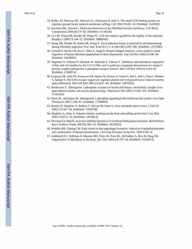
Oncogenic Ras transformed fibroblasts lose the cell clustering response to serumSince the differential response of fibroblasts to PDGF and serum/LPA media reflectedpromigratory and procontractile environments, we predicted that oncogenic Ras transformedhuman fibroblasts would no longer show this difference. Oncogenic Ras transformed cellsmigrate in serum or LPA almost as well as PDGF [36]. Figure 10 presents phase-contrastimages from videos of control and oncogenic Ras transformed cells interacting with 1.5 mg/ml collagen matrices in serum medium. Control cells in serum medium formed clusters asalready described. In parallel experiments, oncogenic Ras transformed cells did not(Supplemental Video 5).
DISCUSSIONPreviously, we demonstrated the importance of promigratory and procontractile growth factorenvironments in the differential regulation of cell motile function [35]. In the current studieswe extend our previous work and demonstrate that promigratory and procontractile growthfactor environments can differentially regulate the balance between individual cell migrationand cell clustering. Cells under promigratory conditions (PDGF medium) migrate asindividuals, whereas cells under procontractile conditions (serum medium) form clusters. Inserum medium, as adjacent cells attempt to spread, they contract and align the matrix in betweenand pull closer to each other. Local matrix alignment establishes preferential paths of furthercell protrusion because of contact guidance [48,49]. A positive feedback loop is established asmore and more cells contract and align the collagen in a given region resulting in furtherclustering.
Since collagen did not accumulate in the cell clusters, cell-cell interactions that formed duringclustering must have been stronger than and possibly even caused the release of cell-collageninteractions. The mechanisms responsible for stabilizing cell-cell interactions in serum-stimulated cell clusters requires further investigation. Adherens junctions may play a role[50] and are believed to be responsible for the synchronized contractile activity ofinterconnected wound fibroblasts [51]. Alternatively, fibronectin receptors interacting withcell surface fibronectin matrix has been implicated in establishing cell-cell interactions requiredto stabilize cell spheroids [52,53].
Rhee et al. Page 5
Exp Cell Res. Author manuscript; available in PMC 2011 January 15.
NIH
-PA Author Manuscript
NIH
-PA Author Manuscript
NIH
-PA Author Manuscript

Fibroblasts and endothelial cells cultured in serum-containing medium on polyacrylamidesurfaces also have been reported to contract into clusters if the polyacrylamide surface issufficiently compliant [37,38]. Similarly, we found that cell clustering was more apparent on1.5 mg/ml collagen matrices compared to 4 mg/ml matrices and was not observed on collagen-coated coverslips.
Serum activates cell contraction through the small G protein Rho [41,44], and clustering wasinhibited by blocking the Rho effector Rho kinase and downstream target myosin II. Additionalexperiments were carried out to learn about the serum factor responsible for cell clustering.Serum lysophospholipids originally were identified as the Rho activators in serum based onuse of activated charcoal to remove lipid agonists [41], and cluster-promoting activity was lostwhen lipid growth factors were removed from serum. Serum contains two lysophospholipidactivators of Rho, LPA and S1P [42,44–46]. Previously, we observed that LPA and S1P bothstimulate fibroblast contraction in collagen matrices. However, S1P inhibits protrusion ofdendritic extensions and cell migration [35]. In the current experiments, we observed that cellclustering occurred similarly with LPA and serum. S1P, on the other hand, was unable tostimulate formation of large cell clusters. Rather, clusters that formed in the presence of S1Pwere much smaller than in LPA. Therefore, we suggest that LPA is the growth primarilyresponsible for serum-stimulated fibroblast clustering activity
Cell clustering was completely reversible by switching from serum to PDGF medium, in whichcase fibroblasts exhibited individual cell migration. Rather than activation of the small G-protein Rho, the initial response of fibroblasts to PDGF stimulation is activation of the smallG-protein Rac [41,54], and fibroblasts in collagen matrices exert much less contractile forcein response to PDGF stimulation compared to serum or LPA [47]. Previous analyses of collagentension lines formed between cell explants in collagen matrices showed that destruction of thecells or their actin cytoskeleton resulted in partial reorganization of collagen and loss of tensionlines [28,55]. Similarly, we found that when clustered fibroblasts were switched from serumto PDGF, tension lines in the collagen became less visible in regions where cells were migratingout of the clusters.
Cell clustering on soft polyacrylamide surfaces and the possibly related feature known asdurotaxis – cell migration from softer to stiffer polyacrylamide surfaces but not the reverse[56,57] -- are both characteristics of motile behavior that are unique to the serum medium, i.e.,a procontractile growth factor environment. As shown in the current work, a promigratorygrowth factor environment permits individual cell migration instead of clustering. Similarly,a promigratory growth factor environment permits cells to move in the opposite direction ofdurotaxis. For instance, in PDGF but not serum or LPA medium, fibroblasts in nested collagenmatrices can migrate out of a ~15 mg/ml collagen inner matrix (stiffness ~600 Pa) into a 1.5mg/ml collagen outer matrix (stiffness ~6 Pa) [35,58] (stiffness measurements, M. Miron-Mendoza, unpublished). The foregoing differences emphasize the importance of carrying outstudies on cell motile function in both promigratory and procontractile growth factorenvironments to gain a comprehensive account.
Whether serum creates a a promigratory or procontractile environment likely will varydepending on cell type. The serum lysophospholipids LPA and S1P interact with a family ofG-protein coupled receptors that are expressed differentially by different cell types [42,45,46]. Depending on receptor expression, LPA and S1P potentially can activate cell contractionor cell migration signaling pathways. S1P1 receptor links to Rac activation and cell migration,whereas S1P2 receptor links to Rho activation, Rac inhibition, and inhibition of cell migration[59–62]. With oncogenic Ras transformed human fibroblasts, PDGF, LPA, and serum are allpromigratory [36], and serum stimulated cell clustering was not observed.
Rhee et al. Page 6
Exp Cell Res. Author manuscript; available in PMC 2011 January 15.
NIH
-PA Author Manuscript
NIH
-PA Author Manuscript
NIH
-PA Author Manuscript

Analysis of tissue cells interacting with 2D surfaces led to the idea that actomyosin-generatedcellular contractile force is required for cells to migrate [19,20]. The situation with cells in 3Dmatrices is more complex. Under some conditions the requirement for actomyosin contractilitycan be low [23] or absent [22]. Since blocking myosin II with blebbistatin inhibited serum-dependent fibroblast clustering but not PDGF-dependent cell migration, our studies provideanother example of cell migration that depends on little if any myosin II-dependent contractileactivity. In contrast to the current findings for cell migration on the surfaces of collagenmatrices, we showed previously that blebbistatin blocks PDGF-dependent cell migrationwithin nested collagen matrices [58]. The difference between myosin II-dependence offibroblast migration on the surface vs. within collagen matrices parallels the situation ofleukocytes exhibiting myosin II-independent or dependent migration depending upon theabsence or presence of steric hindrance in the collagen matrices through which the cells aremoving [22]. Without resistance from strong adhesive forces or steric hindrance, a myosin II-independent mechanism such as actin polymerization and depolymerization [63,64] may besufficient to provide the motor for cell migration.
It has become increasingly clear that matrix mechanics plays a critical role in cell and tissuemorphogenesis [1–5]. We suggest that whether the growth factor environment is promigratoryor procontractile also will play an important role. Differential cell migration vs. clusteringdepending on growth factor environment has the potential to contribute to tissue morphogeneticresponses such as mesenchymal condensation [65,66]. In addition, although connective tissuefibroblasts do not form clusters, interconnected cell-cell networks have been shown to playimportant roles in coordinated cell mechanical activity [51,67]. Individual cell migrationrequires disruption of these networks.
Supplementary MaterialRefer to Web version on PubMed Central for supplementary material.
AcknowledgmentsWe are indebted to Drs. Matt Petroll and William Snell for their advice and suggestions. This research was supportedby grants from the National Institutes of Health GM031321.
References1. Nelson CM, Bissell MJ. Of extracellular matrix, scaffolds, and signaling: tissue architecture regulates
development, homeostasis, and cancer. Annu Rev Cell Dev Biol 2006;22:287–309. [PubMed:16824016]
2. Griffith LG, Swartz MA. Capturing complex 3D tissue physiology in vitro. Nat Rev Mol Cell Biol2006;7:211–24. [PubMed: 16496023]
3. Yamada KM, Cukierman E. Modeling tissue morphogenesis and cancer in 3D. Cell 2007;130:601–10. [PubMed: 17719539]
4. Rhee S, Grinnell F. Fibroblast mechanics in 3D collagen matrices. Adv Drug Deliv Rev. 20075. Wozniak MA, Chen CS. Mechanotransduction in development: a growing role for contractility. Nat
Rev Mol Cell Biol 2009;10:34–43. [PubMed: 19197330]6. Grinnell F. Fibroblasts, myofibroblasts, and wound contraction. J Cell Biol 1994;124:401–4. [PubMed:
8106541]7. Silver FH, Siperko LM, Seehra GP. Mechanobiology of force transduction in dermal tissue. Skin
Research and Technology 2002;8:1–21. [PubMed: 12005114]8. Tomasek JJ, Gabbiani G, Hinz B, Chaponnier C, Brown RA. Myofibroblasts and mechano-regulation
of connective tissue remodelling. Nat Rev Mol Cell Biol 2002;3:349–63. [PubMed: 11988769]9. Petroll WM, Cavanagh HD, Jester JV. Assessment of stress fiber orientation during healing of radial
keratotomy wounds using confocal microscopy. Scanning 1998;20:74–82. [PubMed: 9530870]
Rhee et al. Page 7
Exp Cell Res. Author manuscript; available in PMC 2011 January 15.
NIH
-PA Author Manuscript
NIH
-PA Author Manuscript
NIH
-PA Author Manuscript

10. Abraham DJ, Eckes B, Rajkumar V, Krieg T. New developments in fibroblast and myofibroblastbiology: implications for fibrosis and scleroderma. Curr Rheumatol Rep 2007;9:136–43. [PubMed:17502044]
11. Hinz B, Phan SH, Thannickal VJ, Galli A, Bochaton-Piallat ML, Gabbiani G. The myofibroblast:one function, multiple origins. Am J Pathol 2007;170:1807–16. [PubMed: 17525249]
12. Dvorak HF. Tumors: wounds that do not heal. Similarities between tumor stroma generation andwound healing. N Engl J Med 1986;315:1650–9. [PubMed: 3537791]
13. Weigelt B, Bissell MJ. Unraveling the microenvironmental influences on the normal mammary glandand breast cancer. Semin Cancer Biol 2008;18:311–21. [PubMed: 18455428]
14. Butcher DT, Alliston T, Weaver VM. A tense situation: forcing tumour progression. Nat Rev Cancer2009;9:108–22. [PubMed: 19165226]
15. Fisher GJ, Varani J, Voorhees JJ. Looking older: fibroblast collapse and therapeutic implications.Arch Dermatol 2008;144:666–72. [PubMed: 18490597]
16. Clark RA, Ghosh K, Tonnesen MG. Tissue engineering for cutaneous wounds. J Invest Dermatol2007;127:1018–29. [PubMed: 17435787]
17. Brown RA, Wiseman M, Chuo CB, Cheema U, Nazhat SN. Ultrarapid engineering of biomimeticmaterials and tissues: Fabrication of nano- and microstructures by plastic compression. AdvancedFunctional Materials 2005;15:1762–1770.
18. Lutolf MP, Hubbell JA. Synthetic biomaterials as instructive extracellular microenvironments formorphogenesis in tissue engineering. Nat Biotechnol 2005;23:47–55. [PubMed: 15637621]
19. Lauffenburger DA, Horwitz AF. Cell migration: a physically integrated molecular process. Cell1996;84:359–69. [PubMed: 8608589]
20. Ridley AJ, Schwartz MA, Burridge K, Firtel RA, Ginsberg MH, Borisy G, Parsons JT, Horwitz AR.Cell migration: integrating signals from front to back. Science 2003;302:1704–9. [PubMed:14657486]
21. Friedl P, Brocker EB. The biology of cell locomotion within three-dimensional extracellular matrix.Cell Mol Life Sci 2000;57:41–64. [PubMed: 10949580]
22. Lammermann T, Bader BL, Monkley SJ, Worbs T, Wedlich-Soldner R, Hirsch K, Keller M, ForsterR, Critchley DR, Fassler R, Sixt M. Rapid leukocyte migration by integrin-independent flowing andsqueezing. Nature 2008;453:51–5. [PubMed: 18451854]
23. Sanz-Moreno V, Gadea G, Ahn J, Paterson H, Marra P, Pinner S, Sahai E, Marshall CJ. Rac activationand inactivation control plasticity of tumor cell movement. Cell 2008;135:510–23. [PubMed:18984162]
24. Gaggioli C, Hooper S, Hidalgo-Carcedo C, Grosse R, Marshall JF, Harrington K, Sahai E. Fibroblast-led collective invasion of carcinoma cells with differing roles for RhoGTPases in leading andfollowing cells. Nat Cell Biol 2007;9:1392–400. [PubMed: 18037882]
25. Wyckoff JB, Pinner SE, Gschmeissner S, Condeelis JS, Sahai E. ROCK- and myosin-dependentmatrix deformation enables protease-independent tumor-cell invasion in vivo. Curr Biol2006;16:1515–23. [PubMed: 16890527]
26. Stopak D, Harris AK. Connective tissue morphogenesis by fibroblast traction. I. Tissue cultureobservations. Dev Biol 1982;90:383–98. [PubMed: 7075867]
27. Guido S, Tranquillo RT. A methodology for the systematic and quantitative study of cell contactguidance in oriented collagen gels. Correlation of fibroblast orientation and gel birefringence. J CellSci 1993;105( Pt 2):317–31. [PubMed: 8408268]
28. Sawhney RK, Howard J. Slow local movements of collagen fibers by fibroblasts drive the rapid globalself-organization of collagen gels. J Cell Biol 2002;157:1083–91. [PubMed: 12058022]
29. Provenzano PP, Eliceiri KW, Campbell JM, Inman DR, White JG, Keely PJ. Collagen reorganizationat the tumor-stromal interface facilitates local invasion. BMC Med 2006;4:38. [PubMed: 17190588]
30. Raeber GP, Lutolf MP, Hubbell JA. Part II: Fibroblasts preferentially migrate in the direction ofprincipal strain. Biomech Model Mechanobiol 2008;7:215–25. [PubMed: 17619206]
31. Beloussov LV, Louchinskaia NN, Stein AA. Tension-dependent collective cell movements in theearly gastrula ectoderm of Xenopus laevis embryos. Dev Genes Evol 2000;210:92–104. [PubMed:10664152]
Rhee et al. Page 8
Exp Cell Res. Author manuscript; available in PMC 2011 January 15.
NIH
-PA Author Manuscript
NIH
-PA Author Manuscript
NIH
-PA Author Manuscript

32. Miron-Mendoza M, Seemann J, Grinnell F. Collagen Fibril Flow and Tissue Translocation Coupledto Fibroblast Migration in 3D Collagen Matrices. Mol Biol Cell 2008;19:2051–2058. [PubMed:18321993]
33. Gao Z, Sasaoka T, Fujimori T, Oya T, Ishii Y, Sabit H, Kawaguchi M, Kurotaki Y, Naito M, WadaT, Ishizawa S, Kobayashi M, Nabeshima Y, Sasahara M. Deletion of the PDGFR-beta gene affectskey fibroblast functions important for wound healing. J Biol Chem 2005;280:9375–89. [PubMed:15590688]
34. Li W, Fan J, Chen M, Guan S, Sawcer D, Bokoch GM, Woodley DT. Mechanism of human dermalfibroblast migration driven by type I collagen and platelet-derived growth factor-BB. Mol Biol Cell2004;15:294–309. [PubMed: 14595114]
35. Jiang H, Rhee S, Ho CH, Grinnell F. Distinguishing fibroblast promigratory and procontractile growthfactor environments in 3D collagen matrices. FASEB J 2008;22:2151–2160. [PubMed: 18272655]
36. Menezes GC, Miron-Mendoza M, Ho CH, Jiang H, Grinnell F. Oncogenic Ras-transformed humanfibroblasts exhibit differential changes in contraction and migration in 3D collagen matrices. ExpCell Res 2008;314:3081–91. [PubMed: 18708049]
37. Guo WH, Frey MT, Burnham NA, Wang YL. Substrate rigidity regulates the formation andmaintenance of tissues. Biophys J 2006;90:2213–20. [PubMed: 16387786]
38. Reinhart-King CA, Dembo M, Hammer DA. Cell-cell mechanical communication through compliantsubstrates. Biophys J 2008;95:6044–51. [PubMed: 18775964]
39. Rhee S, Grinnell F. P21-activated kinase 1: convergence point in PDGF- and LPA-stimulated collagenmatrix contraction by human fibroblasts. J Cell Biol 2006;172:423–32. [PubMed: 16449192]
40. Rhee S, Jiang H, Ho CH, Grinnell F. Microtubule function in fibroblast spreading is modulatedaccording to the tension state of cell-matrix interactions. Proc Natl Acad Sci U S A 2007;104:5425–30. [PubMed: 17369366]
41. Ridley AJ, Hall A. The small GTP-binding protein rho regulates the assembly of focal adhesions andactin stress fibers in response to growth factors. Cell 1992;70:389–99. [PubMed: 1643657]
42. Goetzl EJ, An S. Diversity of cellular receptors and functions for the lysophospholipid growth factorslysophosphatidic acid and sphingosine 1-phosphate. Faseb J 1998;12:1589–98. [PubMed: 9837849]
43. Jalink K, van Corven EJ, Hengeveld T, Morii N, Narumiya S, Moolenaar WH. Inhibition oflysophosphatidate- and thrombin-induced neurite retraction and neuronal cell rounding by ADPribosylation of the small GTP-binding protein Rho. J Cell Biol 1994;126:801–10. [PubMed:8045941]
44. Moolenaar WH. Lysophosphatidic acid signalling. Curr Opin Cell Biol 1995;7:203–10. [PubMed:7612272]
45. Gardell SE, Dubin AE, Chun J. Emerging medicinal roles for lysophospholipid signaling. TrendsMol Med 2006;12:65–75. [PubMed: 16406843]
46. Watterson KR, Lanning DA, Diegelmann RF, Spiegel S. Regulation of fibroblast functions bylysophospholipid mediators: potential roles in wound healing. Wound Repair Regen 2007;15:607–16. [PubMed: 17971005]
47. Kolodney MS, Elson EL. Correlation of myosin light chain phosphorylation with isometriccontraction of fibroblasts. J Biol Chem 1993;268:23850–5. [PubMed: 8226923]
48. Friedl P, Maaser K, Klein CE, Niggemann B, Krohne G, Zanker KS. Migration of highly aggressiveMV3 melanoma cells in 3-dimensional collagen lattices results in local matrix reorganization andshedding of alpha2 and beta1 integrins and CD44. Cancer Res 1997;57:2061–70. [PubMed: 9158006]
49. Dickinson RB, Guido S, Tranquillo RT. Biased cell migration of fibroblasts exhibiting contactguidance in oriented collagen gels. Ann Biomed Eng 1994;22:342–56. [PubMed: 7998680]
50. El Sayegh TY, Kapus A, McCulloch CA. Beyond the epithelium: cadherin function in fibrousconnective tissues. FEBS Lett 2007;581:167–74. [PubMed: 17217950]
51. Hinz B, Gabbiani G. Cell-matrix and cell-cell contacts of myofibroblasts: role in connective tissueremodeling. Thromb Haemost 2003;90:993–1002. [PubMed: 14652629]
52. Robinson EE, Zazzali KM, Corbett SA, Foty RA. Alpha5beta1 integrin mediates strong tissuecohesion. J Cell Sci 2003;116:377–86. [PubMed: 12482923]
53. Robinson EE, Foty RA, Corbett SA. Fibronectin matrix assembly regulates alpha5beta1-mediatedcell cohesion. Mol Biol Cell 2004;15:973–81. [PubMed: 14718567]
Rhee et al. Page 9
Exp Cell Res. Author manuscript; available in PMC 2011 January 15.
NIH
-PA Author Manuscript
NIH
-PA Author Manuscript
NIH
-PA Author Manuscript
54. Ridley AJ, Paterson HF, Johnston CL, Diekmann D, Hall A. The small GTP-binding protein racregulates growth factor-induced membrane ruffling. Cell 1992;70:401–10. [PubMed: 1643658]
55. Sawhney RK, Howard J. Molecular dissection of the fibroblast-traction machinery. Cell MotilCytoskeleton 2004;58:175–85. [PubMed: 15146536]
56. Lo CM, Wang HB, Dembo M, Wang YL. Cell movement is guided by the rigidity of the substrate.Biophys J 2000;79:144–52. [PubMed: 10866943]
57. Wang HB, Dembo M, Hanks SK, Wang Y. Focal adhesion kinase is involved in mechanosensingduring fibroblast migration. Proc Natl Acad Sci U S A 2001;98:11295–300. [PubMed: 11572981]
58. Grinnell F, Rocha LB, Iucu C, Rhee S, Jiang H. Nested collagen matrices: a new model to studymigration of human fibroblast populations in three dimensions. Exp Cell Res 2006;312:86–94.[PubMed: 16256985]
59. Sugimoto N, Takuwa N, Okamoto H, Sakurada S, Takuwa Y. Inhibitory and stimulatory regulationof Rac and cell motility by the G12/13-Rho and Gi pathways integrated downstream of a single Gprotein-coupled sphingosine-1-phosphate receptor isoform. Mol Cell Biol 2003;23:1534–45.[PubMed: 12588974]
60. Goparaju SK, Jolly PS, Watterson KR, Bektas M, Alvarez S, Sarkar S, Mel L, Ishii I, Chun J, MilstienS, Spiegel S. The S1P2 receptor negatively regulates platelet-derived growth factor-induced motilityand proliferation. Mol Cell Biol 2005;25:4237–49. [PubMed: 15870293]
61. Brinkmann V. Sphingosine 1-phosphate receptors in health and disease: mechanistic insights fromgene deletion studies and reverse pharmacology. Pharmacol Ther 2007;115:84–105. [PubMed:17561264]
62. Peters SL, Alewijnse AE. Sphingosine-1-phosphate signaling in the cardiovascular system. Curr OpinPharmacol 2007;7:186–92. [PubMed: 17280869]
63. Bottino D, Mogilner A, Roberts T, Stewart M, Oster G. How nematode sperm crawl. J Cell Sci2002;115:367–84. [PubMed: 11839788]
64. Mogilner A, Oster G. Polymer motors: pushing out the front and pulling up the back. Curr Biol2003;13:R721–33. [PubMed: 13678614]
65. Newman SA, Bhat R. Activator-inhibitor dynamics of vertebrate limb pattern formation. Birth DefectsRes C Embryo Today 2007;81:305–19. [PubMed: 18228262]
66. Widelitz RB, Chuong CM. Early events in skin appendage formation: induction of epithelial placodesand condensation of dermal mesenchyme. J Investig Dermatol Symp Proc 1999;4:302–6.
67. Goldsmith EC, Hoffman A, Morales MO, Potts JD, Price RL, McFadden A, Rice M, Borg TK.Organization of fibroblasts in the heart. Dev Dyn 2004;230:787–94. [PubMed: 15254913]
Rhee et al. Page 10
Exp Cell Res. Author manuscript; available in PMC 2011 January 15.
NIH
-PA Author Manuscript
NIH
-PA Author Manuscript
NIH
-PA Author Manuscript

Figure 1. Fibroblast spreading in PDGF medium on 1.5 mg/ml and 4 mg/ml collagen matricesFibroblasts were cultured 4 hr on 1.5 mg/ml and 4 mg/ml collagen matrices in PDGF medium.At the end of incubations, samples were fixed and stained for actin and vinculin. Judging fromappearance of stress fibers and focal adhesions, cells on 4 mg/ml collagen matrices developedhigh cell-matrix tension state. Scale bar, 50 μm (insert; 20 μm)
Rhee et al. Page 11
Exp Cell Res. Author manuscript; available in PMC 2011 January 15.
NIH
-PA Author Manuscript
NIH
-PA Author Manuscript
NIH
-PA Author Manuscript

Figure 2. Fibroblast spreading in PDGF and serum (FBS) media on 1.5 mg/ml and 4 mg/ml collagenmatrices and collagen-coated coverslipsFibroblasts were cultured 4 hr (A) and 16 hr (B) on 1.5 mg/ml and 4 mg/ml collagen matricesand collagen-coated coverslips in PDGF and serum media. At the end of incubations, sampleswere fixed and stained for actin. On 1.5 mg/ml collagen matrices, PDGF medium favoredformation of dendritic extensions. Serum medium delayed cell spreading. After 16 hr, cells inPDGF medium were dispersed. Cells in serum medium were clustered. Formation of dendriticextensions, delayed cell, and cell clustering did not occur on collagen-coated coverslips. Scalebar, 50 μm.
Rhee et al. Page 12
Exp Cell Res. Author manuscript; available in PMC 2011 January 15.
NIH
-PA Author Manuscript
NIH
-PA Author Manuscript
NIH
-PA Author Manuscript

Figure 3. Morphometric analysis of fibroblast spreading in PDGF and serum media on 1.5 mg/mland 4 mg/ml collagen matrices and collagen-coated coverslipsFibroblasts were cultured 1 hr, 4 hr and 16 hr on 1.5 mg/ml and 4 mg/ml collagen matrices andcollagen-coated coverslips in PDGF and serum media. At the end of incubations, cells werefixed and stained for actin and evaluated by morphometric analysis. Marked differencesbetween PDGF and serum media in formation of dendritic extensions and time of cell spreadingoccurred for cells on 1.5 mg/ml collagen matrices. These differences were less evident with 4mg/ml collagen matrices and not detected with collagen-coated coverslips.
Rhee et al. Page 13
Exp Cell Res. Author manuscript; available in PMC 2011 January 15.
NIH
-PA Author Manuscript
NIH
-PA Author Manuscript
NIH
-PA Author Manuscript

Figure 4. Cell migration in PDGF medium vs. clustering in serum mediumPhase-contrast images from videos of fibroblasts interacting with 1.5 mg/ml collagen matricesin PDGF (Supplemental Video 1) and serum media (Supplemental Video 2) with the samefield shown after ~6hr, ~12 hr, and 24 hr. Alignment of collagen fibrils between cells (arrows)becomes increasingly apparent in serum but not PDGF medium. Cells moved as individualsin PDGF medium but contracted into clusters in serum medium. Scale bar, 100 μm.
Rhee et al. Page 14
Exp Cell Res. Author manuscript; available in PMC 2011 January 15.
NIH
-PA Author Manuscript
NIH
-PA Author Manuscript
NIH
-PA Author Manuscript

Figure 5. Role of Rho kinase and myosin II in cell clusteringFibroblasts were cultured 16 hr on 1.0 mg/ml collagen matrices in PDGF or serum (FBS) mediaas indicated. Other additions were 10 μM Y27632 (Y) (Rho kinase inhibitor) or 20 μMblebbistatin (Bleb) (myosin II inhibitor). For FBS + charcoal, serum was treated with tworounds of activated charcoal. At the end of incubations, samples were fixed and stained foractin and with propidium iodide. Fibroblasts remained dispersed in PDGF medium, in mediumcontaining charcoal-treated serum, and in serum medium with Rho kinase or myosin IIinhibitors added. Scale bar, 100 μm.
Rhee et al. Page 15
Exp Cell Res. Author manuscript; available in PMC 2011 January 15.
NIH
-PA Author Manuscript
NIH
-PA Author Manuscript
NIH
-PA Author Manuscript

Figure 6. Effects of LPA and S1P on cell clusteringFibroblasts at the cell numbers indicated were cultured 16 hr on 1.0 mg/ml collagen matricesin 10 μM LPA or 1 μM S1P as shown. At the end of incubations, samples were fixed andstained for actin and with propidium iodide. LPA caused cell clustering similar to serum. WithS1P, clusters were smaller and limited to nearby cells. Scale bar, 100 μm.
Rhee et al. Page 16
Exp Cell Res. Author manuscript; available in PMC 2011 January 15.
NIH
-PA Author Manuscript
NIH
-PA Author Manuscript
NIH
-PA Author Manuscript

Figure 7. PDGF stimulated migration is not inhibited by blocking myosin IIFibroblasts were cultured 16 hr on 1.5 mg/ml collagen matrices in PDGF media with 20 μMblebbistatin added as indicated. Cell migration was recorded by time-lapse microscopy.Randomly selected individual cell migration tracks were copied and combined into a singlefigure. Persistence of directional migration was determined morphometrically by measuringD/T ratios (direct distance from start to end point [D] divided by the total track distance [T]).Addition of blebbistatin caused cells to exhibit abnormally long dendritic extensions (arrows)but had little effect on overall cell migration in PDGF medium (Supplemental Video 3). Scalebar, 100 μm.
Rhee et al. Page 17
Exp Cell Res. Author manuscript; available in PMC 2011 January 15.
NIH
-PA Author Manuscript
NIH
-PA Author Manuscript
NIH
-PA Author Manuscript

Figure 8. Reversibility of cell clustering patternsFibroblasts were cultured for an initial period of 6 hr on 1.0 mg/ml collagen matrices in PDGFand serum media. Subsequently, the samples were rinsed and placed into PDGF and serummedia as indicated for an additional 18 hr. At the end of the initial 6 hr period and after 24 hr,samples were fixed and stained for actin and with propidium iodide (PI). Morphogenetic cellpatterns in PDGF and serum media were completely reversible. Scale bar, 100 μm.
Rhee et al. Page 18
Exp Cell Res. Author manuscript; available in PMC 2011 January 15.
NIH
-PA Author Manuscript
NIH
-PA Author Manuscript
NIH
-PA Author Manuscript

Figure 9. Reversibility of morphogenetic patternsFibroblasts were cultured for an initial period of 16 hr on 1.0 mg/ml collagen matrices in serum(FBS) media. Subsequently, half the samples were rinsed and placed into PDGF media. At theend of an additional 4 hr period, samples were fixed and stained for actin and also observedby phase contrast microscopy. Alignment of collagen fibrils between cell clusters was evidentin serum (arrows). In PDGF region, where cells were migrating out of clusters, the alignmentof collagen fibrils became less obvious (arrows) Scale bar, 100 μm.
Rhee et al. Page 19
Exp Cell Res. Author manuscript; available in PMC 2011 January 15.
NIH
-PA Author Manuscript
NIH
-PA Author Manuscript
NIH
-PA Author Manuscript
Figure 10. Migration of oncogenic Ras transformed cells in serum mediaPhase-contrast images from videos of control (BR5) and oncogenic Ras transformed BR5fibroblasts interacting with 1.5 mg/ml collagen matrices in serum medium. Unlike control cellsthat clustered, oncogenic Ras transformed cells moved as individuals in serum medium(Supplemental Video 4). Scale bar, 100 μm.
Rhee et al. Page 20
Exp Cell Res. Author manuscript; available in PMC 2011 January 15.
NIH
-PA Author Manuscript
NIH
-PA Author Manuscript
NIH
-PA Author Manuscript