The flavonoid tangeretin activates the unfolded protein response and synergizes with imatinib in the...
10
RESEARCH ARTICLE The flavonoid tangeretin activates the unfolded protein response and synergizes with imatinib in the erythroleukemia cell line K562 Sofie Lust 1,2 , Barbara Vanhoecke 2 , Mireille Van Gele 3 , Jan Philippe ´ 4 , Marc Bracke 2 and Fritz Offner 1 1 Department of Hematology, University Hospital Ghent, Ghent, Belgium 2 Laboratory of Experimental Cancer Research, Department of Radiation Oncology and Experimental Cancer Research, University Hospital Ghent, Ghent, Belgium 3 Department of Dermatology, University Hospital Ghent, Ghent, Belgium 4 Departments of Clinical Chemistry, Microbiology and Immunology, University Hospital Ghent, Ghent, Belgium Received: April 27, 2009 Revised: June 16, 2009 Accepted: July 5, 2009 We explored the mechanism of cell death of the polymethoxyflavone tangeretin (TAN) in K562 breakpoint cluster region-abelson murine leukemia (Bcr-Abl1) cells. Flow cytometric analysis showed that TAN arrested the cells in the G 2 /M phase and stimulated an accumulation of the cells in the sub-G 0 phase. TAN-induced cell death was evidenced by poly(ADP)-ribose poly- merase cleavage, DNA laddering fragmentation, activation of the caspase cascade and down- regulation of the antiapoptotic proteins Mcl-1 and Bcl-x L . Pretreatment with the pancaspase inhibitor Z-VAD-FMK_blocked caspase activation and cell cycle arrest but did not inhibit apoptosis which suggest that other cell killing mechanisms like endoplasmic reticulum (ER)- associated cell death pathways could be involved. We demonstrated that TAN-induced apoptosis was preceded by a rapid activation of the proapoptotic arm of the unfolded protein response, namely PKR-like ER kinase. This was accompanied by enhanced levels of glucose-regulated protein of 78 kDa and of spliced X-box binding protein 1. Furthermore, TAN sensitized K562 cells to the cell killing effects of imatinib via an apoptotic mechanism. In conclusion, our results suggest that TAN is able to induce apoptosis in Bcr-Abl1 cells via cell cycle arrest and the induction of the unfolded protein response, and has synergistic cytotoxicity with imatinib. Keywords: Apoptosis / Cell cycle arrest / Flavonoid / Imatinib / Unfolded protein response 1 Introduction Flavonoids are widely distributed polyphenolic compounds in plants and in food. They exhibit a wide variety of biolo- gical effects including antioxidative, antiangiogenic, anti- invasive, antiproliferative and anti-inflammatory activities [1]. Tangeretin (TAN) is a polymethoxyflavone naturally present in citrus peel oil. TAN has been studied in breast cancer cells by Bracke et al. [2–4], who demonstrated that TAN inhibits invasion and proliferation of MCF-7/6 cells at least in part by affecting cell–cell interactions. TAN also exerts antiproliferative effects in lung carcinoma cells [5], in colorectal carcinoma cells [6] and in lymphoid cell lines (HL-60) [7] at 10 4 M in vitro. Recently, an increasing number of reports link flavonoid-induced cell death to the induction of endoplasmic reticulum (ER) stress [8, 9]. Correspondence: Professor Fritz Offner, Department of Hema- tology, University Hospital Ghent, De Pintelaan 185, 9K12IE, 9000 Ghent, Belgium E-mail: [email protected] Fax: 132-9-332-2737 Abbreviations: AnnV, annexin V; ATF, activating transcription factor; BA, brefeldin A; Bcr-Abl, breakpoint cluster region-abel- son murine leukemia; CHOP, C/EBP homologous protein-10; CI, combination index; CML, chronic myelogenous leukemia; DAPI, 4 0 -6-diamidino-2-phenylindole; eIF2a, eukaryotic translation initiation factor 2; ER, endoplasmic reticulum; GRP78, glucose- regulated protein of 78 kDa; IRE1, inositol-requiring enzyme 1; LC, living cells; MTT, 3-(4,5-dimethylthiazol-2-yl)-2,5-diphenyl- tetrazoliumbromide; PARP, poly(ADP)-ribose polymerase; PERK, PKR-like ER kinase; PI, propidium iodide; TAN, tangeretin; TG, thapsigargin; UPR, unfolded protein response; XBP1, X-box binding protein 1 & 2010 WILEY-VCH Verlag GmbH & Co. KGaA, Weinheim www.mnf-journal.com Mol. Nutr. Food Res. 2010, 54, 823–832 823 DOI 10.1002/mnfr.200900186
-
Upload
independent -
Category
Documents
-
view
0 -
download
0
Transcript of The flavonoid tangeretin activates the unfolded protein response and synergizes with imatinib in the...
RESEARCH ARTICLE
The flavonoid tangeretin activates the unfolded protein
response and synergizes with imatinib in the
erythroleukemia cell line K562
Sofie Lust1,2, Barbara Vanhoecke2, Mireille Van Gele3, Jan Philippe4, Marc Bracke2
and Fritz Offner1
1 Department of Hematology, University Hospital Ghent, Ghent, Belgium2 Laboratory of Experimental Cancer Research, Department of Radiation Oncology and Experimental Cancer
Research, University Hospital Ghent, Ghent, Belgium3 Department of Dermatology, University Hospital Ghent, Ghent, Belgium4 Departments of Clinical Chemistry, Microbiology and Immunology, University Hospital Ghent, Ghent, Belgium
Received: April 27, 2009
Revised: June 16, 2009
Accepted: July 5, 2009
We explored the mechanism of cell death of the polymethoxyflavone tangeretin (TAN) in K562
breakpoint cluster region-abelson murine leukemia (Bcr-Abl1) cells. Flow cytometric analysis
showed that TAN arrested the cells in the G2/M phase and stimulated an accumulation of the
cells in the sub-G0 phase. TAN-induced cell death was evidenced by poly(ADP)-ribose poly-
merase cleavage, DNA laddering fragmentation, activation of the caspase cascade and down-
regulation of the antiapoptotic proteins Mcl-1 and Bcl-xL. Pretreatment with the pancaspase
inhibitor Z-VAD-FMK_blocked caspase activation and cell cycle arrest but did not inhibit
apoptosis which suggest that other cell killing mechanisms like endoplasmic reticulum (ER)-
associated cell death pathways could be involved. We demonstrated that TAN-induced apoptosis
was preceded by a rapid activation of the proapoptotic arm of the unfolded protein response,
namely PKR-like ER kinase. This was accompanied by enhanced levels of glucose-regulated
protein of 78 kDa and of spliced X-box binding protein 1. Furthermore, TAN sensitized K562
cells to the cell killing effects of imatinib via an apoptotic mechanism. In conclusion, our results
suggest that TAN is able to induce apoptosis in Bcr-Abl1 cells via cell cycle arrest and the
induction of the unfolded protein response, and has synergistic cytotoxicity with imatinib.
Keywords:
Apoptosis / Cell cycle arrest / Flavonoid / Imatinib / Unfolded protein response
1 Introduction
Flavonoids are widely distributed polyphenolic compounds
in plants and in food. They exhibit a wide variety of biolo-
gical effects including antioxidative, antiangiogenic, anti-
invasive, antiproliferative and anti-inflammatory activities
[1]. Tangeretin (TAN) is a polymethoxyflavone naturally
present in citrus peel oil. TAN has been studied in breast
cancer cells by Bracke et al. [2–4], who demonstrated that
TAN inhibits invasion and proliferation of MCF-7/6 cells at
least in part by affecting cell–cell interactions. TAN also
exerts antiproliferative effects in lung carcinoma cells [5], in
colorectal carcinoma cells [6] and in lymphoid cell lines
(HL-60) [7] at 10�4 M in vitro. Recently, an increasing
number of reports link flavonoid-induced cell death to the
induction of endoplasmic reticulum (ER) stress [8, 9].
Correspondence: Professor Fritz Offner, Department of Hema-
tology, University Hospital Ghent, De Pintelaan 185, 9K12IE,
9000 Ghent, Belgium
E-mail: [email protected]
Fax: 132-9-332-2737
Abbreviations: AnnV, annexin V; ATF, activating transcription
factor; BA, brefeldin A; Bcr-Abl, breakpoint cluster region-abel-
son murine leukemia; CHOP, C/EBP homologous protein-10; CI,
combination index; CML, chronic myelogenous leukemia; DAPI,
40-6-diamidino-2-phenylindole; eIF2a, eukaryotic translation
initiation factor 2; ER, endoplasmic reticulum; GRP78, glucose-
regulated protein of 78 kDa; IRE1, inositol-requiring enzyme 1;
LC, living cells; MTT, 3-(4,5-dimethylthiazol-2-yl)-2,5-diphenyl-
tetrazoliumbromide; PARP, poly(ADP)-ribose polymerase; PERK,
PKR-like ER kinase; PI, propidium iodide; TAN, tangeretin; TG,
thapsigargin; UPR, unfolded protein response; XBP1, X-box
binding protein 1
& 2010 WILEY-VCH Verlag GmbH & Co. KGaA, Weinheim www.mnf-journal.com
Mol. Nutr. Food Res. 2010, 54, 823–832 823DOI 10.1002/mnfr.200900186

The accurate function of the ER plays a crucial role in
securing cell survival. Therefore, the cell has developed
rescue mechanisms, namely the unfolded protein response
(UPR) and the ER-associated protein degradation in order to
cope with ER stress. Under ER stress, unfolded or misfolded
proteins accumulate in the ER lumen and/or the ER
calcium homeostasis is disrupted, which triggers the UPR.
This stress response is characterized by a strong upregula-
tion of the ER-resident chaperone GRP78/BiP (glucose-
regulated protein of 78 kDa). GRP78 controls the three ER
stress-transducers, namely the activating transcription factor
6 (ATF6), the inositol-requiring enzyme 1 (IRE1) and PKR-
like ER kinase (PERK). When the UPR fails to reverse
prolonged ER stress this will ultimately lead to programmed
cell death via mitochondria-dependent or -independent
pathways [10].
Chronic myelogenous leukemia (CML) is characterized
by the presence of the Philadelphia chromosome which
is the product of a reciprocal translocation between chro-
mosome 9 and 22. As a consequence, a fusion protein,
namely breakpoint cluster region-abelson murine leukemia
(Bcr-Abl), is formed which leads to the constitutive
activation of the Abl tyrosine kinase [11]. Selective tyrosine
kinase inhibitors of Bcr-Abl, such as imatinib, dasatinib and
nilotinib, are among the most successful non-chemother-
apeutic agents used in current CML treatments. However,
resistance to current therapies is emerging, making
new drug development and/or combination treatments
necessary.
In this study, we show for the first time that TAN induces
activation of the proapoptotic arm of the UPR and cell
cycle arrest in Bcr-Abl1 cells. Moreover, we demonstrate
that TAN enhances the cell killing effect induced by
imatinib.
2 Materials and methods
2.1 Cell culture
The human erythroleukemia cell line K562 was obtained
from American Type Culture Collection (ATCC). K562 cells
were cultured in suspension in RPMI1640 medium
supplemented with 10% fetal bovine serum (Greiner bio-
one, Wemmel, Belgium), 100 IU/mL penicillin, 100mg/mL
streptomycin and 0.56 mg/mL fungizone (Gibco BRL,
Merelbeke, Belgium) and kept at 371C in a humidified
atmosphere of 5% CO2 in air.
2.2 Chemicals and antibodies
Purified 5,6,7,8,40-pentamethoxyflavone (TAN) was kindly
provided by the Department of Citrus Lakeland (Florida, USA).
TAN was dissolved in DMSO as a stock solution of 10�2 M,
from which further dilutions were made. Z-VAD-FMK (stock:
100 mM), and the ER stress inducers brefeldin A (BA) (stock:
2.5 mg/mL) and thapsigargin (TG) (stock: 1 mM) were
dissolved in DMSO, and were respectively from BD Bios-
ciences (Erembodegem, Belgium) and Sigma (Bornem,
Belgium). For western blot following primary antibodies
were used: rabbit anti-cleaved caspase-3 (Asp175) from Cell
Signaling Technology (Beverly, MA, USA), rabbit anti-Bax,
anti-Mcl-1, anti-GRP78, anti-X-box binding protein 1 (anti-
XBP1), mouse anti-Bcl-xL, anti-CHOP (C/EBP homologous
protein-10) from Santa Cruz Biotechnology (Santa Cruz, CA,
USA), mouse anti-eukaryotic translation initiation factor 2
(anti-eIF2a) and rabbit anti-phospho-eIF2a (Ser52) from
Biosource (Nevele, Belgium), rabbit anti-caspase-8, mouse anti-
Bak, anti-caspase-7, anti-poly(ADP)-ribose polymerase (anti-
PARP) from BD Biosciences (San Diego, USA), mouse anti-
caspase-9 from Stressgen (Victoria, Canada), anti-a-tubulin
from Sigma, goat anti-Bid from R&D systems (Immuno-
Source, Halle, Belgium) and mouse anti-procaspase-3 from
Abcam (Cambridge, UK).
2.3 MTT assay
The mitochondrial activity of K562 cells was measured
using the 3-(4,5-dimethylthiazol-2-yl)-2,5-diphenyl-tetra-
zoliumbromide (MTT) assay. Cells were seeded in triplicate
in 96-well plates and treated with various concentrations of
TAN. Incubation was performed at 371C for 24 h until 96 h.
After treatment, MTT solution (5 mg/mL) was added in each
well and incubated overnight at 371C. Formazan was
dissolved in DMSO and measured at 490 nm in a multiplate
reader. The IC50 was extrapolated from polynomial regres-
sion analysis of experimental data.
2.4 Cytospin and May–Gr .unwald–Giemsa staining
Cells were spun onto a microscope slide for 5 min at 400 g
under medium acceleration in a cytospin centrifuge. After
air drying, slides were stained with May–Gr .unwald–Giemsa
(Sigma) by a Mirastainer (Merck, Darmstadt, Germany)
according to the manufacturer and observed under the
microscope. Mitotic cells were analyzed and expressed as
mitotic cell counts. At least 100 cells were counted for each
condition. Each condition was tested in duplicate in two
independent experiments.
2.5 Fluorescence microscopy
After treatment, cells were washed once with PBS followed
by fixation in cold methanol for 15 min. After fixation, cells
were washed with PBS prior to staining with 40-6-diamidino-
2-phenylindole (DAPI; Sigma) in the dark at room
temperature for 15 min. Before analyzing the cells by Carl
Zeiss Axio Vision microscope, cells were washed with PBS.
824 S. Lust et al. Mol. Nutr. Food Res. 2010, 54, 823–832
& 2010 WILEY-VCH Verlag GmbH & Co. KGaA, Weinheim www.mnf-journal.com

2.6 DNA fragmentation assay
DNA from K562 cells treated with 1% DMSO, 50 or 100mM
TAN for 12, 24 and 48 h was extracted using the Suicide Track
DNA Ladder Isolation kit (Calbiochem, San Diego, CA).
Samples were analyzed on a 1.5% agarose gel. A positive DNA
ladder control (HL-60 cells treated with 0.5 mg/mL actinomy-
cin D for 19 h) was supplied with the kit.
2.7 Western blot analysis
After treatment for different times, cell lysates were
prepared and immunostaining of the blots was performed
as described previously [12]. Proteins were separated
by gel electrophoresis on a 10 or 16% polyacrylamide
gel and transferred onto nitrocellulose membranes.
Immunostaining of the blots was done using primary anti-
bodies, followed by the horseradish peroxidase-conjugated
secondary antibody and detection was performed using
the chemiluminescence ECL kit (GE Healthcare,
Diegem, Belgium). Quantification of the autoradiograms
was done using the Quantity One Software (Bio-Rad, Eke,
Belgium).
2.8 Flow cytometric analysis
K562 cells were cultured in duplicate for 24 and 48 h. After
treatment, cells were washed once with ice-cold PBS and
collected by centrifugation. After staining with annexin V
(AnnV)–FITC and propidium iodide (PI) (human
AnnV–FITC detection kit, Bender MedSystems Diagnostics,
Vienna, Austria), the percentage of living cells (LC) was
assessed by flow cytometry. For analysis of the cell cycle
distribution, the Coulters DNA PrepTM Reagents Kit
(Beckman Coulter, Fullerton, CA, USA) was used according
to the manufacturer’s recommendations. After treatment,
cells were washed with PBS and exposed to DNA Prep LPR
for 1 min, followed by incubation with DNA Prep Stain for
15 min at room temperature. Flow cytometric analysis was
performed on a Beckman Coulter Cytomics FC500
flow cytometer (Beckman Coulter). Cell cycle fractions
(G0/G1, S and G2/M) were quantified using WinCycle
software (Phoenix Flow Systems, San Diego, CA,
USA) while the sub-G0 fractions was quantified
manually.
2.9 RT-PCR and real-time quantitative RT-PCR
Total RNA was extracted from K562 cells using the RNeasy
Mini Kit (Qiagen, Venlo, The Netherlands) according
to the manufacturer’s instructions. DNase treatment,
cDNA synthesis and SYBR Green I reverse transcription-
PCR were performed as described previously [13, 14]
(http://genomebiology.com/2002/3/7/research/0034.1). The
primer sequences are available from the real-time PCR
primer and Probe Database (gene: primer-ID; HSPA5(GRP78): 3477) [15]. The comparative Ct method was used
for quantification. To correct for differences in RNA quan-
tities and cDNA synthesis efficiency, relative gene expres-
sion levels were normalized using the geometric mean of
three housekeeping genes (RPL13A, UBC and HPRT1)
according to Vandesompele et al. [14]. The normalized
expression level (%) of GRP78 of each treated sample was
compared with that of its corresponding untreated one
(solvent control) at a particular time point.
To amplify the fragments coding for XBP1(u) and XBP1(s), we selected a primer set described by Ding et al. [16].
PCR reactions were performed in a 1x TaKaRa ExTaqTM
PCR reaction mixture (TaKaRa Bio, Shiga, Japan) according
to the manufacturer’s instructions. PCR amplifications were
performed for 35 cycles on a PTC-0150 Minicycler (Bio-Rad)
using the following cycling parameters: 951C for 30 s, 601C
for 30 s and 721C for 1 min with a final extension period of
721C for 10 min. Samples were loaded on a 3% agarose gel.
RNA isolation and cDNA preparation of the BCR-ABLfusion gene was performed as follows. Total cellular RNA
was extracted with the Trizol method (Invitrogen, Merel-
beke, Belgium) and cDNA was synthesized from 1mg RNA
with the Eurogentic Reverse Transcription Core Kit (Euro-
gentec, Seraing, Belgium) according to the manufacturer’s
instructions. PCR amplification was performed by an
iCyclerTM Real-Time Detection System (Bio-Rad) using the
qPCRTM Core Kit (Eurogentec) and following cycling para-
meters (50 cycli): 951C for 15 s, 601C for 30 s and 601C for
30 s with a final extension period of 251C for 2 min. Primer
sequences are summarized in Table 1.
Table 1. Primer sequences used in the RT-PCR and real-timeQ-PCR
Name of theprimer
Primer sequences
XBP-1 FW TGGTTGCTGAAGAGGAGGCGGAAGXBP-1 REV GAAAAGGGAGGCTGGTAAGGAACGRP78 FW GGCCGCACGTGGAATGGRP78 REV ACCTCCAATATCAACTTGAATGTATGGENF501 BCR TCCGCTGACCATCAAYAAGGAENR561 ABL CACTCAGACCCTGAGGCTCAAENF402 BCR CTGGCCCAACGATGGCGAENF1003 CG-ABL
(house)TGGAGATAACACTCTAAGCATAACTAA
AGGTENR1063 CG-ABL
(house)GATGTAGTTGCTTGGGACCCA
ENP541 BCR-ABL CCCTTCAGCGGCCAGTAGCATCTGAENP2 1043 CG-ABL
(house)CCATTTTTGGTTTGGGCTTCACACCATT
CG-ABL (house), control gene abelson gene; ENF, Europeannetwork forward primer; ENP, European network Taqman probe;ENR, European network reverse primer; FW, forward; REV,reverse.
Mol. Nutr. Food Res. 2010, 54, 823–832 825
& 2010 WILEY-VCH Verlag GmbH & Co. KGaA, Weinheim www.mnf-journal.com
2.10 Data analysis
Data are presented as the mean7SD for two separate
experiments. Statistical analysis of the data between treated
and solvent control group was performed with Student’s
t-test and a p-value less than 0.05 was considered significant.
The synergistic effect of imatinib and TAN on K562 cells
was analyzed using the median dose analysis of a
commercially available software program (CalcuSyn,
Biosoft, Ferguson, MO, USA) [17, 18]. A combination index
(CI) less than 1 indicated synergism and a CI greater than 1
indicated antagonism.
3 Results
3.1 TAN impairs proliferation via G2=M arrest
without affecting differentiation of K562 cells
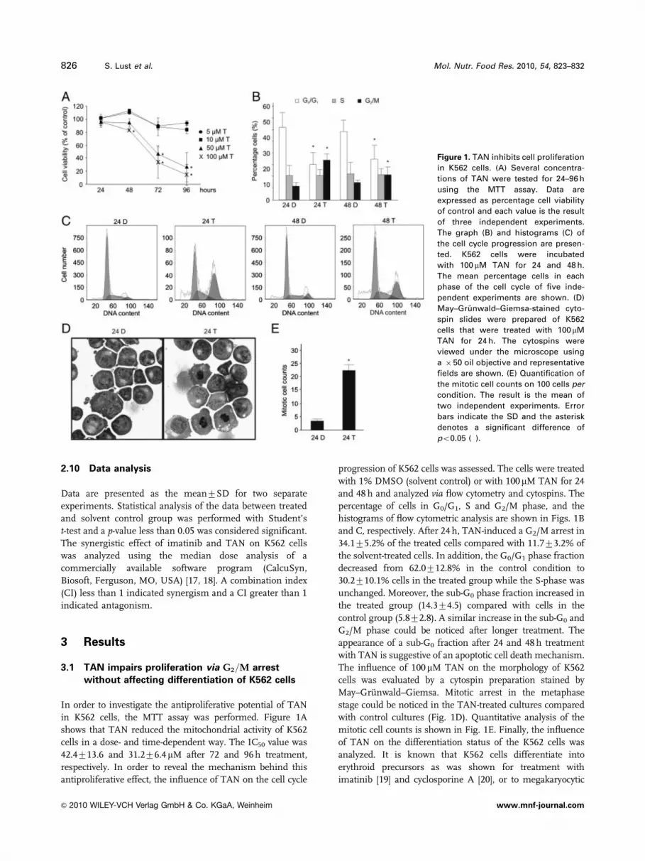
In order to investigate the antiproliferative potential of TAN
in K562 cells, the MTT assay was performed. Figure 1A
shows that TAN reduced the mitochondrial activity of K562
cells in a dose- and time-dependent way. The IC50 value was
42.4713.6 and 31.276.4mM after 72 and 96 h treatment,
respectively. In order to reveal the mechanism behind this
antiproliferative effect, the influence of TAN on the cell cycle
progression of K562 cells was assessed. The cells were treated
with 1% DMSO (solvent control) or with 100mM TAN for 24
and 48 h and analyzed via flow cytometry and cytospins. The
percentage of cells in G0/G1, S and G2/M phase, and the
histograms of flow cytometric analysis are shown in Figs. 1B
and C, respectively. After 24 h, TAN-induced a G2/M arrest in
34.175.2% of the treated cells compared with 11.773.2% of
the solvent-treated cells. In addition, the G0/G1 phase fraction
decreased from 62.0712.8% in the control condition to
30.2710.1% cells in the treated group while the S-phase was
unchanged. Moreover, the sub-G0 phase fraction increased in
the treated group (14.374.5) compared with cells in the
control group (5.872.8). A similar increase in the sub-G0 and
G2/M phase could be noticed after longer treatment. The
appearance of a sub-G0 fraction after 24 and 48 h treatment
with TAN is suggestive of an apoptotic cell death mechanism.
The influence of 100mM TAN on the morphology of K562
cells was evaluated by a cytospin preparation stained by
May–Gr .unwald–Giemsa. Mitotic arrest in the metaphase
stage could be noticed in the TAN-treated cultures compared
with control cultures (Fig. 1D). Quantitative analysis of the
mitotic cell counts is shown in Fig. 1E. Finally, the influence
of TAN on the differentiation status of the K562 cells was
analyzed. It is known that K562 cells differentiate into
erythroid precursors as was shown for treatment with
imatinib [19] and cyclosporine A [20], or to megakaryocytic
Figure 1. TAN inhibits cell proliferation
in K562 cells. (A) Several concentra-
tions of TAN were tested for 24–96 h
using the MTT assay. Data are
expressed as percentage cell viability
of control and each value is the result
of three independent experiments.
The graph (B) and histograms (C) of
the cell cycle progression are presen-
ted. K562 cells were incubated
with 100 mM TAN for 24 and 48 h.
The mean percentage cells in each
phase of the cell cycle of five inde-
pendent experiments are shown. (D)
May–Gr .unwald–Giemsa-stained cyto-
spin slides were prepared of K562
cells that were treated with 100 mM
TAN for 24 h. The cytospins were
viewed under the microscope using
a � 50 oil objective and representative
fields are shown. (E) Quantification of
the mitotic cell counts on 100 cells per
condition. The result is the mean of
two independent experiments. Error
bars indicate the SD and the asterisk
denotes a significant difference of
po0.05 (�).
826 S. Lust et al. Mol. Nutr. Food Res. 2010, 54, 823–832
& 2010 WILEY-VCH Verlag GmbH & Co. KGaA, Weinheim www.mnf-journal.com

lineages after treatment with phorbol 12-myristate 13-acetate
[21]. Therefore, K562 cells were incubated for 4 days with
10mM TAN and stained for glycophorin A (a marker for
erythroid cells), CD33 and CD31 (a marker for monocytic
cells) and CD61 (a marker for megakaryocytes). However, no
stimulation of differentiation could be observed (data not
shown).
3.2 TAN induces apoptosis in K562 cells
The increase in sub-G0 fraction is suggestive of an apopto-
sis-like cell death mechanism. Therefore, we performed
AnnV–PI staining followed by flow cytometry after treat-
ment of K562 cells with 50 or 100mM TAN for 24 and 48 h.
Figure 2A shows that the mean percentage of LC decreased
in a dose- and time-dependent way. Although a minor
downregulation was noticed with 50 mM TAN after 24 h, a
significant decrease could be noticed after 48 h (55.478.7%
LC) compared with solvent control (95.076.8% LC).
However, 100mM TAN induced cell death already after 24 h
(75.379.1% LC for treated culture; 97.5712.3% LC for
control culture) and was more pronounced after 48 h
(44.4711.3% LC for treated culture; 93.875.4% LC for
control culture). Furthermore, we examined other char-
acteristics of an apoptotic cell death mechanism namely
Figure 2. TAN induces cell death via an apoptotic mechanism. (A) The mean percentage LC of six independent experiments was deter-
mined after 24 and 48 h treatment with 50 and 100mM TAN or corresponding solvent control concentration (DMSO). (B) Exposure of K562
cells to 100mM TAN or 1% DMSO for 12, 24 and 48 h resulted in the appearance of a DNA laddering pattern. (C) Lysates were prepared
after different time periods. K562 cells were treated with 100 mM TAN or solvent control (DMSO) and membranes were stained for
procaspase-9, -8, -7, -3, cleaved caspase-3 and tubulin as loading control. A dilution of TAN resulted in cleavage of PARP after 24 h
treatment. One representative blot is shown in (C) and (D). The effect of TAN on the formation of apoptotic bodies was determined via
fluorescence microscopy. K562 cells were treated for 24 h with 100 mM TAN, or solvent and/or pretreated for 1 h with 50 mM Z-VAD-FMK.
After the incubation period, cells were stained with DAPI (E). Simultaneously, the mean percentage cells of three independent experi-
ments in each stage of the cell cycle and the effect on PARP and cleaved caspase-3 were determined, respectively via flow cytometry
(F) and via western blot (G). One representative blot is illustrated. Error bars present the SD and a significant difference of po0.05 (�) is
shown.
Mol. Nutr. Food Res. 2010, 54, 823–832 827
& 2010 WILEY-VCH Verlag GmbH & Co. KGaA, Weinheim www.mnf-journal.com
DNA laddering fragmentation, PARP cleavage, processing
of caspases and cell morphology. TAN-induced apoptosis
was evidenced by DNA laddering fragmentation starting
from 100 mM TAN after 24 and 48 h treatment (Fig. 2B).
Furthermore, as shown in Fig. 2C, 100mM TAN induced
PARP cleavage already after 12 h. After 24 h, PARP cleavage
was considerably enhanced, starting from 75mM (Fig. 2D).
In addition, 100mM TAN resulted in a decrease of procas-
pase-7 and procaspase-3 after 12 h accompanied by a minor
decrease in procaspase-9, which was more apparent after
24 h. In contrast, no activation of caspase-8 could be detected
(Fig. 2C). One of the key morphological features of late stage
apoptosis is the appearance of apoptotic bodies which could
be visualized by a DAPI-staining of K562 cells treated for
24 h with 100 mM TAN (Fig. 2E).
To find out if the effect of TAN on cell death was caspase-
dependent, the pancaspase inhibitor Z-VAD-FMK was used.
Pretreatment with 50 mM Z-VAD-FMK significantly reduced
the sub-G0 population, e.g. 32.974.6% cells in TAN-treated
cells in contrast to 19.475.7% cells in Z-VAD-FMK-
pretreated cells, induced no G2/M arrest and a minor
decrease in G0/G1 phase compared with TAN-treated cells
(Fig. 2F). Moreover, PARP cleavage and caspase-3 activation
were abolished after 24 h of cotreatment (Fig. 2G) but cell
death was not completely inhibited (data not shown). DAPI
staining confirmed these results (Fig. 2E).
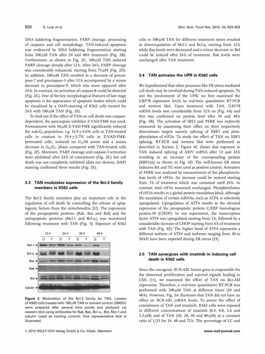
3.3 TAN modulates expression of the Bcl-2 family
members in K562 cells
The Bcl-2 family members play an important role in the
regulation of cell death by controlling the release of apop-
togenic factors from the mitochondria [22]. The expression
of the proapoptotic proteins (Bak, Bax and Bid) and the
antiapoptotic proteins (Mcl-1 and Bcl-xL) was monitored
following treatment with TAN (Fig. 3). Exposure of K562
cells to 100 mM TAN for different treatment times resulted
in downregulation of Mcl-1 and Bcl-xL starting from 12 h
while Bax levels were decreased and a minor decrease in Bid
could be noticed after 24 h of treatment. Bak levels were
unchanged after TAN treatment.
3.4 TAN activates the UPR in K562 cells
We hypothesized that other processes like ER stress-mediated
cell death may be involved during TAN-induced apoptosis. To
test the involvement of the UPR, we first examined the
GRP78 expression levels by real-time quantitative RT-PCR
and western blot. Upon treatment with TAN, GRP78mRNA levels rose considerably from 12 h on (Fig. 4A) and
this was confirmed on protein level after 24 and 48 h
(Fig. 4B). The activation of IRE1 and PERK was indirectly
measured by examining their effect on their respectively
downstream targets namely splicing of XBP1 and phos-
phorylation of eIF2a. To study the effect of TAN on XBP1
splicing, RT-PCR and western blot were performed as
described in Section 2. Figure 4C shows that exposure to
TAN induced splicing of XBP1 mRNA after 12 and 24 h
resulting in an increase of the corresponding protein
(XBP1(s)) as shown in Fig. 4D. The well-known ER stress
inducers BA and TG were used as positive control. Activation
of PERK was analyzed by measurement of the phosphoryla-
tion levels of eIF2a. An increase could be noticed starting
from 3 h of treatment which was sustained until 24 h. In
contrast, total eIF2a remained unchanged. Phosphorylation
of eIF2a results in a global protein translation block, although
the translation of certain mRNAs, such as ATF4, is selectively
upregulated. Upregulation of ATF4 results in the elevated
expression of the proapoptotic protein C/EBP homologous
protein-10 (CHOP). In our experiment, the transcription
factor ATF4 was upregulated starting from 3 h, followed by a
considerable increase of CHOP starting from 6 h of treatment
with TAN (Fig. 4E). The higher band of ATF4 represents a
different isoform of ATF4 and isoforms ranging from 30 to
50 kD have been reported during ER stress [23].
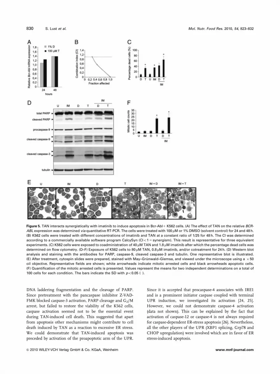
3.5 TAN synergizes with imatinib in inducing cell
death in K562 cells
Since the oncogenic BCR-ABL fusion gene is responsible for
the abnormal proliferation and survival signals leading to
CML [11], we examined the effect of TAN on Bcr-Abl
expression. Therefore, a real-time quantitative RT-PCR was
performed with 100 mM TAN at different times (24 and
48 h). However, Fig. 5A illustrates that TAN did not have an
effect on BCR-ABL mRNA levels. To assess the effect of
cotreatment of TAN and imatinib, K562 cells were exposed
to different concentrations of imatinib (0.4, 0.8, 1.6 and
3.2 mM) and of TAN (10, 20, 40 and 80 mM) at a constant
ratio of 1/25 for 24, 48 and 72 h. The percentage of LC and
Figure 3. Modulation of the Bcl-2 family by TAN. Lysates
of K562 cells treated with 100 mM TAN or solvent control (DMSO)
were prepared after several time points and analyzed via
western blot using antibodies for Bak, Bax, Bcl-xL, Bid, Mcl-1 and
tubulin (used as loading control). One representative blot is
illustrated.
828 S. Lust et al. Mol. Nutr. Food Res. 2010, 54, 823–832
& 2010 WILEY-VCH Verlag GmbH & Co. KGaA, Weinheim www.mnf-journal.com

dead cells ( 5 fraction affected cells) was assessed on flow
cytometry by AnnV–PI double staining. The fraction affec-
ted cells were used to determine the CI via the software
program CalcySyn. CI values lower than 1 corresponded to a
synergistic effect. As such, synergistic interactions between
imatinib and TAN could be demonstrated after 48 h of
treatment (Figs. 5B and C). In addition, the effect of the
cotreatment and the single drugs on caspase activation was
evaluated. Figure 5D shows that imatinib and TAN alone
already induced cleavage of PARP and activation of caspase-
3 and -9 after 24 h. However, cotreatment enhanced the
effect and confirmed the synergistic effect of imatinib and
TAN. Moreover, the effect of both drugs on cell morphology
and the cell cycle were analyzed by May–Gr .unwald–
Giemsa-stained cytospins. Figure 5E shows that after 24 h
imatinib induced clearly detectable morphological changes
of erythroid differentiation of K562 cells compared with
untreated cells, while figures of a mitotic arrest could be
detected in the TAN-treated cells. Cotreatment of TAN and
imatinib induced a mitotic arrest and appearance of apop-
totic bodies in K562 cells. Quantitative analysis of mitotic
cell counts confirmed these results (Fig. 5F).
4 Discussion
Bcr-Abl1 leukemia is characterized by the activation of
different signal transduction pathways that confer a survival
advantage, promote proliferation and migration. New drug
development in this disease primarily focuses on specific
inhibitors of the BCR-ABL fusion gene. Since mutants will
continue to develop, the development of alternatives
remains an important topic. Therefore, interactions between
the Bcr-Abl tyrosine kinase inhibitor, e.g. imatinib and other
potential target candidates are attractive strategies to over-
come imatinib-resistance.
The aim of this study was to investigate the molecular
mechanism of TAN in the Bcr-Abl1 K562 cell line. Previous
reports already demonstrated the antiproliferative potential
of TAN in cancer cells in vitro in colorectal carcinoma cells [6]
and in the promyelocytic leukemia HL-60 cell line [7]. In this
study, we show that TAN suppressed the proliferation of
K562 cells through a G2/M arrest. In addition, it stimulated
the accumulation of cells in the sub-G0 fraction, suggestive
for an apoptotic cell death mechanism. TAN-induced apop-
tosis was confirmed by the processing of caspase-3, -7, -9,
Figure 4. TAN activates the UPR to induce cell death in K562 cells. K562 cells were treated with 100 mM TAN or with 1% DMSO (solvent
control) for different incubation periods. (A) Real-time quantitative RT-PCR was performed to analyze GRP78 mRNA expression. The effect
on GRP78 mRNA level was confirmed via western blot (B). Splicing of the ER stress-induced XBP1 was analyzed via PCR reaction and
western blot. (C) After the amplification reaction, samples were loaded on a 3% agarose gel to distinguish unspliced XBP1 (312 bp) and
spliced XBP1 (286 bp). As positive control K562 cells were treated with 10 mg/mL BA during 24 h. The asterisk (�) represents an aspecific
band of XBP1. (D) K562 cells were treated with TAN and the ER stress inducers BA (10 mg/mL) and TG (1 mM) during 6 h. The membrane
was stained with XBP1. (E) Lysates were further analyzed via western blot for ATF4, CHOP, total and phospho-eIF2a, and GRP78. Tubulin
was used as loading control. One representative gel and blot are shown.
Mol. Nutr. Food Res. 2010, 54, 823–832 829
& 2010 WILEY-VCH Verlag GmbH & Co. KGaA, Weinheim www.mnf-journal.com
DNA laddering fragmentation and the cleavage of PARP.
Since pretreatment with the pancaspase inhibitor Z-VAD-
FMK blocked caspase-3 activation, PARP cleavage and G2/M
arrest, but failed to restore the viability of the K562 cells,
caspase activation seemed not to be the essential event
during TAN-induced cell death. This suggested that apart
from apoptosis other mechanisms might contribute to cell
death induced by TAN as a reaction to excessive ER stress.
We could demonstrate that TAN-induced apoptosis was
preceded by activation of the proapoptotic arm of the UPR.
Since it is accepted that procaspase-4 associates with IRE1
and is a prominent initiator caspase coupled with terminal
UPR induction, we investigated its activation [24, 25].
However, we could not demonstrate caspase-4 activation
(data not shown). This can be explained by the fact that
activation of caspase-12 or caspase-4 is not always required
for caspase-dependent ER-stress apoptosis [26]. Nevertheless,
all the other players of the UPR (XBP1 splicing, Grp78 and
CHOP upregulation) were involved which are in favor of ER
stress-induced apoptosis.
Figure 5. TAN interacts synergistically with imatinib to induce apoptosis in Bcr-Abl1 K562 cells. (A) The effect of TAN on the relative BCR-
ABL expression was determined via quantitative RT-PCR. The cells were treated with 100 mM or 1% DMSO (solvent control) for 24 and 48 h.
(B) K562 cells were treated with different concentrations of imatinib and TAN at a constant ratio of 1/25 for 48 h. The CI was determined
according to a commercially available software program CalcySyn (CIo1 5 synergism). This result is representative for three equivalent
experiments. (C) K562 cells were exposed to coadministration of 40 mM TAN and 1.8 mM imatinib after which the percentage dead cells was
determined on flow cytometry. (D–F) Exposure of K562 cells to 80mM TAN, 0.8mM imatinib, and/or cotreatment for 24 h. (D) Western blot
analysis and staining with the antibodies for PARP, caspase-9, cleaved caspase-3 and tubulin. One representative blot is illustrated.
(E) After treatment, cytospin slides were prepared, stained with May–Gr .unwald–Giemsa, and viewed under the microscope using a � 50
oil objective. Representative fields are shown; white arrowheads indicate mitotic arrested cells and black arrowheads apoptotic cells.
(F) Quantification of the mitotic arrested cells is presented. Values represent the means for two independent determinations on a total of
100 cells for each condition. The bars indicate the SD with po0.05 (�).
830 S. Lust et al. Mol. Nutr. Food Res. 2010, 54, 823–832
& 2010 WILEY-VCH Verlag GmbH & Co. KGaA, Weinheim www.mnf-journal.com

In contrast to our results, TAN is also able to activate the
‘adaptive UPR’ under certain circumstances. Previous research
performed by Takano et al. [27] showed that methoxyflavones
have strong protective effects against ER stress. TAN’s
protective effects were associated with enhanced expression of
UPR target genes (GRP78 and CHOP) and phosphorylation of
eIF2a. The reason why in our study TAN activates the
‘‘terminal UPR’’ may be explained by the dose used in the
experiments, 100mM TAN in contrast to a maximum of 40mM
by Takano et al. Recently, Piwocka et al. [28] demonstrated that
the Bcr-Abl fusion protein is a strong modulator of the ER
calcium release. Expression of Bcr-Abl correlated with a
decreased amount of ER releasable calcium and higher
expression of GRP78 and CHOP compared with non-Bcr-Abl
expressing cells. Therefore, it is not surprising that specific
inhibition of Bcr-Abl with imatinib increased the ER releasable
calcium. Moreover, the ER-associated caspase-12 was activated
during imatinib-induced apoptosis [28, 29].
Furthermore, the potential of ER stress-induced cell death
in Bcr-Abl1 K562 cells and CML was recently highlighted by
the synergism between arsenic trioxide-induced ER stress and
imatinib-induced intrinsic apoptosis where coadministration
resulted in increased upregulation of GRP78, CHOP, ATF6
levels, and processing of caspase- 7, -8 and -9 [30]. Inspired by
these results, we investigated the combination of TAN and
imatinib in K562 cells. We found a synergistic cytotoxic effect
evidenced by an accumulation of apoptotic cells and cells in
mitotic arrest, and stronger effects on caspase activation.
In conclusion, our results suggest that the flavonoid TAN
is an inducer of a G2/M arrest and of cell death, with signs
of both classical mitochondrial caspase-9-mediated and of
ER stress-mediated apoptosis. Furthermore, TAN shows a
synergistic cytotoxic effect combined with imatinib. We
hypothesize that the ER is a genuine target to tackle in Bcr-
Abl1 leukemias and suggest that the synergism of ER stress
inducers and tyrosine kinase inhibitors has potential for
further investigation.
The authors wish to thank Roselien Schelfaut and Jean Roelsvan Kerckvoorde for their excellent technical assistance. BV wassupported by the Vlaamse Liga tegen Kanker (E. VerscheurenFonds) and by the Concerted Research Initiative of the GhentUniversity (GOA project 01G013A7). MVG is a postdoctoralfellow of the Fund for Scientific Research-Flanders (Belgium).Imatinib was kindly provided by Elisabeth Buchdunger(Novartis Pharma, Basel, Switzerland).
The authors have declared no conflict of interest.
5 References
[1] Birt, D. F., Hendrich, S., Wang, W., Dietary agents in cancer
prevention: flavonoids and isoflavonoids. Pharmacol. Ther.
2001, 90, 157–177.
[2] Bracke, M., Vyncke, B., Opdenakker, G., Foidart, J. M. et al.,
Effect of catechins and citrus flavonoids on invasion in vitro.
Clin. Exp. Metastasis 1991, 9, 13–25.
[3] Bracke, M. E., Bruyneel, E., Vermeulen, S. J., Vennekens, K.,
Citrus flavonoid effect on tumor invasion and metastasis.
Food Technol. 1994, 48, 121–124.
[4] Bracke, M. E., Boterberg, T., Depypere, H., Stove, C. et al., in:
Buslig, B., Manthey, J. (Eds.), Flavonoids in Cell Function,
Kluwer Academic/Plenum Publishers, New York 2002,
pp. 135–139.
[5] Chen, K.-H., Weng, M.-S., Lin, J.-K., Tangeretin suppresses
IL-1X-induced cyclooxygenase (COX)-2 expression through
inhibition of p38 MAPK, JNK, and AKT activation in
human lung carcinoma cells. Biochem. Pharmacol. 2007, 73,
215–227.
[6] Pan, M.-H., Chen, W.-J., Lin-Shiau, S.-Y., Ho, C.-T., Lin, J.-K.,
Tangeretin induces cell-cycle G1 arrest through inhibiting
cyclin-dependent kinases 2 and 4 activities as well as
elevating Cdk inhibitors p21 and p27 in human colorectal
carcinoma cells. Carcinogenesis 2002, 23, 1677–1684.
[7] Hirano, T., Abe, K., Gotoh, M., Oka, K., Citrus flavone
tangeretin inhibits leukemic HL-60 cell growth partially
through induction of apoptosis with less cytotoxicity on
normal lymphocytes. Br. J. Cancer 1995, 72, 1380–1388.
[8] Yeh, T.-C., Chiang, P. C., Li, T. K., Hsu, J. L. et al., Genistein
induces apoptosis in human hepatocellular carcinomas via
interaction of endoplasmic reticulum stress and mitochon-
drial insult. Biochem. Pharmacol. 2007, 73, 782–792.
[9] Park, J. W., Woo, K. J., Lee, J. T., Lim, J. H. et al., Resveratrol
induces pro-apoptotic endoplasmic reticulum stress in
human colon cancer cells. Oncol. Rep. 2007, 18, 1269–1273.
[10] Boelens, J., Lust, S., Offner, F., Bracke, M. E., Vanhoecke,
B. W., The endoplasmic reticulum: a target for new anti-
cancer drugs. In Vivo 2007, 21, 215–226.
[11] Druker, B. J., Translation of the Philadelphia chromosome
into therapy for CML. Blood 2008, 112, 4808–4817.
[12] Lust, S., Vanhoecke, B., Van Gele, M., Boelens, J. et al.,
Xanthohumol kills B-chronic lymphocytic leukemia cells by an
apoptotic mechanism. Mol. Nutr. Food Res. 2005, 49, 844–850.
[13] Vandesompele, J., De Paepe, A., Speleman, F., Elimination
of primer-dimer artifacts and genomic coamplification
using a two-step SYBR green I real-time RT-PCR. Anal.
Biochem. 2002, 303, 95–98.
[14] Vandesompele, J., De Preter, K., Pattyn, F., Poppe, B. et al.,
Accurate normalization of real-time quantitative RT-PCR
data by geometric averaging of multiple internal control
genes. Genome Biol. 2002, 18, RESEARCH0034.
[15] Pattyn, F., Speleman, F., De Paepe, A., Vandesompele, J.,
RTPrimerDB: the real-time PCR primer and probe database.
Nucleic Acids Res. 2003, 31, 122–123.
[16] Ding, L., Yan, J., Zhu, J., Zhong, H. et al., Ligand-independent
activation of estrogen receptor a by XBP-1. Nucleic Acids Res.
2003, 31, 5266–5274.
[17] Chou, T.-C., Talalay, P., Quantitative analysis of dose-effect
relationships: the combined effects of multiple drugs or
enzyme inhibitors. Adv. Enzyme Regul. 1984, 22, 27–55.
Mol. Nutr. Food Res. 2010, 54, 823–832 831
& 2010 WILEY-VCH Verlag GmbH & Co. KGaA, Weinheim www.mnf-journal.com

[18] Chou, T.-C., in: Chou, T.-C., Rideout, D. C. (Eds.), Synergism
and Antagonism in Chemotherapy, Academic Press, San
Diego 1991, pp. 61–102.
[19] Jacquel, A., Herrant, M., Legros, L., Belhacene, N. et al.,
Imatinib induces mitochondria-dependent apoptosis
of the Bcr-Abl-positive K562 cell line and its differentia-
tion toward the erythroid lineage. FASEB J. 2003, 17,
2160–2162.
[20] Sawafuji, K., Miyakawa, Y., Kizaki, M., Ikeda, Y., Cyclosporin
A induces erythroid differentiation of K562 cells through
p38 MAPK and ERK pathways. Am. J. Hematol. 2003, 72,
67–69.
[21] Herrera, R., Hubbell, S., Decker, S., Petruzzelli, L., A role for
the MEK/MAPK pathway in PMA-induced cell cycle arrest:
modulation of megakaryocytic differentiation of K562 cells.
Exp. Cell Res. 1998, 238, 407–414.
[22] Danial, N. N., BCL-2 family proteins: critical checkpoints of
apoptotic cell death. Clin. Cancer Res. 2007, 13, 7254–7263.
[23] Denoyelle, C., Abou-Rjaily, G., Bezrookove, V., Verhaegen, M.
et al., Anti-oncogenic role of the endoplasmic reticulum
differentially activated by mutations in the MAPK pathway.
Nat. Cell. Biol. 2006, 8, 1053–1063.
[24] Nakagawa, T., Zhu, H., Morishima, N., Li, E. et al., Caspase-
12 mediates endoplasmic-reticulum-specific apoptosis and
cytotoxicity by amyloid-X. Nature 2000, 403, 98–103.
[25] Hitomi, J., Katayama, T., Eguchi, Y., Kudo, T. et al., Invol-
vement of caspase-4 in endoplasmic reticulum stress-
induced apoptosis and AX-induced cell death. J. Cell Biol.
2004, 165, 347–356.
[26] Obeng, E. A., Boise, L. H., Caspase-12 and caspase-4 are not
required for caspase-dependent endoplasmic reticulum
stress-induced apoptosis. J. Biol. Chem. 2005, 280,
29578–29587.
[27] Takano, K., Tabata, Y., Kitao, Y., Murakami, R. et al., Meth-
oxyflavones protect cells against endoplasmic reticulum
stress and neurotoxin. Am. J. Physiol. Cell Physiol. 2007, 92,
C353–C361.
[28] Piwocka, K., Vejda, S., Cotter, T. G., O’Sullivan, G. C.,
McKenna, S. L., Bcr-Abl reduces endoplasmic reticulum
releasable calcium levels by a Bcl-2-independent mechan-
ism and inhibits calcium-dependent apoptotic signaling.
Blood 2006, 107, 4003–4010.
[29] Pattacini, L., Mancini, M., Mazzacurati, L., Brusa, G. et al.,
Endoplasmic reticulum stress initiates apoptotic death
induced by STI571 inhibition of p210 bcr-abl tyrosine
kinase. Leuk. Res. 2004, 28, 191–202.
[30] Du, Y., Wang, K., Fang, H., Li, J. et al., Coordination of
intrinsic, extrinsic, and endoplasmic reticulum-mediated
apoptosis by imatinib mesylate combined with arsenic triox-
ide in chronic myeloid leukemia. Blood 2006, 107, 1582–1590.
832 S. Lust et al. Mol. Nutr. Food Res. 2010, 54, 823–832
& 2010 WILEY-VCH Verlag GmbH & Co. KGaA, Weinheim www.mnf-journal.com