
Rb-Raf-1 interaction disruptor RRD-251 induces apoptosis in metastatic melanoma cells and synergizes...
18
Rb-Raf-1 interaction disruptor RRD-251 induces apoptosis in metastatic melanoma cells and synergizes with dacarbazine Sandeep Singh # , Rebecca Davis # , Vignesh Alamanda, Roberta Pireddu, Daniel Pernazza, Said Sebti, Nicholas Lawrence, and Srikumar Chellappan * Drug Discovery Department, H. Lee Moffitt Cancer Center and Research Institute, Tampa, FL-33612 Abstract Metastatic melanoma is an aggressive cancer with very low response rate against conventional chemotherapeutic agents such as dacarbazine (DTIC). Inhibitor of Rb-Raf-1 interaction (RRD-251) was tested against the melanoma cell lines SK-MEL-28, SK-MEL-5 and SK-MEL-2. RRD-251 was found to be a potent inhibitor of melanoma cell proliferation, irrespective of V600E B-Raf mutation status of the cell lines. In a SK-MEL-28 xenograft experiment, RRD-251 exerted a significant suppression of tumor growth compared to vehicle (p=0.003). Similar to in vitro effects, tumors from RRD-251 treated animals showed decreased Rb-Raf-1 interaction in vivo. Growth suppressive effects of RRD-251 were associated with induction of apoptosis as well as a G 1 arrest, with an accompanying decrease in S-phase cells. RRD-251 inhibited Rb phosphorylation, and downregulated E2F1 protein levels in these cells. Real-time PCR analysis showed that RRD-251 caused downregulation of cell cycle regulatory genes thymidylate synthase (TS) and cdc6 as well as anti-apoptotic gene Mcl-1. Combinatorial treatment of RRD-251 and DTIC resulted in a significantly higher apoptosis in DTIC resistant cell lines SK-MEL-28 and SK-MEL-5, as revealed by increased Caspase-3 activity and PARP cleavage. Since aberrant Rb/E2F pathway is associated with melanoma progression and resistance to apoptosis, these results suggest that the Rb-Raf-1 inhibitor could be an effective agent for melanoma treatment, either alone or in combination with DTIC. Keywords Rb-E2F pathway; Melanoma; Apoptosis; Cell cycle; Dacarbazine INTRODUCTION The incidence of melanoma is continuously rising and has increased more than six fold over the last 50 years (1,2). Melanoma is highly resistant to the conventional chemotherapeutic agent dacarbazine (DTIC) and has a response rate of only 15-20% with the median response duration of only four months (3-5). In addition, the new treatments have also failed to significantly improve the survival time (6-8). Thus, it is essential to identify new therapeutic targets for better treatment of the disease. Several studies have suggested that in human melanoma cells all three Rb family pocket proteins (pRb, p107 and p130) are hyper-phosphorylated and several E2F family members * Corresponding Author: Srikumar Chellappan, [email protected], Phone: (813) 745-6892. # These authors contributed equally to this paper The authors have no conflicts of interest to disclose. NIH Public Access Author Manuscript Mol Cancer Ther. Author manuscript; available in PMC 2011 December 7. Published in final edited form as: Mol Cancer Ther. 2010 December ; 9(12): 3330–3341. doi:10.1158/1535-7163.MCT-10-0442. NIH-PA Author Manuscript NIH-PA Author Manuscript NIH-PA Author Manuscript
-
Upload
independent -
Category
Documents
-
view
2 -
download
0
Transcript of Rb-Raf-1 interaction disruptor RRD-251 induces apoptosis in metastatic melanoma cells and synergizes...
Rb-Raf-1 interaction disruptor RRD-251 induces apoptosis inmetastatic melanoma cells and synergizes with dacarbazine
Sandeep Singh#, Rebecca Davis#, Vignesh Alamanda, Roberta Pireddu, Daniel Pernazza,Said Sebti, Nicholas Lawrence, and Srikumar Chellappan*Drug Discovery Department, H. Lee Moffitt Cancer Center and Research Institute, Tampa,FL-33612
AbstractMetastatic melanoma is an aggressive cancer with very low response rate against conventionalchemotherapeutic agents such as dacarbazine (DTIC). Inhibitor of Rb-Raf-1 interaction(RRD-251) was tested against the melanoma cell lines SK-MEL-28, SK-MEL-5 and SK-MEL-2.RRD-251 was found to be a potent inhibitor of melanoma cell proliferation, irrespective of V600EB-Raf mutation status of the cell lines. In a SK-MEL-28 xenograft experiment, RRD-251 exerted asignificant suppression of tumor growth compared to vehicle (p=0.003). Similar to in vitro effects,tumors from RRD-251 treated animals showed decreased Rb-Raf-1 interaction in vivo. Growthsuppressive effects of RRD-251 were associated with induction of apoptosis as well as a G1 arrest,with an accompanying decrease in S-phase cells. RRD-251 inhibited Rb phosphorylation, anddownregulated E2F1 protein levels in these cells. Real-time PCR analysis showed that RRD-251caused downregulation of cell cycle regulatory genes thymidylate synthase (TS) and cdc6 as wellas anti-apoptotic gene Mcl-1. Combinatorial treatment of RRD-251 and DTIC resulted in asignificantly higher apoptosis in DTIC resistant cell lines SK-MEL-28 and SK-MEL-5, asrevealed by increased Caspase-3 activity and PARP cleavage. Since aberrant Rb/E2F pathway isassociated with melanoma progression and resistance to apoptosis, these results suggest that theRb-Raf-1 inhibitor could be an effective agent for melanoma treatment, either alone or incombination with DTIC.
KeywordsRb-E2F pathway; Melanoma; Apoptosis; Cell cycle; Dacarbazine
INTRODUCTIONThe incidence of melanoma is continuously rising and has increased more than six fold overthe last 50 years (1,2). Melanoma is highly resistant to the conventional chemotherapeuticagent dacarbazine (DTIC) and has a response rate of only 15-20% with the median responseduration of only four months (3-5). In addition, the new treatments have also failed tosignificantly improve the survival time (6-8). Thus, it is essential to identify new therapeutictargets for better treatment of the disease.
Several studies have suggested that in human melanoma cells all three Rb family pocketproteins (pRb, p107 and p130) are hyper-phosphorylated and several E2F family members
*Corresponding Author: Srikumar Chellappan, [email protected], Phone: (813) 745-6892.#These authors contributed equally to this paperThe authors have no conflicts of interest to disclose.
NIH Public AccessAuthor ManuscriptMol Cancer Ther. Author manuscript; available in PMC 2011 December 7.
Published in final edited form as:Mol Cancer Ther. 2010 December ; 9(12): 3330–3341. doi:10.1158/1535-7163.MCT-10-0442.
NIH
-PA Author Manuscript
NIH
-PA Author Manuscript
NIH
-PA Author Manuscript
(E2F1, E2F2, E2F3 and E2F4) are present in unbound, transcriptionally active forms (9-13)The retinoblastoma tumor suppressor protein, Rb, plays a vital role in regulating mammaliancell proliferation primarily by its interaction with the E2F family of transcription factors(14,15). Rb binds to E2Fs 1, 2, and 3 and suppresses their transcriptional activity, inhibitingtheir ability to drive the expression of proliferative promoters (16,17). In response tomitogenic signals, Rb is hyper-phosphorylated through a cascade of phosphorylation events,which leads to its inactivation and dissociation from E2Fs. Free E2Fs can activelyparticipate in the transcription of target genes such as cdc6 and thymidylate synthase (TS),facilitating S-phase entry and cell cycle progression (17).
Several kinases can phosphorylate Rb. For example, cyclin-dependent kinases such asCDK4 and CDK2 phosphorylate Rb and this is essential for the G1 to S phase transition(18). Recently, we had shown that the signaling kinase Raf-1 can physically bind andphosphorylate Rb very early in the cell cycle, facilitating its further hyper-phosphorylationby CDKs and eventual inactivation (19,20). We have previously reported that the disruptionof Rb-Raf-1 interaction by an eight-amino acid peptide (corresponding to Raf-1 residues10-18) prevented Rb phosphorylation even in the later stages of G1. This suggests thatpreventing the binding of Raf-1 to Rb keeps Rb in a functional, hypophosphorylated formwith its tumor suppressor role intact (20). Our attempts to identify a small moleculedisruptor of Rb-Raf-1 interaction resulted in the characterization of RRD-251, which hadpotent anti-proliferative, anti-angiogenic and anti-tumor activity against non-small cell lungcarcinoma cells in vitro and in vivo (21). Here we have explored the efficacy of RRD-251 intargeting melanoma cells SK-MEL-28, SK-MEL-5 and SK-MEL-2.
We find that RRD-251 can inhibit the growth of melanoma cells by induction of apoptosisas well as cell cycle arrest. We further show that pre-treatment of cells with DTIC enhancedthe pro-apoptotic effects of RRD-251 on these cells. Overall, these data suggest thatRRD-251 possesses potent anticancer activity against melanoma in vitro and in vivo.Furthermore, it appears that the combination of DTIC and RRD-251 would be a viablestrategy to combat the growth and progression of melanoma.
MATERIALS and METHODSCell lines and reagents
The metastatic melanoma cell lines, SK-MEL-28, SK-MEL-5 and SK-MEL-2 werepurchased from ATCC (Manassas, VA) and cultured in MEM containing 10% fetal bovineserum (FBS; Mediatech) and maintained in 5% CO2 at 37 °C. While these cell lines wereoriginally purchased from ATCC, we did not revalidate them. DTIC was obtained fromSigma and 100 mM stock solution was prepared by dissolving the drug in DMSO. RRD-251was synthesized by our in-house chemistry laboratory. It is a benzyl-isothiourea compoundwith chloride as the counter ion (19). The chemical structures of RRD-251 and DTIC areshown in Supplementary Figure 1. Unless not indicated, cells were treated with 50 μmol/Lconcentration of DTIC and RRD-251. Primary antibodies against PARP and Caspase-3 wereobtained from Cell Signaling Technology, Rb and Raf-1 were obtained from BDTransduction Laboratories and Mcl-1, Bcl-2 and Bax were obtained from Santa CruzBiotechnology.
Proliferation and soft agar colony formation assayActively growing human melanoma cells were plated in 96-well plates at a density of 7500cells/well in triplicates. After 24 hr, cells were treated with 20 and 50 μmol/L concentrationsof RRD-251 or the solvent (DMSO). Cell proliferation assay was carried out with MTT(Thiazolyl Blue Tetrazolium Bromide) after 24 hr of drug treatment. Briefly, cells were
Singh et al. Page 2
Mol Cancer Ther. Author manuscript; available in PMC 2011 December 7.
NIH
-PA Author Manuscript
NIH
-PA Author Manuscript
NIH
-PA Author Manuscript
incubated with the 1 mg/mL MTT solution at 37 °C for 1 hr. The reaction was terminatedwith DMSO that solubilizes the formazan product formed. Absorbance at 540 nm wasrecorded using plate reader. A soft agar colony-formation assay was used as described byKinkade et. al. 2008 (21). Over the bottom agarose (0.5%), 5,000 logarithmically growingmelanoma cells were mixed with 0.3% agarose along with the indicated doses of RRD-251.Drugs were added twice weekly in complete media to the respective wells. After incubatingfor 14 days, colonies were stained with MTT (1 mg/mL) for 1 hr at 37°C. The experimentwas repeated twice in duplicate.
In vivo xenograft experiments in nude miceSK-MEL-28 cells were harvested in PBS and mixed with equal volume of Matrigel (BDBiosciences, Bedford, MA) and implanted subcutaneously into the right and left flanks (10 ×106 cells per flank) of 8-wk-old female athymic nude mice (Charles River) as describedpreviously (21). When xenograft tumor growth was established, treatment by intra-peritoneal injection of 0.1 ml of the drug or the vehicle was initiated. Tumor volumes weredetermined by measuring the length (L) and the width (W) and calculating the volume (V =L × W2 / 2). Statistical significance between control and treated animals were evaluatedusing Student’s t test.
Cell cycle analysis by flow cytometryCells were treated with indicated dose of RRD-251 for given time. Following treatment,cells were scrapped and collected. Cell pellet was washed in PBS and resuspended in 0.1 mlof citrate/DMSO buffer (250 mmol/L sucrose, 40 mmol/L Na3C6H5O7.2H2O, 5% DMSO,pH 7.6). The pellets were then frozen at −80°C. Samples were processed as suggested inVindelov method and cell cycle analysis was performed by flow cytometry (22).
Lysate preparation, immunoprecipitation and Western blottingCell lysates were prepared by lysis-buffer containing NP40, as described earlier (20) (21).Tumor lysates were prepared with T-Per tissue lysis buffer (Pierce) and a Fisher PowerGen125 dounce homogenizer (21). Physical interaction between proteins in the tumors wasanalyzed by immunoprecipitation–western blot experiments using 200 μg of lysate and 1 μgof the indicated antibody (20).
Real-time PCRMelanoma cells were treated with RRD-251 for indicated time. DMSO treated cells wereused as vehicle-control. Total RNA was isolated by an RNeasy miniprep kit from QIAGENfollowing the manufacturer’s protocol. One microgram of RNA was used for first-strandcDNA synthesis using the iScript cDNA synthesis kit (Bio-Rad). A fraction (1/20) of thefinal cDNA reaction volume was used in each PCR. Primer sequences are as follows: 5’-CTG CCA GCT GTA CCA GAG AT-3’ (TS forward primer), 5’-ATG TGC ATC TCCCAA AGT GT-3’ (TS reverse primer), 5’-CCC CAT GAT TGT GTT GGT AT-3’ (Cdc6forward primer), 5’-TTC AAC AGC TGT GGC TTA CA-3’ (Cdc6 reverse primer), 5’-ATGCTT CGG AAA CTG GAC AT-3’ (Mcl-1 forwad primer), 5’-TCC TGA TGC CAC CTTCTA GG-3’ (Mcl-1 reverse primer), 5’-CTC AAC ACG GGA AAC CTC AC-3’ (18Sforward primer), and 5’-AAA TCG CTC CAC CAA CTA AGA A-3’ (18S reverse primer).Real-time PCR was performed on a Bio-Rad iCycler and analyzed by ddCt method.
Caspase 3/7 activity assay and TUNEL assayCells were grown in eight well chamber slides and treated with RRD-251 for 24 hr. TUNELstaining was performed using DeadEnd™ TUNEL Assay kit (Promega) as per themanufacture’s instructions. Actively growing melanoma cells were plated in clear bottom
Singh et al. Page 3
Mol Cancer Ther. Author manuscript; available in PMC 2011 December 7.
NIH
-PA Author Manuscript
NIH
-PA Author Manuscript
NIH
-PA Author Manuscript
96-well plates at a density of 7500 cells/well in triplicates. After 24 hr, cells were treatedwith 50 μmol/L concentrations of DTIC or the solvent (DMSO). 48 hr post-DTIC treatment,RRD-251 was added in the respective wells and further incubated for 4 hr. Caspase activitywere measured by quantitative luminescence assays using caspase-3/7–activatable DEVD-aminoluciferin (Caspase-Glo 3/7 kit, Promega). Assays were done according to themanufacturer’s protocol and plates were read in a luminometer (Perkin Elmer).
Comet assayThe Comet assay was performed in its alkaline version following published protocols(23,24). Cells were treated with 50 μmol/L DTIC for 48 hr. Afffter treatment, cells weremixed with 0.8% (w/v) low melting point agarose and the mixture spread onto two slidespre-coated with 1.5% (w/v) normal melting point agarose. The slides were covered withcoverslips and were refrigerated for 10 min to solidify the agarose. Next, the coverslips wereremoved and the slides were immersed in lysis solution (2.5 Mol/L NaCl, 100 mmol/LEDTA, 10 mmol/L Tris-HCl (pH 10) containing 1% (v/v) Triton X-100) for 60 min at 4 °C.Subsequently, the slides were placed in an alkaline buffer (1 mmol/L EDTA, 300 mmol/LNaOH, pH > 13) for 20 min at 4 °C for the DNA to unwind. For electrophoresis slides wereequalibarated in 1× TBE for 10 minutes following electrophoresis in the same buffer at 0.52V/cm for 5 min. Slides were washed in water, dried at room temperature and fixed in 100%ethanol for 5 min. The slides were stained with 80 μL of 2 μg/ml ethidium bromide andrinsed with water. Randomly selected cells from each sample and %DNA damage wasanalyzed using LAI Automated Comet Assay Analysis System (Loats Associates Inc.,Westminster, MD).
RESULTSIn-vitro and in-vivo anti-melanoma effects of RRD-251
Melanoma cells harbor persistently hyperphosphorylated Rb proteins and this appears to beassociated with melanoma progression and resistance to apoptosis. We, therefore,investigated the effects of Raf/Rb disruptor RRD-251 on melanoma tumors. As a first step,the effect of RRD-251 on cell survival and anchorage-dependent cell growth was tested by aMTT assay. SK-MEL-28, SK-MEL-5 and SK-MEL-2 cells were grown in the absence orpresence of increasing concentrations (10, 20 and 50 μmol/L) of RRD-251 for 24 hr.RRD-251 treatment resulted in a dose dependent decrease in cell survival in all the three celllines (Fig. 1A). The IC50 value for SK-MEL-28, SK-MEL-5 (both carrying a V600E mutantB-Raf) and SK-MEL-2 (carrying wild-type B-Raf) were found to be 28.7μmol/L, 37.3μmol/L and 48 μmol/L respectively. Next, the effect of RRD-251 on the anchorage-independentgrowth of these cell lines in soft agar was examined. As shown in Fig. 1B, RRD-251 couldsignificantly suppress anchorage-independent colony formation of SK-MEL-28 and SK-MEL-5 and SK-MEL-2 cells in a dose dependent manner. These data show that RRD-251treatment produces strong inhibition of viability and proliferation of melanoma cells,irrespective of their B-Raf status.
Given the ability of RRD-251 to exert anti-proliferative effects in cultured cells, we nextexamined whether RRD-251 could inhibit tumor growth in vivo in nude mouse xenograftmodels. SK-MEL-28 cells mixed with matrigel were implanted by subcutaneous injection innude mice, and the tumors were allowed to reach an average size of 376 mm3 in size beforeintraperitoneal (i.p.) administration of RRD-251 or vehicle. As shown in Fig. 1C, tumorsfrom vehicle treated mice grew to an average size of 733±86 mm3. In contrast, tumors didnot grow significantly (average tumor size at the end of treatment was 362±34 mm3) in micetreated with RRD-251. This anti-tumor effect of RRD-251 was statistically significant after9 days of treatment and remained significant throughout the duration of the experimental
Singh et al. Page 4
Mol Cancer Ther. Author manuscript; available in PMC 2011 December 7.
NIH
-PA Author Manuscript
NIH
-PA Author Manuscript
NIH
-PA Author Manuscript
period. The body weight of vehicle or RRD-251 treated mice were not affected (Fig. 1D).This set of experiments suggests that RRD-251 has anti-cancer activities in vitro and in vivoon melanomas.
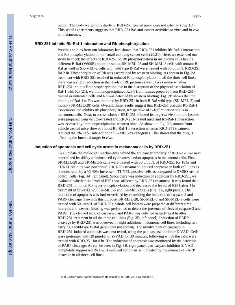
RRD-251 inhibits Rb-Raf-1 interaction and Rb-phosphorylationPrevious studies from our laboratory had shown that RRD-251 inhibits Rb-Raf-1 interactionand Rb phosphorylation in non-small cell lung cancer cells (20,21). Here, we extended ourstudy to check the effects of RRD-251 on Rb phosphorylation in melanoma cells havingdifferent B-Raf (V600E) mutation status. SK-MEL-28 and SK-MEL-5 cells with mutant B-Raf as well as SK-MEL-2 cells with wild type B-Raf were treated with 50 μmol/L RRD-251for 2 hr. Phosphorylation of Rb was ascertained by western blotting. As shown in Fig. 2A,treatment with RRD-251 resulted in reduced Rb phosphorylation in all the three cell lines;there was a slight reduction in the levels of Rb protein as well. To examine whetherRRD-251 inhibits Rb phosphorylation due to the disruption of the physical association ofRaf-1 with Rb (21), we immunoprecipitated Raf-1 from lysates prepared from RRD-251-treated or untreated cells and Rb was detected by western blotting. Fig. 2B shows that thebinding of Raf-1 to Rb was inhibited by RRD-251 in both B-Raf wild type (SK-MEL-2) andmutant (SK-MEL-28) cells. Overall, these results suggest that RRD-251 disrupts Rb-Raf-1association and inhibits Rb phosphorylation, irrespective of B-Raf mutation status inmelanoma cells. Next, to assess whether RRD-251 affected its target in vivo, tumors lysateswere prepared from vehicle-treated and RRD-251-treated mice and Rb-Raf-1 interactionwas assessed by immunoprecipitation-western blots. As shown in Fig. 2C, tumors fromvehicle treated mice showed robust Rb-Raf-1 interaction whereas RRD-251 treatmentreduced the Rb-Raf-1 interaction in SK-MEL-28 xenografts. This shows that the drug isaffecting the intended target in vivo.
Induction of apoptosis and cell cycle arrest in melanoma cells by RRD-251To elucidate the molecular mechanisms behind the anticancer property of RRD-251, we nextdetermined its ability to induce cell cycle arrest and/or apoptosis in melanoma cells. First,SK-MEL-28 and SK-MEL-5 cells were treated with 50 μmol/L of RRD-251 for 18 hr andTUNEL staining was performed. RRD-251 treatment induced apoptosis in both cell lines asdemonstrated by a 30-40% increase in TUNEL-positive cells as compared to DMSO treatedcontrol cells (Fig. 3A, left panel). Since there was induction of apoptosis by RRD-251, weevaluated whether the level of E2F1 was affected by RRD-251 treatment. It was found thatRRD-251 inhibited Rb hyper-phosphorylation and decreased the levels of E2F1 after 4 hrtreatment in SK-MEL-28, SK-MEL-5 and SK-MEL-2 cells (Fig. 3A, right panel). Theinduction of apoptosis was further verified by examining the induction of caspase-3 andPARP cleavage. Towards this purpose, SK-MEL-28, SK-MEL-5 and SK-MEL-2 cells weretreated with 50 μmol/L of RRD-251; whole cell lysates were prepared at different timeintervals and western blotting was performed to detect the presence of cleaved caspase-3 andPARP. The cleaved band of caspase-3 and PARP was detected as early as 4 hr afterRRD-251 treatment in all the three cell lines (Fig. 3B, left panel). Induction of PARPcleavage by RRD-251 was observed in eight additional melanoma cell lines, including twocarrying a wild-type B-Raf gene (data not shown). The involvement of caspases inRRD-251-induced apoptosis was next tested, using the pan-caspase inhibitor Z-VAD. Cellswere pretreated with 20 μmol/L of Z-VAD for 30 minutes, following which the cells weretreated with RRD-251 for 8 hr. The induction of apoptosis was monitored by the detectionof PARP cleavage. As can be seen in Fig. 3B, right panel, pan-caspase inhibitor Z-VADcompletely suppressed RRD-251-induced apoptosis as indicated by the absence of PARPcleavage in all three cell lines.
Singh et al. Page 5
Mol Cancer Ther. Author manuscript; available in PMC 2011 December 7.
NIH
-PA Author Manuscript
NIH
-PA Author Manuscript
NIH
-PA Author Manuscript
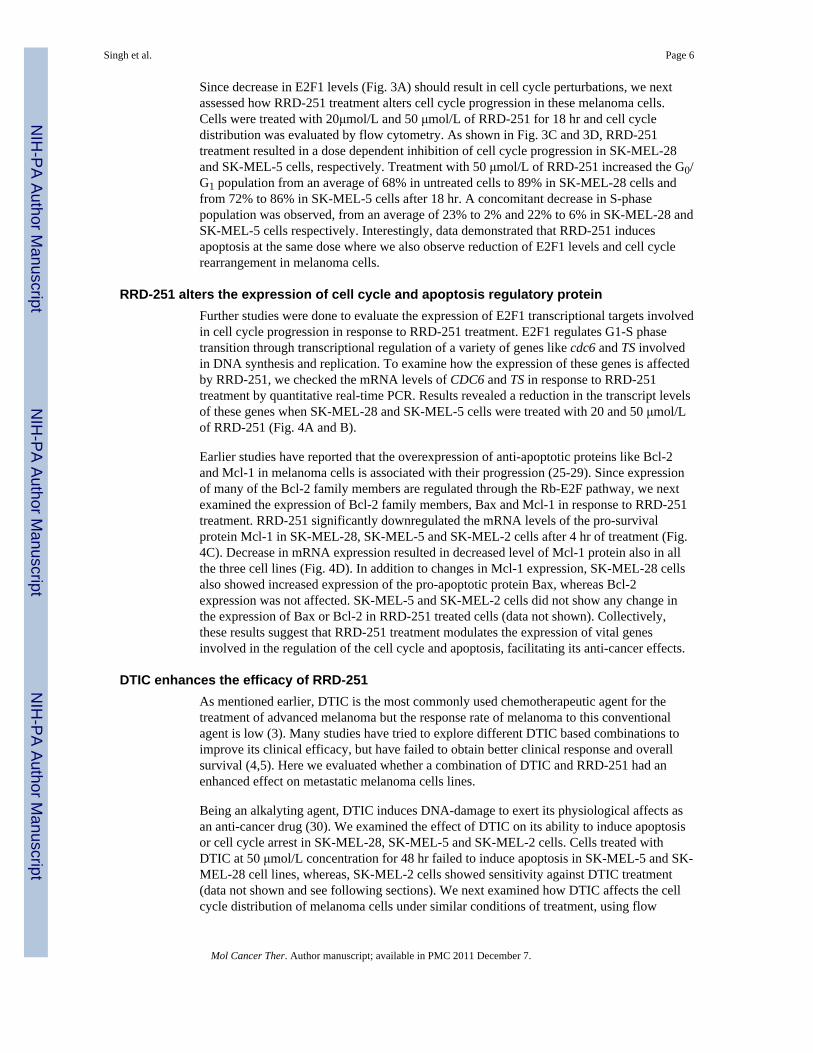
Since decrease in E2F1 levels (Fig. 3A) should result in cell cycle perturbations, we nextassessed how RRD-251 treatment alters cell cycle progression in these melanoma cells.Cells were treated with 20μmol/L and 50 μmol/L of RRD-251 for 18 hr and cell cycledistribution was evaluated by flow cytometry. As shown in Fig. 3C and 3D, RRD-251treatment resulted in a dose dependent inhibition of cell cycle progression in SK-MEL-28and SK-MEL-5 cells, respectively. Treatment with 50 μmol/L of RRD-251 increased the G0/G1 population from an average of 68% in untreated cells to 89% in SK-MEL-28 cells andfrom 72% to 86% in SK-MEL-5 cells after 18 hr. A concomitant decrease in S-phasepopulation was observed, from an average of 23% to 2% and 22% to 6% in SK-MEL-28 andSK-MEL-5 cells respectively. Interestingly, data demonstrated that RRD-251 inducesapoptosis at the same dose where we also observe reduction of E2F1 levels and cell cyclerearrangement in melanoma cells.
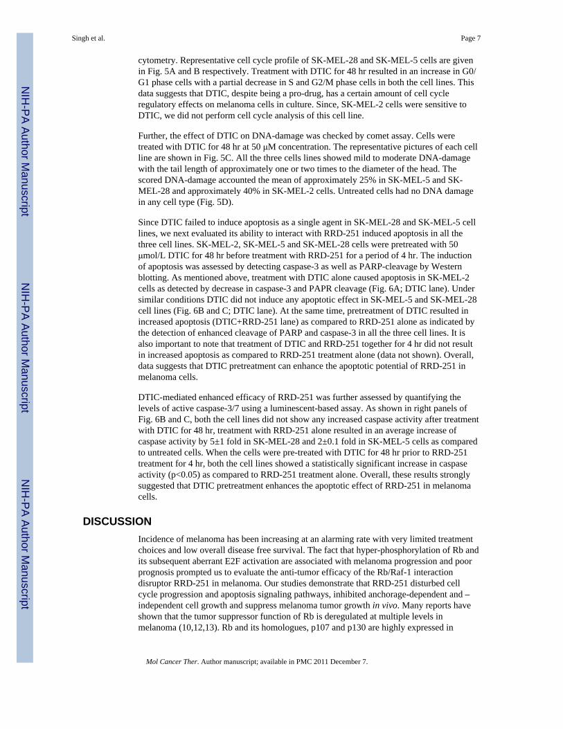
RRD-251 alters the expression of cell cycle and apoptosis regulatory proteinFurther studies were done to evaluate the expression of E2F1 transcriptional targets involvedin cell cycle progression in response to RRD-251 treatment. E2F1 regulates G1-S phasetransition through transcriptional regulation of a variety of genes like cdc6 and TS involvedin DNA synthesis and replication. To examine how the expression of these genes is affectedby RRD-251, we checked the mRNA levels of CDC6 and TS in response to RRD-251treatment by quantitative real-time PCR. Results revealed a reduction in the transcript levelsof these genes when SK-MEL-28 and SK-MEL-5 cells were treated with 20 and 50 μmol/Lof RRD-251 (Fig. 4A and B).
Earlier studies have reported that the overexpression of anti-apoptotic proteins like Bcl-2and Mcl-1 in melanoma cells is associated with their progression (25-29). Since expressionof many of the Bcl-2 family members are regulated through the Rb-E2F pathway, we nextexamined the expression of Bcl-2 family members, Bax and Mcl-1 in response to RRD-251treatment. RRD-251 significantly downregulated the mRNA levels of the pro-survivalprotein Mcl-1 in SK-MEL-28, SK-MEL-5 and SK-MEL-2 cells after 4 hr of treatment (Fig.4C). Decrease in mRNA expression resulted in decreased level of Mcl-1 protein also in allthe three cell lines (Fig. 4D). In addition to changes in Mcl-1 expression, SK-MEL-28 cellsalso showed increased expression of the pro-apoptotic protein Bax, whereas Bcl-2expression was not affected. SK-MEL-5 and SK-MEL-2 cells did not show any change inthe expression of Bax or Bcl-2 in RRD-251 treated cells (data not shown). Collectively,these results suggest that RRD-251 treatment modulates the expression of vital genesinvolved in the regulation of the cell cycle and apoptosis, facilitating its anti-cancer effects.
DTIC enhances the efficacy of RRD-251As mentioned earlier, DTIC is the most commonly used chemotherapeutic agent for thetreatment of advanced melanoma but the response rate of melanoma to this conventionalagent is low (3). Many studies have tried to explore different DTIC based combinations toimprove its clinical efficacy, but have failed to obtain better clinical response and overallsurvival (4,5). Here we evaluated whether a combination of DTIC and RRD-251 had anenhanced effect on metastatic melanoma cells lines.
Being an alkalyting agent, DTIC induces DNA-damage to exert its physiological affects asan anti-cancer drug (30). We examined the effect of DTIC on its ability to induce apoptosisor cell cycle arrest in SK-MEL-28, SK-MEL-5 and SK-MEL-2 cells. Cells treated withDTIC at 50 μmol/L concentration for 48 hr failed to induce apoptosis in SK-MEL-5 and SK-MEL-28 cell lines, whereas, SK-MEL-2 cells showed sensitivity against DTIC treatment(data not shown and see following sections). We next examined how DTIC affects the cellcycle distribution of melanoma cells under similar conditions of treatment, using flow
Singh et al. Page 6
Mol Cancer Ther. Author manuscript; available in PMC 2011 December 7.
NIH
-PA Author Manuscript
NIH
-PA Author Manuscript
NIH
-PA Author Manuscript
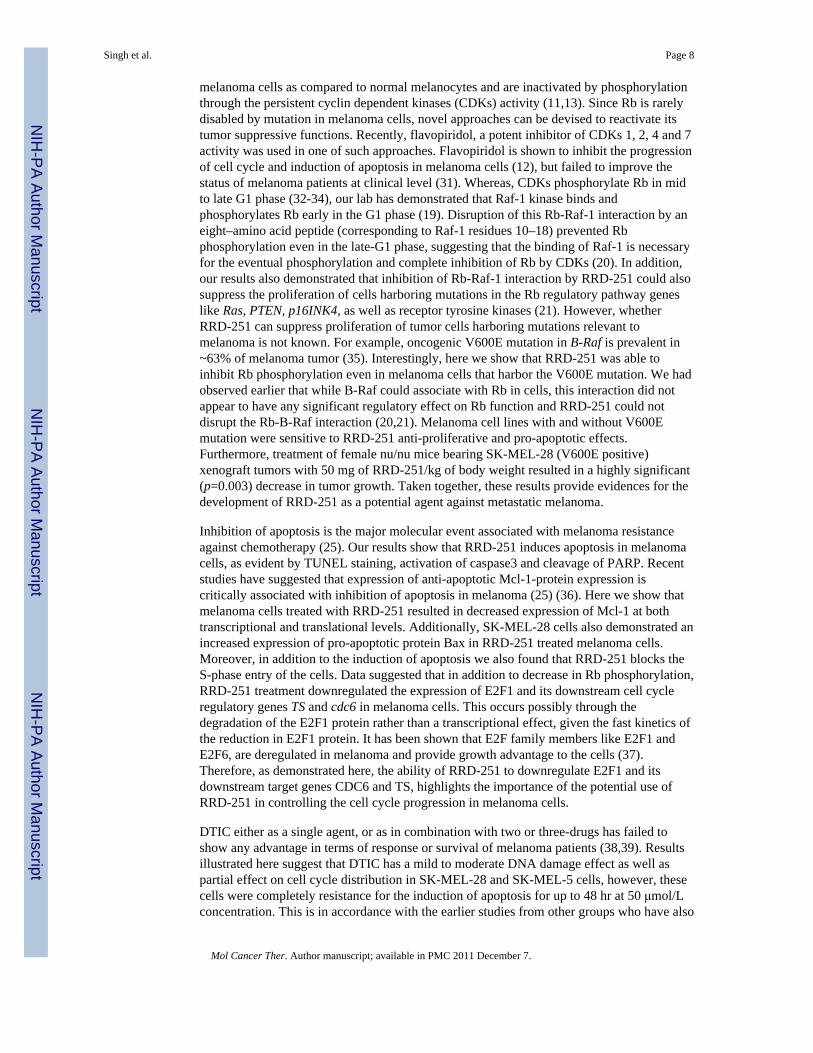
cytometry. Representative cell cycle profile of SK-MEL-28 and SK-MEL-5 cells are givenin Fig. 5A and B respectively. Treatment with DTIC for 48 hr resulted in an increase in G0/G1 phase cells with a partial decrease in S and G2/M phase cells in both the cell lines. Thisdata suggests that DTIC, despite being a pro-drug, has a certain amount of cell cycleregulatory effects on melanoma cells in culture. Since, SK-MEL-2 cells were sensitive toDTIC, we did not perform cell cycle analysis of this cell line.
Further, the effect of DTIC on DNA-damage was checked by comet assay. Cells weretreated with DTIC for 48 hr at 50 μM concentration. The representative pictures of each cellline are shown in Fig. 5C. All the three cells lines showed mild to moderate DNA-damagewith the tail length of approximately one or two times to the diameter of the head. Thescored DNA-damage accounted the mean of approximately 25% in SK-MEL-5 and SK-MEL-28 and approximately 40% in SK-MEL-2 cells. Untreated cells had no DNA damagein any cell type (Fig. 5D).
Since DTIC failed to induce apoptosis as a single agent in SK-MEL-28 and SK-MEL-5 celllines, we next evaluated its ability to interact with RRD-251 induced apoptosis in all thethree cell lines. SK-MEL-2, SK-MEL-5 and SK-MEL-28 cells were pretreated with 50μmol/L DTIC for 48 hr before treatment with RRD-251 for a period of 4 hr. The inductionof apoptosis was assessed by detecting caspase-3 as well as PARP-cleavage by Westernblotting. As mentioned above, treatment with DTIC alone caused apoptosis in SK-MEL-2cells as detected by decrease in caspase-3 and PAPR cleavage (Fig. 6A; DTIC lane). Undersimilar conditions DTIC did not induce any apoptotic effect in SK-MEL-5 and SK-MEL-28cell lines (Fig. 6B and C; DTIC lane). At the same time, pretreatment of DTIC resulted inincreased apoptosis (DTIC+RRD-251 lane) as compared to RRD-251 alone as indicated bythe detection of enhanced cleavage of PARP and caspase-3 in all the three cell lines. It isalso important to note that treatment of DTIC and RRD-251 together for 4 hr did not resultin increased apoptosis as compared to RRD-251 treatment alone (data not shown). Overall,data suggests that DTIC pretreatment can enhance the apoptotic potential of RRD-251 inmelanoma cells.
DTIC-mediated enhanced efficacy of RRD-251 was further assessed by quantifying thelevels of active caspase-3/7 using a luminescent-based assay. As shown in right panels ofFig. 6B and C, both the cell lines did not show any increased caspase activity after treatmentwith DTIC for 48 hr, treatment with RRD-251 alone resulted in an average increase ofcaspase activity by 5±1 fold in SK-MEL-28 and 2±0.1 fold in SK-MEL-5 cells as comparedto untreated cells. When the cells were pre-treated with DTIC for 48 hr prior to RRD-251treatment for 4 hr, both the cell lines showed a statistically significant increase in caspaseactivity (p<0.05) as compared to RRD-251 treatment alone. Overall, these results stronglysuggested that DTIC pretreatment enhances the apoptotic effect of RRD-251 in melanomacells.
DISCUSSIONIncidence of melanoma has been increasing at an alarming rate with very limited treatmentchoices and low overall disease free survival. The fact that hyper-phosphorylation of Rb andits subsequent aberrant E2F activation are associated with melanoma progression and poorprognosis prompted us to evaluate the anti-tumor efficacy of the Rb/Raf-1 interactiondisruptor RRD-251 in melanoma. Our studies demonstrate that RRD-251 disturbed cellcycle progression and apoptosis signaling pathways, inhibited anchorage-dependent and –independent cell growth and suppress melanoma tumor growth in vivo. Many reports haveshown that the tumor suppressor function of Rb is deregulated at multiple levels inmelanoma (10,12,13). Rb and its homologues, p107 and p130 are highly expressed in
Singh et al. Page 7
Mol Cancer Ther. Author manuscript; available in PMC 2011 December 7.
NIH
-PA Author Manuscript
NIH
-PA Author Manuscript
NIH
-PA Author Manuscript
melanoma cells as compared to normal melanocytes and are inactivated by phosphorylationthrough the persistent cyclin dependent kinases (CDKs) activity (11,13). Since Rb is rarelydisabled by mutation in melanoma cells, novel approaches can be devised to reactivate itstumor suppressive functions. Recently, flavopiridol, a potent inhibitor of CDKs 1, 2, 4 and 7activity was used in one of such approaches. Flavopiridol is shown to inhibit the progressionof cell cycle and induction of apoptosis in melanoma cells (12), but failed to improve thestatus of melanoma patients at clinical level (31). Whereas, CDKs phosphorylate Rb in midto late G1 phase (32-34), our lab has demonstrated that Raf-1 kinase binds andphosphorylates Rb early in the G1 phase (19). Disruption of this Rb-Raf-1 interaction by aneight–amino acid peptide (corresponding to Raf-1 residues 10–18) prevented Rbphosphorylation even in the late-G1 phase, suggesting that the binding of Raf-1 is necessaryfor the eventual phosphorylation and complete inhibition of Rb by CDKs (20). In addition,our results also demonstrated that inhibition of Rb-Raf-1 interaction by RRD-251 could alsosuppress the proliferation of cells harboring mutations in the Rb regulatory pathway geneslike Ras, PTEN, p16INK4, as well as receptor tyrosine kinases (21). However, whetherRRD-251 can suppress proliferation of tumor cells harboring mutations relevant tomelanoma is not known. For example, oncogenic V600E mutation in B-Raf is prevalent in~63% of melanoma tumor (35). Interestingly, here we show that RRD-251 was able toinhibit Rb phosphorylation even in melanoma cells that harbor the V600E mutation. We hadobserved earlier that while B-Raf could associate with Rb in cells, this interaction did notappear to have any significant regulatory effect on Rb function and RRD-251 could notdisrupt the Rb-B-Raf interaction (20,21). Melanoma cell lines with and without V600Emutation were sensitive to RRD-251 anti-proliferative and pro-apoptotic effects.Furthermore, treatment of female nu/nu mice bearing SK-MEL-28 (V600E positive)xenograft tumors with 50 mg of RRD-251/kg of body weight resulted in a highly significant(p=0.003) decrease in tumor growth. Taken together, these results provide evidences for thedevelopment of RRD-251 as a potential agent against metastatic melanoma.
Inhibition of apoptosis is the major molecular event associated with melanoma resistanceagainst chemotherapy (25). Our results show that RRD-251 induces apoptosis in melanomacells, as evident by TUNEL staining, activation of caspase3 and cleavage of PARP. Recentstudies have suggested that expression of anti-apoptotic Mcl-1-protein expression iscritically associated with inhibition of apoptosis in melanoma (25) (36). Here we show thatmelanoma cells treated with RRD-251 resulted in decreased expression of Mcl-1 at bothtranscriptional and translational levels. Additionally, SK-MEL-28 cells also demonstrated anincreased expression of pro-apoptotic protein Bax in RRD-251 treated melanoma cells.Moreover, in addition to the induction of apoptosis we also found that RRD-251 blocks theS-phase entry of the cells. Data suggested that in addition to decrease in Rb phosphorylation,RRD-251 treatment downregulated the expression of E2F1 and its downstream cell cycleregulatory genes TS and cdc6 in melanoma cells. This occurs possibly through thedegradation of the E2F1 protein rather than a transcriptional effect, given the fast kinetics ofthe reduction in E2F1 protein. It has been shown that E2F family members like E2F1 andE2F6, are deregulated in melanoma and provide growth advantage to the cells (37).Therefore, as demonstrated here, the ability of RRD-251 to downregulate E2F1 and itsdownstream target genes CDC6 and TS, highlights the importance of the potential use ofRRD-251 in controlling the cell cycle progression in melanoma cells.
DTIC either as a single agent, or as in combination with two or three-drugs has failed toshow any advantage in terms of response or survival of melanoma patients (38,39). Resultsillustrated here suggest that DTIC has a mild to moderate DNA damage effect as well aspartial effect on cell cycle distribution in SK-MEL-28 and SK-MEL-5 cells, however, thesecells were completely resistance for the induction of apoptosis for up to 48 hr at 50 μmol/Lconcentration. This is in accordance with the earlier studies from other groups who have also
Singh et al. Page 8
Mol Cancer Ther. Author manuscript; available in PMC 2011 December 7.
NIH
-PA Author Manuscript
NIH
-PA Author Manuscript
NIH
-PA Author Manuscript

demonstrated the resistance of SK-MEL-28 cells against DTIC treatment (40,41).Interestingly, results from our study showed that cells pretreated with DTIC resulted in astatistically significant enhancement of the ability of RRD-251 to induce caspase activityand apoptosis. At the same time, since DTIC alone had no pro-apoptotic activity, it wassynergistically enhanced by RRD-251 treatment in SK-MEL-5 and SK-MEL-28 cells.Notably, these results suggest that after DTIC exposure, DTIC-sensitive SK-MEL-2 as wellas DTIC-resistant melanoma cells like SK-MEL-28 and SK-MEL-5 cells show bettersensitivity towards RRD-251 induced apoptosis. Thus, this combination may be promisingfor the treatment of cancer patients with DTIC-resistant melanoma.
In conclusion, this work shows that RRD-251 has potent anti-tumoral activity againstmetastatic melanoma in vitro and in vivo. Results indicate that the decrease in Rbphosphorylation, downregulation of E2F1 and its downstream cell cycle regulators such asTS and cdc6 as well as decrease in anti-apoptotic protein Mcl-1 and upregulation of pro-apoptotic protein Bax may be involved in RRD-251 induced dual effects with cell cyclearrest as well as induction of apoptosis. Furthermore, we demonstrate that the induction ofapoptosis in melanoma cell lines was significantly enhanced by the combined treatment ofDTIC and RRD-251 as compared to either treatment used alone. Overall, these resultsprovide a rationale for the clinical evaluation of RRD-251 in combination with DTIC inadvanced melanoma patients. Further elucidation of the apoptotic pathways and in vivostudies will aid in the effective implementation of this combination-based therapy againstmetastatic melanoma.
Supplementary MaterialRefer to Web version on PubMed Central for supplementary material.
AcknowledgmentsThis study was supported by the Donald Adams Comprehensive Melanoma Center as well as the grant CA118210from the NCI. We thank Michael Damit for technical assistance. Support from the Core Facilities at Moffitt CancerCenter is acknowledged.
Grant Support: CA1182110 (SC, SS, NL)
References1. Oliveria S, Dusza S, Berwick M. Issues in the epidemiology of melanoma. Expert Rev Anticancer
Ther. 2001;1:453–9. [PubMed: 12113112]2. Jemal A, Murray T, Samuels A, Ghafoor A, Ward E, Thun MJ. Cancer statistics, 2003. CA Cancer J
Clin. 2003;53:5–26. [PubMed: 12568441]3. Sun W, Schuchter LM. Metastatic melanoma. Curr Treat Options Oncol. 2001;2:193–202.
[PubMed: 12057119]4. Atallah E, Flaherty L. Treatment of metastatic malignant melanoma. Curr Treat Options Oncol.
2005;6:185–93. [PubMed: 15869730]5. Eggermont AM, Kirkwood JM. Re-evaluating the role of dacarbazine in metastatic melanoma: what
have we learned in 30 years? Eur J Cancer. 2004;40:1825–36. [PubMed: 15288283]6. Stein JA, Brownell I. Treatment approaches for advanced cutaneous melanoma. J Drugs Dermatol.
2008;7:175–9. [PubMed: 18335655]7. Eberle J, Kurbanov BM, Hossini AM, Trefzer U, Fecker LF. Overcoming apoptosis deficiency of
melanoma-hope for new therapeutic approaches. Drug Resist Updat. 2007;10:218–34. [PubMed:18054518]
8. Lorigan P, Eisen T, Hauschild A. Systemic therapy for metastatic malignant melanoma--fromdeeply disappointing to bright future? Exp Dermatol. 2008;17:383–94. [PubMed: 18312390]
Singh et al. Page 9
Mol Cancer Ther. Author manuscript; available in PMC 2011 December 7.
NIH
-PA Author Manuscript
NIH
-PA Author Manuscript
NIH
-PA Author Manuscript
9. Curtin JA, Fridlyand J, Kageshita T, et al. Distinct sets of genetic alterations in melanoma. N Engl JMed. 2005;353:2135–47. [PubMed: 16291983]
10. Nelson MA, Reynolds SH, Rao UN, et al. Increased gene copy number of the transcription factorE2F1 in malignant melanoma. Cancer Biol Ther. 2006;5:407–12. [PubMed: 16481740]
11. Bartkova J, Lukas J, Guldberg P, et al. The p16-cyclin D/Cdk4-pRb pathway as a functional unitfrequently altered in melanoma pathogenesis. Cancer Res. 1996;56:5475–83. [PubMed: 8968104]
12. Halaban R, Cheng E, Smicun Y, Germino J. Deregulated E2F transcriptional activity inautonomously growing melanoma cells. J Exp Med. 2000;191:1005–16. [PubMed: 10727462]
13. Halaban R. Rb/E2F: a two-edged sword in the melanocytic system. Cancer Metastasis Rev.2005;24:339–56. [PubMed: 15986142]
14. Hiebert SW, Chellappan SP, Horowitz JM, Nevins JR. The interaction of RB with E2F coincideswith an inhibition of the transcriptional activity of E2F. Genes Dev. 1992;6:177–85. [PubMed:1531329]
15. Chellappan SP, Hiebert S, Mudryj M, Horowitz JM, Nevins JR. The E2F transcription factor is acellular target for the RB protein. Cell. 1991;65:1053–61. [PubMed: 1828392]
16. Harbour JW, Dean DC. Rb function in cell-cycle regulation and apoptosis. Nat Cell Biol.2000;2:E65–7. [PubMed: 10783254]
17. Knudsen ES, Buckmaster C, Chen TT, Feramisco JR, Wang JY. Inhibition of DNA synthesis byRB: effects on G1/S transition and S-phase progression. Genes Dev. 1998;12:2278–92. [PubMed:9694794]
18. Leone G, DeGregori J, Jakoi L, Cook JG, Nevins JR. Collaborative role of E2F transcriptionalactivity and G1 cyclindependent kinase activity in the induction of S phase. Proc Natl Acad Sci US A. 1999;96:6626–31. [PubMed: 10359762]
19. Wang S, Ghosh RN, Chellappan SP. Raf-1 physically interacts with Rb and regulates its function:a link between mitogenic signaling and cell cycle regulation. Mol Cell Biol. 1998;18:7487–98.[PubMed: 9819434]
20. Dasgupta P, Sun J, Wang S, et al. Disruption of the Rb--Raf-1 interaction inhibits tumor growthand angiogenesis. Mol Cell Biol. 2004;24:9527–41. [PubMed: 15485920]
21. Kinkade R, Dasgupta P, Carie A, et al. A small molecule disruptor of Rb/Raf-1 interaction inhibitscell proliferation, angiogenesis, and growth of human tumor xenografts in nude mice. Cancer Res.2008;68:3810–8. [PubMed: 18483265]
22. Vindelov LL, Christensen IJ, Nissen NI. A detergent-trypsin method for the preparation of nucleifor flow cytometric DNA analysis. Cytometry. 1983;3:323–7. [PubMed: 6188586]
23. Singh NP, McCoy MT, Tice RR, Schneider EL. A simple technique for quantitation of low levelsof DNA damage in individual cells. Exp Cell Res. 1988;175:184–91. [PubMed: 3345800]
24. Tice RR, Agurell E, Anderson D, et al. Single cell gel/comet assay: guidelines for in vitro and invivo genetic toxicology testing. Environ Mol Mutagen. 2000;35:206–21. [PubMed: 10737956]
25. Tang L, Tron VA, Reed JC, et al. Expression of apoptosis regulators in cutaneous malignantmelanoma. Clin Cancer Res. 1998;4:1865–71. [PubMed: 9717813]
26. Leiter U, Schmid RM, Kaskel P, Peter RU, Krahn G. Antiapoptotic bcl-2 and bcl-xL in advancedmalignant melanoma. Arch Dermatol Res. 2000;292:225–32. [PubMed: 10867810]
27. Wong RP, Khosravi S, Martinka M, Li G. Myeloid leukemia-1 expression in benign and malignantmelanocytic lesions. Oncol Rep. 2008;19:933–7. [PubMed: 18357378]
28. Zhuang L, Lee CS, Scolyer RA, et al. Mcl-1, Bcl-XL and Stat3 expression are associated withprogression of melanoma whereas Bcl-2, AP-2 and MITF levels decrease during progression ofmelanoma. Mod Pathol. 2007;20:416–26. [PubMed: 17384650]
29. Boisvert-Adamo K, Longmate W, Abel EV, Aplin AE. Mcl-1 is required for melanoma cellresistance to anoikis. Mol Cancer Res. 2009;7:549–56. [PubMed: 19372583]
30. Lee SM, Thatcher N, Dougal M, Margison GP. Dosage and cycle effects of dacarbazine (DTIC)and fotemustine on O6-alkylguanine-DNA alkyltransferase in human peripheral bloodmononuclear cells. Br J Cancer. 1993;67:216–21. [PubMed: 8431354]
Singh et al. Page 10
Mol Cancer Ther. Author manuscript; available in PMC 2011 December 7.
NIH
-PA Author Manuscript
NIH
-PA Author Manuscript
NIH
-PA Author Manuscript
31. Burdette-Radoux S, Tozer RG, Lohmann RC, et al. Phase II trial of flavopiridol, a cyclindependent kinase inhibitor, in untreated metastatic malignant melanoma. Invest New Drugs.2004;22:315–22. [PubMed: 15122079]
32. Harbour JW, Luo RX, Dei Santi A, Postigo AA, Dean DC. Cdk phosphorylation triggerssequential intramolecular interactions that progressively block Rb functions as cells move throughG1. Cell. 1999;98:859–69. [PubMed: 10499802]
33. Knudsen ES, Wang JY. Differential regulation of retinoblastoma protein function by specific Cdkphosphorylation sites. J Biol Chem. 1996;271:8313–20. [PubMed: 8626527]
34. Knudsen ES, Wang JY. Dual mechanisms for the inhibition of E2F binding to RB by cyclin-dependent kinase-mediated RB phosphorylation. Mol Cell Biol. 1997;17:5771–83. [PubMed:9315635]
35. Brose MS, Volpe P, Feldman M, et al. BRAF and RAS mutations in human lung cancer andmelanoma. Cancer Res. 2002;62:6997–7000. [PubMed: 12460918]
36. Keuling AM, Felton KE, Parker AA, Akbari M, Andrew SE, Tron VA. RNA silencing of Mcl-1enhances ABT-737-mediated apoptosis in melanoma: role for a caspase-8-dependent pathway.PLoS One. 2009;4:e6651. [PubMed: 19684859]
37. Halaban R, Miglarese MR, Smicun Y, Puig S. Melanomas, from the cell cycle point of view(Review). Int J Mol Med. 1998;1:419–25. [PubMed: 9852245]
38. Huncharek M, Caubet JF, McGarry R. Single-agent DTIC versus combination chemotherapy withor without immunotherapy in metastatic melanoma: a meta-analysis of 3273 patients from 20randomized trials. Melanoma Res. 2001;11:75–81. [PubMed: 11254118]
39. Mouawad R, Sebert M, Michels J, Bloch J, Spano JP, Khayat D. Treatment for metastaticmalignant melanoma: Old drugs and new strategies. Crit Rev Oncol Hematol. 2009
40. Lillehammer T, Engesaeter BO, Prasmickaite L, Maelandsmo GM, Fodstad O, Engebraaten O.Combined treatment with Ad-hTRAIL and DTIC or SAHA is associated with increasedmitochondrial-mediated apoptosis in human melanoma cell lines. J Gene Med. 2007;9:440–51.[PubMed: 17410615]
41. Munoz-Alonso MJ, Gonzalez-Santiago L, Zarich N, et al. Plitidepsin has a dual effect inhibitingcell cycle and inducing apoptosis via Rac1/c-Jun NH2-terminal kinase activation in humanmelanoma cells. J Pharmacol Exp Ther. 2008;324:1093–101. [PubMed: 18089842]
Abbreviations List
DTIC Dacarbazine
Singh et al. Page 11
Mol Cancer Ther. Author manuscript; available in PMC 2011 December 7.
NIH
-PA Author Manuscript
NIH
-PA Author Manuscript
NIH
-PA Author Manuscript
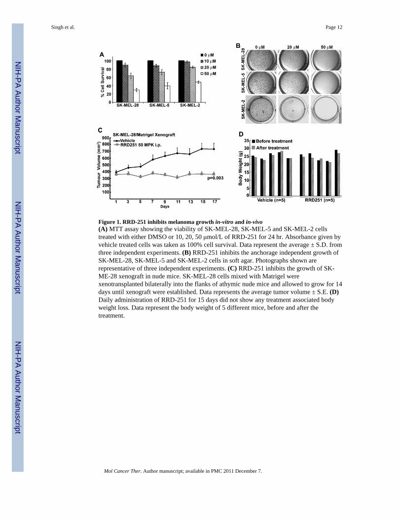
Figure 1. RRD-251 inhibits melanoma growth in-vitro and in-vivo(A) MTT assay showing the viability of SK-MEL-28, SK-MEL-5 and SK-MEL-2 cellstreated with either DMSO or 10, 20, 50 μmol/L of RRD-251 for 24 hr. Absorbance given byvehicle treated cells was taken as 100% cell survival. Data represent the average ± S.D. fromthree independent experiments. (B) RRD-251 inhibits the anchorage independent growth ofSK-MEL-28, SK-MEL-5 and SK-MEL-2 cells in soft agar. Photographs shown arerepresentative of three independent experiments. (C) RRD-251 inhibits the growth of SK-ME-28 xenograft in nude mice. SK-MEL-28 cells mixed with Matrigel werexenotransplanted bilaterally into the flanks of athymic nude mice and allowed to grow for 14days until xenograft were established. Data represents the average tumor volume ± S.E. (D)Daily administration of RRD-251 for 15 days did not show any treatment associated bodyweight loss. Data represent the body weight of 5 different mice, before and after thetreatment.
Singh et al. Page 12
Mol Cancer Ther. Author manuscript; available in PMC 2011 December 7.
NIH
-PA Author Manuscript
NIH
-PA Author Manuscript
NIH
-PA Author Manuscript
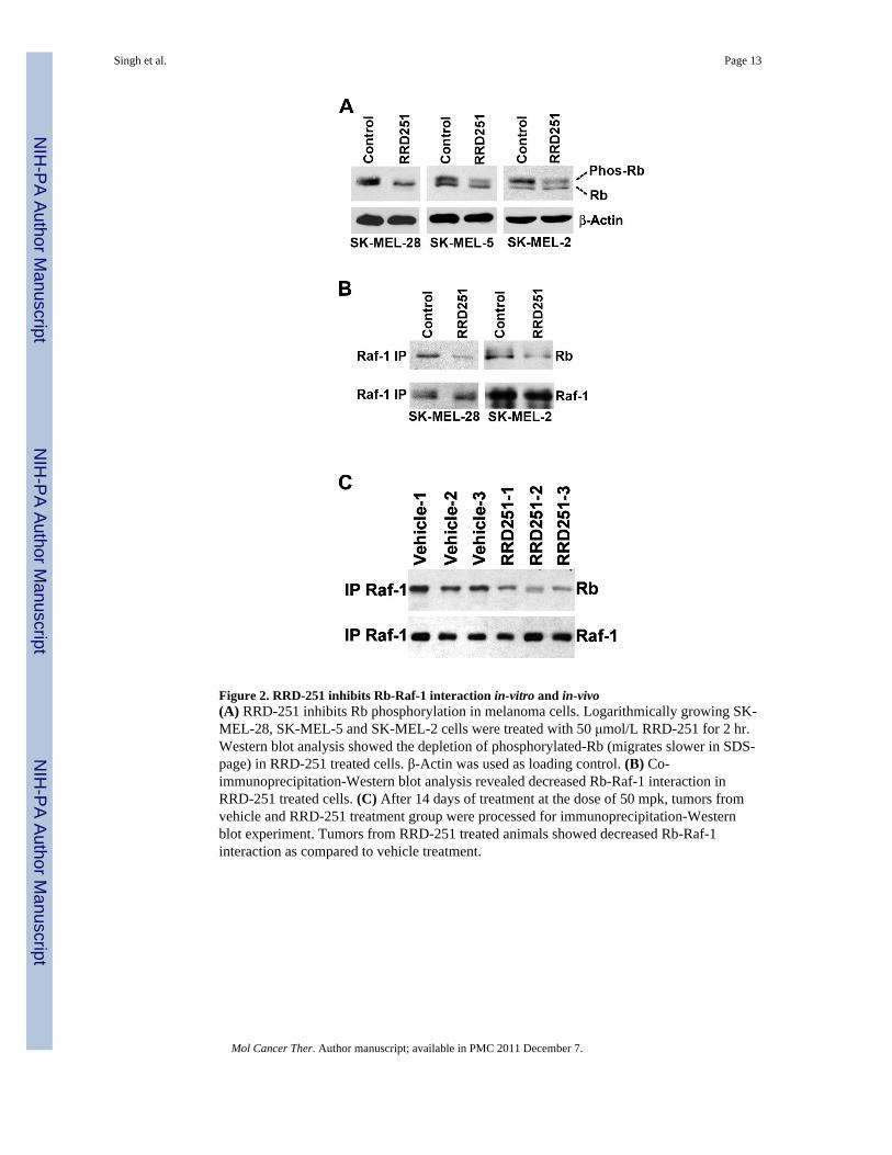
Figure 2. RRD-251 inhibits Rb-Raf-1 interaction in-vitro and in-vivo(A) RRD-251 inhibits Rb phosphorylation in melanoma cells. Logarithmically growing SK-MEL-28, SK-MEL-5 and SK-MEL-2 cells were treated with 50 μmol/L RRD-251 for 2 hr.Western blot analysis showed the depletion of phosphorylated-Rb (migrates slower in SDS-page) in RRD-251 treated cells. β-Actin was used as loading control. (B) Co-immunoprecipitation-Western blot analysis revealed decreased Rb-Raf-1 interaction inRRD-251 treated cells. (C) After 14 days of treatment at the dose of 50 mpk, tumors fromvehicle and RRD-251 treatment group were processed for immunoprecipitation-Westernblot experiment. Tumors from RRD-251 treated animals showed decreased Rb-Raf-1interaction as compared to vehicle treatment.
Singh et al. Page 13
Mol Cancer Ther. Author manuscript; available in PMC 2011 December 7.
NIH
-PA Author Manuscript
NIH
-PA Author Manuscript
NIH
-PA Author Manuscript
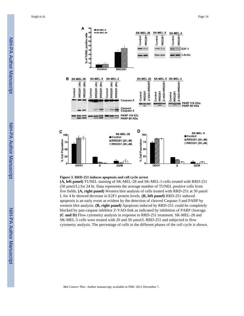
Figure 3. RRD-251 induces apoptosis and cell cycle arrest(A, left panel) TUNEL staining of SK-MEL-28 and SK-MEL-5 cells treated with RRD-251(50 μmol/L) for 24 hr. Data represents the average number of TUNEL positive cells fromfive fields. (A, right panel) Western blot analysis of cells treated with RRD-251 at 50 μmol/L for 4 hr showed decrease in E2F1 protein levels. (B, left panel) RRD-251 inducedapoptosis is an early event as evident by the detection of cleaved Caspase-3 and PARP bywestern blot analysis. (B, right panel) Apoptosis induced by RRD-251 could be completelyblocked by pan-caspase inhibitor Z-VAD-fmk as indicated by inhibition of PARP cleavage.(C and D) Flow cytometry analysis in response to RRD-251 treatment. SK-MEL-28 andSK-MEL-5 cells were treated with 20 and 50 μmol/L RRD-251 and subjected to flowcytometry analysis. The percentage of cells in the different phases of the cell cycle is shown.
Singh et al. Page 14
Mol Cancer Ther. Author manuscript; available in PMC 2011 December 7.
NIH
-PA Author Manuscript
NIH
-PA Author Manuscript
NIH
-PA Author Manuscript

Figure 4. RRD-251 alters the expression of cell cycle and apoptosis regulators(A and B) Real-time PCR analysis for TS and cdc6 from the mRNA prepared after 18 hr ofRRD-251 treatment in SK-MEL-28 (A) and SK-MEL-5 (B). Expression of these genes wasdecreased in a dose dependent manner. (C) RRD-251 treatment was done for 4 hr and themRNA expression of Mcl-1 was analyzed by real-time PCR method. (D) Western blotanalysis for Mcl-1 and Bax after 4 hr treatment of RRD-251.
Singh et al. Page 15
Mol Cancer Ther. Author manuscript; available in PMC 2011 December 7.
NIH
-PA Author Manuscript
NIH
-PA Author Manuscript
NIH
-PA Author Manuscript

Figure 5. DTIC partially inhibits the cell cycle progression(A and B) SK-MEL-28 (A) and SK-MEL-5 (B) cell were treated with 50 μmol/L dose ofDTIC for 48 h and subjected for Flow cytometry analysis. The representative cell cycleprofile is shown. Histograms on the right represent the percentage of cells in the differentphases of the cell cycle form three independent experiments. (C) Damaged DNA wasvisualyzed by the Comet assay in SK-Mel-5, SK-Mel28 and SK-MEL-2 cells followingexposure to 50 μmol/L DTIC for 48 h. Representative micrograph of fluorescent DNA stainof control cells, showing undamaged DNA remaining. whereas DTIC-treated cells showingdenatured DNA fragments migrating out from cell. (D) %DNA damage was quantified andmean is represented with standered deviation as error bars.
Singh et al. Page 16
Mol Cancer Ther. Author manuscript; available in PMC 2011 December 7.
NIH
-PA Author Manuscript
NIH
-PA Author Manuscript
NIH
-PA Author Manuscript
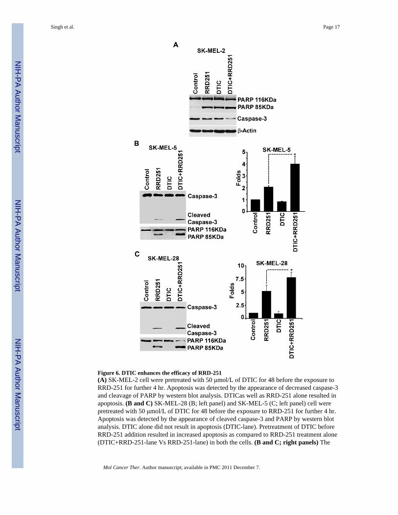
Figure 6. DTIC enhances the efficacy of RRD-251(A) SK-MEL-2 cell were pretreated with 50 μmol/L of DTIC for 48 before the exposure toRRD-251 for further 4 hr. Apoptosis was detected by the appearance of decreased caspase-3and cleavage of PARP by western blot analysis. DTICas well as RRD-251 alone resulted inapoptosis. (B and C) SK-MEL-28 (B; left panel) and SK-MEL-5 (C; left panel) cell werepretreated with 50 μmol/L of DTIC for 48 before the exposure to RRD-251 for further 4 hr.Apoptosis was detected by the appearance of cleaved caspase-3 and PARP by western blotanalysis. DTIC alone did not result in apoptosis (DTIC-lane). Pretreatment of DTIC beforeRRD-251 addition resulted in increased apoptosis as compared to RRD-251 treatment alone(DTIC+RRD-251-lane Vs RRD-251-lane) in both the cells. (B and C; right panels) The
Singh et al. Page 17
Mol Cancer Ther. Author manuscript; available in PMC 2011 December 7.
NIH
-PA Author Manuscript
NIH
-PA Author Manuscript
NIH
-PA Author Manuscript

caspase-3 activity was quantified using Caspase Glo 3/7 kit in SK-MEL-28 and SK-MEL-5cells. The pretreatment of DTIC before RRD-251 exposure resulted in significantly higher(p value <0.05) caspase activity than these treatments alone in both the cell lines.
Singh et al. Page 18
Mol Cancer Ther. Author manuscript; available in PMC 2011 December 7.
NIH
-PA Author Manuscript
NIH
-PA Author Manuscript
NIH
-PA Author Manuscript