Aluminum as an endocrine disruptor in female Nile tilapia (Oreochromis niloticus)
6
Aluminum as an endocrine disruptor in female Nile tilapia (Oreochromis niloticus) T.G. Correia a , A.M. Narcizo a , A. Bianchini b , R.G. Moreira a, ⁎ a Instituto de Biociências, Universidade de São Paulo, R. do Matão, Trav.14, n. 321, 05508-090, São Paulo, SP, Brazil b Instituto de Ciências Biológicas, Universidade Federal do Rio Grande, Av. Itália km 8, Campus Carreiros, 96201-900, Rio Grande, RS, Brazil abstract article info Article history: Received 27 November 2009 Received in revised form 2 February 2010 Accepted 2 February 2010 Available online 16 February 2010 Keywords: Aluminum Cortisol 17β-estradiol 17α-hydroxyprogesterone Plasma ions pH Proteins Lipids The effects of aluminum on plasma ion, lipid, protein and steroid hormone concentration were evaluated in Oreochromis niloticus broodstock females. Lipid and protein concentrations from the gonads and liver were also measured. Experiments were performed at neutral and acidic water pH. Four groups of fish were tested for 96 h: 1) control conditions at neutral water pH; 2) control conditions at acidic water pH (CTR-Ac); 3) aluminum at neutral water pH (Al-N); and 4) aluminum at acidic water pH (Al-Ac). Aluminum and acidic water pH exposure caused no ionoregulatory disturbances. Total lipid concentration increased in the mature gonads and decreased in the liver, suggesting an acceleration of lipid mobilization to the ovaries in animals exposed to aluminum. However, a decreased protein concentration in ovaries was also observed. Exposure of control fish to acidic water pH caused an increased concentration of plasma 17α-hydroxyprogesterone. However, females exposed to aluminum at acidic water pH showed a decreased of plasma 17α-hydroxyprogesterone and cortisol. No differences in plasma 17β-estradiol were observed. The physiological mechanisms underlying the disturbances observed are discussed focusing on reproduction. We suggest that aluminum can be considered an endocrine disrupting compound in mature O. niloticus females. © 2010 Elsevier Inc. All rights reserved. 1. Introduction Aluminum (Al) is a harmful metal to the aquatic ecosystem, being responsible for events of toxicity with serious ecological consequences. To date, no normal physiological functions in biological systems are attributed to this metal (Nayak, 2002). Physiological alterations frequently observed in different fish species exposed to Al are mainly related to cardiovascular (Laitinen and Valtonen, 1995), hematologic (Barcarolli and Martinez, 2004), respiratory, ionoregulatory (Poléo, 1995), reproductive (Vuorinen et al., 2003), metabolic (Brodeur et al., 2001), and endocrine (Waring et al., 1996) disturbances, beyond structural gill damage (Peuranen et al., 1993). In freshwater fish, Al causes acute ionoregulatory and respiratory disturbances due to the deposition of Al 3+ on the gills (Poléo, 1995). Al also affects reproduction in the rainbow trout due to a concentration- dependent decrease in vitellogenesis. This effect is a consequence of a reduced expression of the mRNA encoding for vitellogenin synthesis (Hwang et al., 2000). In turn, acidic waters are known to impair fish reproduction, affecting fecundity, egg viability, spawning success (Mount et al., 1988), gonad development, and gamete production (Vuorinen and Vuorinen, 1991). These effects lead to serious con- sequences to fish populations such as a decreased number and size of fish (Mount et al., 1988). In light of the above, it would be expected that fish exposure to Al at acidic water pH would lead to a more pronounced response than that observed after a single exposure to the metal. In fact, Al has shown to be more toxic to fish in acidic waters (Alstad et al., 2005). Therefore, the present study was undertaken to investigate the effects of Al on several physiological endpoints associated with ionic and osmotic regulation, reproductive hormones, and energy metabolism in mature females of the Nile tilapia Oreochromis niloticus. 2. Material and methods 2.1. Fish Mature females of the Nile tilapia O. niloticus (Perciformes, Cichlidae) weighing 85.9 ± 9.8 g were obtained from a commercial fish farm. They were maintained in ponds at the Ponte Nova Fish Farm (23°35′33.8″S and 45°58′09.1″W) at Salesópolis (São Paulo State, Brazil). 2.2. Experimental design Female fish (n = 48) were randomly divided into 4 groups (12 fish in each group), which were tested in duplicate. Fish from each replicate were held in a 145-L tank (6 fish per tank). The experimental groups tested were: 1) fish kept under control conditions in water at neutral pH (CTR-N); 2) fish kept under control conditions in acidic Comparative Biochemistry and Physiology, Part C 151 (2010) 461–466 ⁎ Corresponding author. Tel.: + 55 11 3091 7531; fax: + 55 11 3091 7568. E-mail address: [email protected] (R.G. Moreira). 1532-0456/$ – see front matter © 2010 Elsevier Inc. All rights reserved. doi:10.1016/j.cbpc.2010.02.002 Contents lists available at ScienceDirect Comparative Biochemistry and Physiology, Part C journal homepage: www.elsevier.com/locate/cbpc
-
Upload
independent -
Category
Documents
-
view
4 -
download
0
Transcript of Aluminum as an endocrine disruptor in female Nile tilapia (Oreochromis niloticus)

Comparative Biochemistry and Physiology, Part C 151 (2010) 461–466
Contents lists available at ScienceDirect
Comparative Biochemistry and Physiology, Part C
j ourna l homepage: www.e lsev ie r.com/ locate /cbpc
Aluminum as an endocrine disruptor in female Nile tilapia (Oreochromis niloticus)
T.G. Correia a, A.M. Narcizo a, A. Bianchini b, R.G. Moreira a,⁎a Instituto de Biociências, Universidade de São Paulo, R. do Matão, Trav.14, n. 321, 05508-090, São Paulo, SP, Brazilb Instituto de Ciências Biológicas, Universidade Federal do Rio Grande, Av. Itália km 8, Campus Carreiros, 96201-900, Rio Grande, RS, Brazil
⁎ Corresponding author. Tel.: +55 11 3091 7531; faxE-mail address: [email protected] (R.G. Moreira).
1532-0456/$ – see front matter © 2010 Elsevier Inc. Aldoi:10.1016/j.cbpc.2010.02.002
a b s t r a c t
a r t i c l e i n f oArticle history:Received 27 November 2009Received in revised form 2 February 2010Accepted 2 February 2010Available online 16 February 2010
Keywords:AluminumCortisol17β-estradiol17α-hydroxyprogesteronePlasma ionspHProteinsLipids
The effects of aluminum on plasma ion, lipid, protein and steroid hormone concentration were evaluated inOreochromis niloticus broodstock females. Lipid and protein concentrations from the gonads and liver were alsomeasured. Experiments were performed at neutral and acidic water pH. Four groups of fish were tested for 96 h:1) control conditions at neutral water pH; 2) control conditions at acidic water pH (CTR-Ac); 3) aluminum atneutral water pH (Al-N); and 4) aluminum at acidic water pH (Al-Ac). Aluminum and acidic water pH exposurecausedno ionoregulatorydisturbances. Total lipid concentration increased in themature gonads anddecreased inthe liver, suggesting an acceleration of lipid mobilization to the ovaries in animals exposed to aluminum.However, a decreased protein concentration in ovarieswas also observed. Exposure of controlfish to acidicwaterpH caused an increased concentration of plasma 17α-hydroxyprogesterone. However, females exposed toaluminum at acidic water pH showed a decreased of plasma 17α-hydroxyprogesterone and cortisol. Nodifferences in plasma 17β-estradiol were observed. The physiological mechanisms underlying the disturbancesobserved are discussed focusing on reproduction. We suggest that aluminum can be considered an endocrinedisrupting compound in mature O. niloticus females.
: +55 11 3091 7568.
l rights reserved.
© 2010 Elsevier Inc. All rights reserved.
1. Introduction
Aluminum (Al) is a harmful metal to the aquatic ecosystem, beingresponsible for events of toxicity with serious ecological consequences.To date, no normal physiological functions in biological systems areattributed to this metal (Nayak, 2002). Physiological alterationsfrequently observed in different fish species exposed to Al are mainlyrelated to cardiovascular (Laitinen and Valtonen, 1995), hematologic(Barcarolli and Martinez, 2004), respiratory, ionoregulatory (Poléo,1995), reproductive (Vuorinen et al., 2003), metabolic (Brodeur et al.,2001), and endocrine (Waring et al., 1996) disturbances, beyondstructural gill damage (Peuranen et al., 1993).
In freshwater fish, Al causes acute ionoregulatory and respiratorydisturbances due to the deposition of Al3+ on the gills (Poléo, 1995). Alalso affects reproduction in the rainbow trout due to a concentration-dependent decrease in vitellogenesis. This effect is a consequence of areduced expression of the mRNA encoding for vitellogenin synthesis(Hwang et al., 2000). In turn, acidic waters are known to impair fishreproduction, affecting fecundity, egg viability, spawning success(Mount et al., 1988), gonad development, and gamete production(Vuorinen and Vuorinen, 1991). These effects lead to serious con-sequences to fish populations such as a decreased number and size offish (Mount et al., 1988).
In light of the above, it would be expected that fish exposure to Alat acidic water pH would lead to a more pronounced response thanthat observed after a single exposure to the metal. In fact, Al hasshown to be more toxic to fish in acidic waters (Alstad et al., 2005).Therefore, the present study was undertaken to investigate the effectsof Al on several physiological endpoints associated with ionic andosmotic regulation, reproductive hormones, and energy metabolismin mature females of the Nile tilapia Oreochromis niloticus.
2. Material and methods
2.1. Fish
Mature females of the Nile tilapia O. niloticus (Perciformes,Cichlidae) weighing 85.9±9.8 g were obtained from a commercialfish farm. They were maintained in ponds at the Ponte Nova Fish Farm(23°35′33.8″S and 45°58′09.1″W) at Salesópolis (São Paulo State,Brazil).
2.2. Experimental design
Female fish (n=48) were randomly divided into 4 groups (12 fishin each group), which were tested in duplicate. Fish from eachreplicate were held in a 145-L tank (6 fish per tank). The experimentalgroups tested were: 1) fish kept under control conditions in water atneutral pH (CTR-N); 2) fish kept under control conditions in acidic
462 T.G. Correia et al. / Comparative Biochemistry and Physiology, Part C 151 (2010) 461–466
water (CTR-Ac); 3) fish exposed to Al in water at neutral pH (Al-N)and; 4) fish exposed to Al in acidic water (Al-Ac).
2.3. Experimental conditions
The experimentwas performed for 96 h in a semi-static systemwithdaily water renewal, under a natural photoperiod. Fish were not fedduring the test period to avoid the excess of organicmatter in thewater.
Alwas added to the experimentalmedia asAl2(SO4)3 (Merck) fromastock solution prepared using Milli-Q water acidified (pH=2.5) with65% HNO3 (Suprapur; Merck). Nominal Al concentrationwas 0.5 µg/mLfor groupsAl-AcandAl-N.Water pH, at thebeginningof theexperiment,was approximately 7.0 and was adjusted with HCl and NaOH (Synth,Brazil) in order to reach thedesirable values of 5.5 for theCTR-Ac andAl-Ac groups and 7.0 for the CTR-N and Al-N groups. Water pH wasmonitored with a digital pH meter (Gehaka, São Paulo, Brazil) beforeand after adjustments. The experimental media were continuouslyaerated, and water temperature (maintained around 21–22 °C) anddissolved oxygen content were daily monitored with an oximeter(model 55; YSI, Yellow Springs, OH, USA).
2.4. Water chemistry
The water used in the experiment was previously filtered throughtwo cellulose filters (IMPAC® — CEP 10, São Paulo, SP, Brazil andAcqualimp®FICZ—10,Valinhos, SP, Brazil) containingactivated charcoal(5 µm) to remove the solid particles and reduce the organic mattercontent andanypossible chemical contaminants. After theseprocedures,water was analyzed for the main physicochemical parameters (Na+:0.086 mM; K+: 0.0027 mM; Ca2+: 0.0057 mM; Mg2+: 0.0016 mM; Fe:0.0022 mM; Cl−: 0.085 mM; and SO4
2− 0.00002 mM; alkalinity: 16 mgCaCO3/L; total carbon: 6.755 mg C/L; organic carbon: 3.934 mg C/L; andinorganic carbon: 2.822 mg C/L) and used in the 145-L test tanks. The Alstock solutionwas added to give a nominalfinal concentration of 0.5 µg/mL. The experimental media were left to stand for 12 h to allowdecantation and to let the system to equilibrate. Al nominal concentra-tionwas thenmeasured and readjusted to 0.5 µg/mL. This concentrationwas chosen in order to represent the aluminum concentration usuallyfound in the main basins in São Paulo State (CETESB, 2008).
For each assay, an additional tank with the same water volume(145 L) was prepared following the same procedures described above.Water from this tank was used to renew the experimental mediumevery 24 h of test. Water sampleswere collected from the experimentaltanks during the renewal period. These samples were filtered with0.45 µmsyringefilter (Minisart®— Sartorius, Goettingen,Germany) andused to quantify NH3, NO2, and dissolved Al concentrations. Total Alconcentration was measured using an atomic absorption spectropho-tometer (GBC, AAS 932 Avanta-Plus, IL, USA). Other analyses wereperformed according to the Standard Methods (2005).
2.5. Biological sampling and physiological analyses
After 96 h of exposure, fish were cryoanesthetized, and a bloodsample (∼2 mL) was collected by puncture of the caudal vein using aheparinized syringe. Fish were then killed by decapitation, followingthe animal care protocols approved by the Biosciences Institute
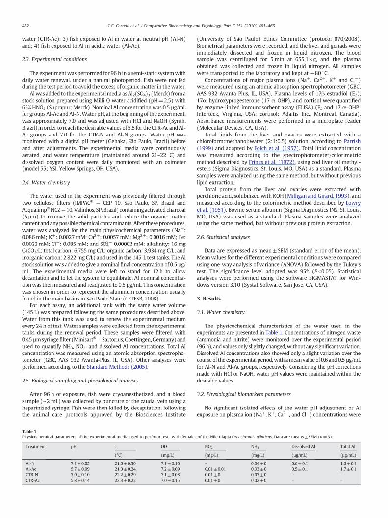
Table 1Physicochemical parameters of the experimental media used to perform tests with females
Treatment pH T OD
(°C) (mg/L)
Al-N 7.1±0.05 21.0±0.30 7.1±0.10Al-Ac 5.7±0.09 21.0±0.24 7.2±0.09CTR-N 7.0±0.10 22.2±0.29 7.1±0.08CTR-Ac 5.8±0.14 22.3±0.22 7.0±0.15
(University of São Paulo) Ethics Committee (protocol 070/2008).Biometrical parameters were recorded, and the liver and gonads wereimmediately dissected and frozen in liquid nitrogen. The bloodsample was centrifuged for 5 min at 655.1×g, and the plasmaobtained was collected and frozen in liquid nitrogen. All sampleswere transported to the laboratory and kept at −80 °C.
Concentrations of major plasma ions (Na+, Ca2+, K+ and Cl−)were measured using an atomic absorption spectrophotometer (GBC,AAS 932 Avanta-Plus, IL, USA). Plasma levels of 17β-estradiol (E2),17α-hydroxyprogesterone (17 α-OHP), and cortisol were quantifiedby enzyme-linked immunosorbent assay (ELISA) (E2 and 17 α-OHP:Interteck, Virginia, USA; cortisol: Adaltis Inc., Montreal, Canada).Absorbance measurements were performed in a microplate reader(Molecular Devices, CA, USA).
Total lipids from the liver and ovaries were extracted with achloroform:methanol:water (2:1:0.5) solution, according to Parrish(1999) and adapted by Folch et al. (1957). Total lipid concentrationwas measured according to the spectrophotometer/colorimetricmethod described by Frings et al. (1972), using cod liver oil methyl-esters (Sigma Diagnostics, St. Louis, MO, USA) as a standard. Plasmasamples were analyzed using the same method, but without previouslipid extraction.
Total protein from the liver and ovaries were extracted withperchloric acid, solubilized with KOH (Milligan and Girard, 1993), andmeasured according to the colorimetric method described by Lowryet al. (1951). Bovine serum albumin (Sigma Diagnostics INS, St. Louis,MO, USA) was used as a standard. Plasma samples were analyzedusing the same method, but without previous protein extraction.
2.6. Statistical analyses
Data are expressed as mean±SEM (standard error of the mean).Mean values for the different experimental conditionswere comparedusing one-way analysis of variance (ANOVA) followed by the Tukey'stest. The significance level adopted was 95% (Pb0.05). Statisticalanalyses were performed using the software SIGMASTAT for Win-dows version 3.10 (Systat Software, San Jose, CA, USA).
3. Results
3.1. Water chemistry
The physicochemical characteristics of the water used in theexperiments are presented in Table 1. Concentrations of nitrogen waste(ammonia and nitrite) were monitored over the experimental period(96 h), andvaluesonly slightly changed,without any significantvariation.Dissolved Al concentrations also showed only a slight variation over thecourseof the experimental period,with ameanvalueof 0.6 and0.5 µg/mLfor Al-N and Al-Ac groups, respectively. Considering the pH correctionsmade with HCl or NaOH, water pH values were maintained within thedesirable values.
3.2. Physiological biomarkers parameters
No significant isolated effects of the water pH adjustment or Alexposure on plasma ion (Na+, K+, Ca2+, and Cl−) concentrations were
of the Nile tilapia Oreochromis niloticus. Data are means±SEM (n=3).
NO2 NH3 Dissolved Al Total Al
(mg/L) (mg/L) (µg/mL) (µg/mL)
– 0.04±0 0.6±0.1 1.6±0.10.01±0.01 0.03±0 0.5±0.1 1.7±0.10.01±0 0.03±0 – –
0.01±0 0.02±0 – –

Table 2Plasma ion concentration (mM) in female Nile tilapia Oreochromis niloticus kept undercontrol conditions at neutral (CTR-N) and acidic (CTR-Ac) pH or exposed to aluminum atneutral (Al-N) or acidic (Al-Ac) pH for 96 h. Data are mean±SEM (n=7–9). Differentletters indicate significant difference mean values among treatments (Pb0.05).
Treatment Na+ Cl− K+ Ca2+
Al-N 221.0±12.8a 112.0±9.4a 8.45±0.79a 1.50±0.34a
Al-Ac 174.2±9.7ab 96.1±11.6a 11.18±0.37a 1.12±0.18a
CTR-N 175.4±13.2ab 80.6±14.0a 9.49±1.18a 2.16±0.29a
CTR-Ac 165.5±18.4b 91.6±14.5a 10.16±1.06a 1.41±0.16a
463T.G. Correia et al. / Comparative Biochemistry and Physiology, Part C 151 (2010) 461–466
observed (Table 2). However, fish from the Al-N group showed asignificantly higher Na+ concentration than those from the CTR-Acgroup (Table 2). Protein and lipid concentrations in the liver, gonads,and plasma showed different patterns of variations for the differentexperimental groups. Concentration of the gonad lipids sharplyincreased (∼2-fold) in fish from the two experimental groups exposedto Al, when compared to their respective control groups (P≤0.001)(Fig. 1A). However, the concentration of hepatic lipids decreased in fishfrom Al groups at both neutral (P≤0.001) and acidic pH (P=0.008)
Fig. 1. Total lipid concentration in the (A) gonads, (B) liver, and (C) plasma in maturefemale Oreochromis niloticus exposed (96 h) to aluminum in neutral (Al-N) and acidic(Al-Ac) water pH. Fish were also kept in neutral (CTR-N) and acidic water pH (CTR-Ac)in the absence of aluminum. Data are mean±SEM. Different letters indicate significantdifferent mean values between treatments (Pb0.05).
Fig. 2. Total protein concentration in the (A) gonads, (B) liver, and (C) plasma inmaturefemale Oreochromis niloticus exposed (96 h) to aluminum in neutral (Al-N) and acidic(Al-Ac) water pH. Fish were also kept in neutral (CTR-N) and acidic (CTR-Ac) water pHin the absence of aluminum. Data are mean±SEM. Different letters indicate significantdifferent mean values between treatments (Pb0.05).
(Fig. 1B). No significant change in the concentration of plasma lipidswasobserved (Fig. 1C).
Data from tissue protein concentrations revealed different pat-terns of response after Al exposure. Fish exposed to Al had a decreasedconcentration of protein in the gonads, but only at neutral water pH(Al-N) (P=0.004). In acidic pH, Al exposure slightly decreased theconcentration of the gonad proteins, but without showing asignificant difference from the respective control (Fig. 2A). Concen-trations of hepatic and plasma proteins were not altered by either Alor acidic exposure (Fig. 2B and C).
Short-term exposure to Al and/or acidic pH did not alter theplasma E2 concentration (Fig. 3). However, the plasma concentrationof the progestogen 17 α-OHP significantly decreased when fish wereexposed to Al in acidic pH (Al-Ac) (P=0.001). On the other hand,exposure to acidic pH in the absence of Al (CTR-Ac) significantlyincreased the 17 α-OHP concentration (P=0.001) when comparedwith the neutral pH (CTR-N) (Fig. 4).
At neutral pH, Al exposure did not significantly change plasma cortisolconcentrations. However, when fish were exposed to Al in acidic pH (Al-Ac), plasma cortisol concentrations significantly decreased compared tothe levels observed in fish exposed to Al at neutral water pH (Al-N)(P=0.003) (Fig. 5).

Fig. 3. Plasma estradiol concentration in mature female Oreochromis niloticus exposed(96 h) to aluminum in neutral (Al-N) and acidic (Al-Ac) water pH. Fish were also keptin neutral (CTR-N) and acidic (CTR-Ac) water pH in the absence of aluminum. Data areexpressed as mean±SEM.
Fig. 4. Plasma 17α-hydroxyprogesterone concentration in mature female Oreochromisniloticus exposed (96 h) to aluminum in neutral (Al-N) and acidic (Al-Ac) water pH.Fish were also kept at neutral (CTR-N) and acidic (CTR-Ac) water pH in the absence ofaluminum. Data are expressed as mean±SEM. Different letters indicate significantlydifferent mean values between treatments (Pb0.05).
464 T.G. Correia et al. / Comparative Biochemistry and Physiology, Part C 151 (2010) 461–466
4. Discussion
Some physicochemical water parameters, such as temperature anddissolved oxygen concentration, can cause significant alterations infish physiology, thus confounding the interpretation of fish responses
Fig. 5. Plasma cortisol concentration in mature female Oreochromis niloticus exposed (96 h)to aluminum in neutral (Al-N) and acidic (Al-Ac) water pH. Fish were also kept in neutral(CTR-N) and acidic (CTR-Ac) water pH in the absence of aluminum. Data are means±SEM.Different letters indicate significant different mean values between treatments (Pb0.05).
to water toxicants (Aragão and Araújo, 2006). Therefore, theseparameters were monitored and kept constant throughout the entireexperiment.
Regarding nitrogenous compounds, it is well established thatammonia is toxic to fish, even at low concentrations in the water. Inaddition, independent of the species sensitivity,fish sizehas a significantinfluence in determining the toxicity of this compound (Ruyet et al.,1997). Despite the difficulties to establish the threshold for toxicity infish, Boyd (1990) and Lemarié et al. (2004) suggested that the toxic limitfor freshwater fish is between 0.05 and 0.2 mg/L. In the present study,ammonia concentration throughout the experiment was always below0.05 mg/L, suggesting that this nitrogen compound had no influence onfish responses to the experimental treatments tested.
As for ammonia, fish sensitivity to nitrite significantly variesamong species. According to Willians and Eddy (1986), comparisonsof the toxic effects of nitrite between species should be done carefullysince water chemistry can modify the observed responses. Accordingto Boyd (1990), safe nitrite concentrations are between 0.02 and0.1 mg/L, and any value higher than that can be stressful to the fish. Inthe present study, nitrite concentration likely did not interfere withfish responses to the experimental treatments. This statement is basedon the fact that the nitrite concentrations in water were always belowthis range. Certainly, the water renewal procedure was important tokeep low levels of nitrogen compounds and also to maintain thedissolved Al concentration around 0.5 µg/mL. Therefore, physiologicalchanges described in the present study are likely due to Al exposureand/or water pH.
Alteration in the gill ion regulatory function is considered the firsttoxic effect in fish exposed to acidic pH and Al (Buckler et al., 1995).Decreases in plasma Na+ and Cl− are the initial alterations observedin fish exposed to acidic pH, independent of the presence of Al in thewater (Mount et al., 1988). This effect is mainly observed after acuteexposure, and acclimation or physiological adjustments to these toxiceffects are generally observed in fish after acute (Barcarolli andMartinez, 2004), subchronic (Brodeur et al., 2001), or chronicexposures (Vuorinen et al., 2003). In this context, the lack of effectof the acid pH or Al exposure on the ionic regulation of the female Niletilapia suggests that this fish was not affected by the experimentalconditions tested or had enough time to compensate for thesepresumed effects.
Acidic pH exposure can also interfere with fish growth andreproduction, affecting the liver's ability to metabolize carbohydratesand synthesize vitellogenin in brook trout (Tam et al., 1987). Sub-lethalAl concentration in acidic pH increases the swimming activity inAtlanticsalmon (Salmo salar) with a significant loss of body mass, thus alteringthe energy budget in salmon living under this condition (Brodeur et al.,2001). Fish utilize glucose for their immediate energy source understressing conditions andmaintain hepatic glycogen by glyconeogenesis.In general, under a stressful condition, cortisol increases muscularcatabolism, and the amino acids mobilized are used in the hepaticglyconeogenesis. In this case, changes in plasma protein levels areindicative of substratemobilization from tissues, especially the liver andmuscle, while changes in the gonad protein concentrations indicatesubstrate deposition, which in turn will be used for oocyte growth(Wendelaar Bonga, 1997). In the present study, mature Nile tilapiaexposed to acidic water pH or Al separately, did not show changes inplasma cortisol. However, fish showed a decrease in plasma cortisolconcentrationswhen simultaneously exposed to Al and acidic pH. In theSouth American fish Rhamdia quelen (Cericato et al., 2008) and Bryconamazonicus (Hori et al., 2008), deleterious effects on cortisol responseswere also observed after acute exposure (96 h) to sub-lethal concentra-tions of agrichemicals and phenol, respectively.
In some studies focusing on conservation physiology, animals areexposed to a mild stress to assess their capacity to react toenvironmental stressors (Wikelski and Cooke, 2006). However, notall environmental stressing conditions are able to induce a detectable
465T.G. Correia et al. / Comparative Biochemistry and Physiology, Part C 151 (2010) 461–466
increase in plasma glucocorticoid concentrations (Rich and Romero,2005; Wikelski and Cooke, 2006), as observed in the present studyafter fish acute exposure to acidic water pH or Al.
According toRichandRomero (2005), changes in the responsivenessof the hypothalamic-pituitary-adrenal axis to adrenocorticotropin(ACTH) and arginine vasotocin (AVT) down-regulate corticosteroidresponses during chronic stress in birds. Furthermore, these authorsreported that basal corticosteroid concentrations and corticosteroidresponses to acute stress were significantly reduced when birds werechronically stressed (e.g. captivity), as observed in the present studywhen fish were acutely exposed to Al in acidic water pH.
Therefore, it is clear that data on plasma glucocorticoid concentra-tions have to be interpreted carefully against a background baselinedata. Increased glucocorticoid levels could indicate an adaptiveresponse of healthy individuals to short-term environmental chal-lenges (Wikelski and Cooke, 2006). On the other hand, when anexpected stressor fails to activate the hypothalamus-pituitary-interrenal (HPI) axis, as observed in the present study after acuteexposure to Al or acidic water pH in fish, it could be that the event isnot actually perceived to be stressful or some mechanistic blockade inactivation of the axis is occurring (Wingfield and Sapolsky, 2003).
Considering the gonad substrates, the decrease in protein concen-tration observed when females were exposed to Al in neutral pH can berelated to the vitellogenin (VTG) absorption mechanism, which ismediated by the follicle-stimulating-hormone (FSH). It is known that Alcan inhibit the activity of kinases (Katsuyama et al., 1989), but it can alsoactivate cAMP-dependent kinases (Johnson and Jope, 1987). Many cellsignaling pathways are regulated by the protein phosphorylation–dephosphorylation processes, which are also the basis for the control ofmanycell functions that are influencedbyextracellular stimulus, such ashormones, mitogenics, carcinogenics, citocynes, neurotransmitters,toxic substances, or metabolites (Aoyama et al., 2003). Therefore, Alcan interferewith the enzymes involved inVTGsynthesis or even impairVTG incorporation by the oocyte or the cleavage process, since VTG iscomposed of highly phosphorylatedmolecules (e.g., phosvitin) (Mugiyaand Tanahashi, 1998).
In general, female fish exhibit a decrease in the hepatic lipidsconcentration during the reproductive period, due to fatty acidmobilization anddeposition inovaries (Sheridanet al., 1983;Hendersonand Tocher, 1987; Sheridan, 1988). Consequently, the results obtainedfor hepatic and gonad lipids in the present study after exposure to Al inneutral or acidicwater pH can be interpreted as an energetic investmentto accelerate spawn. We suggest that, mainly due to the mechanism ofasynchronic oocyte development, O. niloticus has the physiologicalplasticity to accelerate the mobilization process of vitellogenesis,resulting in an increased lipid concentration in ovaries. Studies withyellow perch from lakes contaminated with other metals (Cd, Zn, andCu) also did not respond by increasing their plasma cortisol and glucoselevels and altered their capacity to build up and use glycogen andtriglyceride reserves (Levesque et al., 2002), as observed in the presentstudy with Nile tilapia. In the yellow perch, liver triglyceride was lowerin fish from contaminated lakes due to an enhanced activity oftriacylglycerol lipase, an enzyme that hydrolyses triglyceride reserves(Levesque et al., 2002). Therefore, the decreased lipid concentrations inthe liver and the increased lipid concentrations in the gonads of O.niloticus exposed toAl couldbeexplainedbyan increased triacylglycerollipase activity after Al exposure.
In addition to the effects described above, Al also showed anendocrine disruptor effect, i.e., acting as an anti-steroidogenic stressor.InmatureNile tilapia, Al and/or acidicwater pHexposure didnot alteredthe plasmaE2 concentration.However, exposure toAl in acidicwater pHdecreased the plasma concentration of 17α-OHPwhen compared withthe acidic control group. Based on this alteration, a possible mechanismof action for Al can be discussed. Considering that progestogens aremainly produced under the control of the pituitary LH (luteinizinghormone) (Young et al., 2005), we suggest that suppression of the
pituitary LH or even hypothalamic GnRH (gonadotropin releasinghormone) synthesis is occurring. However, due to the known effects ofAl and other metals on the enzyme activities, the possibility of astimulation of 20β-hydroxysteroid dehydrogenase (20β HSD), anenzyme that converts 17 α-OHP into 17α,20β-dihydroxy–4–pregnen–3–one (17,20 P), the main MIS (maturation-inducing steroid) in fish,cannot be ruled out (Young et al., 2005). In fact, this hypothesiscorroborates our suggestion that Al is inducing the acceleration ofspawn in O. niloticus females.
The 17 α-OHP decrease induced by the Al exposure in acidic waterpH was paralleled by a decreased cortisol concentration, as discussedabove. These changes in hormone profiles should be explainedconsidering the pathways of steroid synthesis, since the fish speciesinvestigated has all the enzymes responsible for corticosteroidsynthesis in the gonads, even if the 11-β hydroxylase (enzyme thatgenerates cortisol from 11-deoxycortisol) gene expression was notabundant in the ovaries (Milla et al., 2009, for review). The low levelsof cortisol and 17 α-OHP observed in the Nile tilapia exposed to Al inacidic water pH can also be explained considering the lowered lipidconcentration in the liver, limiting steroid synthesis from cholesterolor by the fact that some xenobiotics (e.g., roundup) can inhibit thetransport of cholesterol to the mitochondria (Walsh et al., 2000).Female Nile tilapia exposed only to acidic water pH in the absence ofAl also showed disturbances in plasma 17 α-OHP, presenting higher17 α-OHP values than females kept in neutral water pH. This findingsuggests that, in fact, dysfunctions in the activity of the enzyme 20βHSD could be occurring.
Like in mammals, the FSH and LH control of the steroid synthesis infish ovaries is a pathway that involves, at least partially, protein kinases(PKA) (Mendéz et al., 2003) and cAMP in the theca follicular layer(Planas et al., 1997). Substances that promote increases in intracellularcAMP also stimulate steroidogenesis (Kanamori and Nagahama, 1988),and cAMP antagonists partially block the steroidogenic action ofgonadotropins (Planas et al., 1997). In fact, the LH stimulatory effecton the steroid synthesis in trout ovaries is related to cAMP/PKAactivation (Mendéz et al., 2003). Therefore, the known effect of Al onabnormal phosphorylation by the activation of cAMP-dependent PKAcan be the cause of the altered plasma level of 17 α-OHP in response tothe LH action after Al exposure. In this context, it is important to notethat Morrissey et al. (1983) observed that Al suppressed parathyroidhormone synthesis due to a decrease in cAMP levels. Therefore, wesuggest that Al interferes with the final maturation phase and spawn inthe Nile tilapia, inducing the production of eggs with lowered proteincontent.
In conclusion, Al exposure in acidic and neutral pH showed to bedeleterious to the reproduction of the female mature Nile tilapiaO. niloticus by accelerating lipid mobilization from the liver anddeposition in the ovaries, decreasing protein deposition in eggs, anddecreasing the plasma levels of 17α-OHP. Based on these results, Al canbe considered as an endocrine disrupting compound (EDC) for matureO. niloticus females. Therefore, we suggest that different reproductivebiomarkers should be investigated in environmental monitoringprograms inmetal contaminated freshwaters, especially the acidic ones.
Acknowledgments
This work was supported by a research grant from FAPESP (#06/50920-5). T. G. Correia was a recipient of a Master fellowship fromCoordenação deAperfeiçoamento de Pessoal deNível Superior (CAPES).A. Bianchini and R. G. Moreira are research fellows from the BrazilianCNPq (Conselho Nacional de Desenvolvimento Científico e Tecnológicofrom Brazil (#300.906/2006-4 and #304.233/2009-9, respectively).Authors would also like to thank the Ponte Nova Fish Farm staff forproviding the facilities to perform the experiments described in thepresent study.

466 T.G. Correia et al. / Comparative Biochemistry and Physiology, Part C 151 (2010) 461–466
References
Alstad, N.E.W., Kjelsberg, B.M., Vøllestad, L.A., Lydersen, E., Poléo, A.B.S., 2005. The sig-nificance ofwater ionic strengthonaluminiumtoxicity inbrown trout (Salmo trutta L.).Environ. Pollut. 133, 333–342.
Aoyama, H., Silva, T.M.A., Miranda, M.A., Ferreira, C.V., 2003. Proteínas TirosinaFosfatases: Propriedades e funções biológicas. Química Nova 26, 896–900.
Aragão, M.A., Araújo, P.A., 2006. Métodos de Ensaios de Toxicidade com OrganismosAquáticos. In: Zagatto, P.A., Bertoletti, E. (Eds.), Ecotoxicologia Aquática: Princípiose Aplicações. Rima, São Carlos, pp. 117–152.
Barcarolli, I.F., Martinez, C.B.R., 2004. Effects of aluminum in acidic water onhematological and physiological parameters of the neotropical fish Leporinusmacrocephalus (Anostomidae). Bull. Environ. Contam. Toxicol. 72, 639–646.
Boyd, C.E., 1990. Water Quality in Ponds for Aquaculture. Alabama AgriculturalExperimental Station, Auburn University, Alabama, USA.
Brodeur, J.C., Okland, F., Finstad, B., Dixon, D.G., Mckinley, R.S., 2001. Effects ofsubchronic exposure to aluminium in acidic water on bioenergetics of AtlanticSalmon (Salmo salar). Ecotox. Environ. Saf. 49, 226–234.
Buckler, D.R., Cleveland, L., Little, E.E., Brumbaugh, W.G., 1995. Surival, sublethalresponses, and tissues residues of Atlantic Salmon exposed toacidic pH andaluminum. Aquat. Toxicol. 31, 203–216.
Cericato, L., Machado Neto, J.G., Fagundes, M., Kreutz, L.C., Quevedo, R.M., Finco, J., Rosa,J.G.S., Koakoski, G., Centenaro, L., Pottker, E., Anziliero, D., Barcellos, L.J.G., 2008.Cortisol response to acute stress in jundiá Rhamdia quelen acutely exposed to sub-lethal concentrations of agrichemicals. Comp. Biochem. Physiol. C 148, 281–286.
Folch, J., Lees, M., Sloane, S.G.H., 1957. A simple method for the isolation andpurification of total lipids from animal tissues. J. Biol. Chem. 226, 497–509.
Frings, C.S., Fendly, T.W., Dunn, R.T., Quenn, C.A., 1972. Improved determination of totallipids by the sulpho-phospho-vanilin reaction. Clin. Chem. 18, 673–674.
Henderson, R.J., Tocher, D.R., 1987. The lipid composition and biochemistry offreshwater fish. Progr. Lipid Res. 26, 281–347.
Hori, T.S.F., Avilez, I.M., Iwama, G.K., Johnson, S.C., Moraes, G., Afonso, L.O.B., 2008.Impairment of the stress response in matrinxã juveniles (Brycon amazonicus)exposed to low concentrations of phenol. Comp. Biochem. Physiol. C 147, 416–423.
Hwang, U.G., Kagawa, N., Mugyia, Y., 2000. Aluminium and cadmium inhibitvitellogenin and its mRNA induction by estradiol-17β in the primary culture ofhepatocytes in the rainbow trout (Oncorhynchus mykiss). Gen. Comp. Endocrinol.119, 69–76.
Johnson, G.V.W., Jope, R.S., 1987. Aluminium alters cyclic AMP and cyclic GMP levels butnot presynaptic cholinergic markers in rat brain in vivo. Brain Res. 403, 1–6.
Kanamori, A., Nagahama, Y., 1988. Involvement of 3’, 5’-cyclic adenosine monophosphatein the control of follicular steroidogenesis of amago salmon (Oncorhynchus rhodurus).Gen. Comp. Endocrinol. 72, 39–53.
Katsuyama, H., Saijoh, K., Inoue, Y., Sumino, K., 1989. The interaction of aluminiumwithsoluble protein kinase C from mouse brain. Arch. Toxicol. 63, 474–478.
Laitinen, M., Valtonen, T., 1995. Cardiovascular, ventilatory and haematologicalresponses of brown trout (Salmo trutta L.) to the combined effects of acidity andaluminium in humic water at winter temperatures. Aquat. Toxicol. 31, 99–112.
Lemarié, G., Dosdat, A., Covès, D., Dutto, G., Gasset, E., Person-Le Ruyet, J., 2004. Effect ofchronic ammonia on growth of European seabass (Dicentrarchus labrax) juveniles.Aquaculture 229, 479–491.
Levesque, H.M., Moon, T.W., Campbell, P.G.C., Hontela, A., 2002. Seasonal variation incarbohydrate and lipid metabolism of yellow perch (Perca flavescens) chronicallyexposed to metals in the field. Aquat. Toxicol. 60, 257–267.
Lowry, O.H., Rosenbrough, N.J., Farr, A.I., Randall, R.J., 1951. Protein measurement withthe Folin phenol reagent. J. Biol. Chem. 193, 265–275.
Mendéz, E., Maeland, M., Skålhegg, B.S., Planas, J.V., 2003. Activation of the cAMP-dependent protein kinase signaling pathway by luteinizing hormone in trout thecalayers. Mol. Cell. Endocrinol. 205, 11–20.
Milla, S., Wang, N., Mandiki, S.N.M., Kestemont, 2009. Corticosteroids: Friends or foes ofteleost fish reproduction? Comp. Biochem. Physiol. A 153, 242–251.
Milligan, C.L., Girard, S.S., 1993. Lactate metabolism in rainbow trout. J. Exp. Biol. 180,175–193.
Morrissey, J., Rothstein, M., Mayor, G., Slatopolsky, E., 1983. Suppression of parathyroidhormone secretion by aluminum. Kidney Int. 23, 699–704.
Mount, D.R., Hockett, J.R., Gern, W.A., 1988. Effect of long-term exposure to acid,aluminum, and low calcium on adult brook trout (Salvelinus fontinalis). Vitelogen-esis and osmoregulation. Can. J. Fisher. Aquat. Sci. 45, 1623–1632.
Mugiya, Y., Tanahashi, A., 1998. Inhibitory effects of aluminium on vitellogenininduction by estradiol-17β in the primary culture of hepatocytes in the rainbowtrout Oncorhynchus mykiss. Gen. Comp. Endocrinol. 109, 37–43.
Nayak, P., 2002. Aluminum: Impacts and disease. Environ. Res. 89A, 101–115.Parrish, C.C., 1999. Determination of total lipid, lipid classes and fatty acids in aquatic
samples. In: Arts, M.T., Wainman, B.C. (Eds.), Lipids in Freshwater Ecosystem.Springer-Verlag, New York, pp. 4–12.
Peuranen, S., Vuorinen, P.J., Vuorinen, M., Tuurala, H., 1993. Effects of acidity andaluminium on fish gills in laboratory experiments and in the field. Sci. Total Environ.134 (S2), 953–967.
Planas, J.V., Goetz, F.W., Swanson, P., 1997. Stimulation of brook trout ovariansteroidogenesis by gonadotropins I and II is mediated by the cyclic adenosine 3’,5’-monophosphate/protein kinase A pathway. Biol. Reprod. 57, 647–654.
Poléo, A.B.S., 1995. Aluminium polymerization — a mechanism of acute toxicity ofaqueous aluminium to fish. Aquat. Toxicol. 31, 347–356.
Rich, E.L., Romero, L.M., 2005. Exposure to chronic stress downregulates corticosteroneresponses to acute stressors. Am. J. Physiol. Regul. Integr. Comp. Physiol. 288,R1628–R1636.
Ruyet, J.P.L., Galland, R., Roux, A.L., Chartois, H., 1997. Chronic ammonia toxicity injuvenile turbot (Scophthalmus maximus). Aquaculture 154, 155–171.
Sheridan, M.A., 1988. Lipid dynamics in fish: aspects of absorption, transportation,deposition, and mobilization. Comp. Biochem. Physiol. B 90, 679–690.
Sheridan, M.A., Allen, W.V., Kerstetter, T.H., 1983. Seasonal variation in the lipidcomposition of steelhead trout, Salmo gairdnerii Richardson, associated with theparr-smolt transformation. J. Fish Biol. 23, 125–134.
Standard Methods for the Examination of Water and Wastewater 21a ed. (2005).Tam, W.H., Birkett, L., Makaran, R., Payson, P.D., Whitney, D.K., Yu, C.K.C., 1987.
Modification of carbohydrate metabolism and liver vitellogenic function in brooktrout (Salvelinus fontinalis) by exposure to lowpH. Can. J. Fish. Aquat. Sci. 44, 360–635.
Vuorinen, P.J., Vuorinen, M., 1991. Effects of long-term prespawning acid/aluminiumexposure on whitefish (Coregonus wartmanni) reproduction and blood and plasmaparameters. Finn. Fish. Res. 12, 125–133.
Vuorinen, P.J., Keinänen, M., Peuranen, S., Tigerstedt, C., 2003. Reproduction, blood andplasma parameters and gill histology of vendace (Coregonus albula L.) in long-termexposure to acidity and aluminium. Ecotox. Environ. Safe. 54, 255–276.
Walsh, L.P., McCormick, C., Martin, C., Stocco, D.M., 2000. Roundup inhibitssteroidogenesis by disrupting steroidogenic acute regulatory (StAR) proteinexpression. Environ. Health Perspect. 108, 769–776.
Waring, C.P., Brown, J.A., Collins, J.E., Prunet, P., 1996. Plasma prolactin, cortisol, andthyroid responses of the brown trout (Salmo trutta) exposed to lethal and sublethalaluminium in acidic soft waters. Gen. Comp. Endocrinol. 102, 377–385.
Wendelaar Bonga, S.E., 1997. The stress response in fish. Physiol. Rev. 77, 591–625.Wikelski, M., Cooke, S., 2006. Conservation physiology. Trends Ecol. Evol. 21, 38–46.Willians, E.M., Eddy, F.B., 1986. Chloride uptake in fresh water teleost and its
relationship to nitrite uptake and toxicity. J. Comp. Physiol. 156B, 867–872.Wingfield, J.C., Sapolsky, R.M., 2003. Reproduction and resistance to stress: when and
how. J. Neuroendocrinol. 15, 711–724.Young, G., Kusakabe, M., Nakamura, I., Lokman, P.M., Goetz, F.W., 2005. Gonadal
Steroidogenesis in Teleost Fish, In: Melamed, P., Sherwood, N. (Eds.), Hormonesand their Receptors in Fish Reproduction, 1st ed. World Scientific Publishing Co.Pte. Ltda., Singapore. 155-223 pp.