Conjugated nanoconstructs of Dacarbazine for the treatment ...
12
Introduction Cancer is the major reason of fatality worldwide. The global cancer burden is estimated to have risen to 18.1 million new cases and 9.6 million deaths in 2019 Nowadays incidence of . non-melanoma and melanoma skin cancers has mutually been increasing over the precedent couple of years. Among all types of cancer cases reported, the probability of occurrence of skin cancer is one in three according to Skin Cancer Foundation Statistics (SCF, 2017). According to WHO report 2019, approximately 3.5 million non-melanoma skin cancers and more than 140,000 melanoma skin cancers occur globally each year (WHO, 2019). Melanoma is one of the most dangerous forms of skin cancer which develops due to damage to skin cells triggering mutation/genetic defects responsible for rapid multiplication of skin cells to form malignant tumor (Nisha et al., 2013). The primary ways of preventing melanoma cancer is to avoid all UV/harmful light exposures but unfortunately increase in environment pollution enhances the vulnerability of cancer (Bharath and Turner, 2009). Since long back nanotechnology in a part or as a whole is extensively applied to treat the life-threatening diseases like cancer. Currently, efforts are being made to target specifically cancer cells without injuring normal cells (Cristina Beiu et al., 2020). Nanotechnology has driven great advancement in drug delivery by improving safety, stability and targeting drug delivery to specific site (Serkan Yaman et al., 2020). Nanomedicine has gained enormous attention in last two decades after approval of nano drug – Doxil by USFDA. The nano drug doxil was developed for improving bioavailability and mean drug residence duration of drug systemically and subsequent discharge at the tumour location (Zhou et al., 2012). Subsequently, despite meticulous Conjugated nanoconstructs of Dacarbazine for the treatment of metastatic melanoma Harish Kumar Chandrawanshi*, Neelima Chandrawanshi, Neelesh Chaubey College of Pharmacy, Sri Satya Sai University of Technology and Medical Sciences, SH-18, Bhopal-Indore Road, Sehore-466002, M. P., India * Corresponding Author: Address for Harish Kumar Chandrawanshi, College of Pharmacy, Sri Satya Sai University of Technology and Medical Sciences, SH-18, Bhopal-Indore Road, Sehore-466002, M.P., India Email: [email protected] Abstract Objective: The prime objective of the study is to develop a dacarbazine loaded formulation with higher drug loading, improved systemic half-life and safety profile which can improve the patient compliance. The Material and Methods: Dacarbazine nanoparticles were processed by modified nanoprecipitation method. The processed nanoparticles are further described for particle size and zeta potential. TEM images of the prepared Dacarbazine loaded PLGA nanoparticles shows that they are smooth, spherical, discrete and uniform. The release of Dacarbazine and other in vitro studies were performed. : A raise in the percentage of the polymer an increase of the particle size was observed. Results Greater value of zeta potential more stable nano suspension and lower value points out the instability. TEM images of the Dacarbazine loaded PLA nanoparticles shows that they are smooth, spherical, discrete and uniform. The increase in concentration of the polymer in the organic phase caused the increase in drug content of the nanoparticles. The percentage drug entrapment efficiency was dependent on the polymer ratio, stirring speed and stirring rpm, entrapment efficiency of the PLA nanoparticles were found to be increased up to drug: polymer ratio of 1:3. An early quick release suggests that some quantity of drug was restricted on the outward of nanoparticles. The prepared Conclusion: nanoparticles exhibited high encapsulation efficiency, high drug content and moreover particle size is in nano range, zeta potential determination shows that they are stable. release other studies provided sustained release of the In vitro drug and stable formulation. Keywords: Dacarbazine, PLGA, PLA, nanoformulation, melanoma Received: 12 November 2020 Revised: 20 December 2020 Accepted: 24 December 2020 Research Article www.ajpp.in DOI: https://doi.org/10.31024/ajpp.2020.6.6.4 2455-2674/Copyright © 2020, N.S. Memorial Scientific Research and Education Society. This is an open access article under the CC BY-NC-ND license (http://creativecommons.org/licenses/by-nc-nd/4.0/). Asian Journal of Pharmacy and Pharmacology 2020; 6(6): 389-400 389
-
Upload
khangminh22 -
Category
Documents
-
view
8 -
download
0
Transcript of Conjugated nanoconstructs of Dacarbazine for the treatment ...

Introduction
Cancer is the major reason of fatality worldwide. The global
cancer burden is estimated to have risen to 18.1 million new
cases and 9.6 million deaths in 2019 Nowadays incidence of .
non-melanoma and melanoma skin cancers has mutually been
increasing over the precedent couple of years. Among all types
of cancer cases reported, the probability of occurrence of skin
cancer is one in three according to Skin Cancer Foundation
Statistics (SCF, 2017). According to WHO report 2019,
approximately 3.5 million non-melanoma skin cancers and
more than 140,000 melanoma skin cancers occur globally each
year (WHO, 2019).
Melanoma is one of the most dangerous forms of skin cancer
which develops due to damage to skin cells triggering
mutation/genetic defects responsible for rapid multiplication
of skin cells to form malignant tumor (Nisha et al., 2013). The
primary ways of preventing melanoma cancer is to avoid all
UV/harmful light exposures but unfortunately increase in
environment pollution enhances the vulnerability of cancer
(Bharath and Turner, 2009).
Since long back nanotechnology in a part or as a whole is
extensively applied to treat the life-threatening diseases like
cancer. Currently, efforts are being made to target
specifically cancer cells without injuring normal cells
(Cristina Beiu et al., 2020). Nanotechnology has driven great
advancement in drug delivery by improving safety, stability
and targeting drug delivery to specific site (Serkan Yaman et
al., 2020). Nanomedicine has gained enormous attention in
last two decades after approval of nano drug – Doxil by
USFDA. The nano drug doxil was developed for improving
bioavailability and mean drug residence duration of drug
systemically and subsequent discharge at the tumour location
(Zhou et al., 2012). Subsequently, despite meticulous
Conjugated nanoconstructs of Dacarbazine for the treatment of metastatic melanoma
Harish Kumar Chandrawanshi*, Neelima Chandrawanshi, Neelesh Chaubey
College of Pharmacy, Sri Satya Sai University of Technology and Medical Sciences, SH-18, Bhopal-Indore Road, Sehore-466002,
M. P., India
* Corresponding Author:Address for
Harish Kumar Chandrawanshi,
College of Pharmacy, Sri Satya Sai University of Technology and
Medical Sciences, SH-18, Bhopal-Indore Road, Sehore-466002, M.P.,
India
Email: [email protected]
Abstract
Objective: The prime objective of the study is to develop a dacarbazine loaded formulation with higher drug loading,
improved systemic half-life and safety profile which can improve the patient compliance. The Material and Methods:
Dacarbazine nanoparticles were processed by modified nanoprecipitation method. The processed nanoparticles are
further described for particle size and zeta potential. TEM images of the prepared Dacarbazine loaded PLGA
nanoparticles shows that they are smooth, spherical, discrete and uniform. The release of Dacarbazine and other in vitro
studies were performed. : A raise in the percentage of the polymer an increase of the particle size was observed. Results
Greater value of zeta potential more stable nano suspension and lower value points out the instability. TEM images of
the Dacarbazine loaded PLA nanoparticles shows that they are smooth, spherical, discrete and uniform. The increase in
concentration of the polymer in the organic phase caused the increase in drug content of the nanoparticles. The
percentage drug entrapment efficiency was dependent on the polymer ratio, stirring speed and stirring rpm, entrapment
efficiency of the PLA nanoparticles were found to be increased up to drug: polymer ratio of 1:3. An early quick release
suggests that some quantity of drug was restricted on the outward of nanoparticles. The prepared Conclusion:
nanoparticles exhibited high encapsulation efficiency, high drug content and moreover particle size is in nano range,
zeta potential determination shows that they are stable. release other studies provided sustained release of the In vitro
drug and stable formulation.
Keywords: Dacarbazine, PLGA, PLA, nanoformulation, melanoma
Received: 12 November 2020 Revised: 20 December 2020 Accepted: 24 December 2020
Research Article
www.ajpp.in
DOI: https://doi.org/10.31024/ajpp.2020.6.6.4
2455-2674/Copyright © 2020, N.S. Memorial Scientific Research and Education Society. This is an open access article under the CC BY-NC-ND license (http://creativecommons.org/licenses/by-nc-nd/4.0/).
Asian Journal of Pharmacy and Pharmacology 2020; 6(6): 389-400 389

screening and extremely low approval rates by the Food and
Drug Administration (FDA), only a few drugs loaded nano-
carriers including Abraxane and Marqibo have already been
approved for cancer treatment (Eifler et al., 2011).
Dacarbazine is one of the active chemotherapeutic agents approved
by Food and Drug Administration for the treatment of melanoma
and Hodgkin's lymphoma. However, poor aqueous solubility,
shorter half life and higher side effects trim down dacarbazine's use.
Various nano-formulations of dacarbazine like nanoemulsions
(Srikanth et al., 2011), cubosomes (Bei et al., 2010) and nano-
structured lipid career (Musallam et al., 2015) are reported in
literature. The prolonged shelf-life, enhanced therapeutic efficiency
and reduced side effect were observed by these nanocarrier systems
(Bei et al., 2010; Ding, 2011; Kakumanu, 2011). Henceforth, with
regard to above discussed facts, the present study aims towards
formulation and development of polymeric PLGA nanoparticles
loaded with dacarbazine and conjugated nanoparticulate drug
delivery system of dacarbazine along with the characterization of
optimized formulation and toxicological, pharmacokinetic and
pharmacodynamic evaluation of optimized formulations in skin
cancer melanoma (Naves et al., 2017).
Materials and methods
Materials
Drug Dacarbazine was obtained from Celon Labs, Hyderabad,
Telangana, PLA & PLGA were obtained from Chem Tech Pro,
Vadodara. All the ingredients used were of analytical grade
satisfying pharmacopoeia standards. Double distilled water was
used for the performance of the experiments.
Pre-formulation studies
This study data is essential to define the drug material and offer a
frame work for the drug mixture with pharmaceutical excipients
that can be used in the dosage form. Hence, identification and
compatibility studies were carried out in preformulation studies
of the selected sample of the drug.
Identification of pure drug
Differential scanning calorimetry (DSC) Fourier transform ,
infrared spectroscopy (FTIR) and X-ray diffraction (XRD)
studies were performed for the identification of drug and
determined the drug-excipient interaction.
Solubility studies
Solubility analysis was conducted by shake flask solubilization
method to dissolve the drug, excipients and other ingredients
used in the formulation of nanoconstructs. UV-Visible
spectrophotometric method was adopted for the determination
of amount of drugs (Prakash et al., 2008).
Melting Point
The fine powder of Dacarbazine was filled in glass capillary tube
(one end was previously sealed) and kept in melting point
apparatus. The melting points were observed and recorded.
Compatibility studies
Differential Scanning Calorimetry (DSC): Differential
scanning calorimeter was carried out by using DSC-60
Shimadzu, Japan. Samples were analyzed in a temperature
ranging from 0–400 °C, at a heating rate of 5 °C/min under
inert nitrogen atmosphere. The samples were prepared by
pressing them in a DSC aluminum pans and subjected to
analysis. This study was carried out physical interactions
between the drug and polymers, if any.
FT-IR Spectroscopy: FTIR spectra were recorded by using
Bruker (ALPHA-T) analyzer. Samples are pressed into a
disc after mixing with KB, and scanned with IR beam from
400 to 4000 cm . Spectral graph of the pure drug, pure -1
polymer and formulated nanoparticles were obtained.
Confirmation of the identity of the raw materials and
determination of the chemical interactions between the
drug and excipients, if any, was obtained from this study.
Determination of λ : max Most of the drugs absorbs the light in
the ultra violet wavelength (200-400 nm), since they have
aromatic groups or contains double bonds. Dacarbazine
(100 mg) was dissolved in 100 ml of phosphate buffer
solution (pH 7.4) (stock solution). This solution was
suitably diluted to obtain 20 μg/ml. The prepared sample
was scanned by using UV/Visible spectrophotometer
between 200-400 nm. The maximum obtained value was
considered as λ for the pure drug.max
Preparation of Dacarbazine loaded PLGA
nanoparticles
Dacarbazine nanoparticles were prepared with modified
Nano precipitation method (Bilati, 2007). Briefly in
modified nano precipitation method specified amount of
Dacarbazine and PLGA was dissolved in 5 ml of acetone.
10 ml of 1% PVA in phosphate buffer was prepared. 9
Added both the solutions and kept for continuous magnetic
stirring for 2 hrs to evaporate the organic solvent. The NP
suspension is then centrifuged at 3,000 rpm for a time
duration of 15 min using high-speed cooling centrifuge
(Remi, C4). Sediment was discarded and the supernatant
was preserved.
Preparation of Dacarbazine loaded PLA nanoparticles
Decarbazine loaded nanoparticles were prepared by the
solvent evaporation or nanoprecipitaion method (Archana et
al., 2012). In this method, acetone is as a solvent for dissolving
PLGA and Dacarbazine, and homogenization was carried out
at 19,000 rpm for a time period of 5 min. This solution was
added drop by drop to previously prepared aqueous polyvinyl
www.ajpp.in
Asian Journal of Pharmacy and Pharmacology 2020; 6(6): 389-400 390
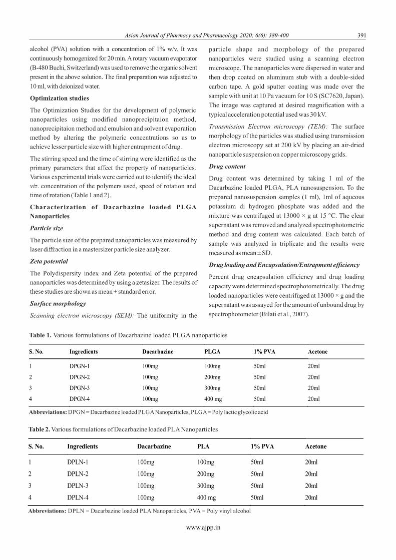
alcohol (PVA) solution with a concentration of 1% w/v. It was
continuously homogenized for 20 min. A rotary vacuum evaporator
(B-480 Buchi, Switzerland) was used to remove the organic solvent
present in the above solution. The final preparation was adjusted to
10 ml, with deionized water.
Optimization studies
The Optimization Studies for the development of polymeric nanoparticles using modified nanoprecipitaion method, nanoprecipitaion method and emulsion and solvent evaporation method by altering the polymeric concentrations so as to
achieve lesser particle size with higher entrapment of drug.
The stirring speed and the time of stirring were identified as the primary parameters that affect the property of nanoparticles. Various experimental trials were carried out to identify the ideal viz. concentration of the polymers used, speed of rotation and time of rotation (Table 1 and 2).
Characterization of Dacarbazine loaded PLGA Nanoparticles
Particle size
The particle size of the prepared nanoparticles was measured by
laser diffraction in a mastersizer particle size analyzer.
Zeta potential
The Polydispersity index and Zeta potential of the prepared nanoparticles was determined by using a zetasizer. The results of these studies are shown as mean ± standard error.
Surface morphology
Scanning electron microscopy (SEM): The uniformity in the
particle shape and morphology of the prepared
nanoparticles were studied using a scanning electron
microscope. The nanoparticles were dispersed in water and
then drop coated on aluminum stub with a double-sided
carbon tape. A gold sputter coating was made over the
sample with unit at 10 Pa vacuum for 10 S (SC7620, Japan).
The image was captured at desired magnification with a
typical acceleration potential used was 30 kV.
Transmission Electron microscopy (TEM): The surface
morphology of the particles was studied using transmission
electron microscopy set at 200 kV by placing an air-dried
nanoparticle suspension on copper microscopy grids.
Drug content
Drug content was determined by taking 1 ml of the
Dacarbazine loaded PLGA, PLA nanosuspension. To the
prepared nanosuspension samples (1 ml), 1ml of aqueous
potassium di hydrogen phosphate was added and the
mixture was centrifuged at 13000 × g at 15 °C. The clear
supernatant was removed and analyzed spectrophotometric
method and drug content was calculated. Each batch of
sample was analyzed in triplicate and the results were
measured as mean ± SD.
Drug loading and Encapsulation/Entrapment efficiency
Percent drug encapsulation efficiency and drug loading
capacity were determined spectrophotometrically. The drug
loaded nanoparticles were centrifuged at 13000 × g and the
supernatant was assayed for the amount of unbound drug by
spectrophotometer (Bilati et al., 2007).
www.ajpp.in
Table 1. Various formulations of Dacarbazine loaded PLGA nanoparticles
Abbreviations: DPGN = Dacarbazine loaded PLGA Nanoparticles, PLGA = Poly lactic glycolic acid
Table 2. Various formulations of Dacarbazine loaded PLA Nanoparticles
Abbreviations: DPLN = Dacarbazine loaded PLA Nanoparticles, PVA = Poly vinyl alcohol
S. No. Ingredients Dacarbazine PLGA 1% PVA Acetone
1 DPGN-1 100mg 100mg 50ml 20ml
2 DPGN-2 100mg 200mg 50ml 20ml
3 DPGN-3 100mg 300mg 50ml 20ml
4 DPGN-4 100mg 400 mg 50ml 20ml
S. No. Ingredients Dacarbazine PLA 1% PVA Acetone
1 DPLN-1 100mg 100mg 50ml 20ml
2 DPLN-2 100mg 200mg 50ml 20ml
3 DPLN-3 100mg 300mg 50ml 20ml
4 DPLN-4 100mg 400 mg 50ml 20ml
Asian Journal of Pharmacy and Pharmacology 2020; 6(6): 389-400 391
In vitro drug release studies
The release profiles of the prepared nanosuspensions in vitro
were studied by diffusion across an artificial membrane to
determine the permeation/release rate of drug from optimized
formulations. Suitable compendia method of dissolution used for
determining the release behavior and kinetics of drug release
from formulation.
Nanosuspension with known concentration of Dacarbazine was
taken in double opened diffusion tube with semipermeable
membrane tied at one end (donor compartment). Specified volume
of buffer was placed in a 250 ml beaker (receptor compartment)
and the nanosuspension loaded diffusion tube was dipped in the
buffer solution. The buffer in the receptor compartment was
constantly agitated using a magnetic stirrer which was maintained
at 37°C. Equivalent volume of the fresh media was added after
withdrawal of each sample from the receptor compartment for
estimation of released drug. The experiment was carried out in
triplicate and the values were reported as mean value ± standard
deviation (Shrinidh et al., 2010).
Stability studies
Stability is used to define as the extent to which the product
remains within specified limits throughout its period of storage
and use. The chemical and physical stability of drug was
determined according to ICH guidelines under accelerated
storage environment (40 ± 2°C/ 75 ± 5% RH) and long term
storage environment (25 ± 2°C/60 ± 5% RH) (ICH guidelines).
Results and discussion
Solubility of pure Dacarbazine
The solubility of drug was determined by for various solvents
like water, ethanol, acetone chloroform etc serial
solubilization method, drug found slightly soluble in almost
all solvents.
Melting point
The melting point of sample of Dacarbazine was observed
and was recorded 214-215°C.
Compatibility Studies
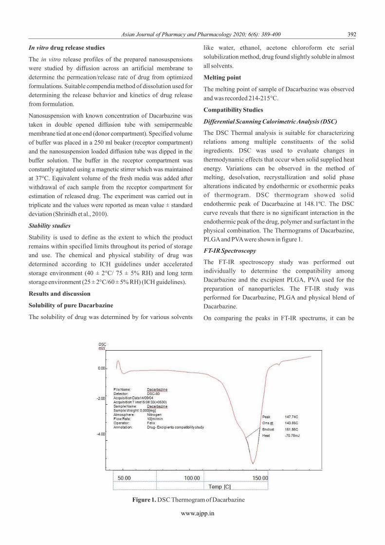
Differential Scanning Calorimetric Analysis (DSC)
The DSC Thermal analysis is suitable for characterizing relations among multiple constituents of the solid ingredients. DSC was used to evaluate changes in
thermodynamic effects that occur when solid supplied heat energy. Variations can be observed in the method of melting, desolvation, recrystallization and solid phase alterations indicated by endothermic or exothermic peaks of thermogram. DSC thermogram showed solid endothermic peak of Dacarbazine at 148.1ºC. The DSC
curve reveals that there is no significant interaction in the endothermic peak of the drug, polymer and surfactant in the physical combination. The Thermograms of Dacarbazine, PLGA and PVA were shown in figure 1.
FT-IR Spectroscopy
The FT-IR spectroscopy study was performed out individually to determine the compatibility among Dacarbazine and the excipient PLGA, PVA used for the preparation of nanoparticles. The FT-IR study was performed for Dacarbazine, PLGA and physical blend of
Dacarbazine.
On comparing the peaks in FT-IR spectrums, it can be
www.ajpp.in
Figure 1. DSC Thermogram of Dacarbazine
Asian Journal of Pharmacy and Pharmacology 2020; 6(6): 389-400 392
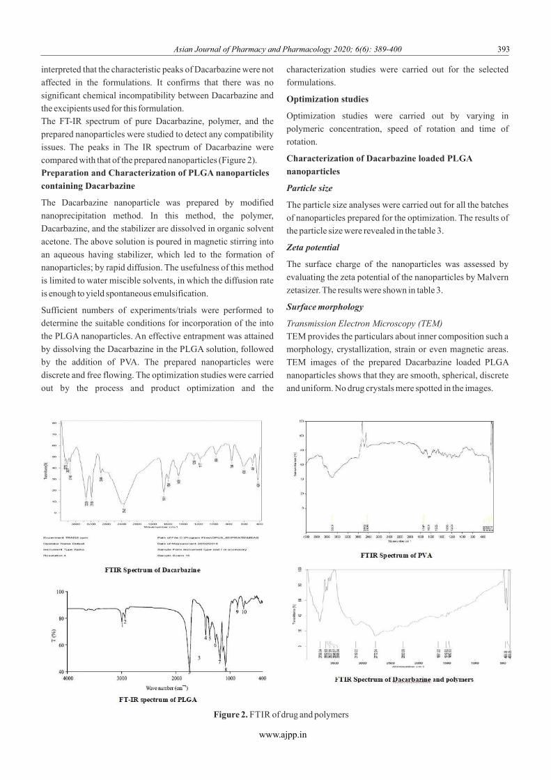
interpreted that the characteristic peaks of Dacarbazine were not
affected in the formulations. It confirms that there was no
significant chemical incompatibility between Dacarbazine and
the excipients used for this formulation.
The FT-IR spectrum of pure Dacarbazine, polymer, and the
prepared nanoparticles were studied to detect any compatibility
issues. The peaks in The IR spectrum of Dacarbazine were
compared with that of the prepared nanoparticles (Figure 2).
Preparation and Characterization of PLGA nanoparticles
containing Dacarbazine
The Dacarbazine nanoparticle was prepared by modified
nanoprecipitation method. In this method, the polymer,
Dacarbazine, and the stabilizer are dissolved in organic solvent
acetone. The above solution is poured in magnetic stirring into
an aqueous having stabilizer, which led to the formation of
nanoparticles; by rapid diffusion. The usefulness of this method
is limited to water miscible solvents, in which the diffusion rate
is enough to yield spontaneous emulsification.
Sufficient numbers of experiments/trials were performed to
determine the suitable conditions for incorporation of the into
the PLGA nanoparticles. An effective entrapment was attained
by dissolving the Dacarbazine in the PLGA solution, followed
by the addition of PVA. The prepared nanoparticles were
discrete and free flowing. The optimization studies were carried
out by the process and product optimization and the
characterization studies were carried out for the selected
formulations.
Optimization studies
Optimization studies were carried out by varying in
polymeric concentration, speed of rotation and time of
rotation.
Characterization of Dacarbazine loaded PLGA
nanoparticles
Particle size
The particle size analyses were carried out for all the batches
of nanoparticles prepared for the optimization. The results of
the particle size were revealed in the table 3.
Zeta potential
The surface charge of the nanoparticles was assessed by
evaluating the zeta potential of the nanoparticles by Malvern
zetasizer. The results were shown in table 3.
Surface morphology
Transmission Electron Microscopy (TEM)
TEM provides the particulars about inner composition such a
morphology, crystallization, strain or even magnetic areas.
TEM images of the prepared Dacarbazine loaded PLGA
nanoparticles shows that they are smooth, spherical, discrete
and uniform. No drug crystals mere spotted in the images.
www.ajpp.in
Figure 2. FTIR of drug and polymers
Asian Journal of Pharmacy and Pharmacology 2020; 6(6): 389-400 393

Drug content and Entrapment efficiency
The Drug content of the prepared Dacarbazine loaded PLGA
nanoparticles were evaluated after making suitable dilutions
using the established analytical method. The drug content of
DPGN-3 was found to be 0.955µg/ml. The rise in concentration
of polymer in the organic phase produced an increase in drug
content of the nanoparticles.
The encapsulation efficiency of Dacarbazine in the PLGA
nanoparticles was 68.50 to 83.15% which is quite satisfactory.
However, the percentage of entrapment efficiency of the drug was
reliant on the polymer ratio, stirring speed and stirring rpm. The
nanoparticles with DPGN-3 shows average percentage of entrapment
efficiency of 83.15%, formulation with DPGN-1, DPGN-2, DPGN-
4, shows 68.50%, 74.72%, and 79.00% respectively. The entrapment
efficiency of The PLGA nanoparticles were originates to be increased
up to drug: polymer ratio of 1:3. This may be due to increased
adsorption of the Dacarbazine on the surface of the polymeric
matrices. However, a further increase of polymeric concentration had
not indicated increase in entrapment efficiency.
In vitro drug release studies
The release of Dacarbazine loaded PLGA nanoparticles in vitro
showed prolonged and sustained release of Dacarbazine.
The results of diffusion studies of optimized in vitro
nanoparticles were shown in the table 4. It was obvious that
in vitro release of Dacarbazine exhibited rapid initial burst
release and then followed by the sustained release up to 24
hrs. An initial quick release suggests that specific quantity
of drug was confined to the surface of nanoparticles.
DPGN-3 shows maximum release of drug when compared
to other batches of nanoparticles.
The comparison of the results of optimized Dacarbazine
PLGA nanoparticles, created on the particle size, zeta
potential, drug content, entrapment efficiency and in vitro
release studies has been shown in table 5. The formulation
DPGN-3 indicated the least particle size, higher zeta
potential, drug content, entrapment efficiency and
sustained drug release. Hence among the different trials of
Dacarbazine loaded PLGA nanoparticles, DPGN-3 has
been identified to carry out further studies.
Stability studies
Stability studies were carried out for DPGN-3. The
prepared Dacarbazine loaded PLGA formulations were
www.ajpp.in
Table 3. Particle size and Zeta potential of the loaded PLGA nanoparticles
Sl. No Code Particle size (nm) Zeta potential (mV)
1 DPGN-1 273±1.7 -27±1.51
2 DPGN-2 266±1.4 -29±1.8
3 DPGN-3 246±2.4 -27±1.4
4 DPGN-4 289±3.2 -25±1.6
Figure 3. TEM micrographs of (A) DPGN-1 (B) DPGN-2 (C) DPGN-3 and (D) DPGN-4 nanoformulation
Asian Journal of Pharmacy and Pharmacology 2020; 6(6): 389-400 394

stored at the following conditions 5 ± 3 °C, 30±2 °C, 65% ± i.e.,
5% RH (long term stability), 40±2 °C, 75% ± 5% (accelerated
stability). Every three months the drug content, release in vitro
studies were determined for the nano-formulation subjected for
long term stability studies.
The drug content of DPGN-3 stored at 5±3°C for a period of 12
months exhibited a slight fall in the drug content when compared
to the initial drug content of the nano-formulation after storing
the sample for 12 months.
The optimized nanoformulation DPGN-3 at 40±2°C, 75±5% RH
after 0, 6, & 12 months indicated substantial decrease in
cumulative drug release when related to the initial cumulative
drug release of the same nanoformulation.
On comparing the drug content after storing the optimized
nanoformulation DPGN-3 at 5±3°C, 30 ± 2°C, 65 ± 5% RH, when
compared to previous data of the same formulation there was a
minor decrease in the drug content after 12 months of the storage.
Every three months the drug release almost remains the in vitro
same for the optimized nano-formulation DPGN-3 stored at
5±3°C, 30 ± 2°C, 65 ± 5% RH up to 12 months.
However, The Dacarbazine nano formulation, which was subjected
for accelerated stability studies on 40 ±2° C, 75±5% RH, shows a
major decrease in drug content and drug release. It may be in vitro
due to storage at high temperature which leads the degradation of
Dacarbazine loaded PLGA nanoparticles. Hence, there is a major
decrease in the drug content and cumulative percentage of in
vitro drug release. Therefore, from the stability studies it was
observed that the prepared Dacarbazine loaded PLGA nano-
formulation DPGN-3 will be stable at 5±3°C, 30 ± 2°C, 65 ±
5% RH for a period of 12 months.
Compatibility Studies
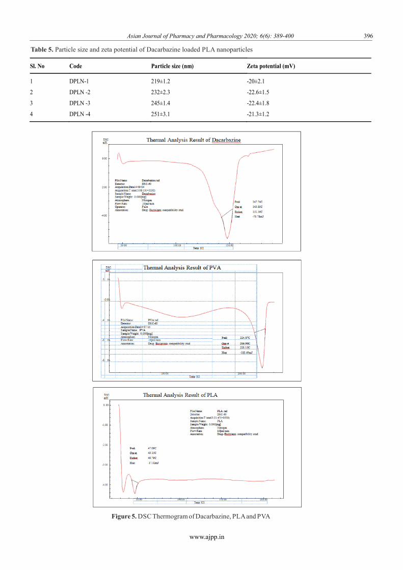
Differential Scanning Calorimetric Analysis (DSC)
Thermal analysis was used to assess fluctuations in
Thermodynamic properties that happen when the material
supplied heat energy. Variations that can be detected in the
method of melting, desolvation, recrystallization and solid
phase changes indicated by endothermic or exothermic peaks at
Thermogram. DSC Thermogram showed solid endothermic
peak of Dacarbazine at 148.4ºC. DSC Thermogram of physical
mixture shows three endothermic peaks at 223.14 ºC
respectively. The DSC curve reveals that there is no major
interface in the endothermic peak of the drug, polymer and
surfactant in the physical mixture. The Thermograms of
Dacarbazine, PLA and PVA were shown in figure 5.
Preparation of PLA nanoparticles
PLA nanoparticles loaded with Dacarbazine were processed
by the nanoprecipitaion method. To optimize the product
parameters and process parameters 04 typical formulations
were designed and studies were carried out for Dacarbazine
loaded PLA nanoparticles. The prepared nanoparticles were
discrete and free flowing.
www.ajpp.in
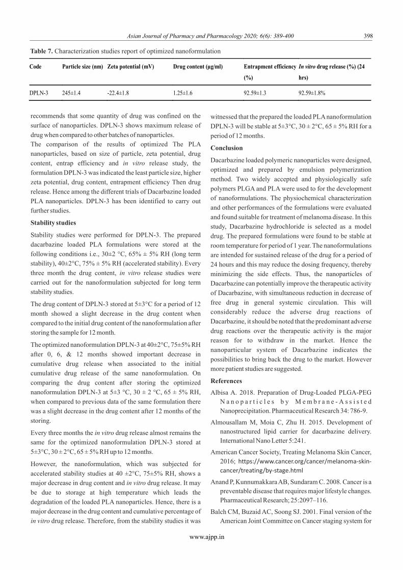
Table 4. Characterization studies report of optimized nanoformulation
Code Particle size
(nm)
Zeta potential (mV) Drug content (µg/ml) Entrapment efficiency (%) In vitro drug release (%)
(24 hrs)
DPGN-3 246±2.4 -27±1.4 0.955±1.3 83.15±1.4 86.67±2.4
Figure 4. In vitro drug release profiles of Dacarbazine loaded PLGA nanoformulations in Phosphate buffer pH .4
Asian Journal of Pharmacy and Pharmacology 2020; 6(6): 389-400 395
www.ajpp.in
Figure 5. DSC Thermogram of Dacarbazine, PLA and PVA
Table 5. Particle size and zeta potential of Dacarbazine loaded PLA nanoparticles
Sl. No Code Particle size (nm) Zeta potential (mV)
1 DPLN-1 219±1.2 -20±2.1
2 DPLN -2 232±2.3 -22.6±1.5
3 DPLN -3 245±1.4 -22.4±1.8
4 DPLN -4 251±3.1 -21.3±1.2
Asian Journal of Pharmacy and Pharmacology 2020; 6(6): 389-400 396
www.ajpp.in
Characterization of Dacarbazine loaded PLA nanoparticles
Particle size
The literature review that the size of particles is extremely reliant
on the preparation method adopted and conditions employed.
Though the previous reports suggests that a rise in the
concentration of the polymer a gain of the particle size. The
Dacarbazine loaded PLA nanoformulations were optimized by
product and process parameters. The mean particle size of
nanoformulations were carried out which was tabulated in table
6. However, the particle size also changes with polymer viscosity
and stirring speed of rotation of the stirrer.
Zeta potential
The surface charges of the nanoparticles were assessed by
evaluating the zeta potential of the nanoparticle by Malvern zeta
sizer.
Surface morphology
Transmission Electron Microscopy (TEM)
The particularly internal composition such morphology,
crystallization, stress or even magnetic fields were studied. TEM
images of PLA nanoparticles containing Dacarbazine were
found to be smooth, spherical and uniform.
Drug content
The samples were examined after making suitable dilutions
using the well well-known analytical procedure. The drug
content of PLA nanoparticles was found to be 0.855, 0.983, 1.205
and 0.857µg/ml for DPLN-1, DPLN-2, DPLN-3 and DPLN-4
respectively. The raise in concentration of polymer in organic
phase produced an increase in drug content of the nanoparticles.
Entrapment efficiency
Drug Entrapment efficiency shows important role in preparation of a drug delivery method particularly for
expensive drugs and directly associated to the Therapeutic properties of the system. The encapsulation efficiency of Dacarbazine in the PLA nanoparticles was within the range of 62.53 to 72.32% which is quite satisfactory. The percentage entrapment efficiency of the optimized Dacarbazine loaded PLA nanoparticles were resolved by
the technique described.
However, the percentage of entrapment efficiency of the drug was reliant on the polymer ratio, stirring speed and stirring rpm. The nanoparticles with DPLN-3 show average percentage of entrapment efficiency of 74.53%, formulation with DPLN-1, DPLN-2, and DPLN-4 show 62.53%, 67.25% and 73.23% respectively. The entrapment
efficiency for PLA nanoparticles were found to be increased up to drug: polymer ratio of 1:3. This may be due to increased adsorption of the dacarbazine on the surface of the polymeric matrices. However, a further increase of polymeric concentration had not indicated increase in entrapment efficiency.
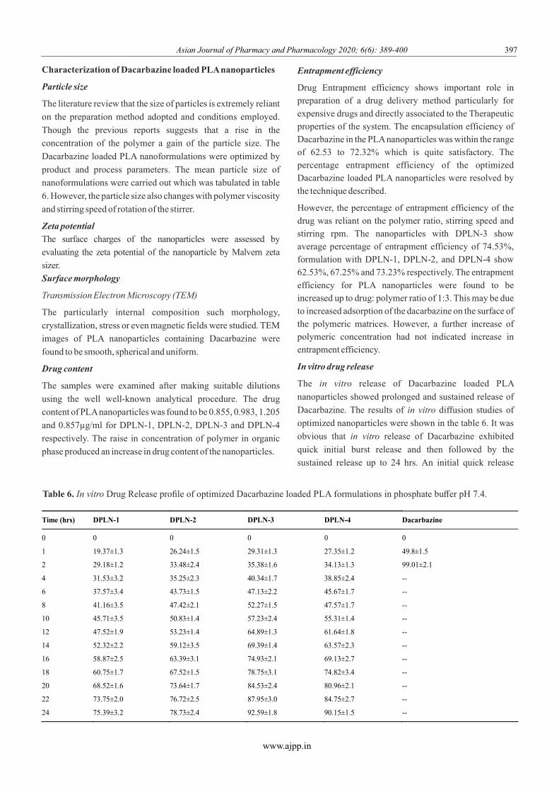
In vitro drug release
The release of Dacarbazine loaded PLA in vitro
nanoparticles showed prolonged and sustained release of
Dacarbazine. The results of diffusion studies of in vitro
optimized nanoparticles were shown in the table 6. It was
obvious that release of Dacarbazine exhibited in vitro
quick initial burst release and then followed by the
sustained release up to 24 hrs. An initial quick release
Time (hrs) DPLN-1 DPLN-2 DPLN-3 DPLN-4 Dacarbazine
0 0 0 0 0 0
1 19.37±1.3 26.24±1.5 29.31±1.3 27.35±1.2 49.8±1.5
2 29.18±1.2 33.48±2.4 35.38±1.6 34.13±1.3 99.01±2.1
4 31.53±3.2 35.25±2.3 40.34±1.7 38.85±2.4 --
6 37.57±3.4 43.73±1.5 47.13±2.2 45.67±1.7 --
8 41.16±3.5 47.42±2.1 52.27±1.5 47.57±1.7 --
10 45.71±3.5 50.83±1.4 57.23±2.4 55.31±1.4 --
12 47.52±1.9 53.23±1.4 64.89±1.3 61.64±1.8 --
14 52.32±2.2 59.12±3.5 69.39±1.4 63.57±2.3 --
16 58.87±2.5 63.39±3.1 74.93±2.1 69.13±2.7 --
18 60.75±1.7 67.52±1.5 78.75±3.1 74.82±3.4 --
20 68.52±1.6 73.64±1.7 84.53±2.4 80.96±2.1 --
22 73.75±2.0 76.72±2.5 87.95±3.0 84.75±2.7 --
24 75.39±3.2 78.73±2.4 92.59±1.8 90.15±1.5 --
Table 6. In vitro Drug Release profile of optimized Dacarbazine loaded PLA formulations in phosphate buffer pH 7.4.
Asian Journal of Pharmacy and Pharmacology 2020; 6(6): 389-400 397
Asian Journal of Pharmacy and Pharmacology 2020; 6(6): 389-400 398
recommends that some quantity of drug was confined on the
surface of nanoparticles. DPLN-3 shows maximum release of
drug when compared to other batches of nanoparticles.
The comparison of the results of optimized The PLA
nanoparticles, based on size of particle, zeta potential, drug
content, entrap efficiency and release study, the in vitro
formulation DPLN-3 was indicated the least particle size, higher
zeta potential, drug content, entrapment efficiency Then drug
release. Hence among the different trials of Dacarbazine loaded
PLA nanoparticles. DPLN-3 has been identified to carry out
further studies.
Stability studies
Stability studies were performed for DPLN-3. The prepared
dacarbazine loaded PLA formulations were stored at the
following conditions i.e., 30±2 °C, 65% ± 5% RH (long term
stability), 40±2°C, 75% ± 5% RH (accelerated stability). Every
three month the drug content, release studies were in vitro
carried out for the nanoformulation subjected for long term
stability studies.
The drug content of DPLN-3 stored at 5±3°C for a period of 12
month showed a slight decrease in the drug content when
compared to the initial drug content of the nanoformulation after
storing the sample for 12 month.
The optimized nanoformulation DPLN-3 at 40±2°C, 75±5% RH
after 0, 6, & 12 months showed important decrease in
cumulative drug release when associated to the initial
cumulative drug release of the same nanoformulation. On
comparing the drug content after storing the optimized
nanoformulation DPLN-3 at 5±3 °C, 30 ± 2 °C, 65 ± 5% RH,
when compared to previous data of the same formulation there
was a slight decrease in the drug content after 12 months of the
storing.
Every three months the drug release almost remains the in vitro
same for the optimized nanoformulation DPLN-3 stored at
5±3°C, 30 ± 2°C, 65 ± 5% RH up to 12 months.
However, the nanoformulation, which was subjected for
accelerated stability studies at 40 ±2°C, 75±5% RH, shows a
major decrease in drug content and drug release. It may in vitro
be due to storage at high temperature which leads the
degradation of the loaded PLA nanoparticles. Hence, there is a
major decrease in the drug content and cumulative percentage of
in vitro drug release. Therefore, from the stability studies it was
www.ajpp.in
witnessed that the prepared the loaded PLA nanoformulation
DPLN-3 will be stable at 5±3°C, 30 ± 2°C, 65 ± 5% RH for a
period of 12 months.
Conclusion
Dacarbazine loaded polymeric nanoparticles were designed,
optimized and prepared by emulsion polymerization
method. Two widely accepted and physiologically safe
polymers PLGA and PLA were used to for the development
of nanoformulations. The physiochemical characterization
and other performances of the formulations were evaluated
and found suitable for treatment of melanoma disease. In this
study, Dacarbazine hydrochloride is selected as a model
drug. The prepared formulations were found to be stable at
room temperature for period of 1 year. The nanoformulations
are intended for sustained release of the drug for a period of
24 hours and this may reduce the dosing frequency, thereby
minimizing the side effects. Thus, the nanoparticles of
Dacarbazine can potentially improve the therapeutic activity
of Dacarbazine, with simultaneous reduction in decrease of
free drug in general systemic circulation. This will
considerably reduce the adverse drug reactions of
Dacarbazine, it should be noted that the predominant adverse
drug reactions over the therapeutic activity is the major
reason for to withdraw in the market. Hence the
nanoparticular system of Dacarbazine indicates the
possibilities to bring back the drug to the market. However
more patient studies are suggested.
References
Albisa A. 2018. Preparation of Drug-Loaded PLGA-PEG N a n o p a r t i c l e s b y M e m b r a n e - A s s i s t e d Nanoprecipitation. Pharmaceutical Research 34: 786-9.
Almousallam M, Moia C, Zhu H. 2015. Development of nanostructured lipid carrier for dacarbazine delivery. International Nano Letter 5:241.
American Cancer Society, Treating Melanoma Skin Cancer, 2016; https://www.cancer.org/cancer/melanoma-skin-cancer/treating/by-stage.html
Anand P, Kunnumakkara AB, Sundaram C. 2008. Cancer is a preventable disease that requires major lifestyle changes. Pharmaceutical Research; 25:2097–116.
Balch CM, Buzaid AC, Soong SJ. 2001. Final version of the American Joint Committee on Cancer staging system for
Code Particle size (nm) Zeta potential (mV) Drug content (µg/ml) Entrapment efficiency
(%)
In vitro drug release (%) (24
hrs)
DPLN-3 245±1.4 -22.4±1.8 1.25±1.6 92.59±1.3 92.59±1.8%
Table 7. Characterization studies report of optimized nanoformulation
Asian Journal of Pharmacy and Pharmacology 2020; 6(6): 389-400 399
www.ajpp.in
cutaneous melanoma. Journal of Clinical Oncology, 19: 3635-48.
Bamrungsap S, Zhao Z, Chen T, Wang L, Li C, Fu T, Tan W. 2012. A Focus on Nanoparticles as a Drug Delivery System. Nanomecine, 7:1253-71.
Bei D, Zhang T, Murowchick JB, Youan BC. 2010. Formulation of Dacarbazine-loaded cubosomes, Part III. P h y s i c o c h e m i c a l c h a r a c t e r i z a t i o n . A A P S PharmSciTech, 11: 1243–9.
Beiu C. 2020. Nanosystems for Improved Targeted Therapies in Melanoma. Journal of Clinical Medicine, 9: 318.
Bharath A, Turner R. 2009. Impact of climate change on skin cancer. Journal of the Royal Society of Medicine, 102: 215-8.
C al i fo r n i a Pa c i f ic Me d ic a l Ce nte r ( CPMC) , Biochemotherapy for Treating Metastatic Melanoma, Sutter Health, http://www.cpmc.org/about/e-health/12-05%20IL-2.html
Cheng J, Teply BA, Sherifi I. 2007. Formulation of Functionalized PLGA-PEG Nanoparticles for in vivo targeted drug delivery. Biomaterials, 28: 869-76.
Das S, Khuda-Bukhsh AR. 2016. PLGA-loaded nanomedicines in melanoma treatment: Future prospect for efficient drug delivery. The Indian Journal of Medical Research, 144:181-93.
Das S, Khuda-Bukhsh AR. 2016. PLGA-loaded nanomedicines in melanoma treatment: Future prospect for efficient drug delivery. Indian Journal of Medical Research, 144:181-93.
Ding B, Wu X, Fan W, Wu Z, Gao J, Zhang W, Ma L, Xiang W, Zhu Q, Liu J, Ding X, Gao S. 2011. Anti-DR5 monoclonal antibody-mediated DTIC-loaded nanopart icles combining chemotherapy and immunotherapy for malignant melanoma: target formulation development and in vitro anticancer activity. International Journal of Nanomedicine, 6: 1991–2005.
Ding B, Zhang W, Wu X. 2016. DR5 mAb-conjugated, DTIC-loaded immuno-nanoparticles effectively and specifically kill malignant melanoma cells in vivo. Oncotarget, 7: 57160-70.
Ding, B, Zhang W, Wu X, Wang X, Fan W, Gao S, Gao J, Ma L, Ding X, Hao Q. 2011. Biodegradable methoxy poly (ethylene glycol)-poly (lactide) nanoparticles for controlled delivery of dacarbazine: preparation, characterization and anticancer activity evaluation. Arican Journal of Pharmacy and Pharmacology, 5: 1369–77.
Eifler AC, Thaxton CS. 2011. Nanoparticle therapeutics:
FDA approval, clinical trials, regulatory pathways, and case study Methods in Molecular Biology 726. , :325–3810.
https://www.ich.org/fileadmin/Public_Web_Site/ICH_Products/Guidelines/Quality/Q1A_R2/Step4/Q1A_R2__Guideline.pdf
International Conference on Harmonization (ICH), Q1A(R) Stability testing of new drug substances and products, ICH Guideline.
Jain A, Jain SK. 2013. Formulation and optimization of temozolomide nanoparticles by 3 factor 2 level factorial designs. Biomaterials, 3: 25102-13.
Jiang G, Li RH, Sun C, Liu YQ, Zheng JN. 2014. Dacarbazine combined targeted therapy versus dacarbazine alone in patients with malignant melanoma: a meta-analysis. PLoS One, 9: 111920.
Kakumanu S, Tagne JB, Wilson TA, Nicolosi RJ. 2011. A nanoemulsion formulation of dacarbazine reduces tumor size in a xenograft mouse epidermoid carcinoma model compared to dacarbazine suspension. Nanomedicine and Nanotechnology 7: 277–83.
Kakumanu S, Tagne JB, Wilson TA, Nicolosi RJ. 2011. A nanoemulsion formulation of dacarbazine reduces tumor size in a xenograft mouse epidermoid carcinoma model compared to dacarbazine suspension, Nanomedicine: Nanotechnology. Biology and Medicine, 7: 277-83.
Lionetti MC, Fumagalli MR, La Porta CAM. 2020. Cancer stem cells, plasticity, and drug resistance. Cancer Drug Resistance, 3:140-8.
Mehrotra A, Pandit JK. 2015. Preparation and Characterization and Biodistribution Studies of Lomustine Loaded PLGA Nanoparticles by Interfacial Deposition Method. Journal of Nanomedicine and Biotherapeutic Discovery, 5:138.
Miglietta A, Cavalli R, Bocca C, Gabriel L, Gasco MR. 2000. Cellular uptake and cytotoxicity of solid lipid nanospheres (SLN) incorporating doxorubicin or paclitaxel. International Journal of Pharmaceutics, 210: 61–7.
Musallam A, Claudia M, Huijun Z. 2015. Development of nanostructured lipid carrier for dacarbazine delivery. International Nano Letters, 5(4):241–8.
Naves LB, Dhand C, Venugopal JR. 2017. Nanotechnology for the treatment of melanoma skin cancer. Progress in Biomaterials, 6: 13–26.
Nisha Oommachen, Vismi V, Soumya S, Jeena CD. 2013. Melanoma skin cancer detection based on skin lesions characterization. IOSR Journal of Engineering (IOSRJEN); 3(2): 52-9.
Noori KM, Khoshayand MR, Mostafavi SH, 2014. Docetaxel Loaded PEG-PLGA Nanoparticles: Optimized drug loading, in-vitro cytotoxicity and in-vivo antitumor effect. Iranian

Asian Journal of Pharmacy and Pharmacology 2020; 6(6): 389-400 400
www.ajpp.in
Journal of Pharmaceutical Research, 13:819-33.
Pamujula S. 2012. Cellular delivery of PEGylated PLGA nanoparticles. Journal of Pharmacy and Pharmacology, 64: 61–7.
Qianqian Liu. 2017. Dacarbazine-Loaded Hollow Mesoporous Silica nanoparticles grafted with folic acid for enhancing antimetastatic melanoma response. ACS Applied Materials & Interfaces, 9 : 21673–87.
Rafiei P, Haddadi A. 2017. Docetaxel-loaded PLGA and PLGA-PEG nanoparticles for intravenous application: pharmacokinetics and biodistribution profile. International Journal of Nanomedicine, 12:935-47.
S k i n C a n c e r F o u n d a t i o n ( S C F ) r e p o r t 2 0 1 7 ; h t t p : / / w w w . s k i n c a n c e r . o r g / s k i n - c a n c e r -information/melanoma
Tsubaki M. 2019. Combination therapy with dacarbazine and statins improved the survival rate in mice with metastatic melanoma. Journal of Cellular Physiology, 234;11045-8.
Vangara KK, Liu JL, Palakurthi S. 2013. Hyaluronic acid-decorated PLGA-PEG nanoparticles for targeted delivery of SN-38 to ovarian cancer. Anticancer Research, 33:2425-34.
Yaman S, Ramachandramoorthy H, Oter G. 2020. Melanoma Peptide MHC Specific TCR Expressing T-Cell Membrane Camouflaged PLGA Nanoparticles for Treatment of Melanoma Skin Cancer. Frontiers in Bioengineering and Biotechnology, 8:943.
Yi JH, Yi SY,Lee HR,Lee SI, Lim DH,Kim JH, Park KW, Lee J.
2011. Dacarbazine-based chemotherapy as first-line treatment in noncutaneous metastatic melanoma: Multicenter, retrospective analysis in Asia. Melanoma Research, 21: 223-7.
Zbytek B, Carlson JA, Granese J, Ross J, Mihm MC, Slominski A. 2008. Current concepts of metastasis in melanoma. Expert Review of Dermatology, 3:569-85.
Zhang Y, Guo L, Roeske RW, Antony AC, Jayaram HN. 2004. Pteroyl-γ-glutamate-cysteine synthesis and its application in folate receptor-mediated cancer cell targeting using folate-tethered liposomes. Analytical Biochemistry, 332:168–77.
Zhou Y, Sridhar R, Shan L, Sha W, Gu X, Sukumar S. Loperamide. 2012. An FDA-approved antidiarrhea drug, effectively reverses the resistance of multidrug resistant MCF-7/MDR1 human breast cancer cells to doxorubicin-induced cytotoxicity. Cancer Investigation, 30:119.