Effects of inhibitors of ion-motive ATPases on the plasma membrane potential of murine...
14
J. Membrane Biol. 126, 123-136 (1992) The Journal of Membrane Biology Springer.Vertag New York Inc. 1992 Effects of lnhibitors of Ion-Motive ATPases on the Plasma Membrane Potential of Murine Erythroleukemia Cells A. Arcangelir M.R. Del Bener A. Becchetti$, E. Wanke +, and M. Olivotto? +Institute of General Pathology, University of Florence, 50134 Florence, Italy, and SDepartment of General Physiology and Biochemistry, University of Milan, 20133 Milan, Italy Summary. The membrane electric effects of N,N'-dicyclohexyl- carbodiimide (DCCD) and vanadate were studied in murine eryth- roleukemia cells (MELC), comparing the patch-clamp technique and the accumulation ratio (ARex p) of [3H]-tetraphenylphospho- nium (TPP§ Electrophysiological measurements showed that both these inhibitors produce, at micromolar concentrations, a 20-30 mV hyperpolarization of resting potential (A%) of MELC, which is abolished when the electrochemical equilibrium potential of K + (EK) is brought close to zero. DCCD and vanadate turned out to have distinct targets on the plasma membrane of MELC (an H + pump and the Na + ,K +- ATPase, respectively). Measurements of ARe• showed that: (i) patch-clamp mea- surements of A% were equivalent to those based on ARe• p of antimycin-pretreated cells (ARANT); (ii) DCCD produced a strong increase in ARAb,, T, that was antagonized by carbonyl cyanide p- trifluoromethoxyphenyl-hydrazone (FCCP) and diethylstilbestrol (DES); (iii) vanadate determined a marked increase in ARAN T that was insensitive to FCCP, but antagonized by ouabain; (iv) incubation in high K* medium (HK) brought ARAN T to 1.0 in the controls, but did not lower this ratio below 3.0 in the presence of DCCD or vanadate; (v) the total amount ofTPP + taken up by the cells was in any case water extractable by a freezing and thawing procedure. On the whole, our data indicate that DCCD and vanadate hyperpolarize the MELC by increasing the K + conductance and, at the same time, enhance the TPP + binding, probably by chang- ing the electrostatic potential profile of the plasma membrane. These effects seem to involve functional modifications of the target pumps, apparently related to the ion-occluding state of these enzymes. Key Words ion-motive ATPase inhibitors leukemia cells . resting potential potassium conductance. TPP* binding elec- trostatic potential of the plasma membrane Introduction Ion-motive ATPases (Pedersen & Carafoli, 1987a, b) mediate crucial cellular processes such as ATP syn- thesis or ion translocation against energy barriers. In general, they are membrane-intrinsic proteins, composed of two functionally distinct moieties: a portion interacting with ATP in the cellular aqueous compartment (the catalytic portion) and the ion- translocating portion across the membrane (Amzel & Pedersen, 1983; A1-Awqati, 1986; Skou, 1988; Fu- tai, Noumi & Maeda, 1989). Various inhibitors greatly contributed to char- acterize diverse types of ion motive ATPases and to elucidate their functional properties (Glynn & Karlish, 1975; Penefsky, 1979; Goffeau & Slayman, 1981; Pedersen & Carafoli, 1987a). In particular, DCCD specifically blocks the proton translocation step through the transmembrane moiety of H +- ATPases (Hoppe & Sebald, 1984), while vanadate predominantly interferes with the ion-translocating activity of Na+,K+-ATPases (Cantley, Resh & Guidotti, 1978; Glynn & Richards, 1982; Beaug6, 1988). Interestingly, both DCCD (De La Pefia et al., 1982; Baroni et al., 1988) and vanadate (Licht- shtein, Mullikin-Kilpatrick & Btume, 1982) have been reported to hyperpolarize the plasma mem- brane of many different types of cells, including yeasts and neuroblastoma cells. These studies have been performed essentially by measuring the intra- cellular accumulation of the lipophylic cation TPP +, but the hyperpolarizing properties of vana- date were confirmed by direct electrophysiological measurements. Although the hyperpolarization in- duced by DCCD in yeasts was traced to a K + efflux through K + channels (De La Pefia et al., 1982; Baroni et al., 1988), while that sustained by vanadate was accompanied by an increase in the membrane conductance (Lichtshtein et al., 1982), the mechanism underlying these processes remains obscure so far. We considered it worthwhile to explore this
-
Upload
independent -
Category
Documents
-
view
3 -
download
0
Transcript of Effects of inhibitors of ion-motive ATPases on the plasma membrane potential of murine...
J. Membrane Biol. 126, 123-136 (1992) The Journal of
Membrane Biology �9 Springer.Vertag New York Inc. 1992
Effects of lnhibitors of Ion-Motive ATPases on the Plasma Membrane Potential of Murine Erythroleukemia Cells
A. Arcangelir M.R. Del Bener A. Becchetti$, E. Wanke +, and M. Olivotto? +Institute of General Pathology, University of Florence, 50134 Florence, Italy, and SDepartment of General Physiology and Biochemistry, University of Milan, 20133 Milan, Italy
Summary. The membrane electric effects of N,N'-dicyclohexyl- carbodiimide (DCCD) and vanadate were studied in murine eryth- roleukemia cells (MELC), comparing the patch-clamp technique and the accumulation ratio (ARex p) of [3H]-tetraphenylphospho- nium (TPP§ Electrophysiological measurements showed that both these inhibitors produce, at micromolar concentrations, a 20-30 mV hyperpolarization of resting potential (A%) of MELC, which is abolished when the electrochemical equilibrium potential of K + (EK) is brought close to zero.
DCCD and vanadate turned out to have distinct targets on the plasma membrane of MELC (an H + pump and the Na + ,K +- ATPase, respectively).
Measurements of ARe• showed that: (i) patch-clamp mea- surements of A% were equivalent to those based on ARe• p of antimycin-pretreated cells (ARANT); (ii) DCCD produced a strong increase in ARAb,, T, that was antagonized by carbonyl cyanide p- trifluoromethoxyphenyl-hydrazone (FCCP) and diethylstilbestrol (DES); (iii) vanadate determined a marked increase in ARAN T that was insensitive to FCCP, but antagonized by ouabain; (iv) incubation in high K* medium (HK) brought ARAN T to 1.0 in the controls, but did not lower this ratio below 3.0 in the presence of DCCD or vanadate; (v) the total amount ofTPP + taken up by the cells was in any case water extractable by a freezing and thawing procedure.
On the whole, our data indicate that DCCD and vanadate hyperpolarize the MELC by increasing the K + conductance and, at the same time, enhance the TPP + binding, probably by chang- ing the electrostatic potential profile of the plasma membrane. These effects seem to involve functional modifications of the target pumps, apparently related to the ion-occluding state of these enzymes.
Key Words ion-motive ATPase inhibitors �9 leukemia cells . resting potential �9 potassium conductance. TPP* binding �9 elec- trostatic potential of the plasma membrane
Introduction
Ion-motive ATPases (Pedersen & Carafoli, 1987a, b) mediate crucial cellular processes such as ATP syn- thesis or ion translocation against energy barriers.
In general, they are membrane-intrinsic proteins, composed of two functionally distinct moieties: a portion interacting with ATP in the cellular aqueous compartment (the catalytic portion) and the ion- translocating portion across the membrane (Amzel & Pedersen, 1983; A1-Awqati, 1986; Skou, 1988; Fu- tai, Noumi & Maeda, 1989).
Various inhibitors greatly contributed to char- acterize diverse types of ion motive ATPases and to elucidate their functional properties (Glynn & Karlish, 1975; Penefsky, 1979; Goffeau & Slayman, 1981; Pedersen & Carafoli, 1987a). In particular, DCCD specifically blocks the proton translocation step through the transmembrane moiety of H +- ATPases (Hoppe & Sebald, 1984), while vanadate predominantly interferes with the ion-translocating activity of Na+,K+-ATPases (Cantley, Resh & Guidotti, 1978; Glynn & Richards, 1982; Beaug6, 1988).
Interestingly, both DCCD (De La Pefia et al., 1982; Baroni et al., 1988) and vanadate (Licht- shtein, Mullikin-Kilpatrick & Btume, 1982) have been reported to hyperpolarize the plasma mem- brane of many different types of cells, including yeasts and neuroblastoma cells. These studies have been performed essentially by measuring the intra- cellular accumulation of the lipophylic cation TPP +, but the hyperpolarizing properties of vana- date were confirmed by direct electrophysiological measurements. Although the hyperpolarization in- duced by DCCD in yeasts was traced to a K + efflux through K + channels (De La Pefia et al., 1982; Baroni et al., 1988), while that sustained by vanadate was accompanied by an increase in the membrane conductance (Lichtshtein et al., 1982), the mechanism underlying these processes remains obscure so far.
We considered it worthwhile to explore this

124 A. Arcangeli et al.: ATPase Inhibitors and Membrane Potential
topic by integrating the TPP + method and the patch-clamp technique, both of which have been employed widely on MELC (Arcangeli & Olivotto, 1986; Arcangeli et al., 1987a,b, 1989a,b). It was thought that this integration should provide infor- mation about the membrane energy profile, which might unravel the electric events, including the surface-charge effects, that occur in the membrane itself, when the ion-translocating moiety of a pump is blocked by specific inhibitors. In fact, the above- mentioned profile depends not only on the resting potential (A%), but also on the membrane dipole potential and on the diffuse double-layer surface potential (McLaughlin, 1977, 1989). While A% can be exactly measured by the patch-clamp technique, accumulation of TPP + in cells and organelles is a function of both the resting potential and a complex of TPP + interactions with membranes, collectively referred to as "binding."
According to a widely accepted model, worked out on neutral phospholipid bilayers, binding con- sists of a confined adsorption of lipophilic ions to potential wells, formed near the membrane-water interfaces by the combination of repulsive electro- static and attractive hydrophobic energy terms (Neumcke & Lfiuger, 1969; Ketterer, Neumcke & Lfiuger, 1971; Flewelling & Hubbell, 1986a,b). This model implies that lipid bilayers possess a dipole potential of several hundred millivolts positive in the interior, a feature of great importance to struc- tural and functional properties of membrane pro- teins, including ion channels (Jordan, 1983). Varia- tions of this potential, modifying the energy barrier in the middle of the membrane, may change the binding wells at the water interfaces. In natural membranes, containing negatively charged lipids, the latter generate a negative surface potential that represents another electrostatic parameter influ- enced (and mirrored) by the binding of lipophilic ions (Andersen et al., 1978; Bakker, 1982).
In this paper, we present patch-clamp and TPP + measurements aimed at clarifying the effects of DCCD and vanadate on both A~p and the membrane electrostatic potential profile in MELC. Electrophysiological data provided evidence that these inhibitors hyperpolarize the plasma mem- brane by increasing the K + conductance, while TPP + experiments indicated that this hyperpolar- ization is a direct consequence of the inhibitor interaction with the target pumps. Integration of data obtained with the two techniques allowed the conclusion that DCCD and vanadate cause a marked increase of TPP + binding, suggesting that changes of the electrostatic potential profile under- lie the membrane electric effects of these inhib- itors.
Materials and Methods
CELL CULTURE
MELC (strain 745A, originally obtained from Dr. Friend and kindly provided by Dr. G.B. Rossi, lstituto Superiore di Sanith, Rome) were routinely cultured in RPMI 1640 medium, supple- mented with 5% foetal calf serum (FCS) ("complete standard medium"), as previously described (Arcangeli & Olivotto, 1986). Cells were harvested from preparatory cultures at 1.8-2.0 • 10 6
cells/ml, centrifuged at 250 x g for 10 min, and, unless otherwise indicated, resuspended in freshly prepared complete medium at 9 • 105 cells/ml. Usually 8 ml of this suspension were transferred into tissue culture flasks with a culture surface of 25 cm 2 and incubated at 37~ in a humidified atmosphere (5% CO2 in air).
ELECTROPHYSIOLOGICAL TECHNIQUES
AtOp was measured using an amplifier Axopatch 1-D (Axon Instru- ments), under the current-clamp configuration of the whole-cell patch-clamp technique (Hamill et al., 1981) on cells with an aver- age diameter of about 8-9 /xm. All experiments were done at room temperature (20-22~ In about 10% of the experiments a remarkable swelling of the cells occurred: these experiments were discarded on the assumption that the cells were not in good physiological condition. Glass pipettes (borosilicate, Hilgenberg, Germany), with a resistance of 5-10 Mf~, were filled with an internal solution adjusted at pH 7.3, containing (in mM): NaCI 14, KCI 120, MgC12 2, 4-(2-hydroxtyethyl)-l-piperazineethanesul- fonic acid (HEPES) 10 and Na2ATP 3; the final Ca 2+ concentra- tion was buffered with 10 mM ethylenebis (oxyethylenenitrilo)- tetra acetic acid (EGTA) at 10 7 M (pCa 7) or about 10 9 M (pCa 9) by adding 4 mM or no CaCI2.
When needed, DCCD or Na-orthovanadate were added to these solutions, at the final concentrations of 5 and 25 p.M, respec- tively. External solutions were delivered with hypodermic nee- dles inserted into a capillary with a small hole (inner diameter 0.4 mm) positioned in the vicinity of the cell under study. Unless otherwise indicated, the external solution was adjusted at pH 7.4 and contained (in mM): NaCI 140, KC1 3, MgCI2 2, CaC12 2, glucose 5 and HEPES 10. Modifications of the electrochemical equilibrium potential of K + (EK) were obtained by changing the concentrations of KCI, either in the external or in the internal solution, and adjusting accordingly the NaCI concentration in order to keep constant the osmolarity. All seals were over 10 Gfl. The plasma membrane resistance was measured by injecting a series of 5-pA impulses (10 Hz) and recording the subsequent shifts of A% (Fig. ID). Analog signals, filtered at 1 kHz, were recorded on a video tape, through a PCM processor (Sony, model 701ES) and analyzed off-line using the pCLAMP hardware-soft- ware system (Axon Instruments).
MEASUREMENT OF ACID EXTRUSION
Acid efflux from the cells was detected by continuously monitor- ing the extracellular pH (pHi) with a conventional combination pH electrode, in a water-jacketed chamber at 37~ containing 1.5 ml of an unbuffered saline of the following composition (in mM): NaCI 143, KC13, CaCl, 2 and MgCla 2. Acid loading of cells was performed essentially according to Swallow, Grinstein & Rotstein, (1988). Cells (4-6 x 10⁶) were incubated in 2 ml of
A. Arcangeli et al.: ATPase Inhibitors and Membrane Potential 125
RPMI 1640 medium, containing 10 mM HEPES and 40 mM NH4CI for 15 min at 37~ After this incubation, cells were centrifuged for 5 min at 200 x g, gently resuspended in the unbuffered saline and immediate ly t ransferred into the chamber . Cells were magnet- ically stirred and the pHe moni tored for about 10 min. Initial pHe at the momen t of cell resuspens ion in the saline varied between 7.50 and 7.70.
D E T E R M I N A T I O N OF I N T R A C E L L U L A R A T P
Hubbell , 1986a,b), we based the use of TPP ~ on the following rationale.
In view of the scanty endoplasmic ret iculum of M E L C (Arcangeli & Olivotto, 1986), we have a s s u m e d that mi tochondr ia are the only cellular organelles whose m e m b r a n e potential could affect the amount of TPP + taken up by the cells. Hence
TPP + 1 ARex p = H2Oi [Tpp+] ~ - 1 + CA G + CArOm + CB (la)
Intracellular ATP was de termined by high pressure liquid chroma- tography (HPLC), according to Pogolotti and Santi (1982). Prepa- ration of cell extracts was as follows: cells were seeded in com- plete medium at 9 x 10⁵/ml in 75-cm 2 t issue culture flasks and incubated for 1 hr at 37~ At the end of this incubation cells were harves ted , centrifuged at 250 x g at 4~ for 5 rain and the pellet extracted with 1 M perchloric acid at 2~ Acid extract was frozen and thawed twice in liquid nitrogen, maintained for 30 rain in ice and then centr ifuged at 13,000 rpm for 20 min at 4~ in a Heraeus centrifuge (Biofuge 17 RS). The superna tant was collected, buf- fered with Tris HCI 1 M + K O H 2 N, and then centrifuged at 3,000 rpm for 5 rain in a Heraeus Biofuge A centrifuge. The superna tant was filtered and used for ATP determination by means of a H P L C apparatus (Waters).
D E T E R M I N A T I O N OF I N T R A C E L L U L A R W A T E R A N D
I N T R A - A N D E X T R A C E L L U L A R ION
C O N C E N T R A T I O N S
Intracellular water (H2Oi) , Na + and K + concentrat ions in intra-
([Na+]i, [K+]i), and extracellular ([Na+]e, [K+]e) media were de- termined as previously described (Arcangeli & Olivotto, 1986; Arcangeli et al., 1987a).
D E T E R M I N A T I O N OF T P P + A C C U M U L A T I O N R A T I O
( A R r
This ratio was de termined as previously described (Arcangeli & Olivotto, 1986).
T H E E S T I M A T E OF W A T E R - E X T R A C T A B L E T P P +
Water-extractable TPP + was determined by labeling M E L C with 3H-TPP- as previously described (Arcangeli & Olivotto, 1986). At the end of labeling, cells were centrifuged at 250 x g for 10 min and the pellet was r e suspended in 250 p,1 of distilled water. Cells were immediate ly frozen and thawed twice, then centrifuged in a Beckman Microfuge at 12,000 x g for 5 rain. Radioactivity in the superna tan t was measured in a Packard Tri Carb 460 D scintillation counter . The percentage of water-extractable TPP + (TPP+e) was calculated by dividing the dpm contained in the supernatant , obtained as above, by the total dpm extracted from the cells as previously described (Arcangeli & Olivotto, 1986).
T H E R A T I O N A L E OF T P P + M E A S U R E M E N T S
Following previous contr ibut ions to this topic (Ket terer et al., 1971; McLaughl in , 1977; Rottenberg, 1979; Felber & Brand, 1982; Azzone , Pietrobon & Zoratti , 1984; Ritchie, 1984; Flewelling &
where ARex p represents the TPP + accumula t ion ratio be tween the intracellular and the extracellular compar tmen t s ; T P P [ is the total TPP + taken up by the cells; H20 i is the intracellular water , [TPP+]~ is the TPP + concentra t ion in the bulk extracellular water; CA% is the AR value accounted for by the AtOp contr ibut ion to ARe• CAt) m is the AR value accounted for by the contr ibut ion to ARex p of the mitochondrial rest ing potential (A~),,,); Ce is the AR value accounted for by the contr ibution to ARex p of the binding (see Introduction) and the term 1 is the value a s s u m e d by ARex p when all the above contr ibut ions are null, so that at the equi-
TPP( librium ~ = [TPP-]i = [TPP+]e where [TPP+]i is the TPP +
concentrat ion in the total cellular water. At the equilibrium partitioning, the TPP + accumula t ion ratio
between the extracellular and the extramitochondr ia l cell com- par tments is governed by A0p according to the Nerns t equat ion, as follows
( [TPP+]em ARp) AqJpTPP + = - 61.5 log \ [Tpp+] ~ = (2a)
where [TPP +]~m is the TPP~ concentra t ion in the ex t rami tochon- drial compar tmen t and - 6 1 . 5 = 2.3 �9 RT/F.
By analogy, the accumulat ion ratio be tween the cell intra- and extramitochondrial compar tmen t s is governed by the mito- chondrial potential (A~,,,) as follows
( [TPP* lira AR m) Ath,,,TPP ~ = - 61.5 log \ [Tpp+]r m = (3)
where [TPP-Ji m is the TPP + concentra t ion in the in t ramitochon- drial compar tment .
According to Eq. (2a), when A% is zero, ARp = 1. Under these conditions CA% of Eq. (la) is zero; on the o ther hand, when Aqjp is different from zero, its contr ibut ion to ARex p will depend both on ARp and on the relative volume of the extramito- chondrial compar tmen t as compared to the cell total volume. Thus , on the whole
CAthp = (ARp - 1) . (S t - Sm)/S t (4a)
where Sr and Sm are the cell total vo lume and the vo lume of the total mitochondrial compar tment .
By analogy
CArOm = (AR,, - 1). Sm/S ,. (Sa)
Recalling Eqs. (2a) and (3), one can express ARm as a funct ion of [TPP*] e, so that Eq. (5a) becomes
126 A. Arcangeli et al.: ATPase lnhibitors and Membrane Potential
[TPP+]im C_X~m -- ARI~ [Tpp+]e 1 . S,,,/St (5b)
which expresses how in Eq. (la) ARex p depends on Aq6,~. In MELC, S,, = 0.039 S~, or (St - S~,,)/St = 0.96 (Arcangeli
& Olivotto, 1986). Hence Eq. (4a) becomes
Ca~ p = ARp - 1. (4b)
Hence, whenever CB = 0, and provided that Aq% is abolished by the mitochondrial inhibitor antimycin A (ANT) (Nicholls, 1982), it follows that
ARAN T = 1 + CA G ~ ARp (lb)
and, substituting in Eq. (2a)
Aqh, TPP * = - 61.5 IogARAN T. (2b)
On the other hand, whenever C e is not negligible
ARAN T -- I + CA• + C B. (lc)
In cells, where, like in MELC, A~p is mainly governed by the K - conductance (Arcangeli et al., 1987a,b, 1989b), CA% can be brought close to zero when EK is approximated to zero by incubat- ing the cells in high K + external medium (HK). Then, in this medium and in ANT, one has
AR/4K+AN T = 1 + C B H K (ld)
which gives an estimate of the binding in H K .
MATERIALS
Tissue culture flasks were purchased from Sterilin; the petri dishes used in patch-clamp experiments were from Falcon. RPMI 1640 medium was obtained from GIBCO Laboratories; FCS was from Flow Laboratories. DCCD, Na-orthovanadate, DES and FCCP were purchased from Aldrich; antimycin A and ouabain were from Sigma. Oligomycin and ATP were obtained from Boeh- ringer Mannheim. Amiloride was a kind gift of Merk Sharp and Dohme. 3H-TPP+ (bromide salt, sp act 24 Ci/mmol) was obtained from Amersham Radiochemical Centre and diluted with cold TPP + provided by ICN, K and K Laboratories. All other reagents were of analytical grade.
TPP +, DCCD, DES, FCCP, antimycin A and oligomycin were dissolved in ethanol and diluted in the selected media imme- diately before use. At the corresponding dilutions, ethanol was always ineffective on the parameters under study.
Results and Discussion
T h E P A T C H - C L A M P DEMONSTRATION OF THE AtOp
HYPERPOLARIZING E F F E C T S OF D C C D AND
V A N A D A T E
The effects of ATPase inhibitors on AqJp were first analyzed by a series of patch-clamp experiments, whereby the inhibitors were added to the cells from
the inside. This allowed the use of very low inhibitor concentrations and ruled out possible hyperpolariz- ing effects caused by vanadate anion diffusion from the outside into the cells (Lichtshtein et al., 1982).
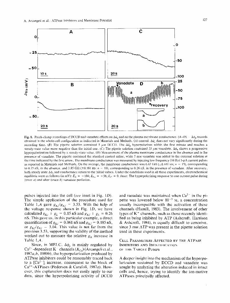
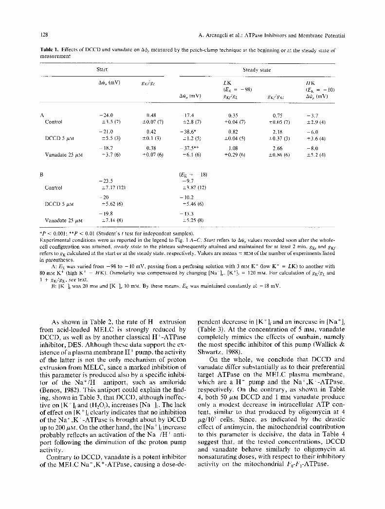
Figure 1 shows typical whole-cell recordings of AOp, either in the absence or in the presence of the ATPase inhibitors. Using the control pipette solu- tion, AOp did not undergo any significant variation during the measuring time (Fig. 1A). When the pi- pette solution contained 5/XM DCCD (Fig. lB) or 25 /ZM vanadate (Fig. 1C), a substantial hyperpolariza- tion of AOp occurred constantly, although at variable times after the beginning of the measurement. Table I,A shows that, at the steady state, both DCCD and vanadate produce, in physiological media, an average significant hyperpolarization (20 mV), with respect to the controls. When E K was lowered from - 9 8 to - 10 mV by increasing [K+]e in the perfusion solutions, Aqjp followed E K either in the absence or in the presence of the inhibitors (Table 1,A). The same occurred when E K was changed to - 18 mV, essentially by lowering the K + concentration in the internal solution (Table 1,B). The fact that in the controls AOp depends on EK indicates that a marginal potassium conductance is present under these condi- tions. Thus, under the simplifying assumption of ohmic relations, the resting equilibrium condition gives gK(AOp -- EK) = --gL(Athp - EL), where gK, EK, gL and E L are the conductances and Nernst potentials for potassium and the leakage pathways, respectively. The ratio gK/gL = -(Athp - EL)/ (AOp - EK) is reported in Table 1,A either for start- ing (gKi/gL) or steady-state (gKf/gL) conditions, as- suming EK = --98 and EL = 10 (Arcangeli et al., 1989b). Notice that all the gKi/gL values were not significantly different from the gKf/gL value of the control. On the contrary, gKf/gL increased in the presence of inhibitors. A comparative index of this effect is reported in Table 1,A as gKf/gKi, which shows that the K + conductance is increased more than twofold by both inhibitors.
On the whole, data in Table 1 demonstrate that the hyperpolarization induced by DCCD and vana- date in physiological media depends on the increase in K + conductance, in keeping with the indirect indi- cations previously reported in yeasts (De la Pefia et al., 1982; Baroni et al., 1988).
This increase most likely accounts for the report that, when added to the external medium, vanadate produces a AOp hyperpolarization associated to a diminution of the electrical resistance of the plasma membrane (Lichtshtein et al., 1982). We confirmed this finding. In fact 5 mM extracellular vanadate de- termined a 30-40 mV hyperpolarization of A% (Fig. 1D), associated to an increase in the membrane con- ductance, as revealed by the response to current
A. Arcangeli et al.: ATPase Inhibitors and Membrane Potential 127
0
- 2 5
- 5 0
> E CL
A
10S
B
O
L 501 20 S
[
C
O
-50
20S -100
~ L anadate D b ~ ~ ' ] - 2 5
a . . ~ ~ ~-50
Fig. 1. Patch-clamp recordings of DCCD and vanadate effects on A% and on the p lasma membrane conductance . (A-D) = Aq~p records obtained in the whole-cell configuration as indicated in Materials and Methods. (A) control. AO~, does not vary significantly during the recording time. (B) The pipette solution contained 5 /zM DCCD. The AtOp hyperpotarizes within the first minute and reaches a s teady-s ta te value more negative than the initial one. (C) The pipette solution contained 25 /zM vanadate . A~p shows a p r o g r e s s i v e hyperpolar izat ion followed by a s teady-state value. (D) Measurement of the plasma membrane conductance in the absence and in the presence of vanadate . The pipette contained the s tandard control saline, while 5 mM vanadate was added to the external solution at the time indicated by the first arrow. The membrane conductance was measured by injecting low f requency (10 Hz) 5-pA current pulses as reported in Materials and Methods . On the average, the membrane conductance was 6.67 Gf~ (_+0.441 SD; n = 15), corresponding to 0.15 nS, in the absence , and 3.85 G ~ (-+0.181 SD; n = 10), corresponding to 0.26 nS, in the presence of vanadate . After recovery , both s teady state A+p and conductance return to the initial values. Under the conditions used in alI these exper iments , e lectrochemical equilibria were as follows (in mV): E K = - 104; ENa = + 56; Ecj = 0. Inset: The hyperpolarizing response to one current pulse during (trace a) and after (trace b) vanadate perfusion.
pulses injected into the cell (see inset in Fig. 1D). The simple application of the procedure used for Table 1,A gave gKf/gKi = 3.53. With the help of the voltage response shown in Fig. 1D, we have calculated gKi + gL = 0.15 nS and gKf + gL = 0.26 nS. This gave us, in this particular example, a direct quantification ofgKi = 0.061 nS and gKf = 0.185 nS, or gKf/gKi = 3.04. This value is not far from the previous 3.53, supporting the validity of the method worked out to measure the relative gK increase in Table 1 ,A.
Since, in MELC, A% is mainly regulated by Ca2+-dependent K + channels (Kca) (Arcangeli et al., 1987a,b, 1989b), the hyperpolarization produced by ATPase inhibitors could be reasonably traced back to a [Ca2+]i increase, consequent to the block of Ca2§ (Pedersen & Carafoli, 1987a). How- ever, this explanation does not easily apply to our data, since the hyperpolarizing activity of DCCD
and vanadate was maintained when Ca z+ in the pi- pette was lowered below 10 .9 M, a concentration usually incompatible with the activation of these channels (Hamill, 1983). The involvement of other types of K § channels, such as those recently identi- fied as being inhibited by ATP (Ashcroft, Harrison & Ashcroft, 1984), is equally difficult to conceive, since 3 mu ATP was present in the pipette solution used in these experiments.
C E L L P A R A M E T E R S A F F E C T E D BY T H E A T P A S E
INHIBITORS A N D I D E N T I F I C A T I O N
OF THE T A R G E T P U M P S
A deeper insight into the mechanism of the hyperpo- larization sustained by DCCD and vanadate was sought by analyzing the alteration induced in intact cells and, hence, trying to identify the ion-motive ATPases principally affected.
128 A. Arcangeli et al.: ATPase Inhibitors and Membrane Potential
Table 1. Effects of DCCD and vanadate on AO~, measured by the patch-clamp technique at the beginning or at the steady state of measurement
Start Steady state
A0p (mV) gKi/gL
A% (mV)
LK H K
(EK = -98) (E K = - 10)
gKf/gL gKJ/gKi A0 o (mV)
A 24.0 0.48 - 17.4 0.35 0.75 - 3.7 Control -+3.5 (7) -+0.07 (7) -+2.8 (7) -+0.04 (7) -+0.05 (7) -+2.9 (4)
- 21.0 0.42 - 38.6* 0.82 2.18 - 6.0 DCCD 5 /xM --+5.5 (3) -+0.1 (3) --+1.2 (5) -+0.04 (5) -+0.37 (3) -+3.6 (4)
-- 18.7 0.38 37.5** 1.08 2.66 -- 8.0 Vanadate 25 /xM -+3.7 (6) -+0.07 (6) --+6.1 (6) -+0.29 (6) -+0.86 (6) +5.2 (4)
(EK = 18) -23 .5 9.7
Control -+7.17 (12) -+3.87 (12)
- 20 - 10.2 DCCD 5 t~M --+5.62 (6) --+5.46 (6)
- - 1 9 . 8 - - 13.3 Vanadate 25/xM -+7.14 (8) _+5.25 (8)
*P < 0.001; **P < 0.01 (Student 's t test for independent samples). Experimental conditions were as reported in the legend to Fig. 1 A-C. Start refers to A+p values recorded soon after the whole- cell configuration was attained, steady state to the plateau subsequently attained and maintained for at least 2 min. gKi and gKf refers to gK calculated at the start or at the steady state, respectively. Values are means -+ SEM of the number of experiments listed
in parentheses. A: EK was varied from - 9 8 to - 10 mV, passing from a perfusing solution with 3 mM K + (low K + = LK) to another with
80 mM K + (high K + = HK). Osmolarity was compensated by changing [Na+]e. [K+]i = 120 raN. For calculation of gK/gc and
1 + gKi/gK, see text. B: [K+]~ was 20 m g and [K+]e 10 mM. By these means, EK was maintained constantly at - 18 inV.
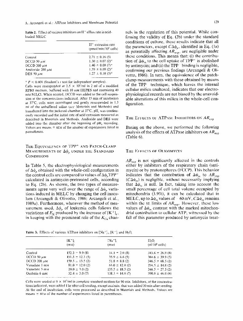
As shown in Table 2, the rate of H + extrusion from acid-loaded MELC is strongly reduced by DCCD, as well as by another classical H+-ATPase inhibitor, DES. Although these data support the ex- istence of a plasma membrane H + pump, the activity of the latter is not the only mechanism of proton extrusion from MELC, since a marked inhibition of this parameter is produced also by a specific inhibi- tor of the Na+/H + antiport, such as amiloride (Benos, 1982). This antiport could explain the find- ing, shown in Table 3, that DCCD, although ineffec- tive on [K+]i and (H2Oi), increases [Na+]~. The lack of effect on [K +]i clearly indicates that no inhibition of the Na +,K+-ATPase is brought about by DCCD up to 200 t~M. On the other hand, the [Na+]~ increase probably reflects an activation of the Na +/H + anti- port following the diminution of the proton pump activity.
Contrary to DCCD, vanadate is a potent inhibitor of the MELC Na + ,K+-ATPase, causing a dose-de-
pendent decrease in [K*]i and an increase in [Na+]i (Table 3). At the concentration of 5 mM, vanadate completely mimics the effects of ouabain, namely the most specific inhibitor of this pump (Wallick & Shwartz, t988).
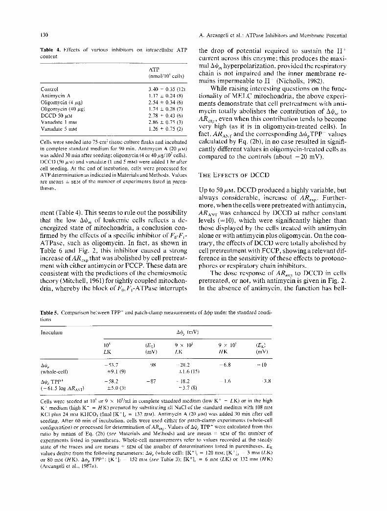
On the whole, we conclude that DCCD and vanadate differ substantially as to their preferential target ATPase on the MELC plasma membrane, which are a H + pump and the Na+,K+-ATPase, respectively. On the contrary, as shown in Table 4, both 50 /XM DCCD and 1 mM vanadate produce only a modest decrease in intracellular ATP con- tent, similar to that produced by oligomycin at 4 txg/107 cells. Since, as indicated by the drastic effect of antimycin, the mitochondrial contribution to this parameter is decisive, the data in Table 4 suggest that, at the tested concentrations, DCCD and vanadate behave similarly to oligomycin at nonsaturating doses, with respect to their inhibitory activity on the mitochondrial F0-FI-ATPase.
A. Arcangeli et al.: ATPase lnhibitors and Membrane Potential
Table 2. Effect of various inhibitors on H - efflux rate in acid-
loaded M E L C
H + extrusion rate (pmol/min/106 cells)
Control 2.71 -+ 0.16 (5) DCCD 50/zM 1.30 • 0.07 (2)* DCCD 200/xM 1.40 • 0.09 (7)* Amiloride 200 /xM 1.39 + 0.09 (2)* DES 50/xM 1.27 • 0.18 (3)*
* P < 0.001 (Student ' s t test for independent samples). Cells were r e suspended at 2.5 x 10⁶/ml in 2 ml of a modified RPMI medium, buffered with 10 mM HEPES and containing 40 mM NHmCI. When needed, DCCD was added to the cell suspen- sion at the concent ra t ions indicated. After 15 man of incubation at 37~ cells were centr ifuged and gently resuspended in 1.5 ml of the unbuffered saline (see Materials and Methods) and transferred into the jacke ted chamber at 37~ pH~ was continu- ously recorded and the initial rate of acid extrusion measured as descr ibed in Materials and Methods . Amiloride and DES were added into the chamber after the beginning of pile recording. Values are means -+ SEM of the number of exper iments listed in parentheses .
T H E E Q U I V A L E N C E OF TPP + A N D P A T C H - C L A M P
M E A S U R E M E N T S OF A~jp U N D E R T HE S T A N D A R D
C O N D I T I O N S
In Table 5, the electrophysiological measurements of kqjp obtained with the whole-cell configuration in the control cells are compared to values of Aq~pTPP + calculated in antimycin-pretreated cells, according to Eq. (2b). As shown, the two types of measure- ments agree very well over the range of A+p varia- tions induced in MELC by changing the cell inocu- lum (Arcangeli & Olivotto, 1986; Arcangeli et al., 1989a). Furthermore, whatever the method of mea- surement used, A+r of leukemia cells follows the variations of EK produced by the increase of [K+]e, in keeping with the prominent role of the Kca chan-
129
nels in the regulation of this potential. While con- firming the validity of Eq. (2b) under the standard conditions of culture, these results indicate that all the parameters, except C2X~p, identified in Eq. (la) as potentially affecting ARexp, are negligible under these conditions. This means that: (i) the contribu- tion of A+m to the cell uptake of TPP § is abolished by antimycin; and (ii) the TPP § binding is negligible, confirming our previous findings (Arcangeli & Oli- votto, 1986). In turn, the equivalence of the patch- clamp measurements with those obtained by means of the TPP § technique, which leaves the internal cellular milieu unaltered, indicates that our electro- physiological records are not biased by the unavoid- able alterations of this milieu in the whole-cell con- figuration.
T H E E F F E C T S OF A T P A S E INH1BITORS ON A R e x p
Basing on the above, we performed the following analysis of the effects of ATPase inhibitors o n ARex p (Table 6).
T H E E F F E C T S OF O L I G O M Y C I N
ARex p is not significantly affected in the controls either by inhibitors of the respiratory chain (anti- mycin) or by protonophores (FCCP). This behavior indicates that the contribution of Aq6n to ARex p (CA+m) is negligible, without necessarily implying that A+,,, is null. In fact, taking into account the small percentage of cell total volume occupied by mitochondria (3.9%), it can be calculated that in MELC, up to AOm values of - 60 mV, CA~,,~ remains within the SE limits of ARex p. However, these low values of A~) m contrast with the marked mitochon- drial contribution to cellular ATP, witnessed by the fall of this parameter produced by antimycin treat-
Table 3. Effects of various ATPase inhibitors on [Na+]i, [ K ' ] i and H20 i
[K+],. [Na+]i H20 , (mM) (raM) (nl/106 cells)
Control 152.3 • 9.9 (8) 14.1 • 2.0 (8) 343.6 • 26.9 (8) DCCD 50/~M 151.5 + 12.3 (5) 35.9 • 6.6 (5) 364.6 • 39.9 (5) DCCD 200 /~M 159.5 • 13.5 (2) 51.8 • 8.8 (2) 346.5 • 68.5 (2) Vanadate 1 mM 81.0 + 12.0 (2) 63.0 • 12.0 (2) 294.5 +-- 14.8 (2) Vanadate 5 mM 20.0 • 3.0 (2) 135.5 • 18.5 (2) 346.5 --+ 27.5 (2) Ouabain 6 mM 32.4 • 3.0 (7) 138.3 • 14.0 (7) 398.0 --+ 46.0 (8)
Cells were seeded at 9 x 10⁵/ml in complete s tandard medium for 90 min. Inhibitors, at the concentra- tions indicated, were added 1 hr after cell seeding, except ouabain, that was added 30 min after seeding. At the end of incubation, cells were processed as described in Materials and Methods. Values are means • SEN of the number of exper iments listed in parentheses .
130
Table 4. Effects of various inhibitors on content
intracellular ATP
ATP (nmol/106 cells)
Control 3.40 _+ 0.35 (12) Ant imycin A 1.17 -+ 0.24 (8) Oligomycin (4/zg) 2.54 -+ 0.34 (6) Oligomycin (40/xg) 1.74 _+ 0.28 (7) DCCD 50/xM 2.78 -+ 0.43 (6) Vanadate 1 mM 2.86 -+ 0.75 (3) Vanadate 5 mM 1.26 + 0.75 (2)
Cells were seeded into 75-cm-' t issue culture flasks and incubated in complete s tandard medium for 90 rain. Ant imycin A (20/ZM) was added 30 rain after seeding; ol igomycin (4 or 40/zg/107 cells), DCCD (50/xM) and vanadate (1 and 5 raM) were added 1 hr after cell seeding. At the end of incubation, cells were processed for ATP determinat ion as indicated in Materials and Methods. Values are means -+ SEM of the number of exper iments listed in paren- theses.
ment (Table 4). This seems to rule out the possibility that the low A+,, of leukemic cells reflects a de- energized state of mitochondria, a conclusion con- firmed by the effects of a specific inhibitor of Fo-F~- ATPase, such as oligomycin. In fact, as shown in Table 6 and Fig. 2, this inhibitor caused a strong increase ofARex p that was abolished by cell pretreat- ment with either antimycin or FCCP. These data are consistent with the predictions of the chemiosmotic theory (Mitchell, 1961) for tightly coupled mitochon- dria, whereby the block of F0,F~-ATPase interrupts
A. Arcangeli et al.: ATPase Inhibitors and Membrane Potential
the drop of potential required to sustain the H + current across this enzyme; this produces the maxi- mal AtOm hyperpolarization, provided the respiratory chain is not impaired and the inner membrane re- mains impermeable to H + (Nicholls, 1982).
While raising interesting questions on the func- tionality of MELC mitochondria, the above experi- ments demonstrate that cell pretreatment with anti- mycin totally abolishes the contribution of Ate,, to ARexp, even when this contribution tends to become very high (as it is in oligomycin-treated cells). In fact, ARaN T and the corresponding A+pTPP + values calculated by Eq. (2b), in no case resulted in signifi- cantly different values in oligomycin-treated cells as compared to the controls (about - 2 0 mV).
T H E E F F E C T S OF D C C D
Up to 50/J,M, DCCD produced a highly variable, but always considerable, increase of ARe• p. Further- more, when the cells were pretreated with antimycin, ARAN T was enhanced by DCCD at rather constant levels (=10), which were significantly higher than those displayed by the cells treated with antimycin alone or with antimycin plus oligomycin. On the con- trary, the effects of DCCD were totally abolished by cell pretreatment with FCCP, showing a relevant dif- ference in the sensitivity of these effects to protono- phores or respiratory chain inhibitors.
The dose response of ARex p to DCCD in cells pretreated, or not, with antimycin is given in Fig. 2. In the absence of antimycin, the function has bell-
Table 5. Compar i son between TPP + and patch-clamp measurements of A~p under the s tandard condi-
tions
Inoculum A+p (mY)
105 (E K) 9 x 10⁵ 9 • 105 (E K) L K (mV) L K H K (mV)
A~p - 5 3 . 7 98 - 2 0 . 2 --6.8 -- 10 (whole-cell) -+9.1 (9) -+ 1.6 (15)
A% TPP + - 5 8 . 2 - 8 7 - 18.2 - 1.6 - 3 . 8 ( - 6 1 . 5 log ARAy T) -+5.0 (3) -+3.7 (8)
Cells were seeded at 105 or 9 • 105/ml in complete s tandard medium (low K + - LK) or in the high K T medium (high K - = H K ) prepared by substi tut ing all NaCI of the s tandard medium with 108 mM KC1 plus 24 mM K H C O 3 (final [K*]~ = 132 raM). Ant imycin A (20 p~M) was added 30 rain after cell seeding. After 60 rain of incubation, cells were used either for patch-clamp exper iments (whole-cell configuration) or processed for determinat ion ofARex p. Values of A~p T p p + were calculated from this ratio by means of Eq. (2b) (see Materials and Methods) and are means -+ SEM of the number of exper iments listed in parentheses . Whole-cell measurements refer to values recorded at the s teady state of the traces and are means -+ SEM of the number of determinations listed in parentheses . E K values derive from the following parameters: bop (whole cell): [K+]i = 120 raM; [K+]e = 3 mM (LK) or 80 mM (HK). A% Tpp+: [K+]i - 152 m g (see Table 3); [K+]e = 6 mM (LK) or 132 mM (HK) (Arcangeli et al., 1987a).
A. Arcangeli et al.: ATPase Inhibitors and Membrane Potential
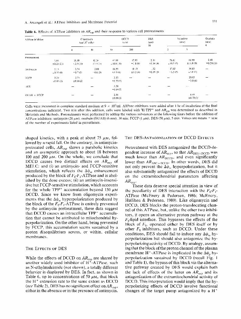
Table 6. Effects of ATPase inhibitors on ARex p and their response to various cell pretreatments
131
ATPase inhibitor Oligomycin DCCD DES (/zg/107 cellst (/ZM) (/ZM)
50
4 40 50 200
Vanadate Ouabain
(mM) (mM)
6
1 5
Pretreatment - - 2.66 18.49 41.34 45.90
-+0.21 (211 +-2.9(10) +-7.5 (7) +-10,9(10/
Amimycin 2.13 2.54 2,84 10.39
+-0.35 (81 +-0,7 (5) +-0.6 (3) +-1.9 (6)
FCCP 2.29 2.73 - - 3.13
z0 .03 (21 -+0.01 (2) +-0.55(5)
DES - - 5.58 +-0.04(2)
OUAB ~- FCCP - - 2.91 -+0,90(3}
17.83 2.14 58.61 62.99 2.68
+1.8 (6) +0.34 (4) -+10.2 (51 +11.9 (9) +0.24(111
18,79 1,52 17.03 34.82
+3.2 (4) +0.19 (5) +3.3 (5) -+3.8 (7)
- - - - - - 20.03 - -
+3,0 (6)
6.03
+-0.70(3)
m
Cells were incubated in complete standard medium at 9 x 10⁵/ml. ATPase inhibitors were added after 1 hr of incubation at the final concentrations indicated. Two min after this addition, cells were labeled with 3H-TPP + and ARex p was determined as described in Materials and Methods. Pretreatments were performed by adding the various substances at the following times before the addition of ATPase inhibitors: antimycin (20 IxM), ouabain (OUAB) (6 mM), 30 min; FCCP (1 b~M), DES (50 txM), 5 rain. Values are means + SEN of the number of experiments listed in parentheses.
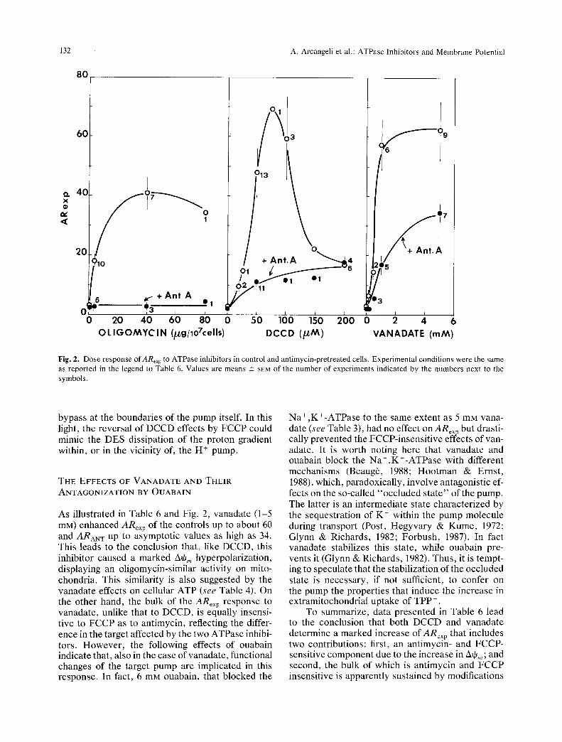
shaped kinetics, with a peak at about 75 ~M, fol- lowed by a rapid fall. On the contrary, in antimycin- pretreated cells, ARex p shows a parabolic kinetics and an asymptot ic approach to about 18 between 100 and 200 p.M. On the whole, we conclude that DCCD causes two distinct effects on ARe• p of MELC: and (i) an antimycin- and FCCP-sensi t ive stimulation, which reflects the A+, n enhancement produced by the block of F0-Fi-ATPase and is abol- ished by the dose excess; (ii) an antimycin-insensi- tive but FCCP-sensi t ive stimulation, which accounts for the whole TPP § accumulat ion beyond 150 /.~M DCCD. Since we know f rom oligomycin experi- ments that the 2~q6,, hyperpolar izat ion produced by the block of the F0-F}-ATPase is entirely prevented by the ant imycin pre t rea tment , these data suggest that DCCD causes an intracellular TPP + accumula- tion that cannot be attr ibuted to mitochondrial hy- perpolarizat ion. On the other hand, being prevented by FCCP, this accumulat ion seems sustained by a proton disequilibrium across , or within, cellular membranes .
THE EFFECTS OF DES
While the effects of DCCD on ARex p are shared by another widely used inhibitor of H+-ATPase , such as N-ethylmaleimide (not shown), a totally different behavior is displayed by DES. In fact, as shown in Table 6, up to concen t ra t ions of 50 tZM, that block the H + extrusion rate to the same extent as DCCD (see Table 2), DES has no significant effect on ARexp, either in the absence or in the presence of antimycin.
THE DES-ANTAGONIZATION OF DCCD EFFECTS
Pretreatment with DES antagonized the DCCD-de- pendent increase of A R e x p , SO that ARDe s + DCCD was much lower than ARDccD, and even significantly lower than ARANT+DCCD. In other words , DES did not only prevent the 2x~Om hyperpolar izat ion, but it also substantially antagonized the effects of DCCD on the extramitochondrial pa ramete rs affecting ARexp.
These data deserve special at tention in view of the peculiarity of DES interaction with the Fo-FI- ATPase (McEnery & Pedersen, 1986; McEnery , Hullihen & Pedersen, 1989). Like ol igomycin and DCCD, DES blocks the proton-translocat ing chan- nel of this ATPase , but, unlike the other two inhibi- tors, it opens an alternative pro ton pa thway at the F0-1ipid interface. This bypasses the effects of the block of F 0, operated either by DES itself or by other F 0 inhibitors, such as DCCD. Under these conditions, DES should fail to induce any A~,,, hy- perpolarization but should also antagonize the hy- perpolarizing activity of DCCD. By analogy, assum- ing that the block of the proton channel of the p lasma membrane H+-ATPase is implicated in the AOp hy- perpolarization sustained by DCCD (recall Fig. 1 and Table I), the bypass of this block via the alterna- tive pa thway created by DES would explain both the lack of effects of the latter on ARex p and its antagonization of the extramitochondria l activity of DCCD. This interpretation would imply that the hy- perpolarizing effects of DCCD involve functional changes of the target pump antagonized by a H +
132 A. Arcangeli et al.: ATPase lnhibitors and Membrane Potential
80
60
,,', 40, X
elr
2 0
1
~10
~C ~3
s'o 16o 260 DCCD ( ~ M )
I
+Ant . A
+ Ant A O! e 3 * I 3
I I
o 2b 40 6o 8'0 o o 2 ,i 6 O L I G O M Y C I N (~gDoTcells) VANADATE (rnM)
Fig. 2. Dose response of ARexp to ATPase inhibitors in control and antimycin-pretreated cells. Experimental conditions were the same as reported in the legend to Table 6. Values are means -+ SEM of the number of experiments indicated by the numbers next to the symbols.
bypass at the boundaries of the pump itself. In this light, the reversal of DCCD effects by FCCP could mimic the DES dissipation of the proton gradient within, or in the vicinity of, the H § pump.
THE EFFECTS OF VANADATE AND THEIR
ANTAGONIZATION BY OUABAIN
As illustrated in Table 6 and Fig. 2, vanadate (1-5 mM) enhanced ARex p of the controls up to about 60 and ARAN T up to asymptotic values as high as 34. This leads to the conclusion that, like DCCD, this inhibitor caused a marked A~,, hyperpolarization, displaying an oligomycin-similar activity on mito- chondria. This similarity is also suggested by the vanadate effects on cellular ATP (see Table 4). On the other hand, the bulk of the ARex p response to vanadate, unlike that to DCCD, is equally insensi- tive to FCCP as to antimycin, reflecting the differ- ence in the target affected by the two ATPase inhibi- tots. However, the following effects of ouabain indicate that, also in the case of vanadate, functional changes of the target pump are implicated in this response. In fact, 6 mM ouabain, that blocked the
Na+,K+-ATPase to the same extent as 5 mM vana- date (see Table 3), had no effect on ARex p but drasti- cally prevented the FCCP-insensitive effects of van- adate. It is worth noting here that vanadate and ouabain block the Na+,K+-ATPase with different mechanisms (Beaug6, 1988; Hootman & Ernst, 1988), which, paradoxically, involve antagonistic ef- fects on the so-called "occluded state" of the pump. The latter is an intermediate state characterized by the sequestration of K § within the pump molecule during transport (Post, Hegyvary & Kume, 1972; Glynn & Richards, 1982; Forbush, 1987). In fact vanadate stabilizes this state, while ouabain pre- vents it (Glynn & Richards, 1982). Thus, it is tempt- ing to speculate that the stabilization of the occluded state is necessary, if not sufficient, to confer on the pump the properties that induce the increase in extramitochondrial uptake of TPP § .
To summarize, data presented in Table 6 lead to the conclusion that both DCCD and vanadate determine a marked increase of ARex p that includes two contributions: first, an antimycin- and FCCP- sensitive component due to the increase in Atp,,; and second, the bulk of which is antimycin and ECCP insensitive is apparently sustained by modifications
A. Arcangeli et al.: ATPase lnhibitors and Membrane Potential 133
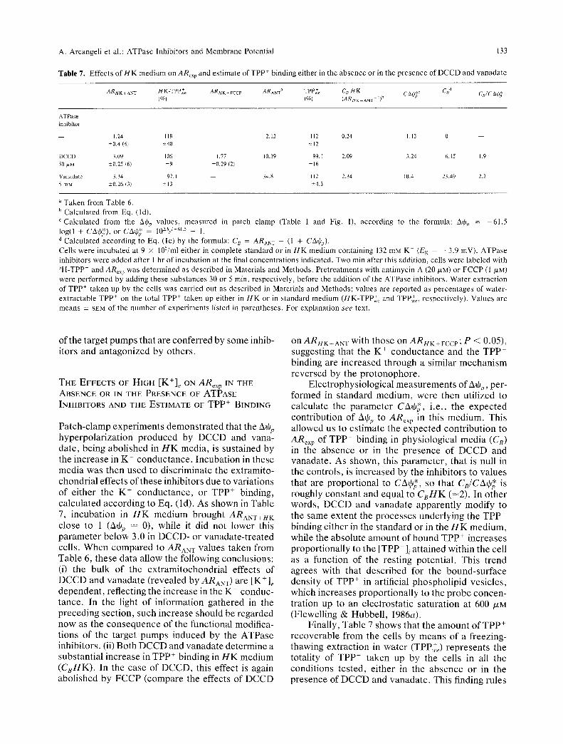
Table 7. Effects of i l K medium o n ARex p and estimate of TPP* binding either in the absence or in the presence of DCCD and vanadate
ARH K + ANT H K-TPP~e ARH K + FCCP ARANT a TPP+e C B H K ~--*Pg~Adt*c CBd C B/C~O~ (%} (%) (ARHK.AN T- I)b
ATPase
inhibitor
- - 1.24 118 - - 2.13 112 0.24 1.13 0 - -
-+0.4 (4t +-40 +-12
DCCD 3,09 106 1.77 10.39 99. I 2.09 3.24 6.15 1.9
50 tam +-0.25 (6) " 9 +0.29 12) +16
Vanadate 3.34 97. I - - 34.8 112 2.34 10,4 23.49 2.2
5 mM --+0.26 (31 +13 +4.3
Taken from Table 6. b Calculated from Eq. (ld). c Calculated from the &qJ~, values, measured in patch clamp (Table 1 and Fig. 1), according to the formula: 2~tOn = -61.5 log(1 + CArat), or CA0* = 10~% '-61'5 - 1. d Calculated according to Eq. (lc) by the formula: C8 = ARANT -- (1 + C A • ) .
Cells were incubated at 9 x 10⁵/ml either in complete standard or in HK medium containing 132 mM K § (EK -- --3.9 mV). ATPase inhibitors were added after 1 hr of incubation at the final concentrations indicated. Two rain after this addition, cells were labeled with 3H-TPP+ and ARex p w a s determined as described in Materials and Methods. Pretreatments with antimycin A (20/*M) or FCCP (1 /LM) were performed by adding these substances 30 or 5 min, respectively, before the addition of the ATPase inhibitors. Water extraction of TPP + taken up by the ceils was carried out as described in Materials and Methods; values are reported as percentages of water- extractable TPP + on the total TPP + taken up either in HK or in standard medium (HK-TPP+,~ and TPP+e, respectively). Values are means -+ SEM of the number of experiments listed in parentheses. For explanation see text.
o f the t a rge t p u m p s tha t a re c o n f e r r e d by s o m e inhib- i tors and a n t a g o n i z e d by o the r s .
THE EFFECTS OF HIGH [K+]~ ON ARex p IN THE ABSENCE OR IN THE PRESENCE OF ATPASE INHIBITORS AND THE ESTIMATE OF TPP + BINDING
P a t c h - c l a m p e x p e r i m e n t s d e m o n s t r a t e d that the AtOp h y p e r p o l a r i z a t i o n p r o d u c e d by D C C D and vana- da te , be ing a b o l i s h e d in H K med ia , is su s t a ined by the i n c r e a s e in K § c o n d u c t a n c e . I n c u b a t i o n in these m e d i a was then u sed to d i s c r i m i n a t e the ex t r a mi to - c h o n d r i a l e f fec t s o f t h e s e inh ib i to r s due to va r i a t ions o f e i the r the K § c o n d u c t a n c e , or T P P § b ind ing , c a l c u l a t e d a c c o r d i n g to Eq. ( ld ) . As s h o w n in Tab le 7, i n c u b a t i o n in H K m e d i u m b rough t ARANT+HK c lose to 1 (AtOp -- 0), whi le it d id not l o w e r this p a r a m e t e r b e l o w 3.0 in D C C D - o r v a n a d a t e - t r e a t e d cel ls . W h e n c o m p a r e d to ARAN w va lues t a k e n f rom Tab le 6, t he se d a t a a l low the fo l lowing conc lus ions : (i) the bu lk o f the e x t r a m i t o c h o n d r i a l e f fec ts of D C C D and v a n a d a t e ( r evea l ed b y ARANT) are [K+]e d e p e n d e n t , re f lec t ing the i n c r e a s e in the K + c onduc - t ance . In the l ight o f i n fo rma t ion g a t h e r e d in the p r e c e d i n g sec t ion , such i n c r e a s e should be r e g a r d e d n o w as the c o n s e q u e n c e o f the func t iona l modi f ica- t ions o f the t a rge t p u m p s i n d u c e d b y the A T P a s e inh ib i to r s . (ii) Bo th D C C D and v a n a d a t e d e t e r m i n e a subs t an t i a l i n c r e a s e in T P P + b ind ing in H K m e d i u m ( C s H K ) . In the ca se o f D C C D , this effect is again a b o l i s h e d b y F C C P ( c o m p a r e the ef fec ts o f D C C D
on ARHK+ANT wi th t h o s e on ARI4K+FCCP; P < 0.05), sugges t ing tha t the K + c o n d u c t a n c e and the T P P + b ind ing are i n c r e a s e d t h rough a s imi la r m e c h a n i s m r e v e r s e d b y the p r o t o n o p h o r e .
E l e c t r o p h y s i o l o g i c a l m e a s u r e m e n t s o f AtOp, per - f o r m e d in s t a n d a r d m e d i u m , we re then u t i l i zed to ca l cu la t e the p a r a m e t e r CAtO*, i . e . , the e x p e c t e d c o n t r i b u t i o n o f AtOp to ARe• in this m e d i u m . This a l l ow e d us to e s t i m a t e the e x p e c t e d c o n t r i b u t i o n to ARex p of TPP + b ind ing in p h y s i o l o g i c a l m e d i a (C 8) in the a b s e n c e o r in the p r e s e n c e o f D C C D and v a n a d a t e . As shown , this p a r a m e t e r , tha t is null in the con t ro l s , is i n c r e a s e d by the inh ib i to r s to va lues tha t a re p r o p o r t i o n a l to CAtO*, so tha t CB/CAtO* is roughly c o n s t a n t and equa l to CBHK (-~2). In o t h e r words , D C C D and v a n a d a t e a p p a r e n t l y m o d i f y to the s ame e x t e n t the p r o c e s s e s u n d e r l y i n g the T P P + b ind ing e i the r in the s t a n d a r d o r in the H K m e d i u m , whi le the a b s o l u t e a m o u n t o f b o u n d T P P + i n c r e a s e s p r o p o r t i o n a l l y to the [TPP + ] / a t t a i n e d wi th in the cel l as a func t ion o f the res t ing po ten t i a l . This t r end agrees wi th that d e s c r i b e d for the b o u n d - s u r f a c e dens i ty o f TPP + in ar t i f ic ial p h o s p h o l i p i d ves i c l e s , wh ich i n c r e a s e s p r o p o r t i o n a l l y to the p r o b e c o n c e n - t r a t ion up to an e l e c t r o s t a t i c s a t u r a t i o n at 600 p,M (F lewel l ing & H u b b e l l , 1986a).
F ina l ly , Tab le 7 s h o w s tha t the a m o u n t o f T P P + r e c o v e r a b l e f rom the cel ls by m e a n s o f a f r eez ing- t hawing e x t r a c t i o n in w a t e r (TPP,+e) r e p r e s e n t s the to ta l i ty o f T P P + t a k e n up by the cel ls in all the cond i t i ons t e s t ed , e i the r in the a b s e n c e o r in the p r e s e n c e o f D C C D and v a n a d a t e . This f inding ru les

134 A. Arcangeli et al.: ATPase Inhibitors and Membrane Potential
out any hydrophobic sequestration of the probe within cellular components and fits in with the TPP + adsorption at the boundaries of lipid bilayers as a function of their electrostatic potential profile. In this light one should conclude that DCCD and vana- date change this profile in MELC plasma membrane.
CONCLUDING REMARKS
Patch-clamp and TPP+-data presented in this paper converge to show that DCCD and vanadate, al- though acting on different targets, increase the K + conductance across the plasma membrane of leuke- mia cells. This increase requires the direct and spe- cific interaction of these inhibitors with the target ATPases, while it is antagonized by inhibitors that block the same pumps with different mechanisms (DES or ouabain). A totally speculative explanation of this finding could be that, upon interaction with the effective inhibitor, the ion-translocating moiety of the pump is transformed into a K+-permeable channel. In this context it should be noted that a mutant in the plasma membrane H+-ATPase gene of Saccharomyces cerevisiae, with a reduced H +- ATPase activity, when examined at the single-chan- nel level, exhibited the activation of a K + channel that was hardly distinguishable from the H +-ATPase itself (Ramirez et al., 1989).
The other important information to emerge from this work derived from the integration of patch- clamp and TPP + data, showing that both DCCD and vanadate alter the complex set of membrane electrostatic parameters reflected by the TPP + bind- ing (see Introduction and comments on Table 7). It is worth recalling here that the internal energy profile of lipid bilayers can be changed by molecules modi- fying their dipole potential (Hladky, 1979; Kleijn et al., 1983; Flewelling & Hubbell, 1986b). We propose here that DCCD and vanadate may alter substan- tially the membrane energy profile by varying the internal dipole potential and/or the double-layer sur- face potential. This effect, undetectable by means of electrophysiological measurements, could: (i) al- ter the TPP + adsorption at the membrane water in- terfaces, accounting for the increase of the binding demonstrated in Table 7 and, hence, of the alter- ations ofARex p reported in Table 6; and (ii) be moni- tored by potential-sensitive proteins spanning the membrane. One of these molecules could be a volt- age-sensitive K + channel either pre-existing in a si- lent configuration or newly formed across the pump upon interaction with specific inhibitors. Alterna- tively, changes of the electrostatic potential profile sustained by the ATPase inhibitors could drastically enhance the Ca 2+ concentration at the boundaries
of the plasma membrane, thus activating calcium- sensitive K + conductance. This can occur even at prohibitive concentrations of this ion in the bulk cellular water. The crucial role of the electrostatic surface potential in determining the Ca 2+ concentra- tion necessary at the binding sites for triggering exo- cytosis has been clearly established by McLaughlin and Whitaker (1988) in sea urchin eggs.
However, the fact that, in the case of the Na+,K+-ATPase, the increase in K + conductance is activated by vanadate and prevented by ouabain suggests that putative changes of the electrostatic potential profile of the membrane are somehow re- lated to the creation of the occluded state of this pump. This would favor the hypothesis that occlu- sion of cations within the ion-translocating moiety of the pumps could locally modify the membrane internal energy profile by interacting, for instance, with the membrane dipole potential (Flewelling & Hubbell, 1986a). This interpretation could also apply to the effects of DCCD on the H+-ATPase of the plasma membrane, if DCCD creates the occlusion o fH + within or in the vicinity of the H + pump. The bypass of this occlusion by FCCP and DES would thus explain the reversal of DCCD effects operated by these substances on either the TPP + binding or the K + conductance.
The authors are indebted to Prof. A. Fonnesu, Chairman of the Institute of General Pathology of the University of Florence, for his support and advice. Thanks are also due to Prof. A. Ferroni, for the critical reading of the manuscript and to Prof. S. Capaccioli for his contribution to ATP determination. We are grateful to M. Cutri for technical assistance.
We gratefully acknowledge the revision of the English by Dr. R. Manning of the Department of Biological Sciences, Univer- sity of Durham, Durham, UK.
A. Arcangeli was supported by a fellowship from the Associ- azione Italiana contro le Leucemie (AIL); A. Beccheni and M.R. Del Bene were supported by fellowships from the Italian Associa- tion for Cancer Research (AIRC).
This work was supported by grants from the AIRC, from the Consiglio Nazionale delle Ricerche (CNR) (Special project "Ion channels") and from the Ministero Pubblica Istruzione.
References
Al-Awqati, Q. 1986. Proton-translocating ATPases. Annu. Rev. Cell Biol. 2:179-199
Amzel, L.M., Pedersen, L. 1983. Proton ATPases: Structure and mechanism. Annu. Rev. Biochem. 52:801-824
Andersen, O.S., Feldberg, S., Nakadomari, H., Levy, S., McLaughlin, S. 1978. Electrostatic interactions among hy- drophobic ions in lipid bilayer membranes. Biophys. J. 21:35-70
Arcangeli, A., Del Bene, M.R., Olivotto, M., Wanke, E. 1989b. Ion channels in cancer cells. In: Membrane Technology. Vol. 64, pp. 65-79, Raven, New York
A. Arcangeli et al.: ATPase lnhibitors and Membrane Potential 135
Arcangeli, A., Del Bene, M.R., Poli, R., Ricupero, L., Olivotto, M. 1989a. Mutual contact of murine erythroleukemia cells activates depolarizing cation channels, whereas contact with extracellular substrata activates depolarizing Ca-' +-dependent K- channels. J. Cell. Physiol. 139:1-8
Arcangeli, A., Olivotto, M. 1986. Plasma membrane potential of murine erythroleukemia cells: Approach to measurement and evidence for cell density-dependence. J. Cell. Physiol. 127:17-27
Arcangeli, A., Ricupero, L., Olivotto, M. 1987a. Commitment to differentiation of murine erythroleukemia cells involves a modulated plasma membrane depolarization through Ca 2+- activated K- channels. J. Cell. Physiol. 132:387-400
Arcangeli, A., Wanke, E., Olivotto, M., Camagni, S., Ferroni, A. 1987b. Three types of ion channels are present on the plasma membrane of Friend erythroleukemia cells. Biochem. Biophys. Res. Commun. 146:1450-1457
Ashcroft, F.M., Harrison, D.E., Ashcroft, S.J.H. 1984. Glucose induces closure of single potassium channels in isolated rat panceratic/3-cells. Nat ,re 312:446-448
Azzone, G.F., Pietrobon, D., Zoratti, M. 1984. Determination of the proton electrochemical gradient across biological mem- branes. Curt. Top. Bioenerg. 13:1-77
Bakker, E.P. 1982. Membrane potential in a potassium transport- negative mutant ofEscheriehia coli K-12. The distribution of rubidium in the presence of vatinomycin indicates a higher potential than that of the tetraphenylphosphonium cation. Bio- chim. Biophys. Acta 681:474-483
Baroni, M., Arcangeli, A., Di Pietro, L., Alberghina, L. 1988. Plasma membrane potential of budding yeast is modulated by glucose and by inhibitors of H +-ATPase and of K + channels. Yeasts 4:$34
Beaug6, L. 1988. Inhibition of translocation reaction by Vana- date. Methods Enzymol. 156:251-267
Benos, D.J. 1982. Amiloride: A molecular probe of sodium trans- port in tissues and cells. Am. J. Physiol. 242:C131-C145
Cantley, L.C., Resh, M.D., Guidotti, G. 1978. Vanadate inhibits the red cell (Na-,K+)ATPase from the cytoplasmic side. Na- ture 272:552-554
De La Pefia, P., Barros, F., Gascon, S., Ramos, S., Lazo P.S. 1982. The electrochemical proton gradient of Saccharomyces. Eur. J. Biochem. 123:447-453
Felber, S.M., Brand, M.D. 1982. Factors determining the plasma- membrane potential of human lymphocytes. Biochem. J. 204:577-585
Flewelling, R.F., Hubbell, W.L. 1986a. Hydrophobic ion interac- tion with membranes. Biophys. J. 49:531-540
Flewelling, R.F., Hubbell, W.L. 1986b. The membrane dipole potential in a total membrane potential model. Biophys. J. 49:541-552
Forbush, B., III. 1987. Rapid release of 42K and 86Rb from an occluded state of the Na,K-pump in the presence of ATP or ADP. J. Biol. Chem. 262:11104-11115
Futai, M., Noumi, T., Maeda, M. 1989. ATP synthase (H +- ATPase): Results by combined biochemical and molecular biological approaches. Annu. Rev. Biochem. 58:111-136
Glynn, I.M., Karlish, S.J.D. 1975. The sodium pump. Annu. Rev. Physiol. 37:13-55
Glynn, I.M., Richards, D.E. 1982. Occlusion of rubidium ions by the sodium-potassium pump: Its implications for the mecha- nism of potassium transport. J. Physiol. 330:17-43
Goffeau, A., Slayman, C.W. 1981. The proton-translocating ATPase of fungal plasma membrane. Biochim. Biophys. Acta 636:197-223
Hamill, O.P. 1983. Potassium and chloride channels in red blood cells. In: Single-channel Recording. B. Sackmann and E. Neher, editors, pp. 451-471, Plenum, New York
Hamill, O.P., Marty, A., Neher, E., Sakmann, B., Sigworth, F. 1981. Improved patch-clamp techniques for high resolution current recording from cells and cell-free membrane patches. Pfluegers Arch. 391:85-100
Hladky, S.B. 1979. The carrier mechanism. Curr. Top. Membr. Transp. 12:53-164
Hootman, S.R., Ernst, S.A. 1988. Estimation of Na+,K+-pump numbers and turnover in intact cells with [3H]ouabain. Meth- ods Enzymol. 156:213-229
Hoppe, J., Sebald, W. 1984. The proton conducting F0-part of bacterial ATP synthases. Biochim. Biophys. Acta 768:1-27
Jordan, P.C. 1983. Electrostatic modeling of ion pores. II. Effects attributable to the membrane dipole potential. Biophys. J. 41:189-195
Ketterer, B., Neumcke, B., L/iuger, P. 1971. Transport mecha- nism of hydrophobic ions through lipid bilayer membranes. J. Membrane Biol. 5:225-245
Kleijn, W.B., Bruner, L.J., Midland, M.M., Wisniewski, S. 1983. Hydrophobic ion probe studies of membrane dipole poten- tials. Biochim. Biophys. Acta 727:357-366
Lichtshtein, D., Mullikin-Kilpatrick, D., Blume, A.J. 1982. Hy- perpolarization of neurobtastoma-glioma hybrid NG 108-15 by vanadium ions. Proc. Natl. Acad. Sci. USA 79:4202-4206
McEnery, M.W., Hullihen, J., Pedersen, P.L. 1989. F0 "proton channel" of rat liver mitochondria. J. Biol. Chem. 264:12029-12036
McEnery, M.W., Pedersen, P.L. 1986. Diethylstilbestrol. A novel F0-directed probe of the mitochondrial proton ATP-ase. J. Biol. Chem. 261:1745-1752
McLaughlin, S. 1977. Electrostatic potentials at membrane-solu- tion interfaces. Curr. Top. Membr. Transp. 25:71-144
McLaughlin, S. 1989. The electrostatic properties of membranes. Anntt. Rev. Biophys. Chem. 18:113-136
McLaughlin, S., Whitaker, M. 1988. Cations that alter surface potentials of lipid bilayers increase the calcium requirement for exocytosis in sea urchin eggs. J. Physiol. 396:189-204
Mitchell, P. 1961. Coupling of phosphorylation to electron and hydrogen transfer by a chemi-osmotic type of mechanism. Nature 191:144-148
Neumcke, B., Lauger, P. 1969. Nonlinear electrical effects in lipid bilayer membranes. II. Integration of the generalized Nernst-Planck equations. Biophys. J. 9:1160-1170
Nicholls, D.G. 1982. Bioenergetics. An introduction to the Chem- iosmotic Theory. Academic, London
Pedersen, P.L., Carafoli, E. 1987a. Ion motive ATPases. I. Ubiq- uity, properties and significance to cell function. Trends Bio- chem. Sci. 12:146-150
Pedersen, P.L., Carafoli, E. 1987b. Ion motive ATPases. II. Energy coupling and work output. Trends Biochem. Sci. 12:186-189
Penefsky, H.S. 1979. Mitochondrial ATPase. Adv. Enzymol. 49:223-280
Pogolotti, A.L., Santi, D.V. 1982. High pressure liquid chroma- tography-ultraviolet analysis of intracellular nucleotides. Anal. Biochem. 126:335-345
Post, R.L., Hegyvary, C., Kume, S. 1972. Activation by adenosin triphosphate in the phosphorylation kinetics of sodium and potassium ion transport adenosine triphosphatase. J. Biol. Chem. 247:6530-6540
Ramirez, J.A., Vacata, V., McCusker, J.H., Haber, J.E., Mortimer, R.K., Owen, W.G., Lecar, H. 1989. ATP-sensitive K + channels
136 A. Arcangeli et al.: ATPase inhibitors and Membrane Potential
in a plasma membrane H'-ATPase mutant of the yeast Sacchar- omyces cerevisiae. Proc. Natl. Acad. Sci. USA 86:7866-7870
Ritchie, R.J. 1984. A critical assessment of the use of lipophilic cations as membrane potential probes. Prog. Biophys. Mol. Biol. 43:1-32
Rottenberg, H. 1979. The measurement of membrane potential and pH in cells, organelles and vesicles. Methods Enzymol. 55:547-569
Skou, J.C. 1988. Overview: The Na,K-pump. Methods Enzymol. 156:1-25
Swallow, C.J., Grinstein, S., Rotstein, O.D. 1988. Cytoplasmic pH regulation in macrophages by an ATP-dependent and N,N'-dicyclohexilcarbodiimide-sensitive mechanism. J. Biol. Chem. 263:19558-19563
Wallick, E.J., Schwartz, A. 1988. Saturation of cardiac glyco- sides with Na + ,K+-ATPase. Methods Enzymol. 156:201-213
Received 13 March 1991; revised 26 September 1991