Intracellular localization of membrane-bound ATPases in the compartmentalized anammox bacterium...
15
Intracellular localization of membrane-bound ATPases in the compartmentalized anammox bacterium ‘Candidatus Kuenenia stuttgartiensis’Laura van Niftrik, 1 * Mary van Helden, 1 Silke Kirchen, 1 Elly G. van Donselaar, 2 Harry R. Harhangi, 1 Richard I. Webb, 3 John A. Fuerst, 3 Huub J. M. Op den Camp, 1 Mike S. M. Jetten 1,4 and Marc Strous 1,5 1 Department of Microbiology, Institute for Water and Wetland Research, Faculty of Science, Radboud University Nijmegen, Heyendaalseweg 135, 6525 AJ Nijmegen, the Netherlands. 2 Cellular Architecture & Dynamics, Utrecht University, Padualaan 8, 3584 CH Utrecht, the Netherlands. 3 Department of Microbiology & Parasitology (JAF)/Centre for Microscopy and Microanalysis (RIW), University of Queensland, Brisbane, Qld 4072, Australia. 4 Department of Biotechnology, Delft University of Technology, Julianalaan 67, 2628 BC Delft, the Netherlands. 5 Max Planck Institute for Marine Microbiology, Celciusstrasse 1, 28359 Bremen, Germany. Summary Anaerobic ammonium-oxidizing (anammox) bacteria are divided into three compartments by bilayer mem- branes (from out- to inside): paryphoplasm, riboplasm and anammoxosome. It is proposed that the anammox reaction is performed by proteins located in the anam- moxosome and on its membrane giving rise to a proton-motive-force and subsequent ATP synthesis by membrane-boundATPases. To test this hypothesis, we investigated the location of membrane-bound ATPases in the anammox bacterium ‘Candidatus Kuenenia stuttgartiensis’. Four ATPase gene clusters were identified in the K. stuttgartiensis genome: one typical F-ATPase, two atypical F-ATPases and a prokaryotic V-ATPase. K. stuttgartiensis transcrip- tomic and proteomic analysis and immunoblotting using antisera directed at catalytic subunits of the ATPase gene clusters indicated that only the typical F-ATPase gene cluster most likely encoded a func- tional ATPase under these cultivation conditions. Immunogold localization showed that the typical F-ATPase was predominantly located on both the out- ermost and anammoxosome membrane and to a lesser extent on the middle membrane. This is consis- tent with the anammox physiology model, and con- firms the status of the outermost cell membrane as cytoplasmic membrane. The occurrence of ATPase in the anammoxosome membrane suggests that anammox bacteria have evolved a prokaryotic organelle; a membrane-bounded compartment with a specific cellular function: energy metabolism. Introduction Anammox bacteria perform anaerobic ammonium oxida- tion (anammox) to dinitrogen gas and are applied for removal of ammonium from wastewater. They are also important in nature where they contribute significantly to oceanic nitrogen loss (Devol, 2003; Kuypers et al., 2003; 2005; Arrigo, 2005; Brandes et al., 2007). Anammox bac- teria belong to the phylum Planctomycetes. One of the unique properties of species within this phylum is that they do not conform to the prokaryotic cell plan: their cells are compartmentalized and are proposed to lack a typical bacterial cell wall (Fuerst, 1995; Lindsay et al., 2001). The planctomycete compartmentalization is in some cases complex but always involves a single intracytoplasmic membrane defining a major cell compartment. Electron microscopy observations, chemical analysis, genome sequencing and resistance to beta-lactam and other cell wall-targeting antibiotics have revealed that plancto- mycetes lack the otherwise universal bacterial cell wall polymer peptidoglycan (König et al., 1984; Liesack et al., 1986; Stackebrandt et al., 1986; Fuerst, 1995) and an outer membrane typical of Gram-negative bacteria. The outermost membrane in planctomycetes has been defined as the cytoplasmic membrane based on the detection of RNA directly on its inner side by immunogold labelling and the position of the membrane and mem- brane surfaces relative to the cell wall visualized in thin sections of cryofixed and freeze-substituted cells and in Accepted 22 May, 2010. *For correspondence. E-mail l.vanniftrik@ science.ru.nl; Tel. (+31) 24 3652563; Fax (+31) 24 3652830. Re-use of this article is permitted in accordance with the Terms and Conditions set out at http://www3.interscience.wiley.com/ authorresources/onlineopen.html Molecular Microbiology (2010) 77(3), 701–715 doi:10.1111/j.1365-2958.2010.07242.x First published online 11 June 2010 © 2010 Blackwell Publishing Ltd
Transcript of Intracellular localization of membrane-bound ATPases in the compartmentalized anammox bacterium...
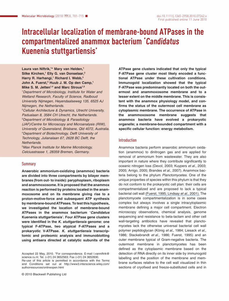
Intracellular localization of membrane-bound ATPases in thecompartmentalized anammox bacterium ‘CandidatusKuenenia stuttgartiensis’mmi_7242 701..715
Laura van Niftrik,1* Mary van Helden,1
Silke Kirchen,1 Elly G. van Donselaar,2
Harry R. Harhangi,1 Richard I. Webb,3
John A. Fuerst,3 Huub J. M. Op den Camp,1
Mike S. M. Jetten1,4 and Marc Strous1,5
1Department of Microbiology, Institute for Water andWetland Research, Faculty of Science, RadboudUniversity Nijmegen, Heyendaalseweg 135, 6525 AJNijmegen, the Netherlands.2Cellular Architecture & Dynamics, Utrecht University,Padualaan 8, 3584 CH Utrecht, the Netherlands.3Department of Microbiology & Parasitology(JAF)/Centre for Microscopy and Microanalysis (RIW),University of Queensland, Brisbane, Qld 4072, Australia.4Department of Biotechnology, Delft University ofTechnology, Julianalaan 67, 2628 BC Delft, theNetherlands.5Max Planck Institute for Marine Microbiology,Celciusstrasse 1, 28359 Bremen, Germany.
Summary
Anaerobic ammonium-oxidizing (anammox) bacteriaare divided into three compartments by bilayer mem-branes (from out- to inside): paryphoplasm, riboplasmand anammoxosome. It is proposed that the anammoxreaction is performed by proteins located in the anam-moxosome and on its membrane giving rise to aproton-motive-force and subsequent ATP synthesisby membrane-bound ATPases. To test this hypothesis,we investigated the location of membrane-boundATPases in the anammox bacterium ‘CandidatusKuenenia stuttgartiensis’. Four ATPase gene clusterswere identified in the K. stuttgartiensis genome: onetypical F-ATPase, two atypical F-ATPases and aprokaryotic V-ATPase. K. stuttgartiensis transcrip-tomic and proteomic analysis and immunoblottingusing antisera directed at catalytic subunits of the
ATPase gene clusters indicated that only the typicalF-ATPase gene cluster most likely encoded a func-tional ATPase under these cultivation conditions.Immunogold localization showed that the typicalF-ATPase was predominantly located on both the out-ermost and anammoxosome membrane and to alesser extent on the middle membrane. This is consis-tent with the anammox physiology model, and con-firms the status of the outermost cell membrane ascytoplasmic membrane. The occurrence of ATPase inthe anammoxosome membrane suggests thatanammox bacteria have evolved a prokaryoticorganelle; a membrane-bounded compartment with aspecific cellular function: energy metabolism.
Introduction
Anammox bacteria perform anaerobic ammonium oxida-tion (anammox) to dinitrogen gas and are applied forremoval of ammonium from wastewater. They are alsoimportant in nature where they contribute significantly tooceanic nitrogen loss (Devol, 2003; Kuypers et al., 2003;2005; Arrigo, 2005; Brandes et al., 2007). Anammox bac-teria belong to the phylum Planctomycetes. One of theunique properties of species within this phylum is that theydo not conform to the prokaryotic cell plan: their cells arecompartmentalized and are proposed to lack a typicalbacterial cell wall (Fuerst, 1995; Lindsay et al., 2001). Theplanctomycete compartmentalization is in some casescomplex but always involves a single intracytoplasmicmembrane defining a major cell compartment. Electronmicroscopy observations, chemical analysis, genomesequencing and resistance to beta-lactam and other cellwall-targeting antibiotics have revealed that plancto-mycetes lack the otherwise universal bacterial cell wallpolymer peptidoglycan (König et al., 1984; Liesack et al.,1986; Stackebrandt et al., 1986; Fuerst, 1995) and anouter membrane typical of Gram-negative bacteria. Theoutermost membrane in planctomycetes has beendefined as the cytoplasmic membrane based on thedetection of RNA directly on its inner side by immunogoldlabelling and the position of the membrane and mem-brane surfaces relative to the cell wall visualized in thinsections of cryofixed and freeze-substituted cells and in
Accepted 22 May, 2010. *For correspondence. E-mail [email protected]; Tel. (+31) 24 3652563; Fax (+31) 24 3652830.Re-use of this article is permitted in accordance with the Termsand Conditions set out at http://www3.interscience.wiley.com/authorresources/onlineopen.html
Molecular Microbiology (2010) 77(3), 701–715 � doi:10.1111/j.1365-2958.2010.07242.xFirst published online 11 June 2010
© 2010 Blackwell Publishing Ltd

freeze-fracture replicas (Lindsay et al., 1997; 2001). Theother, innermost, membrane has been defined as an intra-cytoplasmic membrane as it is within the cytoplasm andtopologically inside the space bounded by the cytoplasmicmembrane. The two compartments that are thus formedhave been named ‘paryphoplasm’ – the outermost com-partment between cytoplasmic and intracytoplasmicmembrane – and ‘riboplasm’ – the innermost compart-ment inside the space bounded by the intracytoplasmicmembrane. The riboplasm is the standard cytoplasmiccompartment containing ribosomes and in most cases thenucleoid.
Anammox planctomycetes have an additional intracy-toplasmic membrane compared with the standard planc-tomycete cell plan described above. The anammox cell isthus divided into three compartments by three individualbilayer membranes, from out- to inside; the paryphop-lasm, riboplasm and anammoxosome compartment(Fig. 1). Anammox bacteria also contain unusual mem-brane lipids with ladderane moieties (linearly concat-enated cyclobutane rings). These ladderane lipids aremajor membrane lipids of anammox membranes and arehypothesized to make these membranes highly imperme-able (Sinninghe Damsté et al., 2002; 2005) and to providestructural integrity to the cell.
The anammoxosome compartment is hypothesized tobe the site where the anammox catabolism takes place.Based on the genome of the anammox bacterium ‘Candi-datus Kuenenia stuttgartiensis’, a biochemical model(Fig. 2) has been proposed where the anammox reaction iscatalysed by several cytochrome c proteins (Strous et al.,2006). In this model, nitrite is first reduced to nitric oxide,then nitric oxide is combined with ammonium to formhydrazine and finally hydrazine is oxidized to dinitrogengas. The four electrons derived from the oxidation of hydra-zine to dinitrogen gas are fed into a respiratory chain at thelevel of ubiquinone via soluble cytochrome c electroncarriers and a hypothetical cytochrome c: ubiquinone oxi-doreductase (Cirpus et al., 2005; Huston et al., 2007). The
anammox reaction thus establishes a proton gradient bythe translocation of protons from the riboplasm to theanammoxosome, giving rise to a proton-motive-force. Thisproton-motive-force could then be used by membrane-bound ATPases to produce ATP. Experimental evidencethat supports the proposed model is the immunogold local-ization of the enzyme hydrazine/hydroxylamine oxi-doreductase (HAO) to the anammoxosome (Lindsay et al.,2001) and the demonstration that all, or almost all, cyto-chrome c proteins are located in the anammoxosome bycytochrome peroxidase staining (van Niftrik et al., 2008a).Furthermore, preliminary results from 31P nuclear magneticresonance (NMR) spectroscopy showed the presence oftwo phosphate peaks in K. stuttgartiensis cells indicatingan intracytoplasmic pH gradient (van der Star et al., 2010).
Some questions concerning the anammox cell planremain. Although it has been assumed that anammoxbacteria share the planctomycete cell plan, there are atleast two possibilities with respect to the nature of the
Fig. 1. Electron micrograph (left) of an anammox bacterium and schematic overview (right) of the different scenarios regarding its cell plan.MB, membrane. Modified from van Niftrik et al. (2008b).
Fig. 2. Postulated model for catabolic anammox reactionscoupled over the anammoxosome membrane in anammox bacteriaresulting in a proton-motive-force and subsequent ATP synthesis.bc1, cytochrome bc1 complex; cyt, cytochrome; hao,hydrazine/hydroxylamine oxidoreductase; hh, hydrazine hydrolase;nir, nitrite reductase; Q, co-enzyme Q. Modified from Strous et al.(2006).
702 L. van Niftrik et al. �
© 2010 Blackwell Publishing Ltd, Molecular Microbiology, 77, 701–715

paryphoplasm (or region equivalent to the paryphoplasm)in anammox bacteria. As in other planctomycetes, theanammox paryphoplasm may represent a space betweena true cytoplasmic membrane and an intracytoplasmicmembrane (Fig. 1; scenario 1). On the other hand, theparyphoplasm might represent a space similar to the peri-plasm of Gram-negative bacteria if the outermost mem-brane of the cell is more like an outer membrane of aGram-negative cell wall and the intracytoplasmic mem-brane is actually the cytoplasmic membrane (Fig. 1; sce-nario 2).
To explore such potential similarities to a Gram-negative cell plan, the genome of K. stuttgartiensis(Strous et al., 2006) was examined by comparativegenomic analysis, which indicated that K. stuttgartiensismay be genetically capable of the biogenesis of a peri-plasm and outer membrane. First, a number of openreading frames (ORFs) were homologous to outer mem-brane porins. These porin homologues were absent in thegenome of the planctomycete Rhodopirellula baltica.Second, the K. stuttgartiensis genome encoded the com-plete TonB system, a protein complex that relays energyfrom the cytoplasmic membrane to the outer membrane todrive a number of outer membrane receptors, five ofwhich were also encoded in the genome. Third, K. stut-tgartiensis encoded a number of typical three-componentGram-negative multidrug exporters, which consist of acytoplasmic membrane, a periplasmic and an outer mem-brane subunit (‘gated porins’). Fourth, a partial peptidogly-can biogenesis pathway was encoded, including anumber of penicillin-binding proteins. The only step notpresent in the peptidoglycan pathway of this bacteriumwas the ability to cross-link the glycan. With respect to allthese four points, R. baltica, another planctomycete with apublicly available genome, contains hardly any geneticpotential for Gram-negative cell wall structure or pepti-doglycan synthesis. This might be consistent with the viewthat the paryphoplasm (Fig. 1; scenario 1) in K. stuttgar-tiensis may actually be more similar to a ‘regular’ peri-plasm (Fig. 1; scenario 2). However, the presence ofthese genes could also be a result of lateral gene transferor be remainders of the evolutionary ancestor ofanammox bacteria, which would then be a Gram-negativebacterium.
In contrast to the genomic evidence that could supportthe paryphoplasm being a periplasmic-like space, thereis experimental evidence that supports the paryphop-lasm being a cytoplasmic compartment with the cyto-plasmic membrane on its outer side and the absence ofa typical bacterial cell wall. First, neither peptidoglycannor a typical outer membrane can be observed in trans-mission electron micrographs of all known species ofanammox bacteria when examined after cryofixation andfreeze-substitution or via classical chemical fixation (van
Niftrik et al., 2008a,b). Second, anammox bacteria andother non-anammox planctomycetes are prone toosmotic collapse under both hypotonic and hypertonicconditions (see Lindsay et al., 1995; 2001; van de Graafet al., 1996), an indication that their structural integrity,normally derived from the presence of a cell wall, is notoptimal. Third, the cell division ring of anammox bacteriais situated in the paryphoplasm compartment (van Niftriket al., 2009). In general, the bacterial cell division ring ison the inside of, and closely opposed to, the cytoplasmicmembrane indicating that the membrane on the outsideof the paryphoplasm (Fig. 1; membrane 1) is the cyto-plasmic membrane. Fourth, the apparent absence ofcytochrome c proteins in the paryphoplasm as indicatedby cytochrome peroxidase staining (van Niftrik et al.,2008a) supports the notion that this cannot be a typicalperiplasmic space analogous to that of Gram-negativebacteria.
A third scenario may reconcile the experimental andgenomic evidence. In this scenario there is a periplasmic-like space outside the outermost membrane (Fig. 1; mem-brane 1), as proposed for some Archaea with S-layerwalls (Küper et al., 2010) and for some Gram-positivebacteria (Zuber et al., 2008). This periplasmic-like spacecould be lost during sample preparation for electronmicroscopy.
The localization of anammox membrane-boundATPases would resolve both the function of the anam-moxosome and the location of the cytoplasmicmembrane. If an ATPase was present on the innermost(anammoxosome) membrane (Fig. 1; membrane 3) thiswould strongly suggest that this compartment is indeedused for the generation of energy analogous to the func-tion of mitochondria in Eukaryotes. The localization of ananammox ATPase to the outermost membrane of theanammox cell (Fig. 1; membrane 1) would indicate thatthis is indeed an energized, cytoplasmic membrane, andnot an outer membrane typical of a Gram-negative cellwall, and that the middle anammox membrane (Fig. 1;membrane 2) is indeed an intracytoplasmic membrane aswas initially proposed on structural grounds (Lindsayet al., 2001).
Three different types of membrane-bound ATPaseshave been described: F-ATPase, A-ATPase andV-ATPase (Grüber et al., 2001; Grüber and Marshansky,2008). V-ATPase (vacuolar-type) functions as an ATP-dependent proton-pump in eukaryotes (Drory and Nelson,2006). F-ATPase and prokaryotic V-ATPase (the latterfound in Bacteria and Archaea) can function both as ion-pump or as ATP synthase (Mitchell, 1961; Müller et al.,1999). The prokaryotic V-ATPase is also known asA-ATPase (archaeal-type) or V/A-ATPase (Grüber et al.,2001; Yokoyama and Imamura, 2005). Subunit composi-tion can differ, but in general the prokaryotic V-ATPase
Localization of membrane-bound ATPases in anammox bacteria 703
© 2010 Blackwell Publishing Ltd, Molecular Microbiology, 77, 701–715

consists of nine subunits and the bacterial F-ATPase con-sists of eight subunits (Stock et al., 2000; Lolkema et al.,2003) (Fig. 3). Membrane-bound ATPases contain twocomponents: a membrane-bound, hydrophobic part (F0/A0/V0) containing the ion channel that is connected by acentral stalk to the cytoplasmic, hydrophilic part (F1/A1/V1)containing the catalytic sites. The way F-ATPase func-tions is described by the binding change (Boyer, 1993;1997) and rotary mechanism (Stock et al., 2000).F1-ATPase contains three catalytic b subunits that performATP synthesis (or in some cases hydrolysis). F0-ATPasetranslocates protons (or sodium ions) across the mem-brane down the electrochemical gradient. In couplingproton transport to ATP synthesis, the F0 and F1 domainsfunction as a pair of rotary motors linked by a commoncentral rotor (gec) and a peripheral stator (bd). The mul-tiple c-subunits (proteolipids) form a ring and rotation ofthis c-ring allows protons to be carried between two partialsubunit-a channels that lead to opposite sites of themembrane. Each c-subunit contains two transmembranehelices that are connected by a cytoplasmic loop and oneprotonizable, carboxylate group (glutamate or aspartate;active carboxylate of Escherichia coli subunit c isAsp-61). The active carboxylate undergoes protonation/deprotonation cycles during proton transport and islocated in helix 2 (Rastogi and Girvin, 1999). Either threeor four protons need to be transported in sequence for agroup of 12 c-subunits to move 120 degrees and promotethe release of one ATP. Among the different membrane-bound ATPase types, the number of proteolipid trans-membrane helices, and the number of proteolipidsubunits per enzyme, differs.
Here, we identified and analysed four ATPase geneclusters in the K. stuttgartiensis genome. We show bytranscriptomic, proteomic and immunoblot analyses thatonly one of these four ATPase gene clusters is likely to beexpressed under the conditions investigated. Antiserumtargeting this typical F-ATPase was used to locate thisanammox membrane-bound ATPase in the anammox cellusing immunogold localization. The typical F-ATPase wasdetected on all three anammox cell membranes but waspredominantly present on both the innermost (anam-moxosome) membrane and outermost membrane of theanammox cell. This indicates that the anammoxosome isused for the generation of ATP, probably vectorially from aproton-motive-force across the anammoxosome mem-brane itself, and is consistent with the identification of theoutermost membrane of the anammox cell as the cyto-plasmic membrane.
Results
ATPase gene clusters in the K. stuttgartiensis genome
Four putative gene clusters encoding membrane-boundATPase complexes were identified in the genome assem-bly of K. stuttgartiensis (Strous et al., 2006) (Fig. 4). Thesyntheny (similarity of gene order) of gene cluster 1 wassimilar to the F-ATPase operon of E. coli with 23–69%sequence identities of individual orthologous genes toclosest homologous genes with experimentally validatedfunction. Note that in the databases where the K. stuttgar-tiensis genome has been deposited, the ORFs encodingfor subunit b and subunit delta are translated in the wrong
Fig. 3. Schematic model of (A) a prokaryoticF-ATPase and (B) a prokaryotic V-ATPase.Assembled from Grüber et al. (2001),Lolkema et al. (2003), Yokoyama andImamura (2005) and Lau and Rubinstein(2010).
704 L. van Niftrik et al. �
© 2010 Blackwell Publishing Ltd, Molecular Microbiology, 77, 701–715
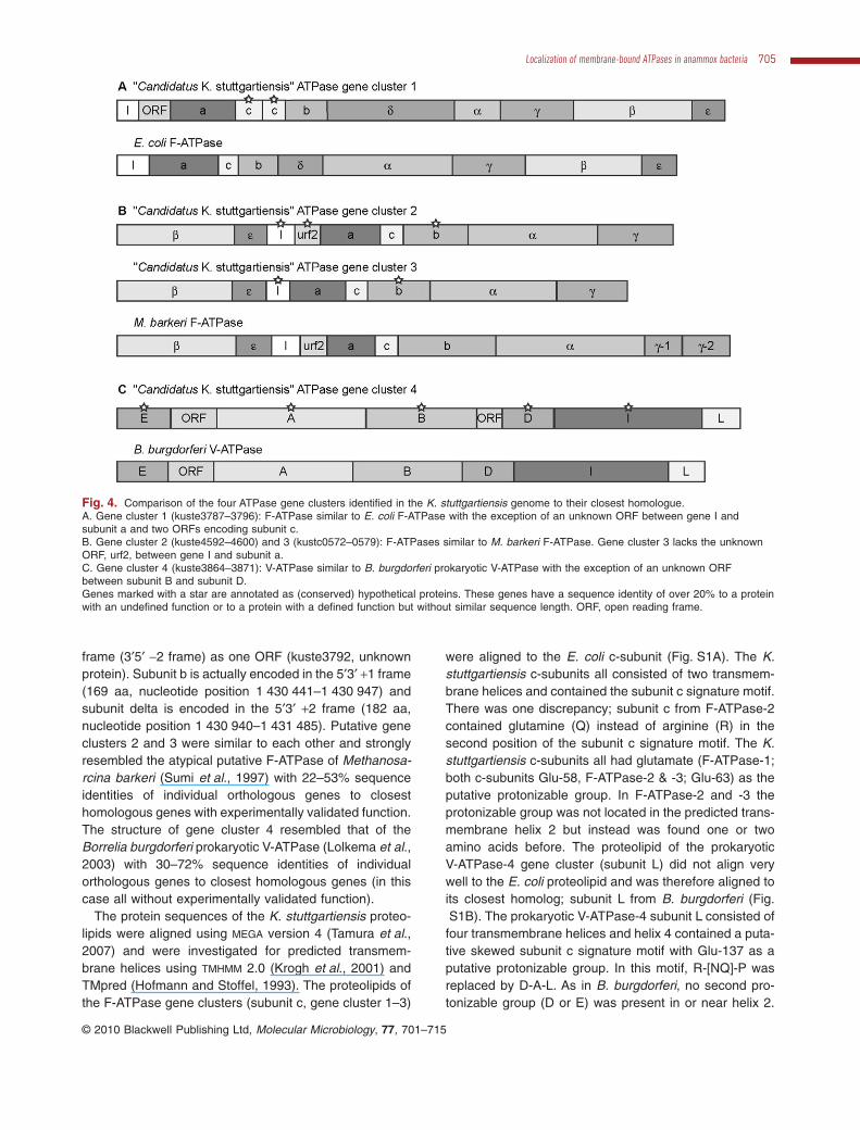
frame (3′5′ -2 frame) as one ORF (kuste3792, unknownprotein). Subunit b is actually encoded in the 5′3′ +1 frame(169 aa, nucleotide position 1 430 441–1 430 947) andsubunit delta is encoded in the 5′3′ +2 frame (182 aa,nucleotide position 1 430 940–1 431 485). Putative geneclusters 2 and 3 were similar to each other and stronglyresembled the atypical putative F-ATPase of Methanosa-rcina barkeri (Sumi et al., 1997) with 22–53% sequenceidentities of individual orthologous genes to closesthomologous genes with experimentally validated function.The structure of gene cluster 4 resembled that of theBorrelia burgdorferi prokaryotic V-ATPase (Lolkema et al.,2003) with 30–72% sequence identities of individualorthologous genes to closest homologous genes (in thiscase all without experimentally validated function).
The protein sequences of the K. stuttgartiensis proteo-lipids were aligned using MEGA version 4 (Tamura et al.,2007) and were investigated for predicted transmem-brane helices using TMHMM 2.0 (Krogh et al., 2001) andTMpred (Hofmann and Stoffel, 1993). The proteolipids ofthe F-ATPase gene clusters (subunit c, gene cluster 1–3)
were aligned to the E. coli c-subunit (Fig. S1A). The K.stuttgartiensis c-subunits all consisted of two transmem-brane helices and contained the subunit c signature motif.There was one discrepancy; subunit c from F-ATPase-2contained glutamine (Q) instead of arginine (R) in thesecond position of the subunit c signature motif. The K.stuttgartiensis c-subunits all had glutamate (F-ATPase-1;both c-subunits Glu-58, F-ATPase-2 & -3; Glu-63) as theputative protonizable group. In F-ATPase-2 and -3 theprotonizable group was not located in the predicted trans-membrane helix 2 but instead was found one or twoamino acids before. The proteolipid of the prokaryoticV-ATPase-4 gene cluster (subunit L) did not align verywell to the E. coli proteolipid and was therefore aligned toits closest homolog; subunit L from B. burgdorferi (Fig.S1B). The prokaryotic V-ATPase-4 subunit L consisted offour transmembrane helices and helix 4 contained a puta-tive skewed subunit c signature motif with Glu-137 as aputative protonizable group. In this motif, R-[NQ]-P wasreplaced by D-A-L. As in B. burgdorferi, no second pro-tonizable group (D or E) was present in or near helix 2.
Fig. 4. Comparison of the four ATPase gene clusters identified in the K. stuttgartiensis genome to their closest homologue.A. Gene cluster 1 (kuste3787–3796): F-ATPase similar to E. coli F-ATPase with the exception of an unknown ORF between gene I andsubunit a and two ORFs encoding subunit c.B. Gene cluster 2 (kuste4592–4600) and 3 (kustc0572–0579): F-ATPases similar to M. barkeri F-ATPase. Gene cluster 3 lacks the unknownORF, urf2, between gene I and subunit a.C. Gene cluster 4 (kuste3864–3871): V-ATPase similar to B. burgdorferi prokaryotic V-ATPase with the exception of an unknown ORFbetween subunit B and subunit D.Genes marked with a star are annotated as (conserved) hypothetical proteins. These genes have a sequence identity of over 20% to a proteinwith an undefined function or to a protein with a defined function but without similar sequence length. ORF, open reading frame.
Localization of membrane-bound ATPases in anammox bacteria 705
© 2010 Blackwell Publishing Ltd, Molecular Microbiology, 77, 701–715

Sequence analysis of the proteolipids indicated that thethree F-ATPases are potentially capable of ATP synthesis.With respect to gene cluster organization, F-ATPase-1contained the complete set of genes known to be part ofthe E. coli F-ATPase and therefore there is no reason whythis gene cluster could not encode for a functionalmembrane-bound ATPase. F-ATPase-2 and -3 lacked agene encoding the delta subunit that in F-ATPase con-nects the F1 and F0 static parts (Fig. 3), although it isunknown whether this would infer dysfunctionality. Theprokaryotic V-ATPase-4 gene cluster contained both cata-lytic subunits (B and A), the proteolipid (L), ion channel (I),
part of the rotor (D) and part of the stator (E), indicatingthat it could encode for a functional membrane-boundATPase.
K. stuttgartiensis transcriptomic and proteomic analysis
To investigate the expression of the four ATPase geneclusters identified in the genome, the transcriptome andproteome of K. stuttgartiensis were analysed for the pres-ence of mRNA or detected peptides respectively(Table 1). The analysis showed that F-ATPase-1, thetypical ATP synthase, was detected at high levels in both
Table 1. Presence of mRNA (transcriptome) and peptides (proteome) from the four ATPase gene clusters present in the genome of K.stuttgartiensis.
Genome Transcriptome Proteome
ORFPutativeATPase subunit aa nt
# unique readsdetected
Relativeexpression
# uniquepeptides detected
Genecoverage (%)
F-ATPase-1kuste3787 I 84 255 50 0.9 ND NAkuste3788 Unknown 124 375 136 1.6 ND NAkuste3789 a 257 774 656 3.7 2 25kuste3790 c 108 327 204 2.8 ND NAkuste3791 c 90 273 317 5.1 1 25kuste3792 b 169 510 882 7.6 ND NAkuste3792 Delta 182 549 1013 8.1 ND NAkuste3793 Alpha 504 1515 1881 5.5 23 66kuste3794 Gamma 290 873 415 2.1 8 29kuste3795 Beta 471 1416 1956 6.1 24 60kuste3796 Epsilon 134 405 413 4.5 3 27
F-ATPase-2kuste4592 Beta 462 1389 74 0.2 ND NAkuste4593 Epsilon 130 393 16 0.2 ND NAkuste4594 I 111 336 31 0.4 ND NAkuste4595 Unknown 101 306 18 0.3 ND NAkuste4596 a 236 711 68 0.4 ND NAkuste4597 c 93 282 15 0.2 ND NAkuste4598 b 255 768 21 0.1 ND NAkuste4599 Alpha 513 1542 101 0.3 ND NAkuste4600 Gamma 299 900 67 0.3 ND NA
F-ATPase-3kustc0572 Beta 454 1365 177 0.6 ND NAkustc0573 Epsilon 134 405 12 0.1 ND NAkustc0574 I 92 279 32 0.5 ND NAkustc0575 a 221 666 39 0.3 ND NAkustc0576 c 88 267 34 0.6 ND NAkustc0577 b 246 741 35 0.2 ND NAkustc0578 Alpha 498 1497 78 0.2 ND NAkustc0579 Gamma 279 840 16 0.1 ND NA
V-ATPase-4kuste3864 E 210 633 99 0.7 ND NAkuste3865 Unknown 180 543 74 0.6 ND NAkuste3866 A 589 1770 531 1.3 1 2kuste3867 B 437 1314 503 1.7 ND NAkuste3868 Unknown 99 300 36 0.5 ND NAkuste3869 D 202 609 21 0.2 ND NAkuste3870 I 583 1752 218 0.6 ND NAkuste3871 L 152 459 66 0.6 ND NA
ORF, open reading frame; aa, amino acids; nt, nucleotides; ND, not detected; NA, not applicable. The relative expression (transcriptome) iscalculated as: [# unique reads detected * length of the reads (75 nt)/gene length (nt)]/coverage of the transcriptome (= 17). The gene coverage(proteome) is calculated as: (# unique peptides detected/# unique peptides predicted) * 100%.
706 L. van Niftrik et al. �
© 2010 Blackwell Publishing Ltd, Molecular Microbiology, 77, 701–715

the transcriptome and proteome. This indicates that thisATPase gene cluster most likely encodes a functionalmembrane-bound ATPase in K. stuttgartiensis. Both atypi-cal F-ATPases-2 and -3 were not detected in the pro-teome and showed very low transcription levels (relativeexpression < 1.0), making it unlikely that these twoATPase gene clusters function in energy generation in K.stuttgartiensis cells at least under the cultivation condi-tions of this study. For the prokaryotic V-ATPase-4 thecatalytic subunits (A and B) were detected in the transcrip-tome (relative expression > 1.0) and one peptide (subunitA) was detected in the proteome. More data will beneeded to make a functional assignment possible for theprokaryotic V-ATPase.
Heterologous expression of the catalytic subunits ofthe four K. stuttgartiensis ATPase gene clustersand subsequent antibody production
Parts of the catalytic beta (F-ATPases) and A (prokaryoticV-ATPase) subunits (Table S1) were cloned andexpressed in E. coli. The purified protein fragments wereused to raise antibodies in rabbit. All four antisera hybrid-ized to the associated heterologous protein on dot blots(data not shown). The antisera were subsequently used totest for the presence of their target proteins in K. stuttgar-tiensis cell-free extract using SDS-polyacrylamide gelelectrophoresis (SDS-PAGE) and immunoblot analysis.The antiserum targeting F-ATPase-1 showed a band atthe expected size (52 kDa) that was absent in both nega-tive controls (incubation with pre-immune serum and incu-bation with only the secondary antibody) (Fig. 5).Incubation with the other antisera (anti-F-ATPase-2,-F-ATPase-3 and -V-ATPase-4) did not result in the detec-
tion of a protein of the expected size, nor of any otherspecific bands (data not shown). These results were con-sistent with the high levels of transcription and expressionobserved for F-ATPase-1 and the undetectable or at leastvery low levels of transcription and expression of the othermembrane-bound ATPases under the applied growthconditions.
Anti-F-ATPase-1 was further tested for its specificityand affinity using immunofluorescence analysis offormaldehyde-fixed K. stuttgartiensis cells. This analysisshowed that the antiserum specifically bound to wholeK. stuttgartiensis cells (Fig. S2) while the pre-immuneserum had only a very faint background at the sameexposure time. This indicated that this antiserum wassuitable for immunogold localization to identify the cellu-lar location of this membrane-bound ATPase in theanammox cell.
Immunogold localization of F-ATPase-1 inK. stuttgartiensis
The antiserum targeting F-ATPase-1 was used in immu-nogold localization to determine its location in anammoxcells prepared via the rehydration method (van Donselaaret al., 2007) and subsequent cryosectioning. All threemembranes of the anammox cell, the outermost mem-brane (membrane 1), middle membrane (membrane 2)and innermost (anammoxosome) membrane (membrane3), which appear as white trilamina in the cryosections,were significantly labelled (Figs 6 and 7 and Fig. S3)compared with the incubation with the pre-immune serum(the negative control). There was some background label-
Fig. 5. Immunoblot analysis of the antiserum directed at thecatalytic beta subunit of the F-ATPase-1 gene cluster found in theK. stuttgartiensis genome. Lane 1: Marker (PageRuler™ PrestainedProtein Ladder Plus, Fermentas); lane 2: incubation with anti-F-ATPase-1; lane 3: incubation with the pre-immune serum; lane 4:incubation with only the secondary antibody. Arrow: expected targetsize (52 kDa).
Fig. 6. Immunogold labelling distribution (average gold particlesper cell per location) in K. stuttgartiensis using the antiserumdirected at F-ATPase-1 and its pre-immune serum. All threemembranes of the anammox cell are significantly labelledcompared with the incubation with the pre-immune serum (thenegative control). OM, outermost membrane (membrane 1); MM,middle membrane (membrane 2); IM, innermost (anammoxosome)membrane (membrane 3); A, anammoxosome; R, riboplasm; ***,extremely statistically significant; *, statistically significant.
Localization of membrane-bound ATPases in anammox bacteria 707
© 2010 Blackwell Publishing Ltd, Molecular Microbiology, 77, 701–715

Fig. 7. A–D. Immunogold localization of the antiserum directed at the catalytic beta subunit of the F-ATPase-1 gene cluster localizes thisATPase to all anammox membranes; the outermost membrane (membrane 1), middle membrane (membrane 2) and innermost(anammoxosome) membrane (membrane 3) in K. stuttgartiensis rehydrated cryosections. See Fig. S3 for annotation of specific gold labels.E and F. Negative control: incubation with the pre-immune serum instead of the antiserum.Scale bars: 250 nm.
708 L. van Niftrik et al. �
© 2010 Blackwell Publishing Ltd, Molecular Microbiology, 77, 701–715
ling in the anammoxosome and riboplasm but this was notsignificantly more in the incubation with the antiserumthan in the incubation with the pre-immune serum. Sincegold particles that could be attributed to more than onemembrane were not taken into account, the labelling ofthe membranes is even underestimated. There was sig-nificantly more labelling on the outermost membrane andthe innermost (anammoxosome) membrane comparedwith the middle membrane while there was no significantdifference between the labelling of the outermost mem-brane and the innermost (anammoxosome) membrane.From these results, it can be concluded that theF-ATPase-1 is predominantly located on the innermost(anammoxosome) membrane (membrane 3) and the out-ermost membrane (membrane 1) and to a lesser extenton the middle membrane (membrane 2).
Discussion
In this study, the intracellular location of membrane-boundATPases in the anammox bacterium K. stuttgartiensiswas addressed to investigate the hypothesis that theanammoxosome compartment and its membrane areused for the generation of energy and to identify thecytoplasmic membrane. Four putative ATPase gene clus-ters (F-ATPase-1, -2, -3, and prokaryotic V-ATPase-4)were identified in the K. stuttgartiensis genome. Tran-scriptomic, proteomic and immunoblot analyses with anti-sera directed at the catalytic subunits indicated thatF-ATPase-1 was the most significant membrane-boundATPase under the growth conditions investigated. Immu-nogold localization showed that this F-ATPase was pre-dominantly present on the innermost (anammoxosome)membrane and outermost membrane of the anammoxcell and to a lesser extent on the middle membrane.
Four ATPase gene clusters in the K. stuttgartiensisgenome
The genome of K. stuttgartiensis encodes four putativeATPase gene clusters: one typical F-ATPase (F-ATPase-1), two atypical F-ATPases (F-ATPase-2 and -3) lackingthe delta subunit and a prokaryotic V-ATPase(V-ATPase-4).
F-ATPase-1 resembled most the typical F-ATPase fromE. coli but also the F-ATPase from Acetobacterium woodii(Müller et al., 2005). The K. stuttgartiensis transcriptomeshowed a high relative expression (ranging from 0.9–8.1)of all genes in the F-ATPase-1 gene cluster and in theproteome, peptides of six of the 11 genes were detected(Table 1). Further, the antiserum directed at the catalyticbeta subunit specifically detected a protein of theexpected size in immunoblot analysis (Fig. 5) and hybrid-
ized specifically to formaldehyde-fixed K. stuttgartiensiscells in immunofluorescence (Fig. S2). In conclusion, thetranscriptome, proteome, immunoblot and immunofluo-rescence data indicated that the F-ATPase-1 gene clusterencodes the major functional membrane-bound ATPasein K. stuttgartiensis.
The gene cluster structure of F-ATPase-2 andF-ATPase-3 strongly resembled that of the atypical M.barkeri F-ATPase (Fig. 4) (Sumi et al., 1997). These geneclusters lacked a gene encoding the delta subunit that inF-ATPase connects the F1 and F0 static parts (Fig. 3).Without this subunit, the ATPase enzyme would probablybe less stable. Therefore, if these gene clusters areexpressed they would have to acquire the delta subunitfrom somewhere else, which would have to be theF-ATPase-1 gene cluster. Another possibility could be thatthe extended version of their b-subunit (255 aa and 246aa compared with 169 aa in F-ATPase-1, Table 1) hasfunctionally replaced the delta subunit. In M. barkeri it isdoubtful whether this F-ATPase is functional because theamino acid sequences of the genes are so deviated fromthe typical F-ATPase and also because mRNA and proteinproducts cannot be detected (Sumi et al., 1997). A func-tional F-ATPase in M. barkeri would be quite unusualanyway since Archaea (including M. barkeri ) typicallyhave prokaryotic V-ATPases and not F-ATPases. Thetranscriptome of K. stuttgartiensis showed a very low rela-tive expression level (< 1.0) for both ATPase gene clustersand no peptides were detected in the proteome (Table 1).Antibodies generated against the catalytic subunits alsocould not detect their target proteins in K. stuttgartiensiscell-free extract under the cultivation conditions of thisstudy. In conclusion, the predicted instability of bothenzymes by the absence of the delta subunit, the very lowexpression level in the transcriptome and absence of pep-tides in the proteome and the inability of the antisera todetect their target proteins in cell-free extract make it veryunlikely that these two gene clusters encode functionalF-ATPases in K. stuttgartiensis. Perhaps these two geneclusters are cryptic, were acquired through lateral genetransfer and were duplicated in a subsequent rearrange-ment event.
The gene cluster structure of V-ATPase-4 resembledmost that of B. burgdorferi prokaryotic V-ATPase (Fig. 4).These two gene clusters, and also the chlamydialprokaryotic V-ATPase gene clusters (Lolkema et al.,2003), do not contain homologues of subunits C and F(part of the central stalk), and G (part of the stator)(Fig. 3). It was not clear from the transcriptome and pro-teome whether gene cluster V-ATPase-4 could encode fora functional membrane-bound ATPase. Only the catalyticsubunits (A and B) were detected, at a low level, in thetranscriptome and only one peptide (subunit A) wasdetected in the proteome. Further, the antiserum targeting
Localization of membrane-bound ATPases in anammox bacteria 709
© 2010 Blackwell Publishing Ltd, Molecular Microbiology, 77, 701–715

the catalytic subunit A could not detect its target protein inK. stuttgartiensis cell-free extract. In conclusion, either theV-ATPase-4 gene cluster is not expressed as a functionalmembrane-bound ATPase under the cultivation conditionsof this study or it is expressed at such low levels that it isdifficult to detect with the current methods.
The intracellular location of F-ATPase-1 inK. stuttgartiensis cells
All data indicated that the F-ATPase-1 gene clusterencoded a functional membrane-bound ATPase capableof ATP synthesis and that the other ATPase gene clusterswere most likely not expressed in the cells used inthis study. Therefore, only the antiserum targetingF-ATPase-1 was used in immunogold localization todetermine its location in anammox cells prepared via therehydration method and cryosectioning (van Donselaaret al., 2007). Note that, even if the atypical F-ATPase-2and -3 could be expressed, cross-reactivity with the anti-serum targeting the beta subunit of F-ATPase-1 was notto be expected since the antibody target of F-ATPase-1showed only a 37% protein sequence identity with thebeta subunits of the atypical F-ATPase-2 and -3. Anti-F-ATPase-1 located the F-ATPase to all three anammoxmembranes (Figs 6 and 7 and Fig. S3). The labelling wasequally high on both the innermost (anammoxosome)membrane (membrane 3) and outermost membrane(membrane 1). Further, both of these membranes weresignificantly more densely labelled than the middle mem-brane (membrane 2) although the labelling on the middlemembrane was still significantly higher than in the nega-tive control. The immunogold labelling of the anammoxmembranes is even underestimated. First, gold particlesthat could be allocated to more than one membrane (i.e.within 25 nm distance from more than one membrane)were not taken into account. Second, labelling in theanammoxosome compartment could still be real inner-most (anammoxosome) membrane labelling consideringthe high curvature of this anammoxosome membrane asvisualized by electron tomography (van Niftrik et al.,2008b). These results indicate that the F-ATPase-1 ispredominantly present on the innermost (anammoxo-some) membrane and outermost membrane and to alesser extent on the middle membrane and that thus allthree membranes are energized and within the essentialcell boundary. This is not consistent with the preliminaryresults using 31P NMR that indicated the presence of twopH peaks and thus most likely two energized membranes(van der Star et al., 2010). It is also inconsistent with theabsence of genes encoding a putative nucleotide trans-porter in the genome of K. stuttgartiensis. Such a trans-porter would be necessary in a scenario with threeenergized membranes. Because most label was found on
the anammoxosome and outer membranes we concludethat these membranes most likely have a proton-motive-force. Because the cytochromes are located inside theanammoxosome, F-ATPase-1 most likely produces ATPon this membrane. This ATP is then hydrolysed again byF-ATPase-1 on the outermost membrane. There are twoexplanations for the presence of F-ATPase in the middlemembrane: it is the apo-complex en route to its finaldestination or it is functional, which we cannot explain atthis moment.
In any case, the results so far indicate that the outer-most membrane (membrane 1) is the cytoplasmic mem-brane as was initially proposed (Fig. 1, scenario 1)(Lindsay et al., 2001). To some extent, the ultrastructureand ATPase immunolabelling of the outermost membraneof K. stuttgartiensis resembles that of the CrenarchaeonIgnicoccus hospitalis (Küper et al., 2010). I. hospitalis alsohas a compartmentalized cell plan and an energizedoutermost membrane. However, in the I. hospitalis case,the outermost compartment has been defined as aperiplasmic-like space based on its low electron density intransmission electron micrographs, which suggested theapparent absence of ribosomes and DNA. Consequently,when the outermost membrane was found to be ener-gized, it was concluded that I. hospitalis has an energized‘outer membrane’ (although this membrane is structurallynot similar to the outer membrane of Gram-negative bac-teria) and ATP synthesis in the ‘periplasm’. In a way, thisresembles the K. stuttgartiensis case but in our case thecompartments and membranes are defined differently.This raises the question what defines the cytoplasm, peri-plasm, cytoplasmic membrane and outer membrane?
The genetic potential found in the K. stuttgartiensisgenome for producing a Gram-negative-like cell wallcould be a cryptic result of lateral gene transfer or be aremainder of the evolutionary ancestor of anammox bac-teria, which would then be a Gram-negative bacterium.However, another possibility is that there is indeed aGram-negative-like cell wall in anammox bacteria but thatthis is lost during sample preparation for electronmicroscopy. Anammox bacteria certainly do not live inosmotically protected areas like the pathogenic cell wall-less mycoplasma species, so are in need of some form ofstructural integrity. Other planctomycetes possess pro-teinaceous cell walls and their cells show considerablestructural integrity, even able in some cases to withstandtreatment with 10% SDS at 100°C (König et al., 1984;Liesack et al., 1986), conditions under which Gram-negative cell walls and especially outer membranes wouldbe expected to disintegrate. Perhaps in the anammoxcase, the ladderane lipids provide the structural integritythat most other bacteria derive from their cell wall. Theselipids have been predominantly found in the anammoxo-some membrane but also occur in one or both of the other
710 L. van Niftrik et al. �
© 2010 Blackwell Publishing Ltd, Molecular Microbiology, 77, 701–715

two anammox cell membranes (Sinninghe Damsté et al.,2002). Alternatively, we may in fact lack some structuralinformation and there may be yet another layer to theanammox cell. There is indeed some evidence of aregular protein surface layer (S-layer) lattice in K. stuttgar-tiensis from freeze-fracture replicas (Fuerst et al., 2006),but such an S-layer, if comparable to those protein orglycoprotein lattices of other bacterial and of archaealcell walls, is unlikely to possess membrane properties.However, in Archaea, which often do not contain other cellwall components besides S-layers, S-layers are proposedto maintain cell shape and can be viewed as exoskeletonsthat contribute to mechanical and osmotic cell stabiliza-tion (Engelhardt, 2007). Perhaps in the anammox bacteriawhere, as in the Archaea, also no other cell wall compo-nents have been found, structural integrity is also derivedfrom an S-layer lattice. Even though the complete loss ofadditional cell layers seems unlikely since many differentfixation procedures have been applied to anammox bac-teria (van Niftrik et al., 2008b), the presence of an outermembrane, exo- (S-layer) or endoskeleton (cytoskeleton)is currently under investigation by imaging anammoxbacteria in their near-native state with cryoelectronmicroscopy.
The localization of an F-ATPase to the anammoxosomemembrane shows that this compartment is indeed usedfor the generation of energy analogous to the function ofmitochondria in Eukaryotes. With its curved membrane tomaximize the membrane area available for the catabolicprocesses to take place (van Niftrik et al., 2008b), thelocalization of the key anammox protein HAO (Lindsayet al., 2001) and cytochrome c proteins (van Niftrik et al.,2008a) to the anammoxosome compartment, and thepresence of an ATP synthase on the anammoxosomemembrane established here, it seems that anammox bac-teria may indeed have evolved a bacterial organelle: aseparate membrane-bounded compartment with a spe-cific function inside the cell: energy metabolism.
Experimental procedures
Kuenenia stuttgartiensis cells
Samples containing an 80% enrichment culture of K. stuttgar-tiensis were taken from a 2 l sequencing batch reactor or a15 l continuous reactor (modified from Strous et al., 1998).
Genome analysis
Kuenenia stuttgartiensis ORFs encoding putative membrane-bound ATPase subunits were blasted (BLASTP) against theNCBI protein database (http://www.ncbi.nlm.nih.gov/BLAST/). Putative homologues were subsequently alignedusing the PIR pairwise alignment tool (http://pir.georgetown.edu/pirwww/search/pairwise.shtml).
Transcriptomic analysis
Kuenenia stuttgartiensis RNA was extracted using theRibopure Bacteria Kit according to the manufacturer’sinstructions from Ambion (Foster City, CA, USA). First-strandcDNA was synthesized with random primers using the Rever-tAid™ H Minus First Strand cDNA Synthesis Kit, and thesecond strand was synthesized using DNA polymerase andmanufacturer’s instructions (Fermentas, Vilnius, Lithuania).The Solexa reads (3.5 million) were mapped using the CLCWorkbench software version 3.7.1 (http://www.clcbio.com).The program reports expression as Reads Per Kilobase ofexon model per Million mapped reads (RPKM).
Proteomic analysis
Kuenenia stuttgartiensis single-cell suspensions were har-vested by centrifugation. The cell pellet was resuspended inone volume of 20 mM potassium phosphate buffer pH 8, andpassed three times through a French pressure cell operatedat 138 MPa. After centrifugation for 15 min at 1700 g at 4°C,a cell-free fraction was obtained as clarified supernatant. Asample of the cell-free extract was denatured by incubationwith 60 mM Tris-HCl buffer pH 8 containing 5% b-mercaptoethanol, 2% sodium dodecyl sulphate (SDS) and25% glycerol for 5 min at 100°C. SDS-PAGE was performedin 10% or 6% slab gels in 375 mM Tris hydrochlorideglycinebuffer, pH 8.8, with approximately 50 mg of protein per lane.After separation of proteins and staining with colloidal Coo-massie Brilliant Blue, the gel lane was cut into four slices, andeach gel slice was destained with three cycles of washingwith successively 50 mM ammonium bicarbonate and 50%acetonitrile (ACN). Sample analysis by LC-MS/MS, includingprotein reduction, alkylation and digestion, was performed asdescribed previously (Ettwig et al., 2010).
Antibody production
For the antisera directed against the catalytic beta(F-ATPase-1, -2 and -3) or A subunits (V-ATPase-4), weexpressed and purified parts of the K. stuttgartiensiskuste3795 (ATPase gene cluster 1), kuste4592 (ATPasegene cluster 2), kustc0572 (ATPase gene cluster 3) andkuste3866 (ATPase gene cluster 4) ORFs in E. coli asdescribed previously (Harhangi et al., 2002), with the follow-ing changes. Primers were designed on the sequences(Table S1). All forward primers started on nucleotide posi-tion 1 of the ORF. For directional cloning, restriction siteswere included in all primers. Further, stop codons wereintroduced in the reverse primers so as to express only anN-terminal His-tag. As an expression vector, pET30a-c(+)(Novagen, Darmstadt, Germany) was used, and as thehost, Rosetta cells (Novagen, Darmstadt, Germany). Theheterologous expressed protein fragments were purifiedusing the nickel-nitrilotriacetic acid (Ni/NTA) protein purifica-tion system (Qiagen, Venlo, the Netherlands) with an 8 Murea, 100 mM NaH2PO4, 10 mM Tris-HCl buffer at differentpH levels (6.3, 6.1, 5.9, 5.7, 5.5, 5.0 and 4.5) and imidazoleconcentrations (300, 250, 200, 150, 100, 50 and 20 mM).The identities of the expressed protein fragments were veri-
Localization of membrane-bound ATPases in anammox bacteria 711
© 2010 Blackwell Publishing Ltd, Molecular Microbiology, 77, 701–715

fied by MALDI-TOF MS peptide mass fingerprinting of atryptic digest of the Ni-NTA purified protein (Harhangi et al.,2002). The expressed protein fragments were used toimmunize rabbits in a 3-month immunization protocol(SEQLAB Sequencing Laboratories Göttingen GmbH, Göt-tingen, Germany). The antisera (anti-F-ATPase-1, -2, -3 andanti-V-ATPase-4) were then used as the primary antiserum(polyclonal, crude serum) in immunoblot analysis and (anti-F-ATPase-1) immunofluorescence and immunogold local-ization as described below.
SDS-PAGE and immunoblot analysis
Kuenenia stuttgartiensis proteins (cell-free extract preparedusing French press) were separated (50 mg of protein perlane) on a 10% SDS-PAGE gel and transferred to acellulose-nitrate membrane (Schleicher & Schuell GmbH,Dassel, Germany) with the semi-dry transfer cell blottingsystem (Bio-Rad, Veenendaal, the Netherlands). Blottingwas performed at 50 mA for 3 h with a transfer buffer thatconsisted of 25 mM Tris, 192 mM glycine and 20% metha-nol. After blotting, the blot was dried and stored at 4°C untilfurther use.
Blots stored at 4°C were washed in MilliQ waterfor 30 min and incubated in blocking buffer; 1% bovineserum albumin (BSA) in Tris-buffered saline (TBS; 10 mMTris-HCl, 0.9% NaCl, pH 7.4) for 1 h. The blot was thenincubated for 1 h in either blocking buffer or rabbit pre-immune serum diluted 500-fold in blocking buffer as thenegative controls or primary antiserum diluted 500-fold inblocking buffer. The blot was washed three times for 10 minin TBS containing 0.05% Tween20 and incubated for 1 h inmonoclonal mouse anti-rabbit IgG alkaline phosphataseconjugate (Sigma, Zwijndrecht, the Netherlands) diluted150.000-fold in blocking buffer. The blot was washed twotimes for 10 min in TBS containing 0.05% Tween20 andthree times for 10 min in TBS. The blot was incubated withthe BCIP/NBT Liquid Substrate System (Sigma, Zwijn-drecht, the Netherlands) for 8 min, rinsed in excessamounts of MilliQ water and dried. All blots were scannedwith the same settings.
Immunofluorescence analysis
Kuenenia stuttgartiensis cells were washed with phosphate-buffered saline (PBS; 0.1 M phosphate, 0.137 M NaCl,2.7 mM KCl, pH 7.4) and fixed in 3% formaldehyde in PBS for120 min while rotating at 4°C. The cells were washed againwith PBS, resuspended in 1:1 ethanol : PBS and stored at-20°C until further use.
Formaldehyde-fixed cells were transferred to 0.075%gelatine, 0.01% chromium coated, six-well diagnostic micro-scope slides (Menzel GmbH & Co. KG, Braunschweig,Germany) and allowed to air dry. Next, the cells were perme-abilized by a 10 min incubation with 0.5% Triton X-100 in TBSand washed three times with TBS. A-specific binding siteswere blocked by a 30 min incubation with blocking buffer (1%BSA in TBS) in a moist incubation chamber at room tempera-ture after which the blocking buffer was removed and thecells were allowed to air dry. Cells were incubated at room
temperature for 1 h with primary antiserum diluted 500- or1000-fold in blocking buffer in a moist incubation chamber.Each specific well was washed twice with 0.1% BSA in TBSafter which the entire slide was washed three times for 10 minin 100 ml of 0.1% BSA in TBS by shaking rigorously. Cellswere allowed to air dry and were then incubated for 1 h withthe secondary antibody, Cy-3-labelled sheep anti-rabbit IgG(Sigma, Zwijndrecht, the Netherlands), diluted 100-fold inblocking buffer at room temperature in a moist incubationchamber in the dark. Each specific well was washed twicewith TBS after which the entire slide was washed three timesfor 10 min in the dark in 100 ml of TBS by shaking rigorously.The cells were allowed to air dry, Vectashield mountingmedium with 4,6-diamidino-2-phenylindole (DAPI) (VectorLaboratories, Burlingame, USA) was added to each well, thecoverslip was sealed with nail polish and the slides werestored in the dark at 4°C until further investigation.
Several control treatments were used. All control treat-ments were performed on K. stuttgartiensis cells unlessstated otherwise, substituting primary antiserum and/or sec-ondary antibody respectively. Four negative controls wereperformed: blocking buffer combined with blocking buffer,blocking buffer combined with secondary antibody, rabbit pre-immune serum combined with secondary antibody and rabbitanti-Nitrosomonas sp. combined with secondary antibody.Two positive controls were performed: rabbit anti-anammoxhydrazine/hydroxylamine oxidoreductase (Schalk et al.,2000) combined with secondary antibody and rabbit anti-Nitrosomonas sp. combined with secondary antibody onNitrosomonas sp. cells.
Cells were investigated at 1.000¥ magnification with aZeiss Axioplan 2 imaging epifluorescence microscope (CarlZeiss B.V., Sliedrecht, the Netherlands).
Sample preparation for immunogold localization:cryofixation, freeze-substitution and cryosectioning(rehydration method) (van Donselaar et al., 2007)
Small aggregates of K. stuttgartiensis cells were cryofixedby high-pressure freezing and freeze-substituted in acetonecontaining 0.5% glutaraldehyde and 1% H2O as describedpreviously (van Niftrik et al., 2008b). After freeze-substitution, fixation was continued for 60 min on ice.Samples were rehydrated in a graded acetone series onice: 95%, 90%, 80% and 70% acetone in water containing0.5% glutaraldehyde, then 50 and 30% acetone in PHEMbuffer (60 mM PIPES, 25 mM HEPES, 10 mM EGTA, 2 mMMgCl2, pH 6.9) containing 0.5% glutaraldehyde, and finally0.5% glutaraldehyde in PHEM buffer. Samples were rinsedin PHEM buffer and embedded in 12% gelatin in PHEMbuffer. The gelatin-embedded cells were cut into smallcubes (1–2 mm3) under the stereo microscope, infiltratedovernight at 4°C with 2.3 M sucrose in PHEM buffer andfrozen in liquid nitrogen.
Samples were cryosectioned using a cryoultramicrotomeUC6/FC6 (Leica Microsystems, Vienna, Austria). Ultrathincryosections (55 nm) were picked up with a drop of 1%methyl cellulose and 1.15 M sucrose in PHEM buffer andtransferred to carbon-formvar-coated grids (copper, 100mesh, hexagonal) for immunogold localization.
712 L. van Niftrik et al. �
© 2010 Blackwell Publishing Ltd, Molecular Microbiology, 77, 701–715
Immunogold localization
Grids containing ultrathin cryosections of K. stuttgartiensiscells were washed for 30 min at 37°C with PBS, incubated for10 min at room temperature on drops of PBS containing20 mM glycine and blocked for 15 min on drops of PBScontaining 1% BSA or 2% skim milk powder (Frema Reform,Germany). After blocking, the grids were incubated for 60 minwith the primary antiserum diluted 50-fold in PBS containing1% BSA or 2% skim milk powder and washed for 12 min ondrops of PBS containing 0.1% BSA or 0.2% skim milkpowder. Grids were incubated for 20 min with protein Acoupled to 10 nm gold (PAG-10, CMC/UMC, Utrecht, theNetherlands), diluted 80-fold in PBS containing 1% BSA or2% skim milk powder and washed for 14 min on drops ofPBS. The cryosections on grids were fixed for 5 min with PBScontaining 1% glutaraldehyde and washed for 10 min ondrops of distilled water. Cryosections were post-stained for5 min with 2% uranyl acetate in 0.15 M oxalic acid pH 7.4 andwashed quickly on two drops of distilled waster and then ontwo drops of 1.8% methyl cellulose containing 0.4% aqueousuranyl acetate on ice. Finally, cryosections were embeddedfor 5 min in 1.8% methyl cellulose containing 0.4% aqueousuranyl acetate on ice after which they were air dried.
Several control treatments were performed. As a positivecontrol, grids were incubated with rabbit anti-anammoxhydrazine/hydroxylamine oxidoreductase (Schalk et al.,2000) as the primary antiserum. Negative controls were:incubation with pre-immune serum instead of primary antise-rum, incubation with affinity-isolated rabbit anti-influenzahaemagglutinin (anti-HA, H6908, Sigma, Zwijndrecht, theNetherlands) as a primary antiserum, and incubation withblocking buffer instead of primary antiserum. Further, PAG-10was used instead of a secondary antibody (such as sheepanti-rabbit IgG) because PAG-10, unlike other secondaryantibodies, gave no background in the negative control withonly PAG-10. Also, we used 10 nm gold because there wassufficient labelling and it was not necessary to use smallergold to prevent steric hindrance.
Grids containing ultrathin cryosections of K. stuttgartiensiscells were investigated at 80 kV in a transmission electronmicroscope (Tecnai12, FEI Company, Eindhoven, theNetherlands). Images were recorded using a CCD camera(MegaView II, AnalySis).
Statistical analysis of immunogold localization
The immunogold labelling of the different anammox mem-branes and compartments using the antiserum directed atF-ATPase-1 was analysed statistically. Gold particles wereallocated to either a membrane [outermost membrane(membrane 1), middle membrane (membrane 2) or inner-most (anammoxosome) membrane (membrane 3)], when in25 nm distance (the approximate length of the antibody-PAG-10 complex) of this membrane or to a compartment(riboplasm or anammoxosome) when in more than 25 nmdistance from a membrane. Labels that were within 25 nmdistance of more than one membrane (and could thusbe allocated to more than one membrane) were nottaken into account. Further, the paryphoplasm as a com-partment was not taken into account because the distance
between the outermost membrane (membrane 1) andmiddle membrane (membrane 2) was usually less than50 nm, so gold particles could always be allocated to eitherthe outermost or middle membrane of anammox bacteria. Intotal, 50 cells were analysed in both the incubation with theantiserum and in the incubation with the pre-immune serum.Labelling was analysed statistically using the t-test (Graph-Pad: http://www.graphpad.com/quickcalcs/) for normal dis-tributed data with equal variances, the Welch’s t-test fornormal distributed data with unequal variances (GraphPad)or the Mann–Whitney U-test (VassarStats: http://faculty.vassar.edu/lowry/VassarStats.html) for non-parametric data.A P-value between 0.05 and 0.01 was consideredsignificant (*), a P-value between 0.01 and 0.0001 very sig-nificant (**) and a P-value of less than 0.0001 extremelysignificant (***).
Relevant accession numbers
NCBI (GenBank) (http://www.ncbi.nlm.nih.gov/)Kuenenia stuttgartiensis: kuste3787 (CAJ74550) tokuste3796 (CAJ74559), kuste4592 (CAJ75354) tokuste4600 (CAJ75362), kustc0572 (CAJ71317) to kustc0579(CAJ71324), kuste3864 (CAJ74627) to kuste3871(CAJ74634). Escherichia coli K-12 MG1655: b3737(AAC76760). Borrelia burgdorferi B31: bb0090 (aac66487).Prosite http://www.expasy.ch/prosite/): ps00605.
Acknowledgements
We would like to thank Katinka van de Pas-Schoonen andDr Boran Kartal for the K. stuttgartiensis enrichment cul-tures, Dr Bruno Humbel for operation of the high-pressurefreezer and Professor Arie Verkleij and Dr Willie Geerts forstimulating discussions. L.v.N. is supported by the Nether-lands Organisation for Scientific Research (NWO, VENIgrant). Planctomycete research in the laboratories of J.A.F.and R.I.W. was supported by the Australian ResearchCouncil.
References
Arrigo, K.R. (2005) Marine microorganisms and global nutri-ent cycles. Nature 437: 349–355.
Boyer, P.D. (1993) The binding change mechanism for ATPsynthase: some probabilities and possibilities. BiochimBiophys Acta 1140: 215–250.
Boyer, P.D. (1997) The ATP synthase – a splendid molecularmachine. Annu Rev Biochem 66: 717–749.
Brandes, J.A., Devol, A.H., and Deutsch, C. (2007) Newdevelopments in the marine nitrogen cycle. Chem Rev107: 577–589.
Cirpus, I.E.Y., de Been, M., Op den Camp, H.J.M., Strous, M.,Le Paslier, D., Kuenen, J.G., and Jetten, M.S.M. (2005) Anew soluble 10 kDa monoheme cytochrome c-552 from theanammox bacterium Candidatus ‘Kuenenia stuttgartiensis’.FEMS Microbiol Lett 252: 273–278.
Devol, A.H. (2003) Nitrogen cycle: solution to a marinemystery. Nature 422: 575–576.
van Donselaar, E., Posthuma, G., Zeuschner, D., Humbel,
Localization of membrane-bound ATPases in anammox bacteria 713
© 2010 Blackwell Publishing Ltd, Molecular Microbiology, 77, 701–715

B.M., and Slot, J.W. (2007) Immunogold labeling ofcryosections from high-pressure frozen cells. Traffic 8: 471–485.
Drory, O., and Nelson, N. (2006) The emerging structure ofvacuolar ATPases. Physiology 21: 317–325.
Engelhardt, H. (2007) Are S-layers exoskeletons? The basicfunction of protein surface layers revisited. J Struct Biol160: 115–124.
Ettwig, K.F., Butler, M.K., Le Paslier, D., Pelletier, E., Man-genot, S., Kuypers, M.M.M., et al. (2010) Nitrite-drivenanaerobic methane oxidation by oxygenic bacteria. Nature464: 543–548.
Fuerst, J.A. (1995) The planctomycetes: emerging models formicrobial ecology, evolution and cell biology. Microbiology141: 1493–1506.
Fuerst, J.A., Webb, R.I., van Niftrik, L., Jetten, M.S.M., andStrous, M. (2006) Anammoxosomes of anaerobicammonium-oxidizing planctomycetes. In MicrobiologyMonographs Volume 2: Complex Intracellular Structures inProkaryotes. Shiveley, J.M. (ed.). Berlin and Heidelberg:Springer-Verlag, pp. 259–283.
van de Graaf, A.A., de Bruijn, P., Robertson, L.A., Jetten,M.S.M., and Kuenen, J.J. (1996) Autotrophic growth ofanaerobic ammonium-oxidizing micro-organisms in a fluid-ized bed reactor. Microbiology 142: 2187–2196.
Grüber, G., and Marshansky, V. (2008) New insights intostructure–function relationships between archeal ATP syn-thase (A1A0) and vacuolar type ATPase (V1V0). Bioessays30: 1096–1109.
Grüber, G., Wieczorek, H., Harvey, W.R., and Müller, V.(2001) Structure–function relationships of A-, F- andV-ATPases. J Exp Biol 204: 2597–2605.
Harhangi, H.R., Steenbakkers, P.J.M., Akhmanova, A.,Jetten, M.S.M., van der Drift, C., and Camp, H.J.M. (2002)A highly expressed family 1 b-glucosidase with transglyco-sylation capacity from the anaerobic fungus Piromyces sp.E2. Biochim Biophys Acta 1574: 293–303.
Hofmann, K., and Stoffel, W. (1993) TMbase – a database ofmembrane spanning proteins segments. Biol Chem HoppeSeyler 374: 166.
Huston, W.M., Harhangi, H.R., Leech, A.P., Butler, C.S.,Jetten, M.S.M., Op den Camp, H.J.M., and Moir, J.W.B.(2007) Expression and characterisation of a major c-typecytochrome encoded by gene kustc0563 from Kueneniastuttgartiensis as a recombinant protein in Escherichia coli.Protein Expr Purif 51: 28–33.
König, E., Schlesner, H., and Hirsch, P. (1984) Cell wallstudies on budding bacteria of the Planctomyces/Pasteuriagroup and on a Prosthecomicrobium sp. Arch Microbiol138: 200–205.
Krogh, A., Larsson, B., von Heijne, G., and Sonnhammer,E.L.L. (2001) Predicting transmembrane protein topologywith a hidden Markov model: application to completegenomes. J Mol Biol 305: 567–580.
Küper, U., Meyer, C., Müller, V., Rachel, R., and Huber, H.(2010) Energized outer membrane and spatial separationof metabolic processes in the hyperthermophilic ArchaeonIgnicoccus hospitalis. Proc Natl Acad Sci USA doi:10.1073/pnas.0911711107.
Kuypers, M.M.M., Sliekers, A.O., Lavik, G., Schmid, M.,Barker Jørgensen, B., Kuenen, J.G., et al. (2003) Anaero-
bic ammonium oxidation by anammox bacteria in the BlackSea. Nature 422: 608–611.
Kuypers, M.M.M., Lavik, G., Woebken, D., Schmid, M.,Fuchs, B.M., Amann, R., et al. (2005) Massive nitrogenloss from the Benguela upwelling system through anaero-bic ammonium oxidation. Proc Natl Acad Sci USA 102:6478–6483.
Lau, W.C.Y., and Rubinstein, J.L. (2010) Structure of intactThermus thermophilus V-ATPase by cryo-EM reveals orga-nization of the membrane-bound V0 motor. Proc Natl AcadSci USA 107: 1367–1372.
Liesack, W., König, H., Schlesner, H., and Hirsch, P. (1986)Chemical composition of the peptidoglycan-free cell enve-lopes of budding bacteria of the Pirella/Planctomycesgroup. Arch Microbiol 145: 361–366.
Lindsay, M.R., Webb, R.I., and Fuerst, J.A. (1995) Effects offixative and buffer on morphology and ultrastructure of afreshwater planctomycete, Gemmata obscuriglobus.J Microbiol Methods 21: 45–54.
Lindsay, M.R., Webb, R.I., and Fuerst, J.A. (1997) Pirellulo-somes: a new type of membrane-bounded cell compart-ment in planctomycete bacteria of the genus Pirellula.Microbiology 143: 739–748.
Lindsay, M.R., Webb, R.I., Strous, M., Jetten, M.S.M., Butler,M.K., Forde, R.J., and Fuerst, J.A. (2001) Cell compart-mentalisation in planctomycetes: novel types of structuralorganisation for the bacterial cell. Arch Microbiol 175: 413–429.
Lolkema, J.S., Chaban, Y., and Boekema, E.J. (2003)Subunit composition, structure, and distribution of bacterialV-type ATPases. J Bioenerg Biomembr 35: 323–335.
Mitchell, P. (1961) Coupling of phosphorylation to electronand hydrogen transfer by a chemiosmotic type ofmechanism. Nature 191: 144–148.
Müller, V., Ruppert, C., and Lemker, T. (1999) Structure andfunction of the A1A0-ATPases from methanogenic Archaea.J Bioenerg Biomembr 31: 15–27.
Müller, V., Lemker, T., Lingl, A., Weidner, C., Coskun, Ü., andGrüber, G. (2005) Bioenergetics of Archaea: ATP synthesisunder harsh environmental conditions. J Mol Microbiol Bio-technol 10: 167–180.
van Niftrik, L., Geerts, W.J.C., van Donselaar, E.G., Humbel,B.M., Webb, R.I., Fuerst, J.A., et al. (2008a) Linking ultra-structure and function in four genera of anaerobicammonium-oxidizing bacteria: cell plan, glycogen storageand localization of cytochrome c proteins. J Bacteriol 190:708–717.
van Niftrik, L., Geerts, W.J.C., van Donselaar, E.G., Humbel,B.M., Yakushevska, A., Verkleij, A.J., et al. (2008b)Combined structural and chemical analysis of the anam-moxosome: a membrane-bounded intracytoplasmic com-partment in anammox bacteria. J Struct Biol 161: 401–410.
van Niftrik, L., Geerts, W.J.C., van Donselaar, E.G., Humbel,B.M., Webb, R.I., Harhangi, H.R., et al. (2009) Cell divisionring, a new cell division protein and vertical inheritance of abacterial organelle in anammox planctomycetes. MolMicrobiol 73: 1009–1019.
Rastogi, V.K., and Girvin, M.E. (1999) Structural changeslinked to proton translocation by subunit c of the ATPsynthase. Nature 402: 263–268.
Schalk, J., de Vries, S., Kuenen, J.G., and Jetten, M.S.M.
714 L. van Niftrik et al. �
© 2010 Blackwell Publishing Ltd, Molecular Microbiology, 77, 701–715

(2000) Involvement of a novel hydroxylamine oxidoreduc-tase in anaerobic ammonium oxidation. Biochemistry 39:5405–5412.
Sinninghe Damsté, J.S., Strous, M., Rijpstra, W.I.C.,Hopmans, E.C., Geenevasen, J.A.J., van Duin, A.C.T.,et al. (2002) Linearly concatenated cyclobutane lipids forma dense bacterial membrane. Nature 419: 708–712.
Sinninghe Damsté, J.S., Rijpstra, W.I.C., Geenevasen, J.A.J., Strous, M., and Jetten, M.S.M. (2005) Structural identi-fication of ladderane and other membrane lipids ofplanctomycetes capable of anaerobic ammonium oxidation(anammox). FEBS J 272: 4270–4283.
Stackebrandt, E., Wehmeyer, U., and Liesack, W. (1986) 16Sribosomal RNA- and cell wall analysis of Gemmata obscuri-globus, a new member of the order Planctomycetales.FEMS Microbiol Lett 37: 289–292.
van der Star, W.R.L., Dijkema, C., de Waard, P., Picioreanu,C., Strous, M., and van Loosdrecht, M.C.M. (2010) Anintracellular pH gradient in the anammox bacterium Kuene-nia stuttgartiensis as evaluated by 31P NMR. Appl MicrobiolBiotechnol 86: 311–317.
Stock, D., Gibbons, C., Arechaga, I., Leslie, A.G., andWalker, J.E. (2000) The rotary mechanism of ATPsynthase. Curr Opin Struct Biol 10: 672–679.
Strous, M., Heijnen, J.J., Kuenen, J.G., and Jetten, M.S.M.(1998) The sequencing batch reactor as a powerful tool forthe study of slowly growing anaerobic ammonium-oxidizingmicroorganisms. Appl Microbiol Biotechnol 50: 589–596.
Strous, M., Pelletier, E., Mangenot, S., Rattei, T., Lehner, A.,Taylor, M.W., et al. (2006) Deciphering the evolution andmetabolism of an anammox bacterium from a communitygenome. Nature 440: 790–794.
Sumi, M., Yohda, M., Koga, Y., and Yoshida, M. (1997)F0F1-ATPase genes from an archaebacterium, Metha-nosarcina barkeri. Biochem Biophys Res Commun 241:427–433.
Tamura, K., Dudley, J., Nei, M., and Kumar, S. (2007) MEGA4:Molecular Evolutionary Genetics Analysis (MEGA) softwareversion 4.0. Mol Biol Evol 24: 1596–1599.
Yokoyama, K., and Imamura, H. (2005) Rotation, structure,and classification of prokaryotic V-ATPase. J BioenergBiomembr 37: 405–410.
Zuber, B., Chami, M., Houssin, C., Dubochet, J., Griffiths, G.,and Daffé, M. (2008) Direct visualization of the outer mem-brane of Mycobacteria and Corynebacteria in their nativestate. J Bacteriol 190: 5672–5680.
Supporting information
Additional supporting information may be found in the onlineversion of this article.
Please note: Wiley-Blackwell are not responsible for thecontent or functionality of any supporting materials suppliedby the authors. Any queries (other than missing material)should be directed to the corresponding author for the article.
Localization of membrane-bound ATPases in anammox bacteria 715
© 2010 Blackwell Publishing Ltd, Molecular Microbiology, 77, 701–715