hydrolyzing Saprospiraceae (Candidatus Epiflobacter spp.) in activated sludge
38
1 1 2 3 4 5 6 7 8 9 10 11 12 13 14 15 16 17 18 19 20 21 Identification and ecophysiological characterization of epiphytic protein -hydrolyzing Saprospiraceae (Candidatus Epiflobacter spp.) in activated sludge Yun Xia, Yunhong Kong, Trine Rolighed Thomsen, and Per Halkjær Nielsen Department of Biotechnology, Chemistry and Environmental Engineering, Aalborg University, DK-9000 Aalborg, Denmark Running title: Identity and ecophysiology of epiphytic microorganisms in activated sludge Keywords: protein-hydrolyzing organisms; activated sludge; Candidatus Epiflobacter spp. Corresponding author: Per Halkjær Nielsen Mailing address: Department of Biotechnology, Chemistry and Environmental Engineering, Aalborg University, Sohngaardsholmsvej 57, DK-9000 Aalborg, Denmark Phone: 45 96358503 Fax: 45 96350558 E-mail: [email protected] 22 ACCEPTED Copyright © 2008, American Society for Microbiology and/or the Listed Authors/Institutions. All Rights Reserved. Appl. Environ. Microbiol. doi:10.1128/AEM.02502-07 AEM Accepts, published online ahead of print on 8 February 2008
Transcript of hydrolyzing Saprospiraceae (Candidatus Epiflobacter spp.) in activated sludge

1
1
2
3
4
5
6
7
8
9
10
11
12
13
14
15
16
17
18
19
20
21
Identification and ecophysiological characterization of epiphytic protein
-hydrolyzing Saprospiraceae (Candidatus Epiflobacter spp.) in activated sludge
Yun Xia, Yunhong Kong, Trine Rolighed Thomsen, and Per Halkjær Nielsen
Department of Biotechnology, Chemistry and Environmental Engineering, Aalborg University,
DK-9000 Aalborg, Denmark
Running title:
Identity and ecophysiology of epiphytic microorganisms in activated sludge
Keywords: protein-hydrolyzing organisms; activated sludge; Candidatus Epiflobacter spp.
Corresponding author: Per Halkjær Nielsen
Mailing address: Department of Biotechnology, Chemistry and Environmental
Engineering, Aalborg University, Sohngaardsholmsvej 57, DK-9000 Aalborg,
Denmark
Phone: 45 96358503
Fax: 45 96350558
E-mail: [email protected] 22
ACCEPTED
Copyright © 2008, American Society for Microbiology and/or the Listed Authors/Institutions. All Rights Reserved.Appl. Environ. Microbiol. doi:10.1128/AEM.02502-07 AEM Accepts, published online ahead of print on 8 February 2008

2
1
2
3
4
5
6
7
8
9
10
11
12
13
14
15
16
17
18
19
20
21
22
ABSTRACT
The identity and ecophysiology of a group of uncultured protein-hydrolyzing epiphytic rods
attached to filamentous bacteria in activated sludge from nutrient removal plants were
investigated by using the full-cycle rRNA approach combined with microautoradiography and
histochemical staining. The epiphytic group consists of 3 closely related clusters, each
containing 11-16 clones. Their closest related cultured isolate is the type strain
Haliscomenobacter hydrossis (ATCC 27775) (<87% similarity) in the family Saprospiraceae
of the phylum Bacteroidetes. Oligonucleotide probes at different hierarchical levels were
designed for each cluster and used for ecophysiological studies. All 3 clusters behaved
similarly in their physiology and were specialized in protein hydrolysis and used amino acids
as energy and carbon sources. They were not involved in denitrification. No storage of
polyphosphate and polyhydroxyalkanoates was found. They all colonized probe-defined
filamentous bacteria belonging to the phyla Chloroflexi, Proteobacteria, and Candidate
phylum TM7 with the exception of cluster 1 which did not colonize TM7-filaments. The 3
epiphytic clusters were all widespread in domestic and industrial wastewater treatment plants
with or without biological phosphorus removal, constituting in total up to 9% of the bacterial
biovolume. A new genus “Candidatus Epiflobacter” is proposed for this epiphytic group in
activated sludge treatment plants, where it presumably plays an important role in the protein
degradation.
ACCEPTED
3
1
2
3
4
5
6
7
8
9
10
11
12
13
14
15
16
17
18
19
20
21
22
INTRODUCTION
Microbial growth on biotic surfaces is widespread in the biosphere (33). Examples are
growth on human tissues in relation to diseases (12) and growth on plant surfaces such as the
rhizosphere, where interactions between the microorganism and plants are well described (34).
However, certain bacteria also attach themselves to other microorganisms. The sheaths of
large filamentous bacteria (e.g. Beggiatoa and Thioploca) are often colonized by other
bacteria such as sulfate-reducing bacteria or bacteria belonging to phylum Bacteriodetes,
where it is believed that they may use sheath material for growth (16). In activated sludge
wastewater treatment plants (WWTPs), growth of small rod-formed microorganisms attached
onto different filamentous bacteria was noticed more than 30 years ago (9). Some of these
filamentous morphotypes with attached growth (epiflora) (Eikelboom Type 0041, 1701 and
1851) can cause bulking problems in activated sludge (40), and the presence of epiflora has
been used as an important criterion for a morphological classification of these unwanted
filamentous bacteria (8, 9).
Limited information exists about the identity and ecophysiology of microorganisms
colonizing filamentous bacteria in activated sludge (43). Recently, we found (45) that most
epiflora bacteria could hybridize with the oligonucleotide probe Sap309, which is designed to
target most members of the family Saprospiraceae in the phylum Bacteroidetes (38). Sap309
not only hybridized with the epiflora bacteria, but also with some filamentous bacteria,
indicating existence of a diverse phylogeny (45). In an attempt to identify undescribed
dominant microorganisms in WWTPs, we designed a series of oligonucleotide probes for
ACCEPTED
4
1
2
3
4
5
6
7
8
9
10
11
12
13
14
15
16
17
18
19
20
21
22
clones retrieved from an EBPR WWTP (Skagen) (17). Of them, probe Bac111 targeting
clones (approx. 50 >1200 bp) distantly related to Haliscomenobacter hydrolysis (ATCC
27775) also hybridized with the epiflora bacteria, confirming the affiliation to Saprospiraceae.
However, although being more specific than Sap309, probe Bac111 also hybridized with some
filaments and cocci so the detailed phylogeny of the epiflora bacteria is still unresolved.
The family Saprospiraceae consists of the genera Aureispira, Haliscomenobacter,
Lewinella, and Saprospira and is represented by 5 type strains and approx. 160 16S rRNA
gene sequences (>1200 bp, by June, 2007), and they are found in various habitats. Large
filamentous microorganisms hybridizing with probe Sap309 are widely distributed in
fresh-water lakes (38) and are believed to play an important role there. Molecular evidence
demonstrates that Saprospiraceae are also present in hypersaline mats (26), and three strains
of gliding bacteria belonging to genus Saprospira have been isolated from marine sponge and
algae from the southern coastline of Thailand (13). H. hydrossis was isolated from activated
sludge (44), and 16S rRNA gene analysis indicated that Haliscomenobacter-related bacteria
were also present in a methanol-fed denitrifying sequencing batch reactor (11). However, to
our knowledge, the presence of epiphytic Saprospiraceae has not been reported from other
habitats than activated sludge.
Members of Bacteroidetes are generally associated with the degradation of complex
organic materials (6, 35, 36), but except for H. hydrossis, little is known about the detailed
ecophysiology of other Saprospiraceae. Isolates of H. hydrossis can hydrolyze starch and
grow aerobically on glucose, N-acetyl-glucosamine, lactose, and sucrose, but not glycerol,
lactate, acetate, and succinate (44). Therefore, in activated sludge, H. hydrossis may be
ACCEPTED
5
1
2
3
4
5
6
7
8
9
10
11
12
13
14
15
16
17
18
19
20
21
22
involved in the hydrolysis of polysaccharides and utilize the hydrolysates as energy and
carbon sources for growth. Interestingly, in our recent efforts to identify microorganisms
involved in protein hydrolysis in activated sludge, the epiflora bacteria hybridizing with the
probe Sap309 or Bac111 showed up as a candidate. They were abundant in different activated
sludge WWTPs, accounting for up to 8-12% of the biomass (45). Thomsen et al. (42) carried
out a detailed study of the ecophysiology of the filamentous bacteria Type 0041 and some of
the epiphytic bacteria. However, as they were unable to identify most of the epiflora bacteria
by any probes, the physiology of epiflora bacteria belonging to Saprospiraceae is uncertain.
In order to understand the function of this large epiflora group in the wastewater treatment
process, there is a need to reveal more details about the identity and physiology of the epiflora
bacteria.
In this study, we investigated the phylogeny of the epiphytic protein-hydrolyzing
Saprospiraceae by using the full-cycle rRNA approach. More specific gene probes were
designed and used to study their in situ ecophysiology by MAR-FISH (microautoradiography
(MAR) combined with fluorescence in situ hybridization (FISH)) and FISH combined with
histochemical staining. The identity of the colonized filamentous bacteria and the distribution
and abundance of the Saprospiraceae epiflora bacteria in full-scale activated sludge WWTPs
were investigated. Finally, their ecological relationship with the filamentous bacteria and their
taxonomy were discussed.
MATERIALS AND METHODS
Sampling and description of WWTPs. Activated sludge samples used in this study
ACCEPTED

6
1
2
3
4
5
6
7
8
9
10
11
12
13
14
15
16
17
18
19
20
21
22
were collected from eight Danish, one American, and one Swedish WWTPs. The influent
characteristics and configuration of the WWTPs are described in Table 3. For all experiments,
fresh sludge samples were taken from the aerobic tanks and transferred to the laboratory
within 0.5-1 h.
Micromanipulation and clone library construction. Fresh sludge samples were
collected from Egaa and Aalborg West (AAV) WWTPs. Filaments with epiflora were
micromanipulated and used as PCR templates for clone library construction following the
procedures previously described (43). 90 clones were picked up from each WWTP. Clones
with correct insert were sequenced (see the following section for more details) with primer 8F
(4), and the partial sequences obtained were compared in the Ribosomal Database Project
(RDP) (5). The sequences having members of Saprospiraceae as their closest relatives were
chosen to be sequenced with 1492R (24) as the reverse primer.
Clone library construction using probe Bac111 as a primer. Community DNA was
extracted from fresh sludge samples obtained from Egaa, AAV, Skagen, Hjorring, and
Aabybro WWTPs (15 Oct. 2006) using the Fast Soil DNA Extraction Kit following the
protocol provided by Qbio-gene (Carlsbad, CA). The extracted DNA was pooled and used for
PCR amplification, using the complementary strand (Bac111F, 5’-
GGGTGAGTAACGCGTACA-3’) of probe Bac111, targeting the epiflora cells as forward
primer and bacterial universal primer 1492R as reverse primer. The PCR cycle used is as
follows: initial denaturation at 94°C for 5 min followed by 30 cycles of denaturation (45s at
94°C), annealing (45s at 55°C), and extension (1 min at 72°C) before a final extension at
72°C for 5 min. The PCR amplicon was confirmed on a 1% agarose gel before being ligated
ACCEPTED
7
1
2
into the pCRII-TOPO vector provided in the TOPO TA cloning Kit (Invitrogen, Groningen,
the Netherlands). Clones with correct insert were sequenced at Macrogen, Inc. Seoul, Korea
(http://www.macrogen.com), employing an ABI 3730 XL automated sequencer (Applied
Biosystems, Foster City, CA, USA).
3
4
5
6
7
8
9
10
11
12
13
14
15
16
17
18
19
20
21
22
Phylogenetic analysis. Partial 16S rRNA gene sequences were retrieved into ARB (28)
and aligned. The aligned sequences were checked with CHECK_CHIMERA tool in the RDP and
Bellerophon (14) for chimeric artifacts before being compared in GenBank (32), using the
BLAST program to search for related sequences with high similarity values. A comparative
analysis of all retrieved sequences and their closely related sequences was performed in ARB
and Mega3 (23), using different algorithms, including neighbor-joining, maximum parsimony
and maximum likelihood and the default setups. Bootstrap values were calculated using the
Mega3 program. Sequence similarities were calculated on the basis of the neighbor-joining
tree using the function provided in ARB.
Oligonucleotide probe design and specification. Oligonucleotide probes were
designed using the function provided in the ARB software. The specificity of these probes
was further confirmed by using the Check Probe program in RDP. The optimal formamide
(FA) concentration of these probes used in FISH was determined and confirmed in different
ways. Paraformaldehyde-fixed sludge samples from AAV, Skagen, Hjorring, Egaa, and
Aabybro WWTPs were used as positive controls for all probes designed. FA concentration
was increased in 5% increments from 0 to 60%. The last FA concentration before the
hybridization signal was lost was used as the optimal concentration. The name, sequence,
specificity, and optimal FA concentration of all the probes designed in this study are listed in
ACCEPTED

8
1
2
3
4
5
6
7
8
9
10
11
12
13
14
15
16
17
18
19
20
21
22
Table 1.
No pure cultures with 0-2 mismatch(es) to the probes designed in this study are
available to be used as negative controls, so the clone-FISH technique (39) was adopted to
find the optimal FA concentration for probes Epi741 and Epi993A, as clones with 0 and 1
mismatch are available in this study. Clones Epr107 and Epr117, which have 0 and 1
mismatch with probe Epi741, respectively, were used to specify probe Epi741. Similarly,
clones Epr8 and Epr97 having 0 and 1 mismatch with probe Epi993A, respectively, were used
to specify probe Epi993A. The clone-FISH procedure and calculation of the optimal FA
concentration for each probe is described elsewhere (18).
No negative controls were used for probes Epi993B and Epi1004, as they have at least
3 and 2 mismatches to all available sequences, respectively. The optimal FA concentration
determined for each of the probes designed in this study was also checked by FISH probing
using a combination of different hierarchical level probes Sap309, Bac111, Epi741, Epi993A,
Epi993B, and Epi1004 on a series of sludge samples from AAV, Egaa, Hjorring, and Aabybro
WWTPs. The coverage ratio of probe EpiMix (a mixture of Epi993A, Epi993B, and Epi1004)
was estimated by examining at least 500 epiphytic cells hybridized with probe Sap309,
Bac111, or Epi741.
Nucleotide sequence accession numbers. The 16S rRNA gene sequences obtained in
this study have been deposited in the GenBank database under accession numbers
EF523437-EF523471 and EU177672-EU177766.
FISH. FISH was carried out according to Amann (2). Besides the new oligonucleotide
probes described in this paper, following were included: EUBmix (equimolar concentration of
ACCEPTED
9
1
2
3
4
5
6
7
8
9
10
11
12
13
14
15
16
17
18
19
20
21
22
EUB338 (1), EUB338II, and EUB338III (7)) targeting most bacteria; a mixture of GFX1223
(3) and GNSB941 (10) targeting most members in phylum Chloroflexi; TM7-905 (15)
targeting Candidate phylum TM7; Aqs997 (42) targeting Aquaspirillum-related bacteria in
class Betaproteobacteria; Sap309 (38) targeting most members in family Saprospiraceae;
Bac111 (17) targeting activated sludge clones in Saprospiraceae. All these probes were
labeled either with Cy3 or FLUOS. Detailed information of most probes is given in
probeBase (27).
Production of exo-enzymes and storage compounds. Sludge samples used to detect
the presence of exo-enzymes and storage compounds were collected from AAV, Hesingborg,
Egaa, Skagen, Hjorring, Horsens, Middelfart, Kerteminde, and Aabybro WWTPs. The ability
of the probe-defined epiflora bacteria to produce the exo-enzymes was detected by using
Enzyme-Labeled Fluorescence (ELF® 97, Molecular Probes, Eugene, OR, USA) combined
with FISH following the procedure described previously (22). The exo-enzymes included
esterase (ELF® 97 acetate), lipase (ELF® 97 palmitate), β-D-galactosidase (ELF® 97 β-D
galactopyranoside), β-D-glucuronidase (ELF® 97 β-D-glucuronide),
chitinase/N-acetylglucosaminidase (ELF® 97 N-acetylglucosaminide; ELF® 97 NAG), and
phosphatase (ELF® Endogenous phosphatase detection kit). The activity of extracellular
proteases was determined by using BODIPY enzyme staining combined with FISH (45).
Neisser staining and Nile blue staining combined with FISH were used to investigate the
ability of a probe-defined cluster to store polyphosphate and polyhydroxyalkanoates (PHA)
(20).
Investigation of possible PHA production from amino acids was carried out in 10 ml
ACCEPTED
10
1
2
3
4
5
6
7
8
9
10
11
12
13
14
15
16
17
18
19
20
21
22
serum bottles under aerobic and anaerobic conditions. The anaerobic condition was achieved
as described for microautoradiographic incubations (19). To fresh sludge samples in serum
bottles (2 ml sludge, approx. 4 g L-1 mixed liquor suspended solids (MLSS)), a mixture of the
amino acids (2 mM final concentration each) from a stock solution was added. The
composition of the amino acid mixture was identical (but unlabeled) to that of the mixture of
labeled amino acids used in MAR incubations (18) (see the following section for further
details). All incubations were carried out on a shaking disk (250 rpm) kept at 20±1ºC for 3 h
before being fixed and stained for PHA (20).
Microautoradiography combined with FISH. MAR-FISH was carried out according
to Lee et al. (25). All sludge samples used in MAR-FISH were obtained from Egaa, AAV,
Hjorring, and Aabybro WWTPs. All the MAR incubation conditions and the labeled
chemicals used are described elsewhere (18, 19) and (21) (for N-acetyl-glucosamine). Briefly,
biomass samples were incubated with a radioactively labeled compound under different
well-defined electron acceptor and electron donor conditions before fixation with freshly
prepared paraformaldehyde in a phosphate-buffered saline buffer. All incubations were carried
out on the same shaking disk as mentioned above.
Identification and enumeration of filamentous bacteria colonized with epiflora
bacteria. FISH probing was used to identify and enumerate the filaments colonized with the
probe-defined epiflora bacteria. Sludge samples used for FISH probing were obtained from
Hjorring, Egaa, Skagen, AAV, and Aabybro WWTPs. Two probes (both Cy3 labeled), one
targeting a group (group or phylum level) of filamentous bacteria and the other an epiflora
cluster, were used in FISH probing.
ACCEPTED
11
1
2
3
4
5
6
7
8
9
10
11
12
13
14
15
16
17
18
19
20
21
22
Biovolume measurement by using quantitative FISH. Quantitative FISH was
carried out as described previously (20). All sludge samples used for FISH probing are
described in Table 5. FISH probing was carried out on gelatine-coated cover glasses (24×60
mm). Activated sludge flocs were efficiently homogenized by rubbing two glass slides with a
20 μL sample (4-5 g MLSS L-1) to form a thin and evenly dispersed biomass layer to ensure a
reliable biovolume measurement. At least 40 microscopic fields (1000×) were analyzed for
each enumeration. Within each field, cells hybridized with a given probe were expressed as a
percentage of the total area of bacteria hybridized by the EUBmix using the functions
provided in Meta Vue (Universal Image Corporation, Downingtown, PA). In some
experiments, the ratio of epiflora bacteria hybridized with probe EpiMix, and those hybridized
with Bac111 was determined. Sludge samples were obtained from Hjorring, Egaa, AAV, and
Aabybro WWTPs. Biomass was double stained with probe EpiMix labeled with Cy3 and
probe Bac111 labeled with FLUOS.
Microscopy. All FISH and MAR-FISH images were taken with an epifluorescence
microscope as previously described (19).
RESULTS
Phylogenetic analysis of the epiphytic Saprospiraceae. To examine the identity of
the epiphytic Saprospiraceae hybridizing with probe Sap309 and Bac111, micromanipulation
of filamentous bacteria carrying many epiphytic bacteria was carried out from 2 WWTPs
(AAV and Egaa). Two clone libraries were constructed, and 180 clones in total were partially
or fully sequenced. Phylogenetic analysis showed that 32 clones fell in Saprospiraceae, but
ACCEPTED

12
1
2
3
4
5
6
7
8
9
10
11
12
13
14
15
16
17
18
19
20
21
22
only 4 of them, after being fully sequenced, grouped with 16S rRNA gene sequences close to
the putative epiflora sequences targeted by probe Bac111. Therefore, another clone library
was constructed by using the complimentary strand of probe Bac111 (Bac111F) as forward
primer and the universal primer 1492R as reverse primer. Community DNA from 5 WWTPs,
where the epiflora bacteria were abundant, was extracted, pooled, and used as template. 150
clones were obtained, and 130 affiliated to members in Saprospiraceae.
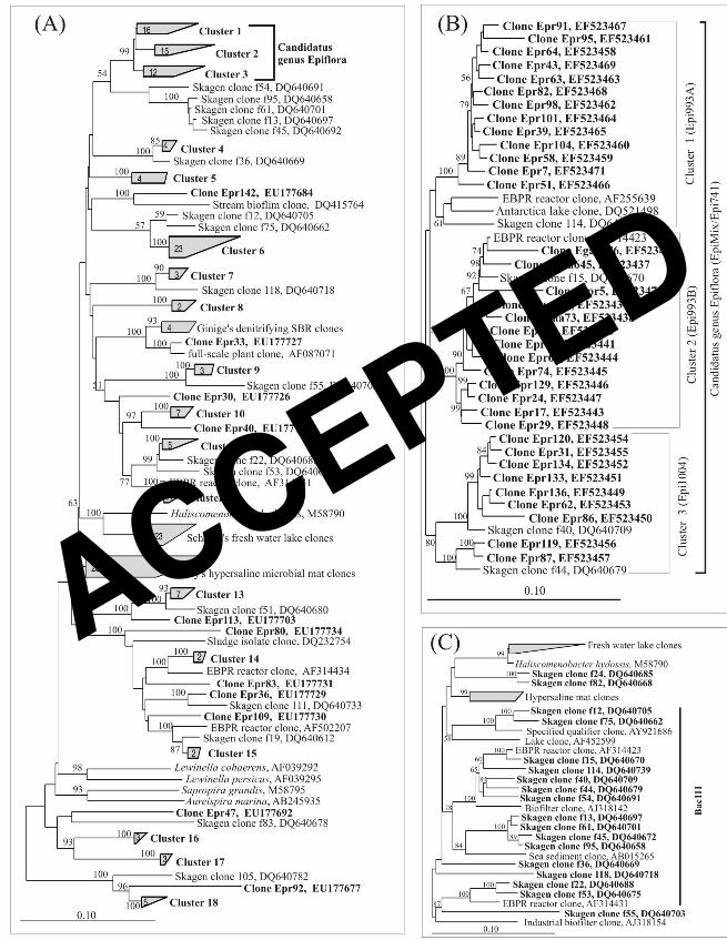
The 130 clones retrieved were distributed widely in family Saprospiraceae (Fig. 1A).
Most clones could be included in 18 clusters, each consisting of 2-16 clones sharing >90%
similarity. Most clones clustered to clones from full-scale WWTPs with a similarity of
85-99% and particularly to those from the Skagen WWTP (17). None of the clones were
closely related to any type strains in the family Saprospiraceaeas, as they had less than 87%
similarity. Of all the clusters, only clusters 1, 2, and 3 are closely related to each other,
containing in total 35 clones from this study and 7 clones from the GenBank database (32)
(Fig. 1B). Other clusters are diversely distributed in Saprospiraceae and relatively far away
from each other.
To further clarify the phylogeny of the epiflora bacteria, the clones putatively
representing the epiflora clusters were screened by using clones targeted by probe Bac111
(mainly Skagen clones, as shown in Fig. 1C) as “internal markers”. It is believed that they
contain representatives of the epiflora bacteria. Clusters 1, 2, and 3 each contains at least 1
marker clone and were chosen for the screening.
Clusters 1, 2, and 3 were further analyzed, and different treeing algorithms
(maximum-parsimony, maximum-likelihood, and neighbor-joining) were adopted to calculate
ACCEPTED

13
1
2
3
4
5
6
7
8
9
10
11
12
13
14
15
16
17
18
19
20
21
22
the phylogenetic trees, and the topology of the trees obtained did not significantly change (Fig.
1B). Clusters 1, 2, and 3 consist of 16, 15, and 11 sequences, respectively, and share within
each cluster 93-99% similarity. The similarity among the different clusters is 90-95% between
cluster 1 and 2, 90-95% between cluster 2 and 3, 91-94% between 1 and 3. The closest type
strain for the putative epiflora group is H. hydrossis (similarity <87.0%), and the similarity is
<82% to other Saprospiraceae type strains, including Aureispira marina, Saprospira grandis,
and Lewinella persicus. The epiflora group also shares <87% similarity with other clones of
uncultured bacteria, including those from fresh water lakes (38), hypersaline microbial mats
(26), and a methanol-feeding denitrifying reactor (11) (Fig. 1A).
Gene probe design and FISH of activated sludge. Gene probes at two hierarchical
levels were designed to target clusters 1, 2, 3, and the Skagen marker cluster consisting of the
4 marker clones. The probe designed for the marker cluster hybridized with only a few cocci
(<1% of total bacterial biovolume, naked eye estimation) and is not included in this report.
The sequence, specificity, and optimal formamide concentration of the probes designed for
clusters 1-3 are listed in Table 1. Probes Epi993A, Epi993B, and Epi1004 were designed to
target cluster 1, 2, and 3, respectively. Probe Epi741 targets all three clusters and a few other
clones. The optimal FA concentration for probes Epi741 and Epi993A using the Clone-FISH
technique generally agreed with those determined by using FISH probing of sludge samples,
although a slightly higher optimal FA concentration was found by Clone-FISH (1.1 times). As
a weak FISH signal was observed from the epiflora cells at the FA concentrations obtained
from Clone-FISH, the FA concentrations (Table 1) determined in FISH probing of sludge
samples were used.
ACCEPTED

14
1
2
3
4
5
6
7
8
9
10
11
12
13
14
15
16
17
18
19
20
21
22
The specificity of the newly designed gene probes was checked by FISH probing
using sludge samples from AAV, Egaa, Hjorring, and Aabybro WWTPs (Table 2). Under the
optimal FA concentrations determined, probes Epi993A, Epi993B, and Epi1004 hybridized
only with epiflora bacteria consisting of single rods or 2-3 rods connected end to end (Fig. 2A,
2B). Epi741 hybridized mainly with epiflora bacteria, but occasionally with short filaments,
too. All epiflora bacteria hybridizing with Epi993A, Epi993B, or Epi1004 targeting the
individual clusters also hybridized with Epi741, Bac111, or Sap309. Application of probes
Epi993A, Epi993B, and Epi1004 in a mixture (EpiMix) yielded the same result. No
cross-hybridization was found among probes Epi993A, Epi993B, and Epi1004. Most (>95%)
epiflora bacteria hybridizing with probe Epi741 also hybridized with EpiMix. All these tests
confirm the specificity of the new cluster-level probes. Not all epiflora bacteria hybridizing
with probes Bac111 or Sap309 (both match well in coverage of the epiflora bacteria, data not
shown) hybridized also with EpiMix. In Egaa and Hjorring WWTPs, 80-90% of the epiphytic
cells hybridized with Sap309 or Bac111 hybridized with EpiMix. The ratio was 70-80% in
AAV and 60-70% in Aabybro.
Morphological observations and attachment style. In all sludge samples examined,
the epiflora bacteria hybridizing with probes Epi993A, Epi993B, and Epi1004 were rods
1.3-1.9µm in length and 0.3-0.4µm in width, and all only attached to different filamentous
bacteria (Fig. 2A, 2B) and not to any other microbial morphotypes. The density of epiflora
bacteria on the filamentous bacteria varied widely from being very densely to sparsely
colonized. Normally, 2 or 3 rods connected end to end attached themselves onto the surfaces
of filamentous bacteria, and they were usually attached perpendicularly to the filament.
ACCEPTED

15
1
2
3
4
5
6
7
8
9
10
11
12
13
14
15
16
17
18
19
20
21
22
Presence of exo-enzymes and storage compounds. The ability of the 3
probe-defined epiflora clusters to produce different exo-enzymes and intracellular storage
compounds was investigated in 5 EBPR and 4 nitrogen removal WWTPs. In the 9 WWTPs
investigated, all the epiflora clusters had exo-proteases activity as revealed by in situ protease
staining (BODIPY FL casein and BSA) combined with FISH. No cell surface associated
activities of esterase, lipase, β-D-galactosidase, β-D-glucuronidase, chitinase, and
phosphatase were found in any of the clusters by using ELF® 97 combined with FISH.
Intracellular accumulation of polyphosphate or PHA was not detected by FISH in
combination with histochemical staining (Neisser staining and Nile blue staining). The
potential effect of incubation with amino acids (substrates they can utilize, see following
section) on their intracellular storage of PHA was also investigated. Incubation of activated
sludge samples from AAV, Hjorring, Egaa, and Aabybro with an amino acid mixture for 3-5 h
under aerobic or anaerobic conditions did not lead to formation of intracellular PHA.
Uptake of organic substrates. The substrate utilization pattern of the individual
epiflora clusters was investigated by using MAR-FISH under different electron acceptor
conditions. Organic substrates, including sugars (glucose, mannose, N-acetyl-glucosamine),
short chain fatty acids (formate, acetate, pyruvate, and propionate), ethanol, a long chain
organic acid (oleic acid), individual amino acids (leucine, aspartate acids, glutamic acid, and
glycine) and a mixture of 14 amino acids were tested in biomass samples from AAV, Egaa,
Hjorring, and Aabybro WWTPs. The 3 epiflora clusters could only take up the mixture of
amino acid, and the uptake was observed under aerobic (Fig. 2C, 2D) and to some extent
anaerobic conditions (Table 3). However, in each cluster, not all the epiflora cells took up
ACCEPTED

16
1
2
3
4
5
6
7
8
9
10
11
12
13
14
15
16
17
18
19
20
21
22
amino acids. The fraction of the epiflora bacteria taking up amino acids was very similar in
AAV, Egaa, Hjorring, and Aabybro WWTPs, being 50-70% under aerobic conditions and
10-50% under anaerobic conditions. The amino acid uptake of the epiflora bacteria was also
investigated under anoxic conditions by adding NO3- or NO2
- in the anaerobic MAR
incubations. In all the WWTPs examined, the ratios (10-50%) of the 3 clusters taking up
amino acids did not change, being in the same ranges as observed under anaerobic conditions,
indicating that they were not involved in denitrification.
The potential growth ability of the 3 clusters was also investigated under anaerobic
and aerobic conditions by using MAR-FISH with labeled amino acids. This was carried out
by pre-incubating the sludge samples with an unlabeled amino acid mixture (with the same
composition as the labeled amino acid mixture they took up) for 3, 6, or 9 h before the labeled
amino acid mixture was added. No anaerobic uptake was observed after 3 h of anaerobic
pre-incubation with unlabeled amino acid mixture, whereas 50-70% of the 3 epiflora clusters
could still take up amino acids after 9 h aerobic pre-incubation in AAV, Egaa, Hjorring, and
Aabybro WWTPs. This indicates that the epiflora bacteria were obligate aerobic, but able to
take up amino acids under short-term anaerobic conditions.
Identification of the filamentous bacteria colonized by epiflora bacteria. The
identity of the filamentous bacteria colonized by the different epiflora clusters was
investigated in 5 activated sludge WWTPs by using FISH. The results are listed in Table 4.
Epiphytic colonization by the 3 epiflora clusters was detected on 3 probe-defined filamentous
bacterial groups (or phyla), which were all abundant in the WWTPs investigated. These were
Aquaspirillum-related filaments (defined by probe Aqs997), Chloroflexi-related filaments
ACCEPTED
17
1
2
3
4
5
6
7
8
9
10
11
12
13
14
15
16
17
18
19
20
21
22
(probe mixture GNSB941+CFX1223), and uncultured TM7-related filaments (probe
TM7-905). Except for cluster 1 (Epi993A), which did not grow on the filaments defined by
probe TM7-905, all epiflora clusters colonized all the probe-defined filamentous groups. They
were universally present in the WWTPs investigated, but showed some differences in their
colonization behavior.
Among the filaments hybridizing with Aqs997 and GNSB941+CFX1223, 10-30%
were colonized by clusters 1, 2, and/or 3 (Table 4). Clusters 2 and 3 colonized TM7 filaments
in some WWTPs, but not in others. In Egaa, Skagen, and AAV WWTPs, 10-20% of the TM7
filaments were colonized by cluster 2, but none in Hjorring and Aabybro. Cluster 3 colonized
1-10% of the TM7 filaments in Hjorring, AAV, and Aabybro WWTPs, but none in Egaa and
Skagen. Colonization of the epiflora clusters on a few other unidentified filamentous bacteria
was also observed.
Distribution and abundance of the individual epiflora clusters in full-scale
activated sludge WWTPs. The biovolumes of each epiflora cluster in 9 full-scale WWTPs
were investigated by using quantitative FISH - see the results in Table 5. Some of the WWTPs
were sampled 3 times to obtain an impression of the fluctuation of the biovolume of each
cluster over 1-3 years. Except for a few occasions, each of the epiflora clusters was present in
all the WWTPs examined. On average, cluster 1 accounted for 2±1% (mean value ± standard
deviation), cluster 2 1±1%, and cluster 3 2±1% of the total bacterial biovolume. The
biovolume measured with Epi741 (group level) in each WWTP generally agreed with the
biovolume measured with cluster level probes (Table 5).
ACCEPTED
18
1
2
3
4
5
6
7
8
9
10
11
12
13
14
15
16
17
18
19
20
21
22
DISCUSSION
Phylogenetic affiliation of probe-defined epiflora bacteria. The epiflora group
identified in this study consists of at least 3 probe-defined clusters. Each cluster contains
11-16 sequences sharing 93-99% similarity and has less than 82% similarity with the
available type stains of the genera in Saprospiraceae and <87% similarity to other
Saprospiraceae clones. Therefore, they should be classified as new taxa in the family
Saprospiraceae. Moreover, based on 97% similarity or a higher threshold (~99%) (41) for
defining a species, each cluster may consist of several species. The clone library constructed
by using probe Bac111 as the primer successfully retrieved the 16S rRNA genes of the
epiflora bacteria.
In addition to the 3 clusters shown in Fig. 1B, the broad probe Epi741 also perfectly
matches 4 other Saprospiraceae clones AJ318142, AF087054, AF255645, and BQ232435
(>1200 bp) from the GenBank database, which could not be grouped with any of the clusters.
However, we found that probe Epi741 also occasionally hybridized with a few filamentous
bacteria not hybridizing with any cluster level probe, so for specific and simultaneous
detection of the 3 epiflora clusters, the EpiMix consisting of Epi993A, Epi993B, and Epi1004
should be used.
EpiMix or Epi741 targeting clusters 1-3 only hybridized with 60-90% of the epiflora
bacteria hybridizing with the broader probe Bac111, so the detailed identity of the 10-30%
epiflora bacteria missed by EpiMix or Epi741 is still not clear. Whether their 16S rRNA
genes were obtained but not detected by the EpiMix, as many of the clusters associated with
Skagen marker clones were not screened, and/or their 16S rRNA genes were not successfully
ACCEPTED

19
1
2
3
4
5
6
7
8
9
10
11
12
13
14
15
16
17
18
19
20
21
22
retrieved, is still unknown. However, EpiMix covers a major fraction of the epiflora bacteria
in activated sludge, and the morphological and ecophysiological characteristics of the epiflora
bacteria hybridizing with EpiMix, Epi741, or Bac111 are very similar. No further efforts,
therefore, were attempted to investigate the detailed phylogeny of the epiflora bacteria missed
by EpiMix. The EpiMix-defined epiflora group represents the protein hydrolyzing epiflora
bacteria well, but probe Bac111 should be used to estimate all or most epiphytic bacteria
belonging to Saprospiraceae.
PCR amplification of the epiflora bacteria containing biomass obtained from
micromanipulation followed by clone library construction retrieved only 4 (out of 32)
epiflora-related 16S rRNA gene sequences, although a significant amount of clones (180
clones) were sequenced. This is mainly due to the difficulty associated with
micromanipulation of filamentous bacteria with epiflora, because they were often partly
inside the sludge flocs, so other loosely attached bacteria might also be present.
Ecophysiology of the probe-defined epiflora bacteria. Each of the epiflora clusters was
characterized ecophysiologically in three ways: 1) organic substrate utilization pattern under
different electron acceptor conditions; 2) production of the storage compounds PHA and
polyphosphate; and 3) production of extracellular enzymes. The resolution of our studies did
not show any differences, so all three epiflora clusters were very similar as regards these
ecophysiological aspects.
All clusters could only utilize amino acids as carbon and energy sources and not the
tested short chain fatty acids, monosaccharides, ethanol, glucosamine, or oleic acid. The
utilization occurred under anaerobic (0-3 h), aerobic, and anoxic conditions. However, they
ACCEPTED

20
1
2
3
4
5
6
7
8
9
10
11
12
13
14
15
16
17
18
19
20
21
22
seemed to be more active under aerobic conditions than under anaerobic conditions, because
the number of epiflora bacteria taking up amino acids under aerobic conditions (50-70%) was
higher than under short-term anaerobic conditions (10-50%). The lack of activity by some of
the epiflora bacteria under both aerobic and anaerobic conditions has also been observed on
other probe-defined bacteria in these WWTPs (19, 31). Whether the bacteria were inactive or
whether they were species or ecotypes not utilizing amino acids is unknown. Their ability to
utilize amino acids under short-term (3h) anaerobic conditions may indicate a capability to
store organic compounds, but the identity of such compounds is not clear, as they did not store
PHA.
All clusters were able to hydrolyze proteins, but not other macromolecules tested.
They could not store polyphosphate, PHA, and were most likely not able to denitrify.
Therefore, they most probably grow in activated sludge by hydrolyzing proteins and using the
hydrolysates (small peptides or amino acids) as energy and carbon sources. The exact types of
proteases they excrete and the amino acids they take up are, however, not known because the
BODIPY-labeled casein and BSA conjugates respond to different proteases (45). Similarly,
the labeled amino acid mixture they took up consists of 14 different amino acids. Some of the
amino acids in the mixture were tested individually (aspartic acid, leucine, glutamic acid, and
glycine), but none of them were utilized, implying either that these amino acids could not be
utilized, or that they could only be utilized in the presence of other amino acids.
Colonization to filaments and distribution in full-scale WWTPs. Interestingly, the
epiphytic bacteria mainly colonized filamentous bacteria belonging to phylum Chloroflexi,
candidate phylum TM7, and filaments related to genus Aquaspirillum, but not to several other
ACCEPTED

21
1
2
3
4
5
6
7
8
9
10
11
12
13
14
15
16
17
18
19
20
21
22
filamentous bacteria present in the sludge samples, such as Candidatus Microthrix parvicella
and other unidentified bacteria. All these filamentous bacteria are common or abundant in
most activated sludge WWTPs with nutrient removal (21, 37, 42, 43). Moreover, no obvious
preference of colonization of a specific filamentous bacterial group was found for any of the
epiflora clusters, with the exception of cluster 1 not colonizing TM7-filaments. Therefore,
attachment may rely on differences in surface structure or other unknown factors.
The advantage of being attached to the filamentous bacteria is still speculative. There
might be a sort of symbiotic relationship between the epiflora bacteria and the filamentous
bacteria. The attachment not only prevents the epiflora bacteria from being washed out from
the WWTP with the effluent, but most importantly facilitates adsorption of macromolecules
from the wastewater as the filaments often protrude into bulk water, where the majority of
organic matter exists in the form of colloids and small particles (29). In return, the epiflora
bacteria may as a protein-hydrolyzing organism provide substrates (amino acids) to their
hosts.
The study showed that the EpiMix-defined epiflora group is an important component
in the microbial community of activated sludge. It constituted up to 10% of the bacterial
biovolume in the 9 WWTPs examined. The group was more abundant in EBPR WWTPs (6%,
mean value of 10 samples from 5 WWTPs) than in N-removal WWTPs with chemical
precipitation of phosphorus (4%, mean value of 8 samples from 4 WWTPs). However,
whether this is due to an ecological selection of the epiflora clusters (or the filamentous
bacteria they colonize) by the anaerobic period in the EBPR systems (the main difference
between EBPR and N-removal WWTPs) or if there are any other reasons has to be further
ACCEPTED

22
1
2
3
4
5
6
7
8
9
10
11
12
13
14
15
16
17
18
19
20
21
22
investigated.
Taxonomic consideration: proposal of Candidatus genus Epiflobacter. The epiflora
group that has been characterized phylogenetically and ecophysiologically in this study
consists of 3 closely related clusters sharing 90-95% similarity. Each cluster contains different
species. However, they are all rods attached to mainly 3 probe-defined filamentous bacteria
and have similar ecophysiology, all being specialized in protein hydrolysis. Therefore, we
propose to classify them into a new genus: Candidatus Epiflobacter, based on the requirement
of the International Committee on Systematic Bacteriology (30).
Candidatus Epiflobacter spp. are uncultured bacteria belonging to the family
Saprospiraceae in the phylum Bacteroidetes and defined by probe EpiMix (a mixture of
probes Epi1004, Epi993A, and Epi993B). They are Gram-negative rods (1.3-1.9µm in length
and 0.3-0.4µm in width), attaching to a range of filamentous bacteria in activated sludge.
They are strict aerobes and specialists in protein hydrolysis, capable of utilizing only (or
primarily) amino acids as energy and carbon sources. They do not form PHA or
polyphosphate granules.
Acknowledgements
This study was supported by the Danish Technical Research Council, grant number
26-04-0115 (Identification and characterization of uncultured bacteria involved in hydrolysis
and fermentation in nutrient removal plants). C. Kragelund, J. L. Nielsen, and A. Schramm
are acknowledged for helpful suggestions and discussions.
ACCEPTED
23
1
2
3
4
5
6
7
8
9
10
11
12
13
14
15
16
17
18
19
20
21
22
REFERENCE
1. Amann, R. I., B. J. Binder, R. J. Olson, S. W. Chisholm, R. Devereux, and D. A.
Stahl. 1990. Combination of 16S ribosomal-RNA-targeted oligonucleotide probes
with flow cytometry for analyzing mixed microbial populations. Appl. Environ.
Microbiol. 56:1919-1925.
2. Amann, R. I., W. Ludwig, and K. H. Schleifer. 1995. Phylogenetic identification
and in-situ detection of individual microbial cells without cultivation. Microbiol. Rev.
59:143-169.
3. Bjornsson, L., P. Hugenholtz, G. W. Tyson, and L. L. Blackall. 2002. Filamentous
Chloroflexi (green non-sulfur bacteria) are abundant in wastewater treatment processes
with biological nutrient removal. Microbiology (UK) 148:2309-2318.
4. Brosius, J., T. J. Dull, D. D. Sleeter, and H. F. Noller. 1981. Gene organization and
primary structure of a ribosomal-RNA operon from Escherichia coli. J. Mol. Biol.
148:107-127.
5. Cole, J. R., B. Chai, R. J. Farris, Q. Wang, S. A. Kulam, D. M. McGarrell, G. M.
Garrity, and J. M. Tiedje. 2005. The Ribosomal Database Project (RDP-II):
sequences and tools for high-throughput rRNA analysis. Nucleic Acids Res.
33:D294-D296.
6. Cottrell, M. T., and D. L. Kirchman. 2000. Natural assemblages of marine
proteobacteria and members of the Cytophaga-Flavobacter cluster consuming low-
and high-molecular-weight dissolved organic matter. Appl. Environ. Microbiol.
66:1692-1697.
ACCEPTED
24
1
2
3
4
5
6
7
8
9
10
11
12
13
14
15
16
17
18
19
20
21
22
7. Daims, H., A. Bruhl, R. Amann, K. H. Schleifer, and M. Wagner. 1999. The
domain-specific probe EUB338 is insufficient for the detection of all Bacteria:
Development and evaluation of a more comprehensive probe set. Syst. Appl.
Microbiol. 22:434-444.
8. Eikelboom, D.H., and van Buijsen, H.J.J. 1983. Microscopic sludge investigation
manual, 2nd ed., TNO research institute of environmental hygiene, Delft.
9. Eikelboom, D. H. 1975. Filamentous organisms observed in activated sludge. Water
Res. 9:365-388.
10. Gich, F., J. Garcia-Gil, and J. Overmann. 2001. Previously unknown and
phylogenetically diverse members of the green nonsulfur bacteria are indigenous to
freshwater lakes. Arch. Microbiol. 177:1-10.
11. Ginige, M. P., J. Keller, and L. L. Blackall. 2005. Investigation of an acetate-fed
denitrifying microbial community by stable isotope probing, full-cycle rRNA analysis,
and fluorescent in situ hybridization-microautoradiography. Appl. Environ. Microbiol.
71:8683-8691.
12. Hall-Stoodley, L., J. W. Costerton, and P. Stoodley. 2004. Bacterial biofilms: From
the natural environment to infectious diseases. Nat. Rev. Microbiol. 2:95-108.
13. Hosoya, S., V. Arunpairojana, C. Suwannachart, A. Kanjana-Opas, and A.
Yokota. 2006. Aureispira marina gen. nov., sp. nov., a gliding, arachidonic
acid-containing bacterium isolated from the southern coastline of Thailand. Int. J. Syst.
Evol. Microbiol. 56:2931-2935.
14. Huber, T., G. Faulkner, and P. Hugenholtz. 2004. Bellerophon: a program to detect
ACCEPTED
25
1
2
3
4
5
6
7
8
9
10
11
12
13
14
15
16
17
18
19
20
21
22
chimeric sequences in multiple sequence alignments. Bioinformatics 20:2317-2319.
15. Hugenholtz, P., G. W. Tyson, R. I. Webb, A. M. Wagner, and L. L. Blackall. 2001.
Investigation of candidate division TM7, a recently recognized major lineage of the
domain bacteria with no known pure culture representatives. Appl. Environ. Microbiol.
67:411-419.
16. Kojima, H., Y. Koizumi, and M. Fukui. 2006. Community structure of bacteria
associated with sheaths of freshwater and brackish Thioploca species. Microb. Ecol.
52:765-773.
17. Kong, Y. H., Y. Xia, J. L. Nielsen, and P. H. Nielsen. 2007. Structure and function of
the microbial community in a full-scale enhanced biological phosphorus removal plant.
Microbiology (UK) (in press).
18. Kong, Y. H., J. L. Nielsen, and P. H. Nielsen. 2005. Identity and ecophysiology of
uncultured actinobacterial polyphosphate-accumulating organisms in full-scale
enhanced biological phosphorus removal plants. Appl. Environ. Microbiol.
71:4076-4085.
19. Kong, Y. H., J. L. Nielsen, and P. H. Nielsen. 2004. Microautoradiographic study of
Rhodocyclus-related polyphosphate accumulating bacteria in full-scale enhanced
biological phosphorus removal plants. Appl. Environ. Microbiol. 70:5383-5390.
20. Kong, Y. H., Y. Xia, J. L. Nielsen, and P. H. Nielsen. 2006. Ecophysiology of a
group of uncultured Gammaproteobacterial glycogen-accumulating organisms in
full-scale enhanced biological phosphorus removal wastewater treatment plants.
Environ. Microbiol. 8:479-489.
ACCEPTED

26
1
2
3
4
5
6
7
8
9
10
11
12
13
14
15
16
17
18
19
20
21
22
21. Kragelund, C., C. Levantesi, A. Borger, K. Thelen, D. Eikelboom, V. Tandoi, Y. H.
Kong, J. van der Waarde, J. Krooneman, S. Rossetti, T. R. Thomsen, and P. H.
Nielsen. 2007. Identity, abundance and ecophysiology of filamentous Chloroflexi
species present in activated sludge treatment plants. FEMS Microbiol. Ecol.
59:671-682.
22. Kragelund, C., J. L. Nielsen, T. R. Thomsen, and P. H. Nielsen. 2005.
Ecophysiology of the filamentous Alphaproteobacterium Meganema perideroedes in
activated sludge. FEMS Microbiol. Ecol. 54:111-122.
23. Kumar, S., K. Tamura, and M. Nei. 2004. MEGA3: Integrated software for
molecular evolutionary genetics analysis and sequence alignment. Brief.
Bioinformatics 5:150-163.
24. Lane, D. J. 1991.16S/23S rRNA sequencing, p. 115-175. In E. Stackebrandt and M.
Goodfellow (ed.), Modern microbial methods: nucleic acid techniques in bacterial
systematics. John Wiley & Sons, Chichester, United Kingdom.
25. Lee, N., P. H. Nielsen, K. H. Andreasen, S. Juretschko, J. L. Nielsen, K. H.
Schleifer, and M. Wagner. 1999. Combination of fluorescent in situ hybridization and
microautoradiography - a new tool for structure-function analyses in microbial
ecology. Appl. Environ. Microbiol. 65:1289-1297.
26. Ley, R. E., J. K. Harris, J. Wilcox, J. R. Spear, S. R. Miller, B. M. Bebout, J. A.
Maresca, D. A. Bryant, M. L. Sogin, and N. R. Pace. 2006. Unexpected diversity
and complexity of the Guerrero Negro hypersaline microbial mat. Appl. Environ.
Microbiol. 72:3685-3695.
ACCEPTED

27
1
2
3
4
5
6
7
8
9
10
11
12
13
14
15
16
17
18
19
20
21
22
27. Loy, A., M. Horn, and M. Wagner. 2003. probeBase: an online resource for
rRNA-targeted oligonucleotide probes. Nucleic Acids Res. 31:514-516.
28. Ludwig, W., O. Strunk, R. Westram, L. Richter, H. Meier, Yadhukumar, A.
Buchner, T. Lai, S. Steppi, G. Jobb, W. Forster, I. Brettske, S. Gerber, A. W.
Ginhart, O. Gross, S. Grumann, S. Hermann, R. Jost, A. Konig, T. Liss, R.
Lussmann, M. May, B. Nonhoff, B. Reichel, R. Strehlow, A. Stamatakis, N.
Stuckmann, A. Vilbig, M. Lenke, T. Ludwig, A. Bode, and K. H. Schleifer. 2004.
ARB: a software environment for sequence data. Nucleic Acids Res. 32:1363-1371.
29. Morgenroth, E., R. Kommedal, and P. Harremoes. 2002. Processes and modeling
of hydrolysis of particulate organic matter in aerobic wastewater treatment - a review.
Water Sci. Technol. 45:25-40.
30. Murray, R. G. E., and E. Stackebrandt. 1995. Taxonomic note - implementation of
the provisional status candidatus for incompletely described prokaryotes. Int. J. Syst.
Bacteriol. 45:186-187.
31. Nielsen, J. L., D. Christensen, M. Kloppenborg, and P. H. Nielsen. 2003.
Quantification of cell-specific substrate uptake by probe-defined bacteria under in situ
conditions by microautoradiography and fluorescence in situ hybridization. Environ.
Microbiol. 5:202-211.
32. Ouellette, B. F. F. 1998. The GenBank sequence database. Bioinformatics 39:16-45.
33. Parsek, M. R., and C. Fuqua. 2004. Biofilms 2003: Emerging themes and challenges
in studies of surface-associated microbial life. J. Bacteriol. 186:4427-4440.
34. Ramey, B. E., M. Koutsoudis, S. B. von Bodman, and C. Fuqua. 2004. Biofilm
ACCEPTED

28
1
2
3
4
5
6
7
8
9
10
11
12
13
14
15
16
17
18
19
20
21
22
formation in plant-microbe associations. Curr. Opin. Microbiol. 7:602-609.
35. Reichenbach, H. 1991. The order Cytophagales. p. 3631-3675. In A. Balows, H.G.
Trüper, M. Dworkin, W. Harder, K.-H. Schleifer, (ed.), The Prokaryotes, 2nd ed.
Springer Verlag, New York.
36. Riemann, L., G. F. Steward, and F. Azam. 2000. Dynamics of bacterial community
composition and activity during a mesocosm diatom bloom. Appl. Environ. Microbiol.
66:578-587.
37. Rossetti, S., M. C. Tomei, P. H. Nielsen, and V. Tandoi. 2005. "Microthrix
parvicella", a filamentous bacterium causing bulking and foaming in activated sludge
systems: a review of current knowledge. FEMS Microbiol. Rev. 29:49-64.
38. Schauer, M., and M. W. Hahn. 2005. Diversity and phylogenetic affiliations of
morphologically conspicuous large filamentous bacteria occurring in the pelagic zones
of a broad spectrum of freshwater habitats. Appl. Environ. Microbiol. 71:1931-1940.
39. Schramm, A., B. M. Fuchs, J. L. Nielsen, M. Tonolla, and D. A. Stahl. 2002.
Fluorescence in situ hybridization of 16S rRNA gene clones (Clone-FISH) for probe
validation and screening of clone libraries. Environ. Microbiol. 4:713-720.
40. Seviour, E. M., C. Williams, B. Degrey, J. A. Soddell, R. J. Seviour, and K. C.
Lindrea. 1994. Studies on filamentous bacteria from Australian activated sludge
plants. Water Res. 28:2335-2342.
41. Stackebrandt, E., and J. Ebers. 2006. Taxonomic parameters revisited: tarnished
gold standards. Microbiol. Today 33:152-155.
42. Thomsen, T. R., B. V. Kjellerup, J. L. Nielsen, P. Hugenholtz, and P. H. Nielsen.
ACCEPTED

29
1
2
3
4
5
6
7
8
9
10
2002. In situ studies of the phylogeny and physiology of filamentous bacteria with
attached growth. Environ. Microbiol. 4:383-391.
43. Thomsen, T. R., J. L. Nielsen, N. B. Ramsing, and P. H. Nielsen. 2004.
Micromanipulation and further identification of FISH-labelled microcolonies of a
dominant denitrifying bacterium in activated sludge. Environ. Microbiol. 6:470-479.
44. Vanveen, W. L., D. Vanderko, E. C. W. A. Geuze, and A. Vandervl. 1973.
Investigations on sheathed bacterium Haliscomenobacter hydrosis gen. nov., sp.nov.,
isolated from activated sludge. Antonie Van Leeuwenhoek J. Microbiol 39:207-216.
45. Xia, Y., Y. H. Kong, and P. H. Nielsen. 2007. In situ detection of protein-hydrolysing
microorganisms in activated sludge. FEMS Microbiol. Ecol. 60:156-165.
ACCEPTED

TABLE 1. New oligonucleotide probes designed to target the three epiphytic clusters belonging to Saprospiraceae.
Probe Sequence (5’-3’) 16S rRNA GC Tm Length Specificity Formamide
Name target site content (ºC)a (nt)b conc. (%)
________________________________________________________________________________________________________________________________________________
Epi741 (S-S-Sap-0741-a-18) CA GCG TCA ATC AAG GCC C 741-759 61.1 53 18 cluster 1-3 and a few other
Saprospiraceae clones 35-40
Epi993A (S-S-Sap-0993A-a-21) CAC CAT CTG TCA CTC ACA TTC 993-1014 47.6 51 21 cluster 1 35-40
Epi993B (S-S-Sap-0993B-a-21) CAC AGW CCG TCA CTC ACA TTC 993-1014 52.4 53 21 cluster 2 35
Epi1004 (S-S-Sap-1004-a-18) GCA CCT TTC AGT GCC GGT 1004-1022 61.1 53 18 cluster 3 35-40
a Tm, melting temperature (according to the manufacturer). b nt, nucleotides.
ACCEPTED
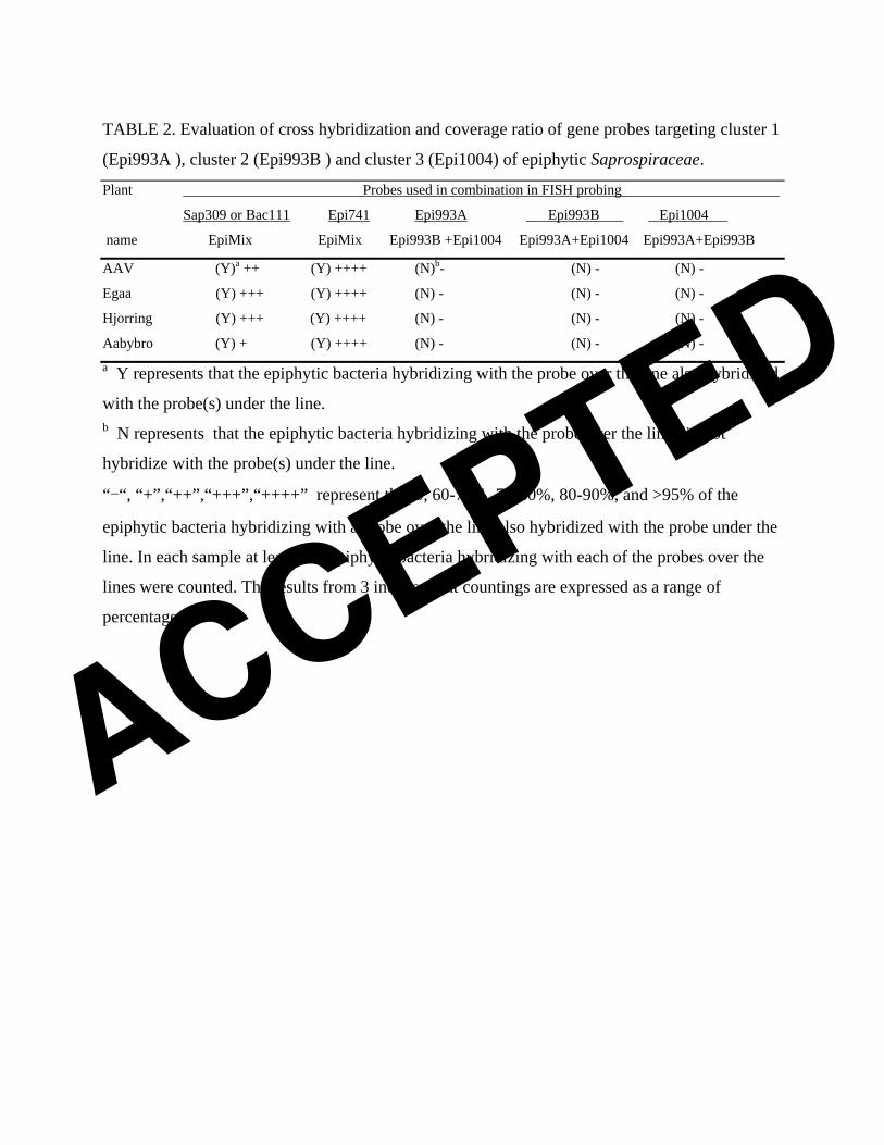
TABLE 2. Evaluation of cross hybridization and coverage ratio of gene probes targeting cluster 1
(Epi993A ), cluster 2 (Epi993B ) and cluster 3 (Epi1004) of epiphytic Saprospiraceae.
Plant Probes used in combination in FISH probing
Sap309 or Bac111 Epi741 Epi993A Epi993B Epi1004
name EpiMix EpiMix Epi993B +Epi1004 Epi993A+Epi1004 Epi993A+Epi993B
AAV (Y)a ++ (Y) ++++ (N)b- (N) - (N) -
Egaa (Y) +++ (Y) ++++ (N) - (N) - (N) -
Hjorring (Y) +++ (Y) ++++ (N) - (N) - (N) -
Aabybro (Y) + (Y) ++++ (N) - (N) - (N) - a Y represents that the epiphytic bacteria hybridizing with the probe over the line also hybridized
with the probe(s) under the line. b N represents that the epiphytic bacteria hybridizing with the probe over the line did not
hybridize with the probe(s) under the line.
“-“, “+”,“++”,“+++”,“++++” represent that 0, 60-70%, 70-80%, 80-90%, and >95% of the
epiphytic bacteria hybridizing with a probe over the line also hybridized with the probe under the
line. In each sample at least 500 epiphytic bacteria hybridizing with each of the probes over the
lines were counted. The results from 3 independent countings are expressed as a range of
percentage.
ACCEPTED
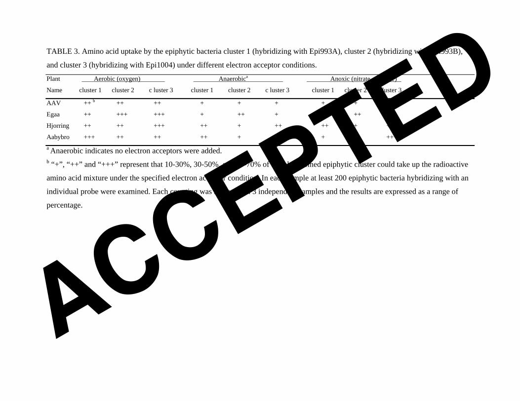
TABLE 3. Amino acid uptake by the epiphytic bacteria cluster 1 (hybridizing with Epi993A), cluster 2 (hybridizing with Epi993B),
and cluster 3 (hybridizing with Epi1004) under different electron acceptor conditions.
Plant Aerobic (oxygen) Anaerobica Anoxic (nitrate or nitrite)
Name cluster 1 cluster 2 c luster 3 cluster 1 cluster 2 c luster 3 cluster 1 cluster 2 c luster 3
AAV ++ b ++ ++ + + + + + +
Egaa ++ +++ +++ + ++ + + ++ +
Hjorring ++ ++ +++ ++ + ++ ++ + +
Aabybro +++ ++ ++ ++ + ++ + + ++ a Anaerobic indicates no electron acceptors were added. b “+”, “++” and “+++” represent that 10-30%, 30-50%, and 50-70% of a probe-defined epiphytic cluster could take up the radioactive
amino acid mixture under the specified electron acceptor condition. In each sample at least 200 epiphytic bacteria hybridizing with an
individual probe were examined. Each counting was repeated on 3 independent samples and the results are expressed as a range of
percentage.
ACCEPTED

TABLE 4. Identity of filamentous bacteria colonized by the 3 epiphytic Saprospiraceae clusters
found in 5 wastewater treatment plants (the probe-defined filamentous bacteria were present in all
plants investigated).
Plants Cluster 1 (Epi993A) Cluster 2 (Epi993B) Cluster 3 (Epi1004) GNSB941 GNSB941 GNSB941
Aqs997a + TM7-905c Aqs997 + TM7-905 Aqs997 + TM7-905
CFX1223b CFX1223 CFX1223
_____________________________________________________________________________________________________________________
AAV + +++ - + + ++ + + +
Egaa +++ ++ - ++ + + +++ + -
Hjorring ++ +++ - ++ ++ - +++ + +
Aabybro + ++ - + + - + + +
Skagen + + - +++ +++ ++ ++ ++ - a probe targeting genus Aquaspirillum-related bacteria. b probe mixture targeting most members of the phylum Chloroflexi. c probe targeting most members of the Candidate phylum TM7.
‘+’, ‘++’ and ‘+++’ represent that 1-10%, 10-20%, and 20-30% of the probe-defined filaments
were colonized with the indicated epiflora cluster, and ‘-’ represents that the probe-defined
epiflora did not colonize the probe-defined filaments. In each sample at least 100 filaments
hybridizing with a probe were counted. The results from 3 independent countings are expressed
as a range of percentage. ACCEPTED
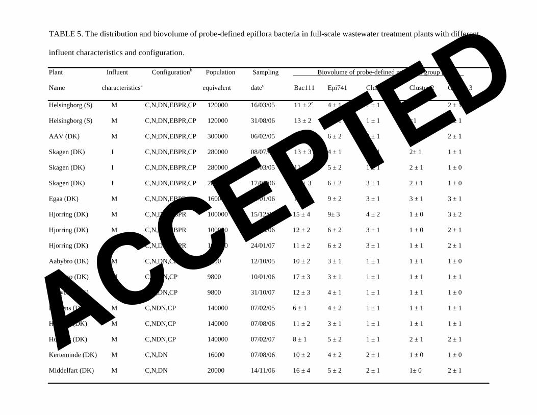
TABLE 5. The distribution and biovolume of probe-defined epiflora bacteria in full-scale wastewater treatment plants with different
influent characteristics and configuration.
Plant Influent Configurationb pulation Sampling Po Biovolume of probe-defined microbial group (%)d
Name characteristicsa equivalent datec Bac111 Epi741 Cluster 1 Cluster 2 Cluster 3
Helsingborg (S) M C,N,DN,EBPR,CP 120000 16/03/05 11 ± 2e 4 ± 1 1 ± 1 <1 2 ± 1
Helsingborg (S) M C,N,DN,EBPR,CP 120000 31/08/06 13 ± 2 5 ± 1 1 ± 1 <1 4 ± 1
AAV (DK) M C,N,DN,EBPR,CP 300000 06/02/05 15 ± 3 6 ± 2 3 ± 1 1 ± 1 2 ± 1
Skagen (DK) I C,N,DN,EBPR,CP 280000 08/07/03 13 ± 3 4 ± 1 2 ± 1 2± 1 1 ± 1
Skagen (DK) I C,N,DN,EBPR,CP 280000 22/03/05 11 ± 2 5 ± 2 1 ± 1 2 ± 1 1 ± 0
Skagen (DK) I C,N,DN,EBPR,CP 280000 17/01/06 18 ± 3 6 ± 2 3 ± 1 2 ± 1 1 ± 0
Egaa (DK) M C,N,DN,EBPR 160000 10/01/06 16 ± 2 9 ± 2 3 ± 1 3 ± 1 3 ± 1
Hjorring (DK) M C,N,DN,EBPR 100000 15/12/05 15 ± 4 9± 3 4 ± 2 1 ± 0 3 ± 2
Hjorring (DK) M C,N,DN,EBPR 100000 30/10/06 12 ± 2 6 ± 2 3 ± 1 1 ± 0 2 ± 1
Hjorring (DK) M C,N,DN,EBPR 100000 24/01/07 11 ± 2 6 ± 2 3 ± 1 1 ± 1 2 ± 1
Aabybro (DK) M C,N,DN,CP 9800 12/10/05 10 ± 2 3 ± 1 1 ± 1 1 ± 1 1 ± 0
Aabybro (DK) M C,N,DN,CP 9800 10/01/06 17 ± 3 3 ± 1 1 ± 1 1 ± 1 1 ± 1
Aabybro (DK) M C,N,DN,CP 9800 31/10/07 12 ± 3 4 ± 1 1 ± 1 1 ± 1 1 ± 0
Horsens (DK) M C,NDN,CP 140000 07/02/05 6 ± 1 4 ± 2 1 ± 1 1 ± 1 1 ± 1
Horsens (DK) M C,NDN,CP 140000 07/08/06 11 ± 2 3 ± 1 1 ± 1 1 ± 1 1 ± 1
Horsens (DK) M C,NDN,CP 140000 07/02/07 8 ± 1 5 ± 2 1 ± 1 2 ± 1 2 ± 1
Kerteminde (DK) M C,N,DN 16000 07/08/06 10 ± 2 4 ± 2 2 ± 1 1 ± 0 1 ± 0
Middelfart (DK) M C,N,DN 20000 14/11/06 16 ± 4 5 ± 2 2 ± 1 1± 0 2 ± 1
ACCEPTED

Districts (N) (USA) M/I C,N,DN 180000 15/12/06 14 ± 3 4 ± 2 3 ± 1 <1 <1
a M and I represent primarily municipal and industrial wastewater, respectively. b C: carbon removal; N: nitrification; DN: denitrification; EBPR: enhanced biological phosphorus removal; CP: chemical phosphorus removal. c The dates the samples were taken (date-month-year). d Percentage of biovolume calculated from all bacteria fluorescing with EUBmix. e The values are expressed as average ± standard deviation based on at least 40 measurements.
ACCEPTED
Figure legends 1
2
3
4
5
6
7
8
9
10
11
12
13
14
15
16
17
18
19
20
21
22
FIG. 1. Panel A is a simplified distance tree (neighbor-joining) built for all the genera and the
main microbial groups in the family Saprospiraceae of the phylum Bacteroidetes. Panel B is a
distance tree built for the 3 clusters of Candidatus genus Epiflobacter as shown in Panel A. Panel
C is a distance tree showing the Skagen clones targeted by probe Bac111. Sequences with bold
names in Panels A and B are obtained from this study. The numbers in the quadrangles of Panel A
represent the numbers of clones in each clone cluster. The bootstrap values in all the panels (only
those >50% are shown) were calculated based on 1000 re-samplings. Scale bars in all panels
represent 1 substitution per 10 nucleotides.
FIG. 2. (A) FISH image of activated sludge after color-combination showing Bacteria hybridized
with EUBmix (Cy5 labeled, set as green) as cells labeled with different colors: epiflora
hybridized with Bac111 (FLUOS labeled, set as blue) as cyan-labeled cells (mixture of green and
blue, marked with arrow (b)), and epiflora hybridized with EpiMix (Cy3 labeled, set as red) as
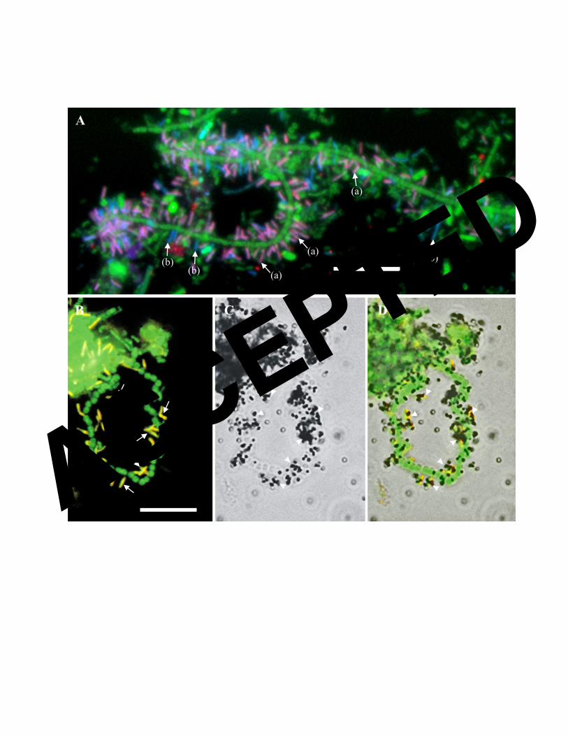
purple-labeled cells (mixture of blue, red and green, marked with arrow (a)). Fig. 2 (B, C&D)
MAR-FISH image of activated sludge with epiflora. (B) FISH image of epiflora hybridized with
EpiMix (Cy3 labeled, set as red) and Bacteria hybridized with EUBmix (FLUOS labeled, set as
green); (C) Bright-field image of MAR of the same field as (B); (D) Overlay of images (D) and
(C). Images (B, C&D) show that the epiflora hybridized with EpiMix (e.g. those marked with
arrows) take up a mixture of labeled amino acids under aerobic conditions. The bars in Panels A
and B represent 10 μm.
ACCEPTED
ACCEPTED
ACCEPTED