Factors affecting the distribution of clinical mastitis among ...
Upload
khangminh22Category
view
7download
0
animals
Review
Mammary Defences and Immunity against Mastitisin Sheep
Angeliki I. Katsafadou †, Antonis P. Politis †, Vasia S. Mavrogianni, Mariana S. Barbagianni,Natalia G. C. Vasileiou, George C. Fthenakis * and Ilektra A. Fragkou
Veterinary Faculty, University of Thessaly, 43100 Karditsa, Greece; [email protected] (A.I.K.);[email protected] (A.P.P.); [email protected] (V.S.M.); [email protected] (M.S.B.);[email protected] (N.G.C.V.); [email protected] (I.A.F.)* Correspondence: [email protected]† These authors have contributed equally and their names are listed alphabetically.
Received: 21 August 2019; Accepted: 24 September 2019; Published: 26 September 2019�����������������
Simple Summary: The article reviews the defence mechanisms and the relevant processes that occurin the udder of sheep. Due to the importance of the udder in milk production, animals display manydefences to protect the organ. These include the teats, the epithelial and the white-blood cells in theudder, the immunoglobulins and chemical substances that all participate in the various processes.These are influenced by many factors, animal- or management-regulated, which must be taken intoaccount in the formulation of prevention schemes against mastitis in sheep.
Abstract: The objectives of this review paper are to present udder defences, including teat of the udder,mammary epithelial cells, leucocytes, immunoglobulins, complement system and chemical antibacterialagents, to describe cooperation and interactions between them and to elaborate on potentials regardingtheir significance in mammary immunisation strategies. The teat of the udder provides initial protectionto the mammary gland. The mammary epithelial cells synthesise antibacterial proteins and theleucocytes produce various inflammation mediators (cytokines or chemokines), phagocytose bacteriaand recognise antigenic structures. In the mammary gland, four immunoglobulins (IgG1, IgG2, IgM andIgA) have important roles against bacterial pathogens. The complement system is a collection of proteins,participating in the inflammatory process through various pathways. Other components contributingto humoral mammary defence include lactoferrin, lysozyme and the lactoperoxidase/myeloperoxidasesystems, as well as oligosaccharides, gangliosides, reactive oxygen species, acute phase proteins(e.g., haptoglobin and serum amyloid A), ribonucleases and a wide range of antimicrobial peptides.Management practices, genetic variations and nutrition can influence mammary defences and shouldbe taken into account in the formulation of prevention strategies against ovine mastitis.
Keywords: ewe; leucocyte; neutrophils; proteomics; subclinical mastitis; teat; vaccination
1. Introduction
Many specific or non-specific mechanisms of varying significance and involvement, whichinclude humoral and cellular immune processes, are responsible for the defence of the mammarygland. Non-specific or specific defence mechanisms active in the mammary gland are part of theinnate or adaptive immune systems, respectively. The innate immune system would function toprovide the initial defence against invading pathogens, preceding adaptive immunity. The adaptiveimmune system would respond more efficiently in cases of repeated infections, but less so against newpathogens. Roles and objectives of the two systems are distinct, but, as they share common pathwaysand mechanisms, they are dependent on each other [1].
Animals 2019, 9, 726; doi:10.3390/ani9100726 www.mdpi.com/journal/animals
Animals 2019, 9, 726 2 of 17
The objectives of this review paper were to present mammary defences (teat of the udder, mammaryepithelial cells, leucocytes, immunoglobulins and complement system, chemical antibacterial agents)and to describe cooperation and interactions between them.
2. The Teat of the Udder
Initial protection of the mammary gland against infections is provided by the teat of the udder.Healthy teats efficiently protect ewes against mastitis by providing the first line of defence againstbacteria invading into the mammary gland during lamb suckling, as well as during milking ofewes [2,3]. Fatty acids present on the teat skin have bacteriostatic properties and can thus limitbacterial numbers around the teat orifice and on the teat surface [4]. The keratinised and coveredwith hydrophobic lipids internal lining of the teat canal entraps invading bacteria, which are thenflushed out at the following milking along with teat canal epithelium during the first outflow of milk(“keratin flush”) [1,4]. Teat closure after milking, effected by the local teat musculature, is paramountfor inhibiting bacterial entrance. However, some closure is achieved 20 to 30 minutes after completionof milking; during that period animals should be prevented from lying down, as this predisposes toincreased exposure of their teats to bacteria [5,6]. It is of note that total closure is not achieved until twohours post-milking [7–9]. It is hence recommended that, after milking, animals are walked to cleanareas in the farm, with feed available in troughs; this practice would contribute to reducing mammaryinfections. Specifically, during the dry-period, accumulation of keratin at the teat orifice seals the teatpreventing bacterial entrance.
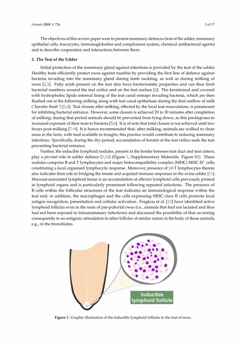
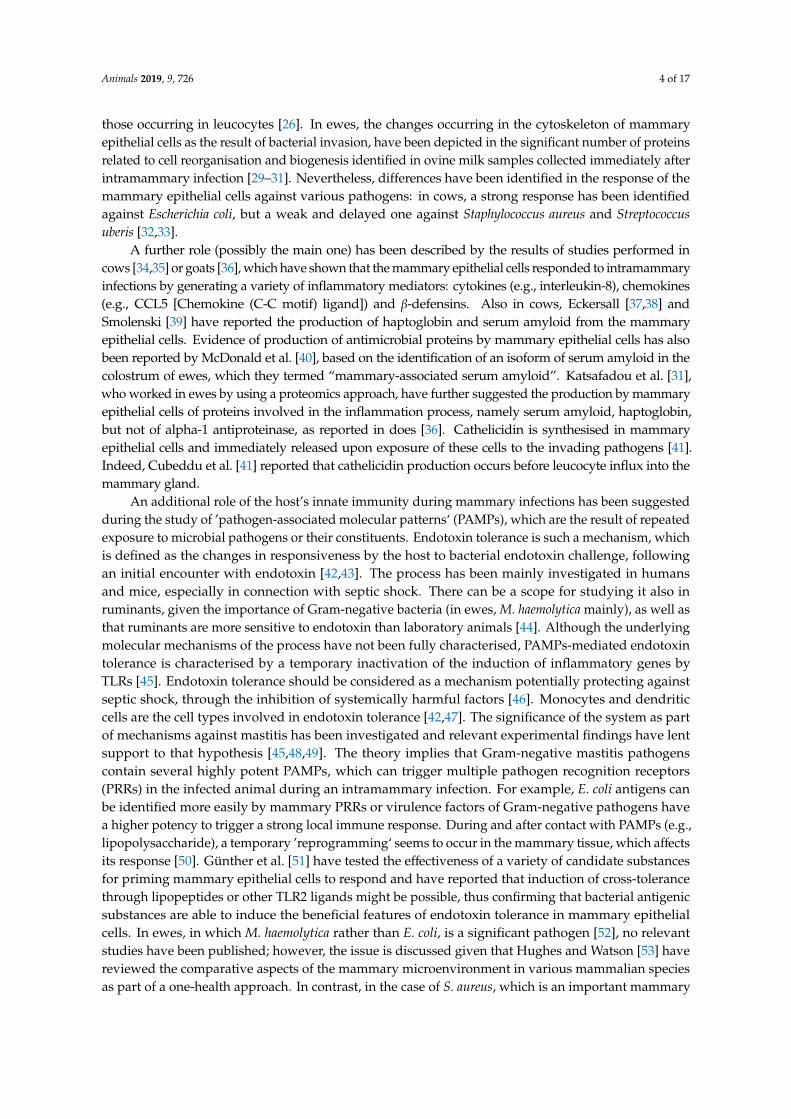
Further, the inducible lymphoid nodules, present at the border between teat duct and teat cistern,play a pivotal role in udder defence [3,10] (Figure 1, Supplementary Materials: Figure S1). Thesenodules comprise B and T lymphocytes and major histocompatibility complex (MHC) MHC-II+ cells,constituting a local organised lymphocytic response. Moreover, presence of γδ T lymphocytes thereinalso indicates their role in bridging the innate and acquired immune responses in the ovine udder [11].Mucosal-associated lymphoid tissue is an accumulation of effector lymphoid cells previously primedin lymphoid organs and is particularly prominent following repeated infections. The presence ofB cells within the follicular structures of the teat indicates an immunological response within theteat end; in addition, the macrophages and the cells expressing MHC-class II cells promote localantigen recognition, presentation and cellular activation. Fragkou et al. [12] have identified activelymphoid follicles even in the teats of pre-pubertal ewes (i.e., animals that had not lactated and thushad not been exposed to intramammary infections) and discussed the possibility of that occurringconsequently to an antigenic stimulation in other follicles of similar nature in the body of those animals,e.g., in the bronchioles.
Animals 2019, 9, x 2 of 17
The objectives of this review paper were to present mammary defences (teat of the udder, mammary epithelial cells, leucocytes, immunoglobulins and complement system, chemical antibacterial agents) and to describe cooperation and interactions between them.
2. The Teat of the Udder
Initial protection of the mammary gland against infections is provided by the teat of the udder. Healthy teats efficiently protect ewes against mastitis by providing the first line of defence against bacteria invading into the mammary gland during lamb suckling, as well as during milking of ewes [2,3]. Fatty acids present on the teat skin have bacteriostatic properties and can thus limit bacterial numbers around the teat orifice and on the teat surface [4]. The keratinised and covered with hydrophobic lipids internal lining of the teat canal entraps invading bacteria, which are then flushed out at the following milking along with teat canal epithelium during the first outflow of milk (“keratin flush”) [1,4]. Teat closure after milking, effected by the local teat musculature, is paramount for inhibiting bacterial entrance. However, some closure is achieved 20 to 30 minutes after completion of milking; during that period animals should be prevented from lying down, as this predisposes to increased exposure of their teats to bacteria [5,6]. It is of note that total closure is not achieved until two hours post-milking [7–9]. It is hence recommended that, after milking, animals are walked to clean areas in the farm, with feed available in troughs; this practice would contribute to reducing mammary infections. Specifically, during the dry-period, accumulation of keratin at the teat orifice seals the teat preventing bacterial entrance.
Further, the inducible lymphoid nodules, present at the border between teat duct and teat cistern, play a pivotal role in udder defence [3,10] (Figure 1, Supplementary Materials: Figure S1). These nodules comprise B and T lymphocytes and major histocompatibility complex (MHC) MHC-II+ cells, constituting a local organised lymphocytic response. Moreover, presence of γδ T lymphocytes therein also indicates their role in bridging the innate and acquired immune responses in the ovine udder [11]. Mucosal-associated lymphoid tissue is an accumulation of effector lymphoid cells previously primed in lymphoid organs and is particularly prominent following repeated infections. The presence of B cells within the follicular structures of the teat indicates an immunological response within the teat end; in addition, the macrophages and the cells expressing MHC-class II cells promote local antigen recognition, presentation and cellular activation. Fragkou et al. [12] have identified active lymphoid follicles even in the teats of pre-pubertal ewes (i.e., animals that had not lactated and thus had not been exposed to intramammary infections) and discussed the possibility of that occurring consequently to an antigenic stimulation in other follicles of similar nature in the body of those animals, e.g., in the bronchioles.
Figure 1. Graphic illustration of the inducible lymphoid follicles in the teat of ewes. Figure 1. Graphic illustration of the inducible lymphoid follicles in the teat of ewes.
Animals 2019, 9, 726 3 of 17
By using culture-independent methods for microbial identification (proteomics methodologies,specifically MALDI-TOF), the existence of microbiota within the teat and the lactiferous ducts ofthe udder of cows has been reported; a possible source of these bacteria can be the intestine, as ithas been found that bacteria from the gut microbiota reach the mammary gland by leucocytes (e.g.,dendritic cells, macrophages, lymphocytes) [13]. In an extensive field study, Vasileiou et al. [14] havereported that prevalence of mammary carriage in ewes was 10.0%. The significance of mammarycarriage (defined as the presence of bacteria in the udder with no increased somatic cell numbers, i.e.,in the absence of inflammation [15,16]) is that the bacteria can play a protective role against invadingpathogens, as well as potentially becoming pathogenic under the effect of various factors, whichdecrease defensive efficacy of hosts or promote pathogenicity of bacteria.
In ewes, Fragkou et al. [17] have concluded that coagulase-negative staphylococci present in highnumbers within the teat duct could afford some protection against invading pathogens. Bacterialpopulations interact among themselves and constitute a ’community‘, where each species contributesto its stability. Many mechanisms by which bacteria and their interactions prevent the invasion andcolonisation of pathogenic microorganisms, have been proposed, but are not all fully understood.Occupation of the host’s epithelial surfaces by members of the microbiota and, thus, prevention ofpathogen adherence on these cells can be particularly important [18], given that adherence on mammaryepithelial cells by the various pathogens is necessary for their multiplication and thereafter for biofilmformation (e.g., staphylococci [19]) or toxin production (e.g., Mannheimia haemolytica [20]). Bacterialcompetition is the situation, during which two bacterial populations compete for multiplicationand survival, often resulting in greater bacterial number reduction or lower growth rate than if thetwo populations were separate [21]. Production of antagonistic substances by bacterial flora andcompetition for necessary nutritional substances between microbiota and invading organisms [22] canalso contribute. Moreover, staphylococci recovered from bovine milk samples have been reported tosecrete bacteriocins, leading, under in vitro conditions, to reducing growth of other pathogens [23].The potential protective role of mammary microbiota can be explained on bacterial interactions.Nevertheless, it may also be proposed that these organisms elicit defence mechanisms, which thuscontribute to a more efficient defensive process exhibited by the teat. For example, inflammationmediators may be present in the teat as the result of bacterial carriage infection; these may facilitate thelocal cellular response and contribute to efficient host defence [12]. Indeed, Rainard and Poutrel [24]have reported that new infections were less frequent in mammary glands already harbouring bacteriatherein; in ewes, this has been corroborated by Fragkou et al. [17].
After bacteria have by-passed the teat duct, a cascade of events forms the immunological responseof the mammary gland and determines the outcome of the elicited inflammatory responses [25].
3. Cells in the Mammary Gland
3.1. Mammary Epithelial Cells
The possible role of the epithelia in the initiation of a defence response and the regulation of itsinitial steps has been studied mainly during the last 20 years [26]. In the case of mammary epithelialcells, this role includes many facets, which can contribute in the effective elimination of invadingintramammary pathogens.
As part of a sensory and recognition function of the mammary epithelial cells, Petzl et al. [27]reported in cases of mastitis, in cows, the presence of increased Toll-Like Receptors 2 (TLR2) on theirapical side, in contrast to the small amounts found in healthy glands.
Moreover, epithelial cells may also perform some degree of phagocytosis, which is performed bymeans of formation of membrane ruffles, which enfold the invading bacteria, fuse and form a pathogencontaining vesicle [26]. Thereafter, the phagosome matures by fusion and fission of endocytic vesicles [28],which is further accompanied by acidification of the lumen mediated by early recruitment of vacuolarATPases and formation of the phagolysosome. Subsequent phagocytosis mechanisms are similar to
Animals 2019, 9, 726 4 of 17
those occurring in leucocytes [26]. In ewes, the changes occurring in the cytoskeleton of mammaryepithelial cells as the result of bacterial invasion, have been depicted in the significant number of proteinsrelated to cell reorganisation and biogenesis identified in ovine milk samples collected immediately afterintramammary infection [29–31]. Nevertheless, differences have been identified in the response of themammary epithelial cells against various pathogens: in cows, a strong response has been identifiedagainst Escherichia coli, but a weak and delayed one against Staphylococcus aureus and Streptococcusuberis [32,33].
A further role (possibly the main one) has been described by the results of studies performed incows [34,35] or goats [36], which have shown that the mammary epithelial cells responded to intramammaryinfections by generating a variety of inflammatory mediators: cytokines (e.g., interleukin-8), chemokines(e.g., CCL5 [Chemokine (C-C motif) ligand]) and β-defensins. Also in cows, Eckersall [37,38] andSmolenski [39] have reported the production of haptoglobin and serum amyloid from the mammaryepithelial cells. Evidence of production of antimicrobial proteins by mammary epithelial cells has alsobeen reported by McDonald et al. [40], based on the identification of an isoform of serum amyloid in thecolostrum of ewes, which they termed “mammary-associated serum amyloid”. Katsafadou et al. [31],who worked in ewes by using a proteomics approach, have further suggested the production by mammaryepithelial cells of proteins involved in the inflammation process, namely serum amyloid, haptoglobin,but not of alpha-1 antiproteinase, as reported in does [36]. Cathelicidin is synthesised in mammaryepithelial cells and immediately released upon exposure of these cells to the invading pathogens [41].Indeed, Cubeddu et al. [41] reported that cathelicidin production occurs before leucocyte influx into themammary gland.
An additional role of the host’s innate immunity during mammary infections has been suggestedduring the study of ’pathogen-associated molecular patterns‘ (PAMPs), which are the result of repeatedexposure to microbial pathogens or their constituents. Endotoxin tolerance is such a mechanism, whichis defined as the changes in responsiveness by the host to bacterial endotoxin challenge, followingan initial encounter with endotoxin [42,43]. The process has been mainly investigated in humansand mice, especially in connection with septic shock. There can be a scope for studying it also inruminants, given the importance of Gram-negative bacteria (in ewes, M. haemolytica mainly), as well asthat ruminants are more sensitive to endotoxin than laboratory animals [44]. Although the underlyingmolecular mechanisms of the process have not been fully characterised, PAMPs-mediated endotoxintolerance is characterised by a temporary inactivation of the induction of inflammatory genes byTLRs [45]. Endotoxin tolerance should be considered as a mechanism potentially protecting againstseptic shock, through the inhibition of systemically harmful factors [46]. Monocytes and dendriticcells are the cell types involved in endotoxin tolerance [42,47]. The significance of the system as partof mechanisms against mastitis has been investigated and relevant experimental findings have lentsupport to that hypothesis [45,48,49]. The theory implies that Gram-negative mastitis pathogenscontain several highly potent PAMPs, which can trigger multiple pathogen recognition receptors(PRRs) in the infected animal during an intramammary infection. For example, E. coli antigens canbe identified more easily by mammary PRRs or virulence factors of Gram-negative pathogens havea higher potency to trigger a strong local immune response. During and after contact with PAMPs (e.g.,lipopolysaccharide), a temporary ’reprogramming‘ seems to occur in the mammary tissue, which affectsits response [50]. Günther et al. [51] have tested the effectiveness of a variety of candidate substancesfor priming mammary epithelial cells to respond and have reported that induction of cross-tolerancethrough lipopeptides or other TLR2 ligands might be possible, thus confirming that bacterial antigenicsubstances are able to induce the beneficial features of endotoxin tolerance in mammary epithelialcells. In ewes, in which M. haemolytica rather than E. coli, is a significant pathogen [52], no relevantstudies have been published; however, the issue is discussed given that Hughes and Watson [53] havereviewed the comparative aspects of the mammary microenvironment in various mammalian speciesas part of a one-health approach. In contrast, in the case of S. aureus, which is an important mammary
Animals 2019, 9, 726 5 of 17
pathogen in ewes [52], activation of the mammary gland in its entirety was found to be necessary forclearance of the infection [49].
3.2. Leucocytes
In healthy mammary glands, all types of leucocytes are present [54–58] (Table 1). Leucocytesoriginate from the blood circulation and are, thus, referred to as ‘somatic cells’.
Table 1. Types of leucocytes present in milk of healthy ewes.
Leucocyte Types Proportion of Total Leucocytes [59–61]
Macrophages 40–85%Neutrophils 5–35%
Lymphocytes 10–20%
The number of leucocytes in healthy mammary glands of ewes has not been firmly established.Many authors have proposed various thresholds for leucocytes in the milk of healthy ewes. Moreover,factors other than infection (e.g., stage of lactation) can also influence leucocyte numbers in normalmilk and these should be taken into account [62]. Possibly, the best approach is the one suggestedby Berthelot et al. [56], who proposed a dynamic approach based on the use of two thresholds ableto discriminate healthy (cell counts < 0.5 × 10⁶ cells mL-1 on each of two samples) from infected(somatic cell counts > 1.0 × 10⁶ cells mL-1 on each of two samples) ewes, coupled with bacteriologicalexaminations in samples found with cell numbers between those two thresholds. There is also a conflictregarding the proportion of epithelial cells in the milk of healthy ewes, with findings ranging widely.Paape et al. [63] indicated that the proportion of epithelial cells was 1% to 2% of all cells in milk, whilstBoutinaud and Jammes [60] reported that epithelial cells in the milk of ewes were at “very low levels”.In sharp contrast to the above findings, Leitner et al. [64] have indicated that epithelial cells constituted∼80% of the cells in the milk of healthy ewes.
Macrophages are the first among the leucocytes present in the mammary gland to counteract theinvading pathogens and to initiate the defensive leucocytic response [65,66]; they produce variousdefence-related components, which include cytokines and chemokines (e.g., tumour necrosis factor,interleukin 8; Table 2), as well as antimicrobial proteins and peptides (e.g., β-defensins and cathelicidins).These cells contribute to the induction of specific local responses, through antigen processing andpresentation to lymphocytes, in association with MHC class II [67]. As a result, within two to fourhours after infection, there is influx of blood constituents, e.g., leucocytes, into mammary tissuesand, hence, into the milk [10,56,68,69]. Increased permeability of the blood–milk barrier by variousmechanisms (e.g., by modulating claudins at the tight mammary junctions) allows blood constituentsand molecules to enter the infected mammary gland. Moreover, the diameter of mammary vesselsand blood volume therein increase soon after infection, leading to the transportation of an increasedamount of blood constituents into the mammary gland [70,71].
Hence, leucocytes (initially neutrophils) enter into the infected mammary gland and proteins alsoleak into the milk [28,29]. That way, the inflammatory response is developed and sustained. The reversephenomenon can also occur: molecules and, more importantly, pathogens that had not been eliminatedby the mammary defences, may pass from the mammary gland into the blood circulation, with effectsof bacteraemia (in case of bacterial entrance from mammary gland into blood) or endotoxaemia (in caseof entrance of bacterial toxins) [29,72]. Influx of neutrophils into the mammary gland (Figure S2)results from simultaneous function of various pathways and the participation of several molecules,e.g., selectins and integrins, which regulate chemotactic activity of leucocytes [67,73,74]. Antigens ofinvading pathogens are processed in macrophages and B lymphocytes and appear on the membranesin association with MHC class I or II; that way, they may be recognised by different lymphocytes [75].
Animals 2019, 9, 726 6 of 17
Table 2. Cytokines involved in mammary defence response.
Cytokine Main Role
Interleukin-1
Upregulation of neutrophil migration to themammary gland, increase of neutrophil numbers,promotion of phagocytic activity in the mammary
gland.
Interleukin-2Upregulation of macrophage proliferation in themammary gland, improvement of lymphocyte
antibacterial properties.
Interleukin-8 Upregulation of neutrophil migration to themammary gland.
Granulocyte colony-stimulating factor Increase of neutrophil numbers, promotion ofphagocytic activity in the mammary gland.
Granulocyte macrophage colony-stimulating factorImprovement of neutrophil chemotaxis and
bactericidal properties, increase of neutrophilnumbers in the mammary gland.
Macrophage colony-stimulating factor Upregulation of macrophage proliferation.
Interferon-γ Promotion of neutrophil phagocytic activity.
Tumour necrosis factor Enhancement of inflammatory process, promotion ofneutrophil phagocytic activity.
Interferon-γ contributes in upregulation of MHC-I expression and MHC-II antigen presentation,increasing the recognition of extraneous (or foreign) peptides by cytotoxic T cells and inducing theactivation of T helper cells. As soon as antigens are recognised, activation of T helper cells occurs; then,these produce cytokines participating in the activation and polarisation of B and T lymphocytes andmacrophages. The intramammary influx of leucocytes during infection, coupled with the desquamationof mammary epithelial cells, lead to an increase of leucocyte numbers in milk. This is an establishedcriterion for diagnosis of subclinical mastitis [54–56,76]. Roles of the various leucocyte subsets aredistinct, but interdependent. Further, increase of leucocytes in milk during infection results in clotformation in milk, which can be detected ultrasonographically (Figure S3) and is used for diagnosis ofsubclinical mastitis [77,78]. Neutrophils phagocytose bacteria and proceed to perform intracellularkilling by rapid release of reactive oxygen species: superoxide radicals and hydrogen peroxide(‘respiratory burst’) [79]. Neutrophils also release various proteins with clear antibacterial activity [73];among these, a vital role is played by cathelicidins [29,73], which are implicated in intracellular bacterialkilling [80,81], as they destroy the lipoprotein membrane of microbes; another antibacterial proteinreleased by neutrophils is S100-A9 protein [73]. Measurement of cathelicidin in milk has also beenfound to improve diagnosis of subclinical mastitis in ewes [29]. Further, macrophages are also involvedin the defence process and participate in bacterial killing, as well as securing a continuing immuneresponse [81]. Lymphocytes recognise a variety of antigenic structures via membrane receptors, whichdefine their specificity, diversity and memory characteristics [82]. According to their molecular type,two different T cell subsets have been characterised: αβ and γδ T cells; in milk, αβ T lymphocytes arepresent in higher numbers than γδ T lymphocytes—these latter cells, are more numerous in milk thanin blood [83]. It has also been reported that lymphocytes subsets in milk were different than in blood;T cytotoxic lymphocytes prevail over T helper lymphocytes in milk, which determine an inversedCD4+/CD8+ (CD: cluster of differentiation) ratio compared to blood [83]. During intramammaryinfections (Figure S4), preferential trafficking of T or B lymphocytes and natural killer cells regulatesspecific and non-specific immunological responses.
Before infection, in the mammary gland αβ T cells prevail, predominantly exhibiting the CD8+
phenotype, which attributes cytotoxic or suppressor functions [84]. CD3+, present on all T lymphocytes,is responsible for transducing intracellularly the message of T-cell receptor and antigen-MHC complex
Animals 2019, 9, 726 7 of 17
binding. CD4+ (T helper) cells produce a variety of immunoregulatory cytokines following antigenrecognition with MHC-II molecules. As mentioned above, in sheep γδ T lymphocytes are moreabundant in mammary parenchyma and lacteal secretions compared to blood [83]; these cells canmediate cytotoxicity, with variable involvement of MHC molecules and, in addition, play a role inantibacterial immunity, particularly in mucosae.
Efficiency of phagocytic killing can determine the severity of the disease to be developed, i.e.,from subclinical to haemorrhagic clinical mastitis [85]. Early expression of the various inflammatoryreaction modulators also plays a role in the final severity of the disease, as it has been associated withreduced numbers of pathogens in the mammary parenchyma [85].
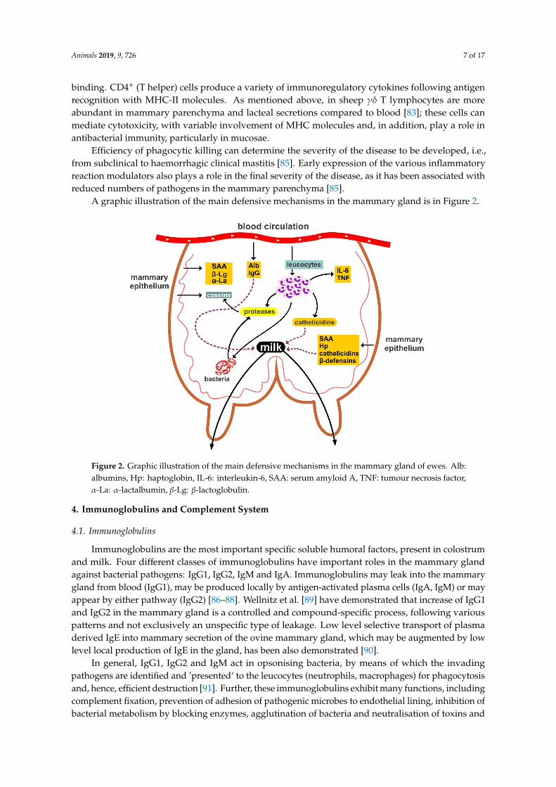
A graphic illustration of the main defensive mechanisms in the mammary gland is in Figure 2.Animals 2019, 9, x 7 of 17
Figure 2. Graphic illustration of the main defensive mechanisms in the mammary gland of ewes. Alb: albumins, Hp: haptoglobin, IL-6: interleukin-6, SAA: serum amyloid A, TNF: tumour necrosis factor, α-La: α-lactalbumin, β-Lg: β-lactoglobulin.
4. Immunoglobulins and Complement System
4.1. Immunoglobulins
Immunoglobulins are the most important specific soluble humoral factors, present in colostrum and milk. Four different classes of immunoglobulins have important roles in the mammary gland against bacterial pathogens: IgG1, IgG2, IgM and IgA. Immunoglobulins may leak into the mammary gland from blood (IgG1), may be produced locally by antigen-activated plasma cells (IgA, IgM) or may appear by either pathway (IgG2) [86–88]. Wellnitz et al. [89] have demonstrated that increase of IgG1 and IgG2 in the mammary gland is a controlled and compound-specific process, following various patterns and not exclusively an unspecific type of leakage. Low level selective transport of plasma derived IgE into mammary secretion of the ovine mammary gland, which may be augmented by low level local production of IgE in the gland, has been also demonstrated [90].
In general, IgG1, IgG2 and IgM act in opsonising bacteria, by means of which the invading pathogens are identified and ’presented‘ to the leucocytes (neutrophils, macrophages) for phagocytosis and, hence, efficient destruction [91]. Further, these immunoglobulins exhibit many functions, including complement fixation, prevention of adhesion of pathogenic microbes to endothelial lining, inhibition of bacterial metabolism by blocking enzymes, agglutination of bacteria and neutralisation of toxins and viruses [92]. IgM antibodies, which are produced in smaller amounts than IgG, are considerably more efficient than IgG in most activities, especially in complement fixation [93]. Sheep colostrum has been found to exhibit higher IgM concentration compared to that of cattle or goats [94]. The significance of opsonisation is easily understood, as without it phagocytosis would be unsystematic and erratic, hence leading to inefficient defence. Nevertheless, it is noted that in milk bacteria are not opsonised by antibodies for subsequent phagocytosis as efficiently as in blood. Finally, IgA acts in bacterial agglutination, which contributes in limiting bacterial dissemination and colonisation.
4.2. Complement System
Figure 2. Graphic illustration of the main defensive mechanisms in the mammary gland of ewes. Alb:albumins, Hp: haptoglobin, IL-6: interleukin-6, SAA: serum amyloid A, TNF: tumour necrosis factor,α-La: α-lactalbumin, β-Lg: β-lactoglobulin.
4. Immunoglobulins and Complement System
4.1. Immunoglobulins
Immunoglobulins are the most important specific soluble humoral factors, present in colostrumand milk. Four different classes of immunoglobulins have important roles in the mammary glandagainst bacterial pathogens: IgG1, IgG2, IgM and IgA. Immunoglobulins may leak into the mammarygland from blood (IgG1), may be produced locally by antigen-activated plasma cells (IgA, IgM) or mayappear by either pathway (IgG2) [86–88]. Wellnitz et al. [89] have demonstrated that increase of IgG1and IgG2 in the mammary gland is a controlled and compound-specific process, following variouspatterns and not exclusively an unspecific type of leakage. Low level selective transport of plasmaderived IgE into mammary secretion of the ovine mammary gland, which may be augmented by lowlevel local production of IgE in the gland, has been also demonstrated [90].
In general, IgG1, IgG2 and IgM act in opsonising bacteria, by means of which the invadingpathogens are identified and ’presented‘ to the leucocytes (neutrophils, macrophages) for phagocytosisand, hence, efficient destruction [91]. Further, these immunoglobulins exhibit many functions, includingcomplement fixation, prevention of adhesion of pathogenic microbes to endothelial lining, inhibition ofbacterial metabolism by blocking enzymes, agglutination of bacteria and neutralisation of toxins and
Animals 2019, 9, 726 8 of 17
viruses [92]. IgM antibodies, which are produced in smaller amounts than IgG, are considerably moreefficient than IgG in most activities, especially in complement fixation [93]. Sheep colostrum has beenfound to exhibit higher IgM concentration compared to that of cattle or goats [94]. The significanceof opsonisation is easily understood, as without it phagocytosis would be unsystematic and erratic,hence leading to inefficient defence. Nevertheless, it is noted that in milk bacteria are not opsonisedby antibodies for subsequent phagocytosis as efficiently as in blood. Finally, IgA acts in bacterialagglutination, which contributes in limiting bacterial dissemination and colonisation.
4.2. Complement System
Complement is a collection of proteins produced mainly by the liver, as well as by macrophages andmonocytes; for component 3 (C3), a local synthesis in the mammary gland has also been suggested [95].The complement effector molecules, circulating in serum and interstitial fluids, exist largely in precursorstates that are activated rapidly in a proteolytic and cascade-like fashion, following recognition ofpathogen-associated molecular patterns and/or noxious self-derived danger-associated molecularpatterns [96]. Complement can be activated systemically in the blood, via three main routes: (i) theclassical pathway, through which uncoated or immunoglobulin-coated antigens are recognised, (ii) thelectin pathway, triggered by the recognition of microbial carbohydrates through mannose bindinglectin, collectins or ficolins followed by activation of the mannose-binding lectin-associated serineproteases and (iii) the alternative complement pathway (also termed the amplification pathway, as itperpetuates complement activation initiated by the classical and/or lectin pathways), characterised bytonic low-level C3 hydrolysis to C3(H2O) [96].
In the mammary gland, the system participates in the immune defence, being involved in evokingand controlling the inflammatory process, in participating in bacterial opsonisation and presentation,in recruiting leucocytes and even in direct killing of pathogens [97,98]. In healthy mammary glands,the complement system is activated only through the alternative complement pathway; the classicalpathway is not functional, as C1q component is absent or present in smaller concentrations than inblood [95]. During inflammation, complement dependent bactericidal and opsonic activities increasein blood [28,99] (Figure S5). Pathogens opsonised by IgG or complement have increased affinity tophagocytose Fc or complement C3b receptors, respectively, promoting phagocyte–bacteria attachmentand bacterial phagocytosis. Yet, in the mammary gland, the alternative complement pathway is notas efficient as in other tissues. In cows, neutrophils within the mammary gland have been found to beless efficient than in blood in the phagocytosis of even opsonised bacteria [95].
Attachment of bacteria to phagocytes is necessary for phagocytosis and, subsequently, intracellularmicrobiocidal function. Nevertheless, some pathogens, including staphylococci, are able to, sometimes,survive within the leucocytes. These bacteria may be killed by phagocytes activated with lymphokines;these are produced by sensitised lymphocytes challenged with specific antigen and induce rapidattraction of leucocytes to lesions. S. aureus strain-specific pathogenicity underscores the importance ofpathogen factors in progression of infectious process [100].
5. Chemical Antibacterial Agents
Other components contributing to humoral mammary defence include lactoferrin, lysozyme andthe lactoperoxidase/myeloperoxidase systems. These participate in immunity modulation, by playinga role in bacteria opsonisation and neutrophil phagocytic activity and performing transcriptionalactivation of various molecules. Other biochemical components therein, which are further partsof the inflammatory response, include oligosaccharides, gangliosides, reactive oxygen species,acute phase proteins (e.g., haptoglobin and serum amyloid A), ribonucleases and a wide rangeof antimicrobial peptides.
Animals 2019, 9, 726 9 of 17
5.1. Lactoferrin
Lactoferrin is an iron-binding glycoprotein, mainly produced by the mammary epithelial cells andin smaller quantities by neutrophils [101,102]. Its expression in milk (Figure S6) is inversely related toalveolar development. The antib acterial effect of lactoferrin is expressed mainly in the epitheliumlining of the ducts and cisterns, but not at the proximal end of the teat canal [103]. It is greatly enhancedin increased bicarbonate ion concentrations and reduced concentrations of the lactoferrin inhibitor,citrate ions, which are present during the dry period. Lactoferrin exerts a bacteriostatic effect mainlyby competing with bacteria for available iron or by binding to bacterial surfaces [104]; hence, it isof particular significance against Gram-negative bacteria (e.g., M. haemolytica, a primary cause ofovine mastitis [52]). Its function includes alteration of integrity and permeability of bacterial cellwall and sensitisation of bacteria to antimicrobial agents. Also, it contributes to bacterial killing andpromotes adhesion and aggregation of neutrophils to the endothelial surface, as well as being involvedin activation of the complement system via the alternative pathway and in antigen-processing by cellsof the reticuloendothelial system and in antibody production [4]. Mean lactoferrin concentrations ininfected ovine mammary glands have been shown to be up to 4.8 times higher than those in healthyones, demonstrating its role in the natural defence mechanisms against mammary gland inflammationin sheep [105]. Moreover, recent work performed in ewes, has indicated that presence of lactoferringenotype AA was associated with lower prevalence of subclinical mastitis [106].
5.2. Lysozyme
Lysozyme derives from blood or is synthesised in the mammary gland from leucocytes duringintramammary infections and has inhibitory or lytic activity mainly against Gram-positive bacteria [4].It is noteworthy that lysozyme exerts its antibacterial action in synergy with antibodies, complementand lactoferrin, increasing susceptibility of bacteria to the various defence mechanisms and functions.In ewes, the potential protective role of lysozyme for the mammary gland has been indicated ina study, in which its concentration was found to be higher in uninfected mammary glands thanin glands with mastitis [58]. Further, lysozyme has been found to contribute in the regulation ofthe inflammatory response and in the immune homeostasis on the epithelial surfaces by activatingregulatory T lymphocytes [107,108].
5.3. Lactoperoxidase
Lactoperoxidase is locally synthesised in the mammary gland, in the presence of thiocyanate ofhepatic origin and hydrogen peroxide of bacterial or endogenous origins [109]. It exerts its antibacterialactivity through the formation of activated oxygen products, e.g., hypothiocyanate, a metaboliteenhancing bactericidal activity of leucocytes. Under in vitro conditions, the system has been foundwith significant antibacterial properties, which, in clinical situations, show similar efficacy only duringthe dry-period; its role during the lactation period is limited, likely because of interference by othermilk proteins (Figure S6). Possibly also, concentrations of thiocyanate ions in milk would dependon nutritional regime and small oxygen tension in the mammary gland might act as an inhibitor forproduction of hydrogen peroxide; these two factors may further account for the limited efficacy of thesystem against mastitis causing pathogens in lactating animals.
6. Modulation of Mammary Defences
6.1. Factors Affecting Mammary Defences
Various factors that may affect mammary defences are relevant to various facets of mammarydefences and immunity and act to enhance or hinder immunity at different levels. Therefore, theyare of importance in the design of mastitis prevention schemes, as well as in the investigation of flockmastitis problems.
Animals 2019, 9, 726 10 of 17
Chapped teats, caused by physical causes (e.g., low temperatures) or chemical agents (e.g.,disinfectants), predispose ewes to mastitis, as the result of bacterial accumulation on teat skin andcompromised antibacterial properties [110–113]. Further, efficient function of the inducible lymphoidnodules at the teat duct can be compromised by physical (e.g., chaps) or microbial (e.g., viral infections)disorders of teats, which thus predispose animals to mastitis [114,115]. Hence, maintenance of teathealth should be an integral part of all mastitis prevention programs in ewes. Moreover, factors relatedto teat morphology can also play a role in development of mammary infections, e.g., short or wideteats can facilitate bacterial entrance into the mammary parenchyma.
Genetic differences in the susceptibility of ewes to mastitis have been reported; indeed, mastitis isconsidered a disease amenable for genetic studies. Differences in the susceptibility to mastitis havebeen associated with particular breeds of sheep [116], as well as with somatic cell counts [56,117].Possibly, polymorphism of MHC genes, which has been detected in sheep [84], may be related tosusceptibility of mastitis, although there is only limited evidence about that. In any case, selection formastitis resistance based on decreased somatic cell counts has been shown to be of value in selectionof individuals with reduced susceptibility to mastitis [118–120]. A recent study has indicated thatgenes differentially expressed in mammary tissues of ewes with clinical mastitis, were involved withregulation of the immune response and the inflammation procedure in these animals [121]; this findingdirectly associates genetic background of ewes with mammary defences.
Nutrition can play a role in regulation of mammary defences and immunity. Energy has beenrecognised as an important factor in promoting phagocytosis and intracellular killing of bacteria byleucocytes; in this context, it has been found that ewes with pregnancy toxaemia were at increased riskof developing mastitis in the immediately post-partum period [122]. Selenium is possibly the moststudied nutrient with regards to immunoregulatory effect [123]. In brief, selenium is a component ofglutathione peroxidase, which can play a protective role against reactive oxygen species damagingvarious cellular components within leucocytes and thus hindering intracellular killing, thereforereduced selenium intake can lead to impaired leucocytic function. Within this frame, the practice ofselenium administration in pregnant ewes at the final stage of gestation, which is aimed to preventselenium deficiency in newborn lambs, may also have an effect in improving post-partum mammarydefences of ewes as well; however, there is no direct evidence for that, but only indirect results [123].To some extent, vitamin E has similar biological properties with selenium and is implicated in at leastthe same pathways as selenium. Zinc is a component of teat keratin and skin; zinc deficiencies canadversely affect the integrity of the teat duct and thus facilitate bacterial entrance. Vitamin A deficiencyhas also been suggested to lead in increased risk of mastitis in ewes, as it may compromise integrity ofepithelia and hence lead to impaired mammary defence [123,124]. Therefore, it becomes evident thatincorrect nutrition may adversely affect animal defences and hence conclude in increased mastitis risk.
6.2. Immunisation
Investigation of immunological responses in the mammary gland of ewes against causalorganisms and clarification of the role of the various immunological components supports attempts toprotect ewes against mastitis, by enhancing immunity and potentiating responses to treatment withantibiotics. For example, it has been shown that local (mammary) administration of immunogens (e.g.,inactivated S. aureus) in non-lactating ewes enhanced kinetics of neutrophil influx with no involvementof complement in the immunological response. The basis of vaccination is the enhancement ofacquired/specific immunity. Vaccination aims to recognise specific determinants of a pathogen thatactivate a selective response leading to bacterial elimination [125].
Vaccines licenced against ovine mastitis aim mainly to protect animals against staphylococcalmastitis. Most currently licenced traditionally anti-staphylococcal vaccines are preparations of wholecell cultures of the vaccinal strain. These are of older technology and suboptimal efficacy. With regardto adjuvants used in these vaccines, it has been found that deposition of milk complement componentson the bacterial surface does not contribute to the defence of the mammary gland. In general,
Animals 2019, 9, 726 11 of 17
administration of these vaccines would lead mainly in reducing the severity of clinical signs of affectedanimals, rather than decreasing the incidence of risk of the disease. More recently, biofilm matrixpolysaccharides have been used for development of protective immune response against S. aureusmastitis in ewes [126]. This approach employs cell-free surface polysaccharide in various vehicles,bacterial unbound cells or bacterial cells embedded in their biofilm matrix in various adjuvants.Vaccination with whole bacterial cells surrounded by matrix with polysaccharide conferred immunityagainst S. aureus mammary infection and mastitis. Nowadays, there is also evidence that this approachalso confers immunity against coagulase-negative staphylococcal species, which are the primary causeof subclinical mastitis [127].
Further, it has been recently reported that intramammary administration of a vaccine alreadylicenced against respiratory infections might also offer a protective effect against M. haemolyticamastitis [128]. This approach contributed to protection of ewes after experimental challenge throughthe induction of a Th17 type response; the protective effect was evident at seven but not at 14 dayspost-challenge. If appropriately improved, such a vaccine may contribute to protecting ewes againstM. haemolytica, the second most-important causal agent of the disease [52].
7. Conclusions
It becomes evident that the defence mechanisms of the mammary gland are complex and actat various levels. Various defences are in place and are applied through many pathways, which aregreatly interdependent. The mechanisms can be enhanced or hindered through health management(including immunisation), which thus becomes of significant importance in mastitis prevention.
Supplementary Materials: The following are available online at http://www.mdpi.com/2076-2615/9/10/726/s1,Figure S1: (a) Inducible lymphoid nodule, present at the border between teat duct and teat cistern, with presenceof lymphocytes (Haematoxylin and eosin [H and E] stain) (Mavrogianni, personal collection); (b) induciblelymphoid nodule, present at the border between teat duct and teat cistern, with presence of T lymphocytes (CD3+
[cluster of differentiation]) (immunohistochemical stain) (Fragkou, personal collection); (c) inducible lymphoidnodule, present at the border between teat duct and teat cistern (H and E stain) (Fragkou, personal collection); (d)inducible lymphoid nodule, present at the border between teat duct and teat cistern (immunohistochemical stain)(Fragkou, personal collection). Figure S2: (a) Presence of neutrophils in milk during acute stage of mammary23 infection (Giemsa stain) (Mavrogianni, personal collection); (b) presence of neutrophils in mammary tissueduring acute stage of mammary infection (H and E stain) (Fthenakis, personal collection). Figure S3: Presenceof clots within the teat cistern of ewes during mastitis, as a consequence of cell accumulation therein, detectedultrasonographically (longitudinal section, image taken and processed on a MyLab® 30 ultrasonography system(ESAOTE SpA, Italy]) with linear transducer, imaging frequency: 12.0 MHz, scanning depth: 30 mm) (Barbagianni,personal collection). Figure S4: (a) Presence of lymphocytes in mammary tissue during chronic stage of mammaryinfection (H and E stain) (Fthenakis, personal collection); (b) presence of lymphocytes in teat during chronic stageof mammary infection (immunohistochemical stain) (Fragkou, personal collection). Figure S5: Identification ofcomplement proteins: complement C3 (CO3) and complement factor B (CFAB) spots on a two-dimensional agarosegel from blood of a ewe with mastitis (protein identification by MALDI-TOF MS) (Katsafadou, personal collection).Figure S6: Identification of lactoferrin (TRFL) and lactoperoxidase (PERL) spots on a two-dimensional agarose gelfrom the milk of a ewe with mastitis (protein identification by MALDI-TOF MS) (Katsafadou, personal collection).
Author Contributions: Conceptualization, A.I.K. and I.A.F.; search of literature, A.I.K., A.P.P. and N.G.C.V.;writing—original draft preparation, A.I.K. and A.P.P.; writing—specific passages and provision of originalphotographs, A.I.K., V.S.M., M.S.B., N.G.C.V., G.C.F., I.A.F.; writing—review and editing, A.I.K., G.C.F. and I.A.F.;correspondence with journal, G.C.F.; supervision, I.A.F.
Funding: This work received no external funding.
Conflicts of Interest: The authors declare no conflict of interest.
References
1. Rainard, P.; Riollet, C. Innate immunity of the bovine mammary gland. Vet. Res. 2006, 37, 369–400. [CrossRef][PubMed]
2. Nickerson, S.C. Mammary resistance mechanisms/anatomical. In Encyclopedia of Dairy Sciences; Roginski, H.,Fuquay, J.W., Fox, P.F., Eds.; Academic Press: London, UK, 2002; pp. 1697–1701.
Animals 2019, 9, 726 12 of 17
3. Mavrogianni, V.S.; Fthenakis, G.C.; Brooks, H.; Papaioannou, N.; Cripps, P.J.; Taitzoglou, I.; Brellou, G.;Saratsis, P. The effects of inoculation of Mannheimia haemolytica into the teat of lactating ewes. Vet. Res. 2005,36, 13–25. [CrossRef] [PubMed]
4. Alnakip, M.E.; Quintela-Baluja, M.; Böhme, K.; Fernández-No, I.; Caamaño-Antelo, S.; Calo-Mata, P.;Barros-Velázquez, J. The immunology of mammary gland of dairy ruminants between healthy andinflammatory conditions. J. Vet. Med. 2014, 31, 659801. [CrossRef] [PubMed]
5. Peeler, E.J.; Green, M.J.; Fitzpatrick, J.L.; Morgan, K.L.; Green, L.E. Risk factors associated with clinicalmastitis in low somatic cell count British dairy herds. J. Dairy Sci. 2000, 83, 2464–2472. [CrossRef]
6. DeVries, T.J.; Dufour, S.; Scholl, D.T. Relationship between feeding strategy, lying behavior patterns, andincidence of intramammary infection in dairy cows. J. Dairy Sci. 2010, 93, 1987–1997. [CrossRef]
7. McDonald, J. Radiographic method for anatomic study of the teat canal: Changes between milking periods.Am. J. Vet. Res. 1975, 36, 1241–1242. [PubMed]
8. Schultze, W.D.; Bright, S.C. Changes in penetrability of bovine papillary duct to endotoxin after milking.Am. J. Vet. Res. 1983, 44, 2373–2375. [PubMed]
9. Alejandro, M.; Roca, A.; Romero, G.; Díaz, J.R. How does the milk removal method affect teat tissue and teatrecovery in dairy ewes? J. Dairy Res. 2014, 81, 350–357. [CrossRef]
10. Fragkou, I.A.; Dagleish, M.P.; Papaioannou, N.; Cripps, P.J.; Boscos, C.M.; Ververidis, H.N.; Orfanou, D.C.;Solomakos, N.; Finlayson, J.; Govaris, A.; et al. The induction of lymphoid follicle-like structures in the ovineteat duct following experimental infection with Mannheimia haemolytica. Vet. J. 2010, 184, 194–200. [CrossRef]
11. Ferreras-Estrada, M.C.; Campo, R.; González-Lanza, C.; Pérez, V.; García-Marín, J.F.; Manga-González, M.Y.Immunohistochemical study of the local immune response in lambs experimentally infected with Dicrocoeliumdendriticum (Digenea). Parasitol. Res. 2007, 101, 547–555. [CrossRef]
12. Fragkou, I.A.; Mavrogianni, V.S.; Papaioannou, N.; Boscos, C.; Cripps, P.J.; Skoufos, J.; Fthenakis, G.C.Presence of sub-epithelial lymphoid tissues in the teat of ewe-lambs and adult ewes. Small Rumin. Res. 2007,70, 286–291. [CrossRef]
13. Rainard, P. Mammary microbiota of dairy ruminants: Fact or fiction? Vet. Res. 2017, 48, 25. [CrossRef][PubMed]
14. Vasileiou, N.G.C.; Cripps, P.J.; Ioannidi, K.S.; Chatzopoulos, D.C.; Gougoulis, D.A.; Sarrou, S.; Orfanou, D.C.;Politis, A.; Calvo Gonzalez-Valerio, T.; Argyros, S.; et al. Extensive countrywide field investigation ofsubclinical mastitis in sheep in Greece. J. Dairy Sci. 2018, 101, 7297–7310. [CrossRef] [PubMed]
15. Verhoeven, P.O.; Gagnaire, J.; Botelho-Nevers, E.; Grattard, F.; Carricajo, A.; Lucht, F.; Pozzetto, B.; Berthelot, P.Detection and clinical relevance of Staphylococcus aureus nasal carriage: An update. Exp. Rev. Anti-Infect.Ther. 2014, 12, 75–89. [CrossRef] [PubMed]
16. Vasileiou, N.G.C.; Chatzopoulos, D.C.; Sarrou, S.; Fragkou, A.I.; Katsafadou, A.I.; Mavrogianni, V.S.;Petinaki, E.; Fthenakis, G.C. Role of staphylococci in mastitis in sheep. J. Dairy Res. 2019, 86, 254–266.[CrossRef]
17. Fragkou, I.A.; Mavrogianni, V.S.; Cripps, P.J.; Gougoulis, D.A.; Fthenakis, G.C. The bacterial flora in the teatduct of ewes can protect against and can cause mastitis. Vet. Res. 2007, 38, 525–545. [CrossRef] [PubMed]
18. Brook, I. The role of bacterial interference in otitis, sinusitis and tonsillitis. Otolaryngol. Head Neck Surg. 2005,133, 139–146. [CrossRef]
19. Melchior, M.B.; Vaarkamp, H.; Fink-Gremmels, J. Biofilms: A role in recurrent mastitis infections? Vet. J.2006, 171, 398–407. [CrossRef]
20. Vilela, C.L.; Fitzpatrick, J.; Morgan, K.L. In vitro adherence and invasion of ovine mammary epithelium byMannheimia haemolytica. Vet. J. 2004, 167, 211–213. [CrossRef]
21. Isenberg, H.D.; D’Amato, R.F. Indigenous and pathogenic microorganisms of humans. In Manual of ClinicalMicrobiology; Lennette, E.H., Balows, H., Hausler, W.J., Jr., Shadomy, H.J., Eds.; American Society forMicrobiology: Washington, DC, USA, 1985; pp. 24–35.
22. Smith, H. The revival of interest in mechanisms of bacterial pathogenicity. Biol. Rev. 1995, 70, 277–316.[CrossRef]
23. De Vliegher, S.; Opsomer, G.; Vanrolleghem, A.; Devriese, L.A.; Sampimon, O.C.; Sol, J.; Barkema, H.W.;Haesebrouck, F.; de Kruif, A. In vitro growth inhibition of major mastitis pathogens by Staphylococcuschromogenes originating from teat apices of dairy heifers. Vet. Microbiol. 2004, 101, 215–221. [CrossRef][PubMed]
Animals 2019, 9, 726 13 of 17
24. Rainard, P.; Poutrel, B. Effect of naturally occurring intramammary infections by minor pathogens on newinfections by major pathogens in cattle. Am. J. Vet. Res. 1988, 49, 327–329. [PubMed]
25. Targowski, S.P. Role of immune factors in protection of mammary gland. J. Dairy Sci. 1983, 6, 1781–1789.[CrossRef]
26. Günther, J.; Seyfert, H.M. The first line of defence: Insights into mechanisms and relevance of phagocytosisin epithelial cells. Semin. Immunopathol. 2018, 40, 555–565. [CrossRef] [PubMed]
27. Petzl, W.; Zerbe, H.; Günther, J.; Yang, W.; Seyfert, H.M.; Nürnberg, G.; Schuberth, H.J. Escherichia coli, but notStaphylococcus aureus triggers an early increased expression of factors contributing to the innate immunedefense in the udder of the cow. Vet. Res. 2008, 39, 18. [CrossRef] [PubMed]
28. Pauwels, A.M.; Trost, M.; Beyaert, R.; Hoffmann, E. Patterns, receptors, and signals: Regulation of phagosomematuration. Trends Immunol. 2017, 38, 407–422. [CrossRef] [PubMed]
29. Katsafadou, A.I. Proteomic Study of Ovine Mastitis associated with Mannheimia haemolytica. Ph.D. Thesis,University of Thessaly, Karditsa, Greece, 2017.
30. Gao, J.; Li, T.; Lu, Z.; Wang, X.; Zhao, X.; Ma, Y. Proteomic analyses of mammary glands provide insight intothe immunity and metabolism pathways associated with clinical mastitis in meat sheep. Animals 2019, 9, 309.[CrossRef]
31. Katsafadou, A.I.; Tsangaris, G.T.; Anagnostopoulos, A.K.; Billinis, C.; Barbagianni, M.S.; Vasileiou, N.G.C.;Spanos, S.A.; Mavrogianni, V.S.; Fthenakis, G.C. Differential quantitative proteomics study of experimentalMannheimia haemolytica mastitis in sheep. J. Proteom. 2019, 205, 103393. [CrossRef]
32. Bauer, I.; Günther, J.; Wheeler, T.T.; Engelmann, S.; Seyfert, H.M. Extracellular milieu grossly alterspathogen-specific immune response of mammary epithelial cells. BMC Vet. Res. 2015, 11, 172. [CrossRef]
33. Günther, J.; Czabanska, A.; Bauer, I.; Leigh, J.A.; Holst, O.; Seyfert, H.M. Streptococcus uberis strains isolatedfrom the bovine mammary gland evade immune recognition by mammary epithelial cells, but not ofmacrophages. Vet. Res. 2016, 47, 13. [CrossRef]
34. Lahouassa, H.; Moussay, E.; Rainard, P.; Riollet, C. Differential cytokine and chemokine responses of bovinemammary epithelial cells to Staphylococcus aureus and Escherichia coli. Cytokine 2007, 38, 12–21. [CrossRef][PubMed]
35. Swanson, K.M.; Stelwagen, K.; Dobson, J.; Henderson, H.V.; Davis, S.R.; Farr, V.C.; Singh, K. Transcriptomeprofiling of Streptococcus uberis-induced mastitis reveals fundamental differences between immune geneexpression in the mammary gland and in a primary cell culture model. J. Dairy Sci. 2009, 92, 117–129.[CrossRef] [PubMed]
36. Brenaut, P.; Lefevre, L.; Rau, A.; Laloe, D.; Pisoni, G.; Moroni, P.; Bevilacqua, C.; Martin, P. Contribution ofmammary epithelial cells to the immune response during early stages of a bacterial infection to Staphylococcusaureus. Vet. Res. 2014, 45, 16. [CrossRef] [PubMed]
37. Eckersall, P.D.; Young, F.J.; Nolan, A.M.; Knight, C.H.; McComb, C.; Waterston, M.M.; Hogarth, C.J.;Scott, E.M.; Fitzpatrick, J.L. Acute phase proteins in bovine milk in an experimental model of Staphylococcusaureus subclinical mastitis. J. Dairy Sci. 2006, 89, 488–501. [CrossRef]
38. Eckersall, P.D.; Young, F.J.; McComb, C.; Hogarth, C.J.; Safi, S.; Weber, A.; McDonald, T.; Nolan, A.M.;Fitzpatrick, J.L. Acute phase proteins in serum and milk from dairy cows with clinical mastitis. Vet. Rec.2001, 148, 35–41. [CrossRef] [PubMed]
39. Smolenski, G.; Haines, S.; Kwan, F.Y.S.; Bond, J.; Farr, V.; Davis, S.R.; Stelwagen, K.; Wheeler, T.T.Characterisation of host defense proteins in milk using a proteomic approach. J. Proteome Res. 2007,6, 207–215. [CrossRef] [PubMed]
40. McDonald, T.L.; Larson, M.A.; Mack, D.R.; Weber, A. Elevated extrahepatic expression and secretion ofmammary associated serum amyloid A 3 (M-SAA3) into colostrum. Vet. Immunol. Immunopathol. 2001, 83,203–211. [CrossRef]
41. Cubeddu, T.; Cacciotto, C.; Pisanu, S.; Tedde, V.; Alberti, A.; Pittau, M.; Dore, S.; Cannas, A.; Uzzau, S.;Rocca, S.; et al. Cathelicidin production and release by mammary epithelial cells during infectious mastitis.Vet. Immunol. Immunopathol. 2017, 89, 66–70. [CrossRef]
42. West, M.A.; Heagy, W. Endotoxin tolerance: A review. Crit. Care Med. 2002, 30, S64–S73. [CrossRef]43. Broad, A.; Jones, D.E.; Kirby, J.A. Toll-like receptor (TLR) response tolerance: A key physiological “damage
limitation” effect and an important potential opportunity for therapy. Curr. Med. Chem. 2006, 13, 2487–2502.[CrossRef]
Animals 2019, 9, 726 14 of 17
44. Hodgson, J.C. Endotoxin and mammalian host responses during experimental disease. J. Comp. Pathol. 2006,135, 157–175. [CrossRef] [PubMed]
45. Petzl, W.; Günther, J.; Pfister, T.; Sauter-Louis, C.; Goetze, L.; von Aulock, S.; Hafner-Marx, A.; Schuberth, H.J.;Seyfert, H.M.; Zerbe, H. Lipopolysaccharide pretreatment of the udder protects against experimentalEscherichia coli mastitis. Innate Immun. 2012, 18, 467–477. [CrossRef]
46. Schukken, Y.H.; Günther, J.; Fitzpatrick, J.; Fontaine, M.C.; Goetze, L.; Holste, O.; Leigh, J.; Petzlg, W.;Schuberth, H.J.; Sipkah, A.; et al. Host-response patterns of intramammary infections in dairy cows.Vet. Immunol. Immunopathol. 2011, 144, 270–289. [CrossRef]
47. Wysocka, M.; Robertson, S.; Riemann, H.; Caamano, J.; Hunter, C.; Mackiewicz, A.; Montaner, L.J.;Trinchieri, G.; Karp, C.L. IL-12 suppression during experimental endotoxin tolerance: Dendritic cell loss andmacrophage hyporesponsiveness. J. Immunol. 2001, 166, 7504–7513. [CrossRef]
48. Günther, J.; Petzl, W.; Zerbe, H.; Schuberth, H.J.; Koczan, D.; Goetze, L.; Seyfert, H.M. Lipopolysaccharidepriming enhances expression of effectors of immune defence while decreasing expression of pro-inflammatorycytokines in mammaryepithelial cells from cows. BMC Genom. 2012, 13, 17. [CrossRef] [PubMed]
49. Breyne, K.; Steenbrugge, J.; Demeyere, K.; Vanden Berghe, T.; Meyer, E. Preconditioning withlipopolysaccharide or lipoteichoic acid protects against Staphylococcus aureus mammary infection in mice.Front. Immunol. 2017, 8, 833. [CrossRef] [PubMed]
50. Petzl, W.; Zerbea, H.; Günther, J.; Seyfert, H.M.; Hussen, J.; Schubert, H.J. Pathogen-specific responses in thebovine udder. Models and immunoprophylactic concepts. Res. Vet. Sci. 2018, 116, 55–61. [CrossRef]
51. Günther, J.; Petzl, W.; Zerbe, H.; Schuberth, H.J.; Seyfert, H.M. TLR ligands, but not modulators of histonemodifiers, can induce the complex immune response pattern of endotoxin tolerance in mammary epithelialcells. Innate Immun. 2017, 23, 155–164. [CrossRef]
52. Gelasakis, A.I.; Mavrogianni, V.S.; Petridis, I.G.; Vasileiou, N.G.C.; Fthenakis, G.C. Mastitis in sheep-The last10 years and the future of research. Vet. Microbiol. 2015, 181, 136–146. [CrossRef]
53. Hughes, K.; Watson, C.J. The mammary microenvironment in mastitis in humans, dairy ruminants, rabbitsand rodents: A One Health focus. J. Mammary Gland Biol. Neopl. 2018, 23, 27–41. [CrossRef]
54. Moroni, P.; Cuccuru, C. Relationship between mammary gland infections and some milk immune parametersin Sardenian ewes. Small Rumin. Res. 2001, 41, 1–7. [CrossRef]
55. Bergonier, D.; Berthelot, X. New advances in epizootiology and control of ewe mastitis. Liv. Prod. Sci. 2003,79, 1–16. [CrossRef]
56. Berthelot, X.; Lagriffoul, G.; Concordet, D.; Barillet, F.; Bergonier, D. Physiological and pathological thresholdsof somatic cell counts in ewe milk. Small Rumin. Res. 2006, 62, 27–31. [CrossRef]
57. Albenzio, M.; Santillo, A.; Caroprese, M.; Ruggieri, D.; Ciliberti, M.; Sevi, A. Immune competence of themammary gland as affected by somatic cell and pathogenic bacteria in ewes with subclinical mastitis. J. DairySci. 2012, 95, 3877–3887. [CrossRef] [PubMed]
58. Souza, F.N.; Blagitz, M.G.; Penna, F.A.M.; Della Libera, A.M.M.P.; Heinemann, M.B.; Cerqueira, M.M.O.P.Somatic cell count in small ruminants: Friend or foe? Small Rumin. Res. 2012, 107, 65–75. [CrossRef]
59. Cuccuru, C.; Moroni, P.; Zecconi, A.; Casu, S.; Caria, A.; Contini, A. Milk differential cell counts in relation tototal counts in Sardinian ewes. Small Rumin. Res. 1997, 25, 169–173. [CrossRef]
60. Boutinaud, M.; Jammes, H. Potential uses of milk epithelial cells: A review. Reprod. Nutr. Dev. 2002, 42,133–147. [CrossRef] [PubMed]
61. Paape, M.J.; Wiggans, G.R.; Bannerman, D.D.; Thomas, D.L.; Sanders, A.H.; Contreras, A.; Moroni, P.;Miller, R.H. Monitoring goat and sheep milk somatic cell counts. J. Dairy Sci. 2007, 68, 114–125. [CrossRef]
62. Albenzio, M.; Figliola, L.; Caroprese, M.; Marino, R.; Sevi, A.; Santillo, A. Somatic cell count in sheep milk.Small Rumin. Res. 2019, 176, 24–30. [CrossRef]
63. Paape, M.J.; Poutrel, B.; Contreras, A.; Marco, J.C.; Capuco, A.V. Milk somatic cells and lactation in smallruminants. J. Dairy Sci. 2001, 84, E237–E244. [CrossRef]
64. Leitner, G.; Merin, U.; Krifucks, O.; Blum, S.; Rivas, A.; Silanikove, N. Effects of intramammary bacterialinfection with coagulase negative staphylococci and stage of lactation on shedding of epithelial cells andinfiltration of leukocytes into milk: Comparison among cows, goats and sheep. Vet. Immunol. Immunopathol.2012, 147, 202–210. [CrossRef] [PubMed]
65. El-Masannat, E.T.S. A Study of Ovine Mastitis with Special Reference to Mastitis caused by Pasteurellahaemolytica. Ph.D. Thesis, University of London, London, UK, 1987.
Animals 2019, 9, 726 15 of 17
66. Fthenakis, G. Ovine Mastitis with Special Reference to Subclinical Mastitis associated with Coagulase-NegativeStaphylococci. Ph.D. Thesis, University of London, London, UK, 1988.
67. Cheville, N.F. Ultrastructural Pathology: The Comparative Cellular Basis of Disease, 2nd ed.; Wiley-Blackwell:Ames, IA, USA, 2009; p. 1000.
68. Persson, W.; Bengtsson, K.B.; Lindberg, A.; Nyman, A.; Unnerstad, H.E. Incidence of mastitis and bacterialfindings at clinical mastitis in Swedish primiparous cows. Influence of breed and stage of lactation.Vet. Microbiol. 2009, 134, 89–94. [CrossRef] [PubMed]
69. Persson Waller, K.; Colditz, I.G.; Flapper, P.; Seow, H.F. Leukocyte and cytokine accumulation in the ovineteat and udder during endotoxin-induced inflammation. Vet. Res. Comm. 1997, 21, 101–115. [CrossRef][PubMed]
70. Barbagianni, M.S. Experimental Study of Pregnancy Toxaemia in Ewes and its Association with Mastitis inthe; Post-Partum Period. Ph.D. Thesis, University of Thessaly, Karditsa, Greece, 2017.
71. Barbagianni, M.S.; Mavrogianni, V.S.; Vasileiou, N.G.C.; Fthenakis, G.C.; Petridis, I.G. Ultrasonographicexamination of the udder in sheep. Small Rumin. Res. 2017, 152, 86–99. [CrossRef]
72. Hodgson, J.C.; Moon, G.M.; Quirie, M.; Donachie, W. Association of LPS chemotype of Mannheimia (Pasteurella)haemolytica A1 with disease virulence in a model of ovine pneumonic pasteurellosis. J. Endotoxin Res. 2003, 9,25–32. [CrossRef] [PubMed]
73. Addis, M.F.; Pisanu, S.; Marogna, G.; Cubeddu, T.; Pagnozzi, D.; Cacciotto, C.; Campesi, F.; Schianchi, G.;Rocca, S.; Uzzau, S. Defense proteins by mammary epithelial cells following Streptococcus uberis infection insheep. Inf. Immun. 2013, 81, 3182–3197. [CrossRef] [PubMed]
74. Winter, P.; Colditz, I.G. Immunological responses of the lactating ovine udder following experimentalchallenge with Staphylococcus epidermidis. Vet. Immunol. Immunopathol. 2002, 89, 57–65. [CrossRef]
75. Vray, B. Macrophages in parasitic infection. In The Macrophage; Burke, B., Lewis, C.E., Eds.; Oxford UniversityPress: Oxford, UK, 2002; pp. 253–304.
76. De Garnica, M.L.; Linage, B.; Carriedo, J.A.; De La Fuente, L.F.; García-Jimeno, M.C.; Santos, J.A.; Gonzalo, C.Relationship among specific bacterial counts and total bacterial and somatic cell counts and factors influencingtheir variation in ovine bulk tank milk. J. Dairy Res. 2013, 96, 1021–1029. [CrossRef]
77. Fthenakis, G.C. California Mastitis Test and Whiteside Test in diagnosis of subclinical mastitis of dairy ewes.Small Rumin. Res. 1995, 16, 271–276. [CrossRef]
78. Fragkou, I.A.; Boscos, C.M.; Fthenakis, G.C. Diagnosis of clinical or subclinical mastitis in ewes. Small Rumin.Res. 2014, 118, 86–92. [CrossRef]
79. Van Oostveld, K.; Paape, M.J.; Dosogne, H.; Burvenich, C. Effect of apoptosis on phagocytosis, respiratoryburst and CD18 adhesion receptor expression of bovine neutrophil. Dom. Anim. Endocrinol. 2002, 22, 37–50.[CrossRef]
80. Van Harten, R.M.; van Woudenbergh, E.; van Dijk, A.; Haagsman, H.P. Cathelicidins: Immunomodulatoryantimicrobials. Vaccines 2018, 6, 63. [CrossRef] [PubMed]
81. Sladek, Z.; Rysanek, D. Neutrophil apoptosis during the resolution of bovine mammary gland injury. Res. Vet.Sci. 2001, 70, 41–46. [CrossRef] [PubMed]
82. Goldsby, R.A. Cells and organs of the immune system. In Kuby Immunology, 6th ed.; Osborne, B.A.,Goldsby, R.A., Kindt, T.J., Eds.; Freeman: New York, NY, USA, 2006; pp. 40–47.
83. Bonelli, P.; Dimauro, C.; Re, R.; Pilo, G.; Dore, S.; Cannas, A.E.; Nicolussi, P.S. Peripheral blood and milkleukocytes subsets of lactating Sarda ewes. Ital. J. Anim. Sci. 2013, 12, e34. [CrossRef]
84. Swiderek, W.P.; Charon, K.M.; Winnicka, A.; Gruszczynska, J. Relationship between blood lymphocytephenotype, DRB1 MHC class II) gene polymorphism and somatic cell count in ewe milk. Bull. Vet. Inst.Pulaway 2006, 50, 73–77.
85. Rainard, P.; Foucras, G.; Boichard, D.; Rupp, R. Low milk somatic cell count and susceptibility to mastitis.J. Dairy Sci. 2018, 101, 6703–6714. [CrossRef] [PubMed]
86. Craven, N.; Williams, M.R. Defences of the bovine mammary gland against infection and prospects for theirenhancement. Vet. Immunol. Immunopathol. 1985, 10, 71–127. [CrossRef]
87. Norcross, N.L. Specific defence mechanisms of the udder. Flem. Vet. J. 1991, 62, 129–139.88. Oestensson, K.; Lun, S. Transfer of immunoglobulins through the mammary endothelium and epithelium
and in the local lymph node of cows during the initial response after intramammary challenge with E. coliendotoxin. Acta Vet. Scand. 2008, 50, 26. [CrossRef]
Animals 2019, 9, 726 16 of 17
89. Wellnitz, O.; Zbinden, C.; Lüttgenau, J.; Bollwein, H.; Bruckmaier, R.M. Different chronological patterns ofappearance of blood derived milk components during mastitis indicate different mechanisms of transferfrom blood into milk. J. Dairy Res. 2015, 82, 322–327. [CrossRef]
90. Hine, B.C.; Hunt, P.W.; Beasley, A.M.; Windon, R.G.; Glover, S.A.; Colditz, I.G. Selective transport of IgE intoovine mammary secretions. Res. Vet. Sci. 2010, 89, 184–190. [CrossRef] [PubMed]
91. Barrio, M.B.; Rainard, P.; Poutrel, B. Milk complement and the opsonophagocytosis and killing ofStaphylococcus aureus mastitis isolates by bovine neutrophils. Microb. Pathogen. 2003, 34, 1–9. [CrossRef]
92. Marnila, P.; Korhonen, H. Immunoglobulins. In Encyclopedia of Dairy Sciences; Roginski, H., Fuquay, J.W.,Fox, P.F., Eds.; Academic Press: London, UK, 2002; pp. 1950–1956.
93. Mehra, R.; Marnila, P.; Korhonen, H. Milk immunoglobulins for health promotion. Int. Dairy J. 2006, 16,1262–1271. [CrossRef]
94. Hernández-Castellano, L.E.; Almeida, A.; Renaut, J.; Argüello, A.; Castro, N. A proteomics study of colostrumand milk from the two-major small ruminant dairy breeds from the Canary Islands: A bovine milk comparisonperspective. J. Dairy Res. 2016, 83, 366–374. [CrossRef] [PubMed]
95. Rainard, P. The complement in milk and defense of the bovine mammary gland against infections. Vet. Res.2003, 34, 647–670. [CrossRef] [PubMed]
96. Arbore, G.; Kemper, C.; Kolev, M. Intracellular complement-the complosome-in immune cell regulation. Mol.Immunol. 2017, 89, 2–9. [CrossRef] [PubMed]
97. DiCarlo, A.; Paape, M. Comparison of C3b binding to bovine peripheral blood and mammary glandneutrophils (PMN). Am. J. Vet. Res. 1996, 57, 151–156.
98. Cunnion, K.M.; Lee, J.C.; Frank, M.M. Capsule production and growth phase influence binding of complementto Staphylococcus aureus. Infect. Immun. 2001, 69, 6796–6803. [CrossRef] [PubMed]
99. Katsafadou, A.I.; Tsangaris, G.T.; Billinis, C.; Fthenakis, G.C. Use of proteomics in the study of microbialdiseases of small ruminants. Vet. Microbiol. 2015, 181, 27–33. [CrossRef]
100. Pereyra, E.A.L.; Sacco, S.C.; Dure, A.; Baravalle, C.; Renna, M.S.; Andreotti, C.S.; Carolina, S.; Monecke, S.;Calvinho, L.F.; Dallard, B.E. Immune response of Staphylococcus aureus strains in a mouse mastitis model islinked to adaptive capacity and genotypic profile. Vet. Microbiol. 2017, 204, 64–76. [CrossRef]
101. Pierce, A.; Legrand, D.; Mazurier, J. Lactoferrin: A multifunctional protein. Med. Sci. (Paris) 2009, 25, 361–369.[CrossRef] [PubMed]
102. Shimazaki, K.; Kawai, K. Advances in lactoferrin research concerning bovine mastitis. Bioch. Cell Biol. 2017,95, 69–75. [CrossRef] [PubMed]
103. Isobe, N. Control mechanisms for producing antimicrobial factors in ruminant mammary gland. Anim. Sci. J.2017, 88, 937–943. [CrossRef] [PubMed]
104. Pan, Y.; Rowney, M.; Guo, P.; Hobman, P. Biological properties of lactoferrin: An overview. Aust. J. DairyTechnol. 2007, 62, 31–42.
105. Lemos, V.F.; Guaraná, E.L.S.; Afonso, J.A.B.; Fagliari, J.J.; Silva, P.S.; Soares, P.C.; de Mendonça, C.L. Effects ofintramammary infection on whey proteinograms of sheep during lactation. Pesq. Bras. 2015, 35, 230–236.[CrossRef]
106. Alekish, M.; Ababneh, H.; Ismail, Z.; Alshehabat, M. The relationship between lactoferrin gene polymorphismand subclinical mastitis in Awassi ewes. J. Anim. Plant. Sci. 2019, 29, 1193–1197.
107. Liu, H.; Zheng, F.; Cao, Q.; Ren, B.; Zhu, L.; Striker, G.; Vlassara, H. Amelioration of oxidant stress by thedefensin lysozyme. Am. J. Physiol. Endocrinol. Metab. 2006, 290, E824–E832. [CrossRef] [PubMed]
108. Lee, M.; Kovacs-Nolan, J.; Chengbo, Y.; Archbold, T.; Fan, M.Z.; Mine, Y. Hen egg lysozyme attenuatesinflammation and modulates local gene expression in a porcine model of dextran sodium sulfate(DSS)-induced colitis. J. Agric. Food Chem. 2009, 57, 62233–62240. [CrossRef]
109. Fox, P.F.; Kelly, A.L. Indigenous enzymes in milk: Overview and historical aspects-part 1. Int. Dairy J. 2006,16, 500–516. [CrossRef]
110. Noble, W.C.; Somerville, D.A. Skin as a habitat. In Microbiology of Human Skin; Noble, W.C., Ed.; Saunders:London, UK, 1974; pp. 3–78.
111. Sieber, R.L.; Farnsworth, R.J. Differential diagnosis of bovine teat lesions. Vet. Clin. N. Am. Large Anim. Pract.1984, 6, 313–321. [CrossRef]
112. Fox, L.K.; Hancock, D.D. Effect of segregation on prevention of intramammary infections by Staphylococcusaureus. J. Dairy Sci. 1989, 72, 540–544. [CrossRef]
Animals 2019, 9, 726 17 of 17
113. Fox, L.K.; Nagy, J.A.; Hillers, J.K.; Cronrath, J.D.; Ratkowsky, D.A. Effects of post-milking teat treatment onthe colonization of Staphylococcus aureus on chapped teat skin. Am. J. Vet. Res. 1991, 52, 799–802. [PubMed]
114. Mavrogianni, V.S.; Cripps, P.J.; Papaioannou, N.; Taitzoglou, I.; Fthenakis, G.C. Teat disorders predisposeewes to clinical mastitis after challenge with Mannheimia haemolytica. Vet. Res. 2006, 37, 89–105. [CrossRef][PubMed]
115. Fragkou, I.A.; Papaioannou, N.; Cripps, P.J.; Boscos, C.M.; Fthenakis, G.C. Teat lesions predispose to invasionof the ovine mammary gland by Mannheimia haemolytica. J. Comp. Pathol. 2007, 137, 239–244. [CrossRef][PubMed]
116. Fragkou, I.A.; Skoufos, J.; Cripps, P.J.; Kyriazakis, I.; Papaioannou, N.; Boscos, C.M.; Tzora, A.; Fthenakis, G.C.Differences in susceptibility to M. haemolytica-associated mastitis between two breeds of dairy sheep. J. DairyRes. 2007, 74, 349–355. [CrossRef] [PubMed]
117. Baro, J.A.; Carriedo, J.A.; San Primitivo, F. Genetic paramerers of test day measure for somatic cell count,milk yield and protein percentage of milking ewes. J. Dairy Sci. 1994, 77, 2568–2662. [CrossRef]
118. Barillet, F.; Rupp, R.; Mignon-Grasteau, S.; Astruc, J.M.; Jacquin, M. Genetic analysis for mastitis resistanceand milk somatic cell score in French Lacaune dairy sheep. Gen. Sel. Evol. 2001, 33, 397–415. [CrossRef]
119. Bishop, S.C. Genetic resistance to infections in sheep. Vet. Microbiol. 2015, 181, 2–7. [CrossRef] [PubMed]120. Rupp, R.; Senin, P.; Sarry, J.; Allain, C.; Tasca, C.; Ligat, L.; Portes, D.; Woloszyn, F.; Bouchez, O.;
Tabouret, G.; et al. A point mutation in suppressor of cytokine signalling 2 (Socs2) increases the susceptibilityto inflammation of the mammary gland while associated with higher body weight and size and higher milkproduction in a sheep model. PLoS Genet. 2015, 11, e1005629. [CrossRef]
121. Li, T.T.; Gao, J.F.; Zhao, X.X.; Ma, Y.J. Digital gene expression analyses of mammary glands from meat ewesnaturally infected with clinical mastitis. R. Soc. Open Sci. 2019, 6, 181604. [CrossRef]
122. Barbagianni, M.S.; Mavrogianni, V.S.; Katsafadou, A.I.; Spanos, S.A.; Tsioli, V.; Galatos, A.D.; Nakou, M.;Valasi, I.; Gouletsou, P.G.; Fthenakis, G.C. Pregnancy toxaemia as predisposing factor for developmentof mastitis in sheep during the immediately post-partum period. Small Rumin. Res. 2015, 130, 246–251.[CrossRef]
123. Giadinis, N.D.; Panousis, N.; Petridou, E.J.; Siarkou, V.I.; Lafi, S.Q.; Pourliotis, K.; Hatzopoulou, E.;Fthenakis, G.C. Selenium, vitamin E and vitamin A blood concentrations in dairy sheep flocks with increasedor low clinical mastitis incidence. Small Rumin. Res. 2011, 95, 193–196. [CrossRef]
124. Koutsoumpas, A.T.; Giadinis, N.D.; Petridou, E.J.; Konstantinou, E.; Brozos, C.; Lafi, S.Q.; Fthenakis, G.C.;Karatzias, H. Consequences of reduced vitamin A administration on mammary health of dairy ewes. SmallRumin. Res. 2013, 110, 120–123. [CrossRef]
125. Azara, E.; Longheu, C.; Sanna, G.; Tola, S. Biofilm formation and virulence factor analysis of Staphylococcusaureus isolates collected from ovine mastitis. J. Appl. Microbiol. 2017, 123, 372–379. [CrossRef] [PubMed]
126. Perez, M.M.; Prenafeta, A.; Valle, J.; Penades, J.; Rota, C.; Solano, C.; Marco, J.; Grillo, M.J.; Lasa, I.;Irache, J.M.; et al. Protection from Staphylococcus aureus mastitis associated with poly-N-acetyl beta-1,6glucosamine specific antibody production using biofilm-embedded bacteria. Vaccine 2009, 17, 2379–2386.[CrossRef] [PubMed]
127. Vasileiou, N.G.C.; Chatzopoulos, D.C.; Cripps, P.J.; Ioannidi, K.S.; Gougoulis, D.A.; Chouzouris, T.M.;Lianou, D.T.; Calvo Gonzalez-Valerio, T.; Guix Vallverdu, R.; Argyros, S.; et al. Evaluation of efficacyof a biofilm-embedded bacteria-based vaccine against staphylococcal mastitis in sheep-A randomized,placebo-controlled field study. J. Dairy Sci. 2019, 102, 9328–9344. [CrossRef] [PubMed]
128. Ballingall, K.T.; Todd, H.; Tassi, R. Intra-mammary vaccine delivery protects sheep against mastitis causedby mannheimia haemolytica infection. In Proceedings of the Abstract Book of International VeterinaryImmunology Symposium, Seattle, WA, USA, 13–16 August 2019; pp. 71–72.
© 2019 by the authors. Licensee MDPI, Basel, Switzerland. This article is an open accessarticle distributed under the terms and conditions of the Creative Commons Attribution(CC BY) license (http://creativecommons.org/licenses/by/4.0/).
Copyright © 2022 FDOKUMEN